Inconsistent reversal of HIV-1 latency ex ... - Research Square
21
Page 1/21 Inconsistent reversal of HIV-1 latency ex vivo by antigens of HIV-1, CMV, and other infectious agents Thomas Vollbrecht ( [email protected] ) University of California San Diego https://orcid.org/0000-0003-1151-8844 Aaron O. Angerstein University of California San Diego Bryson Menke VA San Diego Healthcare System Nikesh M. Kumar University of California San Diego Michelli Faria Oliveira University of California San Diego Douglas D. Richman University of California San Diego John C Guatelli University of California San Diego Research Keywords: latency, HIV-1, antigen, CD4 T cell Posted Date: October 29th, 2020 DOI: https://doi.org/10.21203/rs.3.rs-35386/v2 License: This work is licensed under a Creative Commons Attribution 4.0 International License. Read Full License Version of Record: A version of this preprint was published at Retrovirology on November 23rd, 2020. See the published version at https://doi.org/10.1186/s12977-020-00545-x.
-
Upload
khangminh22 -
Category
Documents
-
view
1 -
download
0
Transcript of Inconsistent reversal of HIV-1 latency ex ... - Research Square

Page 1/21
Inconsistent reversal of HIV-1 latency ex vivo byantigens ofHIV-1, CMV, and other infectious agentsThomas Vollbrecht ( [email protected] )
University of California San Diego https://orcid.org/0000-0003-1151-8844Aaron O. Angerstein
University of California San DiegoBryson Menke
VA San Diego Healthcare SystemNikesh M. Kumar
University of California San DiegoMichelli Faria Oliveira
University of California San DiegoDouglas D. Richman
University of California San DiegoJohn C Guatelli
University of California San Diego
Research
Keywords: latency, HIV-1, antigen, CD4 T cell
Posted Date: October 29th, 2020
DOI: https://doi.org/10.21203/rs.3.rs-35386/v2
License: This work is licensed under a Creative Commons Attribution 4.0 International License. Read Full License
Version of Record: A version of this preprint was published at Retrovirology on November 23rd, 2020. Seethe published version at https://doi.org/10.1186/s12977-020-00545-x.

Page 2/21
AbstractBackground
A reservoir of replication-competent but latent virus is the main obstacle to a cure for HIV-1 infection.Much of this reservoir resides in memory CD4 T cells. We hypothesized that these cells can be reactivatedwith antigens from HIV-1 and other common pathogens to reverse latency.
Results
We obtained mononuclear cells from the peripheral blood of antiretroviral-treated patients withsuppressed viremia. We tested pools of peptides and proteins derived from HIV-1 and from otherpathogens including CMV for their ability to reverse latency ex vivo by activation of memory responses.We assessed activation of the CD4 T cells by measuring the up-regulation of cell-surface CD69. Weassessed HIV-1 expression using two assays: a real-time PCR assay for virion-associated viral RNA and adroplet digital PCR assay for cell-associated, multiply spliced viral mRNA. Reversal of latency occurred ina minority of cells from some participants, but no single antigen induced HIV-1 expression ex vivoconsistently. When reversal of latency was induced by a speci�c peptide pool or protein, the extent wasproportionally greater than that of T cell activation.
Conclusions
In this group of patients in whom antiretroviral therapy was started during chronic infection, the latentreservoir does not appear to consistently reside in CD4 T cells of a predominant antigen-speci�city.Peptide-antigens reversed HIV-1 latency ex vivo with modest and variable activity. When latency wasreversed by speci�c peptides or proteins, it was proportionally greater than the extent of T cell activation,suggesting partial enrichment of the latent reservoir in cells of speci�c antigen-reactivity.
BackgroundBy preventing cells from becoming newly infected, combined antiretroviral therapy (cART) can suppressthe replication of the human immunode�ciency virus-1 (HIV-1) to nearly undetectable levels. However,latently infected cells persist, are remarkably stable, and once activated yield new infectious virus [1–3].Consequently, interruption of cART is followed by a relapse of viral replication and viremia, whichultimately causes depletion of CD4 T cells and immunode�ciency if left untreated [4].
Latently infected cells form a "viral reservoir" that is established early after infection with replication-competent proviruses integrated into the genomes of CD4 cells, with a substantial proportion in restingmemory CD4 T cells [5–8]. During acute infection, HIV-1 causes immune activation, preferentiallyinfecting activated HIV-1 speci�c CD4 T cells [9]. This suggests the possibility that the viral reservoir isbiased toward such HIV-speci�c memory cells.
Page 3/21
A currently favored approach to eradicate latently infected T cells is to reactivate viral transcription usingsmall molecules as latency reverting agents (LRAs) while maintaining cART [10]. The goal of thisapproach is to activate latent viruses that are integrated into host memory CD4 T cells so that the cellsdie from viral cytopathic effect or are eliminated by immune surveillance. These strategies have so farshown very limited success [11–14]. Agents under investigation as LRAs include various types of proteinkinase C (PKC) activators (for example, bryostatin-1), HDAC inhibitors (for example, panobinostat andromidepsin), and BRD4 inhibitors ( for example, JQ1), among others [15–19]. Most of these LRAs lackspeci�city for the cells that contain the viral reservoir. Consequently, some of them have a potential fortoxicity; for example, agents targeting PKC-signaling might broadly affect cellular metabolism [20,21]. Incontrast, activation of latently infected memory CD4 T cells by their cognate antigens is speci�c.However, the effectiveness of an antigen as a LRA will likely depend on the fraction of the latent reservoirthat resides in cells that respond to it.
Since HIV-1 preferentially infects activated CD4 T cells [5,9], and since the reservoir is partly establishedearly during infection when most of those cells are responding to HIV, many reservoir cells might be HIV-speci�c. Alternatively, if HIV-infection is left untreated while CD4 T cells are responding to either chronicpersistent infections such as those caused by CMV or EBV or recurrent infections such as in�uenza, thenreservoir cells might be speci�c for those pathogens. More generally, if reservoir cells have a limitedbreadth of antigen speci�city, then only a few antigens might be needed to reverse latency in the majorityof cells while activating only a small fraction of the CD4 T cell population.
In this study, we hypothesized that the latent reservoir can be reactivated using antigens to activatespeci�c memory CD4 T cells harboring latent provirus. We evaluated whether peptide antigens speci�c toHIV, as well as to common pathogens such as CMV, could act as LRAs to reactivate the HIV-1 reservoirwithout causing general T cell activation. Support for this hypothesis came from previous studiessuggesting that peptide antigens of HIV-1 induced viral protein expression in CD4 T cells fromparticipants with suppressed viremia [22–24]. Additionally, we tested the hypothesis that the presentationof antigens by dendritic cells optimizes the reversal of latency (reviewed in [25]). We found that in somecases antigenic peptides or proteins reversed latency, but they rarely did so to a substantial extent, and noparticular antigen was active in all cases. When peptides or protein antigens reversed latency, they did soto an extent greater than would be predicted based on their induction of T cell-activation. This suggeststhat in occasional individuals the latent reservoir is disproportionally present in cells of speci�c antigen-reactivity, a �nding potentially consistent with clonal expansion of speci�c memory CD4 T cells.
ResultsStudy participants and cell preparation
We obtained up to 300 ml of peripheral blood from 19 HIV-1 positive participants (one on two occasions);three were ultimately excluded from the data presented, because their cells did not show evidence oflatency reversal after stimulation with antibodies to CD3 and CD28 (the positive T cell activation control).

Page 4/21
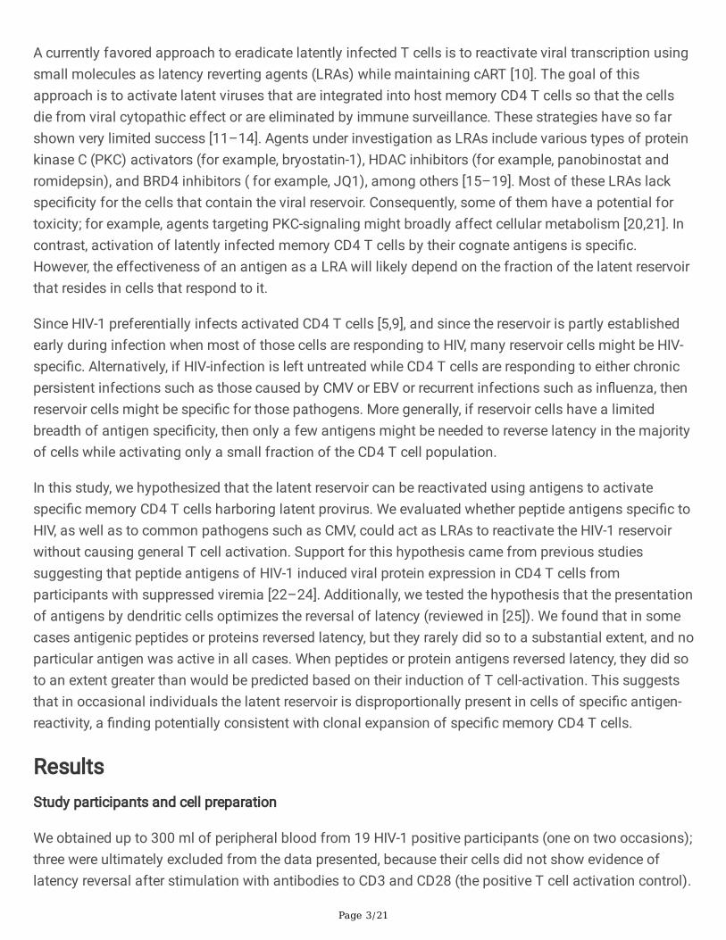
All participants had a CD4 T cell count of at least 500 cells/µl of peripheral blood and fully suppressedviremia (less than 20 copies/ml) for at least one year. For the participants for whom data are available,HIV-1 infection was diagnosed �ve to over twenty-seven years before their blood was obtained for thisstudy (Table 1).
We isolated peripheral blood mononuclear cells (PBMC) by density centrifugation. Constitutiveexpression of MHC class-II molecules is con�ned to professional antigen presenting cells (APCs). Toensure optimal antigen presentation to CD4 T cells in the context of MHC class-II, we kept professionalAPCs - B cells and monocytes - in the cell population by depleting CD8 T cells from the PBMC rather thanisolating the CD4 T cells. Our rationale for removing CD8 T cells was to avoid potential killing ofreactivated reservoir cells as well as the potential secretion of suppressive mediators. CD8 T celldepletion from participant PBMC was con�rmed by �ow cytometry, and residual CD8 T cells were usually< 0.1 % (data not shown).
Identi�cation of antigen speci�c CD4 T cell responses by IFN-γ ELISpot assay
Production of interferon (IFN)-γ by CD4 T cells is a hallmark of the Th1-type CD4 T cell phenotype and istypically associated with an effective host defense against intracellular pathogens (reviewed in [26]).Therefore, we �rst used an IFN‐γ enzyme-linked immune absorbent spot (ELISpot) assay to evaluate theability of peptide pools to activate and induce an immune response in the CD4 T cells within the CD8depleted cell populations.
We screened IFN-γ production in response to peptide pools containing 15mer overlapping peptides from:1) Cytomegalovirus (CMV) matrix protein 65 (pp65); 2) Candida albicans mannoprotein MP65; 3) amixture of 14 previously identi�ed optimal MHC class-II restricted epitopes from human cytomegalovirus(HHV-5), Epstein-Barr virus (HHV-4), in�uenza A, and Clostridium tetani toxoid (pool named CEFT); 4) HIV-1 Gag; 5) HIV-1 Pol; 6) HIV-1 Env; and 7) HIV-1 Nef. For each condition, we stimulated 100,000 CD8depleted PBMC with 1 µg/ml per individual peptide. We used anti-CD3/CD28-coated beads as well asphorbol myristate acetate in combination with ionomycin (PMA/Iono) as a positive control for maximal Tcell stimulation. Both positive controls typically saturated the signals on the ELISpot plate and did notallow quanti�cation (Figure 1A). We used DMSO as a negative control, because the peptide pools andindividual peptides were reconstituted in DMSO. Wells treated with DMSO typically showed <10 spotforming cells (SFC)/million cells. IFN‐γ responses of varying magnitudes were measured for thesepeptide pools in CD8 depleted PBMCs from a subset of participants (Figure 1A). For three participants, wetested the 123 Gag 15mer peptides singly at a concentration of 12.5 µg/ml per peptide with 100,000 CD8depleted PBMC per well to identify the speci�c peptides within the pool that induced IFN-γ production inthe CD4 T cells (Figure 1B). Individual peptides that yielded >50 SFC/million CD8 depleted PBMC weresubsequently pooled to generate a participant-customized ELISpot selected Gag peptide pool used insome of the latency reactivation experiments (Figure 1B; indicated by asterisks). We veri�ed for oneparticipant that the observed IFN-γ production was not a result of contamination with CD8 T cells bycomparing two ELISpot assays, one with CD8 depleted PBMC and one with isolated CD8 T cells. The

Page 5/21
ELISpot assay with the isolated CD8 T cells showed IFN-γ production in response to different peptidescompared to the assay with CD8 depleted PBMC, suggesting that the different epitopes were recognizedin the context of MHC class-I and MHC class‐II (Figure 1C). Overall, these data indicated that with thepossible exception of the peptides derived from Candida MP65, the peptide pools activated IFN-γproduction in CD4 T cells, presumably in an antigen-speci�c manner.
Virion-release from latently infected cells after antigenic stimulation measured by real-time RT-qPCR
For nine participants, we stimulated �ve million CD8 depleted PBMC with each peptide pool inquadruplicate for seven days in the presence of the integrase inhibitor raltegravir [1 µM]. Raltegravirprevented viral spread and ensured that the measured cell-free, virion-associated (cf)-RNA was releaseddirectly from reactivated reservoir cells and not a consequence of propagation of virus in the culture.Depending on the yield of isolated cells from each participant, we stimulated the cells with overlappingpeptide pools from Gag, CMV, Candida, CEFT, Pol, Env, and Nef at a concentration of 1 µg/ml per peptide.DMSO was used as negative control and plate‐bound antibodies against CD3 and CD28 were used as amaximum-stimulation control.
We �rst assessed the activity of the peptide pools with respect to immune activation by measuring theearly activation marker CD69. After two days of culture, an aliquot of ~50,000 cells from each conditionwas stained for cell-surface CD69 and CD4 and analyzed by �ow cytometry. The DMSO controls showedbaseline CD69 positive CD4 T cells ranging from 1% to 12%, whereas the plate‐bound anti-CD3 and anti-CD28-stimulated cells showed the maximum T cell activation for each participant, which ranged from45% to 87% CD69 positive CD4 T cells (Figure 2A). In general, none of the peptide pools activated morethan 20% of the CD4 T cells.
After seven days of antigen-stimulation in the presence of raltegravir, we measured released virion-associated RNA to assess reversal of latency. The culture supernatants were cleared of debris andcellular contaminants by low-speed centrifugation, and the virions were isolated by ultracentrifugationthrough 20% sucrose. Cell-free (cf)‐RNA was isolated and cf-HIV-1 Gag RNA was measured using real-time RT-qPCR (Figure 2B). The results were normalized to the cf-RNA yields after maximum stimulationwith antibodies to CD3 and CD28 for each participant. One participant was evaluated twice, at timesapproximately one year apart (Figure 2B, �lled circles and open circles). At the �rst evaluation, thisparticipant's cells showed relatively high fractional levels of latency-reversal in response to severalpeptide pools including the Gag and CEFT pools, despite a limited up-regulation of CD69 (Figure 2A, �lledcircles). At this time, this participant's cells yielded cf-RNA in response to the C. albicans peptide pool,although this pool did not show activity in the IFN-γ ELISpot assay using cells from other participants(Figure 1A). Also, at this time, this participant's cells had the highest baseline expression of cf‐RNA in theDMSO control. At the later time (open circles in Figure 2B), this participant's cells showed a differentpattern of latency reversal: under 5% of the positive control values in all cases except for the Nef peptidepool, which was over 10% of the positive control value. Cells from the other participants shown in Figure2 yielded modest latency reversal after exposure to the peptide pools (less than 15% of the positive

Page 6/21
control values). The HIV-1 Nef peptide pool appeared the most consistent among multiple participants,although the fraction of latency reversal relative to the positive control was less than 15%.
Viral cell-associated mRNA induction after antigenic stimulation measured by droplet digital (dd)PCR
To con�rm and extend the results obtained measuring cf-RNA, we evaluated latency reversal in a secondgroup of participants by measuring cell-associated (ca) HIV-1 mRNA using a ddPCR assay that detectsmultiply spliced Tat/Rev transcripts [27]. We stimulated nine million CD8 depleted PBMC from sixparticipants with each peptide pool, again in the presence of 1 µM raltegravir to prevent viral spread.Depending on the yields of participant cells, CD8 depleted PBMC were stimulated with peptide pools fromCMV, CEFT, Gag, or a participant-customized ELISpot-selected Gag peptide pool at a concentration of1 µg/ml and plated in three-fold limiting dilutions and six replicates. As before, we used DMSO as thenegative control and plate‐bound antibodies to CD3 and CD28 as a maximum-stimulation control.Cellular activation was measured by the up-regulation of CD69 after 48 hours of incubation. Consistentwith the results above (Figure 2A), we observed that the peptide pools did not activate more than 20% ofthe CD8 depleted PBMC (Figure 3A).
After �ve days of culture in the presence of 1 µM raltegravir, we collected the cells and isolated ca‐RNA tomeasure the amounts of multiply spliced Tat/Rev mRNA using the ddPCR assay. Overall, the ddPCRmRNA data showed no consistent induction of the expression of multiply spliced Tat/Rev mRNA by thedifferent peptide pools. With some exceptions the peptide pools reversed latency only modestly whencompared to the positive control (Figure 3B). In cells from one participant (indicated by open diamond),the Gag peptide pool induced Tat/Rev mRNA to 75% of the positive control value, but for this participantthe DMSO control value was also unusually high (24%). Notably, in cells from another participant(indicated by "x"), the ELISpot-selected Gag-peptide pool induced Tat/Rev mRNA to 50% of the positivecontrol value, while the DMSO control value in this case was low (under 3%). This participant also hadpartial latency reversal in response to peptide pools of CMV (18% of the positive control) and the CEFTmixture (29% of the positive control).
Antigen presentation by autologous monocyte-derived dendritic cells (DC)
We considered that suboptimal antigen presentation might render the above experiments less sensitive tothe potential activity of antigens as LRAs. Therefore, we isolated, differentiated, and matured autologousmonocyte-derived DC from two additional participants and used these mature DC as antigen-presentingcells. We also used complete proteins in addition to peptide pools as antigens to better simulate naturalantigen processing and presentation (Figure 4 and Table 2). In these co-culture experiments, like thoseabove, we included the integrase inhibitor raltegravir to block the spread of infection during the seven dayincubation (Figure 4B), but for one participant we also omitted the raltegravir and incubated the cells for18 days to determine how allowing viral propagation would affect the results (Table 2).
Consistent with our experiments using CD8 depleted PBMC, the use of autologous DCs to present antigendid not cause substantial T cell activation measured by CD69 up-regulation (Figure 4A), nor did it yield

Page 7/21
consistent latency reversal by any peptide or protein antigen (Figure 4B). For both participants, we usedcf-RNA for the initial readout (Figure 4B). The data of one participant indicated that in the presence ofraltegravir, the Gag peptide pool reversed latency to almost 40% of the positive control value, but the otherpeptide pools and complete proteins were inactive.
For the participant in whom the Gag peptide pool appeared active (square symbol in Figure 4B), we alsoomitted raltegravir from parallel cultures: in this condition the intact p55 Gag protein yielded substantialcf-RNA (1 x 10¹⁰ copies/ml) re�ecting marked viral outgrowth, but neither SIV Gag nor any other antigendid (Table 2). Notably, although the Gag peptide pool did not yield amounts of cf-RNA comparable tomaximum-stimulation or the p55 Gag protein, it differed from the other conditions, yielding 3 x 10⁴
copies/ml of cf-RNA compared to the low or undetectable copies for DMSO and the other antigens (Table2). For this participant (square symbol in Figure 4B), we also used ddPCR of ca Tat/Rev mRNA as thereadout (Table 2). The data indicated that in the presence of raltegravir, the CMV, CEFT, and Gag peptidepools each reversed latency substantially. The activity of the Gag peptide pool (64% of maximum-stimulation) measured by the ca-RNA (Tat/Rev mRNA) readout in the presence of raltegravir wasconsistent with that measured using the cf-RNA (virion-RNA) readout (35% of maximum-stimulation). Incontrast, when raltegravir was omitted, only HIV‐1 p55 protein yielded detectable induction of Tat/RevmRNA: stimulation with p55 yielded 7.5 x 10⁶ copies/ml compared to 1.2 x 10⁹ copies/ml after maximumstimulation. Overall, these results suggested that antigen presentation by DCs did not markedly increasethe degree or consistency of latency reversal by these peptides and proteins. The results also suggestedthat prolonged culture can change the conclusions of these latency reversal experiments relative to short-term, "single-cycle" readout, either amplifying or reducing the signals.
DiscussionWe sought to determine whether the viral reservoir resides in latently infected CD4 T cells of a limitedspeci�city and whether the reservoir can be reactivated with speci�c antigens. We used two differentmethods of antigenic stimulation and two different assays to detect latency-reversal. While previousstudies have suggested the potential of HIV-1 peptides as LRAs using ex vivo CD4 positive T cells fromparticipants with prolonged suppressed viremia [22,23], our results indicate that the inducible viralreservoir does not consistently reside in HIV-speci�c cells. Nonetheless, in some participants herein (threeof sixteen), Gag and Gag-peptides were relatively effective at reversing latency.
The IFN-g ELISpot assay con�rmed that the antigenic peptide pools used in these experiments inducedIFN-g production in CD8-T cell-depleted PBMC, but in general they did not consistently induce substantiallatency-reversal. In the case of Gag peptides, latency-reversal was not substantially improved by“customizing” the peptide pool by including only the peptides to which the participant had an ELISpotresponse, although only three participants were tested with such customized pools. Apparently, theantigens tested induced at best only partial reactivation of the latent reservoir. One study demonstratedthat even maximum CD4 T cell activation did not induce the entire replication-competent latent reservoirand that repeated stimulation increased the extent of latency-reversal [28]. In this regard, our experimental

Page 8/21
design provided the possibility for multiple cell activation events, since the CD8 depleted PBMC werecultured for �ve to seven days with the antigens. Alternatively, the latent reservoir might be differentiallydistributed among memory CD4 T cells with distinct speci�cities in different participants, and it mightinclude naïve CD4 T cells. In these scenarios, results such as those obtained here would be expected.
In case the method of antigen-presentation used for most of our study - using peripheral bloodmonocytes and B cells loaded with peptides - was suboptimal, we tested co-culture of CD4 T cells withdifferentiated and matured monocyte-derived autologous DC that were loaded with antigenic peptidepools or proteins. DC are professional antigen presenting cells and e�ciently induce CD4 and CD8 T cellresponses [29]. A major function of DC is to interact with CD4 T cells and present antigen, and DC havebeen linked to both cell-mediated HIV-1 infection as well as the establishment of latency [30–32].Moreover, a recent study supports that autologous monocyte derived DC can enhance antigen-presentation to reverse latency [24]. Nonetheless, a potential confounder in the design of that study wasthat the cultures were maintained in the absence of antiretroviral drugs and the viruses reactivated fromlatency were allowed to spread ([29] and reviewed in [25]). This allows the readout to re�ect not only theinitial reversal of latency, but also the ability of the culture to amplify the virus, an ability that likely is inturn due to the extent of cellular activation. For example, if a large number of cells respond to a givenantigen, but only a small fraction of them harbor latently infected cells, then a substantial signal for"latency reversal" can nonetheless result from ampli�cation due to viral replication. We potentiallyobserved this effect in one of our participants whose DCs were loaded with p55 protein, which yielded avery low signal in the presence of raltegravir but a robust signal in its absence. We also observed theopposite effect in the same participant in the case of the CEFT peptide pool: low signal in the cell-associated Tat/Rev mRNA assay in the presence of raltegravir, but none in its absence. In principle, thislatter discordance could be due to transient production of mRNA from viral genomes that are defective.Alternatively, both of these types of discordant results could be due to stochastic distributions of cells ofdifferent antigen speci�city among the samples that are intended as replicates, but due to the very lowfraction of latently infected cells might in fact yield variable results.
Our study has several caveats. The number of participants is small, especially regarding the use ofdendritic cells, where only two participants were tested. Although we con�rmed that most of the peptidepools stimulated at least some of the cells under the conditions tested, these conditions or the poolsthemselves might yet be suboptimal for antigen-presentation. Despite the use of 5-9 million CD8 depletedPBMC per condition tested, stochastic variation among the wells treated with different antigens couldaffect the results as noted above. Notably, in two of the three examples of substantial latency reversal byGag-peptides, the DMSO control values were unusually high; this suggests an unknown condition thatmight have biased those results, such as medical non-compliance before the sample was obtained. Inone of those cases, a repeat analysis of the participant one year later yielded lower values afterstimulation with the DMSO control and the Gag-peptides, consistent with this possibility.
Taken together our results suggest a high complexity of the latent viral reservoir with respect to itsdistribution among antigen-speci�c CD4 T cells. The reservoir does not seem concentrated in a CD4 T cell

Page 9/21
subset speci�c for a certain antigen, but rather is likely located in CD4 T cells of diverse speci�city. Aminority of instances herein supported preferential residence of the reservoir in HIV-speci�c cells, but themajority did not. The occasional participants in whom the reservoir does seem concentrated in speci�ccells are consistent with the notion of clonal expansion of latently infected cells in response to speci�cantigen. Alternatively, the reservoir in these participants could be coincidentally concentrated in a subsetof CD4 T cells that are speci�c for a certain antigen.
Importantly, the timing of ART-initiation and in particular the duration of chronic infection before cARTmight in�uence the antigen-speci�city of the reservoir-cells. For example, in participants who initiate cARTpromptly during primary infection, the fraction of reservoir cells speci�c to HIV-1 might be the highest.This hypothesis remains important to test, because it could provide a path to latency-reversal with HIV-1antigens and potential eradication in that speci�c subset of patients.
ConclusionsStimulation of T cell responses by peptide or protein antigens reversed HIV-1 latency ex vivo, but rarely toa substantial extent, and no particular antigen was active in all cases. When the reversal of latency bypeptides or proteins occurred, the extent was greater than would be predicted based on the extent of Tcell-activation. In these individuals, the latent reservoir seems disproportionally present in cells of speci�cantigen-reactivity, a �nding potentially consistent with clonal expansion of speci�c memory CD4 T cells.
MethodsStudy participant samples
We received up to 300 ml blood whole from 19 study participants with the following inclusion criteria:HIV-1 positive, suppressed viremia (<50 cps/ml blood) for at least one year, and a CD4 T cell count of atleast 500 cells/µl blood. Clinical data is shown in Table 1.
Isolation of PBMC, CD8 T cell depletion, and stimulation with peptide pools
Peripheral blood mononuclear cells (PBMCs) were isolated from 300 ml whole blood donations usingLymphoprep (Stemcell Technologies) density gradient centrifugation. CD8 T cells were depleted bypositive selection using the EasySep™ Human CD8 Positive Selection Kit (Stemcell Technologies) andEasySep magnet (Stemcell Technologies). Depletion of CD8 T cells was con�rmed by staining an aliquotof 50,000 CD8 depleted PBMC with anti-CD3-FITC (clone: OKT3), anti-CD4-PE (clone: OKT4) and anti-CD8-APC (clone: HIT8a) �uorescent antibodies and analysis with an Accuri C6 �ow cytometer and software(BD Biosciences).
Antigen reactivity of CD8 T cell depleted PBMC to the peptide pools was determined where indicated byIFN‐g ELISpot assay (see below), and cells were subsequently stimulated with the various antigen peptidepools (see below) at 1 µg/ml each peptide. After 48 hours of antigen stimulation, an aliquot of ~50,000

Page 10/21
CD8 depleted PBMC of each condition was stained for anti-CD4-APC (clone: OKT4) and anti-CD69-PE(clone: FN50) and analyzed using the Accuri C6 �ow cytometer and software (BD Biosciences). Allantibodies for �ow cytometry were obtained from BioLegend.
Peptides and peptide pools
For antigen stimulation of CD8 depleted PBMC we used pools of 15mer peptides overlapping by 11amino acids. These peptide pools were speci�c for: HCMV pp65 (138 peptides), PepMix Candida (MP65)(JPT - 92 peptides), PepMix CEFT-MHC-II-pool (JPT - 14 peptides selected from de�ned HLA class II-restricted T-cell epitopes of Clostridium tetani, Epstein-Barr virus (HHV-4), Human cytomegalovirus (HHV-5), and In�uenza A), HIV-1 consensus B Gag (123 peptides), HIV-1 consensus B Env (211 peptides), HIV-1consensus B Pol (249 peptides), HIV-1 consensus B Nef (49 peptides). Peptides were >80% pure. Stocksolutions were made at a concentration of 5 mg/ml reconstituted in DMSO for all peptides and peptidepools, and further diluted with R10 media to generate working stocks at a concentration of 200 µg/ml.The 123 peptides of HIV-1 consensus B Gag peptide-pool were also tested individually using an IFN-gELIspot assay (see below).
For experiments using matured monocyte-derived DC, we used proteins of CMV pp65 (Miltenyi Biotec),HIV-1 Gag p55 (Abcam) and SIV Gag p55 (Proteinsciences) as a control.
The following reagents were obtained through the NIH AIDS Reagent Program, Division of AIDS, NIAID,NIH: HCMV pp65 Peptide Pool (cat# 11549) [33–35], HIV-1 Consensus B Gag Peptide Pool (cat# 12425),HIV-1 Consensus B Gag Peptide Set (cat# 8117), HIV-1 Consensus B Pol Peptide Pool (cat# 12438), HIV-1Consensus Subtype B Env Peptide Pool (cat# 12540), HIV-1 Consensus Subtype B Nef Peptide Pool (cat#12545), and HIV-1 Consensus Subtype B Nef Peptide Set (cat# 5189).
Generation of mature monocyte derived dendritic cells (DC)
CD14 monocytes were isolated from PBMC using magnetically labelled anti-CD14 antibodies (StemcellTechnologies). Monocytes were cultured for �ve days in Iscove's Modi�ed Dulbecco's Media (Gibco)complemented with 10% fetal bovine serum, penicillin/streptomycin (Gibco), granulocyte-monocytecolony-stimulating factor (GM-CSF, 1000 IU/ml, R&D Systems), and interleukin‐4 (IL-4, 1000 IU/ml, R&DSystems). On day �ve, maturation factors [interferon (IFN)-α (1000 IU/ml, R&D Systems), IFN-γ (1000U/ml, R&D), IL‐1β (10 ng/ml, R&D Systems), tumor necrosis factor (TNF)-α (25 ng/ml, R&D Systems), andpolyinosinic:polycytidylic acid (20 ng/ml, Sigma-Aldrich)] were added to the cultures for 48 hours, aspreviously described [36]. DC-maturation was con�rmed by staining the cells with antibodies againstCD11c, CD80, CD83, CD86, CD143, CD169, CD197 (all antibodies were obtained from Biolegend) andanalysis by �ow cytometry. Mature DC were loaded with the antigen for two hours, washed in IMDMmedia and subsequently co-cultured with autologous CD4 T cells in complete IMDM in the presence ofthe integrase inhibitor raltegravir [1 µM] at a ratio of 1:10 (100,000:1 million) for seven days (or in theabsence of raltegrvir for 18 days) in 12-well plates. Culture supernatants and cells were collected on day

Page 11/21
seven (presence of raltegravir) or day 18 (absence of raltegravir) for quantitation of HIV-1 cf‐RNA and ca-RNA.
Interferon-gammaELISpot assay
Freshly isolated CD8 depleted PBMC were seeded in 96-well polyvinylidene di�uoride-backed plates(MAIP S45, Millipore) that had been coated with an anti-IFN-g MAb 1-D1k (0.5 µg/ml, Mabtech) overnightat 4°C.
HIV-speci�c CD4 T-cell responses were quanti�ed, as described previously [37]. Overlapping peptides wereadded to 1 x 10⁵ CD8 depleted PBMC per well at a �nal concentration of 12.5 µg/ml. The plates were thenincubated for a 14-16 hours at 37°C and 5% CO2. IFN-g positive were detected using a biotinylatedsecondary anti-IFN-g MAb 7-B6-1 (0.5 µg/ml, Mabtech), a streptavidin-alkaline phosphatase conjugate(Mabtech) and TMB substrate (Mabtech).
IFN-g-producing cells were counted and expressed as spot-forming cells (SFC) per 106 PBMC. Negativecontrols were always < 10 SFC per 106 cells. As positive controls, we incubated PBMC withphytohemagglutinin or anti-CD3/CD28 coated beads. Wells were considered positive if they had at least20 SFC/106 PBMC.
Real-time qRT-PCR
Five million CD8 depleted PBMC were stimulated with the respective peptide pools at a concentration of 1µg/ml and plated on a 6-well cell culture plate. After seven days of culture in the presence of the integraseinhibitor raltegravir [1 µM] supernatant was collected from each well and centrifuged for �ve minutes at300 x g to remove cells and debris. Virions were harvested by centrifugation of cell-free culturesupernatants through a 20% sucrose cushion at 23,500 x g for one hour at 4°C. The pelleted virusparticles were resuspended in 200 μl PBS and cell-free (cf)-RNA was isolated using the Viral RNAextraction Kit (Roche) following the manufacturer’s instructions. Cell free viral RNA was eluted in 20 µland real-time RT-qPCR was performed using Superscript III Platinum One-step qRT-PCR Kit (Invitrogen)and 10 µl cf-RNA, in duplicate. The PCR reactions used Cy5-labeled gag probes together with thecorresponding forward and reverse primers. Cycling was performed in an 7900HT Sequence DetectionSystem (Applied Biosystems) with the following parameters: 50°C for 15 minutes for the RT-step and95°C for 2 minutes, followed by 50 cycles of 95°C for 15 seconds and 60°C for 30 seconds, and a �nalstep of 50°C for 10 minutes. A standard curve of pNL4-3 plasmid DNA in duplicate, 10-fold serial dilutionswas included on each plate, as well as water negative controls. The limit of detection of this assay was100 copies/ml gag DNA.
The following reagent was obtained through the NIH AIDS Reagent Program, Division of AIDS, NIAID, NIH:Raltegravir from Merck & Company, Inc (Cat # 11680).
Droplet digital PCR

Page 12/21
Nine million CD8 depleted PBMC were stimulated in the presence of 1 µM raltegravir with the respectivepeptide pools at a concentration of 1 µg/ml and plated on a 96-well plate with seven serial three-folddilutions, starting with a concentration of 1 × 10⁶ CD8 depleted PBMC per well and six replicates for eachdilution. After seven days the cells were pelleted, and ca-RNA was extracted using MagMAX-96 Total RNAIsolation Kit (Thermo Fisher) and the KingFisher Flex Magnetic Particle Processor (Thermo Fisher)following manufacturer’s instructions. Isolated HIV-1 RNA was reverse transcribed using the iScriptAdvanced cDNA Synthesis Kit (BioRad) and the following conditions: 46°C for 20 minutes, followed by95°C for 1 minute. The generated cDNA was subsequently subjected to ddPCR. The sample cDNA andddPCR reaction mixture was loaded into a QX-200 emulsi�cation device (Bio-Rad) and droplets wereformed following the manufacturer's instructions. Samples were transferred into a 96-well reaction plateand sealed with a pre-heated 96‐well heat sealer (Eppendorf) for 2 seconds. Each reaction consisted of a20 µl solution containing 10 µl ddPCR Probe Supermix, 900 nM primers, 250 nM probe, and template DNAwith the following cycling conditions: 10 minutes at 95°C, 40 cycles each consisting of a 30 seconddenaturation at 94°C followed by a 58°C extension for 60 seconds, and a �nal 10 minutes at 98°C. Aftercycling, droplets were analyzed immediately or stored at 4°C overnight until analysis. An aliquot of theisolated DNA was used for host cell RPP30 (Ribonuclease subunit P30) ddPCR and cycled with sameparameters described above. Copy numbers were calculated as the mean of the three ddPCR replicatemeasurements and normalized to one million CD8 depleted PBMC as determined by RPP30 levels. Thelimit of detection of the ddPCR assay for HIV-1 DNA using the same primer-probe set was previouslydescribed as 0.7 copies per million of cells [38]. The detected number of RPP30 copies in each ddPCRreaction was used to estimate the number of cells per aliquot.
DeclarationsEthics approval and consent to participate: The study was approved by the UC San Diego and the SanDiego VA Healthcare System institutional review boards. All participants gave written informed consentfor participating.
Consent for publication: Not applicable.
Availability of data and materials: source data available on request.
Competing interests: the authors declare no competing interests.
Funding: This work was supported by grants from the National Institutes of Health [R33AI116194 to JG],[R01AG061066 to MFO], [1UM1AI126619, through the Delaney Collaboratories for AIDS Research onEradication], and [1UM1Al126620, through BEAT-HIV]; the James B. Pendleton Charitable Trust; and theConselho Nacional de Desenvolvimento Cientí�co Tecnológico (CNPq)-Brazil [245954/2012 to MFO].
Authors' contributions: TV, AOA, BM, and MNK did the experiments; TV, MFO, DDR, and JG wrote themanuscript.

Page 13/21
Acknowledgements: We thank Dr. Maile Karris and Ms. DeeDee Pacheco for arranging and providingperipheral blood from many of the participants in this study, and the Clinical and Genomics andSequencing Cores of the UCSD CFAR [P30 AI036214, supported by the following NIH Institutes andCenters: NIAID, NCI, NHLBI, NIA, NICHD, NIDA, NIDCR, NIDDK, NIGMS, NIMH, NIMHD, FIC, and OAR)].
AbbreviationsddPCR, droplet digital PCR; DMSO, dimethylsulfoxide; LRA, latency reversal agent; PBMC, peripheralblood mononuclear cells.
References1. Chun TW, Stuyver L, Mizell SB, Ehler LA, Mican JA, Baseler M, et al. Presence of an inducible HIV-1
latent reservoir during highly active antiretroviral therapy. Proc Natl Acad Sci USA. 1997;94:13193–7.
2. Finzi D, Hermankova M, Pierson T, Carruth LM, Buck C, Chaisson RE, et al. Identi�cation of a reservoirfor HIV-1 in patients on highly active antiretroviral therapy. Science. 1997;278:1295–300.
3. Wong JK, Hezareh M, Günthard HF, Havlir DV, Ignacio CC, Spina CA, et al. Recovery of replication-competent HIV despite prolonged suppression of plasma viremia. Science. 1997;278:1291–5.
4. Davey RT, Bhat N, Yoder C, Chun TW, Metcalf JA, Dewar R, et al. HIV-1 and T cell dynamics afterinterruption of highly active antiretroviral therapy (HAART) in patients with a history of sustainedviral suppression. Proc Natl Acad Sci USA. 1999;96:15109–14.
5. Chun TW, Engel D, Berrey MM, Shea T, Corey L, Fauci AS. Early establishment of a pool of latentlyinfected, resting CD4(+) T cells during primary HIV-1 infection. Proc Natl Acad Sci USA.1998;95:8869–73.
�. Chun T-W, Finzi D, Margolick J, Chadwick K, Schwartz D, Siliciano RF. In vivo fate of HIV-1-infected Tcells: Quantitative analysis of the transition to stable latency. Nat Med. 1995;1:1284–90.
7. Finzi D, Blankson J, Siliciano JD, Margolick JB, Chadwick K, Pierson T, et al. Latent infection of CD4+T cells provides a mechanism for lifelong persistence of HIV-1, even in patients on effectivecombination therapy. Nat Med. 1999;5:512–7.
�. Perelson AS, Essunger P, Cao Y, Vesanen M, Hurley A, Saksela K, et al. Decay characteristics of HIV-1-infected compartments during combination therapy. Nature. 1997;387:188–91.
9. Douek DC, Brenchley JM, Betts MR, Ambrozak DR, Hill BJ, Okamoto Y, et al. HIV preferentially infectsHIV-speci�c CD4 + T cells. Nature. Nature Publishing Group; 2002;417:95–8.
10. Hamer DH. Can HIV be Cured? Mechanisms of HIV persistence and strategies to combat it. Curr HIVRes. 2004;2:99–111.
11. Archin NM, Liberty AL, Kashuba AD, Choudhary SK, Kuruc JD, Crooks AM, et al. Administration ofvorinostat disrupts HIV-1 latency in patients on antiretroviral therapy. Nature. 2012;487:482–5.
12. Marsden MD, Loy BA, Wu X, Ramirez CM, Schrier AJ, Murray D, et al. In vivo activation of latent HIVwith a synthetic bryostatin analog effects both latent cell “kick” and “kill” in strategy for virus

Page 14/21
eradication. PLoS Pathog. 2017;13:e1006575.
13. Rasmussen TA, Tolstrup M, Brinkmann CR, Olesen R, Erikstrup C, Solomon A, et al. Panobinostat, ahistone deacetylase inhibitor, for latent-virus reactivation in HIV-infected patients on suppressiveantiretroviral therapy: a phase 1/2, single group, clinical trial. The Lancet HIV. 2014;1:e13–21.
14. Søgaard OS, Graversen ME, Leth S, Olesen R, Brinkmann CR, Nissen SK, et al. The DepsipeptideRomidepsin Reverses HIV-1 Latency In Vivo. PLOS Pathogens. Public Library of Science;2015;11:e1005142.
15. Boehm D, Calvanese V, Dar RD, Xing S, Schroeder S, Martins L, et al. BET bromodomain-targetingcompounds reactivate HIV from latency via a Tat-independent mechanism. Cell Cycle. Taylor &Francis; 2013;12:452–62.
1�. Bullen CK, Laird GM, Durand CM, Siliciano JD, Siliciano RF. New ex vivo approaches distinguisheffective and ineffective single agents for reversing HIV-1 latency in vivo. Nature Medicine. NaturePublishing Group; 2014;20:425–9.
17. Laird GM, Bullen CK, Rosenbloom DIS, Martin AR, Hill AL, Durand CM, et al. Ex vivo analysis identi�eseffective HIV-1 latency–reversing drug combinations. J Clin Invest. American Society for ClinicalInvestigation; 2015;125:1901–12.
1�. Marsden MD, Wu X, Navab SM, Loy BA, Schrier AJ, DeChristopher BA, et al. Characterization ofdesigned, synthetically accessible bryostatin analog HIV latency reversing agents. Virology.2018;520:83–93.
19. Sloane JL, Benner NL, Keenan KN, Zang X, Soliman MSA, Wu X, et al. Prodrugs of PKC modulatorsshow enhanced HIV latency reversal and an expanded therapeutic window. PNAS. National Academyof Sciences; 2020;117:10688–98.
20. Clutton G, Xu Y, Baldoni PL, Mollan KR, Kirchherr J, Newhard W, et al. The differential short- and long-term effects of HIV-1 latency-reversing agents on T cell function. Scienti�c Reports. NaturePublishing Group; 2016;6:30749.
21. Jiang G, Dandekar S. Targeting NF-κB Signaling with Protein Kinase C Agonists As an EmergingStrategy for Combating HIV Latency. AIDS Research and Human Retroviruses. Mary Ann Liebert, Inc.,publishers; 2014;31:4–12.
22. Demoustier A, Gubler B, Lambotte O, de Goër M-G, Wallon C, Goujard C, et al. In patients on prolongedHAART, a signi�cant pool of HIV infected CD4 T cells are HIV-speci�c. AIDS. 2002;16:1749–1754.
23. Shete A, Thakar M, Singh DP, Gangakhedkar R, Gaikwad A, Pawar J, et al. Short Communication: HIVAntigen-Speci�c Reactivation of HIV Infection from Cellular Reservoirs: Implications in the Settingsof Therapeutic Vaccinations. AIDS Research and Human Retroviruses. Mary Ann Liebert, Inc.,publishers; 2011;28:835–43.
24. Kristoff J, Palma ML, Garcia-Bates TM, Shen C, Sluis-Cremer N, Gupta P, et al. Type 1-programmeddendritic cells drive antigen-speci�c latency reversal and immune elimination of persistent HIV-1.EBioMedicine. 2019;43:295–306.

Page 15/21
25. Kristoff J, Rinaldo CR, Mailliard RB. Role of Dendritic Cells in Exposing Latent HIV-1 for the Kill.Viruses. Multidisciplinary Digital Publishing Institute; 2020;12:37.
2�. Kak G, Raza M, Tiwari BK. Interferon-gamma (IFN-γ): Exploring its implications in infectiousdiseases. Biomolecular Concepts. De Gruyter; 2018;9:64–79.
27. Richman DD, Huang K, Lada SM, Sun X, Jain S, Massanella M, et al. Replication competence ofvirions induced from CD4+ lymphocytes latently infected with HIV. Retrovirology. 2019;16:4.
2�. Ho Y-C, Shan L, Hosmane NN, Wang J, Laskey SB, Rosenbloom DIS, et al. Replication-CompetentNoninduced Proviruses in the Latent Reservoir Increase Barrier to HIV-1 Cure. Cell. 2013;155:540–51.
29. Banchereau J, Steinman RM. Dendritic cells and the control of immunity. Nature. 1998;392:245–52.
30. Evans VA, Kumar N, Filali A, Procopio FA, Yegorov O, Goulet J-P, et al. Myeloid Dendritic Cells InduceHIV-1 Latency in Non-proliferating CD4+ T Cells. PLOS Pathogens. Public Library of Science;2013;9:e1003799.
31. Kumar NA, Sluis RM van der, Mota T, Pascoe R, Evans VA, Lewin SR, et al. Myeloid Dendritic CellsInduce HIV Latency in Proliferating CD4+ T Cells. The Journal of Immunology. American Associationof Immunologists; 2018;201:1468–77.
32. Rappocciolo G, Jais M, Piazza P, Reinhart TA, Berendam SJ, Garcia-Exposito L, et al. Alterations incholesterol metabolism restrict HIV-1 trans infection in nonprogressors. mBio. 2014;5:e01031-01013.
33. Kern F, Faulhaber N, Frömmel C, Khatamzas E, Prösch S, Schönemann C, et al. Analysis of CD8 T cellreactivity to cytomegalovirus using protein-spanning pools of overlapping pentadecapeptides.European Journal of Immunology. 2000;30:1676–82.
34. Kern F, Bunde T, Faulhaber N, Kiecker F, Khatamzas E, Rudawski I, et al. Cytomegalovirus (CMV)Phosphoprotein 65 Makes a Large Contribution to Shaping the T Cell Repertoire in CMV‐ExposedIndividuals. J INFECT DIS. 2002;185:1709–16.
35. Maecker HT, Dunn HS, Suni MA, Khatamzas E, Pitcher CJ, Bunde T, et al. Use of overlapping peptidemixtures as antigens for cytokine �ow cytometry. Journal of Immunological Methods. 2001;255:27–40.
3�. Zaccard CR, Watkins SC, Kalinski P, Fecek RJ, Yates AL, Salter RD, et al. CD40L Induces FunctionalTunneling Nanotube Networks Exclusively in Dendritic Cells Programmed by Mediators of Type 1Immunity. The Journal of Immunology. American Association of Immunologists; 2015;194:1047–56.
37. Altfeld MA, Trocha A, Eldridge RL, Rosenberg ES, Phillips MN, Addo MM, et al. Identi�cation ofDominant Optimal HLA-B60- and HLA-B61-Restricted Cytotoxic T-Lymphocyte (CTL) Epitopes: RapidCharacterization of CTL Responses by Enzyme-Linked Immunospot Assay. Journal of Virology.2000;74:8541–9.
3�. Strain MC, Lada SM, Luong T, Rought SE, Gianella S, Terry VH, et al. Highly precise measurement ofHIV DNA by droplet digital PCR. PLoS ONE. 2013;8:e55943.
Tables
Page 16/21
Table 1. Clinical data for the study participants. PID – Patient ID; n.a. – data not available;dx - diagnosis.
PID age gender race date ofinfection
date ofdiagnosis
ART start CMV+ EBV+ Influenzavaccine
1 52 male white, non-hispanic
11/19/1989 6/1/1992 8/1/1998 n.a. n.a. n.a.
2 58 male white, non-hispanic
n.a. 1993 1 mo postdx
n.a. n.a. n.a.
3 62 male black n.a. 1996 n.a. n.a. n.a. n.a.4 59 male american
indian1996 2006 2006 n.a. n.a. n.a.
5 48 female hispanic n.a. 2006 3/1/2007 n.a. n.a. n.a.6 63 female white, non-
hispanic2000 2000 1 mo post
dxn.a. n.a. n.a.
7 66 female white, non-hispanic
2007 2010 1 mo postdx
n.a. n.a. n.a.
8 53 male hispanic 7/2013 7/2013 7/2013 n.a. n.a. n.a.9 58 male white, non-
hispanicn.a. 9/11/2007 10/30/2007 n.a. n.a. n.a.
10 50 male white, non-hispanic
n.a. 1/15/1997 12/12/2001 n.a. n.a. n.a.
11 50 male white, non-hispanic
2/2007 2/2008 2/2018 n.a. n.a. n.a.
12 51 male white, non-hispanic
11/2001 2001 n.a. n.a. n.a. n.a.
13 59 male hispanic 1992 1998 2002 n.a. n.a. n.a.14 56 male hispanic 2004-06 2009 2009 n.a. n.a. n.a.15 73 male white, non-
hispanic1994 5/7/2003 5/2003 n.a. n.a. n.a.
16 55 male black n.a. n.a 10/2006 n.a. n.a. n.a.17 40 male n.a. n.a. 6/2014 7/2014 n.a. n.a. 11/2014 ;
10/2015 ;10/2016 ;9/2017
18 62 male n.a. n.a. 1991 1991 positive n.a. 2000-2012;11/201311/2014;10/2016
19 59 male white n.a. 1991 1991 negative(2008)
n.a. 2009-2014;11/201510/2017
Table 2. RNA measurements of latency reversal after presentation of the indicated antigensby dendritic cells for one of the participants shown in Figure 4; cells were cultured with orwithout raltegravir. Values in parentheses are the percent of the α-CD3/α-CD28 control.

Page 17/21
Assay
DMSO α-CD3 +α-CD28
CMVpp65
HIV-1p55Gag
SIV p55Gag
CMVpp65
peptidepool1
CEFT2
peptidepool
HIV-1Gag
peptidepool
cf3-RNA raltegravir4
n.d.5(0)
24,000(100)
n.d.(0)
130(0.5)
n.d.(0)
107(0.4)
n.d.(0)
8300(35)
cf-RNA no
raltegravir6
n.d.(0) 1.2x10¹⁰
(100)n.d.(0) 1x10¹⁰
(87)370
(3x10⁻⁶)440
(4x10⁻⁶)n.d.(0)
33,000(2.7x10⁻⁴)
ca7-RNAraltegravir4
n.d.(0)
151(100)
3(2)
n.d.(0)
n.d.(0)
14(9)
30(20)
96(64)
ca-RNAno
raltegravir6
n.d.(0) 1.2x10⁹
(100)n.d.(0) 7.5x10⁶
(0.63)n.d.(0)
n.d.(0)
n.d.(0)
50(4x10⁻⁶)
1all peptide pools were used at 1 µg/ml each peptide2CEFT: pool of class II restricted peptides from CMV, EBV, influenza virus, and tetanustoxoid.3cf: cell-free, virion-associated RNA in copies/ml4raltegravir was used at 1 µM to block viral propagation and the cells were stimulated for 7days5n.d.: not detected6cells were stimulated for 18 days in the absence of raltegravir7ca: cell-associated, Tat/Rev mRNA in copies/µg RNA
Figures

Page 18/21
Figure 1
IFN-γ ELISpot. A: CD4 T cell response to pooled 15mer peptides with 11 amino acid overlap, speci�c forCMV pp65 (138 peptides), CEFT (14 peptides selected from de�ned HLA class-II restricted T-cell epitopesof Clostridium tetani, Epstein-Barr virus (HHV-4), Human cytomegalovirus (HHV-5), and In�uenza A), HIV-1consensus B Gag (123 peptides), HIV-1 consensus B Env (211 peptides), HIV-1 consensus B Pol (249peptides), HIV-1 consensus B Nef (49 peptides). Example wells are shown for each peptide pool; theexamples are from different participants. Below are the bar graphs of three study participants depictingthe IFN-γ response to the tested peptide pools. B: CD4 T cell responses of three participants towards 123individual overlapping 15mer HIV-1 consensus B Gag peptides. The x-axis shows the sequence of everyother single peptide. SFC – spot forming cells. Marked by asterisks are the respective peptides that were

Page 19/21
subsequently used for the participants' customized ELISpot-selected Gag peptide pools. C: Comparisonof PBMC (bottom) and CD8 depleted PBMC from the same blood donor. Marked by asterisks are peptidesthat were associated with CD8 T cell responses that were not present in the assay using CD8 depletedPBMC.
Figure 2
Immune activation and latency reversal after stimulation of CD8 depleted PBMC with antigenic peptidepools measured by real-time RT-qPCR of released virions. A: Flow cytometric detection of CD69expression levels on CD4 T cells after 48 hours of stimulation with the indicated peptide pools. All peptidepools were used at a concentration of 1 µg/ml per peptide. B: Expression levels of cell-free HIV-1 Gag RNAafter seven days of stimulation with the indicated peptide pools measured by real-time RT-qPCR detectingHIV-1 Gag (n = 9). Cells were incubated in the presence of raltegravir to prevent viral propagation. Thevalues graphed are the means of quadruplicate samples (5x10⁶ CD8 depleted cells for each sample) foreach participant and each condition. Each participant's cells are shown using a distinct symbol; the openand closed circles are cells from the same participant obtained on two occasions approximately one yearapart. Values were normalized to the positive control (antibodies to CD3 and CD69) for each participant.DMSO is the negative control.

Page 20/21
Figure 3
Immune activation and latency reversal after stimulation of CD8 depleted PBMC with antigen peptidepools measured by droplet digital (dd)PCR of cellular Tat/Rev mRNA. A: Flow cytometric detection ofCD69 expression levels on CD4 T cells after 48 hours stimulation with the indicated peptide pools. Allpeptide pools were used at a concentration of 1 µg/ml per peptide. B: Expression levels of cell-associatedmultiply spliced HIV-1 Tat/Rev after �ve days stimulation with the indicated peptide pools (n = 6). Nine-million cells from each participant were stimulated as indicated, then plated in three-fold limiting dilutionsand six replicates. "ELISpot" refers to a custom, participant-speci�c Gag-peptide pool selected by IFN-𝛾ELISpot assay of individual peptides. Cells were incubated in the presence of raltegravir to prevent viralpropagation. Each participant's cells are shown using a distinct symbol. Some participant's cells were nottested with every peptide pool. Values were normalized to the positive control (antibodies to CD3 andCD69) for each participant. DMSO is the negative control.

Page 21/21
Figure 4
Latency reversal by co-culture with antigen-loaded dendritic cells (DC). A: Flow cytometric detection ofCD69 expression levels on CD4 T cells after 48 hours stimulation with the indicated peptide pools orwhole proteins and DCs. All peptide pools were used at a concentration of 1 µg/ml per peptide. B:Expression levels of cell-free HIV-1 Gag RNA measured by real-time RT-qPCR after seven days of co-stimulation with DC presenting the indicated proteins and peptide pools at a concentration of 1 µg/ml perpeptide in the presence of 1 µM raltegravir (RAL). Each participant's cells are shown using a distinctsymbol (n = 2).