Sulcation of the intraparietal sulcus is related to symbolic but ...
11
HAL Id: hal-03481959 https://hal-univ-paris.archives-ouvertes.fr/hal-03481959 Submitted on 15 Dec 2021 HAL is a multi-disciplinary open access archive for the deposit and dissemination of sci- entific research documents, whether they are pub- lished or not. The documents may come from teaching and research institutions in France or abroad, or from public or private research centers. L’archive ouverte pluridisciplinaire HAL, est destinée au dépôt et à la diffusion de documents scientifiques de niveau recherche, publiés ou non, émanant des établissements d’enseignement et de recherche français ou étrangers, des laboratoires publics ou privés. Distributed under a Creative Commons Attribution - NonCommercial - NoDerivatives| 4.0 International License Sulcation of the intraparietal sulcus is related to symbolic but not non-symbolic number skills M. Roell, A. Cachia, A.A. A Matejko, O. Houdé, D. Ansari, G. Borst To cite this version: M. Roell, A. Cachia, A.A. A Matejko, O. Houdé, D. Ansari, et al.. Sulcation of the intraparietal sulcus is related to symbolic but not non-symbolic number skills. Developmental Cognitive Neuroscience, Elsevier, 2021, 51, pp.100998. 10.1016/j.dcn.2021.100998. hal-03481959
-
Upload
khangminh22 -
Category
Documents
-
view
0 -
download
0
Transcript of Sulcation of the intraparietal sulcus is related to symbolic but ...

HAL Id: hal-03481959https://hal-univ-paris.archives-ouvertes.fr/hal-03481959
Submitted on 15 Dec 2021
HAL is a multi-disciplinary open accessarchive for the deposit and dissemination of sci-entific research documents, whether they are pub-lished or not. The documents may come fromteaching and research institutions in France orabroad, or from public or private research centers.
L’archive ouverte pluridisciplinaire HAL, estdestinée au dépôt et à la diffusion de documentsscientifiques de niveau recherche, publiés ou non,émanant des établissements d’enseignement et derecherche français ou étrangers, des laboratoirespublics ou privés.
Distributed under a Creative Commons Attribution - NonCommercial - NoDerivatives| 4.0International License
Sulcation of the intraparietal sulcus is related tosymbolic but not non-symbolic number skills
M. Roell, A. Cachia, A.A. A Matejko, O. Houdé, D. Ansari, G. Borst
To cite this version:M. Roell, A. Cachia, A.A. A Matejko, O. Houdé, D. Ansari, et al.. Sulcation of the intraparietal sulcusis related to symbolic but not non-symbolic number skills. Developmental Cognitive Neuroscience,Elsevier, 2021, 51, pp.100998. �10.1016/j.dcn.2021.100998�. �hal-03481959�

Developmental Cognitive Neuroscience 51 (2021) 100998
Available online 6 August 20211878-9293/© 2021 Published by Elsevier Ltd. This is an open access article under the CC BY-NC-ND license (http://creativecommons.org/licenses/by-nc-nd/4.0/).
Sulcation of the intraparietal sulcus is related to symbolic but not non-symbolic number skills
M. Roell a,b,c,h, A. Cachia a,d, A.A. Matejko c,e,f, O. Houde a,g, D. Ansari c,e,1, G. Borst a,g,*,1
a Universite de Paris, Laboratoire de Psychologie du Developpement et de l’Education, UMR CNRS 8240, Paris, France b Ecole des Neurosciences de Paris (ENP), Paris, France c Numerical Cognition Laboratory, Department of Psychology and Brain & Mind Institute, London, Ontario, Canada d Universite de Paris, Imaging biomarkers for brain development and disorders, UMR INSERM 1266, GHU Paris Psychiatrie & Neurosciences, F-75005, Paris, France e University of Western Ontario, Canada f Center for the Study of Learning, Department of Pediatrics, Georgetown University, Washington DC, USA g Institut Universitaire de France, Paris, France h Faculty of Psychology and Educational Sciences, KU Leuven, Leuven, Belgium
A R T I C L E I N F O
Keywords: Sulcal morphology Intraparietal sulcus Mathematical abilities Symbolic number abilities Non-symbolic number abilities
A B S T R A C T
Understanding the constraints, including biological ones, that may influence mathematical development is of great importance because math ability is a key predictor of career success, income and even psychological well- being. While research in developmental cognitive neuroscience of mathematics has extensively studied the key functional regions for processing numbers, particularly the horizontal segment of intraparietal sulcus (HIPS), few studies have investigated the effects of early cerebral constraints on later mathematical abilities. In this pre- registered study, we investigated whether variability of the sulcal pattern of the HIPS, a qualitative feature of the brain determined in-utero and not affected by brain maturation and learning, accounts for individual dif-ference in symbolic and non-symbolic number abilities. Seventy-seven typically developing school-aged children and 21 young adults participated in our study. We found that the HIPS sulcal pattern, (a) explains part of the variance in participant’s symbolic number comparison and math fluency abilities, and (b) that this association between HIPS sulcal pattern and symbolic number abilities was found to be stable from childhood to young adulthood. However, (c) we did not find an association between participant’s non-symbolic number abilities and HIPS sulcal morphology. Our findings suggest that early cerebral constraints may influence individual difference in math abilities, in addition to the well-established neuroplastic factors.
1. Introduction
Mathematical competence, including an understanding of non- symbolic (e.g., arrays of items, such as dots) and symbolic (e.g., Arabic digits such as 3 or 8) quantities, is crucial to everyday activities in most modern cultures, from managing a budget, to paying taxes, and calculating a tip at a restaurant. Previous research suggests that math ability is an important factor in predicting career success, income and even psychological well-being (Parsons and Bynner, 2005; Rivera-Batiz, 1992; Rose and Betts, 2004). It is therefore not only important to investigate how math ability develops, but also to map the various ge-netic, social, cognitive and neural constraints on its development.
At the molecular level, twin studies suggest that genetic factors have a significant influence on mathematical ability (Hart et al., 2009; Haworth et al., 2007; Oliver et al., 2004). At the social level, studies demonstrate that income levels (Jordan et al., 2009), the amount of mathematical input in the language of teachers (Klibanoff et al., 2006), the home learning environment (Melhuish et al., 2008), and parental education (DeFlorio and Beliakoff, 2015) (Melhuish et al., 2008), all play a role in mathematical development. At the cognitive level, studies reported that domain general abilities such as working memory or inhibitory control contribute to the development of math abilities (Bull et al., 2008; Bull and Scerif, 2001; Cragg and Gilmore, 2014; Libertus et al., 2011; Passolunghi and Siegel, 2001; St Clair-Thompson and
* Corresponding author at: Laboratory for the Psychology of Child Development and Education, CNRS UMR 8240, Sorbonne, 46 rue Saint-Jacques, 75005, Paris, France.
E-mail address: [email protected] (G. Borst). 1 D. Ansari and G. Borst contributed equally to this work.
Contents lists available at ScienceDirect
Developmental Cognitive Neuroscience
journal homepage: www.elsevier.com/locate/dcn
https://doi.org/10.1016/j.dcn.2021.100998 Received 28 September 2020; Received in revised form 28 June 2021; Accepted 3 August 2021

Developmental Cognitive Neuroscience 51 (2021) 100998
2
Gathercole, 2006). At the neural level, positron emission tomography (PET) (Fias et al., 2003; Pesenti et al., 2000) and functional magnetic resonance imaging (fMRI) (Dehaene et al., 2003; Holloway and Ansari, 2010) studies provided evidence that the horizontal segment of the IPS (HIPS) is one of the key structure whose activity is correlated with mathematical abilities. Studies of brain anatomy have also found re-lationships between brain structure and mathematical abilities. In their longitudinal study, Kuhl et al. (2020) focused on the trajectory from kindergarten to second grade in elementary school when children start formal mathematical education. They found significant links between the rate of neuroplastic change in cortical thickness and children’s early mathematical abilities. They observed that half the variance in arith-metic abilities was explained by cortical thickness change in the right IPS. Given that these brain imaging studies are based on quantitative functional (BOLD signal activity) and anatomical (e.g. cortical thickness and white matter microstructure) features that are plastic and change with age, training, and learning, it is difficult to disentangle the neu-roplastic effects from early neurodevelopmental constraints that predate the development of math abilities.
To evaluate the early neurodevelopmental constraints on future mathematical abilities, one needs to focus on characteristics of the brain that are not plastic and not affected by brain maturation or learning. The sulcal pattern of the brain is a good candidate as this qualitative feature of the cortex anatomy is determined in utero (Chi et al., 1977; Mangin et al., 2010) and is stable over development (Cachia et al., 2016; Tissier et al., 2018a) unlike quantitative features of the cortical sheet, such as thickness and surface area and curvature, which undergo drastic changes after birth (Giedd and Rapoport, 2010; Raznahan et al., 2010; Zatorre et al., 2012). Research has shown that sulcal pattern contributes to behaviour in both typical (Cachia et al., 2016; Tissier et al., 2018a; Borst et al., 2016; Cachia et al., 2017) and atypical populations (Fornito et al., 2006; Fujiwara et al., 2007; Molko et al., 2003; Provost et al., 2003; Watanabe et al., 2014). In typically developing individuals, two studies have provided evidence that the sulcal pattern (interrupted vs continuous sulcus) of the occipito-temporal sulcus (OTS), one of the core regions of the reading network, is associated with reading abilities in children (Borst et al., 2016) and in adults (Cachia et al., 2017). However, despite the critical role of the HIPS in maths, no study has already investigated whether the sulcal pattern of this region could constrain the development of mathematical abilities.
Therefore, the aim of the present study was to determine whether variability of the sulcal pattern of the HIPS, a proxy of early neuro-developmental constraints, is related to individual differences in arith-metic ability, non-symbolic and symbolic numerical abilities. Participants completed a measure of arithmetic ability (math fluency task), a measure of non-symbolic ability (a dot array comparison task) and a measure of symbolic number ability (a number comparison task). We identified the qualitative sulcal pattern of the HIPS with a three- dimensional, mesh-based reconstruction of the cortical folds that was based on the anatomical images of each participant’s brain (Cachia et al., 2016; Borst et al., 2016; Cachia et al., 2017; Borst et al., 2014; Tissier et al., 2018b). We determined whether the left and right HIPS sulcal pattern was “sectioned” or “not sectioned” by a perpendicular branch based on the classification proposed by Zlatkina & Petrides (Zlatkina and Petrides, 2014). We then examined whether this sulcal pattern was related to differences in arithmetic abilities as well as non-symbolic and symbolic comparison abilities. We expected partici-pant with a “sectioned” HIPS to have better arithmetic, non-symbolic and symbolic number abilities than participants with a “not sectioned” HIPS in line with previous studies on reading abilities suggesting that participants with an interrupted sulcal morphology of the OTS had better reading abilities (Borst et al., 2016; Cachia et al., 2017).
We also investigated whether the effect of the HIPS sulcal pattern on mathematical abilities was modulated by the age of the participants by comparing 3 age groups: 26 Grade 1 participants (approximatively 6 years of age), 51 Grade 2–4 participants (Grade 2, 3 and 4 participants,
approximatively 9 years of age) and 21 adult participants. We distin-guished the Grade 1 from Grade 2− 4 participants since mathematical teaching in Grade 1 is very different from Grade 2 onwards. Moreover, Grade 1 participants had only one out of the 3 mathematical measures (Math fluency).
We also wished to determine the specificity of the potential effects of the HIPS sulcal pattern on arithmetic abilities and number comparison. This was accomplished by investigating the effect of the sulcal pattern of a cortical area not related to math abilities, specifically the anterior part of the OTS which is distant from the number form area (Yeo et al., 2017), on these two numerical abilities. All these key analyses were preregis-tered (https://osf.io/w3zvc/register/5771ca429ad5a1020de2872e).
2. Method
2.1. Participants
To address the aims outlined above, we analyze three existing dataset containing the structural MRI data and the behavioral data collected respectively in 1st graders, 2nd to 4th graders and adults. Note that no data was collected in 1st graders in the dataset we had access to in the symbolic and the non-symbolic comparison computerized tasks. In the first dataset, we analyzed the data of 31 Grade 1 children, five participants were excluded due to excessive movements during the T1 MRI sequence rendering it impossible to run cortex segmentation and sulcal labelling. A total of 26 (14 females) Grade 1 children were included in the final analyses with a mean age of 6.37 ± 0.2 years.
Fifty-nine children in Grade 2–4 were drawn from a previous dataset (Matejko and Ansari, 2017, 2019; Matejko et al., 2018, 2019). Eight participants were excluded because of excessive movements during the T1 MRI sequence. Therefore, a total of 51 Grade 2–4 children (23 fe-males) were included in the final analyses with a mean age of 8.9 ± 0.9 years.
Twenty-six adult participants were drawn out from a previous dataset (Matejko and Ansari, 2019; Matejko et al., 2019), five partici-pants were excluded due to excessive movements during the T1 MRI sequence. Thus, a group of 21 (9 females) adults were included in the analysis with a mean age of 22.4 ± 2.2 years.
All participants were fluent English speakers and had normal or corrected to normal vision. The Health Sciences Research Ethics Board at the University of Western Ontario approved the study. In the case of adults, they gave informed consent and were reimbursed for their participation in the study. In the case of children (Grade 1, Grades 2–4), children’s caregivers gave informed consent, and children were reim-bursed for their participation in the study. The functional data has already been published (Matejko and Ansari, 2017, 2019; Matejko et al., 2018, 2019). However, no research has been conducted on the sulcal morphology data so far.
2.2. Behavioral measures
2.2.1. Math achievement Math fluency was assessed in all three age groups (Grade 1, Grades
2–4, and Adults) using the Woodcock Johnson test of achievement Math fluency subtest (Woodcock et al., 2003). For this measure, participants were to accurately answer as many simple math calculations as possible in 3 min. In all analyses, we use raw score as Matejko & Ansari (Matejko and Ansari, 2017) found that participants’ age was negatively correlated with standard scores on the Woodcock Johnson III.
2.2.2. Number comparison Participants in the Grade 2–4 and in the Adult groups performed
computerized measures of symbolic and non-symbolic comparison tasks. For both tasks, we computed the accuracy rate for each partici-pant. Due to the nature of the experimental protocol, participants in Grade 1 did not complete the symbolic and non-symbolic tasks.
M. Roell et al.

Developmental Cognitive Neuroscience 51 (2021) 100998
3
In the non-symbolic comparison task, participants were to select the array with most dots from the two arrays of dots (ranging from 1 to 9 dots) presented on the screen. Participants pressed a button with their right hand if the array on the right side of the screen had the most dots and pressed the button with their left hand if the array on the left side of the screen had the most dots. The side with the array of more dots was counterbalanced. The total surface area of the two dot arrays was the same on half of the trials, whereas on the other half of the trials the total circumference of the two dot arrays was the same. The pairs of dot arrays were selected according to four different ratio categories of increasing difficulties: from .11 to .25, from .33 to .5, from .57 to .71 and from .75 to .89. The stimulus was presented for 850 ms followed by a 3000 ms fixation point. Participants could respond from the onset of the stimulus until the offset of the fixation point. Participants first performed a block of 6 practice trials and then performed two blocks of 32 trials separated by a break.
The symbolic comparison task was identical to the non-symbolic comparison task except that here, participants were to select the larger number from two Arabic digits (from 1 to 9) presented on the screen. As such, the number pairs were the same for the symbolic and non-symbolic tasks, and the trial list was the same for all participants except that the trial order was selected randomly for each participant. Moreover, the order of the non-symbolic and symbolic comparison task was counterbalanced across participants.
2.2.3. Intelligence For each participant, IQ was assessed using the Kaufman Brief In-
telligence test (K-BIT) (Kaufmann and Kaufman, 1993). The K-BIT is a brief individually administered measure of verbal intelligence. The verbal measure of intelligence is derived from the receptive vocabulary subtest and the riddles subtest. We created a composite measure of verbal intelligence for each participant by averaging scores for both verbal subtests. Nonverbal intelligence was measured using the matrices subtest. In all analyses, we used K-BIT raw scores for verbal and nonverbal because age was found to negatively correlate with K-BIT standard scores in Grade 2− 4 participants (Yeo et al., 2017).
2.3. MRI acquisition
Structural (T1) MRIs were acquired with a 3 T Siemens Prisma scanner (for adults and Grade 2–4) and 3 T Siemens Tim Trio (for Grade 1). In both the 3 T Siemens Prisma and 3 T Siemens Tim Trio, a high- resolution structural MRI (MPRAGE sequence with 192 slices; resolu-tion: 1 × 1 × 1 mm; duration = 5 min and 21 s; TR = 2,300 ms; TE = 2.98 ms; TI = 900 ms; flip angle = 9 ◦) was obtained for all par-ticipants. These MRIs were adapted for sulcus segmentation required for the three-dimensional reconstruction of the fine individual cortical folds.
2.4. MRI analysis
Analysis of anatomical data was performed with the Morphologist toolbox using BrainVISA 4.5 software with standard parameters (Man-gin et al., 2004) (http://brainvisa.info/). We used an automated pre-processing step to skull-strip the T1 MRIs and to segment the brain tissues. No linear nor non-linear spatial normalization to a common space (e.g. MNI or Talairach space) was applied to MRIs to avoid po-tential bias that may result from the sulcus shape deformations induced by the warping process. Brain mask was used to estimate the brain outer surface for each subject following an isotropic closing procedure of the sulcal outer surface. The surface area of the resulting envelope that swaddles the brain surface (referred to as ‘hull’) was collected for the left and right hemisphere of each subject. The automatic segmentation of the cortical folds throughout the cortex was based on the skeleton of the gray matter/cerebrospinal fluid mask. The corresponding median sulcal surface spans the entire space contained in a sulcus, from the fundus to
its intersection with the hull. This procedure yields a stable and robust sulcal surface definition that is not affected by variations in the cortical thickness or the gray matter/white matter contrast. At each processing step, and for each MRI, images were visually checked, and we observed no motion artifacts and no segmentation errors.
Once the sulci were manually labelled, three complementary quan-titative features were derived for each sulcus: the surface area (area of the median sulcal surface), the sulcal depth (mean geodesic distance between the external line of the sulcus, on the brain hull, and the bottom line of the sulcus) and the cortical thickness (mean distance between the pial cortical surface and the white-gray interface). To control for inter- individual differences in global brain volume, the quantitative sulcal features were normalized as follows: the surface area was divided by the total brain hull area and the cortical thickness was divided by the sulcal depth.
2.5. Classification of HIPS and OTS sulcal patterns
The sulcal patterns of the HIPS and OTS patterns were visually assessed using three-dimensional, mesh-based reconstruction of cortical folds. All MRI data were anonymized, and the HIPS and OTS sulcal patterns were independently classified by two experts (inter-rater reli-ability of 89.8 %). If the two experts disagree in the classification of the sulcus morphology of a given participants, a third expert joined the two other experts to reach a consensus. Manual labelling of HIPS in the left and right hemispheres and the OTS in the right hemisphere was carried out blind to possible confounding information, including participant’s age, numeracy scores, sulcal pattern in the other region of interest (HIPS and OTS) as well as the label of the sulcal pattern in the contralateral hemisphere.
2.5.1. Horizontal branch of the intra-parietal sulcus (HIPS) The HIPS identification was based on both anatomical and functional
criteria. Following Zlatikna & Petrides’ (Zlatkina and Petrides, 2014) atlas, we used the Central Sulcus and the Post-Central Sulcus as land-mark to localize the Horizontal segment of the IPS (HIPS) extending posterior to these sulci and as far as the sulcus of Brissaud and the anterior part of the paraoccipital sulcus, see Fig. 1.
This anatomical identification was cross-validated using functional information based on the mean positions of the activation coordinates for symbolic and non-symbolic number processing: (-34; -48; 44) for left HIPS and (36; -46; 44) for right HIPS. Theses coordinates were taken from a Activation Likelihood Estimation (ALE) meta-analysis of 57 studies (Sokolowski et al., 2017) revealing brain regions with conver-gent cluster of activation between symbolic and non-symbolic number processing.
Right and Left HIPS sulcal pattern were then classified as “Sectioned” or “Not Sectioned” based on the presence or absence of a fold totally sectioning the HIPS (see Fig. 2).
2.5.2. Occipito-temporal sulcus (OTS) The anterior part of the lateral Occipital Temporal Sulcus (OTS) was
also classified to determine the specificity of the effects of the HIPS sulcal pattern on numerical and mathematical abilities. We focused on the anterior part of the OTS given that the posterior part of the left OTS hosts the VWFA but also hosts the Number Form Area (NFA) (Grotheer et al., 2016, 2018; Hannagan et al., 2015; Merkley et al., 2016; Shum et al., 2013). In contrast, the anterior part of the left OTS does not host either the VWFA or the NFA. An anatomical criterion, specifically the y-coordinate of the posterior extremity of the brainstem (PEB), was used as a limit to define the anterior and posterior part of the left and right OTS. This anatomical criterion, which can accurately and reliably dissociate the part of the OTS in the left hemisphere hosting or not hosting the VWFA, was validated in a previous study (Cachia et al., 2017).
The anterior part of the lateral OTS was classified as “interrupted” or
M. Roell et al.

Developmental Cognitive Neuroscience 51 (2021) 100998
4
“continuous”, based on the presence (i.e. interrupted sulcus) or absence (i.e., continuous sulcus) of an interruption of the occipito-temporal sulcus (Borst et al., 2016; Cachia et al., 2017), see Fig. 3.
2.6. Statistical analyses
We first conducted a chi-square test in order to compare the sulcal
pattern distribution of the left and right hemisphere for the whole group and then for each age group.
In order to determine whether the sulcal patterns of the Right and Left HIPS were associated with the participant’s math fluency, symbolic and non-symbolic number comparison abilities, we performed analyses of covariance (ANCOVA) based on a linear model including the Right HIPS sulcal pattern (“Sectioned” vs “Not sectioned”), the Left HIPS sulcal
Fig. 1. Parietal region with the HIPS sulcus (light green) and relevant landmark sulci on the lateral surface: the paraoccipital sulcus (green), the sulcus of Brissaud (dark green), the central sulcus (red), the superior branch of the post- central sulcus (orange), the inferior branch of the post-central (yellow), the lateral fissure (dark blue), the superior temporal sulcus and its branches (different shades of pink) and the su-perior parietal sulcus (light blue). (For inter-pretation of the references to colour in this figure legend, the reader is referred to the web version of this article).
Fig. 2. Sulcal patterns of the HIPS. Example of a “Sectioned” (on the left) HIPS and a “Not Sectioned” HIPS. The HIPS sulcus is depicted in green and the branch completely sectioning the HIPS is depicted in blue. (For interpretation of the references to colour in this figure legend, the reader is referred to the web version of this article).
Fig. 3. Sulcal patterns of the OTS. Example of a “Interrupted” (on the left) and a “Continuous” Occipito-temporal sulcus (OTS, sulci are depicted in blue). The anterior part of the OTS was defined as the portion above the posterior extremity of the brainstem (dashed line). (For interpretation of the references to colour in this figure legend, the reader is referred to the web version of this article).
M. Roell et al.
Developmental Cognitive Neuroscience 51 (2021) 100998
5
pattern (“Sectioned” vs “Not sectioned”), Gender (“Female” vs “Male”), Age Group (“Grade 1”and/or “Grade 2− 4” vs “Adults”) as categorical factors and IQ (verbal and nonverbal K-bit) as continuous covariates. Analyses included gender as a covariate because gender has previously been shown to have a potential effect on sulcal anatomy (Duchesnay et al., 2007). IQ were also added as a covariate because of their possible effects on mathematical abilities (Passolunghi et al., 2008). Importantly, the symbolic and non-symbolic comparison tasks were not administered to the Grade 1 participants. To evaluate the specificity of the effect of the HIPS sulcal pattern on symbolic number abilities, the same analyses were performed again but replacing the HIPS factor by a factor related to OTS sulcal pattern (“Interrupted” vs “Continuous”).
We also examined whether the number of branches may have an additive effect on participant’s math fluency, symbolic and non- symbolic number comparison abilities, we performed the same anal-ysis as described above but we replaced the categorical factor Right HIPS sulcal pattern (“Sectioned” vs “Not sectioned”) with the continuous factor Right HIPS number of branches (number of branches segmenting the Right HIPS: 0, 1,2,3, …) and the categorical factor Left HIPS sulcal pattern (“Sectioned” vs “Not sectioned”) with the continuous factor Left HIPS number of branches (number of branches segmenting the Left HIPS: 0, 1, 2, 3, …).
All these statistical analyses were done in accordance with the pre-registered report. Main effects and interactions in the ANCOVAs were probed with F tests. A two-tailed p value of less than 0.05 was considered statistically significant. The relative importance of each factor in the ANCOVAs was estimated using the ‘lmg’ metric, also known as “hier-archical partitioning’’, which provides a decomposition of the total variance (adjusted R2) and has the advantage to robustly adjust for other factors in the model (Gromping, 2006).
In an exploratory analysis (i.e., not preregistered), we determined the extent to which quantitative features (i.e., sulcal depth, surface area and cortical thickness) of the HIPS were associated with individual difference in mathematical abilities. To do so, we performed the same analysis as described above but we replaced the categorical factors Right and Left HIPS sulcal patterns (“Sectioned” vs “Not sectioned”) with the continuous factors Right and Left HIPS sulcal depth, surface area or cortical thickness.
3. Results
3.1. Sulcal pattern distribution
Chi-square analysis of the sulcal pattern distribution in the whole
sample demonstrated that the proportion of “Sectioned” vs “Not Sectioned” did not differ between Left and Right HIPS, X
2(1) = 0.53, p = .47, see Table 2.
Chi-square analysis of the effect of age on sulcal pattern distribution demonstrated that adults, Grade 2− 4 and Grade 1 participants did not differ in the proportion of “Sectioned” vs “Not Sectioned” in the Left HIPS, X2(2) = 4.63, p = .09, and Right HIPS, X2(2) = 0.38, p = .83, see Table 2.
Chi-square analysis of the sulcal pattern distribution in each age group demonstrated that the proportion of “Sectioned” vs “Not Sectioned” did not differ between Left and Right HIPS in the adult group, X
2(1) = 0.03, p = .86, the Grade 2− 4 group, X2(1) = 0.95, p = .33, and the Grade 1, X2(1) = 0.02, p = .88, see Table 2.
3.2. Math fluency
We found that the sulcal pattern of the right HIPS sulcal pattern, F(1, 82) = 4.42, p = .04, ηp
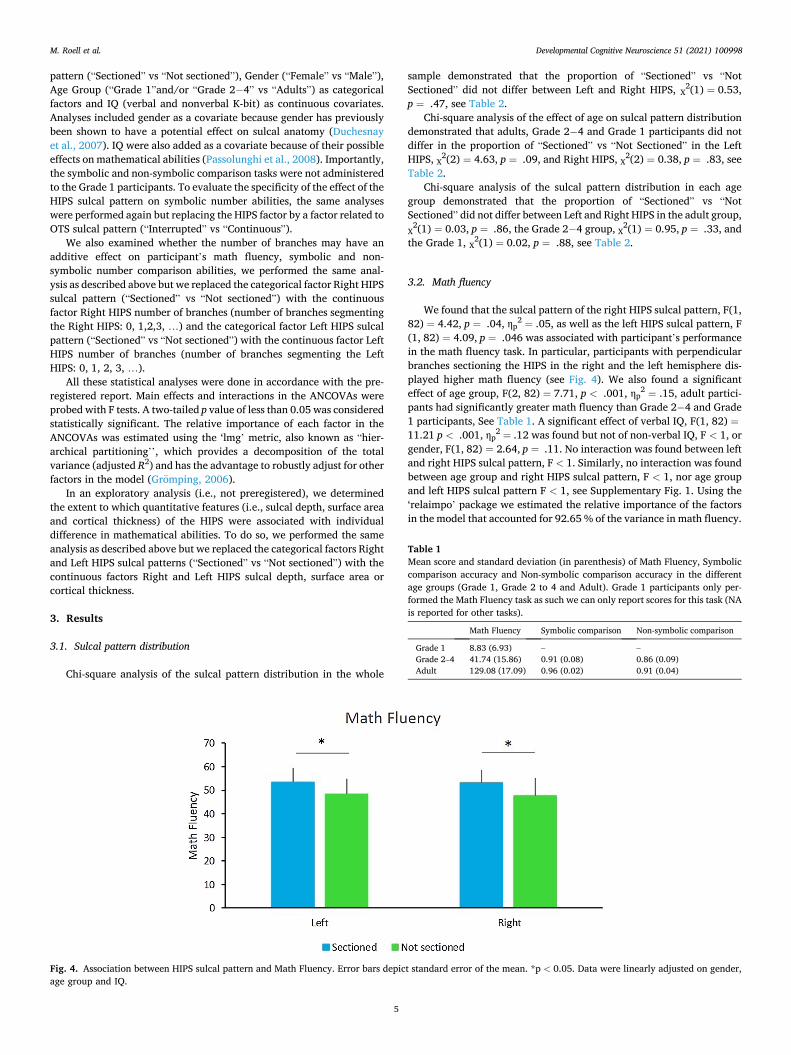
2 = .05, as well as the left HIPS sulcal pattern, F (1, 82) = 4.09, p = .046 was associated with participant’s performance in the math fluency task. In particular, participants with perpendicular branches sectioning the HIPS in the right and the left hemisphere dis-played higher math fluency (see Fig. 4). We also found a significant effect of age group, F(2, 82) = 7.71, p < .001, ηp
2 = .15, adult partici-pants had significantly greater math fluency than Grade 2− 4 and Grade 1 participants, See Table 1. A significant effect of verbal IQ, F(1, 82) =11.21 p < .001, ηp
2 = .12 was found but not of non-verbal IQ, F < 1, or gender, F(1, 82) = 2.64, p = .11. No interaction was found between left and right HIPS sulcal pattern, F < 1. Similarly, no interaction was found between age group and right HIPS sulcal pattern, F < 1, nor age group and left HIPS sulcal pattern F < 1, see Supplementary Fig. 1. Using the ‘relaimpo’ package we estimated the relative importance of the factors in the model that accounted for 92.65 % of the variance in math fluency.
Fig. 4. Association between HIPS sulcal pattern and Math Fluency. Error bars depict standard error of the mean. *p < 0.05. Data were linearly adjusted on gender, age group and IQ.
Table 1 Mean score and standard deviation (in parenthesis) of Math Fluency, Symbolic comparison accuracy and Non-symbolic comparison accuracy in the different age groups (Grade 1, Grade 2 to 4 and Adult). Grade 1 participants only per-formed the Math Fluency task as such we can only report scores for this task (NA is reported for other tasks).
Math Fluency Symbolic comparison Non-symbolic comparison
Grade 1 8.83 (6.93) – – Grade 2–4 41.74 (15.86) 0.91 (0.08) 0.86 (0.09) Adult 129.08 (17.09) 0.96 (0.02) 0.91 (0.04)
M. Roell et al.

Developmental Cognitive Neuroscience 51 (2021) 100998
6
More specifically, age group explained 42.33 %, verbal IQ explained 18.81 %, non-verbal IQ explained 11.23 %, gender explained 0.48 %, right IPS sulcal pattern explained 0.27 % and left IPS sulcal pattern explained 0.49 % of the total variance in math fluency. Additionally, the interaction between age group and right IPS sulcal pattern explained 0.21 % and the interaction between age group and left IPS sulcal pattern explained 0.18 % of the variance in math fluency.
In addition, the number of perpendicular branch sectioning the left or right HIPS was not associated with the performance of participants in the math fluency task as suggested by the lack of main effect in the right HIPS sulcal pattern, F(1,85) = 3.05, p =. 08, and left HIPS sulcal pattern, F(1,85) = 1.48, p =. 22.
Exploratory analysis revealed that the cortical thickness of the Left IPS F(1,88) = 6.00, p = .01, η2 = .06 was associated with performance in the math fluency task. Other main or interaction effects involving HIPS quantitative features were not significant.
Moreover, the sulcal pattern of the right, F < 1 or left, F(1, 85) =1.48, p = .22, anterior OTS was not associated with the performance on the math fluency task, suggesting that the effect reported on the HIPS sulcal pattern is somewhat specific. We found a main effect of age group, F(2, 85) = 6.60, p = .002, ηp
2 = .13, adult participants having signifi-cantly greater math fluency than Grade 2− 4 and Grade 1 participants, but no interaction effect between age group and the right, F(2, 85) =1.01, p = .37, or left, F < 1, anterior OTS, see Supplementary Tables 1 and 2. A main effect of verbal IQ, F(1, 85) = 11.34, p = .001, ηp
2 = .11 and gender, F(1, 85) = 5.56, p = .02, ηp
2 = .06 was found but not of
non-verbal IQ, F < 1. Analysis of the complete model including the IPS as well as the OTS
yielded a main effect of age group, F(2, 79) = 6,94, p < .001, ηp2 =.14,
right HIPS sulcal pattern, F(1, 79) = 4.20, p = .04, ηp2 =.05, and left
HIPS sulcal pattern, F(1, 79) = 4.12, p = .04, ηp2 =.04, along with no
main effect of right, F < 1, or left, F < 1, OTS sulcal morphology.
3.3. Symbolic and non-symbolic comparison task
In the symbolic number comparison task, we found a main effect of the sulcal pattern of the right HIPS sulcal pattern, F(1,60) = 7.13, p =
.009, ηp2 = .10, and the left HIPS sulcal pattern, F(1,60) = 10.21, p =
.002, ηp2 = .14. Participants with a perpendicular branch sectioning the
HIPS in either hemisphere displayed better performance in the symbolic number comparison task (see Fig. 5). No significant effect of age group (“Grade 2− 4” vs “Adult”) was found, F(1,60) = 1.36, p = .24, see Table 1. Nor did we find a significant main effect of verbal IQ, F < 1, non-verbal IQ, F(1,60) = 1.24, p = .26, or gender, F < 1. No significant interaction between left and right HIPS sulcal pattern, F < 1. Nor was an interaction between age group and right HIPS sulcal pattern, F(1,60) =2.04, p = .16, or age group and left HIPS sulcal pattern, F(1,60) = 1.95, p = .16, found, see Supplementary Fig. 2. Using the ‘relaimpo’ package we estimated the relative importance of the factors in the model that accounted for 32.68 % of the variance in the symbolic number com-parison task. More specifically, age group explained 3.56 %, verbal IQ explained 1.55 %, non-verbal IQ explained 2.30 %, gender explained 0.38 %, right IPS sulcal pattern explained 7.01 % and left IPS sulcal pattern explained 9.66 % of the variance in the symbolic number com-parison task. Additionally, the interaction between age group and right IPS sulcal pattern explained 2.14 % and the interaction between age group and left IPS sulcal pattern explained 2.28 % of the variance in the symbolic number comparison task.
The participant’s performance in the symbolic number comparison task was associated with the number of perpendicular branches sectioning the left HIPS sulcal pattern, F(1,62) = 6.18, p = .01, or right HIPS sulcal pattern, F(1,62) = 5.71, p = .01. Participants with the more branches sectioning both the right, or the left HIPS, had better accuracy scores than participants with less branches sectioning the left or the right HIPS, see Fig. 6.
Same analyses were run on RT and did not lead to any significant results (ps > .07).
Exploratory analysis did not reveal any significant main or interac-tion effects involving HIPS quantitative features on performance in the symbolic number comparison task.
Table 2 Contingency table representing the proportion of participants with a “Sectioned” and “Not Sectioned” left and right HIPS in the different groups (Grade 1, Grade 2 to 4 and Adult).
Right HIPS
Sectioned Not Sectioned Total
Left HIPS
Grade 1 Sectioned 13 6 19 Not Sectioned 5 2 7 Total 18 8 26
Grade 2− 4 Sectioned 14 11 25 Not Sectioned 18 8 26 Total 32 19 51
Adult Sectioned 6 4 10 Not Sectioned 7 4 11 Total 13 8 21
Total Sectioned 33 22 55 Not Sectioned 30 13 43 Total 63 35 98
Fig. 5. Association between HIPS sulcal pattern and the accuracy rates in the Symbolic number comparison task. Error bars depict standard error of the mean. *p < 0.05 and **p < 0.01. Data were linearly adjusted on gender, age, group and IQ.
M. Roell et al.

Developmental Cognitive Neuroscience 51 (2021) 100998
7
Additionally, the sulcal pattern of the anterior OTS was not found to be associated with participants’ performance in the symbolic number comparison task, respectively and F < 1 for the right and F(1,62) =2.47, p = .12 for the left hemisphere. No age group effect was found, F < 1, nor did we find an interaction between age group and left, F < 1 or right OTS, F < 1, see Supplementary Tables 1 and 2. No effect of verbal IQ, F < 1, non-verbal IQ, F < 1, or gender, F(1,62) = 1.59, p =
.21, was found. Analysis of the complete model including the IPS as well as the OTS
yielded a main effect of right HIPS sulcal pattern, F(1,58) = 6.26, p = .01, ηp
2 =.09, left IPS sulcal pattern, F(1,58) = 10.57 p < .001, ηp2
=.15, along with no main effect of age group, F(1,58) = 1.30, p = .25, of right, F < 1, or left, F < 1, OTS sulcal morphology.
In the non-symbolic comparison task, we did not find a main effect of sulcal pattern of the right HIPS sulcal pattern, F(1,60) = 2.28, p = .13 or the left HIPS sulcal pattern, F < 1, see Fig. 7. We did not find a main effect of age group (“Grade 2− 4” vs “Adult”), F < 1, see Table 1. Nor did we find a significant main effect of verbal IQ, F(1,60) = 1.89, p = .17, non-verbal IQ, F < 1, and gender, F < 1, was observed. No significant interaction between left and right HIPS, F(1,60) = 1.88, p = .17. Nor did we find an interaction between age group and right HIPS sulcal pattern, F(1,60) = 3.42, p = .07, nor age group and left HIPS sulcal pattern, F < 1, see Supplementary Fig. 3. Using the ‘relaimpo’ package we estimated the relative importance of the factors in the model that accounted for 20.87 % of the variance in the non-symbolic number comparison task. More specifically, age group explained 5.24 %, verbal IQ explained 2.43 %, non-verbal IQ explained 0.55 %, gender explained 0.07 %, right IPS sulcal pattern explained 2.95 % and left IPS sulcal pattern explained 1.79 % of the variance in the non-symbolic number comparison task. Additionally, the interaction between age group and right IPS sulcal pattern explained 4.38 % and the interaction between age group and left IPS sulcal pattern explained 0.66 % of the variance in
the non-symbolic number comparison task. Same analyses were run on RT and did not lead to any significant
results (ps > .22). Exploratory analysis did not reveal any significant main or interac-
tion effects involving HIPS quantitative features on performance in the non-symbolic number comparison task.
Additionally, the sulcal pattern of the anterior OTS was not found to be associated with participants’ performance in the non-symbolic number comparison task, respectively F < 1 for the right and F < 1 for the left hemisphere. No age group effect was found, F < 1, nor did we find an interaction between age group and right, F < 1 or left OTS, F < 1, see Supplementary Tables 1 and 2. No significant main effect of verbal IQ, F(1,62) = 1.55, p = .21, non-verbal IQ, F < 1, and gender, F < 1, was observed.
Analysis of the complete model including the IPS as well as the OTS yielded no main effect of age group, F < 1, right HIPS sulcal pattern, F (1,58) = 1.82, p = .18, left HIPS sulcal pattern, F < 1, left OTS, F < 1, or right OTS, F < 1, sulcal morphology.
4. Discussion
In the present study, we investigated whether the sulcal pattern of the HIPS, marker of early neurodevelopmental constraints, is related to individual difference in arithmetic performance as well as symbolic and non-symbolic number abilities in typically developing children and adults.
Our study provides the first evidence that differences in the sulcal patterns of the HIPS explain part of the variability observed in math fluency and symbolic number comparison in typically developing chil-dren and adults. More specifically, we found that children and adults with perpendicular branches sectioning the HIPS displayed better arithmetic and symbolic numerical abilities than participants with no
Fig. 6. Linear association between number of branches sectioning the HIPS and Symbolic number task. Error bars depict standard error of the mean. *p < 0.05. Data were linearly adjusted on gender, age, group and IQ.
Fig. 7. Association between HIPS sulcal pattern and the accuracy rates in the Non-Symbolic number comparison task. Error bars depict standard error of the mean. Data were linearly adjusted on gender, age, group and IQ.
M. Roell et al.

Developmental Cognitive Neuroscience 51 (2021) 100998
8
perpendicular branch sectioning the HIPS. As anticipated, we found that symbolic number comparison abilities were affected by the left and right HIPS sulcal pattern. The results reported at the anatomical level are consistent with the ones at the functional level reported in a study with an overlapping sample of participants (Matejko and Ansari, 2017). In Matejko and Ansari’s (Yeo et al., 2017) study, activation of both the right and left HIPS during an arithmetic problem-solving task was associated with better performance in the symbolic number comparison task and in the Math Fluency task. Consistent with theses results as the functional level, we found that the both the right and the left HIPS sulcal pattern contribute to individual difference in math fluency.
We suspect that the consistency of the findings reported at the anatomical level in our study and the findings reported at the functional level in Matejko & Ansari’s (Yeo et al., 2017) could be due to the effect of cortical folding on the underlying cytoarchitecture, which could in turn affect the functional level in response to performing a task. Our assumption is consistent with studies showing not only a relation be-tween cortical folding and cerebral function but also between the cortical folding and the underlying cytoarchitecture (Fischl et al., 2008; Weiner et al., 2017; Zilles et al., 2013). In particular, a study using an algorithm-based approach for the definition of areal borders, identified two distinct areas in the IPS which were found to have consistent cytoarchitectonic characteristics as well as consistent topographic rela-tion with each other (Choi et al., 2006). However, the location and the spatial extent of these two areas varied considerably from one individual to the other. Such variability was attributed by the authors to be related to the variability in the sulcal pattern of the IPS (Choi et al., 2006). Moreover, a sectioned IPS corresponds anatomically to the presence of a gyrus, which is associated with a bundle of white matter fibers, inter-rupting the course of the IPS furrow. As such a sectioned IPS may confer an advantage by reflecting an underlying difference in the size of white-matter bundles, indicating a higher number, greater fiber diam-eter and/or greater myelination in the bundles connecting the IPS, thus optimizing the wiring costs and conductivity speed (Assaf et al., 2020; Klyachko and Stevens, 2003). Besides, more branching in the region of the IPS could also lead to a greater regional cortical surface area and thickness (Hilgetag and Barbas, 2006). However, given that the HIPS suclal depth, cortical surface area and cortical thickness were not associated with mathematical abilities it is unlikely that the association between the sulcal morphology of the HIPS and mathematical abilities are driven by the effect of quantitative characteristics of the HIPS (surface area, cortical thickness or sulcal depth) that are dependent on environmental or educational changes.
Interestingly, we did not find an association between HIPS sulcal pattern and non-symbolic ability. The lack of association may be due to several reasons. A number of studies are questioning the strong associ-ation between symbolic and non-symbolic numbers (see Leibovich & Ansari (Choi et al., 2006)). Indeed, a number of brain imaging studies challenge the existence of an abstract representation of numerical magnitude in the IPS. In their study Bulthe et al. (2014) applied multi-voxel pattern analysis of fMRI data to unravel the neural activity patterns of symbolic and non-symbolic numbers and their possible overlap. Results showed no overlapping representation for symbolic and non-symbolic numbers. Similarly, in a fMRI adaptation study examining cross-format processing, Cohen Kadosh et al. (2011) found that when the presented item changed in format but not in numerical quantity (e.g. a change from the representation of 12 dots to the symbolic representation “12”) the same intraparietal regions that respond to magnitude change were activated to a greater degree. As such, it may be that symbolic and non-symbolic number rely on different brain areas and therefore could be differentially impacted by sulcal morphology. Interestingly, as non-symbolic number abilities have been proposed as a precursor of mathematics abilities (Leibovich and Ansari, 2016; Bulthe et al., 2014), we would have expected that non-symbolic abilities to be more sensitive than symbolic abilities to early neurodevelopmental factors such as the sulcal morphology. However, the opposite has been found in this study.
Of note, the current findings are in line with findings from recent studies that general mathematical abilities are more heritable, and thus more dependent on early determined genetic factors, than non-symbolic mathematical abilities (Cohen Kadosh et al. (2011); Le Guen et al., 2018). These findings are consistent with our findings by suggesting that heritable biological factors, such as the sulcal morphology (Le Guen et al., 2018; Pizzagalli et al., 2020), play a greater role on general math and symbolic number abilities than non-symbolic math abilities. An alternative explanation for the lack of effect of the sulcal morphology on non-symbolic math abilities could also be a byproduct of the reliability of non-symbolic number tasks which are less reliable and somewhat noisier than other math tasks (Pizzagalli et al., 2020).
Importantly, the association between HIPS sulcal morphology and symbolic number abilities was found to be stable across the ages examined (i.e., no interaction effect between age group and HIPS sulcal morphology were found for both math fluency and symbolic number comparison). Moreover, we did not find any difference in sulcal pattern distribution between the different age groups. These results support the notion that the sulcal pattern of the HIPS is stable over time (see similar stability in other brain regions Cachia et al., 2016; Tissier et al., 2018b) and that its influence on symbolic number abilities is constant throughout the course of development.
Although our study was performed on a relatively large sample, especially for an MRI study with children, and focused on complemen-tary anatomical features of cortex anatomy, there should be some call for caution when interpreting the results. Indeed, although our findings support a relationship between sulcation and later arithmetic and symbolic abilities, a direct causal link has yet to be evidenced. To pro-vide evidence of such causal link, a longitudinal study would be needed to demonstrate that sulcation before math skill acquisition constrains future math ability in the same individuals. Another way to provide evidence for a causal role of the sulcal morphology on future learning, training studies need to be conducted to determine whether learning gains vary in function of the sulcal morphology of the participants matched on their pre-training abilities Additionally, although sulcal morphology may play a role in mathematical skill acquisition, other anatomical factors that are more experience dependent such as grey matter volume, cortical thickness and functional activation are also likely to play a role.
In conclusion, this study provides the first evidence that HIPS sulcal pattern contributes to the variability in symbolic number abilities in typically developing children and adults, suggesting that early cerebral constraints may influence individual difference in math abilities. Addi-tionally, the association between HIPS sulcal pattern and symbolic number abilities was found to be stable by age highlighting the stability of this association throughout development.
Data statement
Due to the confidential nature of the MRI data, participants have been assured that the raw data would remain confidential and would not be shared.
Declaration of Competing Interest
The authors declare that they have no known competing financial interests or personal relationships that could have appeared to influence the work reported in this paper.
Appendix A. Supplementary data
Supplementary material related to this article can be found, in the online version, at doi:https://doi.org/10.1016/j.dcn.2021.100998.
M. Roell et al.

Developmental Cognitive Neuroscience 51 (2021) 100998
9
References
Assaf, Y., Bouznach, A., Zomet, O., Marom, A., Yovel, Y., 2020. Conservation of brain connectivity and wiring across the mammalian class. Nat. Neurosci. 23 (7), 805–808.
Borst, G., Cachia, A., Vidal, J., Simon, G., Fischer, C., Pineau, A., et al., 2014. Folding of the anterior cingulate cortex partially explains inhibitory control during childhood: a longitudinal study. Dev. Cogn. Neurosci. 9, 126–135.
Borst, G., Cachia, A., Tissier, C., Ahr, E., Simon, G., Houde, O., 2016. Early cerebral constraints on reading skills in school-age children: an MRI study. Mind Brain Educ. 10 (1), 47–54.
Bull, R., Scerif, G., 2001. Executive functioning as a predictor of children’s mathematics ability: inhibition, switching, and working memory. Dev. Neuropsychol. 19 (3), 273–293.
Bull, R., Espy, K.A., Wiebe, S.A., 2008. Short-term memory, working memory, and executive functioning in preschoolers: longitudinal predictors of mathematical achievement at age 7 years. Dev. Neuropsychol. 33 (3), 205–228.
Bulthe, J., De Smedt, B., Op de Beeck, H.P., 2014. Format-dependent representations of symbolic and non-symbolic numbers in the human cortex as revealed by multi-voxel pattern analyses. NeuroImage 87, 311–322.
Cachia, A., Borst, G., Tissier, C., Fisher, C., Plaze, M., Gay, O., et al., 2016. Longitudinal stability of the folding pattern of the anterior cingulate cortex during development. Dev. Cogn. Neurosci. 19, 122–127.
Cachia, A., Roell, M., Mangin, J.-F., Sun, Z.Y., Jobert, A., Braga, L., et al., 2017. How interindividual differences in brain anatomy shape reading accuracy. Brain Struct. Funct. Available: http://link.springer.com/10.1007/s00429-017-1516-x.
Chi, J.G., Dooling, E.C., Gilles, F.H., 1977. Gyral development of the human brain. Ann. Neurol. 1 (1), 86–93.
Choi, H.-J., Zilles, K., Mohlberg, H., Schleicher, A., Fink, G.R., Armstrong, E., et al., 2006. Cytoarchitectonic identification and probabilistic mapping of two distinct areas within the anterior ventral bank of the human intraparietal sulcus. J. Comp. Neurol. 495 (1), 53–69.
Cohen Kadosh, R., Bahrami, B., Walsh, V., Butterworth, B., Popescu, T., Price, C.J., 2011. Specialization in the human brain: the case of numbers. Front. Hum. Neurosci. Available: http://journal.frontiersin.org/article/10.3389/fnhum.2011.00062/abstra ct.
Cragg, L., Gilmore, C., 2014. Skills underlying mathematics: the role of executive function in the development of mathematics proficiency. Trends Neurosci. Educ. 3 (2), 63–68.
DeFlorio, L., Beliakoff, A., 2015. Socioeconomic status and preschoolers’ mathematical knowledge: the contribution of home activities and parent beliefs. Early Educ. Dev. 26 (3), 319–341.
Dehaene, S., Piazza, M., Pinel, P., Cohen, L., 2003. Three Parietal circuits for number processing. Cogn. Neuropsychol. 20 (3‑6), 487–506.
Duchesnay, E., Cachia, A., Roche, A., Riviere, D., Cointepas, Y., Papadopoulos- Orfanos, D., et al., 2007. Classification based on cortical folding patterns. IEEE Trans. Med. Imaging 26 (4), 553–565.
Fias, W., Lammertyn, J., Reynvoet, B., Dupont, P., Orban, G.A., 2003. Parietal representation of symbolic and nonsymbolic magnitude. J. Cogn. Neurosci. 15 (1), 47–56.
Fischl, B., Rajendran, N., Busa, E., Augustinack, J., Hinds, O., Yeo, B.T.T., et al., 2008. Cortical folding patterns and predicting cytoarchitecture. Cereb. Cortex 18 (8), 1973–1980.
Fornito, A., Yücel, M., Wood, S.J., Proffitt, T., McGorry, P.D., Velakoulis, D., et al., 2006. Morphology of the paracingulate sulcus and executive cognition in schizophrenia. Schizophr. Res. 88 (1‑3), 192–197.
Fujiwara, H., Hirao, K., Namiki, C., Yamada, M., Shimizu, M., Fukuyama, H., et al., 2007. Anterior cingulate pathology and social cognition in schizophrenia: a study of gray matter, white matter and sulcal morphometry. NeuroImage 36 (4), 1236–1245.
Giedd, J.N., Rapoport, J.L., 2010. Structural MRI of pediatric brain development: what have we learned and where are we going? Neuron 67 (5), 728–734.
Gromping, U., 2006. Relative importance for linear regression in R: the package relaimpo. J. Stat. Softw. 27.
Grotheer, M., Ambrus, G.G., Kovacs, G., 2016. Causal evidence of the involvement of the number form area in the visual detection of numbers and letters. NeuroImage 132, 314–319.
Grotheer, M., Jeska, B.L., Grill-Spector, K., 2018. A preference for mathematical processing outweighs the selectivity for Arabic numbers in the inferior temporal gyrus. NeuroImage. Available: http://linkinghub.elsevier.com/retrieve/pii/S1 05381191830274X.
Hannagan, T., Amedi, A., Cohen, L., Dehaene-Lambertz, G., Dehaene, S., 2015. Origins of the specialization for letters and numbers in ventral occipitotemporal cortex. Trends Cogn. Sci. 19 (7), 374–382.
Hart, S.A., Petrill, S.A., Thompson, L.A., Plomin, R., 2009. The ABCs of math: a genetic analysis of mathematics and its links with reading ability and general cognitive ability. J. Educ. Psychol. 101 (2), 388–402.
Haworth, C.M.A., Kovas, Y., Petrill, S.A., Plomin, R., 2007. Developmental origins of low mathematics performance and normal variation in twins from 7 to 9 years. Twin Res. Hum. Genet. 10 (1), 106–117.
Hilgetag, C.C., Barbas, H., 2006. Role of mechanical factors in the morphology of the primate cerebral cortex. PLoS Comput. Biol. 2 (3), 14.
Holloway, I.D., Ansari, D., 2010. Developmental specialization in the right intraparietal sulcus for the abstract representation of numerical magnitude. J. Cogn. Neurosci. 22 (11), 2627–2637.
Jordan, N.C., Kaplan, D., Ramineni, C., Locuniak, M.N., 2009. Early math matters: kindergarten number competence and later mathematics outcomes. Dev. Psychol. 45 (3), 850–867.
Kaufmann, A., Kaufman, N., 1993. Kaufman brief intelligence test. J. Psychoeduc. Assesment. 11, 98–101.
Klibanoff, R.S., Levine, S.C., Huttenlocher, J., Vasilyeva, M., Hedges, L.V., 2006. Preschool children’s mathematical knowledge: the effect of teacher « math talk ». Dev. Psychol. 42 (1), 59–69.
Klyachko, V.A., Stevens, C.F., 2003. Connectivity optimization and the positioning of cortical areas. Proc. Natl. Acad. Sci. 100 (13), 7937–7941.
Kuhl, U., Friederici, A.D., Skeide, M.A., Friederici, A.D., Emmrich, F., Brauer, J., et al., 2020. Early cortical surface plasticity relates to basic mathematical learning. NeuroImage 204, 116235.
Le Guen, Y., Auzias, G., Leroy, F., Noulhiane, M., Dehaene-Lambertz, G., Duchesnay, E., et al., 2018. Genetic influence on the sulcal pits: on the origin of the first cortical folds. Cereb. Cortex 28 (6), 1922–1933.
Leibovich, T., Ansari, D., 2016. The symbol-grounding problem in numerical cognition: a review of theory, evidence, and outstanding questions. Can. J. Exp. Psychol. 70 (1), 12–23.
Libertus, M.E., Feigenson, L., Halberda, J., 2011. Preschool acuity of the approximate number system correlates with school math ability: Approximate number system and math abilities. Dev. Sci. 14 (6), 1292–1300.
Mangin, J.-F., Riviere, D., Cachia, A., Duchesnay, E., Cointepas, Y., Papadopoulos- Orfanos, D., et al., 2004. A framework to study the cortical folding patterns. NeuroImage 23, S129–38.
Mangin, J.-F., Jouvent, E., Cachia, A., 2010. In-vivo measurement of cortical morphology: means and meanings. Curr. Opin. Neurol. 1.
Matejko, A.A., Ansari, D., 2017. How do individual differences in children’s domain specific and domain general abilities relate to brain activity within the intraparietal sulcus during arithmetic? An fMRI study: understanding IPS Activity during Arithmetic. Hum. Brain Mapp. 38 (8), 3941–3956.
Matejko, A.A., Ansari, D., 2019. The neural association between arithmetic and basic numerical processing depends on arithmetic problem size and not chronological age. Dev. Cogn. Neurosci. 37, 100653.
Matejko, A.A., Hutchison, J.E., Ansari, D., 2018. Developmental specialization of the left intraparietal sulcus for symbolic ordinal processing. Cortex. Available: https://linki nghub.elsevier.com/retrieve/pii/S0010945218304064.
Matejko, A.A., Hutchison, J.E., Ansari, D., 2019. Developmental specialization of the left intraparietal sulcus for symbolic ordinal processing. Cortex 114, 41–53.
Melhuish, E.C., Phan, M.B., Sylva, K., Sammons, P., Siraj-Blatchford, I., Taggart, B., 2008. Effects of the home learning environment and preschool center experience upon literacy and numeracy development in early primary school. J. Soc. Issues 64 (1), 95–114.
Merkley, R., Wilkey, E.D., Matejko, A.A., 2016. Exploring the origins and development of the visual number form area: a functionally specialized and domain-specific region for the processing of number symbols? J. Neurosci. 36 (17), 4659–4661.
Molko, N., Cachia, A., Riviere, D., Mangin, J.-F., Bruandet, M., Le Bihan, D., et al., 2003. Functional and structural alterations of the intraparietal sulcus in a developmental dyscalculia of genetic origin. Neuron 40 (4), 847–858.
Oliver, B., Harlaar, N., Hayiou Thomas, M.E., Kovas, Y., Walker, S.O., Petrill, S.A., et al., 2004. A twin study of teacher-reported mathematics performance and low performance in 7-year-olds. J. Educ. Psychol. 96 (3), 504–517.
Parsons, S., Bynner, J., 2005. Does Numeracy Matter More? National Research and Development Center for Adult Literacy and Numeracy, London, 44 p.
Passolunghi, M.C., Siegel, L.S., 2001. Short-term memory, working memory, and inhibitory control in children with difficulties in arithmetic problem solving. J. Exp. Child Psychol. 80 (1), 44–57.
Passolunghi, M.C., Mammarella, I.C., Altoe, G., 2008. Cognitive abilities as precursors of the early acquisition of mathematical skills during first through second grades. Dev. Neuropsychol. 33 (3), 229–250.
Pesenti, M., Thioux, M., Seron, X., De Volder, A., 2000. Neuroanatomical substrates of Arabic number processing, numerical comparison, and simple addition: a PET study. J. Cogn. Neurosci. 12 (3), 461–479.
Pizzagalli, F., Auzias, G., Yang, Q., Mathias, S.R., Faskowitz, J., Boyd, J.D., et al., 2020. The reliability and heritability of cortical folds and their genetic correlations across hemispheres. Commun. Biol. 3 (1), 510.
Provost, J.-B.L., Bartres-Faz, D., Paillere-Martinot, M.-L., Artiges, E., Pappata, S., Recasens, C., et al., 2003. Paracingulate sulcus morphology in men with early-onset schizophrenia. Br. J. Psychiatry 182 (3), 228–232.
Raznahan, A., Cutter, W., Lalonde, F., Robertson, D., Daly, E., Conway, G.S., et al., 2010. Cortical anatomy in human X monosomy. NeuroImage 49 (4), 2915–2923.
Rivera-Batiz, F.L., 1992. Quantitative literacy and the likelihood of employment among young adults in the United States. J. Hum. Resour. 27 (2), 313.
Rose, H., Betts, J.R., 2004. The effect of high school courses on earnings. Rev. Econ. Stat. 86 (2), 497–513.
Shum, J., Hermes, D., Foster, B.L., Dastjerdi, M., Rangarajan, V., Winawer, J., et al., 2013. A brain area for visual numerals. J. Neurosci. 33 (16), 6709–6715.
Sokolowski, H.M., Fias, W., Mousa, A., Ansari, D., 2017. Common and distinct brain regions in both parietal and frontal cortex support symbolic and nonsymbolic number processing in humans: a functional neuroimaging meta-analysis. NeuroImage 146, 376–394.
St Clair-Thompson, H.L., Gathercole, S.E., 2006. Executive functions and achievements in school: shifting, updating, inhibition, and working memory. Q. J. Exp. Psychol. 59 (4), 745–759.
Tissier, C., Linzarini, A., Allaire-Duquette, G., Mevel, K., Poirel, N., Dollfus, S., et al., 2018a. Sulcal polymorphisms of the IFC and ACC contribute to inhibitory control variability in children and adults. eneuro 5 (1). ENEURO.0197-17.2018.
M. Roell et al.

Developmental Cognitive Neuroscience 51 (2021) 100998
10
Tissier, C., Linzarini, A., Allaire-Duquette, G., Mevel, K., Poirel, N., Dollfus, S., et al., 2018b. Sulcal polymorphisms of the IFC and ACC contribute to inhibitory control variability in children and adults. eneuro 5 (1). ENEURO.0197-17.2018.
Watanabe, H., Nakamura, M., Ohno, T., Itahashi, T., Tanaka, E., Ohta, H., et al., 2014. Altered orbitofrontal sulcogyral patterns in adult males with high-functioning autism spectrum disorders. Soc. Cogn. Affect. Neurosci. 9 (4), 520–528.
Weiner, K.S., Barnett, M.A., Lorenz, S., Caspers, J., Stigliani, A., Amunts, K., et al., 2017. The cytoarchitecture of domain-specific regions in human high-level visual cortex. Cereb. Cortex 27 (1), 146–161.
Woodcock, R.W., McGrew, K.S., Mather, N., Schrank, F.A., 2003. Woodcock-Johnson III Diagnostic Supplement to the Tests of Cognitive Abilities: (724772011-008).
American Psychological Association. https://doi.org/10.1037/e724772011-008. Available.
Yeo, D.J., Wilkey, E.D., Price, G.R., 2017. The search for the number form area: a functional neuroimaging meta-analysis. Neurosci. Biobehav. Rev. 78, 145–160.
Zatorre, R.J., Fields, R.D., Johansen-Berg, H., 2012. Plasticity in gray and white: neuroimaging changes in brain structure during learning. Nat. Neurosci. 15 (4), 528–536.
Zilles, K., Palomero-Gallagher, N., Amunts, K., 2013. Development of cortical folding during evolution and ontogeny. Trends Neurosci. 36 (5), 275–284.
Zlatkina, V., Petrides, M., 2014. Morphological patterns of the intraparietal sulcus and the anterior intermediate parietal sulcus of Jensen in the human brain. Proc. R. Soc. B Biol. Sci. 281 (1797), 20141493–20141493.
M. Roell et al.