Interactions of Bartonella henselae with Myeloid Angiogenic ...


Success of antiviral therapy in chronic hepatitis C infection relates to functional status of myeloid dendritic cells
Deepa Rana, Yogesh Chawla* & Sunil K. Arora
Departments of Immunopathology & *Hepatology, Postgraduate Institute of Medical Education & Research, Chandigarh, India
Received February 12, 2013
Chronic hepatitis C infection poses a major global health predicament and appears to be potent threat to mankind. The treatment in wide use is interferon/ribavirin combination therapy which is generally effective in about 60-70 per cent of patients carrying genotype 3 and causes significant morbidity. The response to therapy is largely guided by limited number of factors such as genotype of virus, rapid virological response, ethnicity, pre-therapy viral load, etc. While involvement of host genetic factors has been a major focus of research in playing an important role in the outcome of disease, the role of immune system cannot be marginalized. Poor cellular trafficking and suboptimal T cell responses in liver, the hall marks of chronic hepatitis C virus infection, might be attributed to defective antigen presentation. Various immunological factors, both innate and adaptive, play role in the pathogenesis of the disease and become dysfunctional in active disease. Recent reports suggest the major impact of functional and numerical status of dendritic cells in deciding the fate of antiviral therapy. In this review we take a look at the involvement of dendritic cells in playing an important role in the response to therapy.
Key words Antiviral therapy - dendritic cells - HCV infection - hepatitis C virus - innate immune response - rapid virological response
Review Article
Indian J Med Res 138, November 2013, pp 766-778
766
Introduction
By the end of 20th century, hepatitis C virus (HCV) started to emerge as a global threat with around 170 million people infected worldwide1. The virus is a blood borne pathogen and the high risk group includes patients undergoing blood transfusions, intravenous drug users, renal patients undergoing frequent dialysis, people involved with tattooing and unsafe sexual activity. The infection with virus can follow either an acute and/or subsequently in majority of the cases a chronic clinical profile. While the acute infection is resolved spontaneously in a few weeks’ time without
any therapeutic intervention, the most disturbing aspect of this infection is that the spontaneous viral clearance is atypical, with nearly 54-86 per cent of the infected individuals progressing to chronic hepatitis2-4. Chronic HCV infection is defined as the persistence of HCV RNA in the blood for at least 6 months and in its more aggressive form in about 5-10 per cent cases progressing to cirrhosis and hepatocellular carcinoma (HCC)3.
This virus has a single-stranded RNA of positive polarity of 9.6 kb nucleotides. The RNA contains one long open reading frame, encoding a polyprotein of approximately 3000 amino acids flanked by
un-translated regions (UTRs) on both 3’ and 5’ ends. Owing to high mutability due to error prone reverse-transcription, the HCV genome, has high degree of genetic variability. To date, at least six genotypes and more than 120 subtypes of the virus have been identified, and more are in the process of being characterized. Genotypes 1a (HCV-1a), 1b, 2a, 2b, and 3a are distributed globally and account for the majority of HCV infections worldwide5. HCV-1b, the most common genotype worldwide, is also the dominant genotype in Asia-Pacific, particularly in Japan, South Korea, China and Taiwan. HCV-2a and 2b are also commonly distributed, particularly in Japan, South Korea and Southern Taiwan6. While genotype-3 of HCV is more prevalent in many countries of Asia Oceania78, the genotype-4 is predominantly found in North Africa and Middle East. Genotype-5 is limited to South Africa9. HCV 6-11 have been detected in South-East Asia recently. Genotypes 7-11 appear to be the variants of HCV-3 (genotype 10a) and HCV-6a (genotypes 7, 8, 9, 11), and have been re-classified as subtypes of genotypes 3 and 66. In India, genotype 3 predominates in the north, east and west India while genotype 1 is more common in southern parts of India8,10-12.
Treatment and response to antiviral therapy
Antiviral agents like interferon (IFN)-α and nucleoside analogue like ribavirin, have been widely used for the treatment of chronic HCV infection to prevent the development of cirrhosis of liver and hepatocellular carcinoma. In addition to causing direct inhibition of viral replication, these agents
modulate antiviral immune responses, which greatly contribute to the successful therapeutic response. The response to the therapy is largely dependent on the genotype of the virus involved. HCV genotype 1b is less sensitive to interferon as compared to genotypes 2 and 313. However, the high cost of therapy makes it far less affordable for many affected individuals, especially in the resource limited countries like India. There have been a few reports available in literature stating the superior response to IFN-based therapies in Asian patients as compared to Caucasians, Hispanics and Afro-Americans in the corresponding HCV genotype14,15.
The outcome or the response to the treatment can be measured using three parameters: (i) biochemical outcome measured in terms of normalization of serum alanine transaminase (ALT) levels, (ii) virological outcome defined by absence of HCV RNA from serum by a sensitive PCR based assay, and (iii) histological outcome measured as <2 point improvement in necro-inflammatory score with no worsening in fibrosis score16,17. So far, the virological response is considered to be the most reliable one. Various definitions used to describe the virological response to therapy are outlined in the Table18.
Predictors of response to therapy
Clinicians and virologists across the world have been making endless efforts to look for certain reliable markers which could help in predicting the likelihood of a patient in achieving sustained virological response (SVR). As mentioned above, the viral genotype has
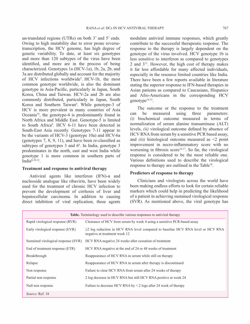
Table. Terminology used to describe various responses to antiviral therapy
Rapid virological response (RVR) Clearance of HCV from serum by week 4 using a sensitive PCR-based assay
Early virological response (EVR) ≥2 log reduction in HCV RNA level compared to baseline HCV RNA level or HCV RNA negative at treatment week 12
Sustained virological response (SVR) HCV RNA negative 24 weeks after cessation of treatment
End of treatment response (ETR) HCV RNA negative at the end of 24 or 48 weeks of treatment
Breakthrough Reappearance of HCV RNA in serum while still on therapy
Relapse Reappearance of HCV RNA in serum after therapy is discontinued
Non response Failure to clear HCV RNA from serum after 24 weeks of therapy
Partial non response 2 log decrease in HCV RNA but still HCV RNA positive at week 24
Null non response Failure to decrease HCV RNA by < 2 logs after 24 week of therapy
Source: Ref. 18
RANA et al: DCs IN HCV ANTIVIRAL THERAPY 767
768 INDIAN J MED RES, NOVEMBER 2013
been one of the most consistently used parameters to predict SVR. Patients infected with genotype 1 are less likely to achieve SVR as compared to those with genotypes 2 and 3. Another important parameter which decides the response to therapy is the pre-treatment HCV RNA level. It has been reported that patients with viral load less than 600,000 IU/ml have higher chances of attaining SVR19.
Apart from these two markers there have been other less reliable baseline parameters linked with a favourable response including the doses of peginterferon (1.5 versus 0.5 µg/kg/week) and ribavirin (>10.6 mg/kg), age <40 yr, non-African-American race, female gender, lower body weight (<75 kg), the absence of insulin resistance, elevated ALT levels (three-fold higher than the upper limit of normal), and the absence of bridging fibrosis or cirrhosis on liver biopsy20-22.
Recently, interleukin (IL) 28 polymorphism has also been highlighted as one of the reliable predictors of response to therapy. The chances of attaining an SVR with Peg-IFN and ribavirin and of spontaneous resolution of HCV infection differ depending on the nucleotide sequence near the gene for IL28B on chromosome 1923,24. The detection of the C or T allele at positions 12979860 is found to be highly predictive. Interestingly, the CC genotype is found more than twice as frequently in persons who have spontaneously cleared HCV infection than in those who had progressed to chronic hepatitis-C infection (CHC)23.
Once the treatment starts, a four week rapid virological response (RVR) has been the single best predictor of an SVR for anti-HCV treatment with peg-interferon and ribavirin combination therapy, irrespective of HCV genotype25,26. Further, depending upon the RVR, the duration of treatment can be shortened or extended16,25,26.
Immunopathogenesis of HCV infection
HCV is not directly cytopathic for the hepatocytes, and brings out progressive liver lesions resulting in end-stage liver disease unless eradicated effectively. It is noteworthy that the cellular immune response to virus is thought to be responsible for both viral clearance and progression to liver disease. However, the patients who successfully resolve the acute phase, mount a multi-epitopic polyclonal cytotoxic T lymphocyte (CTL) response to several viral antigens. In a study on injection drug users, those who resolved previous HCV infection were 12 times less likely to be reinfected to develop persistent infection than people
infected for the first time. The median peak of HCV RNA levels in those who became re-infected, were two logs lower than individuals who got infected for the first time to develop chronic infection. These findings suggest that at least in some individuals, a protective immunity develops against the virus, which is capable of complete or partial control of HCV infection17.
In contrast, this response is absent, short-lived or much weaker in patients who are unable to clear the virus and progress on to become chronically infected. Thus, it is rational to conclude that the outcome of HCV infection (viral clearance versus viral persistence) is determined primarily by the vigour and quality of the cellular immune response27. Once the virus survives the initial assault by the host immune response, it uses several strategies to nullify the selective immunological pressure during the later phases of infection. The virus alters its antigenic epitopes recognized by T cells and neutralizing antibodies to escape immune surveillance3, 4 It may also subvert immune functions in an antigen specific manner, from innate to adaptive immunity.
Innate immune response in HCV infection
After the virus infects the liver, viral replication continues and viral particles are continuously released into the circulation. It is considered that innate immunity not only provides an immediate response to viral infection, helping to clear the virus during initial period, but also shapes the nature of the adaptive immune response to viral infection28. The different components of both innate and adaptive immune response are so interconnected that the defects of one or more may lead to the establishment of chronicity of HCV infection.
The first line of defense is provided by the natural killer (NK) and NKT cells, as their numbers are shown to be relatively increased in the liver compared to the periphery. These cells are activated in the liver, where expression of IFNα and IFN-inducible genes are extremely high during the early phase of hepatitis virus infection29. Activated NK and NKT cells eliminate virus-infected hepatocytes directly by cytolytic mechanisms as well as by secreting IFNγ, which inhibits replication of HCV through a non-cytolytic mechanism30. An important role for NK cells in early HCV infection has recently been suggested by Khakoo et al31 who showed that the activation threshold of NK cells might be lower in HCV patients, which might support HCV clearance. Interaction of NK cells with HCV E2 protein by its

receptor, CD81, inhibits NK cell activity32, whereas it causes stimulation of T and B lymphocytes. NK cells from HCV-infected patients are impaired in their capacity to activate dendritic cells due to the production of IL-10 and tumour growth factor (TGF)-β33. Not surprisingly, HCV has evolved multiple strategies to counter the host’s NK cell response. Peripheral blood NK cells are reduced in chronic HCV patients compared to healthy individuals33. The reduction in NK cell frequency may be a consequence of HCV infection, or a predisposing factor to chronic HCV disease, and both hypotheses have some scientific support. In individuals with chronic HCV infection, a reduction in peripheral blood NK cell frequency in individuals with chronic HCV as compared to spontaneous resolvers has been noted, while NK cell frequency increases following successful antiviral therapy34,35. IL-15, a pivotal cytokine for NK cell development, proliferation and function, may be relevant to this observation. Meier et al36 showed a significant reduction in IL-15 levels in HCV patients as compared to healthy controls and demonstrated that exogenous IL-15 rescued their NK cells from apoptosis, increasing ex vivo proliferation and function. Several studies have reported an increase in circulating CD56bright NK cells, but not CD56dim
component in this population in chronic HCV infection as compared to acute cases37,38.
Adaptive immune response in HCV infection
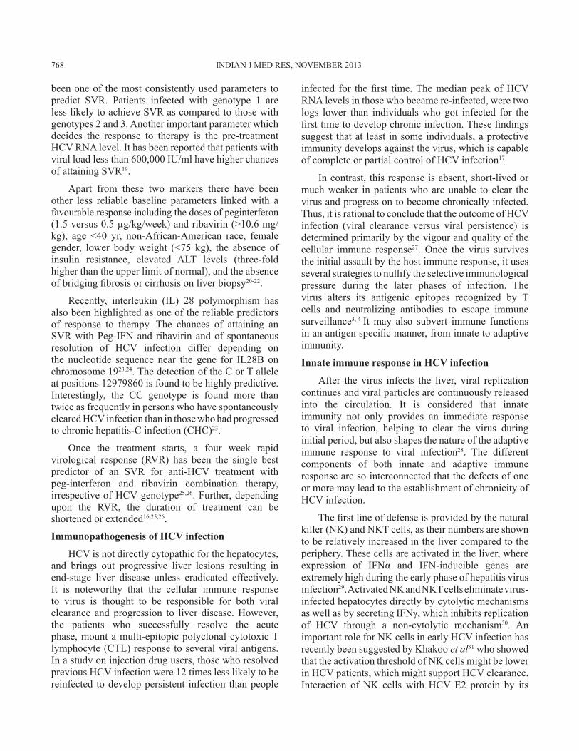
While most studies on the human adaptive immune responses against HCV are concentrated on the T cell responses, the role of neutralizing antibodies in the protective immunity against HCV has only recently gained attention39,40. Even though there are less number of cases with self-limited HCV infection that have been studied so far, a pattern of vigorous and strong HCV specific T cell response has been observed40-42. Such responses were observed only during the early phase of acute disease41,43 and sustained for long after the clearance of HCV40. These responses were generally targeted at multiple major histocompatibility complex (MHC) restricted epitopes rather than a dominant epitope42. In contrast, patients with chronic HCV infection usually have weak or defective T cell responses against HCV, as indicated by low frequencies of the specific T cells44, short-lived responses44,45, narrowly targeted epitopes44, as well as defects in the effector functions of the specific T cells45. HCV specific CD8+ T cells in chronic hepatitis C patients possess lesser capacity to proliferate and produce lesser amounts of IFNγ in response to HCV antigens46 (Fig. 1).
Taken together, these studies strongly suggest that the host T cell responses are a key factor in determining the outcome of HCV infection. Interestingly, during the acute phase of self-limiting HCV infection, there is a brief span of dysfunction of HCV specific CD8+ T cells43,44, suggesting that a transient down-modulation of the effector functions of specific CD8+ T cells may be a host strategy to keep a check at the tissue damage, caused by the cytotoxic CD8+ T cells at the early stage of infection when viral replication is at its peak. On the contrary, there is a report which failed to find a relationship between CTL response and HCV eradication47.
Although, little is known regarding the exact cause for the failure of the host’s adaptive immunity against HCV, many of these defects can be attributed to deficiencies in innate immune responses leading to progression to chronic HCV infection. To understand the mechanism behind apparently weak or abnormal T cell immunity to HCV in chronic hepatitis, it is important to explore the initial events leading to the antiviral adaptive immunity, which mainly pertains to the processing and presentation of viral antigens and onward signaling. In this scenario it seems pertinent to investigate the role of dendritic cells during this situation.
Dendritic cells in HCV related chronic hepatitis
The immune system works as a well-coordinated squad of different types of cells whose final mission is to eliminate invading pathogens. Dendritic cells (DCs) orchestrate the entire course of events as these are essential for activation of both T helper cells and cytotoxic T-lymphocytes. Dendritic cells not only form an important component of the innate immune surveillance but also play a significant role in shaping the nature of adaptive immune response against various pathogens and tumour cells. Among different subsets of DCs, myeloid-derived DCs (mDCs) are the most important antigen presenting cells with the capability to capture and process antigens, express lymphocyte co-stimulatory molecules, migrate to lymphoid organs and secrete cytokines to initiate B and T cell responses48. While the plasmacytoid dendritic cells (pDCs) play a unique role in secreting large amounts of type I interferons, their efficiency as antigen-presenting cells (APCs) has not been completely understood. However, it has been reported that during viral infection pDCs not only secrete immunomodulatory cytokines, but also recognize infected cells and function as antigen
RANA et al: DCs IN HCV ANTIVIRAL THERAPY 769
cross-presenting cells to trigger the antiviral immune response with specific reference to activation of virus specific CD8+ cells49,50.
Besides activating lymphocytes, DCs also have the important function as mediators of peripheral immune tolerance and maintenance of immune homeostasis48. The two conflicting roles of DCs have been related to their different patterns of maturation and cytokine production51,52. The functions of DCs are precisely controlled to either activate or tolarize the T cells depending on the nature of antigens which may be foreign or self. Any perturbation to this control may lead to serious consequences including impaired immunity to infecting pathogens, causing persistent infections. Many viruses have devised strategies to counteract antiviral immunity by down-modulating the functions of DCs.
The response of dendritic cells to any infection in general and initial HCV infection in particular, at early stage is critical in determining the course and outcome
of the disease. However, studying the immune responses in patients at early stage of HCV infection is difficult because the acute infection is usually asymptomatic, and by the time the disease is identified, it has already passed the initial acute phase and progressed into chronic infection. It has been shown that HCV specific T cells appear early after infection inducing strong T cell response during acute HCV infection40. To activate HCV specific T cells and induce strong antiviral immune responses, DCs have to respond to HCV viral proteins. Moreover, HCV seems to successfully disrupt the coordinated activity of the innate immune cells to result in deficient adaptive immune response and prevent pathogen elimination.
There are controversial reports on the function of myeloid DCs in chronic HCV infection. While a couple of studies have observed no functional defects in mDCs in HCV-infected individuals53-55, the findings from other studies indicate that blood-derived mDCs from HCV infected patients are functionally and
Fig. 1. The immune response in viral hepatitis C involves both the innate and adaptive immune system. Innate immunity involves activation of resident liver macrophages (MØ), dendritic cells (DCs), natural killer (NK) cells, and NKT cells, whereas CD4+ T cells, CD8+ T cells, and B lymphocytes are effectors of adaptive immunity. CTL, cytotoxic T lymphocyte; HCV, hepatitis C virus; IFN, interferon; IL, interleukin; MHC, major histocompatibility complex; TNF, tumour necrosis factor.(Reproduced with permission from John Wiley and Sons Ltd., New Jersey, USA [Clin Liver Dis 2006; 10 : 753-71]17.
770 INDIAN J MED RES, NOVEMBER 2013

numerically impaired55-60. An increase in the frequency of mDCs during acute HCV infection has been shown to be associated with viral clearance in a small study, whereas the deficiency of mDC might be an important factor in the development of chronic state of the infection61. The mDCs of patients with chronic HCV infection have impaired abilities to stimulate allogeneic CD4+ T cells and to produce IL-12p70 compared with those from healthy volunteers62. Although DCs can bind to HCV and may even get infected with this virus and possibly act as HCV reservoirs63, but there has not been much evidence of viral replication within these cells.
The phenotype and cytokine production profile of bone marrow–originated mDCs can be generated from peripheral blood monocytes [monocyte-derived DCs (mo-DCs)] in the presence of IL-4, granulocytes-macrophage colony-stimulating factor (GM-CSF) and serve as an ex vivo model for mDCs. Data on the function/phenotype of mo-DCs during acute HCV infection are scarce, and the opinions on the function of mo-DCs in chronic HCV are divided. Although Longman et al53 reported normal phenotypic characteristics and allogeneic functions in mo-DCs in humans and Larsson et al64 identified similar findings in a non human primate model of chronic HCV infection, the majority of researchers find functional abnormalities in mo-DCs of humans with chronic HCV infection65-67. We and others have demonstrated that decreased expression of the co-stimulatory molecules CD83 and CD86, increased production of IL-10 and decreased levels of IL-12, may lead to impaired capacity to stimulate allogeneic T cells in vitro, and may cause defective maturation of mDCs in patients with chronic HCV infection regardless of viral genotype or patient age/sex across a wide geographic localization of patients65-68.
In vitro experiments have effectively shown that upon exposure of human mo-DCs to cell culture-grown HCV (HCVcc) genotype 1a or 2a, there was a significant inhibition of DC maturation and was associated with low HLA-DR expression and high production of IL-10. Furthermore, DCs exposed to HCVcc were impaired in their functional ability to stimulate antigen-specific CD4+ and CD8+ T cell responses69. DCs, upon exposure to HCV core protein, showed downregulation of major histocompatibility molecules (HLA-DR) and co-stimulatory molecules (CD80, CD86) and induction of IL-10 producing T cells70. The HCV may induce modulation of the cytokine response, especially of increase in IL-10
and decrease in IL-12 production that may result in altered HCV specific T cell responses in chronic HCV infection. In another report, lipopolysaccharide (LPS)-stimulated mo-DC from HCV patients showed a mature phenotype but with a significantly decreased expression of IL-12, could be responsible for the Th1 defect since the intensity of the Th1 defect was directly related to the intensity of IL-12 decrease71. We have also recently shown that the monocytes from healthy donors when differentiated in the presence of HCV core and NS5 proteins developed maturation defects as these could not upregulate the HLA-DR alongwith CD83, CD80 and CD86 on toll like receptor (TLR)-4 mediated stimulation68. Peripheral blood DC of chronically infected patients have been shown to express less IL-12 than control cells from healthy individuals60 and a small population of circulating myeloid DC displayed an impaired response to specific TLR stimulation72. Viral components can either bind to TLR and activate their signaling pathway or block TLR function by interfering with intracellular intermediates.
Dendritic cells and antiviral therapy
There have been a few studies in which role of DCs have been studied in context of antiviral therapy57,66,73. In general, it has been demonstrated that the early response to antiviral treatment leads to the improvement of DC functions which was not observed in patients who remained viraemic on therapy. Evaluating the defects in maturation and chemotaxis in peripheral mDCs and pDCs from therapeutic responders and non-responders, revealed that successful antiviral therapy normalized many phenotypic and functional abnormalities of pDCs from patients with HCV infection74.
Maturation defects were detected in the mo-DCs from genotype-3 infected chronic hepatitis C (CHC) patients when checked before the initiation of therapy, however, it was very interesting to find that all those CHC patients whose mo-DCs could upregulate the surface expression of activation (CD83), and co-stimulatory (CD80 and CD86) molecules on their surface after TLR-4 mediated stimulation ex-vivo even before the initiation of therapy, were able to subsequently achieve SVR after completion of antiviral treatment, while the patients whose cells showed an inability to upregulate these molecules initially, did not achieve SVR later on as well68. This suggested that the response to antiviral therapy in CHC is somehow related to the functional status of DCs. This finding is in agreement with a previous report wherein it was demonstrated that the pDCs were functionally activated in terms of increased
RANA et al: DCs IN HCV ANTIVIRAL THERAPY 771
chemotaxis even before therapy in patients who subsequently achieved SVR, suggesting that increased baseline inflammation is associated with failure to respond to the antiviral therapy74 (Fig. 2).
Furthermore, DCs from patients who achieved SVR in our study68 demonstrated an improvement in the maturation capacity after completion of treatment. On the other hand, non responders to therapy continued to show maturation defects in their mo-DC. The functional improvement of mo-DCs was directly related to the successful clearance of virus in responders upon completion of therapy68 (Fig. 3). It clearly suggests that defect seen in chronically infected patients is limited to the period of active viral replication but can be resolved after viral clearance. This observation is corroborated by similar reports demonstrating clearance of HCV RNA from circulating DCs in CHC patients who achieved rapid virological response (RVR)57 indicating that therapy with IFNα may induce improved DC function by reducing the load of HCV in vivo. However, the role of antiviral agents, especially that of type-1 IFNs on maturation and activation of DCs, is controversial. It has been proven that in vitro addition of IFNα to DC cultures of CHC patients improves their function in terms of increased expression of activation and co-stimulatory molecules75,76. On the other hand, McRae et al77 have shown that IFNα and β inhibit the in vitro differentiation of mo-DCs from normal subjects. It has been suggested that ribavirin is not strictly antiviral in its action, but rather alters the T-cell balance in the immune system and attenuates the DC function in vitro and in vivo78.
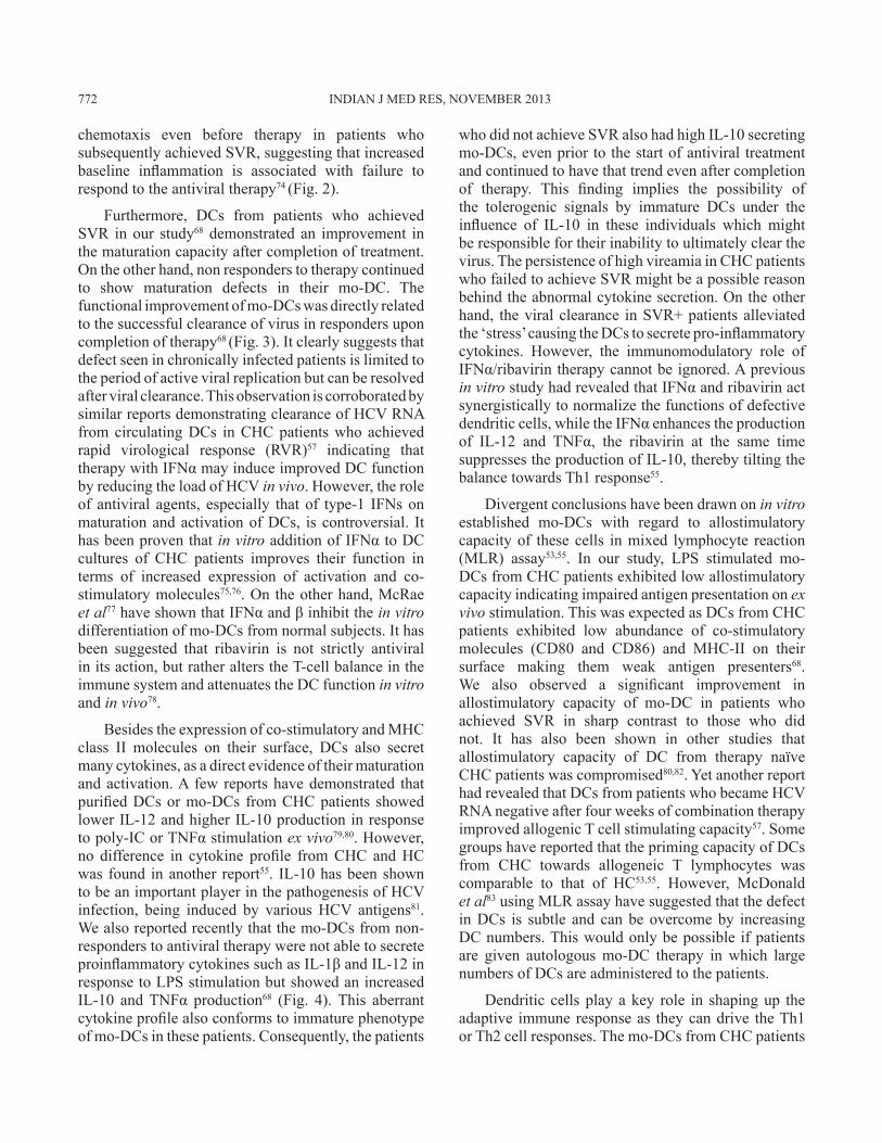
Besides the expression of co-stimulatory and MHC class II molecules on their surface, DCs also secret many cytokines, as a direct evidence of their maturation and activation. A few reports have demonstrated that purified DCs or mo-DCs from CHC patients showed lower IL-12 and higher IL-10 production in response to poly-IC or TNFα stimulation ex vivo79,80. However, no difference in cytokine profile from CHC and HC was found in another report55. IL-10 has been shown to be an important player in the pathogenesis of HCV infection, being induced by various HCV antigens81. We also reported recently that the mo-DCs from non-responders to antiviral therapy were not able to secrete proinflammatory cytokines such as IL-1β and IL-12 in response to LPS stimulation but showed an increased IL-10 and TNFα production68 (Fig. 4). This aberrant cytokine profile also conforms to immature phenotype of mo-DCs in these patients. Consequently, the patients
who did not achieve SVR also had high IL-10 secreting mo-DCs, even prior to the start of antiviral treatment and continued to have that trend even after completion of therapy. This finding implies the possibility of the tolerogenic signals by immature DCs under the influence of IL-10 in these individuals which might be responsible for their inability to ultimately clear the virus. The persistence of high vireamia in CHC patients who failed to achieve SVR might be a possible reason behind the abnormal cytokine secretion. On the other hand, the viral clearance in SVR+ patients alleviated the ‘stress’ causing the DCs to secrete pro-inflammatory cytokines. However, the immunomodulatory role of IFNα/ribavirin therapy cannot be ignored. A previous in vitro study had revealed that IFNα and ribavirin act synergistically to normalize the functions of defective dendritic cells, while the IFNα enhances the production of IL-12 and TNFα, the ribavirin at the same time suppresses the production of IL-10, thereby tilting the balance towards Th1 response55.
Divergent conclusions have been drawn on in vitro established mo-DCs with regard to allostimulatory capacity of these cells in mixed lymphocyte reaction (MLR) assay53,55. In our study, LPS stimulated mo-DCs from CHC patients exhibited low allostimulatory capacity indicating impaired antigen presentation on ex vivo stimulation. This was expected as DCs from CHC patients exhibited low abundance of co-stimulatory molecules (CD80 and CD86) and MHC-II on their surface making them weak antigen presenters68. We also observed a significant improvement in allostimulatory capacity of mo-DC in patients who achieved SVR in sharp contrast to those who did not. It has also been shown in other studies that allostimulatory capacity of DC from therapy naïve CHC patients was compromised80,82. Yet another report had revealed that DCs from patients who became HCV RNA negative after four weeks of combination therapy improved allogenic T cell stimulating capacity57. Some groups have reported that the priming capacity of DCs from CHC towards allogeneic T lymphocytes was comparable to that of HC53,55. However, McDonald et al83 using MLR assay have suggested that the defect in DCs is subtle and can be overcome by increasing DC numbers. This would only be possible if patients are given autologous mo-DC therapy in which large numbers of DCs are administered to the patients.
Dendritic cells play a key role in shaping up the adaptive immune response as they can drive the Th1 or Th2 cell responses. The mo-DCs from CHC patients
772 INDIAN J MED RES, NOVEMBER 2013

Fig. 2. Differential elevations of chemokine receptors and maturation markers in dendritic cell (DC) subtypes before and after therapy. SOO2 is screening visit 2, 2 wk before initiation of therapy. FU24 is 24 wk after cessation of treatment. Median fluorescence intensity (MFI) values for each patient are shown. The horizontal line is the median. A two-tailed non parametric Wilcoxon signed rank test was used for paired data or the two-tailed Mann-Whitney test was used to calculate P values. (B) CD83 MFI on plasmacytoid and myeloid DCs (pDCs, mDCs); CD40 MFI for pDCs, mDCs; HLA-DR for pDCs and mDCs. SVR, sustained virological response.(Reproduced with permission from British Medical Association, London, UK [Gut 2009; 58 : 964-73]74.
RANA et al: DCs IN HCV ANTIVIRAL THERAPY 773

Fig. 3. Effect of treatment on expression of (A) HLA-DR, (B) CD83, (C) CD80 and (D) CD86 on monocyte-derived dendritic cells (mo-DCs) from chronic hepatitis-C patient (CHC)-II pre and post-treatment (in terms of fold increase in mean fluorescence intensity, MFI). Expression of all molecules increases significantly in SVR+ although no change is seen in SVR- patients. SVR, sustained virology response.(Reproduced with permission from John Wiley and Sons Ltd., Philadelphia, USA [Liver Int 2012; 32 : 1128-37]68.
Fig. 4. Cytokine profile of monocyte-derived dendritic (mo-DCs) cells pre- and post-lipopolysaccharide (LPS) stimulation. Secretion of IL1β, IL12 and TNF-α increased significantly in LPS stimulated mo-DCs after therapy in SVR+ patients, although those from SVR-patients secreted high IL10 before and after therapy. HC, healthy controls; SVRO+, treatment naïve sustained virological responders; SVR6+, responders post 6 months treatment; SVR6-, non-responders post 6 months treatment.(Reproduced with permission from John Wiley and Sons Ltd., Philadelphia, USA [Liver Int 2012; 32 : 1128-37]68.
774 INDIAN J MED RES, NOVEMBER 2013
secreted lower quantities of Th1 cytokines like IFNγ, IL-2, IL-12 but higher quantities of Th2 cytokines like IL4 and IL10, in MLR supernatants which got reversed on successful treatment in therapy responders, suggesting that the combination therapy of IFNα and ribavirin alters the cytokine profile of maturing DCs to induce Th1 reactivity55,68. This highlights the dual role of IL-10 in chronic HCV patients. In fact, the IL-10 released from DC may hamper the Th1 responses, by inducing tolerogenic T-cell responses. The activated T cells, on one hand are fundamental to the eradication of HCV infection, are also responsible for injury to the liver tissue due to activated cytotoxic T cell response against HCV-infected hepatocytes84. Therefore, the production of IL-10 in CHC patients may be detrimental to the control of viral infection on one hand but may also be favourable to the prevention of overwhelming liver inflammation and injury on the other.
Based on these observations it can be proposed that the inflammatory state in chronic viral hepatitis impairs the DC functioning by making them incapable to mature. The viral clearance in CHC patients after cessation of treatment improves the status of DCs which further helps in rejuvenating the adaptive immune response in these individuals. The observation that DCs from patients achieving SVR in response to antiviral treatment projected an active and mature phenotype upon LPS stimulation even before start of treatment emphasizes the use of immunological markers in predicting response to therapy. Further studies are warranted to identify the factors that may be responsible for the sustained functional defects in DCs in non-responders, to find an appropriate agent that may help in reversing these defects and achieve viral clearance.
ReferencesShepard CW, Finelli L, Alter MJ. Global epidemiology 1. of hepatitis C virus infection. Lancet Infect Dis 2005; 5 : 558-67.Alter MJ, Margolis HS, Krawczynski K, Judson FN, Mares 2. A, Alexander WJ, et al. The natural history of community-acquired hepatitis C in the United States. The Sentinel Counties Chronic non-A, non-B Hepatitis Study Team. N Engl J Med 1992; 327 : 1899-905.Liang TJ, Rehermann B, Seeff LB, Hoofnagle JH. 3. Pathogenesis, natural history, treatment, and prevention of hepatitis C. Ann Intern Med 2000; 132 : 296-305.Lauer GM, Walker BD. Hepatitis C virus infection. 4. N Engl J Med 2001; 345 : 41-52.Bukh J, Purcell RH, Miller RH. At least 12 genotypes of 5. hepatitis C virus predicted by sequence analysis of the putative
E1 gene of isolates collected worldwide. Proc Natl Acad Sci USA 1993; 90 : 8234-8.Simmonds P, Bukh J, Combet C, Deleage G, Enomoto N, 6. Feinstone S, et al. Consensus proposals for a unified system of nomenclature of hepatitis C virus genotypes. Hepatology 2005; 42 : 962-73.Nakai K, Win KM, Oo SS, Arakawa Y, Abe K. Molecular 7. characteristic-based epidemiology of hepatitis B, C, and E viruses and GB virus C/hepatitis G virus in Myanmar. J Clin Microbiol 2001; 39 : 1536-9.Valliammai T, Thyagarajan SP, Zuckerman AJ, Harrison TJ. 8. Diversity of genotypes of hepatitis C virus in southern India. J Gen Virol 1995; 76 : 711-6.Smuts HE, Kannemeyer J. Genotyping of hepatitis C virus in 9. South Africa. J Clin Microbiol 1995; 33 : 1679-81.Chowdhury A, Santra A, Chaudhuri S, Dhali GK, Maity SG, 10. Naik TN, et al. Hepatitis C virus infection in the general population: a community-based study in West Bengal, India. Hepatology 2003; 37 : 802-9.Hissar SS, Goyal A, Kumar M, Pandey C, Suneetha PV, 11. Sood A, et al. Hepatitis C virus genotype 3 predominates in North and Central India and is associated with significant histopathologic liver disease. J Med Virol 2006; 78 : 452-8.Panigrahi AK, Roca J, Acharya SK, Jameel S, Panda SK. 12. Genotype determination of hepatitis C virus from northern India: identification of a new subtype. J Med Virol 1996; 48 : 191-8.Ghany MG, Strader DB, Thomas DL, Seeff LB. Diagnosis, 13. management, and treatment of hepatitis C: an update. Hepatology 2009; 49 : 1335-74.Dev AT, McCaw R, Sundararajan V, Bowden S, Sievert W. 14. Southeast Asian patients with chronic hepatitis C: the impact of novel genotypes and race on treatment outcome. Hepatology 2002; 36 : 1259-65.Hepburn MJ, Hepburn LM, Cantu NS, Lapeer MG, Lawitz EJ. 15. Differences in treatment outcome for hepatitis C among ethnic groups. Am J Med 2004; 117 : 163-8.Mangia A, Santoro R, Minerva N, Ricci GL, Carretta V, 16. Persico M, et al. Peginterferon alfa-2b and ribavirin for 12 vs. 24 weeks in HCV genotype 2 or 3. N Engl J Med 2005; 352 : 2609-17.Szabo G, Dolganiuc A. HCV immunopathogenesis: virus-17. induced strategies against host immunity. Clin Liver Dis 2006; 10 : 753-71.EASL Clinical Practice Guidelines: Management of hepatitis 18. C virus infection. J Hepatol 2011; 55 : 245-64.Manns MP, McHutchison JG, Gordon SC, Rustgi VK, 19. Shiffman M, Reindollar R, et al. Peginterferon alfa-2b plus ribavirin compared with interferon alfa-2b plus ribavirin for initial treatment of chronic hepatitis C: a randomised trial. Lancet 2001; 358 : 958-65.Fried MW, Shiffman ML, Reddy KR, Smith C, Marinos G, 20. GoncalesFL Jr, et al. Peginterferon alfa-2a plus ribavirin for chronic hepatitis C virus/infection. N Engl J Med 2002; 347 : 975-82.Hadziyannis SJ, Sette H Jr, Morgan TR, Balan V, Diago 21. M, Marcellin P, et al. Peginterferon-alpha2a and ribavirin
RANA et al: DCs IN HCV ANTIVIRAL THERAPY 775

combination therapy in chronichepatitis C: a randomized study of treatment duration and ribavirin dose. Ann Intern Med 2004; 140 : 346-55.Romero-Gomez M, Del Mar Viloria M, Andrade RJ, Salmeron 22. J, Diago M, Fernandez-Rodriguez CM, et al. Insulin resistance impairs sustainedresponse rate to peginterferon plus ribavirin in chronic hepatitis C patients. Gastroenterology 2005; 128 : 636-641.Ge D, Fellay J, Thompson AJ, Simon JS, Shianna KV, 23. Urban TJ, et al. Genetic variation in IL28B predicts hepatitis C treatment-induced viral clearance. Nature 2009; 461 : 399-401.Thomas DL, Thio CL, Martin MP, Qi Y, Ge D, O’Huigin C, 24. et al. Genetic variation in IL28B and spontaneous clearance of hepatitis C virus. Nature 2009; 461 : 798-801.Yu JW, Wang GQ, Sun LJ, Li XG, Li SC. Predictive value of 25. rapid virological response and early virological response on sustained virological response in HCV patients treated with pegylated interferon alpha-2a and ribavirin. J Gastroenterol Hepatol 2007; 22 : 832-6.Dalgard O, Bjoro K, Hellum KB, Myrvang B, Ritland S, Skaug 26. K, et al. Treatment with pegylated interferon and ribavarin in HCV infectionwith genotype 2 or 3 for 14 weeks: a pilot study. Hepatology 2004; 40 : 1260-5.Bertoletti A, Ferrari C. Kinetics of the immune response 27. during HBV and HCV infection. Hepatology 2003; 38 : 4-13.Kadowaki N, Ho S, Antonenko S, Malefyt RW, Kastelein RA, 28. Bazan F, et al. Subsets of human dendritic cell precursors express different toll-like receptors and respond to different microbial antigens. J Exp Med 2001; 194 : 863-9.Su HC, Nguyen KB, Salazar-Mather TP, Ruzek MC, Dalod 29. MY, Biron CA. NK cell functions restrain T cell responses during viral infections. Eur J Immunol 2001; 31 : 3048-55.Frese M, Schwarzle V, Barth K, Krieger N, Lohmann V, Mihm 30. S, et al. Interferon-gamma inhibits replication of subgenomic and genomic hepatitis C virus RNAs. Hepatology 2002; 35 : 694-703.Khakoo SI, Thio CL, Martin MP, Brooks CR, Gao X, 31. Astemborski J, et al. HLA and NK cell inhibitory receptor genes in resolving hepatitis C virus infection. Science 2004; 305 : 872-4.Crotta S, Stilla A, Wack A, D’Andrea A, Nuti S, D’Oro U, 32. et al. Inhibition of natural killer cells through engagement of CD81 by the major hepatitis C virus envelope protein. J Exp Med 2002; 195 : 35-41.Jinushi M, Takehara T, Tatsumi T, Kanto T, Miyagi T, Suzuki 33. T, et al. Negative regulation of NK cell activities by inhibitory receptor CD94/NKG2A leads to altered NK cell-induced modulation of dendritic cell functions in chronic hepatitis C virus infection. J Immunol 2004; 173 : 6072-81.Bonavita MS, Franco A, Paroli M, Santilio I, Benvenuto R, 34. De Petrillo G, et al. Normalization of depressed natural killer activity after interferon-alpha therapy is associated with a low frequency of relapse in patients with chronic hepatitis C. Int J Tissue React 1993; 15 : 11-6.Golden-Mason L, Madrigal-Estebas L, McGrath E, Conroy 35. MJ, Ryan EJ, Hegarty JE, et al. Altered natural killer cell subset distributions in resolved and persistent hepatitis C
virus infection following single source exposure. Gut 2008; 57 : 1121-8.Meier UC, Owen RE, Taylor E, Worth A, Naoumov N, Willberg 36. C, et al. Shared alterations in NK cell frequency, phenotype, and function in chronic human immunodeficiency virus and hepatitis C virus infections. J Virol 2005; 79 : 12365-74.Morishima C, Paschal DM, Wang CC, Yoshihara CS, Wood 37. BL, Yeo AE, et al. Decreased NK cell frequency in chronic hepatitis C does not affect ex vivo cytolytic killing. Hepatology 2006; 43 : 573-80.Bonorino P, Ramzan M, Camous X, Dufeu-Duchesne T, Thelu 38. MA, Sturm N, et al. Fine characterization of intrahepatic NK cells expressing natural killer receptors in chronic hepatitis B and C. J Hepatol 2009; 51 : 458-67.Logvinoff C, Major ME, Oldach D, Heyward S, Talal A, Balfe 39. P, et al. Neutralizing antibody response during acute and chronic hepatitis C virus infection. Proc Natl Acad Sci USA 2004; 101 : 10149-54.Gerlach JT, Diepolder HM, Jung MC, Gruener NH, Schraut 40. WW, Zachoval R, et al. Recurrence of hepatitis C virus after loss of virus-specific CD4(+) T-cell response in acute hepatitis C. Gastroenterology 1999; 117 : 933-41.Takaki A, Wiese M, Maertens G, Depla E, Seifert U, Liebetrau 41. A, et al. Cellular immune responses persist and humoral responses decrease two decades after recovery from a single-source outbreak of hepatitis C. Nat Med 2000; 6 : 578-82.Lauer GM, Barnes E, Lucas M, Timm J, Ouchi K, Kim AY, 42. et al. High resolution analysis of cellular immune responses in resolved and persistent hepatitis C virus infection. Gastroenterology 2004; 127 : 924-36.Thimme R, Oldach D, Chang KM, Steiger C, Ray SC, Chisari 43. FV. Determinants of viral clearance and persistence during acute hepatitis C virus infection. J Exp Med 2001; 194 : 1395-406.Lechner F, Wong DK, Dunbar PR, Chapman R, Chung RT, 44. Dohrenwend P, et al. Analysis of successful immune responses in persons infected with hepatitis C virus. J Exp Med 2000; 191 : 1499-512.Ulsenheimer A, Gerlach JT, Gruener NH, Jung MC, Schirren 45. CA, Schraut W, et al. Detection of functionally altered hepatitis C virus-specific CD4 T cells in acute and chronic hepatitis C. Hepatology 2003; 37 : 1189-98.Wedemeyer H, He XS, Nascimbeni M, Davis AR, Greenberg 46. HB, Hoofnagle JH, et al. Impaired effector function of hepatitis C virus-specific CD8+ T cells in chronic hepatitis C virus infection. J Immunol 2002; 169 : 3447-58.Wong DK, Dudley DD, Afdhal NH, Dienstag J, Rice CM, 47. Wang L, et al. Liver-derived CTL in hepatitis C virus infection: breadth and specificity of responses in a cohort of persons with chronic infection. J Immunol 1998; 160 : 1479-88.Banchereau J, Briere F, Caux C, Davoust J, Lebecque S, 48. Liu YJ, et al. Immunobiology of dendritic cells. Annu Rev Immunol 2000; 18 : 767-811.Hoeffel G, Ripoche AC, Matheoud D, Nascimbeni M, Escriou 49. N, Lebon P, et al. Antigen crosspresentation by human plasmacytoid dendritic cells. Immunity 2007; 27 : 481-92.
776 INDIAN J MED RES, NOVEMBER 2013

Lui 50. G, Manches O, Angel J, Molens J-P, Chaperot L, Plumas J. Plasmacytoid dendritic cells capture and cross-present viral antigens from influenza-virus exposed cells. PLoS One 2009; 4 : e7111.Crispe IN. Hepatic T cells and liver tolerance. 51. Nat Rev Immunol 2003; 3 : 51-62.Kubo T, Hatton RD, Oliver J, Liu X, Elson CO, Weaver CT. 52. Regulatory T cell suppression and anergy are differentially regulated by proinflammatory cytokines produced by TLR-activated dendritic cells. J Immunol 2004; 173 : 7249-58.Longman RS, Talal AH, Jacobson IM, Albert ML, Rice CM. 53. Presence of functional dendritic cells in patients chronically infected with hepatitis C virus. Blood 2004; 103 : 1026-9.Piccioli D, Tavarini S, Nuti S, Colombatto P, Brunetto M, 54. Bonino F, et al. Comparable functions of plasmacytoid and monocyte-derived dendritic cells in chronic hepatitis C patients and healthy donors. J Hepatol 2005; 42 : 61-7.Barnes E, Salio M, Cerundolo V, Francesco L, Pardoll D, 55. Klenerman P, et al. Monocyte derived dendritic cells retain their functional capacity in patients following infection with hepatitis C virus. J Viral Hepat 2008; 15 : 219-28.Szabo G, Dolganiuc A. Subversion of plasmacytoid and 56. myeloid dendritic cell functions in chronic HCV infection. Immunobiology 2005; 210 : 237-47.Tsubouchi E, Akbar SM, Horiike N, Onji M. Infection and 57. dysfunction of circulating blood dendritic cells and their subsets in chronic hepatitis C virus infection. J Gastroenterol 2004; 39 : 754-62.Wertheimer AM, Bakke A, Rosen HR. Direct enumeration and 58. functional assessment of circulating dendritic cells in patients with liver disease. Hepatology 2004; 40 : 335-45.Della Bella S, Riva A, Tanzi E, Nicola S, Amendola A, 59. Vecchi L, et al. Hepatitis C virus-specific reactivity of CD4+-lymphocytes in children born from HCV-infected women. J Hepatol 2005; 43 : 394-402.Della Bella S, Crosignani A, Riva A, Presicce P, Benetti A, 60. Longhi R, et al. Decrease and dysfunction of dendritic cells correlate with impaired hepatitis C virus-specific CD4+ T-cell proliferation in patients with hepatitis C virus infection. Immunology 2007; 121 : 283-92.Perrella A, Atripaldi L, Bellopede P, Patarino T, Sbreglia C, 61. Tarantino G, et al. Flow cytometry assay of myeloid dendritic cells (mDCs) in peripheral blood during acute hepatitis C: possible pathogenetic mechanisms. World J Gastroenterol 2006; 12 : 1105-9.Dolganiuc A, Kodys K, Kopasz A, Marshall C, Do T, Romics 62. L Jr, et al. Hepatitis C virus core and nonstructural protein 3 proteins induce pro- and anti-inflammatory cytokines and inhibit dendritic cell differentiation. J Immunol 2003; 170 : 5615-24.Goutagny N, Fatmi A, De Ledinghen V, Penin F, Couzigou P, 63. Inchauspe G, et al. Evidence of viral replication in circulating dendritic cells during hepatitis C virus infection. J Infect Dis 2003; 187 : 1951-8.Larsson M, Babcock E, Grakoui A, Shoukry N, Lauer G, 64. Rice C, et al. Lack of phenotypic and functional impairment in dendritic cells from chimpanzees chronically infected with hepatitis C virus. J Virol 2004; 78 : 6151-61.
Kanto T, Hayashi N, Takehara T, Tatsumi T, Kuzushita N, 65. Ito A, et al. Impaired allostimulatory capacity of peripheral blood dendritic cells recovered from hepatitis C virus-infected individuals. J Immunol 1999; 162 : 5584-91.Auffermann-Gretzinger S, Keeffe EB, Levy S. Impaired 66. dendritic cell maturation in patients with chronic, but not resolved, hepatitis C virus infection. Blood 2001; 97 : 3171-6.Bain C, Fatmi A, Zoulim F, Zarski JP, Trepo C, Inchauspe G. 67. Impaired allostimulatory function of dendritic cells in chronic hepatitis C infection. Gastroenterology 2001; 120 : 512-24.Rana D, Duseja A, Dhiman RK, Chawla YK, Arora SK. 68. Functional reconstitution of defective myeloid dendritic cells in chronic hepatitis C infection on successful anti-viral treatment. Liver Int 2012; 32 : 1128-37.Saito K, Ait-Goughoulte M, Truscott SM, Meyer K, Blazevic 69. A, Abate G, et al. Hepatitis C virus inhibits cell surface expression of HLA-DR, prevents dendritic cell maturation, and induces interleukin-10 production. J Virol 2008; 82 : 3320-8.Zimmermann M, Flechsig C, La Monica N, Tripodi M, Adler 70. G, Dikopoulos N. Hepatitis C virus core protein impairs in vitro priming of specific T cell responses by dendritic cells and hepatocytes. J Hepatol 2008; 48 : 51-60.Waggoner SN, Hall CH, Hahn YS. HCV core protein 71. interaction with gC1q receptor inhibits Th1 differentiation of CD4+ T cells via suppression of dendritic cell IL-12 production. J Leukoc Biol 2007; 82 : 1407-19.Rodrigue-Gervais IG, Jouan L, Beaule G, Sauve D, Bruneau 72. J, Willems B, et al. Poly(I:C) and lipopolysaccharide innate sensing functions of circulating human myeloid dendritic cells are affected in vivo in hepatitis C virus-infected patients. J Virol 2007; 81 : 5537-46.Pachiadakis I, Chokshi S, Cooksley H, Farmakiotis D, 73. Sarrazin C, Zeuzem S, et al. Early viraemia clearance during antiviral therapy of chronic hepatitis C improves dendritic cell functions. Clin Immunol 2009; 131 : 415-25.Mengshol JA, Golden-Mason L, Castelblanco N, Im KA, 74. Dillon SM, Wilson CC, et al. Impaired plasmacytoid dendritic cell maturation and differential chemotaxis in chronic hepatitis C virus: associations with antiviral treatment outcomes. Gut 2009; 58 : 964-73.Luft T, Luetjens P, Hochrein H, Toy T, Masterman KA, 75. Rizkalla M, et al. IFN-alpha enhances CD40 ligand-mediated activation of immature monocyte-derived dendritic cells. Int Immunol 2002; 14 : 367-80.Miyatake H, Kanto T, Inoue M, Sakakibara M, Kaimori A, 76. Yakushijin T, et al. Impaired ability of interferon-alpha-primed dendritic cells to stimulate Th1-type CD4 T-cell response in chronic hepatitis C virus infection. J Viral Hepat 2007; 14 : 404-12.McRae BL, Nagai T, Semnani RT, van Seventer JM, van 77. Seventer GA. Interferon-alpha and -beta inhibit the in vitro differentiation of immunocompetent human dendritic cells from CD14(+) precursors. Blood 2000; 96 : 210-7.Shiina M, Kobayashi K, Kobayashi T, Kondo Y, Ueno Y, 78. Shimosegawa T. Dynamics of immature subsets of dendritic
RANA et al: DCs IN HCV ANTIVIRAL THERAPY 777

cells during antiviral therapy in HLA-A24-positive chronic hepatitis C patients. J Gastroenterol 2006; 41 : 758-64.Gelderblom HC, Nijhuis LE, de Jong EC, teVelde AA, Pajkrt 79. D, Reesink HW, et al. Monocyte-derived dendritic cells from chronic HCV patients are not infected but show an immature phenotype and aberrant cytokine profile. Liver Int 2007; 27 : 944-53.Averill L, Lee WM, Karandikar NJ. Differential dysfunction 80. in dendritic cell subsets during chronic HCV infection. Clin Immunol 2007; 123 : 40-9.Rigopoulou EI, Abbott WG, Haigh P, Naoumov NV. Blocking 81. of interleukin-10 receptor--a novel approach to stimulate
T-helper cell type 1 responses to hepatitis C virus. Clin Immunol 2005; 117 : 57-64.Yakushijin T, Kanto T, Inoue M, Oze T, Miyazaki M, Itose I, 82. et al. Reduced expression and functional impairment of Toll-like receptor 2 on dendritic cells in chronic hepatitis C virus infection. Hepatol Res 2006; 34 : 156-62.MacDonald AJ, Semper AE, Libri NA, Rosenberg WM. 83. Monocyte-derived dendritic cell function in chronic hepatitis C is impaired at physiological numbers of dendritic cells. Clin Exp Immunol 2007; 148 : 494-500.McCaughan GW. Liver transplantation in chronic hepatitis B 84. and C. J Gastroenterol Hepatol 2000; 15 (Suppl): E172-4.
Reprint requests: Dr Sunil K. Arora, Professor, Department of Immunopathology, Postgraduate Institute of Medical Education & Research, Chandigarh 160 012, India e-mail: [email protected]
778 INDIAN J MED RES, NOVEMBER 2013
Copyright © 2022 FDOKUMEN