Transcriptional Regulation of BMP4 in the XenopusEmbryo: Analysis of Genomic BMP4 and Its Promoter
Upload
independentCategory
view
1download
0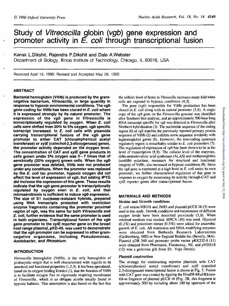
© 1990 Oxford University Press Nucleic Acids Research, Vol. 18, No. 14 4149
Study of Vitreoscilla globin (vgb) gene expression andpromoter activity in E. coli through transcriptional fusion
Kanak LDikshit, Rajendra P.Dikshit and Dale A.WebsterDepartment of Biology, Illinois Institute of Technology, Chicago, IL 60616, USA
Received April 13, 1990; Revised and Accepted May 29, 1990
ABSTRACT
Bacterial hemoglobin (VtHb) is produced by the gram-negative bacterium, Vitreoscilla, in large quantity inresponse to hypoxic environmental conditions. The vgbgene coding for VtHb has been cloned in E. coli whereit is expressed strongly by its natural promoter. Theexpression of the vgb gene in Vitreoscilla istranscriptionally regulated by oxygen. When E. colicells were shifted from 20% to 5% oxygen, vgb specifictranscript increased. In E. coli cells with plasmidscarrying transcriptional fusions of the vgb genepromoter to either CAT (chloramphenicol acetyltransferase) or xylE (catechol-2,3-dioxygenase) genes,the promoter activity depended on the oxygen level.The concentration of CAT and xylE gene products incells grown under 5% oxygen was 5 - 7 times that ofaerobically (20% oxygen) grown cells. When the vgbgene promoter was deleted, VtHb was not producedunder any conditions. When the promoter was replacedby the E. coli tac promoter, hypoxic oxygen did notaffect the level of expression of vgb, but adding IPTGdid increase the expression of this gene. These resultsindicate that the vgb gene promoter is transcriptionallyregulated by oxygen even in E. coli, and thatmicroaerobiosis is sufficient to induce vgb expression.The size of S1 nuclease-resistant hybrids, preparedusing RNA transcripts protected with restrictionenzyme fragments containing the promoter proximalregion of vgb, was the same for both Vitreoscilla andE. coli, further evidence that the same promoter is usedin both organisms. Transcriptional fusion of the vgbgene promoter to the xylE reporter gene on the broadhost range plasmid, pKD-49, was used to demonstratethat the vgb promoter can be expressed in other gram-negative organisms, including Pseudomonas,Azotobacter, and Rhizobium.
INTRODUCTION
Vitreoscilla hemoglobin (VtHb), is the only hemoglobin ofprokaryotic origin that is well characterized with regards to itsstructural and functional properties (1,2,3). It has been speculated,based on its oxygen binding kinetics (1), that the function of VtHbis to facilitate oxygen flux to vigorously respiring membranesof Vitreoscilla, which is an obligate aerobe usually found inhypoxic habitats. This assumption is also based on the fact that
the cellular level of heme in Vitreocilla increases many fold whencells are exposed to hypoxic conditions (4,5).
The gene (vgb) responsible for VtHb production has beencloned in E. coli along with its natural promoter (3,6). A singlecopy of the vgb gene on the Vitreoscilla genome was identifiedafter Southern blot analysis, and an approximately 500 base longRNA transcript specific for vgb was detected in Vitreoscilla afterNorthern hybridization (3). The nucleotide sequence of the codingregion (6) of vgb matches the previously reported primary proteinsequence of VtHb (2) and exhibits some sequence similarity withleghemoglobin genes (6). However, the non-coding upstreamregulatory region is remarkably similar to E. coli promoters (7).The regulation of expression of vgb has been shown to be at thelevel of transcription (8,9). The cellular level of the enzymes,delta-aminolevulinic acid synthetase (ALAS) and methemoglobin(metHb) reductase, necessary for structural and functionalintegrity of VtHb, also increased in hypoxic conditions (8). Sincethe vgb gene is expressed at a high level in E. coli under its ownpromoter, we further characterized regulation of this gene inresponse to oxygen by monitoring its activity through CAT andxylE reporter genes after transcriptional fusion.
MATERIALS AND METHODS
Strains and Growth conditions
E. coli strains HB101 and JM83 and plasmid pUC8:16 (3) wereused in this study. Growth conditions and maintenance of differentoxygen levels have been described previously (3,8). Whenminimal medium was needed, M9CA (10) was used. Glycerol(0.2%) and potassium nitrate (0.5%) were added for anaerobicgrowth of E. coli. All restriction and DNA modifying enzymeswere obtained from Bethesda Research Laboratories(Gaithersburg, MD) or New England Biolabs Inc.(Beverly, MA).Plasmid pDR 540 and promoter probe vector pKK232-8 (11)were obtained from Pharmacia, Piscataway, NJ, and pVDX18(12) was a generous gift from Dr. Vojo Deretic.
Plasmid constructionThe strategy for constructing reporter plasmids with CAT(chloramphenicol acetyl transferase) and xylE (catechol2,3-dioxygenase) transcriptional fusion is shown in Fig. 2. Fusionwith CAT gene was created by ligating the HindDI-MluI/Klenowfill-in fragment of plasmid pUC8:16 (Fig. 2B, this fragment isapproximately 500 bp including about 180 bp upstream of the
by guest on July 12, 2016http://nar.oxfordjournals.org/
Dow
nloaded from

4150 Nucleic Acids Research, Vol. 18, No. 14
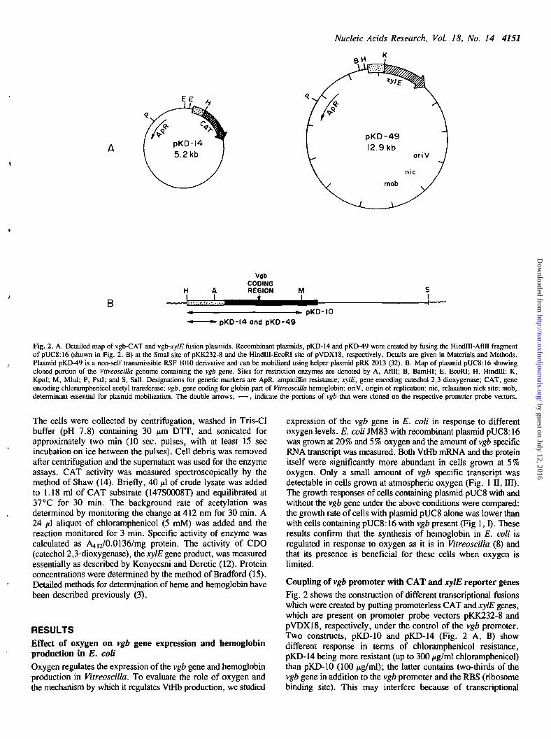
structural gene) at the Smal site of promoter probe vectorpKK232-8 which contains the promoterless CAT gene preceedingthe multiple cloning site. The resulting plasmid pKD-10 (Fig.2) contains the vgb promoter and two-thirds of the vgb codingregion. Orientation of the insert with respect to the CAT genewas checked by Afin digestion which has a unique site withinthe non-coding region of the vgb. Plasmid pKD-14, where onlythe vgb promoter region was directly coupled with CAT gene,was derived from pKD-10 by removing an Aflll-Mlul restrictionfragment and self ligation of the plasmid after filling the endswith the Klenow fragment of DNA polymerase. Both plasmidconstructs provide chloramphenicol resistance to the host cells.
Plasmid pUC8:17 carrying the promoterless vgb gene wasconstructed by removing the HindlTI-Aflll fragment from plasmidpUC8:16 (Fig. 2B) after treatment with these enzymes. Therestriction fragments were separated on low melting agarose gel(1.1%) and the larger Hindm-Alfn fragment was isolated,purified through a Nick Pack column (BRL), and self-ligated withT4 DNA ligase after filling the ends with Klenow DNApolymerase.
Plasmids pDR 540:16 and pDR 540:17 which carry the vgbgene with its native promoter and the E. coli tac promoter,respectively, were constructed according to the scheme shownin Fig. 4 ,1 . The HindlH-BamHI fragment of plasmid pUC8:16,which contains the vgb gene along with is promoter, was ligatedat the Hindm-BamHI site of plasmid pDR 540 to produce pDR540:16. In the control plasmid, pDR 540:17, the vgb promoterwas replaced with the E. coli tac promoter by cloning the Aflll-Sall fragment of pUC8:16 at the BamHI site of pDR 540 afterfilling in the ends with Klenow DNA polymerase followed byblunt end ligation. Colonies expressing the vgb gene under controlof the tac promoter were identified on plates supplemented with5 mM IPTG on the basis of their color difference due to highexpression of VtHb. The presence and orientation of the insertwas checked by restriction enzyme analysis. E. coli JM 103carrying the above plasmid contructs, i.e., pDR 540 (control),pDR 540:16, and pDR 540:17 were used to prepare RNA forcomparative vgb transcript analysis after Northern blotting asdescribed previously (8).
Another transcriptional fusion was created on the broad hostrange plasmid pVDX18 to assess vgb promoter activity inorganisms other than E. coli. For that, the Hindlll-Aflll fragmentof pUC8:16 (Afin end made blunt with the Klenow fragmentof DNA polymerase) was ligated at the HindHI-EcoRI (EcoRIend made blunt) site of pVDX18, which keeps the vgb promoterin the right orientation with respect to the promoterless xylE gene.Clones that contained an insert which promoted transcription inthe direction of the xylE gene were detected by the yellow colorof the colonies which appeared after spraying with a solution of100 mM catechol in 50 mM potassium phosphate buffer (pH 7.5).
SI protection analysis of vgb transcriptsThis was performed using a radioactive DNA probe containingthe 5' promoter proximal region and part of the vgb codingregion. To prepare the labelled probe, plasmid pUC8:16 wastreated with MM and the 5' end was labelled with polynucleotidekinase and [32P]ATP, after removing the 5' terminal phosphateby treatment with calf intestine phosphatase, essentially asdescribed by Maniatis et. al (10). The labelled fragments wereseparated on a 2% low melting agarose gel, and the HindHI-MluIfragment was isolated and purified through a Nick Pack column(BRL). Approximately 200 /*g of RNA from each strain (i.e.,
n
O 2 •» 6TIME (Hours)
m
20%
B
Fig. 1. vgb gene expression and hemoglobin synthesis in response to oxygen levels.I. E. coli JM83 with plasmids pUC8 or pUC8:16 was grown in LB containing100 /ig/ml Ampicillin at 37°C under different oxygen levels. DO level wasmaintained by bubbling a mixture of air and nitrogen through the medium. Sampleswere withdrawn with a sterile syringe at different intervals and absorbance wasmeasured at 600 nm. With pUC8 at 20% ( • ) and 5% (A) oxygen levels andwith pUC8:16 at 20% (O) and 5% (A) oxygen. II. CO-difference spectra ofE. coli JM83 carrying plasmid pUC8:16 under different oxygen levels. Cellswere suspended in 0.02 M phosphate buffer (7.2) at a final concentration of 20mg/ml (wet wt) and CO-difference spectra were recorded after bubbling CO intothe sample cuvette. A. Base line B. 20% oxygen and C. 5% oxygen. HI. Relativelevel of vgb specific transcript in E. coli grown under 20% and 5% oxygen levelswhich were maintained throughout growth by bubbling continuously a mixtureof air and nitrogen (8). Amount of total RNA blotted: A. 5 jig, B. 10 /ig, C.20 /tg, D. 30 ng. The relative intensities of the blots measured with a densitometerfor the 5% oxygen level were 1.00, 1.72, 2.34, and 3.09, respectively, and forthe 20% oxygen were 0.12, 0.18, 0.33, and 0.25, respectively.
Vitreoscilla, and E. coli carrying plasmids pUC8:16 andpUC8:17) was mixed with the labelled DNA probe (20,000 cpm),denatured at 80°C for 25 min and allowed to anneal at 52 °Cfor 6 h according to the procedures described in references 10and 13. The DNA-RNA hybrids were then treated with 200 unitsof SI nuclease (Pharmacia, NJ) and the protected fragments wereanalyzed on a 4% denaturing polyacrylamide urea (7 M) gel.
Isolation of RNA and dot blot analysisDetailed procedures for RNA isolation and vgb specific transcript
analysis have been described previously (8). Films were scannedwith a Model EC910 Densitometer from E-C Apparatus Corp.
Enzymes assayPromoter activity in cells harbouring the reporter plasmids wasassayed by measuring the enzyme activity in cell free extracts.
by guest on July 12, 2016http://nar.oxfordjournals.org/
Dow
nloaded from
Nucleic Acids Research, Vol. 18, No. 14 4151
K
BHI
AI
VgbCODINGREGION M
I
pKD-14 and pKO-49pKD-IO
Fig. 2. A. Detailed map of vgb-CAT and vgb-xylE fusion plasmids. Recombinant plasmids, pKD-14 and pKD-49 were created by fusing the HindlH-Aflll fragmentof pUC8:16 (shown in Fig. 2. B) at the Smal site of pKK232-8 and the HindlU-EcoRI site of pVDX18, respectively. Details are given in Materials and Methods.Plasmid pKD-49 is a non-self transmissible RSF 1010 derivative and can be mobilized using helper plasmid pRK 2013 (32). B. Map of plasmid pUC8:16 showingcloned portion of the Vitreoscilla genome containing the vgb gene. Sites for restriction enzymes are denoted by A, Aflll; B, BamHI; E, EcoRI; H, Hindlll; K,Kpnl; M, Mlul; P, PstI; and S, Sail. Designations for genetic markers are ApR, ampicillin resistance; xylE, gene encoding catechol 2,3 dioxygenase; CAT, geneencoding chloramphenicol acetyl transferase; vgb, gene coding for globin part of Vitreoscilla hemoglobin; oriV, origin of replication; nic, relaxation nick site; mob,determinant essential for plasmid mobilization. The double arrows, -—•, indicate the portions of vgb that were cloned on the respective promoter probe vectors.
The cells were collected by centrifugation, washed in Tris-Clbuffer (pH 7.8) containing 30 /tm DTT, and sonicated forapproximately two min (10 sec. pulses, with at least 15 secincubation on ice between the pulses). Cell debris was removedafter centrifugation and the supernatant was used for the enzymeassays. CAT activity was measured spectroscopically by themethod of Shaw (14). Briefly, 40 /tl of crude lysate was addedto 1.18 ml of CAT substrate (147S0008T) and equilibrated at37°C for 30 min. The background rate of acetylation wasdetermined by monitoring the change at 412 nm for 30 min. A24 |tl aliquot of chloramphenicol (5 mM) was added and thereaction monitored for 3 min. Specific activity of enzyme wascalculated as A412/0.0136/mg protein. The activity of CDO(catechol 2,3-dioxygenase), the xylE gene product, was measuredessentially as described by Konyecsni and Deretic (12). Proteinconcentrations were determined by the method of Bradford (15).Detailed methods for determination of heme and hemoglobin havebeen described previously (3).
RESULTSEffect of oxygen on vgb gene expression and hemoglobinproduction in E. coli
Oxygen regulates the expression of the vgb gene and hemoglobinproduction in Vitreoscilla. To evaluate the role of oxygen andthe mechanism by which it regulates VtHb production, we studied
expression of the vgb gene in E. coli in response to differentoxygen levels. E. coli JM83 with recombinant plasmid pUC8:16was grown at 20% and 5% oxygen and the amount of vgb specificRNA transcript was measured. Both VtHb mRNA and the proteinitself were significantly more abundant in cells grown at 5%oxygen. Only a small amount of vgb specific transcript wasdetectable in cells grown at atmospheric oxygen (Fig. 1 II, HI).The growth responses of cells containing plasmid pUC8 with andwithout the vgb gene under the above conditions were compared:the growth rate of cells with plasmid pUC8 alone was lower thanwith cells containing pUC8:16 with vgb present (Fig 1,1). Theseresults confirm that the synthesis of hemoglobin in E. coli isregulated in response to oxygen as it is in Vitreoscilla (8) andthat its presence is beneficial for these cells when oxygen islimited.
Coupling of vgb promoter with CAT and xylE reporter genes
Fig. 2 shows the construction of different transcriptional fusionswhich were created by putting promoterless CAT and xylE genes,which are present on promoter probe vectors pKK232-8 andpVDX18, respectively, under the control of the vgb promoter.Two constructs, pKD-10 and pKD-14 (Fig. 2 A, B) showdifferent response in terms of chloramphenicol resistance,pKD-14 being more resistant (up to 300 /ig/ml chloramphenicol)than pKD-10 (100 /tg/ml); the latter contains two-thirds of thevgb gene in addition to the vgb promoter and the RBS (ribosomebinding site). This may interfere because of transcriptional
by guest on July 12, 2016http://nar.oxfordjournals.org/
Dow
nloaded from
4152 Nucleic Acids Research, Vol. 18, No. 14
polarity with efficient translation of the CAT gene from thesecond RBS site present on plasmid pKK232-8 (11) sincepKD-14, which has a different attachment of the vgb promoterregion, strongly expressed CAT. This plasmid is constructed fromthe Hindm-Afin fragment of pUC8:16, in which the AfUI siteis within the RBS site of vgb. When this fragment was clonedon pVDX18 in the proper orientation, the xylE gene was stronglyexpressed. The xylE fusion was specially created to assess thevgb promoter activity in organisms other than E. coli becausethe presence of this reporter gene is easily detectable by the yellowcolor of colonies on spraying plates with a solution of catechol.
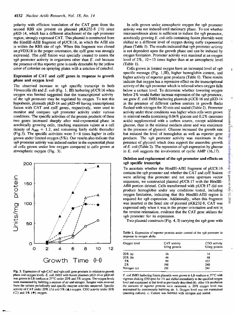
Expression of .CAT and xylE genes in response to growthphase and oxygen levelThe observed increase in vgb specific transcript in bothVitreoscilla(8) and E. coli (Fig. 1, HI) harboring pUC8:16 whenoxygen was limited suggested that the transcriptional activityof the vgb promoter may be regulated by oxygen. To test thishypothesis, plasmids pKD-14 and pKD-49 having transcriptionalfusion with CAT and xylE genes, respectively, were used tomonitor and compare vgb promoter activity under variousconditions. The specific activities of the protein products of thesetwo genes increased sharply after mid-exponential phase inaerobically growing cells, reaching maximum values at a celldensity of A ^ = 1.2, and remaining fairly stable thereafter(Fig.3). The specific activities were 5—8 times higher in cellsgrown under limited oxygen (5%). Our results also indicate thatvgb promoter activity was induced earlier in the exponential phaseof cells grown under low oxygen compared to cells grown atatmospheric oxygen (Fig. 3).
200
10 12
Growth Time (H)
Fig. 3. Expression of vgb-CAT and vgb-xylE gene products in relation to growthphase and oxygen levels. E. coli JM83 with fusion plasmids pKD-14 or pKD-49was grown in LB medium at 37°C under 20% and 5% oxygen. The oxygen levelswere maintained by bubbling a mixture of air and nitrogen. Samples were removedfrom the culture periodically and specific enzyme activities measured. Specificactivity of CAT under 20% (A) and 5% (A) oxygen. CDO activity under 20%(O) and 5% ( • ) oxygen.
In cells grown under atmospheric oxygen the vgb promoteractivity was not induced until stationary phase. To test whethermicroaerobiosis alone is sufficient to induce the vgb promoter,aerobically growing E. coli cells containing fusion plasmids wereshifted to a different level of oxygen during early exponentialphase (Table 1). The results indicated that vgb promoter activityis not dependent upon the growth phase and can be induced byoxygen limitation. Promoter activity was maximal at an oxygenlevel of 2%, 10—15 times higher than at an atmospheric level(Table 1).
Cells grown in limited oxygen have an increased level of vgbspecific message (Fig. 1,111), higher hemoglobin content, andhigher activity of reporter gene products (Table 1). These resultsindicate that oxygen has a repressive effect on the transcriptionalactivity of the vgb promoter which is relieved when oxygen fallsbelow a certain level. To determine whether lowering oxygenbelow 2 % would further increase expression of the vgb promoter,we grew E. coli JM83 harboring plasmids pUC8:16 and pKD-14in the presence of different carbon sources in growth flasksflushed with nitrogen for 30 min and sealed (Table 2). Promoteractivity under these conditions was higher when cells were grownin minimal media (containing 0.04% glucose and 0.2% casaminoacids) supplemented with a carbon source, except additionalglucose, than in the minimal medium alone and was maximumin the presence of glycerol. Glucose increased the growth ratebut reduced the level of hemoglobin as well as reporter geneproducts. The vgb promoter activity was maximum in thepresence of glycerol which does support the anaerobic growth,of E. coli (Table 2). The repression of vgb expression by glucosein E. coli suggests the involvement of cyclic AMP (16,17).
Deletion and replacement of the vgb promoter and effects onvgb specific transcriptTo ascertain whether the Hindlll-Aflll fragment of pUC8:16contains the vgb promoter and whether the CAT and xylE fusionswere utilizing this promoter and not some upstream vectorsequences, we constructed plasmid pUC8:17 with the HindHI-Afin portion deleted. Cells transformed with pUC8:17 did notproduce hemoglobin under any conditions tested, includingoxygen limitation, indicating that this Hindlll-Aflll region isrequired for vgb expression. Additionally, when this fragmentwas inserted at the Smal site of plasmid pKK232-8, CAT wasexpressed only when it was_ in the proper orientation and not inthe reverse orientation, evidence that the CAT gene utilizes thevgb promoter for its expression.
Two plasmid constructs (Fig. 4,1) carrying the vgb gene with
Table 1. Expression of reporter proteins under control of the vgb promoter inresponse to oxygen shifts.
Oxygen level CAT activityU/mg protein
CDO activityU/mg protein
20% (a)20% (b)
5%2%
Nitrogen (c)
164499
198191
1468
157240241
E. coli JM83 harboring fusion plasmids were grown in LB medium at 37°C withvigorous shaking (250 rpm) for 3 h and shifted immediately to the specified oxygenlevel and maintained at that level as previously described (8). After 1 h incubationthe amounts of reporter proteins were measured, a. 20% oxygen level wasmaintained by continuously bubbling air. b. Oxygen level was not maintained(standing culture), c. Culture was bubbled with nitrogen and sealed.
by guest on July 12, 2016http://nar.oxfordjournals.org/
Dow
nloaded from
Nucleic Acids Research, Vol. 18, No. 14 4153
Table 2. Effect of carbon sources on expression of hemoglobin and CAT.
Carbonsources
ControlSuccinateLactateGlucoseGlycerol
Growthatmosphere
airN2N2N2N2
Total Hbnmol/g wet wt
18321520163
297
CAT activityU/mg protein
8110112238
202
E. coli JM83 cultures with plasmids pUC8:16 or pKD-14 were grown overnightin 100 ml minimal media (9) supplemented with nitrate and different carbon sources(0 2%) under the indicated growth atmosphere at 37°C. Oxygen was flushed bybubbling nitrogen into each 250 ml flask for 30 min through a sterilized 0.45H Millipore filter and the flasks sealed with a rubber stopper plus parafilm.
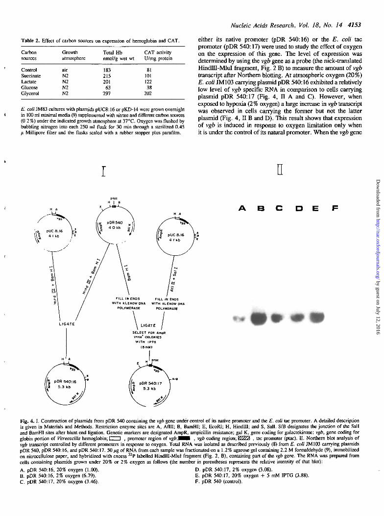
either its native promoter (pDR 540:16) or the E. coli tacpromoter (pDR 540:17) were used to study the effect of oxygenon the expression of this gene. The level of expression wasdetermined by using the vgb gene as a probe (the nick-translatedHindTU-MluI fragment, Fig. 2 B) to measure the amount of vgbtranscript after Northern blotting. At atmospheric oxygen (20%)E. coli JM103 carrying plasmid pDR 540:16 exhibited a relativelylow level of vgb specific RNA in comparison to cells carryingplasmid pDR 540:17 (Fig. 4, II A and C). However, whenexposed to hypoxia (2% oxygen) a large increase in vgb transcriptwas observed in cells carrying the former but not the latterplasmid (Fig. 4, II B and D). This result shows that expressionof vgb is induced in response to oxygen limitation only whenit is under the control of its natural promoter. When the vgb gene
r n
• E
FILL IN ENDSWITH KLENOW ONA
POLYMERASE
FILL IN ENDSWITH KLENOW DNA
POLYMERASE
LIGATE
SELECT FOR AmpRVIH0* COLONIESWITH IPTG
(SoMI
Fig. 4. I. Construction of plasmids from pDR 540 containing the vgb gene under control of its native promoter and the E. coli tac promoter. A detailed descriptionis given in Materials and Methods. Restriction enzyme sites are A, Aflll; B, BamHI; E, EcoRI; H, Hindlll; and S, Sail. S/B designates the junction of the Sailand BamHI sites after blunt end ligation. Genetic markers are designated AmpR, ampicillin resistance; gal K, gene coding for galactokinase; vgb, gene coding forglobin portion of Vitreoscilla hemoglobin; i i , promoter region of vgb.W^A , vgb coding region; y///A , tac promoter (ptac). II. Northern blot analysis ofvgb transcript controlled by different promoters in response to oxygen. Total RNA was isolated as described previously (8) from E. coli JM103 carrying plasmidspDR 540, pDR 540:16, and pDR 540:17. 50 /ig of RNA from each sample was fractionated on a 1.2% agarose gel containing 2.2 M formaldehyde (9), immobilizedon nitrocellulose paper, and hybridized with excess 32P labelled HindHI-MluI fragment (Fig. 2, B), containing part of the vgb gene. The RNA was prepared fromcells containing plasmids grown under 20% or 2% oxygen as follows (the number in parentheses represents the relative intensity of that blot):
A. pDR 540:16, 20% oxygen (1.00). D. pDR 540:17, 2% oxygen (3.08).B. pDR 540:16, 2% oxygen (6.79). E. pDR 540:17, 20% oxygen + 5 mM IPTG (3.88).C. pDR 540:17, 20% oxygen (3.46). F. pDR 540 (control).
by guest on July 12, 2016http://nar.oxfordjournals.org/
Dow
nloaded from
4154 Nucleic Acids Research, Vol. 18, No. 14
was cloned under control of the tac promoter it did not respondto hypoxia, but addition of IPTG, an inducer of the tac promoter,increased the level of vgb transcript (Fig. 4, IIE). No vgb specificmRNA was detected in cells containing control plasmid pDR 540(Fig. 4, H F).
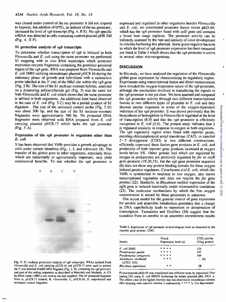
SI protection analysis of vgb transcriptsTo determine whether transcription of vgb is initiated in bothVitreoscilla and E. coli using the same promoter we performedSI mapping with in vivo RNA transcripts which protectedrestriction enzyme fragments containing the promoter-proximalregion of the vgb gene. RNA was prepared from Vitreoscilla andE. coli JM83 carrying recombinant plasmid pUC8:16 during thestationary phase of growth and hybridized with a radioactiveprobe labelled at the 5' end of the Mlul site within the vgb gene(Fig. 2 B). The size of the SI nuclease resistant hybrids, analyzedon a denaturing polyacrylamide gel (Fig. 5) was the same forboth Vitreoscilla and E. coli which shows that the same promoteris utilized in both organisms. An additional faint band observedin the case of E. coli (Fig. 5 C) may be a partial product of SIdigestion . The size of the untreated control probe (Fig. 5 D)was about 500 bp, and the size of the SI nuclease-resistantfragments were approximately 380 bp. No protected DNAfragments were observed with RNA prepared from E. colicarrying plasmid pUC8:17 which lacks the vgb promoter(Fig. 5 A).
Expression of the vgb promoter in organisms other thanE. coli
It has been observed that VtHb provides a growth advantage tocells under certain situations (Fig, 1,1, and reference 18). Thetransfer of the globin gene to other organisms, especially thosewhich are industrially or agriculturally important, may yieldcommercial benefits. To test whether the vgb promoter is
- 1 2 1
Fig. 5. S1 nuclease protection analysis of vgb transcripts. RNAs isolated fromVitreoscilla and E. coli carrying pUC8:16 and pUC8:17 were used to protectthe 5'-end labelled HindHI-MluI fragment (Fig. 2, B), containing the vgb promoterand part of the coding sequences as described in Materials and Methods. A 123bp DNA ladder (BRL) was used as the size standard. The SI protected fragmentfrom A, pUC8:17 control; B, Vitreoscilla; C, pUC8:16; D, unprotected anduntreated control fragment.
expressed and regulated in other organisms besides Vitreoscillaand E. coli, we constructed promoter fusion vector pKD-49,which has the vgb promoter fused with xylE gene and containsa broad host range replicon. The promoter activity can beindirectly assessed by the rate and intensity of color developmentin colonies harboring this plasmid. Some gram-negative bacteriain which the level of vgb promoter expression has been measuredare listed in Table 3 which shows that the vgb promoter is activein several other microorganisms.
DISCUSSION
In this study, we have analysed the regulation of the Vitreoscillaglobin gene expression by characterizing its regulatory region.Experiments using transcriptional fusion and direct measurementshave revealed the oxygen-responsive nature of the vgb promoter,although the mechanism involved in transducing the signals tothe vgb promoter is not yet clear. We have assessed and comparedthe vgb promoter activity through two different transcriptionalfusions in two different types of plasmids in E. coli and theyshowed similar responses in terms of the oxygen-dependentinduction of the vgb promoter. It was shown previously that thebiosynthesis of hemoglobin in Vitreoscilla is regulated at the levelof transcription (8,9) and that the vgb promoter is efficientlyrecognised in E. coli (3,6). The present study indicates that itis regulated similarly in response to oxygen in both organisms.The vgb regulatory region when fused with reporter genes,encoding chloramphenicol acetyl transferase (CAT), or catechol2—3 dioxygenase (CDO) in two different constructions,efficiently expressed these fusion gene products in E. coli, andproduction of both reporter gene products increased at oxygenlevels below 5%. Other genetic loci which are regulated byoxygen in prokaryotes are positively regulated by fnr or oxyRgene products (19,20,21), but the vgb gene promoter sequence(6) does not show any protein binding domain for these oxygeninduced positive regulators. Cytochrome <i of E. coli, which likeVtHb is synthesized in response to low oxygen, also showstranscriptional regulation and does not require the fnr geneproduct (22). Similarly, in Rhizobium meliloti expression of thenifA gene is induced maximally under microaerobic conditions(23). The molecular mechanisms by which the free oxygenconcentration is sensed by these promoters is unknown.
One recent model for the general control of gene expressionfor aerobic and anaerobic metabolism postulates that a changein DNA superhelicity leads to repression or derepression oftranscription. Yamamoto and Droffner (24) suggest that thetransition from an aerobic to an anaerobic environment results
Table 3. Expression of vgb promoter in heterologous hosts as measured by thereporter gene protein, CDO.
Strains
E. coli JM83Pseudomonas putidaPseudomonas aeruginosaAzotobacter vinellandi
(ATCC 475)Rhizobium japonicum
VisualExpression level (a)
* * * ** * * ** * * *
* * *
* *
CDO activityU/mg protein
124147144- (b)
28
Fusion plasmid pKD-49 was transferred into different hosts by triparental filtermating (32) using E. coli HB101 harboring the helper plasmid pRK 2013. a.The yellow color of the highest intensity that was observed in recombinant coloniesafter spraying with catechol solution is indicated by * * * *. b. Not determined.
by guest on July 12, 2016http://nar.oxfordjournals.org/
Dow
nloaded from
Nucleic Acids Research, Vol. 18, No. 14 4155
in gross changes in chromosomal superhelicity, which in turnactivate expression of a large number of genes. Oxygen regulatedtranscription of several genes, for example nif genes inRhodopseudomonas capsulata and Klebsiella pneumoniae (25)and photosynthetic genes in Rhodobacter capsulatus (26), maybe mediated by changes in the DNA superhelix. Similarly, thetonB promoter is very sensitive to changes in DNA supercoilingwhich occur in response to oxygen availability (27). Furtherexperimentation is required to determine if such a mechanismalso governs vgb expression, or whether there is negative orpositive regulation, governed by repressor or activator proteins,respectively.
One negative regulatory mechanism that affects the expressionof vgb besides oxygen is suggested by the inhibition of thesynthesis of globin and reporter gene proteins in the presenceof glucose. In microorganisms, several genes are controlledthrough glucose repression (16) which operates by affecting thecellular level of cyclic AMP (17) and such a mechanism mayalso control vgb expression in E. coli. Independent evidence forthe involvement of cyclic AMP or a cyclic AMP receptor protein,which are key elements in transcriptional control of the glucosesensitive operon (28), in vgb gene regulation has been reportedrecently (9). The transcriptional fusions described in this reportwill be helpful in extending the study in this direction.
In Vitreoscilla the biosynthesis of two enzymes, 6-amino-levulinic acid synthetase, the first enzyme in the heme biosyntheticpathway, and NADH-methemoglobin reductase, which keeps theprotein in its physiologically functional ferrous form, are alsoregulated by oxygen (8) as is Vitreoscilla catalase which has theprobable function of protecting the cell from hydrogen peroxideproduced by the autooxidation of VtHb (29). Whether theseproteins are regulated coordinately by oxygen using the sameor independent mechanisms is not yet known.
The SI mapping analysis (Fig. 5) furnished additional evidencethat both Vitreoscilla and E. coli use the same promoter. Onlyone SI-protected DNA fragment was observed in Vitreoscillaand it was identical to the size of the major fragment from E.coli. An additional minor band, about 35 bp larger andrepresenting 20% of the total intensity, observed in thepolyacrylamide gel for E. coli (Fig. 5 C) could be the result ofincomplete SI nuclease digestion. Alternatively, it could beinterpreted as evidence for the existence of a second promoterwhich has been reported by others using primer extension analysisabout 50 bp upstream of the first promoter (9). The length ofthe promoter region in our cloned vgb gene is at least as longas that used for the latter experiment (150 bp upstream of thestructural gene). Since there is no evidence for a second promotersite in the SI protected fragments from Vitreoscilla, either thepromoter region is recognized differently in E. coli or the extraband observed is an experimental artifact.
Under hypoxic conditions, E. coli cells that can synthesizeVtHb grow better than those cells that cannot. In Rhizobium,leghemoglobin has been shown to facilitate oxygen diffusion andrespiratory activity under conditions of very low oxygen pressure(30). VtHb is closely associated with respiratory membranes inVitreoscilla (1,31) and also in recombinant E. coli (31), whichlocation may facilitate respiration by a similar mechanism.
The vgb promoter in the broad host range pKD-49 was alsoexpressed in some other gram-negative bacteria with varyingefficiency (Table 3). We have also transferred the entire vgb genetogether with its own promoter into Pseudomonas where it ishighly expressed and produces hemoglobin in relatively large
amounts. Manipulation of vgb and other genes in organismsbesides Vitreoscilla and E. coli using the regulatory propertiesof this promoter appears very feasible.
ACKNOWLEDGEMENT
We are grateful to Dr. A.M. Chakrabarty for providing strainsof Pseudomonas and Azotobacter, Dr. V. Deretic for plasmidpVDX18, and to Dr. Ben Stark for helpful advice. This workwas supported by NIH grant GM27085.
REFERENCES
1. Webster, D.A. (1987) In Eiehhorn, G.L. & Marzilli, L.G. (eds) Advancesin Inorganic Biochemistry, Elsevier NY, Vol.7 pp. 245-265.
2. Wakabayashi, S., Matsubara, H., and Webster, D.A. (1986) Nature 522,481-483.
3. Dikshit, K.L. and Webster, D.A. (1988) Gene 70, 377-386.4. Boerman, S.J. and Webster, D.A. (1982) J. Gen. Applied Microbiol. 28,
35-43.5. Lamba, P. and Webster, D.A. (1980) J. Bacteriol. 142, 169-173.6. Khosla, C. and Bailey, J.E. (1988) Mol. Gen. Genet. 214, 158-161.7. McClure, W.R. and Hawley, D.K.(1983) Nucleic Acid Res. 8, 2237-2254.8. Dikshit, K.L., Spaulding, D., Braun, A. and Webster, D.A. (1989) J.Gen.
Microbiol. 135, 2601-2609.9. Khosla, C. and Bailey, J.E. (1989) J. Bacteriol. 171, 5995-6004.
10. Maniatis, T., Fritsch, E.F., and Sambrook, J. (1982) Molecular Cloning:A Laboratory Manual. Cold Spring Harbor, NY.
11. Brosius, J. and Lupski, J.R. (1987) Methods Enzymol. 153, 54-68.12. Konyecsni, W.M. and Deretic, V. (1988) Gene 74, 375-386.13. Berk, R.J., and Sharp, P.A. (1977) Cell 12. 721-732.14. Shaw, W.V. (1975) Methods Enzymol. 43, 737-755.15. Bradford, M. (1976) Analytical Biochem. 72, 248-254.16. Magasanik, B. and Neidhardt, F.C. (1987) In Escherichia coli and Salmonella
typhimurium: Cellular and Molecular Biology, Am. Soc. Microbiol.,Washingon, D.C. pp. 1313-1317.
17. Hempfling, W.P. and Beeman, D.K. (1971) Biochem. Biophys.Res. commun.45, 924-930.
18. Khosla, C. and Bailey, J.E. (1988) Nature 331, 633-635.19. Jamieson, D.J. and Higgins, C.F. (1986) J. Bacteriol. 168, 389-397.20. Jones, H.M. and Gunsalus, R.P. (1987) J. Bacteriol. 769, 3340-3349.21. Christman, M.F., Store, G. and Ames, B.N. (1989) Proc. Natl. Acad. Sci.
USA 86, 3484-3488.22. Georgiou, CD. , Dueweke, T.J. and Gennis, R.B. (1988) J. Bacteriol. 170,
961 -966.23. Ditta, G.E., Palomeres, V.A. and Kim, C.H. (1987) J. Bacteriol. 169,
3217-3223.24. Yamamoto, N. and Droffner, M.L. (1985) Proc. Natl. Acad. Sci. USA 82,
2077-2081.25. Kranz, R.G. and Haselkorn, R. (1986) Proc. Natl. Acad. Sci. USA 83,
6805-6809.26. Zhu, Y.S. and Hearst, J.E. (1988) Proc. Natl. Acad. Sci. USA 85,
4209-4213.27. Dorman, C.J., Barr, G.C., Bhriain, N.N. and Higgins, C.F. (1988) J.
Bacteriol. 170, 2816-2826.28. de Crombrugghe, B., Busby, S. and Buc, H. (1984) Science224, 831-838.29. Abrams, J. and Webster, D.A. (1990) Arch. Biochem. Biophys. (Accepted
for publication).30. Wittenberg, J.B., Appleby, C.A. and Wittenberg, B.A. (1972) J. Biol. Chem.
247,527-531.31. Khosla, C. and Bailey, J.E. (1989) J. Mol. Biol. 210, 79-89.32. Riess, G. and Puhler, A. (1984) In Puhler, A. & Timmis, K.N. (eds)
Advanced Molecular Genetics, Springer-Verlag, Berlin, pp. 51—63.
by guest on July 12, 2016http://nar.oxfordjournals.org/
Dow
nloaded from

by guest on July 12, 2016http://nar.oxfordjournals.org/
Dow
nloaded from
Copyright © 2022 FDOKUMEN