Study of the disease ass of chromosome 16, at
354
Study of the disease ass of chromosome 16, at brea CHATRI SETTASATIAN B.Sc. (Medical Technology) M.Sc. (Biochemistry) A thesis submitted in fulfrlment of the requirements for the Degree of Doctor of Philosophy Department of Paediatric 3 The Universþ of Adelaide Adelaide, Australia arm in( V July, 2003
-
Upload
khangminh22 -
Category
Documents
-
view
3 -
download
0
Transcript of Study of the disease ass of chromosome 16, at
Study of the disease assof chromosome 16, at
brea
CHATRI SETTASATIAN
B.Sc. (Medical Technology)
M.Sc. (Biochemistry)
A thesis submitted in fulfrlment of the requirements for
the Degree of Doctor of Philosophy
Department of Paediatric 3
The Universþ of Adelaide
Adelaide, Australia
armin(
V
July, 2003
Table of Contents
Page
Summary
Declaration
List of Publications
Acknowledgements
Abbreviations
Chapter 1: Literature Review
Chapter 2: Materials and Methods
Chapter 3: Characteúzation of Transcription Unit 3 (T3)
Chapter 4z Chancterization of Transcription Unit 12 (T12)
and Mutation Study of Spastic Paraplegia gene, SPGT 118
Chapter 5: Exon Amplification Study
and Characteúzation of Transcription Unit 13 (T13) I45
Chapter 6: Characteñzation of Transcription Unit 18 (T18) 180
Chapter 7: General Discussion 190
References 195
I
V
VI
VII
IX
54
86
1
Appendix: Publication
Summarv
The loss of the long arm of chromosome 16 has commonly been observed in.lhl 'sporadic breast and prostate cancers. Loss of heterozygosity (LOH) studies have identified
the tclomeric region at 16q24.3 to be one of the regiorf frequentþ lost in early stage breast I
cancer, suggesting the presence of tumor suppressor gene(s) involved in breast
tumorigenesis. Breast cancer is the most common malignant tumor among women and
comprises up to l8% of all cancers occurring in women. Due to the high incidence of breast
cancer and the correlation of this cancer with the LOH on 16q, identification of the tumor
suppressor gene(s) in this region has been targetted which will provide results applicable for
t,"fdiagnostic and therapeutic approaches. To this end, a detail physical and transcription map
of the 16q24.3 LOH has been est¿blished and used as a framework for the identification of
candidate genes.
The primary aim of this study was to clone the genes on this chromosome region and
identiff the candidate gene that may be involved in the development and progtession of
breast cancer. A number of genes that were characlenzed showed no evidence of
involvement in breast cancer but showed homology to other known functional proteins.
Some of these genes were predicted to have significant physiological function and one was
shown to be involved in one form of genetic disease.
The initial study was the identification of a minimal region of genomic loss at 16q24.3
in breast cancer. Collaborative studies of ¡"tíí¿fOH of a l6q24.3region were undertaken in
breast çancer cell lines and breast cancer samples. It was shown in some of the tumor
samples that LOH is restricted to less than I Mb encompassing the 16q24.3 physical map.
Subsequently, two genes were charactenzed from the tentative transcripts mapped to this
region.
\
I
The first gene, designated transcription unit 3 (73), codes for a protein containing two
known frmctional motiß with sequence homology to those of known proteiÍi f,redicted to )
have a function involved in transcription regulation. To study if this g"n"ïroolrocd in breast )<
cancer development, mutation analysis was undertaken in a set of tumor samples showing
16q24.3 LOH. No evidence_s of mutations w-ere detected and therefore this gene is unlikelV / ,, , ,
to be a tumor suppressor, at least for breast çancer. A partial sequence of the mouse
homologue of this gene has been reported and its expression appeared to be developmentally
regulated. To extend this study, a cDNA clone of the mouse homologue incorporating the
complete coding sequence was isolated and used to study its expression in mouse embryos.
Whole body in situ hybndization was conducted in mouse embryos and revealed the
expression of this gene predominantly in the developing nervous systÞms of the embryo.
The results from this study together with that from a previous report suggested that this gene
may be important in the embryonic development. .t
The second gene, by analysis of its protein sequence, was predicted to have frrnction
equivalent to the yeast mitochondrial metalloprotease necessary for mitochondrial protein
complex assembly. This gene is fotrnd to be identical to a published gene, ,SPG7, involved in
one type of neurodegenerative disease/, tþe hereditary spastic paraplegia (HSP). Subsequent
studies were undertaken. The genomic structure of SPGT was charactenzed, from a set of
overlapping genomic clones and was used to study gene mutations in a number of patients
with spastic paraplegia, both hereditary and sporadic cases. Mutations were detected in one
HSP family and the only afÊected case of other family. This study showed that SPGT
mutations involve in a small subgroup of HSP patients, consistent with the known genetic
heterogeneity of this disorder.
Since other genes identified at 16q24.3 also showed no evidence of mutations in breast
cancer, the region was extended by analysis of a flanking large genomic clone. Exon
il
trapping experiments were then used to identifr possible genes within this genomic segment.
Two groups of trapped exons were selected and characterized. By utilizing several
molecular biology techniques, as well as public sequence database homology searches, a
large cDNA with a complete open reading frame was assembled. This gene, assigned as
transcription unit 13 (TI3), encodes a high molecular weight protein containing nuclear
localization signals. Tl3 also contains a set of repeat sequence motifs similar to those of the
BRCA|-associated ring domain protein (BARDI) as well as those of the inhibitors of
nuclear factor trappa B (I-ËB). Since mutations of BRCAI are associated with the
development of hereditary breast cancer, and BARDI is required for its fltnction, Tl3 was
speculated to be one of the components involved in the pathway of BRCAI ñmction.
However, a mutation study failed to detect any tumor-restricted alterations in the TI3
sequence in a set of breast cancer samples withl6q24.3 LOH. This may indicate thatTl3 is
not the target of mutation inactivation in breast cancer. Alternatively, inactivation of 213
may be by a mechanism other than gene sequence mutation. \./
ln addition to Tl3, another gene on the same genomic segment was also characterized.
This gene, designated transcription unit 18 (718), resulted from the assembly of DNA
sequence from a number of partial cDNA sequences deposited in public database, from
reverse transcription cDNA amplification products, and from direct sequencing of a oDNA
clone. 218 codes for a protein member of AMP-binding protein, likety belonging to the
protein family of fatty acyl CoA ligase. Given the possible function of T18 in fatty acid
metabolism, this gene was not considered to be a tumor suppressor and therefore was not
subjected to the mutation analysis in breast cancer.
Although this study failed to identiff a candidate tumor suppressor gene involved in
breast cancer, the possible fi,nctions of the genes which were characteÅzed will provide
information for further biological studies for their involvement in other genetic disorders.
ilI
Among these, SPGT is an example of a gene that is involved in a genetic disease. T\e T3
gene, which is likely to be involved in embryonic development, may also be the target of a
developmentally related genetic disorder or be mutated in other types of tumors. Tl3, due to
its possible association with BRCAI function and/or relation with the I-kB, may be involved
in cancer progression but is targeted by other mechanism of gene inactivation. Continuation
of the functional studies of both T3 and TI 3 by others are underway and is beyond the scope
of this thesis.
ry


Ackn
,l)The study presenþ in this thesis was conducted in the Department of Cytogenetics /-
and Molecular Genetics, The 'Women's and Children's Hospital, in Adelaide. I am very
much grateful to the Department for providing me all facilities and resources that uffo*'i[fr" )
initiation and completion of all research work.
I would like to express my respectful thanks to my supervisors, Associate Professor
David Callen and Professor Grant Sutherland for their support, encouragement, and expert
supervision throughout this work. I also wish to thank them for critical review of this
thesis. Thanþou also to the Department of Paediatrics, The University of Adelaide for
their coordination of all aspects of the PhD program.
I am extremely grateful to The Royal Thai Government for providing a scholarship
and support during my study in Australia.
I also thank all members of the Department who were always very helpful in both
laboratory works and other hand-on works while I was in the Department. In particular, I
like to thank Dr. Scott Whitmore and Dr. Iozef Gecz for providing their appreciative'
technical knowledge and expert advice throughout the course of this study.
I also would like to thank Dr. Anne-Marie Cleton-Jansen in Leiden for the LOH data
and for providing the precious breast tumor DNA samples; Dr. Ram Seshadri, Sandra
Goldup, and Brett McCallum from the Flinders ttedical Centre in Adelaide for additional
LOH data as well as breast tumor DNA samples; Dr. Timothy Cox, and Sonia Donati of
VII

the Department of Genetics, the University of Adelaide for both materials and technical
assistance in whole-body in situ hybridizalion study of the mouse embryos.
Finally I would like to thank my family, in particula¡ my parents for their genuine
love and support. I also thank my wife Nongnuch who always provides help and support,
her love, patience, and truly understanding are extremely uppr"riuti#'.
VM
BAC:
bp:
BLAST:
oDNA:
cM:
CGH:
dbEST:
dNTP:
DNA:
DCIS:i,,;EST:
ET:
FAA:
FAB:
FISH:
HEX:
Kb:
LCIS:
LOH:
Mb:
trg:
pl:
mg:
Abbreviations
bacterial artifi cial chromosome.
base pairs.
basic local alignment tool.
complementary deoxyribonucleic acid.
centimorgan.
comparative genomic hybridization.
database of expressed sequence tags.
deoxynucleotide triphosphate.
deoxyribonucleic acid.
ductal carcinoma in situ.¿2.¿t.,4. ''r ' ", .'i ' i
expreSsed sequence tag.
trapped exon.
Fanconi anemia complementary group A.
Fanconi anemia./Breast cancer.
fluoresence in situ hybridization.
I -fluorescent 4,7,2' 4' 5'7',-hexachloro-6-carboxyfluorescein
kilobase pairs.
lobular carcinoma in situ.
loss of heterozygosity.
megabase pairs.
microgram.
microlitre.
milligram.
x

ng:
ml:
mRNA:
NCBI:
ORF:
PAC
PCR:
PSI-BLAST:
PSSM:
RACE:
RFLP
RNA:
RT-PCR:
SSCP:
STRP:
STS
THC:
TIGR:
UTR:
millilitre
messenger ribonucleic acid.
National Center for Biotechnology lnformation.
nanogffim.
open reading frame.
Pl (phage) artifrcial chtomosome.
polymerase chain reaction.
position-specific iterating BLAST.
position specifrc scoring matrix.
rapid amplification of oDNA ends.
restriction fragment length polymorphism.
ribonucleic acid.
reverse transcription polymerase chain reaction.
single stranded conformation polymorphism.
short tandem repeat polymorphism.
sequence tagged site.
tentative human consensus sequence.
The Institute of Genomic Research.
untranslated region.
Variable number of tandem repeats.
'Women's and Children's Hospital
Yeast artificial chromosome.
VNTR:
WCH
YAC:
X

Chapter 1
Literature Review

1.6.1 Construction of the Physical and Transcription map
1.6.2 Studies of the Genes within the 16q24.3 minimal LOH region
1.7 Aims and Scope of The StudY
50
51
52
1.1 Introduction
Genes are the fundamental units of every cell governing all cellular functions' Genes
are encoded by DNA, the basic biomolecule of the genome. The genetic information stored
in DNA dictates the structure of every gene product and delineates every part of the
organism. In eukaryotic cells, the genome is organized in the form of chromosomes situated
within the nucleus. Genes express their functions by encoding messages through mRNA
transcriptions which are then translated into functional proteins. This central dogma of
genetics was first proposed by Francis Crick in 1957. The final messages of the genes'
proteins, then exert their functions in determining several cellular phenotypes both
morphology and physiotogy. In multicellular organisms, complex organization of the cells
and the formation of specific tissues and organs require that gene expression are strictly
controlled, such that certain proteins are produced in the right cell, in the right amount, and
at the right time. Any deviation of this control may lead to a number of adverse conditions
that affect normal physiological processes.
Variations of the genes exist in human as well as other organisms and lead to the
phenotypic variations among individuals. Some human phenotype such as red-green color
blindness may be regarded as normal variants rather than a clinical disorder. However, some
gene variants may cause phenotypes that perturb the normal physiology and development.
These types of variant, therefore, associate with disorders or disease syndromes and are
generally described as "disease associated genes" or "disease genes". Some genes are
indispensable to embryonic function, so that deleterious mutations result in embryonic
lethality and are therefore unrecorded in humans. Some variants that cause abolition of gene
flrnction may normally have no effect on the phenotype because other non-allelic genes also
supply the same function, so called "genetic redundancy". Other forms of gene defect
a
1

disrupt the cellula¡ systems that control normal cell growth and differentiation- Such defect
can lead to uncontolled cell proliferation and subsequent neoplastic transformation.
The identification of genes associated with human diseases is important for the
investigation of cause and mechanism of the disease, which can lead to the development of
therapeutic intervention as well as of diagnostic application. To date, only a limited
percentage of the genes causing more than 5,000 Mendelian disorders has been identified
(McKusick, lggg). A number of identification approaches have been used to identiff such
disease genes. Before 1980, only a few human disease genes had been identified, these
involved a number of diseases with a known biochemical basis where purification of the
gene product was possible. Advances in recombinant DNA technology and the
establishment of the Human Genome Project, in the mid-1980s, allowed new approaches for
disease gene identification. In the last few years, the candidate gene and the positional
cloning approaches have played a major role in disease gene identification. The candidate
gene approach relies on partial knowledge of the disease gene firnction. This is based on the
availability of previously identified genes, whose features such as sequence domain and
expression pattern suggest that they may be implicated in the disease. In positional cloning,
the isolation of target gene relies exclusively on the map position of the disease locus in the
genome. The resources from the Human Genome Project have greatþ facilit¿ted this map-
based gene discovery. These include the development of new polymorphic genetic markers
and their physical locations on each human chromosome, which are necessary for the
genetic mapping of disease loci. ln addition, a large collection of partially sequenced cloned
cDNAs known as express sequence tags (ESTs) and the collection of complete hanscribed
sequences ofthe hypothetical genes deposited in public sequence database, have also been
valuable resources for gene identification. Another disease-gene identification strategy, a
position candidate gene approach, has become an altemative for positional cloning. This
2
combines knowledge of the map position of the disease locus with the availability of
candidate genes mapped to the same chromosome region'
uHuman chromosome 16 is one of chromosomes having been targeted for disease gene
identification. By linkage analysis, a ntrnber of disease loci have been mapped to this
chromosome, including the loci for familial Mediterranean fever (Flvß) at 16p13.3 (Pras ef
al., 1992), Crohn's disease at 16q72 (Hugot et a1.,1996), fanconi anemia complementation(-
group A (FAA) at 16q24.3 (Pronk et a1.,1995), and recently mapped, a recessive hereditary
spastic paraplegia (HSP) also at 16q24.3 ( De Michele et a1.,1993). In addition to linkage
mapping for such hereditary disease loci, the loss of heterozygosity (LOþ mapping, was
also used to define the chromosome region 16q24.3 as one of the region likely to harbor
tumor suppressor gene (s) associated with sporadic breast canc,er (dìscussed in section
r.s.4).
The refined mapping of both the FAA interval and the breast cancer tumor suppressor
region indicated that they were both located \¡rithin the same critical genomic region of
16q24.3 (The Fanconi anaemia/Breast cancer consortium,lgg6; Cleton-Jansen ¿f a1.,1994).
V/ith the objectives of cloning the FAA gene and a putative breast cancer tumor suppressor
gene, the primary physical map of the critical region was developed comprising an
integrated cosmid contig of approximately 650 kb. Based on this cosmid contig, a
combination of exon amplification and cDNA selection methods was successfully used to
isolate the candidate gene for FAA (The Fanconi anaemia./Breast cancer consortium,1996).
Further to this, a physical map was extended to nearly Mb utilizing cosmid, BAC, and
PAC clones. This physical map was then used as a framework for the development of
transcription map for subsequent gene identification (Whitnore et al.,l998a), especially for
the isolation of candidate breast cancer tumor suppressor gene (s). As part of the
chromosome 16-breast cancer project, the primary aim of the present study was to clone and
P
1fl+<..t'e,.u
Þ 0 1,+.t, ¿," 1
3
characterize the genes within this 16q24.3 LOH region for the candidate breast cancer tumor
suppressor, utilizing the available physical and transcription map (Whitmore et al.,l998a).
The following sections provide principles for disease gene mapping and gene isolation.
The mechanism of carcinogenesis with colorectal cancer as a model is discussed, and
"^_folorflty discussion on the present knowledge of breast cancer genetics and ,b
carcinogenesis. Studies on chromosome l6q LOH in sporadic breast cancer are also
discussed. Finally, the objective and scope of the present study are given in the last section.
1.2 Mapoine of Disease Genes
Identifrcation of genes associated with human diseases primarily requires the
knowledge of the approximate location of the genes on chromosomes for subsequent cloning
and identification of the candidate genes.
1.2.1 Genetic Mapping of Mendelian Trait
Traits or diseases caused by defects in a single major gene or biochemical pathway are
called Mendelian or single gene traits. Mapping of a disease-associated locus is based o4.{
genetic linkage and requires a detailed linkage map of the genome. Genetic linkage is
determined by measuring the frequency that two genetic loci are separated by meitotic
recombination and is described as "recombination frequency" or "recombination fraction"
(using a symbol *0") which is between 0-0.5. The correlation is the closer the two loci are,
the lower the 0 between them. The genetic distance between two loci is then measured based
on the O with the measuring unit is "centiMorg*"4("tr¡). Genetic distance is generally \:
correlated with the physical distance, which is defined by the number of base pair (þ) of
(.tt-J-Ak4"-- 4t)
olt{*^:o 6t
4
lZr-'ø\
DNA sequence between two genetic loci. One centiMorgan is correlated with the distance of
approximately one megabase (llvIb: 106 base pairs) ' <"4'a c'').4'-J.-'
Linkage analysis testjfor co-segregation of a marker and disease phenotype within a Å
pedigree to determine whether a genetic ma¡ker and a disease predisposing locus are
physicalty linked, that i7 in close physical proximity to each other. By typing ma¡ker loci at >
known locations in the genome, each marker can be tested for linkage to a disease or trait
and approximate the location of the disease or trait to the chromosomal region harboring the
linked markers. When large, multi-generation pedigrees are available, linkage analysis is a
powerful technique for the localization of disease genes'
l.2.2Mlaipping of ComPlex Disorder
In contrast to Mendelian traits, complex or multifactorial diseases result from the
interaction of multiple genes and environmental factors. These complex disorders include
cardiovascular disease, rheumatoid artbritis, diabetes, some for4þf psychological disordet ,X
such as schizophrenia" and cancer. The complex, non-Mendelian pattern of inheritance due
to the involvement of multiple genes and the influence of environmental factors in suchp't 'tJb'(a
disorders makes it impossible or rarelynto find the large multi-generation pedigrees suitable
for mapping of the associated genes by direct linkage analysis. However, in some form of
complex disorders near-Mendelian families can be selected for linkage analysis. For
instance, in breast cancer, few families can be found with many affected individuals in a
pattern consistent with autosomal dominant inheritance whereby linkage analysis can be use
for mapping of disease gene. Alternative procedwes for mapping complex traits namely "sib
pair analysis" and "association analysis" are described elsewhere (Thomson and Esposito,
tgee),
5
1.3 ldentification of Human l)isease Genes
procedures have been developed for the identification of genes associated with human
diseases. Without the data of genomic sequence encompassing the candidate region
positional cloning approach is the one that has successfully been used to identif several
disease-associated genes. It involves construction of a physical map of the candidate region
for subsequent identification of the disease-associated gene.
1.3.1 Construction of the Physical Map encompassing the Candidate region
Once the disease locus is mapped to a region of the chromosome, construction of a
physical map encompassing such chromosome region is required to allow subsequent
identification of candidate genes. Essentially, a physical map consists of an overlapping set
of large genomic clones that are arranged in a contiguous order, with respect to the
chromosome orientation. Cosmid clones (average insert size of 40 kb) have initially been
used to build the genomic contig of a physical map. Alternatively, the larger genomic clones
used to build a contig can be yeast artificial chromosomes (YACs), bacterial artificial
chromosomes (BACs), or Pl phage recombinant (PACs). YACs have an advantage over
other cloning systems in that their large insert size (350-1000 kb on average) facilitates the
construction of maps with long-range continuity (Burke et al., 1987; Burke, 1991).
However, most YAC libraries have high rates of chimerism and deletions that can limit their
utility (Green et al., l99l; Selleri et al., lgg2). BAC and PAC clones, though ptovidfd
smaller insert size (average 120-200 kb and 75 kb respectiveþ offer high clonal stability
and reduced cloning biases (Shizuya et al., 1992; Ioannou et al., 1994). Hoïvever, a more
refined genome contig, ¡lilizing cosmid clones may also be required for subsequent
construction of transcript map for further gene isolation.
6
1.3.2 Finding Genes in Cloned DNA
The established contig of cloned genomic DNA provides a basis for the isolation ofIv-o,
genes within the critical interval. A variety of strategies@e been developed and used for
gene detection.
1.3.2.1 Gene detectìon by CpG Island Mappìng
This method is based on the knowledge that about half of all vertebrate genes are
characterized by the presence of C-G rich regions consisting of nonmethylated CpG
dinucleotides, originally called *HpaII Tiny Fragment (HTF) islandJ'. The C-G content of þ
these regions is about 60-70% compared with the average 40Yo for the human genome.
These CpG islands are most often found at the 5' end of the genes, associated with the
promoter region (Bird, L987;Antequera and Bird, 1993). CpG islands have been found to be
associated with all house-keeping genes and about 40% of tissue restricted genes (Larsen et
a1.,1992; Antequera and Bird, 1993).
The presence of CpG island can be detected by the use of restriction endonucleases
with CpG dinucleotides in their recognition sequence, such as NofI (recognition sequence 5'
GCGGCCGC a',), -BssHII (5', GCGCGC a',), MluI(S',ACGCGT 3',), and Narl(S', GGCGCC
3'). Since most of these restriction enzymes a¡e inhibited by enzymatic methylation of
cytosine residues, they are more likely to cleave DNA at the location of CpG islands than at
intra- or intergenic regions that are usually methylated. Therefore, long-range restriction
maps can be derived that identit the location of genes over large distances @ird, 1986;
l9S7). Such an approach has allowed identification of CpG islands and has assisted in the
identification of many genes (Rommens et al., 1989; Tribioli et al., 1994). The results,
however, sometimes depend on the known tissue-specific va¡iation in methylation patterns
at CpG islands. In cloned sources of DNA, where the original pattern of cytosine
.\
7
4{r.1".^.:--i
methylation is erased, only the clustering of restriction sites for CpG^enzymes is often taken
as an indication for CPG islands.
1.3.2.2 Gene Detection Bøsed on Recognítíon of Exans
This gene detection method is based on the utilization of splice sites in a functional
assay (Auch and Reth, 1990; Duyk et a1.,1990; Buckler et a1.,1991). In principle, genomic
fragments are cloned into the intron of a reporter construct that provides splice acceptor and
donor sites. Upon transfection into a suitable cell line, the RNA is expressed and the mRNA
splicing machinery generates chimeric mRNAs containing exon fragments from the cloned
piece of DNA. Because the sequences flanking the novel exon are known, polymerase chain
reaction (PCR) can be used to ampliff sufficient amounts of oDNA for cloning and
subsequent sequence analysis. This procedure, generally called "eJcon trapping!' 01 "exon
amplification", was a popular gene detection method because of its ability to recognize
transcribed seqgences regardless of their tissue expression pattern and also provided a high
success rate in a number of positional cloning projects in the early 1990s.
Because exon trapping is based on splice site recognition, artifacts can be generated.
Three classes of exon fragments are always detected: (Ð true exons, which are recovered via
the utilization of bona fide splice sites, (ii) exon fragments that are trapped using one bona
fide splice site and a second cryptic splice site, (iii) fragments that are trapped because two
cryptic splice sites are used. The fnst two types of exon fragments are generally acceptable,
because they provide at least partial sequence information about exons. Only the third class
is problematic, because it is the result of artifactual splicing reactions.
The classical versions of exon tap/amplification vectors were designed to detect only
internal exons, exons that contain both splice acceptor and splice donor sites. However,
k)
8
alternative modifications of the system have been described, whereby the 3'-terminal exon
having only a splice acceptor site can also be detected (Datson et al',1994)'
1.3.2.3 Gene Detection Based on Sequence ldentíty between Genomic DNA and cDNA
This method, termed "cDNA selection", is based on the formation of heteroduplexes
between genomic DNA and cDNA (Lovett et al., l99l; Parimoo et al., 1991)- The fust
development of cDNA selection was based on the use of cDNA inserts, amplified from a
cDNA library of interest using vector specific primers, to screen the genomic clones- These
.DNA products are radioactively labeled and used to probe YAC or cosmid inserts that have
been immobilized on nylon membranes. The cDNA inserts that specifically hybridize to the
cloned genomic DNA are then eluted from the membranes and re-amplified with vector
specific primers. The resulting cDNA sublibraries, enriched for expressed sequences from
the genomic region, are then cloned and analyzed'
The second-generation version of this method has been developed where both sources
of DNA (genomic and cDNA) are converted into a form that is easily amplifrable by PCR.
This is achieved by ligation of adapters to their respective ends (Morgarr et al-, 1992;Kom
et al., lgg2). By utilizing two adapters that difler in sequence, cDNA and genomic DNA
fragments can be amplified independentþ from each other by their specific adapter primers.
In the procedure, briefly, the amplified cDNA fragments are mixed with the amplifred
fragments of genomic DNA generated from particular genomic region. The mixture is
denatured and allowed to reanneal, from which both homoduplexes and heteroduplexes are
formed. To allow for the separation of heteroduplexes (cDNA:genomic DNA) from
homoduplexes of cDNA fragments, the amplifred fragments of genomic DNA are labeled,
for instance by biotinylation of their specific primer prior used for PCR. This allows the use
of affinity chromatography or atrinity beads (eg. streptavidin coated magnetic beads) for
9
I
sepÍ*ation. The selected cDNA fragments can subsequently be isolated from heteroduplexes
by pCR using cDNA specific-adapter primers. Since the heteroduplex formation occtrs only
between the fragment of genomic DNA and cDNA with sequence identity, the selected
'DNA, therefore, represents the coding sequence of the gene in particular genomic region.
Because the selected cDNA fragments are usually larger in size than the tapped exons,
transition to full length transcripts is usually easier than that from tapped exons. This
method has been successful in the isolation of a number of genes (Rommens et al-, 1993;
peterson et al., 1994; Baens et a1.,1995) including those responsible for human diseases
(Gecz et al., 1993; Onyango et al-, 1998)
1. 3.2.4 Posítional Candidate Analysßirr
This approach is based on the fact that,number of sequenced genes or parÇof genes, Y ,r
often in the form of expressed sequenced tags (EST) and their chromosomal localization are
increasing rapidly and are publicly available (Ballabio, 1993; Collins, 1995a). This
procedure combines the mapping of a disease locus to a defined region with the availability
of candidate genes mapped to the same chromosome region. Many mapped genes are ofq ,lul.{.
known function, or"function^can be predicted, though some are novel and are of unknown I
function. In many cases reasonable hypotheses as to the nature of a gene associated with a
given clinical phenotype can be made. Relevant candidate genes can then be analyzed for
.J- 1
the presence of disease-associated mutations. This approach has made a successive impact
on the isolation of many disease-associated genes (Xu ef al., 1998; Pandey and Lewitter,
L999;Hofinann et a1.,2001; Bleck et al-,2001)
1.3.2.5 Dírect Sequencíng of lhe Critícal regíon
10
Determination of the nucleotide sequence across a critical interval may provide the best
possible way to identiff candidate genes. With this approach, the coding region can be
identified by computerized gene detection metho$ Indeed, large-scale sequencing has !
already been successfully applied in the positional cloning project, for instance the
identification of a second breast cancü susceptibility gene, BRCA2 (V/ooster et al-,1995).
In addition , a Large number of partial cDNA sequences (ESTs) as well as complete
sequences of cloned cDNAs or even full range sequences of novel transcripts are available
and can be retrieved from public databaseJ These can facilitate the identification of genes K
utilizing homology search tools for the sequence homology between genomic DNA and
cDNA from databasej
1.3.3 Generation of Transcript Maps
The transcribed sequence information isolated by the above methods either as cDNA
clones or trapped exons can then be mapped to their corresponding genomic clones in the
critical region. This creates an overall map of the transcribed sequences arranged in order
with respect to the genomic clone-contig. The transcribed sequences that arrange in cluster
can be grouped and treated as a "hanscription unif', representing a tentalive gene.
The resulting transcript maps can faciliøte cloning of the genes related to the disease locus.
1.4 Human Cancer: A Complex Genetic Disease
Cancer is a generic term for genetic diseases involving uninhibited cellula¡
proliferation caused by the mutations of multiple genes that result in the disruption of the
normal harmonious checks and balances contollingrihis O-ùt within normal cells. These
mutations usually occur in genes that play a vital role in the regulation of cell growth and
differentiation. In normal tissues, homeostasis is maintained by ensuring that as each stem
11
O.rl. 4a. t o' 'ú¿
cell divides,^only orr"if ttt" two daughters remains in the stem cell comparhent, while the
other is committed to a pathway of differentiation (Cairns, 1975). The control of cell
multþlication will therefore be the consequence of simals affecting these processes. These
signals may be either positive or negative, and the acquisition of tumorigenicity results from
genetic changes that affect these control points.
1.4.1 Genes involved in Cancer
The information obtained from the cytogenetic analysis of cancer cells with recurrent
chromosome abnormalities together with the study of hereditary cancer syndromes has
provided a way for the identification of genes involved in cancer.
The first evidence of a genetic event in cancer was the discovery of the Philadelphia
chromosome (Pht) in chronic myelocytic leukemia (Clvfl-) (Nowell and Hungerford, 1960).
This chromosome was first believed to be an abnormal chromosome 21. After a decade, it
was Rowley (in 1973) who demonstrated that Phr originated from a specific translocation
between chromosomes 9 and 22.Tenyear later molecular cloning of the tanslocation break-
points showed that the translocation intemrpted a break- point cluster region (BCR) gene on
chromosome 22 and the Abelson oncogene (c-ABL) on chromosome 9 and created a new
hybrid BCR-ABL oncogene (De Klein et al., 1982; Heisterkamp et al., 1983, 1985).
Expression of this chimeric oncogene produces a tyrosine kinase related to the ABL product
but with abnormal transforming properties. Other chromosomal translocations rËrJa""U,
for example those seen in the majority of cases of Burkitt's leukemiu irl',nol',r.¿ tft"
chromosome translocations t(8;14), t(2;8), and t(8;22) (Croce and Nowell, 1985; Klein,
l9S9). These translocations lead to the activation of the c-Myc oncogene at the chromosome
8 break-point as a consequence of the conhol of immunoglobulin gene heavy, kappa or
lambda chain regulatory elements located at the break-points of chromosome 14, 2 artd 22
T2

respectively (Rabbits, l9g4). Non-random or recurrent chromosomal aberrations have also
been reported in a number of solid tumors, many of which involved gene fusion and
activation of oncogenes at the break-points (Sanchez-García, 1997; Mitelman et al-,1997).
Activation of "oncogenes" provides a positive signal for tumor progression while
another goup of genes, "tumor suppressors", act as negative regulators of cellular
proliferation. Inactivation of these tumor suppressor genos is considered to initiate
uncontrolled cellular growth and tumor fomration'
The concept of a tumor suppressor gene is derived from two lines of evidence. The
first, from cell hybridization experiments, showed that the neoplastic phenot¡'pe can often be
suppressed by fusion of tumor cells with normal cells, resulting in the outgrowth of non-
tumorigenic hybrids (reviewed by Harris, l98S). These observations indicated that normal
cells were donating genetic information capable of suppressing the neoplastic transformation
of the hybridized tumor cells. This suppressing ability is an important functional
characteristic of tumor suppressor genes.
The second line of evidence was from epidemiological studies by Knudson in inherited
retinoblastoma, a childhood tumor of retinal tissue (Knudson, l97l). Knudson postulated
that development of retinoblastoma requires two successive mutations of the homologous
alleles in the cell genome. From this "two-hit" hypothesis two forms of mutation were
proposed in familial and sporadic retinoblastomas. h familial retinoblastoma, which occurs
bilaterally in most cases, the first mutation is constitutional occurring in the germline and is
therefore inherited, while the second mutation is a subsequent somatic event in the
retinoblast. In contrast, sporadic retinoblastoma results from two independent somatic
mutations in the retinoblast and is thus unlikely to be bilateral. The "two-hit" event required
for the development of retinoblastoma is likely to inactivate a gene which the normal
function is important in regulating the nonnal cellular growth. Cytogenetic analysis has
/
13
shown that a few hereditary retinoblastoma cases carried a constitutional chromosome band
13q14.1 deletion in all somatic cells (Francke and K*g, 1976; Knudson et al., 1976)'
Further study in sporadic cases has also occasionally detected 13q14 deletions in tumor cells
(Balaban et al., lg82). Molecular genetic studies (Cavenee et al., 1983) as well as the
subsequent isolation of the gene (Friend et a1.,1986) have shown that Knudson's two elusive
genetic targets were the two copies of the retinoblastoma tumor suppressor gene (RBI)
located on the long arm of chromosome 13 at 13q14. The two mutational events, either
constitutional or sporadic involved the inactivation of both functional copies of this gene.
The RBl gene has subsequently been characterized and found to be mutated or lost in
several cancers, including osteosarcoma (Friend et al., 1986), small cell carcinoma of the
lung (Harbotn et al., 1988; Horowitz et a1.,1990), bladder cancer (Horowitz et a1.,1990)
and breast carcinomas (Lee et al., 1988; T'Ang et al., 1988; Horowitz et al., 1990)'
Subsequent studies have established that the tumor suppressor function of RB protein (pRB)
is through the inhibition of the cell cycle during the G1/S phase progression (reviewed by
'Weinberg, 1995).
Studies in other hereditary cancer syndromes have resulted in the isolation of other
tumor suppressor genes with recessive germline mutations predisposing individual to cancer
formation. These also are consistent with Knudson's "two-hit" model for complete
inactivation of a tumor suppressor gene. These include the APC gene in familial
adenomatous polyposis (FAP), a syndrome predisposing to colorectal cancer (Bodmer et al.,
1987; Groden et al., l99l; Nishisho et al.,l99l); TP53 gene in Li-Fraumeni syndrome, a
syndrome of multþle cancer predisposition (Li and Fraumen, 1975:' 1982; Malkin et al.,
1990; Srivastava et al., 1990); and the VHL gene in von Hippel-Lindau s¡mdrome, a
syndrome with multiple hemangioma and predisposition to renal cell carcinom4
phaeochromocytoma, and pancreatic tumor (Lattf et al-,1993).
T4
T
1.4.2 Tumor Suppressor Gene and Loss of Heterozygosity
The involvement of tumor suppressor genes, such as the RB/ gene in tumorigenesis of
retinoblastomq appears to be by mutations resulting in loss of function. In the inherited
form of retinoblastoma" the somatic mutation that affects the second allele of the kBI gene
¿7'
is likely tobeþéchromosomal event, which uncovers the constitutional recessive mutation.
This event was demonstrated by Cavenee and colleagues (1933). They utilized a series of
RFLp (restriction fragment length DNA polymorphism) markers at chromosome 13q14 to
type DNA ûom the surgically removed tumor material and from blood samples taken from
the same patient. They found that in several patients the constitutional lymphocyte DNA
was heterozygous for some l3ql4 markers while the tumor cells were apparently
homozygous. Such apparent homozygosity or "loss of heterozygosity (LOÐ" was suggested
to be the somatic equivalent of Knudson's "second hit". This LOH resulted in the loss of one
functional copy of a tumor suppressor gene but also included nearby DNA markers on the
chromosome. As a result of combined cytogenetic analysis and stirdies of additional 13q14
markers, a number of chromosomal mechanisms leading to the loss of the wild-t¡'pe
ftrnctional allele, were proposed (lllgure 1.1). These include the loss of an entire
chromosome by mitotic non-disjunction, mitotic recombination, unbalanced translocation or
chromosome deletion. Subsequently, such events were often associated with reduplication of
the remaining chromosome. The frequency of LOH in tumor cells has been estimated to be
at least two order of magnitude higher than that of point mutation, and thus it is the favored
mechanism to eliminate the wild-t¡'pe allele of atumor $rppressor gene (Weinberg, l99l).
Since the loss of a specific chromosome region usually affects both the putative tumor
suppressor gene and the neighbouring genes or genetic markers, typing of genetic markers
can therefore be used to identiff the chromosome region likely to harbor the putative tumor
15

Marker A I
Marker B I
AT
.,
\ü
t
,
(iv(i)
Rb w
BT ,,
AI
Rb
BI
I 1 t2
Rb Rb
l1
I
Rb
,
Rb
I I
NTNT NTNT NTNT NTNT NTNT
Fieure 1.1 : Diagram representing the mechanism of loss of wild-type allele (w) in retinoblastoma
(RB) studied by Cavenee et al., (1983). (y' Loss of a whole chromosome by mitotic non-disjunction.(ri) Loss and followed by reduplication of the Ró chromosome, (üì) Mitotic recombination proximal tothe .l?ó locus, followed by segregation of both Rá-bearing chromosome into one daughter cell. (ív)Deletion of the wild-type allele. (u) Pathogenic point mut¿tion of the wild type allele. The
corresponding results of genotyping normal (N) and tumor (T) DNA for the two markers, A and B, are
represented by the figures underneath that indicate the patterns of loss of heterozygosity (LOH) in (i),(ii), (iii), and (iv), but not (v).
AIA2BIBI
B2
A1
ß2
A1A2BI
AIA2BI

suppressor gene through LOH analysis. The LOHs at many chromosome regions have been
found in several solid tumors indicating the possible location of multiple tumor suppressor
genes involved in tumorigenesis.
1.4.3 Multistage Carcinogenesis: The Paradigm of Colorectal Cancer
Human carcinogenesis is a multistage process starting from a clonal expansion of the
cell that gains a selective g¡owth advantage by genetic events. Further genetic alterations
accumulated during progression cause the phenotypic alteration of the cells to change into
more malignant phenot¡pes. Clinically, cancer development and progression can be divided
into sequential events: from normal cells to preneoplastic lesions, to primary tumors and
frnally to more aggtessive metastases. Molecular genetic studies have revealed that such
development and progression of human cancer requires multþle genetic alterations. This
multistage process is well illustrated by studies of colorectal cancers.
Studies have defined ¡wo forms of hereditary predisposition to colorectal cancer:
familial adenomatous poþosis (FAP) and hereditary non-polyposis colorectal cancer
(HNpCC). These two diseases have providedfinformation leading to an understanding of
colorectal carcinogenesis. It is assumed that other human tumors will follow a similar,
though not the same, tumorigenic pathway.
In FAP a stepwise model of colorectal carcinogenesis was developed by Fearon and
Vogelstein (1990). Mutations of the adenomatous polyposis coli tumor suppressor gene
(APq initiate the formation of preneoplastic lesion, the "adenomatous poþs", in bothr /, "v,, ¡
1.
inherited (FAP) and sporadic cases.'The next stage in progression to malignancy involves
&_lr,Loncogenic K-ras mutations which arose during the adenomatous stage. Mutations of IP53
and deletions of chromosome l8q coincidey' with the transition to malignancy. Subsequent
f
16
work has identifred additional genetic events as well as the molecular pathways perturbed by
each of these mutations.
T\e ApC mutation appears to be the rate-limiting event for colorectal tumorigenesis-
Individuals with germline mutations of ,4PC are at high risk, but do not necessarily develop
colorectal cancer. However, tumor fomration will be initiated by a somatic mutation of the
wild type ApC allele inherited from an unaffected parent (Ichii et al., 1992; Levy et al-,
1994;Luongo et al.,lgg4). Furthermore, somatic mutations of both alleles of the APC gene
were also found in a large number of sporadic colorectal tumors (Miyoshi et al-, 1992;
powell et al.,lgg2). Although the APC gene product is also ubiquitously expressed in other
tissues than the colonic epithelium, individuals with constitutional APC mutations rarely
developed tumors in other organs. Thus it was speculated that APC gene behaves as the
,'gate¡eeper" of colonic epithelial cell proliferation and its inactivation is required for net
cellula¡ growth (Kinzler and Vogelstein, 1996). Nonnally, "gatekeeper genes" are required
for maintaining a constant cell number in renewing cell populations, ensuring that cells
respond appropriately to the situations requiring net cell proliferation, such as in tissue
damage. Mutation of the gatekeeper thus leads to the permanent imbalance of cell g¡owth
over cell death. The gatekeeper functionof APC is important in colorectal tumor formation.
Mutations of other genes known to be involved in the tumorigenic process, such as ZP53
and K-ras, fail to promote neoplastic transformation of colonic epithelial cell if the
gatekeeper gene, APC, is still intact (Kinzter and Vegelstein, 1996). Other genes may also(ní,n'n. ,utc,4
perform the gatekeeper role in other tissues, theée ard the NF1 gene in Schwann cells, the
RBI gene in retinal epithelial cells, and the VHL gene in kidney cells (reviewed in Knudson,
1ee3)
The APC tumor suppressor gene encodes a large multidomain protein (APC) which
appears to interact and modulate the cytoplasmic level of p-catenin, a downstream effector
t7
of the wnt signaling pathway (Rubinfeld et al.,1993; Gumbiner, 1995). APC in association
with serine threonine glycogen synthase kinase (GSK)-3Þ regulates the low levels of free p-
catenin. GSK-3p phosphorylates both APC and p-catenin facilitating APC/p-catenin
complex formation and subsequent targeting p-catenin degradation (Rubinfeld et a1.,1996;
Aberle et al., 1997; Behrens et a1.,1998). ln APC mutant colonic cells, degradation of
cytoplasmic p-catenin is disrupted and the levels of p-catenin rise dramatically, suggesting
its role in the transformation of colonic epithelium into benign polyps (Peifer, 1997)-
p-Catenin was originally identified on the basis of its association with cadherin
adhesion molecules. p-catenin and its homologue, Armadillo protein in Drosophil4 were
recognized as essential components of the Wnt/Wingless signaling pathway (Gumbiner,
1995). Wnt/Wingless signals are known to direct many key developmental decisions
including the regulation of anterior-posterior and dorsal-ventral pattern in both flies and
vertebrates (Miller and Moon, 1996). The V/nt/Wingless signal, by antagonizing GSK-3P
activity, disrupts the APC/p-catenin association, which prevents p-catenin from degradation
and therefore stabilizes p-catenin @apkoff et al., 1996). High level of p-catenin then
transduces the signal by translocating into nucleus and interacting with T-cell
factorllymphoid enhancer factor (TcflLef¡ famity of HMG box transcription factor. TCF-4
is the predominant member of this family of transcription factors in colonic epithelial cell
(Korinek et al.,lgg7). Upon binding with p-catenin, TCF-4 is activated and it up-regulates
many responding genes including the oncogenes MYC and CCNDI which play role in cell
growth by promoting the Gl-'S phase of the cell cycle (fle et a1.,1998; Mann et a1.,1999l'
Tetsu and McCormick, 1999). These findings suggest that p-catenin is a "driver" that up-
regulates TCF-responsive genelcritical for proliferation and transfonnation of colonic
epithelial cells. Mutations of p-catenin gene (CTNNBI) has been reported in a subset of
")
18
colorectal tumors with intact APC (Morin et aI., 1997; Sparks et a1.,1998; Samowitz et al',
lggg). These mutations alter the N-terminal domain of p-catenin, a critical region significant
for the down-regulation of p-catenin stability, resulting in maintaining its higb level and
fransducing function through rcF-4 binding (Morin et al., 1997; Rubinfeld et al.' 1997)'
These mutations therefore turn CTIWB1 into an oncogene.
The studies of HNpcc have provided the evidence for another goup of genes,
lL"
mutations of which increase genomic instability. The colorectal tumor in^Hl'IPCC patient x
has been shown to exhibited widespread alterations of simple repeated DNA sequence such
as potyA tractland "microsatellites", including CA repeats (Aaltonen et al', 1993)' These
d UtCn'tJ.,A'l
alterations generally pigoúnc"a as "microsatellite instability" (MSI) have also been \'
described in a subset of sporadic colorectal cancer (Peinado et al-, 1992; Ionov et al'' 1993;
Thibodeau et a1.,1993). The genes responsible for HNPCC have now been identified as a
g.oup of mismatch repair (MMR) genes, these include hMSH2, LMSH6, hMLHI' hPMSl,
and ùPMS2 (Peltomaki and de la chapelle, 1997). However, the majority of HNPCC
patients carry mutations of uMSH2 arñ uMLHL MMR genes encode proteins involved in
DNA mismatch repair, a complex enzymatic proof-reading system that corrects base pair
mismatches that a¡ise during DNA replication'
Inactivation of both alleles "fft" gene is required for colorectal tumors to develop
in HNpCC patients. In general, patients wittì HNPCC have one normal allele of the relevant
MMR gene: this wild-type allele is sufficient to maintain normal level of MMR protein
(parsons et al., lgg3). It is only when the witd-type allele is inactivated during
tumorigenesis, through a gross chromosome event, loss of heterozygosity or a subtle
intragenic mutation, that MMR is abolished and mutations accumulate _9;:í, ,j;l*1,3rnj)r,r..-u,
The progeny of this cell, with "mutator phenotype", then accumulate mutations^kf oncogenes
and tumor suppressor genes, thus resulting in clonal expansion and tumorigenesis.
t9
The relationship between MSI mutator phenotype in HNPCC and mutatiotts of APC' K-
ras, or Tp53 remains unsettled. However, it has been demonstrated that mutation rates in
tumor cells with MMR deficiency are two to three orders of magnitude higber than in
normal cells or tumor cellgwithout the mutator phenotype (Bhattacharyya et al. 1994;
Shibata et al., 1994; Eshleman et al., 1995). This may accelerate the mutation of genes
involved in colorectal tumorigenesis. Mutation of the APC gene has been reported in only
approximately 2l% of tumors \¡vith MSI from HNPCC patients (Konishi et al-, 1996)'
However, up to 43Vo of HNPCC, MSl-positive tumors, with wild-type APC }iras been shown
to harbor CTNNBI mutations (Miyaki et al.,l999a). The CZIDIB1 mutations detected in
these HNpCC tumors resulted in the alteration of regulatory domain at the N-terminus of p-
catenin. This likety allows p-catenin to escape from APC-induced degradation and
subsequent exerts it activity through TCF-4 binding. Therefore, these indicate the common
involvement of the V/nt (ApC/p-catenin) signaling pathway in colorectal tumorigenesis
. - ^q- - r--- -¿-r-:r:- :^- ^co ^^+^-:- :- IJNTDññ keither by inactivation of APC inf f or by stabilizing of B-catenin in HNPCC.
Other signaling pathways are also involved in colorectal carcinogenesis in botnf A/ ?
and HNpCC as well as in sporadic colon cancer. These include the transforming g¡owth(f t' :' tt! .,'.' "i ¡ ¿.
factor beta (TGF-p) pathway and the p53 pathway. TGF-P is a potent inhibitor of nonnal
epithelial cell growth including colonic epithelium (Kurokowa et al., 1987; reviewed in
Markowiø and Roberts, 1996). TGF-B initiates cellular responses by binding to the t)'pe tr
receptor for TGF-p (TGF-BRII). This is followed by the recruitment and activation of the
tllpe I receptor througb phosphorylation. Signaling to the nucleus is mediated through the
signaling effectors, SMAD proteins. The type I receptor, with kinase activity, first
phosphorylates SMAD2 and SMAD3, which subsequentþ form a complex wittì SMAD4.
This hetero-oligomeric complex translocates to the nucleus and modulates transcription of
specific genes through cis-regulatory SMAD-binding sequences. This results in activation of
20
such tårget as the genes for inhibitors of cyclin-dependent kinases (Cdk): P2lwAFtrcnt,
plsttKnb, md p27wl that inhibit complex formation of Cdk4,6 and Cdk2 with their related
cyclins. Cdk4,6-cyclin D and Cdk2-cyclin E complexes are required for the promotion of
cell cycle from Gl to S phase by inhibiting the activity of tumor suppressor protein pRB.
Inhibition of Cdk-cyclin complex fonnation therefore a¡rests the cell cycle progression.
Mutations of the TGF1NI gene have been reported in most colorectal tumors with
MSI, both HNPCC and MSI positive sporadic tumors, comprising approximately 13% of all
colorectal cancer (Lu et al., 1995; Markowitz et al., 1995; Parsons et al., 1995). These
mutations occur predominantly in the repeated sequences viz a short GT-repeat or a stretch
of l0 adenines, (A)ro, in coding regions of TGFQNI. Functional studies showed that these
mutations render cells resistant to the g¡owth-inhibitory effects of TGF-B suggesting the
contribution of TGF-p pathway alteration in colorectal tumorigenesis (Markowitz et al.,
1995). In addition, suppression of tumorigenicþ was also observed in receptor-negative
colon cancer cells after restoration of the TGF-P receptor by stable transfection with
L,., .,TGF7NI (Wang et al., L995; Ye et al., 1999). There haúe- been reported an increased
incidence of TGFflRII mutation in advanced stages of colorectal adenomas such as high-
grade dysplasia and highly progressed adenomas that contained regions of invasive
adenocarcinoma (Grady et a1.,1993). This suggests that fGFPRn mutation is a late event in
colorectal adenoma and correlates withthe progression from adenomato carcinoma.
Although the TGFflHII mutation is very cornmon among human colon cancers with
MSI (MSI positive), mutation of this gene appears to be low in colon cancers without MSI
(MSI negative). Grady et al. (1999) have reported the presence of TGFQRII mutation in
approximately 15% of MSI negative colon cancers tested. The same gouP, however,
a
2T
demonstrated that an additional 55Yo of the MSI negative colon cancers exhibited TGF-p
signaling abnormalities downstrearn to TGF-PRtr (Grady et al.,1999).
A number of studies have revealed defects in the components downstream from TGF-
pRII in colon cancer. Mutations of SMAD2 were observed in about 4% (Eppert et al.' 1996;
Riggins et al., 1996, lggT) while SlvIAD4 mutations have been reported in approximately
30% of colorectal cancers (MacGrogan et, al., 1997; Takagi et al., 1996; Thiagalingann et al.,
t" (, 'J
1996). The LOH on chromosome 18q þave frequentþ been detected during progression of \
colorectal carcinomas in both FAP and sporadic cases. Among the genes situatç/wittrin ttris ',t
LOH region, SMAD2 and SMAD4 gene have been investigated. lnactivatioû mutations,d
however, were observed mostþ in SMAD4 in invasive carcinomas as well as in distant
metastases (Miyaki et al., 1999b). This finding is consistent with the involvement of the
TGF-p signaling defect in late stage adenoma and the progression to invasive carcinoma
(Grady et a1.,1998). The importance of the SMAD proteins in colorectal carcinogenesis has
also been observed in experimental mice. Mice with compound heterozygous mutation of
the Apc gene (APCa7l6 mice) develop multþle adenomas in small intestine and colon, but
rarely progress to invasive carcinoma (Takaku et a1.,1993). However, homozygous deletion
of Smad4 resulted in embryonic lethality. Crossbreeding of APCaTl6 mice with mice bearing
a heterozygous mutation in Smad4 resulted in mice with intestinal poþs that developed
into malignant tumo$much more quickly than those observed in APCaTl6 mice (Takaku er .\
a1.,1998,1999).
Inactivation of the p53 tumor suppressor pathway is also involved in the progression of
colorectal carcinoma. While mutations of TP53 have been found in the majority of
colorectal cancers, the cancers with MSI mutator phenotype are usually wild t¡'pe fo¡ TP53
(Ionov et a1.,1993; Kim ef a1.,1994; Konishi et a1.,1996).In addition to its known cellular
function with a central role in cell growth a¡rest (Donehower and Bradley,1993; Levine,
a
22
1993), p53 is one of the most potent regulators of apoptosis in response to DNA damage
(yonish-Rouach, 1996;Kastan et a1.,1991). The p53 protein mediates its apoptotic function
by transactiv atng BAX (Mþshita and Reed, 1995), a member of the BCL-2 gene family
(cory, 1995; White, 1996) that promote apoptosis (oltvai et al., 1993). In a nurnber of
studies, somatic frame-shift mutations in the BAX gene have been found in colon cancers of
MSI mutator phenotype, both HNPCC and sporadic cases (Rampino et a1.,1997; Yamamoto
et a|,1997, 1998).
Overall, the paradigm of colorectal cancer provides a comprehensive view of human
carcinogenesis that is multistage and involves multiple cellular pathways. Initiation may
occllr either by mutations of a gate keeper gene ttrat lead to uncontolled cell proliferation,,,. 4r
c
and or of a gene or genes tfrufärnotlr" in genetic integrity. The latter thèn cause mutator
^ fr..
phenogpe that accelerates mutation of other genes including those involve in gate keeping
pathway.
1.5 Breast Cancer
Breast cancer is the most common female malignant tumor with an incidence of one in
ten among women in the Westem world. Mortality from breast cancer is usually due to
unresponsiveness to therapy and the development of distant metastases. V/ith a highly
variable clinical course, the disease usually progress rapidly with short sr¡rvival in some
patients, while others have a long disease-free interval, followed by the appearance of
distant metastases several years after the initial and Wallgren, 1985).
Breast cancers are derived from the epithelial cells that line the milk gland and terminal
duct lobular unit. Microscopically, the duct system of the breast consists of branching ducts,
which extend from the nipple area into the fibro-adipose tissue and terminates blindly in
breast lobules. In each lobule, the duct branches into a cluster of terminal ductules, each
23
forming a glandular structure or "acinus". The cells lining of duct and lobular system are
cuboidal epithelium and are supported by myoepithelium and basement membrane' cancer
cells that remain \¡/ithinthe basement membrane of acinus and terminal duct are classified as
,,in situ,, or ,,non-invasive". Invasive carcinoma is described when the cancer cells infiltrate
through basement membrane of the ducts and lobules into the surrounding normal tissues.
There are two distinctive histological types of breast cancer: ductal carcinoma and lobular
carcinoma. Ductal carcinoma is defined when cancer arose from epithelial cells lining the
duct system outside the lobule. It is described as ductal carcinoma in situ (DCIS) if the
neoplastic cells confined within the basement membrane. Lobular carcinoma is a neoplastic
proliferation of epithelial cells lining the terminal ducts and ductules (acini) within the
lobular system. Clinically, the most common histological type is the invasive forrr of ductal
carcinoma or "infilhating ductal carcinoma" which constitutes up to 90% of all breast cancer
types.
1.5.1 Genetic Predisposition to Breast Cancer
Among the risk factors for breast carìcer, a positive family history appears to be the
most important. Clinical epidemiologic studies suggested that breast cancer could be
transmitted as an autosomal dominant trait in certain families (Go et al., 1983). Segregation
analysis in a series of families indicated that approximately 5-10% of alt breast cancer and
ovarian cancer cases are consistent with autosomal dominant inheritance and the age of
development of breast cancer is significantþ reduced compared with sporadic cases
(Newman et a1.,1988; Schildkraut et al-,1989).
1.5.1.1 Breøst Cancer assocÛated Gene: BRCAL and BRCA2
24
Studies of families with early-onset breast cancer, defined as before the age of 46,
allowed the localization of the first breast cancer susceptibility gene locus, designated as
BRCAI,to the chromosome region l7ql2-21(Hall ef al., 1990)- However, only 40% of the
families analyzed showed linkage to the polymorphic marker, D17574, that is located
adjacent to BRCAL This indicates genetic heterogeneity. Ottrer linkage studies in five
families, where both breast and ovarian cancer segregated, also demonstated that
predisposition to breast and ovarian cancer was linked to Dl7S74 marker in three families
(Narod et a1.,1991).
Additional analysis of 214 families subsequently confimred the linkage of cancer
predisposition to ttre BRCAI locus in nearly all families with both breast and ovarian cancer.
However, only 45vo of families with breast cancer alone showed linkage to this locus. This
provided further evidence of genetic heterogeneþ, indicating the existence of one or more
additional genes (Easton et a1.,1993). Linkage studies in families with at least one case of
male breast cancer in addition to early onset female breast cancer also failed to show linkage
wtfh BRCAT at l7ql2-21, agatn confinning the existence of another gene or genes (Stratton
et a1.,1994).
A genome-wide genetic linkage studies in families unlinked to BRCAI and including
several with male breast cancer, resulted in the mapping of the BRCA2locus to chromosome
l3ql2-13 (Wooster et al., 1994). Families with breast cancer linked to BRCA2 a¡e
distinguished by a high incidence of male breast cancer. Risk of breast cancer among males
predicted from linkage analysis to carry BRCA2 mutations is 6Yo by age 70; inherited
mutations n BRCA2 may be involved n 15% of all male breast cancer (Szabo and King,
le95).
Risks of female breast cancer are similar for BRCAI and BRCA2. Howevet, BRCAI is
responsible for a higher proportion of cases of inherited breast and ova¡ian cancer than
25
BRCA2. Evaluation of 200 families with at least four cases of breast cancer showed that
approximately 50% of these families have convincing linkage to BRCAL,3O% linkage to
BRCA2 a,,d 20Vo showed no linkage to either BRCAI or BRCA2 (Szabo and King, 1995)'
Such a high proportion of unlinked families may reflect the existence of another possible
BRCA locus.
ln lgg4,positional cloning was successfully used to isolate the BRCAI gene (Miki ef
at.,1994)and this was followed by the identificationof BRCA2 ayear later (Woostet et al.'
19es).
T\e BRCAI gene is composes of 24 exons spanning over 80 kb of genomic DNA and
encodes a 7.g kb transcript which produces a large protein of 1863 amino acids (Miki et al.,
lgg4).Although the BRCAI protein as a whole shows no sequence similarity to any known
protein, it reveals at least three conserved functional domains base on the sequence
similarity with those of known proteins. A highly conserved zinc-binding RING finger
domain is located between residues 20-68. The RING class zinc fingers are known to be
involved in protein-protein interactions (Saurin et a1.,1996). By using the BRCAI RING
finger domain as "bait" in a yeast-two hybrid screening V/u ef al. (1996) identified another
RlNG-domain protein, BARDI @RCAl-associated RING domain), which binds to
BRCAI. The RING domains of both BRCAI and BARD1 are necessary, but not sufftcient,
to mediate this interaction (Wu et al., 1996). There are two tandem copies of a motif,
designated as the BRCT domain, located at residues 1699-1736 and 1818-1855 (Koonin er
at.,1996). BRCT domains are also forurd in a duplication at a carboryl terminus of BARDI
(Wu ef a1.,1996).The function of the BRCT domain is unknown, however it has been found
in several proteins involved in cell cycle regulation or DNA repair such as RAD9, XRCCI,
RAD4, RAP1 and in 53BPI, a p53 binding protein (Callebaut and Mornon, 1997). The
nuclear magnetic resonance structure of a BRCT domain from the human DNA repair
26
protein, XRCCI, suggests BRCT's involvement in the interaction of heterodimeric partrers
(Zhurg et al.,199Bb). The region between amino acids 1214-1223 in BRCA1 shows a
sequence similarity to the ganin consensus (Jensen et a1.,1996). However, the functional
signifrcance of this domain in BRCAI is not known'
Similar to BRCAl,the BRCA2 gene is a large gene spanning approximately 80 kb of
genomic DNA and is composed of 27 exons, encoding a very large protein of 3418 amino
acids (Wooster ef a1.,1995; Tavtigian et a1.,1996). The BRCA2 protein, however shows no
similarity to BRCAI, and contains no RING finger or BRCT repeated sequence motifs.
BRCA2 contains eight copies of a 30-80 amino acid repeat, temred BRC repeat, located
between residues 1000-2030. The BRC repeats in BRCA2 show a high degree of sequence
conservation among human, mousie, and chicken. This repeat motif has been shown to
interact witlì RAD51, a human homologue of the E.coli RecA protein, suggesting a role for
the BRCA2 protein in recombination and repair of double-stranded DNA break (Wong ef
al., 1997 ; Zhang et al., I 998a)
1.5.1.2 BRCAI ønd BRCA2functions and B¡eost Cance¡
1'¡
Evidence$/have'accumulated during the past few years and indicated that BRCAI and
BRCA2 are involved in common biological pathways. Mutations of either of these genes
disrupt these pathways and predispose individuals to the development of early-onset
breaslovarian cancer. Both BRCAI and BRCA2 are localized to the nuclear comparhnent
(Sculty et al.,1996;Bertwistle et al.,lgg|)and appear to nfaVþfe in maintaining genomic
integrity through their involvement in cell cycle checkpoint, regulation of homologous
recombination and DNA double-stranded break repair.
Studies have shown that during the cell cycle, BRCAI and BRCA2 are preferentially
expressed at late Gl-early S phase transition, with a peak of expression at S phase and
27
I
remain elevated during G2-M transition (Gudas et al., 1996; Wang et al-, 1997)' This
suggests a fi,rnction during or following DNA replication. BRCAI is h1'perphosphorylated
during the Gl-S transition by the cdk-cyclin complex (Rufter et a1.,1999) and has been
shown to induce cell cycle arrest by binding to hypophosphorylated pRB (Aprelikova et aI''
lggg). The pRB is an essential component in the regulation of Gl-S transition, and pRB is
active when it is hypophosphorylated (Ewen et al., 1994; Weinberg, 1995). In addition,
BRCAI may also regulate the G2-M checkpoint as well as contol the assembly of mitotic
spindles and the appropriate segregation of chromosome to daughter cells. Evidence from
mouse embryonic frbroblasts carrying a targeted deletion of Brcal exon 11 showed that the
Gl-S checkpoint was maintained but there was failure to arest cell at the G2-M checkpoint
(Xu et at., 1999).In a significant number of mutant cells, centrosomes were extensively
amplified, resulting in abnormal chromosome seglegation and aneuploidy. Since BRCAI
À.¿
localizes to, and interacts with"þrotein comparünent of the centrosome during mitosis (Hsu
and White, 1998), mutant BRCAI may also induce genetic instability by disrupting the
regulation of centrosome duplication-
BRcA2rnutations -uy **involvo'lin the disruption of a mitotic checþoint. Tumors
from Brca2 knockout mice are defective in the spindle assembly checþoint and acquire
mutations in p53, Bubl and Mad3L which are the components of the mitotic checþoints
that assess kinetochore activity to determine the corrective alignment of chromosomes on
the spindles (Cahill et a1.,1998; Lee et al., 1999).In addition, mouse fibroblasts that are
homozygous for Brca2 truncation undergo proliferation a¡rest and show cbromosomal
aberrations. However, with the presence of the mutant form of p53 and Bubl, this growth
defect and chromosomal aberration can be overcome and neoplastic transfonnation initiated
(Lee et at., 1999). These suggest fit ,ole for inactivation of the mitotic checþoint during
tumor development in BRcA2-deficient cells.
28
A role for BRCA1 and BRCA2 in homotogous recombination and DNA break repair is
suggested by the biochemical interaction of BRCAI and BRCA2 with proteins known to be
involved in these pfocess, especially the RAD5I protein Qhwrg et al-, 1998a). RAD51
protein is required for recombination events during mitosis and meiosis as well as
recombinational repair of double-stranded DNA breaks (Shinohara et a1.,1992; Baumann ef
a|.,1997).
BRCAI was shown to interact witlì RAD51 in both mitotic and meiotic cells (Scully ef
al., 1997a) in a complex that also contains BARDI (Jn et al., 7997). Furthermore, both
BRCAI and BRCA2 interacted and co-localizedto this complex, forming punctate nuclear
foci during the S phase of the cell cycle (Chen et al.,l99Sb). ln response to DNA damage,
the complex of BRCAI-RADsI-BRCA2 re-localizes into macrostructures containing
replicating DNA and proliferating cell nuclear antigens (PCNA), where BRCAI undergoes
phosphorylation (Scully et al., 1997b; Chen et al., l99Sb). The DNA damage initiated-
BRCAI phosphorylation is dependent onATM (ataxia-telangiectasia-mutated) gene product
and /or ATR (a gfoup of ATM-related kinase) (Cortez et al.,1999; Gatei et al-,2000; Lee et
a1.,2000). Phosphorylation allows the dissociation of BRCA1 from its protein parürer, CflP,
a protein of unknown function that associated with the transcriptional repressor CtBP (Li et
a1.,1999). Dissociation of BRCA1 from CtIP might enable BRCAI to mediate its function
in association with RAD5I/BRCA2 and other components in double-stranded DNA break
reparr.
phosphorylated BRCAI migbt also activate transcription of other DNA-damage
response genes. BRCAI was shown to induce expression of both GADD4ï andp2l,the
DNA darnage-responsive genes which are the specific targets for p53 transactivation
(Harkin et al., 1999;reviewed in Irminger-Finger et a1.,1999). GADD45 in association with
the c-Jun N-terminal kinase / stess-activated protein kinase (JNIISAPK) signal pathway
29
can lead cells to apoptosis ( Takekawa and Saito, 1998). The p2lwÆr/cPl protein is one of
the potent cyclin-dependent kinase (Cdk) inhibitors. Inhibition of the Cdk-cyclin complex
formation, Cdk4,6-cyclinD and Cdk2-cyclinE, allows the pRB protein to escape from
phosphorylation by Cdk and therefore exe4dits cell cycle suppression function.
In addition to contributing to recombinational repair of double shand breaks, BRCA1
has also been implicated in other different DNA repair pathways including transcription-
coupled base excision repair of oxidative DNA damage (Gowen et a1.,1998; Abbott et al-,
1999). The transcription-coupled process repairs damagd-ONA more rapidly in >'
transcriptionally active loci compared with the whole genome.
Overall, the tumor suppressing function of both BRCAI and BRCA2 proteins are likely
to involve their role in DNA double-strand break repair as well as in cell cycle checþoint
control in dividing cells. Gene targeting experiments have provided insights into the
relationship between the defect of BRCA genes and breast cancer. BRCAI ot BRCA2
knockout mouse embryos die during the time of gastrulation. These embryos exhibit a
proliferative defect and induction of the p53-dependent cell cycle inhibitor, p2I gene. Since
BRCAI regulates DNA break repair, its inactivation may lead to spontaneous abnormalities
in DNA structure. The induction of p21 n BRCAI knockout embryos therefore reflects the
activation of a DNA damage-dependent checkpoint. The resulting cell cycle delay may have
adverse effects on a gastrulating embryo and lead to "death by checkpoinf'. Consistent with
this hypothesis, nullirygosity of either TP53 or p21 delays the death of BRCAI or BRCA2
knockout embryos (Deng and Scott, 2000; Welcsh et a1.,2000). It was speculated that
BRCAI defect might have similar effects in adult tissues, by precipitating spontaneous DNA
structural abnormalities and undergoing "checkpoint mediated growth arresf'. If, however,
inactivation of BRCAI occurred in a pre-malignant cell that had already defected in key
checkpoint proteins, such as p53 or p2l,the resulting aberrant DNA structure from loss of
30
BRCAI function might be tolerated without cell cycle arrest (Welcsh et al-,2000)- This
might promote neoplastic transformation'
BRCA protein fi¡nctions involve '{ double-stand break repair and cell cycle
checkpoint. Their functions are, therefore, important for the maintenance of genetic stability
duringcelldivision.Inactivation¡/ofnncAlandBRCA2proteinsappea(toberestictedfor
tumorigenesis of breast and ovarian epithelial cells, comparable to the APC inactivation that
is specific for colorectal tumorigenesis. However, unlike APC protein "gate keeping
function", the BRCA proteins behave as "caretaker" for genome integrity and are likely to
be conserved in most tissues. Their defects likely promote global genomic instability
(mutator phenotype) that accelerates tumor progression. This seems to contradict the
observed tissue specificþ of tumor development in BRCA mutations. Individuals carrying
either BRCA1 or BRCA2 germline mutations have never or rarely been reported to develop
cancer of other tissues than breast and ovary'
Tissue specificity of BRCA-linked cancer risk could be relevant to the hormone-
dependent nature of both breast and ovarian epithelium. Proliferation of both tissues is,
indeed, under the influence of estrogenic hormones. There is evidence that some estrogen
le r¿t i ' r^+ i1!^,
metabolites cai. adducqDNA, and so could act as carcinogens and induce tissue specific
DNA damage (Fishman et al., 1995; Liehr, 2000). This effect might be exacerbated by
BRCA mutations as well as by dysfunction of related DNA repair pathways. In addition,
BRCA1 has been reported to inhibit estrogen-dependent transactivation by the estrogen
receptor cr (ERa) through its direct interaction with ERcr (Fan ør ø1.,1999).In this regard,
A.
BRCAI play5 role in the modulation of estrogen signaling pathways and, hence, the
expression of hormone-responsive genes.
1.5.1.3 Mutatìon spectrum of ßRCAI and BRCA2
31
The mutation spectrum of both BRCAI arrd BRCA2}ø,ve been registered in the Breast
Cancer Information Core (BIC) data base (http://www.nheri.nih.eov/Infiamural-research
/Lab transfer/Bic). These include more than 200 difterent germ-line mutations n BRCAI añ
over 100 such mutations in BRCA2, all of which are clearly associated with cancer
susceptibility. There is no evidence of mutations clustering in any particular region.
Mutations are scattered throughout the coding sequence of both BRCAI and BRCA2' Of the
known BRCA| and BRCA2 mutations, - 95%o are predicted to result in premature
termination of translation.
A survey of large numbers of BRCAI linked families and high-risk women have
revealed a few mutations that are seen recurrently in some population (Shattuck-Eidens er
al., 1995; Couch and V/ebe4 7996). These include a mutation within the RlNG-finger
domain (lg5deLAG) that is found in approximately l2Yo of BRCAI mutation carriers, a
mutation within exon 20 (5382insc), comprises -10olo, and a mutation wittìin exon 11
(41g4de14). The lg5delAG mutation was seen n -20yo of Ashkenazi breast cancer cases
diagnosed irnder age of 40 (Fitzgerald et a1.,1996; Offit et al.,1996).
L a,aRecurrent BRCA2 mutations üß also been reported. The 6503delTT mut¿tion has been '/'
detected in several British and French families. In Ashkenazi Jews, 6l74delT is the main
BRCA2 mutation and has been detected in yYo of breast cancer cases, diagnosed before age
40 (Neuhausen ef at., 1996). Finally, 999del5 is the main mutation in Icelandic BRCA2
families (Thorlacius et al., 1996).
1.5.1.4 Other syndromes showíng predispositíon to Breast Cancer
Other hereditary syndromes with predisposition to breast cancer are considerably rarer
than BRCAI and BRAC2 mutations. These include Li-Fraumeni Syndrome, Ataxia-
telangiectasi4 and Cowden disease.
32
Li-Fraumeni Syndrome (LFS) is a rare autosomal dominantþ inherited syndrome of
early onset breast cancer and other diverse cancers, notably soft tissue sarcoma'
osteosalcoma brain tumor, ler¡kemia, and adrenocortical carcinoma (Li and Fraumeni, 1975;
Lynch et al.,l97g;Li and Fraumeni, 1982;Li et a1.,1988). The average age of breast cancer
in LFS is 35 years with a higþ rate of bilaterality (Garber et a1.,1991). The genetic basis for
LFS is caused by mutations in the tumor suppressor gene TP53, located at chromosome
l7pl3.l (Malkin et a1.,1990; Srivastava et al',1990)'
patients with Atq:cia-telangiectasia (AT), a rare autosomal recessive disorder,
experience progressive neurological degeneration (e.g. cerebral ataxia), telangiectases of
skin and eyes, immgnodeficiency, developmental abnormalities, h¡tpersensitivþ to ionizing
radiation, and an increased susceptibility to various cancers including breast cancer and
haematologic malignancies (Gatti et a1.,1991). A gene mutated in AT (AT^4),located at
chromosome arm llq22-23, codes for a protein member of protein kinase family
homologfo\ to the catalytic subunit of phosphoinositide 3-kinase (Pl3-kinase) (Savitsþ er"\)
at.,1995). predisposition of AT patient to a wide-range of cancers may be accounted for by
the ATM protein function, *hi"fliovoW$n genomic stability. The ATM protein, in
response to DNA damage, phosphorytates and activates many key proteins in the regulation
of the cell cycle checkpoint and DNA damage repair pathways. These include p53,
Chkl/Chk2/hCdsl, c-Abl, and Mrel l/RadsO^IBSI (Baskaran et al., 1997; Shafrnan et al-,
t997; Banin et a1.,1998; Canman et a1.,1998; Matsuoka et al.,1998; review in Dasika er
al.,1999).
A number of studies have suggested that women heterozygous for mutations at the
ATM loøts have an increased relative risk for breast cancer (Swift et al., I 991 ; Easton, 1994l'
Inskip et al., 1999; Janin et al., 1999). However, this has not been confrmed by other
studies (Wooster et al., 1993; Fitzgerald et al., 1997; Chen et a1.,1998a). Nevertheless, a
33
recent study surveying a selected goup of patients with breast canoer has demonstrated that
there is a high percentage of ATM germline mutations among patients with sporadic breast
cancer @roeks et a1.,2000). Furthermore, two reports have provided evidence that ATM is
directþ involved in the regulation of the product of the breast cancer gene BRCAI (Cortez et
a1.,1999; Gatei et a1.,2000). As discussed in section 1.5.1.2, BRCAI is involved in the cell
cycle control and DNA repair processes through its association with Rad51. This functional
interaction of the ATM and BRCA1 proteins argues in favor of the involvement of particular
aspects of ATM function in predisposition to breast cancer.
ln Cowden syndrome (CS), a rare autosomal dominant syndrome, affected members
tend to develop multiple hamartomas and other benign tumors including oral papillomatosis,
gastrointestinal polyposis, and female genital tract tumoß, ð well as an increased
susceptibility to breast and thyroid malignancies (Hanssen and Fryns, 1995). Linkage
analysis studies have localized the gene responsible for CS to chromosome 10q22-23 (Nelen
et al.,1996). This chromosome region has also been represented as an important target area
in several human tumors including glioblastom4 prostate and breast cancer, endometrial
tumor, and haematologic malignancies (Pfeifer et a1.,1995; Rasheed et a1.,1995; Albarosa
et al., 1996; Trybus et al., 1996; Kobayashi et al., 1997; Bose ef al., 1998). The gene,
designated PTEN or MMACI, has been identifred as a tumor suppressor located within the
CS criticat region, 10q23.3, and mutated in several advanced cancers (Li et al.,1997; Steck
et al., l9g7). This gene has also been designated as TEPI ($ransforming growth factor-p
regulated and gpithelial-cell-enriched phosphatase) for its putative signaling effectors (Li
and Sun, lggT). Finally, germline mutation of PTEN has been shown to be the cause of
Cowden syndrome (Liaw et a1.,1997)-
The roles of PTEN protein in tumor suppression is attributable to the induction of cell
cycle arest and apoptosis (reviewed in Datria" 2000). PTEN is a dual specific phosphatase
34
and fi¡nctions by antagonizing the phosphoinositide 3-kinase @I3-kinase) activity, thus
preventing the formation of PIP-3 (phosphatidylinositol triphosphate). This results in the
down regulation of the PlP-3-dependent activation of Akt, a proto-oncogene
serine/threonine kinase. Activated Atct is the upsteam component of anti-apoptotic
pathways as well as the pathway maintaining cyclinDl mediated cell proliferation (Coffer ef
al., L998). Another cell proliferation arrest pathway mediated by PTEN is the induction of
the p27(KIPI)ICDKNIB gene expression. The p27*'protein is a cyclin-dependent kinase
(CDK) inhibitor. By forming a complex with cyclin E, p27wr can inhibit the CDK2
function, thus preventing the phosphorylation of pRB resulting in the inhibition of Gl+S
progression in the cell cYcle.
1.5.2 Genetics of Sporadic Breast Cancer
Sporadic breast cancer comprises ovet 90%o of all breast cancer cases. However, there
is little detailed knowledge regarding the biological processes and the genes involved for the
genesis of this type of breast cancer. In general, cancer susceptibility genes are tumor
suppressors and require mutation of both alleles present within a nomral cell for neoplastic
transformation to occur. The mutations are usually predicted to result in the inactivation of
gene frrnctions. In herediøry cancer, there is a constitutional inactivating mutation of the
tumor suppressor gene. Subsequentþ, in somatic tissue the wild-type allele is usually lost by
a variety of mechanisms (see section 1.4.2), which is usually detectable as loss of
heterozygosity (LOH) of polymorphic markers in the vicinity of the susceptibility locus.
This results in complete inactivation of the tumor suppressor gene in somatic tissue which
can lead to the development of tumor. This two-mutation hypothesis of tumor suppressor
gene, which was articulated and elaborated by Knudson, also predicts that genes that confer
a risk of cancer as a result of germline mutations are likely to be somatically mutated in
35
Chanter I Literalure Rp,view
sporadic cancer of the same type (Knudson, 1993). This has pfoved to be the case for the
paradigm of the kBI gene and for several other genes such as VHL andNF2' However,
these typical features of tumof suppressol gene can' only in part' apply for the most
prevalent inherited breaslovarian cancer predisposing genes' BRCAI and BRCA2'
In most cancers arising n BRCAT ot BRCA2 mutation carriers, inactivation of germline
mutation is rendered by loss of the wild-type allele (Collins et al., 1995b; Cornelis et al-,
1995). Since, approximately 50-75Vo of sporadic breast and ovarian cancers exhibit LOH on
chromosome lTql¡-q2l and about 30-40% show LOH on chromosome 13q12-q13, it was
widely anticipated that somatic BRCAI and BRCA2 mutations would be found in sporadic
breast and ovarian cancers. However, no cleff disease-causing somatic mutations have been
described in either gene in sporadic breast cancer, and somatic mutations in ovarian cancer
are very rare (FutreaI et al., 1994; Lancaster et al., 1996). It is plausible that LOH on
chromosome l3q and l7q in sporadic cancers does not target t}rre BRCAI and BRCA2 gene'
but other as yet unidentified tumor suppressor genes in the same vicinity. It is also possible
that BRCAI and BRCA2 are inactivated by other mechanisms than the mutations of coding
sequences. Reduction of BRCAI expression have been reported in a portion of sporadic
breast cancer patients and is associated with a malignant phenotype (Holt et a1.,1996; Holt,
1997; Seery et al., 1999; Wilson et al., 1999; Yoshikawa et a1.,1999). This suggests that
reduction or absence of BRCAI may contribute to the pathogenesis of a significant
percentage of sporadic breast cancers. Aberrant methylation of CpG island associated with
transcriptional promoter of the gene has u""o pÇà( to inactivate the gene and underline)
such reduction. A number of studies have demonstrated the aberrant methylation of the
BRCAT promoter region in a subset of sporadic breast cancers that showed reduction or
absence of BRCAI expression and mostly in advanced stage (Dobrovic and Simpfendorfer,
1997; Magdinier et al., 1998; Mancini et al., 1998; Rice ef al., 1998; Catteau et al., 1999;
36
I
Esteller et a1.,2000). However, not all sporadic breast cancer with reduced BRCAIl/')
expression exhibited promoter CpG methylation, suggesting other inactivaçdmechanism; /
could exist. Methylation of the putative BRCA2 promoter has not been detected in a large
series of sporadic breast cancers (Collins et al., 1997). BRCAI and BRCA2 arc therefore
likely to play a role in carcinogenesis only in a subset of sporadic breast cancer' and that
indicates the existent of other responsible genes'
Amplification and/or overexpression of proto-oncogenes are also the mechanisms that
promote tunìor progression. Of many well-described oncogenes to date, only a few may
havårole in breast cancer progression that may contribute to the prognostic implicatiol. TheA
most studied oncogene in breast cancer is ERBB2, which also known as HER2 or neu.
ERBB2located at chromosome l7ql1 encodes a tyrosine kinase growth factor receptor with
high homology to epidermal growth factor receptor (Coussens et a1.,1985; Jardines et al-'
Lgg3).It was shown in tansgenic mice that activation or overexpression of ETRBB2 resulted
in the genesis of mammary tumors (Bouchard et aI., 1989; Muller et al., 1988).
Amplification and overexpression of EHBB2 have been reported n - 20% of sporadic
invasive breast cancers (Slamon et a1.,1987; Ali et a1.,1988; Tandon et al-,1989; Borg et
al., l99l) and in 40-60% of comedocarcinoma (van de Vijver et al., 1988; Allred et al-,
lgg2). Comedocarcinoma is a specific histological $'pe of ductal carcinoma in situ (DCIS)
of the breast that has a higher proliferation rate than other DCIS þpe, resulting in greater
clinical severity and microinvasion (Lagios et a1.,1989). The higher frequency of ERBB2
overexpression in comedo type of DCIS than that in invasive cancer may suggest that this
type of DCIS is a separate biological entity with its own tumor progression, involving early
and preponderant deregulation of ERBB2. Altematively, overexpression of EfuBB2 nay
decline in the later stage of tumorigenesis, assuming that comedocarcinomas are the
'r
37

precursor of invasive cancefs, or development of invasive carcinoma is de novo without
passing through a comedo-type DCIS'
Amplification of the proto-oncogene MYC,also known as c-Myc'was first reported in a
cultwed cell line from breast cancer. It was also shown that overexpression of c-Myc in
transgenic mice resulted in mammary tumors (Muller et al',1988)' Approximately 20-25%
of primary breast cancers have been shown to have c-Myc amplifrcation (Escot et al', 1986;
Varley et al., 1987; Berns et al., 1992; Borg et al., 1992)' while - 80% of tumors have
shown overexpression without amplification (El-Ashry and Lippman' 1994)' This may
indicate that c-Myc overactivation in mammary tumor cells is by a mechanism other than
gene amplification. Amplifrcation of c-Myc has been reported to be associated with high-
grade breast cancers (Vartey et a1.,19S7). However, studies in lynph node metastases from
patients whose primary tumor showed c-Myc amplification, failed to detect such
amplification in metastatic cells, suggesting that amplification may be an early event in
tumor progression and is not a prerequisite for metastatic phenotype (Shiu et al-,1993)' The
proto-onco gene c-Myc, localized at chromosome 8q24, encodes a protein characteristic of a
transcription factor that regulate the expression of a number of genes involved in cell cycle
progression and apoptosis @yan and Bernie, 1996). For example, MYC transactivates the
expression of the Cdc2ia-phosphatase gene the product of which prevents the inhibitory
phosphorylation of Cdk2 and Cdk4, components of cell cycle progression (Galaktionov ef
al., 1996). MYC protein also down-regulates expression of p27re1, a Cdk2 inhibitor
(Alevizopoulos ef al., 1997)-
Amplification of the chromosome region 11q13 has been observed in about I5-20Yo of
breast cancers (Lammie and Peters, l99l; Schuuring et al.,1992), and has been shown to be
involved in the local recurence of primary cancers (Champème et a1.,1995). Among many
genes identified in this region only the cyclin DI gene (CCNDI) is likely the target of such
38
amplification in breast cancer and is overexpÌessed in - 45% of breast carcinomas, most of
which are both estrogen and progesterone receptor positive (Gillett et a1.,1994; Bartkova er
a1.,1994). Cyclin (cyc) is a protein farnily involved in driving the various stages of the cell
cycle by forming an active complex with cyclin dependent kinase (Cdk). CyclinDl in
association with Cdk4/6, as the cycDl-Cdk4/6 complex, facilitates the transition from Gl to
S phase by phosphorylation inactivation of the RB protein (Ewen, 1994; Weinberg, 1995).
Transgenic mice carrying the CCNDI gene driven for overexpression have been shown to
alter mammary cell proliferation and develop mammary adenoca¡cinomas (Wang et al.,
lgg4). Further studies have shown that mice homozygously null fot CCNDI failed to
undergo the proliferative changes of the mammary epithelium in association with pregnancy
(Sicinski et al., 1995). This may indicate the role for CCNDI in steroid-induced
proliferation of the mammary epithelium.
1.5.3 Loss of Heterozygosity and Sporadic Breast Cancer
Cytogenetic analysis of breast cancer has identified a number of chromosomal changes.
The most frequent is numerical alterations including trisomies of chromosomes 7, 18 and 20,
and monosomies of 8, I l, 13, 16, l7,22,and X (reviewed in Devilee and Cornelisse, 1994).
However, the most common chromosomal aberrations observed in near-diploid tumors
without metastases are trisomy 7, loss of 17 and 19, and over-representation of lq, 3q, and
6p (Thompson ef al., 1993). Structr¡ral changes including terrrinal deletion and unbalanced
./non-reciprocal tanslocations 2té
most frequentþ involved chromosomes 1, 6 and l6q.
Furthennore, the breakpoints of structural aberrations cluster to several segments, including
lp22-q11, 3pll, 6pll-13,7p11-qll, Spll-qll, l6q, and l9ql3 (Thompson et al., 1993).
Chromosomal aberrations such as chromosome loss and partial deletion provide evidence
for somatic genetic alterations that may affect the genes involved in breast carcinogenesis.
39
Furthermore, with the development of polymorphic genetic markers with established map
locations on each chromosome, a more refinoþenetic analysis has been used to investigate
the recurent loss of specific cbromosome regions in breast cancef.
Allelotyping of breast cancer using several chromosomal markers, both RFLP
(restiction fragment length polymorphism) and STRP (short tandem repeat polymorphism),
has identified several regions of alletic imbalance (or loss of heterozygosity: LOH)
suggesting the possible locations of tumor suppressor Ce$Oata compiled from more than
30 studies has revealed a consensus of allelic loss affecting over 11 chromosome arms at a
frequency of more than 25Yo @evilee and Comelisse, 1994). The chromosome arms l6q
and l7p appeared to be the most frequent loss, affecting more than 50% of tumors, whereas
the others such as lp, lq, 3p, 6q, 8p, llp, l3q, l7q, l8q, and 22qwerc affected at a
frequency of25'40Yo.
Althougþ these chromosomal losses occur at high frequency in breast cancer, the
existing difficulties are to determine the relevance of such losses to breast carcinogenesis.
Since in most cases the tumors analyzed were of the invasive type or of advanced stages, it
is questionable whether these losses are the causative factors of tumorigenesis or the
conseqgences of general genomic instability associated with advancq/"*"et. It is also
possible that certain losses may be associated with the selective progression or clonal
evolution of tumors to a more advanced fumor rather than related to early events related to
the genesis of tumo( To overcome this problem, LOH analyses have been conducted in
p
't
preinvasive breast carcinomas to ,"gio^ of allelic loss that may be involved in the
early stage of breast cancer. A study of a total of 6l preinvasive breast cancers (DCIS) using
5l polymorphic markers covering 39 chromosome aüns has identified significant LOH for
marker loci at 8p (18.7%),13q(l8Yo), l6q Q8.6%),17p (37.5%), and l7q(15.9%) (Radford
et a1.,1995). Another study performed a comparative LOH analysis in 23 DCIS samples and
40
Chaoler 1 Review
29 invasive ductal carcinomas utilizing a total of 20 polymorphic markers for each group of
samples (Aldaz et a1.,1995). In this study the LOH profiles observed in invasive cancers
were similar to those of data compiled from 30 other studies @evilee and Comelisse, 1994).
However, allelic losses at chromosome regions lp, 3p,3q,6p, llp, 16p, 18p, 18q, and22q
were not observed or observed at a very low percentage in DCIS samples, as compared with
those in invasive carcinomas (Aldaz et a1.,1995) suggesting their involvement in relatively
late stage of breast cancer progression. Furthermore, the losses at 7p,7q, l6q, l7p, and l1q
appeared to be involved in an early event in breast cancer since they were also observed in
25-1¡0% of DCIS cases (Brenner and Aldaz, 1995). The high incidence of allelic loss at the
chromosome 16q region, and the involvement in both early and late stage of breast cancer
suggests the existent of tumor suppressor gene or genes in this region. Mutation of this
tumor suppressor is likely to significantþ contribute to tumorigenesis of a large proportion
ofbreast cancers.
1.5.4 Chromosome 16 and Loss of Heterozygosity in Breast cancer
Human chromosome 16 has been predicted by LOH studies to harbor putative tumor
suppressor genes. Frequent LOH on the long arm of chromosome 16,l6q, has been reported
not only in breast cancer but also in other cancers. These include hepatocellular carcinoma
(Tsuda et al., 1990), prostate carcinoma (Carter et al., 1990; Bergerheim et al., l99l;
Kunimi et a1.,1991), ovarian cancer (Sato ef al., l99lb), central nervous system primitive
neuroectodermal tumor (Thomas and Raffel, 1991), and \Milm's tumor (Maw et a1.,1992).
This indicates that significant regions within l6q may ha¡bor tumor suppressor genes that
play a common role in tumorigenesis in these tumors.
In breast and prostate cancer, a number of detailed LOH studies, using polymorphic
genetic markers saturated at l6q, have been reported and indicated that frequent regions of
4t
LOH consistent for both cancers map to at least three regions at 16q22.1, 16q23-2-9,24-1,
and 16q24.3 -qter (summarize d n Figure 1. 2)
The initial study was performed n2l9 sporadic primary breast tumors, of which 193
were invasive ductal carcinomas, utilizing seven RFLP markers spanning the long arm of
chromosome l6 (Sato et al.,199la). A significant fraction of tumors tested, -sl%o (78/153)
has been identifred as having LOH for at least one 16q marker. In this study, a chromosome
segment between the FIP gene locus and a distal marker, Dl6Sl57, was detected as a
cornmon region of deletion. Of the tumors tested, it was noted that a group of tumors
showing progressive malignant phenotype (eg. large tumor size and lymph node metastasis)
also appeared to involve the LOH of markers on other chromosomes. This might indicate
that accumulation of genetic alterations contribute to tumor pathogenesis in primary breast
a
cancer
In an analysis utilizing l8 RFLP markers from l6q, detailed LOH mapping was
conducted in 78 breast cancers and identified 38 tumors with l6q LOH (Tsuda et al.,1994)-
Of 38 cancers showing l6q LOH, 23 of which were suggested by analysis to have partial
allelic imbalance on 16q while 15 tumors appeared to have LOH of the entire long arm. The
most frequent LOH was observed in the 16q24.2-qter region between the markers Dl6S43
or Dl65155 and the telomere. Further analysis for the association of 16q24.2-qter LOH with
clinicopathological parameters of the tumors was undertaken in a total of 234 tumors
including those previously tested. The overall incidence of such LOH was then determined
tobe 52Vo in the total series of tumors exarrined. This high incidence of 16q24.2-qter LOH
appeared in tumors of all histological grades and clinical stages, ranging from low-grade
intaductal tumors to high grade and invasive ductal ca¡cinomas and those with lymph node
metastases. This suggested the presence of a tumor suppressor gene involved in the early
stage of breast cancer development. Eight tumors exhibited LOH of the markers between
42
l6cen-q22.1 but not at 16q24.2-qter. This group consisted mainly of firmors of an advance
stage with an aggressive phenotype. This suggested the existence of additional inactivation
of a tumor suppressor gene in this region and this gene may be involved in the promotion of
biologically aggressive phenotypes of breast cancer.
The RFLP markers, though initially utilized as a useñrl tool for LOH analysis, are quite
limited in number, and provide a low degree of genetic map resolution and informativeness.
The identifrcation of microsatellite repeats has allowed the use of polymorphic microsatellite
markers, also know as STRP markers which are highly polymorphic and distributed across
the human genome. These new markers have been used in the construction of detailed
genetic linkage maps of the human genome (NIIVCEPH collaborative mapping gfouP, 1992;
Weissenbach et a1.,1992; Buetow et a1.,1994; Gyapy et al.,1994; Matise et a1.,1994) añ
are ideal for detailed LOH studies.
The combination of RFLP markers and new STRP markers spanning chromosome 16q
were used in LOH mapping of 79 sporadic breast cancer cases (Cleton-Jansen et al., 1994).
In this study, with a total of twenty 16q markers selected, 63Vo of tumors (50179) showed
LOH for at least one marker and about 20% of tumors appeared to have lost the entire long
arm. However, in a limited number of tumors examined two common regions of allelic
imbalance were obseryed in the tumors. The first region at 16q24.3, wð defined by 7
tumors exhibiting the loss of one or moÍe of 3 ma¡kers reshicted to the telomeric region,
with one tumor indicated the loss between APRT and D16S303. The second region was
detected in another 7 tumors displaying the interstitial LOH of l6q markers and was defined
by one of these tumors to be restricted at l6q22.l between markers Dl6S398 and D16S301.
These two LOH regions correspond with those reported by Tsuda et al. (1994), suggesting
the existence of at least two tumor suppressor genes involved in the genesis of breast cancer.
a
43
In addition, the LOH region presented at 16q22.2-q24.2 (Sato ef al.,l99la) may indicate yet
another region for a tumor suppressor gene.
Similar results were also obtained from another study in 150 sporadic primary breast
tumors, where four l6q markers identified 101 tumors (67W showing allelic imbalance for
at least one of these ma¡kers (Skirnisdottir et al., 1995). Further analysis with additional
markers on these l0l tumors revealed that 53 had partial or interstitial lost. Analysis of the
LOH pattern in the tumors exhibiting resticted loss has identified two common regions,
both appear to overlap with those in the previous reports (Tsuda et a1.,1994; Cleton-Jansen
et al., 1995). The fnst region lies between Dl6S4l3 and telomere, at 16q24.3, and the
second region spanning from markers D16S260 to D16S265, which covering the l6q22.l
regron.
The high incidence of 16q LOH and the common region of restricted loss were also
confirmed by another independent studies of sporadic breast carcinomas. Allelic imbalance
mapping was undertaken in a set of 46 primary tumors with nine 16q markers and has
identified the l6q LOH of one or more markers n 65Vo (30146) of these tumors @orion-
Bonnet et al., 1995). About one third of these l6q LOH tumors involved a region that
appeared to overlap with those previously defined LOH. Furtherrrore, the sarne gloup
(Driouch et al., 1997) conducted a deletion map of l6q in 24 breast cancer matastases
obtained by fine needle biopsy. The results also confrmed three regions that overlapped
with the previously identified region of LOHs.
In a study that included 210 sporadic breast cancers and utilized 14 microsatellite
markers localized to l6q, similar common LOH regions for the long arm of chromosome 16
were defined.h67% of these tumors there was LOH for at least one l6q marker Qidaet al.,
1997). Two minimal regions were also identifred in a sub-set of tumors showing interstitial
LOH. The first region located between markers Dl6S512 and Dl6S5l5 at 16q23.2-q24.1
a
44
was define dby 7 tumors, and this region overlapped with that previously described (Sato ef
al., lggla; Driouch et al., lggT). The second region mapped to 16q24.3 between markers
D16S413 and Dl6s303 was identified in 3 tumors, and this region also agrees with all
previous reports.
Deletion mapping of chromosome 16q was also undertaken in the early stage breast
cancer (DCIS). Studies of 35 tumor samples microdisected from DCIS lesions and the use
of 20 microsatellite markers at 16q have identified 3l tumors (89Vù with l6q LOH (Chen er
at., 1996). The use of microdissected tumor material has provided samples with minimal
contamination of normal stromal tissue and may result in the high percentage of allelic
imbalance in this study. Although LOHs seen in most of these tumors appealed to be
complex losses of l6q markers, the results also showed tb¡ee overlapping regions of allelic
loss commonly reported in other studies. In the previous study of DCIS by the same group
(Aldaz et al.,lgg5), a significant LOH was also observed at the marker Dl65413 atl6q24.3
when compared with other loci.
Fine mapping of physical deletion at chromosome 16q in prostate cancers was initially
conducted utilizing fluorescence in situ hybridization (FISH) (Cher et al., 1995). Region-
specific chromosome 16 cosmid contig probes were used to hybridize to the interphase
prostatic carcinoma nuclei and identified 15 of 30 tumors (50%) with physical deletion at
16q24. The same goup later caried out LOH studies in prostate adenocarcinoma with l0
polymorphic markers on l6q (Godfrey et al., 1997). The data indicated at least two
commonly deleted regions at 16q22 andl6q24.2-qter with the frequency of 50% and 56% of
informative cases respectively. In a study of 32 paired norrral/prostate tumor samples
utilizing ll microsatellite markers mapped to l6q, 16 of 3l informative cases (5lo/o) showed
allelic imbalance of at least one of the markers (Osman et al., 1997). The results also
45
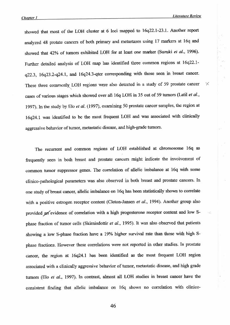
CltuplerRevÍew
showed that most of the LOH cluster at 6 loci mapped to l6q22.l-23.1. Another feport
analyzed.48 protate cancers of both primary and metastases using 17 markers at 16q and
showed ttrat 42Yo of tumors exhibited LOH for at least one marker (Suzuki et a1.,1996).
Further detailed analysis of LOH map has identifred three common regions at 16q22-l-
q22.3, 16q23.2-q24.1, and 16q24.3-qtt corresponding with those seen in breast cancer-
These three commo\LoH regions were also detected in a study of 59 prostate cancer \{
cases of various stages which showed over all l6q LOH in 35 out of 59 tumors (Latil et al-,
lggT).ln the study by Elo et al. (1997), examining 50 prostate cancer samples, the region at
l6q24.l was identified to be the most frequent LOH and was associated with clinically
aggtessive behavior of tumor, metastatic disease, and high-gfade tumors.
The recurrent and common regions of LOH established at chromosome 16q as
frequently seen in both breast and prostate cancers might indicate the involvement of
common tumor suppressor genes. The correlation of allelic imbalance at l6q with some
clinico-pathological parameters was also observed in both breast and prostate cancers. In
one study of breast cancer, allelic imbalance on l6q has been statistically shown to correlate
with a positive estrogen receptor content (Cleton-Jansen et al., 1994). Another group also
providedslevtdence of correlation with a high progesterone receptor content and low S-
phase fraction of tumor cells (Skirnisdottir et al., 1995). It was also observed that patients
showing a low S-phase fraction have a lgYo htgher survival rate than those with high S-
phase fractions. However these correlations were not reported in other studies. ln prostate
cancer, the region at l6q24.l has been identified as the most frequent LOH region
associated with a clinically aggressive behavior of tumor, metastatic disease, and high grade
tumors (Elo et al., 1997).In confas! almost atl LOH studies in breast canoer have the
consistent finding that allelic imbalance on l6q shows no conelation with clinico-
46
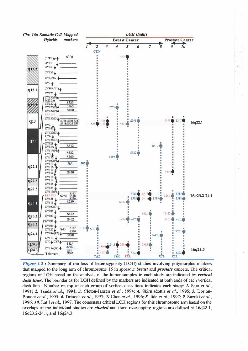
Chr. 16q Somatic Cell MappedHybrids markers
cYBoþ)f é-CY148 |
-
cvr+o1v
cYl35 +cYl38(D)+
cY1 +cYlsA(Pl)+-CY126
CYI22
FRA I óB
LOH studirc
345678Breast Cancer Prostate Cancer€9102
CEN
1
sro0ç
s265
I
?ItII¡¡IIII¡IIIIIIIIt8
I
oII!II¡I
TEL
s498
ItIIIIItIIII
öI
s422
s:orf
IIo!IIII
a:
t2S5
¡I¡IIII¡IIIIIII!!
t!?:¡ttllll
I I s¡orOrSa2f :lrrlaII¡
I¡I
II
III
II
: s522Oar
,,,ö:liått
s347(l¡
sreso
CYICY6
CYIl2cY2+
I
HP.IIIIIItItIIII!IIIIIIItIt
srszfI
s260
s5r5? s.{r5Qs5r8Ç s.rr6(ì s5r
t6q22.l
16q23.2-24.1
16q24.3
s+sof
s2ô6
s422
s402
s289
s402
as5l5os5r8o
I!IIIII
s4o2i
I
?IIIIII
s3o3a
s520
Is402?
ITIIIIIIrs4Ia
IIo!IaI¡I
oöIIIT¡¡III¡I!t
¡s43?
tIII
ls¿.,1PRa
I sru:fTEL
s4l3 l3t s4 t3tcvrrnpz¡fTelomere 5303
¡
lEL
Fisure 1.2 : Summary of the loss of heterozygosity (LOH) studies involving polymorphic markersthat mapped to the long arm of chromosome 16 in sporadic breast and prostøte cancers. The criticalregions of LOH based on the analysis of the tumor samples in each study are indicated by verlicaldash lines. The boundaries for LOH defined by the markers are indicated at both ends of each verticaldash line. Number on top of each group of vertical dash lines indicates each study: 1. Sato et al.,l99l; 2. Tsuda et al., 1994; 3. Cleton-Jansen et al., t9941, 4. Skirnisdottir et al., 1995; 5. Dorion-Bonnet et a1.,1995; ó. Driuoch et a1.,1997;7. Chen et a1.,1996;8. Iida et a1.,1997;9. Suzuki et al.,1996; 10. Latll et al., 1997 . The consensus critical LOH regions for this chromosome arm based on theoverlaps of the individual studies are shaded and th¡ee overlapping regions are defined at 16q22.1,
16q23.2-24.1, and 16q24.3
q13
q22.1
q22.2
q22.3
q23.2
q24.t
q2J. r
pathological parameters such as difFerences in tumor size, histology grade, clinical stage,
lymph node status, tumor ploidy, age atdiagnosis, or family history of breast cancer.
1.5.S Studies of Candidate Genes localized at 16q Loss of Heterozygosity regions
A number of candidate genes mapped to the 16q LOH regions have been studied for
their possible involvement in breast cancers. The epithelial cadherin gene (CDHI) which
mapped within l6q22.l region codes for a protein, E-cadherin, important for the
maintenance of cell-cell adhesion in epithelial tissues (Mansouri et al., 1988; Berx et al-,
1995b). The loss of function of E-cadherin and /or related proteins contributes to the
increased proliferation, tumor invasion and metastasis in many solid tr¡mors including breast
cancer (Ilyas and Tomlinson, 1997). Mutations of CDHI have been reported in breast cancer
cell lines (pierceall et a1.,1995; Hiraguri et al.,l99S) and over 50Yo of infiltrating lobular
breast tumors (Berx et al., 1995u 1996). ln the latter, mutations have been detected in
combination with LOH of the wild-type locus. However, no mutations tn CDHI have been
detected in ductal carcinomas (Kashiwaba et al., 1995; Berx et al., t995a, 1996)- These are
the most common type of breast cancer and belong to the major goup for which LOH of
l6q22.l has been reported. It is therefore suggested that other candidate genes exist in this
region that are involved in the progression of ductal carcinoma of the breast.
The CTCF gene located in the l6q22.l LOH region codes for an evolutionary
conserved DNA-binding nuclear protein that has a role in the regulation of vertebrate
cellular MYC gene expression (Lobanenkov et al., 1990; Klenova et al., 1993; Filþova ef
a1.,1996). Over-expression of MIrC is one of the major oncogenic factors involve in various
types of malignancy, including breast cancer progression. Mutations of the genes, including
CTCF, that regulate MYC expression may contribute to the pathogenesis of breast cancer.
CTCF may therefore be a candidate tumor suppressor for breast carcinogenesis. Genomic
47
CltanterRevÍew
reruïangement of CTCF have been reported in trryo breast cancer cell lines and one of four
analyzedpaired DNA samples from primary breast cancer patients (Filippova et a1.,1998).
However, no subsequent additional mutation analysis or other evidence has been published
to confirm thatCTCF is involved in breast cancer'
Two candidate genes have been mapped to the LOH region at 16q23.2-q24.1. The first,
a breast cancer anti-estrogen resistance I (BCARI) gene has initially been identified through
a retroviral insertion mutagenesis of the human estrogen-dependent breast cancer cell line
ZR-75-l (Dorssers et a1.,1993). It has been shown that integration of replication defective
retrovirus at a common site, the BCAR1 locus, could transform the cells from estrogen-
dependent to an anti-estrogen resistance phenotype. These cells lost estrogen receptor
expression and became estrogen-independent @orssers et al., 1993). T\e BCARI was
initially mapped to the chromosome band 16q23 by in sifz hybridization (Dorssers et al.,
lg93). The gene, BCARl,has subsequentþ been cloned and mapped to the region between
the somatic hybrid breakpoints CY145 and CY117 (Brinkman et a1.,2000). This gene, as
shown by its protein sequence, is a homologue of the mouse and rat pl30Cas gene
(Brinkman et a1.,2000). Overexpression of BCARI has been shown to confer anti-estrogen
resistance onZp.-/S-l breast cancer cells and the in vi¿ro insertion of retrovirus appears to
activate BCARI promoter resulting in its overexpression (Brinkman et a1.,2000). Ir general,
about two thirds of all breast cancers produce functionally active estrogen receptor (ER) and
are therefore eshogen-dependent (Horwitz et a1.,1985; Foekens et a1.,1990; Clarke et al.,
IggT). These tumors respond well to treaûnent with anti-eshogens which inhibit the
estrogen response pathway and thus arrest tumor growth (Jordan et a1.,1990; Musgtove ef
al., 1993). However, nearly half of ER positive tumors develop resistance to this anti-
estrogen therapy and eventually the majority of tumors that initially respond to anti-
48
estrogens develop resistant metastases (Foekens et a1.,1994). The involvement of BCARI in
breast cancer progression by promoting the anti-estrogen resistant phenotype indicates the
likelihood that this gene behaves as a tumor modifier rather than a tumor suppressor.
The second candidate gene has recentþ been isolated from the genomic region between
the markers Dl6S5l8 and Dl6S5l6 within 16q23.2-q24.1 by mean of physical map
construction prior to the identification of the gene (Bednarek et al., 2000). The gene,
designated WWOX, codes for a protein containing two WW domains coupled to a region
with high homology to the short-chain dehydrogenase/reductase family of enzymes.
Overexpression of tí44rOXhasbeen demonstrated in breast canoer cell lines when compared
with normal breast cells. The high expression of WI4/OX has also been observed in other
hormonally regulated tissues such as testis, ovary, and prostate. By its expression pattern,
the presence of short-chain dehydrogenase/reductase domain and the specific amino acid
features suggest the role for WWOX in steroid metabolism. However, mutation studies
failed to detect any alteration of this gene in breast cancer showing hemizygous for the 16q
genomic region, suggesting that Wltt'OX is probably not a tumor suppressor gene (Bednarek
et a|.,2000).
The LOH region at 16q24.3 has initially been mapped between the mmker Dl6S4l3 or
the APRT gene and the marker D16S303 close to the telomere (Cleton-Jansen at al., t994;
Skirnisdottir et al., 1995; Dorion-Bonnet et al., 1995). This region coveñ¡ the 16q segment
between mouse/human somatic cell hybrid breakpoint CYISA(D2) and the telomere (Callen
et a1.,1995). V/ithin this region, a number of known genes have been localized and some of
these genes may be a candidate tumor suppressor, which requires firrther analysis. A
collaborative study to refine the LOH at 16q243 utilizing additional polymorphic markers
distributed in this region has initially provided evidence suggesting the possible minimal
49
11
LOH region between markers D16S3026 and Dl6S303 (Cleton-Jensen, personal
communication). Concurrentþ, a det¿iled physical and transcription map has been
established that encompasses this minimal region (Whiûnore et a1.,1998a). The construction
of this 16q24.3 physical and transcription map and the studies of some known genes mapped
to this region are discussed in the next section'
TraGenes
The construction of the detail physical and transcription map at 16q243 region was
initiated to provide a basis for the isolation of candidate tumor suppressor gene or genes
(Whitmore et al.,l998a). This was the minimum 16q24.3 region defined by LOH studies.
1.6.1 Construction of the Physical and Transcription map
Initially, a total of nine separate markers that mapped between the somatic hybrid
breakpoint CYISA(D2) and the l6q telomere (Callen et a1.,1995) were used as probes to
screen a chromosome 16 cosmid library. The identified cosmid clones were organized into
six groups of cosmid contigs as nucleations for subsequent cosmid walking. Cosmid walking
and restriction site mapping was successfully used to extend and link these clusters. This
generated a physical map of approximately 950 kb extending from the marker Dl6S303 to
the region proximal to D16S3026. This physical map contained a total of 103 overlapping
clones with a minimum tiling path of 35 cosmid clones (Whimore et al-,1998a).
To identiff the transcripts within this physical map, exon trapping (Buckler et al.,
l99l; Burn et al., 1995) was used on selected overlapping cosmids. A total of 7l trapped
exons were identifted, l7 of which were identical to characterized genes known to map to
this LOH region (Whitnore et al.,l998a). Among these are the FAA, PISSLRE, and PRSM|
genes. The DNA sequences of 23 clones were homologous to ESTs in the sequence database
50
at NCBI and were grouped into 15 different transcripts. The remaining 31 trapped exons
showed no database sequence homologies, although 23 had þ{/onen reading frames >
suggesting that they were parts of genes.
The physical map that was established in this chromosomal region provided a genomic
framework for subsequent gene identification. The trapped exons were mapped back to the
original genomic clones to form exon clusters, each represented a possible transcript with a
known physical location. This integrated physical and transcription map of 16q24.3 LOH
region (Whitmore et a1.,1998a, and summaÅzed in Figure 1.3) provides the basis for the
identification of the tumor suppressor gene:;related to breast cancer development and
progtession. Furthermore, this map can also be used for the isolation of genes that may be
involved in other genetic diseases. One such example is the gene for f""""i anemia 7
complimentary group A (FAA). Tlne FAA gene \¡/as isolated during the initial stage of
physical map construction utilizing both exon trapping and cDNA selection procedures (The
Fanconi anaemia./Breast cancer consortium, 1996).
1.6.2 Studies of the Genes within tb¡el6q24.3 minimal LoH region
At the early stage of this study a number of known genes had been located to this
region, these included BBCL, ClvIAR, DPEPI, and MCIR. These genes were possible
candidates as a tumor suppressor and therefore required further investigation.
The breast basic conserved gene (BBCI) encodes a protein showing sequence
homology to the human ribosomal protein L13 and decreased expression of BBCI mRNA
has been detected in malignant breast tumors compared to that in benign tumors (Adams ef
a1.,1992). The Ll3 protein has been shown to contain a DNA binding motif characteristic of
transcription factors (Tsurugi and Mitsui, l99l), suggesting BBCI is unlikely to be a
candidate tumor suppressor gene targeted by LOH. Mutation analysis of this gene, however,
51
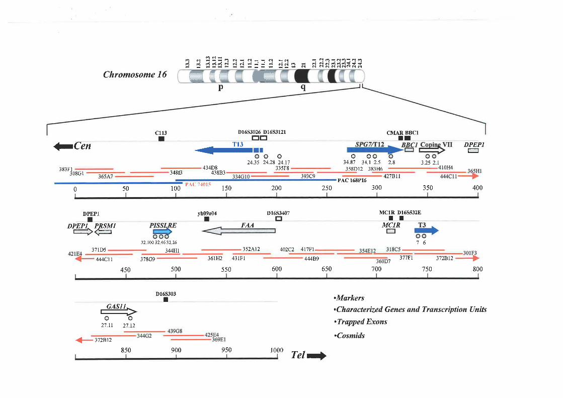
6d:ó N J--tâ N- NE- d
-ñ F c!6-î¡(t)-êl(.)t É:ÉÉsdsijj= si'e ñ NNËR*Rñxñ'
Chromosome 16
c113I
50
p
D16S3026 Dl6S3l2rEE
T13
-
oo24.35 24.28 24.17
335F8
PAC 7{015150 200
358Dr2 383H6
427BllPAC 168P16
300
q
CMAR BBC1ll
JCen
0
ooo34.87 34.r 2.5 2.8 3.25 2.r
A1ñTJA
DPEPI
383F1 434D8-"--jo3Cr 348ß 43883393C9
100 250 350 400
F
úFTFLÅL m
DPEPl
DPEPT PRSMItr+€3',71D5
32 .100 32 46 32 26
344H1
378G9
500
352¡^12 402C2 4l7Fl
MC1R D16S532ETIMCIRE
76
377F1J6IJD I
700 750
vh09¡04 D1653407E
600
FAA
4MCII 36[12 43lFl
550
44489 3',728t2
450 6s0 800
D165303T .Markers
.Characterized Genes and Transcríptíon Uníts
.Trøpped Exons
.Cosmids27.11 27.t2
þ3728t2850 900 950 1000 Tel)
has been undertaken in breast cancer samples exhibiting LOH restricted to 16q24.3, but no
evidence of mutations were detected in these samples (Moerland et a1.,1997).
The cellula¡ matrix adhesion regulator (CMAR) gene was originally isolated from a
colorectal cell line cDNA library and has been shown by gene transfection to enhance the
binding of the cell to the extracellular matrix component, collagen t)¡pe I @ullman and
Bodmer, lg92). Since CMAR is involved in cell adhesion, then loss of CMAR frrnction may
be associated with the initial step of tumor invasion and metastasis. However, mutation
analysis in breast cancer has excluded CMAR as a candidate breast tumor suppressor
(Moerland et al.,lgg7). Furthermore, CMAR sequence has now been shown to be a part of
the 3' untranslated region of the gene involved in spastic paraplegia, SPG7, isolated from
this chromosome region (discussed tn chøpter 1).
The renal dipeptidase gene (DPEPI) was originally isolated from a kidney cell cDNA
library by subtractive hybridization with V/ilms' tumor mRNA (Austruy et al-, 1993).
Reduced expression of DPEPI mRNA was shown in Wilms' tumor compared with its
expression in mature kidney. However no further evidence of its involvement in
tumorigenesis has been reported, it is therefore not a target gene for breast cancer LOH. The
melanocyte stimulating hormone receptor (MCIR) gene may not be considered as a
candidate gene, since its function is associated with the regulation of pigmentation in
keratinocle (Valverde et al., I 995).
1.7 Aims and Scope of The Studv
The strong association of LOH at the chromosome region 16q24.3 and sporadic breast
cancer lead to the establishment of chromosome 16-breast cancer project with the ultimate
goal for cloning of the breast cancer-associated tumor suppressor gene. Following the
successful positional cloning of the FAA gene, the detail physical map at this region has
52
o.
been expanded encompassing þ region of approximately 950 kb. This physical map is
bounded proximally by D16S3026 and distally by D16S303. Subsequently, the transcript
map has been established on this physical framework and provided the basis for cloning of
candidate gene(s).
The aim of the project presented in this study was to clone and characterize the genes
from this physical region for the identifrcation of the candidate tumor suppressor associated
with sporadic breast cancer. This will primarily involve the isolation of full-length transcript
sequence of the genes. Expression profile of each gene will also be established.
Characterization of the gene sequence and its protein product by sequence homology as well
as functional motif searching may provide a possible function of the corresponding protein.
The candidate genes based on the possible function compatible with a tumor suppressor will
be further characteized by mutation screening of DNA isolated from tumors defining the
critical LOH region atl6q24.3-
Other characterized genes that are not involved in breast cancer but show protein
sequence homology to known gene with significant physiological function will also be
studied in detail.
53

Chapter 2
Materials and Methods
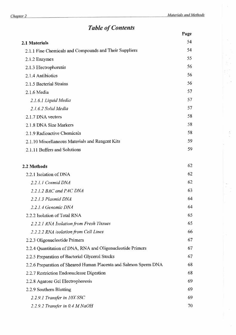
Table of Contents
2.l Materials
2.1.1 Fine Chemicals and Compounds and Their Suppliers
2.l.2Enzymes
2.1.3 Electrophoresis
2.1.4 Arrtibiotics
2.1 .5 Bacterial Strains
2.I.6Media
2.1.6.1 Liquid Media
2.1.6.2 Solid Media
2.1.7 DNA vectors
2.1.8 DNA Size Markers
2. 1.9 Radioactive Chemicals
2.1.10 Miscellaneous Materials and Reagent Kits
2.l.ll Buflers and Solutions
2.2 Methods
2.2.1lsolation of DNA
2.2.L1 Cosmid DNA
2.2.1.2 BAC and PAC DNA
2.2.1.3 Plasmid DNA
2.2.1.4 Genomic DNA
2.2.2[solation of Total RNA
2.2.2.I RNA Isolationfrom Fresh Tissues
2.2.2.2 RNA isolationfrom Cell Lines
2.2.3 Oligonucleotide Primers
2.2.4 Quantitation of DNA, RNA and Oligonucleotide Primers
2.2.5 Preparation of Bacterial Gþerol Stocks
2.2.6Preparation of Sheared Human Placenta and Salmon Sperm DNA
2.2.7 Reslriction Endonuclease Digestion
2.2.8 Agarose Gel Electrophoresis
2.2.9 Southern Blotting
2.2.9.1 Transfer in I0X SSC
2.2.9.2 Transfer in 0.4 M NaOH
Page
54
54
55
56
56
56
57
57
57
58
58
58
59
59
62
62
62
63
64
64
65
65
66
67
67
67
68
68
69
69
69
70
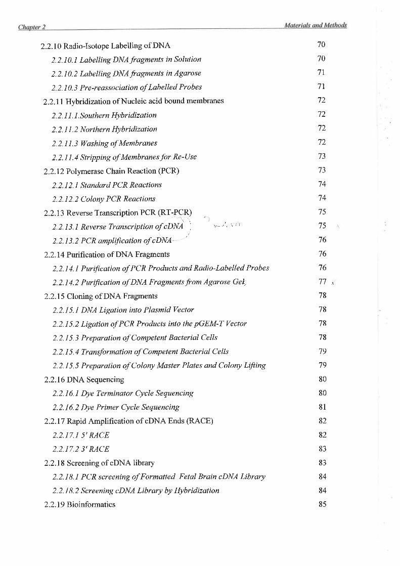
2.2.10 Radio-Isotope Labelling of DNA
2.2.10.1 Labelling DNAfragments in Solution
2.2.10.2 Labelling DNAfragnents in Agarose
2. 2. I 0. 3 Pre-reas s ociation of Labelled Probes
2.2.1 1 Hybridization of Nucleic acid bound membranes
2. 2. 1 1. l.Southern Hybridization
2. 2. 1 1. 2 Northern Hybrídization
2.2.11.3 Washing of Membranes
2. 2. 1 1. 4 Stripping of Membranes for Re-Use
2.2.12 Polymerase Chain Reaction (PCR)
2.2.12.1 Standard PCR Reactions
2.2.12.2 Colony PCR Reactions
2.2.13 Reverse Transcription PCR (RT;PCÐ
2.2.13.1 Reverse Transcription of,cDNA' ) ''- 'o" \ I i
2.2.13.2 PCR amplification of zDNA
2.2.14 Purification of DNA Fragments
2.2.14.1 Purification of PCR Products and Radio-Labelled Probes
2.2.14.2 Purification of DNA Fragments from Agarose Geþ
2.2.15 Cloning of DNA Fragments
2.2.15.1 DNA Ligation into Plqsmid Vector
2.2.15.2 Ligation of PCR Products into the pGEM-T Vector
2.2.15.3 Preparøtion of Competent Bacterial Cells
2.2.15.4 Tronsþrmøtion of Competent Bacterial Cells
2.2.15.5 Preparation of Colony Master Plqtes and Colony Lifting
2.2.16 DNA Sequencing
2.2.16.1 þe Terminøtor Cycle Sequencing
2.2.16.2 þe Primer Cycle Sequencìng
2.2.17 Rapid Amplification of cDNA Ends (RACE)
2.2.17.1 5',RACE
2.2.17.2 3',RACE
2.2.18 Screening of cDNA library
2.2.18.1 PCR søeening of Formatted Fetal Brain cDNA Library
2.2.18.2 Screening cDNA Library by Hybridization
2.2.19 Bioinformatics
70
70
7t
7l
72
72
72
72
73
73
74
74
75
75
76
76
76
77x
78
78
78
78
79
79
80
80
81
82
82
83
83
84
84
85
.\
2.1 terials
2.1.1 Fine Chemicals and Compounds ¡nd Their Suppliers
Ammonium sulphate Ajax Chemicals, Australia
Bacto-agar sigma chemical co., usA
Bacto-tryptone Difco Laboratories, usA
Boric acid Ajax
BSA (bovine serum albumin'pentax fraction V) Sigma
Chloroform
DAPI (diamidino phenyþdole dihydrochloride)
DEPC (diethylpyrocarbonate)
Deoxy-nucleotide triphosphate (dNTPs)
Dextran sulphate
N, N-dimethyl formamide
DMEM (Dulbecco's modified Eagles medium)
DMS O (dimethylsulPhoxide)
DTT (dithiothreitol)
EDTA (ethylenediamine tetraacetic acid; NazEDTA-2H2O)
Ethanol (99.5%vlv)
FCS (fetal calf serum)
Ficoll (Type 400)
Formamide (deionised before use)
Glucose
Glutamine
Glycerol
Humanplacental DNA
IPTG (isopropylthio-p-D-galactosidase)
Isoarnyl alcohol (IAA)
Isopropanol
N-lauroylsarcosine (SarkosYl)
LipofectACE
Magnesium chloride (MgClz.6HzO)
Magnesium sulPhate (MgSO¿.7HzO)
BDH Lab Supplies
Sigma
BDH
Amersham Biosciences
Amersham Biosciences
or Promega
Sigma
Trace Biosciences
Sigma
Sigma
Ajax
BDH
Trace Biosciences
Sigma
Fluka Chemika
Ajax
Trace Biosciences
Ajar
Sigma
Progen
Ajax
Ajan
Sigma
Gibco-BRL (Life Technology)
Ajax
Ajax
54
Maltose
Þ ME ( P -mercaptoethanol)
Mixed bed resin (20-50 mesh)
MOPS (3-fN-morpholino]propane sulphonic acid)
Opti MEM-I (powder sachet)
Paraffrn oil
PEG (polyethylene glYcol) 3350
Phenol
Polyvinylpyrolidone (PVP-40)
Potassium dihydrogen orthophosphate (KH2POa)
Propidium iodide
Salmon sperm DNA
Sodium acetate
Sodium carbonate (NaHCO3)
Sodium chloride
tri-sodium citrate
Sodium dihydrogen orthophosphate (NaFIzPO¿.2HzO)
Sodium dodecyl sulPhate (SDS)
di- S odium hydrogen orthophosphate (NazHPO¿. THzO)
Sodium hydroxide (NaOH)
Sucrose
Tris-base
Tris-HCl
Triton X-100
Yeast extract
BDH
BDH
BioRad, USA
Sigma
Gibco-BRL
Ajax
Sigrna
Wako Ind., Japan
Sigma
Ajax
Sigrna
Calbiochem
Ajær
Astra Pharm., Australia
Ajax
Ajax
Ajax
BDH
Ajax
Ajax
Ajax
Boehringer Mannheim
Boehringer Mannheim
Ajax
Gibco-BRL
2.l.2E,nzynes
All restriction enrymes were purchased from either New England Biolabs (Beverly,
Massachusetts, USA) or Progen @risbane, Queensland, Australia). Each enzyme \¡ias
supplied with the appropriate digestion buffer and bovine serum albumin (BSA) if required.
All other enzymes not part of kits are listed below along with their suppliers.
CIAP (Calf intestinal alkaline phosphatase) Boehringer Mannheim
55
E. c oli DNA polymerase (Klenow fragment)
Proteinase K
RNaseH
RNAsin
Superscriptru RNase H- reverse transcrþtase
T4 DNA ligase
T4 Polynucleotide kinase
Zaq DNA polYmerase
X-gal (5-Bromo-4-chloro-3-indoyl-B-D galactosidase)
2.1.3 Electrophoresis
Acrylamide (40%)
Agarose: Molecular BiologY grade
APS (Arnmonium persulPhate)
Bis Q% Bis solution)
Bromophenol blue
Ethidium bromide
Formaldehyde (37o/ù
MDE (2Xgelsolution)
TEMED (N, ¡{ ¡4 ¡f '-tetramethylethylenediamine)
Xylene cyanol
2.1.4 Antibiotics
{mpicillin
Benzyl penicillin
Chloramphenicol
Kanamycin
Tetracycline
New England Biolabs
Merck
Promega
Promega
Gibco-BRL
Progen
Amersham Biosciences
Gibco-BRL
Progen
BioRad
Progen or Amersham
Biosciences
BioRad
BioRad
BDH
Sigma
Ajax
FMC bioproducts, USA
BioRad
BDH
Sigma
CSL
Sigma
Progen
Sigrna
I
2.1.5 Bacterial Strains
All bacterial strains were kept at -70 oC in LB-Broth + líYo glycerol. Each strain was
56

streaked for single colonies on LB-Agar plates containing an appropriate antibiotic (see
below) from stock.
E.coli XLI-Blue MRF': L(muA)183 L(mcrCB-hsdSMR-mn)173 endAl supÐ44 thi-I
relAl lac fF' proAB tacfZLMIS Tn10 (Tet)]'. This strain was a host for recombinant
plasmids and was pwchased from Stratagene. XLI-Blue cells were streaked for single
colonies on LB-tetracycline plates then propagated overnight at 37r.oC in LB-Broth .../,!,.1:
supplemented with tetracycline (15 pglml).
E.coli Yl090r-: L(tac)U169 L(lon)? AraD i,39 strA supF muA trpC22::TnI| (TeÐ
þMC9 Amp' Te{l mcrB hsdR. (Note: pMCg is pBR322 wlthlacf inserted). This strain was
a host for recombinant Àgt 1l bacteriophage and was supplied with the Clontech 5' Stretch
fetal brain oDNA library. These cells were streaked for single colonies on LB-tetracycline
plates and subsequently propagated overnight at 37 "C in LB-Broth supplemented with 10
mM MgSO ¿ arrd 0.2Yo (vþ maltose.
2.1.6 Media
All liquid media was prepared with double distilled water and was sterilised by
autoclaving. For the preparation of solid media plates, antibiotics were added once the
autoclaved media had cooled to a tem¡rerature of approximately 50 "C.
2.1.6.1 Líquì.d Medìa
LB-Broth (Lwia-Bertaini Broth): lYo (wlv) Bactotryptone; 0.5Yo (w/Ð yeast extact; 1olo
(w/v) NaCl; adjust pH to 7.5 with 5 N NaOH before autoclave.
OMI (pH 7.1): I sachet of Opti MEM-I; 28 ml NaHCO:; 80 ml FCS; 55 nM pMe; I ml
benzyl penicillin, p e r litr e.
Supplemented DMEM þH 7.0): 13 g DMEM;44 ml NaHCO:; 4 mM glutamine; I ml
benzylpenicillin; I 00 ml FCS, per litre.
57
TSS: LB-Broth; l0% (w/v) PEG 3350; 5o/o (vlv) DMSO; 50 mM MgCl2.
2.1.6.2 Solid Media
LB-Agar: LB-Broth; 1.5% (wlv) Bacto-agar.
LB-Ampicillin: LB-Broth; I .SYo (wlv) Bacto-agar; 100 pglml ampicillin.
LB-Chloramphenicol: LB-Broth;l.5Yo (w/Ð Bacto-agar;3a Wghnl chloramphenicol.
LB-Kanamycin: LB-Broth;1.5% (w/Ð Bacto-agar; 50 pglml kanamycin.
LB-Tetracycline: LB-Broth; l.5Yo (w/v) Bacto-agar; 15 pgiml tetracycline.
Top Agar: LB-Broth; 0.7Yo Bacto-agú.
2.1.7 DNA Vectors
pSPL3B was used for exon trapping from BAC and PAC DNA. This vector was kindly
provided by Dr. Tim Connors and Dr. Greg Landes (USA).
pUClg was used for the subcloning of cosmid DNA fragments and was purchased from
New England Biolabs.
2.1.8 DNA Size Markers
SPPI (EcoRI digested )
pUClg (HpaII digested)
Digest III (î,cIS57 Sam 7 HindW þ){-174 HaeItr'
restricted)
2.1.9 Radioactive Chemicals
o-32P1dcrP (3,ooo cilrnmol)
¡y-32l1dltt (5,ooo cilmmol)
Progen or Bresatec
Bresatec
Amersham Biosciences
Amershan Biosciences
Amersham Biosciences
58
2.1.10 Miscellaneous Materials and Reagent Kits
ABI Prismru DyePrimer Cycle sequencing kit
ABI Prismru DyeTerminator Cycle sequencing kit
BigDye Terminator Cycle sequencing kit
Breast cancer cell lines
CloneAMP pAMPlO kit
ExpressHYB solution
Genescreen Plus Nylon membranes
HaIfIERM DyeTerminator sequencing reagent
Labelling mix-dCTP
Multiple tissue Northern Blot (Catalogue # 7760-l)
Human RNA Master Blot (Catalogue # 7770-l)
Oligo-dTrz-rs
pGEM-T cloning kit
Prep-A-Gene purification kit
Qiagen Plasmid mini kit
QlAquick purification kit
SMARTTM PCR cDNA Library Construction kit
(Catalogue # Kl051-l)
Random hexamers
RTthrcverse transcriptase RNA PCR kit
Trizol
3MM'Whaünan paper
Perkin Elmer
Perkin Elmer
Perkin Elmer
ATCC, USA
Gibco-BRL
Clontech
Dupont
GenPack
Amersham Biosciences
Clontech
Clontech
Gibco-BRL
Promega
BioRad
Qiagen
Qiagen
Clontech
Amersham Biosciences
or Perkin Elmer
Perkin Elmer
Gibco-BRL
Lab Supply
I
2.l.ll Buffers and Solutions
All solutions were prepared with sterile double distilled water followed by autoclaving
at 120 oC for 15 to 30 minutes. Solutions provided with kits are not mentioned. Buffers and
solutions routinely used in this study were as fellows:
10X Agarose gel loading bufi[er: 100 mM Tris-HCl (pH 8.0); 200 mM EDTA (pH 8.0);
2.0% (wlv) sarkosyl; 15% (wlv) ficoll400; 0.lolo bromophenol blue; 0.lolo xylene cyanol.
s9
lX pAMPlO annealing buffer: 20 mM Tris-HCl þH 8.a); 50 mM KCI; 1.5 mM MgCl2.
Cell lysis buffer: 0.32ll.l sucrose; l0 mM Tris-HCl; 5 mM MgCl2; l% (vlv) Triton X-100
C,H 7.s).
Colony denaturing solution: 1.5 mM NaCl; 0.5 M NaOH.
Colony neutralising solution: 3 M NaCl; 0.5 M Tris-HCl.
De-ionised formamide: 2 g of mixed bed resin/S0 ml formamide. Mix for -l hotr then filter.
l00X Denhart's solution: 2% (wlv) ficoll 400;2% (wiÐ polyvinylpirolidone; 2% (wlv)
BSA.
10X dNTP labelling solution: I mM labelling mix -dCTP-
Filter denaturing solution: 0.5 M NaOH.
Filter neutralising solution: 0.2 M Tris-HCl; 2X SSC-
5X First strand buffer (supplied with Superscript reverse transcriptase): 250 mM Tris-HCl
(pH S.3); 375 mM KCI; 15 mM MgCl2.
Formamide loading buffer: 50% (vlv) glycerol; 1 mM EDTA (pH 8.0); 0.25% (w/v)
bromophenol blue; 0.25yo (w/v) xylene cyanol.
Gel denaturing solution: 2.5 M NaCl; 0.5 M NaOH.
Gel neutralising solution: 1.5 M NaCl; 0.5 M Tris-HCl (IrH 7-5).
10X Labelling buffer:0.5 M Tris-HCl (pH 7.5);0.1 M MgClz; l0 mM DTT;0.5 mg/ml
BSA; 0.05 Azøolttl random hexamers (Amersham Pharmacia Biotech).
60
10X MOPS buffer: 0.2 M MOPS; 50 mM NaAc; 1 mM EDTA.
Phenol: buffered with Tris-HCl (IrH 7.4).
PBS (Phosphate buffered saline): 138 mMNaCl; 2.7 rtrJvIKCl; 8.1 mMNazHPO+.7HzO;1.2
rnM KHzPO4 (pH 7.4).
lQX PCRbuffer (supplied twthTag DNA polymerase):200 mM Tris-HCl (pH 8.0); 500
mM KCL.
2X PCR mix: 33 mM NH4)zSO¿; 133 mM Tris-HCl (pH 8.8); 20 mM pME (added fresh
each time); 0.013 mM EDTA;0.34 mglml BSA; 20% (vlv) DMSO; 0.4 mM dNTPs.
3X Proteinase K buffer: l0 mM NaCl; 10 mM Tris-HCl; 10 mM EDTA ûrH 8.0).
RNA hydrolysing solution: 50 mM NaOH; 1.5 M NaCl.
lQX RNA loading dye:50o/o glycerol; lmM EDTA GH 8.0); 0.25yo bromophenol blue;
0.25% xylene cyanol.
RNA neutralising solution: 0.5 M Tris-HCl (çH7.Ð;1.5 mMNaCl.
SM buffer: 50 mM Tris-HCl; 100 mM NaCl; 8 Mm MgSOa; 0.01% (v/v) gelatin (pH 7.5)
3 M Sodium acetate pH 5.2 (NaOAc): 24.6 g sodium acetate/l00 ml; pH 5.2 with acetic
acid.
Southern hybridization solution:50Vo (vþ de-ionised formamide; 5X SSPE; 2% SDS; lX
Denhart's; l\Y, (wlv) dextran sulphate; 100 pgl.ml denatured salmon speÍn DNA.
20X SSC: 3 M NaCl; 0.3 M tri-sodium citrate ûrH 7.0).
20X SSPE: 3.6 M NaCl; 0.2MNaHzPO¿.2HzO;0.02 M EDTA.
6I
lX TBE: 90 mM Tris-base; 90 mM boric acid; 2.5 mM EDTA (pH 8'0)
TE: l0 mM Tris-HCl (pH 7.5); 0.1 mM EDTA.
X-gal: 50 mM (2%wlv) solution, usingN,N,dimethyl-fonnamide as diluent.
2.2
The methods described in this chapter are generally used in common throughout this
thesis and most are based on the standa¡d procedures presented in Molecular Cloning: A
þlorotory Mønual,2ndFld(Sambrook l9S9) unless otherwise indicated. Those procedures X
specific for each chapter are presented in detail in the corresponding method section-
2.2.1 Isolation of DNA
All DNA isolations except for genomic DNA were based on the alkaline lysis method
adapted from the procedure described in Sambrook et al. (1989). The DNA was
subsequently purified utilizing Qiagen column technique according to the manufacturer
conditions (Qiagen Plasmid Mini Handbook, 1995) with slight modifications. All buffers
and columns were supplied with Qiagen kit.
2.2.1.1 Cosmid DNA
Each cosmid was received as bacterial stab in nutrient agar and was streaked and gro\iln
on LB-Kanamycin plate. A single colony was picked out and grown overnight at 37 "C tn
200 ml of LB-broth with kanamycin (50 pglml). Culture was spun down at 4,000 rpm for 15
minutes and bacterial pellet was resuspended in 2 ml of resuspending bufter (Pl). Two ml of
lysis buffe r (P2) was then added and carefirlly mixed, *rd ^tltäéd
completelv ly# bv
standing at room temperature for 5 minutes. Two ml of buffer P3 was added to cell lysate
with gently mixed and followed by 10 minute incubation on ice. This mixture was then spun
62
at 13,000 rpm for 15 min, the supematant was transferred to a new tube and spun for another
l0 minutes. Dwing the second spin a Qiagen Tip-20 column was equilibrated with I ml of
QBT buffer. The supernatant from second spin was applied to the equilibrated column and
allowed to drain completely through. The column was subsequentþ washed with 4 ml of
buffer QC and the DNA was eluted into a 1.5 ml eppendorf tube with 0.8 ml of buffer QF.
To precipitate the DNA, 0.6 ml isopropanol was added, mixed and spun at 13,000 rpm for
30 minutes. The DNA pellet was washed with I ml of 70% ethanol, spun for a further 5
minutes, and allowed to air-dried for 20 minutes. The DNA pellet was dissolved in 15 ul of
sterile Milli-Q water and quantifred by spectrophotomety (2-2-4)
2.2.1.2 BAC and PAC DNA
Bacterial stabs of BAC and PAC clones were streaked for the isolation of single
A
colonies on LB-Chloramphenicol and LB-Kanamycin plates respectively.-single colony of . ,!
BAC clone was grown ovemight at 37 oC in 200 ml of LB-broth containing
chloramphenicol (34 pglml). Cells were pelleted the next day by spinning at 4,000 rpm and
were subjected to DNA isolation. PAC clone was initially gro\iln overnight at37 "C in l0 ml
of LB-broth with kanamycin (50 pglml). The next day, 7 ml of this cultr¡re was transferred
to 200 ml of new LB-kanamycin broth and grown for 1.5 hours at 37 "C. After ttre addition
of IPTG to a final concentration of 0.5 mM, the cultrne was gfowrt for ftrther 5 hours and
then spun down at 4,000 rpm. Supernatant was removed and cell pellet was left overnight at
-20 "C.Isolation of both BAC and PAC DNA was performed by using Qiagen Tip-100
column with the same procedtre used for cosmid DNA isolation while the volume of buffer
being used was adjusted. These included the use of 4 ml of buffer Pl, P2 and P3 per DNA
isolation, with the Tip-100 columns were equilibrated with 4 ml QBT buffer, and washed
with l0 ml buf[er QC. DNA was eluted into l0 ml sterile tube with 5 ml of QF buffer, then
63
aliquoted into 6 eppendorf tubes. DNA was precipitated with 0.7 volumes of isopropanol,
and pellets were washed, as per cosmid DNA preparation. Individual dried DNA pellets
were re-dissolved in 5 pl of sterile Milli-Q water, using wide bore pipette tþs to prevent
shearing of the DNA, then pooled into one tube and quantified by spectophotomeny (2.2.$.
2.2.1.3 Pløsmíd DNA
For each plasmid clone, a single colony was grown overnight at 37 "C in 20 ml LB-
broth containing 100 pglml arrpicillin. Plasmid DNA was isolated using Qiagen Tip-20
column with all procedures followed the manufacturer conditions (Qiagen Plasmid Mini
Handbook,l995).In the final step, plasmid DNA pellet was re-dissolved in 15 pl of sterile
Milli-Q water before spectrophotometric quantification.
2.2.1.4 Genomíc DNA
DNA was isolated from peripheral blood lymphocytes, and breast cancer cell lines by
the methods adapted from Wyman and White (1980). All DNA preparations were kindly
performed by either Shirley Richa¡dson or Jean Spence (Department of Cytogenetics and
Molecula¡ Genetics, V/CÐ. All cell lines were maintained by Sharon Lane and Cathy
Derwas (Department of Cytogenetics and Molecular Genetics, WCH)-
Cell lysis buffer was added either to a blood sample or to a cell pellet obtained from a
cell line culture to a volume of 30 ml. The tube of suspended cells was left on ice for 30
minutes, then spun at 3,500 rpm for 15 minutes at 4 "C. Part of supernatant 20 ml, was
removed by aspiration, followed by the addition of a fi¡rther 20 ml of cell lysis buffer,
resuspended and a repeat centrifugation. The supernatant was aspirated and discarded, and
3.25 ml of 3X Proteinase K buffer, 500 pl of l0Yo (WV) SDS and 200 pl Proteinase K (at
l0 mg/ml) was added and mixed with the pellet. After an ovemight incubation at 37 'C with
64
mixing, 5 ml of phenol was added and followed by a l5-minute þcubati3n at room
temperature on a rotator. The solution wris spun at 3,000 rpm for l0 minutes, ttre aqueous
layer was removed and transferred to a new clean fube, and phenol was added to a volume
of l0 ml. After a 10 minute-incubation at room temperature, the mixture was then
centrifuged for l0 minutes, and the aqueous layer was transferred to a clean tube.
Chloroform was added to a final volume of 10 ml, the sample was mixed and re-centifuged,
and the aqueous layer was transferred to a new clean tube. DNA was precipitated by mixing
with 300 ¡rl of 3 M NaOAc (pH 5.2) and l0 ml of absolute ethanol. After centrifugation,
DNA pellet was washed \¡vitlì 70Yo ethanol, air-dried, and dissolved in 100 pl of TE.
2.2.2Ilsolation of Total RNA
The isolation of RNA from all tissues and cell lines was carried out under strict
RNAse-free conditions. All solutions were treated with 0.2% (vlv) DEPC, overnight at room
'(il¡-ttemperature with shaking before arf/oclaved. All pipettors were cleaned with RNAsaZAP
(Ambion) before used in combination with the use of filtered pipette tþs. RNA isolation was
based on the procedure of Chomcrynski and Sacchi (1987) utilizing the Trizol reagent. All
isolated RNA samples wçre stored at-70 "C.
2.2.2.1 RNA Isolationfrom F¡esh Tßsues
RNA was isolated from a number of tissues obtained from a 20 week-old male human
fetus. Access to these samples was kindly granted by Dr.Roger Byard @eparhnent of
Histopathology, WCtf. Selected tissues were frozen in liquid nitrogen and stored at -70 "C
r¡ntil RNA preparation was required. In each preparatior¡ I ml of Trizol reagent was used for
every 100 mg of ûozen tissue. Approximately 100 to 500 mg of tissue was removed from
the frozen stock and placed in a sterile mortar dish containing a small amount of liquid
v
65
nitrogen to keep the sample frozen. The tissue was ground to a fine powder with a sterile
pestle. Liquid nitrogen was periodically added to the ground tissue to prevent thawing. The
mortar and pestle had previously been washed thorougbly with ethanol, rinsed with DEPC-
treated water, then heat teated for 4 hours at 160-180 "C to inactivate any residual RNAses.
The ground tissue was then transferred to a l0 ml tube containing the appropriate amount of
Trizol reagent. The powder was resuspended by gentle mixing and transferred to a sterile 5
ml (Lab SuppÐ. The solution was homogenized until lumps of powder
were no longer visible, then transferred to a 1.5 ml eppendorf tube, and left at room
temperature for 5 minutes. One-tenth the volume of chloroforln was added and the tube was
shaken for 25 seconds and placed on ice for a frirther 5 minutes. The sample was then spun
at 12,000 rpm at 4 "C for 15 minutes, and the supernatant was transferred to a new tube. An
equal volume of isopropanol was added to this tube, and followed by incubation at 4 oC for
15 minutes. After centrifugation at 4 "C for 15 minutes, the RNA pellet was washed in 800
¡i of 75% ethanol and allowed to air-dry for 15 minutes. The RNA pellet was dissolved in
50 pl of DEPC-feated water, and pooled into one tube if the original tissue homogenate was
transferred into more than one eppendorf tube. A second precipitation step was performed
involving the addition of one-twentieth the volume of 4 M NaCl and 2 volumes of
100%(absolute) ethanol to the dissolved RNA. This was followed by incubation at -20 oC
for I hour then spgn for 15 minutes at 4 "C. The RNA peltet was again washed with 800 pl
of 75Vo ethanol, allowed air-drying for 20 minutes, and dissolved in DEPC-treated water.
RNA concentration was measured by spectrophotometry (2.2.4). The integrity of the RNA
preparation was tested by running a small aliquot on a 0.8olo agalose gel electrophoresis.
2.2.2.2 RNA Isolatíonfrom Cell Lines
RNA isolation from cell lines followed the same procedure as that for RNA isolation
66
from tissue sources. Typically, 1x10⁷ cells obtained from cell cultr¡re were washed in PBS
and sptrn down at 1,200 rpm for 10 minutes. The cells were then resuspended in I ml of
Trizol reagent. This was subsequently followed by the procedwes identical to those
described h2.2.2.1.
2.2.3 Oligonucleotide Primers
The oligonucleotide primers for both PCR and sequencing were generally designed to
contain approximately 50Yo G-C content, and an annealing temperature of 60 oC, calculated
by 2x(A+T)+4x(GrC) as per Suggs et al. (1981). Ideally, primers were not to possess runs
of identicat bases and not to contain four contiguous base pairs of inter-strand or intra-strand
complementary. ln addition, primers were designed such that G-C, C-C. C-G, or G-G bases
were present at thei¡ terminal 3' ends wherever possible. All oligonucleotides were
purchased commercially from either Gibco-BRL or Bresatec (Adelaide, Australia).
2.2.4 Qtantitation of DNA, RNA and Oligonucleotide Primers
Samples were diluted in water and their absorption of I-IV light (at wavelength of
260nm) was measured. The absorbance was multþlied by the dilution factor and a
conversion factor (absorption co-effrcient) for particular gpe of nucleic acid being
quantitated. Relevant conversion factors were 50 for double stranded DNA, 40 for RNA,
and 33 for oligonucleotides, which gave a concenhation in pglml. The spectrophotometers,
either Pharmacia Biotech Ultrospec 3000 or CECIL CE-2020 were used throughout this
experiment
2.2.5 Preparation of Bacterial Gþcerol Stocks
All bacterial clones were maintained as glycerol stocks. 'lhese were prepared from a 10
67
ml aliquot of overnight culture that had been used for DNA isolation. The 10 ml culture
aliquot \À/as spun down at 3,000 rpm. After the supernatant wris removed, the pellet was
resuspended in I ml of LB-Broth containing 15% gþerol and store at -70 "C until DNA
preparation was required, whereby a 5 ¡11 aliquot was used to streak plate for single colonies.
2.2.6Preparation of Sheared Human Placenta and Salmon Sperm DNA
Desiccated human placental DNA and salmon sperm DNA was pwchased
commercially and reconstituted in sterile TE to a concentration of 5 mg/ml and 20 mgiml
respectively. Aliquots of I ml were transfened to 1.5 ml eppendorf tubes and incubated at
100 "C for 6 to 20 hows. Periodicall¡ l-pl aliquots were taken and analysed on 1.5%
agarose gel electrophoresis until the average sized fragments were below 700 bp. The
concentration of the samples were then analysed by spectrophotomefiy and adjusted to their
starting values (2. 2. 4)
2.2.7 Restriction Endonuclease Digestion
Digestion of DNA with restriction endonuclease was performed using appropriate
reaction buffer that was supplied and recommended by the manufacturer. Digestion was
generally ca¡ried out in l0 pl volume, ovemight at 37 "C unless otherwise specifred. If
recommended, BSA at a final concentration of 100 pglml was included in the digestion
reaction.
For 10 pg of genomic DNA isolated from tissue, blood lymphocyte or cell line,
digestion was performed in 50 pl reaction volume with 20 units of the restriction enzyme.
To confimr the completion of digestion, 5 pl aliquot was run by electrophoresis on 0.8 %
agarose gel(2.2.8).
Digestion of cosmid, BAC, or PAC DNA was undertaken in a 15 pl volume with 5
68
units of each restriction enzyme, for standard 250 ng of cosmid or 500 ng of BAC or PAC.
The digestion reaction was scaled up when desired.
plasmid DNA digest was generally desired for the isolation of cloned cDNA insert. The
çDNA clones were either purchased from Genome Systems or obtained by screening cDNA
library. For each 5 ¡rg of plasmid, DNA was digested with 20 r¡nites of the appropriate
restriction enzyme in a 50 ¡rl reaction volume.
2.2.8 Agarose Gel Electrophoresis
Electrophoresis of DNA materials was generally perfiormed in agalose gel at gel
percentage ranging from 0.8% to 2.5 % (wlv) in lx TBE buffer. The analysis of plasmid,
ðt, cosmid, BAC or PAC DNA digests were usually performed in 0.8% gels, and run at 100
t,* volts. The PCR products were analysed in 1.5 to 2.5Yo agarose gels depending on the size of'-{
the expected products, and run at 110-120 volts. All electrophoresis was caried out in lx
TBE buffer. Each DNA sample was mixed with agarose gel loading buffer (to lx final
concentration) prior to loading. After electrophoresis, gels were stained n 0.02Yo ethidium
bromide solution for 30 minutes, and DNA was visualised under LIV light.
2.2.9 Southern Blotting
2.2.9.1 Trønsfer ín 10X SSC
This method was adapted from that originally described by Southern (1975) and was
used for the transfer of restriction digested genomic DNA. After electrophoresis, the agarose
gel was soaked in 500 ml of gel denatrning solution for 60 minutes, followed by 500 ml of
gel neutralizing solution for a further 60 minutes. Prior to the completion of gel
neutralization step, a piece of Genescreen Plus nylon membrane was cut to size and soaked
in water for 10 minutes followed by 15 minutes soaked in 10X SSC. The gel was then
69
placed face down on a blotting tay containing 10X SSC, and overlaid with the pre-treated
membrane. After air bubbles were carefully removed, 2 pieces of 3MM Whatman paper
soaked in lgx SSC were placed on the membrane, followed by three dry sheets. A wad of
paper towels was then placed over this and allowed to fansfer overnight. After overnight
transfer, the position of gel wells was marked on the membrane, which was then soaked for
I minute in filter denaturing solution followed by a 2 minutes soak in filter neutalÏzng
solution. The membrane was then left to dry overnight or baked in the microwave oven for 5
minutes at half energy Power.
""'._È
¡
l/-' ' "-" 1''i
2.2.9.2 Transfer ìn 0.4 M NaOH
This method was adapted from that described by Reed and Mann (1985) and was used
for the transfer of digested plasmid, cosmid, BAC or PAC DNA. After the electrophoresis,
the gel was stained in ethidium bromidef photograph taker¡ and the DNA size marker
positions on gel were stabbed with a needle and ink. GeneScreen Plus membrane cut to size
was soaked in water for 5 minutes, followed by 0.4 N NaOH for another 5 minutes. The gel
was blotted in 0.4 N NaOH ovemight, without any pre-treatrnent, as described above except
that the blotting tray contained 0.4 N NaOH. After ovemight blotting, the position of the
wells and DNA size markers was marked and the membrane was treated in filter
neutralizing solution for 2 minutes. The membrane was then air-dried or dried in the
microwave oven as above.
2.2.10 Radio-Isotope Labelling of DNA
Double-stranded DNA fragments were labelled with ¡a-32l]dctt using a modified
procedure to that described by Feinberg and Vogelstein (1983).
2.2.10.1 Løbellíng DNA Fragmcnts in SolutÍon
t"'f \1
70
ln a standard tabelling reaction, 50 ng of double stranded DNA was made up to 34 pl
with sterile water, mixed, and incubated at 100 oC for 5 minutes. After spinning down the
contents, 5 ¡rl of lOX dNTP labelling solution, 5 pl of lOX labelling buffer, 5 pl of [a-
32t1dCtl (50 pCi), and 5 units of E colf DNA polymerase I were added. This was incubated
at37 "C for 30 minutes. The reaction was stopped with the addition of I pl of 0.5 M EDTA,
and if required, un-incorporated t"p] dCTP was removed using QlAquick columns
(2.2.14.1). DNA probes that required blocking of repetitive sequences were then pre-
reassociated (2.2.10.3). Probes not requiring pre-blocking were heat denatured at 100 oC for
5 minutes before adding to pre-hybridízed filters (2.2.11.1).
2.2.10.2 Labellíng DNA Frøgments in Agørose
The DNA fragment contained in agarose was heated at 100 "C for 2 minutes. An -^."4eii*'aliquot of approximately 50 ng was then removed and transferred to a tube with sterile water 1s,"-" .,
æe"'y''
such that the volume was 34 pl. This solution was heated at 100 "C for 5 minutes and the
same steps asin 2.2.10.1 were subsequently followed.
2. 2. I 0. 3 Pre-reassocìøtìon of Labelled Probes
Probes suspected to cont¿in human repetitive elements or probes to be used for
Northern hybridization had their repetitive elements blocked, before hybridization with the
membrane, with sheared denatured human placental DNA (2.2.6). The procedure used was
based on that of Sealy et al. (1985). To the labelled probe (50 pl reaction), 100 pl of human
placental DNA (5 mg/ml), and 50 pl of 20X SSC were added. This was incubated at 100 "C
for 10 minutes, on ice for I minute, followed by an incubation at 65 "C for at least I how.
The probe could then be added directly to the pre-hybridized membranes (2.2.1I.I).
7t
2.2.Llllybridization of Nucleic Acid Bound Membranes
All hybridizations, including Northern Blot and RNA Master Blot hybridizations were
performed in aHYBRID orbital midi oven.
- t--, (r'(
14-4'\2. 2. 1 1. 1.So uthern Hybrìdìzøtions \Ad
Southern filters were hybridized based on a modification of (r Membranes
were placed in glass bottles and pre-wet in 5X SSC for I minute. This solution w¿rs replaced
with either l0 ml of Southern hybridization solution (for small bottles) or 15 ml (for large
bottles) and filters were pre-hybridized at 42 "C for at least 2 hours. Once the DNA probe
had been labelled and either denatured (2.2.10.1) or pre-reassociated (2.2.10.3), it was added
directly to the pre-hybridized filters.
2. 2. 1 1. 2 Northern Hybridìzation
Northern membrane, commercially purchased was hybridized according to protocols
supplied with the Clontech multiple tissue Northern blots (User manual, PT 1200-1).
Membranes were pre-soaked in 5X SSC for I minute, then pre-hybridized at 65 "C for at
least 2 hor¡rs in l0 ml of ExpressHyb solution containing denatured salmon spenn DNA
(100 pglrnl). After the DNA probe had been labelled (2.2.10), cleaned (2.2.14.1), and pre-
reassociated (2.2.10.3), the pre-hybridization solution was removed from the membrane, and
replaced with a fresh 10 ml of ExpressHyb solution containing the probe and salmon sperm
DNA (100 pglml). Hybridization proceeded overnight at 65 "C. Commercial Northern blots
were hybridtzed with the control p-Actin probe supplied with the membrane to test for the
integrity and loading of the RNA samples.
2. 2. 1 1.3 llashìng of Membranes
72
Southern membranes were washed based on procedures presented in Sambrook et al.
(1939). Filters were treated sequentially at 42 "C for l0 minutes in solutions containing 2X
SSC and 1% SDS. If high background was still evident, the filters were washed for another
20 minutes at 42 "C :ui-2x SSC and l% SDS (heated to 65 oC first), followed by another 20
minutes was in 0.lX SSC and l%o SDS at 65 "C if needed-
Northem membranes were washed according to manufacturer specifications (User
manual, PT1200-l). Filters were first rinsed in 200 ml of a solution containing 2X SSC and
0.05% SDS at room temperature for I minute at a time. Counts were monitored after each
rinse. Following this, fotr 10 minute washes at room temperature in the same solution were
performed with constant monitoring after each wash. If the counts were still high (>50 cpm),
the membranes were washed in a solution containing 0.1X SSC and 0.1% SDS for 5 minutes
at a time at 65 "C. Al1 washed membranes were exposed for the appropriate time to X-
OmatK XK-l Kodak diagnostic film either at room temperature or -70 "C.
2.2.11.4 Strípping of Membranesfor Re'Use
Southern membranes were stripped of radio-labelled probe by incubating the washed
filters at 45 "C for 30 minutes in a solution containing 0.4 M NaOH. The filters were then
transferred to a solution containing 0.1% SDS; O.lX SSC; 0.2 M Tris-HCl (pH 7.5), and
incubated a further 15 minutes. Northern membranes were stripped of radio-labelled probe
by the addition of a solution of 0.5Yo (dv) SDS that had been heated to 100 "C, followed by
an incubation at room temperature until cool.
2.2.12 Polymerase Chain Reaction (PCR)
All PCR reactions were performed using Zaq DNA polymerase in one of two bufFer
al.1987) or 10X PCR buffer (supplied bysystems; 2X PCR mix (modification of
73\.. cl'1\.
Gibco-BRL). In each case 1.5 mM MgClz \¡/âs used unless high background or no PCR
products were obtained with a control template. If this occurred, the PCR conditions were
optimised by varying the concentration of MgCl2 used in the reaction. All PCR reactions
were set up on ice, and were performed in 0.5 ml PCR tubes using a thermal cycler 480
(perkin Elmer Cetus) or a PCR express thermal cycler (Hybaid). All colony PCRs were
performed in1.2ml PCR tubes using an FTS-960 thermal sequencer (Corbett Research).
2.2.12.1 Stando¡d PCR Reactìons
Routine PCR reactions were performed in 10 pl volumes using I pl of 10X PCR buffer,
0.2 ¡tI of l0 mM dNTPs, 0.3 pl of 50 mM MgClz,75 ng of each primer, 0.25 units of Taq
DNA polymerase, and template DNA. Template DNA amounts were 100 ng for genomic
and cell line DNA, l0 ng for BAC and PAC DNA, and I ng for plasmid DNA. All reactions
were overlayed with a drop of paraffrn oil and incubated at 94 "C for 2 minutes, followed by
35 cycles of 94 oC for 30 seconds; 60 'C for 1 minute; 72 "C for 2 minutes, followed by a
final elongation step of 72 "C for 7 minutes. Each reaction was then run on an agarose gel of
the appropriate percentage (2.2.8). When the cloning of PCR products was anticipated, the
original reaction was scaled up to 20 pl. Half was then examined on an agarose gel while the
remainder was kept for cloning into üre pGEM-T vector (2-2.15.2)-
2.2.12.2 Colony PCR Reactions
A single colony was picked with a sterile disposable pipette tþ, streaked onto a grid
position on a fresh L-Agar plate containing the appropriate antibiotic (master plate), and the
tip placed into a PCR tube containing a drop of paraffin oil. The tubes were left for 10
minutes at room temperature during which the PCR reaction mix was prepared. This mix
consisted of 5 pl of 2X PCR mix, 0.3 pl of 50 mM MgClz, 0.2 pl of each primer (30 ng of
74
each), 3.6 pl of sterile water, and 0.2 pl of Zaq DNA polyrrerase (0.25 units). The pipette
tips were removed from the PCR tubes and the tubes were incubated at 99 "C for l0
minutes. The samples were then held at 80 "C, and l0 pl of the prepared PCR reaction mix
was added to each tube below the level of the oil. The tubes were incubated'at 95 'C for 2
minutes followed by 35 cycles of 95 "C for 15 seconds; 60 oC for 30 seconds; 72 "C for 2
minutes, and a final extension step of 7 minutes at 72 "C. In general, 5 pl of each reaction
was examined on a 2.5Yo agatose gel.
2.2.13 Reverse Transcription PCR (RT-PCR)
2.2. 1 3. 1 Reverse TranscriPtìon
The procedures \ilere adaPted those supplied with the Superscript enzyme. All
reactions were ca¡ried out on ice at all times. Three micrograms of total RNA or 100 ng of
polyA* 6RNA was added to either 50 pmoles of random hexamers (Perkin Elmer) or 100
pmoles of oligo dTrz-rs. The volume was made up to 11.5 pl with the addition of DEPC-
treated water, mixed, and incubated at 65 oC for 5 minutes. Following a I minute incubation
on ice, the contents of the tube were spun down briefly, and 4 pl of 5X 1$ strand buffer, 2 pl
of 0.1 M DTT, I ¡rl of 10 mM dNTPs, and 0.5 pl (20 units) of RNAsin were added. This
was incubated at 42 "C for 2 minutes, followed by the addition of I ¡rl (200 units) of
Superscript reverse transcriptase, and a firther incubation at 42 "C for 30 minutes. The
reaction was terminated by incubation at 70 "C for l0 minutes and samples were kept at -20
oC until needed. A control reaction was included for each individual experiment where the
reverse transcriptase enzyme was omitted. This was to test for genomic contanination
present within the RNA template that may be seen following the PCR step (2.2.13.2). AII
polyA* mRNA samples were kindly provided by Dr. Jozef Gecz (Deparbnent of
Cytogenetics and Molecular Genetics, WCH).
75
cDNA
2.2.13.2 PCR Amplífrcation of cDNA
Two microlitres of ttre reverse transcription reaction was combined with 2 pl of 10X
PCR buffer, 0.4 pl of l0 mM dNTPs, 0.6 pl of 50 mM MgCl2, 1 pl of each primer (150 ng
of each), and the volume made up to 19 pl with sterile water. After the addition of I ¡i Taq
DNA polymerase (0.5 units) to all tubes, including those tubes without reverse transcriptase,
one drop of parafün oil was added, and the tubes were incubated at 94 "C for 2 minutes. The
samples were then incubated at 94 "C for 30 seconds; 60 "C for I minute; 72 "C for 2
minutes for 35 cycles, followed by a final elongation step at 72 "C for 7 minutes. Ten
microlitre aliquots of all RT-PCR products were analysed on 2.5Yo (wlv) agarose gels while
the remaining aliquot for many reactions was kept for subcloning into the pGEM-T vector
(2.2.15.2) or for direct purification using QlAquick columns (2.2.14.1) for sequencing
purposes. A positive control PCR to test the success of the cDNA synthesis was done for
each reverse transcription reaction with primers to the esterase D @SfD) housekeeping gene
(GenBank Accession number M13450). These primers give rise to a 452 bp amplicon from
gDNA template only. Primer sequences of ESTD are þrward,5' GGA GCT TCC CCA
ACT CAT AJA.I{ TGC C 3' (nt 423-447) and reverse, 5' GCA TGA TGT CTG ATG TGG
TCA GTA A 3' (nt 875-851).
2.2.14 Purification of DNA Fragments
2.2.14.1 PurìJícatíon of PCR Producß and RadioLabelled Probes
PCR products were cleaned according to manufacturer protocols using QlAquicks,
columns (QiageÐ and butrerpd which \ilere supplied with the kit. Briefly, the remaining /
PCR reaction was removed from the paraffrn oil and transferred to a clean tube. Each
sample was mixed with a 5X volume of buffer PB. This mixhrc was added to a purification
76
column, and spun for I minute at 13,000 rpm. The collection tube was drained and 750 pl of
buffer PE was then added, followed by a I minute spin at 13,000 rpm. The collection tube
was again drained, and the column spun again. The DNA was eluted by the addition of 50 pl
of sterile water to the column, followed by centrifugation at 13,000 rpm for I minute with a
clean collection tube. The puifred DNA was quantitated by spectrophotometry (2.2.4).
RadioJabelled probes were purified from un-incorporated nucleotides using the same
procedure as described above. However, only 700 pl of buffer PE was used to reduce the
chance of contaminating the centrifuge from overfilling of the spin column.
2.2.14.2 Purilicatíon of DNA FragmentsfromAgarose Gel
Restriction fragments excised from agarose gels were purified from the agarose using
the Prep-A-Gene purification system (Bio-Rad) according to manufactwer conditions.
Where possible, fragments to be isolated \¡/ere run in 0.8% (w/Ð gels, and trimmed of
excess agarose once cut from the gel. Five mit/:'
microgram of DNA excised. Three gel slice vo
excised band, followed by a l0 minute incubation at 50 "C to dissolve the agarose. After
addition of the required amount of matrix, the tube was vortexed briefly, and incubated at
room temperature for 10 minutes on a rotating wheel. The tube was then spun for 30
seconds, and the supernatant removed. The pellet was rinsed and resuspended in binding
bufter equivalent to 25X the amount of added matrix, and spun a further 30 seconds. The
pellet was then washed twice with a 25X matrix volume of wash bufler. The pellet was then
air-dried for l0 minutes at room temperature, resus¡rended in at least I pellet volume of
sterile water, and incubated at 42 oC for 5 minutes. The tube was then spun for 30 seconds
and the supematant transferred to a fresh tube. The above step was repeated and the
supernatants pooled. The concentration of the DNA eluted was then determined by
a
77
snd MetlrodsChuoÍer 2
spectrophotometrY (2. 2. 4).
2.2.15 Cloning of DNA X'ragments
2.2.15.1 DNA Lìgatìon ínto Plasmid Vector
(t,o/" . ,o')
21ltigations yeere performed in a volume of l0 pl at 15 oC for 3 to 16 hours. Typical
ligations involved 1:1 molar ratios of the vector and the DNA fragment to be cloned based
on methods described in Sambrook et al. (1989).ln most reactions 50 ng of vector DNA
was used. Ligations included 5 r¡nits of T4 DNA ligase (NßÐ using the buffer supplied with
the enz¡rme.
2.2.15.2 Lígatìon of PCR Producß into thepGEM-T Vecto¡
The pGEM-T vector system (Promega) was employed to clone DNA fragments
amplifred by PCR using Zaq DNA polymerase. This system takes advantage of the addition
of a single adenosine residue by thermostable DNA polymerase to the 3' end of all duplex
molecules (except for the PCR products generated by Pfu DNA polymerase)- The A
overhangs are base-paired with the single thymidine overhangs present in the pGEM-T
vector, facilitating the ligation. In most cases, 1 pl of the completed PCR reaction \ilas added
to I pt of the pGEM-T vector, I pl of lOX ligation buffer, and I ¡rl of T4 DNA ligase in a
10 pl volume. Ligations were performed overnight at 15 oC-
2.2.15.3 Preparatíon of Competent Bacteriøl Cells
Bacterial cells were made competent based on a modification of Chung et al. (1989).
XL-l Blue cells were steaked for single colonies on an LB-Tetracycline plate from a 5 pl
aliquot of a gþerol stock. A single colony was grown overnight in 10 ml of LB-Broth plus
tetracycline (15 pglml) at 37 oC. The next day, 50 mt of LB-Broth plus tetracycline (15
78
pglml) was seeded with I ml of the overnight culture and grown until an Aøoo of 0.3 was
reached (approximately 1.5 to 2 hows). The cells were then transferred to a 50 ml tube and
spun at 2,000 rpm for 10 minutes. The supematant was removed and the cells were carefully
resuspended in 5 ml of ice-cold TSS media. The cells were left on ice for 10 minutes before
being transformed (2.2.15.4). Any left over cells were tansferred to ice-cold 1.5 nl
eppendorf tubes, as 500 ¡rl aliquot, and frozen in liquid nitrogen. Frozen cells were then
stored at -70 "C until required.
2.2.15.4 Transþrmatíon of Competent Bacterial Cells
The procedure utilized was modified from the technique described in Chung et al.
(1939). For each reaction, 100 ¡rl of competent 1;1 Blue cells were thawed on ice and 5 pl
of the ligation reaction was added. Following gentle mixing, the cells were placed at 4 "C
for 30 minutes. During this step, 880 ¡rl of LB-Broth was placed in a l0 ml tube and glucose
was added to a concentration of 20 mM. The cells and DNA mix were then added, and the
tube was incubated at 37 "C for I hour. For vectors with blue/white colour selection, an
aliquot of 200 ¡rl of transfomred cells, 40 pt of X-Gal (20 mg/ml) and 20 pl of 200 mM
IpTG were then spread on an LB-Ampicillin plate, and incubated overnight at 37 "C.
Otherwise, 200 ¡tlof transformed cells alone was spread onto an LB-Ampicillin plate.
2.2.15.5 Prepøration of colony Master Plates and colony Lìfting
Colonies representing potential recombinant molecules were picked and streaked onto a
gridded master plate. Following an ovemight incubation at 37 "C, inserts of gridded clones
were subsequentþ amplified by colony PCR (2.2.12.2) or transferred to nylon membranes
for screening purposes. The procedure to transfer bacterial clones to membranes was based
on a modified version to that described by Grunstein and Hogness (1975). Master plates
79
containing colonies for transfer were first placed at 4 "C r¡ntil chilled. Agar plates containing
individually gridded bacterial colonies were overlaid with a membrane and markers used to
orient the filters were stabbed into the edge of the membrane using a needle and ink. This
was left for 5 minutes. The membrane was removed and placed colony side up on a sheet of
Whahan 3MM paper soaked in colony denaturing solution and left for 7 minutes. The
membrane was then transferred to a sheet of Whafnan 3MM paper soaked in colony
neutralisation solution and left for 5 minutes. This step was repeated on a separate sheet of
Whatman paper, and the membranes were then soaked in 500 ml of 2X SSC for 5 minutes,
air-dried for l0 minutes, and baked at 65 "C for I hor¡r in an oven. In some inst¿nces
duplicate colony lifts were done in which a second membrane was placed onto the agar plate
once the first one had been removed. All steps subsequent to this were as stated above.
2.2.16 DNA Sequencing
The sequencing of both double stranded plasmid DNA and PCR product DNA was
achieved with cycle sequencing using reagents supplied with the ABI Prism Dye Terminator
or Dye primer (-2lMl3 forward and Ml3RPl) Cycle Sequencing kit. Electrophoresis was
performed on a DNA sequencer, Applied Biosystems Model 373A, initially by Dr. Josef
Gecz or Dr. Julie Nancarrow @eparhent of Cytogenetics and Molecular genetics, WCÐ,
or later on the same machine when it was relocated to the Austalian Genome Research
Facility in Brisbane. Custom electrophoresis for DNA sequencing was also provided by
molecular patlrology sequencing service, Institute of Medical and Veterinary Science
(IMVS), Frome Road, Adelaide-
2.2.16.1 þe Termínøtor Cycle Sequencìng
For each sequencing reaction, either 180 ng of prnified PCR product of DNA or 500 ng
80
of plasmid DNA was mixed with 20 ng of the desi¡ed primer, 4 pl of the Dye Terminator
mix, 4 pl of halfTERM Dye Terminator Sequencing Reagent, and made up to a volume ofb¿*j
20 ¡tlwith sterile water. Afte2ov8rlaid with a drop of paraffin oil, the tubes were incubated
for 25 cycles of 96 oC for 30 seconds; 50 oC for 15 seconds; 60 "C for 4 minutes. The
sample was then purified using procedures provided with the kit (Revision A, 1995). This
involved removal of the sample U.î tfoil and transfer to an 1.5 ml eppendorf tube
containing 2 pl of 3 M NaOAc (pH 5.2) and 50 pl of 95% ethanol. Following a 10 minute
incubation on ice, the tubes were spun at 13,000 rpm for 30 minutes, and the supematant
was discarded. The pellet was ten washed with 250 ¡ú of 707o ethanol, spun for a further 5
minutes and the ethanol removed. The pellet was allowed to dry for l0 minutes at room
temperature, and then kept at 4 "C in the dark until run on a sequencing gel.
2.2.16.2 Dye Prímer Cycle Sequencíng
For the sequencing of each template, four separate sequencing reactions were needed.
To the frst two tubes, one containing 4 pl of dd-ATP mix and the other containing 4 pl of
dd-CTp mix, I pl (200 ng) of DNA template was added. To the remaining two tubes, one
containing 8 ¡rl of dd-GTP mix and the other containing I pl of dd-TTP mlx,2 pl (a00 ng)
of DNA template was added. After the addition of a drop of paraffin oil, the tubes were
incubated for 15 cycles of 95 oC for 30 seconds; 55 'C for 30 seconds; 70 "C for I minute,
followed by a further 15 cycles of 95 "C for 30 seconds;70 "C for I minute. Sarrrples were
then purified using procedures provided with the kit @evision B, 1995). All forn samples
from each template were removed from the oil and combined in a tube containing 80 pl of
95Yo ethanol. Following a 10 minute incubation on ice, the tubes were spun at 13,000 rpm
for 30 minutes then the supematant was removed. The pellet was washed with 250 pl of
70Yo ethanol, spun for 5 minutes, and air-dried for 10 minutes after removal of the
81
gel.
supernatant. Samples were subsequentþ kept at 4 "C in the dark until run on a sequencing
2.2.17 Rapid Amplifrcation of cDNA Ends (RACE)
2.2.17.1 í'RACE
A procedure for the identification of the 5' ends of transcripts was adapted from the
,.SMART', PCR method (Clontech, USA) that has been developed for the selective
amplifrcation of full-length cDNAs. According to the manufacturer, this method is based on
the use of SMART oligonucleotide to capture the 5' end of the mRNA and serves as a short,
extended template at the 5' end for the reverse transcription reaction. This allows synthesis
of the first strand cDNA that contains the complete 5' end of the mRNA as well as the
sequence complementary to the SMART oligonucleotide, which then serves as a universal
PCR priming site (SMART anchor) in the subsequent amplification.
Most of the components were supplied with the kit (PT3000-1, Clontecþ except the
gene specific primers (GSP) which are listed in the relevant chapters that used this
technique.
In the initial step, synthesis of the first strand cDNA, I pg of total RNA or 100 ng of
polyA* 6RNA was incubated with 50 pmoles of random hexamers (Perkin Ehner) and I
pM of SMART oligonucleotide (5' TACGGCTGCGAGAAGACGACAGAAGGG 3',
Clontech). This reaction was carried out in the first strand reaction buffer containing 2 mM
DTT, I mM {NTP, and 200 units of MMLV reverse transcriptase (Superscript, Gibco-
BRL). The reaction was allowed at 42 "C for I hr and was stopped by placing the tube on
ice. The cDNA amplification step was caried out using 5' PCR anchoring primer and the
gene specific primer (GSP). Two microliters of the first strand oDNA was added to 80 pl of
RNAse-free deionized water, l0 pl of lOx Klen Taq PCR buffer, 2 ¡ú of dNTP mix, 2 pl of
82
2
5' PCR primer, 2 ¡il of gene specific primer (GSP1), and 2 pl 50x Advantage KlenTaqht+!,:.;o ¡.:f.¡-,,1 ,. 1. .,' l, .'"
Polymerase Mix. After ^yd and slun td'collect the contents at the bottom of the tube, theh.-{ t.?^;,
reaction mixture was overlaid with 2 drops of mineral oil, -lid closed, and the tube yere
placed in a 95 oC preheated thermal cycler. PCR was caried out by incubation at 95 oC for
I min and followed by 20 cycle of 95 "C, 15 sec; 68 "C, 15 sec; 60 oC, 30 sec; and 72 "C fot
5 min. This was followed by two rounds of the nested PCR. The first PCR was undertaken
using a GSP2 and 5' PCR primers, and the second was GSP3 and 5' PCR primer in the
st¿ndard PCR reaction buffer and Taq DNA polymerase (2.2.13). Ten microlitres of the
PCR products were analysed on l.5Yo (w/Ð agarose gels, and the remaining sample was
cloned into the pGEM-T vector (2.2.15.2) and subsequently sequenced (2.2.16) using either
vector primers or gene specific primers.
2.2.17.2 3',RACE
The 3'RACE procedure adapted from Gecz et aI. (1997) was used to determine the 3'
sequence of the transcript. First strand cDNA was reverse transcribed from poly A+ mRNA
utilizing an anchored oligo-dT primer, 5'-CCATCCTAATACGACTCACTATAGGGCT-
CGAGCGGC(T)IS\rN-3' (V is any of the four nucleotide, A, C, G, T, and N is C, G, or A ).
Second strand cDNA was sythesized by a linea¡ PCR using a biotin labeled (biotinylated)
gene specific primer. Gene specific biotinylated cDNAs were then captured on streptavidin-
coated magnetic beads (DYNAL). The 3' end was subsequentþ PCR amplified using the
nested gene specific primers and the primers specific for anchoring sequence of the oligo-dT
primer. The amplified fragment was ligated into pGEM-T vector (2.2.15.2) for subsequent
cloning and sequencing.
83
2.2.18 Screening of cDNA library
2.2.18.1 PCR screening of Formøtted Fetal Brøìn cDNA Library
A phage library of random-primed and oligo-dT-primed (5'-Stretch Plus) cDNA from
human fetal brain (Clontech) was previously amplified and formatted for subsequent rapid
screening (Whitmore, personal communication). The formatting procedure involved plating
a 1.5 fold representation of all clones in the library on one hundred 15 cm LB-Agar plate
(15,000 phage per plate). The amplifred clones from each plate were then transferred to a 10
ml tube, a total of 100 tubes. An aliquot was taken from each tube to produce a combination
of 40 pools. These pools, representing the library, were subsequently used for templates in
PCR screening. The phage vector, ¡.-gt11, contains the priming sites for gtll forward and
reverse primers flanking the cloning region. This allows amplif,rcation of the insert.
'When using a combination of vector and gene specific primers PCR screening of 40
pools allows amplification of the gene specific 5' cDNA fragment for further analysis. PCR
screening of these pools was also used to identifu a specific fraction of the library from the
original 100 tubes (fractions), for subsequent screening by hybridization(2.2.18.2).
2.2.18.2 Screening cDNA Library by Hybridizøtion
Screening of the library by hybridisation was performed for the isolation of specific
cDNA clone. The competent host for phage entry, Y1090 E.coli cells, were first grown in 50
ml of LB-Broth, 100 pl of I M MgSOa, and 100 ¡il of 20Yo maltose, ovemight at 37 oC. The
cells were precipitated by spinning at 3000 rpm for 5 minutes. The pellet w¿rs resuspended in
10 ml of 10mM MgSO4 and kept on ice. This stock was use to inoculate another 10 ml of 10
mM MgSOa to the cell density of approximately OD6¡s : I and placed on ice for use as host
cell. During this period, 15 cm LB-Agar plates were pre-warmed at 37 oC for subsequent
phage plating.
A selected fraction of phage library, as screened by PCR, was diluted into 2 to 3 serial
84
dilutions in SM buffer. Each diluted library was then add to 500 pl of the host cells that was
resuspended in 10mM MgSOa The tubes were incubated at 37 "C for 30 minutes. During
this period, 8 ml of melted Top Agar was aliquoted into 10 ml tubes and left at 50 oC. For
each of diluted phage, the mixture of cells and phage were then added to the Top Agar,
mixed by inversion, then poured onto a pre-warmed 15 cm LB-Agar plate. After the Top
Agar had set, the plates were incubated ovemight at 37 "C. After ovemight incubation,
phage plaques were transferred to nylon membranes as "phage library filters" for subsequent
phage-hybridization screening. Phage library filters were prepared in duplicate from each
plate utilizing the same procedure used for colony lifting, described in section 2.2.15.5.
Filter hybridization and washing was carried out as described in section 2.2.I 1.1 .
2.2.19 Bioinformatics
The URLs of tools and database, which were used for the analysis of DNA and protein
sequences in this thesis are given n Tablc 2.1.
85
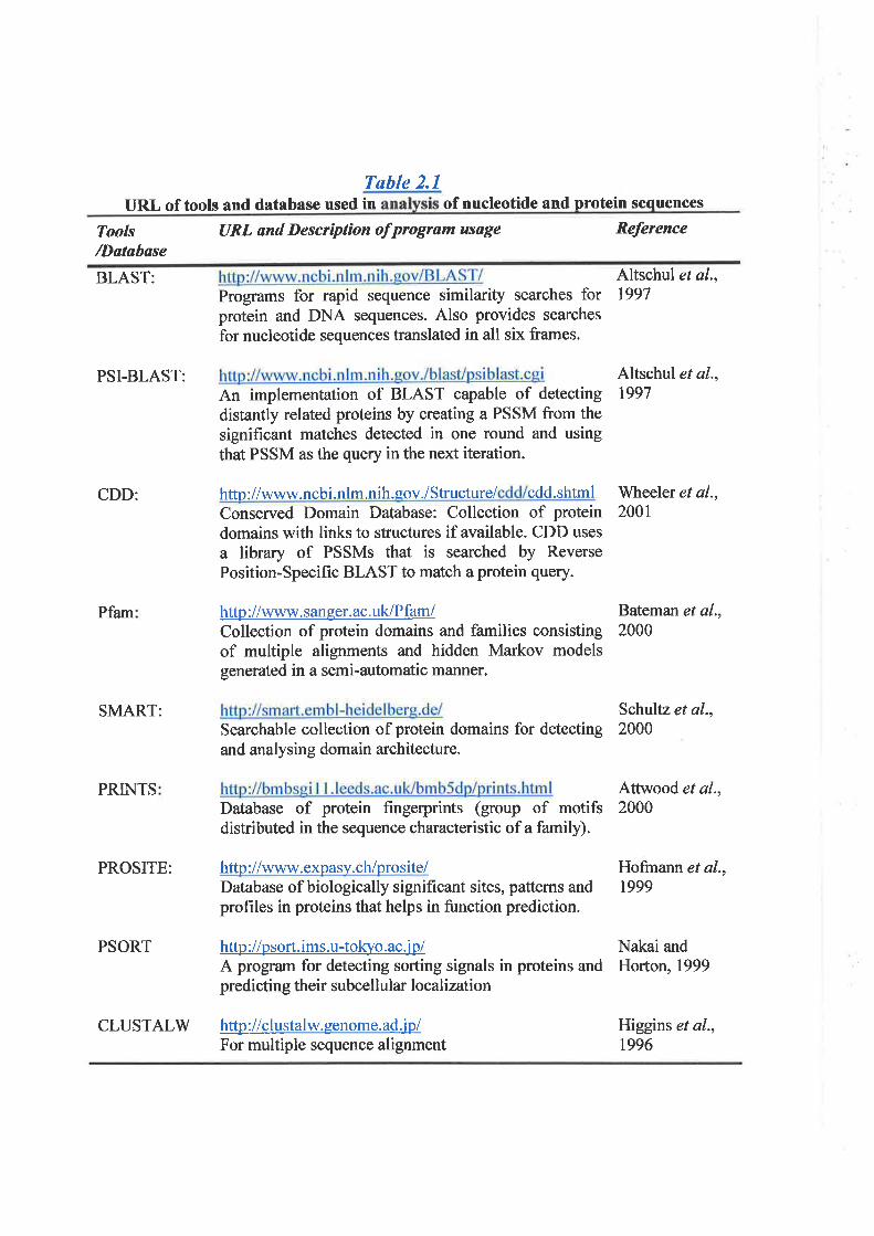
Tøble 2.1URL of tools and database used in of nucleotide and protein sequences
Tools/I)atøbase
URL and Descrìption of program usage Reference
BLAST:
PSI-BLAST:
CDD:
Pfam
SMART:
PRINTS
Programs for rapid sequence similarþ searches forprotein and DNA sequences. Also provides searches
for nucleotide sequences translated in all six frames.
An implementation of BLAST capable of detectingdistantly related proteins by creating a PSSM from thesignificant matches detected in one round and usingthat PSSM as the query in the next iteration.
http://www.ncb i.nlm.nih.eov./Structure/ cdd.shtmlConserved Domain Database: Collection of proteindomains with links to structures if available. CDD uses
a library of PSSMs that is searched by ReversePosition-Specific BLAST to match a protein query.
http ://www. saneer. ac. uk/Pfam/Collection of protein domains and families consistingof multiple alignments and hidden Markov modelsgenetated in a semi-automatic manner.
Database of protein fingerprints (group of motifsdistributed in the sequence characteristic of a family).
PROSITE: http://www.expasy.ch/prosite/Dat¿base of biologically significant sites, patterns andprofiles in proteins that helps in function prediction.
PSORT http ://psort. ims. u-toKvo.ac j p/A program for detecting sorting signals in proteins andpredicting their subcellular localization
http ://clustalw. genome. ad jp/For multiple sequence alignment
Altschul et al.,1997
Schultz et al.,Searchable collection of protein domains for detecting 2000and analysing domain architecture.
Altschul et al.,1997
'Wheeler et al.,
200t
Bateman et al.,2000
Attwood et al.,2000
Hofmann et al.,1999
Nakai andHorton, 1999
Higgins et al.,t996
CLUSTALW

Gha r3
Charact erization of Transcri ption U n it
3 (r3)
Tøble of Contents
3.l lntroduction
3.2 Methods
3 .2.1 ldentification of the Transcript sequence
3.2. 1. I Database searching
3.2.1.2 cDNA library screening
3.2.1.3 ldentification of the transcript using genomic sequence
3.2.1.4 PCR qnd RT-PCR
3 .2.2 Expression Analysis
3.2.3 In silico Analysis of Protein function
3.2.4Whole-body in situ hybridization of the mouse embryos
3. 2. 4. I Prehybridization of embryos
3.2.4.2 Preparation of RNA probes and hybridization of embryos
3. 2. 4. 3 P ost-hybridization w oshe s and embryo blocking
3. 2. 4. 4 Antibody binding
3. 2. 4. 5 P ost-antibody w ashe s and histochemistry
3.2.5 Study for T3 mutations in Breast Cancer
3.3 Results
3.3.1 Determination of Transcription Unit 3 g3) Transcribed Sequence
3.3.1.1 Database onalysís and cDNA sequencing
3. 3. 1. 2 Transcrípt Sequence Analysis
3.3.2 Expression analysis of Zj
3.3.3 Gene Structure of TJ-S
3.3.4 Determination of T3-L oDNA Sequence
3. 3. 4. I Database Analysis
3.3.4.2 Evidence of a Mouse transuipt homologous to T3-L
3.3.4.3 T3-L Sequence Assembly
3.3.5 Transcript Sequence Analysis of T3-L and mouse orthologue
3.3.6 Protein Sequence and Function Analysis
3. 3. 7 Genomic Organization and Altemative Splicing
3.3.8 Expression Study of Z3
3. 3. 8. 1 Differential Expression
3.3.8.2 Whole-body in situ hybridization of mouse embryo
Page
86
88
89
89
89
89
90
90
9t
9l
9l
92
93
94
94
95
96
96
96
97
98
98
99
99
r00
101
103
104
105
108
108
108

3.3.9 Mutation Analysis of 7J in Breast Cancer
3.4 Discussion
109
110
3.1 Introduction
The refined LOH studies of 16q243 in breast cancer have revealed that the likely
minimal LOH region is defined by the markers D16S303 and D16S2306. In addition,
detailed physicat and transcription maps were also established in this region (Whihore ef
al., 1998a), to form a framework for the identification of candidate tumor suppressor
gene(s). Two known genes, FAA and PISSLRE, mapped to this region (FAB consortium,
19961- Whitmore et al.,l998a), and based on their functions were previously suggested to
be possible candidate genes. However, none of these genes showed mutations in breast
cancer samples showing either restricted 16q24.3 LOH or complex LOH of 16q (Cleton-
Jansen et al., L999; Crawford et a1.,1999). Other recentþ identified genes in this region,
GASI l, CI6orfl (Whitmore et al., 1998b), and Copine 211(Savino et al., 1999) also failed
to demonstrate gene mutations in breast caûcer. Additional genes and transcripts were,
therefore identifred in the region for consideration as a candidate gene involved in breast
cancer
In the physical region between the MCIR and GASI I genes (Fígure 1.3), a cluster off,
two trapped exons, ET6 and ET7, isolated from the overlapping cosmids 3llC5 and 301F3/
(Whitmore et al., 1998a) were selected for gene identification. These trapped products
were less than l0 kb apart. They were grouped and assigned, based on their order in the
physical map, as the transcription unit 3 (fri). Based on database analysis, ET6 was shown
to have a sequence homology to a database entry MMDEF85, a partial 5' sequence of the
mouse def-8 gene. This is a developmentally regulated gene identified from cells of the
haemopoietic system.
Def-8 is a novel mouse gene, which was isolated by utilizing the retroviral gene-trap
vector carrying a B-galactosidase-neomycin fusion gene to infect myeloid progenitor cell
lines (Hotfilder et al., 1999). This gene-trap vector was constructed such that a splice
86
acceptor signal derived from c-fos was inserted at the 5' end of the lacVneo fusion gene,
which itself lacked an endogenous ATG start codon. Upon integration into actively
transcribed loci, the infected cells then gave rise to the clones that express a fusion of the
endogenous, trapped gene with the lacVneo rcporter. The gene-trap integration (GTI)
clones were subsequentþ isolated by G418 selection and a p-galactosidase positive
phenotype. To study the expression of developmentatly regulated genes during myeloid
differentiation, GTI clones were induced in order to allow differentiation in vitro into
either macrophages or granulocytes. Gene expression was monitored by histochemical
determination of p-galactosidase activity. From this expression analysis, 8 GTI clones each
representing different trapped genes were isolated and were designated def (difterentially
expressed in FDCP-Mix) gene, as def-l to def-8. Expression of all def genes in
undifferentiated progenitor cells has been shown to be down-regulated upon diflerentiation
into either macrophages or granulocytes (tlotfilder et a1.,1999). 'Whereas def-l to def-4
expression has been maintained upon macrophage differentiation, their expression is
down-regulated during granulocyte differentiation. Expression of def-5 to def-8 appears to
be down-regulated upon diflerentiation into both macrophages and granulocytes.
Down-regulation of both endogenous def-6 and def-8, representing the second SouP,
has also been observed dwing cell differentiation by Northern hybridization, using isolated
trapped gene products as probes (Hotfilder et a1.,1999). Whereas down-regulation of def-8
expression is restricted to macrophages and granulocytes, def-6 expression also appears to
be down-regulated upon erythrocyte differentiation. Further, Northem analysis of both def-
6 and def-8 has also been undertaken during mouse development and in adult tissue.
Expression of both genes is observed in mouse embryonic stages from day 7 to dzy 17.
Among adult tissues examined, expression of def-6 appears to be prominent in spleen,
which is a haemopoietic organ, and minor in lung, with a very low signal in heart, muscle
87
and kidney. In contrast, def-8 appears to be expressed in all adult tissues.
The expression profiles of def-8 in non-haemopoietic tissue suggest the importance of
this gene during embryonic development as well as a requirement for fully differentiated
tissue. Despite the determination of gene expression, the gene sequence of def-8 is only
available in databases as a partial 5' sequence of mRNA. The homology of the trapped
exon, ET6 to the def-g 5' sequence indicates that there is likely to be a human homologue
located at the 16q243 LOH region. Identification of the fult-lengfh transcript of the human
gene will provide more detail of the gene sequence as well as its predicted coding protein.
Based on the developmentally regulated expression, this gene was selected for
characterization and considered to be a potential candidate gene involved in breast cancer
iLa
outhï"o"ris. To achieve this primary aim, the ñrll-length transcript sequence of hr¡manY*"Ã-"--' ^
gene will be identified and its expression determined in various tissues. A predicted protein
sequence will also be characterized to identifr its possible function through protein
lvt u'''¡ " ç
database homology searches and,,functiorial motif prediction tools. Characterization of the
genomic structure will also be undertaken to provide information on the exon/intron
boundaries for gene mutation screening in breast cancer samples. Finally, the expression
study in mouse embryoes will be determined. This will require the isolation of a mouse
.DNA clone from gDNA libraries which will be used for RNA probe preparation for
hybridization to whole body mouse embryo
3.2 Methods
The following methods outline the general procedure used in this chapter. Most of the
detailed technical procedures are given in *Materials and Methods" (chapter 2)- Only the
methods that are specific to this chapter are given in detail.
¡
88
3.2.1 Identilication of the Transcript sequence
3.2. 1. 1 Døtahase searching
BLAST (Basic Local Alignment Search Tool) programs (Altschul et a1.,1997) were
used for sequence homology searches through public sequence databases. For the transcript
sequence identification, both ET6 and ET7 sequences were initially used, by BLASTN (for
nucleotide seqgence homology searches), to search for homologous cDNAs from both EST
and non-redundant dat¿bases. The homologous cDNA sequences, MMDEFS5 and N35521'
were first obtained and were subsequently used to compare with other cDNAs for further
sequence analysis. The cDNA sequences of clones isolated from cDNA libraries (described
in 3.2.1.2) were also used to search for homologous sequences.
3.2.1.2 cDNA líbrary screening
A human fetal brain cDNA library (formatted from the random primed, for 5'
stretched, and the oligo-dT primed libraries, Clontecþ and an oligo-dT primed cDNA
library from human fetal liver (Strategene) were screened utilizing trapped exon ET6 as a
probe. ET6 of 134 bp was excised from the pAMPlO vector with Not IISaI I double
digestion, crP32Jabeled by random priming, and hybridizedto cDNA library filters (section
2.2.18.2). Duplicate plaques that gave hybridized signal were picked, resuspended in SM
buffer, and eluted overnight. The eluates were re-plated, and plaques were re-hybridized
until plaque-purifred. Positive plaques were isolated and cDNA inserts were directly
amplified from pwified phages using vector primers and gene-specific primers (Table 3.1),
and sequenced by the Dye-terminator method @erkin-Elmer) on ABI 373 DNA sequencer
using the same set of primers above.
3.2.1.3 Identìftcøtion of the transcrípt usíng genomÛc sequence
89
Genomic sequence of the cosmid clone 301F3 (Kremmidiotis, G. and Gardner, 4.,
personal communication, lggg)was used to probe ""nr"JoJ"O.rences
from both human and\
mouse EST databases. The homologous EST sequences were analyzed and primers
designed for subsequent RT-PCR experiment. To optimize for both human and mouse
sequences, primers were designed from the homologotls regions that are human-mouse
identical (Table J.1). For the identification of human transcript, RT-PCR was carried out
on the total RNA isolated from human fetal heart. The cDNA products were separated by
agarose gel electrophoresis. The cDNA bands were purified from the gel and sequenced.
The gDNA clone (qo68a07) conesponding to EST A1309221that matched the 3' region of
the transcript was also purchased, DNA isolated and sequenced. The corresponding mouse
cDNA was determined by PCR amplification from mouse cDNA libraries derived from
two mouse tissues, fetal brain (Gibco, Life Technology) and embryo at 10-5 day post
ùl'*'coitus (d.p.c.) (Gibco, Life Technology).Mouse cDNA clone (courtesy from Dr Timothy
Cox, Department of Genetics, Adelaide University) was also isolated from mouse fetal
brain 9DNA library (Strategene) utitizingôcDNA probe derived from RT-PCR productl\
mentioned above.
3.2.1.4 PCR ønd RT-PCR
All PCR and RT-PCR procedures were performed as described in section 2.2.12.1 and
section 2.2.13. The sequences of all oligonucleotide primers used for the identifrcation of
the transcript sequence are presented in Tøble 3.f. Those of primers designed for
determination of the exon-intron boundaries are given n Table 3.2.
3.2.2 Expression Analysis
The transcription products were analysed by
90
hybridization procedure Y
(2.2.11.2). Northern blot filter containing 2 ¡ry of poly(A)+ mRNA from six different
human tissues were hybridized according to manufactwer's instructions in "ExpressHyb"
hybridization solution (Ctontech). Two cDNA probes were used in separated hybridization
reactions. The fust was a gDNA insert of clone yx6la06 that showed a sequence homology
to that of ET7-ET6. The second probe derived from RT-PCR products that were generated
according to the homologous ESTs of the genomic region down stream from the first probe
(see Figure 3.4).
Differential expression of the transcript was determined by hybridization of the
second probe to human RNA dot blot filter (Human RNA Master Blot , Catalogue # 7770-
1, Clontecþ that contains mRNAs from various human tissues of both fetal and adult
stages. Hybridization was also performed in "ExpressHyb" and followed the instruction
recommended by manufacturer.
3.2.3Ín sìlico Analysis of Protein function
The BLASTP program was used to compare the T3 protein \Mith the sequences of
known, functional, and novel proteins in the public protein databases (NCBI). The
functional domains of the protein were predicted using three software programs, Profile
Scan, Pfam, and SMART. Protein localization analysis was by PSORT.
3.2.4 Whole-body in sítu hybridization of the mouse embryos
This part was performed at Deparhnent of Genetics, Adelaide University, under kind
supervision of Dr. Timotþ Cox, and with the assistance of Miss Sonia Donati.
3. 2.4. 1 Prehybrìdizttíon of embryos
Embryos at mid-gestation period (average 10.5 d.p.c) had been previously harvested,
9I
fixed, and stored in 100% methanol. They were re-hydrated by sequential rinsing in (Ð
75olo methanol I 25% pBS, (ii) 50olo methanol I 50% PBS, and (äi) 2ío/omethanol I 75%
pBS at 4 oC, each for 5 minutes. Embryos were washed a further three times in PBT l0-l%
Tween-2g þoþxyethylene-sorbitan monolaurate) in PBS] for 5 minutes each at room
temperature, then treated wittì l0 Fglml Proteinase K in PBS for an appropriate amount of
time. For a general guideline, 9.5 d.p.c. embryos were treated for l0 minutes, with each
additional day of development requiring an additional 2 minutes. After Proteinase K
treatment, embryos were washed twice with PBT for 5 minutes each and re-fixed in PG-
pBT 10.2% glutaraldehyde (EM-grade, Sigma), 4Yo paruformaldehyde (PFA) in PBTI at
room temperature for 20 minutes. After fixation, embryos were washes five times in PBT,
each for 5 minutes at room temperature. The embryos were transferred to 5 ml capped
tubes and washed at room temperature, once with a l:l mix of HB and PBT for 15 minutes
/rynd once with HB for 10 minutes tHB (hybridization bufler): 50% formamide; 5x SSC
(pH a.5); 50 pglml heparig 0.1% Tween-201. The tube was then frlled with HB containing
100 Fglml tRNA and 100 pglml sheared denatured herring spenn DNA, and
prehybridization was allowed for 4 hours at70 "C-
3.2.4.2 Preparation of RNA probes and hybrídizttíon of embryos
Both sense and anti-sense RNA probes were prepared from cloned oDNA fragment in
pBluescriptsK (pBSK, Strategene) vector. The mouse nf3 cDNA fragment of 576 bp was
previously cloned into pBSK, in the 5'+3' direction between Hindfr. and BamHl sites of
the multiple cloning site region. The recombinant plasmid was linearized either by Hindfn
or BamHI digestion to obtain the DNA templates for synthesis of anti-sense and sense
RNA probes respectively. The linearized plasmid was purified by phenoVchloroform
extraction, followed by ethanol precipitation. To I pl of lineaized plasmid (1 ¡rglpl), 2 pl
92

of lgx transcription buffer containing 0.1 M DTT, 2 ¡ú of 10x NTP labelling mixture
containing DIG-UTP (digoxigenin-UTP), I ¡rl of RNase inhibitor (20 units/¡rl), RNase-free
water to a final volume of 18 pl, and 2 pl of either T3 or T7 RNA polymerase (20
units/pl) were added depending on the linearization of the plasmid and the RNA probe
being synthezised. The reaction was allowedat3T "C for 2 hours. The RNA transcript was
ana1yzed by electrophoresis of lpl aliquot of the reaction mixture on lVo agafose gel in
TBE, followed by ethidium bromide staining and LIV illumination. The amount of RNA
synthesized was estimated from the band intensity by assuming that a l0 fold increase in
intensity of the RNA band to that of the plasmid represented approximately l0 pg of
probe. To remove the DNA template, 2 pl of DNase I (10 tmits/ pl, RNase-free) was added
to the reaction mixture and incubated for a further 15 minutes at 37 "C. After l5-minute
incubation , 2.5 ¡il of 4 M LiCl and 75 ¡tl prechilled 100% ethanol were added and mixed.
The mixture was left at -70 "C for 30 minutes and followed by centrifugation at 12,000xg
for 15 minutes. fn" {/t"twas washed with 50 pl of TIYocold ethanol (vÐ, dried, and
dissolved in RNase-free water to a final concentration of approximately I pglpl. The RNA
probe, DlG-labelled, was denatured in I ml HB containing 100 pdrnl tRNA and 100
pglml sheared denatured herring sperm DNA, at 95 "C for 3 minutes then placed at70 "C
to equilibrate. The denatured RNA probe \¡ras then added to the embryos in hybridization
mix (3.2.4.1) to a final concentation of I ¡rglml and hybridization was allowed overnight
at 7 0 " C, with agitation.
3.2.4.3 Post-hybridìzatíon washes and embryo blockìng
After the completion of embryo hybridization the following washes were carried out
in HB, SSC-FT (2x SSC pH 4.5; 50% formamide;0.7Yo Tween-2O), and lx TBST [10x
93

stock: 8% (w/v) NaCl; 0.2Y' (wlv)KCl;1% Tween-20; 25 rîl' lM Tris pH 7-5 in the final
volume 100 ml]. Embryos were washed twice with HB at70 "C, each for 30 minutes, then
twice with SSC-FT for 5 minutes each at room temperature. This followed by three washes
with ssc-FT at 65 .C for 30 minutes each. Embryos were allowed to cool down to room
temperature and washed three times, for 5 minutes each, with lx TBST at room
temperature. The embryos were then blocked by incubating in 10% heat-inactivated sheep
serum in TBST, for l-3 hours at room temperature'
3. 2.4.4 AntíhodY binding
The anti-DIG antibody, alkaline phpsphatase conjugated (Boehringer Mannheim) was
'll. r r!' r "'-' ' ' o'r '' i" ri (lt' ¿{ ¡
+'
used to determine the hybridiàed embryos. The antibody was first pre-adsorbed by
incubating 0.5 pl of anti-DIG antibody in I ml TBST containing l% heat-inactivated sheep
serum and2 mg heat-inactivated mouse powder. The reaction wrn allowed for I hour at 4_
oC, with agitation. After completion, the reaction tube was centrifuged at 15,00\for 5
¿minutes and the supernatant containing pre-adsorbed antibody was collected. To allow
antibody binding, the solution was removed from the pre-blocked embryos (3.2-a3) and
was replaced with pre-adsorbed antibody and incubateþvernight at 4 "C.
3. 2. 4. 5 Post-antìbody was hes and hßtochemistry
After the completion of overnight incubation with pre-adsorb antibody, the excess
antibody conjugate was removed and the embryos were washed three times with TBST at
room temperature, each for 5 minutes. The embryos were then transferred to the new vials
and washed a further five times, for I hour each, at room temperature \¡/ith TBST
containing 2 mM Levamisole. They were left ovemight at 4 oC in the new
TB ST/Levamisole, with agitation.
I
94
Prior to color development, the embryos were wash three times, for l0 minutes each,
at room temperature in alkaline phosphate buffer (APB) containing 2 mM Levamisole
(APB: 100 mM NaCl; 50 mM MgCl; 0.lolo Tween-20; 100 mM Tris, pH 9.5)' To develop
the color, the embryos were incubated in ABP/2mM Levamisole containing 4.5 pVml NBT
and 3.5 p1/ml BCIP. This was caried out at room temperature under dark conditionjon a
l"s P ^^ o"t"'
shaker t¿ble. The reaction was allowed for d period of time until the color, dark purple, was
visible, by observing the progress of the reaction for brief intervals under a dissecting
microscope. The color reaction was stopped by removing and rinsing the embryos three
times at room temperature in PBT containing 1 mM EDTA and lYo glacial acetic acid. The
embryos were subsequently washed a frrrther three times, each for 5 minutes, in PBT / I
mM EDTA I lYo glacial acetic acid. The embryos were then stored, at 4 "C, in 80%
glyceral arrd lYo acetic acid in PBT. Each embryo was examined under the dissecting
microscope and the photographs were taken for fi¡rther analysis.
3.2.5 Study for T3 mutations in Breast Cancer
Mutation study was undertaken on the genomic DNA isolated from two panels of a,(
total 48 breast cancer tissue samples and ûom 24 breast cancer cell lines. The breast cancer/\
tissue samples were taken from þé patients, with inform consent, from two medical
centers: the Flinder Medical Center, South Australi4 and the GuflHospital, Lodon, UK.
Screening for gene mutation was by SSCP analysis of the genomic PCR fragment utilizing
the primer pairs designed for the amplification of each exon, including part of its flanking
introns. All primers were with a fluorescene dye (HEX). The IIEX labelled
genomic PCR products \Mere separated by eletrophoresis on the non-denaturing
polyacrylamide gel, O.i1,alrU 6Yo gelwlth2%gþerol in TBE buffer, using the Gene Scan
2000 (Corbett Research). PCR products showing a conformational change were
95

Table 3.1Sequences and Positions of the Primers used for ?3 Transcript Sequence
I)etermination
No Prìmer Prímer Sequence (5' -+3')
NucleotídePosítíon in the
Trønscript
Common Primers for Human and Mouse homologouscDNAn1 35521-P cagccgggagacaqatgcgag2 35521--T caggtgggatgctatggaatatg3 35521-U gcttgttgaaggggttgagg4 35521-R catcacgcgttcagggcagcg5 N355-D gtgcaagcaggtgattctgg6 35521-B grtqtaacaccatcatctggg7 T3EA-F gtgttattaccqctgtcacagI T3EA-R ctcgqcacagcggtaatcc9 T3EB1-F actgcagccactgccactg10 T3EB1-R caccagctcctccacgtag11 T3EB2-F cttcatcacctgcagqgag12 T3EB2-R ctccctgcaggtgatgaag13 T3EC-F ggcagcattttgtggagaacgI4 T3ED-F cagccacacgtctgtgtgc15 T3ED-R gacgtgtggctgtcgaacgI6 T3XE-F accacttgtcccaagtgtgcI7 A'I309-R tgcaggatcccatcgcctaac1B T3EF-R cccacactgttgatccaacc
Positíons in thehuman transcrìpt
29-49s8-80
t3r-772253-233343-362535-554586-606121-709798-816982-964
]-027 -1,0457045-r02'71083-1103L27 9-1291I290-7272134 6-13 65221 5-22553 0 52-3 033
Prímers Specifrc for Mo use transcrípt
tcctt ctgcaccatagagatggcaaggccatctctat ggtggaggccaatgataa ct gacagat cacatgtctgtcctgagtc
Positions in themouse transcrþt
1765-1785L792-11'1322'13-2253301_6-3036
79 MT3-A20 MT3-B2I MT3-C22 MT3-D
Vector Primers
232425262'7
2829
T3T1M13FM13Rgt11Fgt11RSP6
attaaccctcactaaagqgataatacgactcactat agggtgtaaaacgacAgccagtcaggaaacagctatgacctggcgacgactcctggagcccgtgacaccagaccaactggtaatgggatttaggtgacactatagr
nAll except primers no. 1,5,17,18 were also usedfor the identification of the monsetranscript sequences.Primers 35521-R and 35521-U were also utilized in the 5'RACE experiment in
combination with SMART 5' primer (2.2.17.1).
reamplified from 100 ng of genomic DNA with corresponding unlabelled primers. The
products were purified and sequenced.
3.3 Results
3.3.1 Determination of Transcription Unit 3 (ft|) Transcribed Sequence
3.3.1.1 Database analysìs and cDNA sequencing
Based on the 16q24.3 physical map (Fìgure 1.3),the trapped exons ET6 and ET7 are
less than l0 kb apart and were considered likely to be part of the same gene. Non-
redundant database searches initialty identified that ET6 showed sequence homology to the
transcript MMDEF85, the 5' sequence of the mouse developmentally regulated gene, def-8.
However, results from analyses of human EST database indicated that both ET6 and ET7
belonged to the same tanscript as shown by their sequence homology to the same ESTs,
yx6la06.rl (entry N35521) and zl<84b04.r1 (entry 44099191). N35521 and AA099l9l
are the 5' sequence of cDNA clones, yx61a06 and zk84b04 respectively, which were
directionally cloned into the vectors. Therefore, the orientation of these trapped products is
ET7-ET6, corresponding with the 5'+3' direction of the available sequences of both ESTs
(Fìgure3./A). Furthermore, sequence alignment of N35521 and MMDEFSS showed that
continuation of homology between human and mouse cDNA W also observed in the
region beyond the 3' end of ET6. These results suggested thatETTlET6 sequence was part
of the human gene, designated "transcription unit 3" (T3), which is homologous to the
mouse def-8 gene.
To extend the transcribed sequence of T3, ET6 was excised from the pAMPl0 vector
and used as a probe for the screening of cDNA libraries (3.2.1.2). The cDNA clone
318C5.6 was obtained from screening a fetal brain cDNA library cloned into the î,gtll
vector (Clontech). ET6 was also used to screen a fetal liver cDNA library inserted nì"ZAP
96
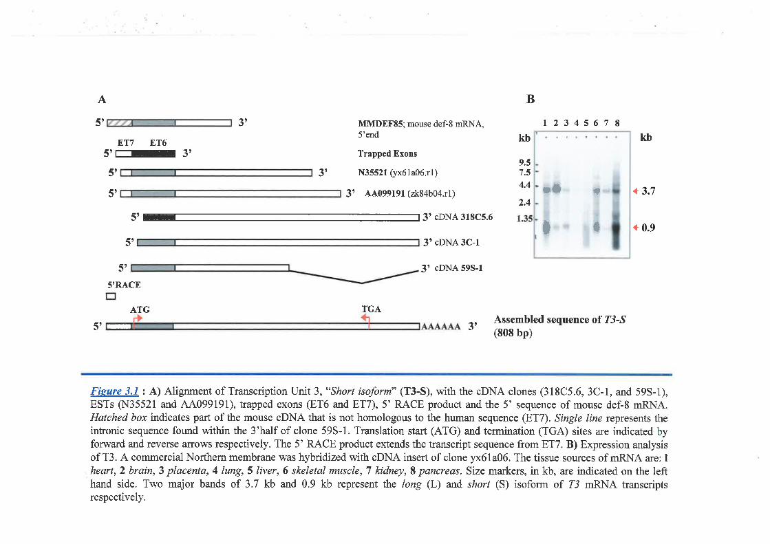
A B
5t 3t MMDEFSS; mouse def-8 mRNA,5'end
Trapped Exons
N35521 $x6la06.rl)
3' 44099191 (2k84b04.r1)
12345678ET7 ET6
5'r-3t5t
5t
3',9.57.5
4.4
kb kb
+ 3.72.4
5' 3' cDNA 318C5.6+ 0.9
5t 3t cDNA 3C-1
J' cDNA 595-1
5'RACE
ATG TGA
5t 3tAssembled sequence of T3-^S
(808 bp)
Fieare 3.1 : A) Alignment of Transcription Unit 3, "Short isoform" (T3-S), with the cDNA clones (318C5.6, 3C-1, and 593-1),ESTs (N35521 and 44099191), trapped exons (ET6 and ET7),5' RACE product and the 5' sequence of mouse def-8 mRNA.Hatched box indicates part of the mouse cDNA that is not homologous to the human sequence (ET7). Single line rcpresents theintronic sequence found within the 3'half of clone 595-1. Translation start (ATG) and termination (TGA) sites are indicated byforward and reverse ¿urows respectively. The 5' RACE product extends the transcript sequence from ET7. B) Expression analysisof T3 . A commercial Northern membrane was hybridized with oDNA insert of clone yx6la06. The tissue sources of mRNA are: Iheart,2 brain,3 placenta, 4 lung, 5 liver,6 skeletal muscle, T kidney, S pancreas. Size markers, in kb, are indicated on the lefthand side. Two major bands of 3.7 kb and 0.9 kb represent the long (L) and short (S) isoform of T3 mRNA transcriptsrespectively.
5'
I *
;0
=
vector (Strategene), from which cDNA clones 3C-l and 3C-2 were isolated. PCR analysis
of 3C-l and3C-2 using the vector primers (T3 and T7) as well as a combination of vector
primer and gene specific primer (35521-R) indicated that both contained the same insert
size. Therefore, in subsequent studies only the 3C-1 clone was analyzed. ln addition, the
pCR product amplified from the region close to the 5' end of cDNA clone yx6la06
(primers 35521-F and -R) was also used to screen the same fetal brain cDNA library
(Clontech) and the cDNA clone 59S-l was isolated. The DNA sequence of 318C5.6, 3C-1,
and 59S-l was determined by using the vector primers as well as the primers designed to
the sequence available from ESTs yx6la06.rl (N35521) and zk84b04.rl (44099191)- The
results showed that both 318C5.6 and 3C-1 shared the same sequence with those of
N35521 and AA099l9l but also extended the sequence down-sheam from their 3' end.
The extended sequence revealed the 3'polyadenylation signal (AATTAA) characteristic of
3'end of the transcript. The cDNA 593-1, although containing an insert of 1.2 kb, larger
than those of 318C5.6 or 3C-1, showed the identical sequence only from the 5' end to a
point later found to be the exon 4 boundary. The remainder of the sequence was intronic
indicating an incomplete splicing product. The 5'ends of all isolated cDNA clones did not,
however, extend beyond the ET7 sequence. Therefore, 5' RACE was also perforrred
(described in section 2.2.17.1) using gene specific primers and the SMART 5' primer
(Tahlc 3./). This allowed an additional 24bp sequence to be determined upstream from the
5' end of ET7. The relationship between the various cDNA clones is shown
diagrammatically in Figure 3.14.
3.3. 1. 2 Transcript Sequence Analysß
The sequence of oDNA clones, trapped exons, and 5' RACE product were assembled
and identified a 808 bp transcript with an open reading frame (ORF) of 591 bp extending
97
from the sta¡t codon, followed by a 3'untranslated region (3'UTR) of 131 bp (Figure 3-2)-
The methionine start codon, ATG, and its surounding nucleotides conform with the Kozak
consensus sequence characteristic of translation start site (Kozak, 1989) and is preceded by
an in-frame stop codon, TAG, 66 bp upstream from the start site. The 73 ORF was
predicted to code for a protein of 197 amino acids. Two possible polyadenylation signals
were identified within the 3'UTR of the transcript, the first (AATTAA) at position 715-
717, and the second (ATTAAA) at position 777-782. From an examination of the DNA
sequence of the cDNA clones isolated from the cDNA libraries and those identified from
the EST database only the second site appeared to be fiurctional.
3.3.2 Expression analysis of TJ
The cDNA insert of clone yx61a06 was used as a probe to hybridize to a commercial
multiple tissue Northern membrane (Clontech). Two prominent bands with difterent
intensity of approximately 0.9 kb and 3.7 kb were detected in all tissues examined (Fígure
3.18). The result indicated that at least two transcription isoforms were expressed but the
expression of the two isoforms varied in different tissue. The transcript that had been
sequenced was equivalent to the short isoform (0.9 kb), designated "T3-S', and appeared
to be highly expressed in heart, skeletal muscle, and pancreas. Expression of Z3-S was
relatively low in brain, placenta" and liver, with very low expression presented in lung and
kidney. The 3.7-kb isoform, assigned as "T3-L", was strongly expressed in brain which
was contrast to the expression of the short isoform. Medium level of T3-L expression was
seen in heart, skeletal muscle, and pancreas. In kidney tissue, expression of T3-I was
stronger than that of T3-S. The remaining tissues showed a low level of expression of Z3-I.
3.3.3 Gene Structure of ?3-S
To explore the gene organization for Z3-S, primers designed to both strands of 73-S
98
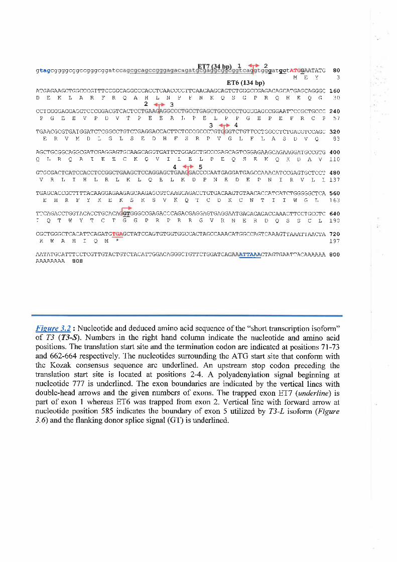
2gtagcggggcggcc gggc ggat a tætÀT TATG 80MEY3
ET6 (134 bp)ATGAGAAGCTGGCCCGTTTCCGGCAGGCCCACCTCAACCCCTTCAACAAGCAGTCTGGGCCGAGACAGCATGAGCAGGGC 160DE KLAR FRQAH LN P ENKQ S G PRQHE QG 30
2 3CCT GGGGAGGAGGT CCCGGACGTCACT C GGGGAGCCGGAATTCCGCTGCCC 240
57PGEEV P DVT PEEAL PE LP PGE PE FRCP34
TGAACGCGTGATGGATCTCGGCCTGTCTGAGGACCACTTCTCCCGCCCTG TCTGTTCCTGGCCTCTGACGTCCAGCERVMDLGLS E DH FS RPVG L FLAS DVA
AGCTGCGGCAGGCGATCGAGGAGTGCAAGCAGGTGATTCTGGAGCTGCCCGAGCAGTCGGAGAAGCAGAAGGATGCCGTG
Q LRQAI E E CKQV T LE L PE Q S E KQKDAV
320
400110
4 5GTGCGAC CCAATGAGGATGAGCCAÄACATCCGAGTGCT CCT 480
),31VRL I H LRLK L QE LK D PNE D E PN ] RVLL
TGAGCACCGCTTTTACAAGGAGAAGAGCAAGAGCGTCAAGCAGACCTGTGACAAGTGTAACACCATCATCTGGGGGCTCAEHRFYKEKSKSVKQTCDKCNTIIWGL
560163
TTCAGACCTGGTACACIQTVùYT
CTGCACA
- GGCCGAGACCCAGACGAGGAGTGAGGAATGAGAGAGACCAAAGTT
CTGGPRPRRGVRNERDQSccTcccrc 640SCL19O
120L9'7
CGCTGGGCTCACATTCAGATGT CTATCCAGTGTGGTGGCCACTAGCCAAACATGGCCAGTCAAAGTTAiU\TTAI\CTARVùAHIQM*
AATATGCATTTCCTCGTTGTACTGTCTACATTGGACAGGGCTGTTCTGGATGAG TTAA.A,CTAGTGAATTACAAÀAAAAJV\AIUUU\ 808
800
Figure 3.2: Nucleotide and deduced amino acid sequence of the "short transcription isoform"of T3 (f3-Ð. Numbers in the right hand column indicate the nucleotide and amino acidpositions. The translation staf site and the termination codon are indicated at positions 7l-73and 662-664 respectively. The nucleotides surrounding the ATG start site that conform withthe Kozak consensus sequence are underlined. An upstream stop codon preceding thetranslation start site is located at positions 2-4. A polyadenylation signal beginning atnucleotide 777 is underlined. The exon boundaries are indicated by the vertical lines withdouble-head affows and the given numbers of exons. The trapped exon ET7 (underline) ispart of exon I whereas ET6 was trapped from exon 2. Vertical line with forward arrow atnucleotide position 585 indicates the boundary of exon 5 utilized by T3-L isoform (Figure3.6) andthe flanking donor splice signal (GT) is underlined.
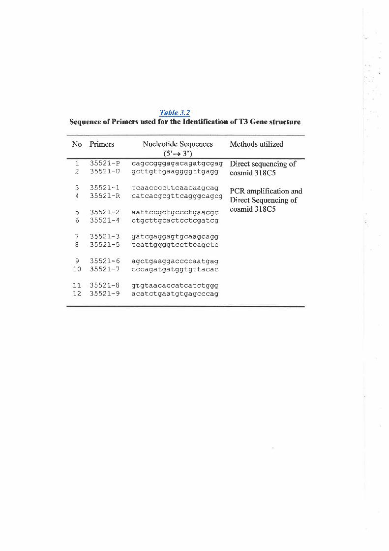
Table 3.2Sequence of Primers used for the Identification of T3 Gene structure
No Primers Nucleotide Sequences(5'+ 3')
Methods utilized
1
2
{
4
6
6
1
I
3552 1-P3552 1-U
35s21-135521-R
35s2r-23552L-4
3552 1-33552 1 -5
I 3552r-610 35521-7
cag ccg gga ga ca gat gcga ggcttgttgaaggggttgagg
tcaaccccttcaacaagcagcat ca cgcgtt cagggcagcg
aattccgctgccctgaacgcctgcttgcactcctcgatcg
gatcgaggagtgcaagcaggt catt ggggt ccttcagrctc
agctgaaggaccccaatgagcccagatgatggtgttacac
gtgtaacaccatcatctgggacatctqaatgtgagcccag
Direct sequencing ofcosmid 318C5
PCR amplification andDirect Sequencing ofcosmid 318C5
11T2
3 552 1-83552 1-9
sequence (Table 3.2) were used to PCR arnplified the genomic DNA from cosmid clone
31gC5, and the amplified products were sequenced from both ends. The same primers
were also used to perform direct sequencing of cosmid 318C5. Alignment of genomic
sequence with that of oDNA allowed the identification of T3-S exon/intron boundaries. As
shown tn Fìgure 3.3, the gene for I3-S consists of 5 exons ranging in size from 60 bp to
353 bp. The splice junction boundaries (Table 3.3) conform to the consensus sequences for
donor and acceptor splicing sites provided by Shapiro and Senapathy (1987). The
translation initiation codon, ATG, is situated \¡rithin exon2 (Fígure 3-2 and 3.34). The 5'
UTR constitutes entire exon 1 and l0 bp 5'part of exon 2. The last exon, exon 5, contains
part of coding sequence, stop codon (TGA), and the 134 bp 3'UTR. The trapped product
ET7 (35 bp) appeared to be trapped from 3' part of exon 1, whereas ET6 was completely
trapped from exon 2.
3.3.4 Detemination f3-I cDNA Sequence
3. 3. 4. 1 Databas e AnølYsß
The result of Northerr analysis indicated that part of T3-L shared the same sequence
with Zj-S. However, it was not known whether they shared the same ORF or only shared
the sequence but with different ORF. Only with the identification of a complete cDNA
sequence of T3-L would the relationship of both forms be addressed. A complete sequence
of Z3-S was used as a query in an attempt to identiff more extended sequence from the
EST database. Search results showed that most of the human ESTs homologous to TJ-S,
from either the 5' or 3' direction(Fìgurc 3.3B), failed to provide more extended sequences.
Some human ESTs appeared to include an extra exon between exons I and 2. This extra
exon, however, did not create any ne\il ORF indicating that this is likely to be an
alternatively spliced form of 5' UTR. The same extra exon was also detected in the 5'
99
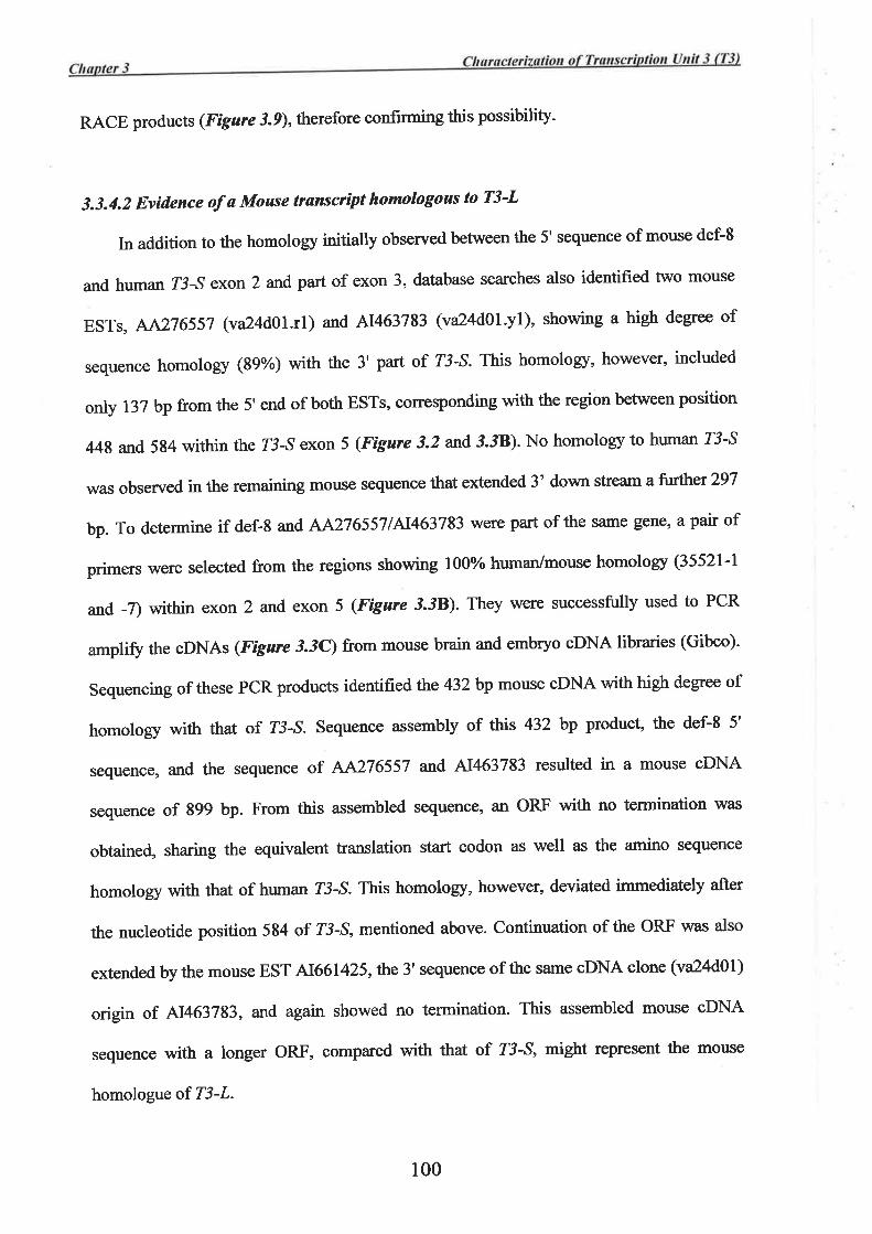
RACE products (Fìgure3.9), therefore confirming this possibility-
3.3.4.2 Evidence of ø Mouse transcript homologous to T3-L
In addition to the homology initially observed between the 5' sequence of mouse def'8
and human 1'j-S exon 2 and part of exon 3, database searches also identified two mouse
ESTs, AA276557 (va2kd}l.rl) and N463783 (va24d01.yl), showing a high degree of
sequence homology (8g%) with the 3' part of Z3-S. This homology, however, included
only l3Z bp from the 5' end of both ESTs, corresponding with the region between position
448 and 584 within the I3-S exon 5 (Fìgure 3.2 and 3.38)' No homology to human T3-S
was observed in the remaining mouse sequence that extended 3' down stream a further 297
bp. To determine if def-8 arrd AA2765571A1463783 were part of the same gene, a pair of
primers were selected from the regions showing l00%o human/mouse homolory (35521'l
and -7) within exon 2 and exon 5 (Figure 3.38). They were successfully used to PCR
ampliff the cDNAs (Figure i.ic) from mouse brain and embryo cDNA libraries (Gibco).
Sequencing of these PCR products identifiedthe 432 bp mouse cDNA with high degree of
homology with that of T3-S. Sequence assembly of this 432 bp product, the def-8 5'
sequence, and the sequence of AA276557 and A1463783 resulted in a mouse oDNA
sequence of g99 bp. From this assembled sequence, an ORF \¡vith no termination was
obtained, sharing the equivalent translation start codon as well as the amino sequence
homology with that of human T3-S. This homology, however, deviated immediately after
the nucleotide position 584 of Z3-S, mentioned above. Continuation of the ORF was also
extended by the mouse EST 41661 425,the 3' sequence of the same cDNA clone (va24d0l)
origin of A1463783, and again showed no termination. This assembled mouse cDNA
sequence with a longer ORF, compared with that of T3-S, might represent the mouse
homologue of T3-L.
100
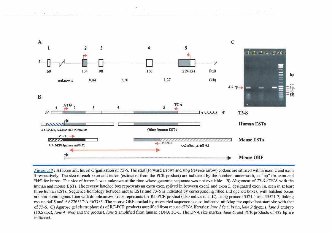
A cI 23 !4
5', 5'
134 98 150 2r9lt34 (bp)
(kb)0.84 r.27
432bp¡,
TGA1 , 3 5
AAAAAA 3' T3.S
Human ESTsAA035322, AA30ó508, HSU46300
3552r-r+ffiMMDEFSS(mouse def-8 5')
Other human ESTs
Mouse ESTs4A276557, A1463783
Mouse ORF
Fisure 3.3 : A) Exon and Intron Organization of Z3-,S. The start (forward arrow) and stop (reverse anow) codons are situated within exon 2 and exon5 respectively. The size of each exon and intron (estimated from the PCR product) are indicated by the numbers underneath, as "bp" for exon and"kb" for intron. The size of intron 1 was unknown at the time where genomic sequence was not available B) Alignment of Z3-,S cDNA with thehuman and mouse ESTs. The reverse hatched box represents an extra exon spliced in between exonl and exon 2, designated exon 14 seen in at least
three human ESTs. Sequence homology between mouse ESTs and Z3-,S is indicated by corresponding filled and opened boxes, with hatched boxes
are non-homologous. Line with double arrow-heads represents the RT-PCR product (also indicates in C), using primer 35521-I and 3552I-7, linkingmouse def-8 and A,^2765571A1463783. The mouse ORF created by assembled sequence is also indicated utilizing the equivalent start site with thatof Z3-,S. C)AgarosegelelectrophoresisofRT-PCRproductsamplifiedfrommouseoDNAlibraries: lanel fetalbrain, lane2thymus, lane3 embryo( 10.5 dpc), lane 4 liver, and the product, lane 5 amplified from human oDNA 3 C- 1 . The DNA size marker, lane 6, and PCR products of 432 bp are
indicated.
60
unknown
bp
501
tgl242190
2.20
B
TG4
5t
<_
3.3.4.3 T3-L Sequence AssemblY
Cosmid 301F3 was part of the cosmid contig within the 16q24.3 physical map from
which trapped exons ET6 and ET7 were isolated. As the large scale sequencing project has
been established, the sequence of cosmid 301F3 was determined (Kremmidiotis, G. and
Gardner, 4., personal communication, 1999). To identiff additional exons within close
proximity to Z3-S, the sequence of 301F3 was used to search database for homology to the
partially sequenced transcripts. As indicated tn Figure 3.4A,301F3 showed regions of
homology to three mouse ESTs, AA276557, A1463783, and 41661425, which were linked
to I3-S exon 5, previously mentioned in section 3-3.4.2.In addition, genomic sequence
homologous to the mouse EST W45991 and human EST 44346746 was also identified in
the adjacent region approximately I kb down stream. Altogether, the regions of homology
between EST sequences and genomic sequence established additional exons initially
designated exons A, 81, 82, C, D, and E, which were possibly part of T3-L- A set of
primers (Table 3.I) designed from the regions showing l00%o human/mouse sequence
homology within some exons were successfully used to RT-PCR ampliff the human
cDNA from human fetal tissue RNA (frgure 3.4). After sequencing, these cDNA
fragments were assembled and formed part of Z3-Z cDNA by linking to the position 584
within Z3-S exon 5. This linked sequence extended another 822 bp and also created a
continuation of ORF from exon 5 with no termination similar to that seen in mouse cDNA
(section 3.3.4.2). Two RT-PCR products spanning from exon A to exon D (Figure 3.44)
were also used to hybridize to a multþle tissue Northern membrane (ClonTech). As
expected, a single band of 3.7 kb corresponding to T3-L was detected (Figure 3.54). The
variation in band intensity between the different tissue was similar to the results seen when
the membrane was probed with Z3-,S oDNA (section 3.3.2).
101
The joined cDNA sequence from exon I to exon E was only 1,406 bp in length with
no evidence of ORF termination nor 3'UTR. Therefore, additional 3' sequence was sought
by further analysis of the EST database. The genomic region, designated exon F, situated
approximately 2.4 kb down stream from exon E (Figure 3.4A) was matched with a group
of human ESTs that belonged to the UniGene cluster Hs.62771. Sequencing of a cDNA
clone (qo68a07) of A1309221, one of these ESTs, identified an insert sequence of l274bp.
This sequence showed a polyadenylation signal (ATTAAA) and a polyadenylation site 13
bp down stream from this signal. This sequence wrlsi then successfully joined with exon E
by RT-PCR using a primer from exon D (ED-F) and the reverse primer (4I309-R)
generated from this cDNA sequence (Table 3.1). The final sequence assembly identified a
complete Z3-I cDNA sequence of 3,525 bp (Figure 3.6), consistent with the size of the
band on Northern analysis.
At this stage the gene designed "?3"codes for at least two transcription isoforms, T3-
S and T3-L, based on the Northern analysis results, the transcribed sequences, and the
evidence of sha¡ed ORF.
In addition to the assembled mouse cDNA sequence (section 3.3.4.2), identification of
the complete mouse transcript sequence homologous to human T3-L was also undertaken.
The same set of primers, which were used in the human T3-L transcribed sequence
assembly (Table 3.1), were also successfully used to amplifr and confinn ttre mouse
gDNA sequence (Figure 3./). Since the mouse EST V/45991 provided the most 3'region
of the available cDNA sequence, the clone mc81g01 that was used to generate this EST
sequence was isolated and DNA sequenced. The extended sequence from W45991 was
then used for additionat dbEST analysis, and followed by the sequential EST searching for
the overlapping oDNA sequences. A contig of mouse ESTs was finally obtained, and.the
sequences were assembled. This extended sequence formed mostþ the 3' UTR of the
t02
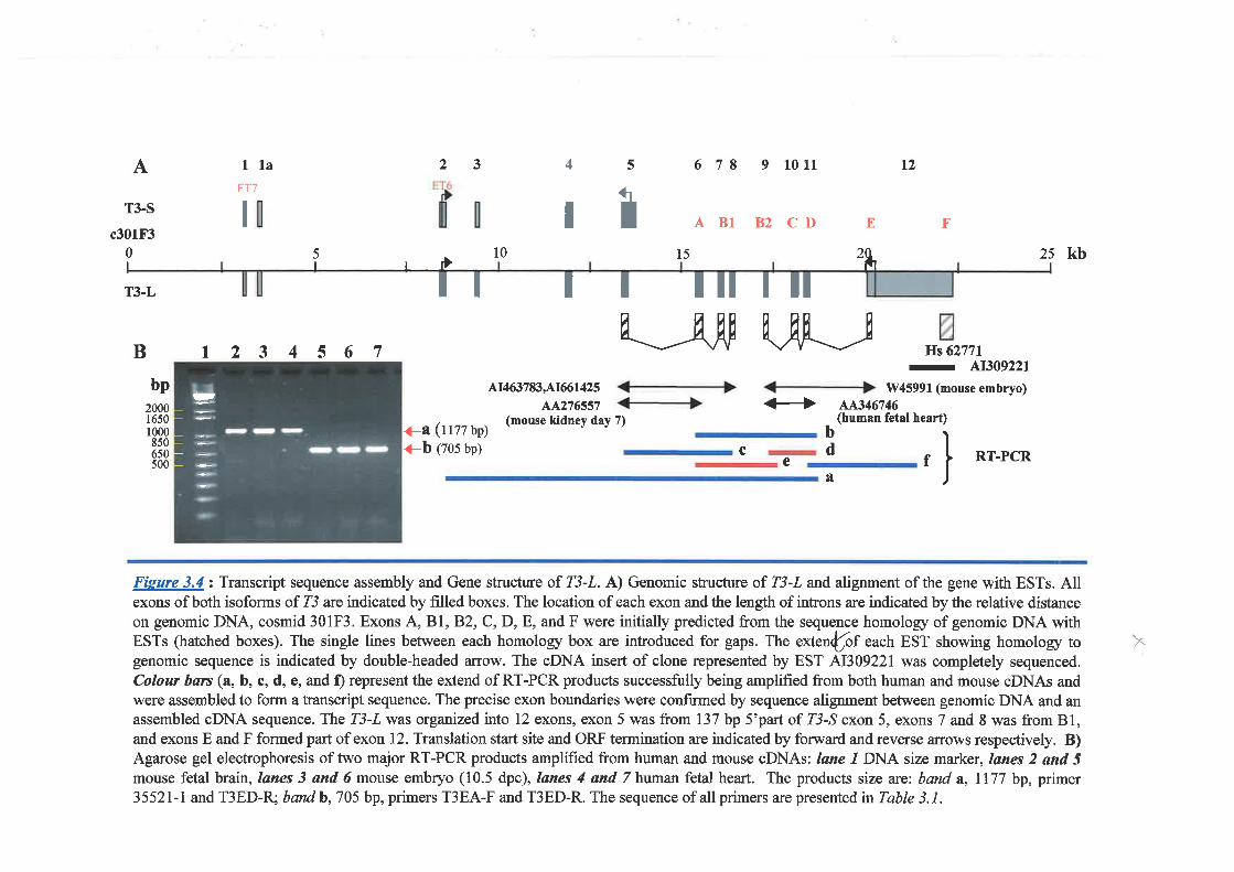
52A llaET7
rù
3
tt
4
I
6 78 9 1011 t2
A 81 B2 CD E F
15
I I Iltttl
T3-S
c301F30I
tT3-L
B t2 3 4 567
10
11t463783A1661425
44276s57(mouse kidney day 7)
5I
25I
2& kb
E_Jru UF_--[bp
200016501000850650500
Hs 62771
-
at309221
1V45991 (mouse embryo)<+ AAs46i46
(human fetal heart)+^ (t177 bp)
+b (zos ¡p)bd f RT-PCR
^
Fieure 3.4: Transcript sequence assembly and Gene structure of T3-L. A) Genomic structure of T3-L and alignment of the gene with ESTs. Allexons of both isoforms of T3 are indicated by frlled boxes. The location of each exon and the length of introns are indicated by the relative distanceon genomic DNA, cosmid 301F3. Exons A, 81, B.2, C, D, E, and F were initially predicted from the sequence homology of genomic DNA withESTs (hatched boxes). The single lines between each homology box are introduced for gaps. The extenfof each EST showing homology togenomic sequence is indicated by double-headed ¿urow. The oDNA insert of clone represented by EST 41309221 was completely sequenced.Colou¡ bars (a, b, c, d, e, and f) represent the extend of RT-PCR products successfully being amplified from both human and mouse cDNAs andwere assembled to form a transcript sequence. The precise exon boundaries were confirmed by sequence alignment between genomic DNA and anassembled oDNA sequence. The T3-L was organized into 12 exons, exon 5 was from I37 bp S'part of Z3-^S exon 5, exons 7 and 8 was from B1,and exons E and F formed part of exon 12. Translation start site and ORF termination are indicated by forward and reverse ¿urows respectively. B)Agarose gel elecfrophoresis of two major RT-PCR products amplified from human and mouse cDNAs: Iane l DNA size marker, lanes 2 ønd 5mouse fetal brain, lønes 3 and 6 mouse embryo (10.5 dpc), lanes 4 ønd 7 human fetal heart. The products size are: band a,II77 bp, primer3552I-l and T3ED-R; band b, 705 bp, primers T3EA-F and T3ED-R. The sequence of all primers are presente d, in Table 3. I .
ce
x
rt
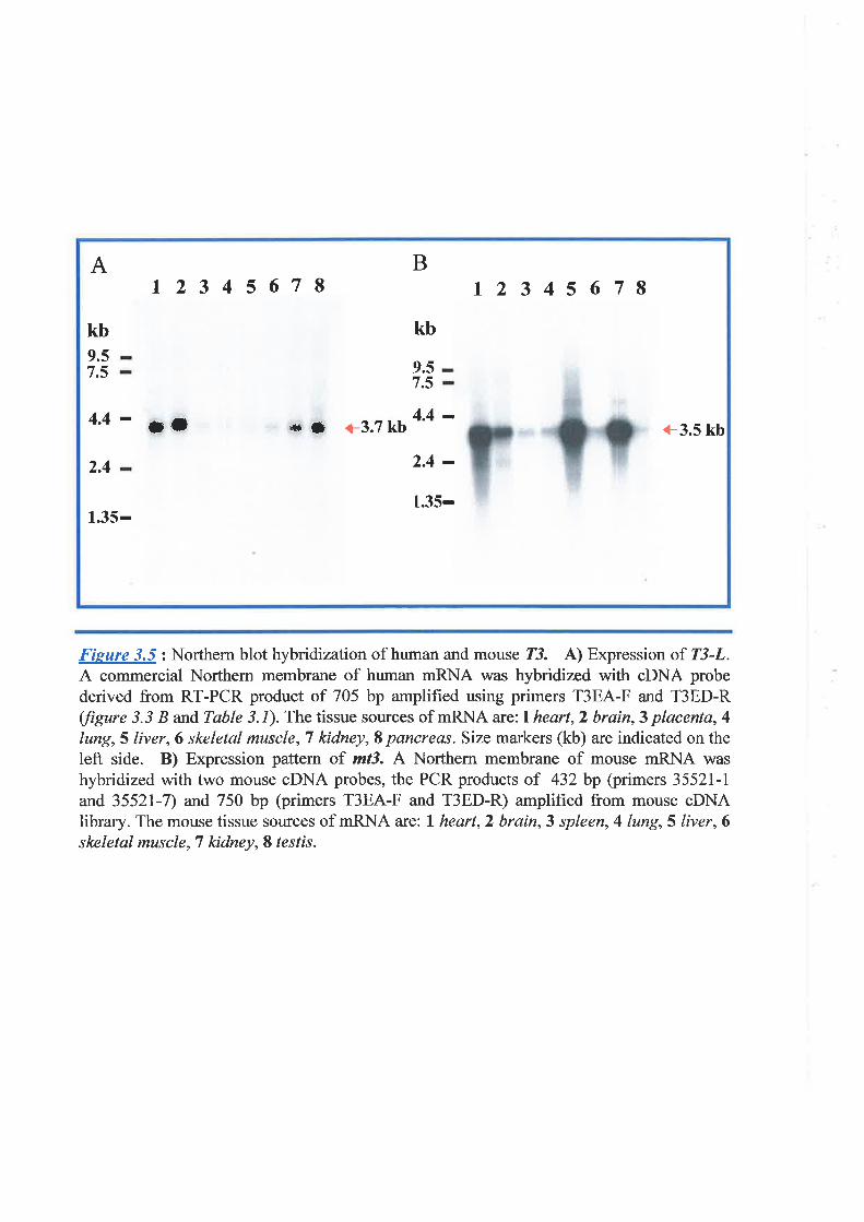
A
kb9.57.5
12345678B
kb
9.5
l2 3 4 s 67I
7.5
4.4- ¡t 4.4 -* {t +3.7 kb +3.5 kb
2.4 2.4 -1.35-
1.35-
Fieure 3.5: Northem blot hybridization of human and mouse 23. A) Expression of T3-L.A commercial Northern membrane of human mRNA was hybridized with cDNA probederived from RT-PCR product of 705 bp amplified using primers T3EA-F and T3ED-R(/ìgure 3.3 B and Table 3. 1). The tissue sources of mRNA are; l heart, 2 brain, 3 placenta, 4
lung, 5 liver,6 skeletal muscle, T kidney, S pancreas. Size markers (kb) are indicated on theleft side. B) Expression pattern of mt3. A Northern membrane of mouse mRNA was
hybridized with two mouse cDNA probes, the PCR products of 432 bp (primers 35521-1
and 35521-7) and 750 bp (primers T3EA-F and T3ED-R) amplihed from mouse cDNAlibrary. The mouse tissue sources of mRNA are: I heart,2 brain,3 spleen,4 lung,5liver,6skeletal muscle, 7 kidney, 8 testis.
mouse transcript showing no homology to that of T3-L. Finally all sequencing-confirmed
mouse gDNA fragments were joined to form a 3,108 bp fanscript sequence of the mouse
orthologue of T3-L (Figure 3.7). Two mouse cDNA fragments corresponding with T3-I
from exon 2-exon 5 and exon Bl-exon E regions \ilere also used to hybridize to the mouse
multiple tissue Northern membrane (Clonetech). In contast to human T3, only a single
band of approximately 3.4 kb was identified in all mouse tissue tested (Figure 3.5 B), with
varying degree of expression. This indicated that the mouse orthologue of T3 gene expressq'e .
only one transcript homologous to T3-L. Since the present mouse transcript was initially
identified from MMDEF 85, the partial 5' sequence of mouse def-8 oDNA (described in
section 3.1, Hotfrlder et al., 1999), it is therefore the complete, full-length, transcribed
sequence ofdef-8gene.
3.3.5 Transcript sequence Analysis of T3-L and mouse orthologue
The transcript sequence of the "long isoform" of T3 (T3-L) and its mouse orthologue,
assigned as mt3(def-8,), consist of 3,525 bp and 3,108 bp respectively. T3-L was the
extension of I3-S from the nucleotide 584 within exon 5 and constituted a 1353 bp ORF by
utilizing the same initiation codon as that of Z3-S (Fígure 3.d). This transcript contains a
large 3' UTR of 2,103 bp with a polyadenylation signal (ATTAAA) situated between
nucleotides 3507 and 3512. This is 13 bp upstream from the polyadenylation site. The T3-L
ORF is predicted to encode a protein of 451 amino acids, and the first 172 amino acids
from the N-terminus is shared with that of Z3-S. The mt3(def-8l transcript, by using the
translation start codon equivalent to that of T3, encodes an ORF of 1344 bp (Fìgure 3.7)
which shows a high degree of sequence homology (-86% identity) with the f3-¿ ORF.
The mt3(def-S/ ORF was predicted to code for a protein of 448 amino acids, which was
-95% identical with that of T3-L.It was followed by a 1,705 bp 3' UTR containing a signal
103
Fieure 3.6 : Nucleotide and deduced amino acid sequence of the "long transcriptionisoform " of T3 (T3-L). The nucleotide and amino acid positions are indicated by numbersin the right hand column . T3-L shares the first 584 nucleotides (exons 1-5) with that of T3-S. The same translation start site and upstream stop codon are also indicated. Thetermination codon is indicated at nucleotide positions 1424-1426. A polyadenylation signalbeginning at nucleotide 3507 is tmderlined. The exon boundaries a¡e indicated by thevertical lines with double-head arrows and the given numbers of exons. Two functionalmotifs, diacylglycerol/phorbol ester (GAG/PE) binding domain, residues 139-189(consensus:H-CxxC-CxxC-HxxC-C) and plant homeodomainJike (PHD) zinc finger,residues 390-431 (consensus: CxxC-CxxC-HxxC-CxxC) are underlined and the amino acidresidues corresponding with consensus are boxed, where "xx" indicates any two aminoacids and "--" indicates various length of amino acid sequence. Detail information of thesetwo domains can be found in "discussion" section.
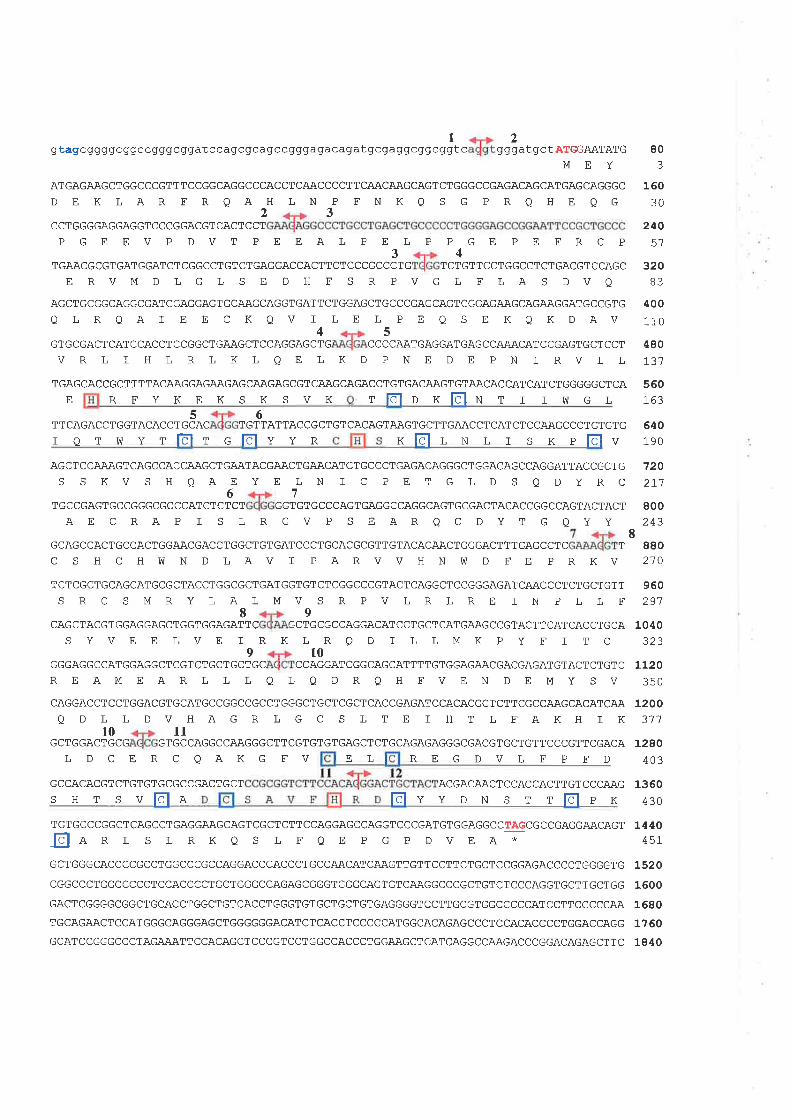
t2gtagcggggc ggccqqgcggatccagcqcagccgggagacagatgcgaggcggcggtc gggatgct ATGGAATATG
MEY
ATGAGAAGCTGGCCCGTTTCCGGCAGGCCCACCTCAACCCCTTCAACAAGCAGTCTGGGCCGAGACAGCATGAGCAGGGC
DEKLAR FRQAHLN P FNKQ S G PRQHE QG
80
16030
240EA
320B3
400
110
480
r31
560163
640190
720271
800243
880210
960291
2CCTGGGGAGGAGGTCCCGGACGT CACTCCT
3
PGE EVP DVT P EEALP E L P P GE PE FRCP34
TGAACGCGTGATGGATCTCGGCCTGTCTGAGGACCACTTCTCCCGCCCTG TCTGTTCCTGGCCTCTGACGTCCAGC
E RVM D L GL S E D H F S R PVGL F LA S DVA
AGCTGCGGCAGGCGATCGAGGAGTGCAAGCAGGTGATTCTGGAGCTGCCCGAGCAGTCGGAGAAGCAGAAGGATGCCGTG
Q L R QAI E E C I(QV I LE L P E Q S E KQK DAV45
GTGCGACTCATCCACCTCCGGCTGAAGCTCCAGGAGCTG CCCCAATGAGGATGAGCCAAACATCCGAGTGCTCCT
VRLIHLRLKLQELKDPNE DEPNIRVLL
TGAGCACCGCTTTTACAAGGAGAAGAGCAAGAGCGTCAAGCAGACCTGTGACAAGTGTAACACCATCATCTGGGGGCTCA
tldlr xlõll¡ r r r úr G LE RFYKEKSKSVK5
ACACCTGCAC T6
GTTATTACCGCTGT CACAGTAAGTGCTTGAACCTCATCTCCAAGCCCTGTGTG
r e r vs Y rlõlr clõlv Y R
TTCAGACCTGGT
xElr N L r s x p[dlvAGCTCCAAAGTCAGCCACCAAGCTGAATACGAACTGAACATCTGCCCTGAGACAGGGCTGGACAGCCAGGATTACCGCTG
S S KV SH QAEY E LN I C PET GLD S QDYRC67TGCCGAGTGCCGGGCGCCCATCTCTCT GGTGTGCCCAGTGAGGCCAGGCAGTGCGACTACACCGGCCAGTACTACT
AE CRAP f S LRGV P SEARQC DYT G QYY8
GCAGCCACTGCCACTGGAACGACCTGGCTGTGATCCCTGCACGCGTTGTACACAACTGGGACTTTGAGCCTC T
C S H C H I¡I N D L AV T P AR VVH N W D F E P R KV
TCTCGCTGCAGCATGCGCTACCTGGCGCTGATGGTGTCTCGGCCCGTACTCAGGCTCCGGGAGATCAACCCTCTGCTGTT
S RC SMRYLALMVSRPVLRLRE f N PLLF89
CAGCTACGTGGAGGAGCTGGTGGAGATTC GCTGCGCCAGGACATCCTGCTCATGAAGCCGTACTTCATÇACÇTGCA
S YVE E LVE T RKLRQD I L LMK PY F I T C9r0GGGAGGCCATGGAGGCTCGTCTGCTGCTGC CCAGGATCGGCAGCATTTTGTGGAGAACGACGAGATGTACTCTGTC
EL
1040
1120350
12003't't
1280403
1360430
144t0451
1520
1600
1680
1760
1840
REAMEARLLL QLO DRQHFVENDEMYSVCAGGACCTCCTGGACGTGCATGCCGGCCGCCTGGGCTGCTCGCTCACCGAGATCCACACGCTCTTCGCCAAGCACATCAA
QDLL DVHAGRLGC S LTE ] HTL EAKH IKl0 11
GCTGGACTGCG GTGCCAGGCCAAGGGCTTCGTGTGTGAGCTCTGCAGAGAGGGCGACGTGCTGTTCCCGTTCGACA
LDCER CQAKGFV REGDVLFPED
GCCACACGTCTGTGTGCGCCGACTGCT ACGACAACTCCACCACTTGT CCCAAG
s H r s v[dl e fõlv Y
TGTGCCCGGCTCAGCCTGAGGAAGCAGTCGCTCTTCCAGGAGCCAGGTCCCGATGTGGAGGCCTAGCGCCGAGGAACAGT
þe R L S L R K Q S L F Q E P G P D V E A *
GCTGGGCACCCCGCCTGGCCCGCCAGGACCCACCCTGCCAACATCAAGTTGTTCCTTCTGCTCCGGAGACCCCTGGGGTG
CGGCCCTGGCCCCCTCCACCCCTGCTGGGCCAGAGCGGGTGGGCAGTGTCAAGGCCCGCTGTCTCCCAGGTGCTTGCTGG
GACTCGGGGCGGCTGCACCTGGCTGTCACCTGGGTGTGCTGCTGTGAGGGGTCCTTGCGTGGCCCCCATCCTTCCCCCAA
TGCAGAACTCCATGGGCAGGGAGCTGGGGGGACATCTCACCTCCCCCATGGCACAGAGCCCTCCACACCCCTGGACCAGG
GCATCCGGGCCCTAGAAATTCCACAGCTCCCGTCCTGGCCACCCTGGAAGCTCATCAGGCCAAGACCCGGACAGAGCTTC
KPDNSTT tõt
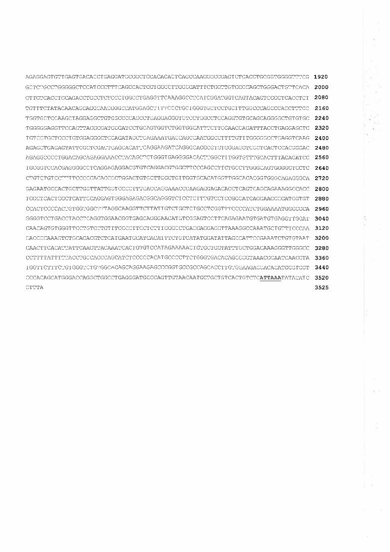
AGAGGAGTGTTGAGTGACACCTGAGGATGCGGCTGCACACACTCAGCCAAGGGCCGAGTCTCACCTGCGGTGGGGTTTCG
GCTCTGCCTGGGGGCTCCATCCCTTTCAGCCACTCGTGGCCTTGGGGATTTCTGGTTGTCCCCAGCTGGGACTGTTCACA
GTTGTCACCTGCAGACCTGCCTCTCCCTGGCCTGAGGTTCAAAGGCCTCATCGGATGGTCAGTACAGTGGGGTCACCTGT
TGTTTCTATACAACAGCAGGGAAGGGGCCATGGAGCTTTTCCCTGCTGGGTGCTCCTGCTTTGGCCCAGCCCACCTTTCC
TGGTGCTCCAAGCTAGGAGGCTGTGGCCCCAGCCTGAGGAGGGTGTCCTGGCCTCCAGGTGTGCAGCAGGGGCTGTGTGC
TGGGGGAGGTTCCAGTTAGGCGATGGGATCCTGCAGTGGTCTGGTGGCATTTCTTGGAACCAGATTTACCTGAGGAGCTC
TGTCCTGCTCCCTGTGGAGGGCTCCAGATAGCTCAGAÄATGACCAGCCAATGGCCTTTTGTTTGGGGGCCTGAGGTCAAG
AGAGCTGAGAGTATTCGCTCGACTGAGCACATTCAGGAAGATCAGGGCAGGCGTGTGGGAGGTCCCTCACTCCACGGGAC
AGAGGCCCCTGGACAGCAGAGGAAACCTACAGCTCTGGGTGAGGGGACACTTGGCTTTGGTGTTTGCACTTTACAGATCC
TGCGGTCCACGAGGGGCCTCAGGAGAGGACGTGTCAGGACGTGGCTTCCCAGCCTTCTGCCTTGGGCAGTGGGGGTGCTC
CTGTCTGTCCTTTTCCCCCACACCCCTGGACTGTGCTTGGCTGTTGGTGCACATGGTTGGCACACGGTGGGCAGAGGGCA
GAGAATGCCACTGCTTGGTTATTGGTCCCCTTTGACCAGGA.AACCCAAGAGGAGACACCTCAGTCAGCAGAAAGGCCACC
TGGCTCACTGGCTCATTCCAGGAGTGGGAGAGACGGCAGGGTCTCCTCTTTGTCCTCCGGCATCAGGAAGGGGATGGTGT
CCACTCCCCACTGTGGTGGCTTTAGGCAAGGTTCTTATTGTCTGCTCTGCCTCGGTTTCCCCATCTGGAÄAATGGGGGCA
GGGGTCCTGACCTACCTCAGGTGGAACGGTGAGCAGGGAACATGTCGGAGTCCTTCAGAGAATGTGATGTGAGGTTGGAT
CAACAGTGTGGGTTCCTGTCCTGTTTCCCCTTCCTCTTTGGGGCTGAGGAGGAGGTTAAAGGCCA.AATGCTGTTTCCCAA
CACCCCAAAGTCTGCACACGTCTCATGAATGCATCACATTTCTGTCATATGGATATTAGCCATTCCGAÀATCTGTGTAAT
CAACTTCACATTATTCAAGTTACA.AATCACTGTGTCCATAGAÄAÀÀCTGTGCTGGTATTTGCTGGACAAAGGGTTGGGCC
CCTTTTATTTTTACCTGCCACCCAGCATCTCCCCCACATGCCCCTTCTGGGTGACACAGCCGGTAÄACGGAATCAACGTA
TGGTTCTTTCTGTGGGTCTGTGGCACAGCAGGAAGAGCCCGGTGCCGCCAGCACCTTGTGGAAGACCACACATGGGTGGT
CCCACAGCATGGGACCAGGCTGGCCTGAGGGATGCCCAGTTGTAACAATGCTGCTGTCACTGTCTCATTÀÀÀTATACATC
CTTTA
1920
2000
2080
2L60
2240
2320
2400
2480
2560
2640
2720
2800
2880
2960
3040
3L20
3200
3280
3360
3440
3520
3525
Fíeure 3.7 : Nucleotide and deduced amino acid sequence of mt3, the mouse orthologueof human 73. The nucleotide and amino acid positions are indicated by the numbers in theright hand column. The translation start site at ATG positions 58-60 and termination site atTAG positions 1402-1404 are highlighted. A polyadenylation signal beginning atnucleotide 3084 is underlined. The exon boundaries are indicated by the vertical lines withdouble-head ¿urows and the given numbers of exons.
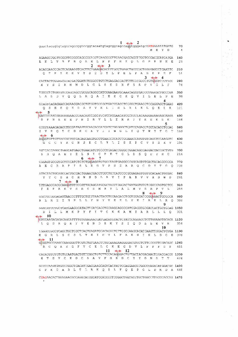
gaactacagtgt agcc agccgqt ccgt2
gtacaatgtagcggcagcc ggga 704
t gc TATGGAATATGATG
MEYD
AGAAGCTGGTTCGGTTCCGGCAGGCCCACCTCAACCCCTTCAACAAGCAGCTTGGTCCGAGGCATCATGA
EKLVRFRQAHLNP FNKQLGPRHHE23
ACAGGAACCCAGTGAGAAGGTCACTTCTG ACTTTGCCTGAGCTGCCCGCTGGGGAGCCTGAATTC
OE P S EKVT SE DT L PEL PAGEPE F
34CACTACTCGGAGCGCATGATGGATCTCGGCCTGTCTGAGGACCACTTTTCCCGCCCTG TCTCTTCCH Y S E RMMDLGL S E DH F S RPVG L F
27051
1110
28
28014
3s098
420L2L
490r44
560r.68
630191
TGGCCTCTGATGTCCAGCAGCTGCGGCAGGCCATCGAAGAATGCAAACAGGTGATCCTGGAGCTGCCCGA
LAS DVQQLRQAI EECKQVI LELPE4
GCAGTCAGAGAAGCAGAAGGACGCTGTGGTGCGGCTGATCCACCTCCGGCTGAAGCTCCAGGAGCTGAAG
QS EKQKDAVVRL I HLRLKLQELK5
CCCCAATGAGGAGGAACCCAACATCCGGGTTCTCCTGGAACATCGCTTCTACAAGGAGA.AGAGCAAGA
D PNEEE PN I RVLLEHRFYKEKS K
5GCGTTAAACAGACCTGCGATAAGTGCAACACCATCATCTGGGGGCTCATCCAGACCTGGTACACCTGCAC
S VK Q T C D K C N T f I W G L I QTúIYT C T
6TGTTGTTACCGCTGTCACAGCAAGTGCCTGAACCTCATCTCCAAACCATGTGTCAGCTCCAAGGTC
GCCYRCH S KCLNL I SKPCVS SKV
AGTCACCAGGCTGAGTATGAGCTGAACATCTGCCCTGAGACCGGGCTGGACAGCCAGGACTACCGCTGTG
S HOAEYELNI CPET GLD S QDYRC67
CGGAGTGCCGTGCTCCCATCTCTCT GGTGTGCCTAGTGAGGCCCGGCAGTGTGACTACACGGGCCA
AEC RAP I S LRGVP SEAROC DYTGQ
ATACTACTGCAGCCACTGCCACTGGAACGACCTGGCTGTCATCCCCGCGAGAGTGGTGCACAACTGGGAC
Y Y C S H C H W N D LA V I P A RV V HN !V D
7002L4
78TTGAGCCACGC GTCCCGTTGCAGCATGCGCTACCTGGCACTGATGGTGTCTCGGCCAGTGCTCCT
110238
84026r
910284
980308
1050331
LL2O354
1190378
L260401
1330424
FE PRKVS RCSMRYLALMVSRPVL
VEELVEIRKL
89GGCTCCGGGAGATTAACCCTTTGCTGTTTAACTACGTGGA.AGAGCTGGTGGAGATCC CTGC
RQ9
GGACATCCTGCTCATGAAGCCATACTTCATCACCTGCAAGGAGGCCATGGAGGCGCGACTACTGCTGCAG
D I L LMK P Y F I TC KEAMEARLLLOl0
CCAAGACAGACAGCATTTTGTGGAGAACGATGAGATGTACTCTATCCAGGACCTCTTGGA.A,GTGCACALQDRQH FVEN DEMYS I QDLLEVH
10TGGGCCGCCTCAGCTGCTCGCTCACTGAGATCCACACGCTCTTCGCCAAGCACATCAAGTTGGACTGTGA
MGRLSCSLTEI HTLFAKHI KLDCEll
TGCCA.AGCCAAGGGGTTCGTCTGTGAACTCTGCAÃAGAAGGGGACGTGCTCTTCCCGTTCGACAGC
CQAKGE VCELCKEGDVL FPFDSll t2
CACACGTCTGTGTGCA.ATGACTGTTCGGCTGTCTTCCAC ACTGTTACTACGACAACTCGACCACGT
RLRETNPLLENY
HTSVCNDCSAVFHRDCYYDNSTT
GCCCCAAGTGTGCCCGGCTCACATTGAGGAAGCAGTCACTATTCCAGGA.ACCTGGCCTAGACATGGATGC 14OO
C P K C A RL T L RK Q S L F O E P G L DM DA 448
CTAGAACACTGGAGAÀCGCCAGGCACCGTATCCACCCCTCCGGCTGGTGCCTGCTGGCCTTGCCCACCAG 14 ? O
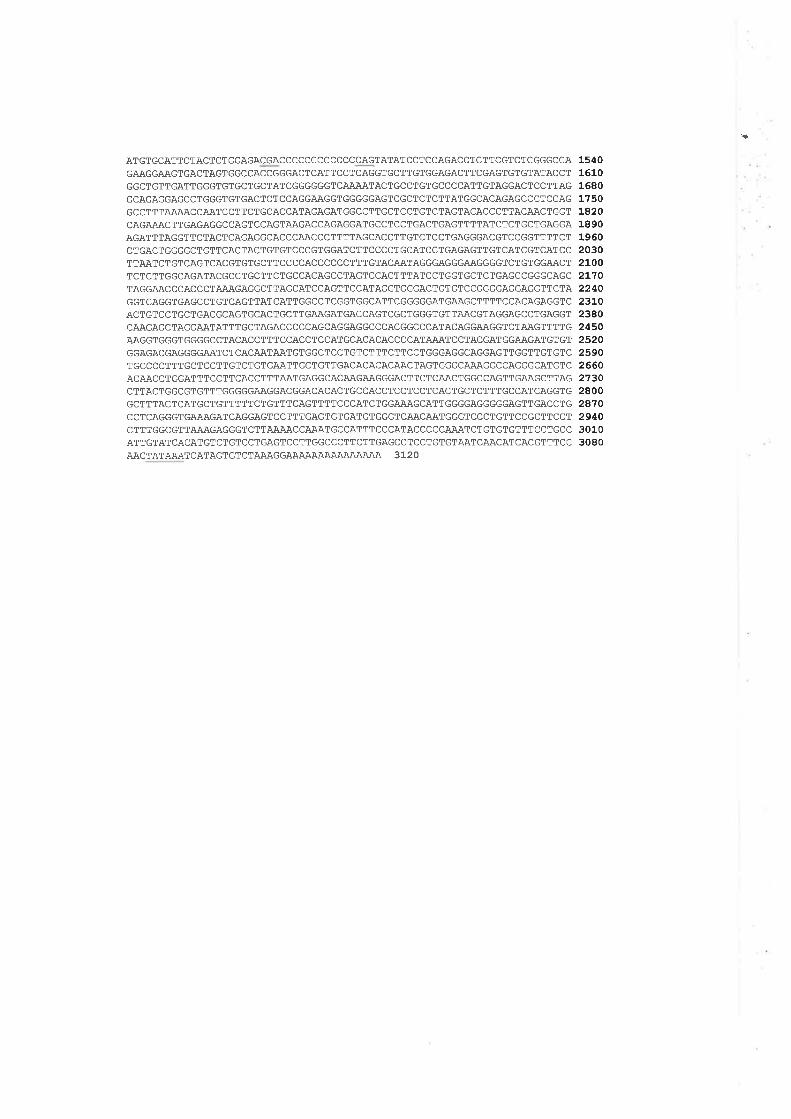
ATGTGCATTCTACTCTGGAGACGACCCCCCCCCCCCCAGTATATCCTCCAGACCTCTTCGTCTCGGGCCA
GAAGGAAGTGACTAGTGGCCACCGGGACTCATTCCTCAGGTGCTTGTGGAGACTTCGAGTGTGTATACCT
GGCTGTTGATTGGGTGTGCTGCTATCGGGGGGTCAÀAATACTGCCTGTGCCCCATTGTAGGACTCCTTAG
GCAGAGGAGCCTGGGTGTGACTCTCCAGGAAGGTGGGGGAGTCGCTCTCTTATGGCACAGAGCCCTCCAG
GCCTTTAAAACCAATCCTTCTGCACCATAGAGATGGCCTTGCTCCTGTCTAGTACACCCTTACAACTGGTCAGAAACTTGAGAGGCCAGTCCAGTAAGACCAGAGGATGCCTCCTGACTGAGTTTTATCTCTGCTGAGGA
AGATTTAGGTTCTACTCAGAGGCACCCAACCCTTTTAGCACCTTGTCTCCTGAGGGACGTCCGGTTTTCT
CTGACTGGGGCTGTTCACTACTGTGTCCCGTGGATCTTCCCCTGCATCCTGAGAGTTGTCATCGTCATCC
TTAATCTGTCAGTCACGTGTGCTTCCCCACCCCCCTTTGTACAATAGGGAGGGAAGGGGTCTGTGGAACT
TCTCTTGGCAGATACGCCTGCTTCTGCCACAGCCTAGTCCACTTTATCCTGGTGCTCTGAGCCGGGCAGC
TAGGAACCCACCCTAAAGAGGCTTAGCATCCAGTTCCATAGGTGGGACTGTGTCCGGGGAGGAGGTTCTA
GGTCAGGTGAGCCTGTCAGTTATCATTGGCCTCGGTGGCATTCGGGGGATGAAGCTTTTCCACAGAGGTCACTGTCCTGCTGACGCAGTGCACTGCTTGAAGATGACCAGTCGCTGGGTGTTAACGTAGGAGCCTGAGGT
CAAGAGCTAGGA.ATATTTGCTAGACCCCCAGCAGGAGGCCCACGGCCCATACAGGAAGGTCTAAGTTTTG
AAGGTGGGTGGGGCCTACACCTTTCCACCTCCATGCACACACCCCATAAATCCTACGATGGAÀGATGTGT
GGAGACGAGGGGAATCTCACAATAATGTGGCTCCTGTCTTTCTTCCTGGGAGGCAGGAGTTGGTTGTGTC
TGCCCCTTTGCTCCTTGTCTGTGAATTGCTGTTGACACACACAACTAGTGGGCAAAGGCCAGGGCATGTC
ACAACCTGGATTTCCTTCACCTTTAATGAGGCACAAGAAGGGACTTCTCAACTGGCCAGTTGAAGCTTAG
CTTACTGGCGTGTTTGGGGGAAGGACGGACACACTGCCACCTCCTCCTCACTGCTCTTTGCCATCAGGTG
GCTTTACTCATGCTGTTTTTCTGTTTCAGTTTTCCCATCTGGAAAGCATTGGGGAGGGGGAGTTGACCTG
CCTCAGGGTGAAAGATCAGGAGTCCTTTGAGTGTGATGTGGGTCAACA.ATGGGTCCCTGTTCCGCTTCCT
CTTTGGCGTTAAAGAGGGTCTTAAAACCAAATGCCATTTCCCATACCCCCAAATCTGTGTGTTTCCTGCCATTGTATCACATGTCTGTCCTGAGTCCTTGGCCCTTCTTGAGCCTCCTGTGTAATCAACATCACGTTTCCAACTATAAATCATAGTGTCTAAAGGA.A.AAAAAA.A.AAAAAA 3L2O
15¿0161016801?s0LA2018901960203021002L10224023LO23AO2450252025902660273028002A10294030103080
',TATfuq.l{,, between nucleotides 3088 and 3093 that is 16 bp upstream from the
polyadenylation site. Homology between mouse lmt3(def-y)l and human Q3-L) transcripts
is limited to the oRF sequence, no homology is observed between the sequences of either
ttre 5'or the 3'UTR.
3.3.6 Protein Sequence and Function Analysis
A high degree of homology between the human Q3-L) and the mouse (mt3)
transcripts, 86Vo identity between the ORF sequences and 95Yo identity between the
predicted proteins, designated ..T3-Lp" and "m6p" respectively, suggest a functional
conservation. The computerised protein localization prediction tool, PSORT, suggests that
the T3-Lp/mt3p protein localizes in the nuclear compartment. Protein fi¡nction was also
estimated, using three functional motif-prediction progr¿lms, Profile Scan, Pfam, and
SMART. Two functional motifs, a diacylgþeroVphorbol ester binding domain (DAG/PE-
bind) and a plant homeodomain-like @HD) zinc finger motit were identified at a
significant expectation (E-value) by all three programs. The DAG/PE-bind was located
between amino acid residues 139 and 189 in T3-Lp, and between residues 136 and 186 in
mt3p (Figure 3.6 and 3.8). The PHD domain was located close to the C-terminus between
residues 389 and 432, andbetween residues 386 and 429,itr T3-Lp and mt3p respectively
(Figure 3.6 and 3.8).
protein database searches, using BLASTP, identifred the T3-L/mt3 amino acid
sequence similarity to a number of proteins. The most significant similarity was to
CGl1534 gene product, a novel protein of 492 amino acids (AE003542) with unknown
function, from Dros ophila melanogasfer. Sequence similarity between T3-Lp/mt3p and
A¡i003542 were 37Vo identity and 57Vo positive (conservation of residues with similar
physico-chemical properties). AE003542 also contains both a DAG/PE-binding domain at
104
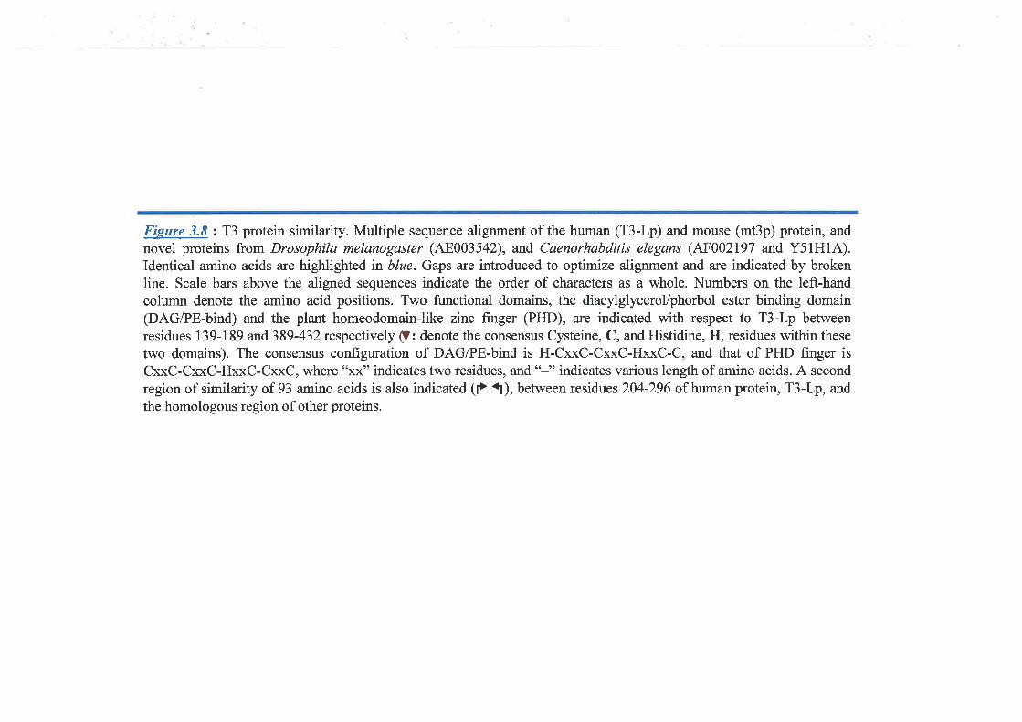
Fieure 3.8 : T3 protein similarity. Multiple sequence alignment of the human (T3-Lp) and mouse (mt3p) protein, and
novel proteins from Drosophila melanogaster (A8003542), and Caenorhabditis elegans (1^F002I97 and Y51H1A).Identical amino acids are highlighted in blue. Gaps are introduced to optimize alignment and are indicated by brokenline. Scale bars above the aligned sequences indicate the order of characters as a whole. Numbers on the left-handcolumn denote the amino acid positions. Two functional domains, the diacylglycerol/phorbol ester binding domain(DAG/PE-bind) and the plant homeodomain-like zinc frnger (PHD), are indicated with respect to T3-Lp betweenresidues 139-189 and389-432 respectively [: denote the consensus Cysteine, C, and Histidine, H, residues within these
two domains). The consensus configuration of DAG/PE-bind is H-CxxC-CxxC-HxxC-C, and that of PHD finger isCxxC-CxxC-HxxC-CxxC, where "xx" indicates two residues, aÍtd"-" indicates various length of amino acids. A second
region of similarity of 93 amino acids is also indicated (È n ), between residues 204-296 of human protein, T3-Lp, and
the homologous region of other proteins.
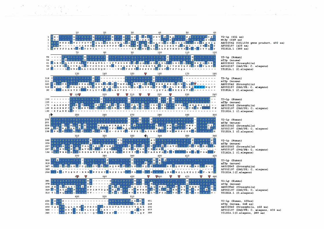
'a:)aaa -, i
IL
Eo1
1
1
I
1
LQÀE
RNEC
sL
DEEE
EDTTSD
EMRE
DGKETssssD G
PINTVSNlEFcPEvsFGeA-
13-Lp (¿51 aa)Mt3p (448 aa)ÀE003542 (Cc11534 gene product, 492 aalA¡'002197 (409 aa)Y5181À.1 (389 aa)
T3-Lp (Hwan)mT3p (mouEe)4E003542 (Drosophi].a)4F002197 (DÀG/ÞE; C. elegans)Y51H14.1 (C.elegas)
T3-Lp (gman)mT3p (mouEê)48003542 (Drosophila)A¡'002197 (DAG/PE; C. elega¡E)Y51H1À,1 (C.elegans)
T3-Lp (Hwn)rîT3p (mouêe)ÀE003542 (Drosophila)ÀF002197 (DÀG/PE; C. elegans)Y51H1.4.1 (C.elegans)
T3-Lp (Huan)mT3p (mouse)ÀE003542 (Dro6ophila)4F002197 (DÀG/PE; C. elegÐs)Y51814.1 (C.elegæs)
13-Lp (Eman)mT3p (mouse)ÀE003542 (Drosophila)4F002197 (DAG/PE, C. elegans)Y51H14.1 (C,elegans)
T3-Lp (Hman)mT3p (ûouee)48003542 (Drosophila)ÀF002197 (DAG/PE; C. eleganE)Y51H1À. 1 (C . elegans )
T3-Lp (Euman)mT3p (mouEe)À8003542 (Drosophila)43002197 (DAG/PE, C. elegans)Y51H1À.1 (C.elegans)
13-Lp (I¡llm, 451aa)mI3p (nouEe, 448 aalÀE003542 (Drosophila, 492 aa\ÀF002197 (DÀG/PE, C. elegans, 409 aa)Y51H1À.1 (C.elegans. 389 aa)
PD E
D
fIKD rs¡Í
NYTFYÀR YE TrL-----
IPGT
vEr o
G',D
L
EYESGwEnYVQËi
OLIHNQSST
ES ÀSNLesr! SRK
rw
-Gr RD
G GS G
NEIDKEVSGAE MMD---D
rExE rE ¡
59-56-61 E5284eE
ÀE LPIP ÀÀENII CIK
GNSÀE
VREQWRL-EYCRDKLK
IFsE
TSDÀ¡¡LI
LSE L wt L RHRRDR VLGN
D
N90 --
114111L20LL2
201229228138
262259247247
3223193Á1
HSI¡
Es ÀMDRQE TR
ÀE SL
QMYFDKe t sEoE
OLGLEÀvQAI QDEÀ vs
PH E¡¡HPrE
Y EEÀQ KADE
MBT
TAÀv
E"E" VMN
N I KN Tc¡oEG-GST
À
a-KFE
E D
D
É
sDMD
DLf.ÀDvDE--- --QDVÀ
SPñ SRKSSVS
2
MQPV
E HPISE--r rrE rx150 ------r12 - à À K R N
169 RGGHNP130 -ETÀEK
IDGÀEE
avL- GV
PRPNrE
sñF-vn-s
TDS
wGIRK
E
sT
L
R1 rE MKSGG xEsP
M
T
FLNIID
SY
L
cÀv
K
N VH R À
BREs sr
r!
L
K
sN-----DDINVDQTV----- NT
cr-NlRsTt¡
LS BKDRGRÀWYEÀNQEK NIKT s YÀHÀP sErGs xloEv s uf r
DI196 D
E
À I T,R DQ G ÀQSN LS so EF QG\ZFR ND R-
zse$n!svs E D F - RRnlw exr r[ns s t onE¡¡I¡E*Eon.nlx s LLKr¡ s r xlv - ¡¡ r
381378406329
P
E314 rL TE: I
¡,rFEw-
IN
LF
frFco
43ÿ31
j :_ -. V / i
i <i {sr/\a
ti.
itl:::r¡ì:-lir.-Y:,'.al
s:a.; isl:-r it
1Sl I lr :,-:rii,i{:ìil'. - : r ! :: I , -l a
v it I I a Y Y
V:i N ] :Y /Jf
lCSi',"/.-,:i ilì
459 IQE388 ---366 ---
K
iiiËiîFiiliîEDDDGVATDDDVTAÀE
i:;;Ê-----Ëi?:i
s
4514¿8Á92409389
VE
its central region and a pHD motif close to the C-terminus, similar to that of T3-Lp/mt3p.
Similarity was also observed in the region of 93 amino acids close to the C-terminal part of
DAG/PE binding domain in all proteins (Fígare 3.8), with -55% identity' These
similarities suggest that 4E003542 is likety to be a T3-Lp homologue. A protein
AF002lg7 from Caenorhabditis elegans, containing a region similar to protein kinase C,
was also similar to T3-Lp/mt3p with 33% identity a¡d53%opositive. AF002197 also shows
three domains: a DAGIpE-bind, a conserved 93 amino acid region, and a C-tenninus PHD
motif, found in T3-Lp/mt3p. The function of either 4E003542 or 4F002197 is unknown.
However, the high degree of conservation among these proteins throughout evolution
suggests a critical and basic cellular function'
3.3.7 Genomic Organization and Älternative Splicing
A complete genomic structure of 73, with respect to T3-L, w6 obtained from the
alignment of the transcribed sequence of T3-L with the genomic sequence from cosmid
301F3. Overall, Zi is organized into 12 exons encompassing a genomic sequence of
approximately 20 kb (Fìgure 3.4). Nl splice junctions of exon/intron boundaries (Table
3.3) obey the consensus sequence for donor and acceptor splicing sites (Shapiro and
Senapathy, 1987). The transcript of T3-L slnres exons 1-4 \¡vith that of T3-S, and its exon 5
derives from the first 142 bp 5' part of I3-S exon 5. At this point there is a splicing donor
site which is utilized fot T3-L transcript (Figare 3.2). The extended sequence utilized for
exon 5 of I3-S is part of intron 5 with respect to T3-L transcript. This sequence includes
rhe continuation of ORF and the 3' UTR of T3-S (Fìgure 3.2). The DAG/PE binding
domain is encoded by exons 5 and 6, whereas exons 11 and 12 codes for a PHD motif.
Exon 12 is the largest exon spanning 2,202 bp of the genomic sequence. It contains part of
ORF, stop codon (TGA), and a large 3'UTR (2,103bp) (Figure 3.Q-
105
To compare the gene structure of mf3 with that of T3, the transcribed sequence of mt3
(3,106 bp) was used to search for a mouse genomic sequence from a high throughput
genomic sequence (htgs) database. A draft mr¡rine genomic sequenoe, 4C023836 (as of
October 2000), was retrieved and aligned vnth mt3 oDNA. The alignment result indicated
t¡¡at mt3 gene was also separated into 12 exons, with all the exon/intron junction
bogndaries also followed the glag rule and surounding consensus sequence of splicing
site (Shapiro and Senapathy, 1987). When compared with human gene (73), the mouse
(mt3) exons appeared to use the equivalent boundaries with that of the human orthologue.
The mt3 exon 12 is also the largest exon, containing part of ORF, stop codon, and 3'UTR
of 1705 bp (Figure 3.7).
In addition to two charactenzed transcriptional isoforms of 73, the 7"3-S andT3-L,the
evidence of alternative splicing of T3 was also observed in the 5' region. Initially in the
transcript in at least three ESTs, 44035322, 44306508, and HSU46300, an extra exon of
97 bp was detected between exon I and exon 2. This extra exon, assigned as exon la,
located \¡rithin the first intron, 484 bp down stream from exon 1, possessed consensus
splice donor and acceptor sites (Figure 3.9c).Inclusion of exon la was also observed in
part of the 5'RACE products amplified from mammary gland and T-cell leukemia cell line
(Jurkat) 6RNA (Figure 3.9). Furthennore, some of these products also included an extra
sequence of 125 bp, between exon la and exon 2. The location of this extra sequence in
genomic DNA appeared to be the sequence that immediately extends from exon la
(Fìgure 3.gC), which also showed the splice donor signal at its 3' boundary. Such
alternative splicing patterns were also exhibited in the RT-PCR experiment on mRNA
derived from breast cancer cell lines, normal mammary gland and fetal btan (Fígure
3.108),using 5'primer (35521-P) against primer from exon 4 (35521-5). Inclusion of exon
106
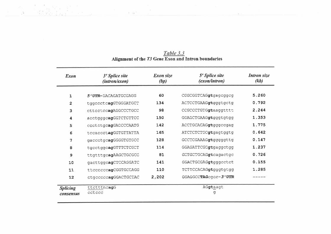
Tøble 3.3Alignment of the ?3 Gene Exon and Intron boundaries
Exon
1
2
3
4
5
6
1
I9
10
11
t2
3' Splice site(íntron/exon)
5 I UTR_GACAGATGCGAGG
tggccctcagGTGGGATGCT
cttcctccagAGGCCCTGCC
acctgggcagGGTCTGTTCC
ccctctgcagGACCCCAATG
tccaccctagGGTGTTATTA
gaccctgcagGGGGTGTGCC
tgcctggcagGTTTCTCGCT
ttgtttgcagAAGCTGCGCC
gacttggcagCTCCAGGATC
ttccccccagCGGTGCCAGG
ctgcccccagGGACTGCTAC
Exon síze(bp)
5t Splìce síte(exon/ìntron)
CGGCGGTCAGgtgagcgqcg
AcTCCTGAAGgtgggtgctg
CCGCCCTGTGgt,aaggtttt
GGAGCTGAAGgtgggtgtgg
ACCTGCACAGgtgggccAag
ATCTCTCTGCgtgagtgg|tg
GCCTCGAAAGgtggggqttg
GGAGATTCGCgfgaggctgg
cCTGCTGCAGgtcagactgc
GGACTGCGAGgtgggcctct
TCTTCCACAGgtgggt gtgg
GGAGGCCTAGcgcc-3'UTR
AGgtaagt
Intron size(kb)
5.260
o.7922.2441.353
L.7750.642
o.L41
L.237
o.726
0.155
L.285
60
134
98
150
L42
165
t28LL4
81
L4L
110
2,202
Splicíng ttctttncagG¡¡l ¡consensus (;uLecc g
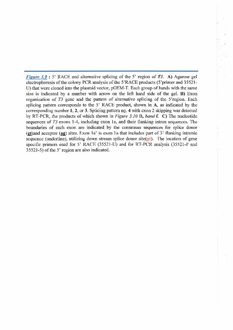
Fieure 3.9: 5'RACE and alternative splicing of the 5' region of T3. A) Agarose gelelectrophoresis of the colony PCR analysis of the 5'RACE products (S'primer and 35521-U) that were cloned into the plasmid vector, pGEM-T. Each group of bands with the same
size is indicated by a number with arrow on the left hand side of the gel. B) Exonorganization of T3 gene and the pattern of alternative splicing of the 5'region. Eachsplicing pattern corresponds to the 5' RACE product, shown in A, as indicated by thecorresponding number 1,2, or 3. Splicing pattem nq. 4 with exon 2 skipping was detectedby RT-PCR, the products of which shown rn Figure 3.10 B, band f. C) The nucleotidesequences of T3 exons 1-4, including exon la, and their flanking intron sequences. Theboundaries of each exon are indicated by the consensus sequences for splice donor(g!)ard acceptor (ag) sites. Exon la' is exon la that includes part of 3' flanking intronicsequence (underline), utilizing down stream splice donor site(gt). The location of gene
specific primers used for 5' RACE (35521-U) and for RT-PCR analysis (35521-P and
35521-5) of the 5' region are also indicated.
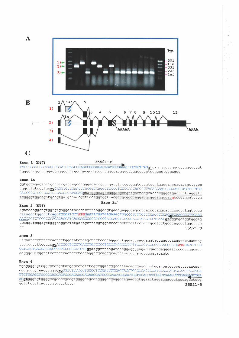
A
B 1 La' 2
1)
?_ zt
3)
4',)
345 6 7 I 9 1011 t2
AAÀA
CExon 1 (ET7)TAGCGGGGCGGCCGGGCGGAT
cggqg ccqq cggggacggggccggcggggacggggcc99c gggg acq gggtcggcggggtcggggctgggaggg
Exon 1aggtgggagagacctqcccccgaggagcccgggagaccgggcgaqctcccgcgggtgtggtgggtggggaggcacagtgctggggTggcTTcTcccTgcegCAGGTGCCGAACCCACGGCCAGGCTTCCGTGGCCAGCAGCCCTAGAGGAATGGCCATCCTGTCCCTGCGAGCCCCTGGGCCCTGGCAGGCGAT a t cacac cttctca tc
ct ca at acacc ttcc tca gcatcccgExon la'
Exon 2 (ET6)agatcaaggctgtggtgtgaqqactacccactttaag qaagtgaaagaggccaqcctcaccccagacaccccagtgtggttggggaaaggctggccctcgcTGGGATGCT TATGATGAGAAGCTGGCCCGTTTCCGGCAGGCCC4,AGCAGTCTGGGCCGAGACAGCATGAG CCTGGGGAGGAGGTCCCGGACGTCACTCCTGAAGtcagggtgggagctgqgcaggtcTctgactgcttacgtggacccctcctttcttcctg'ccgcgtcctgcgcagccctggctttccc 35521-u
Exon 3ctqaatccctttcccactcctggctatctcagctcctccctaggggatgggaggcaggaggtagcagctgacgctccacacctgtccccgtcttcctccggAGGCCCTGCCTGAGCTGCCCCCTGGGGAGCCGGAATTCCGCTGCCCTGAACGCGT GGATCTCGGCCTGTCTGAGGACCACTTCTCCCGCCCTGTGgÞaaggttttagatctcgg aqgggaqagqgactgagggaacccccaaggcaggaagggcCagggtttccttgtcactccctcccaggtggcagggcagtgtcctgtgagctggggtacagtc
Exon 4tgaggggtqtcggggtctgctctgggcctgtctcggcggatgggccttaacagggagctcctgcaggatgggcctttgactgccCCCgCCCCCAACCTgggCESGGTCTGTTCCTGGCCTCTGACGTCCAGCAGCTGCGGCAGGCGATCGAGGAGTGCAAGCAGGTGA
qctctctctcaggcggtggtctctc 3552 1 _5
35521-PCCAGCG CcccccTcAcgþgagcggcgcggggccggc SSSSL
bp
1)>2l>3)>
501404331242190
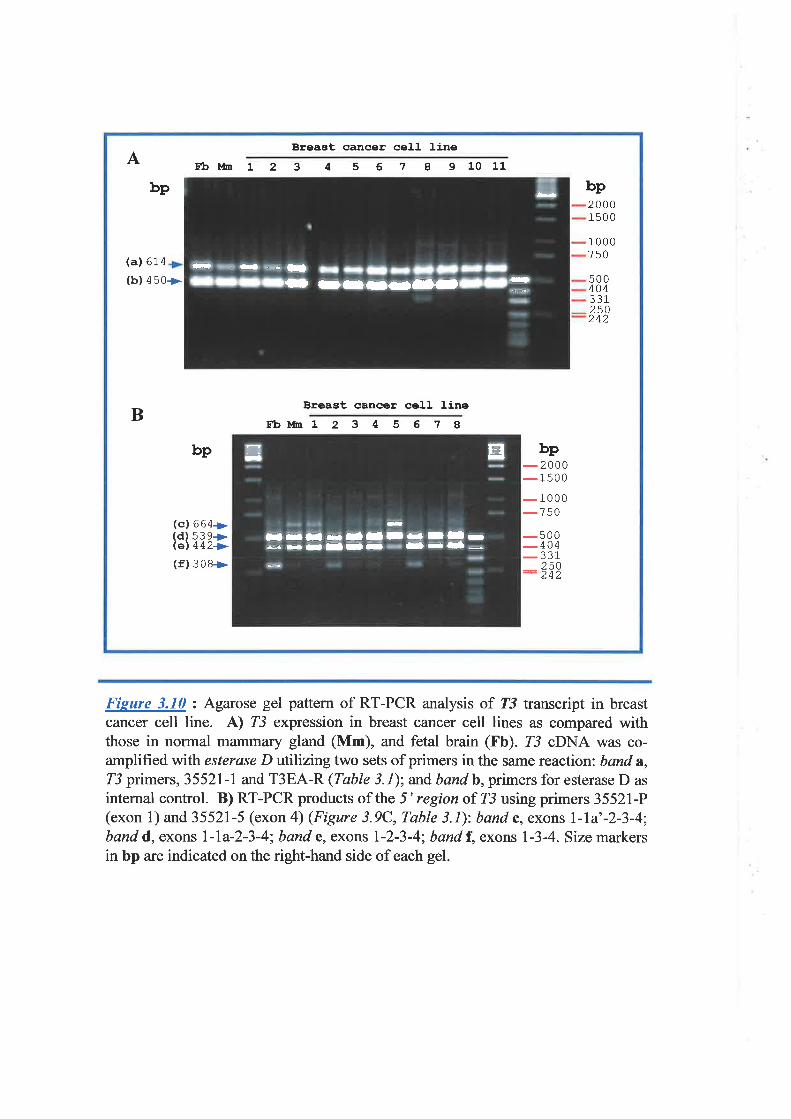
Breast cancer cell lineA E:b¡,tu L 2 3 4 5 6 7 I 910 11
bp bp2000150 0
(a) 614
1000750
(b) 450> 500404331250242
BBreast cancer cel]- line
Eb!,h 1 2 3 4 5 6 7 A
bp bp
- 2000
-1500-
1000
-750(cl 664+>(d) s39+(el 442> -500-
404
- 331(f) 308> 250
- 242
-lö-rÕ- -
¡¡-
rFll ----a
-
I
Ir¡l nllII - Iilrd
--Il-- -¡-- -
ð EI-
Fieure 3.10 : Agarose gel pattern of RT-PCR analysis of T3 transcript in breastcancer cell line. A) rc expression in breast cancer cell lines as compared withthose in normal mammary gland (Mm), and fetal brain (Fb). Z3 cDNA was co-amplifred vnth esterase D utilizingtwo sets of primers in the same reaction; band a,
intemal control. B) RT-PCR products of the 5' region of Z3 using primers 35521-P(exon l) and 35521-5 (exon 4) (Figure 3.9C, Table 3.1): band c, exons l-la'-2-3-4;band d, exons l-la-2-3-4; band e, exons l-2-3-4; band f, exons l-3-4. Size markersin bp are indicated on the right-hand side of each gel.
la was observed in both ?3-S and ?3-I transcript in the RT-PCR product generated by
using the exon la primer and either T3-S specifrc (35521-9) or T3-L specific (T3-EA-R)
primer. Since the translation initiation codon was located within exon 2, all these splice
forms maintained the same ORF.
Alternative splicing then generated a number of transcription variants of the existing
transcription isoforms, T3-S and T3-L.
One interesting feature, the exon 2 skipping isofomr, was observed in fetal brain and
some breast ca¡1cer cell lines, but not in normal mammary gland (Figure 3.108) nor fetal
kidney. Skipping of exon 2 resulted in the removal of part of the transcript containing an
ATG start codon at nucleotide positions 7l-73. However, the sequence downstream from
this skipped exon, has another inframe ATG codon within exon 3 at positions 251-253-
The sequence immediately adjacent to this ATG codon conforms with the Kozak
consen$N for the translation sta¡t site. Therefore this provided a second start codon. The
use of this second start codon will result in a protein that is 60 amino acids shorter from its
N-terminus compared with the protein from the normal start codon. This protein product,
however, still maintains the functional motifs, DAG/PE-bind (amino acids 139-189) and
pHD zinc finger (amino acids 389-432). Since this altemative fomr of T3 was observed
only in fetal brain and some breast cancer cell lines, it might represent a unique tissue-
restricted isofonn of this gene.
Alternative splicing of the 5' region was also observed in mt3, with the inclusion of
various alterrative exons between exons I and 2. As shown tn Fìgure 3.f-1, inclusion of
exons lb and lc were found in both EST AV/763180 and 5' PCR products from mouse
cDNA libraries. The use of exon la appeared to be independent of exon I and likely to be
specific to fetal brain, since such product was not detected in a mouse embryo cDNA
Iibrary. Utilization of exon 1a was also seen in MMDEF 85, the 5' sequence of def-8.
t07
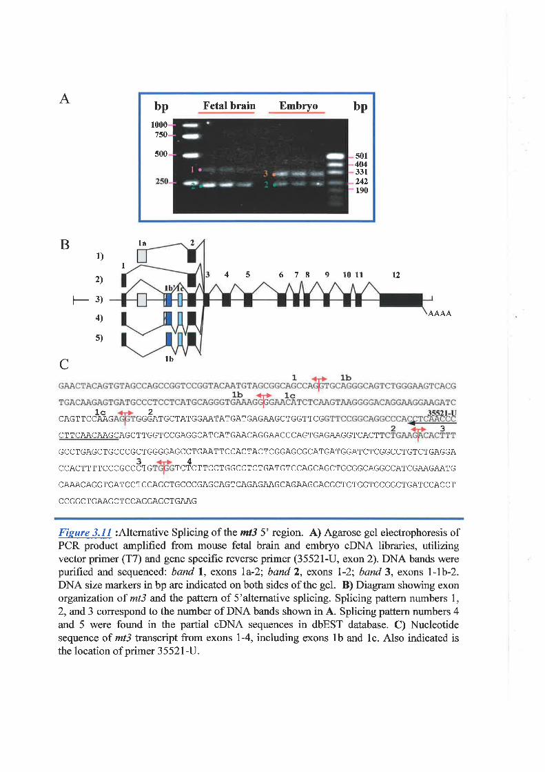
A
B 1a 2
1)
2)
l- 3)
4)
s)
I4S 6 7 I 9 1011 t2
I
1b
AúA,Aú{
3
C
lc2CAGTTCCAAG GGGATGCTATGGAATATGATGAGAAGCTGGTTCG
CT TCAACAAGCAGCTTGGTCCGAGGCATCATGAACAGGAACCCAGTGAGAAGGTCACTT
GCCTGAGCTGCCCGCTGGGGAGCCTGAATTCCACTACTCGGAGCGCATGATGGATCTCGGCCTGTCTGAGGA34
CCACTTTTCCCGCCCTG TCTCTTCCTGGCCTCTGATGTCCAGCAGCTGCGGCAGGCCATCGAAGAATG
CAÄACAGGTGATCCTGGAGCTGCCCGAGCAGTCAGAGAAGCAGAAGGACGCTGTGGTGCGGCTGATCCACCT
CCGGC T GAAGC TCCAGGAGCT GAAG
Fieure 3.ll :Altemative Splicing of the mt3 5' region. A) Agarose gel electrophoresis ofPCR product amplifred from mouse fetal brain and embryo cDNA libraries, utilizingvector primer (T7) and gene specific reverse primer (35521-U, exon 2). DNA bands werepurified and sequenced: band I, exons la-2; band 2, exons l-2; band 3, exons l-lb-2.DNA size markers in bp are indicated on both sides of the gel. B) Diagram showing exonorganization of mt3 and the pattem of 5'alternative splicing. Splicing pattern numbers l,2, and 3 correspond to the number of DNA bands shown in A. Splicing pattern numbers 4and 5 were found in the partial cDNA sequences in dbEST database. C) Nucleotidesequence of mt3 transcript from exons l-4, including exons lb and lc. Also indicated isthe location of primer 35521-U.
bp
501404331242r90
bp Fetal brain Embryo
r000750
500
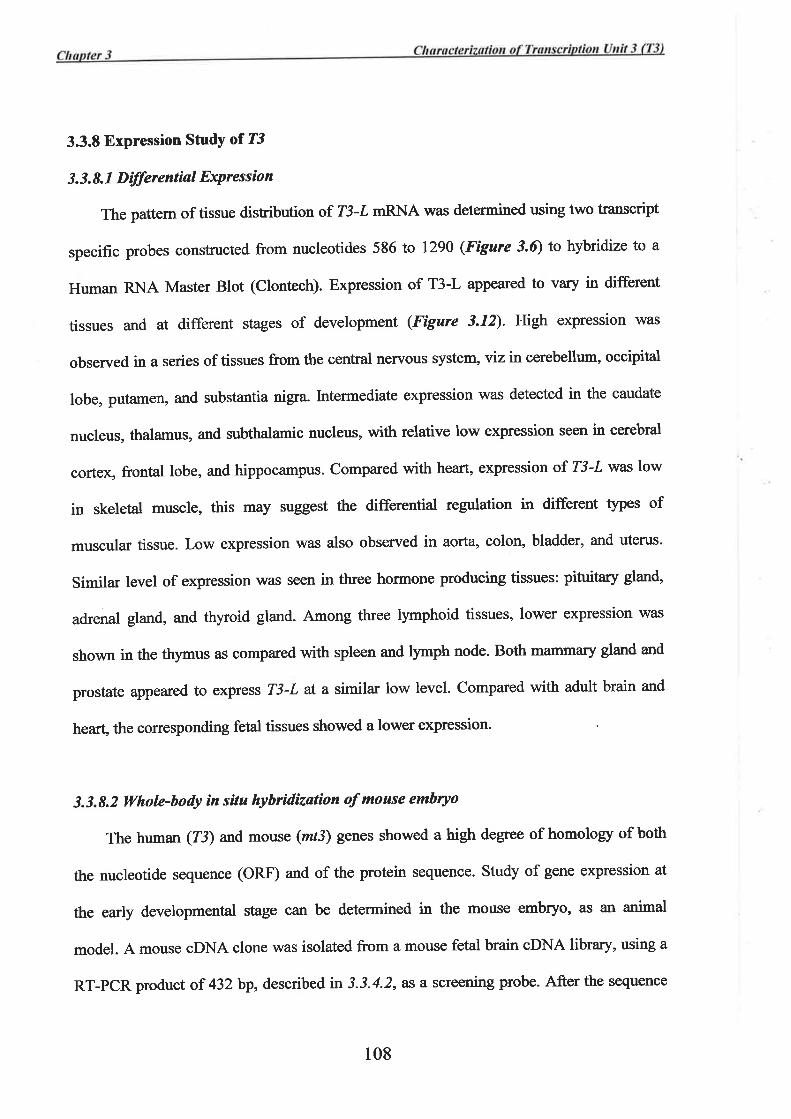
3.3.8 Expression StudY of T3
3. 3. 8. 1 Dìlferentìal ExPressìon
The pattem of tissue distribution of T3-L mRNA was determined using two transcript
specific probes constructed from nucleotides 586 to 1290 (Figure 3.6) to hybridize to a
Human RNA Master Blot (Clontech). Expression of T3-L appeared to vary in different
tissues and at different stages of development (.Fþure 3.12). High expression was
observed in a series of tissues from the central nervous system, viz in cerebellum, occipital
lobe, putamen, and substantia nigra. lntermediate expression was detected in the caudate
nucleus, thalamus, and subthalamic nucleus, with relative low expression seen in cerebral
cortex, frontal lobe, and hippocampus. Compared with heart, expression of T3-L was low
in skeletal muscle, this may suggest the differentiat regrrlation in different $pes of
muscular tissue. Low expression was also observed in aorta" colon, bladder, and uterus.
Similar level of expression was seen in three hormone producing tissues: pituitary gland,
adrenal gland, and thyroid gland. Among three lymphoid tissues, lower expression was
shown in the thymus as compared with spleen and lymph node. Both mammary gland and
prostate appeared to express T3-L at a similar low level. Compared with adult brain and
heart, the corresponding fetal tissues showed a lower expression
3.3.8.2 Whole-body ìn situ hybrìdizatíon of moase embryo
The human (73) and mouse (mt3) genes showed a high degree of homology of both
the nucleotide sequence (ORF) and of the protein sequence. Study of gene expression at
the early developmental stage can be determined in the mouse embryo, as an animal
model. A mouse cDNA clone was isolated from a mouse fetal brain cDNA library, using a
RT-pCR product of 432 bp, described n 3.3.4.2, Æ a screening probe. After tlre sequence
108
wholebrain
amygdala caudatenucleus
cere-bellum
cerebralcortex
frontallobe
hippo-campus
medullaoblongata
occipitallobe
putamen substanti¡nigra
temporallobe
thalamussub-
thalamicnucleus
spinalcord
heart aorta skeletalmuscle
colon bladder uterus prostate stomach
testis ovary pancreâs pituitarygland
adrenalgland
thyroidgland
salivarygland
mommârlgland
kidney liver smallintestine
spleen thymus oeripheraleukocyte
lymphnode
bonemarrow
appendix lung trachea placenta
fetalbrain
fetalheart
fetalkidney
fetalliver
fetalspleen
fetalthymus
fetallung
yeastlotal RN,A
yeasttRNA
E.colirRNA
E.coliDNA
poly r(A)IIumanCotlDNA
humanDNA
100 ng
humanDNA
5ü) ng
(Ð I 2 3 4 5 6
23 4 s67 8
7 I
A
B
c
D
E
F
G
H
(ii) A
B
C
D
E
F
G
H
I
o
a
*
*üe4
O-*|ll*
a -a1Í
a
*{t
Fieure 3.12: Differential Expression of T3. (i) Diagram of the tissue origin and positionof human polyA+ RNA dot-bloted on a nylon membrane. (ii) Hybridization with the
[32P]dcTP-labelled T3 oDNA probe derived from the region between exons 6 and 11 (nt586-1290). The amount of polyA+ RNA in each dot was normalizedby manufactrrerusing eight different housekeeping gene cDNA probes (Clontech)
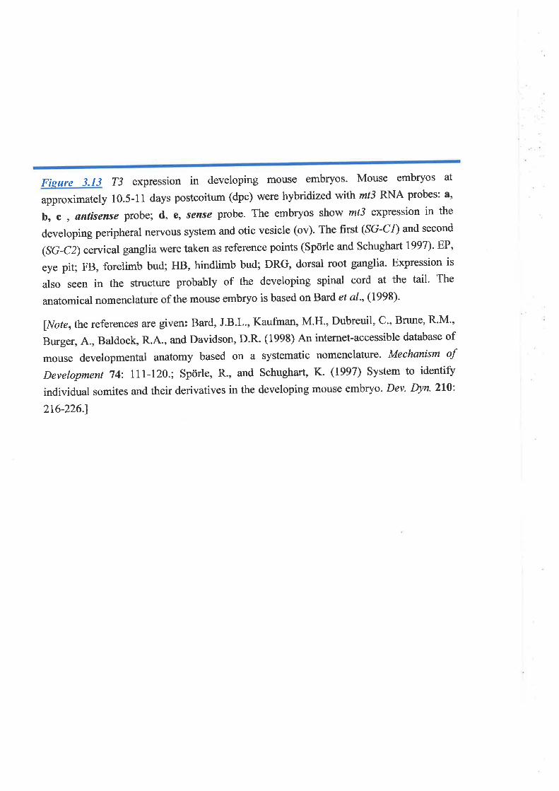
Fisure 3.13 T3 expression in developing mouse embryos. Mouse embryos at
approximately 10.5-11 days postcoitum (dpc) were hybridized wfttl mt3 RNA probes: a,
b, C , antisense probe; d, e, sense probe. The embryos show mt3 expression in the
developing peripheral nervous system and otic vesicle (ov). The fnst (SG-C1) and second
(Sc-C2)cervical ganglia were taken as reference points (Spörle and Schughart 1997). EP'
eye pit; FB, forelimb bud; HB, hindlimb bud; DRG, dorsal root ganglia. Expression is
also seen in the structwe probably of the developing spinal cord at the tail. The
anatomical nomenclatuie of the mouse embryo is based on Bard et al., (1998).
lNoterthe references are given: Bard, J.B.L., Kaufman, M.H., Dubreuil, C', Bfune, R'M',
Burger, A., Baldock, R.4., and Davidson, D.R. (1998) An internet-accessible database of
mouse developmental anatomy based on a systematic nomenclaturc. Mechanism of
Development 74: lll-120.; Spörle, R., and Schughart, K. (1997) System to identiff
individual somites and their derivatives in the developing mouse embryo. Dev. þn.210:.
2t6-226.1
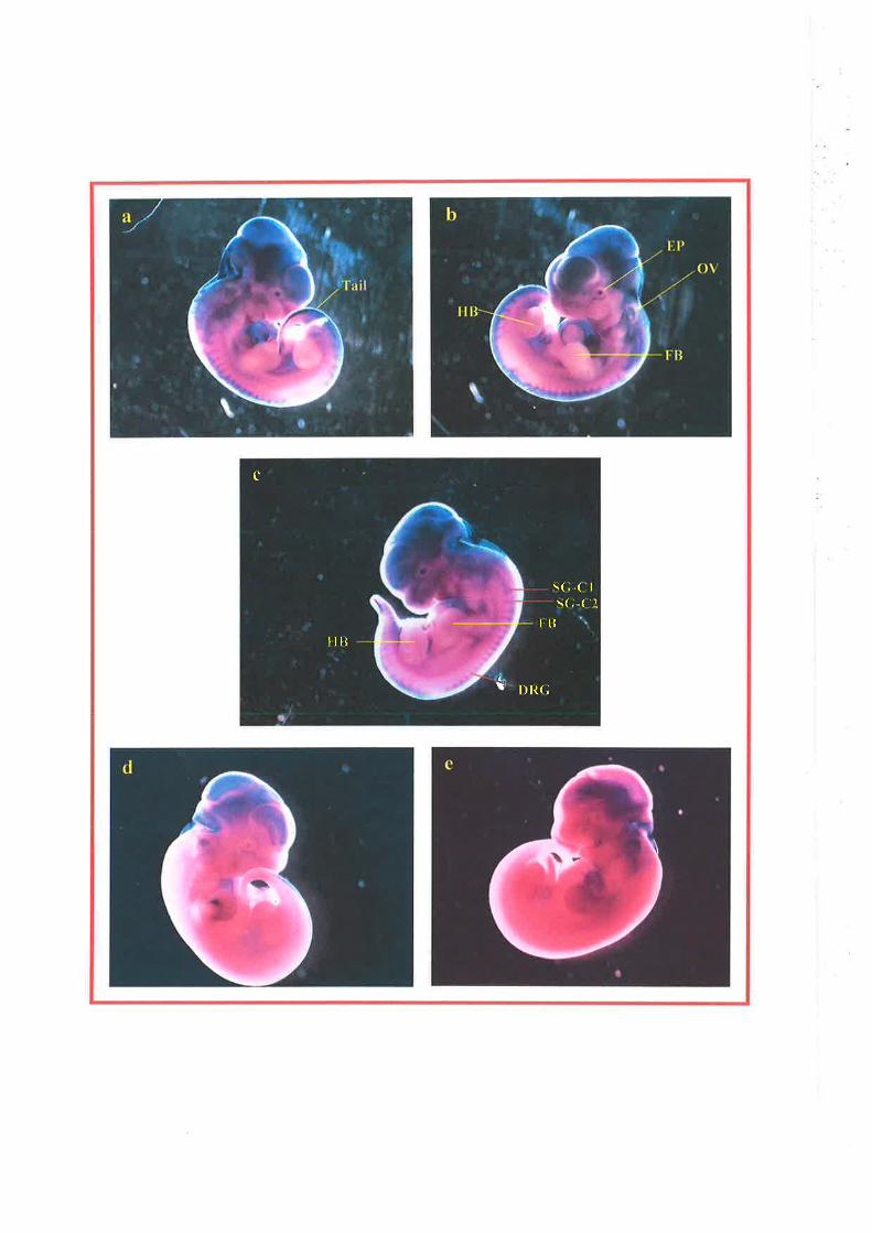
t.tllilt
c
t r)rì(;
\(,.( I
\(; ('l
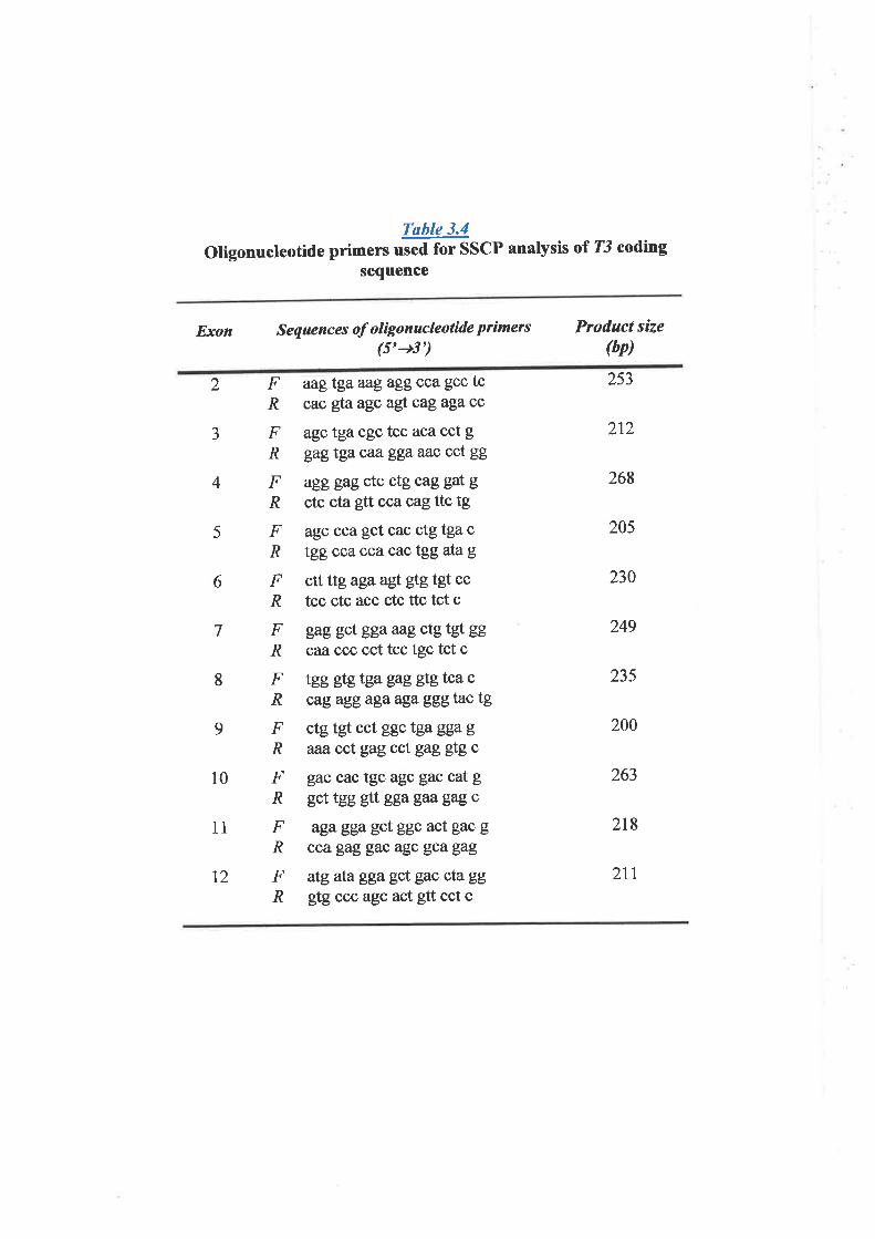
Table 3.4
oligonucleotide primers used for sscP analysis or T3 coding
sequence
Exon Sequences of olúgonucleotide primers(5'+3')
Product size(bp)
2
J
4
5
F aagtgaaagaggcca gcc tcR cac gfaagc ag!"caga5acc
F agctgacgctcc acacctgR gag tga caa' gga aac cct gg
F agggagctcctg caggatg.R ctc cta glt cca cag ttc tg
F agc cca gct cac ctg tga c
R tgg cca ccacactggatag
F ctt ttg agaag!. gtg tgt cc
R tcc ctc acc ctc ttc tct c
F gag gct ggaaag ctg tgt gg
lR caîccc cct tcc tgc tct c
F teegtgtgagag gtg tca cA cag agg aga aga ggg tac tg
F ctg tgt cct ggc tgaggagR aaacctgagcctgagÚgc
F gaccactgcagc gaccatgR gct tgg {tt gga gaa gag c
F agagga gct ggc act gac g
R ccagaggacagcgcagag
F atg ata gga gct gac cta gg
R gtg ccc agc act gtt cct c
253
6
7
8
212
268
205
230
249
235
200
263
218
2tt
9
10
ll
12
identþ was confirmed, a restriction digested fragment of 576 bp corresponding with the
region between exons 3 and 7 was excised and cloned into the pBluescript vector. The
same restricted digested product was also used to hybridize to a mouse multiple tissue
Northem blot. The RNA probes, both sense and anti-sense strands, were generated from
this cloned 576 bp cDNA and used to hybridize to the mouse embryoes at stage between
10.0-11.5 days post coitus (dpc). As shown in Figure 3.13,the hybridized signal was seen
only in embryo hybridized with anti-sense RNA probe but not with sense probe.
Expression signal was seen in the structure likely to be neural tube, otic vesicle, and eye
pit. Expression was also seen in developing structure of peripheral nervous system
especially in peripheral nerve ganglia.
3.3.9 Mutation Analysis of T3 in Breast Cancer
Single strand conformational polymorphism (SSCP) analysis was used for a mutation
screening of T3 in two panels of 24 paired normal and tumor DNA samples from breast
cancer patients and 24 breast cancer cell lines. Primers were designed to allow the
amplif,rcation of each exon that included at least 50 bp of its flanking introns (Table 3.Q.
The primers were fluorescent labelled, the PCR product of each exon was denatured and
analysed using a SSCP gel electrophoresis. A mobility band shift of exon 4 was observed
in a number of breast cancer samples and cell line DNAs. This band shift, however, was
also detected in the paired normal sample of each tumor as well as l0 out of 50 normal
control samples. It was therefore considered to be a DNA polymorphism, which was
present in the general population. Sequence analysis of shifted-band PCR product of exon
4 reveals a single nucleotide polymorphism (C/T) in intron 3 (IVS3-18C-+T).
Polymorphisms, \¡/ere also identified in exon 9 and exon 12, since they were seen in some
tumors and their paired normal as well as tumor cell line and control DNAs. Sequence
109
analysis was perfomred only in exon 12 PCR products, fiom one cell line and one normal
control, and revealed three single nucleotide polymorphism: [VSll-l4C+G, IVSll-
37C+T, and 1351T+G. Pollmorphism at position 1351, although causing codon change,
ACT+ACG, does not change the coding amino acid (T, threonine). From SSCP screening,
no other changes were observed and therefore no mutations were detected which were
restricted to these breast cancer samples and cancer cell lines.
In breast cancer, other mutation mechanisms may also be involved and cause the
reduction of gene expression or errors in splicing and therefore exon skipping which may
lead to the disruption of ORF. To test for these possibilities, RT-PCR was performed using
poly-A RNA isolated from 12 breast cancer cell lines. The results were compared with
those of two control RNAs, normal mammary gtand and fetal brain. No reduction of
6RNA expression was detected, in contrast expression of T3-L appeared to be up-
regulated in more than half of the cell lines tested (Figure 3.10 ^).
The isoform where
exon 2 was skipped was identified in three cell lines, but only as a fraction of all mRNA
products. This isofomr was not seen in normal marnmary gland, but was detected in fetal
brain as previously described (section 3.3.7, Fígure 3.10 B), and was therefore considered
to be an isoform specifically expressed in fetal brain and tumor tissues.
3.4I)iscussion
The transcribed sequences of a gene, designated "transcription unit 3 (T3)" have been
successfully identified. Initially, two trapped exons, ET6 and ET7, were used as the
starting materials for both database searches and cDNA library screening. The cDNA
sequence of 808 bp was first obtained, completing with 5' UTR, ORF, and 3' UTR with
polyadenylation signal. Based on the results of Northem analysis T3 appearcd to code for
two transcription isoforms of 0.9 and 3.7 kb. The first corresponds to a 808-bp transcript
110
that was assigned as "2tr3-S''. Subsequently, with the available genomic sequence
encompassing this gene, cosmid 301F3 (Figure I.3), the transcribed sequence of the
second isoform of 3,525 bp, designated "T3-L", and its complete gene structure were
identifred.
T1¡e T3 gene structure was initially determined for the short isoform, 73-S. Since at
the early stage of this project there was no genomic sequence available for this region, this
was determined by sequencing of genomic PCR amplification products and by partial
sequencing of the genomic clone utilizing transcript sequence specific primers. Following
the large-scale genomic sequencing of the region, the genomic organization of the entire
gene was characterized. The T3 gene consists of 12 exons encompassing the genomic
sequence of approximately 20 kb. It was shown that ET7 was trapped from the 3' part of
the exon I which constituted the 5' UTR. Nucleotide sequence analysis subsequentþ
revealed a sequence similar to the consensus of a splicing acceptor at the 5' boundary of
ET7. This mimic consensus sequence, in combination with the true exon/intron boundary
at the 3' end of exon l, therefore enabled the trap vector to capture the "exon",ET7 .
Zj-,S isoform shares the fust 4 exons and part of exon 5 with T3-L. The 3' terminal
exon for Z3-S is the exon 5 and the sequence that extends into intron 5 of T3-I. This
sequence includes part of the extended ORF, 3' UTR, and a polyadenylation signal,
ATTAJqA specific for Z3-S (Figure 3.2). Tlte presence of specific polyadenylation signal
suggests that transcriptional process of T3-S may be independent from that of T3-L. More
commonly a single primary transcript is generated and different isoforms are generated by
alternative splicing of the intemal exon or exons. ln 73, the two isoforms are likely
generated during transcription tbrough the selective use of independent polyadenylation
signal specific for each isoform. Transcription process by RNA polymerase would either
utilize the first ATTAAA signal (within intron 5) to generate the I3-S transcript or nm
111
ilr ,{p"!rñ'g this region until the second signal, also ATTAAA, close to the end of exon 12 was
transcribed to complete the T3-L transcipt (Fígure 3.6). According to the Northern
analysis result, the two principle tanscription isoforms of T3 were ubiquitously expressed
with varying degree of expression among the tissue tested. Interestingly, the difference in
degree of expression was also observed between the two isoforms in individual tissues.
Such variation between these two isoforms may also suggest independent transcriptional
regulation.
pre-mRNA 3' end selection has been reported for the gene that utilize multiple
poly(Ð signal similar to that of 73. The best cha¡acterized exarnple is the regulation of
immunoglobulin M (IgM) heavy chain synthesis during B-cell differentiation. ln this
process, switching from the membrane-bound form of IgM heavy chain (¡rm) to the
secreted form (ps) involves the selective use of a downstream Frm-specific poly(A) site in
B-cell and of an upsteam ps-specific poly(Ð site in plasma cell respectively- Such
selective use of poly(A) site appears to be modulated by the abundance of polyadenylation
factors that participate in pre-mRNA 3' end processing complex (Edwalds-Gilbert and
Milcarek, 1995; Takagaki et al., 1996)-
,Á r, ''i\. ør¡", mechanism utilizing the splicing factors also involv".¡á ttt" regulation of pre-
'RNA processing to determine the 3' terminal exon. As has been shown during alternative
processing of pre-mRNA from the human genes for calcitonin/calcitonin gene-related
peptide (CT/CGRP) (Lou et al.,l99S) where processing choice involves tissue-specific
recognition of altemative 3'-terminal exons, exon 4 or 6 (Amara et a1.,1982; Rosenfeld ef
al.,l9B4).In thyroid C cells, exon 4 is recognized as 3'-terminal exon with the usage of its
poly(Ð site, and the resulting mRNA molecule, exon I to 4, produces calcitonin (CT)
peptide. In net¡onal cells, exon 4 is excluded and the mRNA molecule containing exons 1,
2,3, s,and 6 is generated utilizing exon 6 as the 3' terminal exon. The CGRP is produced
*iJ,,,!,,
rt2
from this 6RNA. It has also been shown that exon 4 recognition requires an RNA-
processing enhancer sequence located within intron downstream of the exon 4 poly(A) site
(Lott et al., 1995;Lou et a1.,1996).
Selective expression of the transcripts 73-S or T3-L may be regulated at least by either
of the above-described mechanisms, though requires further investigation, leading to the l, '," ,.
differential expression of these two isoforms seen between diflerent tissue as well as
different isoform within the same tissue.
Zj showed a transcribed sequence homology to a partial sequence (MMDEF85) of
mouse def-g mRNA, a transcript isolated as showing developmentally regulated expression
in haematopoietic cells. The complete mouse transcript homologous to the human Z3-I
was also identified by sequencing of the mouse oDNA clones and an overlapping set of
RT-pCR products isolated from mouse cDNA libraries. This mouse transcribed sequence
provided a resource for interspecies cross-referencing of the 73 gene. Surprisingly, the
result of Northern analysis of this mouse transcript demonstrated only one prominent
product of 3.4 kb mRNA corresponding with its transcript sequence. There was no short
isoform expressed from this mouse gene. The nucleotide sequence homology of the mouse
transcript to T3-L is particular striking by showing 87% identþ across the ORF, with 95%
identity at its predicted coding amino acid sequence. This mouse transcript was designated
"mt3" referred to the mouse gene, orthologous to the human T3 gene. The absence of the
short transcript isoform in the mouse orthologue (mt3) may indicate the lack of enhancer
sequence element downsheam of mt3 exon 5. This may further suggest the recent
evolution of the gene sequence inT3 that may contain enhancer element and facilitate 3'
end polyadenylation processing of exon 5. As a result, Z3 gene codes for two protein
isoforms of 197 amino acids for T3-Sp and of 451 amino acids for T3-Lp.
113
In silico functional analysis of the predicted T3 protein of 451 amino acids (T3-Lp)
revealed at least two functional motifs, a DAG/PE binding domain in the central region
and a pHD zinc finger motif at its C-terminus. In T3-Sp, most of the predicted amino acid
sequence comprises part of the N-terminal region of T3-Lp. There is no known functional
motif in this region'
The DAG/pE binding domain (pF00130) is a cysteine-rich motif about 50 amino acid
residues long and essential for binding of diacylgþerol @AG) and its analogue, phorbol
ester (pE). DAG is an important second messenger in cell signaling, and its analogue (PE)
appeafs to be potent tumor promoters (Castagna 1987)' This domain binds two zinc ions
via the six cysteines and two histidines that are conserved and configured as HC¿HCz. It is
a conserved functional motif present in several proteins and is most likely involved in
cellular signaling pathways. A family of serine/threonine protein kinases, collectively
known as protein kinase C (PKC), is a classical kinase that contains one or two copies of
tlris DAG/pE-binding domain, also known as the "Cl domain", at its N-terminal region.
The Cl domain has been shown to bind DAG/PE in a phospholipid and zinc-dependent
fashion (Ono ef al., t989)-
DAG/PE binding domain has also been found in other proteins either possessing or
lacking kinase activity. These include diacylglycerol kinase (DGK): a protein that
phosphorylates the second-messenger DAG to phosphatidic acid (PA), chimaerin: a protein
family possessing GTpase-activating protein activity, raflmil family of serine/threonine
protein kinase, and Vav protein: a 95 kDa protooncogene product with its expression
resfücted to cells of hemopoeitic origin. These proteins are involved in cell signaling
pathways, and some are known to promote cell-cycle progression and migration (reviewed
in van Blitterswijk and Houssa, 2000; reviewed in Kazarriretz, 2000; Bonnefoy-Berard e/
a1.,1996)
rt4
The PHD domain, a zinc fingerJike motif, is also a cysteine-rich domain binding to
two zinc ions. This type of zinc finger has been identified in more than 40 proteins
including those of human (Aasland et aI-, 1995; Satra eú al', L995; Stec ef al" 1998;
Hasenpusch-Theil et al., 1999;Lu et al-, 1999; Bochar et al',2000)' It also consists of
eight ligands with seven cysteines and one histidine, arranged in a unique c4HC3 pattern
spanning approximately 50-80 residues. The PHD finger is distinct from other class of zinc
finger motifs such as the RING finger and LIM domain. The PHD finger containing
proteins are thought to belong to a diverse gfoup of transcriptional regulators possibly
affecting eukaryotic gene expression by influencing chromatin structure. Recent data
provide evidence that pHD frnger proteins are associated with chromatin remodelling
complexes @ochar et a1.,2000) or contribute to histone acetylation (Loewith et al-,2000).
Furthermore, the PHD finger has been shown to activate transcription in yeast, plant, and
animal cells (Halb ach et a1.,2000). The presence of PHD motif in T3-Lp/mt3p implicated
its possible frurction involving in transcriptional regulation, therefore supported the notion
that T3-Lp/mt3p is a nuclear protein.
The importance of the PHD frnger is evident from the reported mutations in PHD
fingers associated with human diseases and provides a good evidence for its fimctional role
in particular proteins. ln the ATRX syndrome, an X-linked form of syndromal mental
retardation associated with alpha-thalassemia, missense mutations in the non-canonical
pHDlike domain of the ATRX gene lead to the characteristic phenotype (Gibbons et al-,
lggT). ATRX has been suggested to play a role in the de-repression or activation of
chromatin, and the interaction between its mouse orthologue, Mo ATRX and mouse
chromatin protein (tpl) has been demonstrated in a yeast two-hybrid system (Le Douarin
et al.,1996). Other examples of PHD finger-containing proteins involved in human disease
include the AISE gene product, which is mutated in the autoimmune disease APECED,
115
autoimmune polyendocrinopathy-candidiasis ectoderrral dystophy (Nagamine et al''
1997; The Finnish-German APECED Consortium, 1997). The AIRE protein harbours a
nuclear localization signal (NLS) and two pHD motifs. Most point mutations associated
with deleterious effect are within or lead to the truncation of one of the PHD domains of
AIRE (Scott et a1.,1998; Rinderle et a1.,1999). Furthermore, AIRE mutants lacking the
pHD motif evhibr¡/altered nuclear localization (Rinderle et al-, 1999), suggesting that ';'4
AIRE mutations in APECED may have their primary effect in the nucleus. ALLI, the
human homologue of Drosophila trithorar, and other PHD-finger containing genes' are
also frequentþ disrupted by chromosomal rearrangements that occur in acute lymphoid
and myeloid leukemias (Gu et al., 1992; Tkachuk et al., 1992; Nakamura et al" 1993;
Parry et a1.,1993).
The presence of both the DAG/pE binding domain and the PHD finger motif suggests
that T3-Lp is involved in a cellular signalling pathway, most likely to promote cell growth.
The most likely function is an involvement in transcription regulation, possibly promotion,
by influencing chromatin structure. These suggestions are consistent with the computerized
predictions of a nucle ar Localization as well as the observation that gene expression is
ubiquitous in embryonic tissues, and all adult tissues. An importance basic function of the
T3-L protein in cellular processes is supported by the conservation of the protein during
evolution from C elegans, D melanogaster,to humans'
Study of mt3 gene expression in the embryo of the mouse (section 3.3.8-2) further
demonstrated the importance of this gene during embryonic development. It is likeþ that
this protein is required for embryonic cell proliferation and differentiation.
Mutation screening of T3 in breast cancer was performed for all 12 coding exons by
SSCp analysis. No evidence of mutations restricted to the cancer cells were detected in two
panels of breast cancer samples showing 16q24.3 LOH nor in breast cancer cell lines. This
r16
indicated that T3 was unlikely to be a tumor suppressor gene targeted by LoH at least in
sporadic breast cancer tested. This is consistent with the predicted possible function of T3
protein (T3-Lp) for promotion rather than suppression of cell growth. Furthermore' it was
observed that expression of T3-L was increased in some breast cancer cell lines'
Carcinogenesis requires at least two cellular events, the loss of cell proliferation
control pathway and the activation of cell cycle pathway, although changes in other
cellular pathways are also required for phenotypic changes into more aggressive forms of
cancer. The possible function of T3 can be categorised into a group that activates or
promotes the cell cycle. T3 protein may therefore be a part of onco-protein network and
interact with several proteins in the network. With this possibility, study of the associated
proteins or the proteins that may interact \¡r'ith T3 is likely to lead to an understanding of
the pathway that involves T3. Many biological techniques can be applied to this' for
example, the yeast two-hybrid system (Fields and Song, 1989), mass-spectrophotometric
analysis of native protein complex purifred by affrnity capture (Pandey et al-,2000; Pandey
and Mann, 2000), and protein üruy for multi-protein interaction analysis. These
procedures, however are beyond the scope of this study'
tt7
Ghapter 4
Charact erization of Transcription U n it
12 (T12) and
Mutation Study of Spastic Paraplegia
gene, SPGT
Table of ContentsPage
ll8120
120
120
a
4.l lntroduction
4.2 Methods
4.2.1 Transcript sequence identification
4.2.1.1 Database searches þr Expressed Sequence Homolog,t
4.2.1. 2 Identification of the cDNA equunr'b by RT-PCR and cDNA
library Screening
4.2.1.3 Determination of the cDNA end
4 -2.2 Expre ssion Analysis
4. 2. 2. 1 Northern blot Hybridization
4. 2. 2. 2 Dffirential Expression Study
4.2.3 Prctein Sequence Analysis
4.2.4 ldentification of Genomic Structure
4.2.5 SPGT Mutation Study
4.3 Results
4.3.1 Determination of Transcript Sequence
4.3.1.1 Database Analysís and cDNA Sequencing
4. 3. 1.2 Transcript Sequence Assembly
4 -3 .2 T ranscript Sequence and Expression Analysis
4.3.3 Protein Sequence Identþ
4.3.4 Genomic Organi zation
4.3.5 SPGT mutation study
4.3.5.1 Nonsense Mutation ìn Exon 5
4.3.5.2 Compound Heterozygous Mutation in Exon I I4.3.5.3 Missense Mutations in Exon 9 and Exon 13
4.4 Discussion
120
t2tt22
122
t22
123
t23
124
124
t24
t25
126
130
131
r32
133
134
135
135
137
4.1 Introduction
In an attempt to search for a new gene that might be targeted by LOH and be a
candidate for a breast cancer tumor suppressor gene, six trapped oxons isolated from the
I6q24.3 physical map (Whitmore et al.l998a) within close proximity were investigated to
see if they were part of the same gene. These trapped products were grouped and from the
order determined by the physical map (Fìgure 1.3) were described as transcription unit 12
(Tl2). T12 was situated approximately 11 kb proximal to the CMAR gene. This chapter
describes the determination of the transcript sequence based on these trapped exons. Tl2
was found to be a transcript of approximately 3.2 kb. The predicted protein sequence was
analyzed in silico. The likely function was a mitochondrial ATP dependent
metalloprotease, based on its high degree of homology to this type of protein in yeast. At
completion of this gene characteizalion, a report appeared (Casari et al., 1998) which
described an identical gene designated SPG7, which mapped to the region 16q24.3 and
was involved in one form of familial neurodegenerative disorder, recessive hereditary
spastic paraplegia (HSP). Although SPGT Q12) was not involved in breast cancer the
involvement of this gene in HSP was studied in more detail.
Familial, or hereditary spastic paraplegia (HSP), comprises a group of
neurodegenerative disorders characteized by slow progressive spasticity and weakness of
the lower extremities (Harding l98l; Fink 1997; Reid I999a). The common
neuropathologic finding is axonal degeneration involving the terminal ends of the longest
fibers of the corticospinal tracts and dorsal columns. The diseases are clinically and
genetically heterogeneous. Two forms of HSP are classified according to the clinical
symptoms. In pure (uncomplicated) HSP the spasticity is the prominent feature and occurs
in isolation. Most patients present with difficulty walking or gait disturbance, noticed
either by themselves or relative. In those with childhood onset, a delay in walking is
118

coÍtmon. In the complicated form spastic paraparesis has been associated with many
conditions and there are additional neurologic symptoms, such as cerebellar ataxia, mental
retardation, dementia, epilepsy, optic atrophy and retinal degeneration (Fink 1997; Reid
1999a). In both clinical forms, autosomal dominant, autosomal recessive and X-linked
inheritance have been described. As well as genetic heterogeneity there is often variable
penetrance and expressivity. Linkage analysis has identified at least eight genetic loci for
the autosomal dominant forms of HSP on chromosomes 14q (SPG3) (Hazan et al., 1993),
2p (SPG$ (Hazan et al., 1994; Hentati et al., 1994b), 15q (^SPGó) (Fink et al., 1995), 8q
(SPc8) (Hedera et al., 1999), 10q (SPG9) (Seri e/ al., 1999), 12q(SPGI0) (Reid et al.,
1999b),19q(SPG12) (Reid et a1.,2000), and2q(SPG1-3) (Fontaine et a1.,2000). Four loci
associated with autosomal recessive HSPs were mapped to chromosome arms 8p (SPG5)
(Hentati et al., 1994a), l6q(SPG7) (De Michele et a1.,1998), l5q (SPG11) (Murillo et al.,
1999), and 3q (SPG14) (Ya".za et al., 2000). Two Xlinked forms of HSP, both
complicated, have been described and are caused by mutation in the Ll cell adhesion
molecule (LICAM) and proteolipid protein (PLP) genes located on regions Xq28 (SPG1)
and Xq22 (SP G2) respectively (Jouet et al., 199 4; Saugier-Veber et al., 1994).
Of the 12 autosomal loci, the genes for SPG4 arñ SPGT have been characterized.
SPG4, the gene responsible for autosomal dominant HSP, encodes a predicted 67.2kÐ
nuclear protein known as "spastin" (}{azan et a1.,1999). The spastin amino acid sequence
shows a high degree of homology with the yeast 265 proteasome subunits, Yta6p and
Tbp6p, which belong to a subclass of the AJd{ protein family (ATPases associated with
diverse cellular activities) (Patel and Latterich, 1998). SPG7, a gene at 16q24.3 involved in
recessive HSP, has also been characterized (Casari et al., 1998). SPGT encodes a
mitochondrial protein called "paraplegin" which is highly homologous to the yeast
mitochondrial ATP-dependent zinc metalloproteases, Afg3p, Rcalp and Ymelp. These
119

yeast proteins exhibit both proteolytic and chaperoneJike functions at the inner
mitochondrial membrane and also belong to the AJA.A. protein family (Patel and Latterich,
1998). SPGT mutations have been reported in three families, two with pure and one with
complicated autosomal recessive HSP (Casari et a1.,1998). Mutations in paraplegin cause
a mitochondria function defect as a result of impaired oxidative phosphorylation
(oxPHos).
The publication of Casari et al. (1998) that reported SPGT did not provide the
complete gene structure and it was not available in the public sequence database. The
genomic structure is necessary as a basis for determining gene mutations associated with a
particular genetic syndrome. This chapter will first describe the cloning and
characteization of what was called the transcription unit l2 (Tl2). This will be followed
by the determination of the genomic structure. Finally, mutation analysis of SPGT will be
explored in a large number of spastic paraplegia cases, both pure and complicated forms.
4.2 Methods
4.2.1 T ranscript Sequence ldentification
4. 2. l. 1 Databas e s e arches for Expressed S equence Homologt
Trapped exon sequences were first used to identi$ the homologous transcribed
sequences from both EST and non-redundant databases, using BLASTN program (Altschul
et al., 1997) available at NCBI. During the transcript sequence identification was
underway, homology searches was also performed on both EST and non-redundant
database utilizing the partial transcript sequence available at that time.
by RT-PCR and cDNA/,!ìbrary Screening4. 2. l. 2 ldentificøtion of the
r20
RT-PCR was initially undertaken to determine the cDNA sequence linking the
trapped exons withinthe close proximity. The procedure described in section 2.2.13 was
carried out utilizing a set of primers designed to the trapped exon sequences (Table 4.1).
The amplified products were subsequently purif,red by QlAquick columns (Qiagen, section
2.2.14.1) or separated by agarose gel electrophoresis and a single DNA band was purifred
from the gel (2.2.14.2). The purifred cDNA fragments were then sequenced (2.2.16.1)
using the same set of primers that were used for RT-PCR. The sequence information was
then assembled.
Screening of the cDNA library was performed utilizing the trapped exon insert as
probe. Trapped product 8T2.5 was excised from the pAMPlO vector by double digestion
with Nor IlSal I. The insert was aP32dCTP-labeled and use as probe to hybridize to the ì.-
fetal brain cDNA library filters (2.2.18). After repeat hybridization for plaque purification
the positive clones were eluted from the phage plaques for subsequent analysis. Sequence
of the cDNA insert was then determined by PCR amplification using the Àgtl l vector
primers, oDNA fragment purification and nucleotide sequencing.
PCR procedure was also use to screen the fetal brain cDNA library for the 5'sequence
of oDNA utilizing the vector primer and gene specific primer. This was performed in order
to extend the 5' sequence of the existing cDNA obtained from RT-PCR and sequence
assembly.
4.2.1.3 Determination of the cDNA end by RACE
Determination of the 5' end or the sequence toward the 5' end of the transcript was
performed utilizing the 5' RACE procedure described in section 2.2.17.1. The nucleotide
sequences of the gene specific primers used for 5' RACE are presented,in Table 4.1
T2I
The 3' RACE procedure (2.2.17.2) was used to determine and extend the 3' sequence
of the transcript. First strand cDNA was synthesized from poly A+ mRNA utilizing a
specifically designed "anchored oligo-dT" primer. Second strand oDNA was synthesized
by a linear PCR using a biotinylated gene specific primer (BioTl2-3Extl, Table 4.1). Tl2
specific biotinylated cDNAs were then captured on streptavidin-coated magnetic beads
(DYNAL) for purification. The 3' end was subsequently amplified by nested-PCR using
the reverse primers specific for anchored sequence and the forward gene specific primers,
T l2-3Bxt2 and T l2-Ext3 (Tab le 4. 1).
4.2.2 Expr ession Analysis
4.2.2.1 Northern blot Hybridization
Northern blot analysis was used to determine the size of the TI2 tanscnpt as well as
its expression pattern. A commercial multiple tissue Northern membrane (Clontech) was
hybridized with a group of ¡c32PldCTPJabelled oDNA probes generated from RT-PCR
products for 1.3-kb cDNA of Tl2 (section 4.2.1). The hybridization protocol is given in
section 2.2.11.2- After washing, the hybridized membrane was exposed to X-ray film and
autoradio graphic developed.
4. 2. 2. 2 Differential Expression Study
To determine the tissue and developmental stage-specific expression of the SPGT
mRNA, a human RNA Master Blot from 50 different fetal and adult tissues (Clontech) was
also hybridized with the same set of oDNA probes used for Northern hybridization. The
hybridization protocol and solution was as recommended by the manufacturer. The
membrane was washed five times with 2 x SSC, 1% SDS at 65 oC and was subjected to
autoradiographic development. The quantity of mRNA spotted for each tissue on the
r22
Master Blot was normalized, as recommended by the manufacturer, by using eight
different housekeeping gene transcripts as probes.
4.2.3 Protein Sequence Analysis
Analysis of protein sequence identity and function was done in silico. The BLASTP
program (Altschul et al., 1997) available at NCBI was used to search for amino acid
sequence homology between T12 arrd protein sequence databases at NCBI. Sub-cellular
localization of the T12 protein was performed utilizing a protein sub-cellular localization
prediction tool, PSORT (Nakai and Horton,1999).
4.2.4 ldentification of Genomic Structure
The genomic structure was determined from three cosmids, 383H6, 427811, and
358D12. These cosmids form a contig and encompass the Tl2lSPG7 region. A
combination of two independent procedures were utilized. The first method involved the
use of PCR amplification of genomic DNA with gene specihc oligonucleotide primers
designed for both strands of oDNA sequence. The amplified genomic products that were
larger than those from cDNA were subsequently purified and sequenced from either end.
The second method was by direct sequencing of cosmid clones utilizing the same set of
gene specific primers. This was used when PCR amplification was not successfrrl because
of a large intervening sequence (intron). All primer sequences and their locations in the
212 oDNA are given in Table 4.2. The exorVintron boundaries were identifred by the
sequence divergence in genomic DNA from that of cDNA. The size of each intron was
determined from the PCR product size and some was estimated from the restriction map of
the cosmid contig (Whitmore et a|.,1998a).
t23
Chøler 4 Cltsraclerizoliolt lf lation Study of SPGT
4.2.5 SPGT Mutation Study
To determine the spectrum of SPGT mutation in sporadic cases of HSP, DNA samples
isolated from I 16 HSP patients from unrelated families were obtained from three centers.
These are the Children's National Medical Center, Research Center for Genetic Medicine,
V/ashington, DC (Dr Francisco Murillo), the Department of Neurology, University of
Michigan, USA (Dr John K Fink), and the Geriatric Research, Education and Clinical
Center, Seattle, USA (Dr Thomas Bird). The available patient data are age of onset of each
patient, the clinical form of either pure or complicated, and the family background. The
DNA samples isolated from 60 clinically normal individuals of Caucasian background
were used as normal control for mutation analysis
Mutation screening was by single-strand conformation polymorphism (SSCP)
analysis (Hayashi, l99l; Nataraj et al-, 1999). Individual exon sequences were PCR
amplified in 20-pl reactions containing [o"P] dCTP with primers designed with respect to
its flanking introns (Table 4.4). Each completed PCR product was added to an equal
volume of formamide dye. The mixture w¿Ìs denatured at 100 "C for l0 min, cooled on ice
and loaded onto non-denaturing polyacrylamide gel (containing 60/0, \yo or lïYo gel,
dependent on PCR product size, and 7Yo glycerol) and MDE gel (FMC bioproducts).
Electrophoresis was carried out at a constant 700 volt overnight at room temperature. Gel
was blotted onto 3MM filter paper and dried under heat and vacuum. Dried gel was
exposed to X-ray film overnight at 10 "C and autoradiograph developed. The samples
showing mobility band shift when compared with normal controls were re-amplified and
sequenced in parallel with controls.
4.3 Results
4.3.1 Determination of Transcript Sequence
t24
A schematic summary of the transcript sequence determination leading to the
assembly of a fuIl-lengfh cDNA transcript of Tl2 is shown in Figure 4.1.
4.3.1.1 Database Analysß and oDNA Sequencing
A group of trapped exons, arranged in the centromeric to telomeric order in the
16q24.3 physical map as 8T23.23, 8T24.42, 8T34.87, ET34.1, 8T2.5, and ET2.8
(Whitmore et al.,l998a) were initially assigned as "transcription unit 12 (TI2)" (Figure
1.3 and 4.1 A). Database searches at this time revealed only two human ESTs, D63198 and
C15128 that showed sequence homology to ET2.5 and ET2.8 respectively. Homology
between the sequences of 8T2.5 and D63 198 was limited to only 126 bp out of the 229 bp
of ET2.5. Since the cDNA clone corresponding to D63198 was not available for analysis,
the trapped product 8T25 was excised from the vector and then used as a probe to screen a
fetal brain oDNA library (Clontech) from which a oDNA clone designed "1.ET2.5" was
obtained. Sequencing of the ?,,8T2-5 oDNA revealed an insert sequence of 836 bp
containing the sequence of both exons ET34.l andBT2.5 (Figure 4.2)
Within the ì"ET2.5 cDNA there was a 55 bp deletion of ET2.5 that resulted in the first
48 bp of 8T2.5 being contiguous with126 bp of the sequence that showed homology to the
EST sequence D63198 (Fìgure 4.2). Analysis of the sequence at the deletion junction at
nucleotide position 48 of 8T2.5 revealed a sequence that resembled the consensus for a
donor-splice site. Therefore, this suggests that there is alternative splicing of the 8T2.5
sequence and that one form was present in î"ET2.5. This alternative splicing suggested that
8T2.5 was likely to be trapped from two adjacent exons present in the genomic sequence
cloned into the trap-vector. To verifu this, primers were designed from both ends of ET2.5
(2.5F1 and2.5F.2, Table 4.1) allid used to PCR amplifr a fragment of 1.4 kb from genomic
125
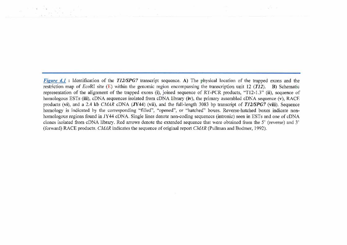
Fieure 4.1 : Identification of the T12/SPG7 Íanscript sequence. A) The physical location of the trapped exons and therestriction map of EcoRI site (E) within the genomic region encompassing the transcription unit 12 (Tl2). B) Schematicrepresentation of the alignment of the trapped exons (i), joined sequence of RT-PCR products, *TI2-I.3" (ii), sequence ofhomologous ESTs (iii), cDNA sequences isolated from cDNA library (iv), the primary assembled oDNA sequence (v), RACEproducts (vi), and a 2.4 kb CMAR cDNA (JY44) (vii), and the tull-length 3083 bp transcript of T12/SPG7 (viii). Sequencehomology is indicated by the corresponding "filled", "opened", or "hatched" boxes. Reverse-hatched boxes indicate non-homologous regions found in JY44 cDNA. Single lines denote non-coding sequences (inhonic) seen in ESTs and one of oDNAclones isolated from oDNA library. Red a:rows denote the extended sequence that were obtained from the 5' (reverse) and 3'(forward) RACE products. CMAR indicates the sequence of original report CMAR (Pullman and Bodmer,1992).
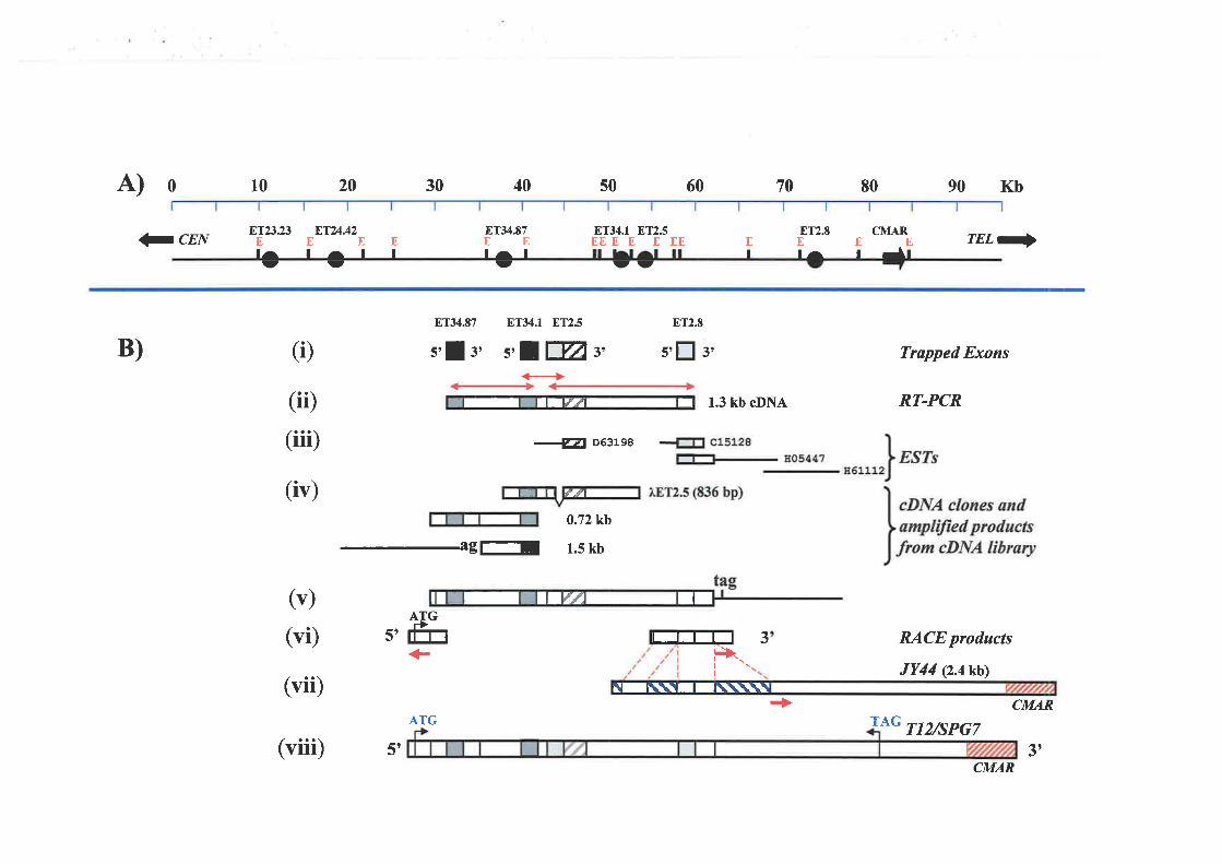
A) 0
?CEN
B)
l^l
10
I'T2323El^rt
20
8T24.42
It
E E Et
30 40 50 60
ET34.I ET25EEE E E EE
It It
8T34.87 8T34.1 8T2.5 ET2.E
5'! l, s,||ffis, s'[ 3'
€
-Æ D63198
8T34.87
tt̂II
EI
EI
70 80
8T2.8El^
90 Kb
(Ð
(iÐ
(iii)
0.72kb
1.5 kb
1.3 kb cDNA
CMAREEI ril
rEL-
Trøpped Exons
RT-PCR
RACE products
JY44 (2.4 kb)
CMAR
T12/SPG7
(iv)
5t
(vii)-agl-r
(v)
(vi)ATGr)flTr 3
<r l+r.t\I
I IUT¿ I
III û/-
IT I I''ÍJ II
ÈIL\\TI\\\\\t
IIIII
(viii) s'
ATG+>
TAG
CMAR3t
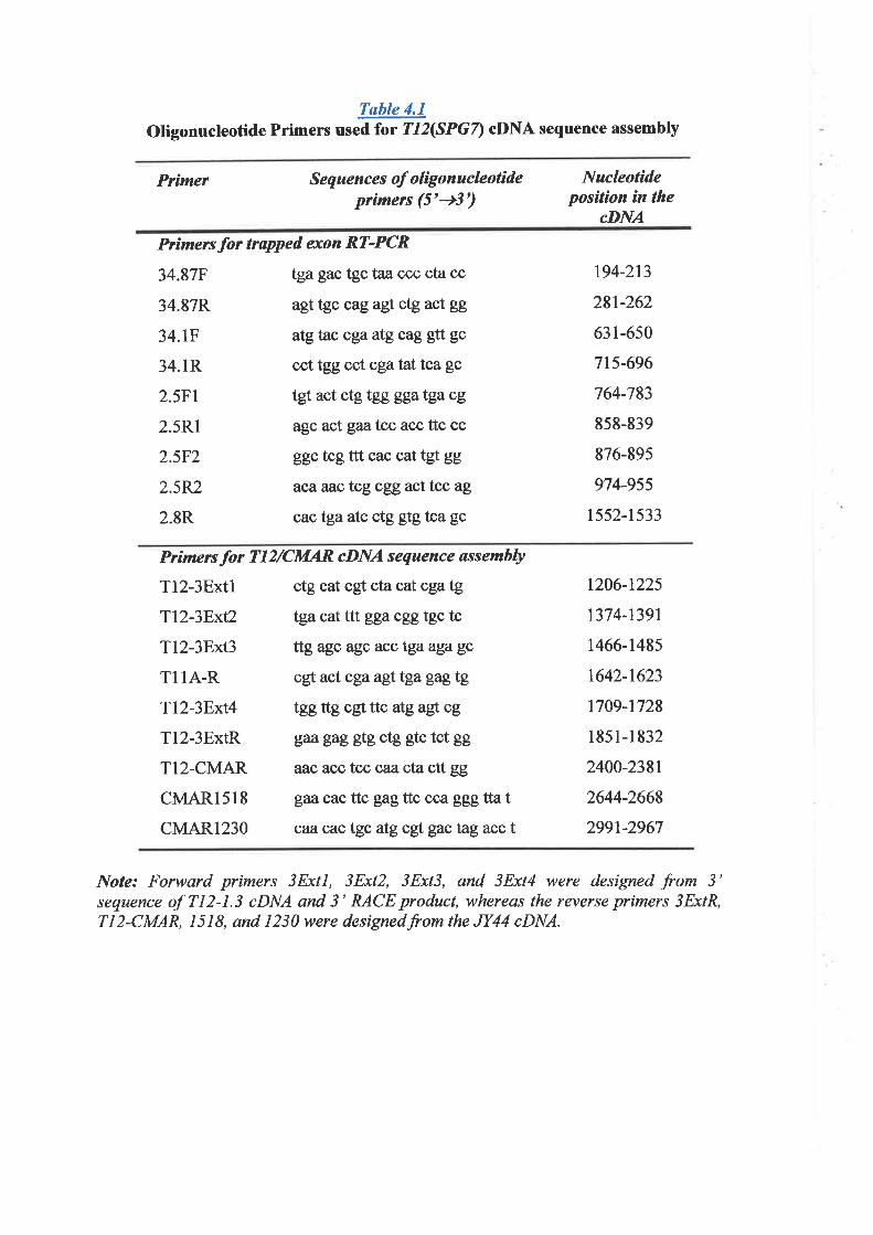
Table 4.1
Oligonucleotide Primers used for TL2(SPGZ) cDNA sequence assembly
Primcr S eq uenc es of o lþ o n uc leotideprimers (5'+3')
Nucleotideposition in the
cDNA
Primersfor trapped exon RT-PCR
34.87F tga gac tgc taa ccc cta cc
34.87R agttgc cag agt ctg act gg
34.1F atgtac cgaatg cag gtt gc
34.1R cct tgg cct cga tattcagc
2.5F1 tgtact ctg tgg ggatgacg
2.5R1 agc act gaatcc acc ttc cc
2.5F2 ggc tcg ttt cac cat tgf gg
2.5R2 acaaaß tcg cgg act tcc ag
2.8R cactga atc ctg gtg tca gc
194-213
281-262
631-650
7t5-696
764-783
858-839
876-895
974-955
t5s2-1533
Prímersfor TI2/CMAR aDNA sequence øssemhly
T12-3Extl ctg cat cgt cta cat cga tg
Tl2-3F;rt2 tgacatfttgga cgg tgc tc
Tl2-3Ext3 ttg agc agcaÊctgaagagc
Tl lA-R cgt act cga ag!.tga gagtg
Tl2-3Bxt4 tgg ttg cgt ttc atgag!cg
Tl2-3ExtR gægag gtg ctg gtc tct gg
TI2-CMAR aac aßctcc caz cta ctt gg
CMAR15I8 gaa cac ttc gagttccca ggg tta t
CMAR1230 caacac tgc atg cgt gac tagacct
1206-1225
1374-t391
1466-1485
t642-1623
t709-1728
1851-1832
2400-2381
2644-2668
2991-2967
Note: Forward primers 3Extl, 3Ext2, 3Ext3, and 3Ext4 were designed from 3'sequence of 712-1.3 cDNA and 3' RACE product, whereas the reverse primers 3ExtR,Tl2-CMAR, 1518, and 1230were designedfromtheJY44 cDNA.
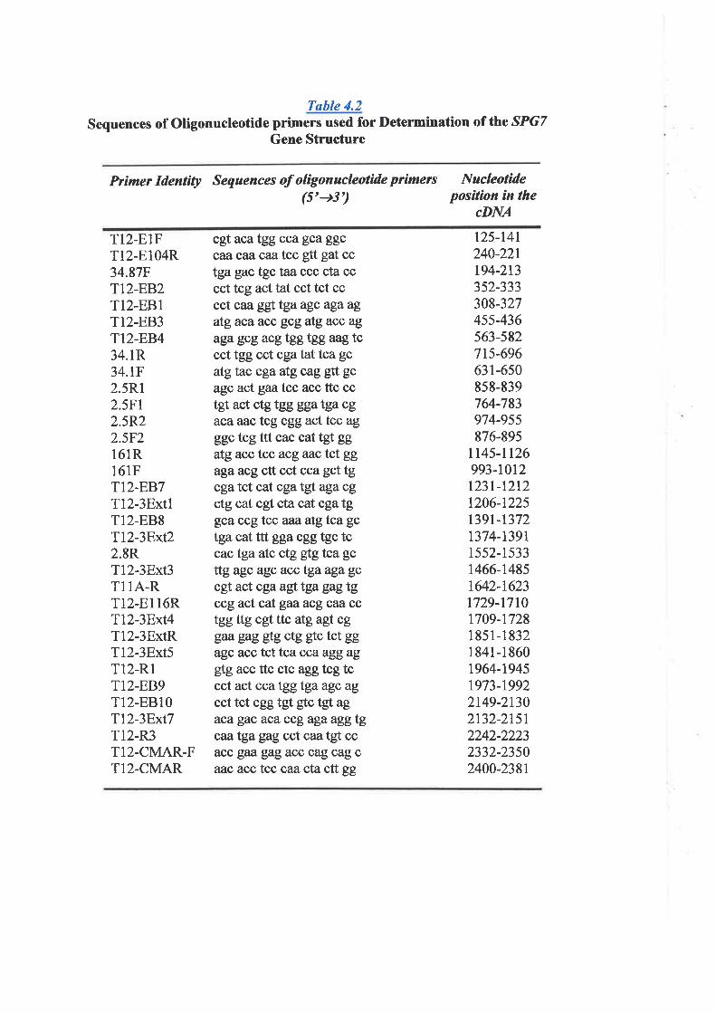
Table 4.2
Sequences of Oligonucleotide primers used for l)etermination of the SPGTGene Structure
Primer Identìty Sequences of olìgonacleotide ptìmets(5'+3')
Nucleotìdeposítíon in the
cDNA
T12.E1FT12-E104R34.87FTI2-EB2TI2.EB1T12-EB3TI2-EB434.1R34.1F2.5R12.5Ft2.5R22.5F216lRl6lFTI2.EB7Tl2-3ExtlT12-EB8Tl2-3Bxt22.8RTl2-3Bxt3T1lA-RTl2-El l6RTl2-3Bxt4T12-3ExtRTl2-3Bxt5Tl2-RlTI2-BB9T12-EBr0Tl2-3Bxt7T12-R3T12-CMAR-FTI2-CMAR
125-t4l240-221194-213352-333308-327455-436563-582715-696631-650858-839764-783974-955876-895
tt45-1126993-1012t23l-12t21206-1225t39l-13721374-13911552-1533t466-t4851642-16231729-17t01709-17281 85 1-1 8321841-18601964-19451973-19922149-2t302132-21512242-22232332-23502400-2381
cgt aca tgg cca gçaggccaacazcaalcc gtt gat cctga gac tgc taa ccc cta cccct tcg act tat cct tct cccct caa ggttga agc aga agatgaca acc gcg atgacc ag
aga gcg acgtggtgg aag tcccttgg cctcgatattcagcatgtac cgaatg cag gtt gc
agc act gaøtcc acc ttc cctgt act ctg tgg gga tga cgaca aac tcg cgg act tcc ag
ggc tcg ttt cac cattgl ggatgacc tcc acg aactct ggagaacg ctt cct cca gct tgcga tct cat cga tgt aga cgctg cat cgf cta cat cga tggca ccg tcc aaaalgtca gc
tgacat ttt gga cgg tgc tccac tga atc ctg gtg tca gc
ttg agc agc acctgaagagccg!. act cga agftga gagtgccg act cat gaa acg c¿ta cctgg ttg cgt ttc atgagl cggM gag gtg ctg gtc tct ggagcacc tcttca ccaaggagglgacc ttc ctc agg tcg tccct act cca tgg tgaagc ag
cct tct cgg tgt gtc tgt agacagac acaccgagaaggtgcaatga gag cct caa tgt ccacc gaa gag acc cag cag caac acctcc caa cta ctt gg
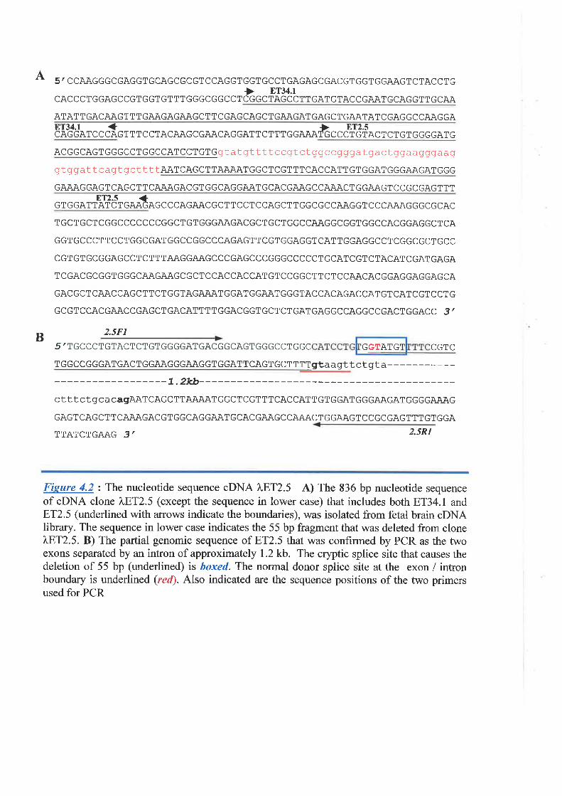
A 5' CCAAGGGCGAGGT GCAGCGCGT C CAGGT GGTGCCTGAGAGCGACGT GGTGGAAGTCTACC T G
) ET34.lCACCCTGGAGCCGT GGT GTTT GGGCGGCCT CGGCTAGCCTTGAT GTACCGAAT GCAGGT T GCAA
ATAT T GACAAGT TT GAAGAGAAGCT TCGAGCAGCTGAAGAT GAGCTGAATAT CGAGGCCAAGGA8T34.1 + >CAGGATCCCAGT TT CCTACAAGCGAACAGGATTCTTTGGAAATGCCCT GTACT CTGTGGGGATG
ACGGCAGTGGGCCTGGCCATCCTGTGgtatgttttccgtctggccgggatgactggaagggaag
gtggattcagtg ct Tt TAATCAGCTTA,UU\TGGCTCGTTTCACCATTGTGGATGGGAAGATGGG
GAAAGGAG TCAGCT T C AAÄ,GACGT GGCAGGAATGCACGAAGCCAAACT GGAAGT CCGCGAGTTTET2.s +
GTGGATTATCT GAAÖAGC CCAGAACGCTT CCTCCAGCT TGGCGCCAAGGT CCCAAAGGGCGCAC
T GCTGCT CGGCCCCCCCGGCT GT GGGAAGACGCTGCT GGCCAAGGCGGTGGCCACGGAGGCT CA
GGTGCCCTTCCTGGCGATGGCCGGCCCAGAGTTCGTGGAGGTCATTGGAGGCCTCGGCGCTGCC
CGT GT GCGGAGCCT CT T TAAGGAAGCCCGAGCCCGGGCCCCCT GCATCGT CTACAT CGAT GAGA
T CGACGCGGTGGGCAAGAAGCGCTCCACCACCATGT CCGGCTT CTCCAACACGGAGGAGGAGCA
GAC GCT CAAC CAGCTTCT GGTAG.A.AATGGATGGAAT GGGTACCACAGACCAT GTCAT CGTCCT G
GCGTCCACGAACCGAGCTGACATTTTGGACGGTGCTCTGATGAGGCCAGGCCGACTGGACC 3 /
2.5F1
5 / TGCCCTGTACTCTGTGGGGATGACGGCAGTGGGCCTGGC CGTC
T GGC C GGGAT GAC T GGAAGGGAAGGT GGAT T CAGT GCT T TTgtaagttctgta-------7.2kb-----
cttt cTgcacagAATCAGCTTAAAATGGCTCGTTTCACCATTGTGGATGGGAAGATGGGGAAAG
GAGTCAGCT T CAAAGACGT GGCAGGAAT GCACGAAGCCAAACT GGAAGTCCGCGAGTTTGT GGA
TTATCTGAAG 3/ 2.5R1
Fisure 4.2: The nucleotide sequence cDNA ),,8T2.5 A) The 836 bp nucleotide sequenceof cDNA clone 7ßT2.5 (except the sequence in lower case) that includes both ET34.1 and8T2.5 (underlined with arrows indicate the boundaries), was isolated from fetal brain cDNAlibrary. The sequence in lower case indicates the 55 bp fragment that was deleted from clone),,8T2.5. B) The partial genomic sequence of ET2.5 that was confirmed by PCR as the twoexons separated by u.t intron of approximately I.2 kb. The cryptic splice site that causes thedeletion of 55 bp (underlined) is boxed. The normal donor splice site at the exon / intronboundary is underlined (recl). Also indicated are the sequence positions of the two primersused for PCR
BGGTATG
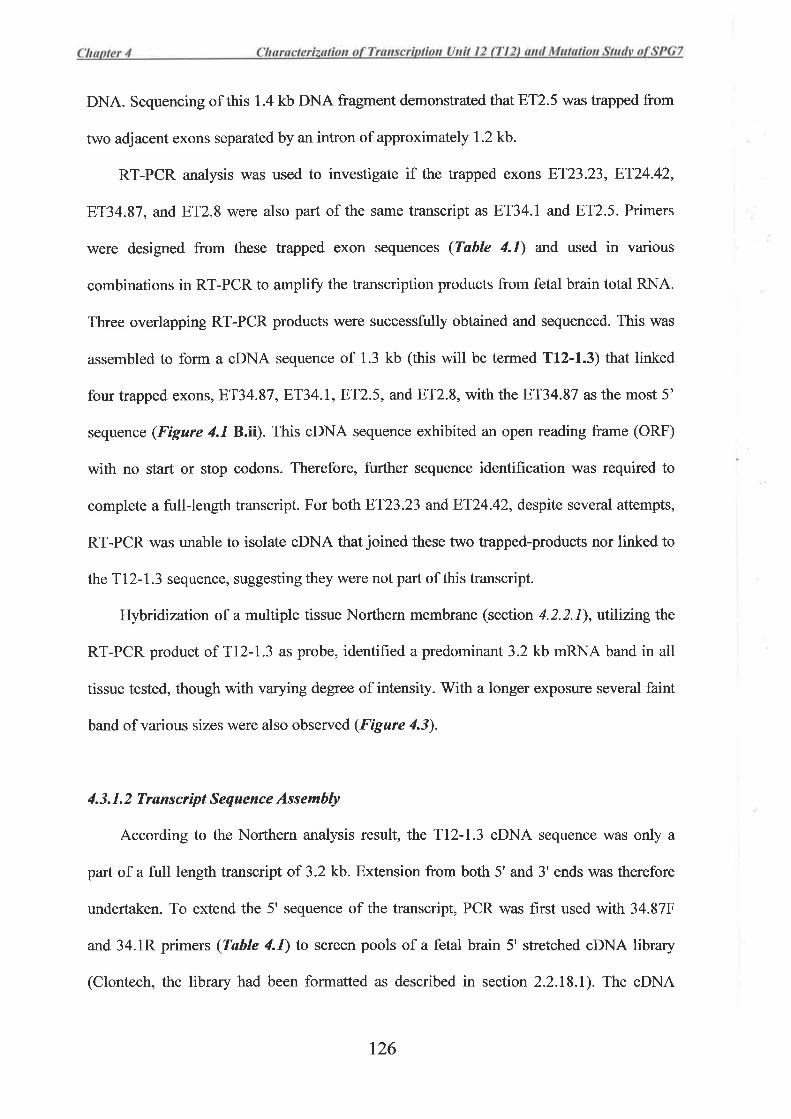
DNA. Sequencing of this 1.4 kb DNA fragment demonstrated that 8T2.5 was trapped from
two adjacent exons separated by an intron of approximately 1.2 kb.
RT-PCR analysis was used to investigate if the trapped exons 8T23.23,8T24.42,
8T34.87, and ET2.8 were also part of the same transcript as ET34.l and ET2.5. Primers
were designed from these trapped exon sequences (Table 4.1) and used in various
combinations in RT-PCR to amplifu the transcription products from fetal brain total RNA.
Three overlapping RT-PCR products were successfirlly obtained and sequenced. This was
assembled to form a cDNA sequence of 1.3 kb (this will be termed T12-1.3) that linked
four trapped exons, 8T34.87 , ET34.|,8T2.5, and ET2.8, with the ET34.87 as the most 5'
sequence (Figure 4.18.ä). This oDNA sequence exhibited an open reading frame (ORF)
with no start or stop codons. Therefore, further sequence identification was required to
complete a full-length transcript. For both 8T23.23 andBT24.42, despite several attempts,
RT-PCR was unable to isolate oDNA that joined these two trapped-products nor linked to
theTl2-7.3 sequence, suggesting they were not part of this transcript.
Hybidization of a multiple tissue Northem membrane (section 4.2.2.1), utilizing the
RT-PCR product of Tl2-1.3 as probe, identifred a predominant 3.2 kb mRNA band in all
tissue tested, though with varying degree of intensity. With a longer exposure several faint
band of various sizes were also observed (Figure 4.3).
4.3. 1. 2 Transcrìpt Sequence Assembly
According to the Northern analysis result, the Tl2-1.3 oDNA sequence was only a
part of a full length transcript of 3.2 kb. Extension from both 5' and 3' ends was therefore
undertaken. To extend the 5' sequence of the transcript, PCR was first used with 34.87F
and 34.1R primers (Table 4.1) to screen pools of a fetal brain 5' stretched cDNA library
(Clontech, the library had been formatted as described in section 2.2.18.1). The cDNA
r26
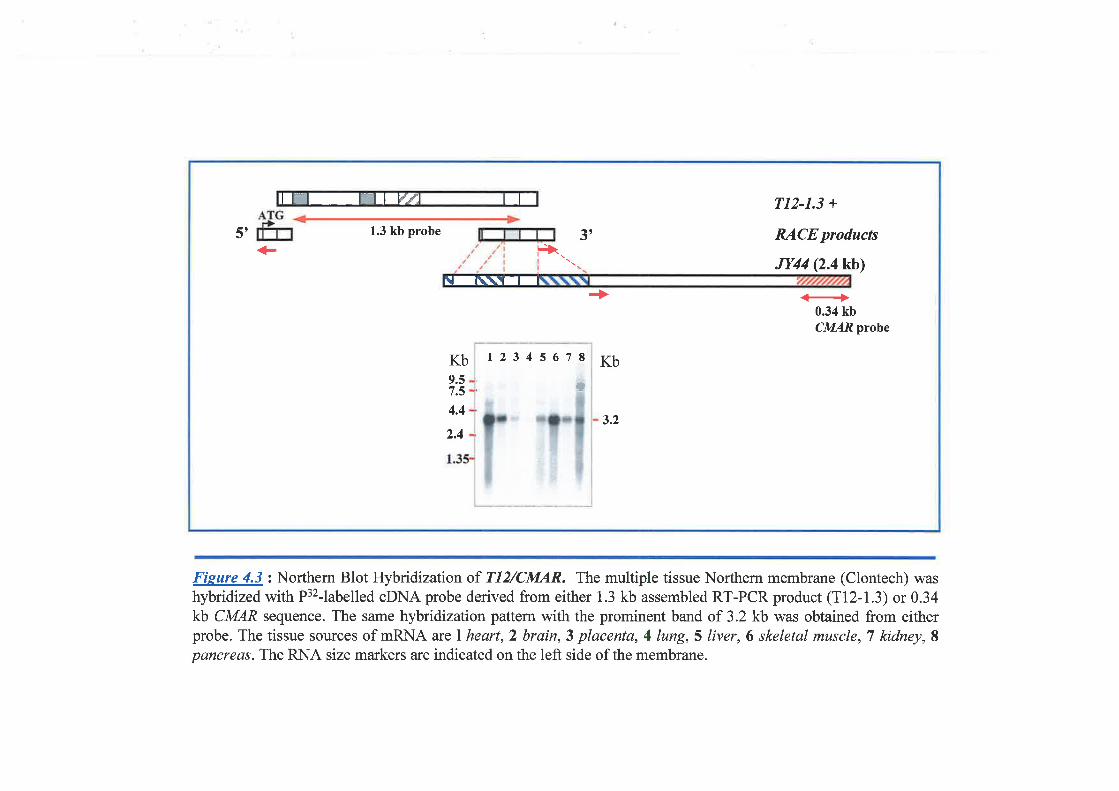
IT II IIflE IIt' 1.3 kb probe 3 ,
TI2-1.3 +
RACE products
JY44 Q.4kb)+ Ê>
+0.34 kbCMARprobe
Kb9.57.5
4.4
2.4
Kb
3.2
rtlt\TfI
1234s678
t
Fieure 4.3 : Northern Blot Hybridization of TI2/CMA,R. The multiple tissue Northern membrane (Clontech) washybridized with P32-labelled cDNA probe derived from either 1.3 kb assembled RT-PCR product (Tl2-1.3) or 0.34kb CMAR sequence. The same hybridization pattern with the prominent band of 3.2 kb was obtained from eitherprobe. Thetissue sources of mRNA arclheart,2 brain,3 placenta,4lung,S liver,6 skeletal muscle,T kidney,Spancreas. The RNA size markers are indicated on the left side of the membrane.
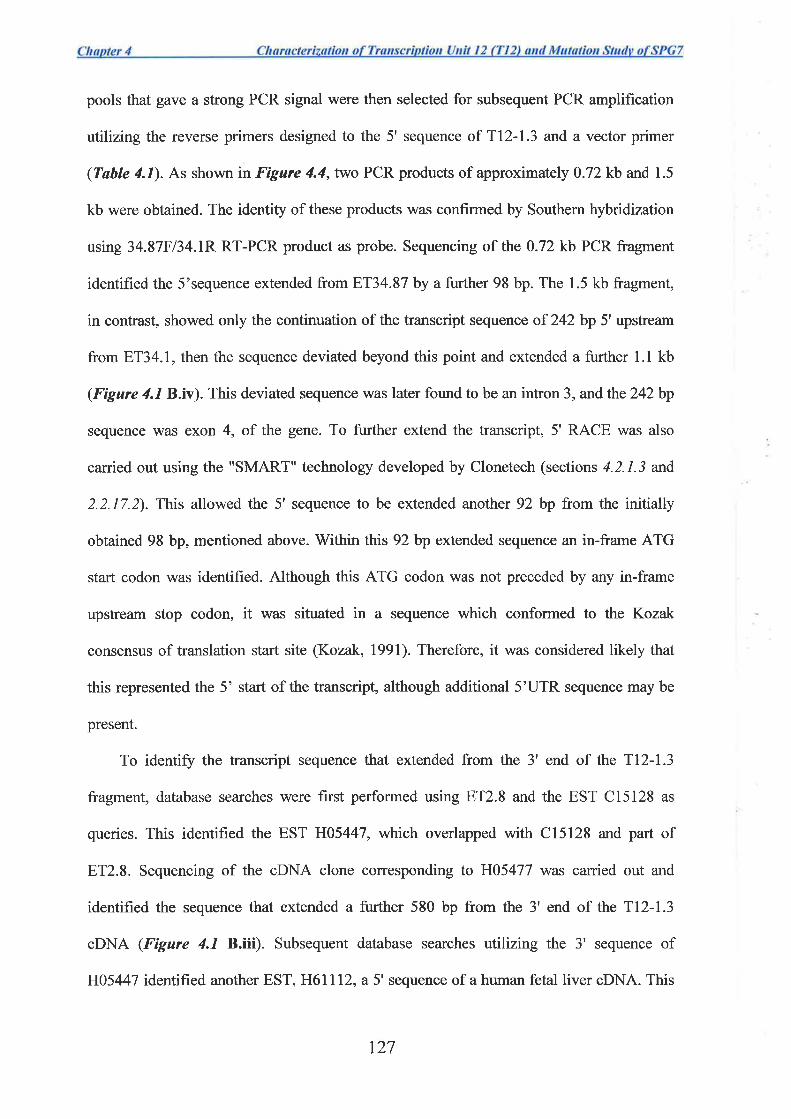
pools that gave a strong PCR signal were then selected for subsequent PCR amplification
utilizing the reverse primers designed to the 5' sequence of Tl2-1.3 and a vector primer
(Tøble /.1). As shown in Figure 4.4,two PCR products of approximately 0.72kb and 1.5
kb were obtained. The identity of these products was confirmed by Southern hybridization
using 34.87F134.1R RT-PCR product as probe. Sequencing of the 0.72 kb PCR fragment
identified the 5'sequence extended from 8T34.87 by a further 98 bp. The 1.5 kb fragment,
in contrast, showed only the continuation of the transcript sequence of 242 bp 5'upstream
from ET34.1, then the sequence deviated beyond this point and extended a further 1.1 kb
(Figure 4.1B.iv). This deviated sequence was later found to be an intron 3, and the242bp
sequence was exon 4, of the gene. To further extend the transcript, 5' RACE was also
carried out using the "SMART" technology developed by Clonetech (sections 4.2.1.3 and
2.2.17.2). This allowed the 5' sequence to be extended another 92 bp from the initially
obtained 98 bp, mentioned above. V/ithin this 92 bp extended sequence an in-frame ATG
start codon was identified. Although this ATG codon was not preceded by any in-frame
upstream stop codon, it was situated in a sequence which conformed to the Kozak
consensus of translation start site (Kozak, 1991). Therefore, it was considered likely that
this represented the 5' start of the transcript, although additional 5'UTR sequence may be
present.
To identiff the transcript sequence that extended from the 3' end of the T12-1.3
fragment, database searches were first performed using 8T2.8 and the EST C15128 as
queries. This identified the EST H05447, which overlapped with C15128 and part of
ET2.8. Sequencing of the cDNA clone corresponding to H05477 was carried out and
identified the sequence that extended a further 580 bp from the 3' end of the Tl2-1.3
cDNA (Figure /.1 B.iii). Subsequent database searches utilizing the 3' sequence of
H05447 identifred another EST, H6l172, a 5' sequence of a human fetal liver cDNA. This
t27
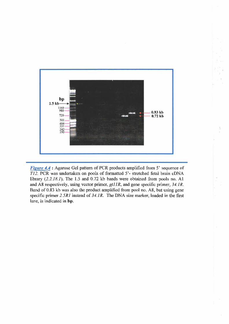
-.
1.5I 160980
720
bp
I
190
0.83 kb0.72kb
Figure 4.4 : Agarose Gel pattem of PCR products amplified from 5' sequence ofTI2. PCF. was turdertaken on pools of formatted 5'- stretched fetal brain cDNAlibrary (2.2.18.1). The 1.5 and 0.72 kb bands were obtained from pools no. A1and A8 respectively, using vector primer, gtl1R, and gene specifrc primer, 34.1R.Band of 0.83 kb was also the product amplifred from pool no. 48, but using gene
specific primer 2.5R1 instead of 34.1R. The DNA size marker, loaded in the firstlane, is indicated in bp.
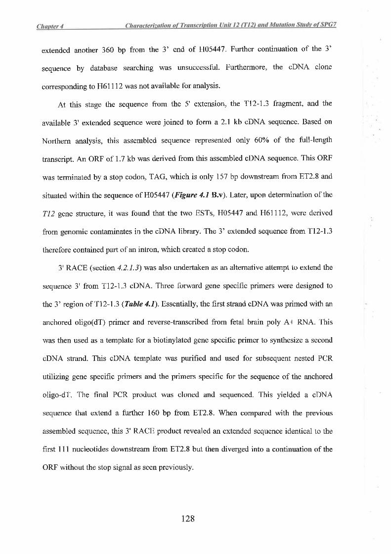
extended another 360 bp from the 3' end of H05447. Further continuation of the 3'
sequence by database searching was unsuccessful. Furthermore, the oDNA clone
corresponding to H6lll2 was not available for analysis.
At this stage the sequence from the 5' extension, the TI2-1.3 fragment, and the
available 3' extended sequence were joined to form a 2.1 kb cDNA sequence. Based on
Northern analysis, this assembled sequence represented only 600/o of the full-length
transcript. An ORF of 1.7 kb was derived from this assembled cDNA sequence. This ORF
was terminated by a stop codon, TAG, which is only 157 bp downstream from ET2.8 and
situated within the sequence of H05447 (Figure 4.1 B.v). Later, upon determination of the
712 gene structure, it was found that the two ESTs, H05447 and H61ll2, were derived
from genomic contaminates in the cDNA library. The 3' extended sequence from T12-1.3
therefore contained part of an intron, which created a stop codon.
3'RACE (section 4.2.1.3) was also undertaken as an alternative attempt to extend the
sequence 3' from Tl2-1.3 cDNA. Three forward gene specific primers were designed to
the 3' region of T12-1.3 (Table y'.1). Essentially, the first strand oDNA was primed with an
anchored oligo(dT) primer and reverse-transcribed from fetal brain poly A+ RNA. This
was then used as a template for a biotinylated gene specific primer to synthesize a second
oDNA strand. This cDNA template was purified and used for subsequent nested PCR
utilizing gene specific primers and the primers specific for the sequence of the anchored
oligo-dT. The final PCR product was cloned and sequenced. This yielded a cDNA
sequence that extend a further 160 bp from ET2.8. When compared with the previous
assembled sequence, this 3' RACE product revealed an extended sequence identical to the
first I 11 nucleotides downstream from ET2.8 but then diverged into a continuation of the
ORF without the stop signal as seen previously.
t28
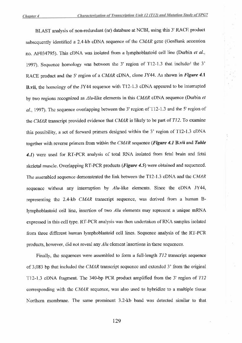
BLAST analysis of non-redundant (nr) database at NCBI, using this 3' RACE product
subsequently identified a2.4-kb cDNA sequence of the CMAR gene (GenBank accession
no. 4F034795). This cDNA was isolated from a lymphoblastoid cell line (Durbin et al.,
l9g7). Sequence homology was between the 3' region of Tl2-1.3 that includut\ the 3'
RACE product and the 5' region of a CMAR cDNA, clone JY44. As shown rn Fìgure 4.1
B.vii, the homology of the JY44 sequence with T12-1.3 cDNA appeared to be intemrpted
by two regions recognized as Alu-like elements in this CMAR cDNA sequence (Durbin ef
al.,1997). The sequence overlapping between the 3' region of T12-1.3 and the 5' region of
the CMARtranscript provided evidence that CMAR is likely to be part of TI2. To examine
this possibilþ, a set of forward primers designed within the 3' region of T12-1.3 cDNA
together with reverse primers from withinthe CMAR sequence (Figure 4.1Bxäand Table
4.1) were used for RT-PCR analysis of total RNA isolated from fetal brain and fetal
skeletal muscle. Overlapping RT-PCR products (Figure 4f) were obtained and sequenced.
The assembled sequence demonstrated the link between the Tl2-1.3 oDNA and the CMAR
sequence without any intemrption by Alu-like elements. Since the cDNA JY44,
representing the 2.4-kb CMAR transcript sequence, was derived from a human B-
lymphoblastoid cell line, insertion of Fwo Alu elements may represent a unique mRNA
expressed in this cell type. RT-PCR analysis was then undertaken of RNA samples isolated
from three different human lymphoblastoid cell lines. Sequence analysis of the RT-PCR
products, however, did not reveal arry Alu element insertions in these sequences.
Finally, the sequences were assembled to form a full-length Tl2 Lranscript sequence
of 3,083 bp that included the CMAR transcript sequence and extended 3' from the original
Tl2-1.3 cDNA fragment. The 340-bp PCR product amplifred from the 3' region of 712
corresponding with the CMAR sequence, was also used to hybridize to a multiple tissue
Northern membrane. The same prominent 3.2-kb band was detected similar to that
t29
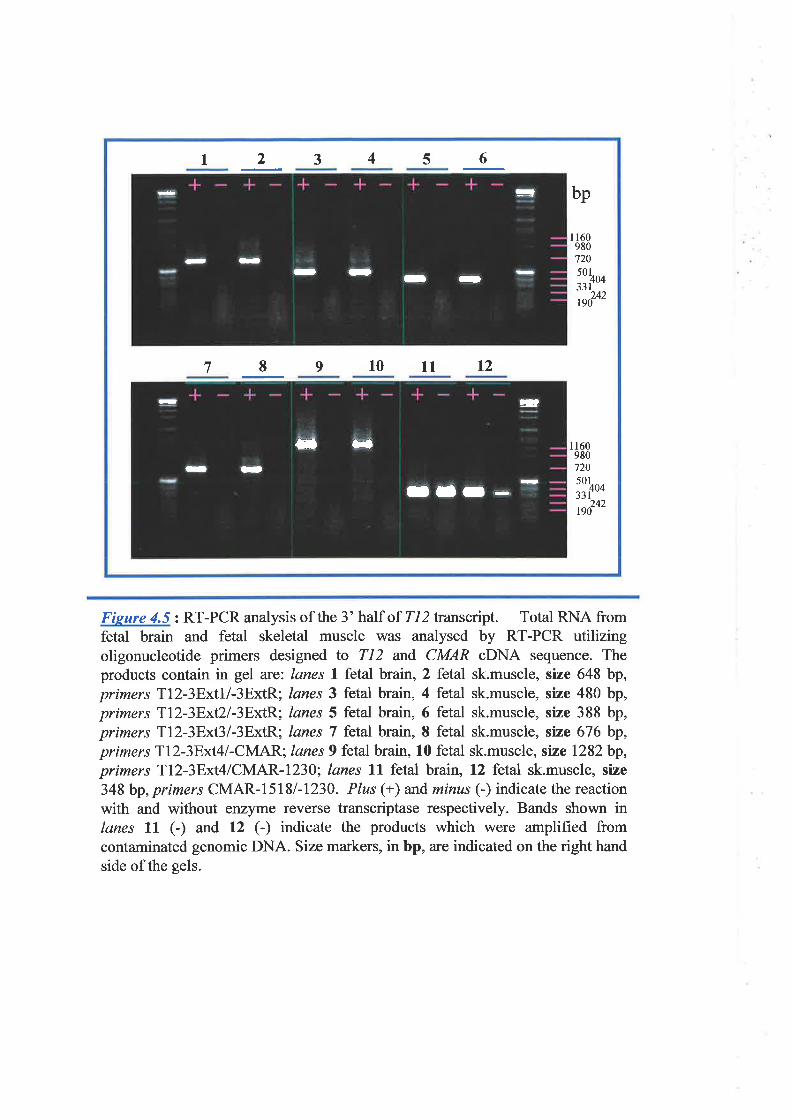
=t
t-D -a-
II
- rI
-llI -
- trt
-ra
-
¡l .a
4 62 3 5I
bp
I7 9101112
I 1609807205oLno331
242190
I 160980720501
zzfoo242
190
Fisure r'.S: RT-PCR analysis of the 3' half of 712 transcnpt. Total RNA fromfetal brain and fetal skeletal muscle was analysed by RT-PCR utilizingoligonucleotide primers designed to 712 and CMAR cDNA sequence. Theproducts contain in gel are:. lanes 1 fetal brain, 2 fetal sk.muscle, size 648 bp,primers Tl2-3Ext1/-3ExtR; lanes 3 fetal brain, 4 fetal sk.muscle, size 480 bp,primers Tl2-3Ext2/-3ExtR; lanes 5 fetal brain, 6 fetal sk.muscle, size 388 bp,primers Tl2-3Ext3/-3ExtR; lanes 7 fetal brain, I fetal sk.muscle, stze 676 bp,primers Tl2-3Ext4/-CMAR; lanes 9 fetal brain, 10 fetal sk.muscle, swe 1282bp,primers Tl2-3Ext4/CMAR-1230; lanes 11 fetal brain, 12 fetal sk.muscle, size
348 bp, primers CMAR-I5181-1230. Plus (+) and minus (-) indicate the reactionwith and without enzyme reverse transcriptase respectively. Bands shown inlanes ll (l and 12 (-) indicate the products which were amplified fromcontaminated genomic DNA. Size markers, in bp, are indicated on the right handside of the gels.
observed when the membrane was probed with the Tl2-1.3 oDNA (Fìgure 4.3).The result
of this Northern hybridization confirmed that CMAR was indeed part of Tl2.Therefote,
the reported2.4-kb CMAR cDNA sequence (JY44) was unlikely to exist as a separate gene.
This was shown in at least two fetal tissues, and three lymphoblastoid cell lines as
determined by RT-PCR, and eight adult tissues tested by Northern analysis.
Insertion of the Alu elements seen in the 2.4 kb version of the CMAR transcript, if
present in Tl2, would truncate up to 405 bp of the ORF by introducing an in-frame stop
codon. Although such Alu insertion was not detected in fetal tissues, lymphoblastoid cell
lines, and in adult tissues, this event may occur as a mechanism of gene inactivation in
tumor tissue. To test if this insertion is involved in breast cancer, RT-PCR of the Tl2
transcript was undertaken in RNA isolated from 12 breast cancer cell lines, using the set of
primers described in Tøhle y'.I. Sequencing of the RT-PCR products obtained from this
experiment did not reveal arry Alu element insertions nor sequence changes indicating
single base mutations. Therefore there was no evidence of T12 inactivation in breast cancer
arising from this mechanism.
4.3.2 T ranscript Sequence and Expression Analysis
The 712 transcript contains an ORF of 2385 bp that was predicted to code for a
protein of 795 amino acids (Fìgure 4.6). The ORF is followed by a 680-bp 3' UTR, with
polyadenylation signal, AATAAA, located 20 bp upstream from the polyadenylation site,
as confirmed by alignment of the transcript sequence against the genomic sequence
obtained by direct sequencing of cosmid 383H6 encompassing the CMAR region. . V/ithin
the 3' UTR an insertion/deletion polymorphism of CACA was frequently observed among
the RT-PCR products and genomic sequence. The CACA insertion/deletion polymorphism
'a
130
Fieure 4.6 : Nucleotide transcript and deduced amino acid sequence of n26PG7\ Thetranscript codes for a protein of 795 amino acid residues. Nucleotide and amino acid positionsare indicated by numbers on the right hand column. The adenine (A) nucleotide of the startcodon is given as the position "l" of the transcript. The stop codon at positions 2386-2388 isdenoted by asterisk undemeath. Polyadenylation signal AATAJA'{ is indicated in blue atpositions 3040-3045. Within the 3' UTR the short region of insertiorVdeletion pol¡rmorphismof CACA is underlined. The transcript is coded for by 17 exons of the gene. The exonboundaries are indicated by the vertical lines with double-head ¿urows and given numbers ofexons. The identity of each trapped product is indicated above its corresponding exon.
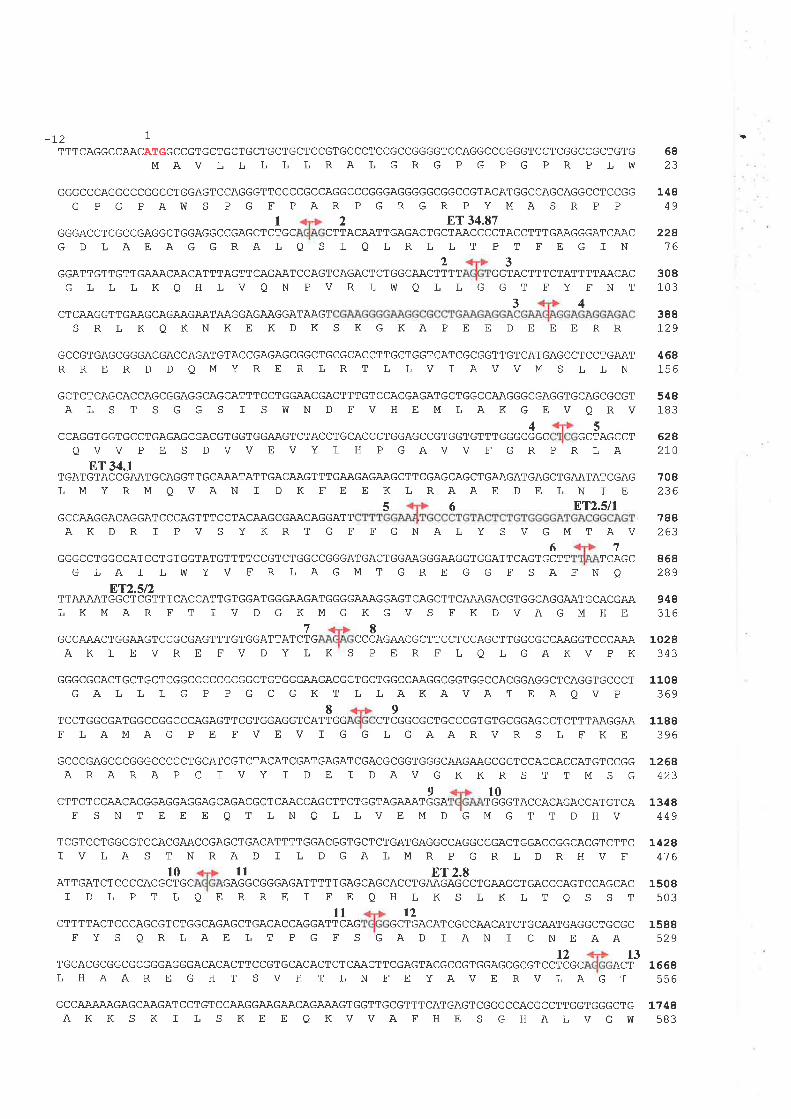
-L2 1
TTTCAGGCCAACATGGCCGTGCTGCTGCTGCTGCTCCGTGCCCTCCGCCGGGGTCCAGGCCCGGGTCCTCGGCCGCTGTGMAV L L L L L RAL G R G P G P G P R P LúT
GGGCCCAGGCCCGGCCTGGAGTCCAGGGTTCCCCGCCAGGCCCGGGAGGGGGCGGCCGTACATGGCCAGCAGGCCTCCGG
G P G PAIV S P G F PAR P G R GR P YMA S R P P.,
GC CTTGD LAEAGGRALQ S L QLRLLT PT FE G IN
GGArrcrrGrrcNU\cAr\cATTTAGTrcAGAArc.AGTcAGAcrcrcGcAAarlr, ""lo"rrrarArrrrAACACGLL LKQHLVQN PVRLWQ LLG GT FY FNT
IGGGACCTCGCCGAGGCTGGAGGCCGAGCTCT
CT CAAG GTT GAAG CAGAAGAATAAGGAGAAGGATAAGT
ET 34.87ACAATTGAGACTGCTAACCCCTACCTTTGAAGGGATCAAC
6823
14849
228't6
308103
388129
468156
548183
628270
708236
788263
868289
948316
1028343
43
S RLKQKNKE KDKS KGKAPEE DEEERR
GCCGTGAGCGGGACGACCAGATGTACCGAGAGCGGCTGCGCACCTTGCTGGTCATCGCGGTTGTCATGAGCCTCCTGAATRRE RD DQMYRE RLRT LLV I AVVM S LLN
GCTCTCAGCACCAGCGGAGGCAGCATTTCCTGGAACGACTTTGTCCACGAGATGCTGGCCA.A.GGGCGAGGTGCAGCGCGTAL S T S G G S I S ù]N D FVH E M L AK G E V Q RV
45CCAGGTGGTGCCTGAGAGCGACGTGGTGGAAGTCTACCTGCACCCTGGAGCCGTGGTGTTTGGGCGGC GCTAGCCT
QVV P E S DVVE VY L H P GAVV F GR PR LAET 34.1
TGATGTACCGAATGCAGGTTGCAAATATTGACAAGTTTGAAGAGAAGCTTCGAGCAGCTGAAGATGAGCTGAATATCGAGLMYRMQVAN I D K FE E KLRAAE DE LN I E
5 6 ßT2.5nGCCAAGGACAGGAÎCCCAGTTT CCTACAAGCGAACAGGATTAKDR I PV S YKRT G F FGNALY S VGMTAV
67GGGCCTGGCCATCCTGTGGTATGTTTTCCGTCTGGCCGGGATGACTGGAAGGGAAGGTGGATTCAGTGCTT TCAGC
G LA I LVü YV F R LAG M T G R E G G F S AFN O
ET2.5t2TTAÄ.AATGGCTCGTTTCACCATTGTGGATGGGAAGATGGGGAAAGGAGTCAGCTTCAÄAGACGTGGCAGGAATGCACGAALKMAR FT I V DGKMGKGV S FK DVAGMHE
78GCCAAACTGGAAGTCCGCGAGTTTGTGGATTATCTG CCCAGAACGCTTCCTCCAGCTTGGCG CCAAGGTCCCAÄA
AKVPK
GGGCGCACTGCTGCTCGGCCCCCCCGGCTGTGGGAAGACGCTGCTGGCCAAGGCGGTGGCCACGGAGGCTCAGGTGCCCT
GALL LG P PGCGKT LLAKAVAT EAQVP
T CCTGGCGATGGCCGGCCCAGAGTTCGTGGAGGTFLAMAGPEFVEV L
GCCCGAGCCCGGGCCCCCTGCATCGTCTACATCGATGAGATCGACGCGGTGGGCAAGAAGCGCTCCACCACCATGTCCGGARARAP C I VY I DE I DAVGKKR S TTMS G
910CTTCTCCAACACGGAGGAGGAGCAGACGCTCAACCAGCTTCTGGTAGA.AATGGA TGGGTACCACAGACCATGTCA
FSNT EE E QT LNQLLVEMDGMGTT DHV
TCGTCCTGGCGTCCACGAACCGAGCTGACATTTTGGACGGTGCTCTGATGAGGCCAGGCCGACTGGACCGGCACGTCTTCI VLAS TNRAD I L DGALMRP GRLDRHVF
l0 il 8T 2.8ATTGATCTCCCCACGCTGC GAGGCGGGAGATTTTTGAGCAGCACCTGAAGAGCCTGAAGCTGACCCAGTCCAGCAC
I DL PTLQERRE I FEQHLKS LKLTQS STll t2
CTTTTACTCCCAGCGTCTGGCAGAGCTGACACCAGGATTCAG GCTGACATCGCCAACATCTGCAATGAGGCTGCGCFYS QRLAE LT P G FS GAD IAN T CNEAA
t2 13TGCACGCGGCGCGGGAGGGACACACTTCCGTGCACACTCTCAACTTCGAGTACGCCGTGGAGCGCGTCCTCGC ACTLHAARE GH T SVH T LN FEYAVE RVLAGT
GCCAAÃAAGAGCAAGATCCTGTCCAAGGAAGAACAGAAAGTGGTTGCGTTTCATGAGTCGGGCCACGCCTTGGTGGGCTGAKK S K T L S KE E QKVVAFHE S GHALVGW
AKLEVRE FVDYLKS PERFLQLG
8CATTGG CT
9CGGCGCTGCCCGTGTGCGGAGCCT CTTTAAGGAA
GAARVRSLFKE
1108369
1188396I
L26A423
13¿8449
L42841 6
1508503
1588529
1668556
L748sB3
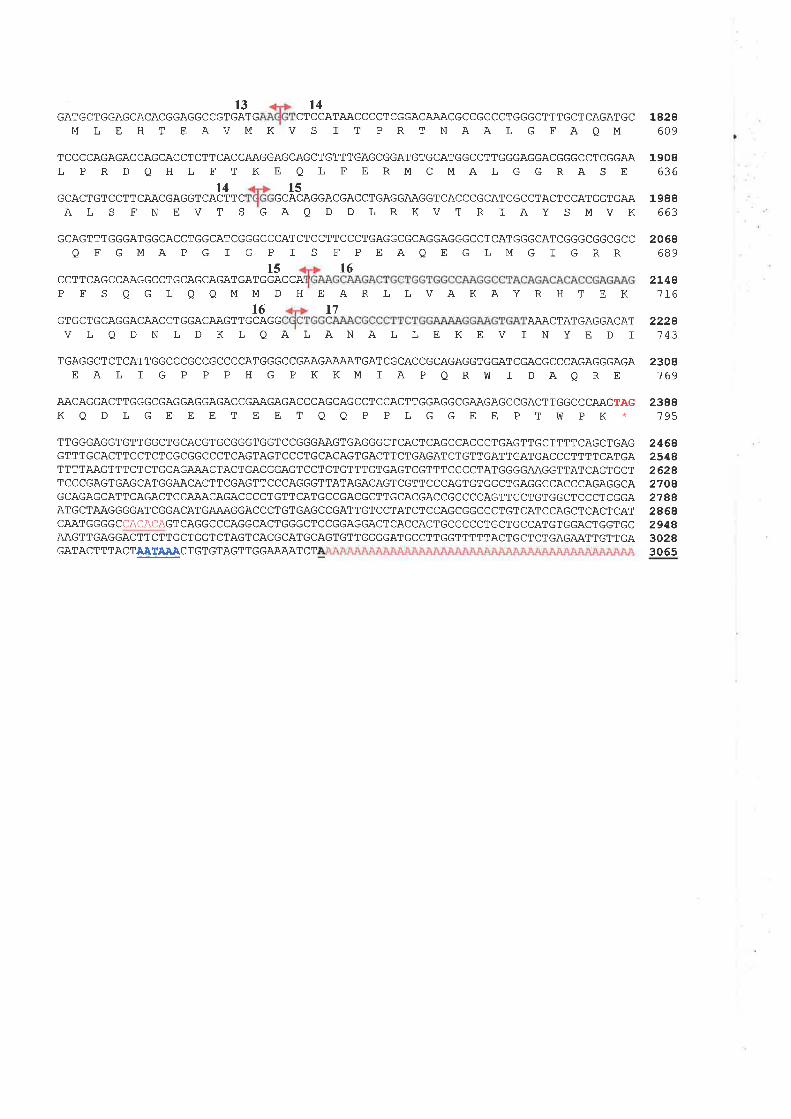
13 t4GATGCTGGAGCACACGGAGGCCGTGATG CTCCATAACCCCTCGGACAAACGCCGCCCTGGGCTTTGCTCAGATGC
M L E H T EAVMKV S I T PRT NAA L G FAQM1828
609
1908636
1988663
t4 15GCACTGTCCTTCAACGAGGTCACTTC GCACAGGACGACCTGAGGAAGGTCACCCGCATCGCCTAL S FN EVT S GAQ D D LRKV T R I A
TCCCCAGAGACCAGCACCTCTTCACCAAGGAGCAGCTGTTTGAGCGGATGTGCATGGCCTTGGGAGGACGGGCCTCGGAAL P R D QH L FT KE Q L FERMCMAL G GRAS E
ACTCCATGGTGAAYSMVK
GCAGTTTGGGATGGCACCTGGCATCGGGCCCATCTCCTTCCCTGAGGCGCAGGAGGGCCTCATGGGCATCGGGCGGCGCC
Q FGMAPG I G P I S F PEAQEGLMG I GRRl5 l6
CCTTCAGCCAAGGCCTGCAGCAGATGATGGACCAPFSQGLOOMMDHEARLLVAKAYRHTEK
2068689
2L48116
2228743
2308769
t6GTGCTGCAGGACAACCTGGACAAGTTGCAGG
l7AAACTATGAGGACAT
V L Q D N L D K L QA ], ANAL L E K E V I N Y E D I
TGAGGCTCTCATTGGCCCGCCGCCCCATGGGCCGAAGA.A.AATGATCGCACCGCAGAGGTGGATCGACGCCCAGAGGGAGAE A I I G P P P H G P KKM I A P Q Rh] I DAQ RE
AACAGGACTTGGGCGAGGAGGAGACCGAAGAGACCCAGCAGCCTCCACTTGGAGGCGAAGAGCCGACTTGGCCCAAGTÀGKQDLGEEETEETOO P PLGGEE PTWPK *
TTGGGAGGTGTTGGCTGCACGTGCGGGTGGTCCGGGAAGTGAGGGCTCACTCAGCCACCCTGAGTTGCTTTTCAGCTGAGGTTTGCACTTCCTCTCGCGGCCCTCAGTAGTCCCTGCACAGTGACTTCTGAGATCTGTTGATTGATGACCCTTTTCATGATTTTAAGTTTCTCTGCAGAAACTACTGACGGAGTCCTGTGTTTGTGAGTCGTTTCCCCTATGGGGA.AGGTTATCAGTGCTTCCCGAGTGAGCATGGAACACTTCGAGTTCCCAGGGTTATAGACAGTCGTTCCCAGTGTGGCTGAGGCCACCCAGAGGCAGCAGAGCATTCAGACTCCAAACAGACCCCTGTTCATGCCGACGCTTGCACGACCGCCCCAGTTCCTGTGGCTCCCTCGGAATGCTAAGGGGATCGGACATGAAAGGACCCTGTGAGCCGATTGTCCTATCTCCAGCGGCCCTGTCATCCAGCTCACTCATCAATGGGGCCACACAGTCAGGCCCAGGCACTGGGCTCCGGAGGACTCACCACTGCCCCCTGCTGCCATGTGGACTGGTGCAAGTTGAGGACTTCTTGCTGGTCTAGTCACGCATGCAGTGTTGGGGATGCCTTGGTTTTTACTGCTCTGAGAATTGTTGAGATACT T TACTAATAÀACTGT GTAGT TGGAÄAAT C T
2388195
24642548262827042788246829483028306s
lon '/
hgle alsobeen described in the previous reports of CMAR (Koyama et al.,1993;Durbin ¿f x
al.,1994).
In addition to the expression study in multiple tissue Northernf tissue distribution of \
212 mRNA expression was also undertaken. A cDNA probe derived from 3' UTR of 212
was used to hybridize to a human mRNA Master Blot from 50 different fetal and adult
tissues (Clontech). As shown in Fígure 4.7, expression was high in a series of tissues from
the central neryous system: in the amygdal4 caudate nucleus, thalamus, subthalamic
nucleus, and spinal cord. Intermediate expression was observed in the frontal lobe of the
cerebrum, the hippocampus, ,faluoblongat4 and putamen, with the lowest expression
in the substantia nigra. Compared with adult brain, heart, and thymus, the corresponding
fetal tissues showed a lower expression. In contrast, the level of expression was
approximately the same in fetal kidney and liver when compared with the corresponding
adult tissues. A high level of mRNA was present in adrenal gland and salivary gland, with
a very low expression in maÍrmary gland and testis
4.3.3 Protein Sequence Identity
With the complete TI2 arrino acid sequence of 795 residues, homology searches of
protein database using the BLASTP algorithm identified homology to a diverse group of
proteins which were member of the AJqA protein family QlTPases 4ssociated with diverse
cellular qctivities). The most striking similarity was observed with the yeast mitochondrial
proteins, Afg3p and Rcalp (both at 55% amino acid identity), and with a lesser similarity
to Ymelp (52% identity). Afg3p, Rcalp and Ymelp are metalloproteases, situated in the
inner membrane of mitochondria and exhibit both proteolytic and chaperone-like functions
(Rep and Grivell, 1996; Patel and Latterich, 1998). V/ithin the TI2 protein, which is
similar to those of yeast, an ATP-binding motif was observed between residues 349 and
131
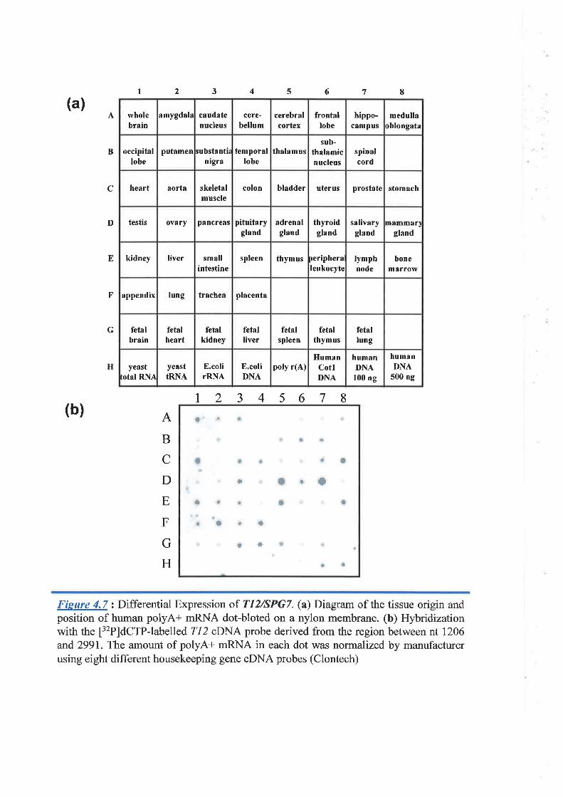
I 2 3 4 5 6
12345678
7 8
(a)
(b)
A
B
C
D
E
F
G
A
B
C
D
E
F
G
H
o' ¡
¡
a + I
I arfo+ I a
rlri
rO
I I
Fieure 4.7: Differential Expression of T12/SPG7. (a) Diagram of the tissue origin andposition of human polyA+ mRNA dot-bloted on a nylon membrane. (b) Hybridizationwith the [32P]dcTP-labelled 212 oDNA probe derived from the region between nt 1206and 2991. The amount of polyA+ mRNA in each dot was normalized by manufacturerusing eight different housekeeping gene cDNA probes (Clontech)
wholebrain
amygdala caudatenucleus
cere-bellum
cerebralcortex
frontallobe
hippo-câmpus
medullaoblongata
occipitallobe
putamen substanti¿nigra
temporallobe
thalamussub-
thalamicnucleus
spinalcord
heart aorta skeletalmuscle
colon bladder uterus prostate stomach
testis ovary pancreâs pituitarygland
adrenalgland
thyroidgland
salivarygland
mâmmarlgland
kidney Iiver smallintestine
spleen thymus peripheraleukocyte
lymphnode
bonemarrolY
appendix lung trachea placenta
fetalbrain
fetalheart
fetalkidney
fetalliver
fetalspleen
fetalthymus
fetallung
yeast:otal RNA
yeasttRN.A,
E.colirRNA
E.coliDN,À
poly r(A)Human
CotlDNA
humanDNA
100 ng
humanDNÀ
5ü) ng
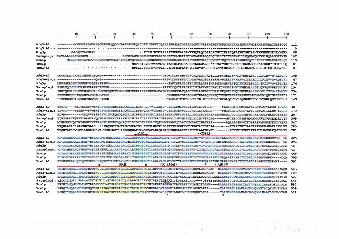
Afg3-I.2Afg3-LluusAfg3pParap]-eginRcalpYoêlpTnel-L1
Af93-I-2Afg3-IJlrugAfg3pPalapleginRcalpYnê1pYnel-f,1
.Afg3-L2Afg3-LlausAfS3pPaleplegínRcalpYEelpYDê1-L1
Àfg3-L2Afg3-L1ulsAfg3pParapleginRcalpYnelpYEê1-L1
Afg3-r'2Afg3-L14rgÀfg3pParapleginRcalpYûê1PYnel-I,1
10 20 30 40 50 60 '70 80 90 100 110 120
-----SLTSLSFGKÀSRISTVKP\ULRSRMPVIIQR¡{TIJSGLATRNTII¡RSTQIRSFHISY|ITRLT\IENRPITIKEGEGKNNGNK
--------M}TVSKII,VSPlr\,1tTI[v:LRIFÀPRLPQIGASLLVQKK9IA¡RSKKFYRffSEIWSGEMPPKKEJADSSGKASNKSTISSIDNSQ--------MFSISS LSHLTNÀ¡'HTPKNTSVSI,SG\¡SVSQNQHRDV\ZPEHE]APSSEPSLNLRDIGLSELKIGQIDQI.VENI,
GGGGGKRGGKKDDS R¡'QKG------ ----DIPVIDDKDERMEELT{EÀIFI¡CGIA{FTI¡LIJKRSGREIT9¡KDFVNMTLSKGVVDRLEV\Ni¡-KREVRV
T,PGFCKGKNISSE9IHTSEVSÀQSFE'EIN------ -------------KT TESTIJA,SSCIYR-HHSRàT.QSTCSDLQYWPVFTQSRGFIST.KSRTRRT'QSTSERL-A
YLHPGAWFGRPRÍ.AT¡dR¡4QVA¡IIDKE EEKLRAAEDEINIE,NORIPVSTKRTG¡I' ------¡ÆGREGCFSA¡IIQLK}'ERFITVDGKMGKG\¡SF
VRQNLLTÀSSAGA\'NPSI,AS--SSSNQSGTEGNE'PSMTSPLYGS-RKEPÍ.¡ÍWVSESTETT/VSRW\ZKWI.LI/FGTLIYSE'SEe--------!'!çITTENTTT,T,KSSEVAD!(SVDI/A¡(IAIVKFETPN!àPSFVKGFIiI.RDRCSD¡/ESI,DKLMKTKNIPTASgDÀ¡X,TGEAECEI,KAgALTQKTI{DSI,RRTRLILF\ZTT,LFGIYGI,--------L¡(NPET,SVRE:RTTTGLDSA\IDPVìqMKIiIVTF
- ArP - <G3B6S>
SRH -+ <R485deÞ ' <4510V>
EEEQTINQI,LVEMDGMGTTDIÍ\':TVI,ÀSTNRADILDGAI;MRPGRI,DRE\ZEIDI.PTI,OERREIFTQHLKSLKLT----QSSTI':IS LTPEE'SGADIÀ¡{IqNEJAÀJ,gÀÀREGBTSV'HTL
O¡
1110
88t02115
8181
19861
]-76194234171169
307L732873083472AO
2AL
42529L401421466395391
543409527543582509511
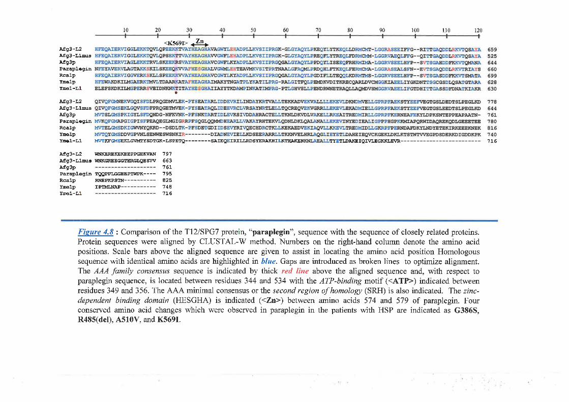
Afg3-l,2Afg3-L1auEAfg3pParapleginRcalpYEelpYEê1-L1
afg3-L2Àfg3-LlnrsÀfg3pParap].eginRcalpYDê1PTEêl-L1
10 20 30 40 50 60 70 80 90 100 ll0 r20
<K56gr> <zn>
a
M\¡TKFGMSEKLGVI'ITYSDTGK-ISPETQ--------SAIE9EIRILI,RDSYERATGILICT¡TAKEH TTETI.DA¡(EIQI\TLEGKKLEVR 1L6
Afg3-L2 Í{NKEREKEKEEPPGEK\'AIiI 791.àfg3-f,luus IìINKGREEGGTERGLQESPV 663Afg3pParaplegin IQQPPLGGEEPrÍ{PK- ---Rcalp RNEPKPSII¡--Ynelp IPIÎ'ÍI¡IAP---Ymel-f,1
161-1958251481L6
Fieure 4.8 : Comparison of the TLZ|SPGT protein, "paraplegin", sequence with the sequence of closely related proteins.Protein sequences were aligned by CLUSTAL-V/ method. Numbers on the right-hand column denote the amino acidpositions. Scale bars above the aligned sequence are given to assist in locating the amino acid position Homologoussequence with identical amino acids are highlighted in blue. Gaps are introduced as broken lines to optimize alignment.The AAA family consensus sequence is indicated by thick red line above the aligned sequence and, with respect toparaplegin sequence, is located between residues 344 and 534 with the ATP-binding motif (<ATP>) indicated betweenresidues 349 and 356. The AJA-A. minimal consensus or the second region of homologt (SRII) is also indicated. The zinc-dependent binding domain (HESGHA) is indicated (<Zn>) between amino acids 574 and 579 of paraplegin. Fourconserved amino acid changes which were observed in paraplegin in the patients with HSP are indicated as G386S,R485(del), A5l0V, and K569I.
Chonier 4 Ch of Transcriotion l2 ff|2) and Mulafìott nf ,lPG7
356, and the AAA family consensus sequence located between residues 344 and 534. The
zinc-dependent binding domain (HESGHA) was found between amino acids 574 arld 579.
The protein \ocalization prediction by the program PSORT suggested a mitochondrial
localization. Two transmembrane segments of T12 were also predicted between amino
acids 144 and 762, and between amino acids 249 and272, which were situated within the
less conserved N-terminal region. Sequence alignment of Tl2 protein and its yeast
homologue as well as human paralogue is presentedin Figure 4-8.
At the completion of this work Casari et al (1998) reported an identical gene called
,,SPG|', that was involved in an autosomal recessive type of hereditary spastic paraplegia
(HSP), a neurodegenerative disorder. The protein product of SPGT was designated
"paraplegin".
4.3.4 Genomic Organ tzztion
Since the report of Casari et al (1998) did not provide data of gene structure of SPGT
and it was not present in the public database, characterization of the gene structure was
,therefore,,undertaken.
Because the complete genomic sequence encompassing this region was not available
at this time, the gene structure was charactenzed by PCR amplification using exon
sequences of the genomic clones and direct sequencing (section 4.2-4). Alignment of the
transcrþt sequence with this resulting genomic sequence determined that the SPGT gene
consists of 17 exons ranging in size between 78 and 242 bp. This gene encompasses
genomic DNA of approximately 52 kb (Figure r'.9) which is contained within three
overlapping cosmids: 383H6, 427Bll, and 358D12. These three cosmids are part of a
cosmid contig of the physical map established at chromosome region 16q24.3 (Whitmore
et al., 1998a). As shown in the Tøble 4.3, all splice junction boundaries conform to the
t32
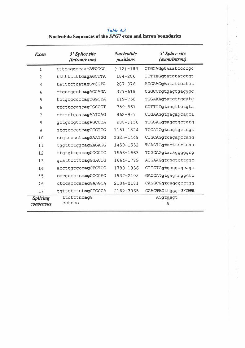
Table 4.3Nucleotide Sequences of the SPGT exon and intron boundaries
Exon 3'Splìce síte(íntron/exon)
Nucleotídepositions
5'Splice site(exon/íntron)
1
2
3
4
5
6
1
I9
10
11
72
13
t415
76
71
tttcaggccaacATGGCC
tttttttttcagAGCTTAtatttctcatagGTGGTAcl.gccggctcagAGGAGA
tctgccccccagCGGCTA
ttcttccggcagTGCCCT
ctttctgcacagAATCAGgctgccgtccagAGCCCA
gtgtcccctcagGCCTCG
ctgtcccctcagGAATGG
tggttctggcagGAGAGG
ttgt.gttgacagGGGCTG
gcattctttcagGGACTG
accttgtgccagGTCTCC
cccgccctccagGGGCAC
ctccactcacagGAAGCA
tgttctttctagCTGGCA
CTGCAGgtaaatccccgc
TTTTAGgtatgtatctgtACGAAGgtatattcatct
CGGCCTgtgagtgagggc
TGGAAAgtatgttggatg
GCTTTTgtaagttctgta
CTGAAGgfgagagcagca
TTGGAGgtaggtgctgtg
TGGATGgtcagtgctcgt
CTGCAGgtcagagccagg
TCAGTGgtacttcctcaa
TCGCAGgtacagggggcg
ATGAAGgtgggtcttggc
CTTCTGgfgaggagcagc
GACCATgtgagtcggctc
CAGGCGgtgaggccctgg
CAAGTAGItggg-3'UIER
(-12 ) -183IB4-286
2B'7 -31 6
317 -6J.8
61 9-7 5B
759-8 61
862-987
98 8-1 150
7L5r-'l.324
L325-r449
1 4 50-1552
1s53-1 663
]-664-1-719
1780-1936
!937-21032ro4-2rgt2r82-306s
Splicingconsensus
ttctttncagGcctccc
AGgfaagts
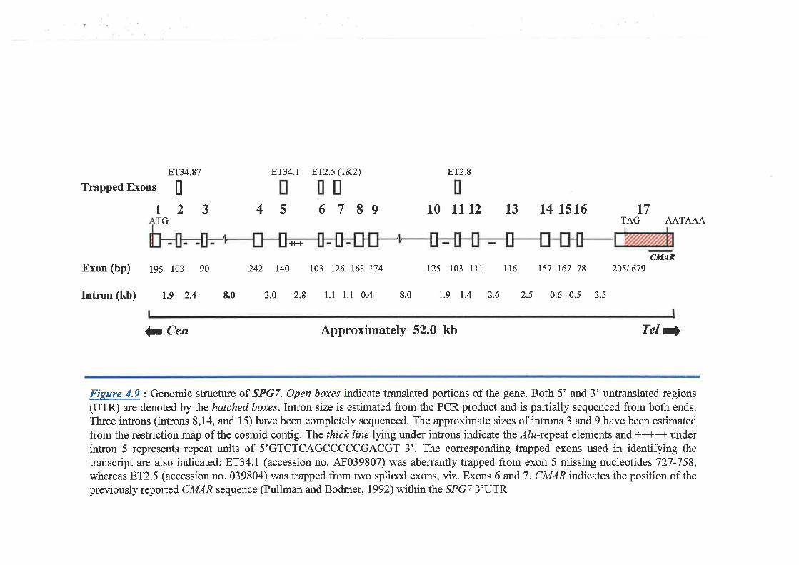
123 4 5
l.tttt
Exon (bp) ßs lo3 90 242 r40 103 t26 163 174
Intron (kb) t.e 2.4 8.0
ET2.8
6
l7 89 10 tlt2 13 14 t5t6
125 103 111 116 r57 167 78 2051679
F'T34.87 ET34.t ET2j 0e2)Trapped Exons I
TGt7
TAG AATAAA
CMAR
2.0 2.8 1.1 1.1 0.4 8.0 t.9 1.4 2.6 2.5 0.6 0.5 2.5
I I I I
Ç Cen Approximately 52.0 kb Tel )
Fieure 4.9: Genomic structure of SPG7. Open boxe,s indicate translated portions of the gene. Both 5' and 3' untranslated regions(UTR) are denoted by the hatched boxes.Intron size is estimated from the PCR product and is partially sequenced from both ends.
Three introns (introns 8,14, and 15) have been completely sequenced. The approximate sizes of introns 3 and t have been estimatedfrom the restriction map of the cosmid contig. The thick line Iying under introns indicate the Alu-repeat elements and +++++ underintron 5 represents repeat units of 5'GTCTCAGCCCCCGACGT 3'. The conesponding trapped exons used in identifting thetranscript are also indicated: ET34.l (accession no. AF039807) was aberrantly trapped from exon 5 missing nucleotides 727-758,whereas 8T2.5 (accession no. 039804) was trapped from two spliced exons, viz. Exons 6 and 7 . CMAR indicates the position of thepreviously reported CMAR sequence (Pullman and Bodmer, t992) within the SPGT 3'UTR
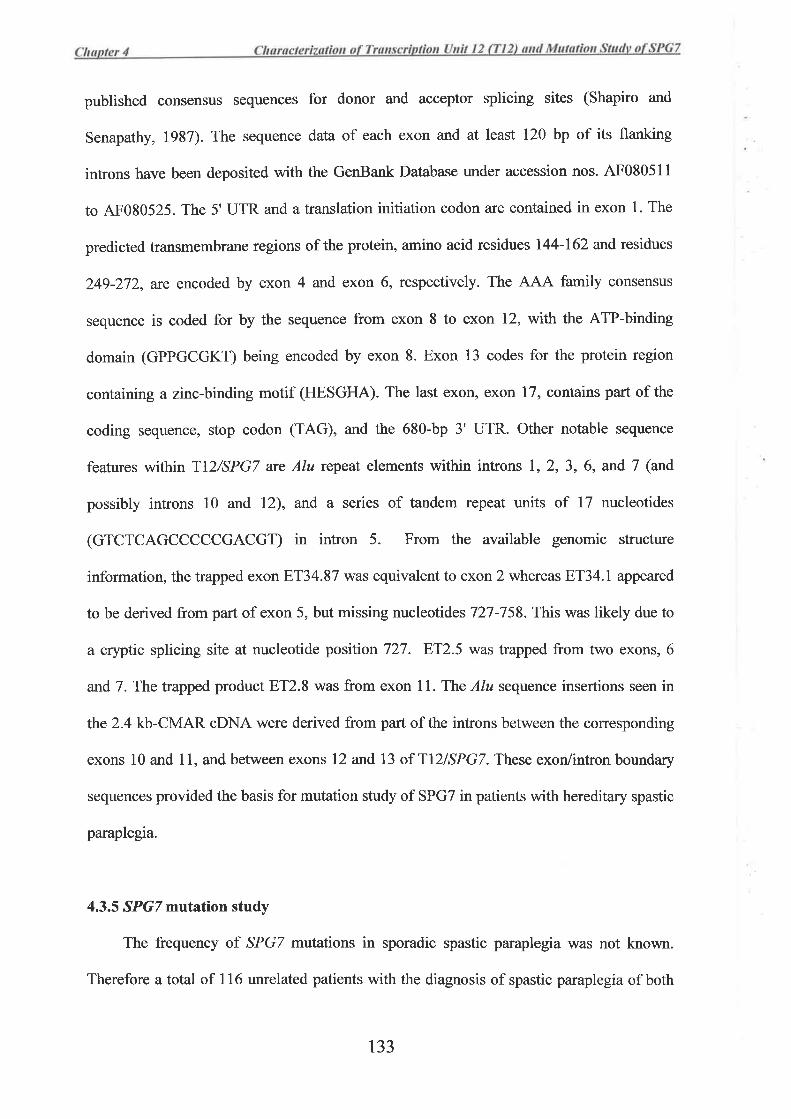
published consensus sequences for donor and acceptor splicing sites (Shapiro and
Senapathy, 1937). The sequence data of each exon and at least 120 bp of its flanking
introns have been deposited with the GenBank Database under accession nos. 4F080511
to AF080525. The 5' UTR and a translation initiation codon are contained in exon 1. The
predicted transmembrane regions of the protein, amino acid residues 144-162 and residues
249-272, are encoded by exon 4 and exon 6, respectively. The A.A{ family consensus
sequence is coded for by the sequence from exon 8 to exon 12, with the ATP-binding
domain (GPPGCGKT) being encoded by exon 8. Exon 13 codes for the protein region
containing a zinc-binding motif (HESGHA). The last exon, exon 17, contains part of the
coding sequence, stop codon (TAG), and the 680-bp 3' UTR. Other notable sequence
features within T|2/SPG7 arc Alu repeat elements within introns 1,2, 3, 6, and 7 (and
possibly introns 10 and l2), and a series of tandem repeat units of 17 nucleotides
(GTCTCAGCCCCCGACGT) in intron 5. From the available genomic structure
information, the trapped exon 8T34.87 was equivalent to exon 2 whereas ET34.1 appeared
to be derived from part of exon 5, but missing nucleotidesT2T-758. This was likely due to
a cryptic splicing site at nucleotide position 727. 8T2.5 was trapped from two exons, 6
and7. The trapped product 8T2.8 was from exon 11. The Alu seqtence insertions seen in
the 2.4 kb-CMAR cDNA were derived from parl of the introns between the corresponding
exons l0 and 11, and between exons 12 and 13 of Tl2lSPG7. These exon/intron boundary
sequences provided the basis for mutation study of SPGT in patients with hereditary spastic
paraplegia.
4.3.5 SPGT mutation study
The frequency of SPGT mutations in sporadic spastic paraplegia was not known.
Therefore a total of I 16 unrelated patients with the diagnosis of spastic paraplegia of both
133
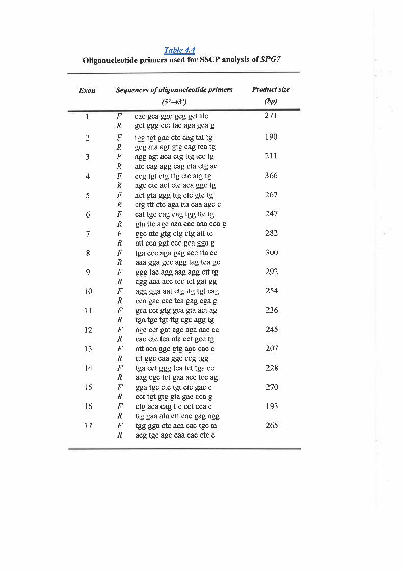
Tsble 4.4Oligonucleotide primers used for SSCP analysis ol SPG7
Exon Sequences of oligonucleolide primers
(5'-+3')
Product size
(bp)
1 F cac gca ggc gcg gct ttcl? gct ggg ccttac agagcag
F tggtgt gac ctc cagtattgi? gcgataagt gtg cagtcatgF agg agt aca ctg ttg tcc tg.R atc cagaggcag cta ctg ac
F ccgtgt ctgttg ctc atgtgR agc ctc act ctc aca ggc tgF act gþ ggg ttg ctc gtc tgA ctg ttt ctc aga tlacazagc c
F cat tgc cag cag tgg ttc tgR gta ttc agc aaa cac aaa cca g
F ggc atc gtg ctg ctg att tcR attcca ggt ccc gcaggagF tga ccc aga gag acc tta ccR aaø gga gcc aggtag tca gc
F gggtac aggaag agg ctt tgR cggaaa, acc tcc tct gat gg
F agg gga aat ctgttg tgt cagR cca gac cactcagagcgagF gca cct gtg gca gla act ag
A tga tgc tgt ttg cgc agg tgF agc cct gat agc aga aac ccR cac ctc tcaatacct gcc tgF atl aca ggc gtg agc cac cl? ttt ggc caaggc ccgtggF tga cct gggtcatct tga ccA aag cgctct gaøacc tcc ag
F ggatgc ctc tgt ctc gac cR cct tgt É¡g gta gac cca g
F ctgaca cag ttc cct cca cR ttg gaa ata ctt cac gag agg
F tgg gga ctc aca cac tgc taR acgtgc agcçaacac ctc c
271
190
2tt
366
267
247
282
300
292
254
236
245
207
228
270
193
265
2
J
4
5
6
7
8
9
10
11
l2
13
t4
15
l6
t7
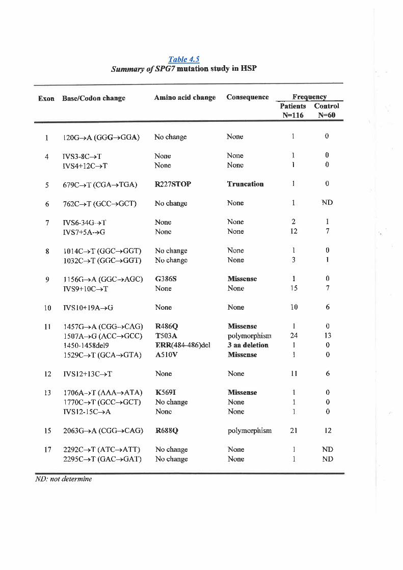
Tøble 4.5Summary of SPGT mutation study in HSP
Exon Base/Codon change Amino acid change Consequence Freque4cyPatients ControlN:116 N=60
1 120G-+A (GGG-+GGA) No change
NoneNone
NoneNone
No change
No change
c386SNone
None
R486QT503AERR(484-486)delA5IOV
None
NoneNone
Truncation
None
NoneNone
NoneNone
MissenseNone
1 0
0
0
0
4 IVS3.8C-+TIVS4+12C-+T
IVS6-34G+TIVST+54-+G
1014C-+T (GGC+GGT)1032C-+T (GGC+GGT)
1156G-+A (GGC+AGC)IVS9+1OC-+T
10 IVSl0+194-+G
l1 1457G-+A (CGG+CAG)15074-+G (ACC-+GCC)1450-l458del9r529C-+T (GCA-+GTA)
1
I
1
J
1
I1
5 679C-+T (CGA+TGA) R227STOP
6 762C-+T (GCC-+GCT) No change ND
1
I
7
8
9
.)
t21
7
1
0
1
0
7
6
0
13
0
0
6
0
0
0
12
NDND
ls 2063G-+A (CGG-+CAG) R688Q
None
Missensepolymorphism3 aa deletionMissense
None
MissenseNoneNone
polymorphism 2l
NoneNone
15
10
I24
I1
11
13
12 IVSl2+13C-+T
17064+T (AAA-+ATA)1770C->T (GCC+GCT)IVS12-15C-+A
l7 2292C->T (ATC-+ATT)2295C-+T (GAC-+GAT)
None
K569INo change
None
No change
No change
1
1
ND: not determine
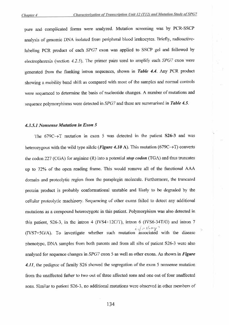
pure and complicated forms were analyzed. Mutation screening was by PCR-SSCP
analysis of genomic DNA isolated from peripheral blood leukocytes. Briefly, radioactive-
labeling PCR product of each SPGT exon \M¿Ìs applied to SSCP gel and followed by
electrophoresis (section 4.2.5). The primer pairs used to ampliff each SPGT exon were
generated from the flanking intron sequences, shown in Table 4.4. Any PCR product
showing a mobility band shift as compared with most of the samples and normal controls
\À/ere sequenced to determine the basis of nucleotide changes. A number of mutations and
sequence polymorphisms were detected in SPGT and these are summarised in Table 4.5.
4.3.5.1 Nonsense Mutøtìon ín Exon 5
The 679C-+T mutation in exon 5 was detected in the patient 526-3 and was
heterozygous with the wild type allele (Figure 4.10 ^).
This mutation (679C-+T) converts
the codon 227 (CGA) for arginine (R) into a potential stop codon (TGA) and thus truncates
up to 72Yo of the open reading frame. This would remove all of the functional AAA
domain and proteolytic region from the paraplegin molecule. Furthermore, the truncated
protein product is probably conformational unstable and likely to be degraded by the
cellular proteolytic machinery. Sequencing of other exons failed to detect any additional
mutations as a compound heterozygote in this patient. Polymorphism was also detected in
this patient, 526-3, in the intron 4 (IVS4+12C|T), intron 6 (IVS6-34T/G) and intron 7
(rVS7+5c/A). To investigate whether such mutation't#;¿;' *i h the disease
phenotype, DNA samples from both parents and from all sibs of patient 526-3 were also
analysed for sequence changes in SPGT exon 5 as well as other exons. As shown in Fígure
4.11,the pedigree of family 526 showed the segregation of the exon 5 nonsense mutation
from the unaffected father to two out of three affected sons and one out of four unaffected
sons. Similar to patient 526-3, no additional mutations were observed in other members of
t34
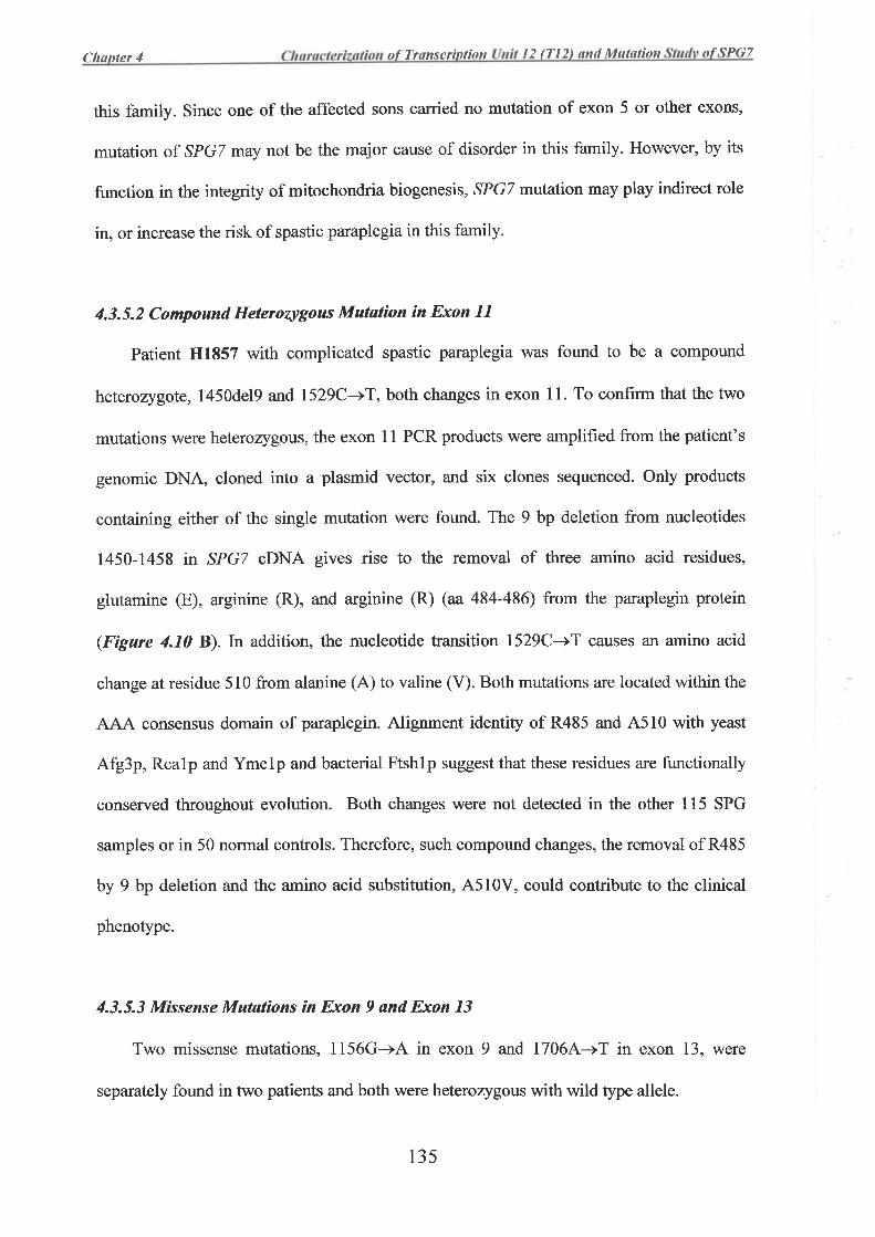
Clrunler 4 of Transc¡inlion l2 ff|2) and MuÍølìon of ,1PG7
this family. Since one of the affected sons carried no mutation of exon 5 or other exons,
mutation of SPGT may not be the major cause of disorder in this family. However, by its
function in the integrity of mitochondria biogenesis, SPGT mutation may play indirect role
in, or increase the risk of spastic paraplegia in this family.
4.3.5.2 Compound Heterozygous Mutation in Exon 1l
Patient H1857 with complicated spastic paraplegia was found to be a compound
heterozygote, l450del9 and 1529C-+T, both changes in exon 11. To confirm that the two
mutations were heterozygous, the exon 11 PCR products were amplified from the patient's
genomic DNA, cloned into a plasmid vector, and six clones sequenced. Only products
containing either of the single mutation were found. The 9 bp deletion from nucleotides
1450-1458 in SPGT cDNA gives rise to the removal of three amino acid residues,
glutamine (E), arginine (R), and arginine (R) (aa 484-486) from the paraplegin protein
(Fígure 4.10 B).In addition, the nucleotide transition 1529C-)T causes an amino acid
change at residue 510 from alanine (A) to valine (V). Both mutations are located within the
AJAA consensus domain of paraplegin. Alignment identity of R485 and A5l0 with yeast
Afg3p, Rcalp and Ymelp and bacterial Ftshlp suggest that these residues are functionally
conserved throughout evolution. Both changes were not detected in the other 115 SPG
samples or in 50 normal controls. Therefore, such compound changes, the removal of R485
by 9 bp deletion and the amino acid substitution, A5l0V, could contribute to the clinical
phenotype.
4.3.5.3 Mßsense Mutøtions ìn Exon 9 and Exon 13
Two missense mutations, 1156G-+A in exon 9 and 1706A-+T in exon 13, were
separately found in two patients and both were heterozygous with wild type allele.
13s
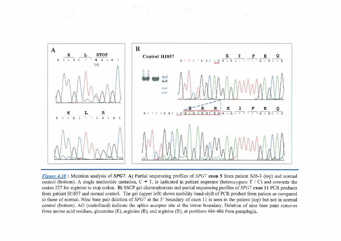
A B
G AA G C T TI{ G AG C
K L STOP
Ttc
Control H1857
fr;
6TT CTG GC AG GAG ATTTTTG AGtr ÀGEIFEA
EIFEA¿
¿RRKAA
LT uA
RG
GTTCTGGCAG GAGATTTTTGAGC AGcTch
Fisure 4.10 z Mutation analysis of SPG7. A) Partial sequencing profiles of SPGT exon 5 from patient 526-3 (top) and normalcontrol (bottom). A single nucleotide mutation, C + T, is indicated in patient sequence (heterozyguos T / C) and converts thecodon 227 for arginine to stop codon. B) SSCP gel electrophoresis and partial sequencing profiles of SPGT exon 11 PCR productsfrom patient Hl857 and normal control. The gel (upper left) shows mobility band-shift of PCR product from patient as comparedto those of normal. Nine base pair deletion of SPGT at the 5' boundary of exon 1 1 is seen in the patient (top) but not in normalcontrol (bottom). AG (underlined) indicate the splice acceptor site at the intron boundary. Deletion of nine base pairs removesthree amino acid residues, glutamine (E), arginine (R), and arginine (R), at positions 484-486 from paraplegin.

C-T-
-T-C
IVS4+|2C/T f -.679C-+T C -
-T-C
T-G- -G
-AIVS6-34G/T
IVST+54/GG-A- -G
-As26-1 s26-2
s26-3 526-4 526-5 526-6 526-7 526-8 526-9
C-T_
-T-C
T C-C T-
-T C--C T-
-T C--C C-
-T T--C C-
:
TT-CC-
T
C
T- -G GT-AG- -G T-
-A G- -G-A
T-G-
GG-AA- -GG¡-A -A
Físure 4.ll Pedigree of family 526 with the Genotype of SPG7. The symbolsindicated are opened squqre, unaffected male; filled square, affected male; andopened circle, unaffected female. Sequence changes are indicated in intron 4(IVS4+12C/T), exon 5 (679C+T), intron 6 (IVS6-34G/T), and intron 7(rvs7+sA/G).
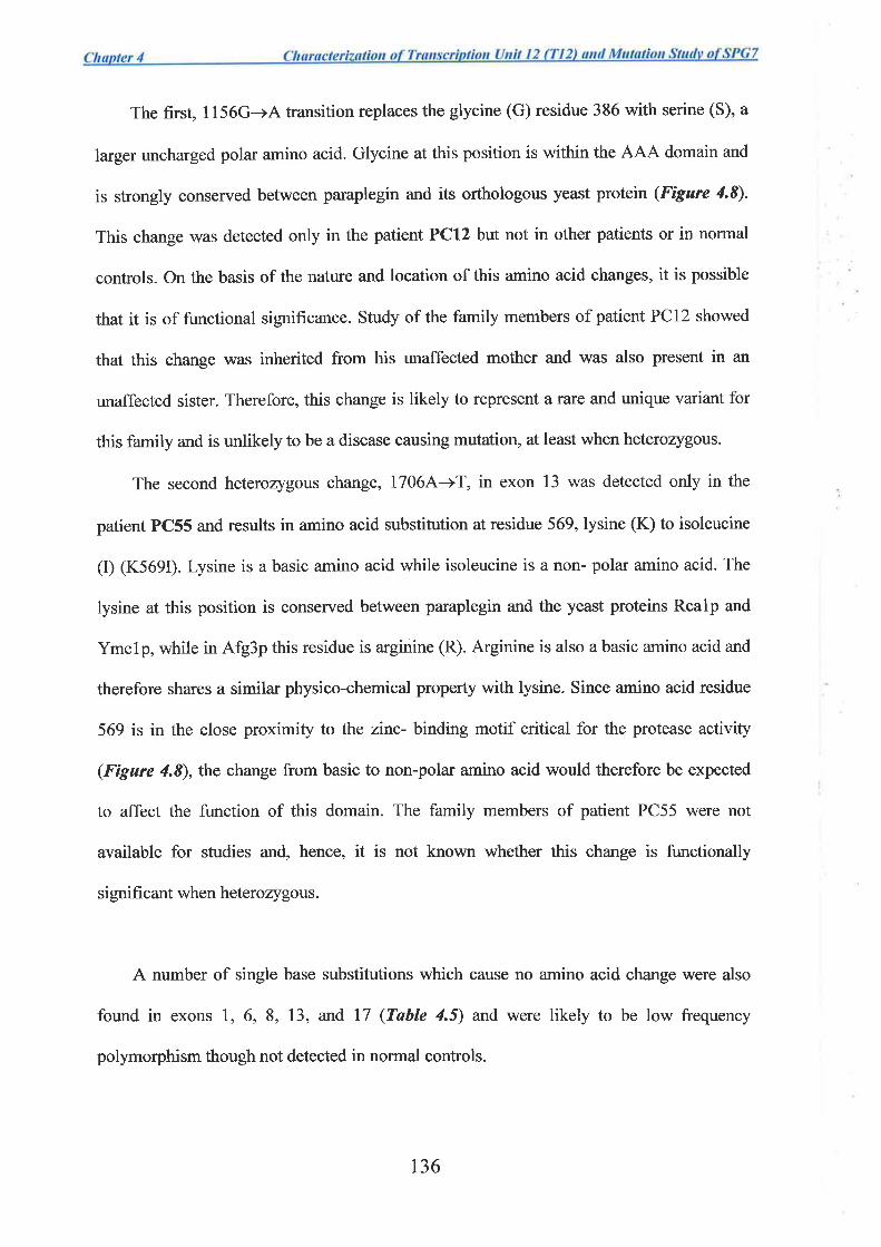
The first, 1156G-+A transition replaces the glycine (G) residue 386 with serine (S), a
larger uncharged polar amino acid. Glycine at this position is within the AJAA domain and
is strongly conserved between paraplegin and its orthologous yeast protein (Figure 4.8).
This change was detected only in the patient PCl2 but not in other patients or in normal
controls. On the basis of the nature and location of this amino acid changes, it is possible
that it is of functional significance. Study of the family members of patient PCl2 showed
that this change was inherited from his unaffected mother and was also present in an
unaffected sister. Therefore, this change is likely to represent a rare and unique variant for
this family and is unlikely to be a disease causing mutation, at least when heterorygous.
The second heterozygous change, 1706A-+T, in exon 13 was detected only in the
patient PC55 and results in amino acid substitution at residue 569, lysine (K) to isoleucine
(I) (K569Ð. Lysine is a basic amino acid while isoleucine is a non- polar amino acid. The
lysine at this position is conserved between paraplegin and the yeast proteins Rcalp and
Ymelp, while in Afg3p this residue is arginine (R). Arginine is also a basic amino acid and
therefore shares a similar physico-chemical property with lysine. Since amino acid residue
569 is in the close proximity to the zinc- binding motif critical for the protease activity
(Figure 4.8), the change from basic to non-polar amino acid would therefore be expected
to affect the function of this domain. The family members of patient PC55 were not
available for studies and, hence, it is not known whether this change is functionally
significant when heterozygous.
A number of single base substitutions which cause no amino acid change were also
found in exons l, 6, 8, 13, and 17 (Table 4.5) and were likely to be low frequency
polymorphism though not detected in normal controls.
t36
4.4 Discussion
The present study describes the isolation and characterization ofa transcript sequence
of 3,083 bp, which was later found to be identical to the subsequently reported SPG7, a
gene involved in a recessive type of hereditary spastic paraplegia (HSP) (Casari et al.,
1998). Initially, from the physical map of chromosome region 16q24.3 (Whitmore et al.,
1998) six trapped exons were grouped together as part of a tentative gene, designated
"transcription unit 12 (Tl2)". Upon analyses, however, only four: 8T34.87, ET34.I,
8T2.5, and ET2.8 were successfully joined and formed part of a transcript sequence.
Northern analysis indicated the fullJength transcript to be approximately 3.2 kb. The
sequence was extended by utilizing both PCR from a 5' stretched cDNA library and 5'
RACE. This extended oDNA sequence contained an in-frame translation initiation codon,
ATG, perfectly embedded in a Kozak consensus sequence, and therefore suggested a true
start site. In contrast, the 3' sequence extension was initially complicated by the finding
that overlapping EST sequences were derived from genomic DNA that contaminated the
cDNA library. 3'RACE was then utilized but this only allowed a short sequence extension.
However, it was then determined that this 3' sequence of Tl2 overlapped with the 5'part of
a newly reported CMAR cDNA sequence of 2.4 kb (Durbin et al., 1997). RT-PCR
experiments were undertaken using T12 primers and primers designed from CMAR
sequence to confirm that they were part of the same transcript. These results finally
allowed the assembly of a 3,083-bp transcript sequence of T12 and also provided the
evidence that CMAR was in fact a part of the 3' UTR of this transcript.
The CMAR gene has originally been reported as an intronless gene with its 459-bp
cDNA that has been predicted to code for a protein of 142 amino acids (Pullman and
Bodmer, 1992). This gene has been demonstrated by transfection experiment to enhance
binding of the cell to extra-cellular matrix component, collagen typel (Pullman and
t37

Bodmer, 1992). Tltis CMAR sequence is situated close to the 3' end of the 3,083-bp Tl2
transcript. From the initial report, the CMAR protein structure has been described to be
integrin pl dependent, with probable tyrosine phosphorylation activation, suggesting a role
in signal transduction (Juliano and HaskilI,1993; Romer et aL,1992; Novelli et a1.,1995).
Furthermore, since this gene has been mapped to the 16q24.3 LOH region observed in
several cancers, it has been proposed that CMAR might function as a tumor suppressor. A
subsequent publication by Durbin et al.(1997) presents a larger CMAR transcript of 2.4kb,
but the ORF was the same as of the original published sequence. This 2.4-kb transcribed
sequence is intemrpted with the insertion of two Alu-like elements within its 5' half. The
original reported coding sequence (Pullman and Bodmeg 1992) is situated within the 3'
region of the 2.4-kb transcript. Within this coding region the CACA insertion
polymorphism has been reported and resulted in the change in the putative ORF (Koyama
et a1.,1993; Durbin et a1.,1994). However, none of these reports of CMAR transcript have
presented evidence of a protein product or a decent Northern result.
The 3,083-bp transcript of Tl2 was predicted to code for a protein of 795 amino
acids. This protein was predicted by sequence homology to have functions similar to the
yeast mitochondrial proteins, Afg3p, Rcalp, and Ymelp. In addition, the protein
localization prediction by PSORT progrrim also suggested a mitochondrial protein. Shortly
following completion of this result, Casari et al (1998) reported the gene, SPG7, which
was identical to Tl2 and coded for the same 795 aa-protein designated "paraplegin". The
result of in silico prediction by PSORT also agreed with that of in vitro protein localization
study of the SPGT protein product presented in this report (Casari et al., 1998). The
orthologous yeast proteins are ATP-dependent metalloproteases belong to a subclass of the
AJA-A protein family, the AAA proteases. They possess both chaperone-like and proteolytic
activities (Rep and Grivell, 1996; Patel and Latterich, 1998). There are three conserved
138
functional domains characteristic of this subgtoup of AJA.A protein: the ATP-binding motif
(GPPGCGKT), AAA minimal consensus sequence, and the zinc-dependent binding motif
(consensus HEXXHA). The AJA,{ consensus domain is crucial for the chaperone-like
activþ in sensing and binding with the substrate (Langer, 2000; Juhola et a1.,2000). This
domain is followed by a region responsible for proteolytic activity with the consensus
metal-binding motif HEXGHA as the proteolytic center (Langer, 2000; Juhola et al.,
2000). All of these functional domains were also found in paraplegin. The firnction of such
yeast mitochondrial metalloproteases involves_.in the quality control of mitochondrial k
protein biogenesis, including protein folding and the assembly of respiratory chain
complexes (Rep and Grivell, 1996; Patel and Latterich, 1998). Defects of these
mitochondrial metalloproteases are likely to be lead to the deterioration of mitochondria
function.
In the yeast mitochondrial inner membrane, Afg3p and Rcalp form subunits of a
hetero-oligomeric proteolytic complex at equimolar amounts (Arlt et al., 1996) with its
active site present at the matrix side. Both Afg3p and Rcalp are anchored to the membrane
by two transmembrane domains in their N-terminal regions. In contrast, Ymelp is a homo-
oligomeric complex (Leonhard et al., 1996) with its catalytic sites facing the
intermembrane space. The Ymelp is anchored by only one transmembrane segment In
analogy to the Afg3p and Rcalp, paraplegin is likely to be anchored to the membrane by
two transmembrane domains in its N-terminal region, which were identified between
amino acid residues 144-162 and between residues 249-272 (section 4.3.3). This similarity
suggests that paraplegin function requires other protein partners to form a functional
complex. The protein partners of paraplegin have not yet been reported. Two recently
identifred human genes, AFG3L2 (Afg3-like 2) and YMEILI (Ymel-like l) on
chromosomes 18 and 10 respectively, have also been reported to encode members of the
t39
Clmnler 4 CItsra cleriz.ul iott of Ilnit l2 (Tl2) and Mufotìon Stutlv of SPGT
mitochondrial ArqA metalloprotease (Banft et al., 1999; Coppola et al., 2000). The
AFG3L2 protein sequence is highly homologous to both Afg3p and Rcalp and similar to
that of paraplegin (Banfr et al., 1999). AFG3L2 has also been reported to localize to
mitochondria. It was likely that AFG3L2 may have function by forming a complex with
paraplegin, hoùever this has yet to be determined from molecular interaction analysis.
YMEIL, on the other hand, appears to be an orthologue of yeast YmeI and its expression in
yeast has been able to complementaymel null mutant (Shah et a1.,2000). Therefore this is
unlikely to be a protein partner of paraplegin.
The genomic structure of SPGT was determined based on the TI2 transcnpt sequence
and three genomic clones spanning the Tl2lSPG7 region: cosmids 383H6, 427811, and
358D12. The gene consists of 17 exons encompassing the genomic sequence of
approximately 52 kb. Based on the exon/intron boundary information, a cryptic splice site
was identif,red in exon 6. During the course of transcript sequence identification, a cDNA
clone ?"8T2.5 containing a sequence between exon 4 and exon 9 of SPGT was isolated
from a fetal brain cDNA library. Sequence analysis revealed a 55-bp deletion of the
sequence corresponding to the 3'part of exon 6. Analysis of the complete exon 6 sequence
identified a cryptic donor spice site (TGgtatgt: consensus score 8l) situated 55 bp upstream
from the 3' end of the exon (Figure 4.1). Therefore, the 55 bp deletion from exon 6 in this
cDNA clone is probably attributable to the use of this cryptic donor splice sequence
alternative to the normal site (TTgtaagt: score 79). This deletion however truncates the
ORF of the transcript by creating an immediate in-frame stop codon. This abenant form of
transcript is not detected by RT-PCR and is considered to be a rare transcription isoform
with no functional significance.
Mutation studies of SPGT were undertaken in 116 unrelated cases of spastic
paraplegia, both pure and complicated forms. Two cases were found that have mutations
140
and were thought to be related to the clinical phenotype. A nonsense mutation was detected
in the patient 526-3, an index case, diagnosed with a pure form of HSP. The family of this
index case consists of normal parents, three affected sons, and four unaffected sons. The
familial pattem of disease transmission was referred to as autosomal recessive based on the
presence of unaffected parents and absence of family history. The SPGT mutation
identified in individual 526-3 was a single base substitution, 679C-+T, resulting in the
change of codon 227 from CGA for arginine to a potential termination codon, TGA. This
change abolishes up to 72%o of the coding sequence and therefore removes all functional
motifs of the protein. The truncated protein product, if exist, is probably conformational
unstable and likely to be degraded by the cellular proteolytic machinery. The most likely
mechanism is the degradation of mRNA that contains premature termination of ORF by
the pathway referred to as nonsense-mediated mRNA decay (NMD) (Culbertson, 1999;
Frischmeyer and Dietz, 1999). This mutation is therefore pathogenic. The mutation,
however, is heterozygous with no other mutations detected to be a compound heterozygote.
Sequencing of this gene in the parents and all sons indicated that the 679C-+T mutation
was inherited from the unaffected father to two out of three affected sons and one out of
four unaffected sons. When heterozygous this mutation would lead to the reduction of
paraplegin synthesis. However, it was unknown whether this contributed to the clinical
phenotype in this family. Both the father and one of his son (526-5), were heterozygous for
this mutation but unaffected, could be caused by a dominant gene with low penetrance.
However, the presence of an afÊected son with no SPGT mutations indicated that it is likely
that a defect of other gene may be the cause of the clinical phenotype in this family. Since
the paraplegin protein is involved in the integrity of mitochondrial function, it is possible
that SPGT mutations even when heterozygous may indirectly contribute to the
'a
t4l

pathogenesis of disease or increase the risk of disease in an individual who caries such
mutation.
A compound heterozygous mutations of exon 11, 1450de19 and 1525C-+T, was
observed in case H1857. Both mutations are in the conserved AJA.A. domain that is
important for paraplegin function. The nine base pair deletion, nt 1450-1458, results in the
loss of three amino acids, glutamine, arginine, and arginine (residues 484-486) from the
highly conserved region of AAA domain (Fìgure r'.8). The removal of these amino acids is
predicted to affect the protein conformation of this domain and therefore affect the
paraplegin function. The fuq.A. domain is important in sensing and binding of paraplegin to
its substrate. This is a chaperoneJike activity. In addition,the 1529C-+T mutation replaces
alanine at amino acid residue 510 with valine (A5l0V) and also occurs within the
conserved sequence of fuAl{ domain. This change would also affect the ArqA motif
function. The mutations as a compound heterozygote are likely to compromise the function
of the paraplegin protein and therefore be consistent with a sporadic case of recessive
spastic paraplegia. The patient H1857 was clinically diagnosed as a complicated spastic
paraplegia and developed the clinical disease at the age of 25. Both of his parents were
reported to be clinical normal but there were no clinical data of other family members and
they were unavailable for study. The same compound heterozygous changes, 1450de19 and
1525C-+T, were also presented in one of 30 index cases, studied by McDermott et al
(2001). This case was also complicated spastic paraplegia. In this family, the father who
was heterozygous for the 1450de19 also presented with a milder form of complicated
spastic paraplegia, suggesting a dominant behavior of this mutation.
Two heterozygous missense mutations, 1156G-+A in exon 9 and 17064-+T in exon
13 were found in patients PClz and PC55 respectively but were not presented in at least 50
normal controls. Both changes cause amino acid substitutions affecting a conserved amino
r42
Chaoter 4 Chsrscterizolion of Trunsuiption Unit 12 (TI2) snd Mutalion Slutlv of SPGT
acid. The 1156G-+A results in the replacement of gþine at residue 386 with serine
(G386S). The 17064+T mutation on the other hand causes amino acid substitution, lysine
by isoleucine (K569I). The amino acid substitutions, G386S and K569I, affect the
conserved amino acid within the fuA.A' domain, and the conserved residue close to a zinc-
binding motif respectively. However, both variants are heterozygous with no other
mutations detected in this gene. 'When analysis was performed in the family members of
either case, such mutations were also detected in the clinical normal individuals. Therefore,
both were unlikely to be related to a clinical phenotype at the heterozygous stage. It is
possible, however, that cariers are at increased risk of developing diseases.
Casari et al (1998) reported three different SPGT mutations in three families of which
all affected cases were homozygous. The first mutation was a 9.5 kb genomic deletion
covering the last five exons of SPG7, including its 3'UTR. This deletion is likely to
intemrpt the transcription process of the gene due to the absence of polyadenylation signal
within 3'UTR of the gene and the down stream sequence element necessary for the
transcription machinery (Lou et al., 1995; Lou et al., 1996). The second was homorygous
2-bp deletion at nucleotide 784-785 (in exon 6) which causes a frame shift that changes the
amino acid sequence down stream and creates a "TAA" stop at the first codon of exon 7.
This abolishes about 60%;o of the ORF. The resulting mRNA is likely to be degraded by the
mechanism called "nonsense-mediated mRNA decay (NMD)" (Culbertson, 1999;
Frischmeyer and Dietz, 1999). The last family was homozygous for an "4" insertion at
position 2228 wfihln exon 17 of SPGT cDNA. This mutation causes a frame shift and
creates a premature termination of the ORF which is predicted to code for a truncated form
of paraplegin that is missing 57 amino acids at the C terminus.
The three mutations reported by Casari et al (1998) were from a screen of 14
unrelated recessive families. This suggested that mutations in this gene may be a relatively
t43
coÍtmon cause of autosomal recessive HSP. The present study, however, demonstrates that
SPGT mutations are rare in sporadic cases and small families with HSP. This is likely the
result of the mode of ascertainment. In the study by Casari et al (1998), each case was
from a single ethnic group in a relatively close small community. All three mutation-
families were consanguineous. In the present analysis, in contrast, all subjects are from a
population with a diverse ethnic mix. In recessive families, there is a low frequency of
linkage to SPG 7 that has been reported in other studies (Coutinho et al.,1999). This also
suggests that SPGT mutations are aÍaÍe cause of spastic paraplegia.
Finally, the expression of SPGT was found ubiquitously in all the tissues tested,
consistent with its mitochondrial function. Mutation of this gene in other syndromes, which
have a causal basis of mitochondrial impairment may also exist. Spastic paraplegia has also
been reported in association with other neurologic disorders, such as Alzheimer's disease
(Orth and Schapira,200l; Turner and Schapira,2001) and some types of epilepsy (Gigli er
a1.,1993;Yih et a1.,1993; Webb et a1.,1997; Nobile et a1.,2002).It is possible that SPGT
mutations and the resulting defects in paraplegin may contribute to these diseases, though
this requires additional study.
r44

Chapter 5
Exon Amplification Study and
Charact erization of Transcription U nit
13 (T13)

Table of Contents
5.l lntroduction
5.2 Methods
5.2.1 Exon Trapping
5.2.1.1 Digestion of BAC DNA
5.2.1.2 Purification of digested DNA by phenol/Chloroþrm Extraction
5.2.1.3 Preparation of the Exon trapping Vector
5.2.1.4 Cloning of BAC digested products into the pSPL-3Ù Vector
5.2.1.5 Preporation of COS-7 Cells
5.2.1.6 Transfection of COS-7 Cells
5.2.1.7 Isolation of Cytoplasmic RNAfrom COS-7 Cells
5.2.1.8 Reverse transcription of Isolated RNA
5.2.1.9 Primary PCR Amplífication
5. 2. 1. I 0 Secondary PCR Ampli/ication
5.2.1.11 Uracil DNA Glycosylase Cloning of Secondary PCR Products
5.2.2Exon Confirmation
5.2.2.1 Colony PCR
5.2.2.2 DNA Isolation qnd Sequencing
5.2.2.3 Physical Mapping of Trapped Products
5.2.2.4 Trapped Exon Sequence Analysis
5.2.3 Identification of Transcript Sequence
5.2.4 In silico Protein Function Analysis
5.3 Results
5.3.1 Exon Trapping
5.3.2 Trupped Exon Sequence Analysis
5.3.3 Physical Mapping of Trapped Products
5.3.4 Verification of the Transcript Sequence
5.3.4.1 Transcript Sequence generatedfrom 561-4 and 561-13
5.3.4.2 Transuipt Sequence of 561-72 and 561-I I4
5.3.5 Assembly of the Transcription Unit 13 (Tl3) Sequence
5.3.6 Analysis of Tl3 Transcript Sequence
5.3.7 Tl3 Protein Sequence Analysis
5.3.8 T13 Gene Structure
Page
t4s
t46
146
147
747
r48
148
149
t49
150
150
151
l5lr52
t52
153
153
153
r54
154
154
155
155
155
158
159
159
t62
r63
166
t66
168
5.3.9 Tl3 Alternative Spliciíg and Ovedapping Transcripts
5.3.10 Mutation Study of T13 in Sporadic Breast Cancer
5.4 Discussion
5.4.1 Exontræping
5.4.2 Characterization of Transcription unit 13 (T13)
169
t7t
t72
172
t75
5.1 Introduction
The construction of the physical and transcription map at 16q24.3 LOH region
(Whitmore et al., 1998a) has allowed the isolation and mapping of many new genes and
potential transcription units. In addition, the map location of known genes in the region
were also established. These included FAA, a gene involved in Fanconi anemia
complementary goup A (The Fanconi anaemia/Breast cancer consortium, 1996); GASI l, a
human homologue of murine growth arrested specific gene (Whitmore et al., 1998b);
transcription unit 3 Q3) (chapter 3); transcription unit 6 Q6) (Whitmore, 1999
unpublished data); SPG7, the gene for recessive spastic paraplegia (Casari et al., 1998;
chapter 4), and a recently identified gene, Copine VII (Savino et al., 1999). The map
location of the PISSLRE gene (Grana et al., 1994) was also established in this region
through its sequence homology to trapped exons isolated from the cosmid contig at
16q24.3.
Among these genes, FAA and PlSSZftE were initially considered to be potential
candidates as a tumor suppressor targeted by LOH in breast cancer. Patients with Fanconi
anemia possess a predisposition to a variety of malignancies such as acute myeloid
leukemia and squamous-cell carcinoma (Butturini et al., 1994). Cells from FA patients
show increased sensitivity to bi-functional DNA crosslinking agents such as diepoxybutane
and mitomycin C, with characteristic chromosome breakage (Auerbach, 1993), a feature of
genomic instability found in most cancer. The P/SSZRE gene on the other hand belongs to
a family of cycline dependent protein serine/threonine kinases that are critical for cell cycle
progtession. Dominant-negative constructs of the gene have been shown to halt cell cycle
progression in G2-M phase and, when overexpressed in U2OS cells, suppress growth (Li et
al., 1995). Despite the potential involvement in genomic instability and cell growth, both
have been excluded by mutation analysis in sporadic breast cancer as tumor suppressors
145
targeted by LOH (Cleton-Jansen ef al-,1999; Crawford et a1.,1999).
Other identified genes in this region including GASI l, SPG7, Copine VII, T6, and T3
have also been excluded by mutation studies as candidate breast cancer related tumor
suppressors (Whitmore et al., 1998b; Settasatian et al., 1999; Savino et al., 1999;
Whitmore, 1999 unpublished data; Chapter 3).
The proximal region of the physical map established at thel6q24.3 breast cancer LOH
region (Whitmore et al., 1998a) consists of a group of overlapping cosmids with five
clones representing the minimal tiling path. This group of cosmids was linked to a longer
cosmid contig by the genomic clone, PAC l68Pl6 (Fìgure 1.3). Of five overlapping
cosmids, the cosmid 383F1 represents the proximal boundary of this group. From this
boundary a BAC clone 561817 was subsequently identified and extended the contig
proximally a further 140 kb (Whitmore, 1999 unpublished data). Within this region a
oDNA sequence of clones 118499 and IMAGE:1098126 were initially mapped and were
assigned as transcription units, T17 and T18 respectively. In addition, the Cadherin 15
gene (CDHI5) has also been mapped to this region (Kremmidiotis ef a1.,1998). To analyse
this extended region for the identification of new genes, the large genomic clone, BAC
56lB17, was chosen for firrther study.
The present studies will describe the use of exon trapping for exploring genes within
this region. Subsequent identification of the genes or potential transcripts from the trapped
products will also be described. Characterization of the gene designated transcription unit
13 (T13) will be desuibed in detail as the major transcript.
5.2 Methods
5.2.1 Exon Trapping
The principle of the exon trapping procedure (Buckler et al., l99l; Church et al.,
r46
1994) is summarised in Figure 5.1. The detailed procedures a¡e described in the
subsequent sections. The sequences of all primers used for exon trapping procedure are
given in Table(j
5.2.1.1 Digestíon of BAC DNA
In a reaction volume of 50 pl, 5 pg of BAC DNA was digested at 37 "C, overnight,
with the restriction enzymes BamHl and Bglll, or with Psfl alone, in the specific buffer
containing 100 pglml of BSA as recommended by the manufacturer. The amounts of
restriction enzymes used were 15 units of BamIIl,30 units of BglII, and 15 units of PsrI.
Since double enzyme digestion was performed in the BamHI specific buffer, which was
not optimal for 100% digestion by BglII, the amout of Bglll used was doubled.
5.2.1.2 PuriJication of digested DNA by PhenoUChloroþrm Extraction
Following restriction enz-yme digestion, an aliquot of 250 ng of each digested DNA
was taken for subsequent analysis. The remaining DNA was adjusted to a final volume of
200 pl with sterile water and mixed well. An equal volume of phenol was then added to
each reaction tube, mixed and followed by centrifugation at 13,000 rpm for 7 minutes. The
aqueous layer was transferred to a new 1.5 ml tube and an equal volume of chloroform/iso-
amyl alcohol (24:l) was added and mixed. Each mixed sample was again centrifuged for 7
minutes at 13,000 rpm and the aqueous layer transferred to a clean eppendorf tube. The
DNA was precipitated by the addition of one-tenth the volume of 3M NaOAc (pH 5.2) and
2 volumes of absolute ethanol, mixed, and kept at - 80 oC for I hour. The samples were
then spun at 13,000 rpm for 30 minutes. DNA pellets were washed with I ml of 70Yo
ethanol, air-dried for 15 minutes, and the DNA was dissolved in 10 pl of sterile water. An
aliquot of 2 ¡il of each sample was analysed by electrophoresis on a0.8%o agatose gel. The
t47
250 ng aliquot of the original digested product was also loaded on the gel to determine the
effrciency of the purification procedure.
5.2.1.3 Preparation of the Exon Trapping Vector
Ten micrograms of the exon trapping vector, pSPL3B, was digested ovemight at 37
oC with 15 units of either PslI or BamHl in a 50 pl reaction buffer. Then, 50 pl of water
was added and the digested products purified with QlAquick columns according to the
manufacturer's instructions (2.2.1a.1). The purifred digested vectors \¡rere then
dephosphorylated with 2.5 units of calf intestinal alkaline phosphatase (CIAP) in a final
volume of 100 prl using the specific buffer supplied with the kit (Boehringer Mannheim).
The reactions were carried out at 37 "C for I hour and followed by a further purification
step using QlAquick columns. The DNA was quantitated by spectrophotometry Q.2.4).
5.2.1.4 Clonìng of BAC digested products into the pSPL3B Vector
The digested BAC DNA was ligated to a linear and dephosphorylated pSPL3B DNA
according to the procedure described in 2.2.15.1.The reaction was performed in a final
volume of 10 pl containing 50 ng of vector and 200 ng of digested BAC DNA. The
^ligation reaction containing 50 ng of digested vector alone was also carried out as a
control.
The transformation reaction was undertaken by the procedwe described in 2.2.15.4,
except for the plating of transformed cells. Instead of plating out 200 pl of the transformed
cells, the cells were spun down and the pellet was resuspended in 100 ¡.r1of LB-broth. The
entire 100 ¡rl was then plated onto a LB-agar/Ampicillin plate, and incubated overnight at
37 "C ( X-gal and IPTG were not spread with the cells). Subcloning of the BAC DNA into
trapping vector was considered successflrl if the number of colonies on the experimental
148
plate exceeded by 10
plate.
folf the number of colonies on the control, vector-only ligation
After overnight incubation, 3 ml of LB-broth plus ampicillin (100 pglml) was added
onto each successful ligation/transformation plate. The cells were scraped off into the
media solution with a sterile glass spreader. The cell suspension was then transferred to a
sterile l0 ml tube. DNA isolation was performed by "plamid mini-prep" using Qiagen Tip-
20 (2.2.r.3).
5.2.1.5 Prepøration of COS-7 cells
The COS-7 cells were grown in supplemented DMEM culture medium. The cells
were passaged one day prior to transfection by placing 8x10s to 16xl0s cells intol0 ml of
supplemented DMEM in 75 cm2 tissue culture flasks. This resulted in 40 to 60Yo
confluence the next day.
5.2.1.6 Transfectìon of COS-7 Cells
The culture flasks were inspected for the appropriate cell confluence and pSPL3B-
BAC DNA was transfected into the cells using the LipofectACE reagent (Gibco-BRL).
Two subcloned DNA samples, PsfI generated and BamHIlBgilI generated, *"r#åh.d toA
one flask of cells per transfection. The transfection was performed essentially as follows.
In a sterile l0 ml tube, 500 ¡rl of Opti-MEM I (OMI) medium was added to 3 ¡rg of each
subcloned DNA to be transfected. In a sterile 1.5 ml eppendorf tube, 500 pl of OMI was
added to 30 pl of LipofectACE and left at room temperature for 5 minutes. This OMI-
LipofectACE mix was then added to the 10 ml tube containing OMl-subcloned DNA and
incubated for another 10 minutes at room temperature. Whilst waiting, the supplemented
DMEM was removed from the COS-7 cell culture and l0 ml of OMI was added to each
149
culture flask. The cells were then incubated for 5 minutes at 37 oC in a 5Yo COz incubator.
After 10 minute incubation of the above l0 ml tube was complete,4 ml of OMI was then
added to each tube containing the subcloned DNA/LipofectACE/OMI mix. The l0 ml of
OMI medium from each flask of cells was removed after 5 minute incubation and the
combined DNA/LipofectACE/OMI above was added to the flask of cells. Following
incubation overnight at 37 "C in a 5Yo COz incubator, the lipid-DNA complex was
removed from the COS-7 cells and l0 ml of supplemented DMEM was added and
incubated for a further 48 hours at37 "C in5Yo COz environment.
5.2.1.7 Isolatìon of Cytopløsmíc RNAfrom COS-7 Cells
All RNA isolation procedures were done under RNAse free conditions as described in
section 2.2.2. After 48 hour incubation, the supplemented DMEM was removed from each
flask and l0 ml of ice-cold PBS was added and the flask was left on ice. The PBS was then
removed from each flask and the wash step was repeated two more times. The washed cells
were removed from culture flask by adding 10 ml of PBS and cells were scraped off using
a sterile plastic scraper (Costar). The cell suspension was transferred into a sterile l0 ml
tube, which was placed on ice, then centrifuged at 1,200 rpm for 5 minutes. After the
supernatant was removed, the cell pellet was resuspended in Trizol reagent (Gibco-BRL)
and RNA was isolated as described in 2.2.2.2.
5.2.1.8 Reverse Transcriptíon of Isolated RNA
The total RNA isolated from COS-7 cells was reverse-transcribed as per the
procedure described in 2.2.13.1, but using the pSPL3B specific primer, SA2. The sequence
of this primer is presented in Table 5.1. In each reaction, I ¡.rl of 20 ¡rM stock of SA2 was
used. After a 30 minute incubation at 42 "C with the Superscript enz.lme, the mixture was
150
heated to 55 "C and maintained for l0 minutes. Two units of RNAseH was then added and
the reaction tube was incubated for a further l0 minutes at 55 "C. The reverse-transcribed
products were stored at -20 "C while not in use.
5. 2. 1.9 Primary PCR AmpliJicatÍon
In the reaction, 8 ¡rl of the reverse-transcribed product was mixed with 4 pl of lOX
PCR buffer, 0.8 pl of l0 mM dNTPs,l.2 pl of 50 mM MgCl2, and2 ¡il each of a 20 ¡rM
stock of oligonucleotides SA2 and SD6 (Tøble 5./). The mixture was made up to a volume
of 39.5 pl with sterile water, and overlaid with a drop of paraffrn oil. The tube was heated
at 94 "C for 5 minutes, followed by holding at 80 "C while 0.5 ¡rl (2.5 units) of Taq DNA
polymerase was added. Each reaction tube was then incubated at 94 oC for I minute, 60 oC
for 1 minute,72 "C for 5 minutes for 6 cycles, followed by a l0 minute incubation at 72
oC. To prevent the amplification of false-positive (vector-only) products in the subsequent
PCR reaction, 2.5 ¡il (25 units) of BslXI was added to the PCR reaction and incubated
overnight at 55 oC. This was followed by incubation with a firther 0.5 prl ( 5 units) of
BslXI for a further 2 hours at 55 "C.
5.2. l. I 0 Secondøry PCR Ampffication
Each reaction was caried out by combining 5 pl of the first PCR reaction with 4.5 pl
of lOX PCR buffer, 1 ¡rl of 10 mM dNTPs, 1.5 pl of 50 mM MgCl2, I ¡rl each of a 20 ¡rM
stock of oligonucleotides dUSD2 and dUSA4 (Table 5.1), and sterile water to a final
volume of 49.5 ¡rl. The contents were mixed and overlaid with a drop of paraffin oil. After
heating the tube at 94 "C for 5 minutes, the tube was maintained at 80 oC while 0.5 pl (2.5
units) of Taq DNA polymerase was added. The tube was then subjected to a 30 cycles of
151
94 "C for I minute, 60 oC for I minute, and 72 "C for 3 minutes. This was followed by a
10 minute incubation at 72 "C. Five microlitres were analysed on a 2.5Yo agarose gel
electrophoresis to examine the potential number of trapped exons amplified.
In this step the oligonucleotides SD2 and SA4 were modified to include deoxy-UMP
residues at their 5' ends (the oligonucleotide sequences are presented, in Table 5.1). This
was to allow cloning of PCR products utilizing the Clone AMP pAMPlO kit (Life
Technologies).
5.2.IJl Uracíl DNA Glycosylase Clonìng of Secondary PCR Products
The PCR products from secondary amplification possess the dUMP-containing
sequence at their 5' termini. Treatment with uracil DNA gþosylase renders dUMP
residues abasic, and unable to base-pair, resulting in 3' protruding termini which can then
be ligated to complementary pAMPlO ends.
The amount of 2 ¡ú of the secondary PCR reaction was mixed with 2 pl of pAMPlO
cloning vector, 15 ¡rl of lX pAMPlO annealing buffer, and 1¡rl (luniÐ of uracil DNA
glycosylase. After mixing, the contents were incubated at 37 oC for 30 minutes. Five
microlitres of the reaction was then transformed into 100 ¡rl of competent E. coli XI--l
Blue cells (2.2.15.3). One-tenth of the transformation reaction was plated on LB-
Ampicillin plates and incubated overnight at37 "C.
5.2.2 Exon Confirmation
To confirm that exon products were trapped, the inserts were amplified, colony PCR
using pAMPlO specific primers flanking the cloning site was hrst carried out to determine
the product size. Clones rwere grouped according to the product size and a representative
clone from each group was chosen for further analysis.
ts2
5.2.2.1 Colony PCR
The recombinant colonies were randomly picked from overnight cultures with a
sterile pipette tip, streaked on a master LB-agar plate for later utilized, and the tip placed in
a PCR tube containing a drop of paraffin oil. The colony PCR procedure was followed as
described in section 2.2.12.2, the only variation was the primers used in the PCR. The
primers specific for pAMPlO used for the colony PCR were pUC-F and pUC-R. Clones
with an insert size of 409 bp, as determined by colony PCR, were omitted from further
analysis. The 409 bp product represented those being generated from vector-only clones
containing no trapped product. Colony PCR was also performed using SA4 and SD2
primers, with the PCR product size of 153 bp was from the vector-only clones and such
clones were omitted from further analysis. The sequences of all primers are included in
Tøhle 5.1.
5.2.2.2 DNA Isoløtion and Sequencing
Clones selected for further analysis were grown in 20 ml of LB-broth plus ampicillin
(100 ¡rglml) and DNA was isolated using Qiagen Tip-20 columns (2.2.1.3). The trapped
products were sequenced using Dyeterminator Sequencing kits (2.2.16.1) with either SD2
or SD4 primers (Tøble 5.1). As the exons were trapped directionally in the trapped vector
via the use of splicing consensus sequence, the DNA sequence obtained for each trapped
product was then aligned in the sense orientation by its relation with the trapped vector
sequence.
5.2.2.3 Physícøl Mapping of Trøpped Products
The DNA inserts of trapped products were removed from the vector with a NotUSalI
153
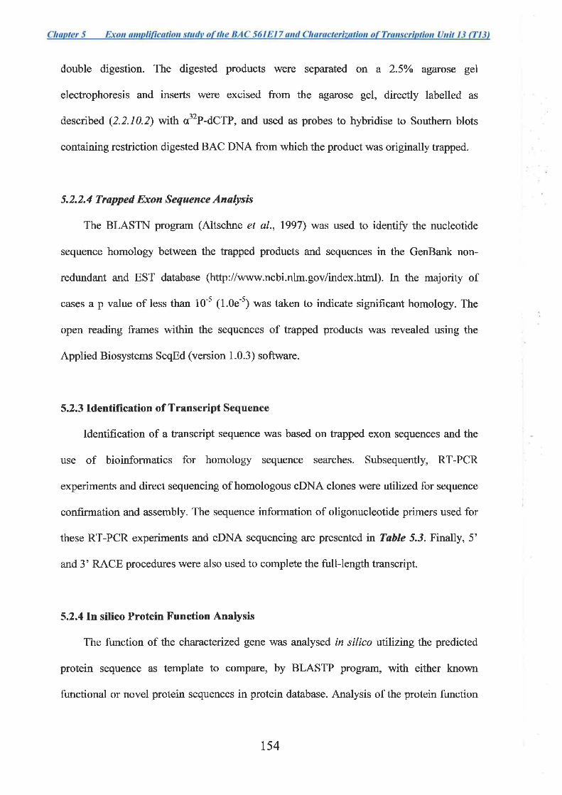
double digestion. The digested products were separated on a 2.5Yo agarose gel
electrophoresis and inserts were excised from the agarose gel, directþ labelled as
described (2.2.10.2) with o32P-dCTP, and used as probes to hybridise to Southern blots
containing restriction digested BAC DNA from which the product was originally trapped.
5.2.2.4 Trapped Exon Sequence Analysß
The BLASTN program (Altschne et al., 1997) was used to identifu the nucleotide
sequence homology between the trapped products and sequences in the GenBank non-
redtmdant and EST database (http://www.ncbi.nlm.gov/index.html). In the majority of
cases a p value of less than 10-s (1.0e-s) was taken to indicate signifrcant homology. The
open reading frames within the sequences of trapped products was revealed using the
Applied Biosystems SeqEd (version 1.0.3) software.
5.2.3 Identification of Transcript Sequence
Identification of a transcript sequence was based on trapped exon sequences and the
use of bioinformatics for homology sequence searches. Subsequently, RT-PCR
experiments and direct sequencing of homologous cDNA clones were utilized for sequence
confirmation and assembly. The sequence information of oligonucleotide primers used for
these RT-PCR experiments and oDNA sequencing are presented tn Table 5.3. Finall¡ 5'
and 3' RACE procedures were also used to complete the full-length transcript.
5.2.4I.n silico Protein Function Analysis
The function of the characterized gene was analysed in silico utilizing the predicted
protein sequence as template to compare, by BLASTP program, with either known
functional or novel protein sequences in protein database. Analysis of the protein function
r54
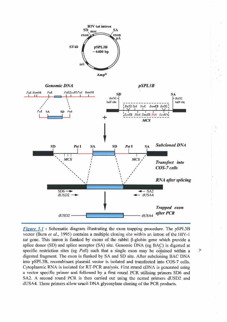
psPL3B- 6400 bp
SD
SV4O
ort
Genomìc DNAPstl BamHf PsfI PsilEcoRIPstl BamIII
Pstl SA SD PstI
psPL3B
SABst)ilhalf site
,Bsf)(I Ssd .Àr'oll Bamflf Bs¡Xl
EcoRI Xmalll
MCS
pstt SA Subcloned DNA
I
HIV tat intron
AmpR
exon
SD.Bst)fl
halfsite
+
SDSD SAIPst
tttMCS
\ \ \ \
+dUSD2 +
dUSD2
IIIItIIIIt
,,I,,I,t,,
I
MCS / ' Trans¡ect ìntoCOS-7 cells
RNA after splicìng
++ dUSA4
Trapped exon
dUSA4 after PCR
Fieure 5.1 : Schematic diagram illustrating the exon trapping procedure. The pSPL3Bvector (Burn et a1.,1995) contains a multiple cloning site within an intron of the HIV-Itat gene. This intron is flanked by exons of the rabbit B-globin gene which provide a
igested atwithin a
AC DNAinto pSPL3B, recombinant plasmid vector is isolated and transfected into COS-7 cells.Cytoplasmic RNA is isolated for RT-PCR analysis. First strand cDNA is generated usinga vector specific primer and followed by a first round PCR utilising primers SD6 andSA2. A second round PCR is then carried out using the nested primers dUSD2 anddUSA4. These primers allow uracil DNA glycosylase cloning of the PCR products.
X
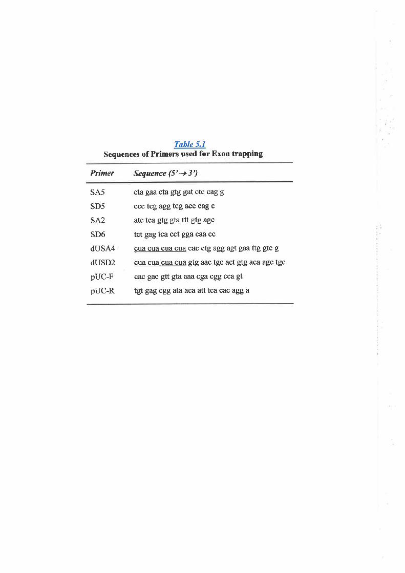
Tøble 5.1
Sequences of Primers used for Exon trapping
Prímer Sequence (5'+ 3')
SA5
SD5
SA2
SD6
dUSA4
dUSD2
pUC-F
pUC-R
cta gaa cta gtg gat ctc cag g
ccc tcg aggfcg acc cag c
atc tca gtg gta ttt gtg agc
tctgagtcacct ggacazcc
cua cua cua cua cac ctg agg agt gaaftg $lc g
cua cua cua cua úgaac tgc act gfgaca agc tgc
cac gac gtt gta aaa" cga cgg cca g!.
tgt gag cggataacaalt tca cac agga
was also carried out using the protein domain prediction program as described in previous
chapters.
5.3 Results
5.3.1 Exon Trapping
Exon trapping was performed on a genomic clone BAC 561E17. This BAC extends
approximately 140 kb from the proximal boundary of the cosmid contig of the 16q24.3
physical map constructed at this time. BAC DNA was double digested with BamHI and
BgIII or with PstI alone and subcloned into the pSPL3B exon trapping vector (Figure 5.1).
The subsequent protocol then involved transfection into COS-7 cells, RT-PCR
amplification of the trapped fragments, which were then cloned into the pAMPlO vector.
From this transformation, a total of 137 colonies were randomly selected from the primary
plates. These were transferred to a master plate for further analysis. Colony PCR was
carried out to identiff the recombinants containing trapped sequences. Colonies that gave
products corresponding with "vector-only" inserts (5.2.2.1) were omitted from further
analysis. The remaining colonies were grouped according to the insert size and one to three
colonies representing each group of the same insert size were then selected and grown for
DNA isolation and sequencing. A total of 36 colonies were finally selected and subjected
to DNA isolation and sequencing.
5.3.2 Trapped Exon Sequence Analysis
Sequence analysis of the selected clones for the presence of open reading frame and
sequence homology to known genes or partially sequenced ESTs in the public sequence
database are summarised in Table 5.2. lîttial analysis indicated that the trapped exons
could potentially code for 5 different transcripts.
155
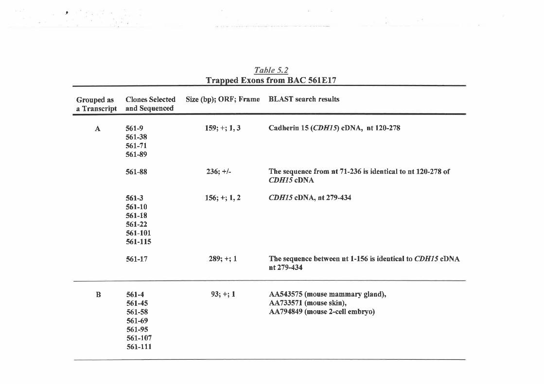
,
Grouped as
a Transcript
B
Clones Selectedand Sequenced
A
Tøble 5.2Trapped Exons from BAC 56lB17
Size (bp); ORF; X'rame BLAST search results
159; +; 1,3 Cadherin 15(CDHIÐ cDNA, nt120-278
236; +l-
561-9561-38561-71s61-89
561-88
561-3561-10561-18s6t-22561-101561-115
561-17
561-4s6l-45561-58561-69561-95561-107561-111
289; +; I
The sequence from ntTl-236 is identical to ntl2O-278 ofCDHIí cDNA
156;+;1,2 CDHIj cDNA, nt279-434
The sequence between nt 1-156 is identical to CDHI5 cDNAnt279-434
93; +; I 44543575 (mouse mammary gland),44733571 (mouse skin),AA794849 (mouse 2-cell embryo)
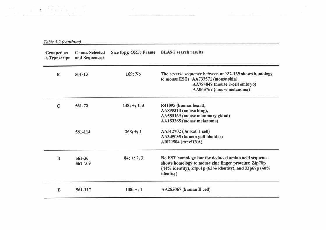
?
2
Grouped asa Transcript
Clones Selected Size (bp); ORF; Frame BLAST search resultsand Sequenced
B
c
D
561-13
56r-72
561-ll4
561-36s61-109
169; No
148; +; 1,3
268¡ +; I
84; +;2,3
The reverse sequence between nt 132-169 shows homologyto mouse ESTs: 114733571(mouse skin),
AA794849 (mouse 2-cell embryo)44065769 (mouse melanoma)
R41095 (human heart),44895310 (mouse lung),44553169 (mouse mammary gland)44153265 (mouse melanoma)
A 312702 (Jurkat T cell)44345035 (human gall bladder)AJ:029504 (rat cDNA)
No EST homology but the deduced amino acid sequenceshows homology to mouse zinc finger proteins: Zfp70p(44% identity), Zfp6lp (62% identity), and Zfp67p (0"/"identity)
E 561-lt7 108; +; 1 44285067 (human B cell)
Trapped exons 561-9, 561-38, 561-71, and 561-89 were from the same exon and had
two possible ORFs either in frame I or frame 3. Homology sequence analysis showed that
they were trapped from the gene sequence corresponding with nucleotides 120-278 of
Cadherin 15 oDNA (GenBank accession no. NM-004933). Human cadherin 15 (CDHI5:
M-cadherin) has previously been mapped to this region (Kaupmann et al., 1992;
Kremmidiotis et al., 1998). The trapped product 561-88, though of a larger size (236 bp)
when compared with that of 561-9 (159 bp), was also trapped from the same part of
CDHL5 (cDNA nt 120-278). Present, however, was an extra 70 bp of 5' sequence. Analysis
of the sequence at this junction of CDHL5 exon and the extra 70 bp indicated the presence
of a nucleotide sequence resembling a splice acceptor consensus. This indicates that part of
the adjacent "intron" was likely to be included in this trapped product (Fìgure 5.2).
The region between nucleotides 279 and 434 of the CDHI5 cDNA were also repeatly
trapped as shown by sequencing of the clones 561-3, 561-10, 561-18, 561-22,561-101,
and 561-115. The trapped product 561-17 was also trapped from this part of the CDHL5
sequence but also included a 133 bp sequence likely to be part of the adjacent 3' intron
(Figure 5.2).
A group of 7 trapped exons represented by clone 561-4 were homologous and were
likely trapped from an exon of an uncharacterized human gene. There was no homology to
human ESTs though homology was found to three overlapping mouse EST sequences.
These were vj82g04.rl, vu73b05.rl, and vr48d04.s1 (GenBank accession no. AA543575,
AA733571, and 4A794849 respectively). These three overlapping ESTs form a cDNA
sequence of approximately 1.1 kb. The sequence homology between 561-4 and mouse
cDNA was high, with 89% identity.
r56
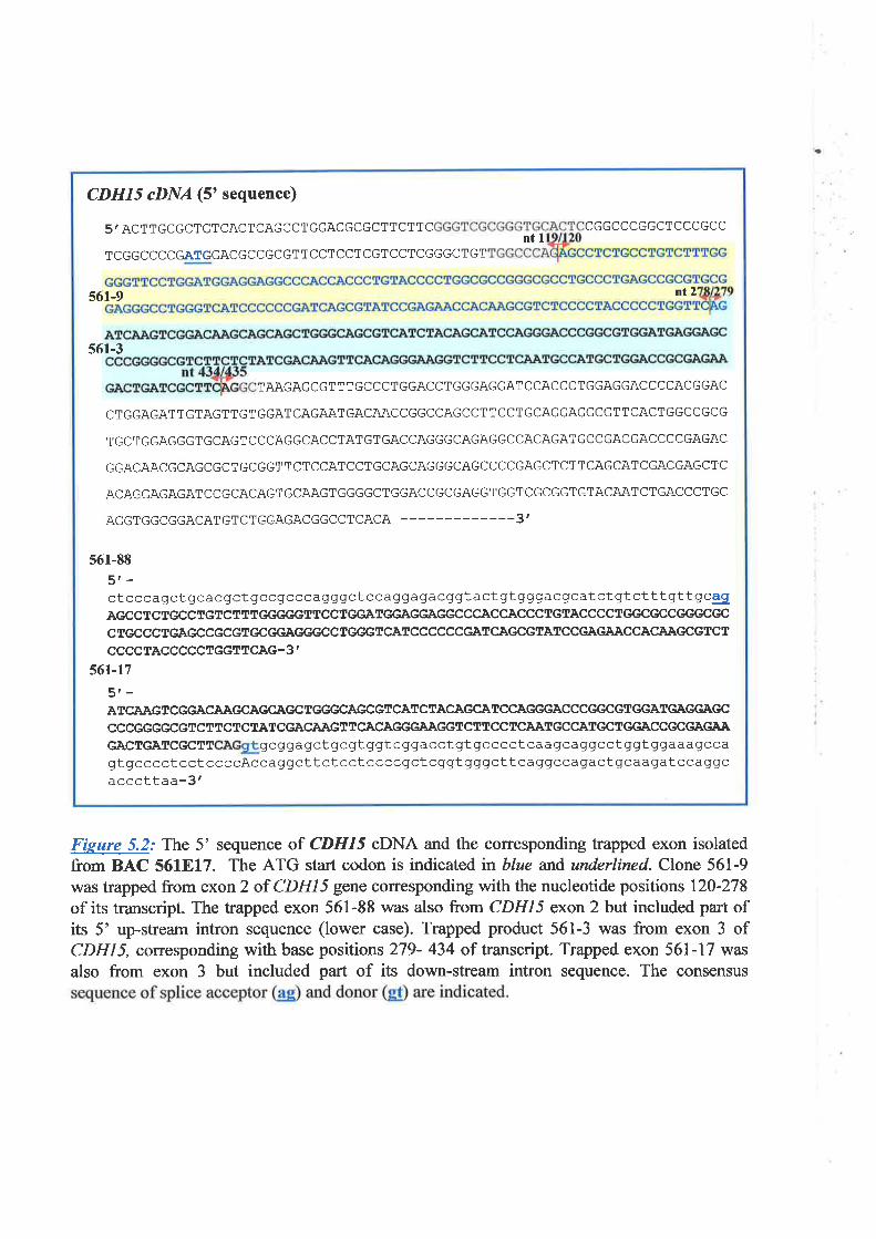
CDHI 5 øDNA (5' sequence)
5' ACT TGCGCTGTCACTCAGCCTGGACGCGCTTCTTC CGGCCCGGCTCCCGCCntl
TCGGCCCCGATGGACGCCGCGTTCCTCCTCGTCCTCGGGCTGT
561-9 nt2
56r-3
TAAGAGCGTTTGCCCTGGACCTGGGAGGATCCACCCTGGAGGACCCCACGGAC
CTGGAGATTGTAGTTGTGGATCAGAATGACAACCGGCCAGCCTTCCTGCAGGAGGCGTTCAClGGCCGCG
TGCTGGAGGGTGCAGTCCCAGGCACCTATGTGACCAGGGCAGAGGCCACAGATGCCGACGACCCCGAGAC
GGACAACGCAGCGCTGCGGTTCTCCATCCTGCAGCAGGGCAGCCCCGAGCTCTTCAGCATCGACGAGCTC
ACAGGAGAGATCCGCACAGTGCAAGTGGGGCTGGACCGCGAGGTGGTCGCGGTGTACAATCTGACCCTGC
AGGTGGCGGACATGTCTGGAGACGGCCTCACA -_--3'
561-88
5r-ctcccagctgcacgctgccgcccagggctceaggagacggtactgtgggacgcatctgtctttgttgcegÀGCCTCTGCCTGTCÎTTGE{,GGTTCCTGGATGGAGGAGGCCCACCACCCTGTACCCCTGGCGCCGGGCGCCTGCCCTGAGCCGCGTGCGGAGGGCCIGGGTCATCCCCCCGATCAGCGTATCCGAGAACCACÀÀGCGTCTCCCCTACCCCCTGGÍTCAG-3'
561-17
5r-ATCAÀGTCGGACAÀGCAGCAGCTGGGCAGCGTCÀTCTACAGCATCCAGGGACCCGGCGTGGATGAGGAGC
CCCGGGGCGTCTTCTCTÀTCGACAÀGTTCACAGGGAAGGTCTTCCTCAATGCCATGCTGGACCGCGÀGAAGÀCTGÀTCGCTT gcggagctgcgtggtcggacctgtgcccctcaagcaggcctggtggaaaqccagtgcccctcctccccAccaggcttctcctccccgctcggtgggcttcaggccagactgcaagatccaggcacccttaa-3'
Fieure 5.2.' The 5' sequence of CDHI5 cDNA and the coresponding trapped exon isolatedfrom BAC 561817. The ATG start codon is indicated in blue and underlined. Clone 561-9
was trapped from exoíz of CDHL5 gene coffesponding with the nucleotide positions 120-278of its transcript. The trapped exon 561-88 was also from CDHIï exon 2 but included part ofits 5' up-stream intron sequence (lower case). Trapped product 561-3 was from exon 3 ofCDH15, corresponding with base positions 279- 434 of transcript. Trapped exon 561-17 was
also from exon 3 but included part of its down-stream intron sequence. The consensus
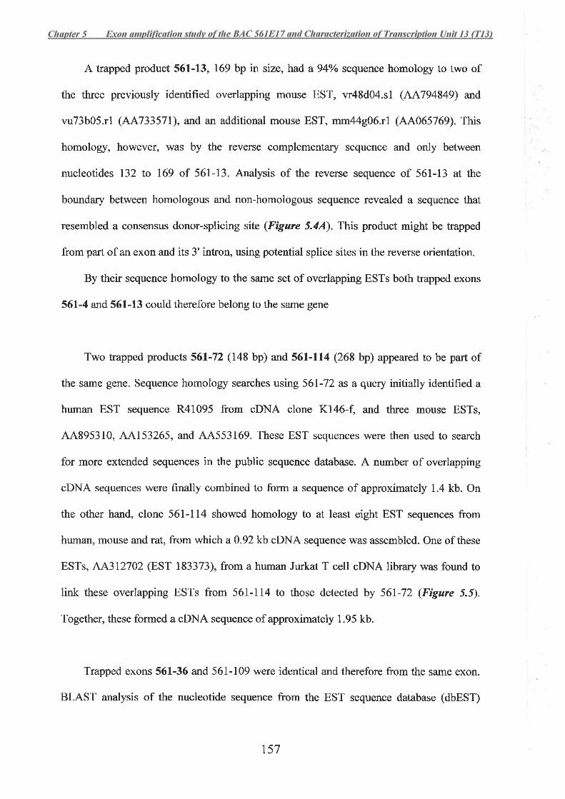
A trapped product 561-13, 169 bp in size, had a 94olo sequence homology to two of
the three previously identified overlapping mouse EST, vr48d04.sl (AA794849) and
vu73b05.rl (AA733571), and an additional mouse EST, mm44g06.rl (44065769). This
homology, however, was by the reverse complementary sequence and only between
nucleotides 132 to 169 of 561-13. Analysis of the reverse sequence of 561-13 at the
boundary between homologous and non-homologous sequence revealed a sequence that
resembled a consensus donor-splicing site (Fìgure 5.4A). This product might be trapped
from part of an exon and its 3' intron, using potential splice sites in the reverse orientation.
By their sequence homology to the same set of overlapping ESTs both trapped exons
561-4 and 561-13 could therefore belong to the same gene
Two trapped products 561-72 (148 bp) and 561-114 (268 bp) appeared to be part of
the same gene. Sequence homology searches using 561-72 as a query initially identified a
human EST sequence R41095 from cDNA clone Kl46-f, and three mouse ESTs,
44895310, 44153265, and 44553169. These EST sequences were theíused to search
for more extended sequences in the public sequence database. A number of overlapping
cDNA sequences were finally combined to form a sequence of approximately 1.4 kb. On
the other hand, clone 561-114 showed homology to at least eight EST sequences from
human, mouse and rat, from which a0.92 kb cDNA sequence was assembled. One of these
ESTs, AA312702 (EST 183373), from a human Jurkat T cell cDNA library was found to
link these overlapping ESTs from 561-114 to those detected by 561-72 (Figure 5.5).
Together, these formed a cDNA sequence of approximately 1.95 kb.
Trapped exons 561-36 and 561-109 were identical and therefore from the same exon.
BLAST analysis of the nucleotide sequence from the EST sequence database (dbEST)
157
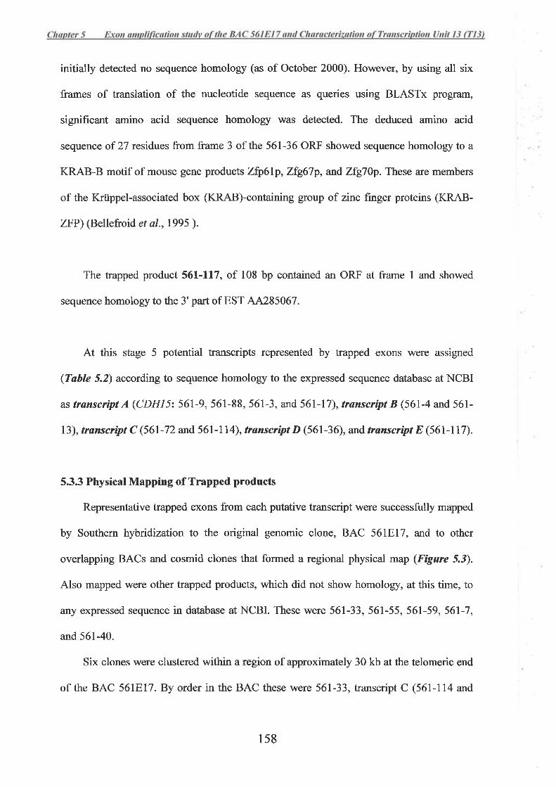
initially detected no sequence homology (as of October 2000). However, by using all six
frames of translation of the nucleotide sequence as queries using BLASTx program,
signifrcant amino acid sequence homology was detected. The deduced amino acid
sequence of 27 residues from frame 3 of the 561-36 ORF showed sequence homology to a
KRAB-B motif of mouse gene products Zþ61p, Zfg67p, and Zfg7Ùp. These are members
of the Krüppel-associated box (KRAB)-containing group of zinc finger proteins (KRAB-
ZFP) (Bellefroid et a1.,1995 ).
The trapped product 561-117, of 108 bp contained an ORF at frame I and showed
sequence homology to the 3'part of EST 44285067.
At this stage 5 potential transcripts represented by trapped exons were assigned
(Table 5.2) according to sequence homology to the expressed sequence database at NCBI
astranscriptA(CDHI5z561-9,56I-88,561-3,and561-17),transøíptB(561-4 and561-
73), trønscript C (561-72 and 561-l l4), transuípt D (561-36), ard transuipt E (561-117).
5.3.3 Physical Mapping of Trapped products
Representative trapped exons from each putative transcript were successfully mapped
by Southern hybridization to the original genomic clone, BAC 56lEl7, and to other
overlapping BACs and cosmid clones that formed a regional physical map (Figure 5.3).
Also mapped were other trapped products, which did not show homology, at this time, to
any expressed sequence in database at NCBI. These were 561-33, 561-55, 561-59,561-7,
and 561-40.
Six clones were clustered within a region of approximately 30 kb at the telomeric end
of the BAC 561El7.By order in the BAC these were 561-33, transcript C (561-114 and
1s8
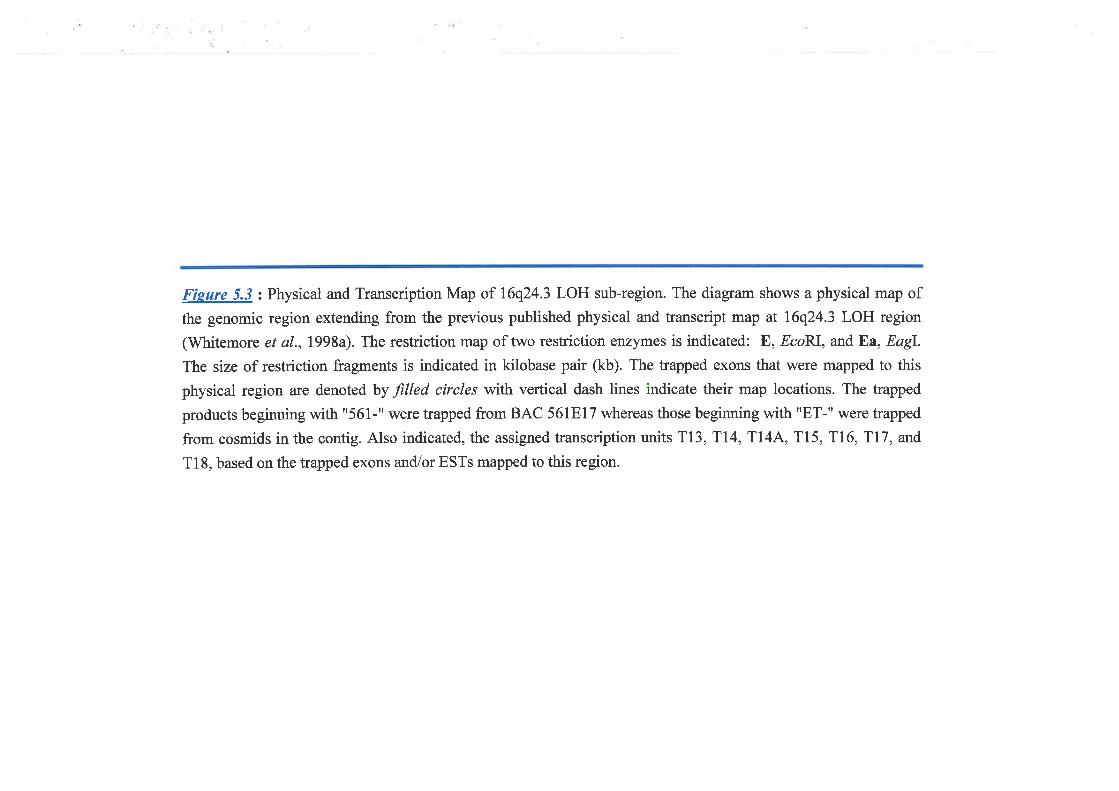
Fíeure 5.3: Physical and Transcription Map of 16q24.3 LOH sub-region. The diagram shows a physical map of
the genomic region extending from the previous published physical and transcript map at 16q24.3 LOH region
(Whitemore et al.,l998a). The restriction map of two restriction enzymes is indicated: E, EcoRI, and Ea, Eøgl.
The size of restriction fragments is indicated in kilobase pair (kb). The trapped exons that were mapped to this
physical region are denoted by filled circles with vertical dash lines indicate their map locations. The trapped
products beginning with "561-" were trapped from BAC 56IEl7 whereas those beginning with rrET-rr were trapped
from cosmids in the contig. Also indicated, the assigned transcription units TI3,Tl4, Tl4A, T15, Tl6, T77, and
Tl8, based on the trapped exons and/or ESTs mapped to this region.
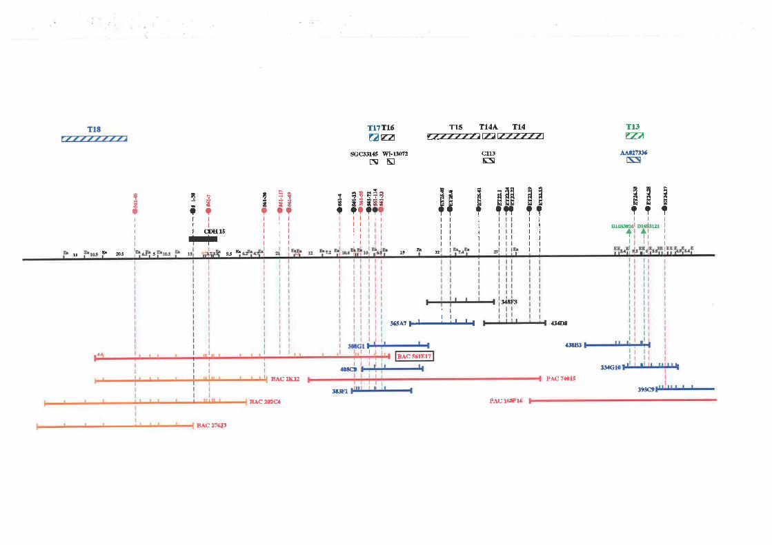
T1317Á
AAqntx
TI5 T14Ä TL4ft77777ar7AÈ-777771
Tt7TI:6nFa
sccrl3145 wl-t3/ù7zE¡Ñ
cll3FE
ttññÞÞ??tt
D16ñìü¿6 121+1itttt+
F
tÞ
?I
I
I
I
I
ddLi Èì
??ltftltttttrl
8Nd¡NÈ¡ È¡n
NÞ
?I
I
I
I
I
$RÊ¡o
I
I
I
I
I
I
I
qIq
?I
I
I
I
I
I
Ir{Þ
?I
I
I
I
I
ì.l.?EEUÇrtt
9glÈE3ÇÇltlltttttt
't
?I
I
I
I
I
!I?
I
I
I
I
I
tt??tttltlttll
ÌIÇ
I
I
CDII 15
!Ç
I
I
I
çE?
I
I
I
I
I
bb9.5ll h û3 s t9
BAC5ó1817
:XtRt
36sa7 FJ-LjJJ.{
I
{3{Dû
aStEt
PAC 74015
I
{0tct :t34Gl0
tr3c9
BAC 2X'L¿
3t381
B.LC2V2c4 PAC 16,1P16
BAC ?76¡3
561-72),561-55, and transcript B (561-13 and 561-4). At this region of the physical map, a
oDNA 118499 (initially designed STS SGC33I45) belonging to a group of oDNA clones
collectively referred to a Unigene Cluster Hs.10238 was also mapped initially by PCR
(Whitmore, S.A. and Powell, J., personal communication), and was assigned as
transcription unit 17 (T17).It was then confirmed by Southern hybridization at this time to
this region of the BAC 561E17.
Three trapped products, 561-59,561-117 (transcript E), and 561-36 (transcript D)
were localizednear the middle region of the BAC 561E17.
The trapped clones 561-38 (CDHL5 nt 120-178) was co-localized with 561-7.In the
present study, PCR products amplified from 5' and 3'region of CDHIS cDNA were also
used as probe for Southem hybridization and mapped specifically to this region of BAC
56tBt7.
Trapped exon 561-40 was mapped close to the proximal end of the BAC and distal to
the region that cDNA clone IMAGE:1098126 Q18) was initially mapped by Southern
hybridization (Whitrnore, S.4., personal communication).
5.3.4 Verification of the Transcript Sequence
Among the trapped products isolated from the BAC 561E17, there were two group of
trapped exons initially assigned as transuipt B (561-4 and 561-13) and transcript C (561-
72 and 561-114) and the trapped products 561-33 and 561-55 clustered at the distal region
of this BAC (Figure 5.3). Due to their physical proximity it was considered likely that they
were part of the same transcript and so were chosen for further study.
5.3.4.1 Transcript Sequence generatedfrom 561-4 and 561-13
Two trapped exons, 561-4 and 561-13 were homologous to the mouse cDNA
159
sequence formed by at least three overlapping mouse ESTs, AA543575, AA733571, and
AA794849. This group was initially assigned as transuipl,8 (section 5.3.2). The order of
these two trapped exons wÍrs arranged with respect to the mouse EST sequences, and,.,ra--t 1^-
showed that 561-13,^ situated approximately 350 bp 5' upstream from the exon 561-4
(Figure 5.4 B). RT-PCR using a forward primer within the 561-13 sequence and a reverse
primer from the 561-4 sequence (Table 5.3) successfully amplified a product of 357 bp
from human fetal heart total RNA. The total human cDNA sequence of 378 bp between
561-13 and 561-4 was assembled and was homologous to the overlapping mouse cDNA
sequences (Figure 5.4 B).
To identiff additional human cDNA sequence extending the 378-bp product, an
assembled mouse cDNA sequence of 1,022 bp generated from the three overlapping mouse
ESTs, mentioned above, was used to screen for homologous human DNA sequences from
both EST and non-redundant databases. The search results identified a genomic sequence
which was part of the PAC 203P18 (GenBank accession no. 297180). This PAC was
derived from the human X-chromosome region Xq27.l-q27.3. Homology between the
PAC 203P18 sequence and that of the mouse cDNA was 88oá. This homology was only
from the first 835 bp of the assembled 1,022-bp mouse cDNA sequence. The remaining
187 bp showed no significant homology (Figure 5.4 B). The 378-bp human cDNA
between exons 561-13 and 561-4 was 100% homologous to this corresponding sequence of
the X chromosome.
The finding that part of genomic DNA from the X chromosome showed sequence
homology to the cDNA sequence of at least 835 bp suggested that this part of the X
,.
chromosome sequence is a processed ps;f.dogene. Further analysis of the PAC 203P18 þ
DNA sequence, which flanked this region of homology, showed that there were adjacent
Ll elements. A 1.39-kb Ll element was situated 338 bp 5' to this 835 bp homologous
160
region and a 0.85-kb Ll element was situated 854 bp 3' to this region. Flanking by the Ll
elements at both ends suggested the likelihood that this DNA segment had been transposed
as a mobile element. A processea pr$aog"ne generally is a DNA fragment being copied )c
from a processed RNA transcript and transposed to the genomic region by the transposable
elements such as Ll repeat (Esnault et a1.,2000). The above evidence again supported that
this genomic sequence of the X chromosome, flanking by Ll elements, was a processed
nrfaoe"o" prr16dog"tt"of the transcript B. The processed
therefore would not be expressed.
The additional sequence of the
did not contain a promotor and À4\
psr.Êdogene which flanked the 835-bp homologous ¡.
sequence was then utilized to detect additional sequence homologies from%atabase.
Homology searches had identified at least one human- and three mouse-ESTs, accession
no. 41203683, A1461784, A1197222, and 4I121535 respectively. These aligned with the
sequence 3' to the region homologous to 561-l3156l-4 product (Fígure 5.48). To further
identifr if these EST sequences belonged to, and formed part of, the same transcript with
561-13156l-4, the human cDNA clone qf54a03 (EST 41203683) was sequenced. The
result showed that the sequence of this clone overlapped with the sequence generated from
the trapped exons 561-4 and 561-13. The combined cDNA sequence of 1,263 bp was again
gene. !"
was undertaken to provide additional >
information related to its original transcript. The total 2,026-bp sequence of this
pseudogene revealed an ORF of 1167 bp which extended from the first nucleotide (Figure
5.48). There was no start codon. A stop codon, TGA, was identified between the
nucleotide positions 1168 and 1170 which was then followed by the non-coding sequence
likely to be a 3'UTR. The stop codon was also detected in the corresponding mouse cDNA
at the homologous and non-homologous junction (Figure 5.48). The lack of significant
161
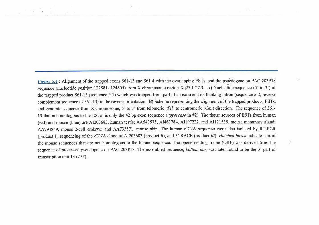
Fieure 5.4: Alignment of the trapped exons 561-13 and 561-4 with the overlapping ESTs, and the ptg,Êdog.tte on PAC 203P18
sequence (nucleotide position 12258I- 124605) from X chromosome region Xq27.l-27.3. A) Nucleotide sequence (5'to 3') of
the trapped product 561-13 (sequence # 1) which was trapped from part of an exon and its flanking intron (sequence # 2, rcverse
complement sequence of 561-13) in the reverse orientation. B) Scheme representing the alignment of the trapped products, ESTs,
and genomic sequence from X chromosome,5'to 3' from telomeric (Tet)to centromeric (Cen) direction. The sequence of 561-
13 that is homologous to the ESTs is only the 42 bp exon sequence (uppercase in#2). The tissue sources of ESTs from human
(red) and mouse (blue) arc A1203683, human testis; AA543575, A1461784, A1197222, and AIl2l535, mouse manìmaf,y gland;
AA794849, mouse 2-celI embryo; and 44733571, mouse skin. The human cDNA sequence were also isolated by RT-PCR
(product r), sequencing of the cDNA clone of A1203683 þroduct ií), and 3' RACE ,(product
ìü). Hatched boxes indicate part of
the mouse sequences that are not homologous to the human sequence. The opend reading frame (ORF) was derived from the
sequence of processed pseudogene on PAC 203P18. The assembled sequence, bottom bar,was later found to be the 3' part of
transcription unit 13 gI3).
)
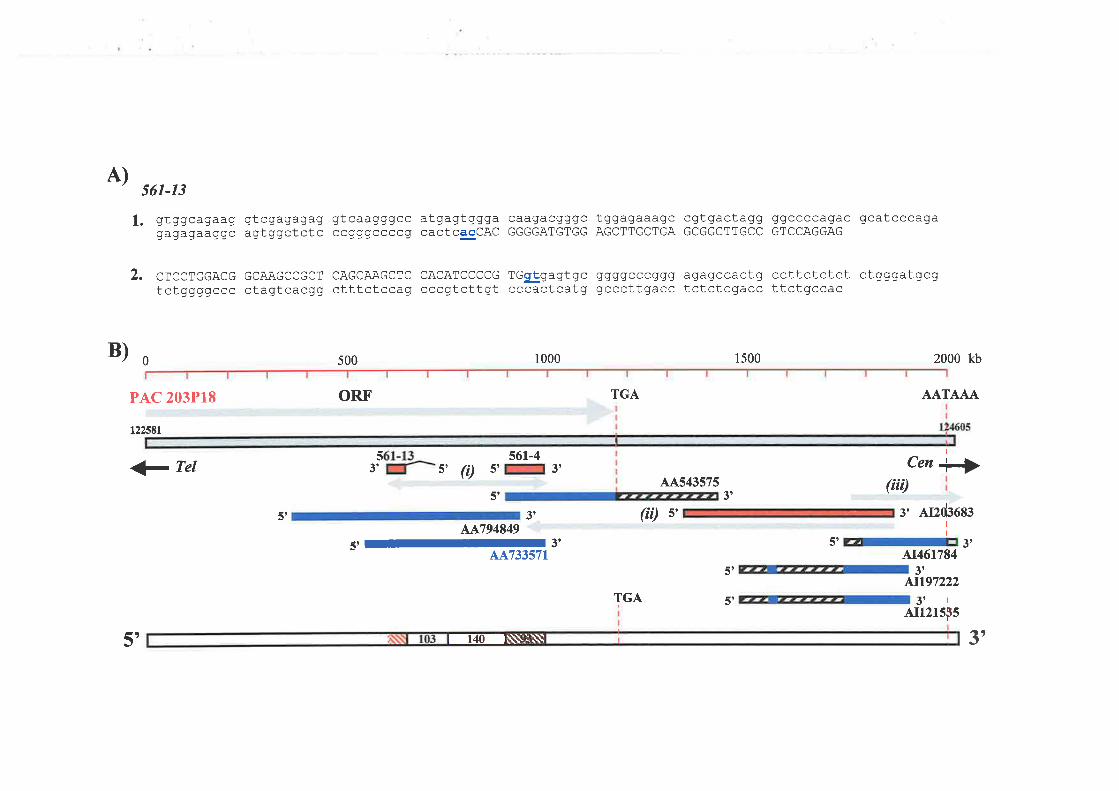
A)
B)0
561-13
1. gtggcagaag gtcgagagag gtcaagggcc atgagtggga caagacgggc tggagaaagc cgtgactagg ggccccagac gcatcccagagagagaaggc agtggctctc ccaggccccg cactcacCAc GGGGATGTGG AGCTTGCTGA GCGGCTTGCC GTCCAGGAG
2. crccrccAcc GCAAGCCGCT cAGCAAGcTc cACATCCccc rGglgagtgc qqqgcccgqq aqagccactg ccttctctct ctgggatgcgtctggggccc ctagtcacgg ctttctccag cccgtcttgt cccactcatg gcccttgacc tctctcgacc ttctgccac
500 1000 1500 2000 kb
PAC 203P18 TGAORF' AATAAA
t22í8t
1 Tel
51 3'AA794849
5'-3'AA733571
Cen ¡,4A543575 (t t)
3's' Af¿d3683
I
5','r,At461784
51 3t4t197222
5t3tlAIr2rsps
53' s' ft)561-4
51 3t
5'
(íi) s'
TGA
5', I4I\\\:Iì\\ 3')

homology of the mouse sequence to that of the human 3' down-stream from the stop codon
was also observed and supported the likelihood that this sequence was the 3' UTR.
Furthermore, the nucleotide sequence characteristic of the polyadenylation signal,
fu{TAJqA, was also present at the 3' end of this pseudogene. With respect to its process
pseudoge-ne, the identiffing transcript sequence (initially given as transcript B) was likely
the 3'part of the mRNA transcript of a gene.
To confirm the 3' end of the transcript, 3'RACE using a set of primers (Table 5.3)
designed from the sense strand of the cDNA qf54a03 was performed (the procedure
described in section 2.2.1D. The result revealed the same sequence as that present in the
pseudogene but with an extra seven nucleotides prior to the polyadenylation site. These
extra nucleotides were later confirmed by the available genomic sequence of PAC clone
74015 that overlapped with BAC 561E17 (the sequence data has been deposited with
GenBank as part ofNT-010542.13).
5.3.4.2 Transuipt Sequence of 561-72 and 561-l14
Trapped exons 561-114 and 561-72 were shown to linked to each other by a set of
overlapping human and mouse EST sequences as mentioned in section 5.3.2 and shown in
Fígure 5.5. To confirm this, primers were designed for RT-PCR analysis of these
overlapping ESTs and trapped products (Table 5.-t). RT-PCR products were successfully
obtained from total RNA isolated from human fetal heart and fetal kidney (Fìgure 5.Q.
These products were purified and sequenced. Three human cDNA clones, ab40b03
(44486025), ah20h11 (44694610), and od53h03 (AA826123), that formed part of this
overlapping sequence, were also chosen for sequencing. The sequences obtained from the
RT-PCR and cDNA clones were then assembled to form a2.2-kb cDNA sequence.
Analysis of clone 561-114,268 bp in size, showed that it was likely to have been
)
-é1-.-
t62
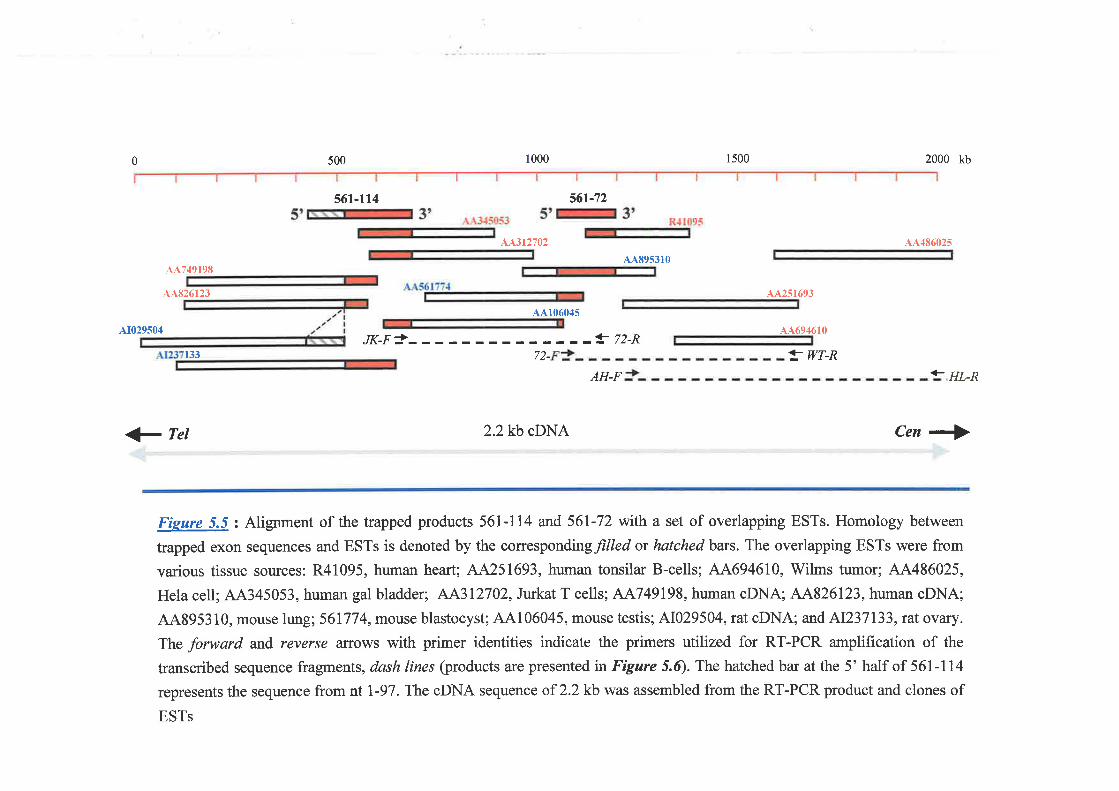
0 500 1000 1500 2000 kb
561-114 561-72
L\312702
44895310
AI029504
AA749198
AA826123
133
aA251693
^dL694610
! WT-R
AA48602s
+- HL-R
Cen +>1 Tel
A4106045
JK-F!- -! 72-R,71
AH-F
2.2kb cDNA
Fieure 5.5 : Alignment of the trapped products 561-114 and 561-72 with a set of overlapping ESTs. Homology between
trapped exon sequences and ESTs is denoted by the correspondingfilled or hatched bars. The overlapping ESTs were from
various tissue sources: R41095, human heart; AA251693, human tonsilar B-cells; AA694610, Wilms tumor; AA486025,
Hela cell; 44345053, human gal bladder; AA312702, Jurkat T cells; AA749198, human cDNA; AA826123, human cDNA;
44895310, mouse lung; 561774, mouse blastocyst; A4106045, mouse testis; A1029504, rat oDNA; and AI237133,rut ovary.
The forward and reverse affows with primer identities indicate the primers utilized for RT-PCR amplification of the
transcribed sequence fragments, dash lines (products are presentedin Figure 5.Q. The hatched bar at the 5' half of 561-114
represents the sequence from nt l-97. The oDNA sequence oî2.2 kb was assembled from the RT-PCR product and clones of
ESTs
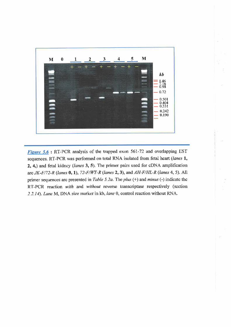
M 0 I 2 3 4 5 M
kb:1.46- 1.16
- 0.98
- 0.72
- 0.501
- 0.404
- 0.331
- 0.242
- 0.190
Fisure 5.ó : RT-PCR analysis of the trapped exon 561-72 and overlapping EST
sequences. RT-PCR was performed on total RNA isolated from fetal heart (lanes l,2, 4,) and fetal kidney (lanes 3, 5). The primer pairs used for cDNA amplification
are JK-F/72-R (lanes O, l), 72-F/WT-R (lanes 2,3), and AH-F/HL-R (lanes 4, 5). Allprimer sequences are presented n Table 5.2a. The p/zs (+) and minus (-) indicate the
RT-PCR reaction with and without reverse transcriptase respectively (section
2.2.14). LaneM, DNA size marker in kb, lane 0, control reaction without RNA.
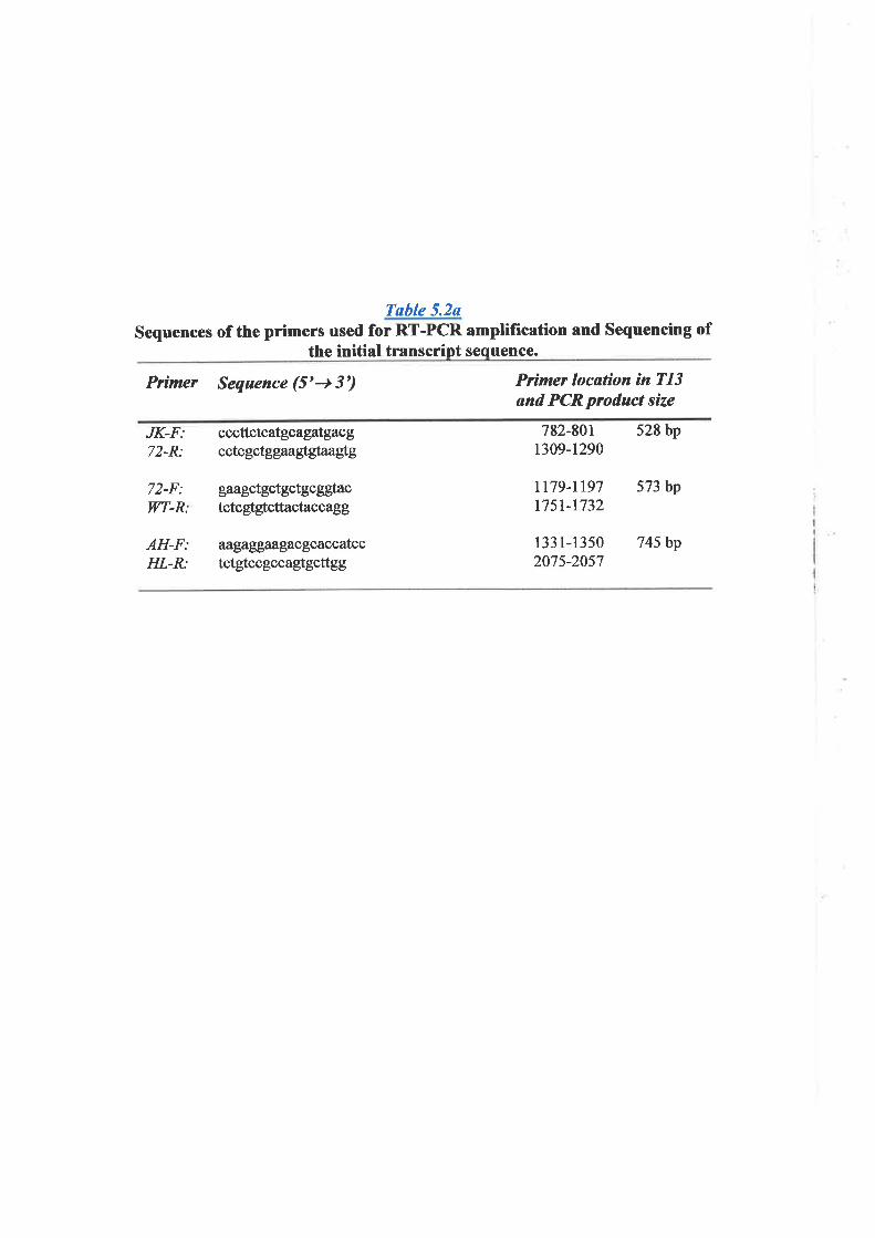
Table 5.2uSequences of the primers used for RT-PCR amplifrcation and Sequencing of
the initial transcript sequence.
Prímq Sequence (5'+ 3') P¡ìmq location ín T13ønd PCRp¡oduct size
JK-F:72-R:
72-F:WT-R:
AH-F:HL-R:
cccttctcatgcagatgacgcctcgctggaaglglaaglg
gaagctgctgctgcggþctctcgtgtcttactaccagg
aîgaggaa;gacgcaccatcctctgtccgccagtgcttgg
782-8011309-t290
tt79-1197l75t-1732
133 l-13502075-2057
528 bp
573bp
745 bp
trapped simultaneously from two adjacent exons. From the results of database searches, the
sequence of 561-114 between nucleotide positions 1 and 97 was only found in a limited
number of cDNA clones. The majority of the identified cDNA clones showed sequence
homology to the region of 561-114 from nucleotide positions 98 to 268 (Fígure 5.5).h
was suggested that the exon corresponding to nucleotides I to 97 of 561-l 14 could have
been altematively included in some transcription products of some tissues, at least those
included in the EST sequence database.
The transcripts B and C were later found to be part of the gene assigned as
transcription unit 13 (T13) described in the next section.
5.3.5 Assembly of the Transcription Unit ß Q13) Sequence.
Transcription unit 13 was originally assigned to the physical map of 16q24.3 LO}J
region from three trapped exons, 8T24.35,8T24.28, añ8T24.17 which clustered within
two overlapping cosmids, 43883 and 334G10 (Figure 5.3). Ms Karen Lower performed
these studies, first by RT-PCR experiments. These, however, failed to provide any
evidence that these exons were part of the same gene. Homology searches of the oDNA
sequence database at NCBI were then performed using the sequence of these exons. Only
8T24.35 identified an overlapping sequence of ESTs. Further homology searches, cDNA
sequencing, and RT-PCR experiments allowed the determination of a cDNA sequence of
2,290 bp. To identiff the transcription product size and expression pattem, a multþle
tissue Northern filter was hybridized with a cDNA insert from the clone zs04a09 which
was part of the assembled 2,290-bp oDNA. The result showed a coÍtmon 9.5 kb mRNA
band with varying degree of expression between the different tissues (Lower, K.M.,
personal communication). Bands of 3.2, 3.0, and 1.0 kb with varying intensity in diflerent
r63
tissue were also detected in this Northern.
During the identification of the combined cDNA sequence of 2.2 kb generated from
the trapped exons 561-114 and 561-72 (transcript C, described in 5.3.4.2) it appeared that
this sequence was identical to the cDNA sequence of TI3, described above. From the
16q24.3 physical map, these two sequences were separated by approximately 120 kb
(Figure 5.3). It is likely that these two oDNA fragments are derived either from the same
gene or from duplicated genes. This was resolved by Southem hybridization to the cloned
DNA of the physical map using RT-PCR products from the TI3 2,290-bp cDNA. The
results showed that most of this sequence mapped to BAC 56lB17 and three cosmids,
308G1, 408C8, and 383F1, which overlapped with the distal end of this BAC (Fígure 5.3).
However, the 5' end of the transcript including 8T24.35, was located in cosmids 43883ü{"4-/¿"'e.
and 334G10 which situated approximately 120 kb from BAC 561E17. This result indicated
that the Tl3 transcript encompassed more than 120 kb of genomic DNA and confirmed
that the two independently isolated sequences were in fact the same transcript.
Northern analysis was also performed using a probe generated from RT-PCR product
of 561-72 and the overlapping ESTs (Fìgure S.fl. This also identified a 9.5 kb band
(Fìgure 5.2). Subsequent cDNA database analysis and oDNA clone sequencing extended
the TI3 cDNA in a 3' direction resulting in a Tl3 cDNA of 4,570 bp. Further extension of
the 3'cDNA sequence based on homology searches to partially sequenced cDNAs in
database (dbEST) was not possible as no additional cDNAs were found which extended
the sequence. Transcript B was also considered to be part of the same gene with transcript
C, and therefore wfihTl3, however, several attempts by RT-PCR failed to link transcript
B with the partial sequence of 4.5 kb of 713.
During the time that this work was in progress, a large scale sequencing project of
t64
chromosom " l{':rblished and generated parts of the genomic sequence within the y
16q24.3 region (Kremmidiotis, G. and Gardner, 4., personal communication, GenBank
accession no. NT-010542.13). One of the genomic sequences was a fragment
encompassing the distal region of 56lEl7. Alignment of the Z/-3 cDNA with this genomic
sequence revealed the partial genomic structure of the TI3 gene. This gene featured a large
intron of approximately 90 kb between exons 2 and 3. The Tl3 exon2had been trapped as
8T24.35 whereas 561-114 allid 561-72 were trapped from exon 5 and exon 8 respectively.
V/ith the availability of genomic sequence a gene prediction program, GeneFINDER, was
utilized to identifr the possible cDNA sequence in this region. This program successfully
predicted and extended the cDNA sequence from the 3' end of the 4,570-bp cDNA.
Interestingly, the extended cDNA sequence îTioíJ*^;d'# genomic sequence and
joined with the transcript B which was identified from the two trapped exons 561-13 and
561-4 situated approximately 10 kb distal from 561-72. Transcript B was identified as the
3'part of cDNA (5.3.4.2) and therefore completed the 3' end of 713. The 5' RACE was
also utilized to identifr the 5' end of 713. This, however, failed to obtain the extended
sequence. The 5' sequence of 713 was, later, extended by the overlapping sequence of
human EST bbl5cO8.yl (4W732837). Finally the 9,282-bp full-length transcript sequence
of TI3 was assembled (Fìgure 5.2), corresponding approximately with its 9.5 kb mRNA
band identified by Northern analysis. The Tl3 cDNA predicted from genomic sequence by
GeneFINDER, with no intron intemrption, was also confirmed by RT-PCR analysis using
a number of primer pairs designed from this cDNA sequence (Table 5.3) and was carried
out on mammary gland poly A+ RNA to avoid genomic contamination. In addition, RT-
PCR products from this and other part of Tl3 transcript sequence all detected the same 9.5
kb mRNA by Northem hybridisation (Figure 5.7).
165
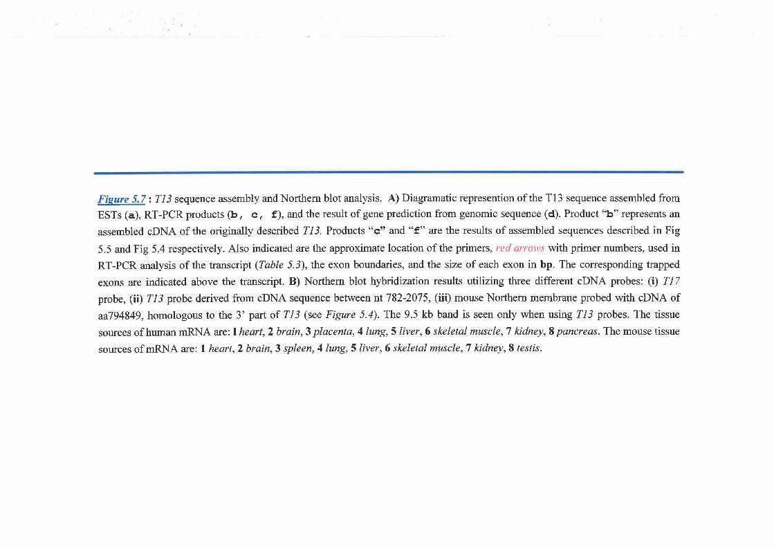
Fieure 5.7: 7/3 sequence assembly and Northern blot analysis. A) Diagramatic represention of the Tl3 sequence assembled from
ESTs (a), RT-PCR products (b, c I f), and the result of gene prediction from genomic sequence (d). Product "b" represents an
assembled cDNA of the origínally described 213. Products "c" aîd "f" are the results of assembled sequences described in Fig
5.5 and Fig 5.4 respectively. Also indicated are the approximate location of the primers, red arcows with primer numbers, used in
RT-PCR analysis of the transcript (Table 5.3), the exon boundaries, and the size of each exon in bp. The corresponding trapped
exons are indicated above the transcript. B) Northern blot hybndization results utilizing three different cDNA probes: (Ð fI7probe, (iÐ Z-t probe derived from cDNA sequence between nt 782-2075, (iii) mouse Northern membrane probed with cDNA of
aa794849, homologous to the 3' partof TI3 (see Figure 5.4). The 9.5 kb band is seen only when using 713 probes. The tissue
sources of human mRNA arc;l heart,2 brain,3 placenta, 4 lung,5 liver,6 skeletal muscle, T kidney, S pancreas. The mouse tissue
sources of mRNA are: I heart,2 brain,3 spleen,4lung,5liver,6 skeletal muscle,T kidney,S testis.
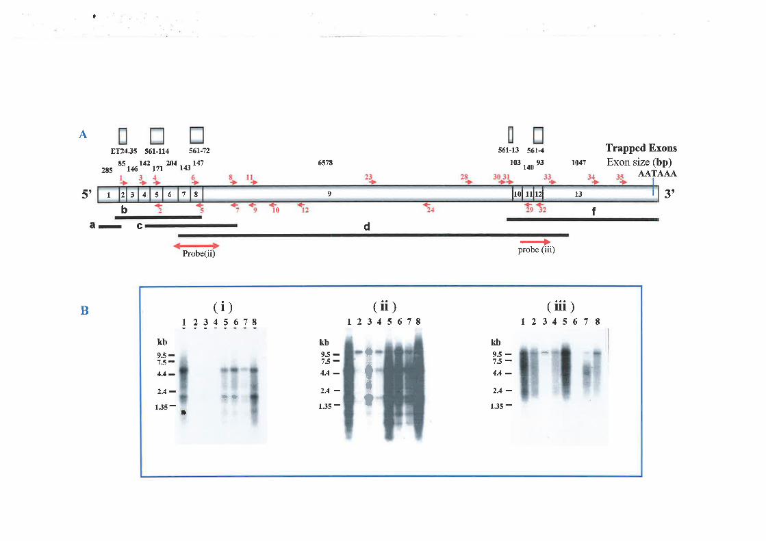
?
A8T2435 561-114
8s t42 204285 146
bâ- C
56tJ2
TßI476578
s61-ß 5614
103 140
93
Trapped ExonsExon size (bp)
AATAAA
3t
104717r
t'0 f
d
Probe(ii) probe (iii)
B
t t 3 4 5 6 7 8 9 Irolulrzl t3
I5
tr'
(ii)1234s678
(iii)1234567I
(
!?3-tkb
9.5-1.5 -4.4 -2.4 -
135 - ]
)678
kb95-7.5-4A-
2.4 -135 -
kb9.5 -7S-4.4 -2.4 -
1.35 -
ff,tt r[
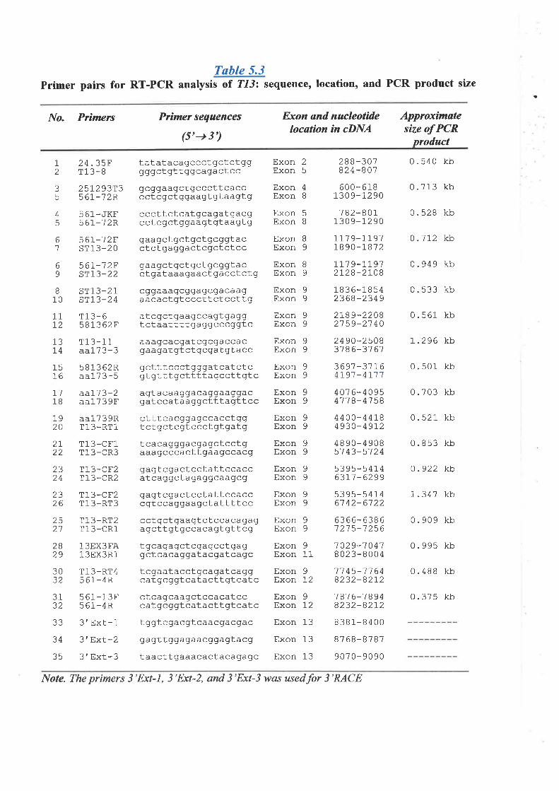
Tuble 5.3Primer pairs for RT-PCR analysis of T1-1: sequence, location, and PCR product size
a
No. Primers Prìmer sequences
(5'-+ 3')
Exon and nucleotidelocøtìon in cDNA
Approximatesíze of PCR
product
12
aJ
5
4EJ
61
69
I10
24.35FT13-8
tatatacagccctgctctgggggctgttggcagactccgcggaagctgcccttcacccctcgctggaagtgtaagtg
cccttctcatgcagatgacgcctcgctggaagtgtaagtggaagctgctgctgcAgtacctctgaggactcgctctccgaagctgctgctgcggtacct gataaagaact gacct ct g
cggaaagcggagcgacaagaacactgtcccttctccttgatcgctgaagccagtgaggtctaattttgaggcccggtcaaagcacgatcgcgaccacgaagatgtctgcAatgtacc
gctttccctgggatcatctcgt gttt gcttttagcctt gt c
agtacaaggacaggaaggacgatccataaggctttagtt cc
ctttcacgqagccacctggtctgctcgtccctgtgatgtcacagggacgagctcctgaaagcccacttgaagccacg
gagt cgact cct att ccaccatcaggctagaggcaagcg
gagtcgactcctattccacccgtccaggaagctattttcc
cct gct gaagtctccacagagagcttgtgccacagtgttcg
tgcagagctcgagcctgaggctcacaggatacgatcagc
tcgaatacctgcagatcaggcat gcggt catactt gt cat c
ctcagcaagctccacatcccat gcggtcatactt gtcatc
ExonExon
ExonExon
Exon 8Exon 9
Exon 9Exon 9
Exon 9Exon 9
Exon 9Exon 9
Exon 9Exon 9
ExonExon
ExonExon
ExonExon
ExonExon
ExonExon
ExonExon
ExonExon
ExonExon
ExonExon
Exon 13
Exon 13
Exon 13
288-30-1824-801
600-618130 9-12 90
782-80113 0 9- t_2 90
1L7 9-1191r890-r87 2
rr1 9-L]912728-2rO8
1836-18542368-2349
2L89-22082159-2'7 40
2490-250837 86-31 61
3691-3'11-64191 -4r114076-40954't1B-4'758
0. s40 kb
0.713 kb
0.528 kb
0.1L2 kb
0.949 kb
0.533 kb
0.561 kb
L.296 kb
0. s01 kb
0.703 kb
0. s21 kb
0.853 kb
0.922 kb
1.347 kb
0.909 kb
0.995 kb
0.488 kb
0.375 kb
25
4B
25r293T356r-12R
5 61_JKF567-12P'
56I-'72Fsr13-20
56I-1 2FsTL3-22
ST13-2Isr13-24
T13-658]-362F
T1 3-1 1aa173-3
581362Raa173-5
aal'|3-2aa17 3 9F
aa173 9RT13-RT1
T13-CF1T13-CR3
T13-CF2T13-CR2
T13-CE2Tl-3-RT3
T13-RT2T13-CR1
13EX3FA13EX3R1
T13-RT4561-4R
561-13F5 61-4R
Exon 5Exon 8
I9
onExonEx
111-2
13L4
1576
711B
1920
2Laa
2324
2326
25./. I
2829
99
99
99
99
99
99
4400- 44L84930- 49r2
1882
911
o
t2
9L2
3032
4890-4908s] 43-5724
5395-541463L1 -6299
5395-s41461 42-6122
6366-63861 21 5-1 256
1 029-7 0 418023-8004
17 45-11 648232-82L2
7 6-7 89432-82L2
8381-8400
8768-8787
907 0- 90 90
3132
34
35
3'Ext-1 tggtcgacgtcaacgacAac
3'ExL-2 gagttggagaacggagtacg
3'Ext-3 taacttgaaacactacagagc
Note. The primers 3'Ext-|, 3'Ext-2, and 3'Ext-3 was usedfor 3'RACE
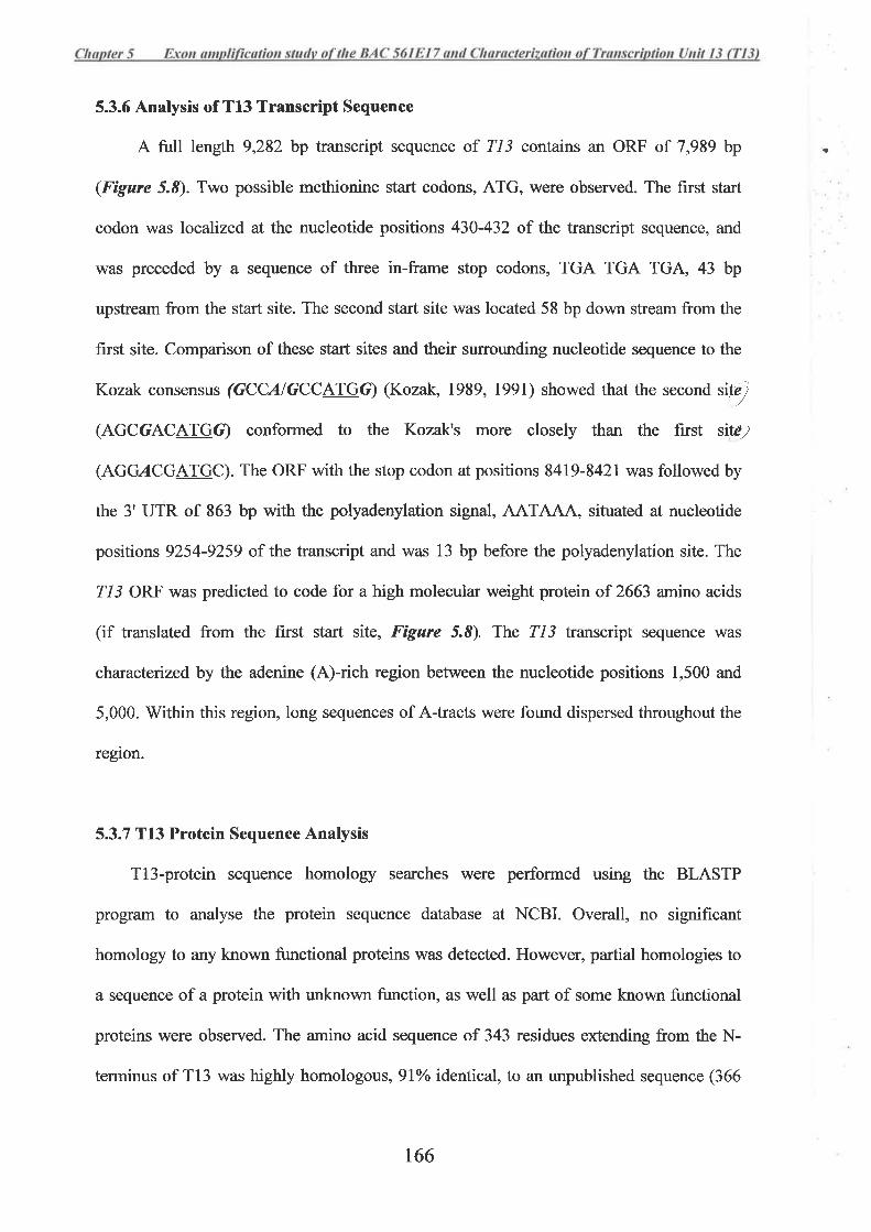
5.3.6 Analysis of T13 Transcript Sequence
A full length 9,282 bp transcript sequence of Tl3 contains an ORF of 7,989 bp
(Figure 5.8). Two possible methionine start codons, ATG, were observed. The first start
codon was localized at the nucleotide positions 430-432 of the transcript sequence, and
was preceded by a sequence of three in-frame stop codons, TGA TGA TGA, 43 bp
upstream from the start site. The second start site was located 58 bp down stream from the
first site. Comparison of these start sites and their surrounding nucleotide sequence to the
Kozak consensus (GCCAIGCCATGC) (Kozak, 1989, 1991) showed that the second sitT
(AGCGACATGG) conformed to the Kozak's more closely than the first sitc¿
(AGG,4CGAIGC). The ORF with the stop codon at positions 8419-8421 was followed by
the 3' UTR of 863 bp with the polyadenylation signal, AATAAA, situated at nucleotide
positions 9254-9259 of the transcript and was 13 bp before the polyadenylation site. The
213 ORF was predicted to code for a high molecular weight protein of 2663 amino acids
(if translated from the first start site, Figure 5.8). The 213 transcript sequence was
charactenzed by the adenine (A)-rich region between the nucleotide positions 1,500 and
5,000. V/ithin this region, long sequences of A-tracts were found dispersed throughout the
region.
5.3.7 T13 Protein Sequence Analysis
Tl3-protein sequence homology searches were performed using the BLASTP
program to analyse the protein sequence database at NCBI. Overall, no signif,rcant
homology to any known functional proteins was detected. However, partial homologies to
a sequence of a protein with unknown firnction, as well as part of some known functional
proteins were observed. The amino acid sequence of 343 residues extending from the N-
terminus of T13 was highly homologous, 9lo/o identical, to an unpublished sequence (366
-a
r66
amino acids) of a nasopharyngeal carcinoma susceptibility protein, LZl6 (Protein database
accession no. NP-037407). Sequence homology was also identified to a 2,062-amino acid
protein of unknown function, KIAA0874. The KIAA0874 protein sequence has been
deduced from a sequence of oDNA isolated from human brain cDNA library (Nagase e/
al., 1998). Two regions of high homology were observed between Tl3 protein and
KIAA0874. The first was a 467 amino acids N-terminal region of Tl3, which was
homologous to the N-terminal region, residues I to 484, of KIAA0874 (45% identical).
The second region of homology was at the C-terminal sequences of both proteins (68%
identical), between amino acid residues 2,322-2,664 of T13 and amino acids 1,730-2,062
of KIAA0874. In addition, a low degree of similarity was also observed between the
remaining region of both proteins.
A sequence of 99 amino acids between residues 167 and265 of Tt3 showed sequence
homology to ankyrin repeats, found in a large number of proteins. The highest degree of
homology was detected to the anþnin domain of BRCAl-associated ring domain protein
(BARD-l, accession no. XP-002364) as well as that of the protein product of NFKBIL2
(nuclear factor of kappa light poþeptide gene enhancer in B-cells inhibitorlike 2) gene,
also known as lknppaBR gene (accession nos. A4H08782 and C4863467). The sequence
homology was 40Yo identical between Tl3, aa l5I-307 , and BARD I , aa 410-569, and 43Yo
identical between amino acids 161-273 of Tl3 and 362-477 of IkappaBR protein. This
anþnin domain region was also detected by three protein domain prediction programs:
SMART, Pfam, and Profile Scan. Within this domain of Tl3, three tandem ankyrinJike
repeats were located between amino acids 167-199,200-232, and233-265. These anþnin
repeat sequences were also conserved in the homologous region of BARDI, IkappaBR,
and KIAA0874. Significant homology (72% identical) was also observed between the
residues 977 and 1056 of T13 protein and the mouse protein 4K019393 , a partial protein
r67
Fieure 5.8 : Nucleotide transcript and deduced amino acid sequence of T13. The
transcript sequence of 9282 bp is expanded up to 6 pages. The nucleotide and amino acid
positions are indicated by numbers in the left hand column. The translation start site and the
termination codon are indicated at positions 430-432 and 8419-8421 respectively. Three
tandem repeats of in-frame stop codon (TGA) preceding the translation start site are located
at positions 379-387 (underline vnth asterisk underneath). A polyadenylation signal
beginning at nucleotide 9254 is underlined. The exon botrndaries are indicated by the
vertical lines with double-head arrows and the given numbers of exons. The corresponding
trapped exons are also indicated:8T24.35, exon 2;561-114, exon 5;561-72, exon 8;561-
13, exon 9 (partial 3' sequence, nt 7858-7899 ); and 561-4, exon 12. Three tandem anlcyrin-
like repeats are located and underlined (red) between amino acids 167-199,200-232, and
233-265. T}ire nuclear localization signals (NLS) at residues 456-475,906-923, 1184-1201,
1208-1236, 1358-1377,1427-1444, 1460-1479, and 1640-1657 are also underlined (black)
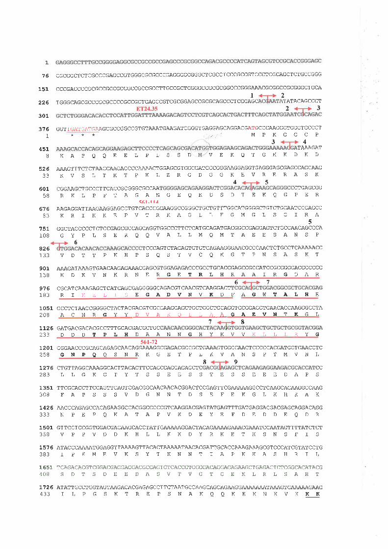
1
76
151
226
301
376I
451B
52633
60158
676B3
CGCGGCTCTCGCCCGAGCCGTGGGCGCGCCCGAGGGGCGGGCTCGCCTCCCGCCGTCCCTCGCAGCTCTGCCGGG
CCCGAGCCCGCGCCGCCGCCGCCGCCGCCTTGCCGCTCGGGCCGCGCGGCCCGGGAAACGCGGCCGCGGGCTGCAl2
TGGGCAGCGCCCGCGCCCCGCCGCTGAGCCGTCGCGGAGCCGCGCAGCCCTCGGAGC ATATACAGCCCT
GGTTGATGATGAAGCGCCGGCCGTGTAAATGAAGATCGGGTGAGGAGCAGGACGATGCCCAAGGGTGGGTGCCCT***','MPKGGCP
3 4AAAGATKD
T
KAPOOEELPLSSD
VDTTPKHP
VEKQTGKKD
751108
AAAGTTTCTCTAACCAAGACCCCAÃ.AACTGGAGCGTGGCGATGGCGGGAAGGAGGTGAGGGAGCGAGCCAGCAAGKV S L T KT PKLE RG D GGKEVRE RAS K
4 5CGGAAGCTGCCCTTCACCGCGGGCGCCAA CTGAGCGG
RKL P FT AGANGE QKD S DTEKQG PER<61-l l¿
AÀGAGGATTAÄ,GAAGGAGCCTGTCACCCGGAAGGCCGGGCTGCTGTTTGGCATGGGGCTGTCTGGAATCCGAGCCK R I K K E P V T R K A G L L F G M G L S G ] R-A
5GGCTACCCCCTCTCCGAGCGCCAGCAGGTGGCCCTTCTCATGCAGATGACGGCCGAGGAGTCTGCCAACAGCCCAG Y P L S E R O QVAL LM QMTAE E SAN S P
6CCCAGTCTACAGTGTGT CAGAAGGGAACGCCCAACTCTGCCTCAA.AAACCSQSTVCQKGTPNSASKT
426133
901158
976183
AAAGATA.AAGTGAACAAGAGAAACGAGCGTGGAGAGACCCGCCTGCACCGAGCCGCCATCCGCGGGGACGCCCGGKDKVNKRNERGETRLHRAAIRGDAR
6 7CGCAT CAÃAGAGCT CATCAGCGAGGGGGCAGACGT CAACGT CAAGGACT
RTKELISEGÀDVNVKD FAGV,]TALHE
1051 GCCTGTAACCGGGGCTACTACGACGTCGCGAAGCAGCTGCTGGCTGCAGGTGCGGAGGTGAACACCAAGGGCCTA2OB A C N R G Y Y D V A K O L L A A GAEVI{ T KG L
112 6 GATGACGACACGCCTTTGCACGACGCTGCI7
233561-72
1201 GGGAACCCGCAGCAGAGCAACAGGAAAGGCGAGACGCCGCTGAAAGTGGCCAACTCCCCCACGATGGTGAACCTC258 GN P Q Q S N RK GE T P L KVAN S P T MVN L
I 91 2 7 6 CTGTTAGGCAAAGGCACTTACACTT CCAGCGAGGAGAGCT
DDDTPLHDAANN GHYKVVKT,T,T,RYG
c283 L L G KG T Y T S S E E S S T E S S E E E DAP S
1351 TTCGCACCTTCCAGTTCAGTCGACGGCAACAACACGGACTCCGAGTTCGA.AAAAGGCCTCAAGCACAAGGCCAAG3OB FA P S S S V D GNN T D S E FE KG LKHKAK
1426 AACCCAGAGCCÀCAGAAGGCCACGGCCCCCGTCAAGGACGAGTATGAGTTTGATGAGGACGACGAGCAGGACAGG333 N P E P Q KATAPVK D E Y E F D E D DE Q D R
1501 GTTCCTCCGGTGGACGACAAGCACCTATTGAAAAAGGACTACAGAIUU\GAAACGAÃATCCAATAGTTTTATCTCT358 V P PV D D KH L L KK DYRKE T K S N S F I S
1576 ATACCCA'UU\TGGAGGTTA.AÄAGTTACACTAJUUU\TAACACGATTGCACCAAAGAAAGCGTCCCATCGTATCCTG383 I P KME VK S Y T KNN T I A P KKA S HR I L
1651 TCAGACACGTCGGACGAGGAGGACGCGAGTGTCACCGTGGGGACAGGAGAGAAGCTGAGACTCTCGGCACATACG4OB S D T S D E E DA S V TV G T GE KIRL S AH T
172 6 ATATTGCCTGGTAGTAAGACACGAGAGCCTTCTAATGCC.AAGCAGCAGA'\GGAiUUUUU\TA,U\GTGN\AIU\GA,\G433 I L P G S K T RE P S NAKQ Q KE KN KVKKK
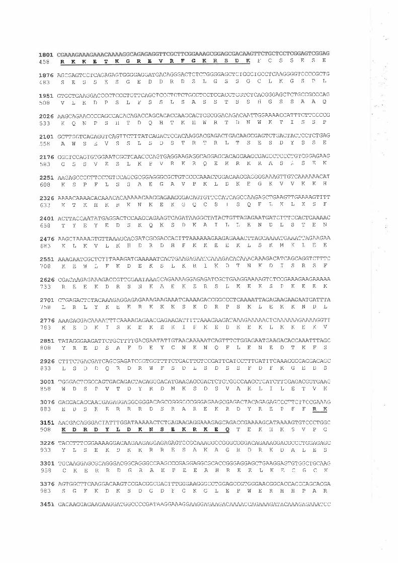
458RKKET KEREVRFG RSDKFCSSESE
18?5 AGCGAGTCCTCAGAGAGTGGGGAGGATGACAGGGACTCTCTGGGGAGCTCTGGCTGCCTCAAGGGGTCCCCGCTG483 S E S S E S G E D DRD S LG S S GC LKG S PL
1951 GTGCTGAAGGACCCCTCCCTGTTCAGCTCCCTCTCTGCCTCCTCCACCTCGTCTCACGGGAGCTCTGCCGCCCAG5OB V LK D P S L F S S L SAS S T S S HG S S AAO
2026 AAGCAGAACCCCAGCCACACAGACCAGCACACCAAGCACTGGCGGACAGACAATTGGAÄ.AACCATTTCTTCCCCG533 K Q N P S H T D Q H T K H W R T D N bI K T I S S P
2101 GCTTGGTCAGAGGTCAGTTCTTTATCAGACTCCACAAGGACGAGACTGACAAGCGAGTCTGACTACTCCTCTGAG558 A}1 S E V S S L S D S T RT R L T S E S DY S S E
21?6 GGCTCCAGTGTGGAATCGCTGAAGCCAGTGAGGAAGAGGCAGGAGCACAGGAAGCGAGCCTCCCTGTCGGAGAAG583 G S S VE S L K PVRKRQE HRKRAS L S E K
2251 AAGAGCCCCTTCCTGTCCAGCGCGGAGGGCGCTGTCCCCAAACTGGACAAGGAGGGGAAAGTTGTCAAAAAACAT608 K S P F L S S AE GAV P KL D KE GKVVKKH
2326 AAAACAAAACACAAACACAJUUU\CAJ\GGAGAAGGGACAGTGTTCCATCAGCCAAGAGCTGAAGTTGAAAAGTTTT633 KT KHKHKN KE KG Q C S I S Q E L K LK S F
2401 ACTTACGAATATGAGGACTCCAAGCAGAAGTCAGATAAGGCTATACTGTTAGAGAATGATCTTTCCACTGAÄAAC658 T YE Y E D S KQK S DKA I L LE N D L S T E N
24?6 AAGCTAA.AAGTGTTAAAGCACGATCGCGACCACTTTA,UUUU\GAAGAGAAACTTAGCNUU\TGAÄATTAGAAGAA683 K LKV L K H D R D H FKKE E KL S KM K L tr E
2551 A'U\GA'\TGGCTCTTTAAAGATGA.AAÄATCACTGAAGAGAATCAAAGACACAAACAAAGACATCAGCAGGTCTTTC7OB KEW L EK D E K S LKR I K DT N KD I S R S F
2626 CGAGAAGAGAAAGACCGTTCGAATA.AAGCAGAAAAGGAGAGATCGCTGAAGGAAAAGTCTCCGAÄAGAAGAÃÀAA733 Rtr E K D R S N KAE KE R S L KE K S P KE E K
2701 CTGAGACTGTACAAAGAGGAGAGAAAGAAGAAATCA.AÄAGACCGGCCCTCAAÄ,ATTAGAGAAGAAGAATGATTTA758 L R L Y K E E R KKK S K D R P S K L E KKN D L
2776 AAAGAGGACA.AÄATTTCAÄAAGAGAAGGAGAAGATTTTTAAAGAAGATAAAGAAAAACTCA'UUUU\GAAÄAGGTT783 KE D K I S KE KE K I FKE D KE K L KKE KV
2851 TATAGGGAAGATTCTGCTTTTGACGAATATTGTAACAAAAATCAGTTTCTGGAGAATGAAGACACCAAATTTAGC8OB Y RE D S A F DE Y CNKN Q F L E N E D T K F S
2926 CTTTCTGACGATCAGCGAGATCGGTGGTTTTCTGACTTGTCCGATTCATCCTTTGATTTCAAAGGGGAGGACAGC833 L S D D QRDRW F S DL S D S S FD FKGE D S
3OO1 TGGGACTCGCCAGTGACAGACTACAGGGACATGAAGAGCGACTCTGTGGCCAAGCTCATCTTGGAGACGGTGAAG858 !f D S P V T D Y R DMK S D S VAK L I L E T VK
3076 GAGGACAGCAAGGAGAGGAGGCGGGACAGCCGGGCCCGGGAGAAGCGAGACTACAGAGAGCCCTTCTTCCGAAAGBB3 E D S KE R R R D S RARE KR D Y RE P F FRK
3151908
3226933
3301958
3376983
3451
AAGGACAGGGACTATTTGGATAJUUU\CTCTGAGAAGAGGAÄAGAGCAGACCGAJUU\GCATAIUU\GTGTCCCTGGCKN EKRKE TEKHKSVPG
TACCTTTCGGAAÃAGGACAAGAAGAGGAGAGAGTCCGCAAAGGCCGGGCGGGACAGAAAGGACGCCCTGGAGAGCYL S E KDKKRRE SAKAGRDRKDALE S
TGCAAGGAGCGCAGGGACGGCAGGGCCAAGCCCGAGGAGGCGCACCGGGAGGAGCTGAAGGAGTGTGGCTGCAAG
CKERRDGRAKPEEAHREELKECGCK
AGTGGCTTCAAGGACAAGTCCGACGGCGACTTTGGGAAGGGCCTGGAGCCGTGGGAACGGCACCACCCAGCACGAS G F K D K S D G D F G KG L E P hT E R H H PAR
GAGAAGGAGAA.GAAGGATGGCCCCGATAAGGAAAGGAAGGAGA,\GACAJUU\CCAGAÄAGATACAÄA,GAGAAATCC
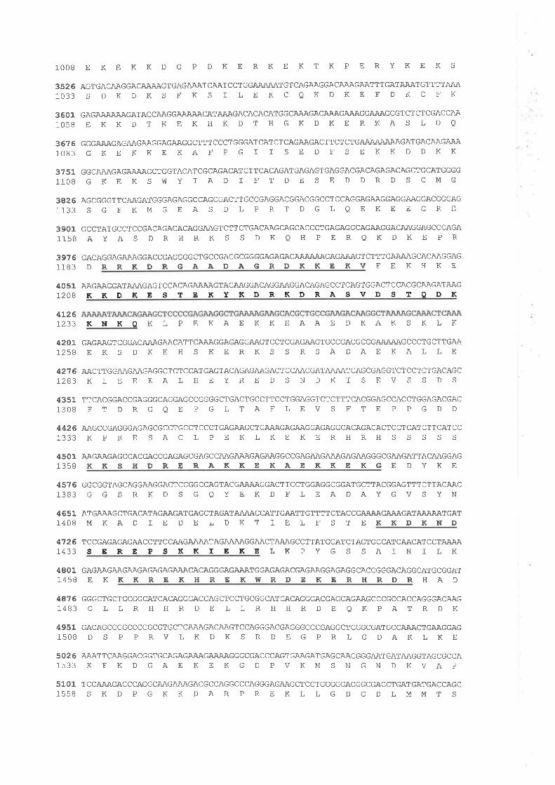
1OOB E K E K K D G P D K E R K E K T K P E R Y K E K S
3s26103 3
36011058
36761083
3751110 B
3426113 3
39011158
3976118 3
AGTGACAAGGACAAÄAGTGAGAAATCAATCCTGGAÄA.AATGTCAGAAGGACAAAGAATTTGATAAATGTTTTAAAS DKDKSEKS I LEKCQKDKE FDKCEK
GAGAAAA.AAGATACCAAGGAA.AAACATA.AAGACACACATGGCAAAGACAÄAGAAAGGAAAGCGTCTCTCGACCAAE KKDTKE KHKDT HGKDKE RKAS LDQ
GGGAAAGAGAAGAAGGAGAAGGCTTTCCCTGGGATCATCTCAGAAGÄ.CTTCTCTGAIUUUUUU\GATGACAAGAÄAGKtrKKE KAF P G I I S E D F S EKK D DKK
GGCAAAGAGAAAAGCTGGTACATCGCAGACATCTTCACAGATGAGAGTGAGGACGACAGAGACAGCTGCATGGGGGKEKSWY IAD I FTDE SE DDRDS CMG
AGCGGGTTCAAGATGGGAGAGGCCAGCGACTTGCCGAGGACGGACGGCCTCCAGGAGAAGGAGGAAGGACGGGAG
S GFKMGEAS DLPRT DGLQEKEEGRE
GCCTATGCCTCCGACAGACACAGGAAGTCTTCTGACAAGCAGCACCCTGAGAGGCAGAAGGACAAGGAGCCCAGAAYAS DRHRK S S DKQH PE RQKDKE PR
GACAGGAGAAAGGACCGAGGGGCTGCCGACGCGGGGAGAGACA]UUUU\GAGAÃAGTCTTTGAAAAGCACAAGGAGD RRKDRGAADAGRDKKE KV F E K H KE
AAGAAGGATAAAGAGTCCACAGAAAAGTACAAGGACAGGAAGGACAGAGCCTCAGTGGACTCCACGCAAGATAAGKKDKESTEKYKDR KDRÀSVD STODK
405112OB
4L26r233 KOKN K L P E KAE K K HAAE I] KAK S K L K
420Lt258
4276L2B3
43511308
GAGAAGTCGGACAÄAGAACATTCAÄAGGAGAGGAAGTCCTCGAGAAGTGCCGACGCGGAÄAAAGCCCTGCTTGAAE KS DKE H S KE RK S S RSADAEKALLE
AAGTTGGAAGAAGAGGCTCTCCATGAGTACAGAGAAGACTCCAI\CGATA,UU\TCAGCGAGGTCTCCTCTGACAGCKLEEEALHEYREDSNDKISEVSSDS
TTCACGGACCGAGGGCAGGAGCCGGGGCTGACTGCCTTCCTGGAGGTCTCTTTCACGGAGCCACCTGGAGACGACFTDRGQEPGLTAFLEVSFTEPPGDD
TCCGAGAGAGAACCTTCCAAGAAAATAGAAÄAGGAACTAAAGCCTTATGGATCTAGTGCCATCAACATCCTAA.AASEREPS KKIEKELK
4426 AAGCCGAGGGAGAGCGCCTGCCTCCCTGAGAAGCTGAAAGAGAAGGAGAGGCACAGACACTCCTCATCTTCATCC1333 K P R E S A C L P E K L K E K E R H RH S S S S S
4501 AAGAAGAGCCACGACCGAGAGCGAGCCAAGAAAGAGAAGGCCGAGAAGAAAGAGAAGGGCGAAGATTACAAGGAG1358 K K S H D R E RA K K E KAE K K E K G E D Y K E
4576 GGCGGTAGCAGGAAGGACTCCGGCCAGTACGAAAAGGACTTCCTGGAGGCGGATGCTTACGGAGTTTCTTACAAC1383 G G S R K D S G Q Y E K D F L E A DAY G V S Y N
4651 ATGAÀAGCTGACATAGAAGATGAGCTAGATAAAACCATTGAATTGTTTTCTACCGAAAAGAAÀGATAAAAATGAT1408 M KA D I E D E L D K T I E L F S T E KKD KND
47261433 PYGSSAINILK
4801 GAGAAGAAGAAGAGAGAGAAACACAGGGAGAAATGGAGAGACGAGAAGGAGAGGCACCGGGACAGGCATGCGGAT1458 E K K K RE K H R E KW RD E K E R H RD R H A D
4876 GGGCTGCTGCGGCATCACAGGGACGAGCTCCTGCGGCATCACAGGGACGAGCAGAAGCCCGCCACCAGGGACAAG1483 G L L R H H R D E L L R H H R D E Q K P A T R D K
4951 GACAGCCCGCCCCGCGTGCTCAÄAGACAAGTCCAGGGACGAGGGCCCGAGGCTCGGCGATGCCAAACTGAAGGAG15OB D S P P RV L K D K S R D E G P R L G DAK L KE
5026 AÄATTCAAGGACGGTGCAGAGAAAGAAAAGGGCGACCCAGTGAAGATGAGCAACGGGAATGATAAGGTAGCGCCA1533 K F K D G A E K E K G D P V K M S N GN D KVA P
5101 TCCAÀAGACCCAGGCAAGAÄAGACGCCAGGCCCAGGGAGAAGCTCCTGGGGGACGGCGACCTGATGATGACCAGC1558 S K D P G K K D AR P R E K L L G D G D L M M T S
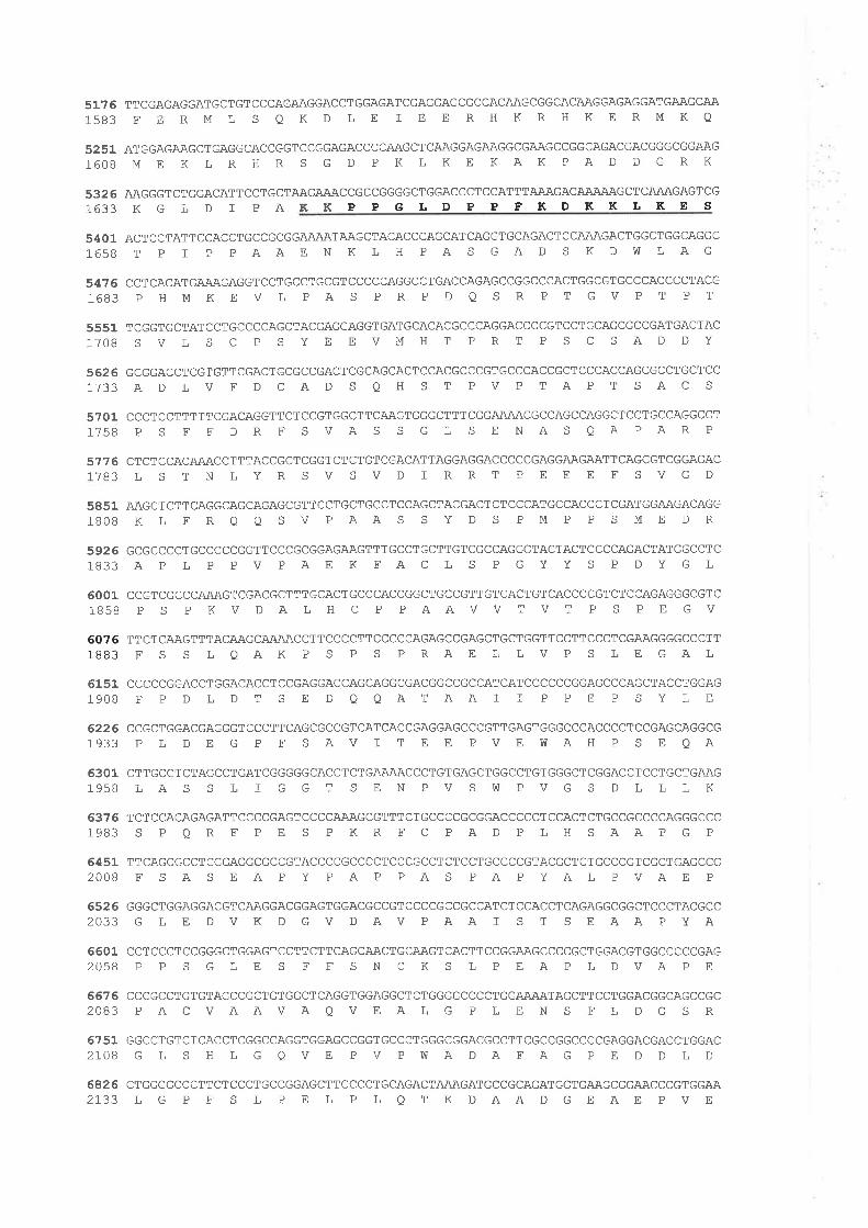
1633KGLDIPAKK
51?5 TTCGAGAGGATGCTGTCCCAGAAGGACCTGGAGATCGAGGAGCGCCACAAGCGGCACAAGGAGAGGATGAAGCAA1583 E E R M L S Q K D L E I E E R H K R H K E R M K O
5251 ATGGAGAAGCTGAGGCACCGGTCCGGAGACCCCAAGCTCAAGGAGAAGGCGAAGCCGGCAGACGACGGGCGGAAG1608 M E K L R H R S G D P K L K E KAK P A D D G R K
5326 AAGGGTCTGGACATTCCTGCTAAGA.AACCGCCGGGGCTGGACCCTCCATTTAAAGACAAA.AAGCTCAAAGAGTCGPPGLDPPFKDKKLKES
5401 ACTCCTATTCCACCTGCCGCGGAÄAATAAGCTACACCCAGCATCAGGTGCAGACTCCAAAGACTGGCTGGCAGGC1658 T P I P P AAE N K L H P A S GA D S K D W L AG
54?6 CCTCACATGAÄAGAGGTCCTGCCTGCGTCCCCCAGGCCTGACCAGAGCCGGCCCACTGGCGTGCCCACCCCTACG1683 P HMKE V L P A S P R P D Q S R P T GV P T P T
5551 TCGGTGCTATCCTGCCCCAGCTACGAGGAGGTGATGCACACGCCCAGGACCCCGTCCTGCAGCGCCGATGACTAC17OB S V L S C P S Y E E V M H T P R T P S C S A D D Y
5626 GCGGACCTCGTGTTCGACTGCGCCGACTCGCAGCACTCCACGCCCGTGCCCACCGCTCCCACCAGCGCCTGCTCC1733 AD L V F D CA D S Q H S T P V P T A P T S A C S
577 61783
57011758
5851lBOB
59261833
60011B5B
60761BB3
CCCTCCTTTTTCGACAGGTTCTCCGTGGCTTCAAGTGGGCTTTCGGAAAACGCCAGCCAGGCTCCTGCCAGGCCTP S F FDR F SVAS S G L S E NAS QAPAR P
CTCTCCACAAACCTTTACCGCTCGGTCTCTGTCGACATTAGGAGGACCCCCGAGGAAGAATTCAGCGTCGGAGACLSTNLYRSVSVDIRRTPEEEFSVGD
AAGCTCTTCAGGCAGCAGAGCGTTCCTGCTGCCTCCAGCTACGACTCTCCCATGCCACCCTCGATGGAAGACAGGKL FRQ O S V PAAS S Y D S PM P P S ME DR
GCGCCCCTGCCCCCGGTTCCCGCGGAGAAGTTTGCCTGCTTGTCGCCAGGGTACTACTCCCCAGACTATGGCCTCAP L P PV PAE K EACL S PGYY S P DYGL
CCGTCGCCCAAAGTCGACGCTTTGCACTGCCCACCGGCTGCCGTTGTCACTGTCACCCCGTCTCCAGAGGGCGTCP S PKVDALH C P PAAVVTVT P S PE GV
TTCTCAAGTTTACAAGCA.AAACCTTCCCCTTCCCCCAGAGCCGAGCTGCTGGTTCCTTCCCTCGAAGGGGCCCTTFS S LQAK P S P S PRAE L LVP S LEGAL
6151 CCCCCGGACCTGGACACCTCCGAGGACCAGCAGGCGACGGCCGCCATCATCCCCCCGGAGCCCAGCTACCTGGAG19OB P P D L D T S E D Q QAT AA I T P P E P S Y L E
6226 CCGCTGGACGAGGGTCCCTTCAGCGCCGTCATCACCGAGGAGCCCGTTGAGTGGGCCCACCCCTCCGAGCAGGCG1933 P L D E G P F S AV I T E E PV E WAH P S E QA
6301 CTTGCCTCTAGCCTGATCGGGGGCACCTCTGAAAACCCTGTGAGCTGGCCTGTGGGCTCGGACCTCCTGCTGAAG1958 L A S S L f G G T S E N P V S h] P V G S D L L L K
637 6 TCTCCACAGAGATTCCCCGAGTCCCCAAAGCGTTTCTGCCCCGCGGACCCCCTCCACTCTGCCGCCCCAGGGCCC1983 S P O R F P E S P K R F C P A D P L H S AAP G P
6451 TTCAGCGCCTCGGAGGCGCCGTACCCCGCCCCTCCCGCCTCTCCTGCCCCGTACGCTCTGCCCGTCGCTGAGCCG2OOB F S A S E A P Y P A P PA S P A P YAL P VAE P
652 6 GGGCTGGAGGACGTCAAGGACGGAGTGGACGCCGTCCCCGCCGCCATCTCCACCTCAGAGGCGGCTCCCTACGCC2033 G L E D V K D G V D AV P AA I S T S E AA P Y A
6601 CCTCCCTCCGGGCTGGAGTCCTTCTTCAGCAACTGCAAGTCACTTCCGGAAGCCCCGCTGGACGTGGCCCCCGAG2O5B P P S G L E S F F S N C K S L P E A P L D VA P E
667 6 CCCGCCTGTGTAGCCGCTGTGGCTCAGGTGGAGGCTCTGGGGCCCCTGGAAAATAGCTTCCTGGACGGCAGCCGC2OB3 P A C VAAVA Q V E A L G P L E N S F L D G S R
6751 GGCCTGTCTCACCTCGGCCAGGTGGAGCCGGTGCCCTGGGCGGACGCCTTCGCCGGCCCCGAGGACGACCTGGAC2LOB G L S H L G Q V E P V P VÙ A D A F A G P E D D L D
6826 CTGGGGCCCTTCTCCCTGCCGGAGCTTCCCCTGCAGACTAÄAGATGCCGCAGATGGTGAAGCGGAACCCGTGGAA2733 L G P E S L P E L P L Q T K DAA D G E AE P V E
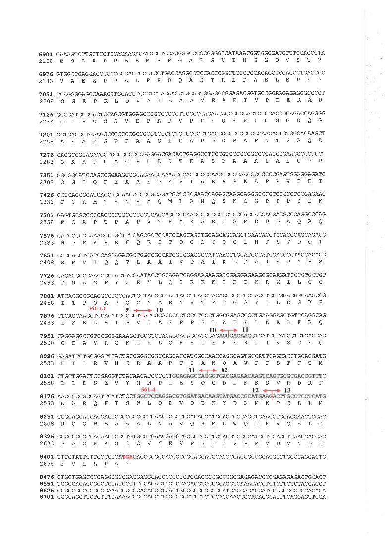
69012L5B
69762r83
70512208
7L262233
720L2258
?2762283
73512308
74262333
75012358
757 62383
GAAAGTCTTGCTCCTCCAGAAGAGATGCCTCCAGGGGCCCCCGGGGTCATAAACGGTGGGGATGTTTCCACCGTAE S LAP PEEMP PGAPGVINGGDVS TV
GTGGCTGAGGAGCCGCCGGCACTGCCTCCTGACCAGGCCTCCACCCGGCTCCCTGCAGAGCTCGAGCCTGAGCCCVAE E P PAL P P DQAS T RL PAE LE PE P
TCAGGGGAGCCAAAGCTGGACGTGGCTCTAGAAGCTGCGGTGGAGGCGGAGACGGTGCCGGAAGAGAGGGCCCGTS G E P K L D VALEAAVEAE T V P E E RAR
GGGGATCCGGACTCCAGCGTGGAGCCCGCGCCCGTTCCCCCAGAACAGCGCCCACTGGGGAGCGGAGACCAGGGG
GD PD S SVE PAPVP PEQRPLG S GDQG
GCTGAGGCTGAAGGCCCCCCCGCCGCGTCCCTCTGTGCCCCTGACGGCCCCGCCCCGAACACTGTGGCACAAGCTAEAE G P PAAS L CA P D G PA PN TVAQA
CAGGCCGCAGACGGTGCCGGCCCCGAGGACGACACTGAGGCCTCCCGTGCCGCCGCCCCAGCCGAAGGCCCTCCT
QAAD GAG P E D D T EAS RAAAPAE G P P
GGCGGCATCCAGCCGGAAGCCGCAGAACCAAAACCCACGGCCGAAGCCCCGAAGGCCCCCCGAGTGGAGGAGATCG G I O P E AAE P K P TAEA P KAP RVE E I
CCTCAGCGCATGACCAGGAACCGGGCGCAGATGCTCGCGAACCAGAGCAAGCAGGGCCCGCCCCCCTCCGAGAAGP QRMT RN RAQMLAN Q S KQ G P P P S E K
GAGTGCGCCCCCACCCCTGCCCCGGTCACCAGGGCCAAGGCCCGCGGCTCCGAGGACGACGACGCCCAGGCCCAG
E CAP T PAPVT RAKARG S E D D DAQAO
CATCCGCGCAAACGCCGCTTTCAGCGCTCCACCCAGCAGCTGCAGCAGCAGCTGAACACGTCCACGCAGCAGACGH PRKRRFQRS TOOLQAOLNT ST OQT
?651 CGGGAGGTGATCCAGCAGACGCTGGCCGCCATCGTGGACGCCATCAAGCTGGATGCCATCGAGCCCTACCACAGC2408 R E V ] O Q T L AA f V D A T K L D A I E P Y H S
7726 GACAGGGCCAACCCCTACTTCGAATACCTGCAGATCAGGAAGAAGATCGAGGAGAAGCGCAAGATCCTGTGCTGT2433 D RAN P Y F E Y L O ] R K K T E E KR K I L C C
7801 ATCACGCCGCAGGCGCCCCAGTGCTACGCCGAGTACGTCACCTACACGGGCTCCTACCTCCTGGACGGCAAGCCG2458 I T P Q A P Q C Y A E Y V T Y T G S Y L L D G K P
561-13 9 +-+ l0?876 CTCAGCAAGCTCCACATCCCCGTdATCGCACCCCCTCCCTCCCTGGCGGAGCCCCTGAAGGAGCTGTTCAGGCAG2483 L S K L H I P V I A P P P S L A E P L K E L F R O
l0TCGAG CTGA7 951 CAGGAGGCCGTCCGGGGAAAGCTGCGT CTACAGCACAGCA
25OBQEAVRGKLRLQHS IEREKLTCGTATCCTGTGAGCAGIVSCEQ
8026 GAGATTCTGCGGGTTCACTGCCGGGCGGCCAGGACCATCGCCAACCAGGCAGTGCCATTCAGCACCTGCACGATG2533 E I L R V H C RAA R T I AN Q AV P F S T C T M
ll t28101 CTGCTGGACTCCGAGGTCTACAACATGCCCCT2558 L L D S E V Y N M P L E S Q G D E N K S V R D R F
81 7 6 AACGCCCGCCAGTTCATCTCC2583 N A R Q F f S W L Q D V D D K Y D R M K T C L L M
8251 CGGCAGCAGCACGAGGCCGCGGCCCTGAACGCCGTGCAGAGGATGGAGTGGCAGCTGAAGGTGCAGGAACTGGAC2608 R Q Q H E A A A L N AV Q R M E W Q L K V Q E L D
832 6 CCCGCCGGGCACAAGTCCCTGTGCGTGAACGAGGTGCCCTCCTTCTACGTGCCCATGGTCGACGTCAACGACGAC2633 P A G H K S L C V N E V P S E Y V P M V D V N D D
8401 TTTGTATTGTTGCCGGCAT ACCGCGGGACGGCCGCAGGACGCAGGCGAGGGCCGCACGGCTGCCCAGGACTG
2638FVLLPA*
8476 CTGCTGAGCCCCAGGGGCGGAGGAGGGAGCGCCCTGTCCACCCGGGCGGGGAGAGACCCCGAGAGAGACTGCACT8551 TGGCCACAGCGCCTCCATCCCTTCCAGACTGGTCCAGACGTCGGGGAGGTGAAACACGTCTCTTCTCTACCAGCT8626 GCCGCGGCGGGGGCAAAGCCCCCAGAGCCTCACTGGCCCCGGCGGGATGAGGAGACCATGCCGGGCGCGCACACA8701 CGGCAGCTTCTGTTTGAAÀACGGCGACCTTCGGGCCCTTTTCTCCAGCAACTGCAGAGGCATTTCAGGAGTTGGA
C

477688518926900190?691519226
GAACGGAGTACATTTTTAAAATTTTTTGTTCCATTCTGATCAACTAAAGAAAATTAAGGTGTCCACCTCTCACAAACAGTGACCTGCAGGTCGGAGGGGCAGGAGCCCCATCCCAGCCGTGGTTCTGTTGCCGCCGAGCTGCGACGGCCTCAGACTGGGCTTCAGACCTGGTGCGGAGCGAGGCGGAGGGACACAGAGCCAGGACTCCCAGCCGTATTGAÄATGGAGTCAAATCCGCGTGGTTTGTATCTTCTTTCCTTTAATCTTGGGCATTTTGTTTCTCTTTCTGAGAACGTAACTTGAAACACTACAGAGCCAATAATCATATA.A.AAAGTGTATCCGTA.AACGCTGTACAGTTTTATATACCTTCCCAATGTACTTTTGCTGCCTTTTAAATAATTTATTTTGCAACCCATTACTGCCCAGTTGTAAATAACTTATGTACTGTA.ACAICACTTTCAGTTATGTGTAAAAAAATAÀATÀÀATTAATTAAAATGCNUUUUUUUU\ 9282

sequence of 485 amino acids. Such a high degree of homology of this mouse protein to
T13 indicated that it might be derived from a mouse homologue of human T13.
Sub-cellular localization of Tl3 protein was also predicted utilizing a program
PSORT. Eight bipartite nuclear localization signals (NLS) were identified within the
region between amino acids 457 and 1,658. These NLS were located at residues 456-475,
906-923, 1184-1201, 1208-1236, 1358-1377, 1427-1444, 1460-1479, ffid 1640-1657.
Further in silico protein sequence analysis did not detect any other functional motifs or
specific sequences that could suggest a possible function of the T13 protein.
5.3.8 T13 Gene Structure
The genomic structure of 713 was characterized using the available genomic
sequence information of two overlapping PAC clones, 74015 and 168P16, encompassing
this region and overlapping with BAC 561E17 (Figure 5.3). Alignment of this genomic
seguence with the sequence of TI3 full-length cDNA allowed the identification of Tl3
gene structure. The TI3 gene consists of 13 exons ranging in size from 85 bp to 6.53 kb,
and spanning the genomic region of approximately 220 kb. The smallest exon is exon 2 (85
bp) while the largest is exon 9 of 6.53 kb. The sequences of the splice junctions of each
exon, summarised n Tøble 5.y', conform to the published consensus sequence of donor and
acceptor splicing sites (Shapiro and Senapatþ, 1987). Both ATG initiation codons (section
5.3.6) are contained within exon 3. The ankyrin repeat motif was encoded by the sequence
of exons 6,7, and 8, while all eight NLSs were coded for by the sequence of exon 9. Exon
13 contained the 3' end of the ORF, the stop codon as well as the complete 863 bp 3'UTR
(Fígure 5.8). The sequence of a partial pseudogene on the X chromosome that was
homologous to part of the transcript sequence of 713 (section 5.3.4.1) included the
sequence from part ofexon 9 to exon 13.
168
Fisure 5.9: Genomic Organrzation of Tl3. Diagram represents the regional physical map of the chromosome 16 at
the band 16q24.3. The map consists of the overlapping cosmids, BAC, and PACs, corresponding with those shown
in Figure 5.3, and encompasses the TI3 gene. The restriction map of EcoRI, E, and EagI, Ea, is indicated in "kb".
Based on the physical map, the T13 gene expands over 170 kb of the genomic region, 5' to 3' from telomeric to
cetromeric direction. Exons 2 and 3 are separated by the largest intron of approximately 100 kb. The map locations
of the trapped products,f lled circlse, and their corresponding exons are also indicated.
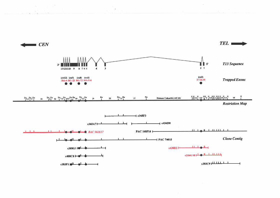
<F CEN
3r
13121110 9 E ?65
(ex12) (eú) (ext) (exÐ9614 361-13 561-12 561-114oo o o
43
TEL-
5r TI3 Sequcnce2l
ET?á35 Trøpped Exons(ef,)
o
2l t2F¡
10
ll I
E¡ t
c34EF3
c36S¡?1J-l_L'¡ c¿l'3DE
BAC 5618T7 PAC 16EP16
DÉEscUEtmm (<41¡ kb) l8
Restrictian Map
pAc 74ols CIan¿ Config
c43EB3
c334Gl0
c393C9
FåEsz2l9
rlt
c3l)acltO|#
c40tc8l#l
¡363¡1 ¡lI<H-f
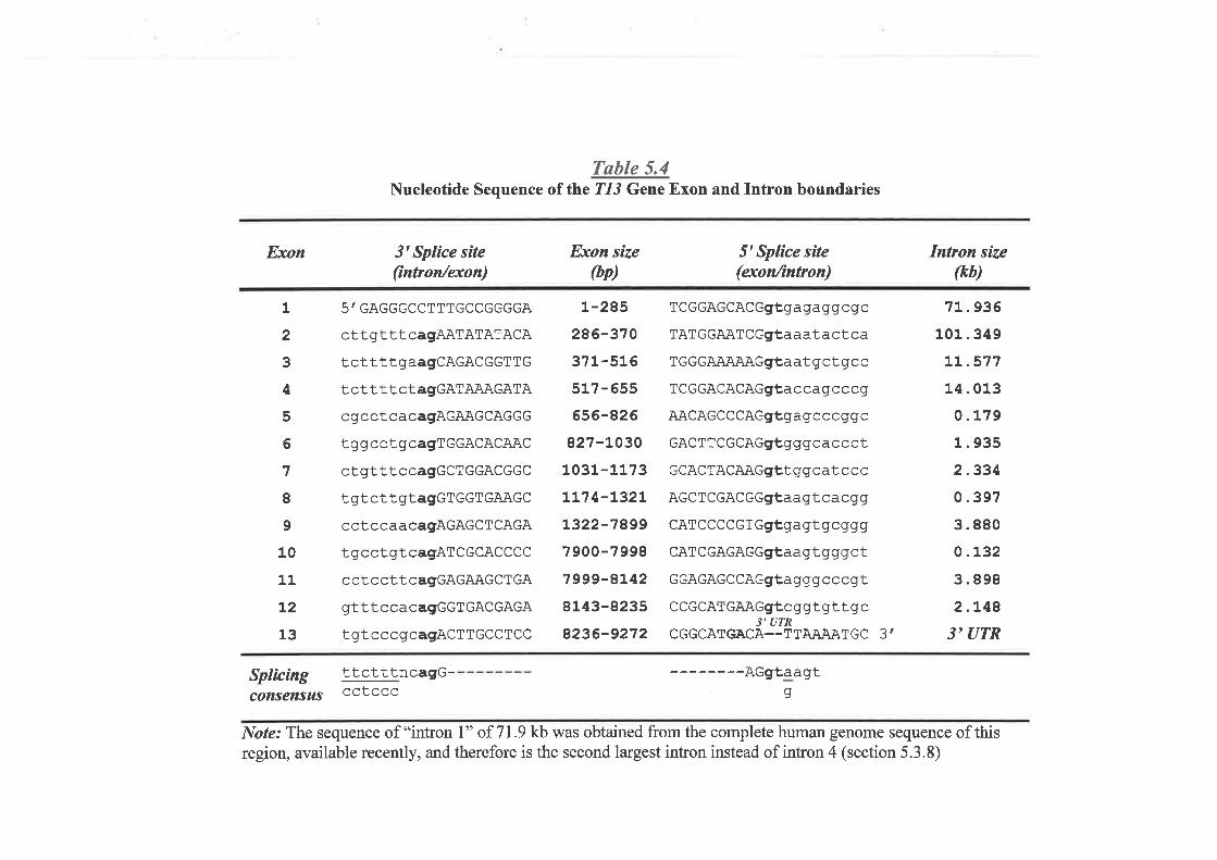
Tøble 5.4Nucleotide Sequence of the ?1J Gene Exon and Intron boundaries
Exon
1
2
3
4
5
6
7
8
9
10
11
L2
13
3t Splìce síte(intron/exon)
5' GAGGGCCTlTGCCGGGGA
cttgtttcagAATATAlACA
tcttttgaagCAGACGGTTG
t ctttt cta9GATAAÄ.GATA
cgcctcacagAGAAGCAGGG
tggcctgcagTGGACACAAC
ct grt tt c cagGCT GGACGGC
tgtcttgtagGTGGTGAAGC
cctccaacagAGAGCTCAGA
tgcctgtcagATCGCACCCC
cctccttcagGAGAAGCTGA
gtttccacagGGTGACGAGA
tgtcccgcagACTTGCCTCC
Exon size
þp)
L-285
286-310
371-516
517-655
656-826
827-LO30
1031-1173
Lt14-L32L
t322-78997 900-7 998
7999-8t42
8143-8235
8236-9272
5'Splíce sìte(exonfrntron)
TCGGAGCACGgt,ga ga gq cg c
TAT GGAATCGgt a aat a ct ca
TGGGA;\iUU\Ggtaat gct gcc
TCGGACACAG9ta cca g c c cq
AACAGCCCAGgtga g c ccg q c
GACT TCGCAGgtg g gca c c ctGCACTACAAGgLI ggcat c c c
AGCT CGACGGgt aa gt ca ca g
CATCCCCGTGgÈga gt g cqgq
CAT CGAGAGGgtaa gt g g gctGGAGAGCCAGgta g gg c c c gtCCGCAT GAAGgtc g gt gt t g c
3'UTRCGGCATGACA--TTAAAATGC 3'
Intron síze(kb)
71. 936
101.349
LL.577
14.013
0.179
1.935
2.334
0.397
3.880
0.132
3.898
2.L48
3'UTR
Splícing ttctttncagG- AGgtaagtg^^+ ^^COnSenSUS ccEccc
Note: The sequence of "intron l" of 7I.9 kb was obtained from the complete human genome sequence of thisregion, available recently, and therefore is the second largest intron instead of intron 4 (section 5.3.8)
The trapped exon 8T24.35 was trapped from exon 2, while 561-114 and 561-72 were
trapped from exons 5 and I respectively. As described in sections 5.3.2 and 5.3.4.1, clone
561-114, size 268 bp, was likely trapped from two adjacent exons. The first exon was the
sequence of 97 bp from 5' end of 561-114, which appeared only in some cDNA clones. It
was located and separated from the second, 171-bp TI3 exon 5, by the intervening
sequence of 497 bp. This extra exon was assigned as exon 5E and was bound at both ends
by the sequences corresponding with the acceptor and donor splicing consensus. The
trapped product 561-13 was isolated from the 3' part of exon 9, but in the reverse
orientation that included part of its intron (describedin 5.3.2 and 5.3.4.1). Exon 12 was
represented by the trapped product 561-4.
The TI3 gene contains an unusual large intron between exons 2 and 3, which is
approximately 90 kb across the genomic sequence. Intron 4 appears to be the second
largest intron of Tl3, spanning the genomic sequence of 14 kb. The genomic structure of
T13 and its organization in the region extending from the published physical map of
I6q24.3 (Whitmore et al.,l998a) is shown inFigure 5.9.
5.3.9 T13 Alternative Splicing and Overlapping Transcripts
During the construction of the extended physical map of 16q243 (Whitmore 1999,
unpublished data), two groups of partial cDNA sequences, designated as Tl6 andTIT were
mapped to the same region where 213 exons were isolated. With the available genomic
sequence of Tl 3, both 716 and TI7 werc precisely located. The 716 was localized within
intron 4 of TI3 along wfih 713 exon 5E, and trapped product 561-33, while TI7 si¡nted
within intron 7 (Figure 5.10). Sequence analysis of cDNA clone os05c06 representing
Tl6 deftned that this partial transcript sequence was not part of T13. lnstead, it appeared to
be transcribed in the opposite direction with that of 713. The trapped exon 561-33 also
r69
showed an ORF and splicing signal at the boundary sequence that suggested its
transcription in the opposite direction with 713. To test if 561-33 was part of TI6, RT-PCR
analysis using primer pairs designed from both 716 and 561-33 sequence was carried out.
This, however, failed to link this trapped exon to T16 sequence. Both 716 and 561-33
might, therefore belong to the different genes overlapping wfthT13. This was omitted from
further study.
TI7 is a partial 0.9-kb cDNA sequence generated from overlapping ESTs. The
majority of EST sequences are derived from the 3' end of the transcript and contain the
polyadenylation signal. The location of this polyadenylation signal indicates that Tl7 is
likely to be transcribed in the same direction as T13.The 717 might be part of T13 as an
alternative isoform or be an independent gene that shares some exons with T13.To fu¡ther
investigate this relationship, RT-PCR experiment was undertaken utilizing various
combinations of primers designed to both 713 exons and TI7 sequences. The overall
results of sequence analysis of the RT-PCR products are surnmarised in Figure 5.10.717
appeared to be an alternative transcript or transcription isoform of TI3 that selectively used
the 3' terminal exon situated within intron 7 of 713. Northem blot hybridization was also
tmdertaken to confirm the existence of this isoform gI7) utilizing RT-PCR product
generated from the assembled 0.9-kb sequence of Tl7 cDNA. As shown in Figure 5.7,the
TI7 probe did not detect the 9.5 kb mRNA band of TI3 but did detect the smaller mRNA
bands similar to those detected by a cDNA probe generated from exons 6, 7, I and 9 of
713.
Alternative inclusion of exon 5E (section 5.3.8) was also confirmed by RT-PCR either
in the full-length 213 transcript or in its 217 isoform, though the quantity of 5E inclusion
products was lower when compared with the products that excluded this exon. Altemative
splicing was also detected between exons 2 arlLd 3, which generated the product that either
t70
a t'i n l.,n
^. ,r,,,'
: ,Iincluded or excluded the CAG tri-nucleotide. Analysis of the genomic sequence flanking I t'
I
1
exon 3 identified two tandem CAG sequences at the 5' splicing boundary of the exon.a7
These tandem CAG sequences could cause the selective usage of 5' splice acceptor andlzlv+-^uf rz¡ta|\
result in the inclusion or exclusion of CAGnbetween exons 2 and 3. Other splicing variants
were also detected during the RT-PCR experiment, but with low quantity. These, however,
were probably the minor products at least in three tissue sources of mRNA used in this
study: Jurkat T-cell, fetal heart, and mammary gland.
Additional transcription isoforms of 713 existed that were represented by oDNA
sequences in the GenBank cDNA database. The diagram showing these oDNA isoforms
comparing with that of TI3 is given in Fìgure 5.10. The first was an isoform represented
by a 1460-bp cDNA clone, geoe ialifrcation no.20562097 (accession no. XM-0S5409).
Sequence analysis revealed ttr. t *Jipt sequence identical to that of Tl3from exon I to
exon 4, then followed by the new exon situated within intron 4. This cDNA sequence
completed with an ORF of 124 arrino acids, utilizing the same ATG start codon as that of
713. The second isoform, which was represented by a partial cDNA sequence (EST
accession no. B1836550), showed the sequence of TI3 thatjoined exon 7 with exon 10 by
skipping exons 8 and 9. This transcription product maintained an ORF of Tl3 from start to
stop codons except part of two skipped exons. It was predicted to code for a proteinof 421
amino acids that shares the first 248 residues with that of 717 artd includes ankyrine
repeats region. The third isoform was represented by a partial cDNA sequence (accession
no. 41684375) of 509 bp isolated from leukemic B-cell library. This clone showed the
sequence that is homologous to T13 from exon I to exon 4 andjoins to exon 8 by skipping
exons 5,6,and7.
x
5.3.10 Mutation Study of T13 in Sporadic Breast Cancer
17l
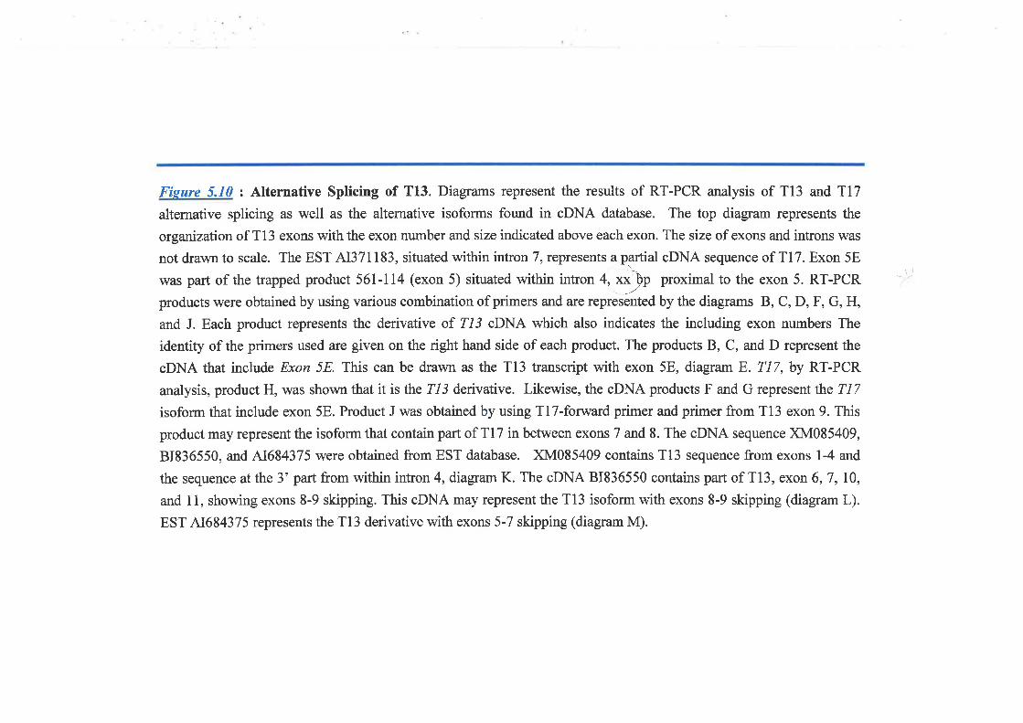
Fisure 5.10 z Alternative Splicing of T13. Diagrams represent the results of RT-PCR analysis of Tl3 and Tl7
alternative splicing as well as the alternative isoforms found in oDNA database. The top diagram represents the
orgarization of T13 exons with the exon number and size indicated above each exon. The size of exons and introns was
notdrawnto scale. TheEST AI37ll83, situatedwithinintronT,representsapartialoDNAsequence of Tl7. Exon5E
was part of the trapped product 561-114 (exon 5) situated within intron 4, xxlp proximal to the exon 5. RT-PCR
products were obtained by using various combination of primers and are ,"rr""lr"OUy tt. diagrams B, C, D, F, G, H,
and J. Each product represents the derivative of Z/3 cDNA which also indicates the including exon numbers The
identity of the primers used are given on the right hand side of each product. The products B, C, and D represent the
cDNA that include Exon 5E This can be drawn as the Tl3 transcript with exon 5E, diagram E. TI Z, by RT-PCR
analysis, product H, was shown that it is the Tl3 denvative. Likewise, the oDNA products F and G represent the TI7
isoform that include exon 5E. Product J was obtained by using Tl7-forward primer and primer from Tl3 exon 9. This
product may represent the isoform that contain part of T17 in between exons 7 and 8. The oDNA sequence XM085409,
B1836550, and 41684375 were obtained from EST database. XM085409 contains Tl3 sequence from exons l-4 and
the sequence at the 3' part from within intron 4, diagrart K. The oDNA 81836550 contains part of Tl3, exon 6,7, I0,and 11, showing exons 8-9 skipping. This oDNA may represent the Tl3 isoform with exons 8-9 skipping (diagram L).
EST 41684375 represents the Tl3 derivative with exons 5-7 skipping (diagram M).
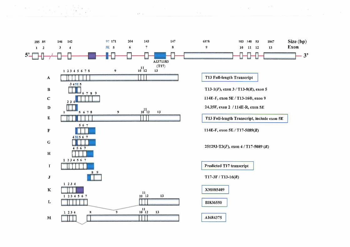
285 85
t2r43
7
204
6
t46 142
3497 r7l5E5
9
r47
I6578 r047
T13 Full-length Transcript
T13-1(Ð, exon 3 / Tf3-8(ft), exon 5
l14E-F, exon 5E / T13-16@, exon 9
24.35F, exon 2 / 1l4E-R, exon 5E
Tl3 Full-length Transcript, include exon 5E
1148-F, exon 5E / T17-5089(R)
251293-T3(F)! exon 4 / Tr7-5089 (À)
Predicted Tl7 transcript
Tr7-3F / T13-16(À)
xM085409
81836550
41684375
13
103 140 93
10 11 129
Size (bp)Exon
5 1'
I 23 4 5 6 7 I11
t0 12
AÍ171183(rt7)
l3
A
B
c
D
E
F
G
H
3 4sE5
67I9
23
I 5 67E
567
911t0 t2 13
11to 12 13
llt0 t2
I
45f.S 6 7
4567
t 23 4 5 6 7
r 23 4
L 23 4 5 6 7
J
K
L
M9
il:tlrtt
tllt r llr
t t:rIttr
trtl;rI
tltrtil
| 23 4 I l3
Study of 713 for its mutation in sporadic breast cancer was perfiormed by other
members of the chromosome 16 I breast cancer group (Lower, K.M., Whitmore, S.4.,
Crawfort, J., McKenzei, O., and Bais, A.J.). PCR-SSCP analysis of all 213 exon and
subsequent sequencing of the products that show mobility band shift were used to screen
for TI3 mutation in 48 breast cancer samples showing either restricted 16q24.3 LOH or
complex LOH of 16q that includes 16q24.3.In addition screening was also conducted in
24 breast cancer cell lines. The overall results shown that by the current methods there
were no mutations detected in these breast cancer samples as well as cell lines.
5.4 Discussion
The principle aim of the present study was to (i) explore the transcribed sequence
within the proximal region extending from the previously reported 16q24.3 LOH physical
map, (ii) charactenze a gene or genes that may be considered to be a candidate for breast
cancer related tumorigenesis. Within this region, BAC 56lB17 was the largest genomic
clone and was chosen for this study.
5.4.1 Exon trapping
Initially there was no genomic sequence of this BAC available, exon amplification
procedure was therefore utilized to provide the basis for gene identification. Exon
amplification or "exon trapping" involves sub-cloning of the restriction-digested fragments
of genomic DNA into an exon trapping vector, pSPL3B. Recombinant clones are
subsequently isolated and transfected into the mammalian cell line, COS7. The multþle
cloning site of the exon trapping vector is designed such that it is flanked by a short
sequence characteristic of exons to provide both "5' donor" aîd "3' acceptor" splicing
signals (Fígure 5.1). Upon transfection into the COST cell line, the primary RNA
172
transcribed from the recombinant vector is processed by the splicing machinery of the host
cell and allows splicing between the flanking exons and any potential exon contained
within the DNA insert. If there is no potential exon sequence within the DNA insert,
splicing will only occur between the flanking exons of the multiple cloning site and thus
result in the "vector-only" product. Finally, the trapped exon sequences are amplified by
RT-PCR from total RNA isolated from the host cell. The RT-PCR primers used are
generated from the flanking exon sequences to ensure the amplifred products are free from
host transcript sequences. These PCR products can subsequently be isolated, by cloning,
for firrther analysis. The advantage of exon trapping is that it can be used to isolate
potential transcribed sequences contained within genomic DNA in the absence of any prior
knowledge of tissue specific gene expression.
A total of 14 individual exons were identified from the BAC 561E17. The size of this
BAC is approximately 140 kb, therefore exons were trapped at an average of I exon for
each l0 kb. This success rate was similar to previous exon trapping from the 900-kb
physical map of 16q24.3 (Whitmore et al., 1998a) as well as those from other
chromosomes (Burn et al., 1996; Chen et al., 1996; Church et al., 1997). Due to the large
number of clones obtained, only one to three clones representing a particular size group
were chosen for sequence analysis. It would be likely that analysis of more clones within
each size group would result in identification of additional different exons.
The proportion of "vector-only" products detected by colony PCR was
approximately 30 o/o. These are mostþ generated when the genomic insert does not contain
an exon. In addition, vector-only products are also generated when the genomic insert
contain an "incomplete exon". A complete or "functional" exon refers to an intact exon
sequence with flanking 5' splice donor and 3' splice acceptor sites. Restriction digestion of
genomic clones that eliminated either of these splice sites from the exon could lead to an
a
173
incomplete exon sequence. In both cases splicing will occur between the flanking exons of
the multiple cloning site. This will create a restriction Bs/XI site (Figure 5.1) in the vector-
only product. Indeed, the trapping procedure is designed to eliminate such products by the
Bs/XI digestion (section 5.2.1.9).It is possible that DNA digestion wrth Bs/XI was not
complete. This was probably due to a high percentage of these type of insert as well as
some impurity that interfered the digestion reaction. This could be improved by
-a
purification of primary PCR products prior to digestion BsfXI enzyme.
Two individual exons were trapped from the CDHLï gene, which has previously
been assigned to this physical region of 16q24.3 through somatic cell hybrid mapping
(Kaupmann et al., 1992; Kremmidiotis et al., 1998). The mapping of these exons to the
BAC 561E17, in the present study, theref'ore provided a ref,rned physical location of this
gene. The genomic structure information of CDHIS was not available at this time.
Alignment of the CDHIS cDNA sequence with the available genomic sequence of this
region showed that these two trapped exons corresponded to the exons 2 and 3 of this gene.
A group of partial cDNA sequences represented by clone IMAGE: 1098126 was also
mapped to this BAC and was later identified to be the 2.3 kb full length transcript of TI8
gene (chapter 6). However, no exons were trapped from the 718. Since the restriction
enzymes used in this exon trapping are BamIII, BgilI, and Psfl. The distribution of these
restriction enzpe sites within this gene may be a result that lead to the generation of
either large restriction fragments, inefficiently ligated into the pSPL3B vector, or small
restriction fragments containing incomplete exon sequences. Therefore, exon happing
would not result in the identification of exons from TI8 gene.
A number of exons were trapped as a result of alternative usage of incorrect splice
sites. The first example was exon 561-88. This exon was trapped from the CDHI5 gene
corresponding with the nucleotides 120-278 of the cDNA (exon 2) but also included part of
.F,'^
t74
the adjacent 5' intron. Similary, exon 561-17 was trapped from CDHIS exon 3,
nucleotides 279-434, that also included part of its flanking 3' intron. The inclusion of
intron sequences seen in both trapped products appeaß to intemrpt the open reading frame
of the transcript and are therefore likely to be a splicing error due to the presence of
alternative splice signals. Alternative splice signals were also utilized in clone 561-13.
This clone was a result of cloning of genomic fragment that included part of TI3 exon 9 in
the reverse orientation.
5.4.2 Characterization of Transcription unit 13 (T13)
Identihcation of the transcribed sequence from the BAC 56lE17 by exon trapping
was based on sequence homology to expressed sequence in the NCBI database and initially
identified five potential transcription units, including that which formed part of CDH15.
Two of these, transcripts B and C, were identified from two independent overlapping sets
of expressed sequences from both human and mouse. These transcripts were mapped in
close proximity at the distal region of BAC 56lB17. They were subsequently identified as
forming part of the transcription unit 13 (713). The transcript B was characterized as the 3'
part of Tl3.Finally, a combination of these two transcripts (B and C), and computerized
analysis of the genomic sequence encompassing this region, lead to the assembly of full-
length transcript sequence of 9 ,282 bp of T I 3 .
During the course of 713 transcript sequence assembly, a large number of expressed
sequences from the GenBank database that matchedTl3 were found, mostly within the
first 5 kb of Tl3 5' sequence. Analysis of the Z/3 transcript sequence identified a number
of long adenosine nucleotide (A) tracts distributed within the region between 2 and 5 kb
from the 5' end. It is suggested that these poly (A) tracts allowed binding of the oligo dT
primer generally used in the reverse-transcription step for cDNA library construction.
'a
t75
Accordingly, a large number of oDNA derivatives of Tl3 may have been reverse-
transcribed from these long A tracts. Furthermore, due to the large transcription size of T13
it would be unlikely that reverse-transcription from the poly(A) tail would generate a full-
length transcript. Together, these could result in the lack of homologous ESTs of the region
down-stream from the first 5 kb of 213.
The TI3 transcript was predicted to code for a large protein of 2663 amino acid
residues. T13 protein contains three tandem ankyrin repeats at the region close to the N-
terminus. Anþrin repeats are one of the most common protein sequence motiß, and
consist of about 33 residues (Sedgwick and Smerdon, 1999). They have been found in
various functionally diverse proteins, such as signal transduction and transcriptional
regulators, Cdk inhibitors, cytoskeletal organizer, developmental regulators, and toxins.
These protein motifs mediate "protein-protein interactions" in a number of different
biological systems, from microbes to humans (Sedgwick and Smerdon, 1999). The
presence of ankyrin repeat motifs within the Tl3 protein molecule therefore provides the
primary evidence that the biological function of this protein requires interaction with an as
yet unknown protein partner or partners. In addition to these anþrin motifs, T13 protein
also possesses eight bipartite nuclear localization signals (NLS) suggesting it is a protein
located in the nucleus. Together, these indicate that Tl3 protein may be involved in
transcriptional processes or in the regulation of other nuclear activities by interacting with
other nuclear proteins.
Partial sequence homology was detected between T13 protein and two known human
proteins, BRCAl-associated RING domain protein I (BARDI) and a protein member of
nuclear factor kappa B (NF-kB) inhibitors, IkappaBR (I-kBR). Interestingly, homology
was between the ankyrin domain region of these three proteins, with all three ankyrin
repeats showing homology. It is possible that homologous ankyrin domains may allow
'a
176
prediction of these protein that involve in the specificity of protein-protein interaction or
be found among the functionally related protein. For instance, BARDI possess three
ankyrin repeats that mediates its BRCAI-independent function. BARDI interacts wirh NF-
kB, via Bcl-3, and modulates its transcriptional activity (Dechend et al., 1999). This
interaction involves binding of BARDI ankyrin domain to Bcl-3. The inhibitors of NF-/rB
(I-kB) on the other hand inactivat¿s NF-kB by binding to and preventing nuclear
localization of NF-kB and hence DNA binding (Chen et a1.,2001). Binding of I-kB fo NF-
kB is also mediated by the ankyrin repeat region. As a member of I-kB fami/y, I-kBR has
also been demonstrated to interact with the p65 subunit of NF-kB and partially inhibft NF-
kB DNA binding activity (Ray e/ al., 1995). Given that the T13 protein possesses three
anþnin repeats homologous to those of BARD1 ønd I-kBR, it may be functionally related
to or involve in the regulation of NF-kB activity. This, however, is speculative and requires
further investigation.
A protein of unknown frrnction, KIAA0874, was the only human protein in the
database that was similar to Tl3. Similarity was high at both the N-terminal and C-
terminal regions of these two proteins. The remaining regions, although showing a lower
degree of homology, also contained a number of nuclear localization signals (NLS) similar
to that of T13. KIAA0874 is a high molecular weigh protein of 2062 amino acids. Because
of these similarities KIAA0874 is likely to be paralogous to T13 and may be functionally
related to the T13 protein. Protein sequence homology was also detected between the first
343 amino acids of Tl3 and LZl6, a protein sequence that is in the NCBI database as a
"nasopharyngeal carcinoma susceptibility gene product" (Protein database accession no.
NP-037407). However, analysis of the 1603-bp LZ16 transcript sequence (GenBank
accession no. AFl2l775) showed that LZl6 was likely to be an incomplete 213 cDNA that
was reverse transcribed from the poly (A) tract between nucleotides position 1778 and
177
1809 of 713. A similar situation was also detected in another version of LZ16 mRNA of
2557 bp (GenBank accession no. 8C017437). This LZL6 appeared to utilize the poly (A)
tract between position 2512 arrd 2556 of Tl3. In addition both LZI6 mRNAs did not
possess polyadenylation signal characteristic of independent mRNAs, therefore confirming
that both were derivatives of the 213 transcript.
The 713 gene consists of 13 exons and encompasses a genomic region of
approximately 120 kb. Intron 2 atd intron 4 appear to be the fnst and the second largest
intron, while exon 9 is the largest exon. Altemative splicing that generated a number of
transcription isoforms or aberrant transcript of both major and minor appeared to involve
the sequence within these two introns. These were likely the result of cryptic splice sits
within this region. Many isoforms, however, gave no ORF and were likely to be the
products of splicing artifacts.
Three transcription isoforms, XM-085409, Tl7, and 81836550 (Figure 5.10) are
likely to be functional based on the present of ORFs by utilizing the same ATG start, in
exon 3, as that of full-length 213. These transcripts could code for proteins that are in
common by their amino terminus with that of Tl3, though varying in length. The predict
protein sequence of 124 amino acids of XM-085409 shares only 75 amino acids with Tl3
protein and excludes the ankyrin domain. The protein products of 717 and 81836550 on
the other hand contain the N-terminal part of T13 protein that includes the ankyrin repeat
domains. The latter also shares part of the carboxy terminal region with that of full-length
Tl3. Together, these protein products may likely function in association with the fulI-
length T13 protein.
Failure to detect any sequence mutation of 713 either in 48 breast cancer samples
showing LOH of chromosome 16 at q24.3 region or 24 breast cancer cell lines might
indicate that T13 \ilas not likely a gene involved in breast cancer pathogenesis.
178

Altematively, Tl3 mutations may be involve in pathogenesis of other cancers because
LOH at this chromosome region have also been reported in cancer of other organs such as
hepatocellular carcinoma (Tsuda et al., 1990), prostatic carcinoma (Carter et al., 1990;
Bergerheim et al., l99l; Kunimi et al., 1991), and ovarian cancer (Sato e/ al., l99lb).
Mechanism other than sequence changes exist that inactivates gene function. Hyper- or
aberrant metþlation of the CpG island at the promotor region of the gene has also been
reported to reduce or suppress gene expression in many genes that behave as tumor
suppressors (Baylin et al.,2001; Ka.pf and Jones, 2002; Plass, 2002). Such mechanism
may also involve inactivation of 713 in breast carcinogenesis. This, however, requires
further investigation.
The TI3 gene product is a large molecule although only one functional domain could
be defined by in silico anaþis. T13 protein may also possess other functional domains, yet )a
to be identified, that are important for its activity. BRCAI, for instance, is also a large
molecule that contains only two known functional domains, Ring and BRCT domains.
However, BRCAI has been reported to interact with several proteins for its biological
functions (section I .5. I . 1). For the Tl 3 protein a search for proteins that interact with the
anlq/rin domain and other parts of the Tl3 protein may provide an understanding of its
biological function.
t79
Chapter 6
Charact erization of Transcription U nit
18 (r1B)
Table of Contents
6.l lntroduction
6.2 Methods
6.2.1 T 18 transcript sequence assembly
6.2.2 Expression Analysis
6.2.3 Sequence Homology and In Silico Protein Analysis
6.2.4 Characterization of the Genomic Structure
6.3 Results
6.3.1 Identification of Tl8 transcrþt sequence
6.3.2 Transcript Sequence and Expression Analysis
6.3.3 Protein Sequence Analysis
6.3.4 Sequence Homologies
6.3.4 Tl8 gene structure
6.4 Discussion
Page
180
180
180
181
181
182
182
182
183
183
184
185
186
6.1 Introduction
Transcription unit 18 (T18) was assigned to the cDNA clone IMAGE:1098126 which
was mapped to the proximal region of the BAC clone 56lB17 (section 5.1). This BAC forms
part of a genomic contig that extended approximatelyl4O kb from the proximal boundary of
the physical map established at chromosome region 16q24.3 (Whitmore et a1.,1998a). Asl,ù<L-a
part of the aim to study genes within this extended region, in addition to T13,718 was also
^selected for gene characterization. The cDNA clone IMAGE:1098126 was isolated from a
cDNA library derived from breast tumor tissue. The partial sequence of this cDNA clone,
EST 44603773, was from its 3' end. This EST belongs to the group of partial oDNA
sequences collectively refer to the UniGene cluster Hs.123661. The EST sequence members
of this cluster are derived from cDNA libraries of various tissue sources, suggesting a
ubiquitous expression of this transcript, which may indicate the essential function of its
protein product.
This chapter describef the isolation of the Tl8 transcript sequence, analysis of its
sequence and pattern of expression, and subsequently charactenzation of the putative
function of its predicted protein. Finally there is discussion of its putative protein fi¡nction
and whether this gene is likely to be a breast cancer associated tumor suppressor or possibly
associated with other human genetic disorders.
6.2 Methods
6.2.1 T18 transcript sequence assembly
A combination of RT-PCR experiments and direct sequencing of the cDNA clone
IMAGE:1098126 were carried out to obtain the full-length transcribed sequence of Tl8
The RT-PCR method was performed as described in section 2.2.13, using a primer set
generated from the partial oDNA sequences, AA25I134 and THCl244l5. The sequence of
180
)s
primers and their corresponding location in the transcribed sequence of 718 are presented in
Table 6.1.The tissue sources of mRNA used for RT-PCR are fetal brain and fetal heart.
The oDNA clone IMAGE:1098126 was purchased, DNA isolated and sequenced by the
Dye-terminator method (2.2.16.1) on an ABI 373 DNA sequencer utilizing vector primers
flanking the inserted oDNA and the primers designed to the sequence generated from the
RT-PCR products (Table 6.1).
6.2.2 Expression Analysis
RT-PCR products generated from fetal brain and fetal heart mRNA utilizing primer set
as shown in Tøhle 6.1 werc separated by agarose-gel electrophoresis and cDNA bands were
purified from the gel (section 2.2.14.2). These purified oDNA fragmants were then used to
hybridize to a multiple tissue Northern membrane, conìmercially available from Clontech,
containing 2 pg of poly(A)+ mRNA from six different human tissues. The tissue sources of
mRNA are heart, brain, placenta, lung, liver, skeletal muscle, kidney, and pancreas. The
hybridization condition was performed according to the manufacturer's recommendation in
"Expres sHyb" hybndization so lution (Clontech).
6.2.3 Sequence Homolory and In Silico Protein Analysis
Nucleotide sequence homology tool, BLASTN, was used to perform homology
searches against nucleotide databases: dbEST, non-redundant database, and genome
sequence database. BLASTP was used to search for homolodííËn r"r"es from various
protein databases. Prediction of protein function was assisted by, (i) protein domain
identification tools, Pfam, SMART, and Prosite, (ii) sub-cellular localization prediction
program, PSORTII, (iii) comparing the sequence with both unknown and known functional
181
I
proteins by multiple sequence alignment tools, CLUSTAL'W, available at GenomeNet
server (Kyoto Center, Japan).
6.2.4 Characterization of the Genomic Structure
The gene structure of 718 was determined by comparing the complete cDNA sequence
with genomic sequence encompassing this region, available from GenBank database. The
transition of exon to intron (exon boundary) was identified from the genomic sequence that
diverges from that of cDNA.
6.3 Results
6.3.1 Identification of T18 transcript sequence
The sequence of 44603773 was initially used to search the GenBank EST database. A
number of homologous EST sequences, all from the 3' end of cDNA, were identif,red and
most contained the consensus polyadenylation signal, A.A.TAJA.A.. Two of these ESTs,
AA25ll34 and 44516361, were assembled to form a sequence that represented the 3'end
of 718. The sequence of EST AA251134 is derived from the 3' end of the oDNA clone
IMAGE:684ß9 of unknown insert size. However, the 5' sequence of this cDNA was also
available as the database entry AA25ll98 (Figure 6.1). T\e sequence of 44251198 was
then used to perform homology searches, and an assembled oDNA sequence, THCI244I5,
was identified from the TIGR database. T}JCl244l5 extended the sequence 5' from
AA251198 by a firther 290 bp. To determine the entire transcribed sequence, primers were
designed to the cDNA sequences of 44251134 and THCl244l5 for RT-PCR experiments
(Figure 6.1 and Table ó.1). Three RT-PCR products were obtained (Figure 6.2) and DNA
sequence determined sequentially from both strands.
t82

0 100 200 300 400 500 600 700 800 900 1000 1100 1200 1300 1400 1500 1600 1700 1800 1900 2000 2100 2200bp
THCL24415 F1+ F2+4.A251
5',AA2SllS 4 (germinal B-cell)
4A:6037 7 3 (breast tumor)IMAGE:1098126
3)
3?
3t
5t
T7FA+ 44516361 (colon tumor)
:F2FA +
RI
+R3
ATG TGA AATAAA5t 3t
: Determination of ?/8 transcript (44251134, 44603773,44516361) that belong to a UniGene12366I were chosen and performd efore the primers, Rland R2, were designed . AA25I198 is the
sequence from the 5' end of the same cDNA clone of 44251134. THC1244L5 is the assembled cDNA sequence from TIGRdatabase that overlaps with 44251198. Primers Fl and F2 were designed from THCl24415 sequence. Clone IMAGE: 1098126 isthe cDNA clone origin of 4A603773 that was isolated for sequence determination. Thick lines with affow heads at both ends denotethe RT-PCR products using three combination of primers: F1/R1, FllR2, and F2lR1. Lines with single arrow head indicate theextendof sequencing of RT-PCR products þrimers Rl, R2, FI,F2, F3) and of cDNA clone IMAGE:1098126 þrimers T7, FA, R3,and R4). The 2.26 kb transcript sequence of T18 was assembled from these sequencing products and revealed an ORF, completedwith start, ATG, and stop, TGA, codons and a polyadenylation signal, AATAAA.
<.R2
R2
+R4
R4
')
IË
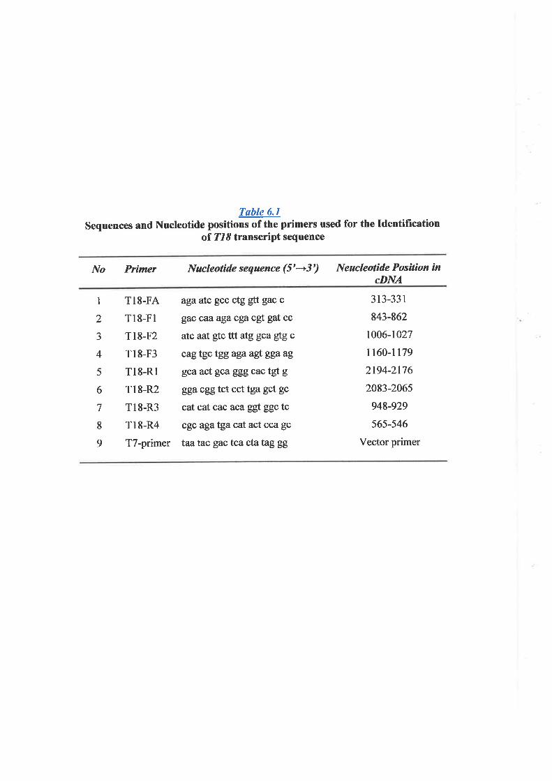
Tøble 6.1
Sequences and Nucleotide positions of the primers used for the IdentificationoI Tl8 transcript sequence
No Prìmer Nucleolíde sequence (5'---+3') Neucleotide Posilíon incDNA
1
2
J
4
5
6
7
8
9
TI8-FA
Tr8-Fl
Tr8-F2
Tl8-F3
Tl8-Rl
Tl8-R2
T18-R3
T18-R4
T7-primer
agaatc gcc ctg gtt gac c
gac caaagacga cgt gat cc
atc aat gtc ttt atggcagfgc
cagtgc tggagaaglggaag
gcaact gcagggcactgtg
ggacggtct cct tga gct gc
cat cat cac aca ggt ggc tc
cgç agatgacat act cca gc
taatac gactcactatzggg
3 13-33 I
843-862
1006-1027
tt60-r179
2194-2176
2083-2065
948-929
565-546
Vector primer

Ml23
bp bp
15801460I 160
980
1352
l 189t24l
't20
Agarose gel electrophoresis of T18 RT-PCR products. The cDNAryvSsm the total RNA isolated from fetal heart. The products ggntain irí,:,ÉEl,
lane l, 1352 bp; lane 2, l24l bp1' and lane 3, 1189 bp were obtained using primerpairs Tl8-Fl/-Rl, T18-FI/-R2, ffid Tl8-F2/-Rl respectively. The DNA marker isloaded inlane M and size-fragments are indicated in bp.
The oDNA clone IMAGE:1098126 was also sequenced utilizing the vector primers
flanking the cDNA insert and the primers designed to the sequence from the RT-PCR
products mentioned above (Figure 6.1). The cDNA sequences from RT-PCR products and
those from sequencing of oDNA clone IMAGE:1098126 were finally assembled to form a
tull-length cDNA of 2,263bp.
6.3.2 Transcript Sequence and Expression Analysis
Analyses of the Tl8, 2,263-bp transcript sequence (Figure ó.3) identifred an ORF of
1728 bp with the start codon, ATG, and stop codon, TGA, at nucleotide positions 149-151
and 1877-1879 respectively. The ORF was followed by a 384-bp 3' UTR. The sequence
surrounding an ATG start (AGTGCAÂIGC), though, weakly conformed to the Kozak
consensus of an initiation start site (GCCA/GCCATGG) (Kozak, 1989). It was, however,
preceded by an in-frame stop codon, TAG, 39 bp upstream from the initiation start site. The
ORF was predicted to code for a protein of 576 amino acids. A sequence characteristic of
polyadenylation signal, AATAAA, was identified at the 3' terminal region and situated 19
bp upstream from a polyadenylation site.
Northern hybridization using RT-PCR products of 718 (section 6.2.2) as probes
identified a prominent mRNA band of approximately 2.5 kb in all tissues tested, though with
varying degrees of expression (Figure 6.4). Thrs mRNA band corresponded with the size of
the identified transcript sequence of T18. With a longer exposure, faint bands of 1.3 and 4.4
kb were also detected. Expression of TI8 as determined by Northern analysis was equally
high in liver, kidney, and pancreas. Medium degree of expression was seen in heart and
skeletal muscle, while expression in brain, placenta, and lung was fairly low.
6.3.3 Protein Sequence Anølysis
183
Fieure 6.3 : The cDNA transcript and deduced amino acid sequence of T18. The transcript is
2262 bp from the first nucleotide to the polyadenylation site (A). The start, ATG, and stop,
TGA, codons are located at positions 148-150 and positions 1876-1878 respectively, and are
highlighted. Also indicated, an inframed stop codone, TAG, that preceded the translation start
site. The sequence of polyadenylation signal, AATAAA, at positions 2238-2243 is
underlined. Within the amino acid sequence, the sequence characteristic of "AMP-binding
domain" is indicated at residues 198-211 (underlined) and the consensus residues are
highlighted.
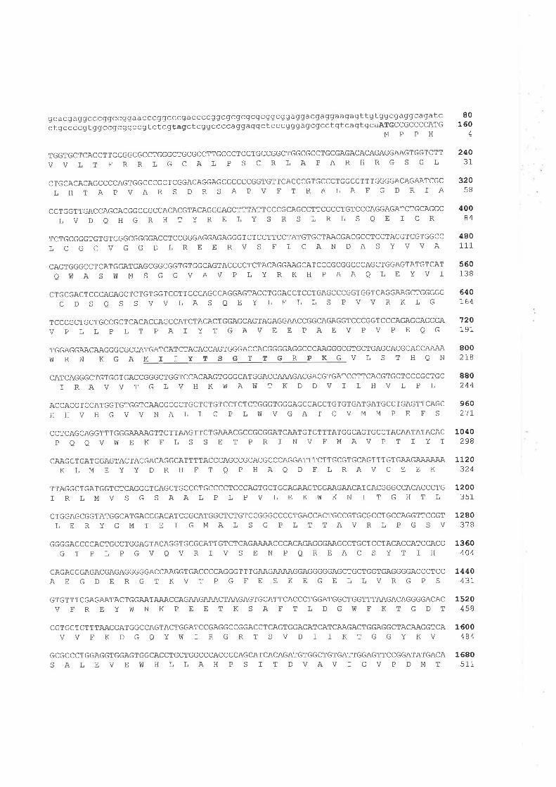
gcacgaggcccggccggaacccggcccgaccccggcgcgcgcgcggcggaggacgaggaagagttgtggcgaggcagatcctgccccgtggccgcggccgtctcAtagctcggccccaggaggctcccgggagcgcctgtcaqtgCaÀTGCCGCCCCATG
MPPH
TGGTGCTCACCTTCCGGCGCCTGGGCTGCGCCTTGCCCTCCTGCCGGCTGGCGCCTGCGAGACACAGAGGAAGTGGTCTT
VVLT FRRLGCAL P S C RLAPARHRGS GL
CTGCACACAGCCCCAGTGGCCCGCTCGGACAGGAGCGCCCCGGTGTTCACCCGTGCCCTGGCCTTTGGGGACAGAATCGCLHTAPVARS DRS APV FTRALAFG DRIA
CCTGGTTGACCAGCACGGCCGCCACACGTACAGGGAGCTTTATTCCCGCAGCCTTCGCCTGTCCCAGGAGATCTGCAGGCLVDQHGRHTYRE LYSRS LRLSQE I CR
TCTGCGGGTGTGTCGGCGGGGACCTCCGGGAGGAGAGGGTCTCCTTCCTATGTGCTAACGACGCCTCCTACGTCGTGGCC
L C G C V G G D L R E E RV S F ], C AN DA S Y VVA
CAGTGGGCCTCATGGATGAGCGGCGGTGTGGCAGTACCCCTCTACAGGAAGCATCCCGCGGCCCAGCTGGAGTATGTCAT
O!ú A S !V M S G G VAV P L Y R K H P AAQ L E YV I
CTGCGACTCCCAGAGCTCTGTGGTCCTTGCCAGCCAGGAGTACCTGGAGCTCCTGAGCCCGGTGGTCAGGAAGCTGGGGGCDS QS SVVIAS QEYLE LLS PVVRKLG
TCCCGCTGCTGCCGCTCACACCAGCCATCTACACTGGAGCAGTAGAGGAACCGGCAGAGGTCCCGGTCCCAGAGCAGGGAV P LL P LT PAI YT GAVB E PAEVPVP E OG
TGGAGGAACAAGGGCGCCATGATCATCTACACCAGTGGGACCACGGGGAGGCCCAAGGGCGTGCTGAGCACGCACCAÄAA
80160
4
24031
32058
40084
480111
560138
6¿0I64
720191
8002IB
880244
960217
1040298
1120324
VüRNKGAMIIYTSGTTG RPKGVLSTHQN
CATCAGGGCTGTGGTGACCGGGCTGGTCCACAAGTGGGCATGGACCAAAGACGACGTGATCCTTCACGTGCTCCCGCTGCI RAVV T G L V H K fü AVü T K D D V I ], H V L P L
ACCACGTCCATGGTGTGGTCAACGCGCTGCTCTGTCCTCTCTGGGTGGGAGCCACCTGTGTGATGATGCCTGAGTTCAGCHHVHGVVNALL C P LWVGAT CVMM P E F S
CCTCAGCAGGTTTGGGAA.AAGTTCTTAAGTTCTGA.AACGCCGCGGATCAATGTCTTTATGGCAGTGCCTACAATATACACPQOVW E KELS S E T PR I NVFMAVP T I YT
CAAGCTGATGGAGTACTACGACAGGCATTTTACCCAGCCGCACGCCCAGGATTTCTTGCGTGCAGTTTGTGAAGAAAAÄAK LM E YY DR H FT Q P HAQ D FLRAVC E E K
TTAGGCTGATGGTCTCAGGCTCAGCTGCCCTGCCCCTCCCAGTGCTGGAGAAGTGGAAGAACATCACGGGCCACACCCTG
I R IMV S G S AAL P L P V L E KIV KN I T G H T L
CTGGAGCGGTATGGCATGACCGAGATCGGCATGGCTCTGTCCGGGCCCCTGACCACTGCCGTGCGCCTGCCAGGTTCCGT], E R Y G M T E I GMA L S G P ], T T AV R L P G S V
GGGGACCCCACTGCCTGGAGTACAGGTGCGCATTGTCTCAGAAAACCCACAGAGGGAAGCClGCTCCTACACCATCCACGGT P LPGVQVRIVS ENPQREACS YT TH
CAGAGGGAGACGAGAGGGGGACCAAGGTGACCCCAGGGTTTGAAGAAAAGGAGGGGGAGCTGCTGGTGAGGGGACCCTCCAE GDE RGTKVT PGFE E KEGELLVRGP S
GTGTTTCGAGAATACTGGAATAÀACCAGAAGA.AACTAAGAGTGCATTCACCCTGGATGGCTGGTTTAAGACAGGGGACACV F R E Y !V N K P E E T K S A F T ], D G W F K T G D T
CGTGGTGTTTAAGGATGGCCAGTACTGGATCCGAGGCCGGACCTCAGTGGACATCATCAAGACTGGAGGCTACAAGGTCAVV F K D G QY![ ] R G R T S V D I I KT GGY KV
GCGCCCTGGAGGTGGAGTGGCACCTGCTGGCCCACCCCAGCATCACAGATGTGGCTGTGATTGGAGTTCCGGATATGACASALEVEWHL LAH P S I T DVAV I GV P DMT
1200351
1280378
1360404
L440431
1520458
1600484
1680511
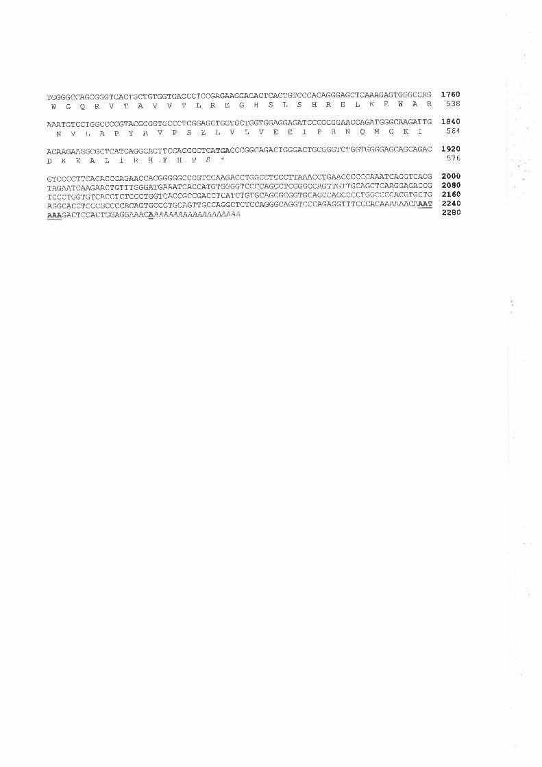
TGGGGCCAGCGGGTCACTGCTGTGGTGACCCTCCGAGAAGGACACTCACTGTCCCACAGGGAGCTCAAAGAGTGGGCCAGVü G Q R V T A V V T L R E G H S L S H R E ]' K E Îü AR
AAATGTCCTGGCCCCGTACGCGGTGCCCTCGGAGCTGGTGCTGGTGGAGGAGATCCCGCGGAACCAGATGGGCAAGATTGNVLAPYAV P SE LVLVE E I PRNQMGKI
ACAAGAAGGCGCTCATCAGGCACTTCCACCCCTCATGACCCGGCAGACTGGGACTGCGGGTCTGGTGGGGAGCAGCAGACDKKALIRHFHPS*
GTCCCCTTCACACCGAGAACCACGGGGGCCCGTCCAAGACCTGGCCTCCCTTAAACCTGAACCCCCCAAATCAGGTCACGTAGAATCAAGAACTGTTTGGGATGAAATCACCATGTGGGGTCCCCAGCCTCGGGCCAGTTGTTGCAGCTCAAGGAGACCGTCCCTGGTGTCACCTCTGCCTGGTCACCGCCGACCTCATCTGTGCAGCGCGGTGCAGCCAGCCCCTGGCCCCACGTGCTGAGGCACCTCCCGCCCCACAGTGCCCTGCAGTTGCCAGGCTCTCCAGGGCAGGTCCCAGAGGTTTCCCACA.AÄAÄACAAATA.AAGACT C CAC T GGAGGAAACAAAAAAAAAAAAAAAAAA
1760538
1840564
192 05-l 6
200020802L6022402280
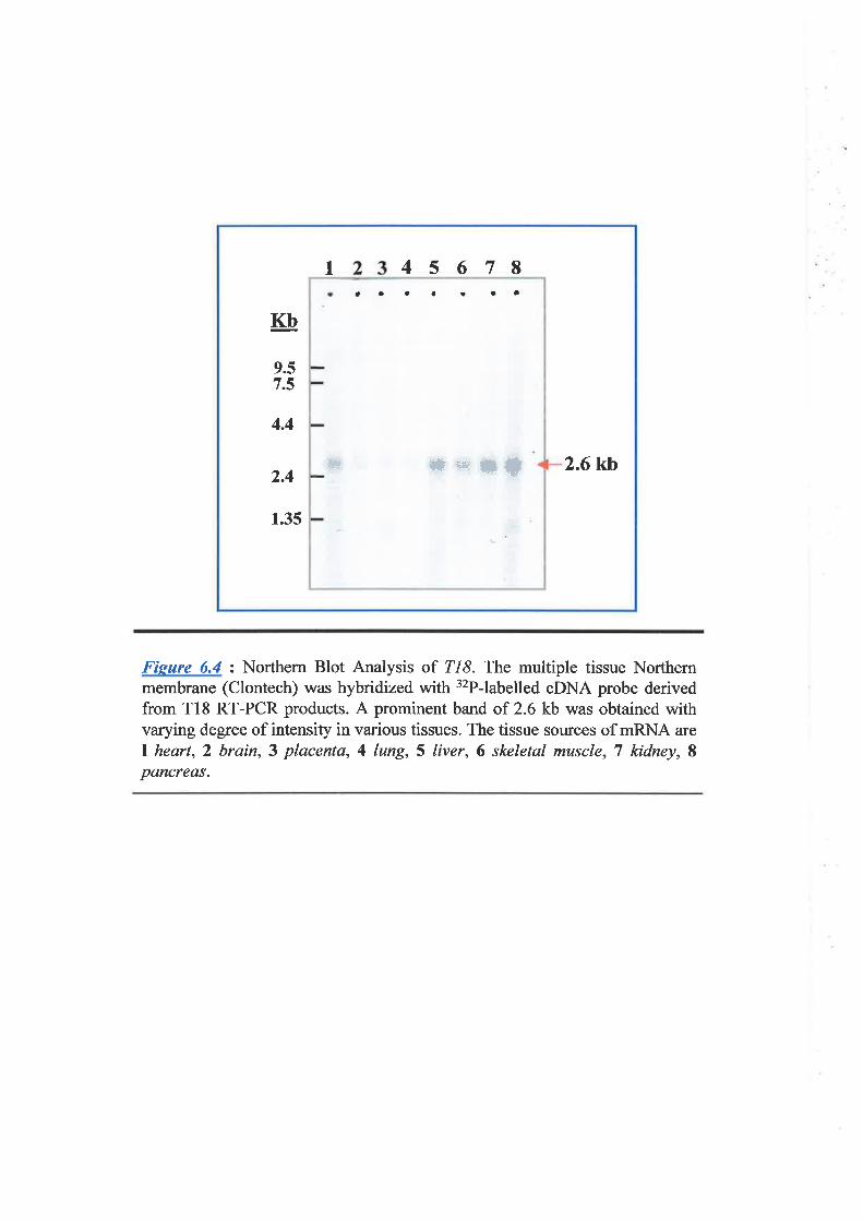
45678I
Kb
9.57.5
4.4
2.4
1.35
2.6kb*r+tü
a a aa o I a
#*
Fisure ó.y' : Northern Blot Analysis of 718. The multiple tissue Northernmembrane (Clontech) was hybridized with 32P-labelled cDNA probe derivedfrom T18 RT-PCR products. A prominent band of 2.6 kb was obtained withvarying degree of intensity in various tissues. The tissue sources of mRNA areI heart, 2 brain,3 placenta, 4 lung, 5 liver, 6 skeletal muscle, 7 kidney, Ipancreas.
Chspler 6 Charoclerizslion of Trsnsc¡iption Unif 18 ffL8)
Analysis of the deduced amino acid sequence of TIS,utilizing three domain-prediction
progrrims: Pfam, Prosite, and SMART, identihed the region between residues 67 and 504 as
a sequence domain characteristic of an AMP-binding enz;¡yme. This suggested that Tl8
protein belonged to this enzqe family. Within this region a conserved *AMP-binding
domain" was also detected between amino acid residues 198-211 (Fìgure 6.3).By utilizing
the PSORT program, the T18 protein was predicted to be localized to the cytoplasmic
compartment and be a membrane-bound protein. The most likely anchoring membrane was
endoplasmic reticulum (ER).
6. 3.4 Sequence Homologies
The nucleotide and predicted amino acid sequences of Tl8 were compared with the
GenBank and EMBL databases utilizing BLASTN and BLASTP algorithms. By non-
redundant database searches, the transcript sequence of Tl8 showed a significant homology
to mRNA sequences of both human and mouse. A human mRNA LOCI97322 of 873 bp
was completely identical to the sequence of TI8 from nucleotide 1386 to its 3'end,
suggesting that mRNA LOC|97322 was a partial sequence and represented the 3' half of
Z/8. Sequence homology to mouse mRNA was detected to LOC234844, a2225-bp mRNA,
and to MGC:37904, an mRNA of 1096 bp. Signifrcant homology between TI8 and mouse
mRNA sequences was only within their ORF (Figure ó.f). The nucleotide sequence of
LOC234844 was then retrieved and the protein sequence deduced, utilizing program *ORF
ftnder" at NCBI. The predicted mouse protein of 583 amino acids was then compared with
that of T18. Significant homology, 80% identical, was demonstrated between these two
proteins (Figure 6.0. V/ith the similar size of both mRNA and protein as well as the protein
sequence homology suggested that the mouse LOC234844 was an orthologue of human
TI8, and at this stage was assigned as mtl9.
'a
184
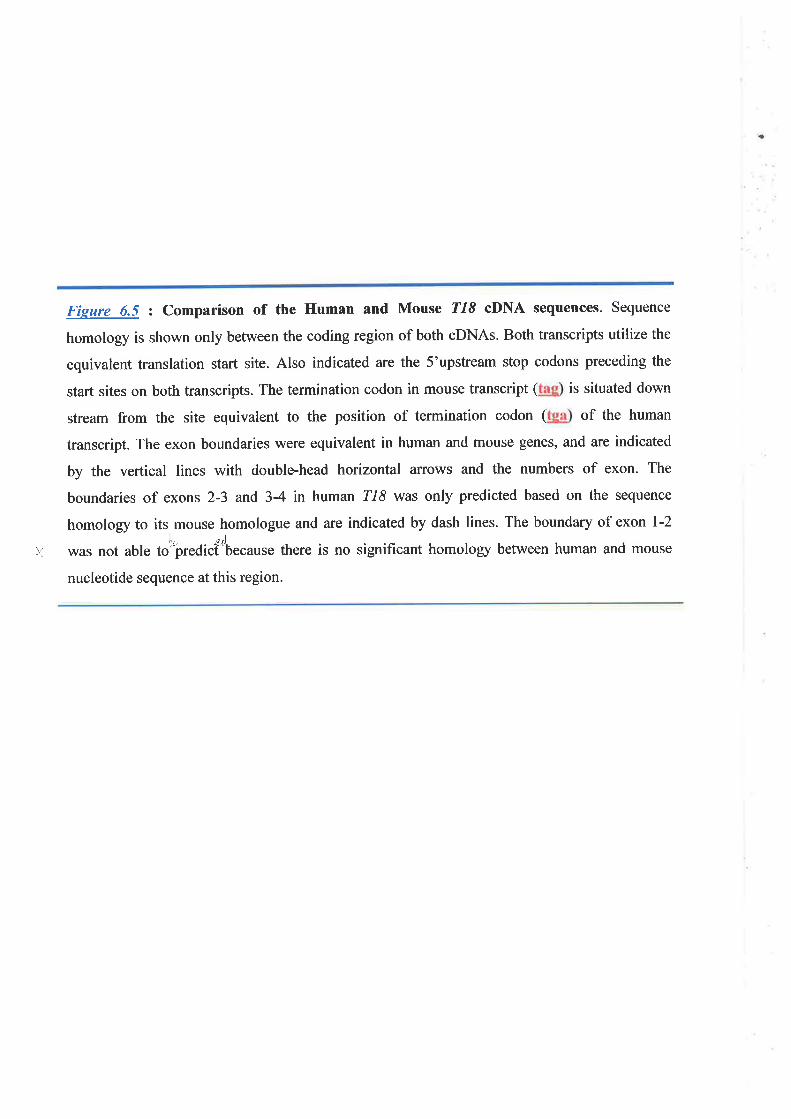
Fieure 6.5 : Comparison of the Human and Mouse T18 cDNA sequences. Sequence
homology is shown only between the coding region of both cDNAs. Both transcripts utilize the
equivalent translation start site. Also indicated are the 5'upstream stop codons preceding the
start sites on both transcripts. The termination codon in mouse transcript ( is situated down
stream from the site equivalent to the position of termination codon ( of the human
transcript. The exon boundaries were equivalent in human and mouse genes, and are indicated
by the vertical lines with double-head horizontal aruows and the numbers of exon. The
boundaries of exons 2-3 and 3-4 in human 218 was only predicted based on the sequence
homology to its mouse homologue and are indicated by dash lines. The boundary of exon 1-2
)1 was not able to'rþredicftbe"ause there is no significant homology between human and mouse
nucleotide sequence at this region.
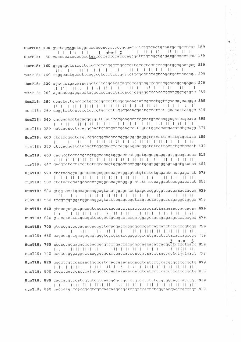
-a
HumT18:
MusTl B :
HunT18:
musT18:
HumTl8:
musTlB:
HumTl8:
musTlB:
HunTl8:
musTlB:
HunT18:
musT18:
HunTl8:
musTlB:
HunTl8:
musTlB:
Hu¡rT18:
musTlB:
HumTl8:
musT18:
HumTl8:
musTlB:
HumT18:
musTlB:
HumT18:
musÎ18:
HumTl8:
musTlB:
100 gtctcg@ctcggccccaggaggctcccgggagcgcctgtcagtqcagEgccgccccat 159
r r I I ll I 1 <l+12 I llll lll lllll ll ll8 O cacccc aaccccgcclgaccccaSlccca gccagt ggttt gt cqgt gt gatgccacctcac 1 3 9
160 gtqgtgctcaccttccggcgcctgggctgcaccttgccctcctgccggctggcgcctgc g 2L9
|t ll lllll llll ll lll lllll lllll I I llll ll140 ttggcactgcccttcaggcgtctcttctggtccttggcctccagtcagctgattcccaga 209
22O aqacacagaggaaqtggtcttctgcacacagccccagtggcccgctcqgacaggagcgcc 27 9
||ll lllll ì I ll llll ll llllll lllll I llll llll I I
2 OO agacaccgaggacatagcctcctgcctaccaccccagaggcccacacqgatgggagtgtc 259
28O ecAqLqttcacccgtgccctggcctttggggacaqaatcgccctggttgaccagcacggc 339
||t llll llllllllllllllllllllllll ll lllll lllll I Illl260 ccgqtattcatccAtgccctggcctttggggacaggattgcccttattgacaaatatggc 31 9
340 cgccacacqtacagggagctttattcccgcagccttcgcctgtcccaggagatctgcagg 399
I ||t llllllll ll lll lllllllll I lll lllllllllllllllll32 0 catcacacctacagggaactgtatgatcgcagcctttqtctggcccaggagatctgcagq 37 9
400 ctctgcgggtqtgtcggcqgggacctccgggaggagaggqtctccttcctatgtgctaac 459
| | lll I llllllllll lll ll llllllllllllll ll I ll38 O cttcagggttgtaaagttggggacctccagqaagaaagggtctccttcctgtgctccaaL 439
460 qacqcctcctacgtcgtggcccagtgggcctcatggatgagcggcggtgtggcagtaccc 519
l|t |lllllll ll ll lllllllllll llllllll ll lllll ll ll ll4 40 gacgtctcctacgttgtaqcacagtgggcctcctggatgagtqgtqgtgttgctgtccca 499
520 ctctacaggaagcatcccgcggcccagctggagtatgtcatctgcgactcccagagctct 579I l| |lllll ll I llllllllllllllll lllll lllllll lllllll
5 00 ctgtactggaaqcaccctgaggcccagctggagtatttcatccaggactcccggagctct 55 9
5BO gtqqtccttgccagccaggagtacctggagctcctgagcccggtggtcaggaagctgggq 639rr I I lllllllllll llllll ll ll ll lll ll llllll
5 60 ttgqtggtgqttggccaggagtacttggagcggctaagtccactggctcagaggctggga 679
640 gtcccgctgctgccgctcacaccagccatctacactggagcagtagaggaaccqqcagag 699||t I ll|lllllll ll lll lllll lllllll llll I ll lllll
620 qLccctcttctgccgctcacgcctgccgtctaccatggagcaacagagaagcccacagaq 61 9
700 gtcccggtcccagagcagggatggaggaacaagggcgccatgatcatctacaccagtggq 759| |l || | lll I ll lll lllllllll llllllllll lll
6B 0 cagccagttgaagagagtgqgtggcgtgaccggggtgccatgatcttctacaccagcggg 1 39
2*r)3760 accacqgggaqqcc:caagggcgtgctgagcacgcaccaaaacatcagggctgt{gtgacc 819
||t ||llllllllll I llllllll llll lll I llllllilllll74 O accacagggaggcccaagggtgcactgagcacccaccqtaacctagccgctgt{gtqac t'7 99
820 qggctgqtccacaaqtgggcatggaccaaagacgacgtgatccttcacgtgctcccgctq 879trrrIt ilrl IlI llr lIl llllllrllllll Illlllll
B 00 gggctggtccactcatgggcgtggactaaaaacgatgtgatccttcacgtcctcccgictq I 5 9
880 caccacgtccatggtgtggtcaacgcgctgctctgtcctctctggqtgggagccacctgt 939!|t lIlt It Il||ll lllll|||t ill||t lll|||llt
B 60 caccatgtccacggcAtggtcaacaagctgctctgtccactctgggtaggagccacctgt 91 9
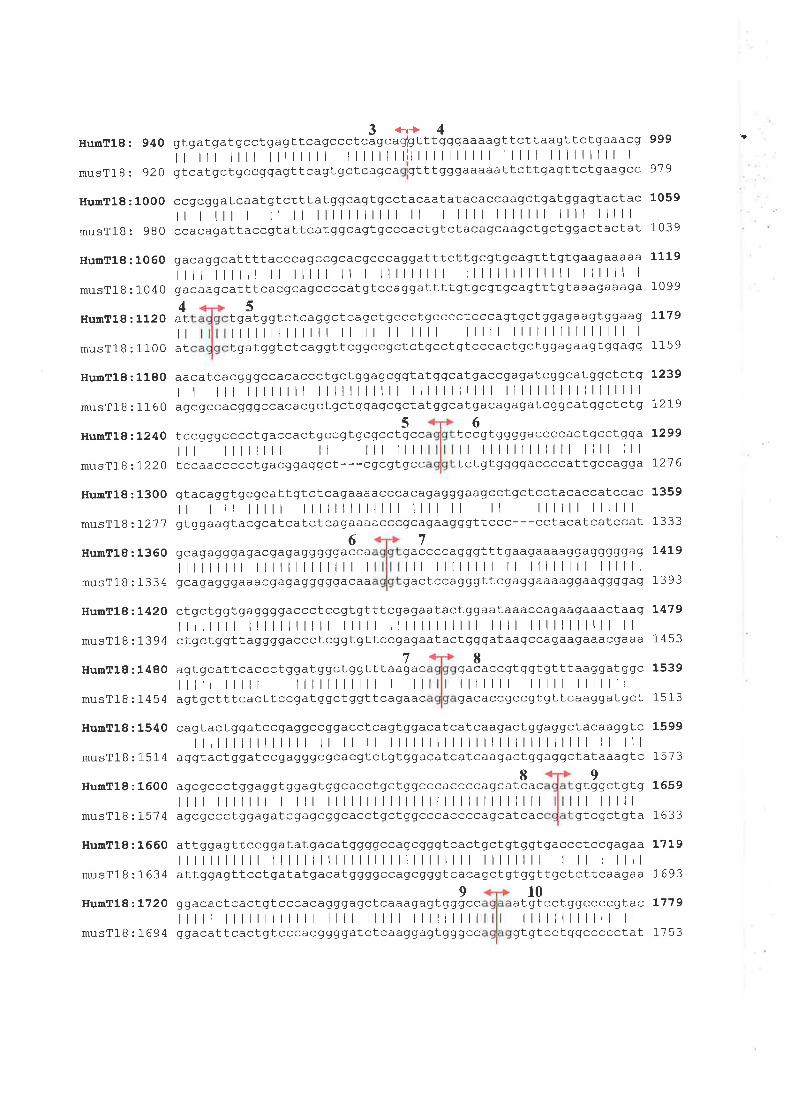
3ar|4HumT18 : 940 gtgatgatgcctgagttcagccctcagcaglgtttgggaaaagttcttaagttctgaaacg
| |t || |||| |llllllllllllllllll lllll lllllllll I
musT18 920 gtcatgctgccggagttcagtgctcagca gtttgggaaaaattcttgagttctgaagcc
999
9'7 9
1059
1039
111 9
1099
t239
]-279
t299
727 6
1359
1333
L4L9
1393
L479
1453
1539
1513
1599
1573
1659
1633
1719
1693
L779
1753
HunT18:1000
musTlB: 980
HumTl8:1060
musT18:10404
HumTl8:1120 atI
musTlB:1100 at
HunT18:1180
musTlB:1160
HumT18:L24O
musTlB z1-220
HumT18:1300
musT18:)-21'7
HumT18:1360
musTlB:1334
HumTl8:L42O
musTlB:1394
HumT18:1480
musT18:1454
HumTl8:1540
musTlB:1514
HuglTl8 :1600
musTlB t]-5'7 4
HumTl8 :1660
musTlB:1634
HunT18:,L720
musTlB 2]-694
ccgcgqatcaat gtctttatggcagt gcctacaatatacaccaagctgatggagtactacI r |t I ll ll lllllllllll ll I llll lllllll llll lllllccacagattaccgtattcatggcagt gcccact gtctacagcaagctgct ggact act at
gacaggcattttacccagccgcacqcccaggatttcttgcgt gcagttt gt gaagaaaaa
|| |llll ll lllll ll I lllllllll llllllllllllll llllll I
gacaagcatttcacgcagccccat gtccaggatttt gt gcgt gcagttt gt aaagaaaga
5tgãtggtctcaggctcagct gccctgcccctcccagtqctggagaagtggaag 1179
|||lllllll ll ll ll lllll lllll lllllllllllllll I
tgatggtctcaggttcggccgctctgcct gttcccactgctggagaagtgqagg 1 1 5 9
aacatcacggqccacaccctgctggagcgqtat ggc ai.g accq agatcgqcat ggctctgI I l||l|lllll lllllllllll lllllllllll llllllllllllllllllagcgccacgggccacacgctgctggagcgctatggcatgacagagatcggcatggctct g
56tccgggcccctgaccactgccgtgcgcctgcc tccgtggggaccccactgcctgga|t ||llll ll lll llll ll llllllllllll llll lllt ccaaccccct gacggaggct ---cgcgtgcc t ct gtggggaccccat tg'ccagga
gtacaggtgcgcattqtctcagaaaacccacagagggaagcctgctcctacaccatccacI I ll ||l lllllllllllll llll ll ll llllll llllllgt ggaagtacgcatcatct cagaaaacccgcagaagggttccc- - - cctacatcatccat
67gcagagggagacgaqagggggacca gaccccaqggtttqaagaaaaggagqgqqag|llll|l lllllllllllll I lll llllllll ll llllllll llllllqcagaqggaaacgagagggggacaa gactccagggttcgaggaaaaggaaggggag
ctgct ggt gaggggaccctccgt gtttcgagaatact ggaataaaccagaagaaactaag|||| ||lllllll lllll llllllllllll llll lllllllllll llctgctggttaggggaccct cAgtgttccgagaat act gggataagccagaagaaacgaaa
78agt gcat tcaccctggat ggct ggt t taagac gacaccgt ggtgt tt aaqgatggc||t ||t lllllllllll l ll lllllll lllll lllllllagt gct t tcact t ccgat ggct ggt tcagaac gacaccgccgtgttcaaggat gct
cagtact gqatcc gaggccggacct cagtggacatcatcaagactggaggctacaaggtcl||||||| | ll ll llllllllllllllllllllllllll ll lll
aggtact ggatccgagggcgcacAtct gtggacatcatcaagact ggaggctataaagtc89
agcgccctggaggt ggagtggcacctgctggcccaccccaqcatcac gtggctgtg|||||lt I lll llllllllllllllllllllllllllll ll lllllagcgccctggagat cgagcggcacctgctggcccaccccagrcatcac gtcgctgta
att qgagtt ccggatatgacat ggggccagcgggtcact gct gtggtgaccctccgagaa|||||t ||llllllllllllllllllllll llllllll I ll Illllattggagttcctgatatgacatggggccagcgggtcacagctgtggttgctcttcaagaa
910ggacactcactgtcccacagggagctcaaagagtgggcc atgtcctggccccqtac||t |t|||| llll lllll lllllllll llllllllllll llggacat tcactgt cccacggggatct caaggagt gggcc gt gtcctggccccct at

HumT18:1780
musT18 ztl 54
HumT18: 1840
musT18:1814
gcggtgccctcggagct ggtgctgqtggaggagatcccgcggaaccagatgggcaagatt 1839l| llllll|llllll lllllllllllllllllll llllllllllllllllll I
gctgtgccctcggagctcct gctggtggaggagatcccccggaaccagatqggcaaggt g 1 B 1 3
gacaagaaggcgctcatcaggcacttccacecctca@cccggcagactqggactgcggqtct gg
I |||t lll lll ll ll llllllll lll I ll I I I I I ll I
aat aagaaggagct gctcaaacagtt gtacccctcaggacagaggagccagccaggceaagqcþg
There was no signifrcant homology of the T18 protein sequence to any known human
protein in the database. lnstead, sequence similarity was identified to a number of proteins
from other species. These proteins also belong to the AMP-binding protein family. The
representative protein sequences were ebiP92l0, an incomplete protein sequence from
Anopheles gambiae;4Y084636, a putative long-chain acyl-CoA synthetase, artd A8012247,
a long-chain fatty acid CoA ligase-like protein from Arabidopsis thaliana;4F118888,
malonyl CoA synthetase from Bradyrhizobium japonicum; U15l8l (xcLC), an acyl-CoA
synthase ftom Mycobacterium leprae; artd 293777 (fadD36), a substrate-CoA ligase from
Mycobacterium tuberculosis. The overall sequence similarity was between 35-44Yo identity.
Showing in Fígure 6.6 is multiple sequence alignment of human T18 protein with its mouse
homologue and similar proteins from other species. Tl8 and all of these proteins shared the
consensus sequence of "AMP-binding domain". ln addition to this domain, at least six other
regions with high degree of sequence homology with respect to Tl8 were also detected
between residues 236-250,326-336, 347-359,376-385, 427-522, and 556-570. Furthermore,
the sequence motif which conforms to the 'Tatty acyl-CoA synthetase (FACS) signature"
(Black et al., 1997) was detected between residues 450 and 473 of T18. FACS signature
motif was also seen in all of these similar proteins. Together with the across species
sequence similarity and the presence of two specific sequence motifs, the AMP-binding
domain and FACS signature, T18 was considered to be a new member of the 'fatty acyl-
C oA synthelase" sub-family.
6.3.4 T18 gene structure
The gene structure of 718 was determined by comparing its transcript sequence with
the genomic sequence available from human genomic sequence database. Since the genomic
sequence encompassing TI8 region was not complete (as of December, 2002), the gene
185
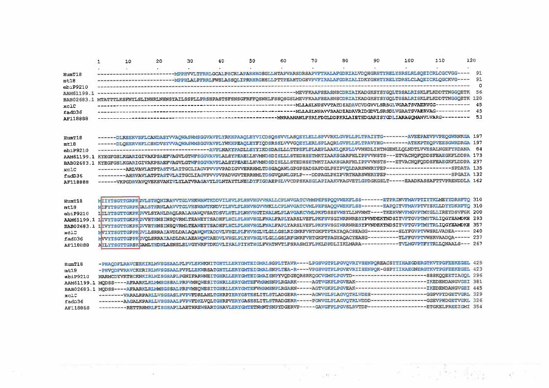
1 10 20 30 40 50 60 70 80 90 100 110 L20
IIu¡nE18nt18ebiP921 0 0
xctg ---------MLLASLÀIPSWTATDI G¡/VLSRSGIVGAÀ1SI¿AERVGG--- 45
fa<Ð36 ---------MLLASI,Ii¡PAWSAADIÀDÀ\¡RIDGD\¿LSRSD SVAER\¡ÀG--- 45
ÀF1 1Bg8B ---MNRAåNA¡ILFSRLE:DGIÐDPKRLAIETHDGARISYGDLIAR;à.GQI{AIiI\UL1¿ARG 53
III¡¡¡T18ûtl8ebiP921.0AÀì161199.18A802683.1:ßclcfadD36A¡.118888
BumT18rût18ebiP9210ÀAì,f61199.18A'B02683.1xc.lcfadD36ÀF118BEB
IIU¡aT18mt18ebiP921.0À.èr,161199.18]È802683.1xclLCfadD36AFt 18888
197197
841"?3
237135:132r62
310310200293351240)a'l
267
STPVNDEKTIÍDSI TRLIQGTE]A¡''DI(E
SYPVIIDEKTNDSI TRLTQGI-EjA¡IIDKE
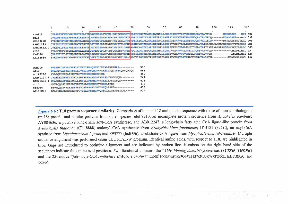
1 10 20 30 40 50 60 90 x0070 80 110
-EAI;DADEI,I-À}T\ZD]ADGI,I
120
---ELK 534--DLK 532
IIuoTlS¡rtl8ebiP9210AAM61199.18A802683.1xclCfadD36Ar.118888
IturTlSnt'18êbiP9210AAM61199.18A802683.1xclCfadD36Ar'118888
4to8
501s65431434464
576583447544608416413s09
Fisure 6.6: T18 protein sequence similarity. Comparison of human T18 amino acid sequence \¡/ith those of mouse orthologous
(mt18) protein and similar proteins from other species: ebiP92l0, an incomplete protein sequence from Anopheles garnbiae;
AY084636, a putative long-chain acyl-CoA synthetase, and 48012247, a long-chain fatty acid CoA ligase-like protein from
Arabidopsis thaliana;4F118888, malonyl CoA synthetase from Bradyrhízobium jøponicum; U15181 (xcLC), an acyl-CoA
synthase from Mycobacterium leprae; and 293777 (fadD36), a substrate-CoA ligase ftom Mycobacterium tuberculosis. Multiple
sequence alignment was petfonned using CLUSTAL-W program. Identical amino acids, with respect to Tl8, are highlighted in
blue. Gaps are introduced to optimize alignment and are indicated by broken line. Numbers on the right hand side of the
sequences indicate the amino acid positions. Two functional domains, the "AMP-binding domain"(consensus:IxYTs6TTGRPß)
and the 25-residue 'Tatty acyl-CoA synthetase (FACS) signature" motif (consensus:DGIILHTGDIGxWxPxGxLK,IIDAKK) are
boxed.
t8
structure of T18 was therefore partially identifred for only 7 exons from its 3' half (Fìgare
ó.5). To explore a putative complete structure of TI8 gene, the mouse homologue, mtIS
cDNA sequence was then aligned with mouse genomic sequence (WGS supercontig
Mm8_WIFeb0l_194) from the mouse genome database at NCBI. The mouse gene, mtl8, is
orgarized into 10 exons encompassing the genomic sequence of 42 kb. The exons are
ranging in size from 103 to 690 bp. Exon 2 is the largest with 690 bp in size. Both start
codon, ATG, and AMP-binding domain are encoded by exon 2. The FACS signatrne is
coded for by the sequence within exons 7 and 8. When compared the partial TI8 gene
structure with that of its mouse homologue, mtl\, they appeared to utilize the equivalent
exon boundaries for all available exons (Fígure ó.5). This may suggest that 218 would also
separate into l0 exons similar to mtl\.
6.4 Discussion
The purpose of this part of the study was to isolate and characterize a gene designated
transcription unit l8 (T18) which was mapped to the proximal region of BAC 561E17. This
BAC form part of the extended contig of the genomic clones of chromosome 16 at the
region 16q24.3. As a result, this gene was predicted to code for a protein of 576 amino acids,
which by in silico analysis was an ER-bound protein containing a sequence signature
characteristic of an AMP-binding enrpe218 showed a high degree of sequence homology
of both the ORF and the protein sequence to its mouse homologue, which was identified as
mtl\. In addition, the T18 protein also showed sequence similarity to proteins from
invertebrate species. These homologies suggested a significant conservation of this gene,
both functional and evolutionary.
The "AMP-binding motif is shared by a number of proteins in both prokaryote and
eukaryote. These proteins include luciferase (EC 1.13.12.7) in insects, alpha-aminoadipate
186
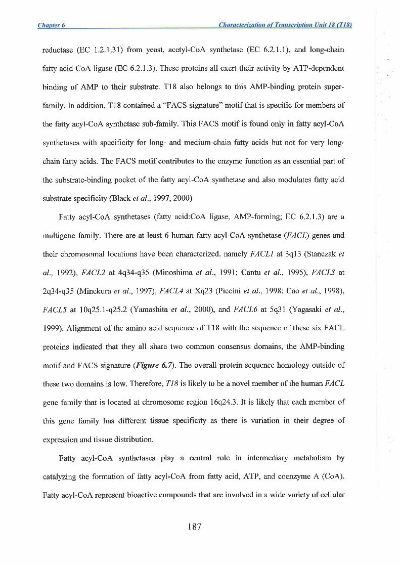
reductase (EC 1.2.1.31) from yeast, acetyl-CoA synthetase (EC 6.2.1.1), and long-chain
fatty acid CoA ligase (EC 6.2.1.3). These proteins all exert their activþ by ATP-dependent
binding of AMP to their substrate. T18 also belongs to this AMP-binding protein super-
family. In addition, T18 contained a "FACS signature" motif that is specific for members of
the fatty acyl-CoA synthetase sub-family. This FACS motif is found only in fatty acyl-CoA
synthetases with specificity for long- and medium-chain fatty acids but not for very long-
chain fatty acids. The FACS motif contributes to the enzyme function as an essential part of
the substrate-binding pocket of the fatty acyl-CoA synthetase and also modulates fatty acid
substrate specificþ (Black et al.,1997,2000)
Fatty acyl-CoA synthetases (fatty acid:CoA ligase, AMP-forming; EC 6.2.1.3) are a
multigene family. There are at least 6 human fatty acyl-CoA synthetase (FACL) genes and
their chromosomal locations have been characterized, namely FACLL at 3q13 (Stanczak et
al., 1992), FACL2 at 4q34-q35 (Minoshima et al., 7991; Cantu et al., 1995), FACL3 at
2q34-q35 (Minekura et a1.,1997), FACL4 atXq23 (Piccini et a1.,1998; Cao et a1.,1998),
FACLS at l0q25.l-q25.2 (Yamashita et a1.,2000), and FACL6 at 5q3l (Yagasaki et al.,
1999). Alignment of the amino acid sequence of Tl8 withthe sequence of these six FACL
proteins indicated that they all share two common consensus domains, the AMP-binding
motif and FACS signature (Fígure 6.7). The overall protein sequence homology outside of
these two domains is low. Therefore, T18 is likely to be a novel member of the human FACL
gene family that is located at chromosome region 16q24.3.It is likely that each member of
this gene family has different tissue specificity as there is variation in their degree of
expression and tissue distribution.
Fatty acyl-CoA synthetases play a central role in intermediary metabolism by
catalyzing the formation of fatty acyl-CoA from fatty acid, ATP, and coenzyme A (CoA).
Fatty acyl-CoA represent bioactive compounds that are involved in a wide variety of cellular
r87
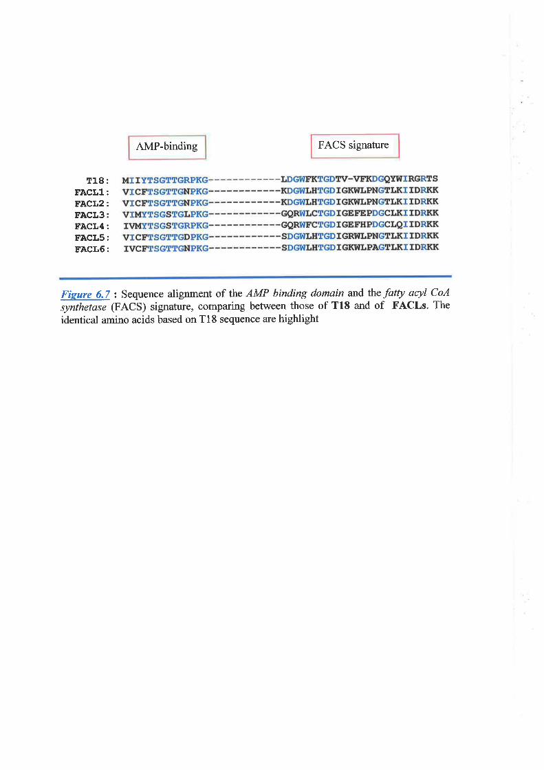
AMP-binding FACS signature
T18:FÀCL1:FACL2:EACL3:FACL4:FACLS:FACL6:
Fieure 6.7 : Sequence alignment of the AMP binding domain and the fotty acyl CoA
synthetase (FACS) signature, comparing between those of T18 and of FACLs. The
identical amino acids based on Tl8 sequence are highlight
processes including protein transport (Glick and Rothman,l987; Pfanner et al., 1989),
enrqe activation or deactivation (Fulceri et al., 19951' Lai et al., 1993; Yamashita et al.,
1995), protein modification (Gordon et al.,l99l; Mclaughlin and Aderem, 1995; Nimchuk
et a1.,2000), cell signaling (Korchak et al., 1994; Shrago et a1.,1995), and transcriptional
control (DiRusso et al., 1999;}Jertz et al., 1998; Raman et al., 1997) in addition to serving
as substrates for B-oxidation and phospholipid biosynthesis. Given their multiple roles, it is
clear that fatty acyl-CoA synthetases occupy a pivotal role in cellular homeostasis.
Although the map location of the Tl8 gene is within the region showing LOH in breast
cancer, by its putative protein fi¡nction this gene was considered unlikely to be a tumor
suppressor. Therefore it has not been screened for breast cancer specific mutations. By
analogy with the other charactenzed members of the FACL gene family, Tl8 is suggested to
be part of the essential cellular machinery that serves for cell survival and is unlikely to be
involved in growth suppression.
Whether 718 is associated with any human disease is not known. However, other
members of the fatty acyl-CoA synthetase family have been reported to be associated with a
number of diseases. Deletion of the X chromosome region q22.3-q23 has been found in
Alport syndrome, elliptocytosis, and mental retardation (Piccini et a1.,1998). This deletion
involves at least four genes, one of which is the gene for fatty acyl-CoA synthetase 4
(FACL4). A subsequent study has indicated that mutations of FACL4 is involved in
nonspecific X-linked mental retardation (Meloni et a1.,2002). Up-regulation of FACL4 has
been reported in colon adenocarcinoma and was for¡nd synergistically with cyclooxygenase-
2 (COX-2) to inhibit apoptosis (Cao et a1.,2001). A rectrrent t(5;12)(q31;pl3) translocation
has been found in three types of hematologic disorders: refractory anemia with excess blasts
and basophilia, acute myelogenous leukemia (AML) with eosinophilia, and acute
eosinophilic leukemia (AEL) (Yagasaki et al., 1999). This translocation involves the fusion
188
of the FACL2 gene with TEL/ETV6. FACLï has been reported to show increased expression,
compared with normal brain, in primary brain tumors, glioma of grade IV malignancy
(Yamashita et a1.,2000). Such associations of the FACL gene family with human disorders
could suggest that TI8 may also have a role, however at this stage there is no known
association with any human disease. Since 218 was highly expressed in liver, kidney, and
pancreas, this gene may have critical function in these tissues. This, however, requires
firther investigation.
189

Chapter 7
General Discussion
7
General l)iscussion
Carcinogenesis is a multistage process in both morphological changes and genetic
alterations. Although several causative agents namely physical, chemical, and biological, are
i.rknown to involvdin the initiation and progression of the tumor, the common underþing lesion >
^isdnon-lethal DNA damage. Subsequent DNA replication and cell division lead to the stage
'v '.
of genetic instability, which is widely accepted to be the cause of many genetic alterations
seen in cancers. One such alteration is the loss of heterozygosity (LOH), which is considered
to be a somatic mechanism associated with the loss of tumor suppressor function (discussed in
section 1.4.2).
The ultimate goal for the study of genetic diseases including cancer is to identiff the
genes, mutations of which associate with disease phenotypes. The contmon path for the studyq
of- disease-associated gene is, firstly, by mapping of the disease-associated locus. The
following step is the identification of a candidate gene that exhibits disease-causing mutation.
Finally, confirmation of such candidate gene is likely by studying its disease-causing(,,t.
phenotype inlhe animal model.
Most of the works described in this thesis were initially undertaken before the first draft
of the human genome sequence was available. Therefore, identification of the gene was based
on the positional cloning approach utilizing the physical and transcription map constructed attt:
the candidate region for the breast cancer tumor suppressor (\ühitmore et al., 1998a)nthe
chromosome band 16q24.3. Exon amplification technique and bioinformatics were utilized to
facilitate the isolation of the transcribed sequence of the genes. Four genes were isolated and
their nucleotide and protein sequences were characteized for their possible function. Only
two of these characterized genes were shown to have function likely to be a candidate tumor
190
7
suppressor. The gene designated "73", by analysis of its protein sequence, was shown to have
function likely to be involved in transcriptional regulation. Tl3 on the other hand was
predicted to code for a large protein containing sequence motifs of nuclear localization signal,
suggesting the protein function in nuclear activity. Subsequent gene mutation study was
utilized to determine if any of these genes was involved in breast carcinogenesis. V/ith the
current method for the detection of nucleotide changes in the coding sequence, there were no
mutations identified that suggest the association of these genes in breast carcinogenesis.
However, other mechanisms of gene mutation could not be ruled out.
Mutations that cause/e gene sequence changes t"?t t:.:11 in either the production of
aberrant protein or the reduction of mRNA expression. Mutation of a single nucleotide that
changela codon usage and subsequent amino acid transition (missense mutation) could lead to
the production of aberrant protein if such amino acid substitution affecf,the native secondary
structure of that protein. The mutation that converts a functional codon into a stop signal
(nonsense mutation) could lead to the prematur¡ dee¡adation o{ nRNA by the mechanism
called " nonsense-mediated mRNA decat'' (Culbertson,1999; Frischmeyer and Dietz,1999).
In addition mutation at the promoter region of the gene may disrupt the promoter function and
lead to the aberrant expression of mRNA (Giedraitis et a1.,2001). Other mechanism, the
promoter hypermetþlation has also been reported to play a role in the down-regulation of
tumor suppressor genes (Wieland et a1.,2000; Yuan et a1.,2001).
Other two genes, Tl2 and TL9)haf were also characteized and were excluded as,z'
candidate tumor suppresso$based on their possible function. Furthermore one of these genes,
t l') I
TI2,later found to be identical to SPG7, a gene involv$in recessive type hereditary spastic
191
paraplegia (HSP). Subsequent determination of the SPGT gene structure was undertaken and
was utilized in the mutation study of a large number of HSP patients from unrelated families.
This study had confirmed that frequency of SPGT mutations was low in a general diverse
population in contrast to the previous report, studied in .¡h( specific ethnic groups (Casari et
al., 1998). The 7/8 on the other hand was shown by its protein sequence identity and
functional motifs to be a member of long chain fatty acyl CoA ligase (FACL) sub-family.
b¿. ('When co-pur"S the Tl8 sequence with those of other FACL proteins, it was suggested that
218 is likely to be a novel member of FACL.
In addition to the isolation and charactenzation of 73, TI2, TI3, and TI Candidate had ' "
also involved in the collaborative LOH study of this 16q24.3 region in breast cancer (Cleton-
Jansen et a1.,2001) and the study of PISSZ,RE as one candidate gene (Crawford et a1.,1999).
I
The detaif information of these studies can be found i" tt/I .rt
r," . " !.i
mahuscript of both publications
gþven in the appendix of the thesis. The LOH study was inic
tially aim to narrow down the
LOH to the smallest overlap regiol, of 16q24.3 LOH. By usingr!tr(
polymorphic markers in this region, LOH study was conducted
cancer tissue samples and their matched peripheral blood leukocytes. During the course of this
LOH study, it appeared that interpretation of the LOH data was complicate'due to the large
number of complex LOH, non-informativeness of the markers, and the contamination of
normal tissue. Other factor such as genetic heterogeniety of the tumor could also be taken intolÀ_
accout. In breast cancer, genetic divergence has been reported by the changes in pattern of
LOH in a number chromosomal loci (Fujii et al., 1996; Lichy et al., 2000), with the1,
progressive stag{of tumo{exhibitçd'ïnore discordant LOH. Furthermore LOH has also been
yéngn-aensity mapped' i/ t ¡1
in'thrëe panels of breast
t92
7
identified in normal tissue adjacent to tumor (Deng et a|.,1996).
Study of the PISSLRE gene was conducted since this gene was mapped to the current
physical map of 16q24.3 LOH region (Whitmore et al., 1998a). The possible function of
PISSLRE gene product as a cyclin-dependent kinase (CDK) had drawn attention that this gene
might be a good candidate tumor suppressor target by LOH. CDK in association with cyclin
as CDllcyclin complex irlolved in various stagdof the cell cycle. The presence of the
PISSLRE protein is necessary for the progression from G2 to M phase, and overexpression ofl¡\
the gene leads to growth suppression (Li et al., 1995). Fórthermore expression of PISSLRE
gene is high in terminally differentiated cells, which are withdr# f.o- the cell cycle
(Brambilla and Draetta, 1994; Grana et al., 1994). Despite this possible growth suppression
function, mutation analysis in breast cancer samples w¿s failed to detect any mutation of this
gene that can be considered to be pathogenic for breast carcinogenesis.
At the initial stage of this study, the rate-limiting stáp was the isolation of the transcript
sequence of the genes in the candidate region. With the laboratory technique available at that
time isolation of the full-length transcript sequence was time consuming. However, when the
large scale sequencing program of this genomic region was established, identification of the
trancribed sequence of the gene was facilitated as shown in the charactenzation of T3 andTl3
transcript as well as their gene structure determination.
The progression of the Human Genome Project to the point where the complete and
finished human genome sequence is available could accerelate the discovering of the genes
associated with human diseases including breast cancer. Indeed a number of projects has
r93
7
benefited in term of gene identification and functional prediction through a number of
programs associated with the human genome project. These include the accumulation of
biological data in the form of computenzed databases and the development of computer-
assisted tools to access, archive, and analysis'this data. The present study had also used such
bioinformatics to assist in the analysis of the gene in both DNA and the predicted protein
sequence. The success of the Human Genome Project could change the way of gene
identification from positional cloning approach to position candidate analysis to selective
candidate gene approach. The later based on a known or a putative function of unknown gene
that can be derived in silico from the genomic information. To this point computational
analysis of the gene is a critical step that can provide clues to the molecular basis of
pathogenesis and invaluable insights for further experimental analysis.
Finally, although the present study was unable to identit a breast cancer tumor
suppressor gene from this chromosome region, 16q24.3, the charactenzed genes could be the
candidates for other human diseases or other types of cancer, yet to be identified. In addition
to the LOH seen in breast and other cancers (mention in section 1.5.4), the presence of the
genes associated with human diseases such as fanconi anemia, the FAA gene, and hereditary
spastic paraplegia, the SPG7, provides an example of the involvement of this chromosome
region as one hot spot for the disease associated gene await for identification and
characleization.
194

References
References
Aaltonen, L.4., Peltomaki, P., Leach, F.S., Sistonen, P., Pylkkanen, L., Mecklin, J.P.,Jarvinen, H., Powell, S.M., Jen, J., Hamilton, S.R., et al. (1993) Clues to the pathogenesisof familial colorectal cancer. Science 260: 812-816.
Aasland, R., Gibson, T.J., and Stewart, A.F, (1995) The PHD finger: implications forchromatin-mediated transcriptional regulation. Trends Biochem. Sci. 2O:56-59.
Abbott, D.tW., Thompson, M.E., Robinson-Benion, C., Tomlinson, G., Jensen, R.4., and Holt,J.T. (1999) BRCA1 expression restores radiation resistance in BRCAl-defective cancercells through enhancement of transcription-coupled DNA repair. J.Biol. Chem. 274:I 8808-1 88 l 2.
Aberle, H., Bauer, 4., Stappert, J., Kispert, 4., and Kemler, R. (1997) p-catenin is a target forthe ubiquitin-proteasome pathway. EMBO J. 16:3797-3804.
Adams, S.M., Helps, N.R., Sharp, M.G.F., Brammat, V/.J., \ù/alker, R.4., and Varley, J.M.(1992) Isolation and characterization of a novel gene with differential expression inbenign and malignant human breast tumors. Hum. Mol. Genet.l: 91-96.
Albarosa, R., Colombo, 8.M., Roz, L., Magnani, L., Polo, 8., Cirenei, N., Giani, C., Conti,A.M.F., DiDonato, S., and Finocchia¡o, G. (1996) Deletion mapping of gliomas suggeststhe presence of two small regions for candidate tumor-suppressor genes in a 17-cMinterval on chromosome 10q. Am. J. Hum. Genet. 58:-1260-1261.
Aldaz, C.M., Chen, T., Sahin, 4., Cunningham, J., and Boncly, M. (1995) Comparativeallelotype of in situ and invasive human breast cancer: high frequerrcy of microsatelliteinstability in lobular breast carcinomas . Cancer Res. 55: 3976-3981.
Alevizopoulos, K., Vlach, J., Hennecke, S., and Amati, B. (1997) Cyclin E and c-Mycpromote cell proliferation in the presence of pl6INK4a and of hypophosphorylatedretinoblastoma family proteins. EMBO J. 17:5322-5333.
Ali, I.U., Campbell, G., Lidereau, R., and Callahffi, R. (1988) Lack of evidence for theprognostic significance of c-rebB2 amplification in human breast carcinoma. OncogeneRes. 3: 139-146.
Allred, D.C., Clark, G.M., Molina, R., Tandon,4.K., Schnitt, S.J., Gilchrist, K.W., Osborne,C.K., Tormey, D.C., and McGuire, W.L. (1992) Overexpression of HER-2lneu and itsrelationship with other prognostic factors change during the progression of in situ toinvasive breast cancer. Hum. Pathol. 23 97 4-979.
Altschul, S.F., Madden, T.L., Schaffer, 4.4., Zhang, J., Zhang, 2., Miller, W., and Lipman,D.J. (1997) Gapped BLAST and PSI-BLAST: a new generation of protein databasesearch programs. Nucleic Acids Res. 25'. 3389-3402.
Amara, S.G., JonaS, V., and Rosenfeld, M.G. (1982) Alternative RNA processing incalcitonin gene expression generates mRNAs encoding different polypeptide products.
195
Nature 298:240-244.
Antequera, F., and Bird, A. (1993) Number of CpG islands and genes in human and mouse.Proc. Natl. Acad. Sci. U.S.A.90: I1995-11999.
Aprelikova, O.N., F*g,8.S., Meissner, E.G., Cotter, S., Campbell, M., Kuthial4 4., Bessho,
M., Jensen, R.A., and Liu, E.T. (1999) BRCA-associated growth arrest is RB dependent.Proc. Natl. Acad. Sci. U.S.A.96: 11866-11871.
Arlt, H., Tauer, R., Feldmann, H., Neupert, W., and Langer, T. (1996) The YTA10-12-complex, an AJqA protease with chaperoneJike activity in the inner membrane ofmitochondria. Cell 85: 875-885.
Attwood, T.K., Croning, M.D., Flower, D.R., Lewis, 4.P., Mabey, J.E., Scordis, P., Selley,J.N., Wright, W. (2000) PRINTS-S: the database formerly known as PRINTS. NucleicAcids Res.28:225-227.
Auch, D., and Reth, M. (1990) Exon trap cloning: using PCR to rapidly detect and cloneexons from genomic DNA fragments. Nucleic Acids Res. 18: 6743-6744.
Auerbach, A.D. (1993) Fanconi anemia diagnosis and the diepoxybutane (DEB) test. Exp.Hematol. 2l:731-733.
Austruy, E., Cohen-Salmon, M., Antignac, C., Beroud, C., Henry, I., Nguyen, V.C.,Brugieres, L., Junien, C., and Cornelisse, C. (1993) Isolation of a kidney cDNA down-expressed in V/ilms' tumor, by a subtractive hybridization approach. Cancer iR¿s. 53:2888-2894.
Baens, M., Aerssens, J., vanZand, K., van den Berghe, H., and Marynen, P. (1995) Isolationand regional assignment of human chromosome l2p cDNA. Genomics 29:44-52.
Balaban, G., Gilbert, F., Nichols, W., Meadows, 4.T., and Shields, J. (1982)Abnormalities ofchromosome 13 in retinoblastoma from individuals with normal constitutionalkaryotypes. Cancer Genet. Cytogenet. 6: 213-221.
Ballabio, A. (1993) The rise and fall of positional cloning? Nature Genet.3:277-279
Banfi, S., Bassi, M.T., Andolfi, G., Marchitiello, A,Zanotta, S., Ballabio,4., Casari, G., andFranco, B. (1999) Identification and Characterization of AFG3L2, a Novel Paraplegin-Related Gene. Genomics 59: 51-58.
Banin, S., Moyal, L., Shieh, S., Taya, Y., Anderson, C.W., Chessa, L., et al. (1998) Enhancedphosphorylation of p53 by ATM in response to DNA damage. Science 281: 1674-1677.
Bartkova, J., Lukas, J., Muller, H., Lutzhft, D., Strauss, M., and Bartek, J. (1994) Cyclin Dlprotein expression and frrnction in human breast cancer. Intl. J. Cancer 57:353-361.
Baskaran, R., Wood, L.D., Whitaker, L.L., Canman, C.E., Morgan, S.E., Xu, Y., et al. (1997)Ataxia telangiectasia mutant protein activates c-Abl tyrosine kinase in response toionizing radiation. Nature 387: 516-519.
r96
Bateman,4., Birney,8., Durbin, R., Eddy, S.R., Howe, K.L., and Sonnhammer, E.L. (2000)The Pfam protein families database. Nucleic Acids Res.28;263-266.
Baumann, P., Benson, F.E., and West, S.C. (1997) Human RAD5I protein promotes ATP-dependent homologous pairing and strand transfer reactions in vitro. Cell 87:757-766.
Baylin, S.8., Esteller, M., Rountree, M.R., Bachman, K.E., Schuebel, K., and Herman, J.G.(2001) Aberrant patterns of DNA methylation, chromatin formation and gene expressionin cancer. Hum. Mol. Genet. l0:.687'692.
Bednarek, 4.K., Laflin, K.J., Daniel, R.L., Liao, Q., Hawkins, K.4., and Aldaz, C.M. (2000)WWOX, a novel WW domain-containing protein mapping to human chromosome16q23.3-24.1, a region frequently affected in breast cÍrncer. Cancer Res. 60: 2140-2145.
Behrens, J., Jerchow, 8.4., 'W-urtele, M., Grimm, J., Asbrand, C., Wirtz. R., Kuhl, M.,Wedlich, D., and Birchmeier, W. (1998) Functional interaction of an axin homolog,conductin, with p-catenin, APC, and GSK3p. Science 280:. 596-599.
Bellefroid, E.J., Marine, J.C., Matera,4.G., Bourguignon, C., Desai, T., Healy, K.C., Bray-Ward, P., Martial, J.4., Ihle, J.N., and Ward,D.C. (1995) Emergence of the ZNF91Kruppel-associated box-containing zinc finger gene family in the last common ancestorof anthropoidea. Proc. Natl. Acad. Sci. U.S.A.92:10757-10761.
Berns, E.M.J.J., Khjn, J.G.M., van Putten, W.L.J., van Staveren, I.L., Portengen, H., andFoekens, J.A. (1992) c-Myc amplification is a better prognostic factor than HER2/neuamplification in primary breast cancer. Cancer Res. 52: ll07-lll3.
Bertwistle, D., Swift, S., Marston, N.J., Jackson, L.E., Crossland, S., Crompton, M.R.,Marshall,C.J., and Ashworth, A. (1997) Nuclear location and cell cycle regulation of theBRCA2 protein. Cancer Res. 57'. 5485-5488.
Berx, G., Cleton-Jansen, A-M., Nollet, F., de Leeuw, W.J., van de Vijver, N{., Cornelisse, C.,and van Roy, F. (1995a) E-cadherin is a tumor/invasion suppressor gene mutated inhuman lobular breast cancers. EMBO J. 14:6107-6115.
Berx, G., Cleton-Jansen, A-M., Strumane, K., de Leeuw, 'W.J., Nollet, F., van Roy, F., andComelisse, C. (1996) E-cadherin is inactivated in a majority of invasive human lobularbreast cancers by trwrcation mutation throughout its extracellular domain. Oncogene 13t9t9-t925.
Berx, G., Staes, K., van Hengel, J., Molemans, F., Bussemakers, M.J., van Bokhoven, 4., andvan Roy, F. (1995b) Cloning and characterization of the human invasion suppressor geneE-cadherin (CDHI ). Genomics 26: 281-289.
Bhattacharyya, N.P., Skandalis, 4., Ganesh, 4., Groden, J., and Meuth, M. (1994) Mutatorphenotypes in human colorectal carcinoma cell lines. Proc. Natl. Acad. Sci. U.S.A. 9l:6319-6323.
Black, P.N., DiRusso, C.C., Sherin, D., MacColl, R., Knudsen, J., and Weimar, J.D. (2000)
t97
Affrnity labeling fatty acyl-CoA synthetase with 9-p-azidophenoxy nonanoic acid and theidentification of the fatty acid-binding site.l Biol. Chem.275:38547-38553.
Black, P.N., Zhang, Q., Weimar, J.D., and DiRusso, C.C. (1997) Mutational analysis of afatty acyl-Coenzyme A synthetase signature motif identifies seven amino acid residues
that modulate fatty acid substrate specificity. J. Biol. Chem.272;4896-4903.
Bleck, O., McGrath, J.4., and South, A.P. (2001) Searching for candidate genes in the newmillennium. Clin. Exp. Dermatol. 26;279-283.
Bird, A.P. (1986) CpG-rich islands and the function of DNA methylation. Nature 321:209-213.
Bird, A.P. (1987) CpG islands as gene markers in the vertebrate nucleus. Trends Genet. 3:342-347.
Bochar, D.4., Savard, J., Wang, W., Laflew, D.W., Moore, P.', Côté, J., and Shiekhattar, R.(2000) A family of chromatin remodeling factors related to V/illiams syndrometranscription factor. Proc. Natl. Acad. Sci. U.5.4.97: 1038-1043.
Bodmer, W., Bailey, C., Bodmer, J., Bussey, H., Ellis,,A.., Gorman, P., Lucibello, F., Mwday,V., Rider, S., and Scambler, P. (1987) Localization of the gene for familial adenomatouspolyposis on chromosome 5. Nature 328: 614-616.
Bonnefoy-Berard, N., Munshi, 4., Yron, I., 'Wu, S., Collins, T.L., Deckert, M., Shalom-Barak, T., Giamp4 L., Herbert, E., Hernandez, J., Meller, N., Coufure, C., and Altman,A. (1996) Vav: function and regulation in hematopoietic cell signaling. Stem Cells 14:2s0-268.
Borg, 4., Baldetorp, 8., Ferno, M., Killander, D., Olsson, H., and Sigurdsson, M. (1991)ErbB2 amplifrcation in breast cancer with rate of proliferation. Oncogene 6: 131-143.
Borg, 4., Baldetolp, 8., Ferno, M., Olsson, H., and Sigurdsson, H. (1992) c-Myc ampli-fication is an independent prognostic factor in postmenopausal breast cancer. Int. J.
Cqncer 51:687-691.
Bose, S., Wang, S.I., Terry, M.B., Hibshoosh, H., and Parsons, R. (1998) Allelic loss ofchromosome 10q23 is associated with tumor progression in breast carcinomas. Oncogene17:123-127.
Bouchard, L., Lamarre, L., Tremblay, P.J., and Jolicoeur, P. (1989) Stochastic appearance ofmammary tumors in transgenic mice carrying the MMTV/c-NEU oncogene. Cell 57: 931-936.
Brambilla, R., and Draetta" G. (1994) Molecular cloning of PISSLRE, a novel putativemember of the cdk family of protein serine/threonine kinases. Oncogene 9:3037-304L
Brenner, 4.J., and Aldaz, C.M. (1995) Chromosome 9p allelic loss and p16lCDKN2 in breastcancer and evidence of p16 inactivation in immortal breast epithelial cells. Cancer Res.
55:2892-2895.
198
Brenner, 4.J., and Aldaz, C.M. (1997) The genetics of sporadic breast cancer. Prog. Clin.Biol. Res.396:63-82.
Brinkman, 4., van der Flier, S., Kok, E.M., and Dorssers, L.C.J. (2000) BCARI, a humanhomologue of the adapter protein p130Cas, and antiestrogen resistance in breast cancer
cells.l Natl. Cancer Inst.92: ll2-120.
Broeks, A., Urbanus, J.H.M., Floore, 4.N., Dahler, E.C., Kliju, J.G.M., Rutgers, E.J.T.,Devilee, P., Russell, N.S., van Leeuwen, F.E., and van 't Veer, L.J. (2000) ATM-heterozygous germline mutations contribute to breast cancer-susceptibility. Am. J. Hum.Genet. 66:494-500.
Buckler,4.J., Chang, D.D., Graw, S.L., Brook, J.D., Haber, D.4., Sharp, P.4., and Housman,D.E., (1991) Exon amplification: a strategy to isolate mammalian genes based on RNAsplicing. Proc. Natl. Acad. Sci. U.5.4.88: 4005-4009.
Buetow, K.H., Weber, J.L., Ludwigsen, S., Scherpbier-Heddeme, T., Duyk, G.M., ShefFreld,V.C., Vy'ang, 2., and Murray, J.C. (1994) Integrated human genome-wide mapsconstructed using the CEPH reference panel. Nature Genet. 6:391-393.
Bullrich, F., Maclachlan, T.K., S*g, N., Druck, T., Veronese, M.L., Allen, S.L., Chiorazzi,N., Koff,4., Heubner, K., Croce, C.M., and Giordano, A. (1995) Chromosomal mappingof members of the cdc2 family of protein kinases, cdk3, cdk6, PISSLRE, and PITALRE,and a cdk inhibitor, p2TKipt, to regions involved in human cancer. Cancer.l?¿s. 55: 1199-1205.
Burke, D.T. (1991) The role of yeast artificial chromosome clones in generating genomemaps. Curu. Opin. Genet. Dev.l:69-74.
Burke, D.T., Carle, G.F., and Olson, M.V. (1987) Cloning of large segments of exogenousDNA into yeast by means of artificial chromosome vectors. Science 236:806-812.
Bum, T.C., Connors, T.D., Klinger, K.W., and Landes, G.M. (1995) lncreased exon-trappingefficiency through modifications to the pSPL3 splicing vector. Gene 16l: 183-187.
Burn, T.C., Connors, T.D., Van Raay, T.J., Dackowski, W.R., Millholland, J.M., Klinger,K.W., and Landes, G.M. (1996) Generation of a transcriptional map for a 700-kb regionsurrounding the polycystic kidney disease type 1 (PKDI) and tuberous sclerosis type 2(TSC2) disease genes on human chromosome 16p3.3. Genome Res. 6:525-537.
Butturini, A., Gale, R.P., Verlander, P.C., Adler-Brecher, 8., Gillio,4.P., and Auerbach, A.D.(1994) Hematologic abnormalities in Fanconi anemia : an International Fanconi AnemiaRegistry study. Blood. 48: I 650-l 655.
Cahill, D.R., Lengaver, C., Yu, J., et al. (1998) Mutation of mitotic checkpoint genes inhuman cancers. Nature 392: 300-303.
Cairns, J . (l9l5) Mutation selection and the natural history of cancer. Nature 255: 197 -200.
199
Callebaut, I., and Momon, J.P. (1997) From BRCA1 to RAP2: a widespread BRCT moduleclosely associated with DNA repair. FEBS Lett. 400:25-30.
Callen, D.F., Lane, S.4., Kozman, H., Ktemmidiotis, G., Whitmore, S.4., Lowenstein, M.,Doggett, N.4., Kenmochi, N., Page, D.C., Maglott, D.R., Nierman, W.C., Murakaw4 K.,Berry, R., Sikela, J.M., Houlgatte, R., Auffray, C., and Sutherland, G.R. (1995)Integration of transcript and genetic maps of chromosome 16 at near-l-Mb resolution:demonstration of a "hot spot" for recombination at l6pl2. Genomics 29: 503-51 l.
Canman, C.E., Lim, D.S., Cimprich, K.4., Tuyu, Y., Tamai, K., Sakaguchi, K., et 41. (1998)Activation of the ATM kinase by ionizing radiation and phosphorylation of p53. Science
281:1677-1679.
Cantu, 8.S., Sprinkle, T.J., Ghosh, B., and Singh, I. (1995) The human palmitoyl-CoA ligase(FACL2) gene maps to the chromosome 4q34-q35 region by fluorescence in situhybridization (FISH) and somatic cell hybrid panels. Genomics 28: 600-602.
Cao, Y., Dave, K.8., Doan, T.P., and Prescott, S.M. (2001) Fatty acid CoA ligase 4 is up-regulated in colon adenoca¡cinoma. Cancer Res. 61: 8429-8434.
Cao, Y., Traer, 8., ZimmeÍnarì, G.4., Mclntyre, T.M., and Prescott SM. (1998) Cloning,expression, and chromosomal localization of human long-chain fatty acid-CoA ligase 4(FACL4). Genomics 49 : 327 -330.
Carter, 8.S., Ewing, C.M., Ward,'W.S., Treiger,8.F., Aalders, T.W., Schalken, J.4., Epstein,J.I., and Isaacs, W.B. (1990) Allelic loss of chromosome l6q and 10q in human prostate
cancer. Proc. Natl. Acad. Sci. U.5.4.87: 8751-8755.
Casari, G., De Fusco, M., Ciarmatori, S., Zeviani, M., Mora, M., Fernandez,P., De Michele,G., Filla, A., Cocozza, S., Marconi, R., Durr, 4., Fontaine, 8., and Ballabio, A. (1998)Spastic paraplegia and OXPHOS impairment caused by rnutations in paraplegin, anuclear-encoded mitochondrial metalloprotease. C ell 93: 97 3 -983.
Castagna, M. (1987) Phorbol esters as signal transducers and tumor promoters. Biol. Cell 593-13.
Catteau, 4., Harris W.H., Xu, C.F., and Solomon, E. (1999) Metþlation of the BRCALpromoter region in sporadic breast and ovarian cancer: correlation with diseasecharacteristics. Onco gene 18; 1957 -1965.
Cavenee, W.K., Dryja, T.P., Phillips, R.4., Benedict, W.F., Godbout, R., Gallie, 8.L.,Murphree, 4.L., Stron5,L.C., and White, R.L. (1983) Expression of recessive alleles bychromosomal mechanisms in retinoblastoma. Nqture 305: 779-784.
Champème, M.H., Bièche, I.,Lizard, S., and Lidereau, R. (1995) 11q13 amplification isinvolved in local recurrence of human primary breast cancer. Genes ChromosomesCancer 12:128-133.
Chen, F., Castranova, V., and Shi, X. (2001) New insights into the role of nuclear factor-kappaB in cell growth regulation. Am. J. Pathol. 159:387-397.
200
Chen, J., Birkholtz, G.G., Lindblom, P., Rubio, C., and Lindblom, A. (1998a) The role ofataxia-telangiectasia heterozygotes in familial breast cancer. Cancer Ìes. 58: 1376-1379.
Chen, J., Silver, D.P., V/alpita, D., Cantor, S.B., Gazdat, 4.F., Tomlinson, G., Couch, F.J.,
Vy'eber, B.L., Ashley, T., Livingston, D.M., and Scully, R. (1998b) Stable interactionbetween the products of the BRCAI and BRCA2 tumor suppressor genes in mitotic andmeiotic cells. Mol. Cell 2: 317 -328.
Chen, T., Sahin, 4., and Aldaz, C.M. (1996) Deletion map of chromosome 16q in ductalcarcinoma in situ of the breast: refining a putative tumor suppressor gene region. CancerR¿s. 56:5605-5609.
Cher, M.L., Ito, T.,'Weidner, N., Carroll, P.R., and Jensen, R.H. (1995) Mapping of regions ofphysical deletion on chromosome l6q in prostate cancer cells by fluorescence in situhybridization (FISH). J. Urol. 153: 249-254.
Chomczynski, P., and Sacchi, N. (1987) Single-step method of RNA isolation by acidguanidinium thiocyanate-phenol-chloroform extraction. Anal. Biochem. 162;156-159.
Church, D.M., Stotler, C.J., Rutter, J.L., Murrell, J.R., Trofatter, J.4., and Buckler, A.J.(1994) Isolation of genes from complex sources of mammalian genomic DNA using exonamplificati on. Nature Genet. 6: 98-1 05.
Church, D.M., Y*g, J., Bocian, M., Shiang, R., and Wasmuth, J.J. (1997) A high-resolutionphysical and transcript map of the Cri du chat region of human chromosome 5p. GenomeRes.7;787-801.
Clarke, R.8., Howell, 4., Potten, C.S., and Anderson, E. (1997) Dissociation between steroidreceptor expression and cell proliferation in the human breast. Cancer Res. 57: 4987-499t.
Cleton-Jansen, A-M., Moerland, E.W., Kuipers-Dijkshoom, N.J., Callen, D.F., Sutherland,G.R., Hansen, 8., Devilee, P., and Cornelisse, C.J. (1994) At least two different regionsare involved in allelic imbalance on chromosome arm 16q in breast cancer. Genes
Chromosomes Cancer 9: 101-107.
Cleton-Jansen, A-M., Moerland, E.Vy'., Pronk, J.C., van Berkel, C., Apostolou, S., Cralvford,J., Savoia, 4., Auerbach,4.D., Mathew, C.G., Callen, D.F., and Cornelisse, C.J. (1999)Mutation analysis of the Fanconi anemia A gene in breast tumours with loss ofheterozygosity at 16q24.3. Br. J. Cancer 79:1049-1052.
Coffer, P.J., Jin, J., and Woodgett, J.R. (1998) Protein kinase B (c-Akt): a multifunctionalmediator of phosphatidylinositol 3-kinase activation. BiochemJ 335 ( Pt 1):l-13.
Collins, F.S. (1995a) Positional cloning moves from perditional to traditional. Nature Genet.
9:347-350.
Collins, N., McManus, R., Wooster, R., Mangion, J., Seal, S., et al. (1995b) Consistent loss ofthe wild-type allele in breast cancers from a family linked to the BRCA2 gene on
20r

chromosome 13q12-13 . Oncogene l0: 1673-167 5.
Collins, N., Wooster, R., and Stratton, M.R. (1997) Absence of methylation of CpGdinucleotides within the promoter of the breast cancer susceptibility gene BRCA2 innormal tissues and in breast and ovarian cancers. Br. J. Cancer 76: 1l5l-l156.
Cornelis, R.S., Neuhausen, S.L., Johansson, O., Arason, 4., Kelsell, D., et al. (1995) Highallele loss rates at l7qI2-q21 in breast and ovarian tumor from BRCAl-linked families.Genes Chromosomes Cancer 13: 203-210.
Coppol4 M., Pizzigoni, 4., Banfi, S., Bassi, M.T., Casari, G., and Incerti, 8.(2000)Identification and characterization of YMEILI, a novel paraplegin-related gene.
Genomics 66:48-54.
Cortez, D., W*g, Y., Qin, J., Elledge, S.J. (I999)Requirement of ATM-dependentphosphorylation of Brcal in the DNA damage response to double-strand breaks. Science286: 1162-1166.
Cory, S. (1995) Regulation of lymphocyte survival by the bcl-2 gene family. Annu. Rev.
Immunol.13: 513-543.
Couch, F.J., and'Weber, B.L. (1996) Mutations and polymorphisms in the familial early-onsetbreast cancer (BRCAI) gene. Breast Cancer Information Core. Hum. Mut.8: 8-18.
Coussens, L., Yang-Feng, T.L., Liao, Y.C., Chen, E., Gray, 4., McGrath, J., et al. (1985)Tyrosine kinase receptor with extensive homology to EGF receptor shares chromosomallocation with Neu oncogene. Science 230: 1132-1139.
Coutinho, P., Barros, J., Zemmouri, R., Guimaraes, J., Alves, C., Chorao, R., Lourenco, E.,Ribeiro, P., Loureiro, J.L., Santos, J.V., Hamri, 4., Paternotte, C., Hazaî, J., Silva,M.C., Prud'homme, J.F., and Grid, D. (1999) Clinical heterogeneity of autosomalrecessive spastic paraplegias: analysis of 106 patients in 46 famllies. Arch. Neurol. 56:943-949.
Crawford, J.,lanzano, L., Savino, M., Whitmore, S.4., Cleton-Jansen, A-M., Settasatian, C.,d'Apolito, M., Seshadd, R., Pronk, J.C., Auerbach, A.D., Verlander, P.C., Mathew, C.G.,Tipping, 4.J., Doggett, N.4., Zelarfte, L., Callen, D.F., and Savoia, A. (1999) ThePISSLRE gene: Structrue, exon skipping, and exclusion as tumor suppressor in breastcancer. Genomics 56: 90-97.
Croce, C.M., and Nowell, P.C. (1985) Molecular basis of human B-cell neoplasia. Blood 65l-7.
Culbertson, M.R. (1999) RNA surveillance. Unforeseen consequences for gene expression,inherited genetic disorders and cancer. Trends Genet. 15: 74-80.
Dahia, P.L. (2000) PTEN, a unique tumor suppressor gene. Endocr. Relat. Cancer 7:ll5-129
Dasika, G.K., Lin, S.J., Zhao, S., Sung, P., Tomkinson, 4., and Lee, E.Y-H.P. (1999) DNAdamage-induced cell cycle checkpoints and DNA strand break repair in development and
202
tumorigene sis. Oncogene l8: 7 883-7 899.
Datson, N.4., Duyk, G.M., van Ommen, G.J., and den Dunnen, J.T. (1994) Specifrc isolationof 3'-terminal exons of human genes by exon trapping. Nucleic Acids Res. 22: 4148-4L53.
Dechend, R., Hirano, F., Lehmann, K., Heissmeyer, V., Ansieau, S., Wulczyn, F.G.,Scheidereit, C., and Leutz, A. (1999) The Bcl-3 oncoprotein acts as a bridging factorbetween NF-kappaB/Rel and nuclear co-regulato rc. Onc o gene l8: 33 I 6-3323 .
De Klein, 4., Geurts van Kessel, 4., Grosveld, G., Bartram, C.R., Hagemeijer, 4., Bootsm4D., Spurr, N.K., Heisterkamp, N., Groffen, J., and Stephenson, J.R. (1982) A cellularoncogene is translocated to the Philadelphia chromosome in chronic myelocyticleukemia. Nature 300: 765-767 .
Deng, C.X., and Scott, F. (2000) Role of the tumor suppressor gene Brcal in genetic stabilityand mammary gland tumor formation. Oncogene 19: 1059-1064.
Deng, G., Lu, Y., Zloûrikov, G., Thor, A.D., and Smith, H.S. (1996) Loss of heterozygosity innormal tissue adjacent to breast carcinomas. Science 274:2057-2059.
De Michele, G., De Fusco, M., Cavalcanti, F., Filla, 4., Marconi, R., Volpe, G., Monticelli,4., Ballabio, 4., Casari, G., and Cocozz4 S. (1998) A new locus for autosomal recessivehereditary spastic paraplegia maps to chromosome 16q24.3. Am. J. Hum. Genet. 63:135-r39.
Devilee, P., and Cornelisse, C.J. (1994) Somatic genetic changes in human breast cancer.Biochimica et Biophysica Acta 1198: I l3-130.
DiRusso, C.C., Black, P.N., and Weimar, J.D. (1999) Molecular inroads into the regulationand metabolism of fatfy acids, lessons from bacteria. Prog. Lipid Res.38: I29-I97.
Dobrovic, 4., and Simpfendorfer, D. (1997) Methylation of the BRCAL gene in sporadicbreast cancer. Cancer Res. 57:3347-3350.
Donehower, L.4., and Bradley, A. (1993) The tumor suppressor p53. Biochim. Biophys. ActaRev. Cancer 1155: 181-205.
Dorion-Bonnet, F., Mautalen, S,, Hostein, I., and Longy, M. (1995) Allelic imbalance studyof 16q in human primary breast carcinomas using microsatellite markers. GenesChromosomes Cancer 14: 171-181.
Dorssers, L.C., van Agthoven, T., Dekker, 4., van Agthoven, T.L., and Kok, E.M. (1993)Induction of antiestrogen resistance in human breast cancer cells by random insertionalmutagenesis using defective retroviruses: identification of Bcar-I, a common integrationsite. MoL Endocrinol. 7:870-878.
Driouch, K., Dorion-Bonnet, F., Briffod, M., Champeme, M-H., Longy, M., and Lidereau, R.(1997) Loss of heterozygosity on chromosome arm l6q in breast cancer metastases.Genes Chromosomes Cancer 19: 185-191.
203
Durbin, H., Novelli, M., and Bodmer, 'W. (1994) Detection of a 4-bp insertion (CACA)frrnctional polymorphism at nucleotide 241 of the cellula¡ adhesion regulatory moleculeCMAR (formerly CAR). Genomics 19: 181-182.
Durbin, H., Novelli, M.R., and Bodmer, W.F. (1997) Genomic and oDNA sequence analysisof the cell matrix adhesion regulator gene. Proc. Natl. Acad. Sci. U. S. A. 94: 14578-14583.
Duyk, G.M., Kim, S., Myers, R.M., and Cox, D.R. (1990) Exon trapping: a genetic screen toidentifu candidate transcribed sequences in clone mammalian genomic DNA. Proc. Natl.Acad. Sci. U.5.A.87: 8995-8999.
Easton, D.F.(l994) Cancer risks in A-T heterozygotes. Int. J. RadiaL Blol. 66(Suppl 6): S177-s182.
Easton, D.F., Bishop, D.T., Ford, D., Crockford, G.P., and Breast Cancer LinkageConsortium. (1993) Genetic linkage analysis in familial breast and ovarian cancer: resultsfrom2l4 families. Am. J. Hum. Genet.52:678-701.
Edwalds-Gilbert, G., and Milcarek, C. (1995) Regulation of poly(A) site use during mouse B-cell development involves a change in the binding of a general polyadenylation factor ina B-cell stage-specific manner. MoL Cell. Biol. 15: 6420-6429.
El-Ashry, D., and Lippman, M.E. (1994) Molecular biology of breast carcinoma. World J.
Surg.lS: 12-20.
Elo, J.P., Harkonen, P., Kyllonen, 4.P., Lukkarinen, O., Poutanen, M., Vihko, R., and Vihko,P. (1997) Loss of heterozygosity at l6q24.l-q24.2 is significantly associated withmetastatic and aggressive behavior of prostate cancer. Cancer Res. 57: 3356-3359.
Eppert, K., Scherer, S.W., Ozcelik, H., Pirone, R., Hoodless, P., Kim, H., Tsui, L.C., Bapat,8., Gallinger, S., Andrulis, I.L., Thomsen, G.H., Wrana, J.L., and Attisano, L. (1996)MADR2 maps to 18q21 and encodes a TGF beta-regulated MAD related protein that isfunctionally mutated in colorectal carcinoma. Cell86: 543-552.
Escot, C., Theiller, C., Lidereau, R., Spyratos, F., Champème, M.H., Gest, J., and Callahffi, R.(1986) Genetic alteration of the c-Myc proto-oncogene (MYC) in human primary breastcarcinomas. Proc. Natl. Acad. Sci. U.S.A.83: 4834-4838.
Eshleman, J.R., Lang,8.2, Bowerfind, G.K., Parsons, R., Vogelstein, 8., Willson, J.K.,Veigl, M.L., Sedwick, W.D., and Markowitz, S.D. (1995) Increased mutation rate at thehprt locus accompanies microsatellite instability in colon cancer. Oncogene l0: 33-37 .
Esnault, C., Maestre, J., and Heidmann, T. (2000) Human LINE retrotransposons generateprocessed pseudogenes. Nature Genet. 24: 363 -367 .
Esteller, M., Silva, J.M., Dominguez, G., Bonilla, F., Matias-Guiu, X., Lerma, E., Bussaglia,E., Prat, J., Harkes, I.C., Repasky, E.4., Gabrielson, E., Schutte, M., Baylin, S.8., andHerman, J.G. (2000) Promoter hypermethylation artd BRCAI inactivation in sporadicbreast and ovarian tumors. J. Natl. Cancer Inst.92: 564-569.
204
Ewen, M.E. (1994) The cell cycle and the retinoblastoma protein family. Cancer MetastasisReviews 13:45-66.
Fan, S., W*g, J.4., Yuan, R., Meng, O., Yuan, R.O., Ma, Y.X., Erdos, M.R., Pestell, R.G.,Yuan, F., Auborn, K.J., Goldberg, I.D., and Rosen, E.M. (1999) BRCAI lnhibition ofEstrogen Receptor Signaling in Transfected Cells. Science 284: 1354-1356-
Fanconi Anaemia/Breast Cancer Consortium (1996) Positional cloning of the Fanconianaemia goup A gene. Nature Genet. 14:.324-328.
Fearon, E.R., and Vogelstein, B. (1990) A genetic model for colorectal tumorigenesis. Ceil6l:759-767.
Feinberg, 4.P., and Vogelstein, B. (1983). A technique for radiolabeling DNA restrictionendonuclease fragments to high specific activity. Anal. Biochem. 132:6-13.
Filippova, G.N., Fagerlie, S., Klenov4 E., Myers, C., Dehner, Y., Goodwin, G.H., Neiman,P.E., Collins, S., and Lobanenkov, V.V. (1996) An exceptionally conservedtranscriptional repressor, CTCF, employs different combinations of zinc fingers to binddiverged promoter sequences of avian and mammalian c-myc oncogenes. Mol. Cell Biol.16:2802-2813.
Filippova, G.N., Lindblom, 4., Meincke, L.J., Klenov4 E.M., Neiman, P.E., Collins, S.J.,
Doggett, N.4., and Lobanenkov, V.V. (1998) A widely expressed transcription factorwith multiple DNA sequence specificity, CTCF, is localized at chromosome segmentl6q22.l within one of the smallest regions of overlap for common deletions in breast andprostate cancers. Genes Chrom. Cancer 22:26-36.
Fink, J.K., Wu, C.T., Jones, S.M., Sharp, G.8., Lange, 8.M., Lesicki, 4., Reinglass, T.,Varvil, T., Otterud, 8., and Leppert, M. (1995) Autosomal dominant familial spasticparaplegia: tight linkage to chromosome I5q. Am. J. Hum. Genet.56: 188-192.
Fink, J.K. (1997) Advances in hereditary spastic paraplegia. Cun. Opin. Neurol. 10: 313-318.
Fishman, J., Osborne, M.P., and Telang, N.T. (1995) The role of estrogen in mammarycarcinogenesis. Ann. NY Acad. Sci. 7 68: 9 I - 1 00.
FitzGerald, M.G., Bean, J.M., Hegde, S.R., Unsal, H., MacDonald, D.J., Harkin, D.P., et al.(1991) Heterozygous ATM mutations do not contribute to early onset of breast cancer.Nature Genet. 15: 307-3 10.
FitzGerald, M.G., MacDonal, D.J., Krainer, M., Hoover, I., O' Neil, 8., et al. (1996) Germ-line BRCAI mutations in Jewish and non-Jewish women with early-onset breast cancer.N. Eng. J. Med.334:143-149.
Foekens, J.4., Rio, M.C., Seguin, P., Van Putten, W.L., Fauque, J, Nap, M., et al. (1990)Prediction of relapse and survival in breast cancer patients by pS2 protein status. CancerRes. 50: 3832-3837 .
20s
Foekens, J.4., Schmitt, M., Van Putten, W.L., Peters, H.4., Kramer, M.D., Janicke, F., et al.(1994) Plasminogen activator inhibitor-l and prognosis in primary breast canoer. J. Clin.Oncol. 12: 1648-1658.
Fontaine, B., Davoine, C.S., Durr, 4., Paternotte, C., Feki, I., Weissenbach, J., Hazar¡ J., andBrice, A. (2000) A new locus for autosomal dominant pure spastic paraplegi4 onchromosome2q24-q34. Am. J. Hum. Genet. 66:702-707.
Francke, U., and K*g, F. (1976) Sporadic bilateral retinoblastoma and l3q-chromosomaldeletion. Med. Pediatr. Oncol. 2: 379-385.
Friend, S.H., Bernards, R., Rogelj, S., V/einberg, R.4., Rapaport, J.M., Albert, D.M., andDryju T.P. (1986) A human DNA segment with properties of the gene that predisposes toretinoblastoma and osteosarcoma. Nature 323 643-646.
Frischmeyer, P.A., and Dietz, H.C. (1999) Nonsense-mediated mRNA decay in health anddisease. Hum. Mol. Genet. S:1893-1990.
Fujii, H., Marsh, C., Cairns, P., Sidransþ, D., and Gabrielson, E. (1996) Genetic divergencein the clonal evolution of breast cancer. Cancer R¿s. 56: 1493-1497.
Fulceri, R., Gamberucci, 4., Scott, H.M., Giunti, R., Burchell, 4., and Benedetti, A. (1995)Fatty acyl-CoA esters inhibit glucose-6-phosphatase in rat liver microsomes. Biochem. J.307:391-397.
Futreal, P.4., Liu, Q., Shattuck-Eidens, D., Cochran, C., Harshman, K, et al. (1994) BRCA1mutations in primary breast and ovarian carcinomas. Science 266: 120-122.
Garber, J.E., Goldstein, 4.M., Kantor, 4.F., Dreyfus, M.G., Fraumeni, J.F., Jr., and Li, F.P.(1991) Follow-up study of twenty-four families with Li-Fraumeni syndrome. Cancer Res.
5l:6094-6097.
Galaktionov, K., Chen, X., and Beach, D. (1996) Cdc25 cell-cycle phosphatase as a target ofc-myc. Nature 382: 5ll-517.
Gatei, M., Scott, S.P., Filippovitch, I., Soronikâ, N., Lavin, M.F., IVeber, 8., and Khanna,K.K. (2000) Role for ATM in DNA damage-induced phosphorylation of BRCAI . CancerR¿s. 60:3299-3304.
Gatti, R.A., Boder, E., Vinters, H.V., Sparkes, R.S., Norman, 4., and Lange, K. (1991)Ataxia-telangiectasia: an interdisciplinary approach to pathogenesis. Medicine 70: 99-tt7.
Gecz, J., Villard, L., Lossi,4.M., Millasseau, P., Djabali, M., and Fontes, M. (1993) Physicaland transcriptional mapping of DXS56-PGKI I Mb region: identification of three newtranscripts. Hum. MoL Genet. 2: 1389-1396.
Gibbons, R.J., Bachoo, S., Picketts, D.J., Aftimos, S., Asenbauer, 8., Bergoffen, J., Berry,S.4., Dahl, N., et al. (1997) Mutations in transcriptional regulator ATRX establish thefunctional significance of a PHD-like domain. Nature Genet. 17:146-148.
206
Giedraitis, V., He, B., Huang, W.X., and Hillert, J. (2001) Cloning and mutation analysis ofthe human IL-18 promoter: a possible role of polymorphisms in expression regulation. JN eur o immuno l. Ll2 : I 46-1 52.
Gigli, G.L., Diomedi, M., Bernardi, G., Placidi, F., Marciani, M.G., Calia, E., Maschio, M.C.,and Neri, G. (1993) Spastic paraplegi4 epilepsy, and mental retardation in severalmembers of a family: a novel genetic disorder. Am. J. Med. Genet. 45:7ll-716.
Gillett, C., Fantl, V., Smith, R., Fisher, C., Bartek, J., Dickson, C., Bames, D., and Peters, G.(1994) Amplifrcation and overexpression of cyclin Dl in breast cancer detected byimmunohistochemical staining. Cancer R¿s. 54: l8l2-18I7 .
Glick, 8.S., and Rothman, J.E. (1987) Possible role for fatty acyl-coenzyme A in intracellularprotein transport. Nature 326: 309-312.
Godfrey, T.8., Cher, M.L., Chhabra" V., and Jensen, R.H. (1997) Allelic imbalance mappingof chromosome 16 shows two regions of common deletion in prostate adenocarcinoma.Cancer Genet. Cytogenet. 98: 36-42.
Go, R.C., Kirg, M.C., Bailey-Wilson, J., Elston, R.C., Lynch, H.T. (1983) Geneticepidemiology of breast cancer and associated cancers in high-risk t-amilies. I. Segregationanalysis. J. Natl. Cancer Inst. Tl:455-461.
Gordon, J.I., Duronio, R.S., Rudnick, D.4., Adams, S.P., and Gokel, G.W. (1991) xxxxTlSxxJ. Biol. Chem. 266: 8647 -8650.
Gowen, L.C., Avrutskaya, 4.V., Latour, 4.M., Koller, B.H., and Leadon, S.A. (1998)BRCA1 required for transcription-coupled repair of oxidative DNA damage" Science 281:1009-1012.
Grady, W.M., Rajput, 4., Myeroff, L., Liu, D.F., Kwon, K., Willis, J., and Markowitz, S.
(1998) Mutation of the type II transforming growth factor-B receptor is coincident withthe transformation of human colon adenomas to malignant carcinomas. Cancer Res. 58:3 101-3 104.
Grady, W.M., Myeroff, L.L., Swinler, S.E., Rajput, 4., Thiagalingam, S., Lutterbaugh, J.D.,et al. (1999) Mutational inactivation of transforming growth factor-B receptor type II inmicrosatellite stable colon cancers. Cancer Res. 59: 320-324.
Grana, X., Claudio, P.P., De Luca, 4., Sang, N., and Giordano, A. (1994) PISSLRE, a humannovel CDC2-related protein kinase. Onco gene 9 : 2097 -2103 .
Green, 8.D., Riethman, H.C., Dutchik, J.E., and Olson, M.V. (1991) Detection andcharactenzation of chimeric yeast artihcial-chromosome clones. Genomics 11:658-669.
Groden, J., Thliveris, 4., Samowitz, W., Carlson, M., Gelbert, L., Albertsen, H., et al. (1991)Identification and characterization of the familial adenomatous polyposis coli gene. Cell66: 589-600.
{
207
Gudas, J.M., Li, T., Nguyen, H., Jenseî, D., Rauscher, F.J., and Cowan, K.H. (1996) Cellcycle regulation of BRCAI messenger RNA in human breast epithelial cells. Cell GrowthDtffer. 7:717-723.
Gumbiner, B.M. (1995) Signal transduction of beta-catenin. Curr. Opin. Cell Biol. 7: 634-640.
Gu, Y., Nakamura, T., Alder, H., Prasad, R., Canaani, O., et al. (1992) The t(4;11)chromosome translocation of human acute leukemias fuses the ALL-( gene, related toDrosophila trithoran, to the AF-4 gene. Cell Tl:701-708.
Gyapy, G., Morisette, J., Dib, C., Fizames, C., Millasseau, P., Marc, S., Bernardi, G., Lathrop,M., and Weissenbach, J. (1994) The 1993-4 Genethon human genetic linkage map.Nature Genet. 7 : 246-339.
Halbach, T., Scheer, N., and Werr, V/. (2000) Transcriptional activation by the PHD finger isinhibited through an adjacent leucine zipper that binds t4-3-3 proteins. Nucleic AcidsRes. 28;3542-3550.
Hall, J.M., Lee, M.K., Newman, 8., Morrow, J.E., Anderson, L.4., Huey, B., and King, M-C.(1990) Linkage of early-onset familial breast cancer to chromosome l7q2I. Science 250:1684-1689.
Hanssen, A.M.N., and Fryns, J.P. (1995) Cowden syndrome. J. Med. Genet.32: ll7-II9.
Harbour, J.W., Lai, S-L., Whang-Peng, J.,Gazdar,4.D., Minna, J.D., and Kaye, F.J. (1988)Abnormalities in structure and expression of the human retinoblastoma gene in SCLC.Science 24L:353-357.
Harding, A.E. (1981) Hereditary'pure'spastic paraplegia: a clinical and genetic study of 22families. J. Neurol. Neurosurg. Psychiat.44: 871-883.
Harkin, P.D., Bean, J.M., Miklos, D., Song, Y-H., Truong, V.E}., Englert, C., Christians, F.C.,Ellisen, L.W., Maheswaran, J., Oliner, J.D., and Harber, D.A. (1999) Induction ofGADD45 and JNIISARK-dependent apoptosis following inducible expression ofBRCAl. Cell 97 : 575-586.
Harris, H. (1988) The analysis of malignancy by cell fusion: the position in 1988. Cancer Res.48:3302-3306.
Hasenpusch-Theil, K., Chadwick, 8.P., Theil, T., Heath, S.K., Wilkinson, D.G., andFrischauf, A-M. (1999) PHF2, a novel PHD finger gene located on human Chromosome9q22. Mamm. Genome l0:294-298.
Hayashi, K. (1991) PCR-SSCP: a simple and sensitive method for detection of mutations inthe genomic DNA. PCR Methods Appl.l: 34-38.
Hazan, J., Fonknechten, N., Mavel, D., Paternotte, C., Samson, D., Artiguenave, F., Davoine,C.S., Cruaud, C., Durr,4., Vy'incker, P., Brottier, P., Cattolico, L., Barbe, V., Burgunder,J.M., Prud'homme, J.F., Brice, 4., Fontaine, 8., Heilig, 8., Weissenbach, J. (1999)
a
208
Spastin, a new fuq,A protein, is altered in the most frequent form of autosomal dominantspastic paraplegia. Nature Genet. 23: 296-303.
Hazart, J., Fontaine, 8., B-yt, R.P., Lamy, C., van Deutekom, J.C., Rime, C.S., Durr, 4.,Melki, J., Lyon-Caen, O., Agid, Y., et al. (1994) Linkage of a new locus for autosomaldominant familial spastic paraplegiato chromosome 2p. Hum. Mol. Genet.3:1569-1573.
Hazarr, J., Lamy, C., Melki, J., Munnich, 4., de Recondo, J., and Weissenbach, J. (1993)Autosomal dominant familial spastic paraplegia is genetically heterogeneous and onelocus maps to chromosome 14q. Nature Genet. 5:163-167.
Hedera, P., Rainier, S., Alvarado, D., Zhao, X., Williamson, J., Otterud, B., Leppert, M., Fink,J.K. (1999) Novel locus for autosomal dominant hereditary spastic paraplegia, onchromosomeSq. Am. J. Hum- Genet. 64:563-569.
He, T-C., Sparks, 4.8., Rago, C., Hermeking, H., Zawel, L., da Costa, L.T., Morin, P.J.,
Vogelstein, B., and Kinzler, K.W. (1998) Identification of c-MYC as a target of the APCpathway. Science 281: 1509-1512.
Heisterkamp, N., Stam, K., Groffen, J., de Klein, A., and Grosveld, G. (1985) Structuralorganization of the BCR gene and its role in the Ph'translocation. Nature 315: 758-761.
Heisterkamp, N., Stephenson, J.R., Groffen, J., Hansen, P., de Klein, 4., Bartram, C.R., andGrosveld, G. (1983) Localization of the c-ABL oncogene adjacent to a translocationbreakpoint in chronic myelocytic leukemia. Nature 306:239-242.
Hentati,,{., Pericak-Vance, M.4., H*g, W.Y., Belal, S., Laing, N., Boustany, R.M., Hentati,F., Ben Hamida, M., Siddique, T. (1994a) Linkage of 'pure' autosomal recessive familialspastic paraplegia to chromosome 8 markers and evidence of genetic locus heterogeneity.Hum. Mol. Genet. 3: 1263-1267.
Hentati,4., Pericak-Vance, M.4., Lennon, F., Wassermarì,8., Hentati, F., Juneja, T., Angrist,M.H., H*g, W.Y., Boustany, R.M., Bohleg4 5., et al. (1994b) Linkage of a locus forautosomal dominant familial spastic paraplegia to chromosome 2p markers. Hum. Mol.Genet. 3: 1867- 1871 .
Hertz, R., Magenheim, J., Berman, I., and Bar-Tana, J. (1998) Fatty acyl-CoA thioesters are
ligands of hepatic nuclear factor-4alpha. Nature 392: 512-516.
Higgins, D.G., Thompson, J.D., and Gibson, T.J. (1996) Using CLUSTAL for multiplesequence alignments. Methods Enzymol. 266 383-402.
Hiraguri, S., Godfrey, T., Nakamurã, H., Graff, J., Collins, C., Shayesteh, L., Doggett, N.,Johnson, K., 'Wheelock, M., Herman, J., Baylin, S., Pinkel, D., and Gray, J. (1998)Mechanisms of inactivation of E-cadherin in breast cancer cell lines. Cancer Res. 58:t972-t977.
Hofmann, K., Bucher, P., Falquet, L., and Bairoch, A. (1999) The PROSITE database, itsstatus in 1999. Nucleic Acids Res. 27:215-219.
209
Hofmann, S.L., Das, 4.K., Lu, J.Y., and Soyombo, A.A. (2001) Positional candidate gene
cloning of CLNI. Adv. Genet. 45:69-92.
Holt, J.T., Thompson, M.E., Szabo, C., Robinson-Benion, C., Arteaga,C.L., King, M.C., and
Jensen, R.A. (1996) Growth retardation and tumour inhibition by BRCA1 . Nature Genet.
12:223-225.
Holt, J.T. (1997) Breast cancer genes: therapeutic strategies. Ann. NY. Acad. Sci. 833:34-41
Horowitz, J.M., Park, S-H., Bogentnann, E., Cheng, J-C., Yandell, D.rùy'., Kaye, F.J., MinnaJ.D., Dryj4 T.P., and V/einberg, R.A. (1990) Frequent inactivation of the retinoblastomaanti-oncogene is restricted to a subset of human tumor cells- Proc. Natl. Acad. Sci. U.S.A.
87:2775-2779.
Horwitz, K.8., Wei, L.L., Sedlacek, S.M., and D' Arville, C.N. (1985) Progestin action andprogesterone recepter structure in human breast cancer: a review. Recent Prog. Horm.Res.4l:249-316.
Hotfilder, M., Baxendale, S., Cross, M.4., and Sablitzky, F. (1999) Def-2, -3, -6, and -8,novel mouse genes differentially expressed in haemopoietic system. Br. J. Haemato. 106:33s-344.
Hsu, L.C. and'White, R.L. (1998) BRCAI is associated with the centrosome during mitosis.Proc. Natl. Acad. Sci. U.S.A.95: 12983-12988.
Hugot, J.-P., Laurent-Puig, P., Gower-Rousseau, C., Olson, J. M., Lee, J. C., Beaugerie, L.,Naom, I., Dupas, J.-L., Van Gossum, 4., Groupe d'Etude Therapeutique des A'ffectionsInflammatoires Digestives, Orholm, M., Bonaiti-Pellie, C.,'Weissenbach, J., Mathew, C.
G., Lennard-Jones, J. E., Cortot, 4., Colombel, J.-F., and Thomas, G. (1996) Mapping ofa susceptibility locus for Crohn's disease on chromosome 16. Nature 379 821-823.
Ichii, S., Horii, 4., Nakatsuru, S., Furuyama, J., Utsunomiya, J., and Nakamura, Y. (1992)
Inactivation of both APC alleles in an early stage of colon adenomas in a patient withfamilial adenomatous polyposis (FAP). Hum. Mol. Genet.l: 387-390.
Iida, 4., Isobe, R., Yoshimoto, M., Kasumi, F., Nakamurl, Y., and Emi, M. (1997)
Localization of a breast cancer tumor-suppressor gene to a 3-cM interval withinchromosome region 16q22. Br. J. Cancer 75;264-267.
Ilyas, M., and Tomlinson, I.P. (1997) The interactions of APC, E-cadherin, and B--catenin intumor development and progtession. J. Pathol. 182: 128-137 -
Inskip,H.M., Kinlen, L.J., Taylor, A.M.R., Woods, C.G., and Arlett, C.F. (1999) Risk ofbreast cancer and other cancers in heterozygotes for ataxia-telangiectasia. Br. J. Cancer
79:1304-1307.
Ioannou, P.4., Amemiya, C.T., Garnes, J., Kroisel, P.M., Shizuya, H., Chen, C., Batzer,M.A., and de Jong, P.J. (1994) A new bacteriophage Pl-derived vector for thepropagation of large human DNA fragments. Nature Genet.6: 84-89.
2to
Ionov, Y., Peinado, M.4., Malkhosyan, S., Shibata, D., and Perucho, M. (1993) Ubiquitous
somatic mutations in simple repeated sequences reveal a new mechanism for colonic
carcinogen esis. Nature 363: 55 8-56 I .
Irminger-Finger, I., Siegel, 8.D., and Leung, W.C. (1999) The functions of breast cancer
susceptibilþ gene I (BRCAI) product and its associated proteins. Biol. Chem. 380: I 17-
r28.
Janin, N., Andrieu, N., Ossian, K., Laugé, 4., Croquette, M.F., Griscelli, C., et al' (1999)
Breast cancer risk in ataxia-telangiectasia (A-T) heteroz,ygotes: heplotype study in French
A-T families. Br. J. Cancer 80: 1042-1045.
Jardines, L., Weiss, M., Fowblo, B., and Greene, M. (1993) Neu (c-erbB2/HER2) and the
epidermal growth factor receptor (EGFR) in breast cancer. Pathobiologt 6l:268-282.
Jensen, R.J., Thomson, M.E., Jetton, T.L., Szabo, C.I., Van deer Meer, R., et al. (1996)
BRCAI is secreted and exhibits properties of a granin. Nature Genet. 12: 303-308.
Jin, Y., Xu, X.L., Y*g, M.C., Wei, F., Ayi, T.C., Bowcock, A.M-, and Baer, R. (1997) Cell
cycle-dependent colocalization of BARDI and BRCAI protein in discrete nuclear
domains. Proc. Natl. Acad. Sci. U.S.A.94:12075-12080.
Jordan, V.C., and Murphy, C.S. (1990) Endocrine pharmacology of antiestrogens as antitumoragents. Endocr. Rev. ll:578-610.
Jouet, M., Rosenthal, A., Armstrong, G., MacFarlane, J., Stevenson, R., Paterson, J.,
Metzenberg, 4., Ionasescu, V., Temple, K., Kenwrick, S. (1994) X-linked spastic
paraplegia (SPGl), MASA syndrome and X-linked hydrocephalus result from mutations
in the Ll gene. Nature Genet. 7:402-407
Juliano, R.L., and Haskill, S. (1993) Signal transduction from the extracellular matrix. J. CellBiol.l20: 577 -585.
Karpf, 4.R., and Jones, D.A. (2002) Reactivating the expression of methylation silencedgenes in human cancer. Oncogene 2125496-5503.
Kashiwaba, M., Tamura, G., Suzuki, Y., Maesawa, C., Ogasawara, S., Sakata, K., and
Satodate, R. (1995) Epithelial-cadherin gene is not mutated in ductal carcinomas of thebreast. Jpn. J. Cancer l?es. 86: 1054-1059.
Kastan, M.B., Onyekwere, O., Sidransky, D., et al. (1991) Participation of p53 protein in thecellular response to DNA damage. Cancer lRes. 51: 6304-6311.
Kaupmann, K., Becker-Follmann, J., Scherer, G., Jockusch, H., and Starzinski-Powitz, A.(1992) The gene for the cell adhesion molecule M-cadherin maps to mouse chromosome8 and human chromosome l6q24.l-qter and is near the E-cadherin (uvomorulin) locus inboth species. Genomics 14: 488-490.
Kazanietz, M.G. (2000) Eyes wide shut: protein kinase C isoenzymes are not only thereceptors for the phorbol ester tumor promoters. Mol. Carcinog.2S;5-ll.
2lr
Kim, H., Jen, J., Vogelstein, B., and Hamilton, S.R. (1994) Clinical and pathological
characteristics of sporadic colorectal ca¡cinomas with DNA replication elrors inmicrosatellite sequences. Am. J. Pathol.145: 148-156'
Kinzler, K.W., and Vogelstein, B. (1996) Lessons from hereditary colorectal cancer. Cell87:
159-170.
Klein, G. (1989) Multiple phenotypic consequences of the lg/Myc translocation in B-cell
derived tumors. Genes Chromosomes Cancer 1: 3-8.
Klenov4 E.M., Nicolas, R.H., Paterson, H.F., Carne, 4.F., Heath, C.M., Goodwin, G.H.,
Neiman, P.8., and Lobanenkov, V.V. (1993) CTCF, a conserved nuclear factor required
for optimal transcriptional activity of the chicken c-myc gene, is an ll'Zn-ftnger protein
differentially expressed in multiple forms. Mol. Cell Biol. 13:7612-7624.
Knudson, A.G. (1971) Mutation and cancer: statistical study of retinoblastoma. Proc. Natl
Acad. Sci. U.S.A.68: 820-823.
Knudson, A.G. (1993) Antioncogenes and human cancer. Proc. NatI. Acad. Sci. U.S.,'4. 90:
10914-10921.
Knudson, 4.G., Meadows, 4.T., Nichols, W.W., and Hill, R. (1976) Chromosomal deletion
and retinoblastoma. N. Engl. J. Med.295'- 1120-1123.
Kobayashi, H., Hosoda, F., Maseh, N., Sakurai, M., lmashuku, S., Ohki, M., and Kaneko, Y.
(lgg7) Hematologic malignancies with the t(l0;11)þ13;q2l) have the same molecular
event and a variety of morphologic or irnmunologic phenotypes. Genes Chromosornes
Cancer 20:253-259.
Konishi, M., Kikuchi-Yanoshita, R., Kiyoko, T', Magatoshi, M., Onda,4., Okumurà,Y',Kishi, N., Iwama, T., Mori, T., Koike, M', Ushio, K., Chiba, M., Nomizu, S., Konishi, F.,
Utsunomiya, J., and Miyaki, M. (1996) Molecular nature of colon tumors in hereditary
nonpolyposis colon cancer, familial polyposis and sporadic colon cancer.
Gqstroenterologt lll 307 -317 .
Koonin, E,.V., Altschul, S.F., and Bork, P. (1996) BRCAI protein products: functional motifs.Nature Genet. 13: 266-267 .
Korchak, H.M., Kane, L.H., Rossi, M.W., and Corkey, B.E. (1994) Long chain acyl
coenzyme A and signaling in neutrophils. An inhibitor of acyl coenzyme A synthetase,
triacsin C, inhibits superoxide anion generation anddegranulation by human neutrophils.
.1. Biol. Chem. 269:30281-30287.
Korinek, V., Barker, N., Morin, P.J., van Wichetr, D., de Weger, R., Kinzler, K.W.,Vogelstein, 8., and Clevers, H. (1997) Constitutive transcriptional activation by u Þ-catènin-Tcf complex in APC-/- colon ca¡cinoma. Science 275:1784-1787.
Korn, 8., Sedlacek, 2., Manc4 4., Kioschis, P., Konecki, D., Lehrach, H., and Poustka, A.(1992) A strategy for the selection of transcribed sequences in the Xq28 region. Hum.
212
Mol. Genet.l:235-242.
Koyam4 K., Emi, M., and Nakamur4 Y. (1993) The cell adhesion regulator (CAR) gene,'
Taqland insertion/deletion polymorphisms, and regional assignment to the peritelomeric
region of 16q by linkage analysis. Genomics 16:264-265.
Kozak, M. (1989) The scanning model for translation: an update. J. Cell Biol. lO8:229-241.
Kozak, M. (1991) Structural features in eukaryotic mRNAs that modulate the initiation oftranslation . J. BioI. Chem. 266: 19867-19870.
Kremmidiotis, G., Baker, E., Crawford, J., Eyre' H.J., Nahmias, J., and Callen' D.F. (1998)
Localization of human cadherin genes to chromosome regions exhibiting cancer-related
loss of heterozygosþ. Genomics 49:467-471.
Kunimi, K., Bergerheim, IJ., Lafsson, I-L., Ekman, P., and Collins, V.P. (1991) Alielotyping
of human prostatic adenocarcinoma. Genomics 11: 530-536.
Kurokow4 M., Lynch, K., and Podolsþ, D.K. (1987) Effects of growth factors on arl
intestinal epithelial cell line: transforming growth factor beta inhibits proliferation and
stimulates differentiation. Biochem. Biophys. Re s. C omm. 142: 7 7 5 -7 82.
Lagios, M.D., Margolin, F.R., Westdahl, P.R., and Rose, M.R. (1989) Mammographically
detection duct carcinoma in sifz. Frequency of local recurrence following tylectomy and
prognostic effect of nuclear grade on local recurence. Cancer 63(4):618-624.
Lai, J.C.K.,Liang,B.B., Jarri, E.J., Cooper, A.J.L., and Lu, D.R. (1993) Differential ef'fects offatty acyl coenzyme A derivatives on citrate synthase and glutamate dehydrogenase. Res.
Commun. Chem. Pathol. Pharmacol. 82: 331-338.
Lammie, G.4., and Peters, G. (1991) Chromosome 11q13 abnormalities in human cancer.
Cancer Cells 3; 413-420.
Lancaster, J.M., Wooster, R., Mangion, J., Phelan, C.M., Cochran, C., et al. (1996) BRCA2mutations in primary breast and ovarian cancers. Nature Genet. 13:238-240.
Larsen, F., Gundersen, G., Lopez, R., and Prydz, H. (1992) CpG islands as gene markers inthe human genome. Genomics 13: 1095-1 107.
Latif, F., Tory, K., Gnarra, J., Yao, M., Duh, F.M., Orcutt, M.L., et al. (1993) Identification ofthe von Hippel-Lindau disease tumor suppressor gene. Science 260; 1317-1320.
Latil, 4., Cussenot, O., Fournier, G., Driouch. K., and Lidereau, R. (1997) Loss ofheterozygosity at chromosome l6q in prostate adenocarcinoma: identification of threeindependent regions. Cancer Res. 57 : 1 05 8- 1 062.
Le Douarin, 8., Nielsen, 4.L., Garnier, J.M., Ichinose, H., Jeanmougin, F., Losson, R., and
Chambon, P. (1996) A possible involvement of 'l'lF-1c¿ and TIFIp in the epigeneticcontrol of transcription by nuclear receptors. EMBO J. 15:6101-6715.
213

Levine, A.J. (1993) The tumor suppressor genes' Annu.Rev' Biochem. 62:623-651.
Lee, E.Y-H.P., To, H., Shew, J-Y., Bookstein, R., sCully, P., and Lee, w-H. (1988)
lnactivation of the retinoblastoma susceptibility gene in human breast carcinoma. Science
241:218-221.
Lee, H., Trainer, 4.H., Friedman, L.S., Thistlethwaite, F.C., Evans, M.J., Ponder, 8.4.,Venkitaraman, A.R. (1999) Mitotic checkpoint inactivation fosters transformation in cells
lacking the breast cancer susceptibility gene, Brsa2. Mol. cell4: l-10.
Lee, J.S., Collins, K.M., Brown, 4.L., Lee, C.H.' Chung, J.H. (2000) hCdsl-mediated
phosphorylation of BRCAI regulates the DNA damage response. Nature 404:201'204-
Leonhard, K., Herrmann, J.M., Stuart, R.4., Mannhaupt, G., Neupert, W., and Langer, T.
(1996) fuq,\ proteases with catalytic sites on opposite membrane surfaces comprise a
proteolytic syite- for the ATP-dependent degradation of inner membrane proteins in
mitochondria. EMBO J.l5:4218-4229. '
Leonhard, K., Stiegler, 4., Neupert, W., and Langer, T. (1999) Chaperone-like activity ofthe fuAl{ domain of the yeast Ymel AJAu\ protease. Nature 398: 348-351.
L"r.y, D.B., Smith, K.J., Beazer-Barclay, Y., Hamilton, S.R., Vogelstein, 8., and Kinzler,
K.W. (1994) Inactivation of both APC alleles in human and mouse tumors. Cancer Res.
54: 5953-5958.
Li, D.M., and Sun, H. (1991) TEPI, encoded by a candidate tumor suppressor locus, is a
novel protein tyrosine phosphatase regulated by transforming growth factor-p. Cancer
Res. 57:2124-2129.
Li, F.P. and Fraumeni, J.F., Jr. (1975) Familial breast cancer, soft-tissue sarcomas, and other
neoplasms. Ann. Intern. Med.83: 833-834.
Li, F.P. and Fraumeni, J.F., Jr. (1982) Prospective study of a family cancer syndrome. JAMA247:2692-2694.
Li, F.P., Frrameni, J.F., Jr., Mulvihill, J.J., Blattnea, W.4., Dreyfuss, M.G., et al. (1988) Acancer family syndrome in twenty-four kindreds. Cancer Res. 48: 5358-5362.
Li, J., Yen, C., Liaw, D., Podsypanina, K., Bose, S., Wang, S., et al. (1997) PTEN, a putativeprotein tyrosine phosphatase gene mutated in human brain, breast and prostate cancer.
Science 275: 1943-1947 .
Li, S., Chen, P.L., Subramanian, T., Chinnadurai, G., Tomlinson, G,. Osborne, C.K., Sharp,
I.D., Lee, W.H. (1999) Binding of CtIP to the BRCT repeats of BRCAI involved in thetranscription regulation of p2I is disrupted upon DNA damage. J. Biol. Chem. 274;1 1334-1 1338.
Li, S., Maclachlan, T.K., De Luca, 4., Claudio, P.P., Condorelli, G., and Giordano, A. (1995)The cdc2-related kinase, PISSLRE, is essential for cell growth and acts in G2 phase ofthe cell cycle. Cancer.Res. 55: 3992-3995.
214
Liaw, D., Marsh, D.J., Li, J., Dahia P.L., Wang, S.I', Zheng,2., Bose, S', Call, K'M', Tsou,
H.C., peacocke, M, Eng, C., and Parsons, R. (1997) Gennline mutations of the PTEN
gene in Cowden disease, an inherited breast and thyroid cancer syndrome. Nature Genet'
16:64-67.
Liehr, J.G. (2000) Is estradiol a genotoxic mutagenic carcinogen? Endocr. Rev.2l'- 40-54.
Lichy, J.H., Dalbegue, F., Zavar, M., WaShington, c., Tsai, M.M., Sheng, Z-M., and
laubenberger, J-.K. (2000) genetic heterogenerty in Ductal Ca¡cinoma of the Breast. Lab-
Invest.80: 291-301'
Liu, 8., Nicolaides, N.c., Markowitz, s., willson, J.K.v., Patsons, R.8., Jen, J.,
Papadopolous, N., Peltomaki, P., de la Chapelle,4., Hamilton, S.R.' Kinzler, K.W., and
Vogelstein, B. (1995) Mismatch repair gene defects in sporadic colorectal cancers withmicrosatellite instability . Nature Genet- 9: 48-55-
Lobanenkov, V.V., Nicolas, R.H., Adler, V.V., Paterson, H., Klenova,8.M., Polotskaja" 4.V.,and Goodwin, G.H. (1990) A novel sequence-specific DNA binding protein whichinteracts with three regularly spaced direct repeats of the CCCTC-motif in the 5' flankingsequence of the chicken c-myc gene. Oncogene 5:1743-1753-
Loewith, R., Meijer, M., Lees-Miller, S.P., Riabowol, K., and Young, D. (2000) Three yeast
proteins related to the human candidate tumor suppressor p33(INGl) are associated withhistone acetyltransferase activities. MoI. C ell. Biol. 20 3 807-3 8 I 6.
Lou, H., Gagel, R.F., and Berget, S.M. (1996) An intron enhancer recognized by splicing
factors activates polyadenylation. Genes Dev. l: 208-219.
Lou, H., Neugebauer, K.M., Gagel, R.F., and Berget, S.M. (1998) Regulation of altemativepolyadenylation by Ul snRNPs and SRp20. MoL Cell. Biol. 18:4977-4985.
Lou, H., Y*g, Y., Cote, G.J., Berget, S.M., and Gagel, R.F. (1995) An intron splicingenhancer containing a 5' splice site sequence in the human calcitonin/calcitonin gene-
related peptide gene. Mol. Cell. Biol.15: 3979-3988.
Lovett, M., Kere, J., and Hinton, L.M. (1991) Direct selection: a method for the isolation ofcDNAs encoded by large genomic regions. Proc. NatL Acad. Sci. U.S.A. 88:9628-9632.
Luongo, C., Moser, 4.R., Gledhill, S., and Dove, V/.F. (1994) Loss of APC* in intestinaladenomas from Min mice. Cancer Res. 54: 5947-5952.
Lu, P.J., Sundquist, K., Baeckstrom, D., Poulsom, R., Hanby,4., Meier-Ewert, S., Jones, T.,Mitchell, M., Pitha-Rowe, P., Freemont, P., and Taylor-Papadimitriou, J. (1999) A novelgene (PLU-l) containing highly conserved putative DNA/chromatin binding motifs isspecifically up-regulated in breast cancer. J. Biol. Chem.274:15633-15645.
Lu, S.L., Akiyam4 Y., Nagasaki, H., Saitoh, K., and Yuas4 Y. (1995) Mutations of the
transforming growth factor-B type II receptor gene and genomic instability in hereditarynonpolyposis colorectal cancer. Biochem Biophys. Res.Commun. 216: 452-457.
215
Lynch, H.T., Mulcahy, G.M., Harris, R.8., Guhgis, H.4., and Lynch, J.F' (1978) Genetic and
pathologic findings in a kindred with hereditary sarcom4 breast cancer, brain tumors,
ieukemia, lung, laryngeal, and adrenal cortical carcinoma. Cancer 4l:2055-2064.
MacGrogan, D., Pergram, M., Slamon, D., and Bookstein, R. (1997) Comparative mutational
anaþsis of DPC4 (Smad4) in prostatic and colorectal carcinomas. Oncogene 15:1111-
1114.
Magdinier, F., Ribieras, S., Lenoir, G.M., Frappart, L., and Dante, R. (1998) Down-regulation
of BRCAI in human sporadic breast cancer: analysis of DNA methylation patterns of the
putative promoter region. Oncogene 17 : 3 I 69 -317 6.
Malkin, D., Li, F.P., Strong,L.C., Fraumeni, J.F., Jr., Nelson, C.8., Kim, D.H., et al. (1990)
Germline p53 mulations in a familial s5mdrome of breast cancer, sarcomas and other
neoplasms. Science 250: 1233-1238.
Mancini, D.N., Rodenhiser, D.I., Ainsworth, P.J., O'Malley, F.P., Singh, S.M., K.g, Vy'., and
Archer, T.K. (1993) CpG methylation within the 5' regulatory region of the BRCAI gene
is tumor specific and includes a putative CREB binding site. Oncogene 16: 1161-1 169.
Mann, 8., Gelos, M., Siedow, 4., Hanski, M.L., Gratchev, A-, Ilyas, M', Bodmer, W.F.,
Moyer, M.P., Riecken, E.O., Buhr, H.J., and Hanski, C. (1999) Target genes of beta-
catenin-T cell factor/Lymphoid-enhancer factor signalling in human colorectal
carcinomas. Proc. Natl. Acad. Sci. U.S.A.96: 1603-1608.
Mansouri, M., Spurr, N., Goodfellow, P.N., ffid Kemler, R. (1988) Charactenzation and
chromosomal localization of the gene encoding the human cell adhesion molecule
uvomorulin . Dffirentiation 38: 67 -7 l.
Markowitz, S., and Roberts, A. (1996) Tumor suppressor activity of the TGF-P pathway inhuman cancers. Cytokine Growth Factor Rev. 7'.93-102.
Markowitz, S., 'WanE, J., Myeroff, L., Parsons, R., Sun, L., Lutterbaugh, J., Fan, R.,Zborowska, 8, Kinzler, K., Vogelstein, 8., Brattain, M., and Willson, J. (1995)
Inactivation of the type II TGF-beta receptor in colon cancor cells with microsatelliteinstability. Science 268 1336-1 338.
Matise, T.C., Perlin, M., and Chakravarti, A. (1994) Automated construction of genetic
linkage map using an expert system (MultiMap): a human genome linkage map. NotureGenet.6: 384-390.
Matsuoka, S., Huang, M., and Elledge, S.J. (1998) Linkage of ATM to cell cycle regulationby the Chk2 protein kinase. Science 282: 1893-1897.
Maw, M.4., Grundy, P.E., Millow, L.J., Eccles, M.R., Dunn, R.S., Smith, P.J. Feinberg,4.P.,Law, D.J., Paterson, M.C., Telzerow, P.E., Callen, D.F., Thompson,4.D., Richards, R.I.,and Reeve, A.E. (1992) A third Wilms' tumor locus on chromosome l6q. Cancer Res.
52:3094-3098.
216
McDermott, C.J., Dayaratne, R.K., Tomkins, J., Lusher, M.E., Lindsey, J'C., Johnson, M.4.,Casari, G., Turnbull, D.M., Bushby, K., and Shaw, P.J' (2001) Paraplegin gene analysis
in hereditary spastic paraparesis (HSP) pedigrees innortheast England. Neurologt 56:
467-471.
McKusick, V.A. (1998) Mendelian Inheritance in Man.12th ed., Baltimore: Johns Hopkins
University Press.
Mclaughliî, S., and Adereffi, A. (1995) The myristoyl-electrostatic switch: a modulator ofreversible protein-membraneinteractions. Trends. Biochem. Sci. 20l.272-276.
Meloni, L, Muscettola, M., Raynaud, M., Longo, I., Bruttini, M., Moizard, M.P., Gomot, M.,
Chelly, J., des Portes, V., Fryns, J.P', Ropers, H.H., Magi, B', Bellan, C., Volpi, N',Yntema, H.G., Lewis, S.E., Schaffer, J.E., and Renieri, A. (2002) FACL4, encoding fatty
acid-CoA ligase 4, is mutated in nonspecific Xlinked mental retardation. Nature Genet.
30:436-440.
Miki, Y., Swensen, J., Shattuck Eidens, D., Futreal, P.4., Harshman, K., et al. (1994) Astrong candidate for the breast and ovarian cancer susceptibility gene BRCAL Science
266:66-71.
Miller, J.R., and Moon, R.'I. (1996) Signal transduction through bet¿-catenin and
specification of cell fate during embryogenesis. Genes Dev.l0:2527-2539.
Minekura, H., Fujino, T., Kang, M.-J., Fujita, T', Endo, Y., and Yamamoto, T.T. (1997)
Human acyl-coenzyme A synthetase 3 cDNA and localization of its gene (,,4CS3) to
chromosome band 2q34-q35. Genomics 42: 180-181.
Minoshima, S., Fukuyama, R., Yamamoto, T., and Shimizu, N. (1991) Mapping of human
long-chain acyl-CoA synthetase to chromosome 4. (Abstract) Cytogenet. Cell Genet. 58:1 888.
Mitelman, F'., Martens, F., and Johansson, B. (1997) A breakpoint map of recurrent
chromosomal rearrangements in human neoplasia. Nature Genet. 15:-417-474.
Miyaki, M., Iijima, T., Kimura, J., Yasuno, M., Mori, T., Hayashi, Y., Koike, M., Shitarâ, N.,Iwam4 T., and Kuroki, T. (1999a) Frequent mutation of beta-catenin and APC genes inprimary colorectal tumors from patients with hereditary nonpolyposis colorectal cancer.
Cancer i?es. 59: 4506-4509.
Miyaki, M., Iijim4 T., Konishi, M., Sakai, K., Ishii,4., Yasuno, M., Hishima, T., Koike, M.,Shitara, N., Iwam4 T., Utsunomiya, J., Kuroki, T., and Mori, T. (1999b) Higherfrequency of Smad4 gene mutation in human colorectal cancer with distant metastasis.
Oncogene 18: 3098-3 I 03.
Miyashita, T., and Reed, J.C. (1995) Tumor suppressor p53 is a direct transcriptional activatorof the human bax gene. Cell80:293-299.
Miyoshi, Y., Nagase, H., Ando, H., Horii, A., Ichii, S., Nakatsuil, S., Aoki, T., Miki, Y.,Mori, T., and Nakamura,Y. (1992) Somatic mutations of the APC gene in colorectal
217
tumors: mutation cluster region in the APC gene. Hum. Mol. Genet.l:229-233.
Moerland, E., Breuning, M.H., Cornelisse, C.J., and Cleton-Jansen A-M., (1997) Exclusion ofBBCI and CMAR as candidate breast tumor-suppressor genes. Br. J. Cancer 76: 1550-
1553.
Morgan, J.G., Dolganov, G.M., Robbins, S.E., Hinton' L.M., and Lovett' M. (1992) The
selective isolation of novel cDNAs encoded by the regions surrounding the human
interleukin 4 and 5 genes. Nucleic Acids Res. 20: 5173-5179.
Morin, P.J., Sparks, AB., Korinek,V., Batker, N., CleverS, H., Vogelstein, 8., and Kinzler,
K.W. (1997) Activation of p-catenin-Tcf signalling in colon cancer by mutation in B-
catenin or APC. Science 275: 1787-1790.
Muller, W.J., Sinn, E., Pattengale, P.K., V/allace, R., and Leder, P. (1988) Single-step
induction of mammary adenocarcinoma in transgenic mice bearing the activated c-neu
oncogene. Cell54:105-115. ¡
Murillo, F.M., Kobayashi, H., Pegorafo, E., Galhvzi, G., Creel, G., Mariani' C., Farina, E.'
Ricci, E., Alfonso, G., Pauli, R.M., and Hoffinan, E.P. (1999) Genetic localization of anew locus for recessive familial spastic paraparesis to l5ql3-15. Neurologt 53: 50-56.
Musgrove, E.4., Hamilton, J.4., Lee, C.S., Sweeney, K.J., Watts, C.K., and Sutherland, R.L.
(1993) Growth factor, steroid, and steroid antagonist regulation of cyclin gene expression
associated with changes in T-47D human breast cancer cell cycle progression. Mol. Cell.
Biol. 13:3577-3587 .
Nagamine, K., Petersor, P., Scott, H.S., Kudoh, J., Minoshim4 S', Heino' M., Krohn, K.J.,
Lalioti, M.D., Mullis, P.E., Antonarakis, S.E., Kawasaki, K., Asakawa, S., Ito, F., and
Shimizu, N. (1997) Positional cloning of the APECED gene. Nature Genet. 17:393-398.
Nagase, T., Ishikawâ, K., Suyama, M., Kikuno, R., Hirosawa, M., Miyajima, N., Tanaka, 4.,Kotani, H., Nomurâ, N., and Ohara, O. (1998) Prediction of the coding sequences ofunidentified human genes. XII. The complete sequences of 100 new cDNA clones frombrain which code for large proteins in vitro. DNA Res. 5: 355-364.
Nakai, K., and Horton, P. (1999) PSORT: a program for detecting sorting signals in proteins
and predicting their subcellular localization. Trends Biochem. Sci. 24:34-36.
Nakamura, T., Alder, H., Gu, Y., Prasad, R., Canaani,O., et al. (1993) Genes on chromosome4,9, and 19 involved in llq23 abnormalities in acute leukemia share sequence homologyand/or common motifs. Proc. NatL Acad. Sci. U.S.A.90;4631-4635.
Narod, S., Feunteun, J., Lynch, H., et al. (1991) Familial breast-ovarian cancer locus onchromosome I7ql2-23. Lancet 338: 82-83.
Nataraj, 4.J., Olivos-Glander, I., Kusukawâ, N., and Highsmith, W.E.J. (1999) Single strand
conformation polymorphism and heteroduplex analysis f'or gel based mutation detection.El e c tr ophor e si s 20 :1 17 7 -l 185 .
2t8
Nelen, M.R., Padberg, G.W., Peeters, E.A.J., Lin,4.Y., van den Helm,8., Frants' R.R', et al.
(1996) Localization of the gene for Cowden disease to 10q22-23. Nature Genet.13: 114-
1 16.
Neuhausen, S., Gilewski, T., Norton, L., Tran, T., McGuire, P., et al. (1996) Recurrent
BRCA2 6174delT mutation in Ashkenazi women affected by breast cançer. Nature Genet.
13:126-128.
Newman, B., Austin, M.4., Lee, M., Kirg, M-C. (1988) Inheritance of human breast cancer:
Evidence for autosomal dominant transmission in high-risk families. Proc. Natl. Acad.
Sci. U.S.A. 85: 3044-3048.
NIIVCEPH Collaborative Mapping Group. (1992) A comprehensive genetic linkage map ofthe human chromosome. Science 258: 67-86.
Nimchuk, 2., Marois, E., Kjemtrup, S., Leister, R.T., Katagiri, F., and Dangl, J.L' (2000)
Eukaryotic fatty acylation drives plasma membrane targeting and enhances function ofseveral type III effector proteins from Pseudomonas syringae. Cell 10l:353-363.
Nishisho, I., Nakamurã,Y., Miyoshi, Y., Miki, Y., Ando, H., Horii, 4., et al. (1991) Mutation
of chromosome 5q21 genes in FAP and colorectal cancer patients. Science 253: 665-669.
Nobile, C., Hinzmann,8., Scannapieco, P., Siebert, R., Zimbello, R., Perez-Tur, J., Sarafidou,
T., Moschonas, N.K., French, L., Deloukffi, P., Ciccodicolâ, 4., Gesk, S., Poz4 J.J., Lo
Nigro, C., Seri, M., Schlegelberger, 8., Rosenthal, A., Valle, G., Lopez de Munain,4.,Tassinari, C.4., and Michelucci, R. (2002) Identifrcation and charactenzation of a novel
human brain-specific gene, homologous to S. scrofa tmp83.5, in the chromosome 10q24
critical region for temporal lobe epilepsy and spastic paraplegia. Gene 282:87-94.
Novelli, M.R., Durbin, H., and Bodmer, W.F. (1995) in Topics in Molecular Medicine, eds.
Seiss, W., Lorenz, R., and Weber, P.C. (Raven, New York), Vol. 1, pp. 179-186<l--
HIGHV/IRE ID:" 9 4 :26: I 45 7 8 : 4 " -->< ! -- /HIGHIVIRE -->.
Nowell, P.C., and Hungerford, D.A. (1960) A minute chromosome in human chronicgranulocytic leukemia . Science 132: 1497 -1499.
Offit, K., Gilewski, T., McGuire, P., Schluger,,A.., Hampel, H., et al. (1996) Germline BRCAL
185delAG mutations in Jewish women with breast cancer. Lancet347:1643-1644.
Oltvai,2.N., Milliman, C.L., and Korsmeyer, S.J. (1993) Bcl-Z heterodimenzes in vivo with aconserved homolog, Bax, that accelerates progr¿ilnmed cell death. Cell74:609-619.
Ono, Y., Fujii, T., Igarashi, K., Kuno, T., Tanak4 C., Kikkaw4 U., and Nishizuka K. (1989)Phorbol ester binding to protein kinase C requires a cysteine-rich zinc-finger-likesequence. Proc. Natl. Acad. Sci. U.S.A. 86: 4868-4871.
Onyango, P., Lubyovâ, B., Gardellin, P., Kurzbauer, R., and Weith, A. (1998) Molecularcloning and expression analysis of f,rve novel genes in chromosome 1p36. Genomics 50;I 87-198.
219
Orth, M., and Schapira, A.H. (2001) Mitochondria and degenerative disorders. Am. J. Med.
Genet. 106:27-36.
Osman, I., Scher, H., Dalbagd, G., Reuter, Y., Zhang,2.F., and Cordon-Cardo, C. (1997)
Chromosome 16 in primary prostate cancer: a microsatellite analysis. Int. J. Cancer 7l:s80-584.
pandey, 4., Andersen, J.S., and Mann, M. (2000) Use of mass spectrometry to study signaling
pathways. Sci STKE.2000(37): PLl.
pandey, 4., and Mann, M. (2000) Proteomics to study genes and genomes. Nature.405: 837-
846.
pandey, 4., and Lewitter, F. (1999) Nucleotide sequence databases: a gold mine for
biologists. Trends Biochem. Sci- 24:276-280.
Papkoff, J., Rubinfeld, B., Schryver, B., and Polakis, P. (1996)'Wnt-l regulates free pools ofcatenins and stabilizes APC-catenin complexes. Mol. Cell. Biol.16:2128-2134.
Parimoo, S., Patanjali, S.R., Shukla, H., Chaplin, D.D., and Weissman, S.M. (1991) cDNA
selection: effrcient PCR approach for the selection of cDNAs encoded in large
chromosomal DNA fragments. Proc Natl. Acad. Sci. U.S.A.88:9623-9627.
Parry, P., Djabali, M., Bower, M., Khristich, J., Waterman, M., et al. (1993) Structure and
expression of the human trithorax-like gene I involved in acute leukemias. Proc. Natl.
Acad. Sci. U.S.A. 90: 4738-47 42.
Parsons, R., Li, G.M., Longley, M.J., Fang, W.H., Papadopoulos, N., Jen, J., de la Chapelle,
4., Kinzler, K.W., Vogelstein, 8., and Modrich, P. (1993) Hypermutability and mismatch
repair deficiency in RER+ tumor cells. Cell75: 1227-1236.
Parsons, R., Myeroff, L.L., Liu, B., Willson, J.K., Markowitz, S.D., Kinzler, K.W., and
Vogelstein, B. (1995) Microsatellite instability and mutations of the transforming growth
factor beta type II receptor gene in colorectal cancer. Cancer -Res. 55: 5548-5550.
Patel, S., and Latterich, M. (1993) The AJA,A. team: related ATPases with diverse functions
Trends Cell Biol. 8: 65-7 l.
Peifer, M. (1997) p-catenin as oncogene: The smoking gun. Science 275: 1752-1753.
Peinado, M.4., Malkhosyan, S., Velazquez, 4., and Perucho, M. (1992) Isolation and
characterization of allelic loss and gains in colorectal tumors arbitrarily primedpolymerase chain reaction. Proc. Natl. Acad. Sci. U.S.A.89: 10065-10069.
Peltomaki, P., and de la Chapelle, A. (1997) Mutations predisposing to hereditary
nonpolyposis colorectal cancer. Adv. Cancer Res. 7l:93-119.
Peterson, 4., Patli, N., Robbins, C., Wang, L., Cox, D.R., and Myers, R.M' (1994) Atranscript map of the Down syndrome critical region on chromosome 21 . Hum. Mol.Genet. 3: 1735-1742.
220
Pfanner N., Orci, L., Glick, 8.S., Amherdt, M., Ardern, S.R', Malhotra' V., and Rothman, J.E.
(1939) Fatty acyl-coerìzJme A is required for budding of transport vesicles from Golgi
cisternae. C ell 59 : 95 -I02.
Pfeifer, S.L., Herzog, T.J., Tribune, D.J., Mutch, D.G., Gersell, D.J., and Goodfellow,
P.J.(1995) Allelic loss of sequence from the long arm of chromosome l0 and replication
effors inendometrial cancers. Cancer l?es. 55: 1922-1926.
Piccini, M., Vitelli, F., Bruttid, M., Pober, 8.R., Jonsson, J.J., Villanova" M., Zollo, M.,
Borsani, G., Ballabio, 4., and Renieri, A. (1998) FACL4, a new gene encoding long chain
acyl-CoA synthetase 4, is deleted in a family with Alport syndrome, elliptocytosis, and
mental retardation. Genomics 47: 350-358.
Pierceall, W.,'Woodard,4., Morrow, J., Rimm, D., and Fearon, E. (1995) Frequent alterations
in E-cadherin and a-and p-catenin expression in human breast cancer cell lines.
Onco gene ll: 13 19 -1326.
Plass, C. (2002) Cancer epigenomics. Hum. Mol. Genet.ll:2479-2488.
Powell, 5.M., Zilz, N., Beazer-Barclay, Y., Bryan, T.M., Hamilton, S.R., Thibodeau, S.N.,
Vogelstein, 8., and Kinzler, K.W. (1992) APC múations occur early dwing colorectal
tumorigene sis. Nature 359 : 23 5 -237 .
PraS, E., Aksentijevich, I., Gruberg, L., Balow, J. 8., Jr., Prosen, L., Dean, M., Steinberg, A.
D., Pras, M., and Kastner, D. L. (1992) Mapping of a gene causing familialMediterranean fever to the short arm of chromosome 16. New Eng. J. Med. 326 1509-
1513.
Pronk, J. C., Gibson, R. 4., Savoia, 4., Wijker, M., Morgan, N. V., Melchionda, S., Ford, D.,
Temtamy, S., Ortega, J. J., Jansen, S., Havenga, C., Cohn, R. J., de Ravel, T. J', Roberts,
I., Westerveld, 4., Easton, D. F., Joenje, H., Mathew, C. G., and Arwert, F. (1995)
Localisation of the Fanconi anaemia complementation group A gene to chromosome
16q24.3. Nature Genet. Ll: 338-340.
Pullman, W.E., and Bodmer, W.F. (1992) Cloning and characterization of a gene that
regulates cell adhesion. Nature 356:529-532.
Rabbits, T.H. (1994) Chromosomal translocations in human cancer. Nature 372:. 143-149.
Radford, D.M., Fair, K.L., Phillips, N.J., Ritter, J.H., Steinbrueck, T., Holt, M.S., and Donis-
Keller, H. (1995) Allelotyping of ductal carcinoma in situ of the breast: deletion of locion 18p, 13q, 16q, 17p and 77q. Cancer Res.55: 3399-3405.
Raman, N., Black, P.N., and DiRusso, C.C. (1997) Charactenzation of the fatty acid-
responsive transcription factor FadR.Biochemical and genetic analyses of the native
conformation and functional domains. J. Biol. Chem. 272:30645-30650.
Rampino, N., Yamamoto, H., Ionov, Y., Li, Y., Sawai, H., Reed, J.C', and Perucho, M. (1997)
Sonatic frameshift mutations in the BAX gene in colon cancers of the microsatellite
22r
mutator phenotype. Science 27 5: 967 -969.
Rasheed, B.K.A., Mclendon, R.E., Friedman, H.S., Friedman, 4.H., Fuchs, H.8., Bigner,
D.D., and Bigner, S.H. (1995) Chromosome 10 deletion mapping in human glioma: a
common deletion region in 10q25. Oncogene l0:2243-2246-
Ray, p., Zhang,D.-H., Elias, J.4., and Ray, A. (1995) Cloning of a differentially expressed I-
kappa-B-related protein. J. Biol. Chem. 270: 10680-10685.
Reid, E. (1999a) The hereditary spastic paraplegias. J. Neurol. 246:995-1003.
Reid, 8., Dearlove, 4.M., Osborn, O., Rogers, M.T., and Rubinsztein, D.C. (2000) A locus
for autosomal dominant "pure" hereditary spastic paraplegia maps to chromosome l9ql3.Am. J. Hum. Genet.66:728-732.
Reid, E., Dearlove, 4.M., Rhodes, M., and Rubinsztein, D.C. (1999b) A new locus for
autosomal dominant "pure" hereditary spastic paraplegia mapping to chromosome 12q13,
and evidence for further genetic heterogeneity. Am. J. Hum. Genet. 65:757-763.
Rep, M., and Grivell, L.A. (1996) I,IBAI encodes a mitochondrial membrane- associated
protein required for biogenesis of the respiratory chain. FEBS Lett. 388: 185-8
Rice, J.C., Massey-Bro',¡in, K.S., and Futscher, B.W. (1993) Aberrent methylation of the
BRCAI CpG island promoter is associated with decreased BRCAI mRNA in sporadic
breast cancer cells. Oncogene 17:1807-1812.
Riggins, G.J., Kinzler, K.W., Vogelstein, 8., and Thiagalingam, S. (1997) Frequency of Smad
gene mutations in human cancers. Cancer Res.57;2578-2580.
Riggins, G.J., Thiagalingam, S., Rozenblum, E.,'Weinstein, C.L., Kern, S.E., Hamilton, S.R.,
Willson, J.K., Markowitz, S.D., Kinzler, K.W., and Vogelstein, B. (1996) Mad-related
genes in the human. Nqture Genet. 13 347-349-
Rinderle, C., Christensen, H.M., Schweiger, S., Lehrach, H., and Yaspo, M.L. (1999) AIREencodes a nuclear protein colocalizing with cytoskeletal filaments: altered sub-cellulardistribution of mutarits lacking the PHD zinc fingers. Hum. Mol. Genet.8:277-290.
Romer, L.H., Burridge, K., and Turner, C.E. (1992) Signaling between the extracellularmatrix and the cytoskeleton: tyrosine phosphorylation and focal adhesion assembly. ColdSpring Harb. Symp. Quant. Biol. 57: 193-202.
Rommens, J.M., Jannuzzi, M.C., Kerem, B-S., Drumm, M.L., Melmer, G., Dean, M.,Rozrnahel, R., Cole, J.L., Kennedy, D., Hidaka, N., Zsiga, M., Buchwald, M.. Riordan,
J.R., Tsui, L-P., and Collins, F.S. (1989) Identiflrcation of the cystic fibrosis gene:
chromosome walking and jumping. Science 245: 1059-1065.
Rommens, J.M., Lin, 8., Hutchinson, G.B., Andrew, S.E., Goldberg, Y.P., Glaves, M.L.,Graham, R., Lai, V., McArthur, J., Nasir, J., Theilmann, J., McDonald, H., Kalchman,
M., Clarke, L.4., Schappert, K., and Hayden, M.R. (1993) A transcription map of the
region containing the Huntington disease gene. Hum. Mol. Genet. 2:901-907 .
222
Rosenfeld, M.G., Amara S.G., and Evans, R.M. (1934) Alternative RNA processing:
determining neuronal phenotype. Scienc e 225: 13 I 5 -1320.
Rowley, J.D. (1973) A new consistent chromosomal abnormality in chronic myelogenous
leukemia identified by quinacrine fluorescence and Giemsa staining. Nature 243:290-293.
Rubinfeld, 8., Albert, I., Porfiri, E., Fiol, C., Munemitsu, S., and Polakis, P- (1996) Binding
of GSK3p to the APC-p-catenin complex and regulation of complex assembly. Science
272:1023-1026.
Rubinfeld, 8., Robbins, P., El-Gamil, M., Albert, I., Porfiri, E., and Polakis, P. (1997)
Stabilization of beta-catenin by genetic defects in melanoma cell lines. ,Sclence 275:
1790-1792
Rubinfeld, 8., Souz4 B., Albert,I., Muller, O., Chamberain, S.H., Masiarz, F.R., Munemitsu,
S., and Polakis, P. (1993) Association of the APC gene product with p-catetin. Science
262: 173l-1734.
Ruffrrer, H., Jiang, w., craig, 4.G., Hunter, T., and Verma, I.M. (1999) BRCA1 is
phosphorylated at Serine 1497 in vivo at a cyclin-dependent kinase 2 phosphorylation
site. Mol. Cell. Biol. 19: 4843-4854.
Rutqvist, L.E., and V/allgren, A. (1985) Long-term survival of 458 young breast cancer
patients. Cancer 55: 658-665.
Ryan, K.M., and Birnie, G.D. (1996) Myc oncogenes: the enigmatic family. Biochem. J. 314:113- 121.
Saha, V., Chapliî, T., Gregorini, 4., A¡on, P., and Young, B.D. (1995) The leukemia-
associated-protein (LAP) domain, a cysteine-rich motif, is present in a wide range ofproteins, including MLL, 4F10, and MLLT6 proteins. Proc. Natl. Acad. Sci. U.5.4.92:9737-9741.
Sambrook, J., Frisch, E.F., and Maniatis, T. (1989) Molecular Cloning: A LaboratoryManual (Cold Spring Harbor Lab. Press, Plainview, NY), 2nd Ed.
Samowitz, W.S., Powers, M.D., Spirio, L.N., Nollet, F., Van Roy, F., and Slattery, M.L.(1999) Beta-catenin mutations are more frequent in small colorectal adenomas than inlarger adenomas and invasive carcinomas. Cancer R¿s. 59: 1442-1444.
Sánchez-Garcia, I. (1997) Consequences of chromosomal abnormalities in tumordevelopment. Ann. Rev. Genet. 3l: 429-453.
Sato, T., Akiyama, F., Sakamoto, G., Kasumi, F., and Nakamura" Y. (1991a) Accumulation ofgenetic alterations and progression of primary breast cancer. Cancer.R¿s. 51: 5794-5799.
Sato, T., Saito, H., Morita, R., Koi, S., Lee, J.H., and Nakamura, Y. (1991b) Allelotype ofhuman ovarian cancer. Cancer ftes. 5l: 5ll8-5122.
223
Saugier-Veber, P., Munnich, 4., Bonneau, D., Rozet, J.M., LeMerrer, M., Gil, R., and-Boespflug-Tanguy, O. (1994) X-linked spastic paraplegia and Pelizaeus -Merzbacher
disease are alleiic disorders at the proteolipid protein locus. Nature Genet. 6:257-262-
Saurin, 4.J., Borden, K.L., Boddy, M.N., Freemont, P'S. (1996) Does this have a familiar
RING? Trends Biochem. Sci. 2l 208-214.
Savino, M., d'Apolito, M., Centra, M., van Beerendonk, H.M., Cleton-Jansen, A-M.,
Whit-ot , S.4., Crawford, J., Callen, D.F., Zelante' L., and Savoia" A. (1999)
Characterization of Copine VII, a new member of the copine family, and its exclusion as
a candidate in sporadict breast cancer with loss of heterozygosity at 16q24.3. Genomics
6l:219-226.
Savitsþ, K., Bar-Shirq 4., Gilad, S., Rotman,G,Ziv, Y., Vanagaitè,L., et al. (1995) Asingle ataxia telangiectasia gene with a product similar to PI-3 kinase. Science 268: 1749-
1753
Schildkraut, J.M., Rich, N., Thomson, V/.D. (19S9) Evaluating genetic association among
ovarian, breast, and endometrial cancer: Evidence for a breast/ovarian cancer
relationship. Am. J. Hum. Genet. 45l-521-529.
Schultz, J., Copley, R.R., Doerks, T., Ponting, C.P., and Bork, P (2000) SMART: a web-
based tool for the study of genetically mobile domains. Nucleic Acids Res.28:231'234'
Schuuring, E., Verhoeven, E., van Tinteretr, H., Peterse, J.L., Nunnik, 8., Thuruússen,
F.B.J.M., Devilee, P., Comelisse, C.J., van de Vijver, M.J., Mooi, W.J., and Michalides,
R.J.A.M. (1992) Arnplification of genes within the chromosome 11q13 region isindicative of poor prognosis in patients with operable breast cancer. Cancer Res. 52:
s229-5234.
Scott, H.S., Heino, M., Peterson, P.,}i4ittaz, L., Lalioti, M.D., Betterle, C., Cohen,4., Seri,
M., Lerone, M., Romeo, G., Collin, P., Salo, M., Metcalfe, R., Weetman,4., Papasawas,
M.P., Rossier, C., Nagamine, K., Kudoh, J., Shirnizu, N., Krohn, K.J., and Antonarakis,
S.E. (1998) Common mutations in autoimmune polyendocrinopatþ-candidiasis-
ectodermal dystrophy patients of different origins. Mol. Endocrinol. 12: lll2-lll9.
Scully, R., Ganesan, S., Brown, M., De Caprio, J.4., Cannistra, S.4., Feunteun, J., Schnitt, S.,
Livingston, D.M. (1996) Location of BRCAI in human breast and ovarian cancer cells.
Science 272: 123-126.
Scully, R., Chen, J., Plug, 4., Xiao, Y., Weaver, D., Feunteun, J., Ashley, T., and Livingston,
D.M. (1997a) Association of BRCA1 with Rad51 in mitotic and meiotic cells. Cell 88:265-275.
Scully, R., Chen, J.J., Ochs, R.L., Keegan, K., Hoekstra, M., Feunteun, J., and Livingston,D.M. (1997b) Dynamic changes of BRCA1 subnuclear location and phosphorylation
state are initiated by DNA damage. Cell90:425-435.
Sedgwick, S.G., and Smerdon, S.J. (1999) The ankyrin repeat : a diversity of interactions on a
224
coûrmon structural framework. Trends Biochem. Sci. 24:311-316.
seery, L.T., Knowlden, J.M., Gee, J.M., Robertson, J.F., Kenny, F.S., Ellis, I.o., and
Ñi"holron, R.I. (1999) BRCA1 expression levels predict distant metastasis of sporadic
breast cancers. Int. J. Cancer 84:258-262-
Selleri, L., Eubanks, J.H., Giovannini, M., Hermanson, G.G., Romo, A'' Djabali, M', Maurer,
S., McElligott, D.L., Smith, M.W., and Evans, G.A. (1992) Detection and
charactenzaiion of "chimeric" yeast artificial chromosoms clones by fluorescent in situ
suppression hybridizati on. G e nomi c s I 4 : 5 3 6-54 I'
Seri, M., Cusano, R., Forabosco, P., Cinti, R., Caroli, F.' Picco, P., Bini, R', ef al. (1999).Genetic
mapping to 10q23.3 -q24.2, in a large Italian pedigree, of a new syndrome
showing Uiiáterat cataracts, gastroesophageal reflux, and spastic paraparesis with
amyotrophy. Am. J. Hum. Genet- 64:586-593.
shafrnan, T., Khannao K.K., Kedar, P., Spring, K., Kozlov, S., Yen, T., et al. (1997)
Interaction between ATM protein and c-Abl in response to DNA damage. Nature 387:
520-523.
Shah, 2.H., Hakkaart G.A.J., Arku, 8., de Jong, L., van der Spek, H., Grivell, L.A. and
Jacobs, H.T. (2000) The human homologue of the yeast mitochondrial fuq.{metalloprotease Ymelp complements ayeastymel disruptant. FEBS Lett. 478:267-270.
Shapiro, M.B., and Senapatþ, P. (1987) RNA splice junctions of different classes ofeukaryotes: Sequence statiatics and frrnctional implications in gene expression. Nucleic
Acids Res. L5: 7155-7174.
Shattuck-Eidens, D., McClure, M., Simard, J., Labrie, F., Narod, S., Couch, F., et al' (1995) Acollaborative survey of 80 mutations in the BRCAI breast and ovarian cancer
susceptibility gene. Implications for presymptomatic testing and screening. JAMA 273:
535-541.
Shibata, D., Peinado, M.4., Ionov, Y., Malkhosyan, S., and Perucho, M. (1994) Genomic
instability in repeated sequences is an early somatic event in colorectal tumorigenesis thatpersists after transformation. Nqture Genet. 6:273-281.
Shinohara, 4., Ogawa, H., and Ogawa, T.(1992) Rad5l protein involved in repair and
recombination in S. cerevísiae is a RecA-like protein. Cell69:457-470.
Shiu, R.P.C., Watson, P.H., and Dubik, D. (1993) c-Myc oncogene expression in estrogen-
dependent and -independent breast cancer. Clin. Chem. 39: 353-355.
Shizuya, H., Birren, B., Kim, U.J., Mancino, V., Slepak, T., Tachiiri, Y., and Simon, M.(1,992) Cloning and stable maintenance of 30O-kilobase-pair fragments of human DNA inEscherichia coli using an F-factor-based vector. Proc. Natl. Acad. Sci. U.S.A. 89: 8794-
8797.
Shrago, E., Woldegiorgis, G., Ruoho, 4.E., and DiRusso, C.C. (1995) Fatty acyl CoA esters
as regulators of cell metabolism. Prostaglandins Leukotriens Essent. Fatty Acids 52: 163-
225

16ó.
Sicinski, P., Donaher, J.L., Parker, S.8., Li, T., Fazeli, 4., Gardnef, H., Haslam, S'2.,
Bronson, R.T., Elledge, S.J., and Weinberg. R.A. (1995) Cyclin Dl provides a link
between development and oncogenes in the retina and breast. Cell82: 621'630.
Sidransþ, D., Tokino, T., Helzlsover, K., Zehnbauer, 8., Rausch, G., et al- (1992) Inherited
p53 gene mut¿tions in breast cancer. Cancer Res. 52 2984-2986.
Skirnisdottit, S., Eiriksdottir, G., Baldursson, T., Barkardottir, R'B., Egilsson, V., and
Ingvarsson, S. (1995) High frequency of allelic imbalance at chromosome region 16q22'
Z{ ¡n human breast cancer: correlation with high PgR and low S phase. Int. J. Cancer
(Pred. Oncol.) 64: 112-116.
Slamon, D.J., Clark, G.M., Wong, S.G., Levin, W.S., Ullrich, A., and McGuire, $/.L. (1987)
Human breast cancer: Correlation of relapse and survival with amplification of the HER-
2/neu oncogene. Science 235: 177-182-
Sparks,4.8., Morin, P.J., Vogelstein,8., and Kinzler, K.W. (1998) Mutational analysis of the
APC/B-catenin/Tcf pathway in colorectal cancer. Cancer Res. 58: 1130-1134.
Srivastava, S., Zou, 2.Q., Pirollo, K., Blattner, 'W.,
and Chang, E.H. (1990) Germ-line
transmission of mutated p53 gene in a cancer-prone family with Li-Fraumeni syndrome.
Nature 348:747-749.
Stanczak, H., Stanczak, J.J., and Singh, L (1992) Chromosomal localization of the human
gene for palmitoyl-CoA ligase (FACL1). Cytogenet. Cell Genet.59: 17-19.
Stec, I., Wright, T.J., van Ommen, G-J.B., de Boer, P.A.J., van Haeringen, 4., Moorman,A.F.M., Altherr, M.R., and den Dunnen, J.T. (1998) WHSCI, a 90 kb SET domain-
containing gene, expressed in early development and homologous to a Drosophiladysmorphy gene maps in the V/olf-Hirschhom syndrome critical region and is fused toIgHint(41Ð multiple myeloma. Hum. Mol. Genet. T: 107I-1082.
Steck, P.4., Pershouse, M.4., Jasser, S.4., Yung, W.K.A., Lin, H., Ligon, 4.H., et al. (1997)Identification of a candidate tumor suppressor gene, MMACI, at chromosome 10q23.3
that is mutated in multiple advanced cancers. Nature Genet. 15:356-362
Stratton, M.R., Ford, D., Neuhasen, S.L., Seal, S., 'Wooster, R., et al. (1994) Familial malebreast cancer is not linked to the BRCAI locus on chromosome l7q. Nature Genet. 7:
103-107.
Suggs, S.V., Wallace, R.8., Hirose, T., Kawashima, E.H., and ltakura, K. (1981) Use ofsynthetic oligonucleotides as hybridization probes: isolation of cloned cDNA sequences
for human beta 2-microglobulin. Proc. Nqtl. Acad. Sci. U.S.A. 78:6613-6617.
Suzuki, H., Komiyâ, 4., Emi, M., Kuramochi, H., Shiraishi, T., Yatani, R., and Shimazaki,J.(1996) Three distinct commonly deleted regions of chromosome ¿um 16q in humanprimary and metastatic prostate cancers. Genes Chrom. Cancer 17: 225-233.
226
Swift, M., Morell, D., Massey, R.E., and Chase, C.L. (1991) Íacidence of cancer in 161
families affected by ataxia-telangiectasia. N. Engl. J. Med.325: 1831-1836.
Szabo, C.I., and Krg, M.C. (1995) Inherited breast and ovarian cancer. Hum. Mol. Genet. 4:
1 81 1-1817.
Takagaki, Y., Seipelt, R.L., Peterson, M.L', and Manley, J.L.(1996) The polyadenylation
fãctor CstF-64 regulates altemative processing of IgM heavy chain pre-mRNA during B-
cell differentiation. Cell 87 : 94I-952.
Takagi, Y., Kohmura"H.,Futamur4 M., Kida" H.' Tanemur4H., Shimokawa, K., and Saji, S'
1f æq Somatic alterations of the DPC4 gene in human colorectal cancers in vivo.
Gastr oent er ol o gt lll: 13 69 -137 2.
Takaku, K., Miyoshi, H., MatsunaEã, A., OshimA M., Sasaki, N., and Taketo, M'M. (1999)
Gastric and duodenal poþ n Smad4 (Dpc4) knockout mice. Cancer l?es. 59: 6113-6117 -
Takaku, K., Oshimq M., Miyoshi, H., Matsui, M., Seldin M.F., and Taketo, M.M. (1998)
Intestinal tumorigenesis in compound mutant mice of both Dpc4(Smad4) and Apc genes.
Cell92: 645-656.
Takekawa, M., and Saito, H. (1993) A family of stress inducible GADD45-like proteins
mediate activation of the stress-responsive MTKI/\4EKK4/\4APKKK. Cell 95: 521'
530.
Tandon, 4., Clark, G., Chamness, G.C., Ullrich, 4., and McGuire, W. (1989) HER-2/neu
oncogene protein and prognostic in breast cancer. J. Clin. Oncol. 7: 1120-1128.
T'Ang, 4., Varley, J.M., Chakraborty, S., Murphree, 4.L., and Fung, T.K. (1988) Structure
reanangement of the retinoblastoma gene in human breast ca¡cinoma. Science 242:- 263-
266.
Tavtigian, S.V., Simard, J., Rommens, J., Couch, F., Shattuck-Eidens, D., et al. (1996) Thecomplete BRCA2 gene and mutations in ch¡omosome l3qJinked kindreds. Nature Genet.
12:333-337.
Tetsu, O., and McCormick, F. (1999) Beta-catenin regulates expression of cyclin Dl in coloncarcinoma cells. Nature 398 422-426.
The Finnish-German APECED Consortium. (1997) An autoimmune disease, APECED<caused by mutations in a novel gene featuring two PHD-type zinc-finger domains. NatureGenet. 17:399-403.
Thibodeau,S.N., Bren, G., and Schaid, D. (1993) Microsatellite instability in cancer of theproximal colon. Science 260: 816-819.
Thiagalingam, S., Lengauer, C., Leach, F.S., Schutte, M., Hahn, S.4., Overhauser, J.,
Willson, J.K., Markowitz, S., Hamilton, S.R., Kern, S.8., Kinzler, K.'W., and Vogelstein,B. (1996) Evaluation of candidate tumour suppressor genes on chromosome 18 incolorectal cancers. Nature Genet. 13: 343-346.
227

Thomas, G.4., and Raffel, C. (1991) Loss of heterozygosity on 6q, 16q, and l7p in human
central nervous system primitive neuroectodermal tumors. Cancer Res. 51: 639-643-
Thompson, F., Emerson, J., Dalton, w., Yang, J-M., McGee, D., Villar, H., Knox, S., Massey'
K., Weinstein, R., Bhattacharyy4 4., and Trent, J. (1993) Clonal chromosome
abnormalities in human breast carcinomas I. Twenty-eight cases with primary disease'
Genes Chrom. Cancer 7: 185-193.
Thomson, G., and Esposito, M.S. (1999) The genetics of complex diseases. Trends Genet.lS:
Ml7-M20.
Thorlacius, S., Olafsdottir, G., Tryggvadottir, L., Neuhausen, S., Jonasson, J.G., Tavtigian,
S.V., Tulinius, H., Ogmundsdottir, H.M., ffid Eyfiord, J.E. (1996) A single BRCA2
mutation in male and female breast cancer families from Iceland with varied cancer
phenotypes . Nature Genet. 13: 117-119.
Tkachuck, D.C., Kohler, S., and Cleary, M.L. (1992) lnvolvement of a homolog ofDrosophila trithorar by llq23 chromosomal translocations in acute leukemias. Cell Tl:691-700.
Tribioli, C., Mancini, M., PlassaÍt,8., Bione, S., Rivel4 S., Sala" C., Torri, G., and Toniolo,
D. (1994) Isolation of new genes in distal Xq28: transcriptional map and identification ofa human homologue of the ARDI N-acetyl transferase of Saccharomyces cerevisiae.
Hum. Mol. Genet.3: 1061-1067.
Trybus, T.M., Burgess, 4.C., Wojno, K.J., Glower, T.W., and Macoska, J.A. (1996) Distinctareas of allelic loss on chromosomal regions lOp and 10q in human prostate cancer.
Cqncer Res. 56: 2263-2267.
Tsuda, H., Callen, D.F., Fukutomi, T., Nakamura, Y., and Hirohashi, S. (1994) Allele loss on
chromosomel6q24.2-qter occurs frequently in breast cancers irrespectively of differences
in phenotype and extent of spread. Cancer,Res. 54: 513-517.
Tsuda, H., Zhang, W., Shimosato, Y., Yokota, J., Terada, M', Sugimurã, T., Miyamwa, T.,and Hirohashi, S. (1990) Allele loss on chromosome 16 associated with progression ofhuman hepatocellular carcinoma. Proc. NatL Acad. Sci. U.S.A.87:6791-6794.
Tsurugi, K., and Mitsui, K., (1991) Bilateral hydrophobic zipper as a hypothetical structurewhich binds acidic ribosomal protein family together on ribosomes in yeast
Saccharomyces cerevisiae. Biochem. Biophys. Res. Comm. l74; 1318-1323.
Turner, C., and Schapira, A.H. (2001) Mitochondrial dysfunction in neurodegenerativedisorders and ageing. Adv. Exp. Med. Biol. 487:229-251.
Valverde, P., Healy, E. Jackson, I., Lees, J.L., and Thody, A.J. (1995) Variants of themelanocyte stimulating hormone receptor gene are associated with red hair and fair skinin humans. Nature Genet.ll: 328-330.
Van Blitterswijk, W.J., and Houssa, ts. (2000) Properties and tinctions of diacylglycerol
228
kinases. Cell Signal 12:595-605.
Van de Vijver, M.J., Peterse, J.L., Mooi, W.J., Wisman, P.' Lomans, J., Dalesio, O., and
Nusse, R. (1938) Neu-protein overexpression in breast cancel. Association with comedo-
type ductai ca¡cinoma in situ and limited prognostic value in stage II breast cancer. N.
Engl. J. Med. 319(19):1239-1245 -
Varley, J.M., Swallow, J.E., Brammar, W.J., Whifiaker, J.L., and Walker, R.A. (1987)
Alterations to either c-erbB-2 (neu) or c-myc proto-oncogenes in breast carcinomas
correlate with poor short-term prognosis. Oncogene l:423-430.
Yazza, G., Zortea, M., Boaretto, F., Micaglio, G.F., Sartod, V., and Mostacciuolo, M.L'(2000) A new locus for autosomal recessive spastic paraplegia associated with mental
retardation and distal motor neuropathy, SPGL4, maps to chromosome 3q27-q28. Am. J.
Hum. Genet. 67 : 504-509.
W*g, J., Sun, L., Myeroff, L., Wang, X., Gentry, L.E., Y*g, J., Liang' I', Zborowska, E',
Markowitz, S., Willson, J.K., and Brattain, M.G. (1995) Demonstration that mutation ofthe type II transforming growth factor-p receptor inactivates its tumor suppressor activity
in replication enor-positive colon carcinoma cells. J Biol. Chem.270:-22044-22049.
W*g, S.C., Lin, S.H., Su, L.K., and H*g, M.C. (1997) Changes in BRCA2 expression
during progression of the cell cycle. Biochem. Biophys. Res. Commun.234:247-251.
Wang, T.C., Cardiff R.D., Zukerberg, L., Lees, E., Arnold, 4., and Schmidt, E.V. (1994)
Mammary hyperplasia and carcinoma in MMTV-cyclin Dl transgenic mice. Nature 369:
669-671.
Webb, S., Flanagan, N., Callaghan, N., and Hutchinson, M. (1997) A family with hereditary
spastic paraparesis and epilepsy . Epilepsia.3S: 495-499.
Weinberg, R.A. (1991) Tumor suppressor genes. Science 254:1138-1146.
Weinberg, R.A. (1995) The retinoblastoma protein and cell cycle control. Cell8l: 323-330.
'Weissenbach, J., Gyapãy, G., Dib, C., Vignal, 4., Morissette, J., Millasseau, P., Vaysseix. G.,
and Lathrop, M. (1992) A second-generation linkage map of the human genome. Nature359:794-801.
Welcsh, P.L., Owens, K.N., and King, M.C. (2000) Insights into the functions of BRCA1 and
BRCA2. Trends Genet. 16 69-74.
Wheeler, D.L., Church, D.M., Lash,4.E., Leipe, D.D., Madden, T.L., Pontius, J.fJ., Schuler,G.D., Schriml, L.M., Tatusovq T.4., Wagner, L., and Rapp, B.A. (2001) Database
resources of the national center for biotechnology information- Nucleic Acids Res.29ztl-t6.
White, E. (1996) Life, death, and the pursuit of apoptosis. Genes Dev. l0: l-15.
Whitmore, S.4., Crawf'ord, J., Apostolou, S., Eyre, H., Baker, .b., Lower, K.M., Settasatian,
229
C., Goldup, S., Seshadri, R., Gibson, R.4., Mathew, C.G., Cleton-Jansen, A'M., Savoia'
4., pront<, J.c., Auerbach, A.D., Doggett, N.4., Sutherland, G.R., and calleru D.F.
(1998a) Construction of a high-resolution physical and transcription map of chromosome
16q24.3: a region of frequent loss of heterozygosity in sporadic breast cancer. Genomics
15:1-8
Whitmore, S.4., Settasatian, C., Crawford, J., Lower, K.M., McCallum, B., Seshadri, R.,
comelisse, c.J., Moerland, E.w., cleton-Jansen, 4.M., Tipping, 4.J., Mathew, c.G.,
Savnio, M., Savoia, 4., Verlander, P., Auerbach, 4.D., Van Berkel, C., Pronk, J.C-'
Doggett, N.4., and Callen, D.F. (199Sb) Cha¡acterization and screening for mutations ofthe-$owth arrest-specifrc 11 (GASll) and C16orf3 genes at 16q24.3 in breast cancer'
Genomics 15:325-331.
Wieland, I., Ropke, 4., Stumm, M., Sell, C., Weidle, U.H., and Wieacker, P.F' (2000)
Molecular characteization of the DICEI (DDX26) tumor suppressor gene in lung
carcinoma cells. Oncol. Res' 12:491-500.
Wilson, C.4., Ramos, L., Villasenor, M.R., Anders, K.H., Press, M.F., Clarke, K., Karlan,8.,chen, J.J., Scully, R., Livingston, D., zuch, R.H., Kanter, M.H., Cohen, S., Calzone, F.J.,
and Slamon, D.J. (1999) Localizztion of human BRCAI and its loss in high-grade, non-
inherited breast carcinomas . Nature Genet' 2l:236-240.
Wong,4.K., Pero, R., Ormonde, P.4., Tavtigian, S.V., Bartel, P.A. (1997) RAD5I interacts
with the evolutionarily conserved BRC motifs in the human breast cancer susceptibility
gene BRCA2. J. Biol. Chem. 272:31941-31944
Wooster, R., Bignell, G., Lancaster, J., Swift, S., Seal, S., et al. (1995) Identification of the
breast cancer susceptibility gene BRCA2. Nature 378:789-791-
.Wooster, R., Ford, D., Mangion, J., Ponder, P.4., Peto, J., Easton, D.F., et al. (1993) Absence
of linkage to the ataxiatelangiectasia locus in familial breast cancer. Hum. Genet. 92: 9l-94.
'Wooster, R., Neuhasen, S.L., Mangion, J., Quirk, Y., Ford, D., et al. (1994) Localisation ofbreast cancer susceptibility gene, BRCA2, to chromosome 13q12-13. Science 265;2088-2090
Wu, L.C., W*g, 2.W., Tsan, J.T., Spillman, M.A., Phung,4., et al. (1996) Identification of aRING protein that can interact in vivo with the BRCAL gene product. Nature. Genet. 14:430-440.
Wyman, A.R., and White, R.. (1980) A highly polymorphic locus in human DNA. Proc. NatlAcad. Sci. U. S. A.77:6754-6758.
Xu, J., Wiesch, D.G., and Meyers, D.A. (1998) Genetics of complex human diseases: genome
screening, association studies and fine mapping. Clin. Exp. Allerg,,28 Suppl 5: 1-5.
Xu, X., Weaver, 2., Linke, S.P., Li, C., Gotay, J., Wang, X.W., Harris, C.C., Ried, T., and
Deng, C.X. (1999) Centrosome amplification and a defective G2-M cell cycle checkpointinduce genetic instability in BRCAI exon ll isoform-deficient cells- Mol. Cell 3:389-
230
395.
Yagasaki, F., Jinnai, I., Yoshida" S., Yokoyama"Y., Matsuda'16. Yagasaki, F., Jinnai' I',-
Yoshida, s., Yokoyam4 Y., Matsuda, ,{., Kusumoto, s., Kobayashi, H., Terasaki, H.,
Ohyashiki, K., Asou, N., Mwohashi, I., Bessho, M., and Hirashimq K. (1999) Fusion ofTELÆTVf to a novel ACS2 in myelodysplastic syndrome and acute myelogenous
leukemia with t(5;12Xq3l;p13) . Genes Chromosomes Cancer 26: 192'202.
yamamoto, H., Sawai, H., and Perucho, M. (1997) Frameshift somatic mutations in gastro-
intestinal cancef of the microsattelite mutator phenotlpe. Cancer Res. 57: M20-4426.
Yamamoto, H., Sawai, H., Weber, T.K., Rodriguez-Bigas, M.4., and Perucho, M. (1998)
Somatic frameshift mutations in DNA mismatch repair and proapoptosis genes inhereditary nonpolyposis colorectal cancer. Cancer R¿s. 58: 997-1003.
Yamashita 4., Watanabe, M., Tonegawq T., Sugiur4 T., and Waku' K. (1995) Acyl-CoA
binding and acylation of UDP-glucuronosyltransferase isoforms of rat liver: their effect
on enzyme activity. Biochem. J.312:-301-308.
Yamashita, Y., Kumabe, T., Cho, Y.-Y., Watanabe, M., Kawagishi, J., Yoshimoto, T., Fujino,
T., Kang, M.-J., and Yamamoto, T.T. (2000) Fatty acid induced glioma cell growth is
mediated by the acyl-CoA synthetase 5 gene located on chromosome 10q25.1-q25.2, a
region frequently deleted in malignant gliomas. Oncogene 19:5919-5925.
Ye, S.C., Foster, J.M., Li, W., Liang, J., Zborowska" E., Venkateswatlu, S., Gong, J., Brattain,
M.G., and Willson, J.K. (1999) Contextual effects of transforming growth factor beta on
the tumorigenicity of human colon carcinoma cells. Cancer Res. 59:4725-4731.
Yih, J.S., W*g, S.J., Su, M.S., Tsai, S.C., Lin, R.H., Lin, K.N., and Liu, H.C. (1993)
Hereditary spastic paraplegia associated with epilepsy, mental retardation and hearing
impairment . Paraplegia 31: 408-41 I .
Yonish-Rouach, E. (1996) The p53 tumour suppressor gene: a mediator of a Gl growth arrest
and ofapoptosis. Experientia 52: l00l-1007.
Yoshikawa, K., Honda, K., Inamoto, T., Shinohara, H., Yamauchi, 4., Sug4 K., Okuyama,
T., Shimada, T., Kodama, H., Noguchi, S., Gazdar,A.F., Yamaoka, Y., and Takahashi, R'(1999) Reduction of BRCAI protein expression in Japanese sporadic breast carcinomas
and its frequent loss in BRCAl-associated cases. Clin. Cancer Res. 5:1249-126I.
Yuan, Y., Mendez,R., Sahin,4., and Dai, J.L. (2001) Hypermetþlation lead to silencing ofthe SIK gene in human breast cancer. Cancer Res. 61: 5558-5561.
Zhang, H., Tombline, G,. Weber, B.L. (1998a) BRCAI, BRCA2, and DNA damage response:
collision or collusion. Cell92: 433-436.
Zhang, X., Morera, S., Bates, P.4., V/hitehead, P.C., Coffer, 4.I., Hainbucher, K., Nash,R.4., Sternberg, M.J., Lindahl, T., and Freemont, P.S. (1998b) Structure of an XRCC1BRCT domain: a new protein-protein interaction module. EMBO J. ll:6404-6411.
23r

Appendix
Publications
Settasatian, C., Whitmore, S.A., Crawford, J., Bilton, R.L., Cleton-Jansen, A.M., (et al.), (1999) Genomic structure and expression analysis of the spastic paraplegia gene, SPG7. Human Genetics, v. 105 (1-2), pp. 139-144.
NOTE:
This publication is included in the print copy of the thesis held in the University of Adelaide Library.
It is also available online to authorised users at:
http://dx.doi.org/10.1007/s004399900087
Crawford, J., Ianzano, L., Savino, M., Whitmore, S., Cleton-Jansen, A.M., Settasatian, C., (et al.), (1999) The PISSLRE gene: structure, exon skipping, and exclusion as tumor suppressor in breast cancer. Genomics, v. 56 (1), pp. 90-97.
NOTE:
This publication is included in the print copy of the thesis held in the University of Adelaide Library.
It is also available online to authorised users at:
http://dx.doi.org/10.1006/geno.1998.5676
Cleton-Jansen, A.M., Callen, D.F., Seshadri, R., Goldup, S., McCallum, B., (et al.), (2001) Loss of heterozygosity mapping at chromosome arm 16q in 712 breast tumors reveals factors that influence delineation of candidate regions. Cancer Research, v. 61 (3), pp. 1171-1177.
NOTE:
This publication is included in the print copy of the thesis held in the University of Adelaide Library.
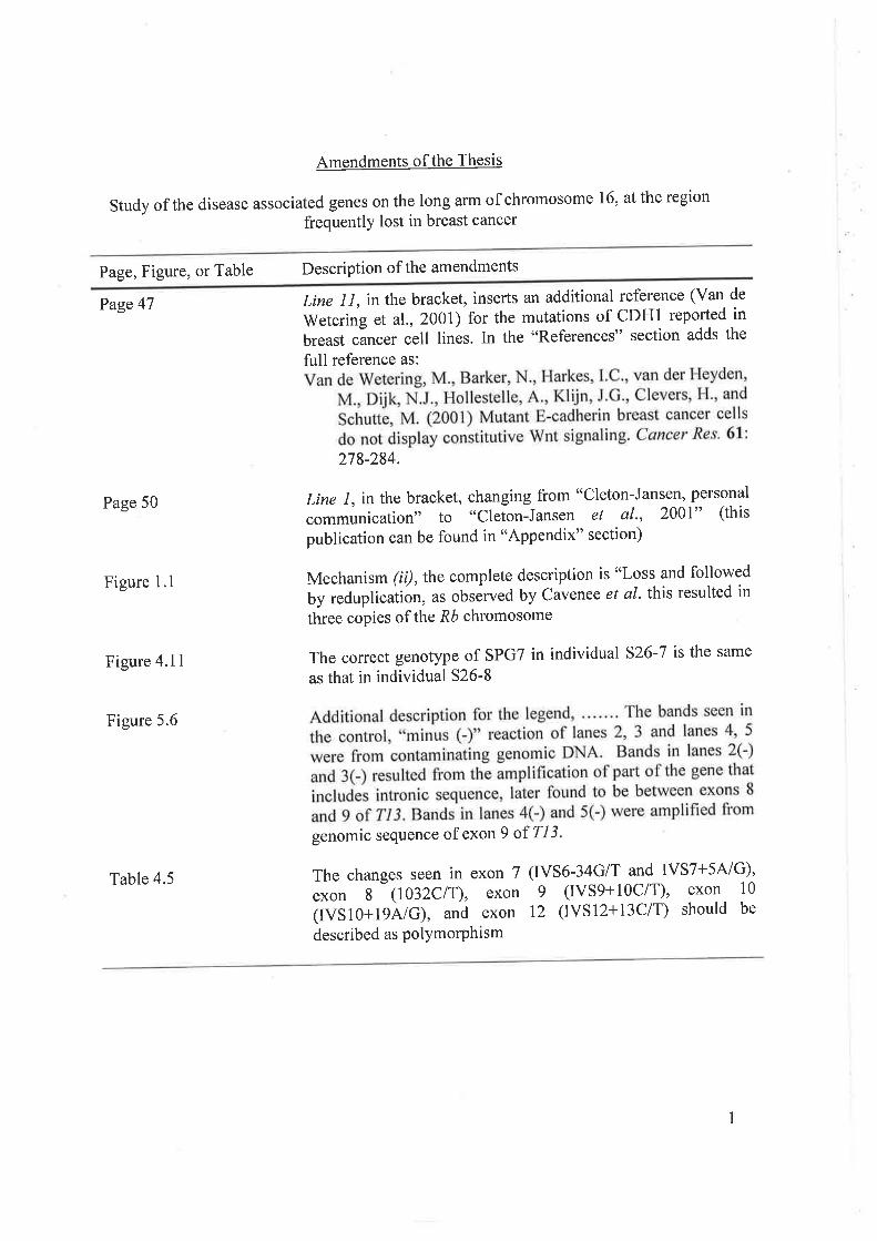
Amendments of the Thesis
study of the disease associated genes on the long arm of chromosome 16, at the region
frequentlY lost in breast cancer
Page, Figure, or Table Description of the amendments
Page 47
Page 50
Figure I .l
Figure 4.11
Figure 5.6
Table 4.5
Line I I, in the bracket, inserts an additional
Wetering et al., 2001) for the mutations ofbreast cancer cell lines. In the "References
reference (Van de
CDH1 reported in" section adds the
full reference as:
278-284.
Line 1, in the bracket, changing from "cleton-Jansen, personal
communication" to "Cleton-Jansen et al', 2001" (this
publication can be found in "Appendix" section)
Mechanism (ii),thecomplete description is "Loss and followed
by reduplicuìion, as obsérved by Cavenee et al. thls resulted in
three copies of the,Ró chromosome
The correct genotype of SPGT in individual s26-7 is the same
as that in individual 526-8
genomic sequence of exon 9 of Tl3 '
ThechangesseeninexonT(IVS6-34G/TandIVST+5A/G),exon 8 (1032CiT), exon 9 (IVS9+10C/T), exon 10
(IVS10+1gAiG),andexon12(IVSl2+13ClT)shouldbedescribed as PolYmorPhism
1
UNIVERSITY OF ADELAIDE
I iltilIilil iltil ilililtil tffi lltililtil lil til llil til til250159?6642
oq?H3+q55
c.2-
Abbreviations (additional)
AMP
APC
APRT
ATM
BARD-1
BRCA
CDH
CDK
CMAR
DAG
FAP
adenosine monophosphate
adenomatous polyposis coli
adenine phosphoribosyl transferase
ataxia-telangiectasia mutated
BRCAl-associated ring domain I
breast cancer associated
cadherin
cyclin-dependent kinase
cellular matrix adhesion regulator
diacylglycerol
familial adenomatous polyposis
hereditary non-polyposis colorectal cancer
hereditary spastic paraplegia
mismatch repair
microsatellite instability
neurofibromatosis
phorbol ester
plant homeodomain
phosphatase and tensin homolog deleted on chromosome 10
retinoblastoma
HNPCC
HSP
MMR
MSI
NF
PE
PHD
PTEN
2
RB