Status of freshwater catfish populations and their habitat within the Cockburn River
International Journal of Aquatic Biology (2013) 1(4): 175-184
ISSN: 2322-5270
Journal homepage: www.NPAJournals.com © 2013 NPAJournals. All rights reserved
Original Article
Studies on reproductive biology of Mystus tengara (Ham.-Buch., 1822), a freshwater catfish of
West Bengal, India
Sandipan Gupta*,1Samir Banerjee
Aquaculture Research Unit, Department of Zoology, University of Calcutta 35, Ballygunge Circular Road, Kolkata- 700019, West Bengal, India
Article history: Received 25 July 2013
Accepted 8 August 2013
Available online 20 August 2013
Keywords:
Mystus Wetland
Gonad
Maturity
Spawning
Sex ratio
Abstract: Studies on reproductive biology are essential to assess culture potential of a fish species.
Mystus tengara is a popular food fish as well as preferred as an ornamental fish in West Bengal. Till
date detailed report on reproductive biology of this fish species in the agro-climatic context of West
Bengal is lacking. Therefore, the present work was aimed to study the detailed reproductive biology of
Mystus tengara with an emphasis on sex ratio, length at first sexual maturity, cycle of gonadal
maturation and spawning periodicity using standard methods. Results of the study revealed female
dominance of the species over male in the population. However, the males showed earlier maturation
than females. Five gonadal maturity stages namely immature, maturing, mature, ripe and spent were
identified both for female and male fishes. Monthly study of gonadosomatic index (GSI), condition
factor and mean ova diameter revealed that the breeding season for this fish species extended from May
to September with a single spawning month in July. Total spawning behaviour along with synchronous
oocytes development was also observed in this fish species.
Introduction
Mystus tengara is commonly known as Tengara
Mystus which is a freshwater species, inhabits both
flowing and standing waters. The species is
distributed in India, Nepal, Bangladesh and Pakistan
(Talwar and Jhingran, 1991). In West Bengal it is
locally known as tengara and is a preferred food fish
due to its good taste, high nutrient profile; and in
recent times it has also got its importance as
ornamental fish too (Gupta and Banerjee, 2012).
Studies on reproductive biology of any fish species
are essential for assessing commercial potentialities
of its stock, life history, culture practice and actual
management of its fishery (Doha and Hye, 1970).
Reproductive potential of a population is one of the
basic exigencies to designate the individuals of that
population in respect to their gonadal conditions
* Corresponding author: Sandipan Gupta
E-mail address: : [email protected]
Tel: +919830082686
(Jhingran and Verma, 1972). In order to make
success in fish culture, it is important to assess the
yearly breeding cycle of culturable fishes
(Stoumboudi et al., 1993). Spawning of fish occurs
during a particular phase of reproductive cycle; some
of them breed once annually while others at regular
intervals throughout the year. Knowledge of gonadal
development and spawning season of a species allow
subsequent studies on spawning frequency of its
population, which is important for its management
(Chakraborty et al., 2007). Study of sex-ratio, length
at first sexual maturity, cycle of maturation and
spawning periodicity etc. are essential part of
reproductive biology investigation of fishes (Reddy,
1979; Vazzoler, 1996).
A number of workers (Qasim and Qayyum, 1961;
Bhatt, 1971a, b; Rao and Sharma, 1984; Roy and
176
Gupta and Banerjee / International Journal of Aquatic Biology (2013) 1(4): 175-184
Hossain, 2006; Musa and Bhuiyan, 2007) have
studied different aspects of reproductive biology of
different species of Mystus. Rastogi and Sexena
(1968) and Guraya et al. (1975) have studied
seasonal changes in morphology and activity of
Mystus tengara ovary. However, till date there is no
report on reproductive biology of Mystus tengara in
the agro-climatic context of West Bengal. The
objective of the present study was to study the
reproductive biology of Mystus tengara collected
from a selected wetland in West Bengal.
Materials and Methods
Monthly samples of Mystus tengara were collected
from an undisturbed wetland Baruipur, South-24-
Paraganas district of West Bengal (Latitude N
22°34', Longitude E 88°43') for a period of 12
months starting from September, 2008. In total, 721
specimens (never less than 50 in a month) were
collected during the entire study period for
evaluation of reproductive biology of the fish.
Sampled fish were brought to the laboratory, total
length (to the nearest of 0.1 cm) was measured by a
measuring scale, washed thoroughly with clean
water, soaked by a blotting paper and total body
weight was measured (to the nearest of 0.01gm) by
an electronic balance (Sartorius, Model No. BT
223S).
Fish specimens were dissected out ventrally to
remove gonads carefully. Surface moisture of
gonads was removed using blotting paper and the
weight and length of the gonads were measured to
the nearest of 0.01 gm and 0.1 cm, respectively.
Sexes of the sampled fish specimens were
determined after examination of the gonads.
Monthly variation in sex ratio was determined from
the total number of two sexes in monthly collected
samples. Chi-square test (Zar, 1999) was performed
to investigate the differences in sex-ratio (monthly
value and over-all value) from the expected ratio of
1:1.
Fish specimens were grouped into different length
classes with interval of 0.5 cm and the length class
in which at least 50% of the fish specimens were
observed to be mature was regarded as length at first
sexual maturity (Rao and Sharma, 1984; Suresh et
al., 2006; Mitra et al., 2007).
Cycle of maturation was studied by macroscopic and
microscopic observation of the different maturation
stages of gonad; male and female gonads were
grouped into different gonadal stages of
development according to Nikolsky (1963).
Additional information for differentiation of gonadal
maturity stages was gathered following the work of
Bhatt (Bhatt, 1970, 1971b) who worked on two other
species of Mystus.
Spawning periodicity was determined by monthly
evaluation of the Gonadosomatic Index (GSI),
condition factor and mean ova diameter. GSI and
Condition Factor (K) were measured using the
following formulae (Htun-Han, 1978):
GSI =Gonad Weight (gm) x 100
Total Body Weight (gm)
K =(TBW − GW)x 100
TL3
Where TBW is total body weight, GW is gonad
weight and TL is total length.
Size frequency distribution of the intra-ovarian
oocytes was studied on monthly basis to determine
the type of oocytes development. For measuring the
size frequency distribution of the intraovarian
oocytes, a small representative part from the middle
portion of the right or left ovary was taken out
separately and put into physiological saline solution
(0.85% NaCl) in a petridish. The ova present in the
ovary samples were separated and spread on a glass
slide to measure the diameter under a microscope
fitted with micrometer following the method of
LeCren (1951). The ocular micrometer reading was
standardized with that of the stage micrometer for
measurement of ova diameter in micrometer (μm)
and then the values were transformed to mm unit.
Ova were then grouped into four size classes;
immature ova (0.10-0.30 mm), maturing ova (0.30-
0.45 mm), mature ova (0.45-0.60 mm) and ripe ova
(0.60-0.85 mm) depending on the maturity status of
the ova and then monthly percentage frequency of
177
Gupta and Banerjee / International Journal of Aquatic Biology (2013) 1(4): 175-184
four size classes was calculated to get the
information on the spawning type.
Results
Sex-ratio: Among the 721 specimens studied, 451
and 270 were observed to be female and male,
respectively (Table 1). The average ratio of males to
females was observed to be 1:1.67. Overall females
showed significant (P<0.01) dominance over males;
though on monthly basis only from May to July
(P<0.01) and also in April, October and November
(P<0.05) significant dominance of females over
males was observed.
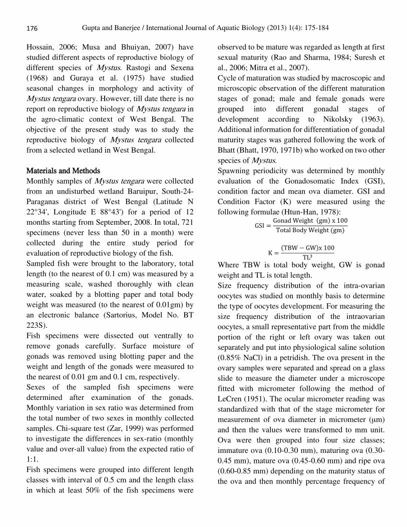
Length at first sexual maturity: The smallest males
with mature gonads were observed to appear in 7.5-
8 cm size group. 50% of all males were observed to
be mature in the 8.5-9 cm size group and all males
above 10 cm were observed with mature testes
during the spawning season. Few females in 8-8.5
cm size group were observed with mature gonads
while 50% of the females were found with mature
gonads in the size group of 9-9.5 cm. All females
Male
(Observed Value)
Female
(Observed value)
Ratio of male
and female P 2 Remark
Month No. of Fish No. % No. %
Sept., 2008 52 24 46.15 28 53.85 1:1.17 0.579 0.31 NS
Oct., 2008 60 22 36.67 38 63.33 1:1.73 0.039 4.27 S*
Nov., 2008 62 22 35.48 40 64.52 1:1.82 0.022 5.23 S*
Dec., 2008 45 18 40.00 27 60.00 1:1.50 0.180 1.80 NS
Jan., 2009 56 22 39.29 34 60.71 1:1.54 0.109 2.57 NS
Feb., 2009 62 25 40.32 37 59.68 1:1.48 0.128 2.32 NS
Mar., 2009 75 31 41.33 44 58.67 1:1.42 0.133 2.25 NS
Apr., 2009 62 22 35.48 40 64.52 1:1.82 0.022 5.23 S*
May, 2009 66 22 33.33 44 66.67 1:2.00 0.007 7.33 S**
June, 2009 65 21 32.31 44 67.69 1:2.09 0.004 8.14 S**
July, 2009 62 19 30.65 43 69.35 1:2.26 0.002 9.29 S**
Aug., 2009 54 22 40.74 32 59.26 1:1.45 0.174 1.85 NS
P = Probability; 𝟀2 = Chi-square; NS = Non Significant; S** = Significant at 1% level; S* = Significant at 5% level
Table 1. Monthly variation of sex ratio in Mystus tengara.
Figure 1. Percentage of mature fish in different length groups in Mystus tengara.
177

178
Gupta and Banerjee / International Journal of Aquatic Biology (2013) 1(4): 175-184
above 10.5 cm were observed with mature ovaries
during the spawning season (Fig. 1).
Cycle of gonadal maturation: Five stages of maturity
of ovary and testes were recognized as follows: Stage I (Immature): Ovaries translucent and
colorless; ova not visible to naked eyes, but under
microscope ova were irregular in shape, transparent,
yolk not formed. Testes whitish in color, very
narrow, thread like, no testicular lobules were
visible.
Stage II (Maturing): Ovaries yellowish white in
color; ova visible to naked eyes but not prominent;
under microscope ova were spherical in shape,
slightly opaque due to deposition of yolk at the
central position. Testes milky white in color, thread
like, appearance of little testicular lobules.
Stage III (Mature): Ovaries deep yellowish in color
and enlarged in size; ova clearly visible to naked
eyes; under microscope spherical in shape and
completely opaque (except the periphery) in
appearance due to presence of yolk. Testes light
yellowish in color, testis lobes increased in size and
length; increased number of testicular lobules.
Stage IV (Ripe): Ovaries reddish yellow in color;
with maximum size; ova clearly visible to naked
eyes; under microscope were spherical in shape and
opaque due to presence of huge amount of yolk. In
this stage, ova were with their full size, came out on
putting light pressure on the abdomen. Testes
yellowish white in color, testes lobes extended much
and two lobes were nearly touching each other.
Testicular lobules increased in number and length.
Milt came out while putting slight pressure on
abdomen.
Stage V (Spent): Ovaries very much reduced in size,
shrunken and reddish in appearance. Under
microscope, few ripe ova along with irregular shaped
small translucent ova were visible. Testes
translucent, thread like and flaccid in appearance; no
testicular lobules were visible.
Females with immature gonads were observed from
October to April; highest percentage being observed
from October to December while lowest percentage
was observed in April. Maturing females first were
observed in January and available till May; highest
and lowest percentage being observed in April and
January, respectively. Mature females were
observed from April to July; highest percentage was
observed in May while lowest percentage in July.
Ripe females were observed from May to August;
highest percentage being observed in July and lowest
percentage in May. Spent females were observed
from July to September; highest percentage being
observed in September and lowest percentage in July
(Fig. 2).
Males with immature gonads were observed from
October to May; highest percentage being observed
in November and December while lowest percentage
was observed during May. Maturing males first were
observed in January and available till June; highest
Figure 2. Monthly percentage of different gonadal maturation stages in female Mystus tengara.

179
Gupta and Banerjee / International Journal of Aquatic Biology (2013) 1(4): 175-184
percentage being observed in April and lowest
percentage in January. Mature males were observed
from May to July; highest percentage was observed
in June while lowest percentage in July. Ripe males
were observed from June to September with highest
percentage being observed in July and lowest
percentage in September. Spent males were observed
from July to October; highest percentage being
observed in September and lowest percentage in July
(Fig. 3).
Gonado Somatic Index (GSI): In both female and
male, GSI was observed to reach peak once a year
during the month of July. The lowest value of GSI
was observed in the month of December; then it
started to increase from January onwards and
reached the peak in July; then dropped down in
August to reach the lowest value again in December
(Fig. 4). In respect to both sexes, GSI showed significant
(P<0.01) positive relationship with Total Body
Weight (TBW), Total Length (TL), Gonad Weight
(GW) and Gonad Length (GL) as follows: GSI = -3.38 + 0.73 TBW (r = 0.39, P<0.01, SE = 4.37)
GSI = -7.59 + 1.06 TL (r = 0.21, P<0.01, SE = 4.65)
GSI = 0.31 + 8.85 GW (r = 0.95, P<0.01, SE = 1.55)
GSI = -8.87 + 6.42 GL (r = 0.63, P<0.01, SE = 3.68)
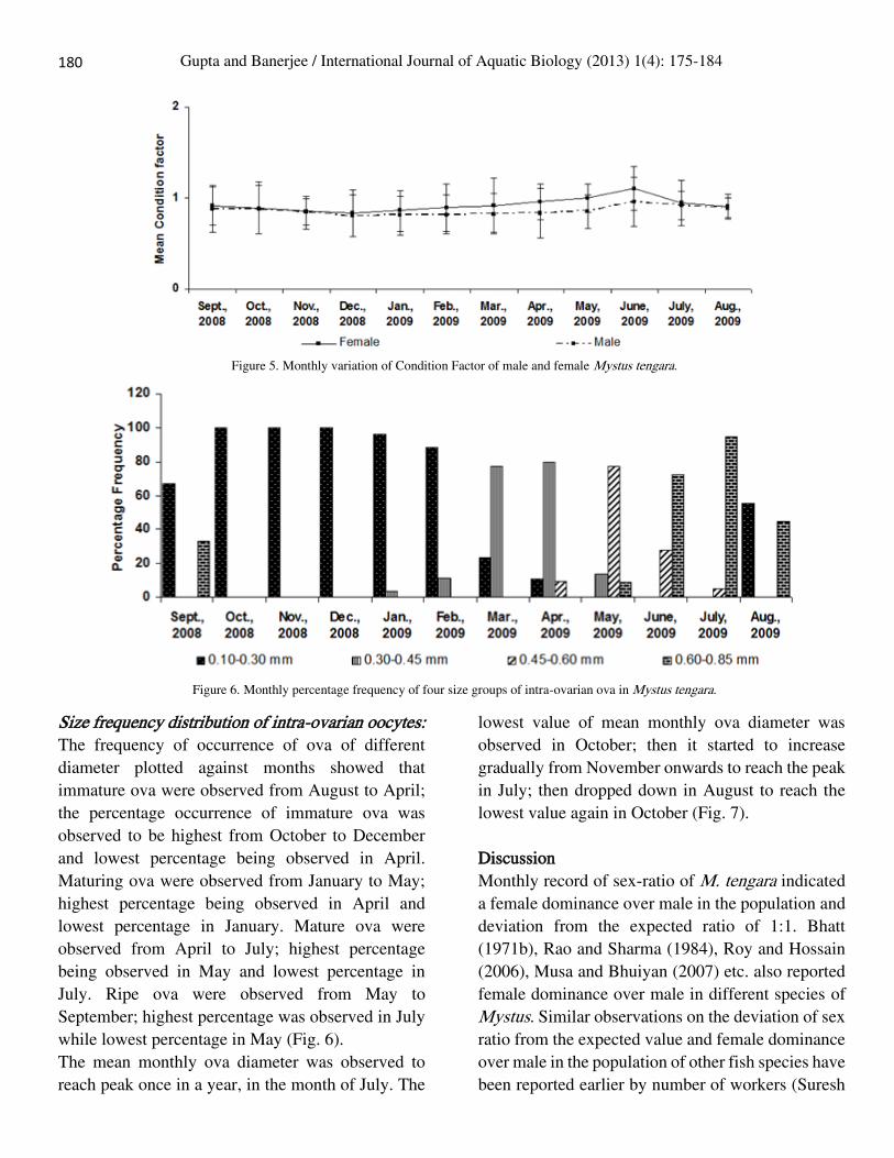
Condition Factor: In both female and male, condition
factor was observed to reach peak once a year during
June. The lowest value of condition factor was
observed in the month of December; then it started
to increase from January onwards and reached the
peak in June; then dropped down in July to reach the
lowest value again in December (Fig. 5).
Figure 4. Monthly variation of Gonado-Somatic-Index (GSI) of male and female Mystus tengara.
Figure 3. Monthly percentage of different gonadal maturation stages in male Mystus tengara.
179
180
Gupta and Banerjee / International Journal of Aquatic Biology (2013) 1(4): 175-184
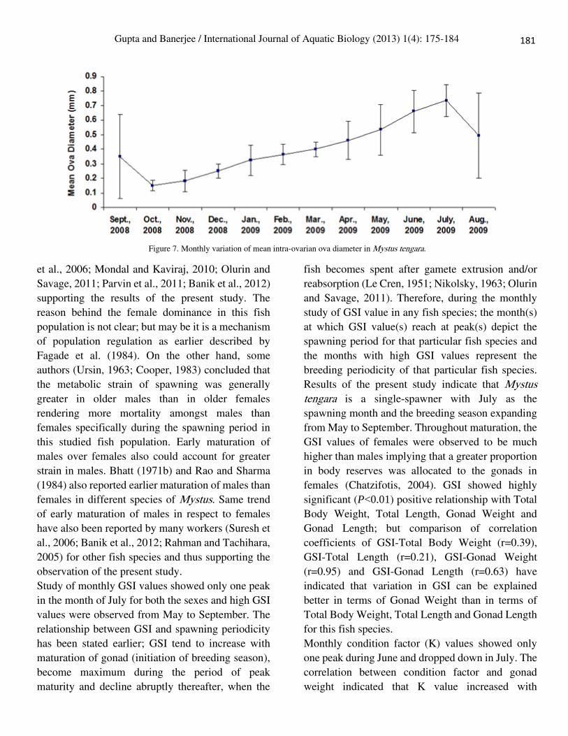
Size frequency distribution of intra-ovarian oocytes: The frequency of occurrence of ova of different
diameter plotted against months showed that
immature ova were observed from August to April;
the percentage occurrence of immature ova was
observed to be highest from October to December
and lowest percentage being observed in April.
Maturing ova were observed from January to May;
highest percentage being observed in April and
lowest percentage in January. Mature ova were
observed from April to July; highest percentage
being observed in May and lowest percentage in
July. Ripe ova were observed from May to
September; highest percentage was observed in July
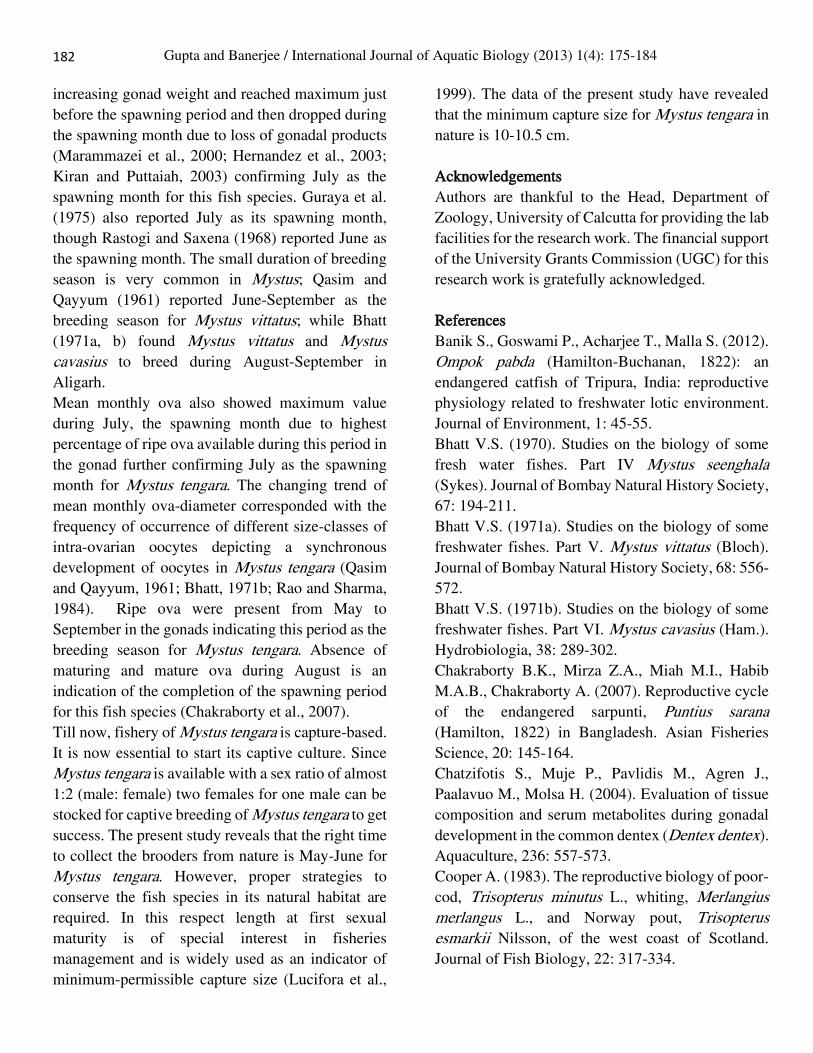
while lowest percentage in May (Fig. 6). The mean monthly ova diameter was observed to
reach peak once in a year, in the month of July. The
lowest value of mean monthly ova diameter was
observed in October; then it started to increase
gradually from November onwards to reach the peak
in July; then dropped down in August to reach the
lowest value again in October (Fig. 7).
Discussion
Monthly record of sex-ratio of M. tengara indicated
a female dominance over male in the population and
deviation from the expected ratio of 1:1. Bhatt
(1971b), Rao and Sharma (1984), Roy and Hossain
(2006), Musa and Bhuiyan (2007) etc. also reported
female dominance over male in different species of
Mystus. Similar observations on the deviation of sex
ratio from the expected value and female dominance
over male in the population of other fish species have
been reported earlier by number of workers (Suresh
Figure 5. Monthly variation of Condition Factor of male and female Mystus tengara.
Figure 6. Monthly percentage frequency of four size groups of intra-ovarian ova in Mystus tengara.
181
Gupta and Banerjee / International Journal of Aquatic Biology (2013) 1(4): 175-184
et al., 2006; Mondal and Kaviraj, 2010; Olurin and
Savage, 2011; Parvin et al., 2011; Banik et al., 2012)
supporting the results of the present study. The
reason behind the female dominance in this fish
population is not clear; but may be it is a mechanism
of population regulation as earlier described by
Fagade et al. (1984). On the other hand, some
authors (Ursin, 1963; Cooper, 1983) concluded that
the metabolic strain of spawning was generally
greater in older males than in older females
rendering more mortality amongst males than
females specifically during the spawning period in
this studied fish population. Early maturation of
males over females also could account for greater
strain in males. Bhatt (1971b) and Rao and Sharma
(1984) also reported earlier maturation of males than
females in different species of Mystus. Same trend
of early maturation of males in respect to females
have also been reported by many workers (Suresh et
al., 2006; Banik et al., 2012; Rahman and Tachihara,
2005) for other fish species and thus supporting the
observation of the present study.
Study of monthly GSI values showed only one peak
in the month of July for both the sexes and high GSI
values were observed from May to September. The
relationship between GSI and spawning periodicity
has been stated earlier; GSI tend to increase with
maturation of gonad (initiation of breeding season),
become maximum during the period of peak
maturity and decline abruptly thereafter, when the
fish becomes spent after gamete extrusion and/or
reabsorption (Le Cren, 1951; Nikolsky, 1963; Olurin
and Savage, 2011). Therefore, during the monthly
study of GSI value in any fish species; the month(s)
at which GSI value(s) reach at peak(s) depict the
spawning period for that particular fish species and
the months with high GSI values represent the
breeding periodicity of that particular fish species.
Results of the present study indicate that Mystus tengara is a single-spawner with July as the
spawning month and the breeding season expanding
from May to September. Throughout maturation, the
GSI values of females were observed to be much
higher than males implying that a greater proportion
in body reserves was allocated to the gonads in
females (Chatzifotis, 2004). GSI showed highly
significant (P<0.01) positive relationship with Total
Body Weight, Total Length, Gonad Weight and
Gonad Length; but comparison of correlation
coefficients of GSI-Total Body Weight (r=0.39),
GSI-Total Length (r=0.21), GSI-Gonad Weight
(r=0.95) and GSI-Gonad Length (r=0.63) have
indicated that variation in GSI can be explained
better in terms of Gonad Weight than in terms of
Total Body Weight, Total Length and Gonad Length
for this fish species.
Monthly condition factor (K) values showed only
one peak during June and dropped down in July. The
correlation between condition factor and gonad
weight indicated that K value increased with
Figure 7. Monthly variation of mean intra-ovarian ova diameter in Mystus tengara.
181
182
Gupta and Banerjee / International Journal of Aquatic Biology (2013) 1(4): 175-184
increasing gonad weight and reached maximum just
before the spawning period and then dropped during
the spawning month due to loss of gonadal products
(Marammazei et al., 2000; Hernandez et al., 2003;
Kiran and Puttaiah, 2003) confirming July as the
spawning month for this fish species. Guraya et al.
(1975) also reported July as its spawning month,
though Rastogi and Saxena (1968) reported June as
the spawning month. The small duration of breeding
season is very common in Mystus; Qasim and
Qayyum (1961) reported June-September as the
breeding season for Mystus vittatus; while Bhatt
(1971a, b) found Mystus vittatus and Mystus cavasius to breed during August-September in
Aligarh.
Mean monthly ova also showed maximum value
during July, the spawning month due to highest
percentage of ripe ova available during this period in
the gonad further confirming July as the spawning
month for Mystus tengara. The changing trend of
mean monthly ova-diameter corresponded with the
frequency of occurrence of different size-classes of
intra-ovarian oocytes depicting a synchronous
development of oocytes in Mystus tengara (Qasim
and Qayyum, 1961; Bhatt, 1971b; Rao and Sharma,
1984). Ripe ova were present from May to
September in the gonads indicating this period as the
breeding season for Mystus tengara. Absence of
maturing and mature ova during August is an
indication of the completion of the spawning period
for this fish species (Chakraborty et al., 2007).
Till now, fishery of Mystus tengara is capture-based.
It is now essential to start its captive culture. Since
Mystus tengara is available with a sex ratio of almost
1:2 (male: female) two females for one male can be
stocked for captive breeding of Mystus tengara to get
success. The present study reveals that the right time
to collect the brooders from nature is May-June for
Mystus tengara. However, proper strategies to
conserve the fish species in its natural habitat are
required. In this respect length at first sexual
maturity is of special interest in fisheries
management and is widely used as an indicator of
minimum-permissible capture size (Lucifora et al.,
1999). The data of the present study have revealed
that the minimum capture size for Mystus tengara in
nature is 10-10.5 cm.
Acknowledgements
Authors are thankful to the Head, Department of
Zoology, University of Calcutta for providing the lab
facilities for the research work. The financial support
of the University Grants Commission (UGC) for this
research work is gratefully acknowledged.
References
Banik S., Goswami P., Acharjee T., Malla S. (2012).
Ompok pabda (Hamilton-Buchanan, 1822): an
endangered catfish of Tripura, India: reproductive
physiology related to freshwater lotic environment.
Journal of Environment, 1: 45-55.
Bhatt V.S. (1970). Studies on the biology of some
fresh water fishes. Part IV Mystus seenghala
(Sykes). Journal of Bombay Natural History Society,
67: 194-211.
Bhatt V.S. (1971a). Studies on the biology of some
freshwater fishes. Part V. Mystus vittatus (Bloch).
Journal of Bombay Natural History Society, 68: 556-
572.
Bhatt V.S. (1971b). Studies on the biology of some
freshwater fishes. Part VI. Mystus cavasius (Ham.).
Hydrobiologia, 38: 289-302.
Chakraborty B.K., Mirza Z.A., Miah M.I., Habib
M.A.B., Chakraborty A. (2007). Reproductive cycle
of the endangered sarpunti, Puntius sarana
(Hamilton, 1822) in Bangladesh. Asian Fisheries
Science, 20: 145-164.
Chatzifotis S., Muje P., Pavlidis M., Agren J.,
Paalavuo M., Molsa H. (2004). Evaluation of tissue
composition and serum metabolites during gonadal
development in the common dentex (Dentex dentex).
Aquaculture, 236: 557-573.
Cooper A. (1983). The reproductive biology of poor-
cod, Trisopterus minutus L., whiting, Merlangius merlangus L., and Norway pout, Trisopterus esmarkii Nilsson, of the west coast of Scotland.
Journal of Fish Biology, 22: 317-334.

183
Gupta and Banerjee / International Journal of Aquatic Biology (2013) 1(4): 175-184
Doha S., Hye M.A. (1970). Fecundity of Padma
River hilsa, Hilsa ilisha (Hamilton). Pakistan Journal
of Science, 22: 176-178.
Fagade S.O., Adebisi A.A., Atanda A.N. (1984).
The breeding cycle of Sarothorodon galilaeus in the
I.I.T.A. Lake, Ibadan, Nigeria. Archive
Hydrobiologie, 100: 493-500.
Gupta S., Banerjee S. (2012). Indigenous ornamental
fish: a new boon in ornamental fish trade of West
Bengal. Fishing Chimes, 32: 130-134.
Guraya S.S., Kaur, R., Saxena, P.K. (1975).
Morphology of ovarian changes during the
reproductive cycle of the fish, Mystus tengara
(Ham.). Acta Anatomica, 91: 222-260.
Hernandez M.D., Egea M.A., Rueda F.M., Martinez
F.J., Garcia G.B. (2003). Seasonal condition and
body composition changes in sharp snout sea bream
(Diplodus puntazzo) raised in captivity.
Aquaculture, 220: 569-580.
Htun-Han M. (1978). The reproductive biology of
the dab Limanda limanda (L.) in the North Sea:
gonosomatic index, hepatosomatic index and
condition factor. Journal of Fish Biology, 13: 369-
378.
Jhingran A.G., Verma D.N. (1972). Sexual maturity
and spawning of Gudusia chapra (Ham.) in Ganga
river system. Proceedings of the Indian National
Science Academy, 42: 207-224.
Kiran B.R., Puttaiah E.T. (2003). Fecundity studies
on Chela untrahi (Day) from Bhadra reservoir,
Karnataka, India. Inland Fisheries Society, 35: 41-
44.
LeCren E.D. (1951). The length-weight relationship
and seasonal cycle in gonad weight and condition in
the perch (Perca fluviatilis). Journal of Animal
Ecology, Oxford, 20: 201-219.
Lucifora L.O., Valero J.L., Garcia V.B. (1999).
Length at maturity of the green-eye spurdog shark,
Squalus mitsukuii (Elasmobranchii: Squalidae) from
the SW Atlantic, with comparisons with other
regions. Marine and Freshwater Research. 50: 629-
632.
Marammazei J.G., Mustafa A., Al Mukhtar M.A.
(2000). The occurrence, feeding and reproduction of
three Barbus spp. in Shadighan marsh. The first
national scientific conference on Barbus spp. in Iran,
Khuzestan Fisheries Research Center. pp: 50.
Mitra K., Suresh V.R., Vinci G.K., Mazumdar N.N.,
Biswas D.K. (2007). Biology and fishery of banded
gourami, Colisa fasciata (Bloch and Schneider 1801)
in a floodplain wetland of Ganga river basin. Asian
Fisheries Science, 20: 409-423.
Mondal D.K., Kaviraj A. (2010). Feeding and
reproductive biology of Indian shad Gudusia chapra
in two floodplain lakes of India. Electronic Journal
of Biology, 6(4): 98-102.
Musa A.S.M., Bhuiyan A.S. (2007). Fecundity of
Mystus bleekeri (Day, 1877) from the River Padma
near Rajshahi city. Turkish Journal of Fisheries and
Aquatic Sciences, 7: 161-162.
Nikolsky G.V. (1963). The ecology of fishes.
Academic Press. London., UK. 352 p.
Olurin K.B., Savage O.D. (2011). Reproductive
biology, length-weight relationship and condition
factor of the African snake head, Parachanna obscura, from River Oshun, South-west Nigeria.
International Journal of Fisheries and Aquaculture,
3(8): 146-150.
Parvin M.R., Al-Misned F.A., Mortuza M.G. (2011).
The fecundity and sex ratio of Labeo boga
(Hamilton) (Cypriniformes: Cyprinidae) of
Rajshahi, Bangladesh. Continental Journal of
Fisheries and Aquatic Science, 5(3): 19-21.
Qasim S.Z., Qayyum A. (1961). Spawning
frequencies and breeding seasons of some freshwater
fishes with special reference to those occurring in the
plains of Northern India. Indian Journal of Fisheries,
8(1): 24-43.
Rahman M.H., Tachihara K. (2005). Reproductive
biology of Sillago aeolus in Okinawa Island, Japan.
Fisheries Science, 71: 122-132.
Rao T.A., Sharma S.V. (1984). Reproductive
biology of Mystus vittatus (Bloch) (Bagridae:
Siluriformes) from Guntur, Andhra Pradesh.
Hydrobiologia, 119: 21-26.
Rastogi R.K., Saxena P.K. (1968). Annual changes
in the ovarian activity of the catfish, Mystus tengara
183
184
Gupta and Banerjee / International Journal of Aquatic Biology (2013) 1(4): 175-184
(Ham.) (Teleostei). Japanese Journal of Ichthyology,
15(1): 28-35.
Reddy P.B. (1979). The fecundity of Channa punctata (Bloch, 1793) (Pisces, Teleostei,
Channidae) from Guntur, India. Proceedings of
Indian Academy of Sciences, 88(2): 95-98.
Roy P.K., Hossain M.A. (2006). The fecundity and
sex ratio of Mystus cavasius (Hamilton)
(Cypriniformes: Bagridae). The Journal of Life and
Earth Science, 1(2): 65-66.
Stoumboudi M.T., Villwock W., Sela J., Abraham
M. (1993). Gonadosomatic index in Barbus
longiceps, Capoeta damascina and their natural
hybrid (Pisces, Cyprinidae), versus spermatozoan
index in the parental males. Journal of Fish Biology,
43: 865-875.
Suresh V.R., Biswas B.K., Vinci G.K., Mitra K.,
Mukherjee A. (2006). Biology and fishery of barred
spiny eel, Macrognathus pancalus Hamilton. ACTA
Ichthyologica et Piscatoria, 36(1): 31-37.
Talwar P.K., Jhingran A.G. (1991). Inland fishes of
India and adjacent countries. Vol-1 and Vol-2.
Oxford and IBH Publishing Co. Pvt. Ltd. New Delhi,
Bombay and Calcutta. 1-1063 p.
Ursin E. (1963). On the seasonal variation of growth
rate and growth parameters in Norway pout (Gadus esmarkii) in the Skagerrak. Meddelelser fra
Danmarks Fiskeri-Og Havundersogelser, 4(12): 17-
29.
Vazzoler A.E.A. de M. (1996). Biologia da
reprodução de peixes Teleósteos: Teoria e prática.
Ed. EDUEM, Maringá. 169 p.
Zar J.H. (1999). Biostatistical Analysis. 4th Edition,
Prentice Hall, New Jersey. 663 p.
Copyright © 2022 FDOKUMEN