Modeling stability in neuron and network function: the role of activity in homeostasis
Upload
independentCategory
view
3download
0
Structure and Function of the D-Galactose Network in Enterobacteria
Zsolt Csiszovszki,a Sandeep Krishna,b László Orosz,c Sankar Adhya,a and Szabolcs Semseyc,d
Laboratory of Molecular Biology, Center for Cancer Research, National Cancer Institute, National Institutes of Health, Bethesda, Maryland, USAa; NCBS, Bangalore, Indiab;Department of Genetics, Eötvös Loránd University, Budapest, Hungaryc; and Niels Bohr Institute, Copenhagen, Denmarkd
ABSTRACT Galactose is important for the survival and virulence of bacteria. In Escherichia coli, galactose is utilized by the Leloirpathway, which is controlled by a complex network. To shed light on the potential functions the galactose network could per-form, we performed bioinformatical analysis of reference genome sequences belonging to the Enterobacteriaceae family. Wefound that several genomes have reduced numbers of components compared to the E. coli galactose system, suggesting that thenetwork can be optimized for different environments. Typically, genes are removed by deletions; however, in Yersinia pestis, thegalactose mutarotase (galM) gene is inactivated by a single-base-pair deletion. Lack of GalM activity indicates that the two ano-mers of D-galactose are used for different purposes, �-D-galactose as a carbon source and �-D-galactose for induction of UDP-galactose synthesis for biosynthetic glycosylation. We demonstrate that activity of the galM gene can be restored by differentsingle-base-pair insertions. During the evolution of Y. pestis to become a vector-transmitted systemic pathogen, many geneswere converted to pseudogenes. It is not clear whether pseudogenes are present to maintain meiotrophism or are in the processof elimination. Our results suggest that the galM pseudogene has not been deleted because its reactivation may be beneficial incertain environments.
IMPORTANCE Evolution of bacteria to populate a new environment necessarily involves reengineering of their molecular net-work. Members of the Enterobacteriaceae family of bacteria have diverse lifestyles and can function in a wide range of environ-ments. In this study we performed bioinformatical analysis of 34 reference genome sequences belonging to the Enterobacteri-aceae family to gain insight into the natural diversity of the D-galactose utilization network. Our bioinformatical analysis showsthat in several species, some genes of the network are completely missing or are inactivated by large deletions. The only excep-tion is the galactose mutarotase (galM) gene of Yersinia pestis, which is converted to a pseudogene by a single-base-pair deletion.In this paper, we discuss the possible consequences of galM inactivation on network function. We suggest that galM was con-verted to a pseudogene rather than being deleted in evolution because its reactivation can be beneficial in certain environments.
Received 17 March 2011 Accepted 6 June 2011 Published 28 June 2011
Citation Csiszovszki Z, Krishna S, Orosz L, Adhya S, Semsey S. 2011. Structure and function of the D-galactose network in enterobacteria. mBio 2(4):e00053-11. doi:10.1128/mBio.00053-11.
Editor Roger Hendrix, University of Pittsburgh
Copyright © 2011 Csiszovszki et al. This is an open-access article distributed under the terms of the Creative Commons Attribution-Noncommercial-Share Alike 3.0 UnportedLicense, which permits unrestricted noncommercial use, distribution, and reproduction in any medium, provided the original author and source are credited.
Address correspondence to Szabolcs Semsey, [email protected].
Evolution of bacteria to populate a new environment neces-sarily involves reengineering of their molecular network.
Changes can affect the elements (e.g., genes) of the network aswell as the interactions in the network. Redundant genes or thosethat antagonize successful colonization in the new environmentcan be inactivated by point mutations or removed by deletions(1). Also, genes required for adaptation to a new niche can beacquired by horizontal transfer (e.g., pathogenicity islands) (2). Amajor determinant of network function is network logic, whichdepends on the interactions between network elements (3). Re-cent studies demonstrated that network logic can be easily engi-neered by mutations in regulatory sequences (4, 5). Rearrange-ments in metabolic pathways were analyzed in Yersinia pestis,which diverged recently from Yersinia pseudotuberculosis, a gas-trointestinal pathogen (6). Analysis of the Yersinia pestis metabolicnetwork suggested excellent agreement between the possible met-abolic reaction pathways and the known nutritional needs ofY. pestis cells (1). It is generally assumed that cellular responses tothe natural levels of perturbations in the concentration of critical
chemical compounds are optimized. Along this logic, capabilitiesof regulatory networks would reflect the potential nature of envi-ronments and the environmental changes cells may face (7).
Members of the Enterobacteriaceae family of bacteria have di-verse lifestyles and can function in a wide range of environments.In this study we performed bioinformatical analysis of 34 refer-ence genome sequences belonging to the Enterobacteriaceae familyto gain insight into the natural diversity of the D-galactose utiliza-tion network. The rationale of using the D-galactose network isthat D-galactose metabolism can be an important factor in viru-lence in different bacteria (8–10). For example, in Erwinia amylo-vora, the causal agent of fire blight of rosaceous plants, galactosemetabolism affects capsule synthesis and virulence (9). In Y. pestis,both the D-galactose transport and metabolism operons areinduced when the temperature is shifted from 26°C to 37°C,which corresponds to the temperature change in the transitionfrom the flea to the mammalian host (11). Also, incorporationof �-D-galactose into the lipopolysaccharide (LPS) of Y. pestisinside the transmitting vector (flea) may be important (12).
RESEARCH ARTICLE
July/August 2011 Volume 2 Issue 4 e00053-11 ® mbio.asm.org 1
m
bio.asm.org
on May 29, 2016 - P
ublished by m
bio.asm.org
Dow
nloaded from
The D-galactose network of Escherichia coli is well characterized(13) and suitable to be used as a reference for the comparativeanalysis.
Escherichia coli utilizes D-galactose by the amphibolic Leloirpathway (Fig. 1). The metabolic steps of D-galactose utilizationare catalyzed by the GalK (galactokinase), GalT (galactose1-phosphate uridylyltransferase), and GalE (UDP-galactose-4-epimerase) proteins. In E. coli, the genes encoding these proteinsbelong to the same operon, together with the galM gene, whichencodes a mutarotase that allows the intracellular interconversionof the two anomers of D-galactose (14), �-D-galactose and �-D-galactose. Only �-D-galactose serves as a substrate of galactoki-nase, the first enzyme of the Leloir pathway. D-Galactose can betransported by two D-galactose-induced transport systems, thelow-affinity GalP and the high-affinity Mgl systems (15). Trans-port and utilization of D-galactose is regulated by the Gal repressor(GalR) and the Gal isorepressor (GalS). These repressors bind thesame set of operators in the regulatory regions of the genes belong-ing to the gal regulon in the absence of D-galactose. D-Galactosebinding to GalR (similar to that to GalS) allosterically inhibits itsoperator binding (13, 16, 17). A recent study demonstrated thatboth of these anomers of D-galactose are effective in the bindingand inactivation of GalR (18).
In the Leloir pathway, only the galE gene product is involved inmaking substrates for biosynthetic glycosylation reactions, whileall of the Gal enzymes are needed for catabolism of D-galactose.Therefore, when D-galactose is not available or not preferred as acarbon source, expression of the gal operon genes is discoordi-nated, resulting mostly in GalE synthesis (19–21). Enterobacteriaevolved different mechanisms to allow such discoordination.These include using small RNA (sRNA)-mediated translationalregulation (22), differentially regulated promoters and naturalpolarity (19–21, 23), and physical separation of the galE gene fromthe rest of the gal operon genes (23).
In this paper, we focus primarily on the role of galactose mutaro-tase in the D-galactose utilization network. We show that the functionof the galM pseudogene of Y. pestis can be restored by single-base-pairinsertions in different ways. Our analysis predicts that the twoD-galactose anomers, �- and �-D-galactose, play different roles inY. pestis. Only �-D-galactose can be used as a carbon source, while�-D-galactose can induce the production of UDP-galactose, a com-pound used in biosynthetic glycosylation reactions.
RESULTSIdentification of elements of the D-galactose network in entero-bacteria. Because metabolic enzyme sequences are highly con-served across species, metabolic networks can be successfully re-constructed primarily based on genome sequence data (24).Therefore, we used the BLAST program (25) to identify DNA andprotein sequences in reference genome sequences of strains be-longing to the Enterobacteriaceae family, which are similar to com-ponents of the D-galactose network of E. coli K-12 MG1655(Fig. 2). We found only four genes, galR (encoding the galactoserepressor), galE (encoding the UDP-galactose-4-epimerase), crp(encoding the cyclic AMP [cAMP] receptor protein [CRP]), andspf (encoding the Spot42 sRNA), which are present in all the ge-nomes studied. All 12 genes studied (galETKM, mglBAC, galR,galS, galP, crp, and spf) are present in 19 of the 34 genomes ana-lyzed. However, in six of these strains, the galE gene is not part ofthe gal operon. Six strains have the high-affinity Mgl transportsystem but not the low-affinity GalP symporter, suggesting thatthe galactose utilization networks of these strains are optimizedfor low-galactose environments (Dickeya dadantii, Dickeya zeae,Sodalis glossinidius, Yersinia enterocolitica, Y. pestis, and Y. pseudo-tuberculosis strains). As opposed to those strains, Edwardsiella ic-taluri, Edwardsiella tarda, Erwinia pyrifoliae, Shigella boydii, andShigella dysenteriae have only the low-affinity transporter, whichrequires high levels of extracellular D-galactose for proper func-
tion. The three Pectobacterium strains andProteus mirabilis lack both transport sys-tems. Genes responsible for galactose me-tabolism (galE, galK, galT, galM) can befound in different arrangements in thestrains analyzed; however, there are twostrains in which we found inactive com-ponents. In Sodalis glossinidius, only thegalE gene is intact, suggesting that in thesecells, UDP-galactose required for biosyn-thetic purposes is produced fromUDP-glucose. In Y. pestis, the galM genecontains a single-nucleotide deletion, re-sulting in a frameshift.
Our bioinformatical analysis showsthat in most of the cases, genes arecompletely missing or are inactivatedby large deletions. The only exception isthe galM gene of Y. pestis, which isconverted to a pseudogene. The samepseudogene was found in all theY. pestis genome sequences (e.g.,GenBank accession no. NC_010159.1,NC_009381.1, NC_008149.1, NC_008150.1,NC_005810.1, NC_003143.1, and NC_004088.1).
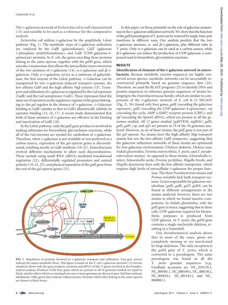
FIG 1 Regulation of proteins involved in D-galactose transport and utilization. Vast grey arrowsindicate the major metabolic flows. This figure is based on the E. coli D-galactose network (13) but forsimplicity shows only the gene products (proteins and sRNA) of the 12 genes involved in the bioinfor-matical analysis. Products of the four genes which are present in all 34 genomes studied are typed inblack, and the others which are missing from one or more genomes are shown in grey. Red lines indicateinhibitions, while green lines indicate enhancements. Proteins which often belong to the same operonare shown in black boxes.
Csiszovszki et al.
2 ® mbio.asm.org July/August 2011 Volume 2 Issue 4 e00053-11
m
bio.asm.org
on May 29, 2016 - P
ublished by m
bio.asm.org
Dow
nloaded from

Functional restoration of the galM pseudogene of Y. pestis.Comparison of the Y. pestis and Y. pseudotuberculosis genomesindicated that the transition from the enteropathogen form to thevector-transmitted systemic pathogen form resulted in more than
300 pseudogenes (26, 27). However, it remains to be answeredwhether pseudogenes are present to maintain meiotrophism orare in the process of elimination. The Y. pestis galM pseudogenediffers from the Y. pseudotuberculosis YPIII galM gene only by a
FIG 2 Presence of genes responsible for D-galactose transport (galP, mglBAC), utilization (galETKM), and regulation of these two processes (galR, galS, crp, spf)in genomes of strains belonging to the Enterobacteriaceae family (GenBank accession numbers are shown on the left, and more details are provided in Materialsand Methods). Filled boxes indicate the presence of intact genes, based on bioinformatical analysis. Empty boxes indicate genes disrupted by extensive deletionsor insertions. The galM pseudogene of Y. pestis, which is inactivated by a single-base-pair deletion, is marked by an asterisk. Arrowheads indicate the directionof transcription for putative operons and ORFs separated by less than 1 kbp.
D-Galactose Network in Enterobacteria
July/August 2011 Volume 2 Issue 4 e00053-11 ® mbio.asm.org 3
m
bio.asm.org
on May 29, 2016 - P
ublished by m
bio.asm.org
Dow
nloaded from

single-base-pair deletion (at nucleotide position 117 of the openreading frame [ORF]) and a same-sense mutation (at nucleotideposition 180). Therefore, this pseudogene can likely be reactivatedby insertion of a single base pair. The GalM protein that would beencoded by the “repaired” Y. pestis galM gene is identical to theY. pseudotuberculosis YPIII GalM protein (GenBank accession no.YP_001721673.1). Interestingly, the Yersinia pseudotuberculosisIP 32953 galM gene encodes a different amino acid at the positionof the required insertion (D instead of N), suggesting that theY. pestis pseudogene can be restored in multiple ways. To test thishypothesis we synthesized the Y. pestis galM pseudogene and itsdifferent “restored” versions and inserted them into a low-copy-number plasmid. The plasmid-borne genes were tested whetherthey can complement a chromosomal galM deletion in E. coli.GalM function was tested by studying growth on intracellularlyproduced �-D-galactose. Cells were grown in the presence ofphenyl-�-D-galactopyranoside as the sole carbon source. Hydro-lysis of phenyl-�-D-galactopyranoside by �-galactosidase gener-ates phenol and �-D-galactose. Phenol is excreted into the culturemedium and does not accumulate in the cell. Cells were grownovernight in LB medium and then washed in M63 minimal me-dium. Washed cells were plated on M63 minimal medium supple-mented with 2 mM phenyl-�-D-galactopyranoside, 0.0004% vita-min B1, 0.5 mM IPTG (isopropyl-thiogalactoside; to induce�-galactosidase production), and 15 �g/ml tetracycline. Wefound that growth strongly depends on the presence of functionalgalM. Cells containing the Y. pestis galM pseudogene showed veryslow growth, similar to that of the E. coli �galM cells harboring theempty plasmid. This slow growth reflects the low level of nonen-zymatic mutarotation of the D-galactose anomers. However, two“restored” versions of GalM (N39, D39) conferred similar growthto wild-type E. coli MG1655 cells (Fig. 3). The difference in the sizeof colonies reflects a minimum doubling time of 135 to 150 minfor the galM� and about 1,000 min for the �galM strains, as mea-sured in liquid cultures. GalM version Q39 grew similarly togalM�, I39 and T39 showed intermediate growth, while K39, R39,and E39 were not growing substantially faster than �galM.
Effect of galM and galP deletion on utilization of extracellu-lar D-galactose in vivo. Y. pestis strains have only one of theD-galactose-induced transport systems. The three genes of thehigh-affinity Mgl (�-methylgalactoside) transport system are partof a single operon, similar to E. coli (mglBAC). However, Y. pestisstrains lack the low-affinity GalP transporter. To simulate thefunction of the reduced network lacking both the low-affinitytransporter and the galactose mutarotase, we created a �galM�galP double mutant of E. coli MG1655. We compared the growthof this double mutant with wild-type and single mutant strains(Fig. 4) in minimal D-galactose medium. We found that deletionof galM increases the time needed to reach stationary phase byabout 100 minutes, while deletion of galP increases it by about200 minutes, compared to the time needed by the wild-type strain.As expected, in the logarithmic phase, the �galM mutant shows agrowth rate similar to that of the wild-type strain (14). However,the double mutant shows growth similar to that of the �galPstrain, suggesting that growth is limited by the lack of the low-affinity transporter and not by the low rate of spontaneousD-galactose mutarotation. Because intracellular mutarotation ofD-galactose is inefficient in the absence of GalM (14), our resultssuggest that besides �-D-galactose, the Mgl transport system canefficiently transport �-D-galactose as well; therefore, the presence
of mutarotase does not substantially increase the rate ofD-galactose utilization. Also, because �-D-galactose inactivatesGalR similarly to �-D-galactose (18, 28), the unused �-D-galactosepool in the cell may result in higher derepression of the gal regulongenes; therefore, it allows higher rates of transport and utilization,which can compensate for the lack of �-D-galactose utilization inthe double mutant.
FIG 3 Complementation of a chromosomal galM mutation (�galM) in E. coliby two restored versions of the Y. pestis galM pseudogene carried on a low-copy-number plasmid. Cells containing a functional copy of galM grow fasteron intracellularly produced �-D-galactose as the sole carbon source. Hydroly-sis of phenyl-�-D-galactopyranoside by �-galactosidase generates phenol and�-D-galactose. Phenol is excreted into the culture medium and does not accu-mulate in the cell. MG1655�galM cells containing plasmid pLG338E carryingthe Y. pestis galM pseudogene (C) and its two restored versions, N39 (D) andD39 (E), were plated on M63 minimal medium containing 2 mM phenyl-�-D-galactopyranoside and also 0.5 mM IPTG to induce �-galactosidase produc-tion. E. coli MG1655/pLG338E (A) and E. coli MG1655�galM/pLG338E (B)were plated as controls for comparison.
Csiszovszki et al.
4 ® mbio.asm.org July/August 2011 Volume 2 Issue 4 e00053-11
m
bio.asm.org
on May 29, 2016 - P
ublished by m
bio.asm.org
Dow
nloaded from
Prediction of regulatory links in the Y. pestis galactose net-work by sequence analysis. By searching for genes similar to theregulator genes of the E. coli D-galactose system, we could confirmthe presence of galR, crp (encoding the cAMP receptor protein),and spf (encoding the Spot42 small RNA). The amino acid se-quences of the GalR proteins in Y. pestis strains are about 80%identical to those of the E. coli GalR protein. The DNA recognitionhelices are identical, suggesting that the same sequences are rec-ognized by the Y. pestis and E. coli GalR proteins. However, thegalS repressor gene was not found in Y. pestis strains. In order topredict regulatory links in the Y. pestis D-galactose network, wecompared the regulatory regions of the Y. pestis galETK, mglBAC,and galR genes with the corresponding regions in E. coli. We foundthat the binding sites of the regulatory proteins in the controlregion of the galETK operon are arranged in a way similar to thatfound in E. coli. There are two GalR binding sites (operators);however, the spacing between the two operators is 1 bp shorterthan that in E. coli. Comparison of the promoter elements sug-gested that the P1galE promoter is functional but that the P2galE
promoter is significantly weaker than that in E. coli. In Y. pestisstrains, the sequence found at the position of the extended �10element of the E. coli P2galE promoter (TTTGTTATGCT) is AATGGCGTGCT, which is not similar to the consensus �10 element.
Based on the sequence similarities, the galR gene is assumed tobe autoregulated in the same way as that in E. coli (13). The regu-latory region and promoter of the mglBAC operon in Y. pestisstrains are also highly similar to those of E. coli; however, the 5=untranslated region of the mglBAC mRNA (about 250 bp in bothspecies) shows no similarity. The sequence found in Y. pestis isconserved in Y. pestis and Y. pseudotuberculosis strains. A similar 5=untranslated region of the mglBAC mRNA is found in Yersiniaenterocolitica, but we could not find any other similar sequences inthe database.
Transcription of the Y. pestis galETK operon is initiatedfrom a single promoter. In order to test the predictions of se-quence analysis, we studied the regulation of the Y. pestis galETKoperon in a purified system. We performed in vitro transcriptionassays using E. coli RNA polymerase (�70), purified Y. pestis GalR(GalRYP), and plasmid templates containing the regulatory regionof the Y. pestis galETK operon. We used a similar plasmid con-struct containing the corresponding regulatory region from E. coli(pSA850) (29) for comparison (Fig. 5). We found that the GalRYP
protein can regulate the E. coli galETKM operon the same way asE. coli GalR. In the presence of GalRYP, the level of P2galE transcrip-tion is increased, while P1galE transcription is repressed. Whenboth GalRYP and HU (from E. coli) are present, both promotersare repressed by DNA looping (30) (Fig. 5). However, unlike inE. coli, transcription of the Y. pestis galETK operon is initiated onlyfrom a single site, corresponding to the start site of the P1galE
promoter in E. coli. GalRYP repressed this promoter equally inboth the presence and absence of HU. Similar to the P1galE pro-moter of E. coli, cAMP-CRP strongly activated the Y. pestis galpromoter, and GalR reduced the effect of cAMP-CRP (Fig. 6).
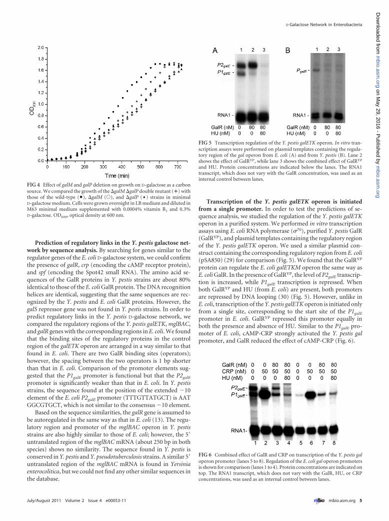
FIG 4 Effect of galM and galP deletion on growth on D-galactose as a carbonsource. We compared the growth of the �galM �galP double mutant (�) withthose of the wild-type (y), �galM (Œ), and �galP (‘) strains in minimalD-galactose medium. Cells were grown overnight in LB medium and diluted inM63 minimal medium supplemented with 0.0004% vitamin B1 and 0.3%D-galactose. OD600, optical density at 600 nm.
FIG 5 Transcription regulation of the Y. pestis galETK operon. In vitro tran-scription assays were performed on plasmid templates containing the regula-tory region of the gal operon from E. coli (A) and from Y. pestis (B). Lane 2shows the effect of GalRYP, while lane 3 shows the combined effect of GalRYP
and HU. Protein concentrations are indicated below the lanes. The RNA1transcript, which does not vary with the GalR concentration, was used as aninternal control between lanes.
FIG 6 Combined effect of GalR and CRP on transcription of the Y. pestis galoperon promoter (lanes 5 to 8). Regulation of the E. coli gal operon promotersis shown for comparison (lanes 1 to 4). Protein concentrations are indicated ontop. The RNA1 transcript, which does not vary with the GalR, HU, or CRPconcentrations, was used as an internal control between lanes.
D-Galactose Network in Enterobacteria
July/August 2011 Volume 2 Issue 4 e00053-11 ® mbio.asm.org 5
m
bio.asm.org
on May 29, 2016 - P
ublished by m
bio.asm.org
Dow
nloaded from

DISCUSSIONFunctional diversity of the D-galactose network in enterobacte-ria. The functional role of different genes in the D-galactose utili-zation network of E. coli has been studied extensively. Therefore,based on the set of genes present in a certain reduced network, it ispossible to formulate predictions about the functional conse-quences of reduction. Functional diversity of the reduced net-works affects both transport and metabolism. Some networkscontain only the low-affinity transporter (5), while others haveonly the high-affinity transport system (6), suggesting optimalperformance in high-galactose and low-galactose environments,respectively. In four cases, we found that both of these transport-ers are missing; therefore, the function of the galactose system islimited to endogenous inducer synthesis (31) and metabolism ofintracellular D-galactose obtained from galactose-containingcompounds. Galactose metabolism is affected in two cases, as fol-lows. In the S. glossinidius network, only the galE gene of the galoperon is functional, indicating that this network is incapable ofamphibolic utilization of D-galactose. In Y. pestis, the last gene ofthe gal operon, galM, is inactivated by a single-base-pair deletion.
D-Galactose utilization in Y. pestis. To understand the conse-quences of galM inactivation, we identified elements and interac-tions in the D-galactose network of Y. pestis by combining exper-imental results with the results of bioinformatical analyses (Fig. 7).The Y. pestis network is less complex than the E. coli D-galactosenetwork. Both the number of elements and the number of inter-action links are reduced. The Y. pestis D-galactose system is regu-lated by a single D-galactose-responsive regulator, which is inter-changeable with E. coli GalR in in vitro experiments. D-Galactosetransport of Y. pestis strains is also simplified, having only thehigh-affinity Mgl transport system, which can transportD-glucose, D-galactose, and �-methylgalactosides in E. coli.
Our results suggest that the Y. pestis D-galactose network, un-
like that in E. coli, can utilize �-D-galactose but not �-D-galactose. Based onthe comparative network analysis pre-sented in this work, we can formulate pre-dictions about the potential D-galactose-related environments and the function ofthe D-galactose network in such environ-ments. Our results show that the simpli-fied network found in Y. pestis is less com-petitive in utilizing large extracellularD-galactose pools; however, it can useD-galactose when present at a constantlow level due to the presence of the high-affinity Mgl transport system. This sug-gests that niches occupied by Y. pestis(e.g., arthropod vector, macrophages,and human blood) (32) are generallypoor in D-galactose. Also, from the ab-sence of galS we can conclude that extra-cellular D-galactose levels are less dynamicin these niches than in the niches occu-pied by E. coli (33).
The D-galactose system is also involvedin the utilization of intracellularD-galactose obtained from the degrada-tion of oligosaccharides (e.g., lactose andmelibiose) (14). Because galM is inacti-
vated in the Y. pestis network, intracellular degradation of �-D-galactose- and �-D-galactose-containing compounds could havedifferent effects. The system can utilize intracellular �-D-galactoseas a carbon source and also to provide building blocks for biosyn-thetic glycosylation. For example, unlike E. coli, Y. pestis has agalactan transport and utilization system which can degrade ex-tracellular galactan into smaller oligomers that are transportedand processed to �-D-galactose oligomers inside the cell (34). TheMgl transport system can transport �-D-galactosides (e.g.,methyl-�-D-galactoside and D-glycerol-�-D-galactoside) (35).However, �-D-galactose obtained from intracellular degradationof such compounds could induce the gal regulon genes but wouldnot be utilized by the system. Induction of the galETK operon by anonmetabolized inducer in the absence of �-D-galactose can servebiosynthetic purposes because GalE can catalyze the productionof UDP-galactose (from UDP-glucose), which is required for bio-synthetic glycosylation (Fig. 7). Such metabolic flow control in theamphibolic D-galactose pathway is common in enterobacteria.Synthesis of D-galactose-containing polysaccharides (e.g., in LPS)is often required for pathogenesis (8–10), and the need for UDP-galactose is independent of D-galactose availability. Differentmechanisms have been reported so far, which take advantage ofthe fact that only the galE gene product is involved in makingsubstrates for biosynthetic glycosylation reactions, and all of thegal enzymes are needed for catabolism of the sugar D-galactose. Acommon mechanism in enterobacteria is discoordinated expres-sion of the gal operon genes. This can be achieved by using a smallregulatory RNA (Spot42) which does not affect translation of GalEand GalT but blocks GalK production (22) or by transcribing thegal operon from two different promoters, one of which transcribesmostly the first gene of the operon, galE, because of natural polar-ity (19–21, 23). In certain pathogenic enterobacteria, the galE geneis physically separated from the other gal operon genes, and its
FIG 7 Predicted D-galactose network in Y. pestis. Vast grey arrows show major metabolic flows (Leloirpathway). Grey ellipses designate intracellular D-galactose and galactoside pools containing �- and�-D-galactose anomers. Red lines indicate transcriptional regulation. The blue line indicates Spot42RNA-mediated translational control. Other enhancements and inhibitions are indicated by green lines.
Csiszovszki et al.
6 ® mbio.asm.org July/August 2011 Volume 2 Issue 4 e00053-11
m
bio.asm.org
on May 29, 2016 - P
ublished by m
bio.asm.org
Dow
nloaded from

expression is independent of intracellular D-galactose levels (9).Intracellular synthesis of a nonmetabolized inducer constitutes anovel mechanism for regulation of the amphibolic D-galactosepathway. The observation that utilization of intracellular �-D-galactose by the Y. pestis network can be turned on by single-base-pair insertions in galM suggests that the galM pseudogene has notbeen deleted in evolution because its reactivation is beneficial incertain environments.
MATERIALS AND METHODSBacterial strains. Genome sequences of 34 strains were involved in thebioinformatical analysis. The strains used are the following: Citrobacterkoseri ATCC BAA-895, Citrobacter rodentium ICC168, Cronobacter saka-zakii ATCC BAA-894, Cronobacter turicensis z3032, Dickeya dadantii3937, Dickeya zeae Ech 1591, Edwardsiella ictaluri 93-146, Edwardsiellatarda EIB202, Enterobacter cloacae subsp. cloacae ATCC 13047, Enterobac-ter sp. strain 638, Erwinia amylovora CFBP1430, Erwinia billingiae Eb661,Erwinia pyrifoliae Ep1/96, Erwinia tasmaniensis Et1/99, Escherichia coliMG1655, Escherichia fergusonii ATCC 35469, Klebsiella pneumoniae 342,Klebsiella variicola At-22, Pantoea ananatis LMG 20103, Pantoea vagansC9-1, Pectobacterium atrosepticum SCRI1043, Pectobacterium carotovo-rum PC1, Pectobacterium wasabiae WPP163, Proteus mirabilis HI4320,Salmonella enterica serovar Typhimurium strain LT2, Serratia pro-teamaculans 568, Shigella boydii CDC3083-94, Shigella dysenteriae Sd197,Shigella flexneri 5 strain 8401, Shigella sonnei Ss046, Sodalis glossinidiusstrain “morsitans,” Yersinia enterocolitica subsp. enterocolitica 8081, Yer-sinia pestis KIM10, and Yersinia pseudotuberculosis IP32953.
E. coli MG1655�galM87::Kanr was described by Bouffard et al. (14).MG1655�galP::cmr and MG1655�galM::Kanr�galP::cmr are derivativesof E. coli MG1655 and MG1655�galM87::Kanr, respectively. The chlor-amphenicol resistance cassette was inserted into the galP gene by recom-bineering (36). The cat gene from plasmid pRFB122 (37) was amplifiedusing the primers GalPupcat (5=-CTCACCTATCTAATTCACAATAAAAAATAACCATATTGGAGGGCATCAAAATGAGACGTTGATCGGCACGTAAGA-3=) and GalPdowncat (5=-CCTCCGCGATGGGAGGAAGCTTGGGGAGATTAATCGTGAGCGCCTATTTCTTACGCCCCGCCCTGCCACTCATCGCAG-3=). Recombineering was performed according tothe protocol described by Datsenko and Wanner (38).
DNA manipulation methods. Bacterial growth conditions and plas-mid manipulations followed the protocols described by Sambrook andRussell (39). Transformations were performed with chemically compe-tent XL1-Blue cells (Stratagene). Restriction endonucleases and DNA oli-gonucleotide primers were purchased from Invitrogen, PCR (GeneAmpXL) and sequencing (ABI Prism) kits from Applied Biosystems, and DNApurification kits from Qiagen. DNA sequencing reactions were analyzedin a PerkinElmer/Applied Biosystems (model 373 A) automated se-quencer.
Plasmid construction. Plasmid pSA850YP was made by insertingthe Y. pestis galETK regulatory region between the EcoRI and PstI sites ofplasmid pSA850 (29). Because of safety considerations, the DNA frag-ment corresponding to the chromosomal region 3350221 to 3350532 ofY. pestis KIM (GenBank accession no. NC_004088.1) was amplifiedfrom Yersinia pseudotuberculosis ATCC 23207, which contains an identi-cal sequence. The primers used for amplification were YPP1 (5=-AAAAGAATTCGCGCACCACAAACAGGACATTCC) and YPP2 (5=-AAAACTGCAGCAATTGCACACAGGTATGGCTACC). The sequenceof the Y. pestis galETK regulatory region in plasmid pSA850YP wasverified.
The plasmid pSEM1026YP for the expression of Y. pestis GalR wascreated by amplifying the galR gene from Yersinia pseudotuberculosisATCC 23207 using primers YPGNCO (5=-AAAACCATGGCCACTATAAAGGATGTTGCCAAGCT) and YPGCTBI (5=-AAAAGGATCCATCAGTGTCATCCCGTAGGCTTGGC) and inserting it between the NcoIand BamHI sites of plasmid pSEM1026 (40). The cloned Y. pseudotuber-
culosis galR gene is 99% identical to the sequence of Y. pestis galR, while theamino acid sequences of the encoded proteins are identical.
The galM pseudogene of Y. pestis (ATCC BAA-1504) was PCR ampli-fied from a genomic DNA preparation (purchased from ATCC) using theprimers YP_Mup_XhoI (5=-TTTTCTCGAGGTTCGCACCACCGTTGCGCAAGAATAC-3=) and YP_Mdn_Acc65I (5=-TTTTGGTACCATCATTCATAACGTCATCATTCATAAC-3=). The resulting DNA fragment wasdigested with XhoI and Acc65I and inserted between the XhoI andAcc65I sites of the low-copy-number plasmid pLG338E, which was de-rived from pLG338 (41) by eliminating the EcoRI site. The open readingframe of galM was restored by 8 different base pair insertions, using PCRmutagenesis with four designed primers and the previously usedYP_Mdn_Acc65I primer. The designed primers are Yps_D (5=-TTTTGAATTCACCAAATTGCAGAATAAAAGCGGTATGACCGTCACCTTTATGGATTGGGGGGCAACCTGGTTATCGGCCATAT-3=), Yps_EKQ(5=-TTTTGAATTCACCAAATTGCAGAATAAAAGCGGTATGACCGTCACCTTTATGVAATGGGGGGCAACCTGGTTATCGGCCATAT-3=),Yps_N (5=-TTTTGAATTCACCAAATTGCAGAATAAAAGCGGTATGACCGTCACCTTTATGAACTGGGGGGCAACCTGGTTATCGGCCATAT-3=), and Yps_ITR (5=-TTTTGAATTCACCAAATTGCAGAATAAAAGCGGTATGACCGTCACCTTTATGABATGGGGGGCAACCTGGTTATCGGCCATATTACCGCTGAAAAAT-3=). The ampli-fied fragments were used to replace the EcoRI-Acc65I fragment in thepLG338 plasmid containing the galM pseudogene. The DNA sequence ofthe inserted fragment was verified in all the constructs created.
Protein purification. Expression and purification of the hexa-histidine-tagged Y. pestis GalR followed the protocol described before forE. coli GalR purification (40). HU protein was purified according to themethod described by Aki et al. (42). CRP was purified as described by Ryuet al. (43).
In vitro transcription. Transcription reactions were performed asdescribed previously (16). The reaction mixture (50 �l) contained 20 mMTris acetate at pH 7.8, 10 mM magnesium acetate, 200 mM potassiumglutamate, 100 �M cAMP, and 2 nM supercoiled plasmid DNA template.GalR and CRP concentrations are as indicated in Fig. 5 and 6. HU wasused at an 80 nM concentration when present. Twenty nanomolar RNApolymerase (USB) was added before incubation of the reactions at 37°Cfor 5 minutes. Transcription was started by the addition of 1.0 mM ATP,0.1 mM GTP, 0.1 mM CTP, 0.01 mM UTP, and 5 �Ci of [�-32P]UTP(3,000 Ci/mmol). Reactions were terminated after 10 minutes by additionof an equal volume of transcription loading buffer (0.025% bromophenolblue, 0.025% xylene cyanol, 0.01 M EDTA, and 90% deionized forma-mide). After heating at 90°C for 3 minutes, the samples were loaded onto7% polyacrylamide-urea DNA sequencing gels. RNA bands were quanti-fied using the ImageQuant PhosphorImager (Molecular Dynamics, CA).Band intensities were corrected by the background and normalized to theRNA1 band intensities of the corresponding lanes.
ACKNOWLEDGMENTS
We thank our colleagues for various input on the project, particularlyThomas Soares and Mofang Liu for purification of HU and CRP andTakácsné Botond Judit for technical assistance.
This research was supported by the Hungarian Scientific ResearchFund (OTKA) grant PD75496 to S.S., by the Danish Council for Indepen-dent Research�Natural Sciences, by the Intramural Research Program ofthe NIH, National Cancer Institute, Center for Cancer Research, UnitedStates, and by the Danish National Research Foundation.
REFERENCES1. Navid A, Almaas E. 2009. Genome-scale reconstruction of the metabolic
network in Yersinia pestis, strain 91001. Mol. Biosyst. 5:368 –375.2. Hacker J, Kaper JB. 2000. Pathogenicity islands and the evolution of
microbes. Annu. Rev. Microbiol. 54:641– 679.3. Sneppen K, Krishna S, Semsey S. 2010. Simplified models of biological
networks. Annu. Rev. Biophys. 39:43–59.4. Hunziker A, Tuboly C, Horváth P, Krishna S, Semsey S. 2010. Genetic
D-Galactose Network in Enterobacteria
July/August 2011 Volume 2 Issue 4 e00053-11 ® mbio.asm.org 7
m
bio.asm.org
on May 29, 2016 - P
ublished by m
bio.asm.org
Dow
nloaded from

flexibility of regulatory networks. Proc. Natl. Acad. Sci. U. S. A. 107:12998 –13003.
5. Mayo AE, Setty Y, Shavit S, Zaslaver A, Alon U. 2006. Plasticity of thecis-regulatory input function of a gene. PLoS Biol. 4:e45.
6. Achtman M, et al. 1999. Yersinia pestis, the cause of plague, is a recentlyemerged clone of Yersinia pseudotuberculosis. Proc. Natl. Acad. Sci. U. S. A.96:14043–14048.
7. Dekel E, Mangan S, Alon U. 2005. Environmental selection of the feed-forward loop circuit in gene-regulation networks. Phys. Biol. 2:81– 88.
8. Maskell DJ, Szabo MJ, Deadman ME, Moxon ER. 1992. The gal locusfrom Haemophilus influenzae: cloning, sequencing and the use of gal mu-tants to study lipopolysaccharide. Mol. Microbiol. 6:3051–3063.
9. Metzger M, Bellemann P, Bugert P, Geider K. 1994. Genetics of galac-tose metabolism of Erwinia amylovora and its influence on polysaccharidesynthesis and virulence of the fire blight pathogen. J. Bacteriol. 176:450 – 459.
10. Robertson BD, Frosch M, van Putten JP. 1993. The role of galE in thebiosynthesis and function of gonococcal lipopolysaccharide. Mol. Micro-biol. 8:891–901.
11. Motin VL, et al. 2004. Temporal global changes in gene expression duringtemperature transition in Yersinia pestis. J. Bacteriol. 186:6298 – 6305.
12. Knirel YA, et al. 2006. Structural features and structural variability of thelipopolysaccharide of Yersinia pestis, the cause of plague. J. Endotoxin Res.12:3–9.
13. Semsey S, et al. 2007. Signal integration in the galactose network ofEscherichia coli. Mol. Microbiol. 65:465– 476.
14. Bouffard GG, Rudd KE, Adhya SL. 1994. Dependence of lactose metab-olism upon mutarotase encoded in the gal operon in Escherichia coli.J. Mol. Biol. 244:269 –278.
15. Wilson DB. 1974. Source of energy for the Escherichia coli galactose trans-port systems induced by galactose. J. Bacteriol. 120:866 – 871.
16. Geanacopoulos M, Adhya S. 1997. Functional characterization of roles ofGalR and GalS as regulators of the gal regulon. J. Bacteriol. 179:228 –234.
17. Weickert MJ, Adhya S. 1993. Control of transcription of gal repressor andisorepressor genes in Escherichia coli. J. Bacteriol. 175:251–258.
18. Lee SJ, Lewis DE, Adhya S. 2008. Induction of the galactose enzymes inEscherichia coli is independent of the C-1-hydroxyl optical configurationof the inducer D-galactose. J. Bacteriol. 190:7932–7938.
19. Adhya S. 2003. Suboperonic regulatory signals. Sci. STKE 2003:pe22.20. Lee HJ, Jeon HJ, Ji SC, Yun SH, Lim HM. 2008. Establishment of an
mRNA gradient depends on the promoter: an investigation of polarity ingene expression. J. Mol. Biol. 378:318 –327.
21. Semsey S, Virnik K, Adhya S. 2006. Three-stage regulation of the am-phibolic gal operon: from repressosome to GalR-free DNA. J. Mol. Biol.358:355–363.
22. Møller T, Franch T, Udesen C, Gerdes K, Valentin-Hansen P. 2002.Spot 42 RNA mediates discoordinate expression of the E. coli galactoseoperon. Genes Dev. 16:1696 –1706.
23. Ullmann A, Joseph E, Danchin A. 1979. Cyclic AMP as a modulator ofpolarity in polycistronic transcriptional units. Proc. Natl. Acad. Sci.U. S. A. 76:3194 –3197.
24. Herrgård MJ, Covert MW, Palsson BØ. 2004. Reconstruction of micro-bial transcriptional regulatory networks. Curr. Opin. Biotechnol. 15:70 –77.
25. Altschul SF, Gish W, Miller W, Myers EW, Lipman DJ. 1990. Basic localalignment search tool. J. Mol. Biol. 215:403– 410.
26. Wren BW. 2003. The Yersiniae—a model genus to study the rapid evolu-tion of bacterial pathogens. Nat. Rev. Microbiol. 1:55– 64.
27. Lerat E, Ochman H. 2005. Recognizing the pseudogenes in bacterialgenomes. Nucleic Acids Res. 33:3125–3132.
28. Brown MP, Shaikh N, Brenowitz M, Brand L. 1994. The allostericinteraction between D-galactose and the Escherichia coli galactose repres-sor protein. J. Biol. Chem. 269:12600 –12605.
29. Lewis DE, Adhya S. 2004. Axiom of determining transcription startpoints by RNA polymerase in Escherichia coli. Mol. Microbiol. 54:692–701.
30. Choy HE, Adhya S. 1992. Control of gal transcription through DNAlooping: inhibition of the initial transcribing complex. Proc. Natl. Acad.Sci. U. S. A. 89:11264 –11268.
31. Death A, Ferenci T. 1994. Between feast and famine: endogenous inducersynthesis in the adaptation of Escherichia coli to growth with limiting car-bohydrates. J. Bacteriol. 176:5101–5107.
32. Oyston PC, Isherwood KE. 2005. The many and varied niches occupiedby Yersinia pestis as an arthropod-vectored zoonotic pathogen. AntonieVan Leeuwenhoek 87:171–177.
33. Semsey S, et al. 2009. Dominant negative autoregulation limits steady-state repression levels in gene networks. J. Bacteriol. 191:4487– 4491.
34. Delangle A, et al. 2007. Characterization of the Erwinia chrysanthemi Ganlocus, involved in galactan catabolism. J. Bacteriol. 189:7053–7061.
35. Wilson DB. 1976. Properties of the entry and exit reactions of the beta-methyl galactoside transport system in Escherichia coli. J. Bacteriol. 126:1156 –1165.
36. Court DL, Sawitzke JA, Thomason LC. 2002. Genetic engineering usinghomologous recombination. Annu. Rev. Genet. 36:361–388.
37. Fekete RA, Chattoraj DK. 2005. A cis-acting sequence involved in chro-mosome segregation in Escherichia coli. Mol. Microbiol. 55:175–183.
38. Datsenko KA, Wanner BL. 2000. One-step inactivation of chromosomalgenes in Escherichia coli K-12 using PCR products. Proc. Natl. Acad. Sci.U. S. A. 97:6640 – 6645.
39. Sambrook J, Russell DW. 2001. Molecular cloning: a laboratory manual.Cold Spring Harbor Laboratory Press, Cold Spring Harbor, NY.
40. Semsey S, Geanacopoulos M, Lewis DE, Adhya S. 2002. Operator-bound GalR dimers close DNA loops by direct interaction: tetrameriza-tion and inducer binding. EMBO J. 21:4349 – 4356.
41. Stoker NG, Fairweather NF, Spratt BG. 1982. Versatile low-copy-number plasmid vectors for cloning in Escherichia coli. Gene 18:335–341.
42. Aki T, Choy HE, Adhya S. 1996. Histone-like protein HU as a specifictranscriptional regulator: co-factor role in repression of gal transcriptionby GAL repressor. Genes Cells 1:179 –188.
43. Ryu S, Kim J, Adhya S, Garges S. 1993. Pivotal role of amino acid atposition 138 in the allosteric hinge reorientation of cAMP receptor pro-tein. Proc. Natl. Acad. Sci. U. S. A. 90:75–79.
Csiszovszki et al.
8 ® mbio.asm.org July/August 2011 Volume 2 Issue 4 e00053-11
m
bio.asm.org
on May 29, 2016 - P
ublished by m
bio.asm.org
Dow
nloaded from
Copyright © 2022 FDOKUMEN