Simultaneous Amplification, Detection, and Analysis of Common Mutations in the Galactose-1-Phosphate...
6
JMD CME Program Simultaneous Amplification, Detection, and Analysis of Common Mutations in the Galactose-1-Phosphate Uridyl Transferase Gene Mohamed Jama,* Lesa Nelson, † Rong Mao,* ‡ and Elaine Lyon * ‡ From the ARUP Institute for Clinical and Experimental Pathology,* Salt Lake City; Axial Biotech, Inc., † Salt Lake City; and the Department of Pathology, ‡ University of Utah, Salt Lake City, Utah Classic galactosemia is an autosomal recessive inherited error of galactose metabolism. It is caused by lack of galactose-1-phosphate uridyl transferase , an enzyme that is required to metabolize galactose-1-phosphate to uridine diphosphate galactose. The build up of galac- tose-1-phosphate is toxic at high levels and can damage the liver , brain , eyes , and other vital organs. Over 200 mutations have been identified in affected individuals. We describe an assay to identify nine target mutations or variants in the galactose-1-phosphate uridyl trans- ferase gene , namely p.Q188R , p.S135L , p.K285N , p.L195P , p.T138M , p.Y209C , IVS2-2 A>G, p.L218L, and p.N314D. A single long-range PCR is followed by a mul- tiplexed nucleotide extension assay (single nucleotide extension) and capillary electrophoresis to detect si- multaneously all nine target mutations/variants. Fifty- four previously characterized samples (47 clinical sam- ples and seven controls) gave a 100% concordance. We also report a nontarget novel mutation , p.L192X , and its profile using single nucleotide extension. This assay can complement the enzyme activity assay and identify familial mutations for testing additional family members. (J Mol Diagn 2007, 9:618 – 623; DOI: 10.2353/jmoldx.2007.070027) Classic galactosemia is an inherited metabolic disorder caused by a deficiency of galactose-1-phosphate uridyl transferase (GALT), an enzyme required to convert galac- tose-1-phosphate to uridine diphosphate galactose. 1–4 The resulting buildup of galactose-1-phosphate leads to poor feeding, vomiting, diarrhea, and cataracts, and if left un- treated, results in physical and mental retardation. 5–8 Over 200 mutations have been identified in patients affected with galactosemia (http://arup.utah.edu/database/galactosemia/ GALT_welcome.php). The six common mutations p.Q188R (c.563AG), p.S135L (c.404 CT), p.K285N (c.855GT), p.L195P (c.584TC), p.T138M (c.413 CT), and p.Y209C (c.626 AG) account for 80% of cases with classic galac- tosemia. A seventh mutation, IVS2-2 AG (c.253-2AG), has been described in a Hispanic population. 9 These mu- tations are considered severe or classic mutations resulting in little or no enzyme activity. Two variants, Los Angeles (LA or Duarte 1) and Duarte, also affect enzyme activity. The LA variant p.L218L (c.652CT) in cis with the p.N314D (c.940AG) increases the enzyme activity to approximately 130%. 10 The Duarte (D) variant (p.N314D, c.940AG) re- duces enzyme activity by approximately 25% and is found in approximately 5% of the general US population. 11,12 This variant is also in cis with the deletion c.-119--116delGTCA in the 5-untranslated region of the GALT gene, thus also affecting a regulatory region. 13,14 The combination of a classic mutation (G) and the D variant shows a decreased enzyme activity but does not typically result in symptoms of classic galactosemia. Galactosemia screening is widely performed on new- borns. Blood specimens are collected on Guthrie cards and assayed for galactose and its metabolite, galactose 1-phosphate, or GALT enzyme levels. 3,5,15,16 Abnormal screening tests are followed by confirmatory testing us- ing enzymatic activity. 17 The GALT enzyme is unstable, and the assay for enzyme activity is best performed on fresh heparin anticoagulated blood. DNA analysis for common mutations can be used in conjunction with en- zyme activity to confirm familial mutations. Our purpose was to design a practical method to detect the seven GALT mutations and the two variants. We chose the mu- tations and variants based on a Texas newborn popula- tion study. 9 The IVS2-2 mutation was included to increase detection in Hispanic populations. PCR primers were designed to produce a single amplicon that included all mutation and variant positions followed by multiplexed single nucleotide extension (SNE). Materials and Methods Samples As part of the State of Utah newborn galactosemia screening, newborns with moderate to high elevated lev- els of galactose, defined as 20 to 30 mg/dL, were sent for Accepted for publication August 8, 2007. Address reprint requests to Elaine Lyon, ARUP Institute for Clinical and Experimental Pathology, 500 Chipeta Way, Salt Lake City, UT 84108. E-mail: [email protected]. Journal of Molecular Diagnostics, Vol. 9, No. 5, November 2007 Copyright © American Society for Investigative Pathology and the Association for Molecular Pathology DOI: 10.2353/jmoldx.2007.070027 618
-
Upload
independent -
Category
Documents
-
view
2 -
download
0
Transcript of Simultaneous Amplification, Detection, and Analysis of Common Mutations in the Galactose-1-Phosphate...

JMD
CME P
rogra
m
Simultaneous Amplification, Detection, and Analysisof Common Mutations in the Galactose-1-PhosphateUridyl Transferase Gene
Mohamed Jama,* Lesa Nelson,† Rong Mao,*‡ andElaine Lyon *‡
From the ARUP Institute for Clinical and Experimental
Pathology,* Salt Lake City; Axial Biotech, Inc.,† Salt Lake City;
and the Department of Pathology,‡ University of Utah, Salt Lake
City, Utah
Classic galactosemia is an autosomal recessive inheritederror of galactose metabolism. It is caused by lack ofgalactose-1-phosphate uridyl transferase, an enzymethat is required to metabolize galactose-1-phosphate touridine diphosphate galactose. The build up of galac-tose-1-phosphate is toxic at high levels and can damagethe liver, brain, eyes, and other vital organs. Over 200mutations have been identified in affected individuals.We describe an assay to identify nine target mutationsor variants in the galactose-1-phosphate uridyl trans-ferase gene, namely p.Q188R, p.S135L, p.K285N,p.L195P, p.T138M, p.Y209C, IVS2-2 A>G, p.L218L, andp.N314D. A single long-range PCR is followed by a mul-tiplexed nucleotide extension assay (single nucleotideextension) and capillary electrophoresis to detect si-multaneously all nine target mutations/variants. Fifty-four previously characterized samples (47 clinical sam-ples and seven controls) gave a 100% concordance.We also report a nontarget novel mutation, p.L192X,and its profile using single nucleotide extension.This assay can complement the enzyme activity assayand identify familial mutations for testing additionalfamily members. (J Mol Diagn 2007, 9:618–623; DOI:10.2353/jmoldx.2007.070027)
Classic galactosemia is an inherited metabolic disordercaused by a deficiency of galactose-1-phosphate uridyltransferase (GALT), an enzyme required to convert galac-tose-1-phosphate to uridine diphosphate galactose.1–4 Theresulting buildup of galactose-1-phosphate leads to poorfeeding, vomiting, diarrhea, and cataracts, and if left un-treated, results in physical and mental retardation.5–8 Over200 mutations have been identified in patients affected withgalactosemia (http://arup.utah.edu/database/galactosemia/GALT_welcome.php). The six common mutations p.Q188R(c.563A�G), p.S135L (c.404 C�T), p.K285N (c.855G�T),p.L195P (c.584T�C), p.T138M (c.413 C�T), and p.Y209C(c.626 A�G) account for 80% of cases with classic galac-
tosemia. A seventh mutation, IVS2-2 A�G (c.253-2A�G),has been described in a Hispanic population.9 These mu-tations are considered severe or classic mutations resultingin little or no enzyme activity. Two variants, Los Angeles (LAor Duarte 1) and Duarte, also affect enzyme activity. The LAvariant p.L218L (c.652C�T) in cis with the p.N314D(c.940A�G) increases the enzyme activity to approximately130%.10 The Duarte (D) variant (p.N314D, c.940A�G) re-duces enzyme activity by approximately 25% and is foundin approximately 5% of the general US population.11,12 Thisvariant is also in cis with the deletion c.-119-�-116delGTCAin the 5�-untranslated region of the GALT gene, thus alsoaffecting a regulatory region.13,14 The combination of aclassic mutation (G) and the D variant shows a decreasedenzyme activity but does not typically result in symptoms ofclassic galactosemia.
Galactosemia screening is widely performed on new-borns. Blood specimens are collected on Guthrie cardsand assayed for galactose and its metabolite, galactose1-phosphate, or GALT enzyme levels.3,5,15,16 Abnormalscreening tests are followed by confirmatory testing us-ing enzymatic activity.17 The GALT enzyme is unstable,and the assay for enzyme activity is best performed onfresh heparin anticoagulated blood. DNA analysis forcommon mutations can be used in conjunction with en-zyme activity to confirm familial mutations. Our purposewas to design a practical method to detect the sevenGALT mutations and the two variants. We chose the mu-tations and variants based on a Texas newborn popula-tion study.9 The IVS2-2 mutation was included to increasedetection in Hispanic populations. PCR primers weredesigned to produce a single amplicon that included allmutation and variant positions followed by multiplexedsingle nucleotide extension (SNE).
Materials and Methods
Samples
As part of the State of Utah newborn galactosemiascreening, newborns with moderate to high elevated lev-els of galactose, defined as 20 to 30 mg/dL, were sent for
Accepted for publication August 8, 2007.
Address reprint requests to Elaine Lyon, ARUP Institute for Clinical andExperimental Pathology, 500 Chipeta Way, Salt Lake City, UT 84108.E-mail: [email protected].
Journal of Molecular Diagnostics, Vol. 9, No. 5, November 2007
Copyright © American Society for Investigative Pathology
and the Association for Molecular Pathology
DOI: 10.2353/jmoldx.2007.070027
618

confirmatory enzyme activity levels and DNA mutationanalysis. DNA was extracted by the MagNaPure instru-ment (Roche, Indianapolis, IN). DNA from 47 de-identi-fied clinical samples previously characterized by ARUPLaboratories (by enzyme activity and/or allele-specificPCR) was tested following an Institutional Review Board-approved protocol for test validation. Six additionalblinded DNA samples previously characterized by theDepartment of Genetics, Emory University School ofMedicine, and known to contain seven mutations and onevariant were also tested. An artificial template was syn-thesized for IVS 2-2A�G heterozygous mutation, for atotal of 54 samples.
Artificial Template for IVS2-2 A�G
To generate the artificial template for IVS2-2 A�G muta-tion18–20 (Figure 1 and Table 1), two oligodeoxyribo-nucleotides of 90 and 91 bases that overlapped by 30 bpwere used to introduce the IVS2-2 G mutation. In addi-tion, two short primers (IVS2-2 forward and IVS2-2 re-verse) were used in the same reaction. This allowedgeneration of the template by overlapping oligodeoxyri-bonucleotides followed by exponential amplification withthe short primers. A 20-�l PCR was performed with 0.5�mol/L of each of the four oligodeoxyribonucleotides, 1�FailSafe Premix D (Epicenter, Madison, WI), and 1.0 unitof FastStart DNA polymerase (Roche). Amplification wasperformed with an initial denaturation at 95°C for 5 min-utes, followed by five cycles of denaturation at 94°C for30 seconds, annealing at 65°C for 30 seconds, and ex-tension at 72°C for 30 seconds to generate the template.This was followed by 30 cycles of denaturation at 94°C for30 seconds, annealing at 55°C for 30 seconds, and ex-tension at 72°C for 30 seconds. Unincorporated primersand dNTPs were removed by incubating with ExoSAP(USB Corporation, Cleveland, OH) as described below.
To mimic a heterozygous sample, the purified artificialtemplate was mixed with a wild-type genomic DNA.
PCR Amplification
PCR was performed using GeneAmp 9700 thermal reac-tion cycler (Applied Biosystems, Foster City, CA). A 20-�lPCR mixture contained 2 �l of extracted DNA (15 to 50ng/�l), 1.0 �mol/L of each forward and reverse primer,5�-GCCTGTCCAGTCTTTGAAGC-3� and 5�-CATTTCG-TAGCCAACCATGA-3� [designed using Primer 321
(http://frodo.wi.mit.edu/cgi-bin/primer3/primer3_www.cgi)], 1� FailSafe Premix D containing PCRbuffer, dNTPs, and MgCl2 (Epicenter), and 1.0 unit ofFastStart DNA polymerase (Roche). Amplification wasperformed with an initial denaturation at 95°C for 5 min-utes, followed by 35 cycles of denaturation at 94°C for 1minute, annealing at 55°C for 1 minute, and extension at72°C for 3 minutes. Unincorporated primers and dNTPswere removed by incubating with 2 units of ExoSAP (USBCorporation) at 37°C for 45 minutes, followed by heatinactivation of enzyme at 80°C for 15 minutes.
Allele-Specific Oligodeoxyribonucleotide SingleNucleotide Extension
Nine extension primers (Integrated DNA Technologies,Inc., Coralville, IA) specific for each mutation were de-signed to hybridize to either the sense or antisensestrand (Table 2). The primers were designed with varyinglengths (4 to 5 bp) to adequately separate the multi-plexed extended products by capillary electrophoresis.Oligodeoxyribonucleotides longer than 50 bases werepurified by high-pressure liquid chromatography. Con-centrations were empirically optimized and ranged from0.1 to 0.4 �mol/L. Two microliters of purified PCR prod-ucts, 2 �l of the nine pooled allele-specific oligodeoxyri-bonucleotides extension primers, and 2.5 �l of SNaPshotMultiplex reagent were used to perform a 10-�l SNEassay according to the manufacturer’s protocol (AppliedBiosystems). The primers were extended by adding asingle fluorescently labeled ddNTP to its 3�-end.22–25
Fluorescent dyes attached to each ddNTP are as follows:dR6G (green) for A, dTAMRA (yellow, which appearsblack on the electropherogram) for C, dR110 (blue) for G,and dROX (red) for T. The reactions were cycled with thefollowing conditions: 25 cycles at 96°C for 10 seconds,50°C for 5 seconds, and 60°C for 30 seconds. Sampleswere treated with one unit of shrimp alkaline phosphataseat 37°C for 45 minutes, followed by heat inactivation ofenzyme at 80°C for 15 minutes.
Figure 1. Schematic of primer locations for the generation of artificial tem-plate for IVS 2-2 A�G mutation. A: IVS 2-2 forward primer; B: IVS 2-2 reverseprimer; C: artificial oligodeoxyribonucleotides template forward primer; andD: artificial oligodeoxyribonucleotides template reverse primer. “X” indicatesthe position of the mutation.
Table 1. Oligodeoxyribonucleotide Design for Artificial Template IVS2-2A�G Mutation
Artificial template forward-IVS2-2F (90 bp)5�-CAGGTAACTGGTGGTATGGGGCAGTGAGTGCTTCTAGCCTATCCTTGTCGGTGGGTGAATCCCCAGTACGATAGCACCTTCCTGTTTGAC-3�Artificial template reverse-IVS2-2R (91 bp)5�-AGCTAGTTGGAGCCAGGTTACCTGGACTGGGGGCATCAGGCTGCAGAGCTGGGAAGTCGTTGTCAAACAGGAAGGTGCTATCGTACTGGGG-3�
The mutation is shown in underline bold italic; the overlapping region is in bold; and short primers are underlined.
Analysis of Common Mutations in GALT 619JMD November 2007, Vol. 9, No. 5
Two microliters of the fluorescently labeled fragments,0.25 �l of GeneScan 120 LIZ internal size standard (Ap-plied Biosystems), and 8 �l of HiDi formamide (AppliedBiosystems) were resolved by electrophoresis on an au-tomated ABI Prism 3100 Genetic Analyzer using POP-6(performance-optimized polymer) on a 50-cm array (Ap-plied Biosystems). Samples were electrokinetically in-jected at 15 kV for 10 seconds and electrophoresed at 15kV for 1960 seconds at 55°C under filter set E5. Raw datawere analyzed with GeneMapper 4.0 (Applied Biosys-tems) programed for automated calling of both normaland mutant alleles based on fragment size and fluores-cence. Allele calls on ABI’s GeneMapper software wereautomated by calculating the mean and SD of the allelesizes between runs and setting a minimum and maximumsize range for each marker. A representative sample ofeach genotype was confirmed by sequencing (data notshown). For an abnormal sample that did not size withinthe expected range, bidirectional sequencing wasperformed.
Results
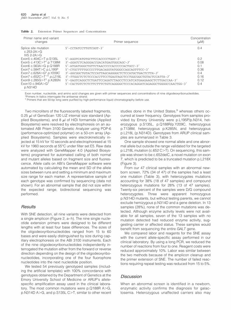
With SNE detection, all nine variants were detected froma single amplicon (Figure 2, a–h). The nine single nucle-otide extension primers were designed to be differentlengths with at least four base differences. The sizes ofthe oligodeoxyribonucleotides ranged from 15 to 60bases and were easily distinguished by size during cap-illary electrophoresis on the ABI 3100 instruments. Eachof the nine oligodeoxyribonucleotides independently in-terrogated the mutation either from the forward or reversedirection depending on the design of the oligodeoxyribo-nucleotides, incorporating one of the four fluorophorenucleotides into the next nucleotide position.
We tested 54 previously genotyped samples (includ-ing the artificial template) with 100% concordance withgenotypes obtained by the Department of Genetics at theEmory University School of Medicine or ARUP’s allele-specific amplification assay used in the clinical labora-tory. The most common mutations were p.Q188R A�G,p.N314D A�G, and p.S135L C�T, similar to other recent
studies done in the United States,9 whereas others oc-curred at lower frequency. Genotypes from samples pro-vided by Emory University were p.L195P/p.N314, het-erozygous p.S135L, p.Q188R/p.Y209C, heterozygousp.T138M, heterozygous p.K285N, and heterozygousp.L218L (p.N314D). Genotypes from ARUP clinical sam-ples are summarized in Table 3.
One sample showed one normal allele and one abnor-mal allele but outside the range validated for the targetedp.L218L mutation (c.652 C�T). On sequencing, this sam-ple was shown to be c.652delC, a novel mutation on exon7, which is predicted to be a truncated mutation p.L218X(Figure 3).
From our 47 clinical samples with an abnormal new-born screen, 72% (34 of 47) of the samples had a leastone mutation (Table 3), with heterozygotes mutationsaccounting for 38% (18 of 47 samples) and compoundheterozygous mutations for 28% (13 of 47 samples).Twenty-six percent of the samples were D/G compoundheterozygotes. Three were apparent homozygousp.N314D mutants, but without testing parents, we cannotexclude hemizygous p.N314D and a gene deletion. In 13samples (28%), none of the common mutations was de-tected. Although enzyme activity levels were not avail-able for all samples, seven of the 13 samples with nomutation detected had reduced enzyme activity, sug-gesting carrier or affected status. These samples wouldbenefit from sequencing the entire GALT gene.
We compared labor and reagents for the SNE assaywith the current allele-specific assay performed in ourclinical laboratory. By using a long PCR, we reduced thenumber of reactions from four to one. Reagent costs werereduced approximately 10%. Labor was similar betweenthe two methods because of the amplicon cleanup andthe primer extension of SNE. The number of failed reac-tions requiring repeat testing was reduced from 15 to 5%.
Discussion
When an abnormal screen is identified in a newborn,enzymatic activity confirms the diagnosis for galac-tosemia. Heterozygous unaffected carriers also may
Table 2. Extension Primer Sequences and Concentrations
Primer name and variantchanges Primer sequence
Concentration(�M)
Splice site mutation 5�-CCTATCCTTGTCGGT-3� 0.4c.253-2A�GIVS 2-2A�G
Exon5 c.404C�T p.S135L 5�-AGGTCATGTGCTTCCACCCCTGGT-3� 0.2Exon5 c.413C�T* p.T138M 5�-GGATCTCAGGGACCGACATGAGTGGCAGC-3� 0.08Exon6 c.563A�G p.Q188R 5�-ATGATGGGCTGTTCTAACCCCCACCCCCACTGCC-3� 0.1Exon7 c.584T�C p.L195P 5�-CTGCTTTTGCCCCTTGACAGGTATGGGCCAGCAGTTTCC-3� 0.36Exon7 c.626A�G* p.Y209C 5�-GGCGGCTGTACTCCATTAGCAGGGGCTCTCCATGCTGACTCTTA-3� 0.4Exon7 c.652C�T *† p.L218L 5�-TTGGCTCTCTCCCACCTTCCTGAGTAGCTCCTGGCGGCTGTACTCCATTA-3� 0.16Exon9 c.285G�T† p.K285N 5�-GAGTCAGGCTCTGATTCCAGATCTAGCCTCCATCATGAAGAAGCTCTTGACCAA-3� 0.12Exon10 c.940A�G†
p.N314D5�-CACTGTCTCTCTTCTTTCTGTCAGGGGCTCCCACAGGATCAGAGGCTGGGGCCAACTGG-3� 0.4
Exon number, nucleotide, and amino acid changes are given with primer sequences and concentrations of nine oligodeoxyribonucleotides.*Primers in italics interrogate the antisense strand.† Primers that are 50-bp long were purified by high-performance liquid chromatography before use.
620 Jama et alJMD November 2007, Vol. 9, No. 5

be identified through newborn screening. Combining amutation panel with enzyme activity identified at leastone family-specific mutation or variant in 72% of thecases. Because galactosemia mutations are ethnic-specific,26 we chose mutations that would represent apan-ethnic population.9,27 Two mutations, p.Q188Rand p.K285N, reportedly account for approximately 69to 88% of galactosemia alleles in Caucasian popula-tions.28 The p.Q188R mutation is considered severe,with no GALT activity in human erythrocytes in thehomozygous state.10 p.Q188R also accounts for ap-proximately 50 to 58% of the mutant alleles in Hispan-ics of Mexican ancestry.29 The IVS2-2A�G is rare buthas been described in Hispanic populations and is
Table 3. Genotypes of 47 Clinical Samples
Mutation Frequency Percentage
No mutation/variant detected (–/–) 13 27.6Q188R/N314D 10 21.3S135L/N314D 2 4.3Q188R/Y209C 1 2.1Q188R/– 11 23.4N314D/– 5 10.6N314D/N314D (apparent
homozygous)3 6.4
K285N/– 1 2.1L218X/– 1 2.1Total 47 �100
Figure 2. Multiplex genotyping of nine common mutations/variants from eight galactosemia samples. A normal wild-type DNA and nine mutations/variantsare shown. For each sample, all nine alleles are analyzed and detected as a wild type or a mutant in each size range (bin). Mutations are labeled as “MUT_”. Vertical scales are peak heights measured in relative fluorescent units, and horizontal scales are ranges of oligodeoxyribonucleotides sizes for normaland mutant genotypes detected with each allele. a: Wild-type genotype; b: heterozygous IVS2-2, A�G, artificial template; c: heterozygous p.S135L, C�T;d: heterozygous p.T138M (reverse strand genotyped), G�A mutation; e: compound heterozygous p.Q188R, A�G and p.Y209C, T�C (reverse strandgenotyped); f: compound heterozygous p.L195P, T�C and p.N314D, A�G; g: heterozygous p.L218L, G�A (reverse strand genotyped), and p.N314D, A�G;h: heterozygous p.K285N, G�T.
Analysis of Common Mutations in GALT 621JMD November 2007, Vol. 9, No. 5

predicted to be a splice-site variant.9 The p.K285Nmutation is also severe but has a frequency of approx-imately 26% in mutant alleles in Eastern Europe-ans.10,30 The p.S135L is the most common mutation inAfrican Americans and accounts for 50% of the muta-tions observed in that population. This mutation is usu-ally associated with a milder clinical phenotype thanp.Q188R, which is the second most common mutationin the Black population.27 Other mutations included inthe panel had lower frequencies but still representedover 1% of the galactosemia alleles. These werep.L195P (2.6%),2,10,17,31,32 p.Y209C (1.3%),28 andp.T138M (2%).9,28,33 The p.T138M has been describedin a galactosemia patient with a milder phenotype.33
By including the LA (which increases activity) andDuarte (which mildly reduces activity) variants, this panelalso clarified intermediate activities by detecting a com-bination of known mutations and genetic variants thatmodify enzyme levels. Compound heterozygous individ-uals for Duarte variant and one classic galactosemiaallele (D/G) will have approximately 25 to 30% of normalGALT enzyme activity, creating a partial reduction inGALT enzyme activity. In most D/G individuals, thecondition is benign, but some individuals can havesymptoms resembling classic galactosemia. One pos-sible explanation for phenotypic variability among D/Gindividuals is modification of the Duarte variant byadditional variants, for example, the E203K variant thatmay stabilize the protein.34
Advantages of SNE include straightforward designand easy optimization. It is also capable of multiplexingand simultaneously detecting multiple mutations orvariants in a single reaction. We designed a PCR assaythat amplified 2115 bp encompassing all of the sevenmutations and two variants. The SNE assay was thenused to detect all of the variants present within eachsample simultaneously. This SNE assay has the advan-tage of being simple and accurate in distinguishingsimple and compound heterozygous and homozygousgenotypes. Automatic base calling on GeneMappersoftware (Applied Biosystems) based on fragment size
and fluorescence on both normal and mutant allelesfurther reduces labor. Samples with an abnormal allelebut with a different extended base or outside the de-fined range should be sequenced to confirm a nontar-geted mutation. The main advantage of this method isa reduced failure rate, thus improving patient care bymore rapid turnaround times. This assay with thesecombinations should rapidly and economically detectand specify the mutation involved in over 80% of indi-viduals with some form of galactosemia. Used as aconfirmatory screening method, it can supplement theenzymatic assay for classic galactosemia.
Acknowledgments
We gratefully acknowledge Friederike Gedge for her helpwith GeneMapper 4.0, Jacquelyn McCowen-Rose for herhelp in scanning photos, Dr. Kasinathan Muralidhara ofEmory University School of Medicine, Department of Ge-netics, and the ARUP Laboratories for supplying GALTsamples.
References
1. Leslie ND: Insights into the pathogenesis of galactosemia. Annu RevNutr 2003, 23:59–80
2. Tyfield L, Reichardt J, Fridovich-Keil J, Croke DT, Elsas 2nd LJ, StroblW, Kozak L, Coskun T, Novelli G, Okano Y, Zekanowski C, Shin Y,Boleda MD: Classical galactosemia and mutations at the galactose-1-phosphate uridyl transferase (GALT) gene. Hum Mutat 1999,13:417–430
3. Beutler E: Galactosemia: screening and diagnosis. Clin Biochem1991, 24:293–300
4. Beutler E, Baluda MC, Sturgeon P, Day R: A new genetic abnormalityresulting in galactose-1-phosphate uridyltransferase deficiency. Lan-cet 1965, 1:353–354
5. Beutler E, Baluda MC: Improved method for measuring galactose-I-phosphate uridyl transferase activity of erythrocytes. Clin Chim Acta1966, 13:369–379
6. Mellman WJ, Tedesco TA: An improved assay of erythrocyte andleukocyte galactose-1-phosphate uridyl transferase: stabilization of
Figure 3. a: Sample with nontargeted mutationat locus p.L218L, showing a C base incorporatedinstead of the expected A base and a shift in thesize. b: DNA sequence analysis showing thevariant to be p.L218Q due to c.652delC.
622 Jama et alJMD November 2007, Vol. 9, No. 5

the enzyme by a thiol protective reagent. J Lab Clin Med 1965,66:980–986
7. Stambolian D: Galactose and cataract. Surv Ophthalmol 1988,32:333–349
8. Shurin SB: Escherichia coli septicemia in neonates with galac-tosemia. N Engl J Med 1977, 297:1403–1404
9. Yang YP, Corley N, Garcia-Heras J: Molecular analysis in new-borns from Texas affected with galactosemia. Hum Mutat 2002,19:82– 83
10. Greber-Platzer S, Guldberg P, Scheibenreiter S, Item C, Schuller E,Patel N, Strobl W: Molecular heterogeneity of classical and Duartegalactosemia: mutation analysis by denaturing gradient gel electro-phoresis. Hum Mutat 1997, 10:49–57
11. Elsas LJ, Dembure PP, Langley S, Paulk EM, Hjelm LN, Fridovich-KeilJ: A common mutation associated with the Duarte galactosemiaallele. Am J Hum Genet 1994, 54:1030–1036
12. Lin HC, Kirby LT, Ng WG, Reichardt JK: On the molecular nature ofthe Duarte variant of galactose-1-phosphate uridyl transferase(GALT). Hum Genet 1994, 93:167–169
13. Elsas LJ, Lai K, Saunders CJ, Langley SD: Functional analysis ofthe human galactose-1-phosphate uridyltransferase promoter inDuarte and LA variant galactosemia. Mol Genet Metab 2001,72:297–305
14. Kozak L, Francova H, Pijackova A, Macku J, Stastna S, Peskovova K,Martincova O, Krijt J: Presence of a deletion in the 5� upstream regionof the GALT gene in Duarte (D2) alleles. J Med Genet 1999,36:576–578
15. Guthrie R: The origin of newborn screening. Screening 1992, 1:5–1516. Beutler E, Baluda MC, Halasz A: Biochemical properties of human
red cell galactose-1-phosphate uridyl transferase (UDP glucose:alpha-D-galactose-1-phosphate uridyltransferase E.C. 2.7.7.12)from normal and mutant subjects. J Lab Clin Med 1966,67:947–954
17. Item C, Hagerty BP, Muhl A, Greber-Platzer S, Stockler-Ipsiroglu S,Strobl W: Mutations at the galactose-1-p-uridyltransferase gene ininfants with a positive galactosemia newborn screening test. PediatrRes 2002, 51:511–516
18. Angelaccio S, Bonaccorsi di Patti MC: Site-directed mutagenesis bythe megaprimer PCR method: variations on a theme for simultaneousintroduction of multiple mutations. Anal Biochem 2002, 306:346–349
19. Burke E, Barik S: Megaprimer PCR: application in mutagenesis andgene fusion. Methods Mol Biol 2003, 226:525–532
20. Ling MM, Robinson BH: Approaches to DNA mutagenesis: an over-view. Anal Biochem 1997, 254:157–178
21. Rozen S, Skaletsky H: Primer3 on the WWW for general users and forbiologist programmers. Methods Mol Biol 2000, 132:365–386
22. Chen Y, Yu YX, Liu XX, Wei L: [SNaPshot technique for detection ofsingle-nucleotide polymorphisms (SNPs) in HBV polymerase generegion of HBV gene]. Chinese. Zhonghua Shi Yan He Lin ChuangBing Du Xue Za Zhi 2005, 19:162–164
23. Grignani P, Peloso G, Achilli A, Turchi C, Tagliabracci A, Alu M,Beduschi G, Ricci U, Giunti L, Robino C, Gino S, Previdere C: Sub-typing mtDNA haplogroup H by SNaPshot minisequencing and itsapplication in forensic individual identification. Int J Legal Med 2006,120:151–156
24. Quintans B, Alvarez-Iglesias V, Salas A, Phillips C, Lareu MV, Car-racedo A: Typing of mitochondrial DNA coding region SNPs of foren-sic and anthropological interest using SNaPshot minisequencing.Forensic Sci Int 2004, 140:251–257
25. Turner D, Choudhury F, Reynard M, Railton D, Navarrete C: Typing ofmultiple single nucleotide polymorphisms in cytokine and receptorgenes using SNaPshot. Hum Immunol 2002, 63:508–513
26. Suzuki M, West C, Beutler E: Large-scale molecular screening forgalactosemia alleles in a pan-ethnic population. Hum Genet 2001,109:210–215
27. Lai K, Langley SD, Singh RH, Dembure PP, Hjelm LN, Elsas 2nd LJ:A prevalent mutation for galactosemia among black Americans. J Pe-diatr 1996, 128:89–95
28. Elsas 2nd LJ, Lai K: The molecular biology of galactosemia. GenetMed 1998, 1:40–48
29. Ng WG, Xu YK, Kaufman FR, Donnell GN, Wolff J, Allen RJ, KoritalaS, Reichardt JK: Biochemical and molecular studies of 132 patientswith galactosemia. Hum Genet 1994, 94:359–363
30. Kozak L, Francova H, Fajkusova L, Pijackova A, Macku J, Stastna S,Peskovova K, Martincova O, Krijt J, Bzduch V: Mutation analysis ofthe GALT gene in Czech and Slovak galactosemia populations: iden-tification of six novel mutations, including a stop codon mutation(X380R). Hum Mutat 2000, 15:206
31. Lai K, Elsas LJ: Structure-function analyses of a common mutation inblacks with transferase-deficiency galactosemia. Mol Genet Metab2001, 74:264–272
32. Bosch AM, Ijlst L, Oostheim W, Mulders J, Bakker HD, Wijburg FA,Wanders RJ, Waterham HR: Identification of novel mutations in clas-sical galactosemia. Hum Mutat 2005, 25:502
33. Shin YS, Gathof BS, Podskarbi T, Sommer M, Giugliani R, Gresser U:Three missense mutations in the galactose-1-phosphate uridyltrans-ferase gene of three families with mild galactosaemia. Eur J Pediatr1996, 155:393–397
34. Elsas LJ, Langley S, Steele E, Evinger J, Fridovich-Keil JL, Brown A,Singh R, Fernhoff P, Hjelm LN, Dembure PP: Galactosemia: a strategyto identify new biochemical phenotypes and molecular genotypes.Am J Hum Genet 1995, 56:630–639
Analysis of Common Mutations in GALT 623JMD November 2007, Vol. 9, No. 5