Structural studies of the O-antigenic polysaccharides from the enteroaggregative Escherichia coli...
13
Carbohydrate Research, 185 (1989) 211-223 Elsevier Science Publishers B.V., Amsterdam - Printed in The Netherlands 211 STRUCTURAL STUDIES OF THE 0-ANTIGENIC POLYSACCHARIDE OF Escherichia co/i 086. WHICH POSSESSES BLOOD-GROUP B ACTIVITY MATS ANDERSSON, NILS CARLIN, KARIN LEONTEIN, ULF LINDQUIST, AND KERSTIN SLETTENGREN Department of Vaccine Development and Production, National Bacteriological Laboratory, S-105 21 Stockholm (Sweden) (Received May 7th, 1988; accepted for publication, July 28th, 1988) ABSTRACT The O-specific side-chains of the lipopolysaccharide from Escherichia coli 086:K2:H2 have been investigated using n.m.r. spectroscopy, methylation analysis, and specific degradations, and shown to be composed of the penta- saccharide repeating-unit ~4)-a-L-Fucp-(1~2)-~-~-Galp-(l~3)-a-~-Gal~NAc-(l~3)-~-~-Gal~NAc-(l~ ; 1 a-D-Galp which represents the biological repeating-unit. The blood-group B activity was con- firmed by an enzyme-linked immunosorbent assay. INTRODUCTION Carbohydrate structures, exposed on the surface of bacterial cells, are important virulence factors and antigens for many pathogenic micro-organisms. Several micro-organisms possess human blood-group activity and, in Gram- negative bacteria, this specificity is often associated with the bacterial O-antigen’. Springer et al. 1,2 have shown that E. coli 086 has a high blood-group B activity. On the basis of chemical, enzymic, and serological investigations and by analogy with the structure of the blood-group B specific oligosaccharide, Springer et LZ!.~,~ and Kochibe et cd4 proposed partial structures for the terminus of the lipopoly- saccharide from E. coli 086 containing the blood-group B trisaccharide 1. cY-L-Fucp-(l-+2)-P-D-Galp-(l+ r’ 1 cu-D-Galp 1 000%6215/89/$03.50 @ 1989 Elsevier Science Publishers B.V.
-
Upload
independent -
Category
Documents
-
view
1 -
download
0
Transcript of Structural studies of the O-antigenic polysaccharides from the enteroaggregative Escherichia coli...

Carbohydrate Research, 185 (1989) 211-223 Elsevier Science Publishers B.V., Amsterdam - Printed in The Netherlands
211
STRUCTURAL STUDIES OF THE 0-ANTIGENIC POLYSACCHARIDE OF
Escherichia co/i 086. WHICH POSSESSES BLOOD-GROUP B ACTIVITY
MATS ANDERSSON, NILS CARLIN, KARIN LEONTEIN, ULF LINDQUIST, AND KERSTIN SLETTENGREN
Department of Vaccine Development and Production, National Bacteriological Laboratory, S-105 21 Stockholm (Sweden)
(Received May 7th, 1988; accepted for publication, July 28th, 1988)
ABSTRACT
The O-specific side-chains of the lipopolysaccharide from Escherichia coli 086:K2:H2 have been investigated using n.m.r. spectroscopy, methylation
analysis, and specific degradations, and shown to be composed of the penta-
saccharide repeating-unit
~4)-a-L-Fucp-(1~2)-~-~-Galp-(l~3)-a-~-Gal~NAc-(l~3)-~-~-Gal~NAc-(l~
; 1
a-D-Galp
which represents the biological repeating-unit. The blood-group B activity was con-
firmed by an enzyme-linked immunosorbent assay.
INTRODUCTION
Carbohydrate structures, exposed on the surface of bacterial cells, are
important virulence factors and antigens for many pathogenic micro-organisms.
Several micro-organisms possess human blood-group activity and, in Gram-
negative bacteria, this specificity is often associated with the bacterial O-antigen’.
Springer et al. 1,2 have shown that E. coli 086 has a high blood-group B activity. On
the basis of chemical, enzymic, and serological investigations and by analogy with
the structure of the blood-group B specific oligosaccharide, Springer et LZ!.~,~ and
Kochibe et cd4 proposed partial structures for the terminus of the lipopoly-
saccharide from E. coli 086 containing the blood-group B trisaccharide 1.
cY-L-Fucp-(l-+2)-P-D-Galp-(l+
r’ 1
cu-D-Galp
1
000%6215/89/$03.50 @ 1989 Elsevier Science Publishers B.V.

212 M. ANDFRSSON et d.
It is evident that there is a similarity in the terminal structure of the
lipopolysaccharide of E. coli 086 and the human blood-group B structure. We now
report structural studies of the O-specific side-chains of the E. coli 086 lipopoly-
saccharidc.
RESULTS AND DISCUSSION
The lipopolysaccharide (LPS) from E. cd’ 086:K2:H2 was delipidated to
give the polysaccharide (PS). Sugar analysis of the PS and determination of the absolute configurationi.” revealed L-fucose, D-&daCtOSe, and 2-amino-2-deoxy-r)-
galactose in the relative proportions 1:2:2 as the main components.
Methylation analysis (Table I, column A) revealed terminal and 2,3-linked
D-Galp, 3-linked II-GalpNAc, 4-linked I.-Fucp, and a small amount of terminal
I_-Fucp.
The ‘H-n.m.r. spectrum of the PS (Table IIA and Fig. 1) contained, infer
afia, five signals in the region for anomeric protons at 6 (I,,,) 5.22 (2 H, not
resolved), 5.04 (1 H. 3.1 Hz), 4.68 (1 H, 7.0 Hz), and 4.56 ( I H, X.4 Hz), a signal
for the NAc groups at 6 2.05 (6 H), and a signal for H-6 of the 6-deoxyhcxosyl
residue at 6 1.21 (3 H, .I,,, 6.2 Hz). The “C-n.m.r. spectrum of the PS (Table IIIA
and Fig. 2) contained five signals for anomeric carbons at ?i (Jc_,.,,_,) 103.6 (164
Hz), 103.2 (164 Hz), 99.7 (173 Hz), 94.3 (170Hz), and 94.1 (173 Hz). The last two
signals appear at unusually high field for a sugars. Similar high-field signals have
been observed, all associated with a typical steric arrangement of substituents
around the glycosidic linkage:-“. In the present PS, where all the sugars have the
TABLE I
METHYLATION ANAI,YSF-S OF NATIVt- 086 AND Ut:GKADATION PRODL~CI’S”
A R C‘ I) )I F
2,3,3-Fuc 0.81 3 2.3.Fuc 0.95 20 25 2,3,4,6-Gal 1.05 25 47 I 0 42 32 3,4.6-Gal 1.21 25 2,4,6-Gal 1 22 30 4,6-Gal 1.33 IY ‘!
1.2,4.5.6-GalNAc 1.37 44 32 21 ‘J.4.6.GalNAc 1.65 5h
2.4,6-GalNAc 1.80 3.7 53 35 76 27 _~_.___~~~ ~~~ ~~ ~~._
“Key: A. native 086; B, Smith-degradation product; C, product after lead tetra-acetate degradation: D-F. oligosaccharides obtained by anhydrous hydrogen fluoride solvolysis and subsequent reduction with sodium borodeuteride; D. disaccharide 4; E, trisaccharide 5; F. tetrasaccharide 6. “2.3,4-Fuc = 2,3.4-tri-O-methyl-i.-Fucose etc. ‘Retention time of the derived atditol acetate. relative to that of I .Y-di- O-acetyl-2.3,4,6-tetra-0-methyl-r,-plucitol on an liltra 2 column (set Experimental).

POLYSACCHARIDE FROM E. CO!i 086 213
52 50 48 46 P.p.m.
Fig. 1. ‘H-N.m.r. spectrum of native polysaccharide (anomeric region).
ga~acto configuration, only the structural element a-D-Galp-(l-+3)-o-Galp, or a
corresponding structure in which one or both of the sugars are changed to D-
GalpNAc, will fulfil the steric requirements. Bock et ~1.‘~ have shown that the
glycosylation shift for the resonances of anomeric carbons can be correlated with
the distance between the protons on the anomeric and aglycon carbon atoms,
respectively. In the region where signals for carbon linked to nitrogen appear,
signals were found at 6 51.9 and 49.6, indicating the presence of one p and one CY
D-GalpNAc. The 13C-n.m.r. spectrum also contained signals for two NAc groups at
Fig. 2. 13C-N.m.r. spectrum of native polysaccharide.

214 M. ANDERSSON et a!.
TABLE II
A Native 086
Atom cr-~-Galp-(I--t3)-~~-Galp-(1~3)-cu-o-GalpNAc-(l~3)-P_~-GalpNAc-(I~~)-~-~.-Fucp-(I~ 2
t
H-l 5.22 4.68 5.04 4.56 5.22 (n.r.) (7.0) (3.1) (8.4) (n.r.)
H-2 3.89 3.89 4.23 4.14 3.78 H-3 3.89 3.95 3.94 3.78 3.54 H-4 3.98 4.21 4.21 4.06 3.78
H-S 4.20 3.65 3.86 3.66 4.33 H-6 3.73 3.78 3.76 3.78 1.21
.-__-__
NAc, 2.05 and 2.05.
B Product after lead tetra-acetate oxidation
Atom
H-l
H-2
H-3 H-4 H-5 H-6
~2)-~-~-Gafp-(I~3)-n-o-GalpNAc-(l~~)-~-~-GalpNAc-~l~4)-a-~-Fucp-(i~ -. --~-_--~
4.64 5.07 4.58 5.22
(7.5) (3.5) (8.2) (3.7) 3.65 4.24 4.14 3.81 3.84 3.96 3.80 3.64 3.93 4.20 4.07 3.83 3.67 3.85 3.67 4.29 3.84 3.79 3.78 1.19
I__
NAc, 2.05 and 2.03.
C Smith-degradation product
Atom
H-l
H-2 H-3
H-4
H-5 H-6
~-~-Galp-(1-3)-a-n-GalpNAc-(1~3)-~-~-GalpNAc-(1~2)-4-deox,v-~.-threitol
4.46 5.07 4.63 3.73
(7.5) (3.8) (8.6) 3.52 4.38 4.08 3.63 3.62 3.86 3.80 3.91
3.92 4.22 4.09 1.16
3.63 3.86 3.64 3.76 3.76 3.80
-
NAc, 2.04 and 2.01.
“See Experimental. Vhemical shifts (p.p.m.), I,,, values (Hz) in brackets
6 23.4 and 23.0 and one signal for C-6 of the 6-deoxyhexosyl residue at 6 16.2.
Thus, it is concluded that the polysaccharide is composed of pentasaccharide
repeating-units containing two p- and three cu-pyranosyl residues.
Smith degradation” of the PS followed by gel chromatography yielded a product that was eluted in the trisaccharide region. Methylation analysis (Table I,
column B) showed that this product contained one terminal D-Gall? group and two

POLYSACCHARIDE FROM E. CO/i 086 215
TABLE III
T-N.M.R.DATAOFNATIVE 086 ANDDEGRADATIONPRODUCTF~
A Native 086
Atom a-~-Galp-(1~3)-~-~-Galp-(l~3)-a-o-GalpNAc-(I~3)-~-D-GalpNAc-(I~4)--rr-~-Fucp-(I-t
2
f
C-l 94.3 103.2 94.1 103.6 99.7
(170) (164) (173) (164) (173) c-2 69.0 13.9 49.6 51.9 69.3 c-3 70.4 IS.5 77.4 75.6 70.3 c-4 70.3 64.7 69.8 64.5 83.4 C-5 72.0 15.3 71.8 76.0 67.8
C-6 62.2 61.8 61.7 61.8 16.2
NCOCH,, 23.4 and 23.0; NCOCH,, 175.0 and 174.7.
B Product afier lead tetra-acetate oxidation
Atom
C-l 103.0 94.1 103.0 100.1 c-2 77.3 49.7 51.9 69.7
c-3 74.4 75.2 75.8 70.0 c-4 69.9 69.6 64.5 83.3
c-5 75.8’ 71.8 76.0c 67.8 C-6 61.8 61.8 61.8 16.3
NCOCH,, 23.3 and 23.0; NCOCH,, 174.9 and 174.7.
C Smith-degradation product
Atom
C-l
c-2 c-3
c-4 c-5 C-6
~-~-Galp-(1~3)-a-~-GalpNAc-(1~3)-~-~-GalpNAc-(l~2)-4-deoxy-~-threitol
105.5 94.7 102.6 62.5
(1621 (172) (163) 71.7 49.1 52.0 85.8 73.5 78.0 75.8 67.9
69.5 69.4 64.7 18.5 75.8 71.9 75.8 61.8 61.8 61.8
NCOCH,, 23.2 and 23.0; NCOCH,, 175.4 and 175.2.
“See Experimental. bChemical shifts (p.p.m.), Jc_I,H_, values (Hz) in brackets. These assignments may be reversed.
3-linked D-GalpNAc residues. The ‘H-n.m.r. spectrum (Table IIC) indicated, inter
ah, the presence of one a-hexopyranosyl residue [6 5.07 (I,,, 3.8 Hz)] and two
/3-hexopyranosyl residues [6 4.63 (J,,, 8.6 Hz) and 4.46 (J,,, 7.5 Hz)]. The ‘H-n.m.r.
spectrum also contained signals for NAc groups at 6 2.04 (3 H) and 2.01 (3 H), and
a signal at 6 1.16 (3 H, 6.4 Hz), which could be assigned to H-4 of the 2-substituted

216 M. ANDERSSON et Uf.
4-deoxy-l>-threitol derived from the 4-substituted L-Fucp residue. The ‘XZ-n.m.r.
spectrum (Table IIIC) contained, inter alia, signals for anomeric carbons at 6 105.5
(Jc.,.,i., 162 Hz), 102.6 (163 Hz), and 94.7 (172 Hz). The ‘H-and IT-n.m.r. spectra
could be fully assigned using 2D-techniques.
The results of the methylation analysis indicated two alternative structures as
one D-GalpNAc residue is cy and the other is /3. The signal at 6 94.7, however,
could be assigned to an a-D-GalpNAc residue in the structural element a-D-
GalpNAc-(l&3)-P-D-GaIpNAc, as discussed above. The 2D-n.0.e. spectrum
(NOESY) also gave connectivities consistent with these results. The oligo-
saccharide glycoside consequently has structure 2.
CH,
HCOH
CH,OH
2
Among the sugar residues in the PS, only the 4-linked L-Fucp residue and the
terminal D-Galp group should be oxidised with periodate. The initial periodate-
oxidation step in the Smith degradation was carried out under conditions that were
expected to cleave all the vicinal diol groups in the polysaccharide. The terminal
D-Galp group, which contains cis-hydroxyl groups, should be more readily oxidised
by periodate than the 4-substituted L-Fucp residue which contains trans-hydroxyl
groups. However, modified Smith-degradations, using 1.1-l .5 mol of periodate per
repeating unit in order to selectively oxidise the former group, gave inconclusive
results.
Perlin et nl. ]*,I3 showed that internal glycosyl residues of many oligo-
saccharides, particularly those that are 4-linked and contain a 2,3-truns-dial, are
resistant to cleavage by lead tetra-acetate in aqueous acetic acid. The PS was
treated in sequence with lead tetra-acetate in aqueous acetic acid, sodium boro-
hydride, and acid under mild conditions. Gel chromatography then yielded one
main product eluted in the void volume. Methylation analysis (Table I, column C)
showed that the product contained one 4-linked L-Fucp, two 3-linked D-GalpNAc
residues, and a 2-linked D-Galp residue. In addition, small amounts of 2,3-linked
D-G+ and terminal n-Galp were detected, indicating that the oxidation was in-
complete. However, it can be concluded that the D-Galp unit removed on oxidation
had been 3-linked to the branched D-G@ residue. This result, in conjunction with
the IH (Table IIB) and ‘“C-n.m.r. (Table BIB) data, demonstrate that the degrada-
tion product contains structure 3.
POLYSACCHARIDE FROM E. coli 086 217
-+4)-cu-L-Fucp-( l-+2)-P-D-Galp-( l-+3)-a-D-GalpNAc-( l-+3)-fi-D-GalpNAc-( l-+
3
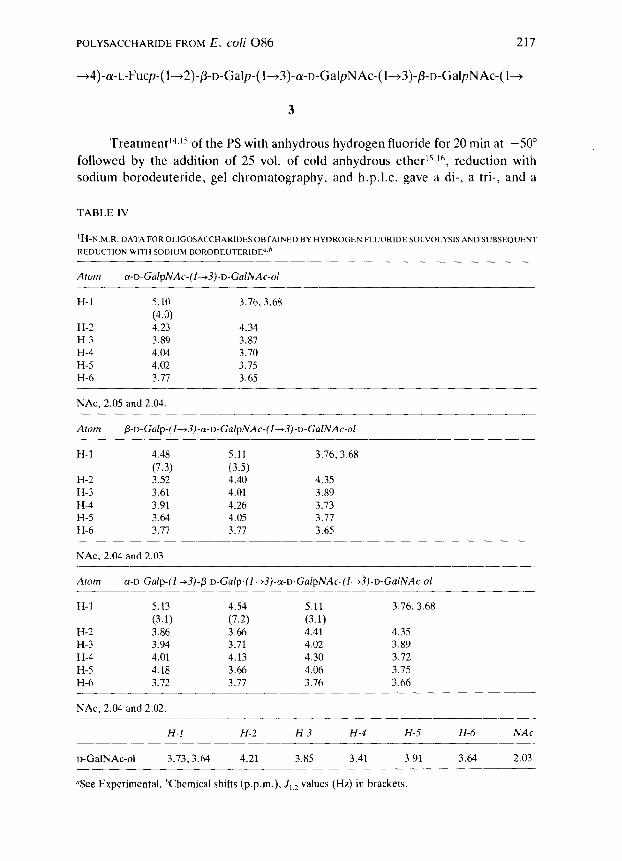
Treatment14.15 of the PS with anhydrous hydrogen fluoride for 20 min at -50”
followed by the addition of 25 vol. of cold anhydrous ether15,1h, reduction with
sodium borodeuteride, gel chromatography, and h.p.1.c. gave a di-, a tri-, and a
TABLE IV
‘H-N M.R.DATAFOROLIGOSACCHARIDESOBTAINEDBYHYDROGENFLUORIDESOLVOLYSISANDSUBSEOUENT
Atom cw-D-GalpNAc-(1~3)-D-GalNAc-ol
H-l 5.10 3.76.3.68
(4.0) H-2 4.23 4.34 H-3 3.89 3.87 H-4 4.04 3.70 H-5 4.02 3.75 H-6 3.71 3.65
NAc, 2.05 and 2.04
Atom
H-l 4.48 5.11 3.76.3.68
(7.3) (3.5) H-2 3.52 4.40 4.35 H-3 3.61 4.01 3.89 H-4 3.91 4.26 3.13 H-5 3.64 4.05 3.77 H-6 3.11 3.77 3.65
NAc, 2.04 and 2.03
Atom a-o-Gaip-(l~~)-~-D-Ga~p-(l--t3)-a-D-Ga/pNAc-(~--t~)-o-Ca~NAc-o~
H-l 5.13 4.54 5.11 3.76,3.68
(3.1) (7.2) (3.1) H-2 3.86 3.66 4.41 4.35 H-3 3.94 3.71 4.02 3.89 H-4 4.01 4.13 4.30 3.72 H-5 4.18 3.66 4.06 3.75 H-6 3.72 3.77 3.76 3.66
NAc, 2.04 and 2.02
H-l H-2 H-3 H-4 H-5 H-6 NAc
D-GalNAc-ol 3.73,3.64 4.21 3.85 3.41 3.91 3.64 2.03
“See Experimental. hChemical shifts (p.p.m.), I,,, values (Hz) in brackets

21x M. ANDEKSSON &al.
TABLE V
Atom a-~,-GalpNA~-II~3)-~~-~;ulNAc-ol
C-l 98.8 c-2 50.8 52.Y C-3 6X.5 77.6 C-4 69.4 71 .I<
c-5 72.6 71.2( C-6 61.9 63.9
NCOCH,. 23.0 and 22.9; NCOCH,. 175.4 and 175.1
Atom ~-~-Galp-ll~.Z)-a-~-GalpNAc-~l~3)-r~-GalNAc-ol
C-l 105.2 98.9 -
c-2 71.7 49.4 52.8 C-3 73.6 77.5< 77.6
c-4 69.6 69.3 71.1
C-S 75.9 72.2 71.1 C-h 61.8 61.9 63.X
NCOCH,. 23.0 and 22.9; NCOC’H,. 175.3 and 175.0
C-l 96.6 105.2 9Y.O
c-2 69.2 70.3 49.4 52.‘)
c-3 70.2 7x.7 7x.0 17.e c-4 70.2 66.1 69.3 71.1
C-S 71.x 75.6 72.2 71.1
C-6 62.0 61 .x 62.0 63.9
NCOCH,, 23.0 and 23.0; NCOCH,. 175.3 and 175.3
c-1 c-2 c-3 C-4 C-S (‘-6 NCOCH_, NCOCH,
D-GalNAc-ol 62.6 52.5 69.9 70.8 71 .o 64.2 22.8
“See Experimental. “Chemical shifts (p.p.m.). [These assignments may he reversed.
175.6
tetra-saccharide. Methylation analysis of these saccharides (Table I, columns D-F)
showed that each contained 3-linked 2-acetamido-2-dcoxy-o-galactitol-l-d but no
L-fucose derivative.
Methylation analysis of the disaccharide-alditol revealed a terminal D-
GalpNAc group and Slinked 2-acetamido-2-deoxy-D-galactitol-l-n. In the ‘H- and
‘“C-n.m.r. spectra of the disaccharide-alditol (Tables IV and V), the signals for the
anomeric proton and carbon appeared at 6 5.10 (_I,,, 4.0 Hz) and 98.8, respectively,
indicating the structure 4.
POLYSACCHARIDE FROM E. CO/i 086 219
a-D-GalpNAc-(l-3)-D-GalNAc-ol
4
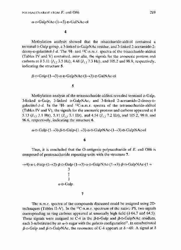
Methylation analysis showed that the trisaccharide-alditol contained a
terminal o-Galp group, a 3-linked D-GalpNAc residue, and 3-linked 2-acetamido-2-
deoxy-D-galactitol-l-d. The ‘H- and 13C-n.m.r. spectra of the trisaccharide-alditol
(Tables IV and V) contained, inter alia, the signals for the anomeric protons and
carbons at 6 5.11 (I,,* 3.5 Hz), 4.48 (J,,z 7.3 Hz), and 105.2 and 98.9, respectively,
indicating the structure 5.
5
Methylation analysis of the tetrasaccharide-alditol revealed terminal D-Galp,
3-linked D-Galp, 3-linked D-GalpNAc, and 3-linked 2-acetamido-2-deoxy-D-
galactitol-l-d. In the ‘H- and 13C-n.m.r. spectra of the tetrasaccharide-alditol
(Tables IV and V), the signals for the anomeric protons and carbons appeared at 6
5.13 (J,,2 3.1 Hz), 5.11 (I,,* 3.1 Hz), and 4.54 (J,,, 7.2 Hz), and 105.2, 99.0, and
96.6, respectively, indicating the structure 6.
6
Thus, it is concluded that the 0-antigenic polysaccharide of E. coli 086 is
composed of pentasaccharide repeating-units with the structure 7.
~4)-(Y-L-Fucp-(1~2)-p-D-Galp-(l~3)-a-D-Gal~NAc-(l~3)-~-D-Gal~NAc-(l~
; 1
cx-D-Galp
7
The n.m.r. spectra of the compounds discussed could be assigned using 2D-
techniques (Tables II-V). In the 13C-n.m.r. spectrum of the native PS, two signals
corresponding to ring carbons appeared at unusually high field (6 64.7 and 64.5).
These signals were assigned to C-4 in the /?-D-Galp and /3-D-GalpNAc residues,
each 3-substituted by an c~-D sugar with the gulacto configuration17. In unsubstituted
P-D-Galp and /3-D-GalpNAc, the resonance of C-4 appears at 6 -69. A signal at 6

220 M. ANIlEKSSON et d.
-64 was also observed in the r”C-n.m.r. spectra of 2, 3. and 6, but not in those of
4 and 5. This is in agreement with the proposed assignment.
In the spectrum of the native PS, the signal at 6 83.4 was assigned to C-4 of
the 4-linked cu-L-Fucp residue. A corresponding signal at 6 83.3 was observed in the
spectrum of 3. The L%Z-n.m.r. spectrum of the Smith-degradation product con-
tained, inter alia, a signal at S 85.8 which was assigned to C-2 of the 4-deoxy-L-
threitol. No other signals were found in this region. In the disaccharide ,!3-D-Gkp-
(l-+4)-a-t_-Fucp-O-Me, the resonance of C-4 of the c~-I.-Fucp residue appeared at
6 81.9, compared to 6 72.8 in unsubstituted a-L-Fucp’s.
In order to verify the blood-group B activity of the E. coli 086: K2:H2
isolate, the bacteria were agglutinated with a blood-group B specific human anti-
serum. The antigenicity of the purified LPS was tested in an enzyme-linked
immunosorbent assay (ELISA) against the same blood-group B antisera used
above. The antiserum had an ELISA end-point titer against E. cofi 086 LPS of
-1000, whereas the binding to the negative control LPS (E. cofi R3 rough LPS and
Sulrnonella typhimurium smooth LPS) was -10 fold lower, i.e., at background
level. Thus, the results confirm Springer’s finding that E. co/i 086 possesses blood-
group B activity.
In the methylation analyses of native PS, a small amount of terminal Fucp
was detected which could be obtained only from the non-reducing end of the
lipopolysaccharide. However, no signals from this residue were observed in the tH-
or “C-n.m.r. spectra, probably due to overlapping signals. The PS also contains
terminal cY-D-Galp groups. Terminal P-D-Galp, present in 2, gives a signal at 6
105.5, and a weak signal in this position was also observed in the spectrum of the
product obtained on lead tetra-acetate oxidation. This p-D-Galp group must have
been derived from the terminal repeating-unit, in which both the c~-L-Fucp and
cu-D-Galp groups have been oxidised. However, no signal in this region was
observed in the spectrum of the native PS. These results therefore confirm that
structure 7 represents the biological repeating-unit.
EXPERIMENTAL
General methods. - Concentrations were performed under- diminished
pressure at $40” (bath) or at room temperature by flushing with air. For g.l.c., a
Hewlett-Packard 5890A instrument, fitted with a flame-ionisation detector, was
used. Separations were performed on an Ultra 2 (cross-linked 5% phenyl methyl
silicone) fused-silica capillary column, using the temperature programme 150” for 1
min, 150”-+250” at S”/min, and 2.50” for 20 min. G.l.c.-m.s. was performed on a
Hewlett-Packard 5790-5970 instrument, using the same phase. All identifications
of mass spectra were unambiguous and will not be discussed. Hydrolysis of unde-
rivatised material was performed with 4~ hydrochloric acid for 2 h at 100”. and
hydrolysis of methylated material was performed with 2M trifluoroacetic acid for 1
h at 120”. Methylation analyses were performed as previously described’“.“‘. The

POLYSACCHARIDE FROM E. coli 086 221
absolute configurations of fucose, galactose, and 2-acetamido-2-deoxygalactose
were determined by g.1.c. of their glycosides with a chiral alcohoV. A differential
refractometer was used for monitoring the effluents from gel chromatography
columns. The treatment with anhydrous hydrogen fluoride was performed in a
specially designed apparatus made of Teflon and Kel-F (Penninsula Laboratories
Inc.). The hydrogen fluoride was dried by distillation over cobalt trifluoride. The
h.p.1.c. equipment comprised a Waters Model 6000A solvent delivery system and
Model U 6K injector. Separations were performed on a Waters Carbohydrate
Analysis Column (3.9 x 300 mm stainless-steel column) with acetonitrile-water as
eluent .
N.m.r. spectroscopy. - Spectra were recorded at 70” for solutions in D,O,
using a JEOL GX-270 instrument. Chemical shifts are reported in p_p.m. relative
to internal 1,4-dioxane (6 67.4) for 13C and internal acetone (6 2.21) for rH
resonances. For assignment of ‘H signals, proton-proton correlation techniques
wcrc used, e.g., COSY, relayed COSY, double relayed COSY, and TQF-COSY.
Sequence information was obtained by NOESY experiments with mixing times of
200 and 400 ms. “C-N.m.r. spectra were assigned mainly using 2D-i3C-‘H
heteronuclear correlation spectroscopy. The amounts of the oligosaccharides
isolated after hydrogen fluoride solvolysis of the PS was insufficient for hetero-
nuclear correlation spectroscopy. When assignments were not straightforward,
selective decoupling experiments were used to distinguish between signals in the
“C-n.m.r. spectrum.
Bacterial strain. - E. coli 086:K2:H2 (85:85 F) was isolated from children
at a neonatal ward at Danderyd Hospital (Stockholm), and verified by Drs. F. and
I. Grskov at Statens Seruminstitut (Copenhagen)?‘.
Isolation and purification of the 0-polysaccharide. - E. coli 086: K2:H2 bacteria were grown in brain heart infusion broth batch cultures (10 L). Bacteria
were killed by the addition of formaldehyde (1% final concentration) and harvested
by centrifugation. Lipopolysaccharide (LPS) was extracted by the hot phenol-water
method2*. The LPS was treated with acetic acid (2%, pH 3.1) for 2 h at loo”,
liberated lipid A was removed by centrifugation, and the supernatant solution was
neutralised, dialysed, and lyophilised. The product was further purified by
chromatography on a column (2.6 x 90 cm) of Bio-Gel P-10.
Enzyme-linked immunosorbent assay (ELISA)23. - Briefly, LPS was coated
to flat-bottomed microtiter plates (A/S Nunc, Roskilde, Denmark) in coating buffer
(0.05~ sodium carbonate, pH 9.6) at an optimal coating dose (0.1 mL, 10 pg/mL)
overnight at 20”. Residual binding sites were blocked by the addition of 1% bovine
serum albumin (0.1 mL) in coating buffer. A blood-group B specific human anti-
serum (Anti B BCA, ABO blood-group serum, BCA, West Chester, U.S.A.) was
used. ELISA end-point titers were determined as described23, employing an
alkaline phosphatase-conjugated swine anti-human IgM (F-chain specific) prepara-
tion (Orion diagnostica, Helsinki, Finland) in conjunction with p-nitrophenyl phos-
phate (1 mg/mL; disodium salt in M diethanolamine buffer, pH 9.6, containing mM

222 M. ANDERSSON et ai.
magnesium chloride). Absorbance was read with a Titertek Multiscan MCC
spectrophotometer at 40.5 nm after 100 min.
Smith degradationl’. - PS (35 mg) was dissolved in O.lM sodium acetate
buffer (10 mL, pH 3.9), and sodium metaperiodate (27 mg) was added. The solu-
tion was kept in the dark for 96 h at 4”. The excess of periodate was reduced with
ethylene glycol (0.1 mL) and the product was isolated by chromatography on a
column (2.6 x 90 cm) of Bio-Gel P-2. A solution of the product in water (10 mL)
was treated with sodium borohydride (500 mg) for 16 h at room temperature, excess
of sodium borohydride was decomposed with acetic acid, and the product was
recovered by gel chromatography (as described above). The product (20 mg) was
treated with 0.5~ trifluoroacetic acid (5 mL) for 48 h at room temperature, the
hydrolysate was concentrated to dryness, and the residue was dissolved in water
and fractionated on a column of Bio-Gel P-2 (as described above). One major
product, 2 (8 mg), was isolated (Table I, column B; Tables IIC and IIIC).
Degradation with lead tetra-acetate’2313. - The PS (31 mg) was dissolved in
water (1.3 mL), acetic acid (6.2 mL) was added, and the mixture was treated with
lead terra-acetate (33 mg). After 21 h, oxalic acid (oxalic acid in acetic acid, 100
mg/mL, 0.2 mL) was added, the mixture was centrifuged, the supernatant solution
was concentrated to dryness, and a solution of the residue in water was lyophilised.
A solution of the product in water (10 mL) was treated with sodium borohydride
(500 mg) for 15 h at room temperature, excess of reagent was decomposed with
acetic acid, and the product was isolated by chromatography on a column (2.6 x
90 cm) of Bio-Gel P-2. The product (20 mg) was treated with 0.5~ trifluoroacetic
acid (5 mL) for 43 h at room temperature, the solution was freeze-dried, and the
residue was fractionated on a column of Bio-Gel P-2 (as above). One major
product, 3 (10 mg), was eluted in the void volume (Table I, column C: Tables IIB
and IIIB).
Partial degradation with anhydrous Izydrogen~~oride14-16. - The PS (50 mg)
was dissolved in anhydrous hydrogen fluoride (2 mL) at -78”. The temperature
was allowed to rise to -50” and the mixture was stirred thereat for 20 min. Cool
anhydrous ether (50 mL) was added, and the mixture was concentrated and co-
distilled with cool ether (4 X 50 mL). A solution of the product in 0.05~ trifluoro-
acetic acid (25 mL) was kept for 30 min at room temperature, then freeze-dried.
The product was dissolved in water and treated with sodium borodeuteride (100
mg) for 16 h at room temperature, the excess of reagent was decomposed with
acetic acid, and the product was fractionated on a column (2.6 x 90 cm) of Bio-Gel
P-2 and further purified by h.p.1.c. Three products were obtained: 4 (2 mg), 5 (2
mg), and 6 (3 mg) (Table I columns D-F. Tables IV and V).
ACKNOWLEDGMENTS
E. coli 086:K2: H2 was obtained from Drs. K. Tullus and S. B. Svenson. WC thank Professor B. Lindberg, Professor L. Kenne. and Drs. I. and F. Grskov for

POLYSACCHARIDEFROM E. coli 086 223
their interest, and Mrs. A. Olin for skillful technical assistance. This work was
supported by grants from the Swedish Medical Research Council (16X-7927-OlA),
and by the National Swedish Board for Technical Development.
REFERENCES
1 G. F. SPRINGER, P. WILLIAMSON. AND W. C. BRANDERS, J. Exp. Med., 113 (1961) 1077-1093. 2 G. F. SPRINGER, E. T. WANG, J. H. NICHOLS. AND J. M. SHEAR, Ann. N.Y. Acad, Sci., 133 (1966)
566-579.
3 G. F. SPRINGER, Ann. N.Y. Acad. Xi., 169 (1970) 134-152. 4 N. KOCHIBEAND S. ISEKI, Jpn. J. Microbial., 12 (1968) 403411. 5 K. LEONTEIN, B. LINDBERG. AND J. LONNGREN, Carbohydr. Res., 62 (1978) 359-362.
6 G. .I. GERWIG, J. P. KAMERLING.AND J. F. G. VLIEFENTHART, Carbohydr. Res., 77 (1979) 1-7. 7 P. A. J. GORIN, Cnrbohydr. Res., 101 (1982) 13-20.
8 P.-E. JANSSON, B. LINDBERG. J. LBNNGREN. C. ORTEGA. AND W. NIMMICH, Carbohydr. Res., 132 (1984) 297-305.
9 N. K. KOCHETKOV, 0. S. CHIZHOV.AND A. S. SHASHKOV, Carbohydr. Res., 133 (1984) 173-185. 10 K. BOCK, A. BRIGNOLE.AND B. W. SIGURSKJOLD,J. Chem. Sot., Perkin Trans. 2, (1986) 1711-1713. 11 I. J. GOLDSTEIN, G. W. HAY. B. A. LEWIS. AND F. SMITH, Methods Carbohydr. Chem., 5 (1965)
361-370. 12 A. S. PERLIN AND A. R. LANSDOWN, Can. J. Chem., 34 (1956) 451-455. 13 P. A. J. GORIN AND A. S. PERLIN, Can. 1. Chem., 35 (1957) 262-267. 14 A. J. M~RTAND W. D. BAUER, J. Biol. Chem., 257 (1982) 1870-1875.
15 A. J. MORT, J.-P. UTILLE, G. TORRI. AND A. S. PERLIN, Carbohydr. Res., 121 (1983) 221-232. 16 J. DEFAYE, A. GADELLE. AND C. PEDERSEN, Carbohydr. Kes., 110 (1982) 217-227. 17 R. U. LEMIEUX. K. BOCK, L. T. J. DELBAERE, S. KOTO. AND S. RAO, Can. J. Chem., 58 (1980)
631453. 18 I. BACKMAN, personal communication. 19 P.-E. JANSSON, L. KENNE, H. LIEDGREN, B. LINDBERG. AND J. LONNGREN, Chem. Commun., Univ.
Stockholm, 8 (1976) l-75.
20 T. J. WAEGHE. A. G. DARVILL, M. MCNEIL. AND P. ALBERSHEIM, Carbohydr. Res., 123 (1983) 281-304.
21 DRS. F. AND I. ORSKOV, personal communication.
22 0. WESTPHAL AND K. JANN, Methods Carbohydr. Chem., 5 (1965) 83-91. 23 N. I. A. CARLIN AND A. A. LINDBERG, Infect. Immun., S3 (1986) 103-109.