
Binaural interaction in the inferior colliculus of the big brown bat, Eptesicus fuscus
Upload
independentCategory
view
0download
0
www.sciencedirect.com
c o r t e x x x x ( 2 0 1 2 ) 1e1 0
Available online at
Journal homepage: www.elsevier.com/locate/cortex
Special issue: Research report
Structural connectivity in a single case of progressiveprosopagnosia: The role of the right inferiorlongitudinal fasciculus
Dario Grossi a, Andrea Soricelli b, Marta Ponari a, Elena Salvatore c,Mario Quarantelli d, Anna Prinster b,d and Luigi Trojano a,*aNeuropsychology Laboratory, Department of Psychology, Second University of Naples, Caserta, Italyb IRCCS Fondazione SDN, Istituto di Ricerca Diagnostica e Nucleare, Naples, ItalycDepartment of Neurological Sciences, School of Biotechnology, Federico II University of Naples, ItalydBiostructure and Bioimaging Institute, CNR, Naples, Italy
a r t i c l e i n f o
Article history:
Received 1 May 2012
Reviewed 29 August 2012
Revised 11 September 2012
Accepted 16 September 2012
Published online xxx
Keywords:
Diffusion tensor imaging
Inferior longitudinal fasciculus
Inferior fronto-occipital fasciculus
Progressive prosopagnosia
* Corresponding author. Neuropsychology LCaserta, Italy.
E-mail address: [email protected] (L
Please cite this article in press as: Grossi Dof the right inferior longitudinal fascicul
0010-9452/$ e see front matter ª 2012 Elsevhttp://dx.doi.org/10.1016/j.cortex.2012.09.010
a b s t r a c t
Progressive prosopagnosia (PP) is a clinical syndrome characterized by a progressive and
selective inability to recognize and identify faces of familiar people. Here we report
a patient (G.S.) with PP, mainly related to a prominent deficit in recognition of familiar
faces, without a semantic (cross-modal) impairment. An in-depth evaluation showed that
his deficit extended to other classes of objects, both living and non-living. A follow-up
neuropsychological assessment did not reveal substantial changes after about 1 year.
Structural MRI showed predominant right temporal lobe atrophy.
Diffusion tensor imaging was performed to elucidate structural connectivity of the
inferior longitudinal fasciculus (ILF) and the inferior fronto-occipital fasciculus (IFOF), the
two major tracts that project through the core fusiform region to the anterior temporal and
frontal cortices, respectively. Right ILF was markedly reduced in G.S., while left ILF and
IFOFs were apparently preserved. These data are in favour of a crucial role of the neural
circuit subserved by right ILF in the pathogenesis of PP.
ª 2012 Elsevier Srl. All rights reserved.
1. Introduction et al., 1995a,b; Rossion et al., 2003; Wada and Yamamoto,
The study of patients with a selective impairment in recog-
nizing faces (acquired prosopagnosia) following right or bilat-
eral temporo-occipital lesions has provided relevant
information about the neural mechanisms involved in face
processing (Bodamer, 1947; Barton et al., 2002; Bruce and
Young, 1986; Damasio et al., 1982; De Renzi, 1986; Farah
aboratory, Department o
. Trojano).
, et al., Structural conneus, Cortex (2012), http://
ier Srl. All rights reserved
2001). Such lesion studies, combined with functional imaging
studies in normal individuals, have demonstrated that regions
of the posterior fusiform gyrus, the inferior lateral occipital
cortex and the posterior superior temporal sulcus (STS) are
involved in face processing (e.g., Allison et al., 1994a, 1999;
Barton, 2008; Kanwisher et al., 1997; Grill-Spector et al., 2004;
Pitcher et al., 2009; Rossion et al., 2003; Sergent et al., 1992).
f Psychology, Second University of Naples, Via Vivaldi 43, 81100
ctivity in a single case of progressive prosopagnosia: The roledx.doi.org/10.1016/j.cortex.2012.09.010
.
c o r t e x x x x ( 2 0 1 2 ) 1e1 02
Within these regions, that are part of the so-called ‘core
system’ for face processing (Haxby et al., 2000), the fusiform
gyrus seems to be especially important for processing facial
identity. More recently, an important role in face recognition
has also been ascribed to the anterior inferior temporal lobe
(Allison et al., 1994b, 1999; Rajimehr et al., 2009; Tsao et al.,
2008), particularly in individuals with developmental proso-
pagnosia (DP), who may show reduced volume of the anterior
part of fusiform gyri (Behrmann et al., 2007; Garrido et al.,
2009).
In a few studies, prosopagnosia has been described as the
main manifestation of a neurodegenerative disorder
(Barbarotto et al., 1995; De Renzi, 1986; Evans et al., 1995;
Gentileschi et al., 1999, 2001; Gainotti, 2003; Tyrrell et al., 1990).
Early patients with progressive prosopagnosia (PP) also showed
visual agnosia, basic visuoperceptual deficits and impairments
in other cognitive domains, associated with posterior cortical
atrophy (DeRenzi, 1986; Tyrrell et al., 1990). In subsequent years
several patients with quite selective progressive disorders of
face recognition have been described. In one patient with
selectivePP, Evansetal. (1995) interpreted theirpatient’spicture
asdue to a visual,modality-selective, inability to access person-
based semantic knowledge; however, the patient eventually
developed (over 9 months) a multi-modal loss of person-based
knowledge. Evans et al.’s patient showed prominent right
hemisphere temporal lobe atrophy; analogous lesions were
reported in further patients with PP who were affected by
analogousmulti-modal deficits ofperson (and face) recognition,
not restricted to the visual modality (Barbarotto et al., 1995;
Gainotti, 2003; Gentileschi et al., 1999, 2001).
A progressive defect in processing face configuration
associated with mild visual agnosia, but with relative preser-
vation of other cognitive domains and intact basic face
perception skills, was described by Joubert et al. (2003), in
a patient who showed atrophy of the right fusiform gyrus and
parahippocampal cortex on quantitative volumetricmeasures
of temporal lobes (Joubert et al., 2004). On the basis of this
observation, the authors proposed that the critical region in
the genesis of PP may lie more posteriorly than what was
previously suggested, and involve the right fusiform gyrus
selectively (Joubert et al., 2004).
This rare form of degenerative process may reflect a right
hemisphere variant of semantic dementia. Actually, current
clinical diagnostic criteria (Neary et al., 1998) consider PP with
insidious onset and gradual progression, often associated to
associative agnosia, as the hallmark of the prosopagnosic
variant of frontotemporal dementia (FTD) (Neary et al., 1998),
more recently defined right-temporal variant (rtFTD; Chan
et al., 2009; Josephs et al., 2009; Miller et al., 1993).
In synthesis, several patients have been described with PP
in the context of a degenerative disorder, and all of them
showed atrophy in the right temporal lobe, but no specific
study has investigated white matter tracts (WMT) in patients
with selective PP (but see Migliaccio et al., 2012, for a DT study
on posterior cortical atrophy). In neurodegenerative disorders
neuropsychological deficits are usually considered as result-
ing from cortical degeneration. However, in recent years the
cortical localization approach for cognitive functions is
evolving towards network-based hypotheses, according to
which cognitive disorders emerge from the interruption of the
Please cite this article in press as: Grossi D, et al., Structural conneof the right inferior longitudinal fasciculus, Cortex (2012), http://
information flowwithin large-scale networks linking different
cortical regions (Bartolomeo, 2011; Catani and ffytche, 2005).
According to this approach, not only cortical lesions but also
damage to WMT between cortical areas can induce network
dysfunction and, hence, cognitive disorders (Mesulam, 2009).
Over the last few years, tractography has allowed hodo-
logical study of the brain and a re-interpretation of several
neurobehavioral syndromes. In DP patients, Thomas et al.
(2009) have found a bilateral reduction in structural integ-
rity of the inferior longitudinal fasciculus (ILF) and of the
inferior fronto-occipital fasciculus (IFOF), two major tracts
that project through the core fusiform region to the anterior
temporal and frontal cortices, respectively (Benson et al.,
1974). However, Thomas et al. (2009) demonstrated that
only the reduction in tract integrity of the right ILF (rILF), and
to a lesser extent, of the right IFOF (rIFOF) correlated with
errors in face recognition tasks, consistent with the notion
that the right hemisphere is more prominent in face pro-
cessing (for a recent review, see Gainotti and Marra, 2011).
The ILF is a ventral associative bundle with long and short
fibres connecting the occipital and temporal lobes namely
lingual and fusiform gyrus (Catani et al., 2003). Damage to
such a tract might disconnect visual processing in the
occipital lobe from memory processing in the temporal lobe
(Habib, 1986; Kawahata and Nagata, 1989; Meadows, 1974;
Takahashi et al., 1995), thus giving rise to a form of associa-
tive prosopagnosia (Fox et al., 2008).
On the basis of evidence collected in DP, here we present
a diffusion tensor imaging (DTI) study performed on a patient
with relatively selective PP to the aim of investigating struc-
tural integrity of ILF and IFOF. In previous single case studies
of patients with neurodegenerative disease, DTI was used for
the purpose of following up progressive deterioration of WMT
(Duning et al., 2009). DTI has also been used in a single patient
with posterior cortical atrophy to demonstrate WMT damage
(Migliaccio et al., 2011). Here, our specific working hypothesis
was that connectivity between occipital and temporal lobes in
the right hemisphere is disrupted in comparison with the left
hemisphere.
2. Methods
2.1. Case report
G.S. was a 65-year-old, right-handed man with 7 years of
formal education; he was a retired factory worker. He came to
our observation in November 2010, complaining of progres-
sive difficulties in recognizing familiar faces over the past 2
years. For instance, he knew and could immediately recall
names of the examiners, but over a period of several weeks he
never recognized them by their faces.
At the time of testing, G.S. was alert, cooperative and well
oriented; his language was fluent and flawless. The patient
was aware of his problems and described them in detail, but
his relatives referred some early personality changes, with
instances of social inadequacy (e.g.,: excessive or inappro-
priate jokes; childish behaviour); the total score on Frontal
Behavioural Inventory (FBI; Alberici et al., 2007), administered
to patient’s son, was 12/72, i.e., below the cut-off point (28.6)
ctivity in a single case of progressive prosopagnosia: The roledx.doi.org/10.1016/j.cortex.2012.09.010

Table 1 e Results of the formal neuropsychologicalassessment.
Test Nov 2010 Feb 2012 Normalcut-off*
Mini Mental State
Examination (MMSE)a28/30 28/30 �23
FABb 14.40 13.40 �13.40
Raven coloured
progressive matrices (PM)c24 28 �14.75
Story recalld 10.8/16 11.4/16 �4.50
Corsi Visuospatial spand 4 6 �3.50
Digit spane 5 5 �3.50
Verbal word spand 3 4 �2.75
Copying simple drawingsd 11.75 12.75 �7.75
Rey complex figuref
Copy 29.9/36 36/36 <23.76
Immediate recall 9/36 10.50/36 <6.44
Delayed recall 8.2/36 13.25/36 <6.33
Phonemic verbal fluencyc 26.2 27.2 <17.35
Semantic verbal fluencyd 15.25 14 �7
Note. *Normal cut-off values refer to age- and education-adjusted
Italian norms; no G.S.’ score was below cut-off.
a Measso et al., 1993.
b Appollonio et al., 2005.
c Caltagirone et al., 1995.
c o r t e x x x x ( 2 0 1 2 ) 1e1 0 3
discriminating frontotemporal lobe degeneration from other
dementias.
Neurological examination was normal. MRI showed
a predominant right temporal atrophy associated to mild
cortical atrophy of remaining supratentorial cortices (Fig. 1).
The analysis of biomarkers in cerebrospinal liquor (ELISA test;
Innogenetics, Ghent, Belgium) revealed normal levels of phos-
phorylated tau (p-tau 181: 28.6 pg/mL; normal values: <33) and
b-amyloid 1e42 (749.3 pg/mL; normal values: 492e1088).
General neuropsychological assessment did not reveal
impairments of verbal and spatial memory, spatial explora-
tion of personal and extrapersonal space, visuoconstructional
skills, abstract logical abilities, verbal fluency and executive
functioning (Table 1). G.S. then underwent a specific neuro-
psychological assessment of face processing, visuospatial
perception, imagery and semantic knowledge.
2.1.1. Assessment of face processing2.1.1.1. FACE MATCHING. On the Benton Facial Recognition Test
(Benton et al., 1983), G.S.’ performance was in the ‘borderline’
range after adjusting raw score for age and educational level
(Table 2). It is interesting to note that he performed flawlessly
on the trials in which camera angle and lighting were held
constant, suggesting that his face recognition impairment
Fig. 1 e Coronal reformats of the 3D-T1w-volume at the
level of the midportion of the hippocampus (top) and of the
amygdala (bottom), showing on the right side a relative
sparing of the hippocampus, with a dilation of the
temporal horn of the lateral ventricle, likely due to loss of
adjacent white matter, parahippocampal gyrus and
amygdala.
d Spinnler and Tognoni, 1987.
e Orsini et al., 1987.
f Caffarra et al., 2002.
Please cite this article in press as: Grossi D, et al., Structural conneof the right inferior longitudinal fasciculus, Cortex (2012), http://
could not be accounted for in terms of elementary visual
deficits. G.S. emphasized that he always tried to rely on details
such as ears or eyebrows in order to succeed on this task.
2.1.1.2. FAMOUS FACE RECOGNITION, NAMING AND IDENTIFICATION. Toassess famous face processing, G.S. was administered the
Famous face recognition and naming test (Rizzo et al., 2002), that
evaluates both visual recognition of known people and verbal
recognition of their names. As for visual recognition, the test
included 50 photographs depicting famous people randomly
intermingled with 50 pictures of unknown people. Photo-
graphs were presented one at a time, and G.S. was asked: (a) to
make a fame judgement, (b) to name the 50 famous people, and
Table 2 e Processing of unfamiliar and familiar faces.
G.S.’scores
Normalcut-off
Unfamiliar faces
Benton Facial Recognition Testa 40/54 41
Familiar faces
Famous face recognition and naming testb
Naming 0c �14.50
Semantic information 0c �22.17
Semantic information from name 24.75 �20.68
False recognitions 0 �14.16
False recognitions from name 0 �12.92
Note. Normal cut-off values refer to age- and education-adjusted
Italian norms.
a Benton et al., 1983.
b Rizzo et al., 2002.
c Denotes a score below age- and education-adjusted cut-off.
ctivity in a single case of progressive prosopagnosia: The roledx.doi.org/10.1016/j.cortex.2012.09.010
c o r t e x x x x ( 2 0 1 2 ) 1e1 04
(c) to provide some semantic information about the person, in
particular, whether the person was alive, if he/she was Italian,
and to which professional category the person was associated.
G.S. was not able to distinguish famous from unknown faces
(Table 2), and could neither name nor identify even celebrities
with very distinctive and unique facial features from their
photographs (e.g., Michael Gorbaciov). G.S. spontaneously
referred to the gender of every single face he was presented
with (e.g., “I have never seen her/him” or “I don’t knowwho he/
she might be”), showing that his gender perception was
preserved. When all visual stimuli were presented, the test
assessed recognition of auditorily presented names of the
same famous people, intermingled with 50 unknown names.
When asked to perform a fame judgement and to provide as
much information as he could about the celebrities upon
verbal presentation of their names, G.S. performed in the
normal range, meaning that his semantic knowledge about
famous people was at least relatively preserved.
2.1.2. Assessment of visuospatial perception2.1.2.1. VISUOSPATIAL ABILITIES. On the Battery for Visuospatial
Abilities (BVA, known in Italy as TERADIC; Angelini and Grossi,
1993; Grossi and Trojano, 1999; Grossi et al., 2002; Trojano
et al., 2004), exploring perceptual and representational
visuospatial abilities, G.S.’ scores were well within the normal
control range according to published norms on tasks assess-
ing discrimination of line length and orientation, coding of
relative position of points within squares, mental rotation,
and analysis and recognition of complex abstract figures
(Table 3). G.S. was relatively impaired in one subtest assessing
discrimination of angle width, and in one ‘representational’
subtest assessing mental construction of abstract figures.
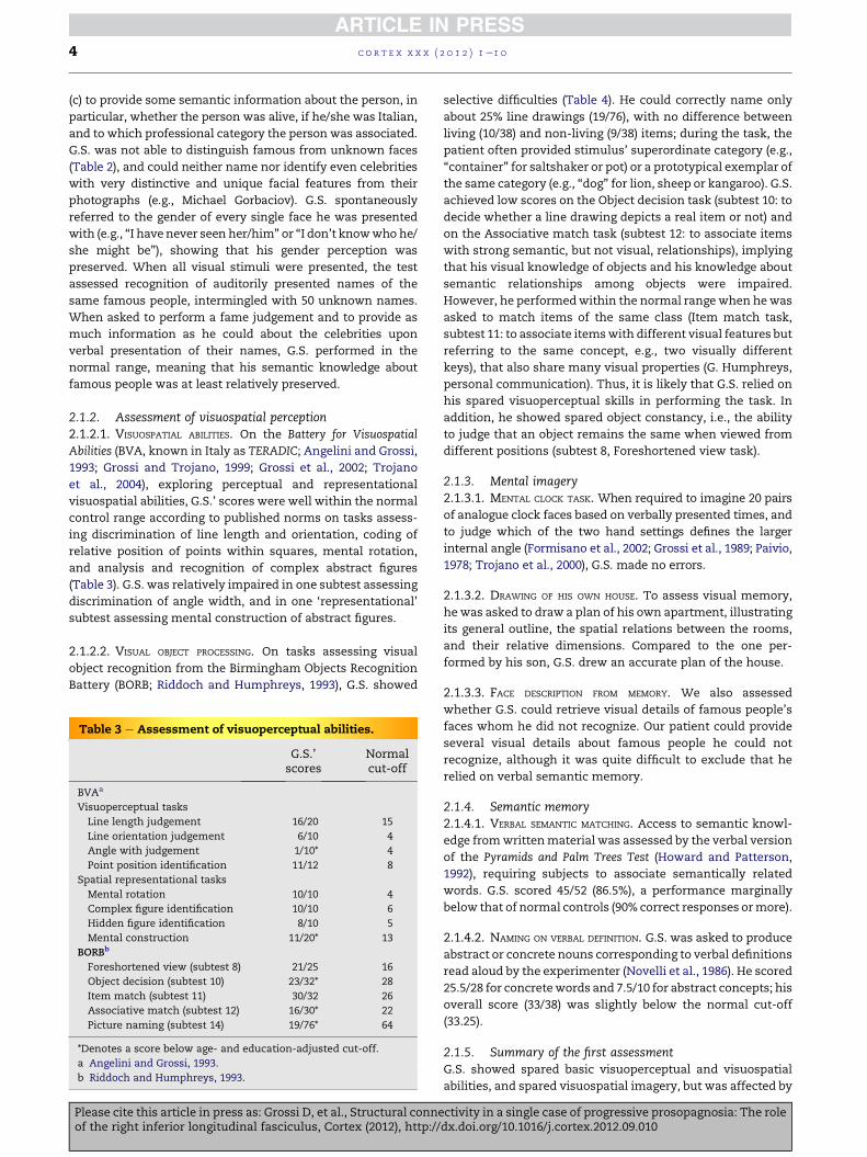
2.1.2.2. VISUAL OBJECT PROCESSING. On tasks assessing visual
object recognition from the Birmingham Objects Recognition
Battery (BORB; Riddoch and Humphreys, 1993), G.S. showed
Table 3 e Assessment of visuoperceptual abilities.
G.S.’scores
Normalcut-off
BVAa
Visuoperceptual tasks
Line length judgement 16/20 15
Line orientation judgement 6/10 4
Angle with judgement 1/10* 4
Point position identification 11/12 8
Spatial representational tasks
Mental rotation 10/10 4
Complex figure identification 10/10 6
Hidden figure identification 8/10 5
Mental construction 11/20* 13
BORBb
Foreshortened view (subtest 8) 21/25 16
Object decision (subtest 10) 23/32* 28
Item match (subtest 11) 30/32 26
Associative match (subtest 12) 16/30* 22
Picture naming (subtest 14) 19/76* 64
*Denotes a score below age- and education-adjusted cut-off.
a Angelini and Grossi, 1993.
b Riddoch and Humphreys, 1993.
Please cite this article in press as: Grossi D, et al., Structural conneof the right inferior longitudinal fasciculus, Cortex (2012), http://
selective difficulties (Table 4). He could correctly name only
about 25% line drawings (19/76), with no difference between
living (10/38) and non-living (9/38) items; during the task, the
patient often provided stimulus’ superordinate category (e.g.,
“container” for saltshaker or pot) or a prototypical exemplar of
the same category (e.g., “dog” for lion, sheep or kangaroo). G.S.
achieved low scores on the Object decision task (subtest 10: to
decide whether a line drawing depicts a real item or not) and
on the Associative match task (subtest 12: to associate items
with strong semantic, but not visual, relationships), implying
that his visual knowledge of objects and his knowledge about
semantic relationships among objects were impaired.
However, he performedwithin the normal rangewhen hewas
asked to match items of the same class (Item match task,
subtest 11: to associate itemswith different visual features but
referring to the same concept, e.g., two visually different
keys), that also share many visual properties (G. Humphreys,
personal communication). Thus, it is likely that G.S. relied on
his spared visuoperceptual skills in performing the task. In
addition, he showed spared object constancy, i.e., the ability
to judge that an object remains the same when viewed from
different positions (subtest 8, Foreshortened view task).
2.1.3. Mental imagery2.1.3.1. MENTAL CLOCK TASK. When required to imagine 20 pairs
of analogue clock faces based on verbally presented times, and
to judge which of the two hand settings defines the larger
internal angle (Formisano et al., 2002; Grossi et al., 1989; Paivio,
1978; Trojano et al., 2000), G.S. made no errors.
2.1.3.2. DRAWING OF HIS OWN HOUSE. To assess visual memory,
he was asked to draw a plan of his own apartment, illustrating
its general outline, the spatial relations between the rooms,
and their relative dimensions. Compared to the one per-
formed by his son, G.S. drew an accurate plan of the house.
2.1.3.3. FACE DESCRIPTION FROM MEMORY. We also assessed
whether G.S. could retrieve visual details of famous people’s
faces whom he did not recognize. Our patient could provide
several visual details about famous people he could not
recognize, although it was quite difficult to exclude that he
relied on verbal semantic memory.
2.1.4. Semantic memory2.1.4.1. VERBAL SEMANTIC MATCHING. Access to semantic knowl-
edge fromwrittenmaterial was assessed by the verbal version
of the Pyramids and Palm Trees Test (Howard and Patterson,
1992), requiring subjects to associate semantically related
words. G.S. scored 45/52 (86.5%), a performance marginally
below that of normal controls (90% correct responses ormore).
2.1.4.2. NAMING ON VERBAL DEFINITION. G.S. was asked to produce
abstract or concrete nouns corresponding to verbal definitions
read aloud by the experimenter (Novelli et al., 1986). He scored
25.5/28 for concretewords and 7.5/10 for abstract concepts; his
overall score (33/38) was slightly below the normal cut-off
(33.25).
2.1.5. Summary of the first assessmentG.S. showed spared basic visuoperceptual and visuospatial
abilities, and spared visuospatial imagery, but was affected by
ctivity in a single case of progressive prosopagnosia: The roledx.doi.org/10.1016/j.cortex.2012.09.010
c o r t e x x x x ( 2 0 1 2 ) 1e1 0 5
relatively selective defects in face and visual object process-
ing. In particular, G.S. was totally unable to name or identify
faces of famous people. When names of the same famous
people were provided, G.S. could retrieve precise semantic
information about almost half of them, a performance within
the normal range. Therefore, our patient proved to be unable
to access person-based semantic knowledge via the visual
modality, but could access the same knowledge by verbal
modality. Moreover, G.S. showed a minor impairment in
processing unknown faces, particularly when he could not
rely on perceptual details, but had to resort to a configura-
tional analysis. This defect of face processing was associated
withmild visual agnosia (not clinically relevant) for both living
and man-made objects. In contrast, the patient showed
preserved semantic knowledge about the objects he could not
recognize when he was provided with their names.
2.1.6. Follow-upFifteen months after the first evaluation (February 2012), G.S.
underwent a new clinical and neuropsychological assess-
ment. The patient was still cooperative and well oriented, and
his language was fluent and flawless, but he was highly
distractible, with increasing difficulties of staying on task.
Although patient’s subjective complaints were still limited to
difficulties in recognizing familiar faces, his caregivers re-
ported increasingly frequent episodes of social inadequacy,
impulsivity, aggressive behaviour and disinhibition. Themost
evident personality changes with respect to the first evalua-
tion reported by patient’s son at the FBI were in the disinhi-
bition subscale (restlessness, irritability, excessive jocularity,
impulsivity, aggression, hyperorality, hoarding); the total FBI
score (32/72) was above the cut-off score thought to identify
FTD patients (28.6; Alberici et al., 2007). Neurological exami-
nation was still normal.
General neuropsychological assessment did not show
changes in verbal and spatial memory, spatial exploration,
visuoconstructional skills, abstract logical abilities, verbal
fluency and executive functioning (see Table 1).
G.S.’ performanceon theBenton Facial Recognition Testwas
substantially unchanged (38/54): G.S. still emphasized that he
relied on details such as general face shape, ears or eyebrows to
perform the task, and performed well with faces presented in
the same perspective and with the same lighting conditions.
To further investigate G.S.’ impairment in face recognition,
and to assess the relative contribution of perceptual processes
and memory to his recognition impairment, he was assessed
on the Cambridge Memory Test for Faces (CMTF; Duchaine
and Nakayama, 2006), and the Cambridge Face Perception
Test (CFPT; Duchaine et al., 2007). G.S.’ scores were compared
to those of 7 age-matched healthy control (HC) participants
(mean age: 68 years, range: 61e77), using the modified t-test
devised by Crawford and Garthwaite (2002) for use with single
cases.
The CFMT includes a study phase in which subjects are
required to familiarize with six target faces presented in three
different views; after the study phase, subjects are asked to
recognize target faces in 54 forced-choice items, consisting of
novel views of one of the six target faces along with two non-
target faces. G.S. scored 31/72; this score was significantly
below the mean score of the HCs (m ¼ 52.4, SD ¼ 6.7; p ¼ .024).
Please cite this article in press as: Grossi D, et al., Structural conneof the right inferior longitudinal fasciculus, Cortex (2012), http://
In the CFPT, subjects are required to arrange six frontal views
of men’s faces according to their similarity to a 3/4 profile view
of a target face. The test images are morphs of six different
individuals with the target face. Each combination of target
and test faces are presented once upright and once inverted,
allowing to assess presence of the “face inversion” effect (i.e.,
better performance for upright than for inverted faces), that is
robust in normal adults (Yovel and Duchaine, 2006) and typi-
cally developed children (Taylor et al., 2004), but is lacking in
patients with acquired prosopagnosia (Farah et al., 1995b) and
DP (Schmalzl et al., 2009). Scores for each item are computed
by summing the deviations from the correct position for each
face. G.S.’ score for upright trials (total deviations: 90) was
significantly above themean score of the age-matched control
group (mean deviations: 45.5, SD ¼ 12.6; p ¼ .016) and very
close to chance performance (93.3 deviations). While controls
showed a robust inversion effect (mean deviations for inver-
ted trials: 65.5, SD ¼ 6.7), G.S. performed better with inverted
trials, and his score was not significantly different from
controls (total deviations: 76; p ¼ .193).
On tasks assessing visual object recognition from BORB,
G.S. did not show any relevant change in performance with
respect to the first evaluation. G.S. could correctly name 18/76
drawings. Moreover, he performed below normal cut-offs on
the Object decision task (subtest 10: 16/32) and on the Asso-
ciative match task (subtest 12: 16/30), but still performed
within the normal range on Itemmatch task (subtest 11: 31/32)
and showed spared object constancy (subtest 8: 20/25).
2.1.7. Summary of the follow-up assessmentAfter 15months, the neuropsychological picture had remained
relatively stable. A more extensive assessment of G.S.’ face
recognition abilities at the follow-up showed an impaired
memory for faces, with associated deficits in perception of
facial similarity. However, the patient could still perform
a perceptual analysis of face details with high accuracy, as
shown by his normal performance on the ‘inverted faces’
condition of the CFPT. Visual object recognition abilities had
remained generally stable. The only relevant change concerned
severity of behavioural and psychological symptoms, particu-
larly those of the disinhibition type.
As it will be discussed to a longer extent below, G.S.’ clinical
picture at the time of the first evaluation conforms to that
described at early stages of PP. This gave us the opportunity of
testing the hypothesis about the role of rILF and rIFOF in PP by
means of DTI.
2.2. DTI data acquisition
G.S. was scanned using a 3T Philips Achieva scanner equipped
with a 8 channels phased-array head coil. The DTI sequence
wasbasedona single-shot spin-echo, echo-planar imaging (EPI)
sequence with diffusion sensitizing gradients applied on either
side of the 180� refocusing pulse. Diffusion weighted images
were acquired with diffusion gradients oriented along 32 non-
collinear directions. Specific DTI parameters were: Repetition
Time (TR) ¼ 9330 msec, Echo Time (TE) ¼ 102 msec, flip
angle ¼ 90�, Field of view (FOV) ¼ 256 � 256 mm2, acquisition
matrix size¼ 128 � 128, 50.2 mm thick axial slices (.7 mm gap),
pixel size ¼ 2 � 2 mm2, diffusion weighting b ¼ 1000 sec/mm2.
ctivity in a single case of progressive prosopagnosia: The roledx.doi.org/10.1016/j.cortex.2012.09.010
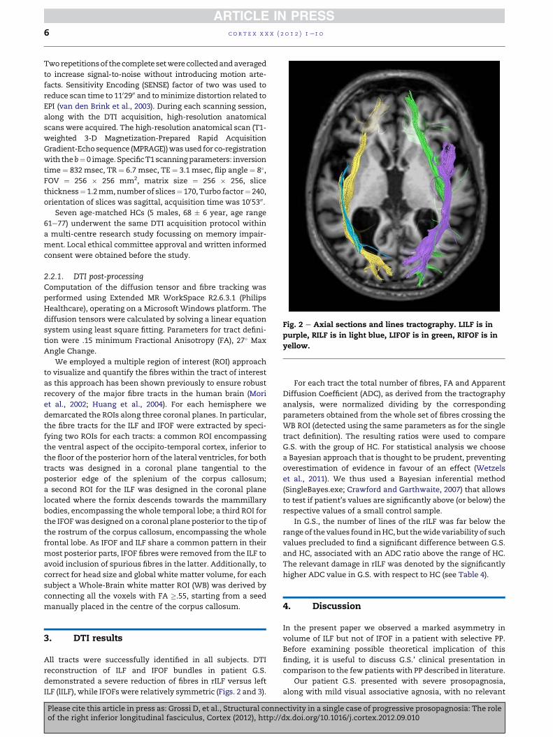
Fig. 2 e Axial sections and lines tractography. LILF is in
purple, RILF is in light blue, LIFOF is in green, RIFOF is in
yellow.
c o r t e x x x x ( 2 0 1 2 ) 1e1 06
Tworepetitionsof thecomplete setwere collectedandaveraged
to increase signal-to-noise without introducing motion arte-
facts. Sensitivity Encoding (SENSE) factor of two was used to
reduce scan time to 1102900 and tominimize distortion related to
EPI (van den Brink et al., 2003). During each scanning session,
along with the DTI acquisition, high-resolution anatomical
scans were acquired. The high-resolution anatomical scan (T1-
weighted 3-D Magnetization-Prepared Rapid Acquisition
Gradient-Echosequence (MPRAGE))wasused for co-registration
with theb¼ 0 image. SpecificT1scanningparameters: inversion
time ¼ 832 msec, TR ¼ 6.7 msec, TE ¼ 3.1 msec, flip angle ¼ 8�,FOV ¼ 256 � 256 mm2, matrix size ¼ 256 � 256, slice
thickness¼ 1.2mm,number of slices¼ 170, Turbo factor¼ 240,
orientation of slices was sagittal, acquisition time was 1005300.Seven age-matched HCs (5 males, 68 � 6 year, age range
61e77) underwent the same DTI acquisition protocol within
a multi-centre research study focussing on memory impair-
ment. Local ethical committee approval and written informed
consent were obtained before the study.
2.2.1. DTI post-processingComputation of the diffusion tensor and fibre tracking was
performed using Extended MR WorkSpace R2.6.3.1 (Philips
Healthcare), operating on a Microsoft Windows platform. The
diffusion tensors were calculated by solving a linear equation
system using least square fitting. Parameters for tract defini-
tion were .15 minimum Fractional Anisotropy (FA), 27� Max
Angle Change.
We employed a multiple region of interest (ROI) approach
to visualize and quantify the fibres within the tract of interest
as this approach has been shown previously to ensure robust
recovery of the major fibre tracts in the human brain (Mori
et al., 2002; Huang et al., 2004). For each hemisphere we
demarcated the ROIs along three coronal planes. In particular,
the fibre tracts for the ILF and IFOF were extracted by speci-
fying two ROIs for each tracts: a common ROI encompassing
the ventral aspect of the occipito-temporal cortex, inferior to
the floor of the posterior horn of the lateral ventricles, for both
tracts was designed in a coronal plane tangential to the
posterior edge of the splenium of the corpus callosum;
a second ROI for the ILF was designed in the coronal plane
located where the fornix descends towards the mammillary
bodies, encompassing the whole temporal lobe; a third ROI for
the IFOFwas designed on a coronal plane posterior to the tip of
the rostrum of the corpus callosum, encompassing the whole
frontal lobe. As IFOF and ILF share a common pattern in their
most posterior parts, IFOF fibres were removed from the ILF to
avoid inclusion of spurious fibres in the latter. Additionally, to
correct for head size and global white matter volume, for each
subject a Whole-Brain white matter ROI (WB) was derived by
connecting all the voxels with FA �.55, starting from a seed
manually placed in the centre of the corpus callosum.
3. DTI results
All tracts were successfully identified in all subjects. DTI
reconstruction of ILF and IFOF bundles in patient G.S.
demonstrated a severe reduction of fibres in rILF versus left
ILF (lILF), while IFOFs were relatively symmetric (Figs. 2 and 3).
Please cite this article in press as: Grossi D, et al., Structural conneof the right inferior longitudinal fasciculus, Cortex (2012), http://
For each tract the total number of fibres, FA and Apparent
Diffusion Coefficient (ADC), as derived from the tractography
analysis, were normalized dividing by the corresponding
parameters obtained from the whole set of fibres crossing the
WB ROI (detected using the same parameters as for the single
tract definition). The resulting ratios were used to compare
G.S. with the group of HC. For statistical analysis we choose
a Bayesian approach that is thought to be prudent, preventing
overestimation of evidence in favour of an effect (Wetzels
et al., 2011). We thus used a Bayesian inferential method
(SingleBayes.exe; Crawford and Garthwaite, 2007) that allows
to test if patient’s values are significantly above (or below) the
respective values of a small control sample.
In G.S., the number of lines of the rILF was far below the
range of thevalues found inHC, but thewidevariability of such
values precluded to find a significant difference between G.S.
and HC, associated with an ADC ratio above the range of HC.
The relevant damage in rILF was denoted by the significantly
higher ADC value in G.S. with respect to HC (see Table 4).
4. Discussion
In the present paper we observed a marked asymmetry in
volume of ILF but not of IFOF in a patient with selective PP.
Before examining possible theoretical implication of this
finding, it is useful to discuss G.S.’ clinical presentation in
comparison to the few patients with PP described in literature.
Our patient G.S. presented with severe prosopagnosia,
along with mild visual associative agnosia, with no relevant
ctivity in a single case of progressive prosopagnosia: The roledx.doi.org/10.1016/j.cortex.2012.09.010
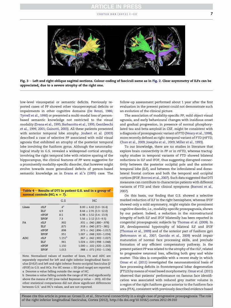
Fig. 3 e Left and right oblique sagittal sections. Colour-coding of fasciculi same as in Fig. 2. Clear asymmetry of ILFs can be
appreciated, due to a severe atrophy of the right one.
c o r t e x x x x ( 2 0 1 2 ) 1e1 0 7
low-level visuospatial or semantic deficits. Previously re-
ported cases of PP showed other visuoperceptual deficits or
impairments in other cognitive domains (De Renzi, 1986;
Tyrrell et al., 1990) or presented a multi-modal loss of person-
based semantic knowledge not restricted to the visual
modality (Evans et al., 1995; Barbarotto et al., 1995; Gentileschi
et al., 1999, 2001; Gainotti, 2003). All these patients presented
with anterior temporal lobe atrophy. Joubert et al. (2003)
described a case of selective PP associated with mild visual
agnosia that exhibited an atrophy of the posterior temporal
lobe involving the fusiform gyrus. Although the neuroradio-
logical study in G.S. revealed a widespread cortical atrophy,
involving the right temporal lobe with relative sparing of the
hippocampus, the clinical features of PP were suggestive for
a prominently modality-specific disorder, that however might
evolve towards more generalized defects of person-based
semantic knowledge as in Evans et al.’s (1995) case. The
Table 4 e Results of DTI in patient G.S. and in a group ofnormal controls (HC; n [ 7).
G.S HC
Lines rILF .4a 8.00 � 4.62 (3.0e16.4)
lILF 4.9 8.44 � 2.91 (3.3e12.0)
rIFOF 8.5 6.98 � 3.52 (3.4e13.9)
lIFOF 7.3 5.56 � 2.12 (3.5e9.5)
FA rILF .902 .931 � .040 (.880e.978)
lILF .875 .918 � .040 (.872e.981)
rIFOF .898 .973 � .042 (.896e1.017)
lIFOF .951 1.007 � .048 (.920e1.034)
ADC rILF 1.151b .967 � .042 (.902e1.027)
lILF .961 1.024 � .020 (.998e1.048)
rIFOF 1.150 1.000 � .102 (.920e1.203)
lIFOF 1.013 .988 � .072 (.932e1.136)
Note. Normalized values of number of lines, FA and ADC are
separately reported for left and right inferior longitudinal fascic-
ulus (l/rILF) and left and right inferior fronto-occipital fasciculus (l/
rIFOF) in G.S. and HCs. For HC mean � SD (and range) are reported.
a Denotes a value falling outside the range of HC.
b Denotes a value falling outside the range of HC and significantly
above the means of HC (one-tailed Bayesian p value ¼ .003). All the
other statistical comparisons did not show significant differences
between G.S.’ and HC’s values, and are not reported.
Please cite this article in press as: Grossi D, et al., Structural conneof the right inferior longitudinal fasciculus, Cortex (2012), http://
follow-up assessment performed about 1 year after the first
evaluation in the present patient could not demonstrate such
an evolution of the clinical picture.
The association of modality-specific PP, mild object visual
agnosia, and early behavioural changes with insidious onset
and gradual progression, in presence of normal phosphory-
lated tau and beta-amyloid in CSF, might be consistent with
a diagnosis of prosopagnosic variant of FTD (Neary et al., 1998),
more recently defined as right-temporal variant of FTD (rtFTD;
Chan et al., 2009; Josephs et al., 2009; Miller et al., 1993).
To our knowledge, there are no studies in literature that
explore brain connectivity in PP or in rtFTD, whereas tractog-
raphy studies in temporal variants of FTD showed bilateral
reductions in ILF and IFOF, thus suggesting disrupted connec-
tivity between the posterior occipital pole and the anterior
temporal lobe (ILF), and between the inferolateral and dorso-
lateral frontal cortices and both the temporal and occipital
cortices (IFOF; Borroni et al., 2007). Suchdata suggested thatDTI
measures can contribute to characterize patientswith different
variants of FTD and their clinical symptoms (Borroni et al.,
2007).
On this basis, our finding that G.S. showed a selective
marked reduction of ILF in the right hemisphere, whereas IFOF
showed only a mild asymmetry, might explain the prominent
cognitive disorder, i.e.,modality-specific prosopagnosia, shown
by our patient. Indeed, a reduction in the microstructural
integrity of both ILF and IFOF bilaterally has been reported in
congenital prosopagnosic subjects by Thomas et al. (2009). In
DP, developmental hypotrophy of bilateral ILF and IFOF
(Thomas et al., 2009) and of the anterior part of fusiform gyri
(Behrmann et al., 2007; Garrido et al., 2009) would impair
maturation of normal face processing skills, and preclude
formation of any efficient compensatory pathway. In the
present patient PPwas related to the atrophy of the rILF, related
to progressive neuronal loss, affecting both grey and white
matter. This idea is compatible with a recent study in which
Omar et al. (2011) investigated the neuroanatomical basis of
face processing deficits in frontotemporal lobar degeneration
(FTLD)bymeansofvoxelbasedmorphometry.Omaretal. (2011)
observed that patients’ performance on famous face identifi-
cation was associated with reduced grey matter volume in
a region of the right fusiform gyrus anterior to the fusiform face
area (FFA), consistentwith previously described evidence based
ctivity in a single case of progressive prosopagnosia: The roledx.doi.org/10.1016/j.cortex.2012.09.010

c o r t e x x x x ( 2 0 1 2 ) 1e1 08
on focal lesionsandneuroimaging studies.On thebasis of these
observations and of the present findings, we could suggest that
the disproportionate atrophy of the rILF can be the specific
hallmark of the dysfunction in the neural circuit dedicated to
face processing in the ventral regionsof the right hemisphere in
our patient. These results are also compatible with the finding
that white matter lesions in the ILF as a result of multiple
sclerosis may induce symptoms of prosopagnosia and object
agnosia (Yamasaki et al., 2004).
In recent years Fox et al. (2008) hypothesized that damage
of ILF might be specifically related to associative proso-
pagnosia with relatively preserved perceptual abilities. In the
present patient both the baseline evaluation and the follow-
up assessment revealed a composite clinical picture in
which minor defects of configurational face analysis were
associated with an impairment to experience visual famil-
iarity for well-known faces, and a substantial impairment to
associate visually perceived faces to preserved semantic
information about people. This is no room here to tackle the
controversial distinction between associative and appercep-
tive forms of prosopagnosia (De Renzi et al., 1991; Duchaine
and Weidenfeld, 2003; see Gainotti and Marra, 2011, for
a recent review), but our data support the hypothesis that ILF
is the most critical fibre pathway connecting regions of the
core system of face processing (see Haxby et al., 2000; Catani
and Thiebaut de Schotten, 2008), as proposed in the past
(Benson et al., 1974; Habib, 1986; Kawahata and Nagata, 1989;
Meadows, 1974; Takahashi et al., 1995). A damage of rILF
would disconnect the occipital face area and the FFA from
each other or from regions in the anterior temporal lobe and
the precuneus (Catani et al., 2003), thus hampering modula-
tion of right occipital lobe on temporal lobe activity. However,
it remains to be explored the possible contribution of short
U-shaped tracts connecting adjacent gyri that have been
identified in human visual regions (Catani et al., 2003).
In conclusion, the selective damage of rILF in the present
patient G.S. suggests possible underpinnings of early stages of
PP, in line with previous DTI findings in DP. Longitudinal
studies will clarify the clinico-anatomical correlates of
evolution of PP and possible relationships with clinical diag-
nosis of different variants of neurodegenerative diseases.
r e f e r e n c e s
Alberici A, Geroldi C, Cotelli M, Adorni A, Calabria M, Rossi G, et al.The Frontal Behavioural Inventory (Italian version)differentiates frontotemporal lobar degeneration variants fromAlzheimer’s disease. Neurological Sciences, 28(2): 80e86, 2007.
Allison T. Electrophysiological studies of human face perception.I: Potentials generated in occipitotemporal cortex by face andnon-face stimuli. Cerebral Cortex, 9(5): 415e430, 1999.
Allison T, Ginter H, McCarthy G, Nobre AC, Puce A, Luby M, et al.Face recognition in human extrastriate cortex. Journal ofNeurophysiology, 71(2): 821e825, 1994a.
Allison T, McCarthy G, Nobre AC, Puce A, and Belger A. Humanextrastriate visual cortex and the perception of faces, words,numbers and colors. Cerebral Cortex, 5: 544e554, 1994b.
Angelini R and Grossi D. La terapia razionale dei disordini costruttivi.S. Lucia: Centro di Riabilitazione, 1993.
Please cite this article in press as: Grossi D, et al., Structural conneof the right inferior longitudinal fasciculus, Cortex (2012), http://
Appollonio I, Leone M, Isella V, Piamarta F, Consoli T, Villa ML,et al. The Frontal Assessment Battery (FAB): Normative valuesin an Italian population sample. Neurological Sciences, 26(2):108e116, 2005.
Barbarotto R, Capitani E, Spinnler H, and Trivelli C. Slowlyprogressive semantic impairment with category specificity.Neurocase, 1: 107e119, 1995.
Bartolomeo P. The quest for the “critical lesion site” in cognitivedeficits: Problems and perspectives. Cortex, 47(8): 1010e1012,2011.
Barton JJS. Structure and function in acquired prosopagnosia:Lessons from a series of 10 patients with brain damage. Journalof Neuropsychology, 2: 197e225, 2008.
Barton JJS, Press DZ, Keenan JP, and O’Connor M. Lesions of thefusiform face area impair perception of facial configuration inprosopagnosia. Neurology, 58(1): 71e78, 2002.
Behrmann M, Avidan G, Gao F, and Black S. Structural imagingreveals anatomical alterations in inferotemporal cortex incongenital prosopagnosia. Cerebral Cortex, 17(10): 2354e2363,2007.
Benson DF, Segarra J, and Albert ML. Visualagnosiaeprosopagnosia. Archives of Neurology, 30: 307e310,1974.
Benton AL, Sivan AB, Hamsher K, Varney NR, and Spreen O.Contributions to Neuropsychological Assessment. New York:Oxford University Press, 1983.
Bodamer J. Die-Prosop-agnosie. Archives of PsychiatricNervenkrankh, 179: 6e54, 1947.
Borroni B, Brambati SM, Agosti C, Gipponi S, Bellelli G,Gasparotti R, et al. Evidence of white matter changes ondiffusion tensor imaging in frontotemporal dementia. Archivesof Neurology, 64(2): 246e251, 2007.
Bruce V and Young A. Understanding face recognition. BritishJournal of Psychology, 77(3): 305e327, 1986.
Caffarra P, Vezzadini G, Dieci F, Zonato F, and Venneri A.Rey-Osterrieth complex figure: Normative values in an Italianpopulation sample. Neurological Sciences, 22(6): 443e447, 2002.
Caltagirone C, Gainotti G, Carlesimo GA, and Parnetti L. Batteriaper valutazione del deterioramento mentale (parte I):descrizione di uno strumento per la diagnosineuropsicologica. Archivio di Psicologia, Neurologia e Psichiatria,55: 461e470, 1995.
Catani M and ffytche DH. The rises and falls of disconnectionsyndromes. Brain, 128: 2224e2239, 2005.
Catani M and Thiebaut de Schotten M. A diffusion tensortractography atlas for virtual dissections. Cortex, 44(8):1105e1132, 2008.
Catani M, Jones DK, Donato R, and ffytche DH. Occipito-temporalconnections in the human brain. Brain, 126: 2093e2107, 2003.
Chan D, Anderson V, Pijnenburg Y, Whitwell J, Barnes J, Scahill R,et al. The clinical profile of right temporal lobe atrophy. Brain,132(Pt 5): 1287e1298, 2009.
Crawford JR and Garthwaite PH. Investigation of the single case inneuropsychology: Confidence limits on the abnormality of testscores and test score differences. Neuropsychologia, 40:1196e1208, 2002.
Crawford JR and Garthwaite PH. Comparison of a single case toa control or normative sample in neuropsychology:Development of a Bayesian approach. CognitiveNeuropsychology, 24: 343e372, 2007.
Damasio AR, Damasio H, and Van Hoesen GW. Prosopagnosia:Anatomic basis and behavioral mechanisms. Neurology, 32(4):331e341, 1982.
De Renzi E. Prosopagnosia in two patients with CT scan evidenceof damage confined to the right hemisphere. Neuropsychologia,24: 385e389, 1986.
De Renzi E, Faglioni P, Grossi D, and Nichelli P. Apperceptive andassociative forms of prosopagnosia. Cortex, 27(2): 213e221, 1991.
ctivity in a single case of progressive prosopagnosia: The roledx.doi.org/10.1016/j.cortex.2012.09.010
c o r t e x x x x ( 2 0 1 2 ) 1e1 0 9
Duchaine BC and Nakayama K. The Cambridge Face Memory Test:Results for neurologically intact individuals andan investigationof its validity using inverted face stimuli and prosopagnosicparticipants. Neuropsychologia, 44(4): 576e585, 2006.
Duchaine BC and Weidenfeld A. An evaluation of two commonlyused tests of unfamiliar face recognition. Neuropsychologia,41(6): 713e720, 2003.
Duchaine BC, Germine L, and Nakayama K. Family resemblance:Ten family members with prosopagnosia and within-classobject agnosia. Cognitive Neuropsychology, 24(4): 419e430, 2007.
Duning T, Warnecke T, Mohammadi S, Lohmann H,Schiffbauer H, Kugel H, et al. Pattern and progression of white-matter changes in a case of posterior cortical atrophy usingdiffusion tensor imaging. Journal of Neurology, Neurosurgery,and Psychiatry, 80(4): 432e436, 2009.
Evans JJ, Heggs AJ, Antoun N, and Hodges JR. Progressiveprosopagnosia associated with selective right temporal lobeatrophy. Brain, 118(1): 1e13, 1995.
Farah MJ, Levinson KL, and Klein KL. Face perception and within-category discrimination in prosopagnosia. Neuropsychologia,33(6): 661e674, 1995a.
Farah MJ, Wilson K, Drain H, and Tanaka J. The inverted faceinversion effect in prosopagnosia: Evidence for mandatory,face-specific perceptual mechanisms. Vision Research, 35:2089e2093, 1995b.
Formisano E, Linden DEJ, Salle FD, Trojano L, Esposito F, Sack AT,et al. Tracking the mind’s image in the brain: Time-resolvedfMRI during visuospatial mental imagery. Neuron, 35: 185e194,2002.
Fox CJ, Iaria G, and Barton JJS. Disconnection in prosopagnosiaand face processing. Cortex, 44(8): 996e1009, 2008.
Gainotti G. Slowly progressive defect in recognition of familiarpeople in a patient with right anterior temporal atrophy. Brain,126(4): 792e803, 2003.
Gainotti G and Marra C. Differential contribution of right and lefttemporo-occipital and anterior temporal lesions to facerecognition disorders. Frontiers in Human Neuroscience, 5: 55,2011.
Garrido L, Furl N, Draganski B, Weiskopf N, Stevens J, Chern-YeeTan G, et al. Voxel-based morphometry reveals reduced greymatter volume in the temporal cortex of developmentalprosopagnosics. Brain, 132: 3443e3455, 2009.
Gentileschi V, Sperber S, and Spinnler H. Progressive defectiverecognition of familiar people. Neurocase, 5(5): 407e424, 1999.
Gentileschi V, Sperber S, and Spinnler H. Crossmodal agnosia forfamiliar people as a consequence of right infero polar temporalatrophy. Cognitive Neuropsychology, 18(5): 439e463, 2001.
Grill-Spector K, Knouf N, and Kanwisher N. The fusiform facearea subserves face perception, not generic within-categoryidentification. Nature Neuroscience, 7(5): 555e562, 2004.
Grossi D and Trojano L. Constructional apraxia. In Denes G andPizzamiglio L (Eds), Handbook of Clinical and ExperimentalNeuropsychology. Psychological Press Hove, 1999: 441e452.
Grossi D, Trojano L, Modafferi A, and Pelosi L. On different role ofhemispheres in Mental Imagery. “O’clock Test” in two cases.Brain and Cognition, 10: 18e27, 1989.
Grossi D, Fragassi FA, Chiacchio L, Valoroso L, Tuccillo R,Perrotta C, et al. Do visuospatial and constructionaldisturbances differentiate frontal variant of frontotemporaldementia and Alzheimer’s disease? An experimental study ofa clinical belief. International Journal of Geriatric Psychiatry, 17:641e648, 2002.
Habib M. Visual hypoemotionality and prosopagnosia associatedwith right temporal lobe isolation. Neuropsychologia, 24:577e582, 1986.
Haxby JV, Hoffman EA, and Gobbini MI. The distributed humanneural system for face perception. Trends in Cognitive Sciences,4: 223e233, 2000.
Please cite this article in press as: Grossi D, et al., Structural conneof the right inferior longitudinal fasciculus, Cortex (2012), http://
Howard D and Patterson K. Pyramids and Palm Trees. Bury St.Edmunds, England: Thames Valley Test Company, 1992.
Huang H, Zhang JY, van Zijl PCM, and Mori S. Analysis of noiseeffects on DTI-based tractography using the brute-force andmulti-ROI approach. Magnetic Resonance in Medicine, 52:559e565, 2004.
Josephs KA, Whitwell JL, Knopman DS, Boeve BF, Vemuri P,Senjem ML, et al. Two distinct subtypes of right temporalvariant frontotemporal dementia. Neurology, 73(18):1443e1450, 2009.
Joubert S, Felician O, Barbeau E, Sontheimer A, Barton JJ,Ceccaldi M, et al. Impaired configurational processing in a caseof progressive prosopagnosia associated with predominantright temporal lobe atrophy. Brain, 126(Pt 11): 2537e2550, 2003.
Joubert S, Felician O, Barbeau E, Sontheimer A, Guedj E,Ceccaldi M, et al. Progressive prosopagnosia: Clinical andneuroimaging results. Neurology, 63(10): 1962e1965, 2004.
Kanwisher N, McDermott J, and ChunMM. The fusiform face area:A module in human extrastriate cortex specialized for faceperception. Journal of Neuroscience, 17(11): 4302e4311, 1997.
Kawahata N and Nagata K. A case of associative visual agnosia:Neuropsychological findings and theoretical considerations.Journal of Clinical and Experimental Neuropsychology, 11: 645e664,1989.
Meadows JC. The anatomical basis of prosopagnosia. Journal ofNeurology, Neurosurgery, and Psychiatry, 37: 489e501, 1974.
Measso G, Cavarzeran F, Zappala G, Lebowitz BD, Crook TH,Pirozzolo FJ, et al. The mini-mental state examination:Normative study of an Italian random sample. DevelopmentalNeuropsychology, 9(2): 77e85, 1993.
Mesulam M. Defining neurocognitive networks in the BOLD newworld of computed connectivity. Neuron, 62: 1e3, 2009.
Migliaccio R, Agosta F, Toba MN, Samri D, Corlier F, de Souza LC,et al. Brain networks in posterior cortical atrophy: A singlecase tractography study and literature review. Cortex, 48(10):1298e1309, 2012.
Migliaccio R, Agosta F, Scola E, Magnani G, Cappa SF, Pagani E,et al. Ventral and dorsal visual streams in posterior corticalatrophy: A DT MRI study. Neurobiology of Aging, 33(11):2572e2584, 2012.
Miller BL, Chang L, Mena I, Boone K, and Lesser IM. Progressiveright frontotemporal degeneration: Clinical,neuropsychological and SPECT characteristics. Dementia,4(3e4): 204e213, 1993.
Mori S, Kaufmann WE, Davatzikos C, Stieltjes B, Amodei L,Fredericksen K, et al. Imaging cortical association tracts in thehuman brain using diffusion tensor-based axonal tracking.Magnetic Resonance in Medicine, 47: 215e223, 2002.
Neary D, Snowden JS, Gustafson L, Passant U, Stuss D, Black S,et al. Frontotemporal lobar degeneration: A consensus onclinical diagnostic criteria. Neurology, 51(6): 1546e1554, 1998.
Novelli G, Papagno C, Capitani E, Laiacona M, Cappa SF, andVallar G. Tre test clinici di ricerca e produzione lessicale.Taratura su soggetti normali. Archivio di Psicologia, Neurologia ePsichiatria, 47: 477e506, 1986.
Omar R, Rohrer JD, Hailstone JC, and Warren JD. Structuralneuroanatomy of face processing in frontotemporal lobardegeneration. Journal of Neurology, Neurosurgery, and Psychiatry,82(12): 1341e1343, 2011.
Orsini A, Grossi D, Capitani E, Laiacona M, Papagno C, andVallar G. Verbal and spatial immediate memory span:Normative data from 1355 adults and 1112 children. ItalianJournal of Neurological Sciences, 8(6): 537e548, 1987.
Paivio A. Comparisons of mental clocks. Journal of ExperimentalPsychology, 1: 61e71, 1978.
Pitcher D, Charles L, Devlin JT, Walsh V, and Duchaine BC. Tripledissociation of faces, bodies, and objects in extrastriate cortex.Current Biology, 19(4): 319e324, 2009.
ctivity in a single case of progressive prosopagnosia: The roledx.doi.org/10.1016/j.cortex.2012.09.010
c o r t e x x x x ( 2 0 1 2 ) 1e1 010
Rajimehr R, Young JC, and Tootell R. An anterior temporal facepatch in human cortex, predicted by macaque maps.Proceedings of the National Academy of Sciences, 106: 1995e2000,2009.
Riddoch MJ and Humphreys GW. BORB: The Birmingham ObjectRecognition Battery. Hove, UK: Lawrence Erlbaum Associates,1993.
Rizzo S, Venneri A, and Papagno C. Famous face recognition andnaming test: A normative study. Neurological Sciences, 23(4):153e159, 2002.
Rossion B, Caldara R, Seghier M, Schuller A-M, Lazeyras F, andMayer E. A network of occipito-temporal face-sensitive areasbesides the right middle fusiform gyrus is necessary fornormal face processing. Brain, 126(Pt 11): 2381e2395, 2003.
Schmalzl L, Palermo R, Harris IM, and Coltheart M. Face inversionsuperiority in a case of prosopagnosia following congenitalbrain abnormalities: What can it tell us about the specificityand origin of face-processing mechanisms? CognitiveNeuropsychology, 26(3): 286e306, 2009.
Sergent J, Ohta S, and MacDonald B. Functional neuroanatomy offace and object processing. Brain, 115(1): 15e36, 1992.
Spinnler H and Tognoni G. Standardizzazione e taratura italianadi test neuropsicologici. Italian Journal of Neurological Sciences,8(Suppl.): 1e12, 1987.
Takahashi N, Kawamura M, Hirayama K, Shiota J, and Isono O.Prosopagnosia: A clinical and anatomical study of fourpatients. Cortex, 31(2): 317e329, 1995.
Taylor MJ, Batty M, and Itier RJ. The faces of development: Areview of early face processing over childhood. Journal ofCognitive Neuroscience, 16(8): 1426e1442, 2004.
Thomas C, Avidan G, Humphreys K, Jung KJ, Gao F, andBehrmann M. Reduced structural connectivity in ventralvisual cortex in congenital prosopagnosia. Nature Neuroscience,12(1): 29e31, 2009.
Please cite this article in press as: Grossi D, et al., Structural conneof the right inferior longitudinal fasciculus, Cortex (2012), http://
Trojano L, Grossi D, Linden DE, Formisano E, Hacker H, Zanella FE,et al. Matching two imagined clocks: The functional anatomyof spatial analysis in the absence of visual stimulation.Cerebral Cortex, 10: 473e481, 2000.
Trojano L, Fragassi NA, Chiacchio L, Izzo O, Izzo G, Di Cesare G,et al. Relationship between constructional and visuospatialabilities in normal subjects and in focal brain-damagedpatients. Journal of Clinical and Experimental Neuropsychology, 26:1103e1112, 2004.
Tsao D, Moeller S, and FreiwaldW. Comparing face patch systemsin macaques and humans. Proceedings of the National Academyof Sciences, 105: 19513e19518, 2008.
Tyrrell PJ, Warrington EK, Frackowiak RS, and Rossor MN.Heterogeneity in progressive aphasia due to focal corticalatrophy. A clinical and PET study. Brain, 113: 1321e1336, 1990.
van den Brink JS, Watanabe Y, Kuhl CK, Chung T, Muthupillai R,Van Cauteren M, et al. Implications of SENSE MR in routineclinical practice. European Journal of Radiology, 46: 3e27, 2003.
Wada Y and Yamamoto T. Selective impairment of facialrecognition due to a haematoma restricted to the rightfusiform and lateral occipital region. Journal of Neurology,Neurosurgery, and Psychiatry, 71(2): 254e257, 2001.
Wetzels R, Matzke D, Lee MD, Rouder JN, Iverson GJ, andWagenmakers E-J. Statistical evidence in experimentalpsychology: An empirical comparison using 855 t tests.Perspectives on Psychological Science, 6(3): 291e298, 2011.
Yamasaki T, Taniwaki T, Tobimatsu S, Arakawa K, Kuba H,Maeda Y, et al. Electrophysiological correlates of associativevisual agnosia lesioned in the ventral pathway. Journal ofNeurological Science, 221(1e2): 53e60, 2004.
Yovel G and Duchaine B. Specialized face perception mechanismsextract both part and spacing information: Evidence fromdevelopmental prosopagnosia. Journal of Cognitive Neuroscience,18(4): 580e593, 2006.
ctivity in a single case of progressive prosopagnosia: The roledx.doi.org/10.1016/j.cortex.2012.09.010
Copyright © 2022 FDOKUMEN