Structural characterization of SmMn 2 GeO 7 single microcrystals by electron microscopy
6
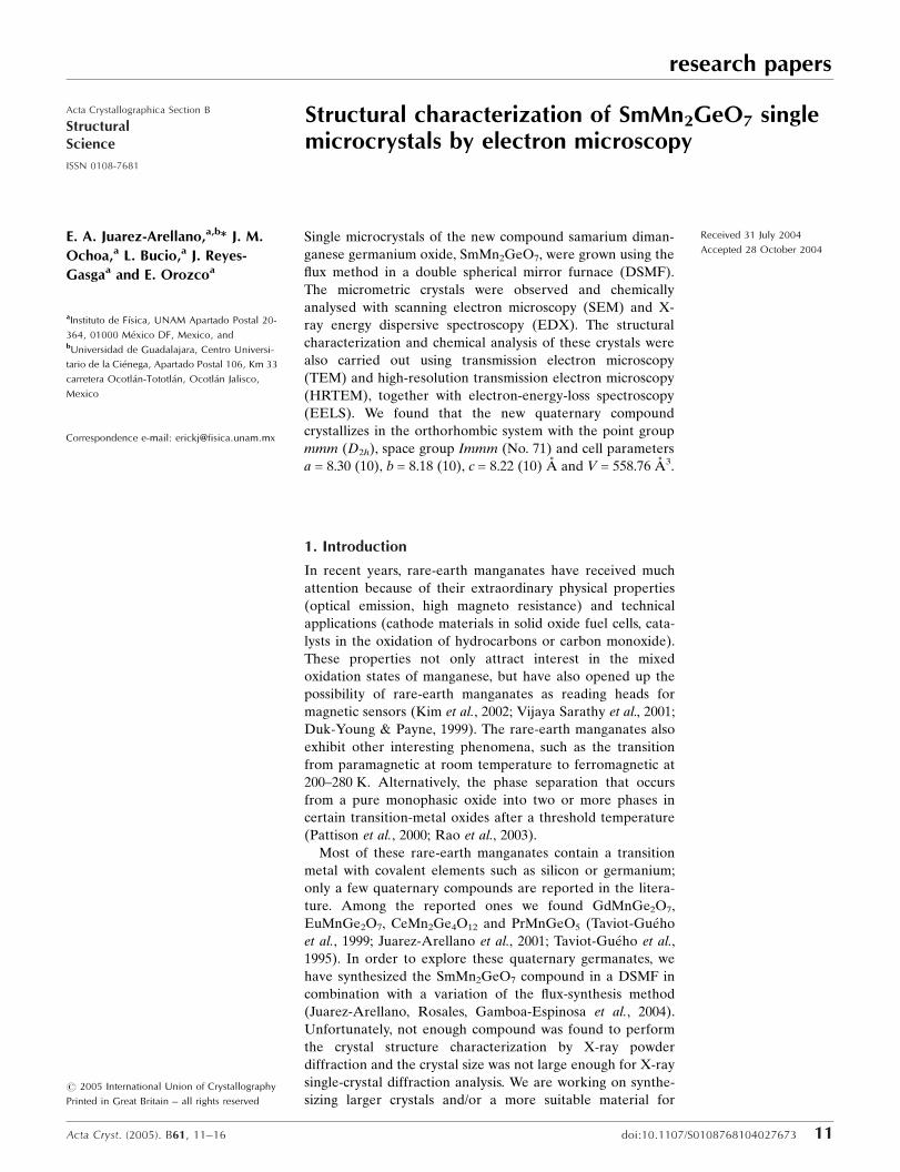
research papers Acta Cryst. (2005). B61, 11–16 doi:10.1107/S0108768104027673 11 Acta Crystallographica Section B Structural Science ISSN 0108-7681 Structural characterization of SmMn 2 GeO 7 single microcrystals by electron microscopy E. A. Juarez-Arellano, a,b * J. M. Ochoa, a L. Bucio, a J. Reyes- Gasga a and E. Orozco a a Instituto de Fı ´sica, UNAM Apartado Postal 20- 364, 01000 Me ´xico DF, Mexico, and b Universidad de Guadalajara, Centro Universi- tario de la Cie ´nega, Apartado Postal 106, Km 33 carretera Ocotla ´n-Tototla ´n, Ocotla ´n Jalisco, Mexico Correspondence e-mail: [email protected] # 2005 International Union of Crystallography Printed in Great Britain – all rights reserved Single microcrystals of the new compound samarium diman- ganese germanium oxide, SmMn 2 GeO 7 , were grown using the flux method in a double spherical mirror furnace (DSMF). The micrometric crystals were observed and chemically analysed with scanning electron microscopy (SEM) and X- ray energy dispersive spectroscopy (EDX). The structural characterization and chemical analysis of these crystals were also carried out using transmission electron microscopy (TEM) and high-resolution transmission electron microscopy (HRTEM), together with electron-energy-loss spectroscopy (EELS). We found that the new quaternary compound crystallizes in the orthorhombic system with the point group mmm (D 2h ), space group Immm (No. 71) and cell parameters a = 8.30 (10), b = 8.18 (10), c = 8.22 (10) A ˚ and V = 558.76 A ˚ 3 . Received 31 July 2004 Accepted 28 October 2004 1. Introduction In recent years, rare-earth manganates have received much attention because of their extraordinary physical properties (optical emission, high magneto resistance) and technical applications (cathode materials in solid oxide fuel cells, cata- lysts in the oxidation of hydrocarbons or carbon monoxide). These properties not only attract interest in the mixed oxidation states of manganese, but have also opened up the possibility of rare-earth manganates as reading heads for magnetic sensors (Kim et al., 2002; Vijaya Sarathy et al. , 2001; Duk-Young & Payne, 1999). The rare-earth manganates also exhibit other interesting phenomena, such as the transition from paramagnetic at room temperature to ferromagnetic at 200–280 K. Alternatively, the phase separation that occurs from a pure monophasic oxide into two or more phases in certain transition-metal oxides after a threshold temperature (Pattison et al., 2000; Rao et al. , 2003). Most of these rare-earth manganates contain a transition metal with covalent elements such as silicon or germanium; only a few quaternary compounds are reported in the litera- ture. Among the reported ones we found GdMnGe 2 O 7 , EuMnGe 2 O 7 , CeMn 2 Ge 4 O 12 and PrMnGeO 5 (Taviot-Gue ´ho et al., 1999; Juarez-Arellano et al. , 2001; Taviot-Gue ´ho et al. , 1995). In order to explore these quaternary germanates, we have synthesized the SmMn 2 GeO 7 compound in a DSMF in combination with a variation of the flux-synthesis method (Juarez-Arellano, Rosales, Gamboa-Espinosa et al., 2004). Unfortunately, not enough compound was found to perform the crystal structure characterization by X-ray powder diffraction and the crystal size was not large enough for X-ray single-crystal diffraction analysis. We are working on synthe- sizing larger crystals and/or a more suitable material for
-
Upload
independent -
Category
Documents
-
view
1 -
download
0
Transcript of Structural characterization of SmMn 2 GeO 7 single microcrystals by electron microscopy
research papers
Acta Cryst. (2005). B61, 11±16 doi:10.1107/S0108768104027673 11
Acta Crystallographica Section B
StructuralScience
ISSN 0108-7681
Structural characterization of SmMn2GeO7 singlemicrocrystals by electron microscopy
E. A. Juarez-Arellano,a,b* J. M.
Ochoa,a L. Bucio,a J. Reyes-
Gasgaa and E. Orozcoa
aInstituto de FõÂsica, UNAM Apartado Postal 20-
364, 01000 MeÂxico DF, Mexico, andbUniversidad de Guadalajara, Centro Universi-
tario de la CieÂnega, Apartado Postal 106, Km 33
carretera OcotlaÂn-TototlaÂn, OcotlaÂn Jalisco,
Mexico
Correspondence e-mail: [email protected]
# 2005 International Union of Crystallography
Printed in Great Britain ± all rights reserved
Single microcrystals of the new compound samarium diman-
ganese germanium oxide, SmMn2GeO7, were grown using the
¯ux method in a double spherical mirror furnace (DSMF).
The micrometric crystals were observed and chemically
analysed with scanning electron microscopy (SEM) and X-
ray energy dispersive spectroscopy (EDX). The structural
characterization and chemical analysis of these crystals were
also carried out using transmission electron microscopy
(TEM) and high-resolution transmission electron microscopy
(HRTEM), together with electron-energy-loss spectroscopy
(EELS). We found that the new quaternary compound
crystallizes in the orthorhombic system with the point group
mmm (D2h), space group Immm (No. 71) and cell parameters
a = 8.30 (10), b = 8.18 (10), c = 8.22 (10) AÊ and V = 558.76 AÊ 3.
Received 31 July 2004
Accepted 28 October 2004
1. Introduction
In recent years, rare-earth manganates have received much
attention because of their extraordinary physical properties
(optical emission, high magneto resistance) and technical
applications (cathode materials in solid oxide fuel cells, cata-
lysts in the oxidation of hydrocarbons or carbon monoxide).
These properties not only attract interest in the mixed
oxidation states of manganese, but have also opened up the
possibility of rare-earth manganates as reading heads for
magnetic sensors (Kim et al., 2002; Vijaya Sarathy et al., 2001;
Duk-Young & Payne, 1999). The rare-earth manganates also
exhibit other interesting phenomena, such as the transition
from paramagnetic at room temperature to ferromagnetic at
200±280 K. Alternatively, the phase separation that occurs
from a pure monophasic oxide into two or more phases in
certain transition-metal oxides after a threshold temperature
(Pattison et al., 2000; Rao et al., 2003).
Most of these rare-earth manganates contain a transition
metal with covalent elements such as silicon or germanium;
only a few quaternary compounds are reported in the litera-
ture. Among the reported ones we found GdMnGe2O7,
EuMnGe2O7, CeMn2Ge4O12 and PrMnGeO5 (Taviot-GueÂho
et al., 1999; Juarez-Arellano et al., 2001; Taviot-GueÂho et al.,
1995). In order to explore these quaternary germanates, we
have synthesized the SmMn2GeO7 compound in a DSMF in
combination with a variation of the ¯ux-synthesis method
(Juarez-Arellano, Rosales, Gamboa-Espinosa et al., 2004).
Unfortunately, not enough compound was found to perform
the crystal structure characterization by X-ray powder
diffraction and the crystal size was not large enough for X-ray
single-crystal diffraction analysis. We are working on synthe-
sizing larger crystals and/or a more suitable material for
analysis of this compound by X-ray powder diffraction.
However, here we want to encourage the use of electron
microscopy to perform a structural and chemical character-
ization.
2. Experimental
The SmMn2GeO7 compound was prepared by modifying the
method reported by Taviot-GueÂho et al. (1995). For the
synthesis, MnO2, GeO2 and SmCl3 were mixed in stoichio-
metric proportions and heated at
323 K in air for 3 d. Afterwards, the
sample was placed inside of an
evacuated quartz tube maintained
at 573 K. The quartz tube was
placed inside a re¯ective furnace
(Lara et al., 1991) and the chemical
transport reaction was carried out
at 1073 K for 12 h (SchaÈ fer, 1964;
Gruehn & Schweizer, 1983). After
the thermal treatment many micro-
crystals were formed on the wall of
the quartz tube, showing a perfect
crystalline appearance. Hereafter
we will name these microcrystals
`crystals' for simplicity. The
resulting product of the reaction
was stirred using ethanol to dissolve
the MnCl2 sub-product. The crystals
were studied with SEM and EDX as
obtained.
In order to perform the TEM
analysis, the crystals obtained were
taken from the tube of quartz and
millied in an agate mortar. The
resulting powder was collected and
supported on Cu grids that had
been previously covered with a
plastic ®lm.
For SEM and EDX analysis, a
Jeol SEM 5200 microscope with a
NORAN-EDX instrument was
used. TEM observations were
carried out with three different
microscopes. For conventional
microscopy an analytical electron
microscope Jeol STEM 100-CX was
used. High-resolution images, Z-
contrast and electron-energy-loss
spectra (EELS), and convergent-
beam diffraction patterns were
obtained with Jeol 4000-EX and
Jeol-FEG-2010-EX microscopes.
3. Results and discussion
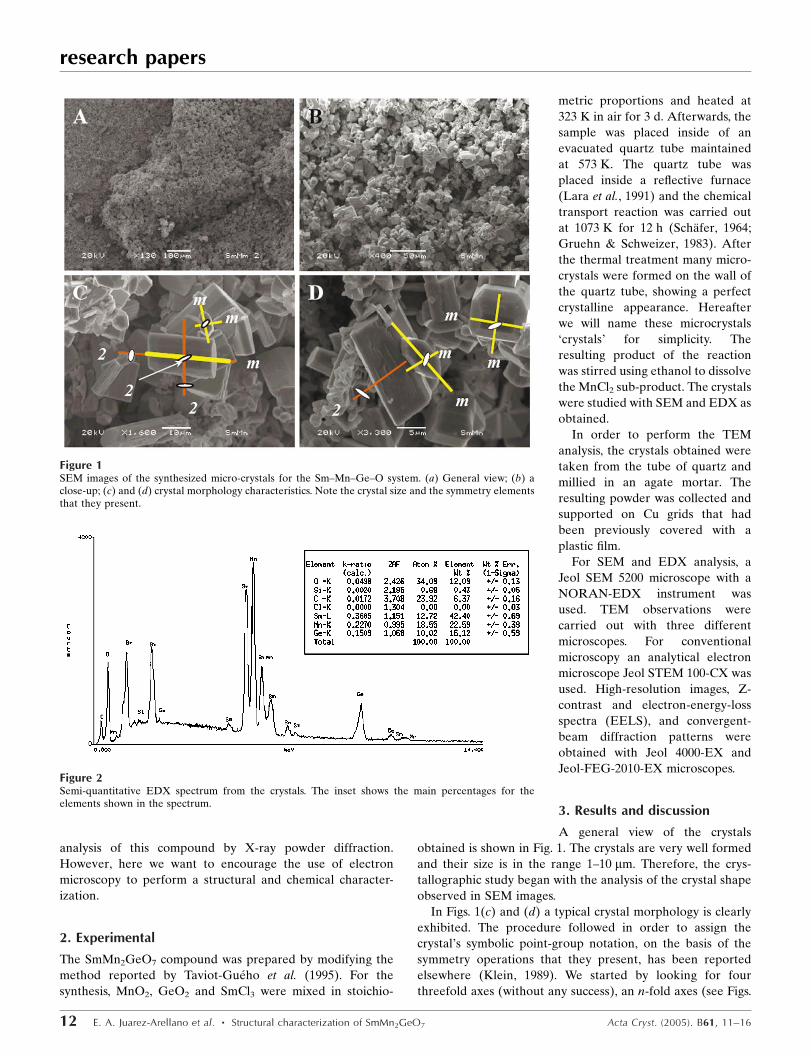
A general view of the crystals
obtained is shown in Fig. 1. The crystals are very well formed
and their size is in the range 1±10 mm. Therefore, the crys-
tallographic study began with the analysis of the crystal shape
observed in SEM images.
In Figs. 1(c) and (d) a typical crystal morphology is clearly
exhibited. The procedure followed in order to assign the
crystal's symbolic point-group notation, on the basis of the
symmetry operations that they present, has been reported
elsewhere (Klein, 1989). We started by looking for four
threefold axes (without any success), an n-fold axes (see Figs.
research papers
12 E. A. Juarez-Arellano et al. � Structural characterization of SmMn2GeO7 Acta Cryst. (2005). B61, 11±16
Figure 1SEM images of the synthesized micro-crystals for the Sm±Mn±Ge±O system. (a) General view; (b) aclose-up; (c) and (d) crystal morphology characteristics. Note the crystal size and the symmetry elementsthat they present.
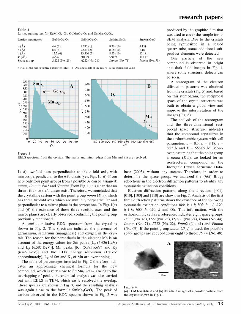
Figure 2Semi-quantitative EDX spectrum from the crystals. The inset shows the main percentages for theelements shown in the spectrum.
1c±d), twofold axes perpendicular to the n-fold axis, with
mirrors perpendicular to the n-fold axis (yes, Figs. 1c±d). From
here only four point groups from a possible 32 can be assigned:
mmm, 4/mmm, �6m2 and 6/mmm. From Fig. 1, it is clear that no
three-, four- or sixfold axes exist. Therefore, we concluded that
the crystalline system with the point group mmm (D2h), which
has three twofold axes which are mutually perpendicular and
perpendicular to a mirror plane, is the correct one. In Figs. 1(c)
and (d) the existence of these three twofold axes and the
mirror planes are clearly observed, con®rming the point group
previously mentioned.
A semi-quantitative EDX spectrum from the crystal is
shown in Fig. 2. This spectrum indicates the presence of
germanium, samarium (manganese) and oxygen in the crys-
tals. The reason for the parenthesis in the element Mn is on
account of the energy values for Sm peaks [L� (5.636 KeV)
and L� (6.587 KeV)], Mn peaks [K� (5.895 KeV) and K�
(6.492 KeV)] and the EDX energy resolution (130 eV
approximately), L� of Sm and K� of Mn are overlapping.
The table of percentages inserted in Fig. 2 therefore indi-
cates an approximate chemical formula for the new
compound, which is very close to SmMn2GeO7. Owing to the
overlapping of peaks, the chemical analysis was also carried
out with EELS in TEM, which easily resolved the overlap.
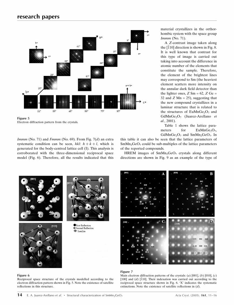
These spectra are shown in Fig. 3, and the resulting analysis
was again close to the formula SmMn2GeO7. The peak of
carbon observed in the EDX spectra shown in Fig. 2 was
produced by the graphite ®lm that
was used to cover the sample for its
SEM analysis. Due to the crystals
being synthesized in a sealed
quartz tube, some additional sub-
product elements were detected.
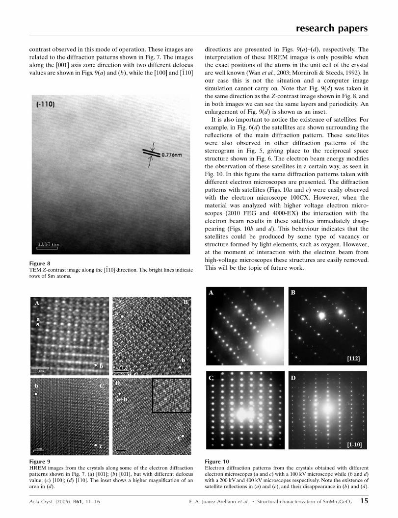
One particle of the new
compound is observed in bright
and dark ®eld images in Fig. 4,
where some structural defects can
be seen.
A stereogram of the electron
diffraction patterns was obtained
from the crystals (Fig. 5) and, based
on this stereogram, the reciprocal
space of the crystal structure was
built to obtain a global view and
improve the interpretation of the
images (Fig. 6).
The analysis of the stereogram
and the three-dimensional reci-
procal space structure indicates
that the compound crystallizes in
the orthorhombic system with cell
parameters a = 8.3, b = 8.18, c =
8.22 AÊ and V = 558.09 AÊ 3. More-
over, assuming that the point group
is mmm (D2h), we looked for an
isostructural compound in the
Inorganic Crystal Structure Data-
base (2003), without any success. Therefore, in order to
determine the space group, we analyzed the (hkl) Bragg
re¯ections in the electron diffraction patterns to identify any
systematic extinction conditions.
Electron diffraction patterns along the directions [001],
[010], [100] and [�110] are shown in Fig. 7. Analysis of the ®rst
three diffraction patterns shows the existence of the following
systematic extinction conditions 0kl: k� l; h0l: h� l; hk0:
h� k; h00: h; 0k0; k and 00l. This information, with the
orthorhombic cell as a reference, indicates eight space groups:
Pnnn (No. 48), I222 (No. 23), I212121 (No. 24), I2mm (No. 44),
Immm (No. 71), F222 (No. 22), Fmm2 (No. 41) and Fmmm
(No. 69). If the point group mmm (D2h) is used, the possible
space groups are reduced from eight to three: Pnnn (No. 48),
research papers
Acta Cryst. (2005). B61, 11±16 E. A. Juarez-Arellano et al. � Structural characterization of SmMn2GeO7 13
Figure 4(a) TEM bright-®eld and (b) dark-®eld images of a powder particle fromthe crystals shown in Fig. 1.
Figure 3EELS spectrum from the crystals. The major and minor edges from Mn and Sm are resolved.
Table 1Lattice parameters for EuMnGe2O7, GdMnGe2O7 and SmMn2GeO7.
Lattice parameters EuMnGe2O7 GdMnGe2O7 SmMn2GeO7 SmMn2GeO7
a (AÊ ) 4.6 (2) 4.735 (1) 8.30 (10) 4.15²b (AÊ ) 8.5 (4) 7.839 (2) 8.18 (10) 8.18c (AÊ ) 12.7 (6) 13.500 (3) 8.22 (10) 12.18³V (AÊ 3) 495.6 501.09 558.76 413.47Space group A222 (No. 21) A222 (No. 21) Immm (No. 71) Immm (No. 71)
² Half of the real `a' lattice parameter value. ³ One and a half of the real `c' lattice parameter value.
Immm (No. 71) and Fmmm (No. 69). From Fig. 7(d) an extra
systematic condition can be seen, hkl: h� k� l, which is
generated for the body-centred lattice cell (I). This analysis is
corroborated with the three-dimensional reciprocal space
model (Fig. 6). Therefore, all the results indicated that this
material crystallizes in the orthor-
hombic system with the space group
Immm (No. 71).
A Z-contrast image taken along
the [�110] direction is shown in Fig. 8.
It is well known that contrast for
this type of image is carried out
taking into account the difference in
atomic number of the elements that
constitute the sample. Therefore,
the element of the brightest lines
may correspond to Sm (the heaviest
element scatters more intensity on
the annular dark ®eld detector than
the lighter ones, Z Sm = 62, Z Ge =
32 and Z Mn = 25), suggesting that
the new compound crystallizes in a
laminar structure that is related to
the structures of EuMnGe2O7 and
GdMnGe2O7 (Juarez-Arellano et
al., 2001).
Table 1 shows the lattice para-
meters for EuMnGe2O7,
GdMnGe2O7 and SmMn2GeO7. In
this table it can also be seen that the lattice parameters of
SmMn2GeO7 could be sub-multiples of the lattice parameters
of the reported compounds.
HREM images of SmMn2GeO7 crystals along different
directions are shown in Fig. 9 as an example of the type of
research papers
14 E. A. Juarez-Arellano et al. � Structural characterization of SmMn2GeO7 Acta Cryst. (2005). B61, 11±16
Figure 6Reciprocal space structure of the crystals modelled according to theelectron diffraction pattern shown in Fig. 5. Note the existence of satellitere¯ections in this structure.
Figure 5Electron diffraction pattern from the crystals.
Figure 7Main electron diffraction patterns of the crystals: (a) [001], (b) [010], (c)[100] and (d) [�110]. Their indexation was carried out according to thereciprocal space structure shown in Fig. 6. `X' indicates the systematicextinctions. Note the existence of satellite re¯ections in (d).
contrast observed in this mode of operation. These images are
related to the diffraction patterns shown in Fig. 7. The images
along the [001] axis zone direction with two different defocus
values are shown in Figs. 9(a) and (b), while the [100] and [�110]
directions are presented in Figs. 9(a)±(d), respectively. The
interpretation of these HREM images is only possible when
the exact positions of the atoms in the unit cell of the crystal
are well known (Wan et al., 2003; Morniroli & Steeds, 1992). In
our case this is not the situation and a computer image
simulation cannot carry on. Note that Fig. 9(d) was taken in
the same direction as the Z-contrast image shown in Fig. 8, and
in both images we can see the same layers and periodicity. An
enlargement of Fig. 9(d) is shown as an inset.
It is also important to notice the existence of satellites. For
example, in Fig. 6(d) the satellites are shown surrounding the
re¯ections of the main diffraction pattern. These satellites
were also observed in other diffraction patterns of the
stereogram in Fig. 5, giving place to the reciprocal space
structure shown in Fig. 6. The electron beam energy modi®es
the observation of these satellites in a certain way, as seen in
Fig. 10. In this ®gure the same diffraction patterns taken with
different electron microscopes are presented. The diffraction
patterns with satellites (Figs. 10a and c) were easily observed
with the electron microscope 100CX. However, when the
material was analyzed with higher voltage electron micro-
scopes (2010 FEG and 4000-EX) the interaction with the
electron beam results in these satellites immediately disap-
pearing (Figs. 10b and d). This behaviour indicates that the
satellites could be produced by some type of vacancy or
structure formed by light elements, such as oxygen. However,
at the moment of interaction with the electron beam from
high-voltage microscopes these structures are easily removed.
This will be the topic of future work.
research papers
Acta Cryst. (2005). B61, 11±16 E. A. Juarez-Arellano et al. � Structural characterization of SmMn2GeO7 15
Figure 8TEM Z-contrast image along the [�110] direction. The bright lines indicaterows of Sm atoms.
Figure 9HREM images from the crystals along some of the electron diffractionpatterns shown in Fig. 7. (a) [001]; (b) [001], but with different defocusvalue; (c) [100]; (d) [�110]. The inset shows a higher magni®cation of anarea in (d).
Figure 10Electron diffraction patterns from the crystals obtained with differentelectron microscopes (a and c) with a 100 kV microscope while (b and d)with a 200 kV and 400 kV microscopes respectively. Note the existence ofsatellite re¯ections in (a) and (c), and their disappearance in (b) and (d).
In the study of the ABGe2O7 compounds (A = rare-earth
elements; B = In, Fe, Mn) our group has synthesized the
compounds AInGe2O7 (A = Fe, Y, Gd, Tb, Ho; Bucio et al.,
2001; Juarez-Arellano, Bucio, Moreno-Tovar et al., 2002;
Juarez-Arellano, Rosales, Bucio & Orozco, 2002; Juarez-
Arellano et al., 2003; Juarez-Arellano, Rosales, Oliver et al.,
2004). The main characteristic of these compounds is the
existence of Ge2O7 diortho groups in the structure (Bucio et
al., 2003). In the new compound we reported, SmMn2GeO7,
this diortho group could still be there, but rather than having a
diortho group formed by two tetrahedra of germanium, it
could have a diortho group formed by the union of a germa-
nium and manganese tetrahedron (MnGeO7).
4. Conclusions
We have performed the structural characterization of micro-
crystals of the compound SmMn2GeO7 by electron micro-
scope. These crystals crystallize in the orthorhombic system
with cell parameters a = 8.3, b = 8.18, c = 8.22 AÊ and V =
558.09 AÊ 3; point group mmm (D2h) and space group Immm
(No. 71).
The authors wish to express theirs thanks to D. Argott de
Juarez, L. Rendon, A. Lara, J. CanÄ etas, P. Mexia, C. MaganÄ a,
C. Zorrilla, S. Tehuacanero, R. HernaÂndez and G. Gamboa-
Espinosa for technical help. They also thank Microscopy
Central Laboratory (LCM-IFUNAM) and DGAPA-UNAM
under projects PAPIIT-IN 113199, IN-104902, IN-120801 and
IN-101003. One of the authors (E. A. Juarez-Arellano)
acknowledges a fellowship from CONACyT.
References
Bucio, L., PeÂrez-Castro, L., Juarez-Arellano, E. A., Moreno-Tovar,R., Rosales, I. & Orozco, E. (2003). Res. Adv. Chem. Mater. 1, 65±75.
Bucio, L., Ruvalcaba-Sil, J. L., Rosales, I., GarcõÂa-Robledo, J. &Orozco, E. (2001). Z. Kristallogr. 216, 438±441.
Duk-Young, J. & Payne, D. A. (1999). Bull. Korean Chem. Soc. 20,824±826.
Gruehn, R. & Schweizer, H. (1983). Angew. Chem. 95, 80±93.Inorganic Crystal Structure Database ICSD Release (2003). National
Institute of Standards & Technology, Gaithersburg. Fachinforma-tionszentrum Karlsruhe.
Juarez-Arellano, E. A., Bucio, L., Hernandez, A. J., Camarillo, E.,Carbonio, R. E. & Orozco, E. (2003). J. Solid State Chem. 170, 418±423.
Juarez-Arellano, E. A., Bucio, L., Moreno-Tovar, R., GarcõÂa-Robledo, J. F. & Orozco, E. (2002). Z. Kristallogr. 217, 201±204.
Juarez-Arellano, E. A., Gamboa-Espinosa, G. V., Lara, J. A., Bucio,L. & Orozco, E. (2001). Latin Am. J. Metall. Mater. 21, 9±12.
Juarez-Arellano, E. A., Rosales, I., Bucio, L. & Orozco, E. (2002).Acta Cryst. C58, i135±i137.
Juarez-Arellano, E. A., Rosales, I., Gamboa-Espinosa, G. V., Lara,J. A., Bucio, L. & Orozco, E. (2004). Cryst. Res. Technol. 39, 833±839.
Juarez-Arellano, E. A., Rosales, I., Oliver, A., Ruvalcaba, J. L.,Carbonio, R. E., Bucio, L. & Orozco, E. (2004). Acta Cryst. C60,i14±i16.
Kim, K. N., Jung, H. K., Park, H. D. & Kim, D. (2002). J. Lumin. 99,169±173.
Klein, C.(1989). Minerals and Rocks, Exercises in Crystallography,Mineralogy and Hand Specimen Petrology, p. 20. New York: JohnWiley and Sons.
Lara, J. A., Riveros, H. G., Reyes-Gasga, J. & YacamaÂn, M. J. (1991).J. Crystal Growth, 109, 137±141.
Morniroli, J. P. & Steeds, J. W. (1992). Ultramicroscopy, 45, 219±239.
Pattison, P., Knudsen, K. D., Cerny, R. & Koller, E. (2000). J.Synchrotron Rad. 7, 251±256.
Rao, C. N. R., Vanitha, P. V. & Cheetham, A. K. (2003). Chem. Eur. J.9, 828±836.
SchaÈfer, H. (1964). Chemical Transport Reaction. New York/London:Academic Press.
Taviot-GueÂho, C., Giaquinta, D., Palvadeau, P. & Rouxel, J. (1995). J.Solid State Chem. 120, 7±11.
Taviot-GueÂho, C., LeÂone, P., Palvadeau, P. & Reuxel, J. (1999). J.Solid State Chem. 143, 145±150.
Vijaya Sarathy, K., Vanitha, P. V., Ram Seshadri, A. K. & Rao,C. N. R. (2001). Chem. Mater. 13, 787±795.
Wan, Z., Liu, Y., Fu, Z., Li, Y., Cheng, T., Li, F. & Fan, H. (2003). Z.Kristallogr. 218, 308±315.
research papers
16 E. A. Juarez-Arellano et al. � Structural characterization of SmMn2GeO7 Acta Cryst. (2005). B61, 11±16