Fluorescence Viewing Systems - Electron Microscopy Sciences
24
Fluorescence Viewing Systems Practical and economical solutions for viewing and photographing fluorescence
-
Upload
khangminh22 -
Category
Documents
-
view
1 -
download
0
Transcript of Fluorescence Viewing Systems - Electron Microscopy Sciences
Fluorescence Viewing Systems
Practical and economical solutions for viewing and
photographing fluorescence

2
Overview The NIGHTSEA Stereo Microscope Fluorescence Adapter adapts just about any stereo microscope (dissecting microscope) for fluorescence with no modification to the microscope itself. The modular design lets you easily switch between several different excitation/emission combinations to work with a variety of fluorescent proteins and other fluorophores. There are now six different excitation/emission combinations available, plus white light.
Adapt your existing lab stereo microscopes for fluorescence
NIGHTSEA Stereo Microscope Fluorescence Adapter
Fluorescence isn’t just for research microscopes anymore... n Now sort on your laboratory-level stereos n Use to facilitate micromanipulation and dissection n Expand from your research lab to your classroom
Modular... n Installs in seconds - just clicks into place n Interchangeable excitation/emission combinations n Move from microscope to microscope n No modification to your microscope needed
Economical — More Glow for the Dough... n Stretch your lab budget n Inexpensive enough for classroom use
Grows as your lab grows... n Buy just what you need now (up to 6 different wavelength sets) n Add more as your needs expand
The Stereo Microscope Fluorescence Adapter system consists of:
• Flexible gooseneck lamp base with power supply
• Adapter for microscope
• Light head • Barrier filter • Filter shield
The light head, barrier filter, and filter shield are interchangeable so that you can easily switch between excitation/emission light+filter combinations.
The microscope mounting adapter fits up to 67mm to work with the majority of stereo microscopes. An oversize adapter and an adapter for the Leica EZ4 series are also available.
Applications This simple system is excellent for:
n Quick screening of your fluorescent genotypes - Drosophila, zebrafish, C. elegans,...
n Genotype sorting n Fluorescence-aided dissection, injection, or micromanipulation n Pre-screening sample preps for confocal or other high-resolution imaging n Freeing up your research-grade fluorescence microscopes for more
demanding work n New faculty start-up budgets n Bringing fluorescence into the teaching laboratory n Coating and failure analysis, circuit board work, defect location,
food safety, paper analysis, and more
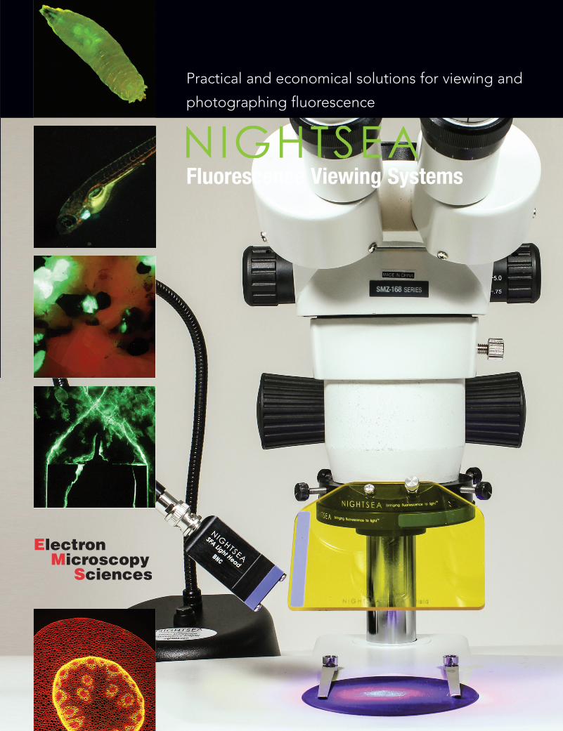
Industry Electronic component failure analysis
Geology Calcite and willemite fluorescence
Fluorescent Proteins GFP-tagged Drosophila larva
Here are samples of what you can see:

3
Once you are set up for one excitation/emission wavelength combination, additional combinations can be added by purchasing a kit that consists of a light head, barrier filter, and viewing shield. These three elements can be removed and replaced in seconds, and color coding ensures that you are using the right combination. The barrier filter clicks on to the ring adapter magnetically, so it is easy to remove it to switch back to white light viewing.
Wavelength Sets Designation Excitation Emission Fluorophores
UV – Ultra Violet 360-380nm 415nm LP DAPI, ... VI – Violet 400-415nm 450nm LP CFP, ... RB – Royal Blue 440-460nm 500nm LP GFP, eGFP, fluorescein... RB-GO – Green Only 440-460nm 500-560nm BP GFP, eGFP, fluorescein... CY – Cyan 490-515nm 550nm LP YFP, Venus, Lucifer Yellow... GR – Green 510-540nm 600nm LP DsRed, dTomato...
Green-Only Barrier Filter The Green-Only (GO) Barrier Filter isolates the green part of the spectrum and is for use with the Royal Blue excitation source. While our other barrier filters are longpass filters this filter is a bandpass, transmitting from approximately 500 to 560nm. The longpass filter has served well for most users who need to visualize green-fluorescent protein (GFP), and if you are exploring fluorescence in nature it is preferable. The primary motivation for adding the green-only filter to the line-up was for the benefit of researchers using GFP in plants such as Arabidopsis thaliana, a common research model. Plants contain chlorophyll, which has a distinctive red fluorescence that can sometimes mask the GFP emission, making it harder to see and photograph.
Lamp Base Light Control Options The SFA lamp base is available in three versions: Standard, DIM, and PULSE. Standard lamp bases have a simple OFF/ON operation. DIM lamp bases feature an OFF/ON/DIM switch to change the intensity of the light. PULSE lamp bases incorporate a BNC connector that accepts a voltage signal to control the excitation source ON/OFF.
Only one control option is available per base. The DIM and PULSE options can be purchased as part of a new system, as a retrofit to an existing base, or in a stand-alone lamp base purchased a la carte.
NIGHTSEA DIM Option
The DIM option adds a combined switch and dimmer control. When you first turn the unit on it is at full power. As you rotate the switch the intensity decreases, reaching about 30% power at the minimum setting. You have finer control in the brighter portion of the adjustment range. An intensity indicator (1-10) makes it easy to record and repeat preferred settings.
NIGHTSEA PULSE Option
The PULSE option adds a BNC connector on the rear of the base that accepts a user-supplied voltage input to turn the light on and off. Some possible applications are:
• External control (computer, function generator, etc.) for precise illumination timing for photoactivation, behavioral experiments
• PWM input for light dimming
• Footswitch for hands-free operation. Contact EMS for details
Dimmer switch option on SFA
Target at max and min excitation
NIGHTSEA Stereo Microscope Fluorescence Adapter (continued)
We tested this new barrier filter with Arabidopsis supplied by Dr. Chip Celenza (Department of Biology, Boston University). These plants express GFP in the roots and vasculature. The images above show examples of plants photographed with the longpass filter (left) and green-only filter (right). There is no chlorophyll in the roots so the GFP is evident there in both images, but the weaker expression in the leaves is much more apparent in the images on the right.
Arabidopsis fluorescence imaged with longpass filter
Arabidopsis fluorescence imaged with bandpass filter
Arabidopsis fluorescence imaged with bandpass filter
Arabidopsis fluorescence imaged with longpass filter
Voltage Input 2.8 - 6.0 VDC for ON, <0.6VDC OFF
Maximum Operation Frequency 10kHz BNC cable not included
See how it works... Learn how to do it... We’ve added video content to our website to help you get to know our latest products even better! Stop by and see what it’s all about.

4
NIGHTSEA Stereo Microscope Fluorescence Adapter (continued)
Ordering Information: Full Systems
Stereo Microscope Fluorescence Adapter, Full System Full system with one illumination color consisting of:
• Lamp Base with Power Supply and International Plug Set • Light Head — Ultraviolet, Violet, Royal Blue, Cyan, or Green • Microscope Mounting Adapter • Barrier Filter • Viewing Shield • Padded Travel Case
Cat. No. Description Qty. Standard Lamp Base SFA-UV Full System with Ultraviolet, Standard Lamp Base each SFA-VI Full System with Violet, Standard Lamp Base each SFA-RB Full System with Royal Blue, Standard Lamp Base each SFA-RB-GO Full System with Royal Blue, Green-Only Barrier Filter, Standard Lamp Base each SFA-CY Full System with Cyan, Standard Lamp Base each SFA-GR Full System with Green, Standard Lamp Base each
DIM Lamp Base SFA-UV-DIM Full System with Ultraviolet, DIM Lamp Base each SFA-VI-DIM Full System with Violet, DIM Lamp Base each SFA-RB-DIM Full System with Royal Blue, DIM Lamp Base each SFA-RB-GO-DIM Full System with Royal Blue, Green-Only Barrier Filter, DIM Lamp Base each SFA-CY-DIM Full System with Cyan, DIM Lamp Base each SFA-GR-DIM Full System with Green, DIM Lamp Base each
PULSE Lamp Base SFA-UV-PULSE Full System with Ultraviolet, PULSE Lamp Base each SFA-VI-PULSE Full System with Violet, PULSE Lamp Base each SFA-RB-PULSE Full System with Royal Blue, PULSE Lamp Base each SFA-RB-GO-PULSE Full System with Royal Blue, Green-Only Barrier Filter, PULSE Lamp Base each SFA-CY-PULSE Full System with Cyan, PULSE Lamp Base each SFA-GR-PULSE Full System with Green, PULSE Lamp Base each
Leica EZ4 Mount Adapter
Full System with Leica EZ4 Adapter The Leica EZ4 Mounting Adapter enables you to use the NIGHTSEA Stereo Microscope Fluorescence System with the Leica EZ4 series of stereo microscopes, with or without integrated camera. Easily attach the barrier filter magnetically and hold the filter shield in place with a thumbscrew. The adapter is available for purchase separately to quickly move between the Leica EZ4 and other stereo microscopes.
Full system with one illumination color consisting of:
• Lamp Base with Power Supply and International Plug Set • Light Head — Ultraviolet, Violet, Royal Blue, Cyan, or Green • Leica EZ4 Mounting Adapter • Barrier Filter • Viewing Shield • Padded Travel Case
Cat. No. Description Qty. Standard Lamp Base SFAZ-UV Full System with Ultraviolet, Standard Lamp Base each SFAZ-VI Full System with Violet, Standard Lamp Base each SFAZ-RB Full System with Royal Blue, Standard Lamp Base each SFAZ-RB-GO Full System with Royal Blue, Green-Only Barrier Filter, Standard Lamp Base each SFAZ-CY Full System with Cyan, Standard Lamp Base each SFAZ-GR Full System with Green, Standard Lamp Base each
DIM Lamp Base SFAZ-UV-DIM Full System with Ultraviolet, DIM Lamp Base each SFAZ-VI-DIM Full System with Violet, DIM Lamp Base each SFAZ-RB-DIM Full System with Royal Blue, DIM Lamp Base each SFAZ-RB-GO-DIM Full System with Royal Blue, Green-Only Barrier Filter, DIM Lamp Base each SFAZ-CY-DIM Full System with Cyan, DIM Lamp Base each SFAZ-GR-DIM Full System with Green, DIM Lamp Base each
PULSE Lamp Base SFAZ-UV-PULSE Full System with Ultraviolet, PULSE Lamp Base each SFAZ-VI-PULSE Full System with Violet, PULSE Lamp Base each SFAZ-RB-PULSE Full System with Royal Blue, PULSE Lamp Base each SFAZ-RB-GO-PULSE Full System with Royal Blue, Green-Only Barrier Filter, PULSE Lamp Base each SFAZ-CY-PULSE Full System with Cyan, PULSE Lamp Base each SFAZ-GR-PULSE Full System with Green, PULSE Lamp Base each
Leica Adapter Only SFAZ-AD Leica EZ4 Adapter only each

5
NIGHTSEA Stereo Microscope Fluorescence Adapter (continued)
Cat. No. Description Qty. SFA-LFS-UV Add-On Light and Filter Set, Ultraviolet each
Cat. No. Description Qty. SFA-LFS-VI Add-On Light and Filter Set, Violet each
Cat. No. Description Qty. SFA-LFS-RB Add-On Light and Filter Set, Royal Blue each
Cat. No. Description Qty. SFA-LFS-RB-GO Add-On Light and Filter Set, Green-Only each
Cat. No. Description Qty. SFA-LFS-CY Add-On Light and Filter Set, Cyan each
Cat. No. Description Qty. SFA-LFS-GR Add-On Light and Filter Set, Green each
Ultraviolet
Violet
Royal Blue
Green-Only
Cyan
Green
• Light Head • Barrier Filter • Viewing Shield • Padded Storage Box
Add-On Light and Filter Sets Each add-on excitation/ emission set consists of:
Collection of Stage 37-38 X. laevis, messenger RNA injected ubiquitous GFP and membrane RFP viewed through filter shields for green and red fluorescence.
The NIGHTSEA Stereo Microscope Fluorescence Adapter in Action... ...at the National Xenopus Resource (NXR) at the Marine Biological Laboratory in Woods Hole, MA — special thanks to NXR Director and Bell Center Scientist Dr Marko Horb and his postdoctoral scientist Dr Matthew Salanga. The fluorescence adapter system worked great for visualizing all of the fluorescence, both injected and transgenic, in the specimens. The pictures below show both positive- and negative-expressing Stage 46 X. laevis under white light, Royal Blue excitation (green fluorescence), and Green excitation (red fluorescence).
In addition to seeing the fluorescence through the eyepieces, you could easily distinguish presence/absence and relative strength of expression just by looking through the filter shield.

Light Head Hangers and Cables The Light Head Hanger system is an accessory for the NIGHTSEA Model SFA Stereo Microscope Fluorescence Adapter. It is a combination of power extension cables and a custom made holder that enables you to take the fluorescence excitation light heads off the gooseneck and mount them directly on the SFA adapter ring. In addition, it provides the capability to drive two light heads from one lamp base.
The extension cables plug into the gooseneck lamp base and can bring power to either one or two light heads.
The Light Head Hanger mounts to the end of any of the thumbscrews on the SFA adapter ring. It can be configured for use with either long or short working distance microscopes.
• Long working distance (LWD) – 6.5 cm (2.56") and greater • Short working distance (SWD) – 6.5-4.6 cm (2.5-1.8") There are two main potential benefits with this accessory.
1) Increase excitation intensity without buying a second lamp base. Without this system, if you wanted to increase the fluorescence excitation intensity of the SFA, you would need to buy a second light head and lamp base. This would take up additional real estate on your laboratory bench. With the Light Head Hanger system you can power two light heads from one base and mount them both directly to the NIGHTSEA adapter ring.
2) Reduce the chance of accidentally bumping the light. Taking the light head off the gooseneck and mounting it directly to our microscope adapter ring reduces the chance of the light head being bumped so that it is not illuminating the subject on the microscope stage to best effect. This might be especially valuable if you are using the system for outreach demonstrations and multiple people will be working around the microscope.
Switch Box Kit The Switch Box Kit enables you to mount two different wavelength excitation light heads on your microscope and switch between them easily. You will still need to manually change the barrier filter, but it eliminates the need to swap out light heads on the gooseneck.
The Switch Box Kit is available only for SFA systems that use the BNC light head connector, and not the older V1 connector.
Cat No. Description Qty. SFA-LHC Single Light Head Cable each SFA-DLHC Dual Light Head Cable each SFA-LHH Light Head Hanger each SFA-HK Hanger Kit for Single Light Head, includes Single Light Head Cable and one Light Head Hanger each SFA-DHK Hanger Kit for Dual Light Heads, includes Dual Light Head Cable and two Light Head Hangers each
Light head hangers configured for LWD (left) and SWD
microscopes (right)
Single SFA light head attached to the gooseneck by a Light Head Cable
Two SFA light heads attached to the gooseneck by a Dual Light Head Cable
6
Cat No. Description Qty. SFA-SWK Switch Box Kit, includes BNC switch box, 3 BNC cables, 2 LH Hangers kit
Two SFA Light Heads mounted to adapter ring with Light Head Hangers
NIGHTSEA Stereo Microscope Fluorescence Adapter Accessories
“We had some issue detecting red fluorescent proteins with some of our weaker transgenic zebrafish lines, but by shining two LED lights onto the same embryo, most of our weakest GFP and mCherry transgenic lines can now be detected. One light head was fine for reasonably bright specimens, and with two lights, the fluorescence is nearly the same as traditional stereomicroscopes priced $15-20K.”
Note: If you are ordering a Dual kit to power two light heads at once for increased intensity, you will need to have two light heads of the same color. Above prices do not include the cost of an additional light head.
The default configuration is for systems with the BNC connector on the lamp base and the light head. Cables are available for the pre-2015 V1 connector on request.

7
NIGHTSEA Stereo Microscope Fluorescence Accessories (continued)
Cat No. Description Qty. SFA-MEK SFA Mobile Extension Kit kit
Cat No. Description Qty. SFA-LH-IR850 IR Light Head each SYS-IR850 IR Light Head plus NIGHTSEA DIM Base each
SFA Mobile Extension Kit The Mobile Extension Kit provides added versatility to your SFA microscope fluorescence adapter system by enabling you to use the light head as a handheld light source. Do you work with both macro and micro subjects? Do you want to screen larger specimens before dissection or other microscope work? Can’t fit everything under the microscope? Then the SFA-MEK Mobile Extension Kit is a way to get more use out of your system.
The kit includes three components: • Rubber-coated hand grip with wrist lanyard and custom-made enclosure
that holds an SFA Light Head securely. The slots on the side of the enclosure provide for cooling and also let you see the color code of the installed light head.
• Barrier filter glasses. See page 23 for more information. • Lightweight extension cable.
To use the kit just take the light head off the SFA gooseneck base, connect it to one end of the extension cable, and attach the other end of the cable to the gooseneck. Insert the light head in the holder and screw down the thumbscrew to secure it in place. Put on the filter glasses that correspond to the light head color and you’re ready to go! If you want to use the Mobile Extension with more than one excitation wavelength you can add additional barrier filter glasses at any time.
Infrared Light Source Researchers sometimes need an infrared light source for non-intrusive observation of behavior. And sometimes the subjects are small and you need to make the observations with a stereo microscope. The SFA-LH-IR850 is an 850nm light source that is compatible with the NIGHTSEA Model SFA Stereo Microscope Fluorescence Adapter. The light head plugs directly into the lamp base supplied with the SFA system. You can acquire the light head as a supplement to the SFA system that you already own for fluorescence observation, or you can acquire it in combina-tion with a lamp base as a stand-alone device.
The light head incorporates a high power 850nm LED and a medium beamwidth diffus-ing lens to create a smooth illumination area. The output is centered at 850nm with a FWHM (full width at half maximum) of approximately 50nm. There is virtually no emission at wavelengths shorter than 750nm.
We recommend that you use the DIM base with the LH-IR850 in order to have finer control of illumination intensity.
Inspecting artwork with the SFA Mobile Extension Kit.
Hand grip with Royal Blue light head installed and connected to the power cable, and the Royal Blue filter glasses.
A test target with circles of 2, 5, and 10cm diameter.
The photo on the left above was taken with a conventional camera under normal room light, and with the IR light head illuminated at full power, directed at the target center from a distance of approximately 10cm (4 in.). The illumination spot is not visible either to this camera or to the naked eye. The photo on the right was made under the same lighting conditions, but with a camera that had been modified to image only infrared light. Please note that infrared light is not visible to the human eye and to many cameras. You will need an appropriate camera to record the observations.
850nm IR Light Head
850nm IR Light Head
on SFA lamp base
Modular White Light Head This is an extremely convenient option for general illumination and as a focusing aid for fluorescence imaging. Now if you are using the NIGHTSEA system for fluorescence you do not need a separate white light source. Just exchange the fluorescence excitation light head module for the white-light module in a matter of seconds.
Cat No. Description Qty. SFA-LH-WH Modular White Light Head each

8
Compact battery pack that can run the Stereo Microscope Fluorescence Adapter (SFA) all day long. The battery enables truly portable operation so that you can set up demos anywhere or explore fluorescence in the field, without having to worry about plugging into the power grid.
Just plug the battery into the SFA base instead of the usual power connector. The battery has been tested at over 8 hours of continuous operation, and it will last even longer if you turn the system off when you don't need it. Plug it into the included charger overnight to recharge.
SFA + Eclipse MicroTent + Battery =
Fluorescence Everywhere Combine the battery with the Eclipse MicroTent and you not only don't need a place to plug in, you don't even need to be in the dark! We have used this combination to do fluorescence microscopy at a beach in the middle of the day, in the desert at high noon, and more. Fluorescence can be found everywhere, and now you have the tools to go there.
Cat No. Description Qty. SFA-BATT Battery and Charger each
Battery Type Nickel Metal Hydride (NiMH)
Capacity 12V, 3.8 Ah
Dimensions 11.4 x 8.9 x 5.6 cm (4.5 x 3.5x 2.19 in)
Weight 0.7 kg (1 lb 9 oz)
Operation Duration 8 hours continuous
Charge Time Overnight
Charger 50/60 Hz, 110/220V (US type plug)
Specifications
Cat No. Description Qty. SFA-TENT Eclipse MicroTent™ each
Eclipse MicroTent™ The patented (US Pat. No. 10,175,467) Eclipse MicroTent™ is a unique product for fluorescence microscopy that provides local darkness around conventional laboratory stereo microscopes. Fluorescence microscopes are kept in dark rooms for good reason - fluorescence can be weak and in many cases it can be difficult to see well if there is any ambient light. Microscopes may be on lab benches in shared spaces, near windows, or in other difficult-to-darken locations such as in the field. Turning off overhead lights can help but inconveniences others.
The Eclipse MicroTent™ creates local darkness around a microscope while still providing easy access to the sample stage and the focus and zoom controls. It is designed for stereo microscopes but could potentially be used with many varieties of compound microscopes.
Features z Opening for the microscope oculars with
elastic sleeve to minimize light entry z Large front flap provides easy access to the
sample stage and can fasten open z Arm slots on sides to provide access to focus
and zoom controls z User-customizable feedthrough patches to
provide additional penetrations for camera port, power cords, CO2 lines, or other features as you need
z Tru-Block™ Eye Shields included with every Eclipse MicroTent™
z Folds flat for storage Dimensions: 46 x 30 x 50 cm (18 x 12 x 20 in.)
Battery and Charger
NIGHTSEA Stereo Microscope Fluorescence Accessories (continued)

9
Cat No. Description Qty.
SFA Light Heads: SFA-LH-UV Light Head, Ultra Violet each SFA-LH-VI Light Head, Violet each SFA-LH-RB Light Head, Royal Blue each SFA-LH-CY Light Head, Cyan each SFA-LH-GR Light Head, Green each
SFA Barrier Filters: SFA-BF-UV Barrier Filter, Ultra Violet each SFA-BF-VI Barrier Filter, Violet each SFA-BF-RB Barrier Filter, Royal Blue each SFA-BF-RB-GO Barrier Filter, Green Only each SFA-BF-CY Barrier Filter, Cyan each SFA-BF-GR Barrier Filter, Green each
SFA Filter Shields: SFA-SH-UV Filter Shield, Ultra Violet each SFA-SH-VI Filter Shield, Violet each SFA-SH-RB Filter Shield, Royal Blue each SFA-SH-RB-GO Filter Shield, Green Only each SFA-SH-CY Filter Shield, Cyan each SFA-SH-GR Filter Shield, Green each
SFA Adapters: SFA-AD Adapter each SFAZ-AD Leica EZ4 Adapter each SFA-XL-AD Oversize Adapter each
SFA Bases: SFA-BASE Standard Base each SFA-BASE-DIM DIM Base each SFA-BASE-PULSE PULSE Base each
SFA a la carte Need an extra, not a set? Order from here:
NIGHTSEA DIM and PULSE Retrofit Options The DIM and PULSE options for the base (see page 3) can be retrofitted to existing Model SFA Stereo Microscope Fluorescence Adapters.
Cat No. Description Qty. SFA-BASE-DIM-R DIM Option — Retrofit each SFA-BASE-PULSE-R PULSE Option — Retrofit each
The combination of Tru-Block Eye Shields and the Eclipse MicroTent™ give you the freedom to do Fluorescence Everywhere™. The photograph on the left below shows NIGHTSEA founder Charles Mazel using the Stereo Microscope Fluorescence Adapter, powered by a battery, in combination with the tent and the Tru-Block Eye Shields to do fluorescence microscopy on Nobska Beach in Woods Hole, Massachusetts.
The image on the right is a fluorescence photograph of a mollusk shell and operculum taken using the system – in the middle of the afternoon. The red fluorescence on the shell arises from chlorophyll in algae growing on the shell.
Eye Shields Light entering your eyes from the side can interfere with what you want to see in microscopy in general, and fluorescence microscopy in particular. Eye cups are available, but the standard ones don't extend far from the microscope and don't do a good job as ambient light increases. Our soft, molded rubber high-sided microscope eye shields are the answer. The tall wings extend up far enough to truly shield your eyes from any level of ambient light and eliminate distractions so that you can see your subject better. Two pairs (one Standard, one Compact) are included with every Eclipse MicroTent™, and you can also purchase them separately.
Tru-Block Eye Shields are available in two sizes:
Standard: fits 36 - 45mm (1.45 - 1.75")
Compact: fits 28 - 37mm (1.10 - 1.46")
Eye Shields, up for use
Eye Shields, folded down
Using the microscope with the eye shields
Cat No. Description Qty. SFA-EYE-S Tru-Block Eye Shields - Standard set SFA-EYE-C Tru-Block Eye Shields - Compact set
NIGHTSEA Stereo Microscope Fluorescence Accessories (continued)

10
Fluorescent mineral sample imaged with the Keyence + NIGHTSEA systems
Keyence VH-ZST lens, fiber optic port with NIGHTSEA adapter and light head
Z100 and ZST lenses – the barrier filter inserts in the Analyzer slot at the top of the lens.
Overview The NIGHTSEA Model SFA Fluorescence Adapter system adds a versatile fluorescence imaging capability to the Keyence VHX series of digital microscopes. The system is being used successfully by a growing number of Keyence owners for a variety of applications. Visit our website for a full gallery of images made with the NIGHTSEA adapter and the Keyence microscope.
Fluorescence solutions for most Keyence lenses Keyence offers a variety of lenses for imaging at different scales. NIGHTSEA offers several variants of our fluorescence adapter system that work with the most commonly used Keyence lenses – the VHX-7100, VH-ZST, -Z100, -Z50, -Z20, and -Z00.
The key elements of any fluorescence system are:
n A light source that produces sufficient energy in the appropriate wavelength range to excite fluorescence in the sample of interest
n A barrier filter in the viewing path that blocks reflected excitation light while transmitting the fluorescence emitted by the sample
NIGHTSEA implements these for the Keyence system with:
n high intensity LED light sources available in five excitation wavelength ranges (see list on next page)
n emission barrier filters that can be added to the Keyence lenses easily and non-invasively
The components of a NIGHTSEA system that adds a single excitation/emission wavelength combination on a Keyence microscope are:
n Flexible gooseneck lamp base with LED drive circuit and intensity control; n Universal power supply (120/240VAC, 50/60Hz) with international plug set n Excitation light source n Emission barrier filter n Barrier filter glasses for fluorescence viewing and eye safety n Padded carrying case
Additional wavelength sets are implemented by adding a matched set (light plus filter set) consisting of a light source, barrier filter, and filter glasses.
NIGHTSEA Model SFA Fluorescence Adapter for Keyence VHX
Cement thin section fluorescence, VH-Z100 lens, Royal Blue excitation, 700x
Epoxy on motor shaft, VH-ZST lens, Ultraviolet excitation, 20x
Z00, Z20, and Z50 lenses – the barrier filter slips over the bottom of the lens.
Here are samples of what you can see:
Lens-specific solutions The implementation approaches for the supported Keyence lenses are summarized here.
VH-Z00, -Z20, -Z50 Light source – placed to the side of the microscope stage
Barrier filter – slips over the bottom of the lens and attaches with thumbscrews
VH-Z100 Light source – placed to the side of the microscope stage
Barrier filter – inserts in the Analyzer slot at the top of the lens
VH-ZST • 20-200 lens Light source – placed to the side of
the microscope stage
Barrier filter – inserts in the Analyzer slot at the top of the lens
• 200-2000 lens Light source – mounts over the
Keyence fiber optic input port (except for UV) providing enhanced performance at high magnification
Barrier filter – inserts in the Analyzer slot at the top of the lens
VHX-7100 Fully Integrated (FI) Head • VHX-E20 and -E100 Light source – placed to the side of
the microscope stage
Barrier filter – inserts in the Analyzer slot at the right side of the FI head
• VHX-E500 and -E2500 Use with these lenses is likely not
practical due to short working distance
Fluorescent-dosed plastic particles, 38 – 45 microns. Image made with ultraviolet excitation. Image courtesy of an explosives detection company.
11
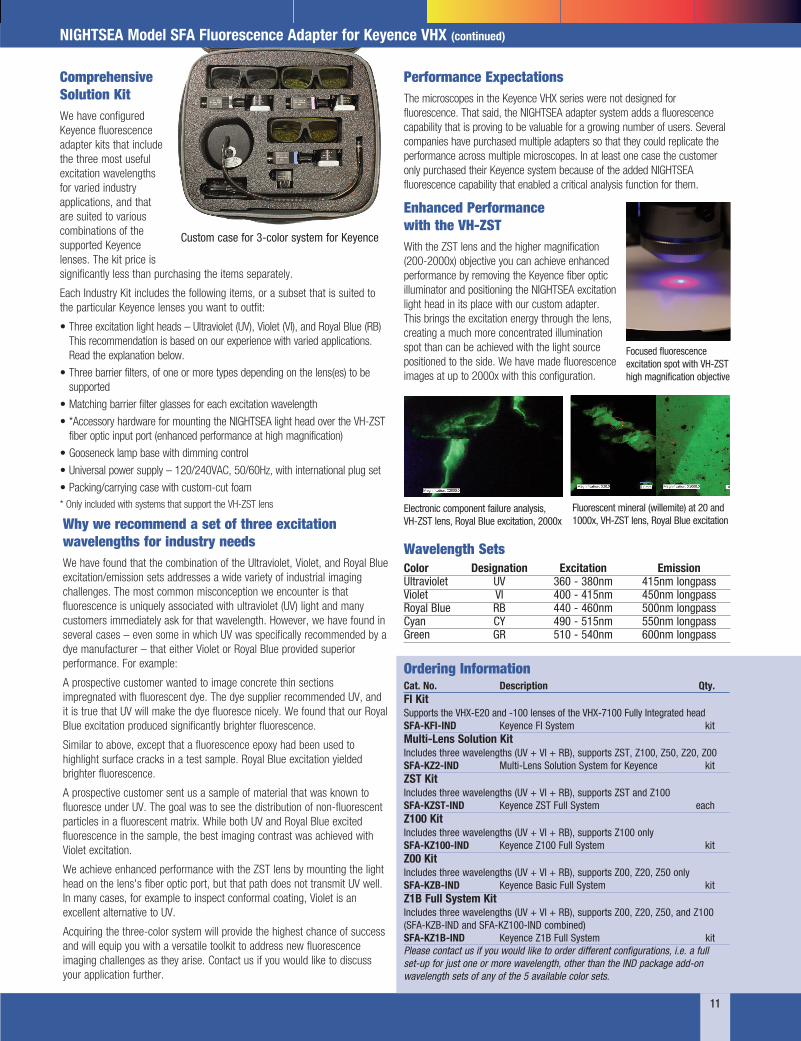
Comprehensive Solution Kit We have configured Keyence fluorescence adapter kits that include the three most useful excitation wavelengths for varied industry applications, and that are suited to various combinations of the supported Keyence lenses. The kit price is significantly less than purchasing the items separately.
Each Industry Kit includes the following items, or a subset that is suited to the particular Keyence lenses you want to outfit:
• Three excitation light heads – Ultraviolet (UV), Violet (VI), and Royal Blue (RB) This recommendation is based on our experience with varied applications. Read the explanation below.
• Three barrier filters, of one or more types depending on the lens(es) to be supported
• Matching barrier filter glasses for each excitation wavelength
• *Accessory hardware for mounting the NIGHTSEA light head over the VH-ZST fiber optic input port (enhanced performance at high magnification)
• Gooseneck lamp base with dimming control
• Universal power supply – 120/240VAC, 50/60Hz, with international plug set
• Packing/carrying case with custom-cut foam * Only included with systems that support the VH-ZST lens
Performance Expectations The microscopes in the Keyence VHX series were not designed for fluorescence. That said, the NIGHTSEA adapter system adds a fluorescence capability that is proving to be valuable for a growing number of users. Several companies have purchased multiple adapters so that they could replicate the performance across multiple microscopes. In at least one case the customer only purchased their Keyence system because of the added NIGHTSEA fluorescence capability that enabled a critical analysis function for them.
Why we recommend a set of three excitation wavelengths for industry needs We have found that the combination of the Ultraviolet, Violet, and Royal Blue excitation/emission sets addresses a wide variety of industrial imaging challenges. The most common misconception we encounter is that fluorescence is uniquely associated with ultraviolet (UV) light and many customers immediately ask for that wavelength. However, we have found in several cases – even some in which UV was specifically recommended by a dye manufacturer – that either Violet or Royal Blue provided superior performance. For example:
A prospective customer wanted to image concrete thin sections impregnated with fluorescent dye. The dye supplier recommended UV, and it is true that UV will make the dye fluoresce nicely. We found that our Royal Blue excitation produced significantly brighter fluorescence.
Similar to above, except that a fluorescence epoxy had been used to highlight surface cracks in a test sample. Royal Blue excitation yielded brighter fluorescence.
A prospective customer sent us a sample of material that was known to fluoresce under UV. The goal was to see the distribution of non-fluorescent particles in a fluorescent matrix. While both UV and Royal Blue excited fluorescence in the sample, the best imaging contrast was achieved with Violet excitation.
We achieve enhanced performance with the ZST lens by mounting the light head on the lens's fiber optic port, but that path does not transmit UV well. In many cases, for example to inspect conformal coating, Violet is an excellent alternative to UV.
Acquiring the three-color system will provide the highest chance of success and will equip you with a versatile toolkit to address new fluorescence imaging challenges as they arise. Contact us if you would like to discuss your application further.
Enhanced Performance with the VH-ZST With the ZST lens and the higher magnification (200-2000x) objective you can achieve enhanced performance by removing the Keyence fiber optic illuminator and positioning the NIGHTSEA excitation light head in its place with our custom adapter. This brings the excitation energy through the lens, creating a much more concentrated illumination spot than can be achieved with the light source positioned to the side. We have made fluorescence images at up to 2000x with this configuration.
Custom case for 3-color system for Keyence
Fluorescent mineral (willemite) at 20 and 1000x, VH-ZST lens, Royal Blue excitation
Electronic component failure analysis, VH-ZST lens, Royal Blue excitation, 2000x
Cat. No. Description Qty. FI Kit Supports the VHX-E20 and -100 lenses of the VHX-7100 Fully Integrated head SFA-KFI-IND Keyence FI System kit Multi-Lens Solution Kit Includes three wavelengths (UV + VI + RB), supports ZST, Z100, Z50, Z20, Z00 SFA-KZ2-IND Multi-Lens Solution System for Keyence kit ZST Kit Includes three wavelengths (UV + VI + RB), supports ZST and Z100 SFA-KZST-IND Keyence ZST Full System each Z100 Kit Includes three wavelengths (UV + VI + RB), supports Z100 only SFA-KZ100-IND Keyence Z100 Full System kit Z00 Kit Includes three wavelengths (UV + VI + RB), supports Z00, Z20, Z50 only SFA-KZB-IND Keyence Basic Full System kit Z1B Full System Kit Includes three wavelengths (UV + VI + RB), supports Z00, Z20, Z50, and Z100 (SFA-KZB-IND and SFA-KZ100-IND combined) SFA-KZ1B-IND Keyence Z1B Full System kit Please contact us if you would like to order different configurations, i.e. a full set-up for just one or more wavelength, other than the IND package add-on wavelength sets of any of the 5 available color sets.
Ordering Information
NIGHTSEA Model SFA Fluorescence Adapter for Keyence VHX (continued)
Wavelength Sets Color Designation Excitation Emission Ultraviolet UV 360 - 380nm 415nm longpass Violet VI 400 - 415nm 450nm longpass Royal Blue RB 440 - 460nm 500nm longpass Cyan CY 490 - 515nm 550nm longpass Green GR 510 - 540nm 600nm longpass
Focused fluorescence excitation spot with VH-ZST high magnification objective
12
Comprehensive Solution Kit We have configured a versatile Hirox fluorescence adapter kit that includes the most useful excitation wavelengths for varied industry applications. The SFA-H-IND kit includes the following items, at a package price that is significantly less than purchasing the items separately:
• Three excitation light heads – Ultraviolet (UV), Violet (VI), and Royal Blue (RB). This recommendation is based on our experience with varied applications
• Three barrier filters paired to the excitation light heads
• Matching barrier filter glasses for each excitation wavelength
• Gooseneck lamp base with dimming control
• Universal power supply – 120/240VAC, 50/60Hz, with international plug set
• Packing/carrying case with custom-cut foam
We offer additional wavelength sets that may be of use for other applications. See table on next page.
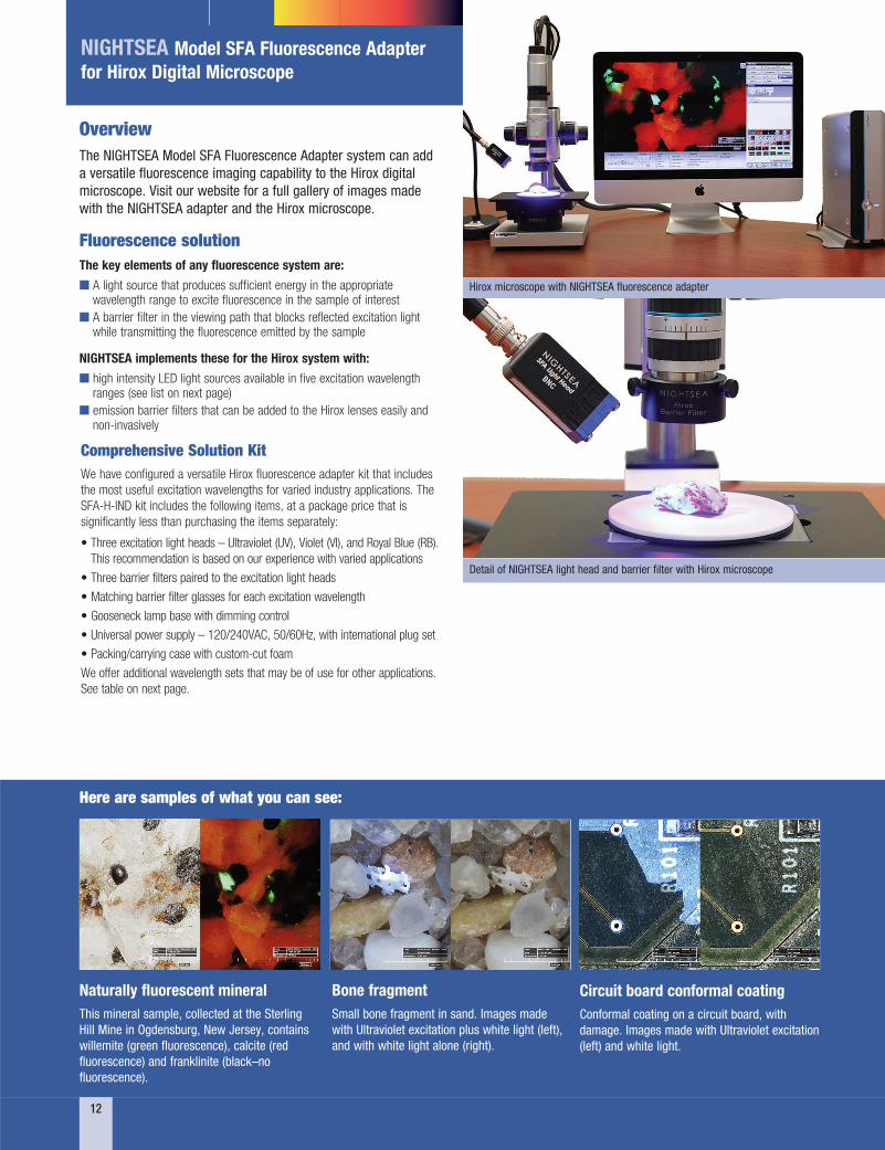
Overview The NIGHTSEA Model SFA Fluorescence Adapter system can add a versatile fluorescence imaging capability to the Hirox digital microscope. Visit our website for a full gallery of images made with the NIGHTSEA adapter and the Hirox microscope.
Fluorescence solution The key elements of any fluorescence system are:
n A light source that produces sufficient energy in the appropriate wavelength range to excite fluorescence in the sample of interest
n A barrier filter in the viewing path that blocks reflected excitation light while transmitting the fluorescence emitted by the sample
NIGHTSEA implements these for the Hirox system with:
n high intensity LED light sources available in five excitation wavelength ranges (see list on next page)
n emission barrier filters that can be added to the Hirox lenses easily and non-invasively
NIGHTSEA Model SFA Fluorescence Adapter for Hirox Digital Microscope
Circuit board conformal coating Conformal coating on a circuit board, with damage. Images made with Ultraviolet excitation (left) and white light.
Bone fragment Small bone fragment in sand. Images made with Ultraviolet excitation plus white light (left), and with white light alone (right).
Naturally fluorescent mineral This mineral sample, collected at the Sterling Hill Mine in Ogdensburg, New Jersey, contains willemite (green fluorescence), calcite (red fluorescence) and franklinite (black–no fluorescence).
Here are samples of what you can see:
Hirox microscope with NIGHTSEA fluorescence adapter
Detail of NIGHTSEA light head and barrier filter with Hirox microscope

13
Why we recommend a set of three excitation wavelengths for industry needs We have found that the combination of the Ultraviolet, Violet, and Royal Blue excitation/emission sets addresses a wide variety of industrial imaging challenges. The most common misconception we encounter is that fluorescence is uniquely associated with ultraviolet (UV) light and many customers immediately ask for that wavelength. However, we have found in several cases – even some in which UV was specifically recommended by a dye manufacturer – that either Violet or Royal Blue provided superior performance. For example:
A prospective customer wanted to image concrete thin sections impregnated with fluorescent dye. The dye supplier recommended UV, and it is true that UV will make the dye fluoresce nicely. We found that our Royal Blue excitation produced significantly brighter fluorescence.
Similar to above, except that a fluorescence epoxy had been used to highlight surface cracks in a test sample. Royal Blue excitation yielded brighter fluorescence.
A prospective customer sent us a sample of material that was known to fluoresce under UV. The goal was to see the distribution of non-fluorescent particles in a fluorescent matrix. While both UV and Royal Blue excited fluorescence in the sample, the best imaging contrast was achieved with Violet excitation.
Acquiring the three-color system will provide the highest chance of success and will equip you with a versatile toolkit to address new fluorescence imaging challenges as they arise. Contact us if you would like to discuss your application further.
In addition to the Comprehensive Solution Kits, you can also purchase single-color full systems that provide everything you need to get started with one excitation/emission combination. Once you have a full system, additional wavelength sets can be ordered separately. These contain the new light head, microscope barrier filter, and barrier filter glasses.
Cat. No. Description Qty. Best Value Full System Kit Includes three wavelengths (UV + VI + RB) SFA-H-IND Best Value Full System kit Single Wavelength Full Systems SFA-H-UV Hirox Single Wavelength Full System - UV kit SFA-H-VI Hirox Single Wavelength Full System - Violet kit SFA-H-RB Hirox Single Wavelength Full System - Royal Blue kit SFA-H-CY Hirox Single Wavelength Full System - Cyan kit SFA-H-GR Hirox Single Wavelength Full System - Green kit Add-on Single Wavelengths SFA-H-LFS-UV Hirox Single Wavelength Add-On - UV each SFA-H-LFS-VI Hirox Single Wavelength Add-On - Violet each SFA-H-LFS-RB Hirox Single Wavelength Add-On - Royal Blue each SFA-H-LFS-CY Hirox Single Wavelength Add-On - Cyan each SFA-H-LFS-GR Hirox Single Wavelength Add-On - Green each
Ordering Information
NIGHTSEA Model SFA Fluorescence Adapter for Hirox (continued)
Wavelength Sets There are five excitation/emission wavelength combinations available for the system.
Color Designation Excitation Emission Ultraviolet UV 360 - 380nm 415nm longpass Violet VI 400 - 415nm 450nm longpass Royal Blue RB 440 - 460nm 500nm longpass Cyan CY 490 - 515nm 550nm longpass Green GR 510 - 540nm 600nm longpass
Fluorescent penetrant highlighting cracks in integrated circuit, 80x, white light (top) and fluorescence under Royal Blue excitation.
Electronic component failure analysis Examining an electronic component that was embedded in epoxy, cross-sectioned, polished, and highlighted with a fluorescent epoxy-like dye. While the manufacturer of the fluorescent dye recommended excitation with ultraviolet light, our Royal Blue light head was used for these images, and was found to be superior to ultraviolet.
Nylon granules with gel defect, 20x (top) and 80x (bottom), white light and fluorescence under Royal Blue excitation.
Gel defect in a nylon granule Nylon 6,6 granules can manifest a process defect called “gel” and if there is too much of this in a production batch it can compromise downstream production. The gel shows up as a brighter fluorescent area within the fluorescing granule. Images made with Royal Blue excitation.

14
Comprehensive Solution Kit We have configured a versatile DVM6 fluorescence adapter kit that includes the most useful excitation wavelengths for varied industry applications. The SFA-DVM6-IND kit includes the following items, at a package price that is significantly less than purchasing the items separately:
• Three excitation light heads – Ultraviolet (UV), Violet (VI), and Royal Blue (RB). This recommendation is based on our experience with varied applications
• Three barrier filters and filter shields paired to the excitation light heads
• Gooseneck lamp base with intensity control
• Universal power supply – 120/240VAC, 50/60Hz, with international plug set
• Packing/carrying case with custom-cut foam
We offer additional wavelength sets that may be of use for other applications. See table on next page.
Overview The NIGHTSEA Model SFA Fluorescence Adapter system can add a versatile fluorescence imaging capability to the Leica DVM6 digital microscope. Visit our website for a full gallery of images made with the NIGHTSEA adapter and the Leica DVM6 microscope.
Fluorescence solution The key elements of any fluorescence system are:
n A light source that produces sufficient energy in the appropriate wavelength range to excite fluorescence in the sample of interest
n A barrier filter in the viewing path that blocks reflected excitation light while transmitting the fluorescence emitted by the sample
NIGHTSEA implements these for the Leica system with:
n high intensity LED light sources available in five excitation wavelength ranges (see list further down page)
n emission barrier filters that can be added to the Leica DVM6 objectives easily and non-invasively
NIGHTSEA Model SFA Fluorescence Adapter for Leica DVM6 Digital Microscope
Concrete thin sectionElectronic component failure analysis
Here are samples of what you can see:

15
Why we recommend a set of three excitation wavelengths for industry needs We have found that the combination of the Ultraviolet, Violet, and Royal Blue excitation/emission sets addresses a wide variety of industrial imaging challenges. The most common misconception we encounter is that fluorescence is uniquely associated with ultraviolet (UV) light and many customers immediately ask for that wavelength. However, we have found in several cases – even some in which UV was specifically recommended by a dye manufacturer – that either Violet or Royal Blue provided superior performance. For example
A prospective customer wanted to image concrete thin sections impregnated with fluorescent dye. The dye supplier recommended UV, and it is true that UV will make the dye fluoresce nicely. We found that our Royal Blue excitation produced significantly brighter fluorescence.
Similar to above, except that a fluorescence epoxy had been used to highlight surface cracks in a test sample. Royal Blue excitation yielded brighter fluorescence.
A prospective customer sent us a sample of material that was known to fluoresce under UV. The goal was to see the distribution of non-fluorescent particles in a fluorescent matrix. While both UV and Royal Blue excited fluorescence in the sample, the best imaging contrast was achieved with Violet excitation.
Acquiring the three-color system will provide the highest chance of success and will equip you with a versatile toolkit to address new fluorescence imaging challenges as they arise. Contact us if you would like to discuss your application further.
In addition to the Comprehensive Solution Kits, you can also purchase single-color full systems that provide everything you need to get started with one excitation/emission combination. Once you have a full system, additional wavelength sets can be ordered separately. These contain the new light head, microscope barrier filter, and barrier filter shield.
Cat. No. Description Qty. Best Value Full System Kit Includes three wavelengths (UV + VI + RB) SFA-DVM6-IND Best Value Full System for DVM6 kit Single Wavelength Full Systems SFA-DVM6-UV DVM6 Single Wavelength Full System - UV kit SFA-DVM6-VI DVM6 Single Wavelength Full System - Violet kit SFA-DVM6-RB DVM6 Single Wavelength Full System - Royal Blue kit SFA-DVM6-CY DVM6 Single Wavelength Full System - Cyan kit SFA-DVM6-GR DVM6 Single Wavelength Full System - Green kit Add-on Single Wavelengths SFA-DVM6-LFS-UV DVM6 Single Wavelength Add-On - UV each SFA-DVM6-LFS-VI DVM6 Single Wavelength Add-On - Violet each SFA-DVM6-LFS-RB DVM6 Single Wavelength Add-On - Royal Blue each SFA-DVM6-LFS-CY DVM6 Single Wavelength Add-On - Cyan each SFA-DVM6-LFS-GR DVM6 Single Wavelength Add-On - Green each
Ordering Information
NIGHTSEA Model SFA Fluorescence Adapter for Leica DVM6 (continued)
Wavelength Sets There are five excitation/emission wavelength combinations available for the system.
Color Designation Excitation Emission Ultraviolet UV 360 - 380nm 415nm longpass Violet VI 400 - 415nm 450nm longpass Royal Blue RB 440 - 460nm 500nm longpass Cyan CY 490 - 515nm 550nm longpass Green GR 510 - 540nm 600nm longpass
Pollen grainMineral fluorescence – calcite (red) and willemite (green)

Ordering Information
Dino-Lite digital microscope with array of NIGHTSEA barrier filters
16
Overview The NIGHTSEA Model SFA Stereo Microscope Fluorescence Adapter can be used with the Dino-Lite series of digital microscopes. While Dino-Lite does offer some models with fluorescence built in, in some cases you can achieve improved performance by using the SFA excitation light sources in combination with matching barrier filters pre-mounted in a Dino-Lite endcap so that they just snap onto the front of the microscope.
Since there is no provision for mounting a filter shield on the Dino-Lite, the SFA system for Dino-Lite will include a pair of barrier filter glasses for each wavelength purchased.
A one-color complete system consists of a gooseneck lamp base with power supply, light head, matching snap-on barrier filter, and barrier filter glasses, all packed in a convenient padded travel case. You can add additional modular excitation/emission wavelength sets at any time. These contain a light head, matching snap-on barrier filter, and barrier filter glasses.
Wavelength Sets Wavelength sets are named and color coded for the color of the excitation light, not the color of the emitted fluorescence.
Designation Excitation Emission
UV – Ultra Violet 360-380nm 415nm longpass VI – Violet 400-415nm 450nm longpass RB – Royal Blue 440-460nm 500nm longpass RB-GO – Green Only 440-460nm 500-560nm bandpass CY – Cyan 490-515nm 550nm longpass GR – Green 510-540nm 600nm longpass
With the Royal Blue (RB) excitation we offer two barrier filters – longpass and bandpass. Read our article on selecting the right passband option for your application.
Note: Wavelength sets are named and color coded for the color of the excitation light, not the color of the emitted fluorescence.
Cat No. Description Qty. One-color complete setup SFA-DL-UV Ultraviolet (360 – 380nm) excitation pair SFA-DL-VI Violet (400 – 415nm) excitation pair SFA-DL-RB Royal Blue (440 – 460nm) excitation with longpass filter pair SFA-DL-RB-GO Royal Blue with Green-Only bandpass filter pair SFA-DL-CY Cyan (490 – 515nm) excitation pair SFA-DL-GR Green (510 – 540nm) excitation pair Modular excitation/emission sets SFA-DL-LFS-UV Ultraviolet excitation pair SFA-DL-LFS-VI Violet excitation pair SFA-DL-LFS-RB Royal Blue excitation pair SFA-DL-LFS-RB-GO Royal Blue excitation with Green-Only bandpass filter pair SFA-DL-LFS-CY Cyan excitation pair SFA-DL-LFS-GR Green excitation pair
NIGHTSEA Fluorescence Adapter for Dino-Lite Digital Microscope
Dino-Lite digital microscope paired with NIGHTSEA fluorescence excitation light source
Dino-Lite Edge Series Digital Microscopes The NIGHTSEA system works best with long working distance Dino-Lite Edge models. Higher magnification units have very short working distances, making it difficult to direct the NIGHTSEA light source onto the subject. Please see our website for more information and ordering.
Red fluorescence from chlorophyll in a leaf. Dino-Lite + NIGHTSEA Royal Blue excitation
A sampling of images we have made with the Dino-Lite/NIGHTSEA combination.
Fluorescent markings in currency. Dino-Lite + NIGHTSEA UV excitation
Gel defect in nylon granules. Dino-Lite + NIGHTSEA Royal Blue excitation

17
NIGHTSEA Applications
Pre-Screening Samples for Fluorescence The NIGHTSEA Model SFA Stereo Microscope Fluorescence Adapter can turn your routine laboratory stereo microscope into a valuable tool for pre-screening your sample preparations for fluorescence before moving on to higher resolution systems.
The Challenge
High resolution imaging of biological samples is heavily based on fluorescence techniques. Confocal, 2-photon, and high resolution compound fluorescence microscopes are almost always a limited resource. They are often located only in imaging core facilities and accessible on a scheduled, pay-per-use basis.
The processes for introducing fluorophores to specimens are not always successful. Staining, introduction of GFP-bearing plasmids to cells, immunohistochemistry – all are fallible. It is not unusual to spend time searching for fluorescence on a high end system when there is not even any there to be found.
The Practical Solution
The NIGHTSEA SFA enables fluorescence pre-screening of specimens on a standard stereo microscope. The detail that you see is not important – the simple presence or absence and general location of fluorescence lets you know whether it is worth taking your specimen to the imaging core. Between the direct expense of the use fee and the time wasted to look at a non-fluorescent specimen it will not take many saved trips for the NIGHTSEA system to more than pay for itself.
One researcher’s work requires staining rabbit psoas muscle fibers with Alexa Fluor 488 Phalloidin. There was some frustration with samples that did not take up the stain. After acquiring the SFA she wrote:
“The NIGHTSEA fluorescence setup is a great way to quickly check whether the stain was successful before we try to image the muscle fiber at a higher magnification on the confocal.”
Arabidopsis Seeds Arabidopsis thaliana is a small flowering plant that is widely used as a model organism for a variety of genetic studies. Dr. Scott Poethig and colleagues at the University of Pennsylvania have developed a novel transgenic strain of A. thaliana that has chromosomal segments with eGFP on one end and dsRed at the other. The segments can be followed in genetic crosses and manipulated via recombination. The transgenic strains will enable a variety of experiments, including phenotypic analyses of mutations with weak or environmentally sensitive phenotypes. They are intended for use in both research and education.
Dr. Poethig was looking for a cost-effective way to sort the genetically modified seeds in a teaching setting. He learned about the new NIGHTSEA Stereo Microscope Fluorescence Adapter and sent a set of seeds for testing. There were five varieties - strong and weak green fluorescence, strong and weak red fluorescence, and non-fluorescent control. All of the variations were easy to see, even with the room lights on.
In the example above, the image on the left was taken with white light illumination, the image in the center with the Royal Blue excitation/emission combination, and the image on the right with the Green excitation/ emission combination. Equipment - NIGHTSEA Stereo Microscope Fluorescence Adapter, Motic SMZ168 trinocular stereo microscope, Canon EOS Rebel T2i camera.
Coral Recruitment Through The Microscope Fluorescence is a valuable tool for coral recruitment research and one of the ways to apply it is to use a stereo microscope to examine corals on settlement tiles or other surfaces. The NIGHTSEA Stereo Microscope Fluorescence Adapter is an economical system that adds fluorescence capability to existing stereo microscopes and is rugged enough for use in field laboratories in remote locations.
The images below are coral polyps viewed through a stereo microscope, with each pair, white-light (left) and fluorescence (right) showing the same area on settlement tiles. These were made by Dr. Alina Szmant (UNCW) during a research project with NIGHTSEA’s Charles Mazel to develop fluorescence tools for coral recruitment research.Another researcher uses zebrafish as a
system to look at the way different toxicants (pharmaceuticals, pesticides, food additives, etc.) alter brain development. He writes:
“Before using NIGHTSEA to screen my samples, I would have to select samples to mount, go to the confocal and then hope that some of my samples were actually fluorescent. Now that I use NIGHTSEA to prescreen my samples I save both time and money by making sure the only samples I image are fluorescent.”
Confocal image of brain of transgenic zebrafish (Dania rerio). Kaede protein – green is unconverted, red is photoconverted. Image courtesy of Robert Thorn, Creton Lab, Brown University.
Rabbit psoas muscle fibers stained with Alexa Fluor 488 Phalloidin, in white light and fluorescence. Images made using NIGHTSEA’s white LED (top) and the Royal Blue excitation/ emission light+filter set. Samples courtesy of Dr. Beth Brainerd and Natividad Chen, Brown University.

18
NIGHTSEA Applications
We heard this over and over as soon as we introduced our Model SFA Stereo Microscope Fluorescence Adapter to the scientific community, and the message continues to resonate. Not only can it handle routine research tasks like sorting, screening, and dissection, but the price point and simplicity make it practical to add it to lab class stereo microscopes. Prior to the SFA the cost (easily $20,000 and up) and complexity of conventional fluorescence stereo microscopes from the major manufacturers were virtually insurmountable barriers to incorporating fluorescence in routine undergraduate laboratory courses. Those high-end systems are terrific for research, but you can’t buy lots of them and you are not going to turn a group of inexperienced undergraduates loose on them. At under $1,100 per unit, simple to use, and rugged enough to stand up to repeated student handling, the NIGHTSEA SFA breaks through the cost and complexity barriers (and there is a discount for orders of 10 or more).
“The relatively low cost enabled us to purchase enough for our students, and they allow us to have students make observations of specimens that they would otherwise not be able to study.”
There are many disciplines – biology, marine science, forensic science, materials evaluation, and more – in which fluorescence is a key means to see what you need to see at the level of the stereo microscope. However, without a practical way to visualize fluorescence you can’t take advantage of this.
“Due to their affordability, we were able to purchase 6 units for our developmental biology teaching lab! A great investment!”
The SFA is a near-universal modular system that attaches to the stereo microscopes that you already own at a price that can be 5% or less of the cost of a ‘conventional’ fluorescence stereo microscope. All you need is one excitation/emission wavelength combination to get started, with the option to add additional wavelength sets (we offer 5 options) at about half the cost of the original system.
The benefit is not just in the initial cost. The SFA attaches in seconds and is extremely rugged so students get a true hands-on experience.
“Our students were very excited to use them and they did not require any special training as the units adapt easily to the stereoscopes that we presently own and are very user friendly.”
“The NIGHTSEA system was extremely easy to set up, it can be moved to different microscopes and the support for the product has been fantastic. I would recommend this product to anyone who is looking for a quick and cheap way to add the ability to image fluorescence to their lab”
Fluorescence for Education and Outreach
Now I can use fluorescence in my classes!“ ”
SFA with microscope/camera for teaching
Observing fluorescent zebrafish under the microscope at BrainFest
SFA in a biology teaching lab

Here are comments from a faculty member at Colgate University:
Students in Developmental Biology Lab were examining the effects of pharmacological agents on development of zebrafish embryos. In order to better visualize the development of the nervous system and vasculature, we used transgenic fish that expressed GFP either throughout their nervous system or in the developing vasculature. The NIGHTSEA system easily adapted to our dissection scopes and allowed students to observe the development of their fish at several different time-points. They could readily observe the transgene expression, and it helped solidify the phenotypes they were observing and allowed them to determine an optimal time to fix their fish for analysis under the compound microscope.
For quick screens it actually worked perfectly well in a bright room. For more intimate looking (more than presence/absence calls), we turned out the room lights. Worked better than I’d hoped it would.
Using routine fluorescence to sort Drosophila larvae The Challenge
Dr. Laura Reed (Dept. of Biological Sciences, University of Alabama, Tuscaloosa) was heading a research program to investigate whether mutations in specific genes in fruit flies, Drosophila melanogaster, affect triglyceride storage.
To gather sufficient material for analysis, Dr. Reed required large numbers of larvae of each genotype. Her program involved testing 84 different genotypes and, for each genotype, 200 or more larvae. A special strain of fruit flies had been genetically engineered to express Green Fluorescent Protein (GFP) driven by an actin promoter (Figure 1). Only the flies without the mutations fluoresced. The clear difference between fluorescent and non-fluorescent larvae made them easy to sort.
For best results, the larvae needed to be collected, sorted, and frozen when at their largest, but before they pupated. However, they were at this stage for only about six hours. With 84 genotypes to be tested and 200+ larvae per genotype, sorting was a major challenge. While Dr. Reed had a large pool of undergraduates available for sorting, the greater challenge was that she only had access to borrowed time on another lab's research fluorescence stereo microscope.
The Practical Solution
Dr. Reed visited the NIGHTSEA booth at the annual Drosophila Research Conference and tested the Stereo Microscope Fluorescence Adapter (SFA) system.
She immediately realized the potential of putting both her undergraduates and four of her existing lab-grade stereo microscopes to work. The SFA provided a practical, economical solution for her limited equipment.
For Dr. Reed, the Royal Blue excitation/emission set provided excellent results.
SFA Advantages
NIGHTSEA’s Stereo Microscope Fluorescence Adapters offer a number of advantages. First, they require no modification to your existing microscope. They just click into place, making them easy to use and easy to exchange, either on one microscope or between different microscopes in the lab.
Secondly, SFAs are economical and expandable. Since Dr. Reed worked only with GFP (blue excitation/green fluorescence), she only needed to purchase one version of SFA. However, as the needs of her lab grew, additional sets could readily be added.
Finally, as demonstrated by Figure 2, the SFA’s bright illumination and excellent barrier filters allow many fluorescence experiments to be conducted under near-ambient lighting. In this case, the overhead lights were turned off and the blinds closed, but the room did not need to be in complete darkness.
As for Dr. Reed? Using NIGHTSEA’s SFA, she could routinely have shifts of two to four undergrads at a time, sorting Drosophila larvae in parallel. 84 genotypes? 200 larvae per experiment? Problem solved!
Figure 1. Non-mutant Drosophila melanogaster expressing GFP.
Figure 3. Students sort larvae using NIGHTSEA’s SFA in Royal Blue. Dr. Reed had shifts of two to four undergrads sorting in parallel.
Figure 2. Larval sorting under ambient lighting.
19
NIGHTSEA Applications
Fluorescing Zebrafish
Zebrafish – GFP fluorescence
Zebrafish embryos – histone H2B-Dendra2
These pictures of fluorescing zebrafish embryos and juveniles were taken using the NIGHTSEA Stereo Microscope Fluorescence Adapter.

20
Screening FISH-Labeled Planarian (Schmidtea mediterranea) FISH (fluorescence in situ hybridization) is routinely used to label features in planarians (Schmidtea mediterranea). The NIGHTSEA Model SFA Stereo Microscope Fluorescence Adapter can be added to just about any existing stereo microscope to create a practical system for screening samples for successful preparation prior to moving to higher resolution imaging techniques.
Fluorescent Axolotl These pictures of GFP-expressing transgenic neurula stage axolotl (Ambystoma mexicanum) embryos were taken using the NIGHTSEA Stereo Microscope Fluorescence Adapter for illumination, with an iPhone 5 camera held up to the eyepiece. It’s a convenient way for students to take photos during labs! Access to specimens courtesy of Dr. Kristi Wharton and Kathy Patenaude, Brown University.
Axolotl (Ambystoma mexicanum), white light.
Axolotl (Ambystoma mexicanum), fluorescence.
NIGHTSEA Applications
Opsin and wntP-2 labeled with FITC (fluorescein isothiocyanate). Specimen courtesy of Lauren Cote, Reddien Laboratory, Whitehead Institute, MIT.
NB.22.1e labeled with rhodamine, highlighting the mouth and the dorsal-ventral boundary. Specimen courtesy of Lauren Cote, Reddien Laboratory, Whitehead Institute, MIT.
Fluorescing C. elegans The pictures of fluorescing transgenic C. elegans in this gallery were all taken using the NIGHTSEA Stereo Microscope Fluorescence Adapter.
GFP C. elegans mCherry C. elegans
YFP C. elegans YFP C. elegans
Fluorescing Xenopus All of the specimen photographs below were taken with a Canon Rebel T2i camera mounted on a Motic trinocular stereo microscope with the NIGHTSEA Stereo Microscope Fluorescence Adapter for illumination and filtering.
Stage 41 X. tropicalis, transgenic OTX-GFP eyes.
Stage 29-30 X. laevis, messenger RNA injected ubiquitous GFP and membrane RFP.
Collection of Stage 37-38 X. laevis, messenger RNA injected ubiquitous GFP and membrane RFP viewed through shield filter for sorting.
Collection of Stage 37-38 X. laevis, messenger RNA injected ubiquitous GFP and membrane RFP viewed through shield filter for sorting.

21
Crack and Failure Analysis with Fluorescence Fluorescence is a valuable tool for failure analysis, helping investigators see what they might otherwise miss. Fluorescence indicators of various types – including fluorescent penetrants, magnetic particles, and other fluorescent dyes – are commonly used to highlight cracks or defects that would otherwise be difficult or impossible to see. The fluorescence makes them stand out in high contrast. The NIGHTSEA fluorescence adapter systems can be used with microscopes at a variety of scales for detailed examination of these features. Here we show images made with a stereo microscope and with a Keyence digital microscope.
Note that all of the images below were made using the Royal Blue excitation/emission option. While this kind of analysis is normally associated with Ultraviolet, we find that the Royal Blue option is an excellent choice for working with a wide variety of fluorescent indicators.
First, some small cracks highlighted with fluorescent penetrants.
Circuit Board Conformal Coating Inspection The conformal coating used on electronic circuit boards fluoresces under excitation by either Ultraviolet or Violet light. The fluorescence is a powerful tool for checking coating integrity.
Concrete Thin Section Fluorescence Fluorescence is a valuable tool in the concrete petrographic world, especially for the examination of thin sections (on the order of 20μm). There are a variety of techniques for introducing the fluorescence to the sample, most commonly by impregnating the sample under vacuum with an epoxy containing a fluorescent dye, or by replacing the water in the sample with a fluorescently marked ethanol.
Fluorescence can make features of interest stand out in high contrast, or can be compared to a reference to make quantitative measurements. Among the characteristics that can be studied are: Pore size and location, Water to cement ratio, Microstructure, Fractures and cracks
Once the fluorescence has been introduced to the sample it is generally examined under a microscope. The NIGHTSEA Model SFA fluorescence adapter system is a simple and economical way to add a versatile fluorescence viewing capability to existing microscopes that operate at a wide range of scales. The dyes used in the concrete examination process are very strongly fluorescent and are excited well by ultraviolet (UV) or blue light. (Note – while the provider of the dye recommends use of UV, our experience is that our Royal Blue light source plus filter combination provides superior results for this and many other fluorescent indicators used in this and related applications.)
The images of concrete thin section fluorescence below were made with a stereo microscope (top row), and a Keyence VHX series digital microscope with the VH-Z100 lens (rows two and three). NIGHTSEA’s Royal Blue excitation was used for all images.
Next we show some cross sections of electronic components that were embedded in epoxy, cross-sectioned, and polished, after which indications were highlighted with an epoxy-like dye with Morton Fluorescent Yellow G as the fluorescent ingredient.
We are grateful to the Department of Mineralogy and Geochemistry, Institute of Geoscience and Geography, University Halle-Wittenberg, Germany for providing the sample for testing.
Cracks in Sonaspection test plate, fluorescent penetrant
TAM panel starburst with fluorescent penetrant
Concrete thin section under blue light excitation
Concrete thin section, white light
100x 200x
500x 700x
NIGHTSEA Applications
Remote phosphor When you are exploring for fluorescence it is essential to also have a white light readily at hand, for several reasons:
Orientation/Safety When you are searching for fluorescence you are essentially working in the dark. You need to orient yourself and stay aware of hazards.
Reading maps, gauges, or notes Trying to read things under the bright blue light can be difficult. White light is easier.
Figuring out what is fluorescing It is quite common that only a part of a subject is fluorescing and it is not at all obvious what it is. Switching to white light lets you see the big picture.
The remote phosphor flip cap that comes standard on the GoBe NIGHTSEA light instantly turns the near-monochromatic blue light into broadband white light that you can use for all of the functions above. The relatively narrow beam of the GoBe blue light is not preserved by the phosphor, so the resultant white light illumination will cover a broader area.
Caution – You should have a completely separate backup white light with you when searching for fluorescence in the dark whether on land or underwater – never rely on just one light.
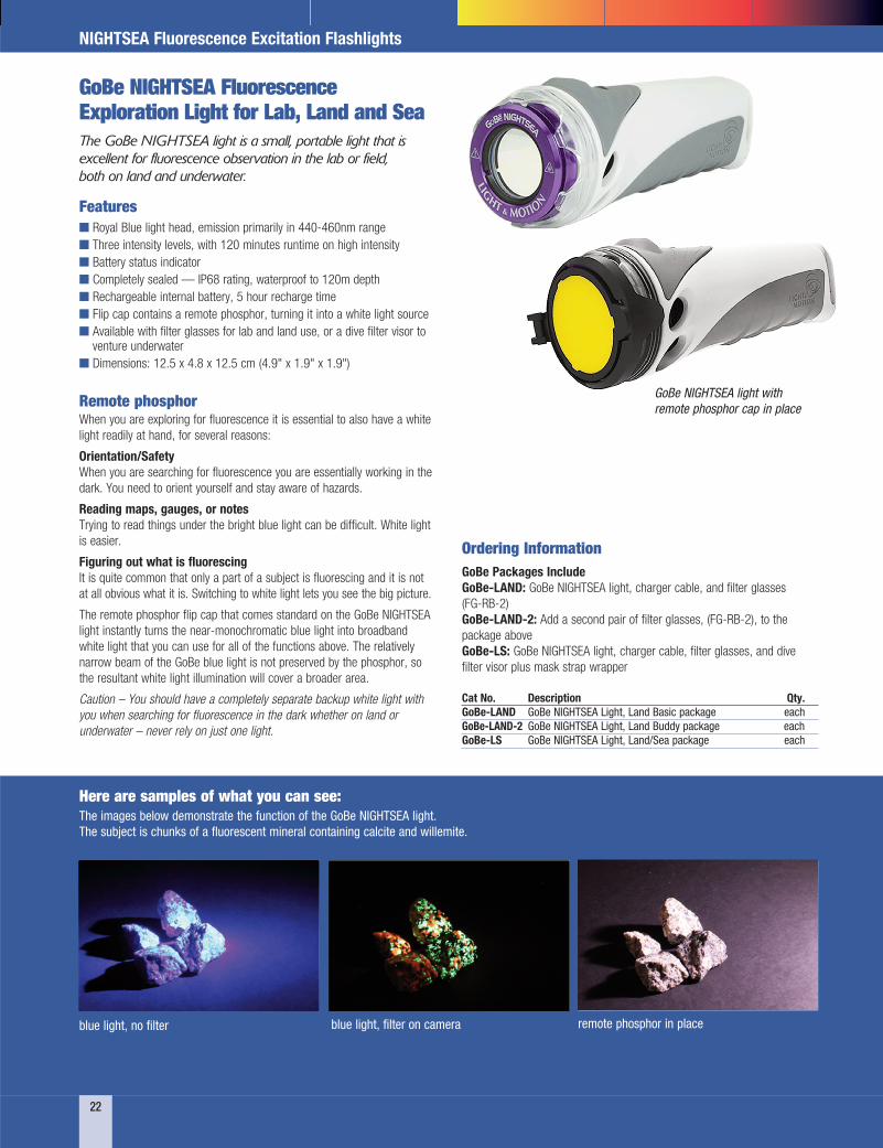
GoBe NIGHTSEA Fluorescence Exploration Light for Lab, Land and Sea The GoBe NIGHTSEA light is a small, portable light that is excellent for fluorescence observation in the lab or field, both on land and underwater.
22
NIGHTSEA Fluorescence Excitation Flashlights
Ordering Information GoBe Packages Include GoBe-LAND: GoBe NIGHTSEA light, charger cable, and filter glasses (FG-RB-2) GoBe-LAND-2: Add a second pair of filter glasses, (FG-RB-2), to the package above GoBe-LS: GoBe NIGHTSEA light, charger cable, filter glasses, and dive filter visor plus mask strap wrapper Cat No. Description Qty. GoBe-LAND GoBe NIGHTSEA Light, Land Basic package each GoBe-LAND-2 GoBe NIGHTSEA Light, Land Buddy package each GoBe-LS GoBe NIGHTSEA Light, Land/Sea package each
GoBe NIGHTSEA light with remote phosphor cap in place
The images below demonstrate the function of the GoBe NIGHTSEA light. The subject is chunks of a fluorescent mineral containing calcite and willemite.
Features n Royal Blue light head, emission primarily in 440-460nm range n Three intensity levels, with 120 minutes runtime on high intensity n Battery status indicator n Completely sealed — IP68 rating, waterproof to 120m depth n Rechargeable internal battery, 5 hour recharge time n Flip cap contains a remote phosphor, turning it into a white light source n Available with filter glasses for lab and land use, or a dive filter visor to
venture underwater n Dimensions: 12.5 x 4.8 x 12.5 cm (4.9" x 1.9" x 1.9")
blue light, no filter blue light, filter on camera
Here are samples of what you can see:
remote phosphor in place

Cat No. Description Qty. DFP-1 DFP Flashlight with RB and GR LEDs, matching filter glasses, carrying case each DFP-CG DFP Flashlight with CY and GR LEDs, matching filter glasses, carrying case each DFP-RC DFP Flashlight with RB and CY LEDs, matching filter glasses, carrying case each DFP-VC DFP Flashlight with VI and CY LEDs, matching filter glasses, carrying case each DFP-VG DFP Flashlight with VI and GR LEDs, matching filter glasses, carrying case each DFP-VR DFP Flashlight with VI and RB LEDs, matching filter glasses, carrying case each DFP-UC DFP Flashlight with UV and CY LEDs, matching filter glasses, carrying case each DFP-UG DFP Flashlight with UV and GR LEDs, matching filter glasses, carrying case each DFP-UR DFP Flashlight with UV and RB LEDs, matching filter glasses, carrying case each DFP-UV DFP Flashlight with UV and VI LEDs, matching filter glasses, carrying case each
FG-RB-1 FG-RB-2 FG-RB-3
NIGHTSEA Barrier Filter GlassesFilter glasses for use with Royal Blue excitation available in 3 styles (below). Styles 1 and 2 fit over eyeglasses, Style 3 does not. Glasses for all other wavelengths only available in Style 2. Glasses meet ANSI Z87.1 impact standards for safety glasses. NOTE: Glasses Description refers to the excitation light source with which they are to be used.
Dimensions: 13 x 8 x 5 cm (5 x 3 x 2 in.) Battery Type: 4 C-cell Bulb Type: Two high intensity 3W LED Burn Time: 4 hours at full power Lamp Life: 10,000 hours
Wavelength Sets Filter Set Excitation Emission Fluorophores UV - Ultra Violet 360-380nm 415nm LP DAPI... VI - Violet 400-415nm 450nm LP BFP, CFP... RB - Royal Blue 440-460nm 500nm LP GFP, fluorescein, lucifer yellow... CY - Cyan 490-515nm 550nm LP YFP, Venus... GR - Green 510-540nm 600nm LP DsRed, TdTomato, RFP...
NIGHTSEA DFP™ Dual Fluorescent Protein Excitation Flashlights Rapid screening of your fluorescent transgenic experiments
The Model DFP Dual Fluorescent Protein Flashlights include two different, switch-selectable high intensity LEDs for excitation of fluorescent proteins. For years the most popular fluorescent colors have been green (GFP, eGFP, fluorescein, etc.) and red (DsRed, TdTomato, etc.), and we offered only one version of the DFP, with a combination of blue and green LEDs for exciting these fluorophores. This is still our 'standard' DFP product, but we are now offering build-to-order combinations using any two of the excitation colors that are available with our popular Model SFA Stereo Microscope Fluorescence Adapter.
With every DFP light you will receive two pairs of barrier filter glasses and a convenient storage case. The glasses are well matched to the excitation so that they block the reflected excitation light while transmitting the fluorescence with high efficiency, providing excellent viewing contrast.
23
NIGHTSEA Fluorescence Excitation Flashlights (continued)
Cat No. Description Qty. FG-UV Filter glasses, Ultra Violet each FG-VI Filter glasses, Violet each FG-RB-1 Filter Glasses, Royal Blue, style 1 each FG-RB-2 Filter Glasses, Royal Blue, style 2 each
Cat No. Description Qty. FG-RB-3 Filter Glasses, Royal Blue, style 3 each FG-RB-GO Filter glasses, Green Only each FG-CY Filter glasses, Cyan each FG-GR Filter Glasses, Green, style 2 each

NIGHTSEA® is a registered trademark of NIGHTSEA, LLC
P.O. Box 550 • 1560 Industry Rd. • Hatfield, PA 19440 Tel: (215) 412-8400 • Fax: (215) 412-8450 email: [email protected] or [email protected] www.emsdiasum.com
Follow us on...