A comparative study of gallstones from children and adults using FTIR spectroscopy and fluorescence...
14
BioMed Central Page 1 of 14 (page number not for citation purposes) BMC Gastroenterology BMC Gastroenterology 2002, 2 Research article A comparative study of gallstones from children and adults using FTIR spectroscopy and fluorescence microscopy Oleg Kleiner 1 , Jagannathan Ramesh 2 , Mahmoud Huleihel 3 , Beny Cohen 4 , Keren Kantarovich 2 , Chen Levi 2 , Boris Polyak 3 , Robert S Marks 3 , Jacov Mordehai 1 , Zahavi Cohen 1 and Shaul Mordechai* 2 Address: 1 Department of Pediatric Surgery, Soroka University Medical Center, Ben Gurion University, Beer Sheva 84101 Israel, 2 Department of Physics, Ben Gurion University, Beer Sheva, 84105, Israel, 3 The Institute for Applied Biosciences, Ben Gurion University, Beer Sheva, 84105, Israel and 4 Department of Chemistry, Ben Gurion University, Beer Sheva, 84105, Israel E-mail: Oleg Kleiner - [email protected]; Jagannathan Ramesh - [email protected]; Mahmoud Huleihel - [email protected]; Beny Cohen - [email protected]; Keren Kantarovich - [email protected]; Chen Levi - [email protected]; Boris Polyak - [email protected]; Robert S Marks - [email protected]; Jacov Mordehai - [email protected]; Zahavi Cohen - [email protected]; Shaul Mordechai* - [email protected] *Corresponding author Abstract Background: Cholelithiasis is the gallstone disease (GSD) where stones are formed in the gallbladder. The main function of the gallbladder is to concentrate bile by the absorption of water and sodium. GSD has high prevalence among elderly adults. There are three major types of gallstones found in patients, White, Black and Brown. The major chemical component of white stones is cholesterol. Black and brown stones contain different proportions of cholesterol and bilirubin. The pathogenesis of gallstones is not clearly understood. Analysis of the chemical composition of gallstones using various spectroscopic techniques offers clues to the pathogenesis of gallstones. Recent years has seen an increasing trend in the number of cases involving children. The focus of this study is on the analysis of the chemical composition of gallstones from child and adult patients using spectroscopic methods. Methods: In this report, we present FTIR spectroscopic studies and fluorescence microscopic analysis of gallstones obtained from 67 adult and 21 child patients. The gallstones were removed during surgical operations at Soroka University Medical Center. Results: Our results show that black stones from adults and children are rich in bilirubin. Brown stones are composed of varying amounts of bilirubin and cholesterol. Green stones removed from an adult, which is rare, was found to be composed mainly of cholesterol. Our results also indicated that cholesterol and bilirubin could be the risk factors for gallstone formation in adults and children respectively. Fluorescence micrographs showed that the Ca-bilirubinate was present in all stones in different quantities and however, Cu-bilirubinate was present only in the mixed and black stones. Conclusions: Analysis based on FTIR suggest that the composition of black and brown stones from both children and adults are similar. Various layers of the brown stone from adults differ by having varying quantities of cholesterol and calcium carbonate. Ring patterns observed mainly in the green stone using fluorescence microscopy have relevance to the mechanism of the stone formation. Our preliminary study suggests that bilirubin and cholesterol are the main risk factors of gallstone disease. Published: 11 February 2002 BMC Gastroenterology 2002, 2:3 Received: 13 November 2001 Accepted: 11 February 2002 This article is available from: http://www.biomedcentral.com/1471-230X/2/3 © 2002 Kleiner et al; licensee BioMed Central Ltd. Verbatim copying and redistribution of this article are permitted in any medium for any purpose, provided this notice is preserved along with the article's original URL.
-
Upload
independent -
Category
Documents
-
view
2 -
download
0
Transcript of A comparative study of gallstones from children and adults using FTIR spectroscopy and fluorescence...

BioMed CentralBMC Gastroenterology
BMC Gastroenterology 2002, 2Research articleA comparative study of gallstones from children and adults using FTIR spectroscopy and fluorescence microscopyOleg Kleiner1, Jagannathan Ramesh2, Mahmoud Huleihel3, Beny Cohen4, Keren Kantarovich2, Chen Levi2, Boris Polyak3, Robert S Marks3, Jacov Mordehai1, Zahavi Cohen1 and Shaul Mordechai*2
Address: 1Department of Pediatric Surgery, Soroka University Medical Center, Ben Gurion University, Beer Sheva 84101 Israel, 2Department of Physics, Ben Gurion University, Beer Sheva, 84105, Israel, 3The Institute for Applied Biosciences, Ben Gurion University, Beer Sheva, 84105, Israel and 4Department of Chemistry, Ben Gurion University, Beer Sheva, 84105, Israel
E-mail: Oleg Kleiner - [email protected]; Jagannathan Ramesh - [email protected]; Mahmoud Huleihel - [email protected]; Beny Cohen - [email protected]; Keren Kantarovich - [email protected]; Chen Levi - [email protected]; Boris Polyak - [email protected]; Robert S Marks - [email protected]; Jacov Mordehai - [email protected]; Zahavi Cohen - [email protected]; Shaul Mordechai* - [email protected]
*Corresponding author
AbstractBackground: Cholelithiasis is the gallstone disease (GSD) where stones are formed in thegallbladder. The main function of the gallbladder is to concentrate bile by the absorption of waterand sodium. GSD has high prevalence among elderly adults. There are three major types ofgallstones found in patients, White, Black and Brown. The major chemical component of whitestones is cholesterol. Black and brown stones contain different proportions of cholesterol andbilirubin. The pathogenesis of gallstones is not clearly understood. Analysis of the chemicalcomposition of gallstones using various spectroscopic techniques offers clues to the pathogenesisof gallstones. Recent years has seen an increasing trend in the number of cases involving children.The focus of this study is on the analysis of the chemical composition of gallstones from child andadult patients using spectroscopic methods.
Methods: In this report, we present FTIR spectroscopic studies and fluorescence microscopicanalysis of gallstones obtained from 67 adult and 21 child patients. The gallstones were removedduring surgical operations at Soroka University Medical Center.
Results: Our results show that black stones from adults and children are rich in bilirubin. Brownstones are composed of varying amounts of bilirubin and cholesterol. Green stones removed froman adult, which is rare, was found to be composed mainly of cholesterol. Our results also indicatedthat cholesterol and bilirubin could be the risk factors for gallstone formation in adults and childrenrespectively. Fluorescence micrographs showed that the Ca-bilirubinate was present in all stonesin different quantities and however, Cu-bilirubinate was present only in the mixed and black stones.
Conclusions: Analysis based on FTIR suggest that the composition of black and brown stonesfrom both children and adults are similar. Various layers of the brown stone from adults differ byhaving varying quantities of cholesterol and calcium carbonate. Ring patterns observed mainly in thegreen stone using fluorescence microscopy have relevance to the mechanism of the stoneformation. Our preliminary study suggests that bilirubin and cholesterol are the main risk factorsof gallstone disease.
Published: 11 February 2002
BMC Gastroenterology 2002, 2:3
Received: 13 November 2001Accepted: 11 February 2002
This article is available from: http://www.biomedcentral.com/1471-230X/2/3
© 2002 Kleiner et al; licensee BioMed Central Ltd. Verbatim copying and redistribution of this article are permitted in any medium for any purpose, provided this notice is preserved along with the article's original URL.
Page 1 of 14(page number not for citation purposes)

BMC Gastroenterology 2002, 2 http://www.biomedcentral.com/1471-230X/2/3
BackgroundGallstone disease remains a serious health concern affect-ing millions throughout the world [1,2]. The formation ofgallstones in vivo takes years and it is quite difficult tomonitor such events from nucleation to the consolidation[3]. Gallstone formation is therefore very poorly under-stood. Surprisingly, in the last few decades there has beensignificant rise in gallstone disease among children [4–6].The vast majority of the reports are on adults [7]. Removalof the gallbladder by surgical methods is the only solutionavailable to the gallstone disease today and therefore, thedisease has a strong impact on children. The main func-tion of the gallbladder is to concentrate bile by absorptionof water and sodium. It concentrates the impermeable sol-utes contained in the hepatic bile by a factor of 5 to 10 andreducing its volume by 80–90%. Gallstones made up ofdifferent compositions display various colors. There arethree major types of stones observed in patients. These area) white b) black and c) brown stones. This classificationbased on the color, was proposed at the NIH workshop[8]. Earlier FTIR and FT-Raman studies suggested an addi-tional category called mixed stones having different pro-portions of cholesterol and bilirubin [7]. Black and browncolor stones contain bilirubin in large amounts in addi-tion to small quantities of cholesterol. The pigmentedstones can be further sub-categorized on the basis of mi-nor variations in chemical composition, such as the pres-ence of calcium carbonate. Three main lipids found in thebile are bile acids, cholesterol and phospholipids [9].
FTIR spectroscopy has been widely applied for structuralstudies on variety of biomolecules [10]. In the past fewyears, the use of FTIR method has enhanced our under-standing in different branches of medicine. Diagnosis ofcancer types such as lung, breast and colon may become areality in the future [11–13]. There are interesting reportsin the literature on the FTIR characterization of gallstonesand kidney stones from adults [14,15]. But there are no re-ports on the FTIR studies related to cases of children,which is becoming alarmingly important in recent years.In the present report, we present comparative results onFTIR characterization of various types of gallstones fromboth adults and children. In addition, we have character-ized various gallstones using fluorescence microscopy(FM), which provided insights into the various fluoro-phores present in the stones.
Materials and MethodsGallstone extractionExpert surgeons from the department of Pediatric surgeryextracted the gallstones during surgery. Initially, the gall-bladder from patients was surgically removed in the de-partment of Pediatric surgery using a LaparoscopicCholecystectomy procedure. The gallstones were extractedfrom the gallbladder and preserved under sterile condi-
tions. The patients consisted of both children and adults.Gallstones samples were collected from 67 adult and 21child patients for this study. GSD (Gall Stone Disease) isrelatively less common among children in comparison toadult population and hence the number of child patientstaken for this study was limited. Helsinki agreement wasobtained to carry out this project at SUMC (Soroka Uni-versity Medical Center) and consent was obtained from allpatients to use the isolated stones for research purpose. Allstone samples were stored in sterile dried condition andlater used for FTIR and Fluorescence microscopic analysis.
FTIR spectroscopeThe FTIR measurements were performed using JASCO615FTIR spectrometer in the frequency range 400–4000 cm-1
at 4 cm-1 resolution. To obtain a high signal/noise ratio128 scans were accumulated for each sample. Initially, thespectra were fitted to parabolic function. Then the RMS(Root Mean Square) was calculated between the originalspectrum and the one, fitted by the function. The area un-der the fitted spectrum was divided by the noise RMS,which is reported as Signal/ Noise Ratio (SNR). The Eval-uate menu provided in the OPUS software performed theentire process automatically. As prescribed by Bruker, SNRwas calculated in the range 1900–2100 cm-1
For each measurement 4–8 mg of finely powdered stonesample was used to make KBr discs. Control spectra wereacquired for cholesterol, bilirubin and calcium carbonatewhich were of high quality (99% pure) and purchasedfrom Sigma Chemical Company. In the case of FTIR-ATRmeasurements, only stone powder was used. The horizon-tal ATR (HATR) setup from PIKE technologies was used.The ZnSe crystal in the ATR setup was 80 × 10 mm and itsthickness was 4 mm. Vector normalization was applied toall the FTIR spectra reported in this study. Out of 67 adultand 21 child stones, which were measured, the spectrahaving signal/noise above 800 are presented in this study.
Fluorescence microscopeThe stone samples were visualized on an Olympus SZX 12fluorescent research stereomicroscope adapted with twostandard filter sets. One allowed blue excitation for obser-vation in the green (filter1) (GFP, YFP etc. excitation filterBP460–490 nm, absorption filter BA510IF, dichroic mir-ror DM505) and the other allowed green excitation forobservation in the red (filter2) (TRITC dying etc., excita-tion filter 460–560 nm, absorption filter O-590, dichroicmirror DM580). The total magnification range was be-tween 11 × – 144×. A 100 W mercury lamp made illumi-nation of samples. Photographs were taken with anOlympus Camedia C-3030 Zoom digital camera adaptedto the microscope. The image size of each photo was 2048× 1536 pixels.
Page 2 of 14(page number not for citation purposes)

BMC Gastroenterology 2002, 2 http://www.biomedcentral.com/1471-230X/2/3
Figure 1FTIR spectra of a) Type I black stone b) Type II black stone c) brown stone from adult patients. Vector normalization wasapplied to the spectra in Figures 1,2,3,4,5,6,7. The spectra presented in figure 1 are the averages of 12 and 10 black and brownstones respectively.
Wavenumber (cm-1)
33
98
292
4
166
1
16
26
157
1
14
05
12
48
69
9
328
9
3398
2933
2866
1627
1571
1463
1376
1250
1053
592
3395 2936
2899
2866
1653
1464
1372
1052
1022
955
797
592
5001000150020002500300035004000
(a)
(b)
(c)
Ab
sorb
ance
(A
.U.)
2948
Page 3 of 14(page number not for citation purposes)
BMC Gastroenterology 2002, 2 http://www.biomedcentral.com/1471-230X/2/3
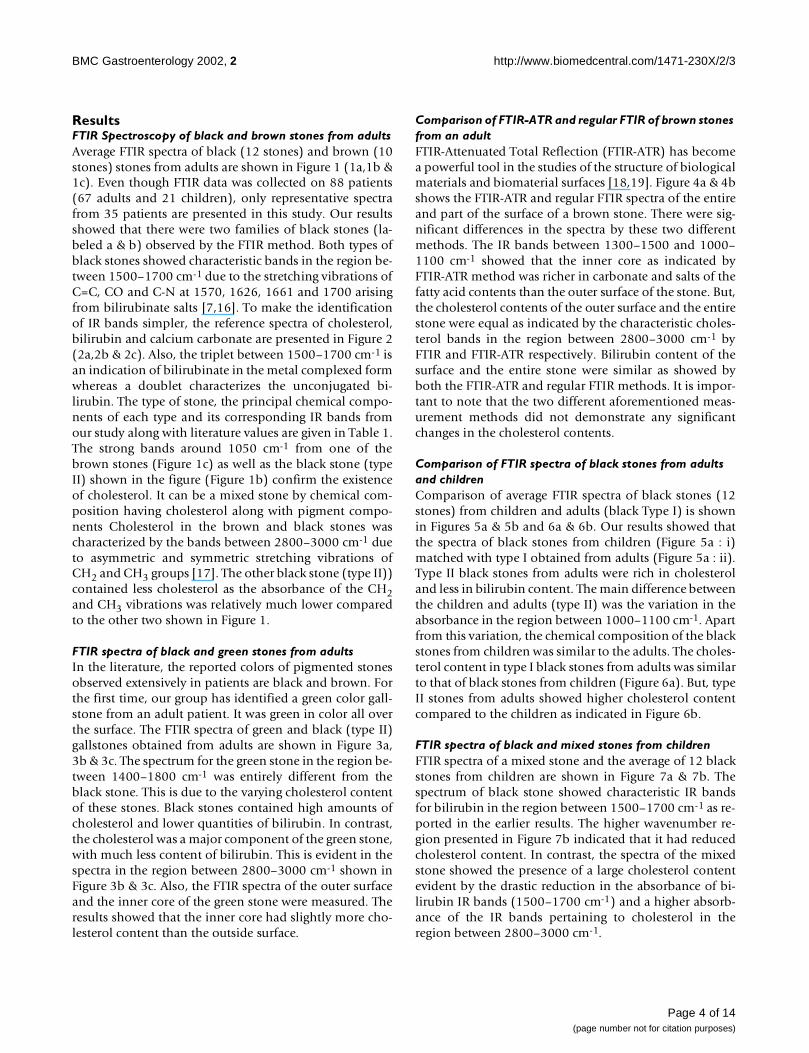
ResultsFTIR Spectroscopy of black and brown stones from adultsAverage FTIR spectra of black (12 stones) and brown (10stones) stones from adults are shown in Figure 1 (1a,1b &1c). Even though FTIR data was collected on 88 patients(67 adults and 21 children), only representative spectrafrom 35 patients are presented in this study. Our resultsshowed that there were two families of black stones (la-beled a & b) observed by the FTIR method. Both types ofblack stones showed characteristic bands in the region be-tween 1500–1700 cm-1 due to the stretching vibrations ofC=C, CO and C-N at 1570, 1626, 1661 and 1700 arisingfrom bilirubinate salts [7,16]. To make the identificationof IR bands simpler, the reference spectra of cholesterol,bilirubin and calcium carbonate are presented in Figure 2(2a,2b & 2c). Also, the triplet between 1500–1700 cm-1 isan indication of bilirubinate in the metal complexed formwhereas a doublet characterizes the unconjugated bi-lirubin. The type of stone, the principal chemical compo-nents of each type and its corresponding IR bands fromour study along with literature values are given in Table 1.The strong bands around 1050 cm-1 from one of thebrown stones (Figure 1c) as well as the black stone (typeII) shown in the figure (Figure 1b) confirm the existenceof cholesterol. It can be a mixed stone by chemical com-position having cholesterol along with pigment compo-nents Cholesterol in the brown and black stones wascharacterized by the bands between 2800–3000 cm-1 dueto asymmetric and symmetric stretching vibrations ofCH2 and CH3 groups [17]. The other black stone (type II))contained less cholesterol as the absorbance of the CH2and CH3 vibrations was relatively much lower comparedto the other two shown in Figure 1.
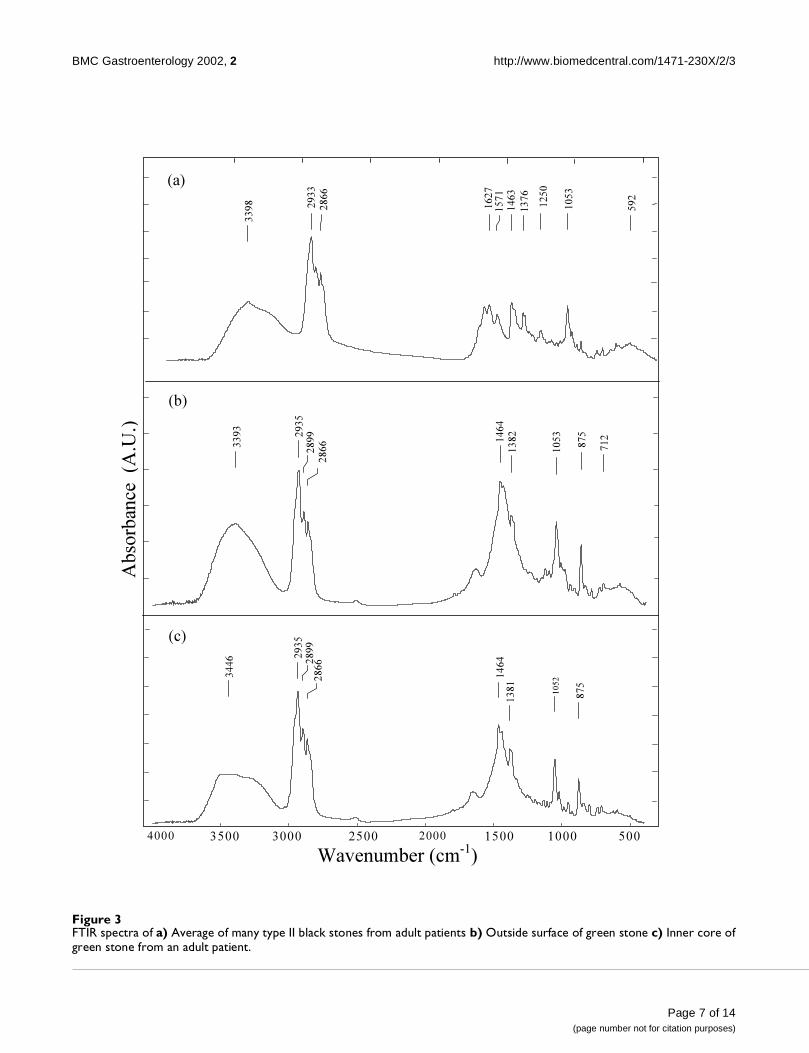
FTIR spectra of black and green stones from adultsIn the literature, the reported colors of pigmented stonesobserved extensively in patients are black and brown. Forthe first time, our group has identified a green color gall-stone from an adult patient. It was green in color all overthe surface. The FTIR spectra of green and black (type II)gallstones obtained from adults are shown in Figure 3a,3b & 3c. The spectrum for the green stone in the region be-tween 1400–1800 cm-1 was entirely different from theblack stone. This is due to the varying cholesterol contentof these stones. Black stones contained high amounts ofcholesterol and lower quantities of bilirubin. In contrast,the cholesterol was a major component of the green stone,with much less content of bilirubin. This is evident in thespectra in the region between 2800–3000 cm-1 shown inFigure 3b & 3c. Also, the FTIR spectra of the outer surfaceand the inner core of the green stone were measured. Theresults showed that the inner core had slightly more cho-lesterol content than the outside surface.
Comparison of FTIR-ATR and regular FTIR of brown stones from an adultFTIR-Attenuated Total Reflection (FTIR-ATR) has becomea powerful tool in the studies of the structure of biologicalmaterials and biomaterial surfaces [18,19]. Figure 4a & 4bshows the FTIR-ATR and regular FTIR spectra of the entireand part of the surface of a brown stone. There were sig-nificant differences in the spectra by these two differentmethods. The IR bands between 1300–1500 and 1000–1100 cm-1 showed that the inner core as indicated byFTIR-ATR method was richer in carbonate and salts of thefatty acid contents than the outer surface of the stone. But,the cholesterol contents of the outer surface and the entirestone were equal as indicated by the characteristic choles-terol bands in the region between 2800–3000 cm-1 byFTIR and FTIR-ATR respectively. Bilirubin content of thesurface and the entire stone were similar as showed byboth the FTIR-ATR and regular FTIR methods. It is impor-tant to note that the two different aforementioned meas-urement methods did not demonstrate any significantchanges in the cholesterol contents.
Comparison of FTIR spectra of black stones from adults and childrenComparison of average FTIR spectra of black stones (12stones) from children and adults (black Type I) is shownin Figures 5a & 5b and 6a & 6b. Our results showed thatthe spectra of black stones from children (Figure 5a : i)matched with type I obtained from adults (Figure 5a : ii).Type II black stones from adults were rich in cholesteroland less in bilirubin content. The main difference betweenthe children and adults (type II) was the variation in theabsorbance in the region between 1000–1100 cm-1. Apartfrom this variation, the chemical composition of the blackstones from children was similar to the adults. The choles-terol content in type I black stones from adults was similarto that of black stones from children (Figure 6a). But, typeII stones from adults showed higher cholesterol contentcompared to the children as indicated in Figure 6b.
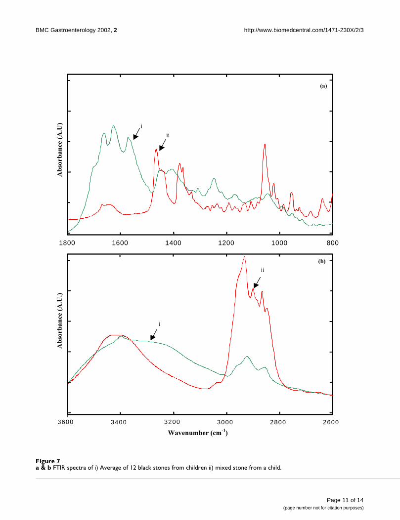
FTIR spectra of black and mixed stones from childrenFTIR spectra of a mixed stone and the average of 12 blackstones from children are shown in Figure 7a & 7b. Thespectrum of black stone showed characteristic IR bandsfor bilirubin in the region between 1500–1700 cm-1 as re-ported in the earlier results. The higher wavenumber re-gion presented in Figure 7b indicated that it had reducedcholesterol content. In contrast, the spectra of the mixedstone showed the presence of a large cholesterol contentevident by the drastic reduction in the absorbance of bi-lirubin IR bands (1500–1700 cm-1) and a higher absorb-ance of the IR bands pertaining to cholesterol in theregion between 2800–3000 cm-1.
Page 4 of 14(page number not for citation purposes)

BMC Gastroenterology 2002, 2 http://www.biomedcentral.com/1471-230X/2/3
Figure 2Reference FTIR spectra of pure a) Cholesterol b)Bilirubin c) Calcium carbonate.
3399
2934
2866
1670
1466
1376
1056
957
3289
2948
1654
.
1549
1401
1282
1080
1020
882
630
1788
1481
1082
873
855
713
5001000150020002500300035004000
(a)
(b)
(c)
2924
Wavenumber (cm-1)
Ab
sorb
ance
(A
.U.)
3398
Page 5 of 14(page number not for citation purposes)

BMC Gastroenterology 2002, 2 http://www.biomedcentral.com/1471-230X/2/3
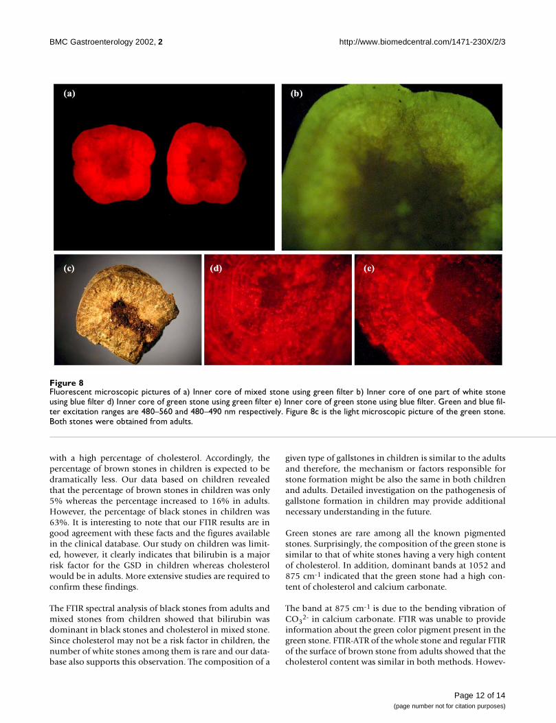
Fluorescence microscopy of gallstonesFluorescence microscopy (FM) has made essential contri-butions in modern cell biology and medicine [20,21]. Inthis article, we present the use of this technique in theanalysis of gallstones to measure its intrinsic fluorescence.The stones were cut under sterile environment into equalhalves to investigate the inner core using fluorescence mi-croscopy. Figure 8a,8b,8c,8d,8e shows the fluorescent mi-crographs of white and green stones using blue and greenfilters. The green and blue filters have the excitation rangebetween 480–560 and 480–490 nm respectively. Figures8a & 8b show the FM picture of a white stone using greenand blue filters. The white stone emitted red and green flu-orescence using green and blue filters. Figure 8c shows alight micrograph of a green stone.
The inner core of the green stone was dark and the outersurface was relatively brighter. This indicated that the in-ner core may contain a pigment like bilirubin and the out-er surface could be made up of cholesterol. The greenstone emitted red fluorescence using both filters. But, theintensity of red fluorescence was higher using a green filterthan with a blue filter. Interestingly, ring patterns wereclearly observed in the green stone using both filters (Fig-ure 8d & 8e).
DiscussionGallstone disease is still one of the most common diges-tive diseases with an overall prevalence of 10% in theUnited States [22,23]. Since the pathogenesis of gallstonesis not clearly understood, its analysis-using chemical andspectroscopic techniques have provided some clues[24,25]. Our studies reported herein on children are ofspecial significance as there are no such earlier reports onchildren in the literature.
The results on black stones suggest that the compositionof bilirubin and cholesterol varies considerably. This isclear from Figure 1a & 1b where Type I black stone had ahigher bilirubin and a lower cholesterol content in con-trast to type II black stone. This was observed consistentlyin other black stones obtained from adult patients. Fromthe absorbance of black and brown stones in the regionbetween 1500–1800 cm-1, it can be deduced that the bi-lirubin content of black stones was much higher than thatof brown stones in adults. In addition, brown stones hada higher cholesterol / bilirubin ratio compared to theblack stones.
None of the black stones from children contain as muchcholesterol as type II black stones from adults. Therefore,we hypothesize that children do not have as many stones
Table 1: Type, occurrence and IR bands of principal component observed in gallstones
Type of Stone Occurrence of stonesa (percents)
Principal component Principal IR bands observed
Literature values (Reference 16).
Black I (adult) 18 Bilirubin 1661, 1626,1570
1670,1640 (OC=O stretching)1575(C=C stretching)
Black II (adult) Cholesterol 3398,2933,2866,1463, 1376,1053
3410,2925 (CH2 and CH3 asym-metric stretching)2860 (CH2 and CH3 symmetric stretching)1460,1380 (CH2 and CH3 bend-ing)1050(C-C stretching)
Black (child) 63 Bilirubin 1661,1626,1571
Same as above
Brown (adult) 16 Cholesterol 3395,2936,2899,2866,1464,1372,1052
Same as above
Green innercore (adult)
ND Cholesterol 2933, 2899,2866,1464,1381,1052
Same as above
Mixed (adult) ND Cholesterol 3395,2933,2866,1465,1376,1056
Same as above
ND represents Not Determined. CaCo3 present in green stone gives rise to two characteristic IR bands at 1445 and 875 cm-1 due to C-O stretch-ing and bending vibrations respectively, aThe listed numbers refer to adults or children.
Page 6 of 14(page number not for citation purposes)
BMC Gastroenterology 2002, 2 http://www.biomedcentral.com/1471-230X/2/3
Figure 3FTIR spectra of a) Average of many type II black stones from adult patients b) Outside surface of green stone c) Inner core ofgreen stone from an adult patient.
3446 2935
2899
2866
1464
1381
1052
875
5001000150020002500300035004000
3398 2933
2866
1627
1571
1463
1376
1250
1053
592
3393
2935
2899
2866 1464
1382
1053
875
712
(a)
(b)
(c)
Wavenumber (cm-1)
Ab
sorb
ance
(A
.U.)
Page 7 of 14(page number not for citation purposes)

BMC Gastroenterology 2002, 2 http://www.biomedcentral.com/1471-230X/2/3
Figure 4a & b i) FTIR-ATR spectrum of entire brown stone ii) Regular FTIR spectrum of outside surface of a brown stone. Both meas-urements were made from a single stone from a patient.
260028003000320034003600
80010001200140016001800
(a)
Ab
sorb
an
ce(A
.U
.)
i
ii
Wavenumber (cm-1
)
Ab
sorb
an
ce(A
.U
.)
ii
i
(b)
Page 8 of 14(page number not for citation purposes)

BMC Gastroenterology 2002, 2 http://www.biomedcentral.com/1471-230X/2/3
Figure 5a & b FTIR spectra of i) black stone from children ii) black stone from adult patients in the region between 800–1800 cm-1.Black stones from adults type I and II are shown in Figures 5a, 6a and 5b, 6b respectively. In the case of adults and children,average spectra of 12 stones are presented.
80010001200140016001800
80010001200140016001800
Ab
sorb
an
ce (
A.U
.)
(a)
Ab
sorb
an
ce (
A.U
)
Wavenumber (cm-1
)
(b)
i
ii
ii
i
Page 9 of 14(page number not for citation purposes)

BMC Gastroenterology 2002, 2 http://www.biomedcentral.com/1471-230X/2/3
Figure 6a & b FTIR spectra of i) Average of 12 black stones from children ii) Type II black stone from 12 adult patients in the regionbetween 2600–3600 cm-1. Black stones type I and II from adults are shown in the Figures 6a and 6b respectively.
260028003000320034003600
260028003000320034003600
Ab
sorb
an
ce(A
.U
.)
(a)
Ab
sorb
an
ce(A
.U
.)
Wavenumber (cm-1
)
(b)
ii
i
i
ii
Page 10 of 14(page number not for citation purposes)
BMC Gastroenterology 2002, 2 http://www.biomedcentral.com/1471-230X/2/3
Figure 7a & b FTIR spectra of i) Average of 12 black stones from children ii) mixed stone from a child.
260028003000320034003600
80010001200140016001800
(a)
Ab
sorb
an
ce (
A.U
)
(b)
Ab
sorb
an
ce (
A.U
.)
Wavenumber (cm-1
)
i
ii
i
ii
Page 11 of 14(page number not for citation purposes)
BMC Gastroenterology 2002, 2 http://www.biomedcentral.com/1471-230X/2/3
with a high percentage of cholesterol. Accordingly, thepercentage of brown stones in children is expected to bedramatically less. Our data based on children revealedthat the percentage of brown stones in children was only5% whereas the percentage increased to 16% in adults.However, the percentage of black stones in children was63%. It is interesting to note that our FTIR results are ingood agreement with these facts and the figures availablein the clinical database. Our study on children was limit-ed, however, it clearly indicates that bilirubin is a majorrisk factor for the GSD in children whereas cholesterolwould be in adults. More extensive studies are required toconfirm these findings.
The FTIR spectral analysis of black stones from adults andmixed stones from children showed that bilirubin wasdominant in black stones and cholesterol in mixed stone.Since cholesterol may not be a risk factor in children, thenumber of white stones among them is rare and our data-base also supports this observation. The composition of a
given type of gallstones in children is similar to the adultsand therefore, the mechanism or factors responsible forstone formation might be also the same in both childrenand adults. Detailed investigation on the pathogenesis ofgallstone formation in children may provide additionalnecessary understanding in the future.
Green stones are rare among all the known pigmentedstones. Surprisingly, the composition of the green stone issimilar to that of white stones having a very high contentof cholesterol. In addition, dominant bands at 1052 and875 cm-1 indicated that the green stone had a high con-tent of cholesterol and calcium carbonate.
The band at 875 cm-1 is due to the bending vibration ofCO3
2- in calcium carbonate. FTIR was unable to provideinformation about the green color pigment present in thegreen stone. FTIR-ATR of the whole stone and regular FTIRof the surface of brown stone from adults showed that thecholesterol content was similar in both methods. Howev-
Figure 8Fluorescent microscopic pictures of a) Inner core of mixed stone using green filter b) Inner core of one part of white stoneusing blue filter d) Inner core of green stone using green filter e) Inner core of green stone using blue filter. Green and blue fil-ter excitation ranges are 480–560 and 480–490 nm respectively. Figure 8c is the light microscopic picture of the green stone.Both stones were obtained from adults.
Page 12 of 14(page number not for citation purposes)

BMC Gastroenterology 2002, 2 http://www.biomedcentral.com/1471-230X/2/3
er, FTIR-ATR showed increased content of fatty acids andcarbonate. Hence, it can be concluded that the inner coreof the stone had more of fatty acids and calcium carbon-ate, which could have initiated the nucleation events forthe stone formation.
Fluorescence microscopy on the inner core of gallstonesrevealed the differences in their chemical composition,which correlated well with FTIR spectroscopy. All thestones (including brown and black stones for which thedata are not shown) emitted red fluorescence using agreen filter. This red fluorescence emission may be due toCa-bilirubinate complex present in the stones. The mixedstone emitted green fluorescence using a blue filter wasobserved due to Cu-bilirubinate, which is present only inmixed and black stones. In black stones, occurrence of po-lymerized Cu-bilirubinate complex was already reportedin the literature [7,26,27]. Even though copper is reportedto be present in the brown stone [7], we rule out the pos-sibility of the presence of Cu-bilirubinate complex in thebrown stones. In brown and green stones, red fluores-cence was observed using both filters. The difference inthe chemical composition can be attributed to the variousmechanisms responsible for the pathogenesis of gall-stones. Certain types of bacteria are detected only in thebrown stones [28,29]. Black stones are associated withhemolysis and liver cirrhosis.
The cross-section of the brown gallstones are seen to haveconcentric ring patterns [30]. Our FM micrograph of agreen stone showed regular ring patterns, which are seenclearly using the blue filter. The explanation for this obser-vation is the periodic precipitation of various bile acidsalong with proper combination of metal ions. Periodicpatterns in biological systems can offer clues to their ori-gin and further analysis may brighten the understandingof the mechanism of the formation of gallstones. Patho-genesis of gallstones is as yet poorly understood. Detailedstudies using a battery of spectroscopic and microbiologi-cal methods will probably lead to a better understandingof gallstone formation leading to better prevention in thefirst place.
ConclusionsFTIR and fluorescence microscopic studies on differenttypes of gallstones from adults and children have shedlight on their chemical composition. Black stones fromadults had varying amounts of bilirubin, and brownstones were rich in cholesterol. Comparison of blackstones from adults and children suggested that bilirubinand cholesterol might be the risk factors in children andadults respectively. The main composition of black stonesfrom adults and children were found to be similar usingFTIR spectroscopy. A new green color stone from an adult,which is rare, has also been reported in our study. The
analysis showed that the principal components of thegreen stone were cholesterol and calcium carbonate. It isinteresting to note that the contents of cholesterol and cal-cium carbonate were higher in the inner core of the brownstones compared to that of outer surface. Fluorescence mi-croscopic analysis indicated that the mixed and blackstones had both Ca-bilirubinate and Cu-bilirubinate. Thebrown and green stones contained only Ca-bilirubinate.The specific ring patterns found in the green stone are in-teresting and needs further investigation.
List of abbreviationsFTIR : Fourier Transform InfraRed;
FM : Fluorescence Microscopy;
HATR : Horizontal Attenuated Total Reflection;
GFP : Green Fluorescent Protein;
YFP : Yellow Fluorescent Protein;
GSD : GallStone Disease;
SUMC : Soroka University Medical Center.
Competing InterestsNone declared
AcknowledgementsWe gratefully acknowledge the research grant from the Faculty of Health at Ben Gurion University of the Negev received by Dr. Jacov Mordehai dur-ing the course of this work. We also acknowledge the Harry-Stern Applied Research Grant Program for partial support.
References1. Harding AJ: Gallstones: Causes and Treatments. William Heine-
mann Medical Books, London, 19642. Kern F, Jr : Epidemiology and natural history of gallstones.
Semin. Liver. Dis 1983, 3:87-963. Swidsinski A, Khilkin M, Pahlig H, Swidsinski S, Priem F: Time de-
pendent changes in the concentration and type of bacterialsequences found in cholesterol gallstones. Hepatology 1998,27:662-665
4. Kumar R, Nguyen K, Shun A: Gallstones and common bile ductcalculi in infancy and childhood Aust. N. Z. J. Surg 2000, 70:188-191
5. Lobe TE: Cholelithiasis and Cholecystitis in children Semn. Ped.Surg 2000, 9:170-176
6. Waldhausen JH, Benjamin DR: Cholecystectomy is becoming anincreasingly common operation in children. Am. J. Surg 1999,177:364-367
7. Paluszkiewicz C, Kwiatek WM, Galka M, Sobieraj D, Wentrup-ByrneD: FT-Raman, FT-IR spectroscopy and PIXE analysis appliedto gallstones specimens Cell. Mol. Biol 1998, 44:65-73
8. Trotman BW, Soloway RD: Pigment gallstone disease: summa-ry of the National Institutes of Health-International Work-shop. Hepatology 1982, 2:879
9. Roslyn JJ, Zinner MJ: Principles of Surgery 19941376-139910. Mantsch HH, Chapman D: Infrared Spectroscopy of Biomole-
cules John Wiley, N.Y, 199611. Gao T, Feng J, Ci Y: Human breast carcinomal tissues display
distinctive FTIR spectra: implication for the histologicalcharacterization of carcinomas Anal. Cell. Pathol. 1999, 18:87-93
Page 13 of 14(page number not for citation purposes)
http://www.ncbi.nlm.nih.gov/entrez/query.fcgi?cmd=Retrieve&db=PubMed&dopt=Abstract&list_uids=6408741
http://www.ncbi.nlm.nih.gov/entrez/query.fcgi?cmd=Retrieve&db=PubMed&dopt=Abstract&list_uids=9500691

BMC Gastroenterology 2002, 2 http://www.biomedcentral.com/1471-230X/2/3
12. Wang H, Wang HC, Huang YJ: Microscopic FTIR studies of lungcancer cells in pleural fluid Sci. Total. Environ. 1997, 204:283-287
13. Salman A, Argov S, Jagannathan Ramesh., Goldstein J, Sinelnikov I, Gu-terman H, Mordechai S: FTIR microscopic characterization ofnormal and malignant human colonic tissues. Cell. Mol. Biol(Nosiy-le-grand) 2001, 47:OL159-OL166
14. Zhou X, Wu J, Soloway RD: Application of Spectroscopy in thestudy of pigment gallstone in China. Biospectroscopy 1997, 3:195-205
15. Estepa L, Daudon M: Contribution of Fourier transform infra-red Spectroscopy to the identification of urinary stones andkidney crystal deposits. Biospectroscopy 1997, 3:347-369
16. Zhou X-S, Shen G-R, Wu J-G, Li W-H, Xu Y-Z, Weng S-F, SolowayRD, Fu X-B, Tian W, Xu Z, Shen T, Xu G-X, Wentrup-Byrne E: ASpectroscopic study of pigment gallstones in China. Biospec-troscopy 1997, 3:371-380
17. Wentrup-Byrne E, Chua-Anusorn W, St Pierre TG, Webb J, RamsayA, Rintoul L: A Spectroscopic study of thalassemic gallstones,Biospectroscopy 1997, 3:409-416
18. Vigano C, Manciu L, Buyse F, Goormaghtigh E, Ruysschaert JM: At-tenuated total reflection IR spectroscopy as a tool to inves-tigate the structure orientation and tertiary structurechanges in peptides and membrane proteins. Biopolymers 2000,55:373-380
19. Chittur KK: FTIR/ATR for protein adsorption to biomaterialsurfaces. Biomaterials 1998, 19:357-369
20. Gustafsson MG: Extended resolution fluorescence microsco-py. Curr. Opin. Struct. Biol. 1999, 9:627-634
21. Robinson JM, Takizawa T, Pombo A, Cook PR: Correlative fluores-cence and electron microscopy on ultrathin cryosectionsbridging the resolution gap J. Histochem. Cytochem 2001, 49:803-808
22. Afdhal NH: Crohn's and Stones. The Am. J. Gastroent, 1999,94:1130-1132
23. Kratzer W, Mason RA, Kächele V: Prevalence of gallstones insonographic surveys worldwide J. Clin. Ultrasound 1999, 27:1-7
24. Wentrup-Byrne E, Rintoul L, Smith JL, Fredericks PM: Comparisonof vibrational Spectroscopic techniques for the characteriza-tion of human gallstones. Appl. Spectrosc. 1995, 49:1028-1036
25. Wu J-G, Zhou X-S, Xu Z, Shen T, Xu Y-Z, Li W-H, Xu D-F, SolowayRD, Wentrup-Byrne E, Xu Z-H, Shi G-R, Deng S-Q, Li X-F, Shi N: Aspectroscopic investigation of the formation mechanism ofpigment gallstones. Biopsetroscopy 1997, 3:381-391
26. Shizsuma K, Iwatani K, Hasai H, Wen XQ, Horiuchi I, Kajiyama G:PIXE analysis of gallstones: trace element analysis and mil-libeam scanning of stone sections. Nucl. Instr. Meth. Phys. Res.1997, B129:401-409
27. Li W-H, Shen G-R, Soloway RD, Yang Z-L, Tong X-B, Wu E, Xu D-F,Wu J-G, Xu G-X: Copper bilirubinate and black pigment gall-stone Biospectroscopy, 1995, 1:149-156
28. Wetter LA, Hamadeh RM, Griffiss JM, Oesterle A, Aagaard B, WayLW: Differences in outer membrane characteristics betweengallstone-associated bacteria and normal bacterial flora. Lan-cet 1994, 343:444-448
29. Swidsinski A, Ludwig W, Pahlig H, Priem F: Molecular genetic evi-dence of bacterial colonization of cholesterol gallstones. Gas-troenterology 1995, 108:860-864
30. Wu J-G, Xu D-F, Liu F, Zhou X-S, Zhang Y-F, Zhou N-F, Hong N-K,Soloway RD: Do ring form in gallstone during growth due tothe Liesegang phenomenon or periodic deposition? Gastroen-terology 1990, 98:A256
Publish with BioMed Central and every scientist can read your work free of charge
"BioMedcentral will be the most significant development for disseminating the results of biomedical research in our lifetime."
Paul Nurse, Director-General, Imperial Cancer Research Fund
Publish with BMC and your research papers will be:
available free of charge to the entire biomedical community
peer reviewed and published immediately upon acceptance
cited in PubMed and archived on PubMed Central
yours - you keep the copyright
[email protected] your manuscript here:http://www.biomedcentral.com/manuscript/
BioMedcentral.com
Page 14 of 14(page number not for citation purposes)All in-text references underlined in blue are linked to publications on ResearchGate, letting you access and read them immediately.
http://www.ncbi.nlm.nih.gov/entrez/query.fcgi?cmd=Retrieve&db=PubMed&dopt=Abstract&list_uids=9335161
http://www.ncbi.nlm.nih.gov/entrez/query.fcgi?cmd=Retrieve&db=PubMed&dopt=Abstract&list_uids=9677150
http://www.ncbi.nlm.nih.gov/entrez/query.fcgi?cmd=Retrieve&db=PubMed&dopt=Abstract&list_uids=9888092
http://www.ncbi.nlm.nih.gov/entrez/query.fcgi?cmd=Retrieve&db=PubMed&dopt=Abstract&list_uids=7905954
http://www.ncbi.nlm.nih.gov/entrez/query.fcgi?cmd=Retrieve&db=PubMed&dopt=Abstract&list_uids=7905954