structural brain changes on MRI: A randomized controlled longitudinal trial
8
This article appeared in a journal published by Elsevier. The attached copy is furnished to the author for internal non-commercial research and education use, including for instruction at the authors institution and sharing with colleagues. Other uses, including reproduction and distribution, or selling or licensing copies, or posting to personal, institutional or third party websites are prohibited. In most cases authors are permitted to post their version of the article (e.g. in Word or Tex form) to their personal website or institutional repository. Authors requiring further information regarding Elsevier’s archiving and manuscript policies are encouraged to visit: http://www.elsevier.com/authorsrights
-
Upload
michiganstate -
Category
Documents
-
view
1 -
download
0
Transcript of structural brain changes on MRI: A randomized controlled longitudinal trial
This article appeared in a journal published by Elsevier. The attachedcopy is furnished to the author for internal non-commercial researchand education use, including for instruction at the authors institution
and sharing with colleagues.
Other uses, including reproduction and distribution, or selling orlicensing copies, or posting to personal, institutional or third party
websites are prohibited.
In most cases authors are permitted to post their version of thearticle (e.g. in Word or Tex form) to their personal website orinstitutional repository. Authors requiring further information
regarding Elsevier’s archiving and manuscript policies areencouraged to visit:
http://www.elsevier.com/authorsrights
Author's personal copy
Clinical Neurology and Neurosurgery 115 (2013) 2419– 2425
Contents lists available at ScienceDirect
Clinical Neurology and Neurosurgery
jou rn al h om epage: www.elsev ier .com/ locate /c l ineuro
Review
Mindfulness based intervention in Parkinson’s disease leads tostructural brain changes on MRIA randomized controlled longitudinal trial
Barbara A. Pickuta,b,g,h,∗, Wim Van Heckea,c, Eric Kerckhofsd, Peter Mariëne,g,Sven Vannestea, Patrick Crasa,b,h, Paul M. Parizela,f
a University of Antwerp, Universiteitsplein 1, B-2610 Wilrijk, Belgiumb Department of Neurology, University Hospital Antwerp, Wilrijkstraat 10, B-2650 Edegem, Belgiumc icoMetrix, Tervuursesteenweg 244, B-3001 Leuven, Belgiumd Faculty of Physical Education and Physical Therapy, Vrije Universiteit Brussel, Laarbeeklaan 103, B-1090 Brussels, Belgiume Department of Clinical and Experimental Neurolinguistics, Vrije Universiteit Brussel, Pleinlaan 2, B-1050 Brussels, Belgiumf Department of Radiology, University Hospital Antwerp, Wilrijkstraat 10, B-2650 Edegem, Belgiumg Department of Neurology and Memory Clinic, Ziekenhuis Netwerk Antwerp, Lindendreef 1, B-2020 Antwerp, Belgiumh Born Bunge Institute, Universiteitsplein 1, B-2610 Wilrijk, Belgium
a r t i c l e i n f o
Article history:Received 28 June 2013Received in revised form 4 September 2013Accepted 2 October 2013Available online 16 October 2013
Keywords:Parkinson’s diseaseMagnetic resonance imagingGray matter densityMindfulness based interventionMindfulness based stress reduction
a b s t r a c t
Objective: The aim of the current study is to investigate structural changes on brain MRI using voxelbased morphometry (VBM) related to an eight-week mindfulness based intervention (MBI) in Parkinson’sDisease (PD).Methods: A total of 27 out of 30 PD patients completed a randomized controlled longitudinal trial. Four-teen patients participated in a structured eight-week program of MBI. Thirteen patients received usualcare (UC) alone. MRI data sets of the brain were obtained at baseline and after eight weeks follow-up. VBManalysis was performed using DARTEL from the SPM8 software. The resulting difference maps were sta-tistically compared to examine gray matter density (GMD) differences. Results were reported at p < 0.001,uncorrected for multiple comparisons.Results: Increased GMD was found in the MBI compared to the UC group in the region of interest (ROI)analysis in the right amygdala, and bilaterally in the hippocampus. Whole brain analysis showed increasedGMD in the left and right caudate nucleus, the left occipital lobe at the lingual gyrus and cuneus, the leftthalamus, and bilaterally in the temporo-parietal junction. In contrast, GMD differences were found inthe UC group in the left anterior lobe and dentate nucleus of the cerebellum.Conclusions: To the best of our knowledge this is the first quantitative analysis of neurobiological effects ofMBI in PD. Increased GMD was found in the MBI group in the neural networks that have been postulated toplay an important role in PD. These areas have also been implicated in the functional networks mediatingthe benefits of meditation.
© 2013 Elsevier B.V. All rights reserved.
Contents
1. Introduction . . . . . . . . . . . . . . . . . . . . . . . . . . . . . . . . . . . . . . . . . . . . . . . . . . . . . . . . . . . . . . . . . . . . . . . . . . . . . . . . . . . . . . . . . . . . . . . . . . . . . . . . . . . . . . . . . . . . . . . . . . . . . . . . . . . . . . . . . . 24202. Materials and methods. . . . . . . . . . . . . . . . . . . . . . . . . . . . . . . . . . . . . . . . . . . . . . . . . . . . . . . . . . . . . . . . . . . . . . . . . . . . . . . . . . . . . . . . . . . . . . . . . . . . . . . . . . . . . . . . . . . . . . . . . . . . . . . 2420
2.1. Study participants . . . . . . . . . . . . . . . . . . . . . . . . . . . . . . . . . . . . . . . . . . . . . . . . . . . . . . . . . . . . . . . . . . . . . . . . . . . . . . . . . . . . . . . . . . . . . . . . . . . . . . . . . . . . . . . . . . . . . . . . . . . . 24202.2. Intervention . . . . . . . . . . . . . . . . . . . . . . . . . . . . . . . . . . . . . . . . . . . . . . . . . . . . . . . . . . . . . . . . . . . . . . . . . . . . . . . . . . . . . . . . . . . . . . . . . . . . . . . . . . . . . . . . . . . . . . . . . . . . . . . . . . 24212.3. MRI data acquisition . . . . . . . . . . . . . . . . . . . . . . . . . . . . . . . . . . . . . . . . . . . . . . . . . . . . . . . . . . . . . . . . . . . . . . . . . . . . . . . . . . . . . . . . . . . . . . . . . . . . . . . . . . . . . . . . . . . . . . . . . . 24212.4. MRI data analysis . . . . . . . . . . . . . . . . . . . . . . . . . . . . . . . . . . . . . . . . . . . . . . . . . . . . . . . . . . . . . . . . . . . . . . . . . . . . . . . . . . . . . . . . . . . . . . . . . . . . . . . . . . . . . . . . . . . . . . . . . . . . . 2421
∗ Corresponding author at: University Hospital Antwerp, Department of Neurology, Wilrijkstraat 10, B-2650 Edegem, Belgium. Tel.: +32 3 821 52 12; fax: +32 3 821 43 12.E-mail addresses: [email protected], [email protected] (B.A. Pickut).
0303-8467/$ – see front matter © 2013 Elsevier B.V. All rights reserved.http://dx.doi.org/10.1016/j.clineuro.2013.10.002
Author's personal copy
2420 B.A. Pickut et al. / Clinical Neurology and Neurosurgery 115 (2013) 2419– 2425
3. Results . . . . . . . . . . . . . . . . . . . . . . . . . . . . . . . . . . . . . . . . . . . . . . . . . . . . . . . . . . . . . . . . . . . . . . . . . . . . . . . . . . . . . . . . . . . . . . . . . . . . . . . . . . . . . . . . . . . . . . . . . . . . . . . . . . . . . . . . . . . . . . . . 24214. Discussion . . . . . . . . . . . . . . . . . . . . . . . . . . . . . . . . . . . . . . . . . . . . . . . . . . . . . . . . . . . . . . . . . . . . . . . . . . . . . . . . . . . . . . . . . . . . . . . . . . . . . . . . . . . . . . . . . . . . . . . . . . . . . . . . . . . . . . . . . . . . 2422
Conflicts of interest . . . . . . . . . . . . . . . . . . . . . . . . . . . . . . . . . . . . . . . . . . . . . . . . . . . . . . . . . . . . . . . . . . . . . . . . . . . . . . . . . . . . . . . . . . . . . . . . . . . . . . . . . . . . . . . . . . . . . . . . . . . . . . . . . . 2424Acknowledgements . . . . . . . . . . . . . . . . . . . . . . . . . . . . . . . . . . . . . . . . . . . . . . . . . . . . . . . . . . . . . . . . . . . . . . . . . . . . . . . . . . . . . . . . . . . . . . . . . . . . . . . . . . . . . . . . . . . . . . . . . . . . . . . . . . 2424References . . . . . . . . . . . . . . . . . . . . . . . . . . . . . . . . . . . . . . . . . . . . . . . . . . . . . . . . . . . . . . . . . . . . . . . . . . . . . . . . . . . . . . . . . . . . . . . . . . . . . . . . . . . . . . . . . . . . . . . . . . . . . . . . . . . . . . . . . . . 2424
1. Introduction
Parkinson’s disease (PD) is the second most common neu-rodegenerative disorder with a world-wide prevalence of approx-imately 10 million people. Projecting on current demographicchanges, the estimated prevalence of PD is expected to double by2050 [1]. As such, PD remains an important public health problemdue to the chronic and progressive nature of this disease [2,3].
In PD large numbers of dopaminergic neurons located withinbasal ganglia circuitry degenerate. Evidence suggests that symp-toms in PD are related to a more extensive pathological processinvolving a progressive caudal to rostral aggregation of alpha-synuclein in select nerve cells [4]. Clinical parkinsonism can beresponsive to pharmacological therapies other than dopaminer-gic agents such as serotonergic and cholinergic drugs. As such, theneuropathology of PD also involves non-dopaminergic systems orcortical structures of the nervous system not directly involved inmotor control [5]. Moreover, structural brain changes in corticaland limbic areas have been reported in PD [6–8]. These com-plex alterations contribute to clinical motor symptoms (includingthe hallmark triad: bradykinesia, tremor, rigidity) and non-motorsymptoms (NMS). NMS which comprise cognitive changes, emo-tional dysregulation, anxiety, apathy, depression, and sensorysymptoms such as pain may antedate motor symptoms and affectthe majority – if not all – of PD patients in time [9]. With PD progres-sion, NMS can be more disabling than the motor symptoms [10]. Ithas been estimated that up to 25% of individuals present with cog-nitive dysfunction in the absence of dementia [11,12]. This may beof concern as it relates to both the understanding and the follow-ing of instructions, pertinent to non-pharmacological interventionssuch as mindfulness.
In a small pilot study using a MBI, we had indications which leadus to hypothesize that PD patients may realize benefits based onself-reported measures (PDQ-39) (unpublished data). The experi-ences of participants in an eight-week mindfulness-based cognitivetherapy (MBCT) course and closely related MBI, indicated thatpatients with PD might benefit by changing patterns of coping,consolidating coping skills and establishing group support afterparticipating in MBCT [13]. These authors also concluded that MBCTis an acceptable form of group intervention benefiting people withPD.
Mindfulness meditation as conceived and described by Kabat-Zinn [14] involves both the understanding and development ofawareness of present-moment experience with a compassionate,non-judgmental stance. MBIs have been shown to improve well-being and quality of life in a mixed clinical population of individualsdealing with stress-related problems [15]. A growing body of liter-ature in non-PD populations has established the efficacy of MBI inreducing symptoms commonly described in NMS of PD such as anx-iety [16,17], depression [16,18], and chronic pain in fibromyalgia[19]. Trait-like effects, extending beyond the actual time that a sub-ject is meditating, have been shown in quality of life improvementsin multiple sclerosis [20].
Anatomical brain MRI studies of long-term meditators haveshown GMD increases in experienced subjects in multiple brainregions in comparison with non-meditating individuals [21–26].In addition, a longitudinal VBM study showed GMD changes inthe left hippocampus, posterior cingulate cortex, temporo-parietal
junction and the cerebellum after eight weeks of MBSR in individ-uals seeking stress reduction [27].
The goal of our study was to evaluate the effects of an eight-week MBI on anatomical brain MRI scans in patients with PD usinga VBM analysis approach. To this end, a total of 30 PD patients wereincluded in a randomized controlled longitudinal study comparinga MBI consisting of a structured eight-week program (n = 14) withusual care (UC) alone (n = 13). Based on the literature we expectedto observe structural differences in the amygdala and hippocampusbetween groups. To the best of our knowledge, this is the first studythat evaluates mindfulness meditation in PD using anatomical MRIscans.
2. Materials and methods
2.1. Study participants
A total of 30 PD patients were recruited from the outpatientneurology clinic of the University Hospital Antwerp, from referringphysicians and through advertisements in the patient support asso-ciation magazine. The study was conducted from May to the end ofJune 2012.
Experienced neurologists verified all the patients for the follow-ing set of criteria: (1) diagnosis of PD according to the UK Brain BankCriteria, (2) patients in Hoehn & Yahr stage I–III, (3) lack of featuressuggestive of atypical parkinsonism, (4) exclusion of usage of neu-roleptics or other drugs that induce parkinsonism 60 days prior toinclusion, (5) optimally treated PD with medication and unlikely tobe requiring anti-PD medication adjustments in the next 3 months,(6) stable dose of all medications for 30 days prior to inclusion, (7)lack of cognitive dysfunction as evidenced by the Montreal Cogni-tive Assessment test (MoCA) (score ≥ 26), (8) no known unstable orlife threatening concomitant disease, (9) no previous mindfulnesstraining, (10) no contra-indications for MRI scanning (i.e., metallicimplants, claustrophobia) and (11) commitment to attend all eightMBI classes and perform the prescribed daily homework.
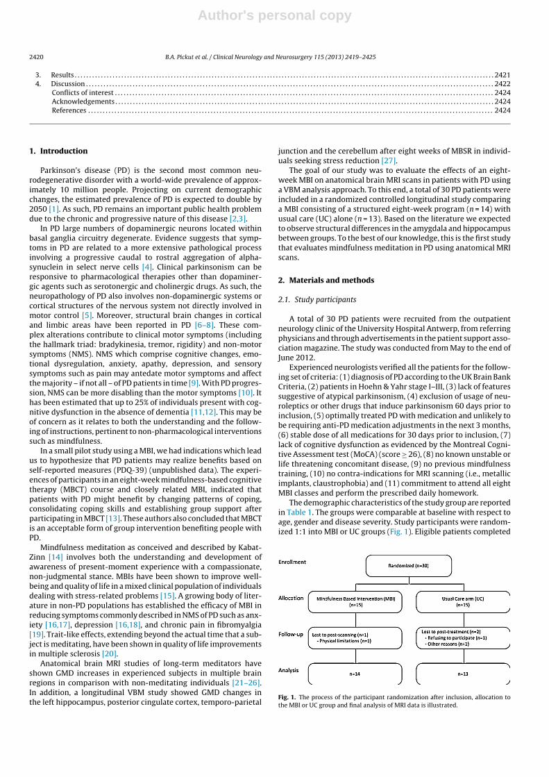
The demographic characteristics of the study group are reportedin Table 1. The groups were comparable at baseline with respect toage, gender and disease severity. Study participants were random-ized 1:1 into MBI or UC groups (Fig. 1). Eligible patients completed
Fig. 1. The process of the participant randomization after inclusion, allocation tothe MBI or UC group and final analysis of MRI data is illustrated.
Author's personal copy
B.A. Pickut et al. / Clinical Neurology and Neurosurgery 115 (2013) 2419– 2425 2421
Table 1Baseline demographic characteristics by intervention status.
Characteristic Overall (n = 27) MBI (n = 14) UC (n = 13) Comparison between groups
Age, y, (SD) 61.8 (9.1) 61.4 (11.3) 62.2 (6.6) t = −.20, p = .84Women, n (%) 13 (48) 7 (50) 6 (46) �2 = .04, p = .84H&Y, (SD) 2.2 (0.4) 2.1 (0.3) 2.2 (0.5) U = 74, p = .34
Legend MBI, Mindfulness Based Intervention Group; UC, Usual Care Group; y, years; H&Y, Hoehn and Yahr; SD, standard deviation.
all pre-intervention (outcome) measures before randomization.Randomization was conducted by a blinded investigator. Allpatient-reported outcome measures were entered into a databaseby personnel blinded to group assignment. Standard protocolapprovals, registration, and patient consent forms were approvedby the Institutional Review Board of the University HospitalAntwerp, Antwerp, Belgium. The study was registered at theClinicalTrials.gov (A service of the U.S. National Institutes ofHealth – NIH) NCT01607697. All participants gave written informedconsent.
2.2. Intervention
Fifteen PD patients included in the study received a MBI closelyfollowing MBSR as described by Kabat-Zinn [14]. The MBI consistedof 2.5 h meetings on eight consecutive weeks without the one-timefull-day (6.5 h) session in the sixth week of practice as described inMBSR. In order to facilitate the development of the capacity formindfulness, the formal mindfulness exercises in body scan, mind-ful movement (also known as mindful yoga) and sitting meditationwere intensively practiced during the eight sessions. To facilitatethe integration of mindfulness into daily life, instructions weregiven on how to practice and integrate mindfulness in everydayactivities such as eating, walking, or performing household chores.Participants also received instructions in using mindfulness forcoping with stress in daily life. Audio recordings containing 45-min guided mindfulness exercises (meditation, body scan, mindfulmovement) were given with instructions for daily home practicecorresponding to the course sequence. Time spent in mindfulnesspractices, as reported by the participants, was recorded weekly foreach participant. The intervention was conducted by two experi-enced mindfulness teachers. All patients in both the UC and MBIgroups received regular, currently optimal medical care during theduration of the study. UC patients were offered the MBI two monthsafter the study.
2.3. MRI data acquisition
All MRI examinations were performed on a 3 Tesla scanner(Magneton Trio Tim, Siemens AG, Siemens Medical Solutions, Erlan-gen, Germany). Acquisition was performed by scanning personnel,blinded to group allocation. A 32-channel head coil was used toobtain images with an isotropic resolution of 1 mm3. For volumet-ric measurements a high resolution anatomical T1-weighted MRdataset was obtained, using a magnetization-prepared rapid acqui-sition gradient echo (MP-RAGE) sequence with 176 slices and afield of view of 256 mm × 192 mm. Repetition time TR and echotime TE were 1910 ms and 3.37 ms, respectively; the flip angle was15◦. All patients underwent an MRI scan at baseline (up to onemonth before) and at eight weeks (after the intervention withinone month).
2.4. MRI data analysis
VBM analysis was performed using Statistical ParametricMapping (SPM)8 software (www.fil.ion.ucl.ac.uk/spm/software/spm8/). All T1-weighted data sets were segmented into gray
matter (GM), white matter (WM) and cerebrospinal fluid (CSF),using a unified tissue segmentation approach [28]. Intensities weremodeled by a gaussian mixture model. Resulting images were biasfield corrected for the smoothly varying intensity inhomogeneitycaused by magnetic field imperfections using discrete cosine trans-formations and were registered into standard space. Subsequently,diffeomorphic anatomical registration through exponentiated liealgebra (DARTEL) was used to account for more detailed shapevariability in the population [29]. The DARTEL registration involvesalternating between computing a template, based on the averagetissue probability maps of all subjects, and warping the tissue mapsof all subjects into increasingly good alignment with the template.Images were then transformed to standard Montreal NeurologicalInstitute (MNI) space, followed by a smoothing procedure with aGaussian Kernel of 12 mm. For each individual patient, the regis-tered baseline T1-weighted image was subtracted from the eightweeks follow-up T1-weighted image to obtain images that repre-sent changes over time for each patient. These subtracted imageswere then compared between both intervention groups using a T-test. For the whole brain analysis, a statistical threshold of p < 0.001(uncorrected for multiple comparisons) with an extent threshold ofat least 5 voxels was used to present the results. In addition, ROIs inthe amygdala and the hippocampus were defined in MNI space toevaluate GMD changes in the hypothesized regions. For the latterROI analysis, an FWE corrected statistical threshold of p < 0.05 wasused.
3. Results
Twenty-seven out of 30 PD patients completed the study(Fig. 1). Two patients, allocated to the UC group, dropped outfor personal reasons (not relevant to the study) during follow-upwhile one patient of the MBI group discontinued study partici-pation due to acute back pain limiting access to the MRI scan.The medication remained stable in 26 out of 27 patients. Onepatient in the UC group had an increase of 50 mg levodopa andthe addition of 50 mg sertraline, as deemed necessary by thegeneral practitioner during the study. Attendance rate at thetraining sessions of the MBI group was 97.3%. Of the 14 partic-ipants in the MBI group, 11 were present for all eight sessionsand 3 subjects missed one session. The participants reportedspending a total of 32,244 min (537.4 h, mean = 38.4 h, SD = 11.9 h)performing formal homework exercises over the 8-week train-ing (average = 55 min per day). The time spent on practicing was47.45% on meditation, 21.79% on body scan and 30.76% on mindfulmovement.
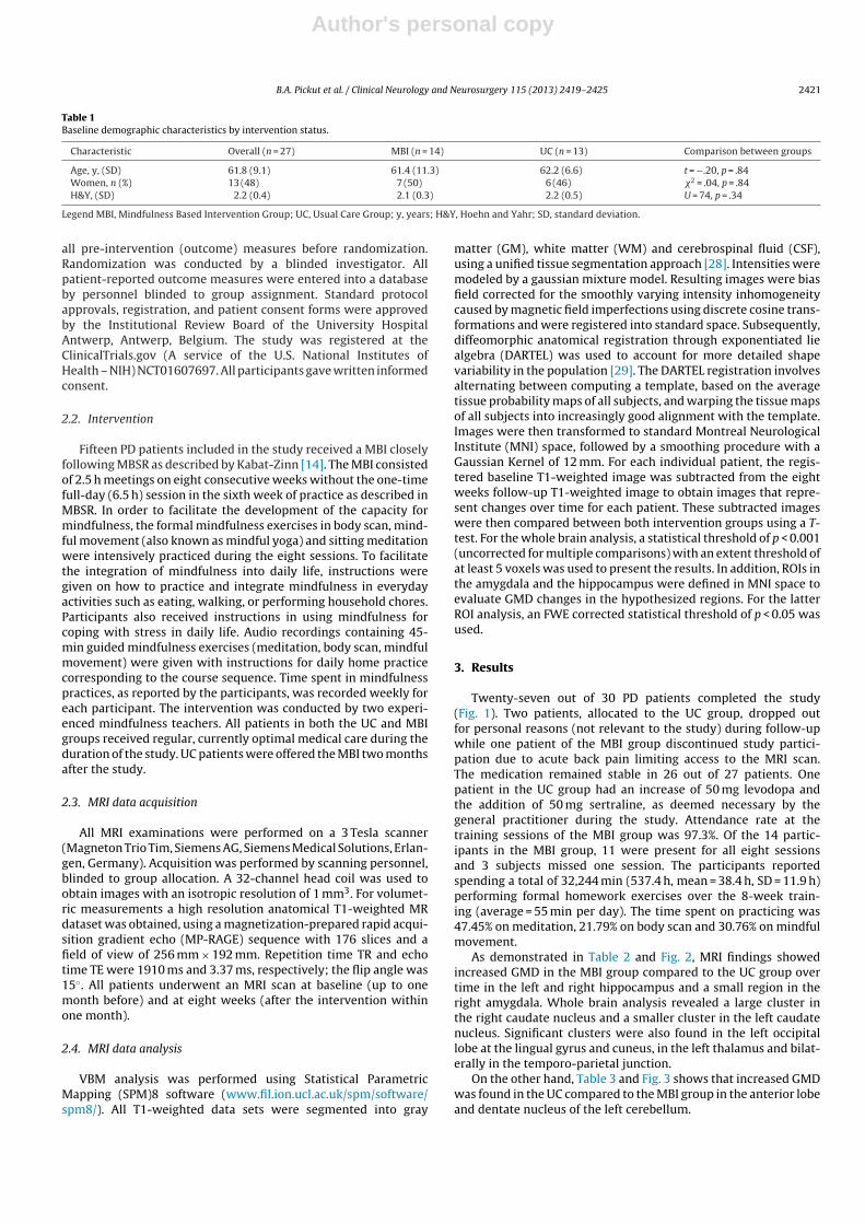
As demonstrated in Table 2 and Fig. 2, MRI findings showedincreased GMD in the MBI group compared to the UC group overtime in the left and right hippocampus and a small region in theright amygdala. Whole brain analysis revealed a large cluster inthe right caudate nucleus and a smaller cluster in the left caudatenucleus. Significant clusters were also found in the left occipitallobe at the lingual gyrus and cuneus, in the left thalamus and bilat-erally in the temporo-parietal junction.
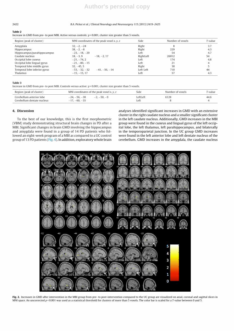
On the other hand, Table 3 and Fig. 3 shows that increased GMDwas found in the UC compared to the MBI group in the anterior lobeand dentate nucleus of the left cerebellum.
Author's personal copy
2422 B.A. Pickut et al. / Clinical Neurology and Neurosurgery 115 (2013) 2419– 2425
Table 2Increase in GMD from pre- to post MBI. Active versus controls: p < 0.001, cluster size greater than 5 voxels.
Region (peak of cluster) MNI coordinates of the peak voxel x, y, z Side Number of voxels T-value
Amygdala 32, −2, −24 Right 8 3.7Hippocampus 38, −2, −8 Right 229 4.3Hippocampus/parahippocampus −23, −18, −20 Left 54 4.7Caudate nucleus 18, −3, 9 −18, −2, 17 RightLeft 26012 54Occipital lobe cuneus −21, −74, 2 Left 174 4.8Occipital lobe lingual gyrus −21, −80, −15 Left 21 4Temporal lobe middle gyrus 32, −45, 5 Right 30 5Temporal lobe inferior gyrus −53, −32, −32 −41, −56, −14 Left Left 710 44Thalamus −15, −15, 17 Left 57 4.3
Table 3Increase in GMD from pre- to post MBI. Controls versus active: p < 0.001, cluster size greater than 5 voxels.
Region (peak of cluster) MNI coordinates of the peak voxel x, y, z Side Number of voxels T-value
Cerebellum anterior lobe −24, −56, −38 −2, −50, −9 LeftLeft 6128 44.6Cerebellum dentate nucleus −17, −60, −35 Left 8 4
4. Discussion
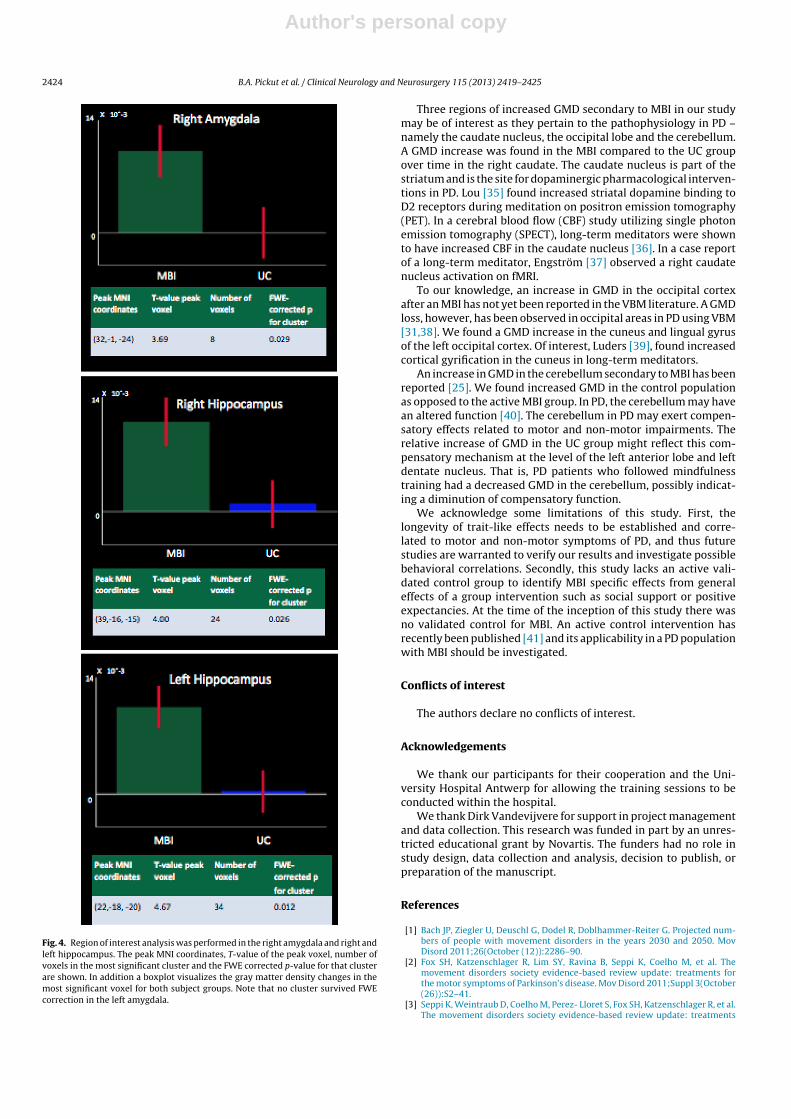
To the best of our knowledge, this is the first morphometric(VBM) study demonstrating structural brain changes in PD after aMBI. Significant changes in brain GMD involving the hippocampusand amygdala were found in a group of 14 PD patients who fol-lowed an eight-week program of a MBI as compared to a UC controlgroup of 13 PD patients (Fig. 4). In addition, exploratory whole brain
analyses identified significant increases in GMD with an extensivecluster in the right caudate nucleus and a smaller significant clusterin the left caudate nucleus. Additionally, GMD increases in the MBIgroup were found in the cuneus and lingual gyrus of the left occip-ital lobe, the left thalamus, left parahippocampus, and bilaterallyin the temporoparietal junction. In the UC group GMD increaseswere found in the left anterior lobe and left dentate nucleus of thecerebellum. GMD increases in the amygdala, the caudate nucleus
Fig. 2. Increases in GMD after intervention in the MBI group from pre- to post-intervention compared to the UC group are visualized on axial, coronal and sagittal slices inMNI space. An uncorrected p < 0.001 was used as a statistical threshold for clusters of more than 5 voxels. The color bar is scaled for a T-value between 0 and 5.
Author's personal copy
B.A. Pickut et al. / Clinical Neurology and Neurosurgery 115 (2013) 2419– 2425 2423
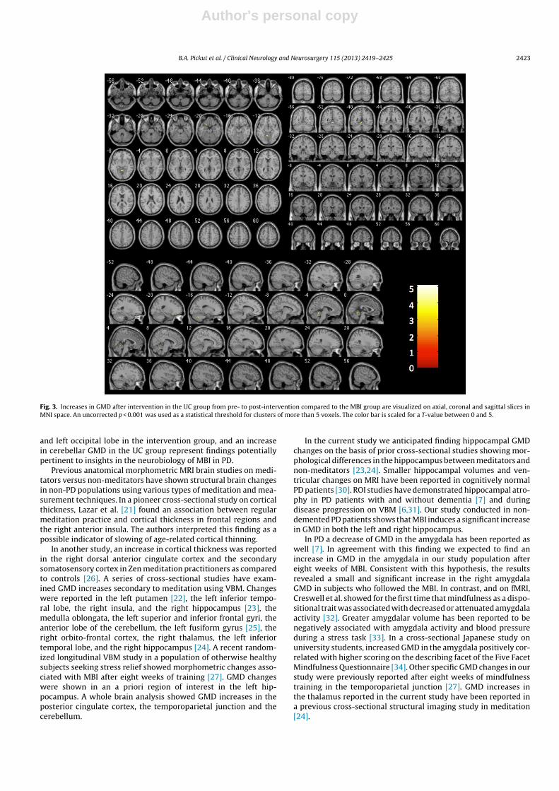
Fig. 3. Increases in GMD after intervention in the UC group from pre- to post-intervention compared to the MBI group are visualized on axial, coronal and sagittal slices inMNI space. An uncorrected p < 0.001 was used as a statistical threshold for clusters of more than 5 voxels. The color bar is scaled for a T-value between 0 and 5.
and left occipital lobe in the intervention group, and an increasein cerebellar GMD in the UC group represent findings potentiallypertinent to insights in the neurobiology of MBI in PD.
Previous anatomical morphometric MRI brain studies on medi-tators versus non-meditators have shown structural brain changesin non-PD populations using various types of meditation and mea-surement techniques. In a pioneer cross-sectional study on corticalthickness, Lazar et al. [21] found an association between regularmeditation practice and cortical thickness in frontal regions andthe right anterior insula. The authors interpreted this finding as apossible indicator of slowing of age-related cortical thinning.
In another study, an increase in cortical thickness was reportedin the right dorsal anterior cingulate cortex and the secondarysomatosensory cortex in Zen meditation practitioners as comparedto controls [26]. A series of cross-sectional studies have exam-ined GMD increases secondary to meditation using VBM. Changeswere reported in the left putamen [22], the left inferior tempo-ral lobe, the right insula, and the right hippocampus [23], themedulla oblongata, the left superior and inferior frontal gyri, theanterior lobe of the cerebellum, the left fusiform gyrus [25], theright orbito-frontal cortex, the right thalamus, the left inferiortemporal lobe, and the right hippocampus [24]. A recent random-ized longitudinal VBM study in a population of otherwise healthysubjects seeking stress relief showed morphometric changes asso-ciated with MBI after eight weeks of training [27]. GMD changeswere shown in an a priori region of interest in the left hip-pocampus. A whole brain analysis showed GMD increases in theposterior cingulate cortex, the temporoparietal junction and thecerebellum.
In the current study we anticipated finding hippocampal GMDchanges on the basis of prior cross-sectional studies showing mor-phological differences in the hippocampus between meditators andnon-meditators [23,24]. Smaller hippocampal volumes and ven-tricular changes on MRI have been reported in cognitively normalPD patients [30]. ROI studies have demonstrated hippocampal atro-phy in PD patients with and without dementia [7] and duringdisease progression on VBM [6,31]. Our study conducted in non-demented PD patients shows that MBI induces a significant increasein GMD in both the left and right hippocampus.
In PD a decrease of GMD in the amygdala has been reported aswell [7]. In agreement with this finding we expected to find anincrease in GMD in the amygdala in our study population aftereight weeks of MBI. Consistent with this hypothesis, the resultsrevealed a small and significant increase in the right amygdalaGMD in subjects who followed the MBI. In contrast, and on fMRI,Creswell et al. showed for the first time that mindfulness as a dispo-sitional trait was associated with decreased or attenuated amygdalaactivity [32]. Greater amygdalar volume has been reported to benegatively associated with amygdala activity and blood pressureduring a stress task [33]. In a cross-sectional Japanese study onuniversity students, increased GMD in the amygdala positively cor-related with higher scoring on the describing facet of the Five FacetMindfulness Questionnaire [34]. Other specific GMD changes in ourstudy were previously reported after eight weeks of mindfulnesstraining in the temporoparietal junction [27]. GMD increases inthe thalamus reported in the current study have been reported ina previous cross-sectional structural imaging study in meditation[24].
Author's personal copy
2424 B.A. Pickut et al. / Clinical Neurology and Neurosurgery 115 (2013) 2419– 2425
Fig. 4. Region of interest analysis was performed in the right amygdala and right andleft hippocampus. The peak MNI coordinates, T-value of the peak voxel, number ofvoxels in the most significant cluster and the FWE corrected p-value for that clusterare shown. In addition a boxplot visualizes the gray matter density changes in themost significant voxel for both subject groups. Note that no cluster survived FWEcorrection in the left amygdala.
Three regions of increased GMD secondary to MBI in our studymay be of interest as they pertain to the pathophysiology in PD –namely the caudate nucleus, the occipital lobe and the cerebellum.A GMD increase was found in the MBI compared to the UC groupover time in the right caudate. The caudate nucleus is part of thestriatum and is the site for dopaminergic pharmacological interven-tions in PD. Lou [35] found increased striatal dopamine binding toD2 receptors during meditation on positron emission tomography(PET). In a cerebral blood flow (CBF) study utilizing single photonemission tomography (SPECT), long-term meditators were shownto have increased CBF in the caudate nucleus [36]. In a case reportof a long-term meditator, Engström [37] observed a right caudatenucleus activation on fMRI.
To our knowledge, an increase in GMD in the occipital cortexafter an MBI has not yet been reported in the VBM literature. A GMDloss, however, has been observed in occipital areas in PD using VBM[31,38]. We found a GMD increase in the cuneus and lingual gyrusof the left occipital cortex. Of interest, Luders [39], found increasedcortical gyrification in the cuneus in long-term meditators.
An increase in GMD in the cerebellum secondary to MBI has beenreported [25]. We found increased GMD in the control populationas opposed to the active MBI group. In PD, the cerebellum may havean altered function [40]. The cerebellum in PD may exert compen-satory effects related to motor and non-motor impairments. Therelative increase of GMD in the UC group might reflect this com-pensatory mechanism at the level of the left anterior lobe and leftdentate nucleus. That is, PD patients who followed mindfulnesstraining had a decreased GMD in the cerebellum, possibly indicat-ing a diminution of compensatory function.
We acknowledge some limitations of this study. First, thelongevity of trait-like effects needs to be established and corre-lated to motor and non-motor symptoms of PD, and thus futurestudies are warranted to verify our results and investigate possiblebehavioral correlations. Secondly, this study lacks an active vali-dated control group to identify MBI specific effects from generaleffects of a group intervention such as social support or positiveexpectancies. At the time of the inception of this study there wasno validated control for MBI. An active control intervention hasrecently been published [41] and its applicability in a PD populationwith MBI should be investigated.
Conflicts of interest
The authors declare no conflicts of interest.
Acknowledgements
We thank our participants for their cooperation and the Uni-versity Hospital Antwerp for allowing the training sessions to beconducted within the hospital.
We thank Dirk Vandevijvere for support in project managementand data collection. This research was funded in part by an unres-tricted educational grant by Novartis. The funders had no role instudy design, data collection and analysis, decision to publish, orpreparation of the manuscript.
References
[1] Bach JP, Ziegler U, Deuschl G, Dodel R, Doblhammer-Reiter G. Projected num-bers of people with movement disorders in the years 2030 and 2050. MovDisord 2011;26(October (12)):2286–90.
[2] Fox SH, Katzenschlager R, Lim SY, Ravina B, Seppi K, Coelho M, et al. Themovement disorders society evidence-based review update: treatments forthe motor symptoms of Parkinson’s disease. Mov Disord 2011;Suppl 3(October(26)):S2–41.
[3] Seppi K, Weintraub D, Coelho M, Perez- Lloret S, Fox SH, Katzenschlager R, et al.The movement disorders society evidence-based review update: treatments
Author's personal copy
B.A. Pickut et al. / Clinical Neurology and Neurosurgery 115 (2013) 2419– 2425 2425
for the non-motor symptoms of Parkinson’s disease. Mov Disord 2011;Suppl3(October (26)):S42–80.
[4] Braak H, Del Tredici K, Rüb U, de Vos RA, Jansen Steur EN, Braak E. Stagingof brain pathology related to sporadic Parkinson’s disease. Neurobiol Aging2003;24(March–April (2)):197–211.
[5] Jellinger K. Heterogeneous mechanisms of mild cognitive impairment inParkinson’s disease. J Neural Transm 2012;119(March (3)):381–2.
[6] Ramirez-Ruiz B, Marti MJ, Tolosa E, Bartrés-Faz D, Summerfield C, Salgado-Pineda P, et al. Longitudinal evaluation of cerebral morphological changes inParkinson’s disease with and without dementia. J Neurol 2005;252(November(11)):52–1345.
[7] Ibarretxe-Bilbao N, Junque C, Tolosa E, Marti MJ, Valldeoriola F, Bargallo N, et al.Neuroanatomical correlates of impaired decision-making and facial emotionrecognition in early Parkinson’s disease. Eur J Neurosci 2009;30(September(6)):1162–71.
[8] Pagonabarraga J, Corcuera-Solano I, Vives-Gilabert Y, Liebaria G, Garcia-Sánchez C, Pascual- Sedano B, et al. Pattern of regional cortical thinningassociated with cognitive deterioration in Parkinson’s disease. PloS One2013;8(1):e54980.
[9] Crosiers D, Pickut B, Theuns J, Deyn PP, Van Broeckhoven C, Martinez-Martin P,et al. Non-motor symptoms in a Flanders-Belgian population of 215 Parkinson’sdisease patients as assessed by the Non-Motor Symptoms Questionnaire. Am JNeurodegener Dis 2012;1(2):160–7.
[10] Chaudhuri KR, Healy DG, Schapira AH. National Institute for Clinical ExcellenceNon-motor symptoms of Parkinson’s disease: diagnosis and management.Lancet Neurol 2006;5(March (3)):235–45.
[11] Aarsland D, Bronnick K, Williams-Gray C, Weintraub D, Marder K, Kulisevsky J,et al. Mild cognitive impairment in Parkinson’s disease: a multicenter pooledanalysis. Neurology 2010;75(September (12)):1062–9.
[12] Litvan I, Goldman JG, Tröster AI, Schmand BA, Weintraub D, Petersen RC,et al. Diagnostic criteria for mild cognitive impairment in Parkinson’s disease:Movement Disorder Society Task Force Guidelines. Mov Disord 2012;27(March(3)):349–56.
[13] Fitzpatrick L, Simpson J, Smith A. A qualitative analysis of mindfulness-based cognitive therapy (MBCT) in Parkinson’s disease. Psychol Psychother2010;83(June (Pt 2)):179–92.
[14] Kabat-Zinn J. Full Catastrophe Living: Using the Wisdom of Your Body andMind to Face Stress, Pain and Illness. New York NY.: Delta Publishing.; 1990.p. 140–6.
[15] Carmody J, Baer RA. Relationships between mindfulness practice and levelsof mindfulness, medical and psychological symptoms and well-being in amindfulness-based stress reduction program. J Behav Med 2008;31(February(1)):23–33.
[16] Hofmann SG, Sawyer AT, Witt AA, Oh D. The effect of mindfulness therapyon anxiety and depression: a meta-analytic review. J Consult Clin Psychol2010;78(April (2)):169–83.
[17] Roemer L, Orsillo SM, Salters-Pedneault K. Efficacy of an acceptance-basedbehavior therapy for generalized anxiety disorder: evaluation in a randomizedcontrolled trial. J Consult Clin Psychol 2008;76(December (6)):1083–9.
[18] Teasdale JD, Segal ZV, Williams JM, Ridgeway VA, Soulsby JM, Lau MA. Preven-tion of relapse/recurrence in major depression by mindfulness-based cognitivetherapy. J Consult Clin Psychol 2000;68(August (4)):615–23.
[19] Grossman P, Tiefenthaler-Gilmer U, Raysz A, Kesper U. Mindfulness train-ing as an intervention for fibromyalgia: evidence of postintervention and3-year follow-up benefits in well-being. Psychother Psychosom 2007;76(4):226–33.
[20] Grossman P, Kappos L, Gensicke H, D’Souza M, Mohr DC, Penner IK, et al. MSQuality of Life, depression, and fatigue improve after mindfulness training: arandomized trial. Neurology 2010;75(September (13)):1141–9.
[21] Lazar SW, Kerr CE, Wasserman RH, Gray JR, Greve DN, Treadway MT, et al. Med-itation experience is associated with increased cortical thickness. Neuroreport2005;16(November (17)):1893–7.
[22] Pagnoni G, Cekic M. Age effects on gray matter volume and attentional perfor-mance in Zen. meditation. Neurobiol Aging 2007;28(October (10)):7–1623.
[23] Hölzel BK, Ott U, Gard T, Hempel H, Weygandt M, Morgen K, et al. Investigationof mindfulness meditation practitioners with voxel-based morphometry. SocCogn Affect Neurosci 2008;3(March (1)):55–61.
[24] Luders E, Toga AW, Lepore N, Gaser C. The underlying anatomical correlates oflong-term. meditation: larger hippocampal and frontal volumes of gray matter.Neuroimage 2009;45(April (3)):672–8.
[25] Vestergaard-Poulsen P, van Beek M, Skewes J, Bjarkam CR, Stubberup M, Ber-telsen J, et al. Long-term meditation is associated with increased gray matterdensity in the brain stem. Neuroreport 2009;20(January (2)):170–4.
[26] Grant JA, Courtemanche J, Duerden EG, Duncan GH, Rainville P. Corticalthickness and pain. sensitivity in zen meditators. Emotion 2010;10(February(1)):43–53.
[27] Hölzel BK, Carmody J, Vangel M, Congleton C, Yerramsetti SM, Gard T, et al.Mindfulness practice leads to increases in regional brain gray matter density.Psychiatr Res 2011;191(January (1)):36–43.
[28] Ashburner J, Friston KJ. Unified segmentation. Neuroimage 2005;26(July(3)):839–51.
[29] Ashburner J. A fast diffeomorphic image registration algorithm. Neuroimage2007;38(October (1)):95–113.
[30] Apostolova L, Alves G, Hwang KS, Babakchanian S, Bronnick KS, Larsen JP, et al.Hippocampal and ventricular changes in Parkinson’s disease mild cognitiveimpairment. Neurobiol Aging 2012;33(September (9)):2113–24.
[31] Burton EJ, McKeith IG, Burn DJ, Williams ED, O’Brien JT. Cerebral atro-phy in Parkinson’s disease with and without dementia: a comparisonwith Alzheimer’s disease, dementia with Lewy bodies and controls. Brain2004;127(April (Pt 4)):791–800.
[32] Creswell JD, Way BM, Eisenberger NL, Lieberman MD. Neural corre-lates of dispositional mindfulness during affect labeling. Psychosom Med2007;69(July–August (6)):560–5.
[33] Gianaros PJ, Sheu LK, Matthews KA, Jennings JR, Manuck SB, Hariri AR. Indi-vidual differences in stressor-evoked blood pressure reactivity vary withactivation, volume, and functional connectivity of the amygdala. J Neurosci2008;28(January (4)):9–990.
[34] Murakami H, Nakao T, Matsunaga M, Kasuya Y, Shinoda J, Yamada J, et al. Thestructure of mindful brain. PloS One 2012;7(9):e46377.
[35] Lou HC, Kjaer TW, Friberg L, Wildschiodtz G, Holm S, Nowak M. A 15O-H2O PETstudy of meditation and the resting state of normal consciousness. Hum BrainMapp 1999;7(2):98–105.
[36] Newberg AB, Wintering N, Waldman MR, Amen D, Khalsa DS, Alavi A. Cere-bral blood flow differences between long-term meditators and non-meditators.Conscious Cogn 2010;19(December (4)):899–905.
[37] Engström M, Söderfeldt B. Brain activation during compassion meditation: acase study. J Altern Complement Med 2010;16(May (5)):597–9.
[38] Summerfield C, Junqué C, Tolosa E, Salgado-Pineda P, Gómez-Ansón B, MartiMJ, et al. Structural brain changes in Parkinson disease with dementia: a voxel-based morphometry study. Arch Neurol 2005;62(February (2)):281–5.
[39] Luders E, Kurth F, Mayer EA, Toga AW, Narr KL, Gaser C. The unique brainanatomy of meditation practitioners: alterations in cortical gyrification. FrontHum Neurosci 2012;6:34.
[40] Wu T, Hallett M. The cerebellum in Parkinson’s disease. Brain 2013;136(March(Pt 3)):696–709.
[41] MacCoon DG, Imel ZE, Rosenkranz MA, Sheftel JG, Weng HY, Sullivan JC, et al.The validation of an active control intervention for Mindfulness Based StressReduction (MBSR). Behav Res Ther 2012;50(January (1)):3–12.