Sozda-f MRI Error TBI-2011
10
Error-related processing following severe traumatic brain injury: An event-related functional magnetic resonance imaging (fMRI) study Christopher N. Sozda a , Michael J. Larson a, e , David A.S. Kaufman f , Ilona M. Schmalfuss b, g , William M. Perlstein a, c, d, ⁎ a Department of Clinical and Health Psychology, University of Florida, Gainesville, FL, United States b Department of Medicine, University of Florida, Gainesville, FL, United States c Department of Psychiatry, University of Florida, Gainesville, FL, United States d McKnight Brain Institute, University of Florida, Gainesville, FL, United States e Departments of Psychology and Neuroscience, Brigham Young University, Provo, UT, United States f Department of Psychology, Saint Louis University, St. Louis, MO, United States g North Florida/South Georgia Veterans Administration Hospital, Gainesville, FL, United States abstract article info Article history: Received 5 April 2011 Received in revised form 10 June 2011 Accepted 27 June 2011 Available online 12 July 2011 Keywords: Traumatic brain injury TBI Cognitive control Performance monitoring Anterior cingulate cortex ACC Continuous monitoring of one's performance is invaluable for guiding behavior towards successful goal attainment by identifying deficits and strategically adjusting responses when performance is inadequate. In the present study, we exploited the advantages of event-related functional magnetic resonance imaging (fMRI) to examine brain activity associated with error-related processing after severe traumatic brain injury (sTBI). fMRI and behavioral data were acquired while 10 sTBI participants and 12 neurologically-healthy controls performed a task-switching cued-Stroop task. fMRI data were analyzed using a random-effects whole-brain voxel-wise general linear model and planned linear contrasts. Behaviorally, sTBI patients showed greater error-rate interference than neurologically-normal controls. fMRI data revealed that, compared to controls, sTBI patients showed greater magnitude error-related activation in the anterior cingulate cortex (ACC) and an increase in the overall spatial extent of error-related activation across cortical and subcortical regions. Implications for future research and potential limitations in conducting fMRI research in neurologically-impaired populations are discussed, as well as some potential benefits of employing multimodal imaging (e.g., fMRI and event-related potentials) of cognitive control processes in TBI. © 2011 Elsevier B.V. All rights reserved. 1. Introduction Physical and neurobehavioral impairments are common sequelae of traumatic brain injury (TBI; Horn and Sherer, 1999), however, even in patients with good neurological recovery, persistent cognitive deficits are among the most pronounced and frequent complaints of TBI survivors (Cicerone et al., 2005; Lovell and Franzen, 1994). Severity-related impairments in “cognitive control,” a set of higher- order executive processes supported by the prefrontal cortex and critical to executive function (Lorist et al., 2005; Miller, 2000; Miller and Cohen, 2001), are thought to underlie some aspects of enduring cognitive dysfunction after brain injury (Larson et al., 2006, 2007a, in press; Perlstein et al., 2004, 2006; Scheibel et al., 2007; Seignourel et al., 2005; Soeda et al., 2005), and current theories of neurobehavioral dysfunction in TBI have been based on observed impairments in cognitive control component processes (Anderson et al., 2002; Burgess and Robertson, 2002; Larson et al., 2006, 2007a; Levine et al., 2002; Perlstein et al., 2006). Numerous functional magnetic resonance imaging (fMRI) studies (i.e., Carter et al., 1999; MacDonald et al., 2000) suggest that cognitive control comprises two broad component processes implemented in a closely interactive, yet dissociable frontal neural network: a regulative/ strategic component supporting the maintenance of task goals, allocation of limited attentional resources, and the implementation of top-down control (MacDonald et al., 2000), and an anterior cingulate cortex (ACC)-mediated evaluative component that supports conflict processing and performance monitoring (i.e., Carter and van Veen, 2007; Kerns et al., 2004; van Veen and Carter, 2002, 2006). These evaluative monitoring processes serve to adjust behavioral perfor- mance toward goal attainment based on the detection of performance errors (Ridderinkhof et al., 2004). Continuous performance monitor- ing is important for guiding behavior towards successful goal- attainment by detecting deficiencies and strategically adjusting responses when current performance is inadequate. An understanding of the neural basis underlying error-related processing is critical not only to identifying the mechanisms through which cognitive control is executed, but also because impairments in self-awareness in TBI International Journal of Psychophysiology 82 (2011) 97–106 ⁎ Corresponding author at: Department of Clinical and Health Psychology, HSC Box 100165, University of Florida, Gainesville, FL 32610, United States. E-mail address: [email protected]fl.edu (W.M. Perlstein). 0167-8760/$ – see front matter © 2011 Elsevier B.V. All rights reserved. doi:10.1016/j.ijpsycho.2011.06.019 Contents lists available at ScienceDirect International Journal of Psychophysiology journal homepage: www.elsevier.com/locate/ijpsycho
Transcript of Sozda-f MRI Error TBI-2011
International Journal of Psychophysiology 82 (2011) 97–106
Contents lists available at ScienceDirect
International Journal of Psychophysiology
j ourna l homepage: www.e lsev ie r.com/ locate / i jpsycho
Error-related processing following severe traumatic brain injury: An event-relatedfunctional magnetic resonance imaging (fMRI) study
Christopher N. Sozda a, Michael J. Larson a,e, David A.S. Kaufman f,Ilona M. Schmalfuss b,g, William M. Perlstein a,c,d,⁎a Department of Clinical and Health Psychology, University of Florida, Gainesville, FL, United Statesb Department of Medicine, University of Florida, Gainesville, FL, United Statesc Department of Psychiatry, University of Florida, Gainesville, FL, United Statesd McKnight Brain Institute, University of Florida, Gainesville, FL, United Statese Departments of Psychology and Neuroscience, Brigham Young University, Provo, UT, United Statesf Department of Psychology, Saint Louis University, St. Louis, MO, United Statesg North Florida/South Georgia Veterans Administration Hospital, Gainesville, FL, United States
⁎ Corresponding author at: Department of Clinical an100165, University of Florida, Gainesville, FL 32610, Un
E-mail address: [email protected] (W.M. Perlstein
0167-8760/$ – see front matter © 2011 Elsevier B.V. Aldoi:10.1016/j.ijpsycho.2011.06.019
a b s t r a c t
a r t i c l e i n f oArticle history:Received 5 April 2011Received in revised form 10 June 2011Accepted 27 June 2011Available online 12 July 2011
Keywords:Traumatic brain injuryTBICognitive controlPerformance monitoringAnterior cingulate cortexACC
Continuous monitoring of one's performance is invaluable for guiding behavior towards successful goalattainment by identifying deficits and strategically adjusting responses when performance is inadequate. Inthe present study, we exploited the advantages of event-related functional magnetic resonance imaging(fMRI) to examine brain activity associated with error-related processing after severe traumatic brain injury(sTBI). fMRI and behavioral data were acquired while 10 sTBI participants and 12 neurologically-healthycontrols performed a task-switching cued-Stroop task. fMRI data were analyzed using a random-effectswhole-brain voxel-wise general linear model and planned linear contrasts. Behaviorally, sTBI patients showedgreater error-rate interference than neurologically-normal controls. fMRI data revealed that, compared tocontrols, sTBI patients showed greater magnitude error-related activation in the anterior cingulate cortex(ACC) and an increase in the overall spatial extent of error-related activation across cortical and subcorticalregions. Implications for future research and potential limitations in conducting fMRI research inneurologically-impaired populations are discussed, as well as some potential benefits of employingmultimodal imaging (e.g., fMRI and event-related potentials) of cognitive control processes in TBI.
d Health Psychology, HSC Boxited States.).
l rights reserved.
© 2011 Elsevier B.V. All rights reserved.
1. Introduction
Physical and neurobehavioral impairments are common sequelaeof traumatic brain injury (TBI; Horn and Sherer, 1999), however, evenin patients with good neurological recovery, persistent cognitivedeficits are among the most pronounced and frequent complaints ofTBI survivors (Cicerone et al., 2005; Lovell and Franzen, 1994).Severity-related impairments in “cognitive control,” a set of higher-order executive processes supported by the prefrontal cortex andcritical to executive function (Lorist et al., 2005; Miller, 2000; Millerand Cohen, 2001), are thought to underlie some aspects of enduringcognitive dysfunction after brain injury (Larson et al., 2006, 2007a, inpress; Perlstein et al., 2004, 2006; Scheibel et al., 2007; Seignourel etal., 2005; Soeda et al., 2005), and current theories of neurobehavioraldysfunction in TBI have been based on observed impairments incognitive control component processes (Anderson et al., 2002;
Burgess and Robertson, 2002; Larson et al., 2006, 2007a; Levineet al., 2002; Perlstein et al., 2006).
Numerous functional magnetic resonance imaging (fMRI) studies(i.e., Carter et al., 1999; MacDonald et al., 2000) suggest that cognitivecontrol comprises two broad component processes implemented in aclosely interactive, yet dissociable frontal neural network: a regulative/strategic component supporting the maintenance of task goals,allocation of limited attentional resources, and the implementationof top-down control (MacDonald et al., 2000), and an anteriorcingulate cortex (ACC)-mediated evaluative component that supportsconflict processing and performance monitoring (i.e., Carter and vanVeen, 2007; Kerns et al., 2004; vanVeen and Carter, 2002, 2006). Theseevaluative monitoring processes serve to adjust behavioral perfor-mance toward goal attainment based on the detection of performanceerrors (Ridderinkhof et al., 2004). Continuous performance monitor-ing is important for guiding behavior towards successful goal-attainment by detecting deficiencies and strategically adjustingresponseswhen current performance is inadequate. An understandingof the neural basis underlying error-related processing is critical notonly to identifying themechanisms throughwhich cognitive control isexecuted, but also because impairments in self-awareness in TBI
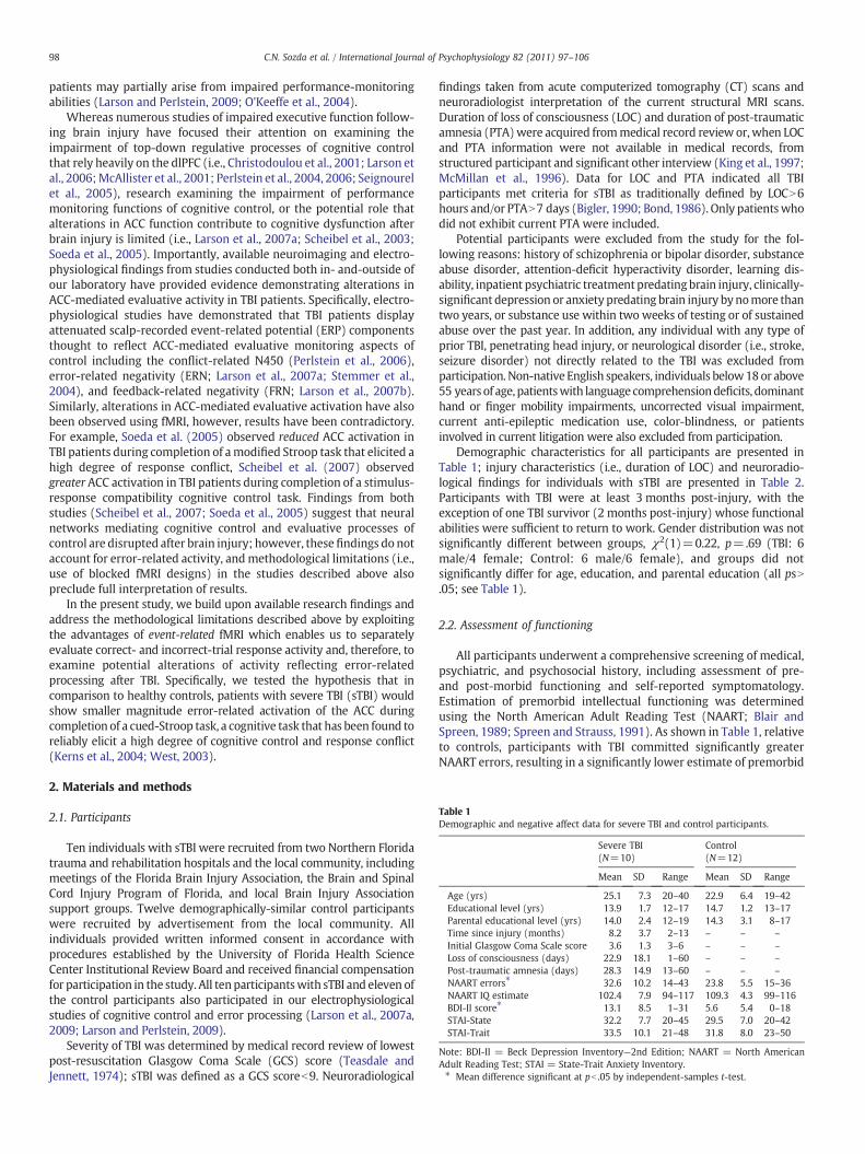
Table 1Demographic and negative affect data for severe TBI and control participants.
Severe TBI(N=10)
Control(N=12)
Mean SD Range Mean SD Range
Age (yrs) 25.1 7.3 20–40 22.9 6.4 19–42Educational level (yrs) 13.9 1.7 12–17 14.7 1.2 13–17Parental educational level (yrs) 14.0 2.4 12–19 14.3 3.1 8–17Time since injury (months) 8.2 3.7 2–13 – – –
Initial Glasgow Coma Scale score 3.6 1.3 3–6 – – –
Loss of consciousness (days) 22.9 18.1 1–60 – – –
Post-traumatic amnesia (days) 28.3 14.9 13–60 – – –
NAART errors⁎ 32.6 10.2 14–43 23.8 5.5 15–36NAART IQ estimate 102.4 7.9 94–117 109.3 4.3 99–116BDI-II score⁎ 13.1 8.5 1–31 5.6 5.4 0–18STAI-State 32.2 7.7 20–45 29.5 7.0 20–42STAI-Trait 33.5 10.1 21–48 31.8 8.0 23–50
Note: BDI-II = Beck Depression Inventory—2nd Edition; NAART = North AmericanAdult Reading Test; STAI = State-Trait Anxiety Inventory.⁎ Mean difference significant at pb .05 by independent-samples t-test.
98 C.N. Sozda et al. / International Journal of Psychophysiology 82 (2011) 97–106
patients may partially arise from impaired performance-monitoringabilities (Larson and Perlstein, 2009; O'Keeffe et al., 2004).
Whereas numerous studies of impaired executive function follow-ing brain injury have focused their attention on examining theimpairment of top-down regulative processes of cognitive controlthat rely heavily on the dlPFC (i.e., Christodoulou et al., 2001; Larson etal., 2006;McAllister et al., 2001; Perlstein et al., 2004, 2006; Seignourelet al., 2005), research examining the impairment of performancemonitoring functions of cognitive control, or the potential role thatalterations in ACC function contribute to cognitive dysfunction afterbrain injury is limited (i.e., Larson et al., 2007a; Scheibel et al., 2003;Soeda et al., 2005). Importantly, available neuroimaging and electro-physiological findings from studies conducted both in- and-outside ofour laboratory have provided evidence demonstrating alterations inACC-mediated evaluative activity in TBI patients. Specifically, electro-physiological studies have demonstrated that TBI patients displayattenuated scalp-recorded event-related potential (ERP) componentsthought to reflect ACC-mediated evaluative monitoring aspects ofcontrol including the conflict-related N450 (Perlstein et al., 2006),error-related negativity (ERN; Larson et al., 2007a; Stemmer et al.,2004), and feedback-related negativity (FRN; Larson et al., 2007b).Similarly, alterations in ACC-mediated evaluative activation have alsobeen observed using fMRI, however, results have been contradictory.For example, Soeda et al. (2005) observed reduced ACC activation inTBI patients during completion of amodified Stroop task that elicited ahigh degree of response conflict, Scheibel et al. (2007) observedgreater ACC activation in TBI patients during completion of a stimulus-response compatibility cognitive control task. Findings from bothstudies (Scheibel et al., 2007; Soeda et al., 2005) suggest that neuralnetworks mediating cognitive control and evaluative processes ofcontrol are disrupted after brain injury; however, thesefindings do notaccount for error-related activity, andmethodological limitations (i.e.,use of blocked fMRI designs) in the studies described above alsopreclude full interpretation of results.
In the present study, we build upon available research findings andaddress the methodological limitations described above by exploitingthe advantages of event-related fMRI which enables us to separatelyevaluate correct- and incorrect-trial response activity and, therefore, toexamine potential alterations of activity reflecting error-relatedprocessing after TBI. Specifically, we tested the hypothesis that incomparison to healthy controls, patients with severe TBI (sTBI) wouldshow smaller magnitude error-related activation of the ACC duringcompletion of a cued-Stroop task, a cognitive task that has been found toreliably elicit a high degree of cognitive control and response conflict(Kerns et al., 2004; West, 2003).
2. Materials and methods
2.1. Participants
Ten individuals with sTBI were recruited from two Northern Floridatrauma and rehabilitation hospitals and the local community, includingmeetings of the Florida Brain Injury Association, the Brain and SpinalCord Injury Program of Florida, and local Brain Injury Associationsupport groups. Twelve demographically-similar control participantswere recruited by advertisement from the local community. Allindividuals provided written informed consent in accordance withprocedures established by the University of Florida Health ScienceCenter Institutional Review Board and received financial compensationfor participation in the study. All tenparticipantswith sTBI and eleven ofthe control participants also participated in our electrophysiologicalstudies of cognitive control and error processing (Larson et al., 2007a,2009; Larson and Perlstein, 2009).
Severity of TBI was determined by medical record review of lowestpost-resuscitation Glasgow Coma Scale (GCS) score (Teasdale andJennett, 1974); sTBI was defined as a GCS scoreb9. Neuroradiological
findings taken from acute computerized tomography (CT) scans andneuroradiologist interpretation of the current structural MRI scans.Duration of loss of consciousness (LOC) and duration of post-traumaticamnesia (PTA)were acquired frommedical record review or,when LOCand PTA information were not available in medical records, fromstructured participant and significant other interview (King et al., 1997;McMillan et al., 1996). Data for LOC and PTA indicated all TBIparticipants met criteria for sTBI as traditionally defined by LOCN6hours and/or PTAN7 days (Bigler, 1990; Bond, 1986). Only patientswhodid not exhibit current PTA were included.
Potential participants were excluded from the study for the fol-lowing reasons: history of schizophrenia or bipolar disorder, substanceabuse disorder, attention-deficit hyperactivity disorder, learning dis-ability, inpatient psychiatric treatment predating brain injury, clinically-significant depression or anxiety predating brain injury bynomore thantwo years, or substance use within two weeks of testing or of sustainedabuse over the past year. In addition, any individual with any type ofprior TBI, penetrating head injury, or neurological disorder (i.e., stroke,seizure disorder) not directly related to the TBI was excluded fromparticipation. Non-native English speakers, individuals below18or above55 years of age, patientswith language comprehensiondeficits, dominanthand or finger mobility impairments, uncorrected visual impairment,current anti-epileptic medication use, color-blindness, or patientsinvolved in current litigation were also excluded from participation.
Demographic characteristics for all participants are presented inTable 1; injury characteristics (i.e., duration of LOC) and neuroradio-logical findings for individuals with sTBI are presented in Table 2.Participants with TBI were at least 3 months post-injury, with theexception of one TBI survivor (2 months post-injury) whose functionalabilities were sufficient to return to work. Gender distribution was notsignificantly different between groups, χ2(1)=0.22, p=.69 (TBI: 6male/4 female; Control: 6 male/6 female), and groups did notsignificantly differ for age, education, and parental education (all psN.05; see Table 1).
2.2. Assessment of functioning
All participants underwent a comprehensive screening of medical,psychiatric, and psychosocial history, including assessment of pre-and post-morbid functioning and self-reported symptomatology.Estimation of premorbid intellectual functioning was determinedusing the North American Adult Reading Test (NAART; Blair andSpreen, 1989; Spreen and Strauss, 1991). As shown in Table 1, relativeto controls, participants with TBI committed significantly greaterNAART errors, resulting in a significantly lower estimate of premorbid
Table 2Injury characteristics and neuroradiological information for all participants with TBI (N=10).
Age(yrs)
Sex Etiology GCS LOC(days)
PTA(days)
Time post injury(months)
Neuro-radiological findings
21 M MVA 3 42 21 10 Skull fracture and right subdural hematoma22 F MVA 3 7 21 10 Bilateral frontal contusions but no other physical injuries20 F MVA 3 14 14 12 1.5 cm high right frontal shear injury in the white matter;
some subarachnoid blood in the quadrigeminal cistern25 F MVA 3 28 28 11 Left occipital condyle fracture21 M MVA 3 28 42 6 Unavailable21 M Motorcycle accident 3 12 33 8 Right temporal intracranial hemorrhage37 M Motorcycle accident 3 28 36 7 Small bilateral intraventricular hemorrhages with no shift40 F MVA 6 1 15 13 Left temporal occipital subarachnoid hemorrhage;
multiple skull fractures24 M Boating accident 3 9 13 2 Right temporal lobe epidural and subdural hematomas;
right anterior middlecranial fossa hematoma
20 M MVA 6 60 60 3 Left frontal subdural hematoma, multiple frontoparietalcontusions and an orbital fracture
99C.N. Sozda et al. / International Journal of Psychophysiology 82 (2011) 97–106
intellectual functioning in TBI participants. However, mean NAART-estimated premorbid WAIS-R FSIQ scores (Spreen and Strauss, 1991),while different between groups, both fell within the average range ofintellectual functioning (Wechsler, 1981).
Participants also completed the Beck Depression Inventory—SecondEdition, (BDI-II; Beck et al., 1996) and State-Trait Anxiety Inventory(STAI; Spielberger et al., 1970), to assess current levels of depressive andanxiety symptomatology, respectively. As shown in Table 1, groups didnot differ in the extent to which they endorsed symptoms of either stateor trait anxiety; however, TBI participants endorsed significantly greaterdepressive symptoms than the control group. Thoughmean BDI-II scoreswere greater for TBI than control participants,means for both groupwerebelow clinical cut-off levels for depression (i.e., 13 for mild depression;Beck et al., 1996). Additionally, although one control participant and fourTBI patients had BDI-II scores reflective of mild depression (i.e., 14; Becket al., 1996), the overall pattern of functional imaging results did notsignificantly differ when high-negative affect individuals (i.e., BDI≥14)were excluded from subsequent follow-up analyses, and as such, allparticipants were included in the analyses reported below.1
2.3. Cognitive activation task
Participants were scanned while they performed a version of thetask-switching cued-Stroop task (Cohen et al., 1999) which was alsoused in our previous behavioral (Seignourel et al., 2005) and ERP(Perlstein et al., 2006) studies. The task is schematicized in Fig. 1. Atthe beginning of each trial, participants were presented visually withan instructional cue (the word “color” or “word”) followed after adelay by the probe (i.e., Stroop) stimulus. Participants were instructedto respondmanually to the stimulus as designated by the instructionalcue, as quickly and accurately as possible. Participants performed twotasks as specified by the instructional cue: word reading and colornaming. In the word-reading task, participants silently read the probeword; in the color-naming task, they silently named the printed colorof the probe word. Three font colors and color words were used (red,green, blue) and presented in each of two congruency conditions(congruent, incongruent). Congruent stimuli consisted of one of thethree color names presented in its own color (i.e., “RED” printed inred); incongruent stimuli consisted of a color name presented in oneof the two remaining colors (i.e., “RED” printed in blue). Participantswere instructed to respond manually, as quickly and accurately aspossible, by pressing one of three color-coded response keys using theindex, middle, and ring fingers on their right hand.
Timing parameters for trial events were: cue-probe delay=12.5 s,probe-cue delay=10 s, for a total trial duration of 22.5 s. Instructional
1 ACC error-related activity did not significantly correlate with BDI scores, collapsedacross groups or for each group separately (rs≤ .025, psN .27).
cues and probe stimuli were presented for 1.5 s each. Participantsperformed 16 blocks of 12 trials each for a total of 192 trials distributedequally across task conditions (i.e., color naming, word reading,congruent, and incongruent). Trial conditions were presented pseudo-randomly with the constraint that an equal number of conditionsoccurred within each block.
All participants were trained in color-button responsemapping to atleast 80% prior to entering the scanner. Though participants receivedfeedback regarding their performance on the color-button responsemapping training, they did not receive feedback on their in-scannerperformance on the cued-Stroop task. E-Prime software 1.1 (PsychologySoftware Tools, Pittsburgh, PA) was used to generate stimuli and recordbehavioral response accuracy and RTs and an Integrated FunctionalImaging System (IFIS; Psychology Software Tools, Pittsburgh, PA)5-buttonMR compatible response boxwasused to acquire participant'sbehavioral responses.
2.4. Functional image data acquisition and reduction
MRI scanning was conducted using a research-dedicated SiemensAllegra 3-Tesla MRI head scanner equipped with a standard head radiofrequency coil. Task stimuli were presented using an LCD screenmounted above the participant's head with IFIS hardware. Functionalimageswere acquired in 35 axial slices rotated approximately 30° abovethe anterior commissure–posterior commissure (AC–PC) line using aT2*-weighted EPI pulse sequence (repetition time, TR=2500 ms; echotime, TE=30ms; flip angle, FA=90°; field of view, FOV=24 cm;64×64 voxels at 3.75 mm3 with .4 mm slice gap). The 30° AC–PC lineoffset was used to decrease signal loss from the orbitofrontal cortex dueto susceptibility artifact (McClure et al., 2004). Prior to functionalscanning, a T1-weighted MP-RAGE high-resolution 3D anatomical
Fig. 1. Schematic of the task-switching cued Stroop task. As shown, task trialscomprised an instructional cue followed after a delay by a stimulus probe to which theparticipant responded with a button press.
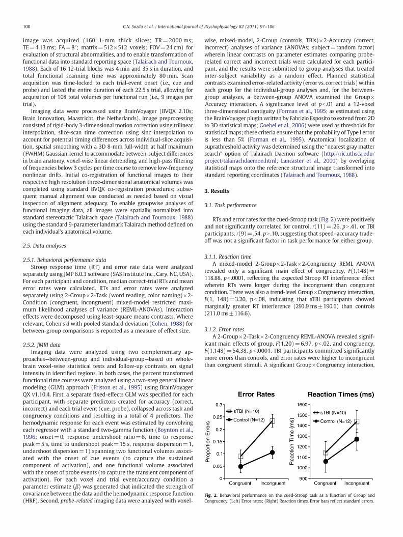
Fig. 2. Behavioral performance on the cued-Stroop task as a function of Group andCongruency. (Left) Error rates; (Right) Reaction times. Error bars reflect standard errors.
100 C.N. Sozda et al. / International Journal of Psychophysiology 82 (2011) 97–106
image was acquired (160 1-mm thick slices; TR=2000 ms;TE=4.13 ms; FA=8°; matrix=512×512 voxels; FOV=24 cm) forevaluation of structural abnormalities, and to enable transformation offunctional data into standard reporting space (Talairach and Tournoux,1988). Each of 16 12-trial blocks was 4 min and 35 s in duration, andtotal functional scanning time was approximately 80 min. Scanacquisition was time-locked to each trial-event onset (i.e., cue andprobe) and lasted the entire duration of each 22.5 s trial, allowing foracquisition of 108 total volumes per functional run (i.e., 9 images pertrial).
Imaging data were processed using BrainVoyager (BVQX 2.10s;Brain Innovation, Maastricht, the Netherlands). Image preprocessingconsisted of rigid-body 3-dimensional motion correction using trilinearinterpolation, slice-scan time correction using sinc interpolation toaccount for potential timing differences across individual-slice acquisi-tion, spatial smoothing with a 3D 8-mm full-width at half maximum(FWHM)Gaussian kernel to accommodate between-subject differencesin brain anatomy, voxel-wise linear detrending, and high-pass filteringof frequencies below 3 cycles per time course to remove low-frequencynonlinear drifts. Initial co-registration of functional images to theirrespective high resolution three-dimensional anatomical volumes wascompleted using standard BVQX co-registration procedures; subse-quent manual alignment was conducted as needed based on visualinspection of alignment adequacy. To enable groupwise analyses offunctional imaging data, all images were spatially normalized intostandard stereotactic Talairach space (Talairach and Tournoux, 1988)using the standard 9-parameter landmark Talairachmethod defined oneach individual's anatomical volume.
2.5. Data analyses
2.5.1. Behavioral performance dataStroop response time (RT) and error rate data were analyzed
separately using JMP 6.0.3 software (SAS Institute Inc., Cary, NC, USA).For each participant and condition, median correct-trial RTs andmeanerror rates were calculated. RTs and error rates were analyzedseparately using 2-Group×2-Task (word reading, color naming)×2-Condition (congruent, incongruent) mixed-model restricted maxi-mum likelihood analyses of variance (REML-ANOVAs). Interactioneffects were decomposed using least-square means contrasts. Whererelevant, Cohen's d with pooled standard deviation (Cohen, 1988) forbetween-group comparisons is reported as a measure of effect size.
2.5.2. fMRI dataImaging data were analyzed using two complementary ap-
proaches—between-group and individual-group—based on whole-brain voxel-wise statistical tests and follow-up contrasts on signalintensity in identified regions. In both cases, the percent transformedfunctional time courses were analyzed using a two-step general linearmodeling (GLM) approach (Friston et al., 1995) using BrainVoyagerQX v1.10.4. First, a separate fixed-effects GLM was specified for eachparticipant, with separate predictors created for accuracy (correct,incorrect) and each trial event (cue, probe), collapsed across task andcongruency conditions and resulting in a total of 4 predictors. Thehemodynamic response for each event was estimated by convolvingeach regressor with a standard two-gamma function (Boynton et al.,1996; onset=0, response undershoot ratio=6, time to responsepeak=5 s, time to undershoot peak=15 s, response dispersion=1,undershoot dispersion=1) spanning two functional volumes associ-ated with the onset of cue events (to capture the sustainedcomponent of activation), and one functional volume associatedwith the onset of probe events (to capture the transient component ofactivation). For each voxel and trial event/accuracy condition aparameter estimate (ß) was generated that indicated the strength ofcovariance between the data and the hemodynamic response function(HRF). Second, probe-related imaging data were analyzed with voxel-
wise, mixed-model, 2-Group (controls, TBIs)×2-Accuracy (correct,incorrect) analyses of variance (ANOVAs; subject=random factor)wherein linear contrasts on parameter estimates comparing probe-related correct and incorrect trials were calculated for each partici-pant, and the results were submitted to group analyses that treatedinter-subject variability as a random effect. Planned statisticalcontrasts examined error-related activity (error vs. correct trials)withineach group for the individual-group analyses and, for the between-group analyses, a between-group ANOVA examined the Group×Accuracy interaction. A significance level of pb .01 and a 12-voxelthree-dimensional contiguity (Forman et al., 1995; as estimated usingthe BrainVoyager pluginwritten by Fabrizio Esposito to extend from 2Dto 3D statistical maps; Goebel et al., 2006) were used as thresholds forstatisticalmaps; these criteria ensure that the probability of Type I erroris less than 5% (Forman et al., 1995). Anatomical localization ofsuprathreshold activity was determined using the “nearest gray mattersearch” option of Talairach Daemon software (http://ric.uthsca.edu/project/talairachdaemon.html; Lancaster et al., 2000) by overlayingstatistical maps onto the reference structural image transformed intostandard reporting coordinates (Talairach and Tournoux, 1988).
3. Results
3.1. Task performance
RTs and error rates for the cued-Stroop task (Fig. 2) were positivelyand not significantly correlated for control, r(11)=.26, pN .41, or TBIparticipants, r(9)=.54, pN .10, suggesting that speed–accuracy trade-off was not a significant factor in task performance for either group.
3.1.1. Reaction timeA mixed-model 2-Group×2-Task×2-Congruency REML ANOVA
revealed only a significant main effect of congruency, F(1,148)=118.88, pb .0001, reflecting the expected Stroop RT interference effectwherein RTs were longer during the incongruent than congruentcondition. There was also a trend-level Group×Congruency interaction,F(1, 148)=3.20, pb .08, indicating that sTBI participants showedmarginally greater RT interference (293.9 ms±190.6) than controls(211.0 ms±116.6).
3.1.2. Error ratesA 2-Group×2-Task×2-Congruency REML-ANOVA revealed signif-
icant main effects of group, F(1,20)=6.97, pb .02, and congruency,F(1,148)=54.38, pb .0001. TBI participants committed significantlymore errors than controls, and error rates were higher to incongruentthan congruent stimuli. A significant Group×Congruency interaction,
101C.N. Sozda et al. / International Journal of Psychophysiology 82 (2011) 97–106
F(1,148)=10.11, pb .002, reflected greater error-rate interferencein patients than controls; least-squares means contrasts revealedthat the two groups did not differ in error rates on the congruentcondition, t(20)=1.18, pN .24, d=.51, but did differ on the incongruentcondition, t(20)=3.68, pb .001, d=1.54, while both groups showedsignificant error-rate interference, ts≥3.11, psb .0025, ds≥2.44.
3.2. Imaging data2
3.2.1. Between-groups analysisThe between-groups comparison used voxel-wise Group×Accu-
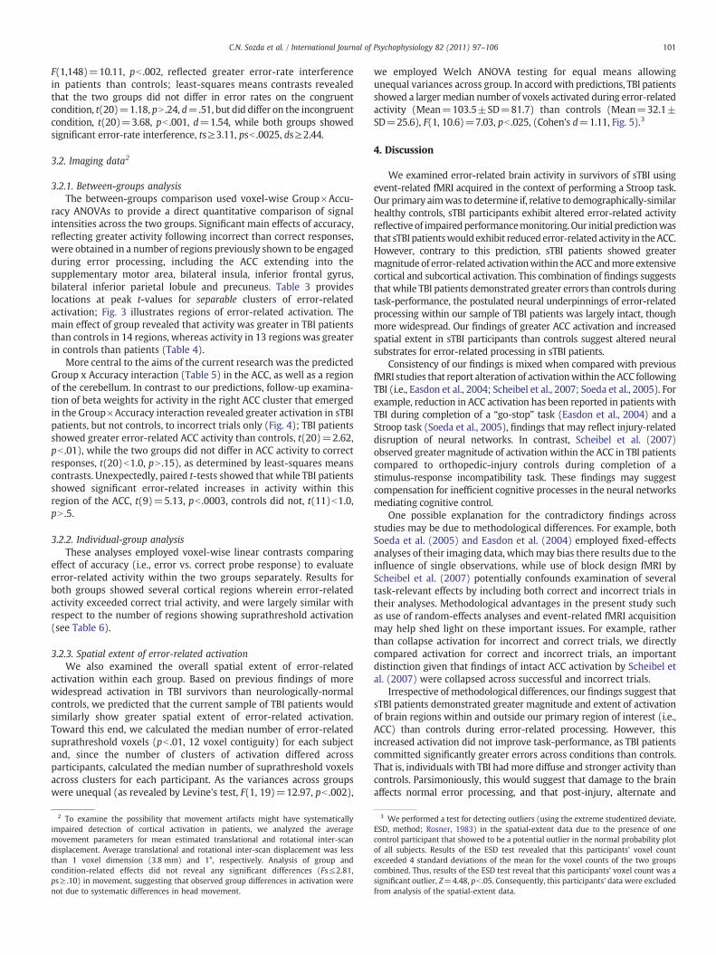
racy ANOVAs to provide a direct quantitative comparison of signalintensities across the two groups. Significant main effects of accuracy,reflecting greater activity following incorrect than correct responses,were obtained in a number of regions previously shown to be engagedduring error processing, including the ACC extending into thesupplementary motor area, bilateral insula, inferior frontal gyrus,bilateral inferior parietal lobule and precuneus. Table 3 provideslocations at peak t-values for separable clusters of error-relatedactivation; Fig. 3 illustrates regions of error-related activation. Themain effect of group revealed that activity was greater in TBI patientsthan controls in 14 regions, whereas activity in 13 regions was greaterin controls than patients (Table 4).
More central to the aims of the current research was the predictedGroup x Accuracy interaction (Table 5) in the ACC, as well as a regionof the cerebellum. In contrast to our predictions, follow-up examina-tion of beta weights for activity in the right ACC cluster that emergedin the Group×Accuracy interaction revealed greater activation in sTBIpatients, but not controls, to incorrect trials only (Fig. 4); TBI patientsshowed greater error-related ACC activity than controls, t(20)=2.62,pb .01), while the two groups did not differ in ACC activity to correctresponses, t(20)b1.0, pN .15), as determined by least-squares meanscontrasts. Unexpectedly, paired t-tests showed that while TBI patientsshowed significant error-related increases in activity within thisregion of the ACC, t(9)=5.13, pb .0003, controls did not, t(11)b1.0,pN .5.
3.2.2. Individual-group analysisThese analyses employed voxel-wise linear contrasts comparing
effect of accuracy (i.e., error vs. correct probe response) to evaluateerror-related activity within the two groups separately. Results forboth groups showed several cortical regions wherein error-relatedactivity exceeded correct trial activity, and were largely similar withrespect to the number of regions showing suprathreshold activation(see Table 6).
3.2.3. Spatial extent of error-related activationWe also examined the overall spatial extent of error-related
activation within each group. Based on previous findings of morewidespread activation in TBI survivors than neurologically-normalcontrols, we predicted that the current sample of TBI patients wouldsimilarly show greater spatial extent of error-related activation.Toward this end, we calculated the median number of error-relatedsuprathreshold voxels (pb .01, 12 voxel contiguity) for each subjectand, since the number of clusters of activation differed acrossparticipants, calculated the median number of suprathreshold voxelsacross clusters for each participant. As the variances across groupswere unequal (as revealed by Levine's test, F(1, 19)=12.97, pb .002),
2 To examine the possibility that movement artifacts might have systematicallyimpaired detection of cortical activation in patients, we analyzed the averagemovement parameters for mean estimated translational and rotational inter-scandisplacement. Average translational and rotational inter-scan displacement was lessthan 1 voxel dimension (3.8 mm) and 1°, respectively. Analysis of group andcondition-related effects did not reveal any significant differences (Fs≤2.81,ps≥ .10) in movement, suggesting that observed group differences in activation werenot due to systematic differences in head movement.
we employed Welch ANOVA testing for equal means allowingunequal variances across group. In accordwith predictions, TBI patientsshowed a larger median number of voxels activated during error-relatedactivity (Mean=103.5±SD=81.7) than controls (Mean=32.1±SD=25.6), F(1, 10.6)=7.03, pb .025, (Cohen's d=1.11, Fig. 5).3
4. Discussion
We examined error-related brain activity in survivors of sTBI usingevent-related fMRI acquired in the context of performing a Stroop task.Our primary aimwas to determine if, relative to demographically-similarhealthy controls, sTBI participants exhibit altered error-related activityreflectiveof impairedperformancemonitoring. Our initial predictionwasthat sTBI patientswould exhibit reducederror-related activity in theACC.However, contrary to this prediction, sTBI patients showed greatermagnitudeof error-related activationwithin theACCandmore extensivecortical and subcortical activation. This combination of findings suggeststhat while TBI patients demonstrated greater errors than controls duringtask-performance, the postulated neural underpinnings of error-relatedprocessing within our sample of TBI patients was largely intact, thoughmore widespread. Our findings of greater ACC activation and increasedspatial extent in sTBI participants than controls suggest altered neuralsubstrates for error-related processing in sTBI patients.
Consistency of our findings is mixed when compared with previousfMRI studies that report alteration of activationwithin the ACC followingTBI (i.e., Easdon et al., 2004; Scheibel et al., 2007; Soeda et al., 2005). Forexample, reduction in ACC activation has been reported in patients withTBI during completion of a “go-stop” task (Easdon et al., 2004) and aStroop task (Soeda et al., 2005), findings that may reflect injury-relateddisruption of neural networks. In contrast, Scheibel et al. (2007)observed greater magnitude of activation within the ACC in TBI patientscompared to orthopedic-injury controls during completion of astimulus-response incompatibility task. These findings may suggestcompensation for inefficient cognitive processes in the neural networksmediating cognitive control.
One possible explanation for the contradictory findings acrossstudies may be due to methodological differences. For example, bothSoeda et al. (2005) and Easdon et al. (2004) employed fixed-effectsanalyses of their imaging data, whichmay bias there results due to theinfluence of single observations, while use of block design fMRI byScheibel et al. (2007) potentially confounds examination of severaltask-relevant effects by including both correct and incorrect trials intheir analyses. Methodological advantages in the present study suchas use of random-effects analyses and event-related fMRI acquisitionmay help shed light on these important issues. For example, ratherthan collapse activation for incorrect and correct trials, we directlycompared activation for correct and incorrect trials, an importantdistinction given that findings of intact ACC activation by Scheibel etal. (2007) were collapsed across successful and incorrect trials.
Irrespective of methodological differences, our findings suggest thatsTBI patients demonstrated greater magnitude and extent of activationof brain regions within and outside our primary region of interest (i.e.,ACC) than controls during error-related processing. However, thisincreased activation did not improve task-performance, as TBI patientscommitted significantly greater errors across conditions than controls.That is, individuals with TBI hadmore diffuse and stronger activity thancontrols. Parsimoniously, this would suggest that damage to the brainaffects normal error processing, and that post-injury, alternate and
3 We performed a test for detecting outliers (using the extreme studentized deviate,ESD, method; Rosner, 1983) in the spatial-extent data due to the presence of onecontrol participant that showed to be a potential outlier in the normal probability plotof all subjects. Results of the ESD test revealed that this participants’ voxel countexceeded 4 standard deviations of the mean for the voxel counts of the two groupscombined. Thus, results of the ESD test reveal that this participants’ voxel count was asignificant outlier, Z=4.48, pb .05. Consequently, this participants’ data were excludedfrom analysis of the spatial-extent data.
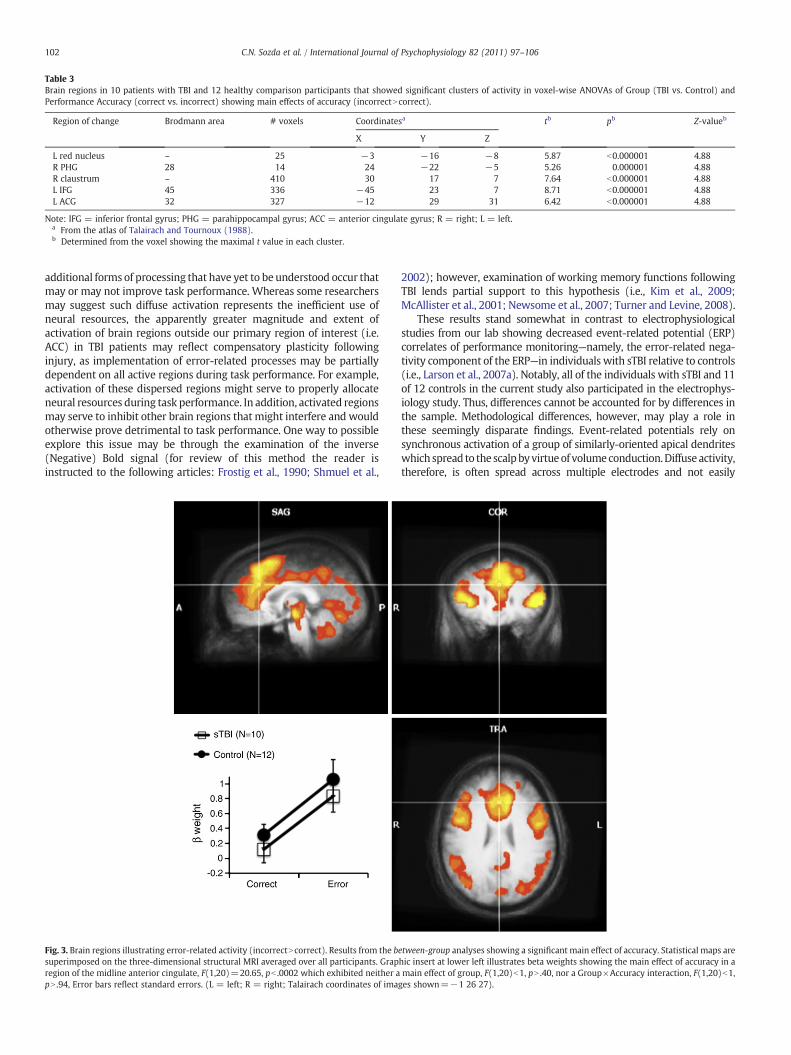
Table 3Brain regions in 10 patients with TBI and 12 healthy comparison participants that showed significant clusters of activity in voxel-wise ANOVAs of Group (TBI vs. Control) andPerformance Accuracy (correct vs. incorrect) showing main effects of accuracy (incorrectNcorrect).
Region of change Brodmann area # voxels Coordinatesa tb pb Z-valueb
X Y Z
L red nucleus – 25 −3 −16 −8 5.87 b0.000001 4.88R PHG 28 14 24 −22 −5 5.26 0.000001 4.88R claustrum – 410 30 17 7 7.64 b0.000001 4.88L IFG 45 336 −45 23 7 8.71 b0.000001 4.88L ACG 32 327 −12 29 31 6.42 b0.000001 4.88
Note: IFG = inferior frontal gyrus; PHG = parahippocampal gyrus; ACC = anterior cingulate gyrus; R = right; L = left.a From the atlas of Talairach and Tournoux (1988).b Determined from the voxel showing the maximal t value in each cluster.
102 C.N. Sozda et al. / International Journal of Psychophysiology 82 (2011) 97–106
additional forms of processing that have yet to be understood occur thatmay or may not improve task performance. Whereas some researchersmay suggest such diffuse activation represents the inefficient use ofneural resources, the apparently greater magnitude and extent ofactivation of brain regions outside our primary region of interest (i.e.ACC) in TBI patients may reflect compensatory plasticity followinginjury, as implementation of error-related processes may be partiallydependent on all active regions during task performance. For example,activation of these dispersed regions might serve to properly allocateneural resources during task performance. In addition, activated regionsmay serve to inhibit other brain regions that might interfere and wouldotherwise prove detrimental to task performance. One way to possibleexplore this issue may be through the examination of the inverse(Negative) Bold signal (for review of this method the reader isinstructed to the following articles: Frostig et al., 1990; Shmuel et al.,
Fig. 3. Brain regions illustrating error-related activity (incorrectNcorrect). Results from the bsuperimposed on the three-dimensional structural MRI averaged over all participants. Grapregion of the midline anterior cingulate, F(1,20)=20.65, pb .0002 which exhibited neither apN .94, Error bars reflect standard errors. (L = left; R = right; Talairach coordinates of ima
2002); however, examination of working memory functions followingTBI lends partial support to this hypothesis (i.e., Kim et al., 2009;McAllister et al., 2001; Newsome et al., 2007; Turner and Levine, 2008).
These results stand somewhat in contrast to electrophysiologicalstudies from our lab showing decreased event-related potential (ERP)correlates of performance monitoring—namely, the error-related nega-tivity component of the ERP—in individuals with sTBI relative to controls(i.e., Larson et al., 2007a). Notably, all of the individuals with sTBI and 11of 12 controls in the current study also participated in the electrophys-iology study. Thus, differences cannot be accounted for by differences inthe sample. Methodological differences, however, may play a role inthese seemingly disparate findings. Event-related potentials rely onsynchronous activation of a group of similarly-oriented apical dendriteswhich spread to the scalpbyvirtueof volumeconduction.Diffuseactivity,therefore, is often spread across multiple electrodes and not easily
etween-group analyses showing a significant main effect of accuracy. Statistical maps arehic insert at lower left illustrates beta weights showing the main effect of accuracy in amain effect of group, F(1,20)b1, pN .40, nor a Group×Accuracy interaction, F(1,20)b1,ges shown=−1 26 27).
Table 4Brain regions in 10 patients with TBI and 12 healthy comparison participants that showed significant clusters of activity in voxel-wise ANOVAs of Group (TBI vs. Control) andPerformance Accuracy (correct vs. incorrect) showing main effects of group.
Region of change Brodmann area # voxels Coordinatesa tb pb Z-valueb
X Y Z
Greater activity in control than in TBI participantsR cerebellum – 783 3 −37 −35 6.44 b0.000001 4.88L fusiform gyrus 37 44 −36 −52 −11 4.38 0.000034 4.14L STG 38 17 −45 5 −11 3.26 0.0016 3.16R LN – 25 24 −19 −2 3.80 0.00028 3.63R thalamus – 24 9 −10 16 4.61 0.000014 4.34L caudate – 120 −12 −7 19 4.04 0.00012 3.85R SFG 10 25 24 50 25 3.27 0.0015 3.17L precuneus 7 57 −21 −64 31 6.04 b0.000001 4.88R precuneus 7 23 21 −70 34 3.78 0.00029 3.62R ACG 24 69 3 −22 34 4.35 0.000038 4.12L MFG 9 17 −30 35 37 3.62 0.0005 3.48L precentral gyrus 4 33 −42 −10 53 3.72 0.00036 3.57L frontal sub-gyral 6 17 −18 5 58 3.43 0.00094 3.31
Greater activity in TBI than in control participantsR lingual gyrus 18 28 9 −79 −5 −3.67 0.00043 3.52L lingual gyrus – 243 −18 −76 1 −5.05 0.000003 4.66R STG 22 92 57 −10 7 −4.07 0.0001 3.89R posterior cingulate 23 21 6 −55 13 −3.47 0.00082 3.35L insula 13 61 −42 −13 22 −3.95 0.00016 3.78L SOG 19 62 −36 −76 25 −4.62 0.000014 4.34R ACG 32 113 15 17 31 −4.60 0.000015 4.33R cingulum – 34 15 −19 31 −3.84 0.00024 3.67L MFG 8 357 −45 11 37 −6.02 b0.000001 4.88R IPL 40 323 39 −31 40 −5.37 0.000001 4.88R MFG 6 175 42 5 40 −4.58 0.000016 4.31L precentral gyrus 4 370 −27 −22 46 −5.50 b0.000001 4.88L SPL 7 13 −33 −50 58 −3.30 0.0014 3.19R precentral gyrus 4 18 15 −25 70 −3.74 0.00033 3.59
Note: IPL = inferior parietal lobule; LN = lentiform nucleus; MFG = middle frontal gyrus; SFG = superior frontal gyrus; SOG = superior occipital gyrus; SPL = superior parietallobule; STG = superior temporal gyrus; ACG = anterior cingulate gyrus; R = right; L = left.
a From the atlas of Talairach and Tournoux (1988).b Determined from the voxel showing the maximal t value in each cluster.
103C.N. Sozda et al. / International Journal of Psychophysiology 82 (2011) 97–106
detected in ERP analyses that are focused on small groups of electrodeschosen based on a priori hypotheses and voltage-related scalp maps.Furthermore, multiple simultaneously active neural generators can leadto changes in ERP morphology and amplitude depending on neuronalorientation (see Luck, 2005). Thus, it is possible that the increased diffuseactivity in individuals with sTBI was seen as a reduced-amplitude ERN.This possibility is supported by a recent multimodal imaging study thatexamined electrophysiological (ERPs) and hemodynamic (fMRI) re-flections of error-related processing in healthy participants (Doñamayoret al. (2011). This study found several cortical and subcortical areas,including the ACC, superior frontal gyrus, precentral gyrus, inferiorfrontal gyrus, and inferior parietal lob involved in error processing. Anadditionalmethodological differencewhich could potentially account forthe inconsistent findings between the Larson et al. (2007a) ERN findingsand current findings of increased ACC activity in TBI patients is taskrelated. Specifically, in contrast to the current study, the Larson et al.
Table 5Brain regions in 10 patients with TBI and 12 healthy comparison participants that showedPerformance Accuracy (correct vs. incorrect) showing significant Group x Accuracy interact
Region of change Brodmann area # voxels Coord
X
L Cerebellumc – 24 −12R ACGd 32 13 18
Note: ACG = anterior cingulate gyrus; R = right; L = left.a From the atlas of Talairach and Tournoux (1988).b Determined from the voxel showing the maximal t value in each cluster.c Activation greater in controls.d Activation greater in TBI patients.
(2007a) study used a single-trial Stroop task that did not include astimulus-preceding cue. Error rates in that study were modestly lowerthan those observed in the present study, possibly suggesting that theworkingmemory burden imposed by task-instructional cueingmay alsoplay a role in the discrepant findings across these two studies.Nevertheless, the use of multiple modalities in similar groups of patientsacross studies from our group represents a strength of this research.Future studies directly addressing these methodological differences areneeded to clarify these seemingly disparate study findings.
It is important to consider that performance monitoring processesof control serve to not only detect errors, but also to adjust behavioralperformance toward goal attainment based on the detection ofperformance errors which can signal when strategic shifts inperformance are necessary (Kerns et al., 2004; Ridderinkhof et al.,2004). Consequently, strategic shifts, in theory, should minimizeconflict on subsequent trials and reduce additional likelihood of
significant clusters of activity in voxel-wise ANOVAs of Group (TBI vs. Control) andions.
inatesa tb pb Z-valueb
Y Z
−49 −35 3.64 0.0005 3.4814 31 −3.17 0.002 3.09

Fig. 4. Brain regions of patients with TBI and healthy comparison participants illustrating significant error-related activity (incorrectNcorrect). Results from the individual-groupanalyses: Yellow illustrates significant error-related activity in TBI patients, blue illustrates error-related activity in healthy controls, and green illustrates error-related activity inwhich the TBI patients and healthy controls overlap. Results from the between-group analysis: Red illustrates a region of the anterior cingulate cortex (ACC) in which there is aGroup×Accuracy interaction; graph insert in lower left illustrates beta weights showing the interaction effect. Error bars reflect standard errors. Statistical maps are superimposedon the three-dimensional structural MRI averaged over all participants. (L = left; R = right; Talairach coordinates of images shown=18 11 34).
104 C.N. Sozda et al. / International Journal of Psychophysiology 82 (2011) 97–106
subsequent incorrect responses (Carter and van Veen, 2007; Kerns etal., 2004; Ridderinkhof et al., 2004; van Veen and Carter, 2002, 2006).Thus, it is possible that although TBI patients may detect errorscorrectly, poor task performance may arise due to impairments insignaling when strategic shifts are needed (i.e., Larson et al., 2006).
# of Error-Related Voxels
# of
err
or-r
elat
ed v
oxel
s
300
250
200
150
100
50
0Controls TBIs
Fig. 5. Box plots illustrating the total number of suprathrehold error-related voxels as afunction of group as derived from the separate-groups contrast errorNcorrect response.Bottom and top of each box represent the lower and upper quartiles, respectively, andthe band near the middle of the box reflects the 50th percentile (median); upper andlower whiskers reflect the maximum and minimum number of voxels.3
Task performance findings indicated that both sTBI and controlparticipants showed significant Stroop RT interference, with sTBIpatients showing marginally greater RT interference than controls.Additionally, sTBI participants showed significantly greater error-rateinterference than healthy controls. This pattern of behavioral results isconsistent with previous literature showing increased Stroop RT anderror-rate interference on versions of the cued-Stroop task (i.e.,Larson et al., 2007a; Perlstein et al., 2006; Seignourel et al., 2005),supporting assertions that sTBI patients show impairments in conflictprocessing, particularly when required to override prepotent re-sponse tendencies.
Several methodological limitations and alternative explanations offindings of the current study warrant further discussion. For example,although all TBI participants were classified as sustaining severeinjuries, the heterogeneity of this population was evidenced byindividual differences in injury mechanism, injury localization, timesince injury, PTA, and LOC (Lezak et al., 2004). Whereas studies usingTBI participants cannot control for all these variables, studies of TBImust acknowledge and appreciate heterogeneity within the TBIpopulation. Secondly, numerous research studies (i.e., Heeger et al.,2000; Logothetis and Wandell, 2004) have postulated that the blood-oxygen level dependent (BOLD) signal measured in fMRI studies is acomplex function reflective of changing levels of cerebral blood flow,blood volume, and oxygen metabolism that occur as a result of neuralactivity. However, the hemodynamic response often lags greatlybehind the neuronal activity that starts the event (Boynton et al.,1996). Moreover, as the BOLD signal measured by fMRI is reflective ofvascular changes correlated with neural activity, and not neural

Table 6Brain regions showing significantly greater error-related activity following incorrect than correct probe response revealed in the individual-group analyses of 12 healthy controls and10 TBI participants.
Region of change Brodmann area # voxels Coordinatesa tb pb Z-valueb
X Y Z
Control participantsL cerebellum – 60 −15 −46 −32 5.02 0.000003 4.66L STN – 128 −9 −10 −8 5.20 0.000001 4.88R insula 13 146 30 14 −5 5.44 0.000001 4.88L IFG 45 189 −45 23 7 5.93 b0.000001 4.88R MTG 39 18 46 −67 19 5.00 0.000003 4.66L cingulate gyrus 32 18 −12 26 31 4.50 0.000022 4.24L precuneus 9 29 −6 −49 43 4.65 0.000012 4.37L MFG 6 36 −12 8 55 5.37 0.000001 4.88
TBI participantsR claustrum – 362 30 17 7 5.26 b0.000001 4.88L IFG 45 298 −48 20 10 6.51 b0.000001 4.88R cingulate gyrus 24 238 12 14 31 5.30 0.000001 4.88L MFG 6 50 −12 29 34 4.82 0.000006 4.53
Note: IFG = inferior frontal gyrus; MFG = middle frontal gyrus; MTG = middle temporal gyrus; STN = subthalamic nuclei; R = right; L = left.a From the atlas of Talairach and Tournoux (1988).b Determined from the voxel showing the maximal t value in each cluster.
105C.N. Sozda et al. / International Journal of Psychophysiology 82 (2011) 97–106
activity directly, any observed injury-related differences may haveresulted from several factors outside of those hypothesized (Hillary etal., 2002). Specifically, differences in injury-related activation patternscould reflect changes in vascular processes or changes due to cellatrophy, rather than neural processes. For example, both human andanimal models of TBI have demonstrated reduced baseline levels ofcerebral blood flow post-injury (i.e., Bouma et al., 1991). Moreover,blood flow abnormalities in patients with moderate-to-severe TBIrelative to comparison subjects have been observed during comple-tion of workingmemory tasks, an effect particularly concerning due toabnormalities in the frontal lobes, an area extensively imaged byresearchers (i.e., Christodoulou et al., 2001; Perlstein et al., 2004).Finally, though many neurocognitive studies of TBI have employeduninjured healthy comparison groups as we have, such studies mayconfound behavioral features that predispose people to TBI. Otherfunctional imaging studies, in contrast, have employed participantswith extracranial orthopedic injury for comparison “to control for ahost of risk factors (e.g., risk-taking behavior) that [may] predisposeto injury and nonspecific effects of injury such as posttraumatic stressthat could affect brain activation” (e.g., Scheibel et al., 2007, p. 36).Such potential confounds are important to address, particularly inlight of the ACC differentiation observed in the present study andfindings that posttraumatic stress may alter ACC activity (e.g., Hayeset al., 2009) and that error-related ACC activity is modulated byaffective factors (e.g., Larson et al., 2009).
5. Summary and conclusions
In conclusion, results from thepresent study extendpreviousfindingsthat the neural networks mediating cognitive control, specifically, error-related processing, are disrupted after sTBI. Despite its limitations, thecurrent study supports the continued neurophysiological examination ofcomplexprocesses of cognitive control. Future studieswill aim to identifyoutcomemeasures of “real-world” functioning that will further examinethe impact of these deficits and inform rehabilitation efforts.
Acknowledgements
This original researchmanuscript, or parts of it, havenot beenandwillnot be submitted elsewhere for publication. This workwas supported byNIH grants K01-MH01857 and R21-MH073076, and by grants from theEvelyn F. McKnight Brain Research Grant Program, and the Florida Brainand Spinal Cord Injury Research Trust Fund awarded toWMP. The workwas submitted by the first author in partial fulfillment of requirements
for the Master of Science degree. We extend our appreciation to AshleyCarroll, Neha Dixit, Cortney Mauer, Megan McIntyre, Drew Nagle, PaulSeignourel, Floris Singletary, Allen Sirizi, and Raechel Steckley for theirassistance in participant recruitment and data acquisition.
References
Anderson, V., Levin, H.S., Jacobs, R., 2002. Executive functions after frontal lobe injury: adevelopmental perspective. In: Stuss, D.T., Knight, R.T. (Eds.), Principles of FrontalLobe Function. Oxford University Press, New York, pp. 504–527.
Beck, A.T., Steer, R.A., Brown, G.K., 1996. Beck Depression Inventory — Second Edition(BDI-II). The Psychological Corporation, USA.
Bigler, E.D., 1990. Neuropathology of traumatic brain injury. In: Bigler, E.D. (Ed.),Traumatic Brain Injury. Pro-ed, Austin, TX.
Blair, J.R., Spreen, O., 1989. Predicting premorbid IQ: a revision of the National AdultReading Test. The Clinical Neuropsychologist 3, 129–136.
Bond, M.R., 1986. Neurobehavioral sequelae of closed head injury. In: Grant, I., Adams,K.M. (Eds.), Neuropsychological Assessment of Neuropsychological Disorders.Oxford University Press, New York, pp. 347–373.
Bouma, G.J., Muizelaar, J.P., Choi, S.C., Newlon, P.G., Young, H.F., 1991. Cerebralcirculation and metabolism after severe traumatic brain injury: the elusive role ofischemia. Journal of Neurosurgery 75, 685–693.
Boynton, G.A., Engel, S.A., Glover, G., Heeger, D., 1996. Linear systems analysis offunctional magnetic resonance imaging in human V1. Journal of Neuroscience 16,4207–4221.
Burgess, P.W., Robertson, I.H., 2002. Principles of the rehabilitation of frontal lobefunction. In: Stuss, D.T., Knight, R.T. (Eds.), Principles of Frontal Lobe Function.Oxford University Press, New York, pp. 557–572.
Carter, C.S., van Veen, V., 2007. Anterior cingulate cortex and conflict detection: anupdate of theory and data. Cognitive, Affective, & Behavioral Neuroscience 7,367–379.
Carter, C.S., Botvinick, M.M., Cohen, J.D., 1999. The role of the anterior cingulate cortexin executive processes of cognition. Reviews in the Neurosciences 10, 49–57.
Christodoulou, C., DeLuca, J., Ricker, J.H., Madigan, N.K., Bly, B.M., Lange, G., et al., 2001.Functional magnetic resonance imaging of working memory impairment followingtraumatic brain injury. Journal of Neurology, Neurosurgery, and Psychiatry 71,161–168.
Cicerone, K.D., Dahlberg, C., Malec, J.F., Langenbahn, D.M., Felicetti, T., Kneipp, S., et al.,2005. Evidence-based cognitive rehabilitation: updated review of the literaturefrom 1998 through 2002. Archives of Physical Medicine and Rehabilitation 86,1681–1692.
Cohen, J., 1988. Statistical Power Analysis for the Behavioral Sciences. ErlbaumAssociates, Hillsdale, NJ.
Cohen, J.D., Barch, D.M., Carter, C.S., Servan-Schreiber, D., 1999. Context-processingdeficits in schizophrenia: converging evidence from three theoretically motivatedcognitive tasks. Journal of Abnormal Psychology 108, 120–133.
Doñamayor, N., Heilbronner, U., Münte, T.F., 2011. Coupling electrophysiological andhemodynamic responses to errors. HumanBrainMapping. doi:10.1002/hbm.21305.
Easdon, C., Levine, B., O'Connor, C., Tisserand, D., Hevenor, S., 2004. Neural activityassociated with response inhibition following traumatic brain injury: an event-related fMRI investigation. Brain and Cognition 54, 136–138.
Forman, S.D., Cohen, J.D., Fitzgerald, M., Eddy, W.F., Mintun, M.A., Noll, D.C., 1995.Improved assessment of significant activation in functional magnetic resonanceimaging (fMRI): use of a cluster-size threshold. Magnetic Resonance in Medicine33, 636–647.
106 C.N. Sozda et al. / International Journal of Psychophysiology 82 (2011) 97–106
Friston, K.J., Holmes, A.P., Worsley, K.J., Poline, J.B., Frith, C.D., Frackowiak, R.S.J., 1995.Statistical parametric maps in functional neuroimaging. NeuroImage 6, 218–229.
Frostig, R.D., Lieke, E.E., Ts'o, D.Y., Grinvald, A., 1990. Cortical functional architecture andlocal coupling between neuronal activity and the microcirculation revealed by invivo high-resolution optical imaging of intrinsic signals. Proceedings of theNational Academy of Sciences of the United States of America 87, 6082–6086.
Goebel, R., Esposito, F., Formisano, E., 2006. Analysis of function image analysis contest(FIAC) data with Brainvoyager QX: from single-subject to cortically aligned groupgeneral linear model analysis and self-organizing group independent componentanalysis. Human Brain Mapping 27, 392–401.
Hayes, J.P., LaBar, K.S., Petty, C.M., McCarthy, G., Morey, R.A., 2009. Alterations in neuralcircuitry for emotion and attention associated with posttraumatic stress symp-tomatology. Psychiatry Research: Neuroimaging 172, 7–15.
Heeger, D.J., Huk, A.C., Geisler,W.S., Albrecht, D.G., 2000. Spikes versus BOLD:what doesneuroimaging tell us about neuronal activity? Nature Neuroscience 3, 631–633.
Hillary, F.G., Steffener, J., Biswal, B.B., Lange, G., DeLuca, J., Ashburner, J., 2002.Functional magnetic resonance imaging technology and traumatic brain injuryrehabilitation: guidelines for methodological and conceptual pitfalls. The Journal ofHead Trauma Rehabilitation 17, 411–430.
Horn, L.J., Sherer, M., 1999. Rehabilitation of traumatic brain injury. In: Grabois, K.M.,Garrison, S.J., Hart, A., Lehmkuhl, L.D. (Eds.), Physical Medicine and Rehabilitation:The Complete Approach. Blackwell Science, Cambridge, MA, pp. 1281–1304.
Kerns, J.G., Cohen, J.D., MacDonald, A.W., Cho, R.Y., Stenger, V.A., Carter, C.S., 2004.Anterior cingulate conflict monitoring and adjustments in control. Science 303,1023–1026.
Kim, Y.H., Yoo,W.K., Ko, M.H., Park, C.H., Kim, S.T., Na, D.L., 2009. Plasticity of the attentionalnetwork after brain injury and cognitive rehabilitation. Neurorehabilitation andNeuralRepair 23, 468–477.
King, N.S., Crawford, S., Wenden, F.J., Moss, N.E., Wade, D.T., Caldwell, F.E., 1997.Measurement of post-traumatic amnesia: how reliable is it? Journal of Neurology,Neurosurgery, and Psychiatry 62, 38–42.
Lancaster, J.L., Woldorff, M.G., Parsons, L.M., Liotti, M., Freitas, C.S., Rainey, L., Kochunov,P.V., Nickerson, D., Mikiten, S.A., Fox, P.T., 2000. Automated Talairach atlas labels forfunctional brain mapping. Human Brain Mapping 10, 120–131.
Larson, M.J., Perlstein, W.M., 2009. Awareness of deficits and error processing aftertraumatic brain injury. Neuroreport 20, 1486–1490.
Larson, M.J., Perlstein, W.M., Demery, J.A., Stigge-Kaufman, D.A., 2006. Cognitive controlimpairments in traumatic brain injury. Journal of Clinical and ExperimentalNeuropsychology 28, 968–986.
Larson, M.J., Stigge-Kaufman, D.A., Schmulfass, I.M., Perlstein, W.M., 2007a. Performancemonitoring, error processing, and evaluative control following severe TBI. Journal of theInternational Neuropsychological Society 13, 961–971.
Larson, M.J., Kelly, K.G., Stigge-Kaufman, D.A., Schmalfuss, I.M., Perlstein, W.M., 2007b.Reward context sensitivity impairment following severe TBI: an event-related potentialinvestigation. Journal of the International Neuropsychological Society 13, 615–625.
Larson, M.J., Kaufman, D.A.S., Kellison, I.L., Schmalfuss, I.M., Perlstein, W.M., 2009.Double jeopardy! The additive consequences of negative affect on performance-monitoring decrements following traumatic brain injury. Neuropsychology 23 (4),433–444.
Larson, M.J., Farrer, T.J., Clayson, P.E., in press. Cognitive control in mild traumatic braininjury: conflict monitoring and conflict adaptation. International Journal ofPsychophysiology.
Levine, B., Katz, D.I., Dade, L., Black, S.E., 2002. Novel approaches to the assessment of frontaldamageandexecutive deficits in traumatic brain injury. In: Stuss, D.T., Knight, R.T. (Eds.),Principles of frontal lobe function. Oxford University Press, New York, pp. 448–465.
Lezak, M.D., Howieson, D.B., Loring, D.W., 2004. Neuropsychological Assessment, 4thedition. Oxford University Press, New York.
Logothetis, N.K., Wandell, B.A., 2004. Interpreting the BOLD signal. Annual Review ofPhysiology 66, 735–769.
Lorist, M.M., Boksem, M.A.S., Ridderinkhof, K.R., 2005. Impaired cognitive control andreduced cingulate activity during mental fatigue. Cognitive Brain Research 24,199–205.
Lovell, M., Franzen, M., 1994. Neuropsychological assessment. In: Silver, J.M., Yudofsky,S., Hales, R.E. (Eds.), Neuropsychiatry of Traumatic Brain Injury. AmericanPsychiatric Press, Washington, pp. 133–160.
Luck, S.J., 2005. An Introduction to the Event-related Potential Technique. MIT Press,Cambridge, MA.
MacDonald, A.W., Cohen, J.D., Stenger, V.A., Carter, C.S., 2000. Dissociating the role ofthe dorsolateral prefrontal and anterior cingulate cortex in cognitive control.Science 288, 1835–1838.
McAllister, T.W., Sparling, M.B., Flashman, L.A., Guerin, S.J., Mamourian, A.C., Saykin, A.J.,2001. Differential working memory load effects after mild traumatic brain injury.NeuroImage 14, 1004–1012.
McClure, S.M., Laibson, D.I., Loewenstein, G., Cohen, J.D., 2004. Separate neural systemsvalue immediate and delayed monetary rewards. Science 306, 503–507.
McMillan, T.M., Jongen, E.L., Greenwood, R.J., 1996. Assessment of post-traumaticamnesia after severe closed head injury: retrospective or prospective? Journal ofNeurology, Neurosurgery, and Psychiatry 60, 422–427.
Miller, E.K., 2000. The prefrontal cortex and cognitive control. Nature Reviews.Neuroscience 1, 59–66.
Miller, E.K., Cohen, J.D., 2001. An integrative theory of prefrontal cortex function.Annual Review of Neuroscience 24, 167–202.
Newsome, M.R., Scheibel, R.S., Steinberg, J.L., Troyanskaya, M., Sharma, R.G., et al., 2007.Working memory brain activation following severe traumatic brain injury. Cortex43, 95–111.
O'Keeffe, F.M., Dockree, P.M., Robertson, I.H., 2004. Poor insight in traumatic braininjury mediated by impaired error processing?: evidence from electrodermalactivity. Cognitive Brain Research 22, 101–112.
Perlstein, W.M., Cole, M.A., Dixit, N.K., Demery, J.A., 2004. Parametric manipulation ofworking memory load in chronic traumatic brain injury. Journal of theInternational Neuropsychological Society 10, 724–741.
Perlstein, W.M., Larson, M.J., Dotson, V.M., Kelly, K.G., 2006. Temporal dissociation ofcomponents of cognitive control dysfunction in severe TBI: ERPs and the cued-Stroop task. Neuropsychologia 44, 260–274.
Ridderinkhof, K.R., Ullsperger, M., Crone, E.A., Nieuwenhuis, S., 2004. The role of themedial frontal cortex in cognitive control. Science 306, 443–447.
Rosner, B., 1983. Percentage points for a generalized EST many-outlier procedure.Technometrics 25 (2), 165–172.
Scheibel, R.S., Pearson, D.A., Faria, L.P., Kotrla, K.J., Aylward, E., Bachevalier, J., et al.,2003. An fMRI study of executive functioning after severe diffuse TBI. Brain Injury17, 919–930.
Scheibel, R.S., Newsome, M.R., Steinberg, J.L., Pearson, D.A., Rauch, R.A., Mao, H.,Troyanskaya, M., Sharma, R.G., Levin, H.S., 2007. Altered brain activation duringcognitive control in patients with moderate to severe traumatic brain injury.Neurorehabilitation and Neural Repair 21, 36–45.
Seignourel, P.J., Robins, D.L., Larson, M., Demery, J.A., Cole, M.A., Perlstein, W.M., 2005.Cognitive control in closed head injury: context maintenance dysfunction orprepotent response inhibition deficit? Neuropsychology 19, 578–590.
Shmuel, A., Yacoub, E., Pfeuffer, J., Van de Moortele, P.F., Andriany, G., Hu, X., Ugurbil, K.,2002. Sustained negative BOLD, blood flow and oxygen consumption response andits coupling to the positive response in the human brain. Neuron 36, 1195–1210.
Soeda, A., Nakashima, T., Okumura, A., Kuwata, K., Shinoda, J., Iwama, T., 2005. Cognitiveimpairment after traumatic brain injury: a functional magnetic resonance imagingstudy using the Stroop task. Neuroradiology 47, 501–506.
Spielberger, C.D., Gorsuch, R.L., Lushene, R.E., 1970. Manual for the State-Trait AnxietyInventory. Consulting Psychologists Press, Palo Alto, CA.
Spreen, O., Strauss, E., 1991. A Compendium of Neuropsychological Tests: Administra-tion, Norms, and Commentary. Oxford University Press, New York.
Stemmer, B., Segalowitz, S.J., Witzke, W., Schönle, P.W., 2004. Error detection inpatients with lesions to the medial prefrontal cortex: an ERP study. Neuropsycho-logia 42, 118–130.
Talairach, J., Tournoux, P., 1988. Co-planar Sterotaxic Atlas of the Human Brain. Thieme,New York.
Teasdale, G., Jennett, B., 1974. Assessment of coma and impaired consciousness: apractical scale. Lancet ii, 81–84.
Turner, G.R., Levine, B., 2008. Augmented neural activity during executive controlprocessing following diffuse axonal injury. Neurology 71, 812–818.
van Veen, V., Carter, C.S., 2002. The anterior cingulate as a conflict monitor: fMRI andERP studies. Physiology and Behavior 77, 477–482.
van Veen, V., Carter, C.S., 2006. Conflict and cognitive control in the brain. CurrentDirections in Psychological Science 15, 237–240.
Wechsler, D., 1981. WAIS-R Manual. Psychological Corporation, New York.West, R., 2003. Neural correlates of cognitive control and conflict detection in the
Stroop and digit-location tasks. Neuropsychologia 41, 1122–1135.


![KLV-30MR1 - Error: [object Object]](https://static.fdokumen.com/doc/165x107/631786651e5d335f8d0a6a63/klv-30mr1-error-object-object.jpg)