Tocopherol measurement in edible products of vegetable origin
Upload
agroparistechCategory
view
1download
0
Structural Behaviour of Lipid Droplets in Protein-stabilized
Nano-emulsions and Stability of α-Tocopherol
Perla Relkin & Jin-Mi Yung & Daniel Kalnin &
Michel Ollivon
Received: 27 November 2007 /Accepted: 31 January 2008 /Published online: 6 March 2008# Springer Science + Business Media, LLC 2008
Abstract A stearin-rich milk fraction was used, alone or incombination with α-tocopherol, for the preparation of oil-in-water sodium caseinate-stabilized nano-emulsions. Fatdroplets in these two emulsions were characterized for theirsize distribution and physical stability against aggregation–coalescence, for their heat-induced structural behaviour,and for their ability to protect α-tocopherol duringoxidation. Inclusion of α-tocopherol led to changes in theparticle size distributions of fat droplets: in the presence ofα-tocopherol, approximately 75% of fat droplets werelower than 1 μm, instead of 100% in the absence ofα-tocopherol. On the other hand, thermal transitionsobserved by differential scanning calorimetry showed thatsupercooling (the increase in differences between temper-ature of initial crystallization and melting completion) washigher in the emulsified milk fat samples containingα-tocopherol. In addition to a decrease in the temperatureof fat crystallization leading this change in the supercoolingeffect of α-tocopherol, small- and wide-angle X-raydiffraction patterns (SAXS and WAXS, respectively)observed under cooling and re-heating cycles showed thatheat-induced polymorphic transitions from2Lα ! 2Lβ0
forms were more impeded in the emulsion containingα-tocopherol. Immobilisation of α-tocopherol in fat drop-
lets composed by high melting temperature milk fat trigly-cerides seemed to lead to its protection against degradation.
Keywords Emulsion . Antioxidant . Nanoencapsulation .
Fat polymorphisms . Fat crystallization . DSC . DRX
Introduction
Nano-emulsions containing lipid droplets with a liquid orsolid core are largely used in cosmetic, pharmaceutical andmedicine applications, as encapsulation matrices for pro-tection and control of drug delivery to a target site.1
α-Tocopherol is a lipid compound considered as anantioxidant by preventing free-radical damage in tissues.2
A high dose of α-tocopherol supplementation is part of thetherapy of people who suffer from abetalipoproteinemia. Inthis case, extremely low levels of plasma cholesterol andtriglycerides cause defective absorption of α-tocopherol.3
Numerous studies have shown that drug release fromnanostructured emulsions and particles may be triggeredby exposure to environmental and storage conditions (pH,metal ions, oxygen or heat exposure or heat). In the foodindustry, dispersed systems containing fat droplets between50 and 1,000 nm in diameter are needed to improve thephysical stability of emulsions against flocculation, cream-ing and coalescence.4,5 With respect to the development offunctional foods, nano-emulsions containing only food-grade ingredients and additives are used as carriers ofα-tocopherol, antioxidants, unsaturated fatty acids, aromacompounds…or other lipophilic sensitive molecules thatneed to be protected from degradation throughout theproduct processing or storage steps. Thus, besides theirformation and physical stability, fat droplets can be
Manuscript dedicated by the authors to the memory of Dr. MichelOllivon, deceased on June 2007.
P. Relkin (*) : J.-M. YungUMR 1211, AgroParisTech,91744 Massy, Francee-mail: [email protected]
D. Kalnin :M. OllivonUMR 8612, Physicochimie des Systèmes Polyphasés,92296 Châtenay-Mallabry, France

considered as functional foods for carrying and ensuring thedelivery of bioactives to a target site within the body afteringestion.6 The emulsification process and ingredientcomplexity play a dominant role in the characteristics offat droplets, such as the particle average diameter and sizedistribution, composition and physical properties ofsurrounding surface layers, and also crystalline fat contentand polymorphism.7–9 These characteristics determine thephysical stability of oil in water emulsions. Studiesperformed on fat thermal transitions showed that crystal-lization temperature of dispersed fat droplets is loweredcompared to bulk fat, and the degree of supercoolingneeded to initiate fat crystallization in globules was shownto differ depending on triacylglycerol composition, meandroplet size and lipophilic or hydrophilic nature ofemulsifiers.10–13 Those emulsifier molecules are capableof partitioning between hydrophilic phases (continuousaqueous phase) or hydrophobic (interior of dispersed fatdroplets) or interface. They have effects not only on theinitial temperature of crystallization but also on the rise ofsolid fat content.
In this work, we studied the effects of partial replace-ment of a milk fat sample by α-tocopherol on the structuralbehaviour of protein-stabilised fat droplets in oil-in-waternano-emulsions with respect to α-tocopherol stability.
Materials and Methods
Materials
α-Tocopherol (DL-α-tocopherol) was purchased from SigmaAldrich (Sfeinheim, Suisse). The sodium caseinate and milkfat samples were provided by Lactalis-France. The milk fatsample, which was dry-fractionated from anhydrous milk fat(AMF), contained a high proportion of long-chain saturatedtriacylglycerols (TAGs; 40% C16:0, 30% C18:0) but a lowproportion of short-chain and unsaturated TAGs (less than10% for C18:1) compared to AMF.14,15
Emulsion Preparation
The emulsion aqueous phase was prepared by dispersingsodium caseinate in phosphate-buffered saline solution(50 mM NaH2PO4 at pH=6.6) at 3.6 wt% protein relativeto final emulsions. The lipophilic phase (40 wt%, relativeto final emulsions) was composed of the stearin-rich milkfat fraction alone or in presence of α-tocopherol (4%wt,relative to the total emulsion weight). The lipid phaseswere melted at 65 °C, mixed with the protein solution andpre-homogenized using an ultra-disperser (UltraTurrax,IKA-Staufen, Germany) at 10,000 rpm for 10 min. Thefour coarse emulsions were then passed six times through
a high-pressure homogeniser (APV Gaulin, Lübeck,Germany) at 600 bar, and the resulting fine-disperseemulsions were cooled to 20 °C before characterization.
Physical Characterization
Particle size distributions (Malvern Mastersizer 2000Instruments, Orsay, France) were evaluated after dilutionin distilled water (20 °C) or in 0.5% sodium dodecylsulfate (SDS) solution (40 °C) and gentle stirring for30 min before sampling. Fat polymorphism was moni-tored using small- and wide-angle X-Ray diffraction(SAX and WAX, respectively) measurements by a labscale equipment (MICROCALIX)16, composed of a longfine-focus sealed X-ray source (ENRAF NONIUS, Cuanode), a multilayer mirror (OSMIC), collimation slits andtwo position-sensitive linear gas detectors (HECUS,Austria) likely in synchrotron workbenches. MICRO-CALIX equipment works at 0<q<0.45 Å−1 (SAX) and1.1<q<2.1 Å−1 (WAX). Calorimetric parameters of fatdroplet crystallization and melting in the emulsions weremonitored using a DSC-7 Perkin-Elmer associated to Pyrissoftware (Perkin-Elmer, Norwalk, USA). In X-ray anddifferential scanning calorimetry (DSC) measurements, weused similar sample masses (around 30 μl), scanningmode (0.5 °C min−1) and a cooling-temperature rangewhere the emulsion aqueous phase did not crystallize.13
Briefly, the samples were loaded in stainless steel pans(DSC) or glass capillaries (GLAS, Muller, Berlin, Germanyfor SAXS and WAXS measurements), heated and cooledfrom 50 °C to −7 °C. Crystallisation and fusion calorimetricparameters (peak temperatures and enthalpy changes) wereobtained from exothermic and endothermic DSC traces,respectively. X-ray diffraction angles were transformedinto short spacing using Bragg’s equation.15,17 To deter-mine the variation in the X-ray diffraction peak’s area as afunction of temperature, the X-ray diffraction patterns werefitted to a Gauss–Lorentz function using Peakfit Software(Jandel Scientific, Erkrath, Germany).
Chemical Characterization
α-Tocopherol concentrations were determined by UV-spectrophotometry and spectrofluorimetry at 290 and327 nm for excitation and emission maximum wavelengths,respectively. The α-tocopherol content in the emulsions’ lipidphases was extracted after dispersion in ethanol (5 vol%),vortex shaking (10 min) and centrifugation (15,000 rpm−7 min at 25 °C). The α-tocopherol content in the resultingsupernatant was determined by spectrofluorimetry using acalibration curve [FI=28,481×(α-tocopherol), R2=0.9987]obtained from freshly prepared solutions of α-tocopherol inethanol at concentrations ranging from 0.01 to 0.25 weight

ratio of α-tocopherol/ethanol. The fluorescence intensityvalues were corrected for absorbance effects, as previouslydescribed.18
α-Tocopherol content in bulk and emulsified fat systemswas determined just after preparation (t0) and after storageat room temperature during 8 weeks before and afterheating at 60 °C for 180 min.
Experimental uncertainties were deduced from threereplications of each of the measurements.
Results and Discussion
Effect of α-Tocopherol on Fat Droplet Size Characteristics
Partial replacement of milk fat by α-tocopherol had effects onthe fat droplet size distribution in the emulsion, as shown inthe curves reported in Figure 1. The particle size distributionwas monomodal and sharp (from 120 to 800 nm) in theabsence of α-tocopherol but much broader (from 120 nm to120 μm) in the emulsion containing α-tocopherol. Thevolume average median diameter (D50) and the percentage offat droplets with diameters lower than 200 nm, %D(<200 nm) and lower than 1 μm, %D (<1 μm) are shownin Table 1. These values were calculated by the Malvernsoftware from emulsions diluted in distilled water (20 °C) orSDS solutions. Fat droplet distributions observed afterdispersion in distilled water or SDS solution differed slightlyfor the emulsion containing α-tocopherol. Particularly, fatdroplets with diameter lower than 1 μm (peak distributionlocated around 1.5 μm) were visible after dispersion in bothdistilled water and in SDS solution, contrary to those locatedat around 60 μm which disappeared after dilution in SDSsolution. Thus, in addition to the particle size distributionbroadening effect, partial replacement of fat by α-tocopherolseemed to cause emulsion destabilization by fat dropletcoalescence (peak at 1.5 μm) and agglomeration (peak at60 μm).
Fat Crystallisation and Melting Behaviour
Crystallization, melting and polymorphism transitions infat samples are accompanied by energy release orabsorption at different temperatures depending on factorssuch as fat composition (TAG chain length and degree ofsaturation/unsaturation), presence of additives and fatdroplet size.10–15,17,19 Examples of DSC curves observedfrom the stearin-rich emulsions in the absence and presenceof α-tocopherol are shown in Figure 2. Regardless of thelipid phase composition, the cooling curves presented onesharp exothermic peak lying between approximately 16 to5 °C, while the re-heating curves presented a broaderendothermic peak lying between 11 to 45 °C. Comparisonbetween calorimetric results observed from bulk andemulsified fat samples in the presence and absence of α-tocopherol is presented in Table 2. ΔT values which werecalculated from Tend (fat-melting completion upon re-heating) and Tonset (initial heat flow release upon cooling)reflected a higher supercooling effect before fat crystalliza-tion from melted fat samples containing α-tocopherol. Wealso observed that addition of α-tocopherol led to a higherdecrease in the globule fat-melting enthalpy (by approxi-mately 25% instead of 10%) due to partial replacement ofcrystalline fat by α-tocopherol. Following the hypothesis of
0
2
4
6
8
10
12
14
0.01 0.1 1 10 100 1000
Particle diameter, µm
Water
0
2
4
6
8
10
12
14
0.01 0.1 1 10 100 1000
Particle diameter, µm
SDS
Vo
lum
e fr
equ
ency
, %
Vo
lum
e fr
equ
ency
, %
a
b
Fig. 1 Fat droplet size distributions observed from emulsions without(dashed lines) and with added α-tocopherol (full line) after dilution indistilled water (left) or SDS solution (right)
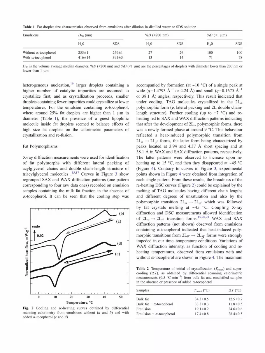
heterogeneous nucleation,19 larger droplets containing ahigher number of catalytic impurities are assumed tocrystallize first, and as crystallization proceeds, smallerdroplets containing fewer impurities could crystallize at lowertemperatures. For the emulsion containing α-tocopherol,where around 25% fat droplets are higher than 1 μm indiameter (Table 1), the presence of a guest lipophilicmolecule inside fat droplets seemed to balance effects ofhigh size fat droplets on the calorimetric parameters ofcrystallization and re-fusion.
Fat Polymorphisms
X-ray diffraction measurements were used for identificationof fat polymorphs with different lateral packing ofacylglycerol chains and double chain-length structure oftriacylglycerol molecules .15,17 Curves in Figure 3 showregrouped SAX and WAX diffraction patterns (one patterncorresponding to four raw data ones) recorded on emulsionsamples containing the milk fat fraction in the absence ofα-tocopherol. It can be seen that the cooling step was
accompanied by formation (at ∼10 °C) of a single peak atwide (q=1.4793 Å−1 or 4.24 Å) and small (q=0.1675 Å−1
or 38.1 Å) angles, respectively. This result indicated thatunder cooling, TAG molecules crystallized in the 2Lα
polymorphic form (α lateral packing and 2L double chain-length structure). Further cooling (up to −7 °C) and re-heating led to SAX and WAX diffraction patterns indicatingthat after the development of 2Lα polymorphic forms, therewas a newly formed phase at around 9 °C. This behaviourreflected a heat-induced polymorphic transition from2Lα ! 2Lβ0 forms, the latter form being characterized bypeaks located at 3.94 and 4.37 Å short spacing and at38.1 Å in WAX and SAX diffraction patterns, respectively.The latter patterns were observed to increase upon re-heating up to 15 °C, and then they disappeared at ∼45 °C(Figure 4). Contrary to curves in Figure 3, experimentalpoints shown in Figure 4 were obtained from integration ofeach single pattern. From these results, the broadness of there-heating DSC curves (Figure 2) could be explained by themelting of TAG molecules having different chain lengthsand different degrees of unsaturation and also by thepolymorphic transition 2Lα ! 2Lβ0 which was followedby fat crystals melting at ∼45 °C. Coupling X-raydiffraction and DSC measurements allowed identificationof 2Lα ! 2Lβ0 transition forms.15,20,21 WAX and SAXdiffraction patterns (not shown) observed from emulsionscontaining α-tocopherol indicated that heat-induced poly-morphic transitions from 2La ! 2Lb0 forms were stronglyimpeded in our time–temperature conditions. Variations ofWAX diffraction intensity, as function of cooling and re-heating temperatures, observed from emulsions with andwithout α-tocopherol are shown in Figure 4. The maximum
Table 1 Fat droplet size characteristics observed from emulsions after dilution in distilled water or SDS solution
Emulsions D50 (nm) %D (<200 nm) %D (<1 μm)
H20 SDS H20 SDS H20 SDS
Without α-tocopherol 255±1 249±1 27 26 100 100With α-tocopherol 416±14 391±3 13 14 71 78
D50 is the volume average median diameter; %D (<200 nm) and %D (<1 μm) are the percentages of droplets with diameter lower than 200 nm orlower than 1 μm
0 10 20 30 40 50
Temperature, ºC
endo
0.02
(a)
(c)
(b)
(d)
Norm
ali
zed
hea
t fl
ow
, m
W.g
-1
Fig. 2 Cooling and re-heating curves obtained by differentialscanning calorimetry from emulsions without (a and b) and withadded α-tocopherol (c and d)
Table 2 Temperature of initial of crystallization (Tonset) and super-cooling (ΔT), as obtained by differential scanning calorimetricmeasurements (0.5 °C min−1) from bulk fat and emulsified samplesin the absence or presence of added α-tocopherol
Samples Tonset (°C) ΔT (°C)
Bulk fat 34.3±0.5 12.5±0.7Bulk fat + α-tocopherol 33.3±0.3 11.8±0.5Emulsion 19.1±0.2 24.6±0.6Emulsion + α-tocopherol 17.4±0.8 28.4±0.5

formation of 2Lα crystals took place upon the cooling step(approximately −5 °C), contrary to the emulsion withoutα-tocopherol where the growing of α crystals seemed tocontinue under the re-heating step up to −3 °C (∼5 min).This indicated that addition of α-tocopherol not onlydecreases the temperature of fat droplet initial crystalliza-tion but also the growing of fat crystals and their poly-
morphisms, as previously reported for other emulsionscontaining different emulsifiers.12,13,21
Ability of Protein-Stabilised Nano-emulsionsfor α-Tocopherol Protection
Mixtures of α-tocopherol and stearin-rich fraction in bulkand emulsified phase were submitted to a heat treatment at60 °C for 180 min, just after their mixing and after storageduring 8 weeks at room temperature. Determination ofα-tocopherol content in these fat samples, by usingspectrofluorimetric measurements, gave the results reportedin Table 3. It was observed that α-tocopherol degradationwas much higher in the bulk fat sample than in the nano-emulsion. After application of the heating test (60 °C for180 min) to bulk fat and emulsion samples containingα-tocopherol, we observed more than 60% loss ofα-tocopherol in the bulk fat sample when heated just after
Dif
fracte
d In
ten
sit
y [
c.p
.s. a.u
. sh
ifte
d f
or
cla
rity
]
0.400.300.200.10
q [Å-1
]
2Lβ’
2L αα
2Lβ’SAX
Heating
Cooling25ºC
20ºC
10ºC
5ºC
- 5ºC
0ºC
15ºC
30ºC
45ºC
Diffra
cte
d In
ten
sity
[c.p
.s. a
.u. s
hifte
d fo
r cla
rity]
1.81.61.41.21.0
q [Å-1
]
2Lα
WAXFig. 3 Examples of X-ray dif-fraction patterns observed atsmall (SAX) and wide (WAX)angle from emulsion withoutα-tocopherol during cooling andre-heating (0.5 °C min−1) atdifferent temperatures(temperature indication is thesame for SAX and WAX-diffraction patterns)
35 15 -5 10 30 50
Temperature, ºC
cooling step heating step
Dif
frac
tion i
nte
nsi
ty, a.
u.
Fig. 4 Variation of X-ray diffraction intensity recorded at wide-angleX-ray diffraction (WAX) from emulsions without (empty symbols) andwith (full symbols) added α-tocopherol, as function of temperaturesduring the cooling and re-heating cycles (0.5 °C min−1)
Table 3 Variation of α-tocopherol content in the bulk and emulsifiedfat samples, as evaluated in the specified conditions. FI is thefluorescence intensity at 327 nm, expressed in arbitrary units (a.u)
Sample Storage conditions FI (a.u) % α-tocopherol
Bulk fat 25 °C, t0 5.7±0.660 °C (180 min), t0 2.2±1.0 39
Emulsion 25 °C, t0 5.2±0.625 °C, 8 weeks 3.6±0.5 7760 °C (180 min) 8 weeks 2.4±0.1 47
Experimental uncertainties are deduced from three different samplings

their mixture. However, we detected less than 50%α-tocopherol loss in the nano-emulsion which was storedat room conditions during 8 weeks prior to heating. Theseresults indicated that food-grade nano-emulsions, contain-ing droplets composed by a stearin-rich milk fat fractionand stabilised by caseinate, are capable of protecting theα-tocopherol against chemical degradation throughout8 weeks storage followed by a heating test.
Concluding Remarks
Previous studies showed that encapsulation of α-tocopherolA in solid lipid particles or emulsion fat droplets led todifferences in α-tocopherol immobilization and change inrelease rates.22 Differences in drug loading and release fromlipid nanoparticles and nano-emulsions were explained bydifferences in drug physical exclusion phenomenon duringhardening.23 In the present study, we showed that under ourpreparation and storage conditions, fat droplets composedby high-temperature melting TAG molecules can protectα-tocopherol against storage degradation at room temper-ature. Fat structural behaviour in protein-stabilized nano-emulsions, as evidenced by DSC and X-Ray diffractionmeasurements, indicated the presence of crystalline fatdroplets at ∼20 °C. In addition to their surrounding interfacialprotein layers, fat droplets seemed to be efficient for protectionof α-tocopherol against degradation. Other experiments areneeded to confirm the role of liquid–solid proportion andcomposition of protein surface layers of fat droplets forα-tocopherol immobilisation, with respect to solubilitychanges in liquid and partially melt solid lipids. Furtherstudies are ongoing to determine effects of TAG composition(saturated and unsaturated) and crystallisation behaviour oncolloidal properties of nano-emulsions with respect toα-tocopherol and antioxidant encapsulation efficiency.
Acknowledgements Dr Claudie Bourgaux (UMR CNRS 8612) isacknowledged for fruitful discussion.
References
1. R.H. Muller, M. Radtke, S.A. Wissing, Adv. Drug. Deliv. Rev. 54,S131–S155 (2002)
2. X. Wang, P.J. Quinn, Biochimie 88, 1883–1888 (2006)3. K. Yokogawa, Y. Shima, T. Hashimoto, M. Hiyajyo et al., Pharm.
Res. 20, 1846–1850 (2003)4. M.R. Mozafari, J. Flanagan, L. Matia-Merino et al., J. Sci. Food
Agric. 86, 2038–2045 (2006)5. M. Hu, J. McClements, E.A. Decker, J. Agric. Food Chem. 51,
1696–1700 (2003)6. G.Y. Park, S. Mun, Y. Park et al., Food Chem. 104, 761–767
(2007)7. J. Coupland, Curr. Opin. Colloid. Interface. Sci. 7, 445–450
(2002)8. N. Tangsuphoom, J.N. Coupland, J. Food Sci. 70, 466–E470
(2005)9. S. Arima, T. Ueji, S. Ueno, A. Ogawa, K. Sato, Colloid Surf. B.
55, 98–106 (2007)10. P. Walstra, E.C.H. Van Beresteyn, Neth. Milk Dairy J. 29, 35–65
(1975)11. D.J. McClements, S.R. Dungan, J.B. German, C. Simoneau, J.E.
Kinsella, J. Food Sci. 58, 117–118 (1993)12. N. Kaneko, T. Horie, S. Ueno, J. Yano, T. Katsuragi, K. Sato, J.
Crystal Growth 197, 263–270 (1999)13. P. Relkin, A. Ait-Talleb, S. ourdet, P.-Y. Fosseux, J. Am. Oil
Chem. Soc. 80, 741–746 (2003)14. R.W. Hartel, K.E. Kaylegian, ed. by N. Garti, K. Sato.
Crystallization Process in Fats And Lipid Systems (MarcelDekker, NY, USA, 2001), pp. 381–427
15. C. Lopez, C. Bourgaux, P. Lesieur, A. Riaublanc, M. Ollivon,Chem. Phys. Lipids 144, 17–33 (2006)
16. http://www.umr-cnrs8612.u-psud.fr/umr-cnrs8612/Francais/pdf/MICROCALIX.pdf
17. M. Ollivon, G. Keller, C. Bourgaux, D. Kalnin, P. Villeneuve, P.Lesieur, J. Thermal Anal. Calor. 83, 219–224 (2006)
18. P. Relkin, D. Mollé, I. Marin, J. Dairy Res. 68, 151–155 (2001)19. S. Hindle, M.J.W. Povey, K. Smith, J. Colloid Int. Sci. 232, 370–
380 (2000)20. T. Awad, Y. Hamada, K. Sato, Eur Lipid Sci. Tech. 103, 735–741
(2001)21. D. Kalnin, O. Schafer, H. Amenitsch, M. Ollivon, Cryst. Growth
Des. 4, 1283–1293 (2004)22. V. Jenning, M. Schäfer-Korting, S. Gohla, J. Control. Release 66,
115–126 (2000)23. F. Castelli, C. Puglia, M.G. Sarpietro, L. Rizza, F. Bonina, Int. J.
Pharmaceutics 304, 231–238 (2005)
Copyright © 2022 FDOKUMEN