Specific detection of Phytophthora nicotianae using the polymerase chain reaction and primers based...
11
European Journal of Plant Pathology 103: 73–83, 1997. 73 c 1997 Kluwer Academic Publishers. Printed in the Netherlands. Specific detection of Phytophthora nicotianae using the polymerase chain reaction and primers based on the DNA sequence of its elicitin gene ParA1 I. Lacourt and J. M. Duncan 1 Scottish Crop Research Institute, Invergowrie, Dundee DD2 5DA, UK (Fax: 01 382 562 426) Accepted 5 August 1996 Key words: diagnostics, PCR, parasiticein, phytopathogenic fungus, oomycetes, zoospores Abstract Six primers based on the sequence of the flanking and coding regions of the elicitin gene ParA1 of Phytophthora nicotianae were tested for specific detection of the fungus by the polymerase chain reaction (PCR). One combination, IL7/IL8, with IL7 in a flanking region and IL8 in a coding region of the gene, gave an intense 378 bp signal with a diverse collection of isolates of P. nicotianae, that included some from black shank disease of tobacco and others from a variety of hosts. The sequence of the amplification product obtained with an isolate that produces elicitin and one that does not, was homologous with the known sequence of the ParA1 gene. The same primer combination gave no signal with sixteen other Phytophthora species tested except for two isolates P. palmivora with which it gave a weak 800 bp signal. It gave no signal with DNA from healthy tobacco and tomato plants but P. nicotianae was detected in inoculated tobacco and tomato plants. Small numbers of zoospores ( 100) trapped onto a nitrocellulose membrane after filtration from suspension were also detected after two successive rounds of PCR. Introduction Soil-borne diseases caused by Phytophthora spp. are often intractable due to the release into soil of resis- tant perennating structures, oospores and/or chlamy- dospores, formed by the pathogen. Healthy nursery material, fungicides and crop rotations are important elements in disease control but they require early diagnosis and detection of the pathogen either in the plant, soil or water. Rapid and accurate identifica- tion and detection of Phytophthora species would improve diagnosis and prophylaxis, especially where several pathogens have the same host range but are not equally severe in the disease that they cause. Moreover, measurements of inoculum in soil or irrigation water could be used to predict the scale of possible losses and for timing fungicide applications. In the case of Phytophthora, detection traditionally requires the isolation and culture of the fungus from soil or diseased plant material with identification based on morphological or physiological characters. This is time-consuming and requires considerable knowledge of the fungus (Tsao, 1983). Recently, new techniques have been used to lessen or eliminate some of these difficulties. Serology has been used to detect Phytoph- thora in a number of instances (Jones and Shew, 1988; Miller et al., 1989; McDonald et al., 1990). Specific DNA probes have also been used to detect sensitively P. nicotianae (Goodwin et al., 1990). However the use of radioactivity prohibits a routine use of this method. Alternatively, the polymerase chain reaction (PCR), can exponentially amplify specific DNA sequences of the fungus by in-vitro DNA synthesis. Both serolog- ical and PCR-based techniques are sensitive and can be used to develop quick and simple diagnostic tools. According to needs, narrow and broad selectivities in detection are feasible. However, PCR primers to detect a single pathogen or many members of a group of related pathogens can be made and tested more quickly and at a lower cost than specific DNA probes or anti- bodies and have therefore considerable potential in a multi-purpose detection method. A single species-specific sequence can be used to produce a PCR-based detection system, for example, highly specific oligonucleotide primers have been obtained from a 1300 base pair (bp) DNA sequence
-
Upload
independent -
Category
Documents
-
view
2 -
download
0
Transcript of Specific detection of Phytophthora nicotianae using the polymerase chain reaction and primers based...
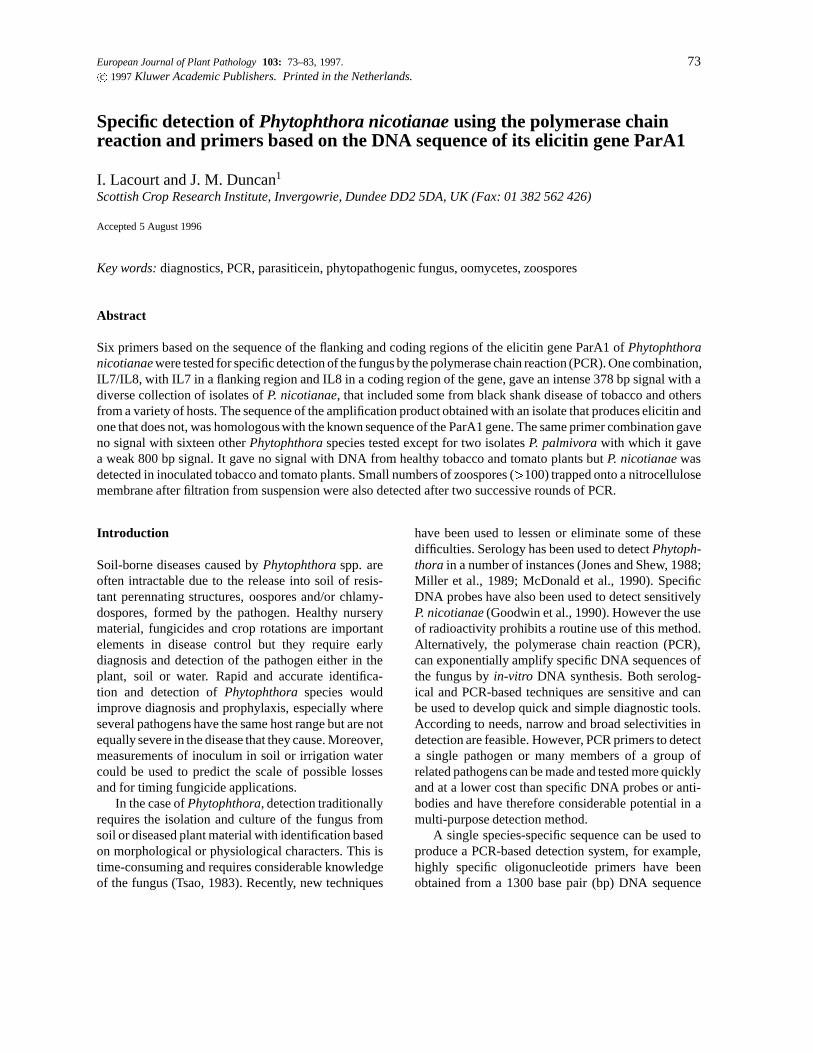
European Journal of Plant Pathology 103: 73–83, 1997. 73c 1997 Kluwer Academic Publishers. Printed in the Netherlands.
Specific detection of Phytophthora nicotianae using the polymerase chainreaction and primers based on the DNA sequence of its elicitin gene ParA1
I. Lacourt and J. M. Duncan1
Scottish Crop Research Institute, Invergowrie, Dundee DD2 5DA, UK (Fax: 01 382 562 426)
Accepted 5 August 1996
Key words: diagnostics, PCR, parasiticein, phytopathogenic fungus, oomycetes, zoospores
Abstract
Six primers based on the sequence of the flanking and coding regions of the elicitin gene ParA1 of Phytophthoranicotianae were tested for specific detection of the fungus by the polymerase chain reaction (PCR). One combination,IL7/IL8, with IL7 in a flanking region and IL8 in a coding region of the gene, gave an intense 378 bp signal with adiverse collection of isolates of P. nicotianae, that included some from black shank disease of tobacco and othersfrom a variety of hosts. The sequence of the amplification product obtained with an isolate that produces elicitin andone that does not, was homologous with the known sequence of the ParA1 gene. The same primer combination gaveno signal with sixteen other Phytophthora species tested except for two isolates P. palmivora with which it gavea weak 800 bp signal. It gave no signal with DNA from healthy tobacco and tomato plants but P. nicotianae wasdetected in inoculated tobacco and tomato plants. Small numbers of zoospores (>100) trapped onto a nitrocellulosemembrane after filtration from suspension were also detected after two successive rounds of PCR.
Introduction
Soil-borne diseases caused by Phytophthora spp. areoften intractable due to the release into soil of resis-tant perennating structures, oospores and/or chlamy-dospores, formed by the pathogen. Healthy nurserymaterial, fungicides and crop rotations are importantelements in disease control but they require earlydiagnosis and detection of the pathogen either in theplant, soil or water. Rapid and accurate identifica-tion and detection of Phytophthora species wouldimprove diagnosis and prophylaxis, especially whereseveral pathogens have the same host range but are notequally severe in the disease that they cause. Moreover,measurements of inoculum in soil or irrigation watercould be used to predict the scale of possible lossesand for timing fungicide applications.
In the case of Phytophthora, detection traditionallyrequires the isolation and culture of the fungus fromsoil or diseased plant material with identification basedon morphological or physiological characters. This istime-consuming and requires considerable knowledgeof the fungus (Tsao, 1983). Recently, new techniques
have been used to lessen or eliminate some of thesedifficulties. Serology has been used to detect Phytoph-thora in a number of instances (Jones and Shew, 1988;Miller et al., 1989; McDonald et al., 1990). SpecificDNA probes have also been used to detect sensitivelyP. nicotianae (Goodwin et al., 1990). However the useof radioactivity prohibits a routine use of this method.Alternatively, the polymerase chain reaction (PCR),can exponentially amplify specific DNA sequences ofthe fungus by in-vitro DNA synthesis. Both serolog-ical and PCR-based techniques are sensitive and canbe used to develop quick and simple diagnostic tools.According to needs, narrow and broad selectivities indetection are feasible. However, PCR primers to detecta single pathogen or many members of a group ofrelated pathogens can be made and tested more quicklyand at a lower cost than specific DNA probes or anti-bodies and have therefore considerable potential in amulti-purpose detection method.
A single species-specific sequence can be used toproduce a PCR-based detection system, for example,highly specific oligonucleotide primers have beenobtained from a 1300 base pair (bp) DNA sequence

74
from P. nicotianae and from an 800 bp sequence fromP. citrophthora (Ersek et al., 1994). Other attemptsto develop PCR-based detection tools have used thetandemly repeated ribosomal RNA (rRNA) gene as asource of DNA sequences (Picard et al., 1992; Bruce etal., 1992). Ribosomes exist in every genome and ribo-somal primers are available to amplify different partsof the rRNA gene from any fungus providing a basis forfurther work (White et al., 1990). The spacer regionsITS1 and ITS2 of the ribosomal units are more variablein sequence than the rRNA genes and have potential fordistinguishing among species (Lee and Taylor, 1992).However, this potential will remain somewhat limiteduntil sequences from a wide range of isolates are avail-able from such complex species as P. megasperma, P.cryptogea and P. drechsleri and the resultant moleculartaxonomy has been clarified.
Alternatively, genes other than the rRNA genescould be the basis of PCR-based tests. Phytophthoraspp. and some closely related oomycetes such asPythium spp. are unique in that they produce a familyof proteins named elicitins (Ricci et al., 1989; Huet etal., 1995). Each species of Phytophthora tested so farsecretes different elicitins which are named after thespecies from which they were purified e.g. cryptogeinfor P. cryptogea, capsicein for P. capsici : : :Advancedstudies showed that several isoforms of elicitins canalso be produced by the same Phytophthora isolate(Le Berre et al., 1994). Using serological techniques,it has been possible to raise antibodies which crossreact with elicitins from several species or specific ofone species (Devergne et al., 1994). Several genesencoding elicitins have been found in P. cryptogea andP. nicotianae isolates (Ricci et al., 1993; Kamoun etal., 1993; Panabieres et al., 1995). Their sequencesshow homology while variability occurs in intergenicspacers (Panabieres, pers. comm.). Therefore primerswith different levels of specificity could be designedas for ribosomal sequences.
Phytophthora nicotianae Breda de Haan, is apathogen on more than 72 genera of plants (Hickman,1958). In the past, this species has been subdividedinto two varieties according to morphological crite-rions, var. nicotianae and var. parasitica; but recentstudies have suggested that there is only one species(Ho and Jong, 1989). This species has also been calledP. parasitica Dastur in which the tobacco-specializedisolates have been grouped in the variety nicotianae(Tucker, 1931). For reasons of anteriority, in this paperit is referred to as P. nicotianae. The fungus producesseveral types and isoforms of elicitin (Yu,1995) and the
amino acids of the principal one, parasiticein, and itsencoding gene ParA1 have been sequenced (Nespouloset al., 1992; Kamoun et al., 1993). Some specializedisolates on tobacco, causing the important black shankdisease, do not appear to produce elicitins (Ricci et al.,1989), or produce them in very much smaller amountsthan isolates from other hosts (Lacourt, 1994). Never-theless these isolates also appear to possess elicitingenes as judged by Southern blots using an elicitinprobe (Kamoun et al., 1993; Ricci et al., 1993).
In this study, we have assessed primers designedaround the sequence of the ParA1 gene for their speci-ficity towards P. nicotianae and their ability to detectthat fungus in infected tomato and tobacco plants andas zoospores in water. Zoospores are an importanttarget for detection because they spread the diseasein water and infect the host after encystment. Theyhave been detected immunologically in water or in soilafter incubation and washing (Cahill and Hardham,1994a) and are probably the most common propagulefound in water; numbers can reach as high as 400per litre in recirculated irrigation water (McDonaldet al., 1994). Their accumulation onto a dipstick hasbeen used to detect P. cinnamomi serologically (Cahilland Hardham, 1994b) and zoospores of P. nicotianaehave also been detected using ELISA by filtration andtrapping on a membrane (Ali-Shtayeh et al., 1991).A similar approach using filter membranes has beenused to detect bacteria, except that PCR was usedinstead of serology to detect the bacteria (Toranzoset al., 1992). In this study we have used this latterapproach to detect zoospores of P. nicotianae usingthe elicitin-based primers.
Materials and methods
Fungal isolates and cultural conditions
Nearly all the isolates (Table 1) of Phytophthora in thestudy came from the collections at SCRI and INRAAntibes. There were thirteen P. nicotianae isolatesfrom various hosts and continents, chosen to givegeographical and pathological diversity. Among themwere producers and non-producers of parasiticein anddifferent groups as determined by analysis of mito-chondrial DNA (Lacourt et al., 1994). There were also33 isolates of another sixteen Phytophthora species.
The fungal isolates were maintained on oatmealagar sloped cultures at 4 �C, except for P. palmivora

75
Table 1. List of Phytophthora isolates tested

76
and P. nicotianae which were maintained at roomtemperature (c. 20 �C). All were cultured on 1% V8juice agar medium in the dark at room temperaturefor seven days after which the mycelium was peeledoff and used to inoculate 10 ml of glucose asparagineliquid medium (GAM: modified medium of Hall et al.(1969), without yeast extract and peptone), in a Petridish. After one week, the mycelium was harvested byfiltration through filter paper (Whatman), washed withdistilled water and either used immediately for DNAextraction or stored frozen (�20 �C).
Inoculation of plants
Two week-old tobacco (Nicotiana tabacum cv Sam-sun) and tomato (Lycopersicon esculentum cv Money-maker) plants were removed from soil and their rootswere washed. Their root tips were cut off and theplantlets were placed in a tube containing tap waterand a plug of mycelium. Tomato plants were inoculatedwith isolate 149 and tobacco plants with isolate 329 ofP. nicotianae. The test tubes were kept in a glasshouseat 24 �C under natural light and water was added regu-larly into the tubes. Symptoms developed after c. oneweek. The plants were stored frozen (�20 �C) untilfurther analysis.
Six week-old tomato plants, kept under the sameconditions of temperature and light, were inoculatedby placing agar plugs of mycelium of isolate 149 onthe cut ends of stems decapitated at various points onthe plants. The progress of the infection was observedover a week and diseased tissues were collected andstored frozen (�20 �C) before analysis.
DNA extraction
Fungal DNAFungal DNA was extracted using a Nucleon II extrac-tion kit (Scotlab Ltd, Glasgow) according to the manu-facturer’s recommendations. Between 150 and 200 mgof compressed wet-weight mycelium were extracted ina protocol scaled down for use in 1.5 ml Eppendorftubes; this yielded 20 to 30 mg of DNA suitable forPCR amplification.
Plant DNAPlant DNA was extracted according to a modifiedprotocol of Edwards et al. (1991): 250 mg of plantmaterial was ground using a pestle in an Eppendorfcontaining 500 ml of lysis buffer (200 mM Tris HCl,
pH 7.5, 250 mM NaCl, 25 mM EDTA, 0.5% SDS)plus 25 mg of Polyvinylpyrrolidone and sterile sand.After centrifuging for 3 min at � 13000 g, the super-natant was extracted with 1 volume of chloroform-isoamylalcohol (24/1, v/v) and then collected aftercentrifugation for 3 min at � 13000 g. DNA wasprecipitated with one volume of isopropanol andcentrifuged at� 13000 g for 5 min. The resultant pelletwas washed three times with 70% ethanol, vacuumdried and resuspended in sterile distilled water to yield10 to 20 mg of DNA suitable for DNA amplification.
Preparation of the zoospores and fixation on amembrane
Zoospores of P. nicotianae were produced according toa protocol of P. Bonnet (INRA Antibes, pers. comm.).Isolates 149 and 329 were cultured on 1% V8 agarmedium for one week as previously described andfour agar plugs of mycelium were transferred to peabroth for one week at 24 �C under light. The resul-tant mycelium was cut with a scalpel into small pieceswhich were transferred onto 2% water agar in Petridishes for four days under the same conditions. Thedishes were placed for 30 min at 4 �C, 15 ml of steriledistilled water at 30 �C were added and after incu-bation for 10 min at 30 �C, the water, now contain-ing the zoospores, was removed, the zoospores werecounted using an haemocytometer and their numberswere adjusted to the desired concentration by addingdistilled water.
The zoospore suspension (20 ml) was filteredthrough a 0.45 mm pore size nitrocellulose filter,4.5 cmdiam, which was then treated according to a modifiedmethod of Toranzos and Alvarez (1992). It was placed‘zoospore’ side upwards on filter papers soaked withthe following solutions and for the following times:20 min. on 10% SDS; 5 min. on 0.5 M NaOH + 1.5M NaCl, then on 1.5 M NaCl + 0.5 mM Tris HCl pH7.5 and finally on 2X SSC. The nucleic acids werefixed onto the nitrocellulose membrane by heating inan oven at 80 �C for two hours and the membrane waswashed in sterile distilled water and allowed to dry.
Design of PCR primers, DNA amplification by PCRand analysis of PCR products
Primer sequencesSix primers based on the sequence of the ParA1gene were synthesised (Figure 1). Some primers were

77
designed to anneal to the coding regions of the ParA1gene in an attempt to ensure that the PCR productswere derived from that gene: IL3, IL8 and IL5 were ofthis type with the first two reading in the reverse senseand the last reading in the forward sense. Other primerswere located in the flanking region upstream from the50 end of the gene (IL7, reading in the forward sense),the signal peptide region of the gene (IL2, reversesense) or in the flanking region downstream from the30 end of the gene (IL6, forward sense). As theseothers were not located in the generally conservedcoding region, it was expected that they might conferspecificity when used in combination with primers inthe coding regions. The primer sequences result from acompromise between several requirements. We chose19 to 20 nucleotides long primer having a GC con-tent which allows annealing temperatures c. 60 �C. Asmuch as possible we also avoided base repetitions orhairpin and primer dimers structure, but this was notentirely possible.
Five different primer combinations were used forPCR on fungal DNA: IL2/IL5, IL3/IL5, IL2/IL6,IL3/IL6 and IL7/IL8. Only IL7/IL8 combination wasused for PCR on DNA extracted from plant samples.All primers were used at the concentration of 1 mMwith 200 mM of each dATP, dCTP, dGTP and dTTP(Pharmacia, Biotech), 2.5 units of Taq polymerase(Gibco BRL) and the PCR buffer supplied with theenzyme (20 mM Tris HCl pH 8.4, 50 mM KCl, 1.5mM MgCl2), in a final volume of 50 ml with 100 to500 ng of DNA as template. With plant DNA, 10 mgml�1 of Bovine Serum Albumin (BSA) were added.Each reaction was sealed with a few drops of mineraloil before the tubes were incubated at 94 �C for 3min, then subjected to 35 cycles of denaturation 94 �Cfor 30 s; annealing at 59 �C (primer combinationsIL2/IL5, IL3/IL6, IL3/IL5) or 65 �C (primer combina-tion IL7/IL8) for 30 s.; and synthesis at 72 �C for 1.5min. Finally, they were incubated for an additional 10min at 72 �C and stored if necessary at �20 �C beforeanalysis.
Amplification of the DNA fixed on the nitrocellulosemembraneA piece of the membrane 35 mm2 and representing2.2% of the total area of the original filter was cut outwith a cork borer. The numbers of zoospores filteredthrough the membrane were calculated so as to give aknown number of zoospores on the removed piece ofmembrane, assuming that the distribution of zoospores
was similar across the whole membrane. The piece ofmembrane was placed in an Eppendorf tube with thefollowing reagents: 1 mM of each primer IL7 and IL8,200 mM of each dATP , dCTP, dGTP and dTTP (Phar-macia, Biotech), 10 mg ml�1 of BSA, 10 units of Taqpolymerase (Gibco BRL) and the PCR buffer suppliedwith the enzyme (20 mM Tris HCl pH 8.4, 50 mMKCl, 1.5 mM MgCl2), in a final volume of 100 ml. Atwo min. ramp time was allowed between denatura-tion, annealing and synthesis of DNA. After an initialdenaturation of 3 min at 94 �C, there were 40 cycles asfollows: 94 �C 30 s., 37 �C 1 min and 72 �C 2 min. Thelow annealing temperature in the first round PCR wasused in accordance with the methodology of Toranzosand Alvarez (1992). The reaction was extended witha final step of 72 �C for 10 min. For a second roundof PCR amplification, 5 ml of the first reaction mix-ture was added to an identical reaction mixture andamplified under standard conditions used for DNA insolution.
PCR controlsFor each PCR experiment, one tube with the PCRmix but without any DNA added was run. The resultswere only considered when no DNA amplification wasvisible on gel electrophoresis for such negative con-trols. As a further check, PCR reactions were madeseparately with each single primer.
Gel electrophoresisPCR products were separated by electrophoresis in 1%agarose gels, in TAE buffer (0.04 M Tris-acetate, 1mM EDTA), stained in ethidium bromide and analysedunder UV light (Sambrook et al., 1989).
Sequencing of PCR productsThe amplified products were obtained under the condi-tions described above with primers IL7/IL8 and DNAof isolates 183 and 26, non-producing (tobacco blackshank) and parasiticein-producing isolates respec-tively. They were purified using the WizardTM DNApurification kit (Promega) according to the manu-facturer’s instructions. They were quantified by gelelectrophoresis, by comparing the intensity of thebands with bands corresponding to known amounts ofa DNA fragment digested from lambda phage DNA(Pharmacia, Biotech), of a size comparable to theamplified fragment. At least 125 ng of double strandPCR amplified DNA were used for each sequencereaction with the PRISMTM Ready Reaction Taq

78
Figure 1. Sequence of the Sal I/Cla I DNA fragment containing the ParA1 gene of Phytophthora nicotianae that encodes the elicitin parasiticein(taken from Kamoun et al. (1993)) with the amino acid sequence of parasiticein in bold above the DNA sequence. Also shown are the origin oftranscription of the gene (#), the location, sequence (in bold italics) and the orientation (arrowed) of primers IL2, IL3, IL5, IL6, IL7 and IL8.
DyedeoxyTM Terminator Cycle Sequence Kit (PerkinElmer) according to the manufacturer’s instructions.The standard annealing temperature was 60 �C and3.2 pg of primer were used for each sequencingreaction. Sequences were produced using both IL7and IL8 primers and the reactions were processedwith an Applied Biosystems 373 Stretch AutomatedSequencer.
Results
Primer combinations
The primer combination IL2/IL6 (see Figure 1 fordetails of primers) did not amplify DNA of P. nico-tianae, even when lower annealing temperatures weretested. IL3/IL5 produced highly polymorphic patterns
which were influenced strongly by concentrations ofdNTPs, primers and MgCl2. Combinations IL2/IL5and IL3/IL6, gave consistent patterns with all twelveisolates of P. nicotianae with which they were testedbut in control experiments, primers IL2 and IL3 gavesome bands when used singly (data not shown). All theprimer combinations involving IL2, IL3, IL5 and IL6were therefore excluded from further investigation.
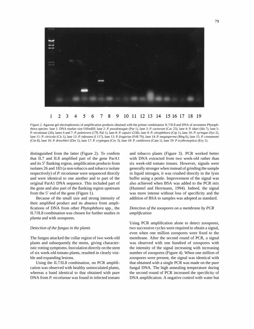
The IL7/IL8 combination amplified an intense 378bp DNA fragment from all P. nicotianae isolates. Infurther tests with sixteen other species representing allsix groups of Phytophthora spp. (Stamps et al., 1990),this combination only amplified DNA from P. nico-tianae and from two of four isolates of P. palmivoraand there was no amplification with DNA from healthyplants. The DNA product from the two isolates of P.palmivora was rather faint and longer (800 bp) thanthat produced from P. nicotianae and could be easily
79
Figure 2. Agarose gel electrophoresis of amplification products obtained with the primer combination IL7/IL8 and DNA of seventeen Phytoph-thora species: lane 1: DNA marker size l/HindIII; lane 2: P. pseudotsugae (Pse 1), lane 3: P. cactorum (Cac 23), lane 4: P. idaei (Ida 7), lane 5:P. nicotianae (26), lanes 6 and 7: P. palmivora (178, Pal 1), lane 8: P. capsici (238), lane 9: P. citrophthora (Ctp 1), lane 10: P. syringae (Syr 2),lane 11: P. citricola (Cit 1), lane 12: P. infestans (I 117), lane 13: P. fragariae (FrR 70), lane 14: P. megasperma (Meg 6), lane 15: P. cinnamomi(Cin 8), lane 16: P. dreschleri (Dre 1), lane 17: P. cryptogea (Cry 3), lane 18: P. cambivora (Cam 1), lane 19: P erythroseptica (Ery 1).
distinguished from the latter (Figure 2). To confirmthat IL7 and IL8 amplified part of the gene ParA1and its 50 flanking region, amplification products fromisolates 26 and 183 (a non-tobacco and tobacco isolaterespectively) of P. nicotianae were sequenced directlyand were identical to one another and to part of theoriginal ParA1 DNA sequence. This included part ofthe gene and also part of the flanking region upstreamfrom the 50 end of the gene (Figure 1).
Because of the small size and strong intensity oftheir amplified product and its absence from ampli-fications of DNA from other Phytophthora spp., theIL7/IL8 combination was chosen for further studies inplanta and with zoospores.
Detection of the fungus in the plants
The fungus attacked the collar region of two week-oldplants and subsequently the stems, giving character-istic rotting symptoms. Inoculation directly on the stemof six week-old tomato plants, resulted in clearly visi-ble and expanding lesions.
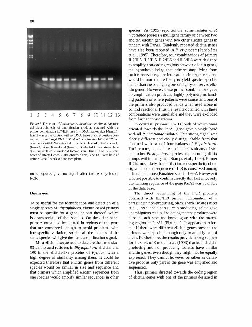
Using the IL7/IL8 combination, no PCR amplifi-cation was observed with healthy uninoculated plants,whereas a band identical to that obtained with pureDNA from P. nicotianae was found in infected tomato
and tobacco plants (Figure 3). PCR worked betterwith DNA extracted from two week-old rather thansix week-old tomato tissues. However, signals weregenerally stronger when instead of grinding the samplein liquid nitrogen, it was crushed directly in the lysisbuffer using a pestle. Improvement of the signal wasalso achieved when BSA was added to the PCR mix(Hummel and Herrmann, 1994). Indeed, the signalwas more intense without loss of specificity and theaddition of BSA to samples was adopted as standard.
Detection of the zoospores on a membrane by PCRamplification
Using PCR amplification alone to detect zoospores,two successive cycles were required to obtain a signal,even when one million zoospores were fixed to themembrane. After the second round of PCR, a signalwas observed with one hundred of zoospores withthe intensity of the signal increasing with increasingnumber of zoospores (Figure 4). When one million ofzoospores were present, the signal was identical withthat obtained with a single PCR was made on the purefungal DNA. The high annealing temperature duringthe second round of PCR increased the specificity ofDNA amplification. A negative control with water but
80
Figure 3. Detection of Phytophthora nicotianae in planta. Agarosegel electrophoresis of amplification products obtained with theprimer combination IL7/IL8; lane 1 – DNA marker size l/HindIII;lane 2 – negative control with no DNA, lanes 3 and 9 positive con-trol with pure fungal DNA of P. nicotianae isolates 149 and 329; allother lanes with DNA extracted from plants: lanes 4 to 7–2 week-old(lanes 4, 5) and 6 week-old (lanes 6, 7) infected tomato stems; lane8 – uninoculated 2 week-old tomato stem; lanes 10 to 12 – stembases of infected 2 week-old tobacco plants; lane 13 – stem base ofuninoculated 2 week-old tobacco plant.
no zoospores gave no signal after the two cycles ofPCR.
Discussion
To be useful for the identification and detection of asingle species of Phytophthora, elicitin-based primersmust be specific for a gene, or part thereof, whichis characteristic of that species. On the other hand,primers must also be located in regions of the genethat are conserved enough to avoid problems withintraspecific variation, so that all the isolates of thesame species will give the same amplification signal.
Most elicitins sequenced to date are the same size,98 amino acid residues in Phytophthora elicitins and100 in the elicitin-like proteins of Pythium with ahigh degree of similarity among them. It could beexpected therefore that elicitin genes from differentspecies would be similar in size and sequence andthat primers which amplified elicitin sequences fromone species would amplify similar sequences in other
species. Yu (1995) reported that some isolates of P.nicotianae possess a multigene family of between twoand ten elicitin genes with two other elicitin genes intandem with ParA1. Tandemly repeated elicitin geneshave also been reported in P. cryptogea (Panabiereset al., 1995). Therefore, four combinations of primersIL2/IL5, IL3/IL5, IL2/IL6 and IL3/IL6 were designedto amplify non-coding regions between elicitin genes,the hypothesis being that primers amplifying fromsuch conserved regions into variable intergenic regionswould be much more likely to yield species-specificbands than the coding regions of highly conserved elic-itin genes. However, these primer combinations gaveno amplification products, highly polymorphic band-ing patterns or where patterns were consistent, one ofthe primers also produced bands when used alone incontrol reactions. Thus the results obtained with thesecombinations were unreliable and they were excludedfrom further consideration.
In contrast, primers IL7/IL8 both of which wereoriented towards the ParA1 gene gave a single bandwith all P. nicotianae isolates. This strong signal wasclearly different and easily distinguishable from thatobtained with two of four isolates of P. palmivora.Furthermore, no signal was obtained with any of six-teen other Phytophthora species, representing all sixgroups within the genus (Stamps et al., 1990). PrimerIL7 is most likely the one that induces specificity of thesignal since the sequence of IL8 is conserved amongdifferent elicitins (Panabieres et al., 1995). However itwas not possible to confirm directly this fact since onlythe flanking sequence of the gene ParA1 was availablein the data base.
The direct sequencing of the PCR productsobtained with IL7/IL8 primer combination of aparasiticein non-producing, black shank isolate (Ricciet al., 1992) and a parasiticein producing isolate gaveunambiguous results, indicating that the products werepure in each case and homologous with the match-ing region of ParA1 (Figure 1). It appears thereforethat if there were different elicitin genes present, theprimers were specific enough only to amplify one ofthem. Furthermore, the results provide strong supportfor the view of Kamoun et al. (1993) that both elicitin-producing and non-producing isolates have similarelicitin genes, even though they might not be equallyexpressed. They cannot however be taken as defini-tive proof as only part of the gene was amplified andsequenced.
Thus, primers directed towards the coding regionof elicitin genes with one of the primers designed in

81
Figure 4. Agarose gel electrophoresis of a second PCR amplification of products obtained with the primer combination IL7/IL8 from variousnumbers of zoospores of isolate 149 of Phytophthora nicotianae fixed on a nitro-cellulose membrane; lane 1 – DNA marker size l/HindIII.Lanes 2 to 7 respectively – zoospore numbers 0, 102, 103, 104, 105, 106; lane 8 – amplification product of a single PCR on pure fungal DNA(isolate 149).
the coding sequence of the parasiticein and the other innon-codingsequence upstream to the 50 end of the geneParA1 have some potential as P. nicotianae-specificprimers. Given that all species of Phytophthora prob-ably have such elicitin genes with flanking sequences,PCR-based detection/ identification/diagnostic tech-niques based on elicitin genes, could be developedfor all species in the genus. Only the sequence of oneelicitin gene was amplified using IL7 and IL8 primers.The choice of repeated sequences such as ribosomalgenes of satellite sequences offers the advantage ofincreasing the level of DNA amplification from thestart of the PCR. However, new refinements of thePCR technique such as the nested PCR (MacManusand Jones, 1995), raises the sensitivity of this tech-nique to a much higher scale than the use of repeatedsequences. Discrete sequences, therefore, could bechosen in preference to repeated sequences if they pre-sented the appropriate specificity.
The signal obtained with IL7/IL8 was intense andspecific enough to detect the pathogen in planta. It wasgenerally stronger in younger than in older tissues.Perhaps the latter were more lignified and containedphenolic compounds that inhibited the Taq polymerase,alternatively, lesions on young tissue may have con-
tained more of the fungus than older ones. By addingBSA to the reaction mixture, this problem was largelyovercome. P. nicotianae is a relatively unimportantpathogen in Scotland and only artificially inoculatedhost material was available for test. Nevertheless, avery similar protocol, but using rRNA gene-based,specific primers instead of elicitin genes, has beenused successfully to detect P. fragariae and P. idaei innaturally infected field-grown raspberry plants (Rubusidaeus) (unpublished results). Again the incorporationof BSA in the reaction increased the sensitivity of thereaction. This result, taken together with the workon inoculated material, indicates that the same tech-nique should be able to detect P. nicotianae in naturallyinfected plants and that it should be possible to identifyP. nicotianae directly in lesions on the host using PCRwith the primers IL7/IL8 without resort to isolationof the pathogen from the host. The IL7/IL8 primershave also been used with scrapings of mycelium froma pure culture of Phytophthora isolated from Dieffen-bachia (dumb cane) to confirm within a few hours itsidentity as Phytophthora nicotianae (Dr T. O’Neill,ADAS, Cambridge pers. comm). Routine diagnosticsand identifications should therefore be possible withina day or less.

82
Also feasible is the detection of zoospores ona nitro-cellulose membrane filter by PCR using aprotocol based on Toranzos et al. (1992) for bacteria.Extended treatment with 10% SDS appeared to besufficient to lyse the zoospores even if they had alreadyencysted. It was not possible to include the whole of afilter membrane in the PCR reaction, only small piecesof it fitted into 0.5 ml Eppendorf tubes used for thePCR reaction. Thus only a small portion of the totalnumber of zoospores applied to the membrane wouldhave been added to the reaction mixture assuming thatthe zoospores, which were applied to the membranein large volumes of water, were all trapped on themembrane and that they were evenly distributed.Nevertheless, there was a steady increase in signalobtained with PCR with increasing amounts ofzoospores filtered through the membrane. After twocycles of PCR, a P. nicotianae signal was visible withan estimated 100 zoospores trapped on a piece of themembrane. The annealing temperature used for DNAamplification of template bound to membrane was verylow in comparison with the temperature used when theDNA is in solution. This was necessary to amplifyenough DNA from template whose structure is modi-fied. This temperature was recommended by Toranzoset al. (1992) and by other authors using degraded DNAas template for PCR (Hauswirth et al., 1994). Thespecificity of the DNA amplification was obtained inthe second round PCR. Indeed, there were no cross-reactions with zoospores of other species trapped onmembranes in our work (results not shown).
The technique is still open to improvement. Forexample, Toranzos et al. (1992) detected very smallnumbers of bacteria by performing a third PCR on theirsamples. Using nested primers, or combining PCRamplification and dot-blot or reverse-blot hybridizationalso gave good results (MacManus and Jones, 1995).Thus, the results offer a realistic alternative to the useof ELISA-based tests for the detection of P. nicotianaein plants and as zoospores.
Acknowledgements
The authors acknowledge the support of the Organ-isation for Economic Cooperation and Development(OECD) by the award of a six month post-doctorategrant to the senior author and of the Scottish OfficeAgricultural and Fisheries Department. They alsothank Dr Pierre Ricci and Dr Frank Panabieres fromINRA Antibes France for useful advice and discus-
sions at the start of the project and Dr David Cooke forproviding other PCR primers.
References
Ali-Shtayeh MS, MacDonald JD and Kabashima J (1991) A methodfor using commercial ELISA tests to detect zoospores ofPhytophthora and Pythium in irrigated water. Plant Disease 75:305–311
Bruce KD, Hiorns WD, Hobman JL, Osborn AM, Strike P andRitchie DA (1992) Amplification of DNA from native popula-tion of soil bacteria by using the Polymerase Chain Reaction.Applied and Environmental Microbiology 58: 3413–3416
Cahill DM and Hardham AR (1994a) A dipstick immunoassayfor the specific detection of Phytophthora cinnamomi in soils.Phytopathology 84: 1284–1292
Cahill DM and Hardham AR (1994b) Exploitation of zoospores taxisin the development of a novel dipstick immunoassay for thespecific detection of Phytophthora cinnamomi. Phytopathology84: 193–200
Devergne JC, Fort MA, Bonnet P, Ricci P, Vergnet C, DelaunayT and Grosclaude J (1994) Immunodetection of elicitinsfrom Phytophthora spp. using monoclonal antibodies. PlantPathology 43: 885–896
Edwards K, Johnstone C and Thompson C (1991) A simple andrapid method for the preparation of plant genomic DNA forPCR analysis. Nucleic Acids Research 19: 1349
Ersek T, Schoelz JE and English JT (1994) PCR amplification ofspecies-specific DNA sequence can distinguish among Phytoph-thora species. Applied and Enviromental Microbiology 60:2616–2621
Goodwin PH, English JT, Neher DA, Duniway JM and KirkpartrickBC (1990) Detection of Phytophthora parasitica from soil andhost tissue with a species-specific DNA probe. Phytopathology80: 277–281
Hall R, Zentmeyer GA and Erwin DC (1969) Approach to the tax-onomy of Phytophthora through acrylamide gel electrophoresisof proteins. Phytopathology 59: 770–774
Hauswirth WW, Dickel CD and Lawlor DA (1994) DNA analysis ofthe windover population. In: Herrmann B and Hummel S (eds)Ancient DNA. Recovery and Analysis of Genetic Material fromPaleontological Archeological, Museum, Medical and ForensicSpecimens (pp. 104–121) Springer-Verlag, New York USA
Hickman CJ (1958) Phytophthora plant destroyer. Transactions ofthe British Mycological Society 41: 1–13
Ho HH and Jong SC (1989) Phytophthora nicotianae (P. parasitica).Mycotaxon 35: 243–276
Huet JC, Le Caer JP, Nespoulos C and Pernollet JC (1995) The rela-tionships between the toxicity and the primary and secondarystructures of elicitin-like protein elicitor secreted by thephytopathogenic fungus Pythium vexans. Molecular Plant-Microbe Interaction 8: 302–310
Hummel S and Herrmann B (1994) General aspects of sample prepa-ration. In: Herrmann B and Hummel S (eds) Ancient DNA.Recovery and Analysis of Genetic Material from Paleontolog-ical Archeological, Museum, Medical and Forensic Specimens(pp. 59–68) Springer-Verlag, New York USA
Jones K and Shew HD (1988) Immunoassay procedure for the detec-tion of Phytophthora nicotianae var nicotianae in soil (abstr.).Phytopathology 78: 1577

83
Kamoun S, Klucher KM, Coffey MA and Tyler (1993) A gene encod-ing a host-specific elicitor protein of Phytophthora nicotianae.Molecular Plant-Microbe Interactions 6: 573–581
Lacourt I (1994) Etude de la specialisation parasitaire de Phytoph-thora parasitica Dastur par l’analyse d’une population d’isolatsd’origines geographiques diverses. These de Doctorat, Univer-site Claude Bernard of Lyon
Lacourt I, Panabieres F, Marais A, Venard P and Ricci P (1994)Intraspecific polymorphism of the fungus Phytophthora para-sitica revealed by the analysis of mitochondrial DNA Restric-tion Fragment Length Polymorphism. Mycological Research 9:562–568
Le Berre J-Y, Panabieres F, Ponchet M, Denoroy L, Bonnet P, MaraisA and Ricci P (1994) Occurence of multiple forms of elicitinin Phytophthora cryptogea. Plant Physiology and Biochemistry32: 1–5
Lee SB and Taylor JW (1992) Phylogeny of five fungus-likeprotoctistan Phytophthora species, inferred from the internaltranscribed spacers of ribosomal DNA. Journal of Molecularand Biological Evolution 9: 636–653
McDonald JD, Stites J and Kabashima J (1990) Comparison ofserological and culture plate methods for detecting species ofPhytophthora, Pythium and Rhizoctonia in ornamental plants.Plant Disease 74: 655–659
MacDonald JD, Ali-Shtaeyh MS, Kabashima J and Stites J (1994)Occurence of Phytophthora species in recirculated nursery irri-gation effluents. Plant Disease 78: 607–611
MacManus PS and Jones AL (1995) Detection of Erwinia carotovaby nested PCR and PCR-dot-blot and reverse-blot hybridisa-tions. Phytopathology 85: 618–623
Miller SM, Petersen FP, Miller SA, Rittembourg JH, Wood SCand Grothaus GD (1989) Development of a direct immuno-assay to detect Phytophthora megasperma in soil. (abstr.) Phyto-pathology 79: 1139
Nespoulos C, Huet JC and Pernollet JC (1992) Structure-functionrelationships of a and b elicitins, signal proteins involved in theplant-Phytophthora interaction. Planta 186: 551–557
Panabieres F, Marais A, Le Berre JY, Fournier D, Penot Iand Ricci P (1995) Characterization of a gene cluster ofPhytophthora cryptogea which encodes for elicitins, proteinsinducing a hypersensitive-like response in tobacco. MolecularPlant-Microbe Interactions 8: 996–1003
Picard C, Ponsonnet C, Paget E, Nesme X and Simonet P (1992)Detection and enumeration of bacteria in soil by direct DNAextraction and Polymerase Chain Reaction. Applied and Envi-ronmental Microbiology 58: 2717–2722
Ricci P, Bonnet P, Huet JC, Sallantin M, Beauvais-Cante F,Bruneteau M, Billard V, Michel G and Pernollet JC (1989) Struc-ture and activity of proteins from pathogenic fungi Phytophthoraeliciting necrosis and acquired resistance in tobacco. EuropeanJournal of Biochemistry 183: 555–563
Ricci P, Trentin F, Bonnet P, Venard P, Mouton-Perronnet F andBruneteau M (1992) Differential production of parasiticein, anelicitor of necrosis and resistance in tobacco, by isolates ofPhytophthora parasitica. Plant Pathology 41: 298–307
Ricci P, Panabieres F, Bonnet, Maia N, Ponchet M, Devergne JC,Marais A, Cardin L, Milat ML and Blein JP (1993) Protein-aceous elicitors of plant defense responses. In: Fritig B andLegrand M (eds) Mechanisms of Plant Defense Response(pp. 136–139) Kluwer Academic Publishers, The Netherlands
Sambrook J, Fritsch EF and Maniatis T (1989) Molecular Cloning,a Laboratory Manual. Second Edition. Nolan C and FergusonM (eds) Cold Spring, Harbor Laboratory Press
Stamps DJ, Waterhouse GM, Newhook FJ and Hall GS (eds) (1990)Revised tabular key to the species of Phytophthora. MycologicalPaper No 162, 28 pp. C.A.B. International Mycological Institute
Toranzos GA and Alvarez AJ (1992) Solid-phase polymerase ChainReaction: Application for direct detection of enteric pathogensin water. Canadian Journal of Microbiology 38: 365–369
Tsao PH (1983) Factors affecting isolation and quantitation ofPhytophthora from soil. In: Erwin DC, Bartnicky-Garcia S andTsao PH (eds) Phytophthora, Its Biology, Taxonomy, Ecologyand Pathology (pp. 219–236) American PhytopathologicalSociety, St Paul, Minnesota
Tucker CM (1931) taxonomy of the genus Phytophthora de Bary.Missouri agricultural Experiment station Research Bulletin 153:1–208
White TJ, Bruns T, Lee S and Taylor J (1990) Amplification anddirect sequencing of fungal ribosomal RNA genes for phyloge-netics. In: PCR Protocols: A Guide to Methods and Applications(pp. 315–321) Academic Press Inc. New York
Yu LM (1995) Elicitins from Phytophthora and basic resistancein tobacco. Proceedings of National Academy of Sciences 92:4088–4094