Spatiotemporal integration of tactile information in human somatosensory cortex
14
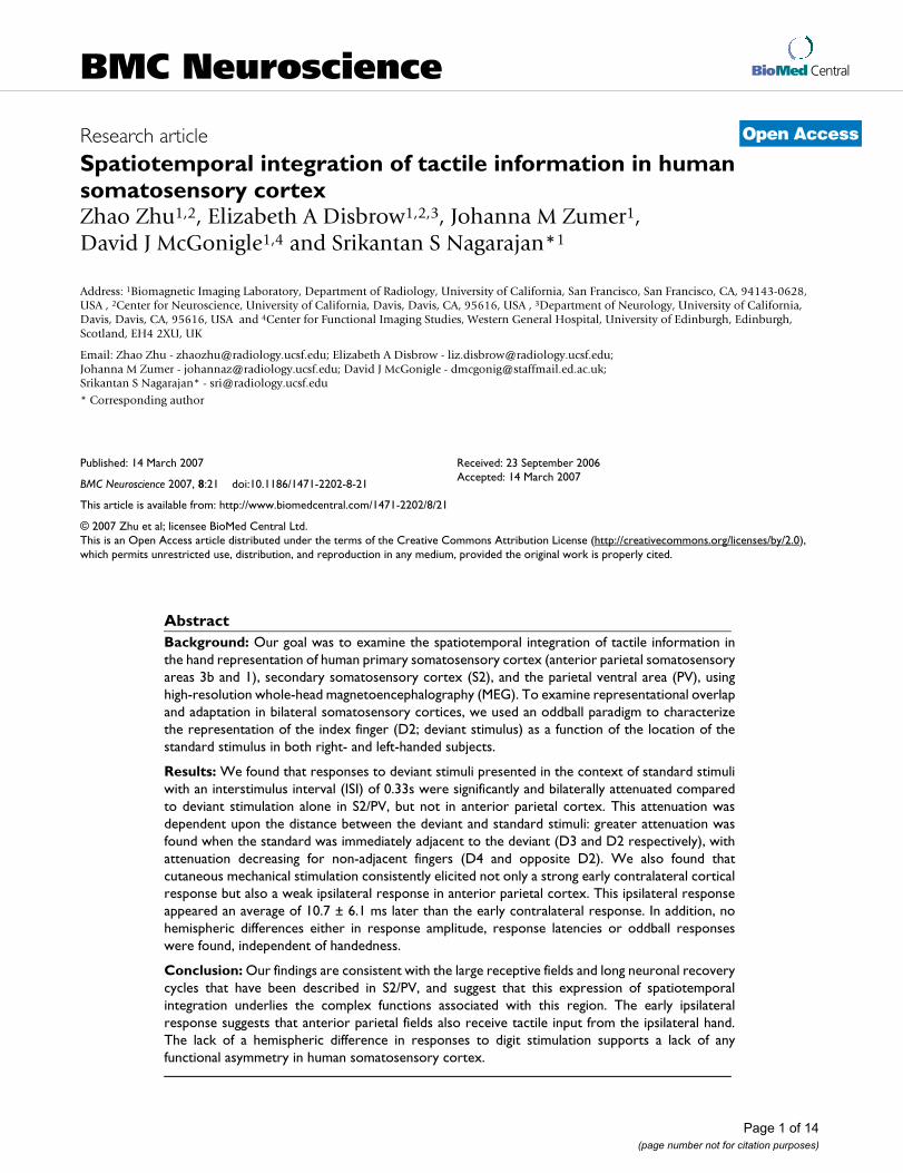
BioMed Central Page 1 of 14 (page number not for citation purposes) BMC Neuroscience Open Access Research article Spatiotemporal integration of tactile information in human somatosensory cortex Zhao Zhu 1,2 , Elizabeth A Disbrow 1,2,3 , Johanna M Zumer 1 , David J McGonigle 1,4 and Srikantan S Nagarajan* 1 Address: 1 Biomagnetic Imaging Laboratory, Department of Radiology, University of California, San Francisco, San Francisco, CA, 94143-0628, USA , 2 Center for Neuroscience, University of California, Davis, Davis, CA, 95616, USA , 3 Department of Neurology, University of California, Davis, Davis, CA, 95616, USA and 4 Center for Functional Imaging Studies, Western General Hospital, University of Edinburgh, Edinburgh, Scotland, EH4 2XU, UK Email: Zhao Zhu - [email protected]; Elizabeth A Disbrow - [email protected]; Johanna M Zumer - [email protected]; David J McGonigle - [email protected]; Srikantan S Nagarajan* - [email protected] * Corresponding author Abstract Background: Our goal was to examine the spatiotemporal integration of tactile information in the hand representation of human primary somatosensory cortex (anterior parietal somatosensory areas 3b and 1), secondary somatosensory cortex (S2), and the parietal ventral area (PV), using high-resolution whole-head magnetoencephalography (MEG). To examine representational overlap and adaptation in bilateral somatosensory cortices, we used an oddball paradigm to characterize the representation of the index finger (D2; deviant stimulus) as a function of the location of the standard stimulus in both right- and left-handed subjects. Results: We found that responses to deviant stimuli presented in the context of standard stimuli with an interstimulus interval (ISI) of 0.33s were significantly and bilaterally attenuated compared to deviant stimulation alone in S2/PV, but not in anterior parietal cortex. This attenuation was dependent upon the distance between the deviant and standard stimuli: greater attenuation was found when the standard was immediately adjacent to the deviant (D3 and D2 respectively), with attenuation decreasing for non-adjacent fingers (D4 and opposite D2). We also found that cutaneous mechanical stimulation consistently elicited not only a strong early contralateral cortical response but also a weak ipsilateral response in anterior parietal cortex. This ipsilateral response appeared an average of 10.7 ± 6.1 ms later than the early contralateral response. In addition, no hemispheric differences either in response amplitude, response latencies or oddball responses were found, independent of handedness. Conclusion: Our findings are consistent with the large receptive fields and long neuronal recovery cycles that have been described in S2/PV, and suggest that this expression of spatiotemporal integration underlies the complex functions associated with this region. The early ipsilateral response suggests that anterior parietal fields also receive tactile input from the ipsilateral hand. The lack of a hemispheric difference in responses to digit stimulation supports a lack of any functional asymmetry in human somatosensory cortex. Published: 14 March 2007 BMC Neuroscience 2007, 8:21 doi:10.1186/1471-2202-8-21 Received: 23 September 2006 Accepted: 14 March 2007 This article is available from: http://www.biomedcentral.com/1471-2202/8/21 © 2007 Zhu et al; licensee BioMed Central Ltd. This is an Open Access article distributed under the terms of the Creative Commons Attribution License (http://creativecommons.org/licenses/by/2.0 ), which permits unrestricted use, distribution, and reproduction in any medium, provided the original work is properly cited.
-
Upload
independent -
Category
Documents
-
view
2 -
download
0
Transcript of Spatiotemporal integration of tactile information in human somatosensory cortex
BioMed CentralBMC Neuroscience
ss
Open AcceResearch articleSpatiotemporal integration of tactile information in human somatosensory cortexZhao Zhu1,2, Elizabeth A Disbrow1,2,3, Johanna M Zumer1, David J McGonigle1,4 and Srikantan S Nagarajan*1Address: 1Biomagnetic Imaging Laboratory, Department of Radiology, University of California, San Francisco, San Francisco, CA, 94143-0628, USA , 2Center for Neuroscience, University of California, Davis, Davis, CA, 95616, USA , 3Department of Neurology, University of California, Davis, Davis, CA, 95616, USA and 4Center for Functional Imaging Studies, Western General Hospital, University of Edinburgh, Edinburgh, Scotland, EH4 2XU, UK
Email: Zhao Zhu - [email protected]; Elizabeth A Disbrow - [email protected]; Johanna M Zumer - [email protected]; David J McGonigle - [email protected]; Srikantan S Nagarajan* - [email protected]
* Corresponding author
AbstractBackground: Our goal was to examine the spatiotemporal integration of tactile information inthe hand representation of human primary somatosensory cortex (anterior parietal somatosensoryareas 3b and 1), secondary somatosensory cortex (S2), and the parietal ventral area (PV), usinghigh-resolution whole-head magnetoencephalography (MEG). To examine representational overlapand adaptation in bilateral somatosensory cortices, we used an oddball paradigm to characterizethe representation of the index finger (D2; deviant stimulus) as a function of the location of thestandard stimulus in both right- and left-handed subjects.
Results: We found that responses to deviant stimuli presented in the context of standard stimuliwith an interstimulus interval (ISI) of 0.33s were significantly and bilaterally attenuated comparedto deviant stimulation alone in S2/PV, but not in anterior parietal cortex. This attenuation wasdependent upon the distance between the deviant and standard stimuli: greater attenuation wasfound when the standard was immediately adjacent to the deviant (D3 and D2 respectively), withattenuation decreasing for non-adjacent fingers (D4 and opposite D2). We also found thatcutaneous mechanical stimulation consistently elicited not only a strong early contralateral corticalresponse but also a weak ipsilateral response in anterior parietal cortex. This ipsilateral responseappeared an average of 10.7 ± 6.1 ms later than the early contralateral response. In addition, nohemispheric differences either in response amplitude, response latencies or oddball responseswere found, independent of handedness.
Conclusion: Our findings are consistent with the large receptive fields and long neuronal recoverycycles that have been described in S2/PV, and suggest that this expression of spatiotemporalintegration underlies the complex functions associated with this region. The early ipsilateralresponse suggests that anterior parietal fields also receive tactile input from the ipsilateral hand.The lack of a hemispheric difference in responses to digit stimulation supports a lack of anyfunctional asymmetry in human somatosensory cortex.
Published: 14 March 2007
BMC Neuroscience 2007, 8:21 doi:10.1186/1471-2202-8-21
Received: 23 September 2006Accepted: 14 March 2007
This article is available from: http://www.biomedcentral.com/1471-2202/8/21
© 2007 Zhu et al; licensee BioMed Central Ltd. This is an Open Access article distributed under the terms of the Creative Commons Attribution License (http://creativecommons.org/licenses/by/2.0), which permits unrestricted use, distribution, and reproduction in any medium, provided the original work is properly cited.
Page 1 of 14(page number not for citation purposes)
BMC Neuroscience 2007, 8:21 http://www.biomedcentral.com/1471-2202/8/21
BackgroundThe spatiotemporal integration of tactile inputs from dif-ferent skin regions and across body parts is an importantfunction of human somatosensory cortex. For example,the integration of inputs across the digits is vital for thesuccessful manual manipulation and identification ofobjects. However, the mechanism of this integration isnot well understood. Good candidates for the perform-ance of this function are the second somatosensory area,S2, and the parietal ventral area, PV. These two fields aremirror symmetric representations of the body's surface [1-4], joined at the representation of the hand. Because it isdifficult to distinguish between the hand representationsof S2 and PV using functional imaging techniques we referto this region as S2/PV.
Electrophysiological recording studies in monkeys indi-cate that neuronal receptive fields in S2 [2,5,6] and PV[2,4,7] are large, encompassing multiple digits or even theentire hand. Further, these receptive fields are often bilat-eral, including, for example, both the contra- and ipsilat-eral hand. Studies of neuroanatomical connections alsoshow that, beside the local homotopic connections in theipsilateral hemisphere, both S2 [1,7] and PV [1,4,7] havedense bilateral connections.
There is also evidence from human imaging studies thatinputs from different skin regions interact in S2/PV. Forexample, bilateral integration of inputs from the handstakes place in human S2/PV [8-12]. In addition, the acti-vation in human S2 evoked by stimulation of one fingercan be modulated by simultaneous [11] or non-simulta-neous [8,9,12] stimulation of other fingers of the samehand.
Another possibility is that integration of inputs from dif-ferent skin regions takes place, at least to some extent, inanterior parietal areas 3a, 3b, 1 and 2 [9,11-15]. PreviousMEG studies have confirmed that tactile stimulation tothe human finger evokes responses in both contralateralanterior parietal fields as well as bilateral S2/PV [16,17].Each finger has a distinguishable somatotopic representa-tion in contralateral anterior parietal cortex [18,19]. How-ever, there is some evidence that interaction among digitrepresentations may also occur in anterior parietal cortex.For example, the strength of the early response to stimula-tion of a single finger was attenuated by simultaneous[11,14,15] or non-simultaneous stimulation of anotherfinger at short (<100 ms) ISIs [13]. Further, the magnitudeof spatial integration decreased with the distance of sepa-ration of the digit representations in anterior parietalfields [11,14]. There is also evidence from electrophysio-logical recording [20] and optical imaging [21] studies inmonkeys that simultaneous stimuli from different skin
regions could be merged together into a single activationzone in anterior parietal cortex.
However, in contrast to S2/PV, human MEG studies sug-gest that responses in anterior parietal cortex to stimula-tion of one hand are not affected by stimulation of theopposite hand [10-12]. This finding is supported by neu-roanatomical results in monkeys indicating that the handrepresentation in 3b is largely acallosal [1,22-25]. Previ-ous MEG work also suggests that the spatial integration ofinputs across the digits is much stronger in S2/PV than inanterior parietal fields [9,11,12]. However, the extent ofspatial integration in S2/PV, and the difference in extentand timing of integration between anterior parietal fieldsand S2/PV has not been quantified. These differences mayrepresent different steps in the complex process of spatio-temporal integration.
In this study, we measured somatosensory evoked fields(SEFs) during non-simultaneous tactile stimulation ofdigits of both hands with an oddball paradigm. We com-pared the extent of spatial integration in the hand repre-sentations in human S2/PV and anterior parietal fields,and discuss the difference in the timing of integration inthese two areas.
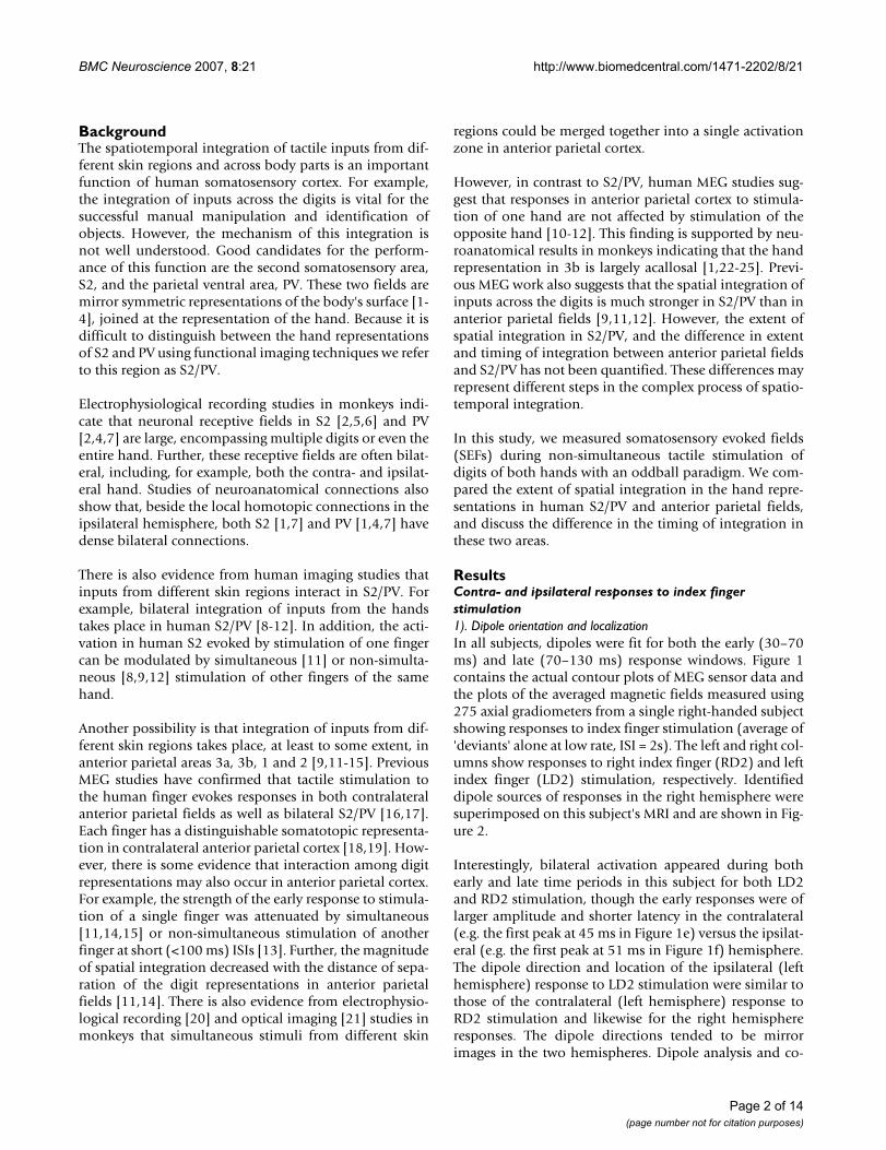
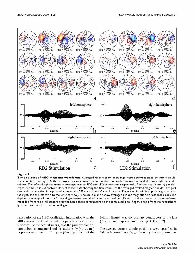
ResultsContra- and ipsilateral responses to index finger stimulation1). Dipole orientation and localizationIn all subjects, dipoles were fit for both the early (30–70ms) and late (70–130 ms) response windows. Figure 1contains the actual contour plots of MEG sensor data andthe plots of the averaged magnetic fields measured using275 axial gradiometers from a single right-handed subjectshowing responses to index finger stimulation (average of'deviants' alone at low rate, ISI = 2s). The left and right col-umns show responses to right index finger (RD2) and leftindex finger (LD2) stimulation, respectively. Identifieddipole sources of responses in the right hemisphere weresuperimposed on this subject's MRI and are shown in Fig-ure 2.
Interestingly, bilateral activation appeared during bothearly and late time periods in this subject for both LD2and RD2 stimulation, though the early responses were oflarger amplitude and shorter latency in the contralateral(e.g. the first peak at 45 ms in Figure 1e) versus the ipsilat-eral (e.g. the first peak at 51 ms in Figure 1f) hemisphere.The dipole direction and location of the ipsilateral (lefthemisphere) response to LD2 stimulation were similar tothose of the contralateral (left hemisphere) response toRD2 stimulation and likewise for the right hemisphereresponses. The dipole directions tended to be mirrorimages in the two hemispheres. Dipole analysis and co-
Page 2 of 14(page number not for citation purposes)
BMC Neuroscience 2007, 8:21 http://www.biomedcentral.com/1471-2202/8/21
registration of the MEG localization information with theMRI scans verified that the anterior parietal area (the pos-terior wall of the central sulcus) was the primary contrib-utor to both contralateral and ipsilateral early (30–70 ms)responses and that the S2 region (the upper bank of the
Sylvian fissure) was the primary contributor to the late(70–130 ms) responses in this subject (Figure 2).
The average current dipole positions were specified inTalairach coordinates (x, y, z in mm): the early contralat-
Time courses of MEG maps and waveformsFigure 1Time courses of MEG maps and waveforms. Averaged responses to index finger tactile stimulation at low rate (stimula-tion condition 1 in Figure 6; the strongest response was observed under this condition) were recorded from a right-handed subject. The left and right columns show responses to RD2 and LD2 stimulation, respectively. The two top (a and d) panels represent the series of contour plots of sensor data showing the time course of the averaged evoked magnetic fields. Each plot shows the sensor data interpolated between the 275 sensors at different latencies. The nasion is pointing up, the right ear is to the right, and the left ear is to the left (top view). Panels b, c, e and f show averaged evoked magnetic field responses; each line depicts an average of the data from a single sensor over all trials for one condition. Panels b and e show response waveforms recorded from half of all sensors over the hemisphere contralateral to the stimulated index finger, c and f from the hemisphere ipsilateral to the stimulated index finger.
Page 3 of 14(page number not for citation purposes)
BMC Neuroscience 2007, 8:21 http://www.biomedcentral.com/1471-2202/8/21
eral response dipoles in left and right hemispheres were (-49.2 ± 1.6, -19.2 ± 2.5, 45.8 ± 2.4) and (48.0 ± 1.4, -14.5± 1.4, 51.3 ± 3.7); the late contralateral response dipolesin left and right hemispheres were (-61.9 ± 3.7, -16.6 ±3.4, 6.9 ± 3.8) and (56.2 ± 2.4, -5.4 ± 4.7, 13.9 ± 4.8),respectively. These localizations in Talairach space wereconsistent with the locations of anterior parietal fields andS2/PV in both hemispheres.
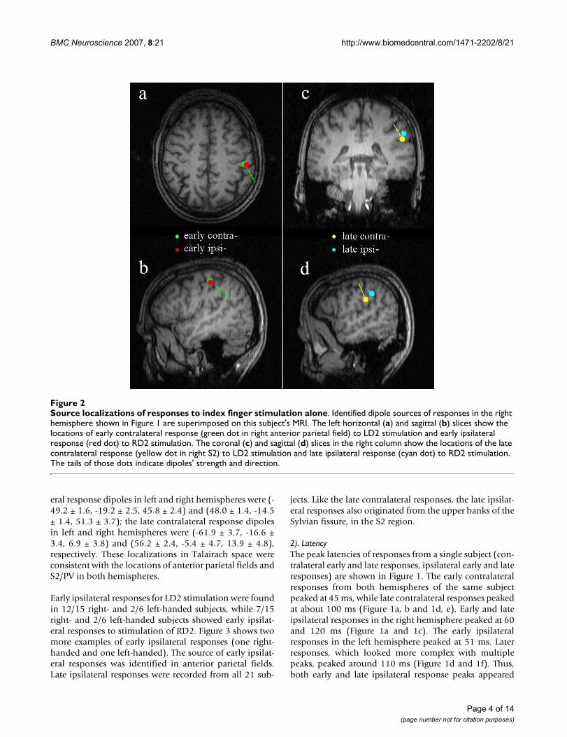
Early ipsilateral responses for LD2 stimulation were foundin 12/15 right- and 2/6 left-handed subjects, while 7/15right- and 2/6 left-handed subjects showed early ipsilat-eral responses to stimulation of RD2. Figure 3 shows twomore examples of early ipsilateral responses (one right-handed and one left-handed). The source of early ipsilat-eral responses was identified in anterior parietal fields.Late ipsilateral responses were recorded from all 21 sub-
jects. Like the late contralateral responses, the late ipsilat-eral responses also originated from the upper banks of theSylvian fissure, in the S2 region.
2). LatencyThe peak latencies of responses from a single subject (con-tralateral early and late responses, ipsilateral early and lateresponses) are shown in Figure 1. The early contralateralresponses from both hemispheres of the same subjectpeaked at 45 ms, while late contralateral responses peakedat about 100 ms (Figure 1a, b and 1d, e). Early and lateipsilateral responses in the right hemisphere peaked at 60and 120 ms (Figure 1a and 1c). The early ipsilateralresponses in the left hemisphere peaked at 51 ms. Laterresponses, which looked more complex with multiplepeaks, peaked around 110 ms (Figure 1d and 1f). Thus,both early and late ipsilateral response peaks appeared
Source localizations of responses to index finger stimulation aloneFigure 2Source localizations of responses to index finger stimulation alone. Identified dipole sources of responses in the right hemisphere shown in Figure 1 are superimposed on this subject's MRI. The left horizontal (a) and sagittal (b) slices show the locations of early contralateral response (green dot in right anterior parietal field) to LD2 stimulation and early ipsilateral response (red dot) to RD2 stimulation. The coronal (c) and sagittal (d) slices in the right column show the locations of the late contralateral response (yellow dot in right S2) to LD2 stimulation and late ipsilateral response (cyan dot) to RD2 stimulation. The tails of those dots indicate dipoles' strength and direction.
Page 4 of 14(page number not for citation purposes)
BMC Neuroscience 2007, 8:21 http://www.biomedcentral.com/1471-2202/8/21
later than contralateral peaks in both hemispheres in thissubject.
The average early and late contralateral response latenciesfor all subjects (right- and left-handed) were not signifi-cantly different across hemispheres (p > 0.05). The ipsilat-eral early (56.2 ± 6.6 ms) and late (116.6 ± 13.1 ms)responses appeared significantly later than contralateralearly (46.8 ± 6.9 ms) and late (108.8 ± 12.5 ms) responsesin all subjects. The mean delay of the early ipsilateralresponses relative to the contralateral responses was 10.7± 6.1 ms (range: 3.5–24 ms), while the delay of the lateipsilateral responses relative to the contralateral responseswas 12.7 ± 13.1 ms (range 3.0–30.8 ms). The mean differ-ence between delays for the early versus late responses wasnot significant (p > 0.05).
3). AmplitudeThe subject's data from Figure 1 clearly show the earlyipsilateral responses above the noise level (Figure 1c and
1f). The amplitudes of this subject's early ipsilateralresponses were 47.3 and 35.8 fT (RMS), while the noiselevels were 13.6 and 9.8 fT (RMS), in the left and righthemispheres, respectively. The average amplitude of theearly ipsilateral response (37.9 ± 2.0 fT) across all subjectswas significantly higher (p < 0.01) than the average noiselevel (14.5 ± 0.9 fT) based on the pre-stimulation periodof 50 ms.
The average amplitude of early and late responses for allsubjects did not show a significant difference betweenhemispheres (p > 0.05), nor was there a significant differ-ence in amplitude of response for right- versus left-hand-ers (p > 0.05). As the strength of early and late responsesdid not show a significant difference between hemi-spheres, the responses to left and right hand stimulationwere combined for further analysis.
The mean amplitude of the early response was signifi-cantly smaller (p < 0.01) for the early ipsilateral response
Two ipsilateral early responses examplesFigure 3Two ipsilateral early responses examples. The top two averaged sensor data plots are from a right-handed subject, the bottom two are from a left-handed subject. Figures in the left column show the two subjects' sensor plots at the early ipsilat-eral (left hemisphere) response peak moment under the LD2 stimulation condition. The peak ipsilateral (left hemisphere) response is still weaker than the contralateral (right hemisphere) response, though the right hemisphere response is not at its peak at this time. Figures in the right column show similar results for RD2 stimulation. Both LD2 and RD2 stimulation elicited bilateral early responses in these two subjects.
Page 5 of 14(page number not for citation purposes)
BMC Neuroscience 2007, 8:21 http://www.biomedcentral.com/1471-2202/8/21
(37.9 ± 2.0 fT) than for the early contralateral response(66.9 ± 3.3 fT). Similarly, the late ipsilateral response(50.4 ± 4.8 fT) was significantly weaker (p < 0.01) thanthe late contralateral response (69.3 ± 5.6 fT). Further, themean amplitude of the early contralateral response (66.9± 3.3 fT) was not significantly different from the late con-tralateral response (69.3 ± 5.6 fT; p > 0.05), while the lateipsilateral response (50.4 ± 4.8 fT) was significantlystronger than the early ipsilateral response (37.9 ± 2.0 fT;p < 0.01; see Figure 4).
Spatial integration between digit representations1). LatencyFigure 5 is an example of the spatial interaction from atypical right-handed subject. This figure shows the aver-aged magnetic field responses recorded from all 275 sen-sors under the different stimulus conditions. Each line isthe average over all trials for a given sensor. The left col-umn in Figure 5 shows contralateral (right hemisphere)sensor waveforms for LD2 stimulation, and the right col-umn shows the contralateral (left hemisphere) sensorwaveforms for RD2 stimulation. The latencies of bothearly and late responses did not change under differentstimulation conditions.
2). AmplitudeIn an example subject, low rate index finger stimulation(mean ISI: 2s) clearly elicited both early and laterresponses (Figure 5a and 5f). The response amplitude wasmarkedly decreased for high rate stimulation (ISI: 0.33 s),and the late response (S2/PV) almost disappeared in bothhemispheres (Figure 5b and 5g). The intervened standardstimuli (D3, D4 and opposite D2) suppressed the lateresponse for D2 (deviant) stimulation in both hemi-spheres. There appeared to be the greatest attenuation forthe same (D2) digit high rate stimulation compared toalternate standard digit (D3, D4 and opposite D2) stimu-lation, and greater attenuation for adjacent (D3) com-pared to non-adjacent (D4 and opposite D2) digitstimulation (Figure 5b–e and 5g–j). The standard stimulireduced the amplitude of the early response for D2 (devi-ant) stimulation in some conditions for this subject (Fig-ure 5c and 5j).
Grand averaged sensor amplitude (RMS; contralateralhemisphere only) and dipole moment (nAm) responsevalues to deviant stimuli are displayed in Figure 4. Bothsensor and moment values of early responses were statis-tically significantly smaller for high rate index finger stim-ulation (ISI: 0.33s) compared to low rate index fingerstimulation (mean ISI: 2s). Both contralateral and ipsilat-eral early amplitude of responses to single finger stimula-tion (D2) were not significantly affected by stimulation ofthe other fingers (D3, D4 and opposite D2).
As with the early responses, sensor and moment values ofthe late responses were statistically significantly smallerfor high rate index finger stimulation (ISI: 0.33s) com-pared to low rate index finger stimulation (mean ISI: 2s).The late response decreased more than the early responseregardless of handedness (p < 0.01; also see Figure 5).
Further, the intervened standard stimuli (D3, D4, andopposite D2) significantly reduced the late contralateraland ipsilateral responses to deviant (D2) stimuli. In thecontralateral hemisphere this effect was greater for theadjacent finger (D3) compared to the non-adjacent fin-gers (D4 or opposite D2). The attenuation of the late con-tralateral response was significantly greater for the samefinger high rate stimulation (D2 alone at high rate) com-pared to non-adjacent finger (D4 or opposite D2) stimu-lation. For the ipsilateral response, there were nodifferences in amplitude reduction related to the locationof intervened standard stimulation. Only opposite D2stimulation differed from high rate D2 stimulation in thelate ipsilateral response. The suppressive effect of oppositeD2 stimulation was significantly stronger for the contral-ateral than the ipsilateral late response (Figure 4).
DiscussionIn summary, we examined the spatiotemporal integrationacross digit representations within anterior parietal soma-tosensory areas and S2/PV, as well as interhemisphericintegration, using non-simultaneous tactile stimulation.We have shown 1) spatial integration of responses in S2/PV, with a decrease in interaction with the increase in sep-aration of digit representations; 2) greater temporal inte-gration of inputs within one digit representation in S2/PVversus anterior parietal fields; 3) both contra- and ipsilat-eral early responses in anterior parietal fields to cutaneousmechanical stimulation. The early ipsilateral response hasa longer latency and lower amplitude than the early con-tralateral response; and 4) processing of cutaneous inputsis symmetrical across hemispheres. What follows is a dis-cussion of these findings in light of previous work fromhuman and non-human primates.
Spatiotemporal integration between digit representations in S2/PVThe distance-dependence of spatial integration betweendigit representations in the S2/PV area was examined inthis study. We found that the amplitude of the contralat-eral late responses to deviant stimuli (D2) decreasedwhen standard stimuli (D3, D4 and opposite D2) wereintervened. Like the distance-dependence of spatial inte-gration in anterior parietal areas [11,14], the attenuationdecreased with the increasing distance of separationbetween receptive fields, toward greater attenuation foradjacent fingers (D3) compared to non-adjacent fingers(D4 and opposite D2).
Page 6 of 14(page number not for citation purposes)
BMC Neuroscience 2007, 8:21 http://www.biomedcentral.com/1471-2202/8/21
Page 7 of 14(page number not for citation purposes)
Spatial integrationFigure 4Spatial integration. Grand average of the peak sensor amplitude (a: RMS) and dipole moment (b: Q) values for early (ante-rior parietal area) and late (S2/PV) responses under different experimental conditions are compared. Asterisks indicate that the responses are significantly (*: p < 0.05; **: p < 0.01) different from others that are linked to it by the lines, using the Bonferroni post-hoc test. c, i, E and L stand for contralateral, ipsilateral, early and later response, respectively. S1 and S2 stand for anterior parietal areas and S2/PV respectively. D2 Low: D2 stimulation alone at low rate (condition 1 in Figure 6; mean ISI: 2s); D2 High: D2 stimulation alone at high rate (condition 2 in Figure 6; ISI: 0.33s); D2d (D3s): D2 deviant plus D3 standard stimuli (condition 3 in Figure 6; ISI: 0.33s); D2d (D4s): D2 deviant plus D4 standard stimuli (condition 4 in Figure 6; ISI: 0.33s); D2d (oD2s): D2 deviant plus opposite D2 standard stimuli (condition 5 in Figure 6; ISI: 0.33s).

BMC Neuroscience 2007, 8:21 http://www.biomedcentral.com/1471-2202/8/21
Page 8 of 14(page number not for citation purposes)
An example of spatial integration obtained from a right-handed subjectFigure 5An example of spatial integration obtained from a right-handed subject. The waveforms show the time courses of the averaged magnetic field responses recorded from 275 sensors under the different conditions. The left and right columns show contralateral sensor response waveforms for LD2 and RD2 stimulation, respectively. a and f: D2 stimulation alone at low rate (condition 1 in Figure 6; mean ISI: 2s); b and g: D2 stimulation alone at high rate (condition 2 in Figure 6; ISI: 0.33s); c and h: D2 deviant plus D3 standard stimulation (condition 3 in Figure 6; ISI: 0.33s); d and i: D2 deviant plus D4 standard stimulation (condition 4 in Figure 6; ISI: 0.33s); e and j: D2 deviant plus opposite D2 standard stimulation (condition 5 in Figure 6; ISI: 0.33s).
BMC Neuroscience 2007, 8:21 http://www.biomedcentral.com/1471-2202/8/21
In agreement with previous MEG work on digit responseinteraction in S2/PV [8,9,11,12,26], our data show thatthe standard stimuli significantly attenuate responses inbilateral S2/PV to deviant (D2) stimulation. The patternof spatial integration in S2/PV is consistent with the factthat neurons in S2 and PV have large receptive fields, fre-quently encompassing multiple digits [2,4-7,27], whilecells in anterior parietal areas do not [28,29].
We also found significant temporal integration in the S2/PV region. The amplitude of the late response was smallerfor high rate versus low rate index finger stimulation, andthe late responses decreased more than the early responseswith high rate stimulation, as previous SEP [30] and SEF[26,31-34] investigations have shown. This result is con-sistent with previous studies revealing the shorter recoverycycle or refractory period for neurons in anterior parietalareas than in S2 [35,36].
Some non-human primate studies have suggested serialinformation processing from anterior parietal areas to S2/PV [37,38], with tactile inputs from anterior parietal fieldsconverging onto S2 [27,39]. However, other studies sug-gest hierarchical equivalence of anterior parietal areas andS2 for tactile processing in cats and new-world primates[40-42]. To date, the debate about serial versus parallelorganization of somatosensory cortex in humans is unre-solved. Both early [43] and late [44,45] concurrent activi-ties in anterior parietal and lateral sulcal areas have beenreported in humans. In contrast, a recent MEG studyrevealed that the onset latency of response following tran-scutaneous electrical stimulation was longer in S2/PVthan in anterior parietal areas [46]. However, we cannotrule out the contribution of activity in S2/PV to earlyresponses and anterior parietal areas to late responses. Itis possible that both serial and parallel processing play arole in tactile perception.
Serial convergence may account for the spatial and tempo-ral specialties in S2/PV. Unlike anterior parietal areas withrestricted and defined neural receptive fields, the large,complex receptive fields within S2/PV may play an impor-tant role in integrating tactile inputs over space and time.For example, the digit representations in S2 and surround-ing fields lack fine somatotopy, and some cells respondedbetter to proprioceptive rather than cutaneous stimuli[27]. In addition, studies have found that 23% of cells inthe S2 region were orientation tuned, often across multi-ple digit pads [27]. These receptive field properties, tunedfor relatively long-range spatial and temporal integration,go some way towards explaining S2's purported involve-ment in functions like tactile object exploration and iden-tification [47-50], bimanual coordination [2,4,6,7], andtactile learning and memory [49,50]. Consistent with thisidea is evidence in humans that damage to parietotempo-
ral cortices (including S2) causes impairment of tactileobject recognition in the absence of basic somesthetic dys-function [51,52].
Spatiotemporal integration between digit representations in anterior parietal somatosensory areasWe found that the amplitude of responses in anterior pari-etal areas to deviant alone stimuli applied to a single digitwas significantly lower at high rate (the ISI is also 0.33s)versus low rate (ISI: 2s) stimulation, as previous studies[26,30-34] have shown. This decrease of amplitude result-ing from increased rate of stimulation was much smallerin anterior parietal areas than in S2/PV. However, theaverage amplitude of early responses in anterior parietalareas for deviant stimulation did not show a significantdecrease when the standard stimuli were interleavedbetween deviants (ISI: 0.33s), though the amplitudedecrease was seen in some cases. Previous work usingnon-simultaneous stimuli with long ISIs (1–4 s) alsoshowed similar results [26,53]. However, an interactionbetween digit representations has been reported in ante-rior parietal areas when using simultaneous [11,14,15] ornon-simultaneous stimuli with short (<100 ms) ISIs [13].These findings are in line with the short recovery cycletime constant (~110 ms) for neurons in anterior parietalareas, which was estimated by fitting an exponential curveto SEF intensities for responses to electrical stimulation ofthe median nerve with different ISIs (100–500 ms) [36].With a long ISI, the intervened standard inputs may arriveout of the recovery period of neurons in anterior parietalareas, with little effect on responses to deviant stimuli.
It is known that there is an inhibitory surround structureof receptive fields in monkey area 3b [54-58]. A three-component model has been proposed in which a laggedinhibitory surround overlaps the excitatory center and itsone or more fixed inhibitory flankers. The fixed (as a spa-tial filter) and lagged (as a temporal filter) surroundinhibitory components confer the spatial and temporalselectivity of neurons in anterior parietal areas [58]. Thespatiotemporal property of inhibitory surround structureof neural receptive fields may account for the restrictedneural receptive field and rapid recovery cycle in anteriorparietal areas observed in the present study.
Ipsilateral responses in anterior parietal areasIn the present investigation we clearly recorded early ipsi-lateral responses with high goodness-of-fit to single fingertactile stimulation (both right- and left-handers). Thoughthe amplitude of the ipsilateral response was very weak insome cases, on average it was 2.5 times greater than thenoise level. An ipsilateral response from anterior parietalfield has also been observed in a small number of subjectsin several previous studies [59-63] using MEG or EEG tomeasure responses to median nerve stimulations. A grow-
Page 9 of 14(page number not for citation purposes)
BMC Neuroscience 2007, 8:21 http://www.biomedcentral.com/1471-2202/8/21
ing body of evidence from monkey studies [64,65] andhuman fMRI studies [66,67] also report ipsilateralresponses in anterior parietal fields, suggesting that ante-rior parietal fields receive tactile input from the ipsilateralhand. Ipsilateral responses were relatively robust in ourstudy, likely due to the cutaneous mechanical stimulationused in our experiment. Recent fMRI studies on bothmonkeys and humans have shown that responses fromipsilateral anterior parietal areas are more robust formechanical versus median nerve electrical stimulation[64].
Single cell recordings from monkeys indicate that someneurons representing multiple digits in areas 2 and 5 havebilateral or ipsilateral receptive fields [68], and a few noci-ceptive neurons at the junction of areas 3b and 1 havelarge, bilateral receptive fields [69]. Thus the source of theearly ipsilateral response is likely not only area 3b, butmay also include input from cells in area 1, and possiblyareas 2 and 5 as well. Recent evidence from electrophysi-ological recording in monkeys suggests that responsesfrom ipsilateral anterior parietal fields are elicited by feed-back rather than feedforward afferents [64], which may berelated to the latency delay of the ipsilateral responseobserved in previous SEP [62] and the current MEG study.It has been suggested that transcallosal pathways maymediate responses from ipsilateral anterior parietal areasboth in humans [59-61,67] and in monkeys [64,65].However, anatomical evidence from macaque monkeysindicates that anterior parietal areas, in particular area 3b,have sparse or absent callosal connections for the handrepresentation [1,22,25]. Callosal connections becomedenser in the more caudal fields, from areas 3b to 1 and 2[24]. Thus, transcallosal pathways mediating the earlyipsilateral responses would be through these caudal areas,or S2/PV.
Hemispheric differencesIn addition, some previous functional imaging studiessuggest functional dominance, or an increased sourceamplitude, in the left versus the right hemisphere inhuman primary [70-72] and secondary [73-77] somato-sensory cortex. An expanded hand representation in leftanterior parietal fields has also been proposed [72,78].However, morphometric and cytoarchitectonic measure-ments show no lateralized differences in somatosensorycortices [79]. Psychophysical tests show no differences inspatial acuity between hands [80,81], although there doesappear to be a left-hemisphere advantage for tactileprocessing of simultaneity judgment [82]. Furthermore,as in our study, previous somatosensory evoked potentialstudies show no difference in topography or responseamplitude between the two hemispheres [83,84]. Thus, itis necessary to reassess the hemispheric asymmetry ofsomatosensory cortex.
The differences between studies may be due to the differ-ent types of stimuli used. The electrical stimulation usedin previous studies showing asymmetry is not receptorspecific, and elicits a more complex cortical activation. Arecent MEG study showed that movement might beinvolved in the lateral asymmetry of somatosensory cor-tex, and the intensity of electrical stimulation oftenexceeded the motor threshold in previous studies [70-75,77]. Passive finger movement was found to evoke lat-erally asymmetrical responses in somatosensory cortex[76]. Thus, the increased response in left somatosensorycortex evoked by electric stimulation may reflect the lat-eral asymmetry of movement rather than tactile informa-tion processing.
ConclusionThis human MEG study revealed that the bilateral spatio-temporal integration in S2/PV takes place over a large cor-tical area and over a long time period. Further, thestrength of integration in this region is distance-depend-ent. The wide overlap of digit receptive fields in S2/PVmight account for complex functions such as manualexploration and bimanual coordination, thought to becrucially dependent on S2/PV. In contrast, the propertiesof spatiotemporal integration in anterior parietal areaswere different from those in S2/PV with significant tem-poral integration while spatial integration was reduced,which is consistent with the critical role of anterior pari-etal fields in spatial discrimination. The early ipsilateralresponse observed in over half of subjects suggests thatanterior parietal fields do accept tactile inputs from theipsilateral side of body. In addition, no tactile responsedifference between hemispheres indicated functionalsymmetry in human somatosensory cortex.
MethodsSubjectsSEFs were recorded in fifteen right-handed and six left-handed healthy adult subjects (10 male and 5 femaleright-handers; 2 male and 4 female left-handers; age range22–40 years), using an Omega 2000 Whole-Cortex MEGSystem (CTF Systems Inc. Port Coquitlam, Canada; 275DC SQUID first-order axial gradiometers). All subjectssigned an informed consent as approved by the Commit-tee on Human Research of the University of California,San Francisco. The Edinburgh inventory [85] was used todetermine the direction and degree of handedness foreach subject. The subjects were seated comfortably andmaintained their head position during MEG testing in amagnetically shielded room. Subjects' hands were placedpalm up on opposite armrests. The subjects closed theireyes, wore earplugs and were asked not to pay attention tothe tactile stimulation.
Page 10 of 14(page number not for citation purposes)

BMC Neuroscience 2007, 8:21 http://www.biomedcentral.com/1471-2202/8/21
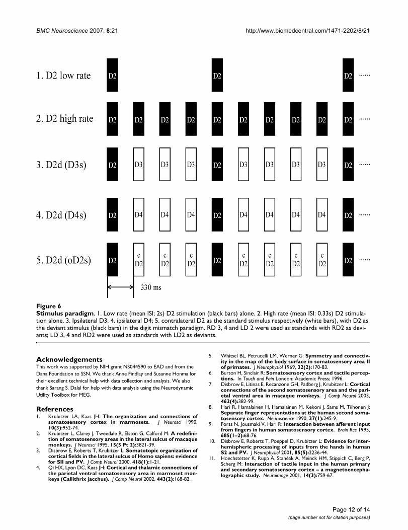
StimuliTactile stimuli were pneumatically driven pulses (~140ms duration) applied to the tips of the digits with balloondiaphragms. A digit oddball paradigm [9,16,30] was usedto examine the representations of the index fingers (D2,infrequent deviant stimulus) while varying the location ofthe frequent standard stimuli across adjacent (D3), non-adjacent (D4) and contralateral (D2) digits. In this odd-ball paradigm, infrequent deviant stimuli interspersedwith frequent standard stimuli were presented. The inten-sity of all stimuli was well above detection threshold at 17PSI (pounds per square inch). The location of the stand-ard was changed between blocks and the order (includingdeviant alone block) was randomized across subjects. Ineach block of 600–900 trials with an ISI of 0.33s and a jit-ter of 10 ms, standard and deviant stimuli were presentedat probabilities of 0.83 and 0.17 respectively. Deviantswere always followed by 3–7 standards to allow for anadaptation to the standard. To compare the tactileresponse to single finger stimulation in different hemi-spheres, deviants were also presented alone at two rates:one equal to that for deviants (mean ISI: 2 s) and the otherequal to that for the compound of deviants and standards(mean ISI: 0.33s) in the mismatch paradigm (Figure 6).
Data recording and analysisData were collected at a sample rate of 1200 Hz. The filter-ing passband for data analysis was 2–40 Hz. About 100artifact-free trials for deviant stimuli (D2) were averagedin each test block. Head position relative to the MEG sen-sors was determined before and after each test block bymeans of three small coils placed at landmark sites(nasion, left and right preauricular points).
MEG data was analyzed using an equivalent currentdipole (ECD) embedded in a spherical conductingmedium. The locations of index finger representations insomatosensory cortex were determined using a dipole fitwith contralateral index finger stimulation alone. SEFs insomatosensory cortex peaking in the time window up to200 ms following stimulus onset were analyzed. The early(30–70 ms) response was analyzed for activation in ante-rior parietal fields; and the late response (70–130 ms) wasanalyzed for activation of S2/PV [11,26]. Sensors record-ing from the hemisphere contralateral to the stimulatedindex finger were chosen to determine the ECD of themost dominant source. The position and orientation ofthe ECD corresponding to the early response were firstfound and fixed; then another dipole corresponding tothe late response was added with the early one fixed, thenwas fitted and fixed successively. Only sources with highgoodness of fit (> 85%) were accepted. Dipoles matchinganterior parietal fields and S2/PV in each hemispherewere identified and fixed based on contralateral index fin-ger stimulation, and they were used for analysis of data
from other stimulation conditions. The response laten-cies, amplitudes (root-mean-square value, RMS) of eachhemisphere and dipole moments (Q value) for all of fourdipole locations were estimated for the different stimula-tion conditions based on peaks within the early and latetime periods.
Eight of the subjects were also scanned using a 1.5T MRIscanner (GE Medical System, Milwaukee, WI) to acquire a3D structural image (flip angle = 40°, TR = 27 ms, TE = 6ms, FOV = 240 × 240 mm, 1.5 mm slice thickness, 256 ×256 × 124 pixels). Three fiducials were placed on the sub-ject at the same three locations as the localizing coils inMEG. This information was used to coregister the MEGdata to the MRI image. Each subject's structural MRI wasnormalized to MNI space using Neurodynamic UtilityToolbox for MEG [86]. Then the MNI coordinates ofdipole positions were converted to Talairach coordinates,using a non-linear transform [87].
Paired Student t-tests were used to assess significant differ-ence between condition pairs. In cases where the numberof conditions exceeds two, repeated measures ANOVAswere used for assessing statistical significance betweenresponses across different stimulation conditions. In thesecases, Bonferroni post-hoc tests were used to assess signif-icance between specific condition pairs, whereby after cor-rection for multiple comparisons a significance thresholdof either p < 0.01 or p < 0.05 was used. Data are presentedas mean values ± standard error of the mean throughout.
List of abbreviationsPV: parietal ventral area
MEG: magnetoencephalography
SEFs: somatosensory evoked fields
ECD: equivalent current dipole
RMS: root mean square
Q: dipole moment
LD2: left index finger
RD2: right index finger
Authors' contributionsZZ, EAD, DJM and SSN designed the study. ZZ and EADdrafted the manuscript. JMZ, DJM and SSN helped to draftthe manuscript. ZZ and JMZ recorded and analyzed MEGand MRI data. All authors read and approved the finalmanuscript.
Page 11 of 14(page number not for citation purposes)
BMC Neuroscience 2007, 8:21 http://www.biomedcentral.com/1471-2202/8/21
AcknowledgementsThis work was supported by NIH grant NS044590 to EAD and from the Dana Foundation to SSN. We thank Anne Findlay and Susanne Honma for their excellent technical help with data collection and analysis. We also thank Sarang S. Dalal for help with data analysis using the Neurodynamic Utility Toolbox for MEG.
References1. Krubitzer LA, Kaas JH: The organization and connections of
somatosensory cortex in marmosets. J Neurosci 1990,10(3):952-74.
2. Krubitzer L, Clarey J, Tweedale R, Elston G, Calford M: A redefini-tion of somatosensory areas in the lateral sulcus of macaquemonkeys. J Neurosci 1995, 15(5 Pt 2):3821-39.
3. Disbrow E, Roberts T, Krubitzer L: Somatotopic organization ofcortical fields in the lateral sulcus of Homo sapiens: evidencefor SII and PV. J Comp Neurol 2000, 418(1):1-21.
4. Qi HX, Lyon DC, Kaas JH: Cortical and thalamic connections ofthe parietal ventral somatosensory area in marmoset mon-keys (Callithrix jacchus). J Comp Neurol 2002, 443(2):168-82.
5. Whitsel BL, Petrucelli LM, Werner G: Symmetry and connectiv-ity in the map of the body surface in somatosensory area IIof primates. J Neurophysiol 1969, 32(2):170-83.
6. Burton H, Sinclair R: Somatosensory cortex and tactile percep-tions. In Touch and Pain London: Academic Press; 1996.
7. Disbrow E, Litinas E, Recanzone GH, Padberg J, Krubitzer L: Corticalconnections of the second somatosensory area and the pari-etal ventral area in macaque monkeys. J Comp Neurol 2003,462(4):382-99.
8. Hari R, Hamalainen H, Hamalainen M, Kekoni J, Sams M, Tiihonen J:Separate finger representations at the human second soma-tosensory cortex. Neuroscience 1990, 37(1):245-9.
9. Forss N, Jousmaki V, Hari R: Interaction between afferent inputfrom fingers in human somatosensory cortex. Brain Res 1995,685(1–2):68-76.
10. Disbrow E, Roberts T, Poeppel D, Krubitzer L: Evidence for inter-hemispheric processing of inputs from the hands in humanS2 and PV. J Neurophysiol 2001, 85(5):2236-44.
11. Hoechstetter K, Rupp A, Stanèák A, Meinck HM, Stippich C, Berg P,Scherg M: Interaction of tactile input in the human primaryand secondary somatosensory cortex – a magnetoencepha-lographic study. Neuroimage 2001, 14(3):759-67.
Stimulus paradigmFigure 6Stimulus paradigm. 1. Low rate (mean ISI; 2s) D2 stimulation (black bars) alone. 2. High rate (mean ISI: 0.33s) D2 stimula-tion alone. 3. Ipsilateral D3; 4. ipsilateral D4; 5. contralateral D2 as the standard stimulus respectively (white bars), with D2 as the deviant stimulus (black bars) in the digit mismatch paradigm. RD 3, 4 and LD 2 were used as standards with RD2 as devi-ants; LD 3, 4 and RD2 were used as standards with LD2 as deviants.
Page 12 of 14(page number not for citation purposes)
http://www.ncbi.nlm.nih.gov/entrez/query.fcgi?cmd=Retrieve&db=PubMed&dopt=Abstract&list_uids=2108231
http://www.ncbi.nlm.nih.gov/entrez/query.fcgi?cmd=Retrieve&db=PubMed&dopt=Abstract&list_uids=2108231
http://www.ncbi.nlm.nih.gov/entrez/query.fcgi?cmd=Retrieve&db=PubMed&dopt=Abstract&list_uids=7751949
http://www.ncbi.nlm.nih.gov/entrez/query.fcgi?cmd=Retrieve&db=PubMed&dopt=Abstract&list_uids=7751949
http://www.ncbi.nlm.nih.gov/entrez/query.fcgi?cmd=Retrieve&db=PubMed&dopt=Abstract&list_uids=7751949
http://www.ncbi.nlm.nih.gov/entrez/query.fcgi?cmd=Retrieve&db=PubMed&dopt=Abstract&list_uids=4975532
http://www.ncbi.nlm.nih.gov/entrez/query.fcgi?cmd=Retrieve&db=PubMed&dopt=Abstract&list_uids=4975532
http://www.ncbi.nlm.nih.gov/entrez/query.fcgi?cmd=Retrieve&db=PubMed&dopt=Abstract&list_uids=4975532
http://www.ncbi.nlm.nih.gov/entrez/query.fcgi?cmd=Retrieve&db=PubMed&dopt=Abstract&list_uids=2243596
http://www.ncbi.nlm.nih.gov/entrez/query.fcgi?cmd=Retrieve&db=PubMed&dopt=Abstract&list_uids=2243596
http://www.ncbi.nlm.nih.gov/entrez/query.fcgi?cmd=Retrieve&db=PubMed&dopt=Abstract&list_uids=2243596
http://www.ncbi.nlm.nih.gov/entrez/query.fcgi?cmd=Retrieve&db=PubMed&dopt=Abstract&list_uids=7583255
BMC Neuroscience 2007, 8:21 http://www.biomedcentral.com/1471-2202/8/21
12. Simões C, Mertens M, Forss N, Jousmäki V, Lütkenhöner B, Hari R:Functional overlap of finger representations in human SI andSII cortices. J Neurophysiol 2001, 86(4):1661-5.
13. Huttunen J, Ahlfors S, Hari R: Interaction of afferent impulses inthe human primary sensorimotor cortex. ElectroencephalogrClin Neurophysiol 1992, 82(3):176-81.
14. Biermann K, Schmitz F, Witte OW, Konczak J, Freund HJ, SchnitzlerA: Interaction of finger representation in the human firstsomatosensory cortex: a neuromagnetic study. Neurosci Lett1998, 251(1):13-6.
15. Tanosaki M, Suzuki A, Takino R, Kimura T, Iguchi Y, Kurobo Y,Haruta Y, Hoshi Y, Hashimoto I: Neural mechanisms for gener-ation of tactile interference effects on somatosensoryevoked magnetic fields in humans. Clin Neurophysiol 2002,113(5):672-80.
16. Hari R, Forss N: Magnetoencephalography in the study ofhuman somatosensorycortical processing. Philos Trans R SocLond B Biol Sci 1999, 354(1387):1145-54.
17. Kakigi R, Hoshiyama M, Shimojo M, Naka D, Yamasaki H, WatanabeS, Xiang J, Maeda K, Lam K, Itomi K, Nakamura A: The somatosen-sory evoked magnetic fields. Prog Neurobiol 2000, 61(5):495-523.
18. Nakamura A, Yamada T, Goto A, Kato T, Ito K, Abe Y, Kachi T, KakigiR: Somatosensory homunculus as drawn by MEG. Neuroimage1998, 7(4 Pt 1):377-86.
19. Maeda K, Kakigi R, Hoshiyama M, Koyama S: Topography of thesecondary somatosensory cortex in humans: a magnetoen-cephalo-graphic study. Neuroreport 1999, 10(2):301-6.
20. Gardner EP, Costanzo RM: Spatial integration of multiple-pointstimuli in primary somatosensory cortical receptive fields ofalert monkeys. J Neurophysiol 1980, 43(2):420-43.
21. Chen LM, Friedman RM, Roe AW: Optical imaging of a tactileillusion in area 3b of the primary somatosensory cortex. Sci-ence 2003, 302(5646):881-5.
22. Jones EG, Powell TP: Connexions of the somatic sensory cortexof the rhesus monkey. II. Contralateral cortical connexions.Brain 1969, 92(4):717-30.
23. Karol EA, Pandya DN: The distribution of the corpus callosumin the rhesus monkey. Brain 1971, 94(3):471-786.
24. Killackey HP, Gould HJ 3rd, Cusick CG, Pons TP, Kaas JH: The rela-tion of corpus callosum connections to architectonic fieldsand body surface maps in sensorimotor cortex of new andold world monkeys. J Comp Neurol 1983, 219(4):384-419.
25. Padberg J, Disbrow E, Krubitzer L: The organization and connec-tions of anterior and posterior parietal cortex in titi mon-keys: do New World monkeys have an area 2? Cereb Cortex2005, 15(12):1938-63.
26. Mertens M, Lütkenhöner B: Efficient neuromagnetic determina-tion of landmarks in the somatosensory cortex. Clin Neuro-physiol 2000, 111(8):1478-87.
27. Fitzgerald PJ, Lane JW, Thakur PH, Hsiao SS: Receptive field prop-erties of the macaque second somatosensory cortex: evi-dence for multiple functional representations. J Neurosci 2004,24(49):11193-204.
28. Nelson RJ, Sur M, Felleman DJ, Kaas JH: Representations of thebody surface in postcentral parietal cortex of Macaca fascic-ularis. J Comp Neurol 1980, 192(4):611-43.
29. Sripati AP, Yoshioka T, Denchev P, Hsiao SS, Johnson KO: Spatio-temporal receptive fields of peripheral afferents and corticalarea 3b and 1 neurons in the primate somatosensory system.J Neurosci 2006, 26(7):2101-14.
30. Kekoni J, Hämäläinen H, Saarinen M, Gröhn J, Reinikainen K, Lehtoko-ski A, Näätänen R: Rate effect and mismatch responses in thesomatosensory system: ERP-recordings in humans. Biol Psy-chol 1997, 46(2):125-42.
31. Tiihonen J, Hari R, Hamalainen M: Early deflections of cerebralmagnetic responses to median nerve stimulation. Electroen-cephalogr Clin Neurophysiol 1989, 74(4):290-6.
32. Wikstrom H, Huttunen J, Korvenoja A, Virtanen J, Salonen O, AronenH, Ilmoniemi RJ: Effects of interstimulus interval on somato-sensory evoked magnetic fields (SEFs): a hypothesis con-cerning SEF generation at the primary sensorimotor cortex.Electroencephalogr Clin Neurophysiol 1996, 100(6):479-87.
33. Mauguiere F, Merlet I, Forss N, Vanni S, Jousmaki V, Adeleine P, HariR: Activation of a distributed somatosensory cortical net-work in the human brain: a dipole modelling study of mag-netic fields evoked by median nerve stimulation. Part II:
Effects of stimulus rate, attention and stimulus detection.Electroencephalogr Clin Neurophysiol 1997, 104(4):290-5.
34. Nagamine T, Makela J, Mima T, Mikuni N, Nishitani N, Satoh T, IkedaA, Shibasaki H: Serial processing of the somesthetic informa-tion revealed by different effects of stimulus rate on the som-atosensory-evoked potentials and magnetic fields. Brain Res1998, 791(1–2):200-8.
35. Schnitzler A, Volkmann J, Enck P, Frieling T, Witte OW, Freund HJ:Different cortical organization of visceral and somatic sensa-tion in humans. Eur J Neurosci 1999, 11(1):305-15.
36. Hamada Y, Otsuka S, Okamoto T, Suzuki R: The profile of therecovery cycle in human primary and secondary somatosen-sory cortex: a magnetoencephalography study. Clin Neurophys-iol 2002, 113(11):1787-93.
37. Pons TP, Garraghty PE, Friedman DP, Mishkin M: Physiological evi-dence for serial processing in somatosensory cortex. Science1987, 237(4813):417-20.
38. Pons TP, Garraghty PE, Mishkin M: Serial and parallel processingof tactual information in somatosensory cortex of rhesusmonkeys. J Neurophysiol 1992, 68(2):518-27.
39. Friedman DP, Murray EA, O'Neill JB, Mishkin M: Cortical connec-tions of the somatosensory fields of the lateral sulcus ofmacaques: evidence for a corticolimbic pathway for touch. JComp Neurol 1986, 252(3):323-47.
40. Zhang HQ, Murray GM, Turman AB, Mackie PD, Coleman GT, RoweMJ: Parallel processing in cerebral cortex of the marmosetmonkey: effect of reversible SI inactivation on tactileresponses in SII. J Neurophysiol 1996, 76(6):3633-55.
41. Zhang HQ, Murray GM, Coleman GT, Turman AB, Zhang SP, RoweMJ: Functional characteristics of the parallel SI- and SII-pro-jecting neurons of the thalamic ventral posterior nucleus inthe marmoset. J Neurophysiol 2001, 85(5):1805-22.
42. Zhang HQ, Zachariah MK, Coleman GT, Rowe MJ: Hierarchicalequivalence of somatosensory areas I and II for tactileprocessing in the cerebral cortex of the marmoset monkey.J Neurophysiol 2001, 85(5):1823-35.
43. Karhu J, Tesche CD: Simultaneous early processing of sensoryinput in human primary (SI) and secondary (SII) somatosen-sory cortices. J Neurophysiol 1999, 81(5):2017-25.
44. Allison T, McCarthy G, Wood CC, Williamson PD, Spencer DD:Human cortical potentials evoked by stimulation of themedian nerve. II. Cytoarchitectonic areas generating long-latency activity. J Neurophysiol 1989, 62(3):711-22.
45. Stancak A, Hoechstetter K, Tintera J, Vrana J, Rachmanova R, KralikJ, Scherg M: Source activity in the human secondary somato-sensory cortex depends on the size of corpus callosum. BrainRes 2002, 936(1–2):47-57.
46. Inui K, Wang X, Tamura Y, Kaneoke Y, Kakigi R: Serial processingin the human somatosensory system. Cereb Cortex 2004,14(8):851-7.
47. Garcha HS, Ettlinger G: The effects of unilateral or bilateralremovals of the second somatosensory cortex (area SII): aprofound tactile disorder in monkeys. Cortex 1978,14(3):319-26.
48. Garcha HS, Ettlinger G: Tactile discrimination learning in themonkey: the effects of unilateral or bilateral removals of thesecond somatosensory cortex (area SII). Cortex 1980,16(3):397-412.
49. Mishkin M: Analogous neural models for tactual and visuallearning. Neuropsychologia 1979, 17(2):139-51.
50. Murray EA, Mishkin M: Relative contributions of SII and area 5to tactile discrimination in monkeys. Behav Brain Res 1984,11(1):67-83.
51. Caselli RJ: Rediscovering tactile agnosia. Mayo Clin Proc 1991,66(2):129-42.
52. Caselli RJ: Ventrolateral and dorsomedial somatosensoryassociation cortex damage produces distinct somestheticsyndromes in humans. Neurology 1993, 43(4):762-71.
53. Huttunen J, Hari R, Leinonen L: Cerebral magnetic responses tostimulation of ulnar and median nerves. Electroencephalogr ClinNeurophysiol 1987, 66(4):391-400.
54. Mountcastle VB, Powell TP: Neural mechanisms subservingcutaneous sensibility, with special reference to the role ofafferent inhibition in sensory perception and discrimination.Bull Johns Hopkins Hosp 1959, 105:201-32.
Page 13 of 14(page number not for citation purposes)
http://www.ncbi.nlm.nih.gov/entrez/query.fcgi?cmd=Retrieve&db=PubMed&dopt=Abstract&list_uids=1371437
http://www.ncbi.nlm.nih.gov/entrez/query.fcgi?cmd=Retrieve&db=PubMed&dopt=Abstract&list_uids=1371437
http://www.ncbi.nlm.nih.gov/entrez/query.fcgi?cmd=Retrieve&db=PubMed&dopt=Abstract&list_uids=9714453
http://www.ncbi.nlm.nih.gov/entrez/query.fcgi?cmd=Retrieve&db=PubMed&dopt=Abstract&list_uids=9714453
http://www.ncbi.nlm.nih.gov/entrez/query.fcgi?cmd=Retrieve&db=PubMed&dopt=Abstract&list_uids=9626677
http://www.ncbi.nlm.nih.gov/entrez/query.fcgi?cmd=Retrieve&db=PubMed&dopt=Abstract&list_uids=6770053
http://www.ncbi.nlm.nih.gov/entrez/query.fcgi?cmd=Retrieve&db=PubMed&dopt=Abstract&list_uids=6770053
http://www.ncbi.nlm.nih.gov/entrez/query.fcgi?cmd=Retrieve&db=PubMed&dopt=Abstract&list_uids=6770053
http://www.ncbi.nlm.nih.gov/entrez/query.fcgi?cmd=Retrieve&db=PubMed&dopt=Abstract&list_uids=4983244
http://www.ncbi.nlm.nih.gov/entrez/query.fcgi?cmd=Retrieve&db=PubMed&dopt=Abstract&list_uids=4983244
http://www.ncbi.nlm.nih.gov/entrez/query.fcgi?cmd=Retrieve&db=PubMed&dopt=Abstract&list_uids=4106844
http://www.ncbi.nlm.nih.gov/entrez/query.fcgi?cmd=Retrieve&db=PubMed&dopt=Abstract&list_uids=4106844
http://www.ncbi.nlm.nih.gov/entrez/query.fcgi?cmd=Retrieve&db=PubMed&dopt=Abstract&list_uids=6643713
http://www.ncbi.nlm.nih.gov/entrez/query.fcgi?cmd=Retrieve&db=PubMed&dopt=Abstract&list_uids=6643713
http://www.ncbi.nlm.nih.gov/entrez/query.fcgi?cmd=Retrieve&db=PubMed&dopt=Abstract&list_uids=6643713
http://www.ncbi.nlm.nih.gov/entrez/query.fcgi?cmd=Retrieve&db=PubMed&dopt=Abstract&list_uids=7419747
http://www.ncbi.nlm.nih.gov/entrez/query.fcgi?cmd=Retrieve&db=PubMed&dopt=Abstract&list_uids=7419747
http://www.ncbi.nlm.nih.gov/entrez/query.fcgi?cmd=Retrieve&db=PubMed&dopt=Abstract&list_uids=7419747
http://www.ncbi.nlm.nih.gov/entrez/query.fcgi?cmd=Retrieve&db=PubMed&dopt=Abstract&list_uids=9288410
http://www.ncbi.nlm.nih.gov/entrez/query.fcgi?cmd=Retrieve&db=PubMed&dopt=Abstract&list_uids=9288410
http://www.ncbi.nlm.nih.gov/entrez/query.fcgi?cmd=Retrieve&db=PubMed&dopt=Abstract&list_uids=2471630
http://www.ncbi.nlm.nih.gov/entrez/query.fcgi?cmd=Retrieve&db=PubMed&dopt=Abstract&list_uids=2471630
http://www.ncbi.nlm.nih.gov/entrez/query.fcgi?cmd=Retrieve&db=PubMed&dopt=Abstract&list_uids=8980411
http://www.ncbi.nlm.nih.gov/entrez/query.fcgi?cmd=Retrieve&db=PubMed&dopt=Abstract&list_uids=8980411
http://www.ncbi.nlm.nih.gov/entrez/query.fcgi?cmd=Retrieve&db=PubMed&dopt=Abstract&list_uids=9246066
http://www.ncbi.nlm.nih.gov/entrez/query.fcgi?cmd=Retrieve&db=PubMed&dopt=Abstract&list_uids=9246066
http://www.ncbi.nlm.nih.gov/entrez/query.fcgi?cmd=Retrieve&db=PubMed&dopt=Abstract&list_uids=9246066
http://www.ncbi.nlm.nih.gov/entrez/query.fcgi?cmd=Retrieve&db=PubMed&dopt=Abstract&list_uids=9593893
http://www.ncbi.nlm.nih.gov/entrez/query.fcgi?cmd=Retrieve&db=PubMed&dopt=Abstract&list_uids=9593893
http://www.ncbi.nlm.nih.gov/entrez/query.fcgi?cmd=Retrieve&db=PubMed&dopt=Abstract&list_uids=9593893
http://www.ncbi.nlm.nih.gov/entrez/query.fcgi?cmd=Retrieve&db=PubMed&dopt=Abstract&list_uids=9987033
http://www.ncbi.nlm.nih.gov/entrez/query.fcgi?cmd=Retrieve&db=PubMed&dopt=Abstract&list_uids=9987033
http://www.ncbi.nlm.nih.gov/entrez/query.fcgi?cmd=Retrieve&db=PubMed&dopt=Abstract&list_uids=9987033
http://www.ncbi.nlm.nih.gov/entrez/query.fcgi?cmd=Retrieve&db=PubMed&dopt=Abstract&list_uids=3603028
http://www.ncbi.nlm.nih.gov/entrez/query.fcgi?cmd=Retrieve&db=PubMed&dopt=Abstract&list_uids=3603028
http://www.ncbi.nlm.nih.gov/entrez/query.fcgi?cmd=Retrieve&db=PubMed&dopt=Abstract&list_uids=1527572
http://www.ncbi.nlm.nih.gov/entrez/query.fcgi?cmd=Retrieve&db=PubMed&dopt=Abstract&list_uids=1527572
http://www.ncbi.nlm.nih.gov/entrez/query.fcgi?cmd=Retrieve&db=PubMed&dopt=Abstract&list_uids=1527572
http://www.ncbi.nlm.nih.gov/entrez/query.fcgi?cmd=Retrieve&db=PubMed&dopt=Abstract&list_uids=3793980
http://www.ncbi.nlm.nih.gov/entrez/query.fcgi?cmd=Retrieve&db=PubMed&dopt=Abstract&list_uids=3793980
http://www.ncbi.nlm.nih.gov/entrez/query.fcgi?cmd=Retrieve&db=PubMed&dopt=Abstract&list_uids=3793980
http://www.ncbi.nlm.nih.gov/entrez/query.fcgi?cmd=Retrieve&db=PubMed&dopt=Abstract&list_uids=8985863
http://www.ncbi.nlm.nih.gov/entrez/query.fcgi?cmd=Retrieve&db=PubMed&dopt=Abstract&list_uids=8985863
http://www.ncbi.nlm.nih.gov/entrez/query.fcgi?cmd=Retrieve&db=PubMed&dopt=Abstract&list_uids=8985863
http://www.ncbi.nlm.nih.gov/entrez/query.fcgi?cmd=Retrieve&db=PubMed&dopt=Abstract&list_uids=2769355
http://www.ncbi.nlm.nih.gov/entrez/query.fcgi?cmd=Retrieve&db=PubMed&dopt=Abstract&list_uids=2769355
http://www.ncbi.nlm.nih.gov/entrez/query.fcgi?cmd=Retrieve&db=PubMed&dopt=Abstract&list_uids=2769355
http://www.ncbi.nlm.nih.gov/entrez/query.fcgi?cmd=Retrieve&db=PubMed&dopt=Abstract&list_uids=7194170
http://www.ncbi.nlm.nih.gov/entrez/query.fcgi?cmd=Retrieve&db=PubMed&dopt=Abstract&list_uids=7194170
http://www.ncbi.nlm.nih.gov/entrez/query.fcgi?cmd=Retrieve&db=PubMed&dopt=Abstract&list_uids=7194170
http://www.ncbi.nlm.nih.gov/entrez/query.fcgi?cmd=Retrieve&db=PubMed&dopt=Abstract&list_uids=6696789
http://www.ncbi.nlm.nih.gov/entrez/query.fcgi?cmd=Retrieve&db=PubMed&dopt=Abstract&list_uids=6696789
http://www.ncbi.nlm.nih.gov/entrez/query.fcgi?cmd=Retrieve&db=PubMed&dopt=Abstract&list_uids=1994134
http://www.ncbi.nlm.nih.gov/entrez/query.fcgi?cmd=Retrieve&db=PubMed&dopt=Abstract&list_uids=8469337
http://www.ncbi.nlm.nih.gov/entrez/query.fcgi?cmd=Retrieve&db=PubMed&dopt=Abstract&list_uids=8469337
http://www.ncbi.nlm.nih.gov/entrez/query.fcgi?cmd=Retrieve&db=PubMed&dopt=Abstract&list_uids=8469337
http://www.ncbi.nlm.nih.gov/entrez/query.fcgi?cmd=Retrieve&db=PubMed&dopt=Abstract&list_uids=2435519
BMC Neuroscience 2007, 8:21 http://www.biomedcentral.com/1471-2202/8/21
Publish with BioMed Central and every scientist can read your work free of charge
"BioMed Central will be the most significant development for disseminating the results of biomedical research in our lifetime."
Sir Paul Nurse, Cancer Research UK
Your research papers will be:
available free of charge to the entire biomedical community
peer reviewed and published immediately upon acceptance
cited in PubMed and archived on PubMed Central
yours — you keep the copyright
Submit your manuscript here:http://www.biomedcentral.com/info/publishing_adv.asp
BioMedcentral
55. Iwamura Y, Tanaka M, Sakamoto M, Hikosaka O: Functional subdi-visions representing different finger regions in area 3 of thefirst somatosensory cortex of the conscious monkey. ExpBrain Res 1983, 51:315-326.
56. DiCarlo JJ, Johnson KO, Hsiao SS: Structure of receptive fields inarea 3b of primary somatosensory cortex in the alert mon-key. J Neurosci 1998, 18(7):2626-45.
57. DiCarlo JJ, Johnson KO: Velocity invariance of receptive fieldstructure in somatosensory cortical area 3b of the alertmonkey. J Neurosci 1999, 19(1):401-19.
58. DiCarlo JJ, Johnson KO: Spatial and temporal structure ofreceptive fields in primate somatosensory area 3b: effects ofstimulus scanning direction and orientation. J Neurosci 2000,20(1):495-510.
59. Schnitzler A, Salmelin R, Salenius S, Jousmaki V, Hari R: Tactileinformation from the human hand reaches the ipsilateralprimary somatosensory cortex. Neurosci Lett 1995, 200(1):25-8.
60. Korvenoja A, Wikstrom H, Huttunen J, Virtanan J, Laine P, Aronen HJ,Seppalainen AM, Ilmoniemi RJ: Activation of ipsilateral primarysensorimotor cortex by median nerve stimulation. Neurore-port 1995, 6(18):2589-93.
61. Korvenoja A, Huttunen J, Salli E, Pohjonen H, Martinkauppi S, PalvaJM, Lauronen L, Virtanen J, Ilmoniemi RJ, Aronen HJ: Activation ofmultiple cortical areas in response to somatosensory stimu-lation: combined magnetoencephalographic and functionalmagnetic resonance imaging. Hum Brain Mapp 1999, 8(1):13-27.
62. Noachtar S, Lüders HO, Dinner DS, Klem G: Ipsilateral mediansomatosensory evoked potentials recorded from humansomatosensory cortex. Electroencephalogr Clin Neurophysiol 1997,104(3):189-98.
63. Kanno A, Nakasato N, Hatanaka K, Yoshimoto T: Ipsilateral area3b responses to median nerve somatosensory stimulation.Neuroimage 2003, 18(1):169-77.
64. Lipton ML, Fu K, Branch CA, Schroeder CE: Ipsilateral hand inputto area 3b revealed by converging hemodynamic and elec-trophysiological analyses in macaque monkeys. J Neurosci2006, 26(1):180-185.
65. Tommerdahl M, Simons SB, Chiu JS, Favorov O, Whitsel BL: Ipsilat-eral input modifies the primary somatosensory cortexresponse to contralateral skin flutter. J Neurosci 2006,26(22):5970-7.
66. Nihashi T, Naganawa S, Sato C, Kawai H, Nakamura T, Fukatsu H, Ish-igaki T, Aoki I: Contralateral and ipsilateral responses in pri-mary somatosensory cortex following electrical mediannerve stimulation – an fMRI study. Clin Neurophysiol 2005,116(4):842-8.
67. Hlushchuk Y, Hari R: Transient suppression of ipsilateral pri-mary somatosensory cortex during tactile finger stimula-tion. J Neurosci 2006, 26(21):5819-24.
68. Iwamura Y, Iriki A, Tanaka M: Bilateral hand representation inthe postcentral somatosensory cortex. Nature 1994,369(6481):554-6.
69. Kenshalo DR Jr, Isensee O: Responses of primate SI corticalneurons to noxious stimuli. J Neurophysiol 1983, 50(6):1479-96.
70. Rossini PM, Narici L, Martino G, Pasquarelli A, Peresson M, Pizzella V,Tecchio F, Romani GL: Analysis of interhemispheric asym-metries of somatosensory evoked magnetic fields to rightand left median nerve stimulation. Electroencephalogr Clin Neuro-physiol 1994, 91(6):476-82.
71. Buchner H, Ludwig I, Waberski T, Wilmes K, Ferbert A: Hemi-spheric asymmetries of early cortical somatosensory evokedpotentials revealed by topographic analysis. Electromyogr ClinNeurophysiol 1995, 35(4):207-15.
72. Jung P, Baumgärtner U, Bauermann T, Magerl W, Gawehn J, StoeterP, Treede RD: Asymmetry in the human primary somatosen-sory cortex and handedness. Neuroimage 2003, 19(3):913-23.
73. Forss N, Hari R, Salmelin R, Ahonen A, Hämäläinen M, Kajola M, Knu-utila J, Simola J: Activation of the human posterior parietal cor-tex by median nerve stimulation. Exp Brain Res 1994,99(2):309-15.
74. Kany C, Treede RD: Median and tibial nerve somatosensoryevoked potentials: middle-latency components from thevicinity of the secondary somatosensory cortex in humans.Electroencephalogr Clin Neurophysiol 1997, 104(5):402-10.
75. Simões C, Alary F, Forss N, Hari R: Left-hemisphere-dominantSII activation after bilateral median nerve stimulation. Neu-roimage 2002, 15(3):686-90.
76. Alary F, Simões C, Jousmäki V, Forss N, Hari R: Cortical activationassociated with passive movements of the human index fin-ger: an MEG study. Neuroimage 2002, 15(3):691-6.
77. Mäkelä JP, Illman M, Jousmäki V, Numminen J, Lehecka M, Salenius S,Forss N, Hari R: Dorsal penile nerve stimulation elicits left-hemisphere dominant activation in the second somatosen-sory cortex. Hum Brain Mapp 2003, 18(2):90-9.
78. Sörös P, Knecht S, Imai T, Gürtler S, Lütkenhöner B, Ringelstein EB,Henningsen H: Cortical asymmetries of the human somato-sensory hand representation in right- and left-handers. Neu-rosci Lett 1999, 271(2):89-92.
79. White LE, Andrews TJ, Hulette C, Richards A, Groelle M, PaydarfarJ, Purves D: Structure of the human sensorimotor system. II:Lateral symmetry. Cereb Cortex 1997, 7(1):31-47.
80. Sathian K, Zangaladze A: Tactile spatial acuity at the human fin-gertip and lip: bilateral symmetry and interdigit variability.Neurology 1996, 46(5):1464-6.
81. Vega-Bermudez F, Johnson KO: Differences in spatial acuitybetween digits. Neurology 2001, 56(10):1389-91.
82. Nicholls ME, Lindell AK: A left hemisphere, but not right hem-ispace, advantage for tactual simultaneity judgments. PerceptPsychophys 2000, 62(4):717-25.
83. Kakigi R, Shibasaki H: Effects of age, gender, and stimulus sideon scalp topography of somatosensory evoked potentials fol-lowing median nerve stimulatiom. J Clin Neurophysiol 1991,8(3):320-330.
84. Kakigi R, Shibasaki H: Effects of age, gender, and stimulus sideon scalp topography of somatosensory evoked potentials fol-lowing posterior tibial nerve stimulatiom. J Clin Neurophysiol1992, 9(3):431-440.
85. Oldfield RC: The assessment and analysis of handedness: theEdinburgh inventory. Neuropsychologia 1971, 9(1):97-113.
86. Dalal SS, Zumer JM, Agrawal V, Hild KE, Sekihara K, Nagarajan SS:NUTMEG: a neuromagnetic source reconstruction toolbox.Neurol Clin Neurophysiol 2004, 2004:52.
87. Brett M: The MNI brain and the Talairach atlas. [http://imaging.mrc-cbu.cam.ac.uk/imaging/MniTalairach].
Page 14 of 14(page number not for citation purposes)
http://www.ncbi.nlm.nih.gov/entrez/query.fcgi?cmd=Retrieve&db=PubMed&dopt=Abstract&list_uids=9502821
http://www.ncbi.nlm.nih.gov/entrez/query.fcgi?cmd=Retrieve&db=PubMed&dopt=Abstract&list_uids=9502821
http://www.ncbi.nlm.nih.gov/entrez/query.fcgi?cmd=Retrieve&db=PubMed&dopt=Abstract&list_uids=9502821
http://www.ncbi.nlm.nih.gov/entrez/query.fcgi?cmd=Retrieve&db=PubMed&dopt=Abstract&list_uids=9870969
http://www.ncbi.nlm.nih.gov/entrez/query.fcgi?cmd=Retrieve&db=PubMed&dopt=Abstract&list_uids=9870969
http://www.ncbi.nlm.nih.gov/entrez/query.fcgi?cmd=Retrieve&db=PubMed&dopt=Abstract&list_uids=9870969
http://www.ncbi.nlm.nih.gov/entrez/query.fcgi?cmd=Retrieve&db=PubMed&dopt=Abstract&list_uids=8584258
http://www.ncbi.nlm.nih.gov/entrez/query.fcgi?cmd=Retrieve&db=PubMed&dopt=Abstract&list_uids=8584258
http://www.ncbi.nlm.nih.gov/entrez/query.fcgi?cmd=Retrieve&db=PubMed&dopt=Abstract&list_uids=8584258
http://www.ncbi.nlm.nih.gov/entrez/query.fcgi?cmd=Retrieve&db=PubMed&dopt=Abstract&list_uids=8741769
http://www.ncbi.nlm.nih.gov/entrez/query.fcgi?cmd=Retrieve&db=PubMed&dopt=Abstract&list_uids=8741769
http://www.ncbi.nlm.nih.gov/entrez/query.fcgi?cmd=Retrieve&db=PubMed&dopt=Abstract&list_uids=9186233
http://www.ncbi.nlm.nih.gov/entrez/query.fcgi?cmd=Retrieve&db=PubMed&dopt=Abstract&list_uids=9186233
http://www.ncbi.nlm.nih.gov/entrez/query.fcgi?cmd=Retrieve&db=PubMed&dopt=Abstract&list_uids=9186233
http://www.ncbi.nlm.nih.gov/entrez/query.fcgi?cmd=Retrieve&db=PubMed&dopt=Abstract&list_uids=8202155
http://www.ncbi.nlm.nih.gov/entrez/query.fcgi?cmd=Retrieve&db=PubMed&dopt=Abstract&list_uids=8202155
http://www.ncbi.nlm.nih.gov/entrez/query.fcgi?cmd=Retrieve&db=PubMed&dopt=Abstract&list_uids=6663338
http://www.ncbi.nlm.nih.gov/entrez/query.fcgi?cmd=Retrieve&db=PubMed&dopt=Abstract&list_uids=6663338
http://www.ncbi.nlm.nih.gov/entrez/query.fcgi?cmd=Retrieve&db=PubMed&dopt=Abstract&list_uids=7529686
http://www.ncbi.nlm.nih.gov/entrez/query.fcgi?cmd=Retrieve&db=PubMed&dopt=Abstract&list_uids=7529686
http://www.ncbi.nlm.nih.gov/entrez/query.fcgi?cmd=Retrieve&db=PubMed&dopt=Abstract&list_uids=7529686
http://www.ncbi.nlm.nih.gov/entrez/query.fcgi?cmd=Retrieve&db=PubMed&dopt=Abstract&list_uids=7555925
http://www.ncbi.nlm.nih.gov/entrez/query.fcgi?cmd=Retrieve&db=PubMed&dopt=Abstract&list_uids=7555925
http://www.ncbi.nlm.nih.gov/entrez/query.fcgi?cmd=Retrieve&db=PubMed&dopt=Abstract&list_uids=7555925
http://www.ncbi.nlm.nih.gov/entrez/query.fcgi?cmd=Retrieve&db=PubMed&dopt=Abstract&list_uids=7925811
http://www.ncbi.nlm.nih.gov/entrez/query.fcgi?cmd=Retrieve&db=PubMed&dopt=Abstract&list_uids=7925811
http://www.ncbi.nlm.nih.gov/entrez/query.fcgi?cmd=Retrieve&db=PubMed&dopt=Abstract&list_uids=9344076
http://www.ncbi.nlm.nih.gov/entrez/query.fcgi?cmd=Retrieve&db=PubMed&dopt=Abstract&list_uids=9344076
http://www.ncbi.nlm.nih.gov/entrez/query.fcgi?cmd=Retrieve&db=PubMed&dopt=Abstract&list_uids=9023430
http://www.ncbi.nlm.nih.gov/entrez/query.fcgi?cmd=Retrieve&db=PubMed&dopt=Abstract&list_uids=9023430
http://www.ncbi.nlm.nih.gov/entrez/query.fcgi?cmd=Retrieve&db=PubMed&dopt=Abstract&list_uids=8628503
http://www.ncbi.nlm.nih.gov/entrez/query.fcgi?cmd=Retrieve&db=PubMed&dopt=Abstract&list_uids=8628503
http://www.ncbi.nlm.nih.gov/entrez/query.fcgi?cmd=Retrieve&db=PubMed&dopt=Abstract&list_uids=1918337
http://www.ncbi.nlm.nih.gov/entrez/query.fcgi?cmd=Retrieve&db=PubMed&dopt=Abstract&list_uids=1918337
http://www.ncbi.nlm.nih.gov/entrez/query.fcgi?cmd=Retrieve&db=PubMed&dopt=Abstract&list_uids=1918337
http://www.ncbi.nlm.nih.gov/entrez/query.fcgi?cmd=Retrieve&db=PubMed&dopt=Abstract&list_uids=1517411
http://www.ncbi.nlm.nih.gov/entrez/query.fcgi?cmd=Retrieve&db=PubMed&dopt=Abstract&list_uids=1517411
http://www.ncbi.nlm.nih.gov/entrez/query.fcgi?cmd=Retrieve&db=PubMed&dopt=Abstract&list_uids=1517411
http://www.ncbi.nlm.nih.gov/entrez/query.fcgi?cmd=Retrieve&db=PubMed&dopt=Abstract&list_uids=5146491