Society for Vascular Surgery practice guidelines for atherosclerotic occlusive disease of the lower...
40
From the Society for Vascular Surgery Society for Vascular Surgery practice guidelines for atherosclerotic occlusive disease of the lower extremities: Management of asymptomatic disease and claudication Society for Vascular Surgery Lower Extremity Guidelines Writing Group: Michael S. Conte, MD, (Co-Chair), a Frank B. Pomposelli, MD, (Co-Chair), b Daniel G. Clair, MD, c Patrick J. Geraghty, MD, d James F. McKinsey, MD, e Joseph L. Mills, MD, f Gregory L. Moneta, MD, g M. Hassan Murad, MD, h Richard J. Powell, MD, i Amy B. Reed, MD, j Andres Schanzer, MD, k and Anton N. Sidawy, MD, MPH, l San Francisco, Calif; Boston, Mass; Cleveland, Ohio; St. Louis, Mo; New York, NY; Tucson, Ariz; Portland, Ore; Rochester, Minn; Lebanon, NH; Hershey, Pa; Worcester, Mass; and Washington, D.C. Peripheral arterial disease (PAD) continues to grow in global prevalence and consumes an increasing amount of resources in the United States health care system. Overall rates of intervention for PAD have been rising steadily in recent years. Changing demographics, evolution of technologies, and an expanding database of outcomes studies are primary forces influencing clinical decision making in PAD. The management of PAD is multidisciplinary, involving primary care physicians and vascular specialists with varying expertise in diagnostic and treatment modalities. PAD represents a broad spectrum of disease from asymptomatic through severe limb ischemia. The Society for Vascular Surgery Lower Extremity Practice Guidelines committee reviewed the evidence supporting clinical care in the treatment of asymptomatic PAD and intermittent claudication (IC). The committee made specific practice recommendations using the GRADE (Grades of Recommendation Assessment, Development and Evaluation) system. There are limited Level I data available for many of the critical questions in the field, demonstrating the urgent need for comparative effectiveness research in PAD. Emphasis is placed on risk factor modification, medical therapies, and broader use of exercise programs to improve cardiovascular health and functional performance. Screening for PAD appears of unproven benefit at present. Revascularization for IC is an appropriate therapy for selected patients with disabling symptoms, after a careful risk-benefit analysis. Treatment should be individualized based on comorbid conditions, degree of functional impairment, and anatomic factors. Invasive treatments for IC should provide predictable functional improvements with reasonable durability. A minimum threshold of a >50% likelihood of sustained efficacy for at least 2 years is suggested as a benchmark. Anatomic patency (freedom from restenosis) is considered a prerequisite for sustained efficacy of revascularization in IC. Endovascular approaches are favored for most candidates with aortoiliac disease and for selected patients with femoropopliteal disease in whom anatomic durability is expected to meet this minimum threshold. Conversely, caution is warranted in the use of in- terventions for IC in anatomic settings where durability is limited (extensive calcification, small-caliber arteries, diffuse infrainguinal disease, poor runoff). Surgical bypass may be a preferred strategy in good-risk patients with these disease patterns or in those with prior endovascular failures. Common femoral artery disease should be treated surgically, and saphenous vein is the preferred conduit for infrainguinal bypass grafting. Patients who undergo invasive treatments for IC should be monitored regularly in a surveillance program to record subjective improvements, assess risk factors, optimize compliance with cardioprotective medications, and monitor hemodynamic and patency status. (J Vasc Surg 2015;-:1-40.) DEVELOPMENT OF THE GUIDELINES DOCUMENT The Society for Vascular Surgery (SVS) Lower Extrem- ity Guidelines Committee began the process by developing a detailed outline of the diagnostic and management choices for peripheral arterial disease (PAD) by stage of dis- ease. Given the broad scope of the field, the committee determined that this document should focus on the evalu- ation and management of asymptomatic disease and inter- mittent claudication (IC). Separate practice guidelines for critical limb ischemia (CLI) will be established in a future document. The committee developed sets of key questions and, with the input of a methodologist, condensed these into topics that framed systematic evidence reviews. The quantity and quality of evidence available was also an From the University of California, San Francisco, San Francscio a ; St. Eliza- beth’s Medical Center, Boston b ; Cleveland Clinic Foundation, Cleveland, Ohio c ; Washington University Medical School, St. Louis d ; Mount Sinai School of Medicine e ; University of Arizona Health Science Center, Tuscon f ; Oregon Health & Science University, Portland g ; Mayo Clinic, Rochester h ; Dartmouth-Hitchcock Medical Center, Lebanon i ; Penn State Hershey College of Medicine, Hershey j ; University of Massachusetts Med- ical School, Worcester k ; George Washington University, Washington, D.C. l Author conflict of interest: This information is available in the Appendix. Independent peer-review and oversight has been provided by members of the Society for Vascular Surgery Document Oversight Committee: Peter Gloviczki, MD (Chair), Mark Eskandari, MD, Thomas Forbes, MD, Glenn LaMuraglia, MD, Michel Makaroun, MD, Russell Samson, MD, Timur Sarac, MD, and Piergiorgio Settembrini, MD. Reprint requests: Michael S. Conte, MD, 400 Parnassus Ave, San Francisco, CA 94143 (e-mail: [email protected]). 0741-5214 Copyright Ó 2015 by the Society for Vascular Surgery. http://dx.doi.org/10.1016/j.jvs.2014.12.009 1
-
Upload
independent -
Category
Documents
-
view
1 -
download
0
Transcript of Society for Vascular Surgery practice guidelines for atherosclerotic occlusive disease of the lower...

From the Society for Vascular Surgery
FrombeOScTRHica
AuthIndethGGT
RepC
0741Cophttp
Society for Vascular Surgery practice guidelines foratherosclerotic occlusive disease of the lowerextremities: Management of asymptomatic diseaseand claudicationSociety for Vascular Surgery Lower Extremity Guidelines Writing Group: Michael S. Conte, MD, (Co-Chair),a
Frank B. Pomposelli, MD, (Co-Chair),b Daniel G. Clair, MD,c Patrick J. Geraghty, MD,d
James F. McKinsey, MD,e Joseph L. Mills, MD,f Gregory L. Moneta, MD,g M. Hassan Murad, MD,h
Richard J. Powell, MD,i Amy B. Reed, MD,j Andres Schanzer, MD,k and Anton N. Sidawy, MD, MPH,l SanFrancisco, Calif; Boston, Mass; Cleveland, Ohio; St. Louis, Mo; New York, NY; Tucson, Ariz; Portland, Ore; Rochester,Minn; Lebanon, NH; Hershey, Pa; Worcester, Mass; and Washington, D.C.
Peripheral arterial disease (PAD) continues to grow in global prevalence and consumes an increasing amount of resourcesin the United States health care system. Overall rates of intervention for PAD have been rising steadily in recent years.Changing demographics, evolution of technologies, and an expanding database of outcomes studies are primary forcesinfluencing clinical decision making in PAD. The management of PAD is multidisciplinary, involving primary carephysicians and vascular specialists with varying expertise in diagnostic and treatment modalities. PAD represents a broadspectrum of disease from asymptomatic through severe limb ischemia. The Society for Vascular Surgery Lower ExtremityPractice Guidelines committee reviewed the evidence supporting clinical care in the treatment of asymptomatic PAD andintermittent claudication (IC). The committee made specific practice recommendations using the GRADE (Grades ofRecommendation Assessment, Development and Evaluation) system. There are limited Level I data available for many ofthe critical questions in the field, demonstrating the urgent need for comparative effectiveness research in PAD. Emphasisis placed on risk factor modification, medical therapies, and broader use of exercise programs to improve cardiovascularhealth and functional performance. Screening for PAD appears of unproven benefit at present. Revascularization for IC isan appropriate therapy for selected patients with disabling symptoms, after a careful risk-benefit analysis. Treatmentshould be individualized based on comorbid conditions, degree of functional impairment, and anatomic factors. Invasivetreatments for IC should provide predictable functional improvements with reasonable durability. A minimum thresholdof a >50% likelihood of sustained efficacy for at least 2 years is suggested as a benchmark. Anatomic patency (freedomfrom restenosis) is considered a prerequisite for sustained efficacy of revascularization in IC. Endovascular approaches arefavored for most candidates with aortoiliac disease and for selected patients with femoropopliteal disease in whomanatomic durability is expected to meet this minimum threshold. Conversely, caution is warranted in the use of in-terventions for IC in anatomic settings where durability is limited (extensive calcification, small-caliber arteries, diffuseinfrainguinal disease, poor runoff). Surgical bypass may be a preferred strategy in good-risk patients with these diseasepatterns or in those with prior endovascular failures. Common femoral artery disease should be treated surgically, andsaphenous vein is the preferred conduit for infrainguinal bypass grafting. Patients who undergo invasive treatments forIC should be monitored regularly in a surveillance program to record subjective improvements, assess risk factors,optimize compliance with cardioprotective medications, and monitor hemodynamic and patency status. (J Vasc Surg2015;-:1-40.)
the University of California, San Francisco, San Francscioa; St. Eliza-th’s Medical Center, Bostonb; Cleveland Clinic Foundation, Cleveland,hioc; Washington University Medical School, St. Louisd; Mount Sinaihool of Medicinee; University of Arizona Health Science Center,usconf; Oregon Health & Science University, Portlandg; Mayo Clinic,ochesterh; Dartmouth-Hitchcock Medical Center, Lebanoni; Penn Stateershey College of Medicine, Hersheyj; University of Massachusetts Med-l School, Worcesterk; George Washington University, Washington, D.C.l
or conflict of interest: This information is available in the Appendix.pendent peer-review and oversight has been provided by members ofe Society for Vascular Surgery Document Oversight Committee: Peterloviczki, MD (Chair), Mark Eskandari, MD, Thomas Forbes, MD,lenn LaMuraglia, MD, Michel Makaroun, MD, Russell Samson, MD,imur Sarac, MD, and Piergiorgio Settembrini, MD.rint requests: Michael S. Conte, MD, 400 Parnassus Ave, San Francisco,A 94143 (e-mail: [email protected]).-5214yright � 2015 by the Society for Vascular Surgery.://dx.doi.org/10.1016/j.jvs.2014.12.009
DEVELOPMENT OF THE GUIDELINESDOCUMENT
The Society for Vascular Surgery (SVS) Lower Extrem-ity Guidelines Committee began the process by developinga detailed outline of the diagnostic and managementchoices for peripheral arterial disease (PAD) by stage of dis-ease. Given the broad scope of the field, the committeedetermined that this document should focus on the evalu-ation and management of asymptomatic disease and inter-mittent claudication (IC). Separate practice guidelines forcritical limb ischemia (CLI) will be established in a futuredocument. The committee developed sets of key questionsand, with the input of a methodologist, condensed theseinto topics that framed systematic evidence reviews. Thequantity and quality of evidence available was also an
1

JOURNAL OF VASCULAR SURGERY2 Conte et al --- 2015
important factor in determining the rationale for the sys-tematic review topics. De novo evidence reviews were un-dertaken to examine the rationale for screening inasymptomatic PAD and the comparative effectiveness ofcurrent treatments for IC. These systematic reviews arepublished jointly with this guideline document.1,2
The committee developed the practice guideline byassigning two or three members to create primarydrafts of each section of the document, highlightingspecific questions where recommendations were neededand appropriate. Each section was then reviewed andrevised by the remainder of the writing group andthe two co-chairs. All guideline recommendationswere reviewed by the full committee and finalized viaan iterative, consensus process. In considering availabletreatment modalities, we focused on options currentlyavailable to patients and physicians in the United States(U.S.).
The Grades of Recommendation Assessment, Devel-opment and Evaluation (GRADE) framework was usedfor determining the strength of recommendation andthe quality of evidence, as previously reported.3 Thequality of evidence is rated as high (A), moderate (B),or low (C). This rating is based on the risk of bias, preci-sion, directness, consistency, and the size of the effect.The strength of recommendation is graded based onthe quality of evidence, balance between benefits andharms, patients’ values, preferences, and clinical context.Recommendations are graded as strong (1) or weak/con-ditional (2). The term “we recommend” is used withstrong recommendations, and the term “we suggest” isused with conditional recommendations.
The methodologist assisted the committee in incorpo-rating the evidence into the recommendations and helpedin rating the quality of evidence and the strength of recom-mendations. Finally, this guideline was reviewed by the SVSDocuments Oversight Committee that peer reviewed thedocument and provided content and methodology expertise.
CONFLICT OF INTEREST
All members of the committee provided updated dis-closures on potential conflicts of interest (COI), in accor-dance with SVS policies.4 The final roster of the LowerExtremity Guidelines Committee is in accordance withthe current SVS COI policy, which is summarized elsewhere(http://www.vascularweb.org/about/policies/Pages/Conflict-of-Interest-Policy.aspx). COI disclosures for eachof the writing group authors are listed at the end of the docu-ment in the Appendix.
1. EPIDEMIOLOGY AND RISK FACTORS
Although the worldwide prevalence of lower extremityPAD is uncertain,5 an estimated 8 to 12 million Americansare affected by PAD.6,7 A clear association between theprevalence of PAD and increased age has been estab-lished.8,9 In an analysis of 2381 patients participating inthe U.S. National Health and Nutrition Examination Sur-vey, the prevalence of PAD was 4.3% overall, with a
prevalence of 0.9% in patients aged between 40 and49 years, 2.5% in patients aged between 50 and 59 years,4.7% in patients aged between 60 and 69 years, and14.5% in patients aged >69 years.8 The prevalence ofPAD is expected to increase in the United States andworldwide as the population ages, cigarette smoking per-sists, and the epidemics of diabetes mellitus, hypertension,and obesity grow.7
A recent meta-analysis of 34 studies that examined theprevalence and risk factors of PAD worldwide shatteredsome preconceived notions related to this disease.5 Witha conservative estimate of >202 million afflicted withthis disease globally, this analysis showed a relative in-crease in PAD prevalence of 23.5% during the first decadeof the new millennium. The most striking increases inprevalence were seen in low-income and middle-incomecountries (28.7%), although significant growth was alsoevident in high-income countries (13.1%). In high-income countries, PAD prevalence is equal betweenwomen and men, whereas in low-income and middle-income countries, PAD prevalence is higher in women,especially at younger ages. Increased longevity (age),smoking, and diabetes are the most strongly associatedrisk factors across all nations.
The economic effect of this growing burden of PAD isbeing experienced acutely in the United States and in manyother industrialized nations. In 2001, the U.S. Medicareprogram spent an estimated >$4.3 billion on PAD-related treatment.7 PAD-related treatment accounted forw13% of all Medicare Part A and B expenditures for pa-tients undergoing treatment for PAD and for 2.3% of totalMedicare Part A and B expenditures during that year.These Medicare costs have continued to increase markedly.Analysis of data from the Reduction of Atherothrombosisfor Continued Health (REACH) Registry estimated totalcosts of vascular-related hospitalizations was $21 billionin the United States in 2004, with most costs associatedwith revascularization procedures.10 Given the ongoingdramatic increases in the use of invasive treatments, thesefigures are likely underestimates of the current costs forPAD care in the United States.
Evidence of underlying PAD may be present in theabsence of symptoms. For the purpose of this document,this is referred to as asymptomatic disease. SymptomaticPADmay present as IC, or with signs or symptoms consistentwith limb-threatening ischemia, often referred to as criticallimb ischemia (CLI). In this guidelines document, we willonly consider IC within the spectrum of symptomatic PAD.
IC is defined as a reproducible discomfort in a specificmuscle group that is induced by exercise and then relievedwith rest. Although the calf muscles are most often affected,any leg muscle group, such as those in the thigh or buttock,may be affected. This condition is caused by arterial obstruc-tion proximal to the affected muscle bed, thereby attenu-ating exercise-induced augmentation of blood flow leadingto transient muscle ischemia. IC is often the first clinicalsymptom associated with PAD and the most common. Itis also well documented that many PAD patients experience

Fig 1. The approximate odds ratios (ORs) for risk factors associated with the development of peripheral arterial disease(PAD). Adapted from Inter-Society Consensus for the Management of Peripheral Arterial Disease (TASC II).9
Fig 2. The natural history of patients with intermittent claudication (IC) treated with non-invasive management. CV,Cardiovascular; MI, myocardial infarction. Adapted from American College of Cardiology/Americal Heart Associationguidelines.43
JOURNAL OF VASCULAR SURGERYVolume -, Number - Conte et al 3
“atypical” leg symptoms that may reflect other pathophysio-logic mechanisms (eg, myopathy) or the overlay of concom-itant conditions, such as neuropathy, arthritis, and lumbarspine disease, that influence lower extremity function.Numerous population-based studies have attempted toascertain the relative proportion of symptomatic patientsamongst all those with PAD; taken in aggregate, thesestudies indicate that the ratio of symptomatic to asymptom-atic PAD is on the order of 1:3.9,11,12
The risk factors associated with PAD are similar to thoseclassically identified in the context of coronary artery disease,although the relative importance of these factors appearsdifferent (Fig 1).8,11,13-18 Investigators from the Framing-ham Heart Study analyzing “factors of risk” for coronary ar-tery disease were the first to identify demographic andcomorbid factors independently associated with systemicatherosclerosis.13,15 Numerous reports since have confirmedthat advanced age, tobacco use, diabetes, hypertension,and hypercholesterolemia are the primary risk factorsassociated with PAD. More recent studies have identified
non-Hispanic black race,8,19 chronic renal insufficiency,8,20
and elevated homocysteine levels21,22 as additional factorsassociated with the onset of PAD. Elevated markers ofinflammation, including high-sensitivity C-reactive protein,interleukin-6, fibrinogen, soluble vascular cell adhesionmolecule-1, soluble intercellular adhesion molecule-1, asym-metric dimethylarginine, b-2 macroglobulin, and cystatin Care novel risk factors whose clinical utility for predictingPAD onset or progression is not yet clear.23-32
2. DIAGNOSIS
Measurement of the ankle-brachial index (ABI) is theprimary method for establishing the diagnosis of PAD. AnABI of #0.90 has been demonstrated to have high sensi-tivity and specificity for the identification of PAD comparedwith the gold standard of invasive arteriography.9 Additionaltests, such as carotid intima-media thickness33,34 andbrachial artery flow-mediated dilation,35-37 have shownpromise but have not been broadly applied because theyrequire more specialized equipment and technical expertise.

Table I. The differential diagnosis for intermittent claudication (IC) (adapted from Inter-Society Consensus for theManagement of Peripheral Arterial Disease [TASC II])9
Condition Location Prevalence Characteristic Effect of exerciseEffectof rest
Effect ofposition
Othercharacteristic
Calf IC Calf muscles 3% of adultpopulation
Cramping,achingdiscomfort
Reproducibleonset
Quicklyrelieved
None May have atypicallimb symptomson exercise
Thigh andbuttock IC
Buttocks, hip,thigh
Rare Cramping,aching,discomfort
Reproducibleonset
Quicklyrelieved
None Impotence. Mayhave normalpedal pulseswith isolatediliac arterydisease
Foot IC Foot arch Rare Severe painon exercise
Reproducibleonset
Quicklyrelieved
None Also may presentas numbness
Chroniccompartmentsyndrome
Calf muscles Rare Tight, burstingpain
After muchexercise(jogging)
Subsides veryslowly
Relief withelevation
Typically heavymuscledathletes
Venousclaudication
Entire leg,worse in calf
Rare Tight, burstingpain
After walking Subsidesslowly
Relief speededby elevation
History ofiliofemoraldeep veinthrombosis,signs of venouscongestion,edema
Nerve rootcompression
Radiates downleg
Common Sharplancinatingpain
Induced bysitting,standing, orwalking
Often presentat rest
Improved bychange inposition
History of backproblems.Worse withsitting. Reliefwhen supine orsitting. Notintermittent
SymptomaticBaker cyst
Behind knee,down calf
Rare Swelling,tenderness
With exercise Present at rest None Not intermittent
Hip arthritis Lateral hip,thigh
Common Achingdiscomfort
After variabledegree ofexercise
Not quicklyrelieved
Improved whennot weightbearing
Symptomsvariable.History ofdegenerativearthritis
Spinal stenosis Often bilateralbuttocks,posterior leg
Common Pain andweakness
May mimic IC Variable reliefbut can takea long timeto recover
Relief bylumbar spineflexion
Worse withstanding andextendingspine
Foot/anklearthritis
Ankle, foot,arch
Common Aching pain After variabledegree ofexercise
Not quicklyrelieved
May be relievedby not bearingweight
Variable, mayrelate toactivity leveland present atrest
JOURNAL OF VASCULAR SURGERY4 Conte et al --- 2015
The incremental value of ABI beyond standard risk scores(eg, Framingham) in predicting future death and cardiovas-cular events has been established by epidemiologic studies.38
An ABI <0.9 or >1.4 portends an increased risk of majorcardiovascular events.
The question of whether screening for PAD by ABIwould yield public health benefit has been examined byseveral groups and remains an area of controversy. Arecent review by the U.S. Preventive Services Task Forcegave ABI screening an indeterminate rating, stating thatthere was insufficient evidence to assess the balance ofbenefits and harms.39 The SVS-commissioned meta-anal-ysis1 demonstrates that ABI testing may incrementallyimprove cardiovascular risk prediction, but existing
evidence does not support broad population screeningof asymptomatic patients for PAD. However, futurestudies may identify targeted subgroups of patients,particularly those not yet on cardioprotective treatmentregimens (eg, patients with diabetes alone, hypertensionalone, or advanced age without clinically evident cardio-vascular disease) that may benefit from PAD screening totrigger more aggressive medical management. To date,inadequate data exist to define these specific subgroups,and broad population screening appears unwarranted.
After a patient is identified with symptoms consistentwith IC and an abnormal ABI, it is important to rule outother potential etiologies that can mimic PAD symptoms.The differential diagnosis for IC is extensive and is

JOURNAL OF VASCULAR SURGERYVolume -, Number - Conte et al 5
summarized in Table I. By studying the characteristicsassociated with each condition listed in Table I, it is clearthat most alternative diagnoses can be confirmed orexcluded by a thorough history and physical examination.Careful characterization of the specific pattern of symp-toms, with special attention to the factors that provoke,exacerbate, and relieve the symptoms, can almost alwaysresult in an accurate diagnosis.
Perhaps worthy of special mention is the differentiationof neurogenic claudication from vasculogenic claudication,because this is the most common clinical diagnostic chal-lenge. In contrast to vasculogenic claudication, neurogenicclaudication most often occurs secondary to nerve rootcompression on exit from the spinal canal. These symptomsmay often include lower extremity pain that is radiating innature, starting at the hips or buttocks and extending downthe affected leg. In addition, radicular pain is frequentlybrought on by simple weight bearing or changes in posture(eg, rising after prolonged sitting) and relieved by a changein position to relieve the load on the spine (eg, lumbarflexion, sitting down). These features are in distinctcontrast to vasculogenic claudication, which is induced byleg exercise and quickly relieved by rest (resulting in adecrease in muscular metabolic requirement), without aneed to change position.
As mentioned, the cornerstone of the patient assess-ment for IC consists of a complete history and physical ex-amination. Qualitative assessment of the extremity for signsof PAD includes the presence of weak or absent distalpulses, the absence of distal hair growth, evidence of dryskin secondary to apocrine gland dysfunction, and in thecase of advanced PAD, nonhealing areas of skin break-down. Quantitative assessment includes noninvasivevascular testing, of which the cornerstone is the measure-ment of the ABI. If the ABI is $1.4 secondary to
2.1. We recommend using the ABI as the first-line noninvasivindividuals with symptoms or signs suggestive of diseanormal (>0.9) and symptoms of claudication are sugg
2.2. We suggest against routine screening for lower extremithistory, signs, or symptoms of PAD.
2.3. For asymptomatic individuals who are at elevated risk, sucpatients, those with an abnormal pulse examination, odisease, screening for lower extremity PAD is reasonabpreventive care, and medical management.
2.4. In symptomatic patients who are being considered for rphysiologic noninvasive studies, such as segmental preaid in the quantification of arterial insufficiency and h
2.5. In symptomatic patients in whom revascularization trearecommend anatomic imaging studies, such as arteriacontrast arteriography.
ABI, Ankle-brachial index; CTA, computed tomography angio
Recommendations: Diagnosis of peripheral arterial disease
noncompressibility of the arteries from calcification, atoe-brachial index is a useful alternative because the digitalarteries are frequently not calcified. A toe-brachial indexvalue of #0.7 is indicative of hemodynamically significantarterial insufficiency.38 Although not necessary in all pa-tients, further noninvasive testing with segmental pressuresand pulse volume recordings can be helpful in objectivelyquantifying the magnitude of the deficit in perfusion andaiding in localizing the level of arterial obstruction.
In the setting of compelling symptoms and normalresults on noninvasive vascular testing at rest, obtainingan ABI with exercise can be helpful. A challenge forestablishing diagnostic criteria for the exercise ABI isthe heterogeneity of the protocols used in vascular labo-ratories.40-42 In general, this test is performed using astandardized treadmill protocol that asks patients towalk at a predetermined speed for a maximum of 5 mi-nutes.38 During the test, patients are asked to tell thepersonnel when they start to feel pain in the legs. Pa-tients are encouraged to finish the entire test. Immedi-ately after getting off of the treadmill, the exercise ABIis calculated. A drop in the ABI to a value #0.9 is indic-ative of a hemodynamically significant arterial obstruc-tion.38 Other more specific criteria include a drop of30 mm Hg or 20% of the baseline ABI with exercise,and a delayed (>3 minutes) recovery.
Additional imaging modalities that can more preciselylocalize arterial lesionsdarterial duplex, computed tomog-raphy angiography (CTA), magnetic resonance (MR) angi-ography (MRA), and contrast arteriographydshould bereserved for patients in whom revascularization treatmentis being considered. For those patients with asymptomaticPAD or IC who are not appropriate candidates for revascu-larization, the costs and potential risks associated withanatomic studies are not warranted.
GradeLevel ofevidence
e test to establish a diagnosis of PAD inse. When the ABI is borderline orestive, we recommend an exercise ABI.
1 A
y PAD in the absence of risk factors, 2 C
h as those aged >70, smokers, diabeticr other established cardiovascularle if used to improve risk stratification,
2 C
evascularization, we suggest usingssures and pulse volume recordings, toelp localize the level of obstruction.
2 C
tment is being considered, wel duplex ultrasound, CTA, MRA, and
1 B
graphy; MRA, magnetic resonance angiography.
(PAD)

JOURNAL OF VASCULAR SURGERY6 Conte et al --- 2015
Summary of evidence: Diagnosis of peripheral arterial d
isease (PAD)Clinical question Data source FindingQuality ofevidence
Accuracy of ABI inpatients suspectedto have PAD
Multiple nonrandomizeddiagnostic studies withcomparison with the goldstandard
ABI <0.9 has a sensitivity ranging from 79% to95% with a specificity of >95%43
A-B
Accuracy of anatomicimaging studies andphysiologicnoninvasive studiesin patients suspectedto have PAD
Nonrandomized diagnosticstudies with comparisonwith the gold standard
The combination of segmental limb pressures andpulse volume recordings had a diagnosticaccuracy of 97%.44 Duplex ultrasound imagingto detect a stenosis $50% in the aortoiliac tract:sensitivity, 86%; specificity, 97%; for thefemoropopliteal tract: sensitivity, 80%; specificity,96%; for the infragenicular arteries: sensitivity,83%; specificity, 84%.45 Accuracy of CT and MRimaging were >90%46,47
B-C
Benefits and harmsof screeningasymptomaticindividuals with ABI
No data No data on benefit or harm in patient-importantoutcomes48
C
Incremental value ofadding ABI totraditional riskassessment tools(Framingham riskassessment)
Meta-analysis of cohortstudies. Evidence isconsidered indirectbecause risk score is asurrogate outcome
Reclassification of risk and change in treatmentrecommendations in w19% of men and 36% ofwomen49
C
ABI, Ankle-brachial index; CT, computed tomography; MR, magnetic resonance.
3. MANAGEMENT OF ASYMPTOMATICPATIENTS WITH PAD
The incidence of asymptomatic PAD in the U.S. popu-lation is substantial, extends across gender and race divisions,and may be readily confirmed by use of the ABI.11,50 Animportant question is whether identification and treatmentof the asymptomatic PAD population provides incrementalhealth benefits beyond that derived from routine cardiovas-cular risk factor assessment and treatment. In addition todiagnosing PAD in patients with exertional leg symptomsor nonhealing wounds, the 2011 American College of Car-diology Foundation/American Heart Association PADGuidelines recommend screening for PAD in all patientsaged >65 years and in all patients aged >50 years with ahistory of diabetes or smoking.51 As noted above, these rec-ommendations run counter to the findings of the SVS-commissioned systematic review,1 which suggests that noclear benefit is derived from screening for PAD in asymp-tomatic patients.
The recommendations of the U.S. Preventative Ser-vices Task Force in 2005 concluded that the harms ofscreening asymptomatic adults for PAD would outweighany benefits.52 The U.S. Preventative Services TaskForce again addressed the issue of ABI screening in its2013 publication39 and concluded that “there is insuffi-cient evidence to determine the balance of benefits and
harms of screening for PAD with the ABI to preventfuture cardiovascular disease outcomes.” The conflictingrecommendations and ongoing controversy demonstratethat although asymptomatic PAD is a sentinel indicatorof cardiovascular morbidity and mortality, specific treat-ment pathways for this large PAD subpopulation remainpoorly defined.
PAD primarily results from atherosclerotic occlusionof the arteries supplying the lower extremity. Conse-quently, management of asymptomatic PAD should bedirected at accepted risk factor modification for patientswith atherosclerosis. Pharmacologic strategies withproven benefit for symptomatic PAD have been empiri-cally applied to the treatment of the asymptomatic PADpopulation. However, as noted below, certain pharma-cologic interventions have failed to show benefit in theasymptomatic population, and others await verification.Nonetheless, accepted preventive strategies for athero-sclerosis are appropriate for asymptomatic disease andfor IC.
Smoking cessation. PAD severity has been shown tocorrelate to the extent of cigarette smoking.53 In a broadsample of PAD patients, including w27% who wereasymptomatic, a community-based intervention (“stopsmoking, keep walking”) increased maximal walking dis-tance and frequency of recreational ambulation.54

JOURNAL OF VASCULAR SURGERYVolume -, Number - Conte et al 7
Antiplatelet therapy. The Aspirin for AsymptomaticAtherosclerosis Trial55 randomized 3350 patients withasymptomatic PAD to treatment with enteric-coated aspirin(100 mg) or placebo. During 8 years of follow-up, no dif-ference in vascular event rates was noted. However, this trialused an epidemiologic method of ABI determination inwhich the lower of the ankle pressures was used to calculatethe ABI. Thus, the individuals in this study might not befully representative of the universe of PAD patients with agreater burden of disease. At present, the benefit of anti-platelet therapy for patients with asymptomatic PAD and noother clinical cardiovascular disease is unknown.
Statin therapy. The Heart Protection Study estab-lished the protective effects of statin therapy in reducingmortality and cardiovascular events among individuals withPAD.56 However, asymptomatic PAD patients were notspecifically included unless they met other criteria, such asdiabetes, hypertension, or other history of clinical cardio-vascular or cerebrovascular disease. In addition to reducingcardiovascular event rates, statin use has been associated withimproved lower extremity functioning.57 This improvementwas not related to improved lipid control or other con-founding factors, and the association was noted in patientswith and without PAD. At present, the benefit of lipid-lowering therapy in patients with asymptomatic PAD wholack other evidence of clinical cardiovascular disease (coro-nary, cerebral) or risk factors (diabetes, hypertension) re-mains unclear. Recently published treatment guidelines forlipid-lowering therapy suggest the use of statins should beconsidered in all individuals with an estimated 10-year risk of
3.1. We recommend multidisciplinary comprehensive smokinwith asymptomatic PAD who use tobacco (repeatedly
3.2. We recommend providing education about the signs anasymptomatic patients with PAD.
3.3. We recommend against invasive treatments for PAD in thhemodynamic measures or imaging findings demonstr
PAD, Peripheral arterial disease.
Clinical question Data source
The effect of smoking cessation inpatients with asymptomatic PAD
Observational studies in vsettings applicable to pawith asymptomatic PAD
Benefit for serial ABI testing(surveillance) in patients withasymptomatic PAD
Sparse data
PAD, Peripheral arterial disease.
Summary of evidence: Management of asymptomatic disea
Recommendations: Management of asymptomatic disease
major cardiovascular events >7.5%.58 This would seem toinclude any individual with established PAD.6,50 Notably,the recommended risk estimation algorithm does notinclude evidence of PAD or the ABI value.58
Exercise and limb function. Although asymptomaticPAD patients do not report exertional leg discomfort by defi-nition, careful assessment reveals impaired lower extremityfunction. An observational study of asymptomatic PAD pa-tients demonstrated slower walking velocity, poorer standingbalance, and other negative functional associations, despitecorrection for age, gender, smoking, and other comorbid-ities.59 Whether targeted physical therapy interventions canreverse decline or improve functional performance andquality of life (QoL) in this population remains unclear.
Surveillance of asymptomatic patients for diseaseprogression. In a small study of asymptomatic PAD pa-tients, 35% of legs had developed new lower extremity arte-rial lesions on duplex scanning, and 26% of patients haddeveloped new IC #1 year after diagnosis.60 It is alsoimportant to note that some asymptomatic PAD patients,particularly those with diabetes, may develop CLI withoutan antecedent history of claudication. The incrementalvalue and frequency of repeat ABI testing in asymptomaticPAD is not established but may be useful in higher-riskpatients (eg, diabetic patients) or those with a lowerbaseline ABI. Regardless of hemodynamic or imagingfindings, invasive treatments for PAD are only indicated forthose with symptoms, with few exceptions noted below(eg, intervention for failing bypass graft or to support de-livery of an indicated cardiovascular implant).
GradeLevel ofevidence
g cessation interventions for patientsuntil tobacco use has stopped).
1 A
d symptoms of PAD progression to 1 Ungraded
e absence of symptoms, regardless ofating PAD.
1 B
FindingQuality ofevidence
arioustients
Smoking cessation reduces overallmortality and morbidity in smokersin general
A
No data on benefits and harms ofsurveillance
C
se

JOURNAL OF VASCULAR SURGERY8 Conte et al --- 2015
4. NONINTERVENTIONAL MANAGEMENT OFTHE PATIENT WITH IC
As noted, IC is the most common clinical manifesta-
tion of PAD. Patients with IC may exhibit a wide rangeof symptom severity and associated effect on daily function.Moreover, concomitant conditions, such as cardiopulmo-nary disease, arthritis, spine disease, and obesity, can mark-edly limit exercise capacity in a synergistic fashion.Therefore, the treatment of IC must be individualizedand based on a careful assessment of risk factors, compli-ance, and the subjective values of the patient. Of para-mount importance at the time of the initial diagnosis ispatient education, both regarding the long-term implica-tions of PAD on cardiovascular health and to allay fearsof amputation (Fig 2). Multiple studies have establishedthat patients with IC are at increased risk for cardiovascularevents, whereas the risk of major amputation is exceedinglylow (<1% per year).6,61 Establishing an appropriate thera-peutic framework of risk reduction, lifestyle modification,and antiatherosclerotic medical therapies should alwaysprecede consideration of invasive procedures for IC.Claudication significantly affects QoL, and this effect isoften underestimated by treating physicians. IC is associatedwith severe functional impairment that can be significantlyimproved by intervention in properly selected patients. Multi-ple studies by McDermott et al59,62,63 have objectively docu-mented the adverse effect of PAD and claudication onpatients’ functional status. Even in patients with mild PAD, re-sults of multiple tests of functional impairment, such as the 6-minute walk test, are significantly worse in PAD patientscompared with those without PAD.64 In addition to reducedfunctioning, severe PAD is associated with reduced survival.63
Patients in the lowest quartile of an office-administered 6-minute walk performance test exhibited significantly increasedmortality (odds ratio [OR], 2.36).
4A. Pharmacotherapy for patients with claudication:Risk reduction
Patients with IC carry a significant systemic burden ofatherosclerosis and are at risk for its associated complications.These patients should have lifelong treatment designed toeliminate or modify known risk factors for atherosclerosis toreduce the risk of cardiovascular complications or death. Inaddition, treatment of risk factors can reduce the risk of peri-procedural complications or death after any invasive treat-ments undertaken for PAD and may improve the patency ofinterventions. Many of the recommendations for risk-factormodification in PAD have been extrapolated from the litera-ture on secondary prevention in coronary artery disease.This represents a notable gap in evidence specific to PADand is particularly relevant in terms of setting defined treat-ment targets that are population-specific and disease-specific.
Smoking cessation. In observational studies, continuedsmoking is associated with higher rates of amputation, death,and myocardial infarction in patients with PAD compared withthose who have quit.65 Continued smoking has been associ-ated with a twofold to threefold increase in the rate of lowerextremity bypass graft failure compared with nonsmokers.66,67
Dyslipidemia. Treatment of dyslipidemia with statinsreduces the likelihood of adverse cardiovascular events inpatients with atherosclerosis.56,68-70 Patients with PADwere designated as high or very high risk for adverse car-diovascular events by the National Cholesterol EducationalProgram Adult Treatment Panel #3 and are advised toundergo treatment to lower low-density lipoproteincholesterol to <100 mg/dL or to <70 mg/dL in veryhigh-risk individuals.71 As noted above, the most recentguidelines on lipid therapy focus on the estimation of 10-year cardiovascular risk rather than specific lipid levels.
Although PAD per se is not included in the suggestedrisk estimation algorithm, historical data suggest that allPAD patients would meet the suggested threshold of a7.5% 10-year risk. It is noteworthy that specific low-densitylipoprotein targets have never been validated in the PAD pop-ulation, who commonly demonstrate a phenotype of dyslipi-demia (low high-density lipoprotein, elevated triglycerides),which contrasts with typical patients with isolated coronaryartery disease. Statin therapy has also improved pain-freewalking time in small studies of patients with IC.72,73 Themechanism of this action is unknown. However, in the Clau-dication: Exercise vs Endoluminal Revascularization(CLEVER) trial,74 conventional medical therapy, includingstatins for atherosclerosis, did not significantly improvewalking ability or symptoms in patients with IC comparedwith supervised exercise or stenting (Section 5C).
Diabetes mellitus. The prevalence of PAD in patientswith diabetes mellitus is estimated to be 29%.75 Although itis unknown whether aggressive treatment to optimizeserum glucose levels decreases the likelihood of adversecardiovascular events in these patients, atherosclerosis tendsto be more aggressive, and amputation rates in diabeticpatients with atherosclerosis of the lower extremity are fiveto 10 times higher than in nondiabetic counterparts. Sen-sory neuropathy and increased susceptibility to infectioncontribute to the elevated rate of amputation.9
Hypertension. There is a strong association betweenhypertension and cardiovascular disease, including PAD;however, the relative risk is less for hypertension than forsmoking or diabetes. Treatment of hypertension is indicatedto reduce cardiovascular events, including congestive heartfailure, stroke, and death.76 There is no evidence that b-adrenergic blockers worsen the symptoms of IC.77
Angiotensin-converting enzyme inhibitors (ACEIs) reducethe risk of death and nonfatal cardiac events in patients withleft ventricular dysfunction.78,79 In the Heart OutcomesPrevention Evaluation study, 4051 patients with PAD treatedwith ramipril had a 25% reduction of cardiac events.80 This isnotable, particularly in the context of a recent trial examiningthe effects of ramipril on walking performance (Section 4B).
Antiplatelet and antithrombotic agents. Numerousstudies have demonstrated the benefit of antiplatelet therapy,especially aspirin, in doses of 75 to 325 mg/d in reducingrates of myocardial infarction, stroke, and vascular-relateddeaths in individuals with symptomatic lower extremityatherosclerosis.81 The American Heart Association practiceguidelines for lower extremity ischemia rated this treatment

JOURNAL OF VASCULAR SURGERYVolume -, Number - Conte et al 9
recommendation class I-A.43 In the 6452 patients with PADin theClopidogrel vs Aspirin In Patients At Risk of IschaemicEvents trial, clopidogrel reduced the myocardial infarction,stroke, or vascular death rate by 23.8% more than aspirinalone.82 Although a single study demonstrated that combi-nation aspirin and clopidogrel therapy was associated with a20% relative risk reduction for myocardial infarction, car-diovascular death, or stroke,83 there is no evidence to datethat combination therapy is a more effective treatment forPAD than a single agent, and bleeding risks are increased.84
Warfarin has been demonstrated to reduce myocardialinfarction or stroke in patients with coronary artery disease,although at the cost of a 4.5-fold increase in majorbleeding.85,86 There is no evidence that warfarin decreasesthe likelihood of adverse events related to PAD alone. Onlyone prospective trial exists comparing the effect of warfarinvs aspirin on graft patency. A similar number of graft occlu-sions occurred in both study cohorts, with a twofoldincreased risk of major bleeding in the warfarin cohort.85
Homocysteine-lowering drugs. Approximately30%ofpatientswith knownPADhave elevated serum levels of homo-cysteine compared with 1% in the general population.9 Folicacid and cobalamin (vitamin B12) have been found to reduceserum homocysteine levels by 25% and 7%, respectively, inclinical trials. However, there are no data demonstrating thatreducing homocysteine serum levels decreases the likelihoodof adverse cardiovascular events in patients with PAD,87
although clinical trials are ongoing.87-89 Pending the out-comes of prospective trials, treating hyperhomocysteinemiawith folic acid to reduce serum levels to <10 mmol/L isgenerally safe and well tolerated but is of no proven benefit.
4B. Pharmacotherapy for patients with IC to improveleg function
Medical management of IC is aimed at symptom relief(Table II) and slowing the progression of atherosclerotic dis-ease. A number of drugs have been evaluated for use in pa-tients with IC, but in the United States, there are currentlyonly two Food and Drug Administration (FDA)-approvedmedicationsdcilostazol and pentoxifylline.90-92 Of note, arecent review by the Royal College of Physicians in theUnited Kingdom identifies naftidrofuryldwidely availablein Europe but not FDA-approved in the United Statesdasthe drug of choice over both cilostazol and pentoxifyllinein the medical management of symptomatic PAD.93,94
Pentoxifylline was the first drug approved by the FDA forIC in 1984. By reducing blood viscosity and retarding plateletaggregation, pentoxifylline use results in improved blood flowand enhanced tissue oxygenation in affected areas. Porteret al91 revealed its effectiveness compared with placebo in adouble-blind, placebo-controlled trial conducted at seven cen-ters with use in outpatients. Pentoxifylline increased pain-freeandmaximalwalkingdistance comparedwithplacebo.Despiteits significant findings in the Porter trial, clinical use of the drughas been limited due to the difficulty in identifying the ICpatient who will predictably benefit.95 Significant positive ef-fects on the ABI at rest or after exercise have not been appre-ciated in multiple trials.90,95,96 Although it has modest effect,
pentoxifylline is well tolerated, safe, and relatively inexpensive.Dosing begins at 400-mg tablets three times per day and canbe titrated up to 1800mg/d. Side effects of nausea, headache,drowsiness, and anorexia have precluded long-term use insome patients. Hypertension can be exacerbated with use.
Cilostazol is a phosphodiesterase inhibitor that sup-presses platelet aggregation and is also a direct vasodilator.Patients can notice improvement in maximal and pain-freewalking distance in as short as 4 weeks.92 Other phosphodi-esterase inhibitors have been noted to increase mortality inpatients with advanced heart failure; thus, cilostazol is contra-indicated in patients with any level of heart failure. In addi-tion to improving blood flow to the limb, there is evidencethat cilostazol and pentoxifylline prevent lipid accumulation,oxidation, and coagulation (ie, preventing further progres-sion of atherosclerosis). However, epidemiologic evidencesuggests that many patients do not receive meaningful symp-tom relief with medical therapy alone. This is likely a result ofthe limited ability of drugs to enhance muscle function orlimb blood flow to the levels observed with therapies suchas exercise training or invasive revascularization.
The benefits of cilostazol in the treatment of IC werecompared with those of pentoxifylline in a randomizedcontrolled trial (RCT) performed by Dawson et al.96 Theyfound that cilostazol therapy significantly increased maximalwalking distance by 107 m (54% increase) compared with a64-m improvement in the pentoxifylline group (30% in-crease). There was no difference in maximal walking distanceimprovement between the pentoxifylline and placebo groups.Regarding the durability of the effect, a recent pooled analysisof seven RCTs demonstrated a significant benefit in maximalwalking distance compared with placebo at 6 months.92
The ACEI ramipril is used in the treatment of hyperten-sion and may also have beneficial effects in patients withPAD and IC. In the Heart Outcomes Protection Evaluationstudy,97 treatment with ramipril reduced cardiovascularevents and mortality even in patients without hypertension.Therefore, ramipril should be considered as a first-linechoice for hypertension treatment in PAD patients, althoughit should be used with caution in the presence of renal arterystenosis. In a recent double-blind, placebo-controlled RCT,ramipril (10 mg/d for 24 weeks) was associated with signif-icant improvements in pain-free and maximal treadmillwalking times and in measures of physical function.98 Giventhe modest size of this trial (212 patients) in three hospitalsin Australia, further multicenter studies with longer follow-up are needed to support the routine use of ramipril for IC.
The vasoactive drug naftidrofuryl oxalate works byenhancing aerobic glycolysis and oxygen consumption inischemic tissues, is commonly used in Europe, but is notcurrently approved in the United States. It has been shownto increase pain-free walking distance.93,94
Levocarnitine increases energy substrate for skeletalmuscle metabolism. In clinical trials, a modest improvementin maximal and pain-free walking distance has been seencompared with placebo; however, no benefit has been notedover exercise alone.99,100 It is available in the United Statesover-the-counter as a dietary supplement.

GradeLevel ofevidence
4.1. We recommend multidisciplinary comprehensive smoking cessation interventions for patientswith IC (repeatedly until tobacco use has stopped).
1 A
4.2. We recommend statin therapy in patients with symptomatic PAD. 1 A4.3. We recommend optimizing diabetes control (hemoglobin A1c goal of <7.0%) in patients with
IC if this goal can be achieved without hypoglycemia.1 B
4.4. We recommend the use of indicated b-blockers (eg, for hypertension, cardiac indications) inpatients with IC. There is no evidence supporting concerns about worsening claudicationsymptoms.
1 B
4.5. In patients with IC due to atherosclerosis, we recommend antiplatelet therapy with aspirin(75-325 mg daily).
1 A
4.6. We recommend clopidogrel in doses of 75 mg daily as an effective alternative to aspirin forantiplatelet therapy in patients with IC.
1 B
4.7. In patients with IC due to atherosclerosis, we suggest against using warfarin for the soleindication of reducing the risk of adverse cardiovascular events or vascular occlusions.
1 C
4.8. We suggest against using folic acid and vitamin B12 supplements as a treatment of IC. 2 C4.9. In patients with IC who do not have congestive heart failure, we suggest a 3-month trial of
cilostazol (100 mg twice daily) to improve pain-free walking.2 A
4.10. In patients with IC who cannot tolerate or have contraindications for cilostazol, we suggest atrial of pentoxifylline (400 mg thrice daily) to improve pain-free walking.
2 B
4.11. We suggest the ACEI ramipril (10 mg/d) to improve pain-free and maximal walking times inpatients with IC. (ACEIs are contraindicated in individuals with known renal arterystenosis).
2 B
ACEI, Angiotensin-converting enzyme inhibitor; PAD, peripheral arterial disease.
Recommendations: Medical treatment for intermittent claudication (IC)
JOURNAL OF VASCULAR SURGERY10 Conte et al --- 2015
4C. Exercise therapy for claudication
Exercise therapy has been a cornerstone in the manage-ment of IC for >40 years and has been the subject of caseseries, randomized trials, and meta-analyses (Table II). Ex-ercise programs for patients with IC have been found to in-crease the distance to onset of claudication and increase thedistance to maximum claudication pain. A meta-analysis of1200 patients determined exercise therapy, compared withplacebo or usual care, provides an overall improvement inwalking ability of 50% to 200%, with improvements main-tained for up to 2 years.101 The American Heart Associa-tion for many years has considered the quality of theevidence supporting exercise therapy in the treatmentof IC to be sufficiently robust to merit a Level Irecommendation.43
Mechanism of benefit of exercise therapy. Exercisetherapy is in essence athletic training, albeit on a muchmore limited scale than that generally associated withcompetitive athletes. Exercise therapy alone has been associ-ated with improvement in walking biomechanics but not animprovement in resting ABI.102 An underlying biochemicalmechanism of benefit is therefore highly likely, but theprecise mechanisms are unknown. Among the potentialbiomechanical or biochemical mechanisms of benefit ofexercise therapy include are enlargement of existing collat-eral vessels, exercise induced angiogenesis, enhanced nitricoxide (NO) endothelium-dependent vasodilatation of the
microcirculation, improved bioenergetics of skeletal muscle,and improved hemorrheology.
Requirements for exercise therapy. Participation inan exercise program for IC first requires an objectivediagnosis, with vascular laboratory testing confirming thepresence of PAD. Such testing may include measurementof the ABI, exercise treadmill testing or peripheral arterialduplex scanning, or both. Initiation of risk factor modifica-tion for atherosclerotic risk factors is a component of anyexercise program. At a minimum, therapy with aspirinand statin medications should also be considered as phar-macologic adjuncts to any exercise program for IC (seeabove). Patients must be screened for sufficient cardiopul-monary reserve to tolerate an exercise program.103
Barriers to exercise therapy. There are both patient-specific and system-specific barriers to participation in exer-cise programs for IC. The exact magnitude of effect of thesebarriers is unknown, but in patients screened for participa-tion in exercise research studies, far less than one-half areever enrolled in the study. Perhaps the most importantpatient-specific limitations are compliance with an exerciseprogram and that many patients with IC have medicalcomorbidities (angina, congestive heart failure, chronicobstructive pulmonary disease, or arthritis) that may pre-clude them from participating. Patients should therefore beevaluated to ensure their medical comorbidities are suffi-ciently well controlled to allow safe participation in such a

Clinical question Data source Finding Quality of evidence
The effect of smokingcessation in patientswith IC
Observational studies invarious settings applicableto patients with IC
Smoking cessation reduces overallmortality and morbidity in smokersin general
A
The effect of lipidlowering therapyon mortality andmorbidity ofpatients with IC
Meta-analysis of 18 RCTs oflipid-lowering therapy inpatients with PAD of thelower limb. Additionalindirect evidence aboutbenefit of statin therapy insecondary cardiovasculardisease prevention is alsorelevant
Lipid-lowering therapy had nostatistically significant effect onmortality (OR, 0.86; 95% CI,0.49-1.50) or total cardiovascularevents (OR, 0.8; 95% CI, 0.59-1.09)but improved total walking distance(152 m; 95% CI, 32.11-271.88 m) andpain-free walking distance (89.76 m;95% CI, 30.05-149.47 m), with nosignificant effect on ABI104
A-B
The effect of diabetescontrol on mortalityand morbidity ofpatients with IC
No direct trials in PAD;indirect evidenceconsidered
Tight glycemic control in patients withtype 2 diabetes reduced amputation(RR, 0.65; 95% CI, 0.45-0.94)105
B
The effect of antiplatelettherapy on mortalityand morbidity ofpatients with IC
Meta-analysis106 of 12 trialsin patients with IC
Antiplatelet agents reduced all cause(RR, 0.76; 95% CI, 0.60-0.98),cardiovascular mortality (RR, 0.54;95% CI, 0.32-0.93), and the risk ofneeding revascularization (RR, 0.65;95% CI, 0.43-0.97). Major bleedingestimate was imprecise (RR, 1.73; 95%CI, 0.51-5.83). In one trial,clopidogrel had a modest advantageover aspirin
A
The effect of cilostazoland pentoxifylline onwalking performancein patients with IC
Meta-analysis107 of 26 trialsin patients with IC
Compared with placebo, maximalwalking distance for cilostazol andpentoxifylline increased by 25% (11to 40 m) and 11% (e1 to 24 m),respectively. Pain-free walking distanceincreased by 13% and 9%, respectively
A for cilostazoland B forpentoxifylline(imprecision)
The effect of ramipril onwalking performance inpatients with IC
One RCT98 (216 patients) At 6 months and relative to placebo,ramipril was associated with an increaseof 75 seconds (95% CI, 60-89 seconds)in mean pain-free walking time,255 seconds (95% CI, 215-295seconds) in maximal walking time; andthe overall Short Form-36 medianphysical component summary score by8.2 points (95% CI, 3.6-11.4 points)
B (possibleimprecisiondue to smallnumberof patients)
ABI, Ankle-brachial index; CI, confidence interval; OR, odds ratio; PAD, peripheral arterial disease; RCT, randomizedcontrolled trial; RR, risk ratio.
Summary of evidence: Medical treatment for intermittent claudication (IC)
JOURNAL OF VASCULAR SURGERYVolume -, Number - Conte et al 11
program. Many of the same factors that may render a patienta poor candidate for exercise therapy should be consideredas relative contraindications to invasive treatments for ICbecause they negatively affect the risk-to-benefit analysis.Thus, an initial attempt at exercise therapy is an appropriateconsideration for most patients with IC before revasculari-zation. Although patients with severe hemodynamiccompromise may improve with an exercise program, there
are clearly patients with such advanced disease and disabilitythat meaningful participation in an exercise program is notrealistic. In addition, although supervised exercise programsare the most effective and well-studied form of exercisetherapy, many U.S. insurance carriers do not currentlyprovide benefits for participation in such programs. At pre-sent, this represents a major obstacle to the use of exercisetherapy for IC in clinical practice.

JOURNAL OF VASCULAR SURGERY12 Conte et al --- 2015
Components of an exercise program for IC. Exer-cise programs for IC potentially consist of various formsof lower extremity exercise alone or in combination(walking, running, cycling, etc) or upper extremity exer-cise, or both, and vary with respect to intervals of training,duration of training, intensity of training, and claudicationend points. Programs may be self-directed, supervised, ofvarying intensity, institution based or home based, and maybe combined with medical or interventional therapies, orboth. A classic meta-analysis of the potential componentsof an exercise program for IC determined the greatest ef-fects were achieved with a >6 month walking programthat had at least three sessions per week of durations>30 minutes per session that used nearly maximal claudi-cation pain as the claudication pain end point. Claudicationpain end point, mode of exercise (walking), and duration ofthe exercise program were all independent predictors ofincreased walking distance with an exercise program.108
Type, duration, and intensity of exercise. The supe-riority of walking over other forms of lower extremity exer-cise, including cycling, stair climbing, tiptoe raises, dancing,and static and dynamic leg exercises, has been demon-strated.111 Moreover, neither lower extremity strengthtraining nor upper extremity aerobic exercise appear toaugment responses to a walking exercise program.109 Low-intensity exercise appears as equally effective as high-intensity exercise in improving claudication parameters,provided the duration of exercise is extended in the low-intensity group to achieve similar levels of exercise expo-sure.110 However, use claudication end points of nearlymaximal pain vs onset of pain does appear to producegreater changes in distance to onset and maximal pain.108
Data supporting nearly maximal pain during exercise arederived from time to maximal claudication pain achievedwith treadmill testing and may actually underestimate ben-efits under the submaximal conditions more characteristic ofeveryday community walking.111
The time length of exercise training sessions as well as itsfrequency and duration are important in achieving maximalbenefit with training sessions: >30 minutes per session pro-vides greater benefit than sessions for <30 minutes, sched-uling more than three sessions per week is more effectivethan <3 sessions per week, and program lengths of>26 weeks are more effective than programs<26 weeks.108
Exercise programs can vary from completely unstruc-tured programs based on patient instruction done on theirown accord to programs that are supervised and institution-ally based. All exercise programs depend on patient compli-ance, so it is not surprising that structured, supervisedexercise programs demonstrate superior outcomes to unsu-pervised programs (home exercise programs) and are there-fore the preferred strategy for exercise therapy when possible.
As previously stated, reimbursement for structured exer-cise programs in the United States is currently lacking, mak-ing self-directed home programs an important alternative formany patients. Home exercise programs may be able to bemodified or supplemented to improve their effectiveness.Patterson et al112 determined a 12-week home-based
exercise program supplemented with a lecture programand weekly exercise instruction resulted in improvement at6 months in initial claudication time and in maximal walkingtime. The improvements were statistically significantcompared with baseline values, although not as great asthose achieved with supervised exercise. Mouser et al113
found that patients completing a home-based exercise pro-gram demonstrated improvement in the initial claudicationdistance and absolute claudication distances, although lessthan what would be expected in a supervised program. Un-fortunately, 47% of those not completing the program drop-ped out by not returning for their follow-up appointment.
Providing patients with regular feedback on their prog-ress and results may improve compliance with home-basedprograms. In one study, providing patients engaged in ahome-based 12-week exercise program of intermittentwalking to nearly maximal claudication pain with a stepmonitor to quantify their progress and results achievedthe same level of patient adherence and increased claudica-tion time and peak walking time to a similar degree as a su-pervised exercise program.114
Supplements to an exercise program. All exerciseprograms for treatment of IC, as noted above, shouldinclude atherosclerotic risk factor modification and bestmedical management. Interventional therapies, percuta-neous or open, can also be viewed as a supplement to anexercise program. Conversely, exercise therapy can beused as a supplement to interventional procedures.
Angioplasty and stenting has been studied as an alter-native to exercise therapy for IC and as a supplement to ex-ercise therapy for IC. A systematic review examined theefficacy of catheter-based techniques as an alternative oras an adjunct to exercise therapy for treatment of IC.115
The end points evaluated in the trials reviewed were mostlywalking distances and QoL parameters. The authorsconcluded that the effectiveness of percutaneous translumi-nal angioplasty (PTA) and supervised exercise training weregenerally equivalent; however, despite similar end points inthe trials, pooling of data was impossible due to markedheterogeneity of the data and only one of the nine random-ized trials was of high quality.
The 6-month results of the CLEVER trial were re-ported in 2012.74 The CLEVER trial randomized 111 pa-tients with IC due to aortoiliac occlusive disease (AIOD) toone of three treatments: optimal medical care, optimalmedical care plus supervised exercise, or optimal medicalcare plus stent revascularization. The primary end pointwas peak walking time on a graded treadmill test at6 months. Secondary end points included assessment ofQoL and free-living step activity.
At 6 months, changes in peak walking time weregreatest with supervised exercise therapy combinedwith optimal medical care compared with both optimalmedical care alone and stenting therapy combined withoptimal medical care. Stenting provided greaterimprovement in peak walking time than optimal medicalcare alone. Measures of improvement in QoL were bothgreater for supervised exercise and stenting therapy

JOURNAL OF VASCULAR SURGERYVolume -, Number - Conte et al 13
compared with optimal medical care alone, but improve-ment in QoL parameters was greater for stent revascular-ization than supervised exercise. A conceptually similartrial, Supervised Exercise Therapy or Immediate PTAfor Intermittent Claudication in patients with an IliacArtery Obstruction (SUPER Study), is planned for 15Dutch centers with enrollment of 400 patients(ClinicalTrials.gov NCT01385774). Primary end pointsat 1 year are maximal walking distance and measuresof health-related QoL.116
The costs of interventional treatment appear to be higherthan those for supervised exercise therapy.117 Overall, at thispoint, there are no compelling data to favor endovascular in-terventions over supervised exercise for treatment of IC inpatients who are candidates for both forms of therapy.
Given its efficacy as primary therapy, it is not surprisingthat a number of small trials have suggested the benefit ofexercise as an adjunct to percutaneous or open interven-tions performed for treatment of IC. A randomized trialof 70 patients treated with a percutaneous intervention pri-marily but not exclusively for AIOD demonstrated the
4.12. We recommend as first-line therapy a supervised exercwalking a minimum of three times per week (30-6012 weeks to all suitable patients with IC.
4.13. We recommend home-based exercise, with a goal of athree to five times per week when a supervised exercislong-term benefit after a supervised exercise program
4.14. In patients who have undergone revascularization therexercise (either supervised or home based) for adjun
4.15. We recommend that patients with IC be followed upwith lifestyle measures (smoking cessation, exercise) ato determine if there is evidence of progression in symABI testing may be of value to provide objective evi
ABI, Ankle-brachial index; IC, intermittent claudication; PAD
Clinical question Data source
The effect of exercise onwalking performanceand morbidity in patientswith IC
Meta-analysis of 22RCTs at low riskof bias101
Compared wsignificant5.12 minuwalking abdistance anthe ABI, m
The effect of supervisedvs nonsupervised exerciseon walking performanceand morbidity in patientswith IC
Meta-analysisof 14 RCTs121
Supervised esignificantwalking diexercise thdistance o
ABI, Ankle-brachial index; CI, confidence interval; IC, interm
Summary of evidence: Exercise therapy
Recommendations: Exercise therapy
addition of supervised exercise therapy to a percutaneousintervention improved absolute claudication distance at6 months compared with percutaneous interventionalone.118 Exercise therapy may also be beneficial afterbypass surgery. In a small randomized study of 14 patientswith IC comparing infrainguinal lower extremity bypassalone vs bypass with the addition of supervised exercise,the investigators found a significant increase in maximalwalking distance with the addition of exercise to bypass.119
In an older study, 75 patients with IC were randomly allo-cated treatment to surgical reconstruction alone, surgicalreconstruction with the addition of supervised training,and supervised exercise alone. The surgical reconstructionswere relatively evenly split between aortoiliac reconstruc-tions and infrainguinal reconstructions, with three multi-level reconstructions and 23 bilateral reconstructions.Symptom-free and maximal walking distance wereimproved in all three groups, with the greatest improve-ment in the patients treated with the combination ofopen surgical reconstruction and supervised exercisetherapy.120
Grade Level of evidence
ise program consisting ofmin/session) for at least
1 A
t least 30 minutes of walkinge program is unavailable or foris completed.
1 B
apy for IC, we recommendctive functional benefits.
1 B
annually to assess compliancend medical therapies as well asptoms or signs of PAD. Yearlydence of disease progression.
1 C
, peripheral arterial disease.
Finding Quality of evidence
ith usual care or placebo, exercisely improved maximal walking time:tes (95% CI, 4.51-5.72 minutes),ility (50% to 200%), pain-free walkingd maximal walking distance, but notortality, or amputation
A
xercise therapy showed statisticallyimprovement in maximal treadmillstance compared with nonsupervisederapy regimens (an increase in walkingf w180 m)
A
ittent claudication; RCT, randomized controlled trial.
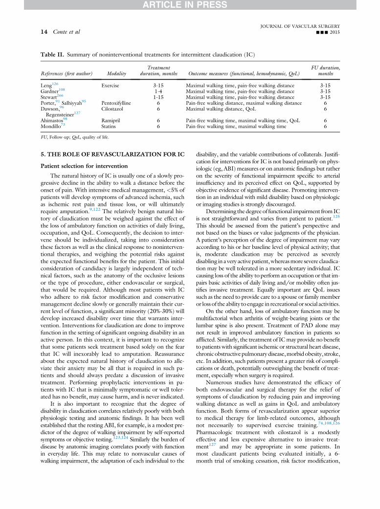
Table II. Summary of noninterventional treatments for intermittent claudication (IC)
References (first author) ModalityTreatment
duration, months Outcome measures (functional, hemodynamic, QoL)FU duration,
months
Leng126 Exercise 3-15 Maximal walking time, pain-free walking distance 3-15Gardner108 1-4 Maximal walking time, pain-free walking distance 3-15Stewart266 1-15 Maximal walking time, pain-free walking distance 3-15Porter,91 Salhiyyah95 Pentoxifylline 6 Pain-free walking distance, maximal walking distance 6Dawson,96
Regensteiner127Cilostazol 6 Maximal walking distance, QoL 6
Ahimastos98 Ramipril 6 Pain-free walking time, maximal walking time, QoL 6Mondillo73 Statins 6 Pain-free walking time, maximal walking time 6
FU, Follow-up; QoL, quality of life.
JOURNAL OF VASCULAR SURGERY14 Conte et al --- 2015
5. THE ROLE OF REVASCULARIZATION FOR IC
Patient selection for intervention
The natural history of IC is usually one of a slowly pro-gressive decline in the ability to walk a distance before theonset of pain. With intensive medical management, <5% ofpatients will develop symptoms of advanced ischemia, suchas ischemic rest pain and tissue loss, or will ultimatelyrequire amputation.9,122 The relatively benign natural his-tory of claudication must be weighed against the effect ofthe loss of ambulatory function on activities of daily living,occupation, and QoL. Consequently, the decision to inter-vene should be individualized, taking into considerationthese factors as well as the clinical response to noninterven-tional therapies, and weighing the potential risks againstthe expected functional benefits for the patient. This initialconsideration of candidacy is largely independent of tech-nical factors, such as the anatomy of the occlusive lesionsor the type of procedure, either endovascular or surgical,that would be required. Although most patients with ICwho adhere to risk factor modification and conservativemanagement decline slowly or generally maintain their cur-rent level of function, a significant minority (20%-30%) willdevelop increased disability over time that warrants inter-vention. Interventions for claudication are done to improvefunction in the setting of significant ongoing disability in anactive person. In this context, it is important to recognizethat some patients seek treatment based solely on the fearthat IC will inexorably lead to amputation. Reassuranceabout the expected natural history of claudication to alle-viate their anxiety may be all that is required in such pa-tients and should always predate a discussion of invasivetreatment. Performing prophylactic interventions in pa-tients with IC that is minimally symptomatic or well toler-ated has no benefit, may cause harm, and is never indicated.
It is also important to recognize that the degree ofdisability in claudication correlates relatively poorly with bothphysiologic testing and anatomic findings. It has been wellestablished that the resting ABI, for example, is a modest pre-dictor of the degree of walking impairment by self-reportedsymptoms or objective testing.123,124 Similarly the burden ofdisease by anatomic imaging correlates poorly with functionin everyday life. This may relate to nonvascular causes ofwalking impairment, the adaptation of each individual to the
disability, and the variable contributions of collaterals. Justifi-cation for interventions for IC is not based primarily on phys-iologic (eg, ABI) measures or on anatomic findings but ratheron the severity of functional impairment specific to arterialinsufficiency and its perceived effect on QoL, supported byobjective evidence of significant disease. Promoting interven-tion in an individual with mild disability based on physiologicor imaging studies is strongly discouraged.
Determining thedegreeof functional impairment fromICis not straightforward and varies from patient to patient.125
This should be assessed from the patient’s perspective andnot based on the biases or value judgments of the physician.A patient’s perception of the degree of impairment may varyaccording to his or her baseline level of physical activity; thatis, moderate claudication may be perceived as severelydisabling in a very active patient,whereasmore severe claudica-tion may be well tolerated in a more sedentary individual. ICcausing loss of the ability to perform an occupation or that im-pairs basic activities of daily living and/or mobility often jus-tifies invasive treatment. Equally important are QoL issuessuch as the need to provide care to a spouse or family memberor loss of the ability to engage in recreational or social activities.
On the other hand, loss of ambulatory function may bemultifactorial when arthritis of weight-bearing joints or thelumbar spine is also present. Treatment of PAD alone maynot result in improved ambulatory function in patients soafflicted. Similarly, the treatment of ICmay provide no benefitto patients with significant ischemic or structural heart disease,chronicobstructivepulmonarydisease,morbidobesity, stroke,etc. In addition, such patients present a greater risk of compli-cations or death, potentially outweighing the benefit of treat-ment, especially when surgery is required.
Numerous studies have demonstrated the efficacy ofboth endovascular and surgical therapy for the relief ofsymptoms of claudication by reducing pain and improvingwalking distance as well as gains in QoL and ambulatoryfunction. Both forms of revascularization appear superiorto medical therapy for limb-related outcomes, althoughnot necessarily to supervised exercise training.74,108,126
Pharmacologic treatment with cilostazol is a modestlyeffective and less expensive alternative to invasive treat-ment127 and may be appropriate in some patients. Inmost claudicant patients being evaluated initially, a 6-month trial of smoking cessation, risk factor modification,

JOURNAL OF VASCULAR SURGERYVolume -, Number - Conte et al 15
exercise, or cilostazol, or a combination, should be initiatedbefore any invasive therapy.
Surgical and endovascular therapy (EVT) are likely tobe similar in efficacy overall, although the quality of sup-porting evidence comparing the two is poor and the likeli-hood of durable clinical success different, especially forextensive disease, more distal disease, and disease involvingthe common or deep femoral arteries where surgery is usu-ally preferred. Specific factors predicting treatment successshould be carefully considered in each individual beforedetermining the optimal strategy.
Anatomic patency and hemodynamic improvement areconsidered necessary (although not sufficient) for clinicalsuccess of revascularization in IC. In the setting of IC,where the limb is not threatened and the natural historyis generally benign, durable benefit at low risk is requiredto justify invasive vascular treatment. The anatomic spec-trum of disease in IC is broad, and has a major impacton both technical success and durability of vascular inter-ventions. In selecting a revascularization strategy forpatients with IC, the expected durability in the circum-stance at hand should be carefully considered. We suggestthat a minimal effectiveness threshold for invasive therapyin IC be a >50% likelihood of sustained clinical improve-ment for at least 2 years. Freedom from hemodynamicallysignificant restenosis in the treated limb is considered a pre-requisite for this goal.
Because anatomic durability is generally inferior forinfrainguinal vs aortoiliac procedures and for bilateral vsunilateral infrainguinal interventions, most experienced cli-nicians have a higher treatment threshold for IC in thesesettings. In bilateral disease, treating physicians shouldconsider the probability of overall efficacy as the productof expected outcomes in each limb, because functionalgains are unlikely if success is achieved and maintained inone limb only. Similarly, as new data are published demon-strating the expected patency outcomes of evolving tech-nologies in various anatomic and clinical settings, thissuggested benchmark should be carefully consideredbefore applying such strategies to everyday practice in clau-dicant patients. Patient-centered outcomes data are sorelyneeded to better define functional gains, symptom relief,and patient perceptions on the relative trade-offs (eg, dura-bility of improvement vs need for repeat interventions) tobetter enable shared decision making in the invasive treat-ment of IC. The concept of a minimal clinically importantdifference has been developed for other chronic diseases toincrease the relevance of study end points to patients and isneeded in this field.128
Anatomic selection factors: Imaging
Once the decision has been made to consider invasivetreatment, patients should undergo imaging studies todetermine the arterial anatomy, the extent of disease,and whether they are best treated with EVT or open sur-gical therapy. This enables a more comprehensive discus-sion about risks, benefits, and durability trade-offs forvarious treatment options. Currently used imaging
modalities include CTA,129,130 MRA,131 duplex ultra-sound imaging,132 and catheter angiography. Althoughall modalities may provide excellent imaging of the arterialcirculation, each has its own unique set of advantages anddisadvantages and may vary in quality and availability frominstitution to institution. Consequently, the modality ofchoice varies widely depending on clinical practice. Thereis insufficient evidence at present to define the most effi-cient, cost-effective strategy for arterial imaging in thispopulation.
Catheter arteriography represents the gold standarddue to superior image resolution and the unique abilityof being able to perform a diagnostic study and EVT atthe same time. However, catheter arteriography is invasiveand may be complicated by contrast nephropathy, allergicreactions, and access-site events.
Modern, multislice spiral CT scans are noninvasive andprovide image resolution of nearly the same quality as con-ventional arteriography. Moreover, the imaging data setcan be reconfigured into different formats, including axial,coronal, sagittal, and three-dimensional images. However,CTA requires a large dose of intravenous contrast and issubject to artifact degradation due to calcification.
MRA has poorer resolution than angiography or CTA,but its images are not degraded by calcium, and like CTA,is noninvasive. Image quality is enhanced by the use of gad-olinium; however, its use is contraindicated in patients withsignificant renal impairment due to the potential risk ofcausing nephrogenic systemic fibrosis. In addition, MRAcannot be used in patients with pacemakers and a varietyof other implanted medical devices.
Duplex ultrasound arterial examination is mostcommonly used as a screening modality to confirm the diag-nosis and to determine the severity of disease both beforeand after treatment. It is occasionally used as a primary im-aging modality during EVT, principally in the setting of iso-lated focal disease in the superficial femoral artery (SFA).133
For patients with severe infrainguinal disease, assess-ment of available vein conduit is another importantelement in the decision process, given the superiority ofgood-quality saphenous vein for femoropopliteal (FP)bypass. Ultrasound vein mapping is therefore recommen-ded as part of the preoperative evaluation of patients whoare being considered as potential open bypass candidates(see below).
Aortoiliac occlusive disease
AIOD, or inflow disease, most commonly leads tobuttock and thigh claudication. In men, bilateral iliac arteryinvolvement or occlusion of the internal iliac arteries may bea cause of vasculogenic erectile dysfunction. With continuedwalking, it is not uncommon for patients with AIOD to alsodevelop claudication in the calf muscles. With bilateral dis-ease, symptoms can be quite severe and disabling due tothe large number of muscle groups being affected.
Invasive treatments for AIOD are performed to pro-vide symptom relief and functional improvements. Theone scenario where treatment of asymptomatic AIOD

Recommendations: General considerations on invasive treatment for intermittent claudication (IC)
GradeLevel ofevidence
5.1. We recommend EVT or surgical treatment of IC for patients with significant functional orlifestyle-limiting disability when there is a reasonable likelihood of symptomatic improvementwith treatment, when pharmacologic or exercise therapy, or both, have failed, and when thebenefits of treatment outweigh the potential risks.
1 B
5.2. We recommend an individualized approach to select an invasive treatment for IC. The modalityoffered should provide a reasonable likelihood of sustained benefit to the patient (>50%likelihood of clinical efficacy for at least 2 years). For revascularization, anatomic patency(freedom from hemodynamically significant restenosis) is considered a prerequisite for sustainedefficacy.
1 C
EVT, Endovascular therapy.
JOURNAL OF VASCULAR SURGERY16 Conte et al --- 2015
may be justified is to provide vascular access for anotherindicated cardiovascular implant (eg, thoracic endovascularaortic repair, endovascular aneurysm repair, transcatheteraortic valve replacement, mechanical circulatory support).
Surgical options for AIOD include direct aortic recon-structions (aortofemoral bypass [AFB], aortoiliac bypass,aortoiliac endarterectomy), which have proven to bemost durable but also have significant morbidity and mor-tality. In patients with suitable anatomy or those deemed tobe at high risk for aortic surgery, or both, extra-anatomicbypasses (axillary-femoral [AxFB], iliac-femoral [IFB],femoral-femoral bypass [FFB]) are less morbid alternativesbut are also less durable.
A tremendous paradigm shift has occurred in the last twodecades in the treatment of AIOD.134 Although intersocietalguidelines previously recommended endovascular proce-dures as primary treatment for more focal disease and tradi-tional surgery for more diffuse disease,9,135 improvements intechnology and endovascular techniques have resulted inEVT replacing open surgical bypass as a primary treatmentfor both focal and advanced AIOD in many cases. For iliacangioplasty using stents, long-term results compare favorablywith open surgery.135-139 Other techniques, including de-vices for crossing long-segment total occlusions,140 stentgrafts,141,142 and hybrid procedures143,144 combining iliacstenting with femoral endarterectomy or with FFB are alter-natives to aortofemoral surgical reconstructions in appro-priate patients with suitable anatomy. Open surgery isgenerally now reserved for patients with such extensive dis-ease that EVT is impossible or ill advised, in patients with se-vere disease and associated aortic aneurysms, and in thosewith failed endovascular interventions (Table III).
5A. Aortoiliac revascularization: Catheter-basedinterventions
Aortic disease. Although open surgical reconstructionfor aortic occlusive disease is considered the gold stan-dard,145,146 there is no question the incidence of aortic andiliac interventions is increasing, and interventional therapieshave become more commonly used in treating this condi-tion.134 There are limited data providing information
regarding the use of interventional therapy for treatment ofaortic occlusive disease. Although initial information reportedthe use of angioplasty as a method of dealing with aorticocclusive disease,147 stenting is the most commonly usedapproach in this vascular bed. Primary technical success ratesfor intervention vary from 90% to 100%, with 1-year primarypatency rates from 75% to 100% and 4-year primary patencyrates of 60% to 80%. Secondary patency can usually be main-tained with repeat percutaneous interventional therapy, with1-year and 5-year secondary patency noted tobe 90% to100%and 60% to 100%, respectively.148-150
Percutaneous approaches can be achieved through afemoral or brachial approach or combinations of the two ap-proaches. Stent types used include balloon-expandable andself-expanding stents,151with orwithout covering. The choiceof stent used relates to the type of disease and sizeof stent avail-able.More calcific diseasewill usually require greater resistanceto crush, which is achieved with balloon-expandable stents,whereas self-expanding stents are more readily available inslightly larger diameters. Few comparative data are availablefor assessing outcomes of these varied stent types. Coveredstent placement in the aorta has few data on which to baseany specific recommendations regarding use.152
Stents should be sized appropriately to the native aorta,with consideration given for the tissue displaced (especiallycalcific disease). Thismaynecessitate undersizing the stent rela-tive to the diameter of thenative normal-caliber aorta to reducethe risk of rupture,which has been reportedwith this approach.
In general, care should be taken not to precludepossible AFB grafting in the future in surgical candidates,such as by extending stents into the perirenal aorta. Stentsshould not be placed across the orifice of the renal arteries,and disease abutting the renal ostia poses increased risk forobstruction or embolization of the renal arteries. The aorticbifurcation is best currently treated with “kissing stents” atthe origin of the iliac arteries or with a combination ofaortic stent placement down to the bifurcation and thenkissing stents placed at the iliac vessel origins.153,154 Theuse of aortic stent grafts for occlusive disease142 has beendescribed in only limited situations, and the routine useof this approach awaits further data acquisition.

Table III. Outcomes of revascularization for aortoiliac occlusive disease (AIOD) in patients with intermittentclaudication (IC)
References (first author) Modality FU duration, years Patency (PAP), %
Yilmaz,154 Soga,161 Ichihashi,160 Indes139 PTA þ stent 5 63-79deVries,157 Rutherford,146 Reed,180 Brewster,182 Chiu166 AFB 5 81-93Cham,176 Melliere,177 Van der Vliet,178 Chiu,166 Ricco175 IFB 5 73-88Criado,267 Ricco,175 Mii268 FFB 5 60-83
AFB, Aortofemoral bypass; FFB, femorofemoral bypass; FU, follow-up; IFB, iliofemoral bypass; PAP, primary assistant patency; PTA, percutaneous trans-luminal angioplasty.
JOURNAL OF VASCULAR SURGERYVolume -, Number - Conte et al 17
Caution should be exercised in the treatment of AIODwhere concomitant aneurysm disease is also present. If ananeurysm is of sufficient size to meet treatment guidelines,therapy should be primarily guided by appropriate aneurysmexclusion with concomitant restoration of unimpeded bloodflow to the lower extremities. In the case of small aneurysms,any treatment considered for symptomatic AIOD shouldachieve simultaneous aneurysm exclusion or not impedeany future open or endovascular aneurysm repair options.
Mortality for endovascular interventions in the aorta canrange from 1% to 3%, and morbidity ranges from 5% to 20%,with aortic rupture a possibility.148-150 Importantly, oneshould be prepared for potential aortic rupture whenembarking on treatment for an aortic lesion with interven-tional therapy. Renal dysfunction has been reported in 2%to 10% of patients. Intensive care unit stay, blood transfusionrequirements, and infection rates are generally lower withEVT than with open aortic reconstructions.155-157
Iliac interventions. Angioplasty remains a therapy fortreatment of iliac artery disease but has largely been sup-planted by a primary stenting approach for this disease. Ingeneral, the more extensive and complex the occlusive dis-ease, the more likely a primary stent approach will improvepatency. For this reason, except for very focal disease, primarystenting of iliac occlusive disease offers the best approach forlong-term patency.137 The use of balloon-expandable vs self-expanding stents has been inadequately studied to claim anadvantage of one device over another; however, certaincharacteristics and locations may favor one stent design overanother. As in other beds, lesions with more calcium orespecially ostial lesions favor the use of balloon-expandablestents, which have greater radial strength and resistance tocrush. This allows for improved expansion and retention ofvessel diameter after stent placement.
The percutaneous approach to iliac disease can vary fromipsilateral to contralateral groin tobrachial, but one shouldbecertain that devices with an appropriate length are availablebefore initiating a procedure. If there is an expectation ofthe brachial approach being used, longer delivery systemsshould be available. When treating from the ipsilateralfemoral approach, one should be certain that placement ofthe most distal stent will not be so close to the sheath accessto prohibit accurate delivery. Here a contralateral or brachialapproach is favored to allow placement of stents to the end ofthe diseased segment, whichmay be to the inguinal ligament.
Treatment of bilateral iliac occlusive disease is indicatedin individuals with appropriate bilateral lesions and
symptoms. Outcomes with bilateral interventions appear tobe similar to those noted in individuals where a single sideis treated; however, it is likely that patency is modestlyreduced compared with unilateral interventions. Treatmentin the common and external iliac arteries appears also tohave similar outcomes. Use of uncovered stents across theorifice of the internal iliac artery will maintain adequate hypo-gastric artery perfusion inmost instances, and it remainsmoreimportant to treat the full extent of the disease than to limitcoverage because of concern regarding stenting across the in-ternal iliac artery origin. In situations where there is concernfor flow preservation through a hypogastric artery, a kissingstent technique can be used at this bifurcation to maintainpatency of both vessels; however, this is rarely necessary.
A key consideration in the treatment of iliac occlusivedisease is the extension of the disease into the femoral ar-tery. Use of stents in the common femoral artery (CFA)is not recommended because they are more likely to frac-ture or fail due to flexion of the artery that occurs withhip flexion. If disease extends into the CFA, the use of ahybrid approach combining femoral endarterectomy withiliac stenting is a better alternative in most patients.
Covered stents have been used in the treatment of iliacocclusive disease.Coveredballoon-expandable stents hadbet-ter primary patency rates when used in more complex lesionsin the iliac artery.141 In the prospective, randomized Coveredvs Balloon Expandable Stent Trial,141 covered balloon-expandable stents demonstrated better primary patency ratesthan bare-metal stents (BMSs) in AIOD, particularly in themore advanced lesions. However, in a more recent single-center, retrospective study, BMSs had superior patency tocovered stents at1 year.158Regardless of anypotential patencyadvantages, covered stents may provide a safety margin in thetreatment of calcified common iliac lesions or ectatic vesselswhere rupture is a distinct possibility. For the external iliac ar-tery, flexible, self-expanding stents are recommendedbecauseof themotion these vessels undergo and thepotential for kink-ing and crimping of balloon-expandable stents placed in thislocation. Similarly, covered versions of these stents have alsobeen used in the external iliac artery, although specific indica-tions favoring one vs the other are not clear.
Initial technical success for iliac stenting varies from 90%to 100% and depends on the extent of the disease, withmore complex lesions having lower initial technical successrates. Long-segment occlusion of the external iliac artery,particularly in women or patients with smaller vessels orcircumferential calcification, or both, remains an important

JOURNAL OF VASCULAR SURGERY18 Conte et al --- 2015
limitation for durable patency.135 The 1-year primary and sec-ondary patency rates range from 70% to 100% and 90% to100%, respectively.159 The 5-year primary and secondarypatency rates are noted to be 60% to 85% and 80% to 95%,respectively.139,159-161 Perioperative mortality can be expectedto be approximately #1%,139,161 and morbidity can rangefrom 5% to 20%.139,159 Long-term outcomes may be inferiorin younger (<50 years) patients, particularly women.162
CFA interventions. Limited data are available to sup-port the use of interventional therapy in occlusive disease ofthe CFAs, but several single-center experiences have beenpublished,163-165 presenting a technical success rate ofnearly 90% and 1-year primary patency rate of 75%. In-formation on longer-term patency is limited, and no in-formation is available regarding stent stability in this areaover even this short period of time. Given the limitedmorbidity and risk entailed with femoral endarterectomy,the use of interventions in this vessel for the present timeshould be limited to those with a prohibitive risk for opensurgical therapy related to local or systemic risk factors.
Hybrid interventions. The use of interventional ther-apies for iliac disease allows treatment of occlusive diseasein patients with limited morbidity; however, when the dis-ease extends into the CFAs, an approach using open surgi-cal techniques to treat the CFA and stents to treat the iliacor inflow vessels offers an alternative to traditional aortofe-moral grafting.142,144,156 In these instances, the endarter-ectomy is extended proximally into the external iliac artery,and stenting is done into the upper area of the endarter-ectomy to limit progression of disease in an interveningsegment. Surgical angioplasty of the femoral artery can beperformed with an eversion or standard patch technique.Stenting of the iliac artery can usually be done from anipsilateral approach, with the sheath entry site well belowthe upper extent of the endarterectomy to allow stents tobe placed through the full length of the diseased segment.
Initial technical success with this approach is reportedat 99% to 100% with 3-year to 5-year primary patency ratesreported at 90% and secondary patency rates of 98% to100%.144 When compared with open aortofemoral recon-struction,166 this approach appears to have similar low mor-tality, with associated reductions in systemic morbidity,infection risks, and a number of postsurgical complicationswhile providing similar patency rates, especially whencomparing secondary patency rates.
5B. Aortoiliac revascularization: Surgery
General considerations. Although endovascular inter-vention has become dominant in this vascular territory, sur-gery continues to have an important role in the currenttreatment of patients with disabling claudication secondaryto AIOD. Relative indications for surgical vs endovascularapproaches will be discussed below but primarily relate todisease distribution, prior interventions performed, andoverall patient risk. A range of surgical options is available,depending on these and other technical considerations.
There are a number of key anatomic considerations thatdirectly influence the choice of an optimal surgical strategy in
AIOD. The nature and extent of aortic disease is pre-eminent. Axial imaging studies, typically CTA, are importantin the revascularization planning. The location and severityof the occlusive lesions, as well as the presence of any aneu-rysmal changes, have direct implications. Noncontrast scansare particularly helpful in preoperative planning to assesscalcification, which can severely complicate clamping and su-turing. Total occlusions, most commonly up to the subrenalaorta, are best approached by direct reconstruction withthromboendarterectomy of the aortic cuff and an end-to-end bypass graft in suitable candidates. Combined occlusiveand aneurysmal disease should be treated by complete exclu-sion of the aneurysmal segment rather than simple bypass.When choosing between end-to-end and end-to-side aorticgraft configuration, the extent of disease in the subrenal aortaand the status of the pelvic circulation aremajor issues.167,168
There are no clear differences in long-term outcomes forend-to-end vs end-to-side aortofemoral grafts169,170; how-ever, the end-to-end technique requires less disease-freeaorta and the graft is somewhat easier to cover with retroper-itoneal tissue. In general, proximal anastomoses should beperformed to the immediate subrenal segment (ie, thezone between the renal and inferior mesenteric arteries)because progression of atherosclerosis is highly likely in themore distal abdominal aorta and may limit durability.
The pattern of iliac disease encountered may be highlyvariable. Unilateral disease, with complete occlusion of bothcommon and external iliac arteries, or occlusion of theexternal iliac artery alone, may be approached surgically witheither in-line (unilateral AFB or IFB) or extra-anatomic(FFB or AxFB) strategies. The choice between these dependson patient risk, status of the contralateral iliofemoral systemand contralateral groin, and suitability of the proximal com-mon iliac or aorta for inflow anastomosis. The presence ofpre-existing stents or stent grafts in any of these segmentswill also influence the choice and conduct of the procedure.
As noted above, the presence and severity of CFA diseaseis a critical point that often dictates whether a purely endovas-cular vs an open surgical or hybrid approach is undertaken.Long-term outcomes and limb status after reconstructionsfor AIOD are highly dependent on continued patency ofthe CFAs and deep femoral arteries (DFAs).171,172 The pres-ence of FP anddistal occlusive disease is also common, partic-ularly in smokers. For patientswithdisabling claudication andrest pain (Rutherford 2-4), inflow reconstruction of signifi-cant AIOD is frequently all that is required to improve symp-toms. A staged approach is therefore recommended in suchpatients with multilevel disease, with re-evaluation of symp-tom status after inflow correction.
Direct (in-line) aortofemoral and iliofemoralreconstruction. Direct surgical revascularization forAIOD is often considered the gold standard for durablevascular interventions, with patency rates >80% at 10 yearsfor AFB or aortoiliac endarterectomy.145,173,174 Patencyrates for unilateral IFB are also typically in the range of 90%at 3 to 5 years.166,175-178 The disease pattern mostamenable to endarterectomy (ie, localized lesions in theterminal aorta and common iliacs) is readily treated by

JOURNAL OF VASCULAR SURGERYVolume -, Number - Conte et al 19
endovascular means; hence, this operation has becomeextremely uncommon in current practice.
Transperitoneal or retroperitoneal approaches may beused without significant differences in outcomes. Unilateraloperations are readily performed via retroperitoneal ap-proaches. In addition to considerations regarding the natureof the proximal anastomosis discussed above, a critical point istreatment of the CFAs and DFAs at the distal anastomosis.Ensuring an adequate caliber profunda outflow is essentialand mandates careful preoperative and intraoperative evalua-tion. In circumstances of truly isolated AIOD and no or min-imal disease in the common femoral/bifurcation, theanastomosis may be performed to the mid-CFA level. In allother circumstances, the arteriotomy in the CFA should allowdirect interrogation of the DFA and SFA orifices, with use ofadjunctive endarterectomy and patch angioplasty as neededbased on burden and location of disease. Failure to addressthis critical point may significantly limit the durability of thebypass graft, because the presence or progression of outflowdisease, or both, is the most common reason for midterm andlate-term graft occlusions. Very rarely, disease spares theexternal iliac arteries and the femoral arteries, and in these cir-cumstances, an aortoiliac bypass may be performed to thedistal external iliac arteries via a transabdominal approach.179
One must be cautious to ensure the absence of any significantfemoral disease by imaging studies in such cases.
Prosthetic grafts (Dacron, expanded polytetrafluoro-ethylene [ePTFE]) are typically used for AFB and IFBand have excellent durability. Small graft sizes (eg, 12 �6 mm) have been associated with decreased patency andshould be avoided.180 In the special circumstance ofinfected or contaminated fields, or removal of a previousinfected graft, autogenous and cryopreserved conduits (ar-tery or vein) have been used with good success.
Perioperative mortality for these procedures isgenerally <3%,181 although morbidity may include cardiac,pulmonary, infectious, wound, and gastrointestinal complica-tions in 10% to 15%. Patency rates for AFB, aortoiliac endar-terectomy, and IFB, as noted, have ranged from 80% to 90%at 5-year and 10-year intervals.156,157,166,173,174,180,182 Func-tional outcomes for claudicant patients, although lessfrequently reported, are generally quite good but depend onthe presence of infrainguinal disease and modification of life-style and risk factors. Long-term complications include limbocclusions, pseudoaneurysm, graft infection, and graft-enteric fistula. Although the overall results are excellent,caution is warranted in certain subgroups of patients whohave demonstrated inferior outcomes, particularly youngerpatients (age<50 years), hypercoagulable patients, and thosewith very small-caliber outflow vessels.180 Younger patientswith premature AIOD are a high-risk group reflecting poorlycontrolled risk factors, underlying genetic or biochemical pre-dispositions, and a more aggressive vascular phenotype.183
Conservative management of younger patients with AIODis advocated because the initiation of surgical or endovascularinterventions at a premature age can lead to accelerated pro-gression toward a more critical stage of disease. Furthermore,recent data from clinical trials74 support the role of exercise
therapy as an initial strategy for claudicant patients with inflowdisease, and this should be advocated as a primary treatmentstrategy, particularly in the younger patient.
Extra-anatomic reconstruction for AIOD. For pa-tients with extensive patterns of AIOD who are deemed tobe at high risk or technical complexity for direct surgical recon-structions, particularly those with advanced ischemic symp-toms, extra-anatomic bypass grafts offer a suitable alternative.In general, extra-anatomic bypass grafts are not considered as afirst-line approach for patientswith ICbecause their long-termdurability and hemodynamic effect are inferior to in-line re-constructions. Their use in patients with claudication shouldbe limited to special circumstances such as graft or stentcomplications, hostile abdomen,orother factors precluding anendovascular or in-line surgical approach.
Key considerations in selecting an extra-anatomic strat-egy include whether the AIOD is unilateral or bilateral(and if bilateral, can unilateral inflow be corrected by suitableendovascular means), nature of prior interventions, and thestatus of the contralateral groin. FFB grafting can be readilydone under regional or even local anesthesia with sedation,offering an important potential advantage. AxFB is chal-lenging to perform under anything but a general anesthetic.
Angiographic imaging (CTA or catheter based) is rec-ommended before performing extra-anatomic bypass graft-ing to fully evaluate the inflow and outflow anatomy. Directangiography is mandatory for FFB if there is any suggestionof disease in the donor iliofemoral system by pulse examina-tion, hemodynamic assessments, or axial imaging. Formalevaluation of the aortic arch vessels is not generally requiredfor AxFB unless there is a discrepancy in brachial pressuresor another reason to suspect brachiocephalic disease.
For FFB, the donor iliac systemmust be free of hemody-namically significant disease, or such diseasedif present andof a localized naturedcorrected by endovascular meanswith confirmed elimination of any translesional pressure gra-dients (<5mmHgmean pressure resting) before performingthe bypass.184 Interrogation and treatment of both donorand recipientCFAs, as noted above, is imperative to optimizelong-termoutcomes. The graft is placed in a deep subcutane-ous, extrafascial tunnel across the suprapubic region of thelower abdomen. Dacron and ePTFE conduits have equaland acceptable results. Autogenous and cryopreserved graftsmay be used for FFB in settings of infection. Care must betaken in regard to the lie of the graft in relation to the lowerabdomen, particularly where there is a significant pannus. Agentle upside down “U” configuration is used, placing theheel of the anastomosis at themid to distal CFA level to avoidkinking when standing upright. Mortality (<2%) andmorbidity (10%) after FFB are generally low. Long-termout-comes are quite acceptable, with patency rates in the 55% to80% range out to 5 years,175 although significantly inferiorto in-line reconstructions. Primary factors affecting patencyare the status of the outflow vessels (ie, presence of severe dis-ease in the SFAorDFAon the recipient side) and progressionor recurrence of disease in the donor iliac system.176,185
AxFB is uncommonly used in the setting of claudication.It may be performed to one or both lower limbs, depending

JOURNAL OF VASCULAR SURGERY20 Conte et al --- 2015
on the clinical circumstances. Because flow rates through thelong prosthetic axillofemoral graft limb are higher with bilat-eral grafts, the bifemoral configuration is generally preferred.The proximal anastomosis should be made to the secondportion of the axillary artery, exposed by division or retrac-tion of the pectoralis major muscle. Externally supportedprosthetic conduits (Dacron or ePTFE) are used to resistcompression along the chest wall. The tunnel should beplaced anterior to the anterior-superior iliac spine and belowthe pectoralis majormuscle, along the anterior axillary line. Avariety of configurations have been used for the cross-femoral limb and distal anastomoses, without apparent influ-ence on the outcome. The inverted “U” configuration, asused in FFB, is most commonly used.
5.3. We recommend endovascular procedures over open sur5.4. We recommend endovascular interventions as first-line
patients with common iliac artery or external iliac arte5.5. We recommend the selective use of BMS or covered sten
iliac artery or external iliac artery occlusive disease, or botpatency.
5.6. We recommend the use of covered stents for treatmentcalcification or aneurysmal changes where the risk ofunprotected dilation.
5.7. For patients with diffuse AIOD (eg, extensive aortic diseaseexternal iliac arteries) undergoing revascularization, we suintervention as first-line approaches. Endovascular intervesubsequent AFB in surgical candidates should be avoided
5.8. EVT of AIOD in the presence of aneurysmal disease shrecommend that the modality used should either achieshould not jeopardize the conduct of any future open
5.9. In all patients undergoing revascularization for AIOD,hemodynamically significant CFA disease is present, w(endarterectomy) as first-line treatment.
5.10. In patients with iliac artery disease and involvement ofprocedures combining femoral endarterectomy with i
5.11. We recommend direct surgical reconstruction (bypass, ereasonable surgical risk and diffuse AIOD not amenabone or more failed attempts at EVT, or in patients widisease.
5.12. In younger patients (age <50 years) with IC, we recomapproach to engage patients and inform them of theeither endovascular or surgical interventions.
5.13. We recommend either axial imaging (eg, CT, MR) or catand planning of surgical revascularization for AIOD.
5.14. When performing surgical bypass for aortoiliac disease,aorta or iliac arteries should be treated as appropriateend-to-side proximal anastomoses.
5.15. For any bypass graft originating from the CFA, the donhemodynamically significant disease or any pre-existinperforming the bypass graft.
BMS, Bare-metal stent; CFA, common femoral artery; CT, com
Recommendations: Interventions for aortoiliac occlusive d
As in all reconstructions for AIOD, careful attention ispaid to the status of the CFA/DFA, and adjunctive endar-terectomy or patch angioplasty are performed as needed.Operative mortality and morbidity for AxFB are low andsimilar to FFB.186 Reported outcomes are inferior to AFB,IFB, and many series of FFB, with 5-year patency rates inthe 50% to 75% range, although the reported results are var-iable and also dependent on the severity of outflow dis-ease.187 As a result of these limitations, AxFB is rarelyadvised for patients with claudication. When used in circum-stances such as aortic graft infection or mycotic aneurysm,patients will often report some degree of functional limita-tion with aggressive exercise due to the inherent hemody-namic limitations of the long axillofemoral conduit.
GradeLevel ofevidence
gery for focal AIOD causing IC. 1 Brevascularization therapy for mostry occlusive disease causing IC.
1 B
ts for aortoiliac angioplasty for commonh, due to improved technical success and
1 B
of AIOD in the presence of severerupture may be increased after
1 C
, disease involving both common andggest either endovascular or surgicalntions that may impair the potential for.
2 B
ould be undertaken cautiously. Weve concomitant aneurysm exclusion oror endovascular aneurysm repair.
1 C
we recommend assessing the CFA. Ife recommend surgical therapy
1 B
the CFA, we recommend hybridliac inflow correction.
1 B
ndarterectomy) in patients withle to an endovascular approach, afterth combined occlusive and aneurysmal
1 B
mend a shared decision-makingpossibility of inferior outcomes with
2 C
heter-based angiography for evaluation 1 Ungraded
concomitant aneurysmal disease of the(exclusion) and is a contraindication to
1 Ungraded
or iliac artery must be free ofg disease should be corrected before
1 Ungraded
puted tomography; EVT, endovascular therapy.
isease (AIOD) in intermittent claudication (IC)

Clinical question Data source FindingQuality ofevidence
The effect of endovascularvs open surgery for AIODon the outcomes of mortality,complications, and patency
Meta-analyses of mostlynonrandomized series(AIOD, not all IC)139
The open bypass group experiencedmore complications and greater 30-day mortality. At 1, 3, and 5 years,primary patency rates were greater inthe open bypass group
B-C
The effect of PTA vs stentplacement for AIOD onthe outcomes of mortality,complications, and patency
Meta-analyses of mostlynonrandomized series(AIOD, data providedfor IC).137
Meta-analyses of mostlynonrandomized series(class C and D aortoiliaclesions)138
Complication and mortality rates weresimilar. Immediate technical successrate (PTA group, 91%; stent group,96%); 4-year primary patency rates forPTA (65% for stenosis, 54% forocclusions) and for stents (77% forstenoses, 61% for occlusions)
B-C
The effect of endovascularvs open surgery for extensiveAIOD on the outcomes ofmortality, complications, andpatency
Meta-analyses ofnonrandomized seriesof EVT for extensiveAIOD188
With endovascular approach, mortalityranged 1.2%-6.7% and complicationsranged 3%-45%. Clinical symptomsimproved in 83% to 100%. Technicalsuccess was achieved in 86% to 100%of the patients. The 4-year or 5-yearprimary and secondary patency rateswere 60% to 86% and 80% to 98%,respectively
B-C
EVT, Endovascular therapy; IC, intermittent claudication; PTA, percutaneous transluminal angioplasty.
Summary of evidence: Interventions for aortoiliac occlusive disease (AIOD)
JOURNAL OF VASCULAR SURGERYVolume -, Number - Conte et al 21
5C. Infrainguinal disease
Occlusive lesions of the FP segment most commonlypresent with IC involving the calf. Isolated lesions of thecrural or foot arteries usually do not cause claudication,although disease involving both segments may cause severecalf claudication and symptoms can involve the foot. Inmany patients, unilateral calf claudication is well toleratedand may be managed conservatively, as previouslydescribed. For patients with more severe symptomsrequiring treatment, a trial of exercise therapy, preferablysupervised training108,126 or approved pharmacologic treat-ment (cilostazol),127 or both, should be undertaken beforeinvasive therapy. If these measures are unsuccessful, inva-sive therapy may be appropriate after a detailed discussionwith the patient. As previously noted, this discussionshould cover the natural history of IC, the risks and bene-fits of open surgery and endovascular interventions, an es-timate of long-term patency, likelihood of symptom relief,and the implications of failed therapy.
In the last decade, vascular specialists have readilyadopted EVT as an attractive alternative to open bypasssurgery for infrainguinal occlusive disease. PTA and stent-ing are the most commonly used EVTs for focal andintermediate-length stenosis.9,189 However, the develop-ment of other techniques and technologies, such as subin-timal angioplasty, devices for crossing and re-enteringlong-segment total occlusions, stent grafts, and mechanical
and laser atherectomy, have made it possible to successfullytreat even advanced disease,190 leading some vascular spe-cialists to advocate an endovascular-first approach for pa-tients undergoing lower extremity revascularization.191 Inmost cases, endovascular procedures are well toleratedwith minimal complications, require short hospital stays,and result in rapid recovery.
However, endovascular procedures are less durablethan surgical bypass and have a greater need for reinter-vention, especially in cases of diffuse stenosis or long-segment total occlusion of the superficial femoral orpopliteal arteries, or both (Table IV). Although the fre-quency with which failed open or EVTs lead directly toclinical worsening is unclear, it undoubtedly occurs witheither modality. This possibility must be carefully consid-ered during discussions with patients, particularly thosewith bilateral disease and more challenging anatomy. Inaverage-risk claudicant patients with advanced FP occlu-sive disease (FPOD), surgical bypass provides better dura-bility, a decreased need for reintervention, and is usuallywell tolerated, with a low rate of complications. In by-passes crossing the knee joint, good-quality saphenousvein is the preferred conduit when available. EVT is areasonable alternative in settings of favorable anatomyand in those with inadequate venous conduit.
For patients with IC, the reduced risk of complications,short recovery time, and rapid return to normal functioning

Table IV. Outcomes of intervention for femoropopliteal occlusive disease (FPOD) in patients with intermittentclaudication (IC)
References (first author) Modality FU duration, years Patency (PAP), %
Hunink,193 Muradin,269 Schillinger270 PTA 2 26-68Schillinger,270 Laird,210 Matsumura211 PTA þ stent 2 51-68Kedora,271 Shackles,272 Geraghty196 Covered stent 1 53-77Pereira,273 Klinkert274 FP vein 5 70-75Robinson,275 Klinkert,274 Pereira273 FP prosthetic 5 40-60
FP, Femoropopliteal; FU, follow-up; PAP, primary patency; PTA, percutaneous transluminal angioplasty.
JOURNAL OF VASCULAR SURGERY22 Conte et al --- 2015
with EVT has lowered the threshold for invasive treatmentto include patients who were previously managed withoutinvasive treatment when the only option was conventionalsurgery. However, there is no conclusive evidence support-ing this more aggressive approach,192 especially comparedwith supervised exercise.76 Treatment guidelines from theAmerican Heart Association43 and the revised Trans-Atlantic Intersociety Consensus document9 recommendthe use of EVT as a first-line treatment for those patientsrequiring invasive therapy for focal and moderate disease,with open bypass recommended for diffuse disease orlong-segment total occlusions, or both. However, the qual-ity of evidence of the long-term efficacy of EVT comparedwith open surgical bypass for the treatment of IC is low.Consequently, the decision of which modality to use mustbe individualized and should take into account other clinicalfactors beyond arterial anatomy, including periproceduralrisks, availability of conduit, and anticipated risk of woundcomplications. Patient preference, after full considerationof the trade-offs, plays an important role as well.
FP revascularization: Catheter-based intervention.IC can be caused by occlusive lesions in the aortoiliacsegment (see previous section), as well as the CFA, SFA,profunda femoral, and popliteal arteries. How often occlu-sive lesions confined to the infrapopliteal arteries result inclaudication remains unclear. Treatment of isolated infra-popliteal disease for relief of claudication is not advised.In patients with multisegment disease, the more proximaldisease should be treated first and usually results inimprovement in symptoms without extending treatmentto the more distal arteries. Endovascular interventions aregenerally safe, with infrequent complications and lowerlevels of morbidity, mortality, and an earlier return tonormal function than surgical bypass.
EVT options for FPOD include PTA alone, especiallyfor short focal lesions <4 cm,193 angioplasty with self-expanding stents,194 angioplasty with balloon-expandablestents,195 angioplasty with covered stent grafts,196,197 athe-rectomy,198-200 antimyoproliferative drug-coated bal-loons,201-203 and drug-eluting stents (DESs).204
Combination EVT involving atherectomy and DESs hasbeen reported in European trials.205
Significant occlusive lesions of the CFA are generallytreated with surgical endarterectomy and patch angioplasty,except in patients with significant comorbidities or hostile
groins precluding surgical treatment. Combined open andendovascular hybrid procedures involving CFA endarterec-tomy and then angioplasty of either proximal iliac artery le-sions (see above) or distal SFA lesions been shown to beeffective for the management of claudication.206,207 EVTof the CFA for claudication is an alternative treatment toopen surgery for selected patients with hostile groins or mul-tiple previous vascular procedures.164,208 Primary interven-tion using balloon angioplasty and self-expanding stentplacement has been reported; however, placement of stentswithin the CFA may be complicated by plaque shifting intothe origin of the profunda femoral artery. Moreover, latefailure resulting in CFA occlusion makes subsequent openor endovascular interventions more complicated. Stent frac-ture or vessel injury due to groin flexion point is an addi-tional concern. Atherectomy has been reported as analternative treatment option that obviates some of theseproblems.209 In general, endovascular approaches to theCFA artery are not well proven, and disease in this arteryis preferably treated surgically.
IC rarely results from isolated profunda femoral diseaseunless there is associated CFA or SFA disease. Endovascu-lar intervention on the profunda femoral artery for claudi-cation symptoms is of unproven value and may carrysubstantial risk to this most important source of collateralflow in the limb. The multiple branch points within theprofunda femoral artery make angioplasty and stentingcomplicated. Similar to the common femoral bifurcation,atherosclerotic plaque near the branch points can shift pla-que during angioplasty and occlude one of the branch ves-sels if not adequately protected.
The SFA is the most common site of atheroscleroticocclusive disease resulting in claudication. The severityof symptoms from occlusive disease in the SFA variesconsiderably, based on the extent of collateralizationfrom the profunda femoral artery to the geniculate collat-eral arteries at the popliteal artery. After failure of an ex-ercise program and optimization of medical therapy,endovascular intervention can be considered. Open surgi-cal bypass success is dependent on arterial inflow,outflow, and the quality of the bypass conduit. Primarypredictors of endovascular success and long-term patencydiffer significantly and include the length of the lesion,degree of stenosis, size of the artery, and degree ofcalcification.

JOURNAL OF VASCULAR SURGERYVolume -, Number - Conte et al 23
PTA alone has been shown to be most effective forshort focal lesions in the SFA (<4 cm). However, all angio-plasties can be complicated by flow-limiting dissection,embolization, and acute arterial recoil with the associatedrisk of abrupt closure. The adjunct use of a self-expanding covered or BMS has been shown to be effectivein improving patency of longer lesions in the SFA and totreat PTA-related complications of dissection and acuterecoil. Several trials have demonstrated the efficacy andpossible superiority of self-expanding stents in the treat-ment of longer SFA lesions. In the Randomized StudyComparing the Edwards Self-Expanding LifeStent vsAngioplasty-alone In Lesions Involving The SFA and/orProximal Popliteal Artery (RESILIENT) study, nitinolBMSs were compared with angioplasty. Treatment of mul-tiple lesions was permitted provided they were treated withone stent. The mean lesion treated was 7.1 cm in stentcohort and 6.4 cm in the angioplasty cohort. The reportedobserved patency at 1 year was 81.3% and 36.7%, respec-tively, for the stent and angioplasty groups.194 In a 3-year follow-up of the RESILIENT study, freedom fromtarget lesion revascularization and clinical success wassignificantly higher in the primary stent cohort, but nodata were available on patency.210
The Study for Evaluating Endovascular Treatments ofLesions in the Superficial Femoral Artery and ProximalPopliteal By Using the Protege EverfLex Nitinol Stent Sys-tem II (DURABILITY II) trial was a single-arm trial inves-tigating the efficacy of a single self-expanding nitinol stent inthe treatment of occlusive lesions in the SFA >4 and <20 cm. Duplex-derived primary patency at 1 year was77.2% for lesions with a mean length of 8.9 cm.211 The Zil-ver PTX trial randomly assigned 471 patients with SFA le-sions averaging 6.5 cm to treatment with a paclitaxel DESor PTA as a primary procedure and to BMS in a subset of110 study patients who required further treatment for im-mediate failure of PTA alone. Patency at 1 year was 83.1%for DES and 32.8% for PTA. In the 110 patients undergoingsalvage stenting for failed PTA, 1-year patency was signifi-cantly better for DES compared with BMS (89.9% vs73%).204 The patency superiority of paclitaxel DES overPTA (74.8% vs 26.5%) and BMS (83.4% vs 64.1%) was sus-tained in a 2-year follow-up report of the same study.212
PTFE-covered self-expanding stents have been used totreat long-segment lesions within the SFA for patientswith claudication, although their superiority to BMSs isas yet unproven. The Viabahn Endoprosthesis withPROPATEN Bioactive Surface (VIA) vs Bare NitinolStent in the Treatment of Long Lesions in SuperficialFemoral Artery Occlusive Disease (VIASTAR) prospectivemulticenter trial213 compared BMSs with heparin-bondedPTFE covered stents in the treatment of long-segmentSFA stenosis and found no statistically significant differ-ence in 1-year primary patency by intention-to-treat anal-ysis, although in the treatment per protocol cohort and inthose with lesions >20 cm, patency was superior in thePTFE group. This study was flawed by protocol violationsin >8% of cases.
The Viabahn vs Bare Nitinol Stent in the Treatment ofLong Lesion Superficial Femoral Artery Occlusive Disease(VIBRANT) trial randomized 148 patients to PTFEcovered or nitinol BMSs for lesions averaging 18 cm inlength. At 3 years, primary patency was nearly identical(24.2% vs 25.9%).196 Some authors have raised cautionabout the failure mode of covered stents in FPOD, witha higher proportion of acute limb ischemia eventscompared with BMS, particularly when distal collateral ves-sels are covered.214 Covered stents may have a role in thetreatment of diffuse in-stent restenosis in the SFA.215 Atthe present time, given the increased cost and lack of clin-ical superiority over BMSs, a primary role for covered stentsin the treatment of IC due to FPOD remains unclear.Balloon-expandable or self-expanding covered stents mayhave a role in the treatment of highly calcified focal SFAlesions, but this has not been prospectively evaluated.
Plaque excision by mechanical atherectomy using cut-ting blades, laser ablation, or “sanding” with a diamond-encrusted burr has been proposed as an alternative toangioplasty and stenting for symptomatic PAD. In a recentmeta-analysis of four randomized studies including only220 patients comparing atherectomy with other establishedtreatments, including angioplasty, stenting, lower extrem-ity bypass, and exercise therapy, the authors concludedthere was no evidence to support the superiority of atherec-tomy over angioplasty for any outcome. They also observedthat the quality of existing evidence is poor and recommen-ded further study with properly powered trials.199
Antimyoproliferative drug-coated balloons have beenevaluated for the treatment of SFA disease in patients withclaudication. The Taxan with Short Exposure for Reductionof Restenosis in Distal Arteries (THUNDER),202 FemoralPaclitaxel (FemPac),203 and Moxy Drug Coated Balloonvs Standard Balloon Angioplasty for the Treatment of Fem-oropopliteal Arteries (LEVANT 1)201 studies demonstratedimproved patency relative to PTA without drug coating butwere limited by small sample sizes, heterogeneous patientpopulations, and incomplete follow-up. Two larger regula-tory trials (LEVANT 2, IN.PACT SFA216) have recently re-ported improved patency for drug-coated versus uncoatedballoon angioplasty in femoropopliteal disease. As a result,the FDA has recently approved two drug-coated balloon de-vices for the treatment of occlusive lesions in the SFA andpopliteal artery. It remains unclear how drug coated balloonangioplasty will compare in durability to other approachessuch as stents referenced above. Bioabsorbable DESs arecurrently being evaluated in Europe but are not availablewithin the United States.
The efficacy of EVT must also be weighed against thepotential for acute and long-term complications. Commonendovascular complications include arterial dissection atthe area of treatment site, arterial perforation, pseudoaneur-ysm creation, acute recoil associated with abrupt closure orrestenosis, embolization distal to the site of intervention,and arteriovenous fistula creation. Implantation of a stentalso carries specific, stent-related risk factors, includingstent fracture, chronic arterial erosion, and perforation.

JOURNAL OF VASCULAR SURGERY24 Conte et al --- 2015
Long-term complications include restenosis with potentialocclusion, loss of collateral branches at the site of the endo-vascular procedure, and late pseudoaneurysm formation.One additional consideration with EVT is its effect on sub-sequent open surgical bypass, which has been reported to berequired in 10% to 25% of patients for failed interventions.In one study by Joels et al,217 EVT altered the level of theexpected outflow target to amore distal level in 30% of pa-tients. Previous EVT, with or without stents, may adverselyaffect graft patency, limb salvage, and amputation-free sur-vival compared with bypasses done as initial therapy forCLI.218 Failure of stented endovascular interventions donefor claudication has resulted in acute limb ischemia, espe-cially when covered stents grafts have been used.219 In addi-tion to these observations, microembolization into theoutflow bed and loss of potential outflow targets havebeen postulated as causes for the inferior results of secondarysurgical bypass in these patients.
Catheter-based treatment of occlusive disease involvingpopliteal or more distal arteries, or both, has not been spe-cifically evaluated for the treatment of IC and presentsadditional problems and the risk of significant complica-tions. Popliteal occlusive disease may result in IC, especiallywhen there are poorly developed collaterals from the pro-funda femoral artery through the geniculate arteries. EVTof popliteal artery occlusive disease is technically feasible;however, its long-term durability is not known, and failurein this location may result in limb-threatening ischemicsymptoms or the need for a distal tibial bypass, or both.Flow-limiting dissection, occlusion, or perforation mayresult in the undesirable need to place a stent across theknee joint. Newer, more flexible stent designs may ulti-mately improve outcomes in the popliteal artery,220 butcomparative studies with adequate follow-up are not avail-able at present. Consequently, it should be undertakenboth with caution and some trepidation for claudication.
In most circumstances, isolated tibial disease does not pre-sent with symptoms of claudication and should not be under-taken for relief of claudication symptoms. Adding tibialangioplasty to a more proximal intervention to improve runoffin the hope of improving patency has not been studied. Thedurability of tibial angioplasty is worse than SFA angioplasty,averaging <40% at 3 years221 in patients undergoing treat-ment for limb salvage, where it is most commonly performed.The need for reintervention at this level is high, and persistentfailure after repeated attempts of reintervention with repeatedfailure may result in CLI requiring a distal bypass for salvageor major limb amputation. Isolated infrapopliteal interven-tions are not recommended for patients with IC.
FP revascularization: Surgery. The guidelines forconservative management of IC have been previously dis-cussed. However, it is important to recognize that the ben-efits of medical therapy and exercise are actually quitemodest. In a recent prospective study, absolute walking dis-tance improvement with a home-based exercise program,the only type available to most patients, was <90 feet.222
The effect of such a modest improvement on functionalability and QoL may be inadequate for many patients.
Bypass surgery has been a mainstay in the invasive treat-ment of IC for 5 decades, although much less frequentlyused in the last 10 to 15 years with the evolution and rapidexpansion of catheter-based therapies (see above). The effi-cacy of surgical bypass for the relief of claudication symptomsis well established. A seminal report documented long-termfunctional outcomes in 14 patients who underwent veinbypass surgery for IC, demonstrating relief of symptomsand improved exercise performance and self-reported com-munity-based walking abilities.223 ABI improved in surgicalpatients by nearly 0.4, peak treadmill walking time by 9 mi-nutes, and pain-free walking time by>6 minutes. Question-naire scores for walking distance improved by 203% andwalking speed by 130%. These improvements were not pre-dicted from routine noninvasive testing alone. The authorswere among the first to suggest that such functional statusoutcomes should be measured directly.223
The perceived morbidity associated with open surgicaltherapy for IC is an important factor in clinical decision mak-ing. As with any surgical procedure, the key to a successfuloutcome is appropriate patient selection. Ideal candidatesfor surgical bypass for claudication should have minimalcomorbidities, good life expectancy, be significantly disabledspecifically by claudication symptoms, and have reasonablerunoff and good conduit available for bypass.
One of the major advantages of bypass compared withangioplasty is durability as measured by patency of theintervention. Van der Zaag et al224 reported the resultsof a randomized trial of angioplasty vs surgical bypass in56 patients with claudication and 5- to 15-cm-long lesionsof the SFA. The primary end point was reocclusion. No30-day deaths occurred in either group, confirming the ob-servations of many others in nonprospective studies thatsurgical bypass for claudication is safe in appropriatelyselected patients. More than half of the angioplasty patientsexperienced a reocclusion. Surgical bypass was associatedwith a significant 31% absolute risk reduction for the endpoint of subsequent reocclusion. Clinical improvement insymptoms was also significantly better for patients who un-derwent bypass (absolute difference 20%). Only one patientamong the 56 enrolled subsequently required amputation;that individual had been initially treated by angioplasty. Noamputations were required in the bypass surgery patients.
Bypass surgery has also been shown to be associatedwith superior functional improvement compared with othertreatment modalities by numerous investigators. Wolfet al225 compared surgery and balloon angioplasty for pe-ripheral vascular disease in a randomized fashion. Bypassand angioplasty both showed sustained improvements in he-modynamics and QoL. Primary success was more oftenachieved with bypass, but the differences were not signifi-cant. Lundgren et al120 compared claudication patientswho underwent surgical reconstruction vs physical trainingalone. Surgery was more effective, but the addition of phys-ical training to surgery improved symptom-free walking dis-tance even further. Surgery was significantly better thanexercise therapy with regard to maximal walking time, ABIimprovement, and peak exercise calf blood flow. A subgroup

JOURNAL OF VASCULAR SURGERYVolume -, Number - Conte et al 25
of patients whose activity was also limited by cardiopulmo-nary disease in addition to claudication failed to demonstratesignificant walking improvement despite improvements inABI and calf flow, emphasizing the importance of careful pa-tient selection when recommending any intervention, espe-cially surgery, for claudication.
In a systematic review of the efficacy of bypass forchronic limb ischemia, the probability of an achieving anunlimited maximal walking performance, defined as at least1000 meters, was 75% to 95% in patients who underwentbypass for claudication compared with only 10% to 20%in those treated solely by exercise training.226 In anotherstudy where patients were randomized to surgical bypass,supervised exercise training, or observation alone, surgicallytreated patients showed a significant improvement inmaximal walking power, stopping distance, postischemicblood flow, and great toe pressure at 1 year.227 Patientsrandomized to physical exercise training did not demon-strate improvements in any outcome measure. Mortalityand amputation rates were identical in both groups.
In a retrospective review, Koivunen and Lukkarinen228
demonstrated that surgically treated patients had superior clin-ical outcomes and health-related QoL compared with EVTand conservative management. Specific improvements in sur-gically treated patients at 1 year included improvement inpain, mobility, sleep, and emotional reactions.
Additional factors determining the success of surgicalbypass for claudication include technical and anatomic fac-tors such as conduit, target vessel, and runoff. Availableprospective, randomized data regarding choice of conduitfor FP bypass demonstrate superior patency for vein grafts,even to an above-knee popliteal target, compared with
5.16. We recommend endovascular procedures over open surgartery not involving the origin at the femoral bifurcat
5.17. For focal lesions (<5 cm) in the SFA that have unsatisfaangioplasty, we suggest selective stenting.
5.18. For intermediate-length lesions (5-15 cm) in the SFA, wexpanding nitinol stents (with or without paclitaxel)angioplasty.
5.19. We suggest the use of preoperative ultrasound vein mappof autogenous vein conduit in patients being consideredof IC.
5.20. We recommend against EVT of isolated infrapopliteal dunproven benefit and possibly harmful.
5.21. We recommend surgical bypass as an initial revascularizadisease, small caliber (<5 mm), or extensive calcificatanatomy for bypass (popliteal artery target, good runrisk.
5.22. We recommend using the saphenous vein as the preferre5.23. In the absenceof suitable vein,we suggest usingprosthetic c
if the above-knee popliteal artery is the target vessel and
EVT, Endovascular therapy; SFA, superficial femoral artery.
Recommendations: Intervention for femoropopliteal occlu
PTFE bypass, after 2 to 3 years of follow-up.229 In mostpatients, sustained walking improvement and improvedQoL depend on maintenance of patency of the surgicalreconstruction. This is particularly important when treatingIC given the better functional ability and longer life expec-tancy compared with patients with limb-threateningischemia.
However, when suitable autologous vein is unavailable,prosthetic bypass for claudication may be reasonable.AbuRahma et al230 reported no difference in primarypatency rates between saphenous vein and PTFE bypassin patients with IC and at least two-vessel to three-vesselrunoff. Assisted primary patency rates were still statisticallyhigher for vein grafts. The quality of the runoff circulationmay also affect the results of surgical treatment for claudi-cation. Zannetti et al231 determined that absence of dia-betes, minimal cardiac comorbidities, and angiogramspredicting near normalization of the postoperative ABIresulted in excellent late outcomes and patient satisfactionin 82% of patients meeting these criteria.
The popliteal artery is the most common outflow vesselwhen an infrainguinal bypass is performed for claudication,usually above the knee. However, properly selected pa-tients without a suitable popliteal artery target may alsobenefit from bypass. In a retrospective study of 57femoral-tibial bypasses performed during a 16-year periodfor IC, graft patency rates were better than tibial bypassfor limb salvage and equivalent to those achieved with FPbypass graft for claudication.232 Vein conduit, 70% ofwhich were saphenous vein, was used in all cases. Inter-viewed patients reported improved walking distance,reduced claudication, and a high degree of satisfaction.
GradeLevel ofevidence
ery for focal occlusive disease of the SFAion.
1 C
ctory technical results with balloon 2 C
e recommend the adjunctive use of self-to improve the midterm patency of
1 B
ing to establish the availability and qualityfor infrainguinal bypass for the treatment
2 C
isease for IC because this treatment is of 1 C
tion strategy for patients with diffuse FPion of the SFA, if they have favorableoff) and have average or low operative
1 B
d conduit for infrainguinal bypass grafts. 1 Aonduit for FPbypass in claudicant patients,good runoff is present.
2 C
sive disease (FPOD) in intermittent claudication (IC)

Clinical question Data source Funding Quality of evidence
Endovascular vs surgicalreconstruction
Four RCTs and sixobservational studiesreporting on 2817patients with FParterial disease233
EVT was associated with lower 30-daymorbidity (OR, 2.93; 95% CI, 1.34-6.41) and higher technical failure(OR, 0.10; 95% CI, 0.05-0.22) thanbypass surgery. No difference in 30-day mortality (OR, 0.92; 95% CI,0.55-1.51). Higher primary patencyin the surgical treatment arm wasfound at 1 (OR, 2.42; 95% CI, 1.37-4.28), 2 (OR, 2.03; 95% CI, 1.20-3.45), and 3 (OR, 1.48; 95% CI,1.12-1.97) years after intervention.Progression to amputation occurredmore commonly in the endovasculargroup at the end of the second (OR,0.60; 95% CI, 0.42-0.86) and third(OR, 0.55; 95% CI, 0.39-0.77) yearof intervention. The bypass grouphad higher amputation-free (OR,1.31; 95% CI, 1.07-1.61) andoverall survival (OR, 1.29; 95% CI,1.04-1.61) rates at 4 years
C (risk of bias, indirectnessbecause most trialsenrolled CLI patients)
The effect of stentingvs no stenting in patientswith IC on morbidity,mortality and patency
Meta-analysis of 8RCTs (968 patientswith IC or CLI andSFA disease)189
Primary patency better with stenting at6 months but not 12 months
C (indirectness due to CLIpatients included andimprecision of long-termoutcome)
Balloon angioplasty withoptional stenting vsroutine stenting withnitinol stents
Meta-analysis of 4RCTs (627 patientswith IC or CLI andSFA disease)234
Mortality was similar in both groups(OR, 0.83; 95% CI, 0.39-1.77).Technical success was significantlyhigher in the stenting group (96% vs64%; OR, 0.31; 95% CI, 0.09-0.92).The 12-month binary restenosis ratewas significantly lower in the primarystenting group (OR, 3.02; 95% CI,1.3-6.71)7
C (indirectness due to CLIpatients included andimprecision)
Comparison of variousstents
Network meta-analysisof 16 RCTs (2532patients with IC orCLI and FP arterialdisease)235
Technical success was highest withcovered stents. Vascular restenosiswas lowest with paclitaxel DES andwith paclitaxel-coated balloons.
Major amputations were rare in alltreatment and control groups(pooled amputation rate of 0.7events/100 person-years)
C (indirect comparisons,CLI patients included,imprecision)
Vein grafts vs PTFE 1 RCT in 43 claudicantpatients (86 limbs)230
Complication rates were 5% for PTFEand 12% for saphenous vein graft, nooperative deaths or perioperativeamputations for either procedure.Primary, assisted primary, andsecondary patency rates at72 months: 68%, 68%, and 77% forPTFE and 76%, 83%, and 85% forsaphenous vein graft
B (imprecision, smallnumber of events)
CI, Confidence interval; CLI, critical limb ischemia; DES, drug-eluting stent; OR, odds ratio; PAD, peripheral arterialdisease; PTFE, polytetrafluoroethylene; RCT, randomized controlled trial; SFA, superficial femoral artery.
Summary of evidence: Intervention for femoropopliteal occlusive disease (FPOD) in intermittent claudication (IC)
JOURNAL OF VASCULAR SURGERY26 Conte et al --- 2015

JOURNAL OF VASCULAR SURGERYVolume -, Number - Conte et al 27
Assessing the efficacy of revascularization for IC
Patients undergoing revascularization for claudicationdesire durable improvement in pain-free walking and func-tional independence. Claudication rarely progresses to limbloss, and as such, treatment with endovascular or open sur-gery should never result in major or minor amputation.Consequently, limb salvage is not considered proof of effi-cacy of any procedure undertaken to treat IC, and in fact,loss of the limb should be considered a catastrophic failureof therapy. The usual efficacy end points in clinical trialsinclude standardized measures of walking ability such asthe initial time to onset of claudication, maximal walkingdistance, and the 6-minute walk test; however, these endpoints are rarely used in clinical practice. Patients undergo-ing lower extremity revascularization for claudicationshould have documented improvement in symptoms aswell as hemodynamic evidence of improvement in lowerextremity perfusion. As stated above, anatomic patency isconsidered a prerequisite for sustained hemodynamicimprovement and clinical benefit in IC.
Postintervention medical treatment
After intervention for lower extremity vascular disease,aggressive medical therapy is indicated not only to preventfuture cardiovascular events but also to improve patency ofthe revascularization. Patients should be counseled on riskfactor modification, as previously described, and haveaccepted pharmacologic treatment for system atheroscle-rosis, especially statins and antiplatelet therapy. In some pa-tients, systemic anticoagulation may also be required.
Antiplatelet agents
Antiplatelet agents are generally used to treat patientsafter lower extremity bypass. Although, antiplatelet therapyhas not been conclusively proven to improve bypass graftpatency, its benefit in decreasing long-term postproceduraladverse cardiovascular events is sufficient indication for theuse of these agents in most patients, who are considered tobe at high risk for cardiovascular complications and stroke.
In a systematic review236 of the effect of antiplatelettreatment compared with placebo on bypass graft patency,patients receiving antiplatelet therapy had improved patencyat 1 year (OR, 0.62; 95% CI, 0.43-0.86). When venous andprosthetic bypasses were analyzed separately, there was noimprovement in 12-month patency in patients undergoingvenous bypass who received acetylsalicylic acid (ASA) ordipyridamole compared with placebo. Conversely, 12-month primary patency was markedly improved in patientsundergoing prosthetic bypass who received ASA comparedwith placebo (OR, 0.22; 95%, CI, 0.12-0.38). Majorbleeding events were more frequent in patients receivingASA therapy but did not reach statistical significance.
Theeffect of addingclopidogrel toASAwas studied in851patients in the Clopidogrel and Acetylsalicylic Acid In BypassSurgery for Peripheral Arterial Disease Trial (CASPAR).237
This placebo-controlled RCT found no difference in outcomebetween patients receiving ASA vs ASA plus clopidogrel un-dergoing lower extremity bypass. However, the subset of
patients undergoing prosthetic bypass (30%), demonstratedimproved patency and limb salvage when receiving ASA com-bined with clopidogrel compared with ASA alone.
Anticoagulation
Several trials have studied the effect of ASA comparedwith warfarin on patency in lower extremity bypass. The pro-spective randomized Dutch Bypass Oral Anticoagulants orASA (BOA) trial85 randomized 2690 patients undergoinglower extremity bypass to coumarin (target internationalnormalized ratio of 3-4.5) vs ASA (81 mg/d). Overall, therewas no difference in patency at 12months in the two cohorts;however, a subgroup analysis demonstrated superior patencyfor patients undergoing vein bypass receiving coumarincomparedwith those receivingASAaloneat12and24months(OR, 0.59; 95% CI, 0.46-0.76). This effect was not seen inthose patients undergoing prosthetic bypass grafts, wherepatency was identical in those receiving ASA and coumarin.Twice as many bleeding complications were observed in pa-tients receiving coumarin as in those receiving ASA.
Despite the findings of the BOA trial, most vascularsurgeons in the United States choose not to routinely anti-coagulate patients undergoing lower extremity vein bypass.However, anticoagulant therapy may be beneficial in spe-cific circumstances where conditions are less than optimal.In a small trial by Sarac et al,238 56 patients undergoinghigh-risk vein bypass (defined as poor-quality conduit orrunoff) were randomized to ASA plus warfarin vs ASAalone. The patients receiving ASA plus warfarin had asignificantly improved patency and limb salvage at 3 years.
The ischemic consequences of graft thrombosis may beameliorated by the use of anticoagulants after bypass sur-gery, especially when using prosthetic grafts. A multicenterprospective, randomized trial of 402 patients undergoingFP bypass with PTFE or saphenous vein and treated withASA plus warfarin or ASA alone found graft thrombosismore commonly resulted in limb-threatening ischemia inprosthetic grafts than in vein grafts. However, patientswith prosthetic graft thrombosis were less likely to presentwith acute limb ischemia if they were receiving warfarin.239
In summary, available clinical evidence does notconclusively support the use of antiplatelet agents toimprove lower extremity vein bypass graft patency,although their use is still warranted to reduce future cardio-vascular ischemic events and stroke. Patency may beimproved with antiplatelet therapy in patients undergoingprosthetic bypass. The use of warfarin anticoagulation afterlower extremity bypass remains controversial, and signifi-cant differences exist in its use between North Americanand European vascular surgeons. In the United States, anti-coagulation is used selectively in vein bypass procedureswith either suboptimal conduit or compromised runoff.Warfarin in addition to ASA is used in many patientsreceiving prosthetic grafts to reduce the ischemic conse-quences of bypass graft thrombosis. However, caution iswarranted given the incremental bleeding risks associatedwith combination therapy, and existing evidence is inade-quate to support a definitive recommendation at this time.

GradeLevel ofevidence
5.24. In all patients after endovascular or open surgical intervention for claudication, we recommendoptimal medical therapy (antiplatelets agents, statins, antihypertensives, control of glycemia,smoking cessation).
1 A
5.25. In patients undergoing lower extremity bypass (venous or prosthetic), we suggest treatment withantiplatelet therapy (aspirin, clopidogrel, or aspirin plus clopidogrel).
2 B
5.26. In patients undergoing infrainguinal endovascular intervention for claudication, we suggesttreatment with aspirin and clopidogrel for at least 30 days.
2 B
Recommendations: Postinterventional medical therapy in intermittent claudication (IC)
JOURNAL OF VASCULAR SURGERY28 Conte et al --- 2015
Endovascular intervention
Limited data are available regarding therapies targeted atpreventing restenosis or occlusion after endovascular proce-dures in patients with IC. A recent systematic review of fourprospective randomized trials did not demonstrate anyimprovement in patency at 12 months with ASA comparedwith placebo.240 Nonetheless, antiplatelet therapy may bewarranted in patients undergoing EVT for claudication aspart of an aggressive medical treatment program to preventlong-term cardiovascular complications such as stroke andmyocardial infarction. In addition, the same review evaluatedthe potential effect of using higher-dose ASA (300-1000 mg) compared with lower-dose ASA (50-300 mg).No beneficial effect was observed with higher doses.240
TwoRCTs investigated the effect of anticoagulation andcilostazol on patency. Koppensteiner et al241 compared theuse of low-molecular-weight heparin (LMWH) and ASA vsASA alone in patients undergoing popliteal angioplasty.Improved patency was observed in patients treated withLMWH for with CLI, but this effect was not observed in pa-tients treated for claudication.241 Iida et al242 observed adecrease in restenosis and reocclusionat6, 12, and24monthsin patients treated with cilostazol compared with ticlopidine,similar to the beneficial effect observed with cilostazol inimproving patency of coronary interventions and FP angio-plasty in patients with end-stage renal failure.243
6. SURVEILLANCEAFTERREVASCULARIZATIONFOR IC
Arterial reconstructions performed for IC may be uni-lateral, bilateral, suprainguinal, infrainguinal, or on occa-sion, unilateral or bilateral combinations of suprainguinaland infrainguinal reconstructions. Depending on the siteand extent of the arterial occlusive process, reconstructionscan be bypass operations with autogenous or prostheticarterial substitutes, open endarterectomy, or various com-binations of catheter-based techniques. Whatever methodis selected for reconstruction, the goal is to improve patientQoL by improving pain-free walking distance and maximalwalking distance while minimizing the need for additionalarterial reconstructive procedures.
Surveillance of vein grafts performed for IC. Auto-genous vein is the preferred conduit for open infrainguinal
arterial reconstructions for treatment of claudication.Approximately one-third of lower extremity vein graftswill ultimately develop stenotic lesions that may threatenpatency. The large majority of such lesions develop withinthe first year of graft implantation; however, vein graftsare never entirely free of the risk of developing stenosis.The risk of developing vein graft stenosis appears greaterin operations performed for CLI, in operations performedwith smaller-caliber venous conduits, procedures usingnonsaphenous vein conduits, and in vein grafts withanastomosis to more distal (tibial or pedal) arteries. Sur-veillance protocols for lower extremity autogenous veingraft reconstructions were developed to detect graft ste-nosis before graft thrombosis and were based on thisnatural history and the assumption that a patent, hemo-dynamically uncompromised, lower extremity arterialreconstruction is optimal for maintaining ambulatoryfunction and QoL. Failure of an arterial reconstructionperformed for claudication will, at the very least, returnthe patient to his or her previous level of preoperativedisability but may occasionally result in more severesymptoms, including limb-threatening ischemia. In addi-tion, performing a secondary bypass for a thrombosedlower extremity vein graft is technically more difficult andcomplex than treatment of a failing but still patent graft.
Surveillance programs of lower extremity vein grafts maybe solely clinical or both clinical and vascular laboratory based.The Inter-Society Consensus for the Management of Periph-eral Arterial Disease (TASC II) working group recommendedpatients treated with lower extremity vein grafts be monitoredfor at least 2 years with a clinical surveillance program thatconsists of an interval history to detect new symptoms, pulseexamination, and measurement of resting and, if possible,postexercise ABIs.9 However, most vein graft arterial recon-structions are thought to fail through the development ofintrinsic stenotic lesions within the venous conduit, at anasto-motic sites, or in the outflow artery distal to the distal anasto-mosis. Most of these lesions occur within the first 12 to18 months after surgery but can continue to develop or prog-ress years later. Relying on clinical assessment alone may misscritical vein graft stenoses that threaten graft thrombosis, espe-cially in patients treated for CLI (see below). Vascularlaboratory-based surveillance programs of lower extremityvein grafts focus on detection using duplex ultrasound surveil-lance (DUS), grading, and monitoring of stenotic lesions

Clinical question Data source Finding Quality of evidence
The effect of antiplatelettherapy on patency, limbsalvage and survival inpatients with IC whounderwent endovascularor open surgicalinterventions
Systematic review244 of 15RCTs in patients withsymptomatic PAD(including CLI)undergoing infrainguinalbypass surgery: ASA orASA þ dipyridamole vsplacebo (6); ASA or ASAand dipyridamol vspentoxifylline (2); ASA vsindobufen (1); ASA vsvitamin K antagonists (2);ASA þ dipyridamole vsLMWH (1); ticlopidinevs placebo (1); ASA vsprostaglandin E1 (1);ASA vs naftidrofuryl (1)
Antiplatelet therapyimproved venous andartificial graft patencycompared with notreatment. More benefitin synthetic grafts
B
The effect of anticoagulantsagents on patency, limbsalvage and survival inpatients with IC whounderwent open surgicalinterventions
Systematic review245 of 14RCTs in patientsundergoing infrainguinalarterial bypass surgery(including CLI)
Anticoagulants reduced therisk of limb loss at thelongest follow-up (OR,0.36; 95% CI, 0.19-0.69)and increased primarypatency when venousgrafts were analyzedseparately (OR, 0.44; 95%CI, 0.14-1.42). Bleedingrisk doubled comparedwith antiplatelets
B-C (rated down due toimprecision andindirectness)
The effect of antiplatelet andanticoagulant drugs forprevention of restenosis/reocclusion afterperipheral EVT
Systematic review240 of22 RCTs with variouscomparisons. Secondaryindirect evidence onbenefits of antiplateletagents in reducingcardiovascular morbidityand mortality
At 6 monthspostintervention,reocclusion was lowerwith high-dose ASA þdipyridamole (OR, 0.40;95% CI, 0.19-0.84), butnot for low-dose ASA þdipyridamole. Nosignificant difference inreocclusion or restenosiswas detected for high-dose ASA vs low-doseASA, ASA/dipyridamolevs vitamin K antagonists,clopidogrel þ aspirin vsLMWH þ warfarin, orticlopidine vs vitamin Kantagonists. Clopidogreland aspirin resulted infewer major bleedingepisodes compared withLMWH þ warfarin
B-C (rated down due toimprecision andindirectness)
ASA, Acetylsalicylic acid; CI, confidence interval; CLI, critical limb ischemia; EVT, endovascular therapy; IC, intermittentclaudication; LMWH, low-molecular-weight heparin; OR, odds ratio; PAD, peripheral arterial disease; RCT, randomizedcontrolled trial.
Summary of evidence: Postinterventional medical therapy
JOURNAL OF VASCULAR SURGERYVolume -, Number - Conte et al 29

JOURNAL OF VASCULAR SURGERY30 Conte et al --- 2015
within the graft or at the anastomoses thought to threatengraft patency, regardless of clinical presentation.
There is an extensive body of literature on the use DUSof lower extremity vein grafts performed for CLI. Moststudies are retrospective analyses of single-institution caseseries. Prospective studies have also focused on patientswith CLI and not claudication. Patients undergoing surgicalbypass for claudication are generally healthier and moreactive than those treated for CLI and are theoreticallymore ambulatory and more apt to report recurrence ofsymptoms earlier than a minimally ambulatory, debilitatedpatient treated for CLI. Grafts placed for claudication usu-ally extend to the popliteal artery, rarely more distally, andhave superior patency compared with vein grafts placed forCLI. It is therefore not clear whether data derived forvascular laboratory-based DUS programs in patients treatedfor CLI are applicable or even necessary for patients treatedfor claudication.
The Vein Graft Surveillance Randomized Trial (VGST)was a prospective study in the United Kingdom that random-ized594patientswithpatentveingrafts30days after surgery246
to clinical surveillance or DUS in addition to clinical surveil-lance. Most the operations were from the CFA to the above-knee or below-knee popliteal artery, and the conduits wereipsilateral reversed saphenous vein in>90% of the procedures.Anastomotic sites and conduits largely mimicked those mostused in vein grafts performed for claudication, even thoughtwo-thirds of theprocedures inVGSTwere forCLI.Acompar-ison of the two surveillance strategies at 18 months found nodifferences in primary, primary assisted, or secondary patency.
A smaller study from Sweden randomized 156 patientswith lower extremity arterial reconstructions to intensivesurveillance to include DUS (n ¼ 79) vs routine clinicalsurveillance (n ¼ 77).247 Forty grafts, equally distributedbetween the two groups, were PTFE grafts. Only twografts in each group were performed for claudication, andtwo-thirds of the grafts were performed to the poplitealartery. Among the vein grafts in the study there wasimproved assisted primary and secondary patency in theintensive surveillance group that included DUS.
Many single-institution series and one large prospectivemulti-institution series demonstrated improved vein graftpatency for patients treated for CLI with a surveillanceprogram using duplex ultrasound detection of graft steno-ses.248-252 In addition to a significant improvement in pri-mary and assisted primary patency of vein grafts monitoredwith a DUS-based program, these studies and others havedemonstrated revised grafts have excellent long-termpatency comparable to grafts never undergoing revi-sion.253,254 None of the studies included patients treatedfor claudication. Whether the magnitude of the benefit ofimproved graft patency achieved in CLI patients, followedwith duplex-based surveillance, would be comparable in pa-tients undergoing vein bypass for claudication remains un-known. Nevertheless, the significance and consistency ofevidence demonstrating benefit for a DUS-based programfor lower extremity vein grafts done for CLI infers somebenefit would be derived for grafts performed for claudication,
especially during the first year after the bypass, although thestrength of the evidence is weak. Optimal intervals for DUSare also notwell defined.Current practice ofmany vascular sur-geons in the United States is to obtain a postoperative duplexultrasound assessment of vein grafts within the first month, at3, 6, and 12 months, and every 6 to 12 months thereafter.
Surveillance of catheter-based interventionsperformed for IC. Residual and early recurrent stenosescommonly occur after endovascular procedures, particularlywhen performed for more complex disease.255 The role ofDUS after EVT is unclear. To date, no randomized trials ofDUS after endovascular intervention have been performed,but many have extrapolated DUS protocols and criteria forpeak systolic velocity (PSV) and velocity ratio (Vr) developedfor infrainguinal vein grafts and applied them to follow-upafter EVT. Duplex ultrasound can localize and grade thepresence and degree of stenosis in the FP segment after an-gioplasty alone as well as after stent placement, particularly inthe SFA, where authors have correlated duplex findings withangiography. Baril et al256 reported that a PSV >275 cm/sand a Vr >3.5 were specific and predictive cutoff values forduplex determination of >80% in-stent restenosis after an-gioplasty and stent placement in the SFA.
Clinical follow-up alone, combined with ABI determina-tion or toe pressure measurements, or both, in limb salvagepatients, and with DUS have all been proposed methods ofsurveillance for catheter-based interventions. Available re-ports to date regarding the accuracy, predictive value, andbenefits of DUS after EVT are conflicting. Mewissenet al257 reportedoneof the earliest experienceswithDUSafterballoon angioplasty of the FP artery. They demonstrated theimportance of hemodynamic assessments (ABI measure-ments and toe pressuremeasurements) indetermining the de-gree of perfusion improvement, but these techniques couldnot discriminate between restenosis or occlusion of the angio-plasty site andprogression of disease proximal and distal to thetreated segment. Early duplex scanning was performed at1month after successful FP angioplasty in59patients.Dupleximaging identified<50%diameter-reducing stenosis at63%ofangioplasty sites and >50% restenosis (Vr >2) in 27% oftreated segments. They further observed that the presenceof >50% stenosis at 30 days postintervention was predictiveof clinical failure at 1 year (P < .001). Although this studyhasbeenused to justify surveillance andprophylactic interven-tion after angiography, this conclusion isquestionablebecauseDUSwas not routinely performed in all patients. Sacks et al258
found no difference in 3-year patency between patients with anormal duplex examination at 48 hours after angioplastycompared with those with an abnormal study (Vr >2.0),arguing against usingDUSfindings as a guide for prophylacticintervention. Spijkerboer et al259 also reported that earlyDUS (1 day) findings did not correlate with clinical orhemodynamic success 1 year after SFA-popliteal angioplasty.In a more recent study, Humphries et al260 reported thatan abnormal duplex examination within the first 30 days oftreatment in patients undergoing infrainguinal EVT forCLI, was associated with an increased subsequent risk ofamputation.

JOURNAL OF VASCULAR SURGERYVolume -, Number - Conte et al 31
Other investigators have reported that despite intensesurveillance, outcomes after long-segment percutaneoustreatment of SFA lesions are suboptimal. Gray et al261 re-ported that even with close surveillance and prophylacticreintervention, anatomic patency after intervention with se-lective stenting for long-segment SFA lesions (mean length,16.5 cm) at 1 year was poor, although clinical outcomeswerefavorable. After tibial interventions, Schmidt et al262 re-ported an angiographic >50% restenosis rate of 31.2% anda treated segment occlusion rate of 37.6% at 3 months aftertreatment of long-segment (>8 cm) tibial lesions, despitehigh rates of clinical success and limb salvage inmost patientswith Rutherford 4 and 5 ischemia. These and other studiessuggest that unlike vein graft surveillance, duplex-derivedpatency is poorly correlated with the clinical success ofcatheter-based interventions, making prophylactic interven-tions on the basis of duplex data highly questionable.
Clinical follow-up and hemodynamic assessment alone af-ter infrainguinal EVT has been proposed. Tielbeek et al263 re-ported aprospective assessmentof 124patients during a5-yearperiod who underwent EVT for femoropopliteal disease.Although a duplex-detected Vr >2.5 at the intervention sitepredicted subsequent occlusionof the treated arterial segment,they observed that only one patient with failure would havereceived a redo endovascular procedure at the time he hadrestenosis, supporting their bias that clinical andhemodynamicassessmentsweremoreuseful thanDUSfor follow-up. Spijker-boer et al264 monitored patients with serial DUS after iliac in-terventions and found that the clinical outcomes of patientswith residual stenosis did not differ from patients with normalDUS studies. They also observed regression of some stenosesover time, without reintervention, an observation that hasbeen confirmed by others after infrainguinal EVT.265
Bui et al265 analyzed a consecutive series of 94 interven-tions in 85 patients for SFA-popliteal artery occlusive disease.Prophylactic interventions were rarely performed, and reinter-ventions were reserved almost exclusively for clinical indica-tions such as recurrent symptoms or failure of wounds toheal. Patients were stratified by whether the initial scan per-formed in the first 30 days after the intervention was normal.
Recommendations: Surveillance after interventions for inte
6.1. We suggest that patients treated with open or endovascwith a clinical surveillance program that consists of ansymptoms, ensure compliance with medical therapies,improvements, pulse examination, and measurementpostexercise ABIs.
6.2. We suggest that patients treated with lower extremity vsurveillance program that consists of clinical follow-up
6.3. We suggest that patients who have previously undergonedeveloped a significant graft stenosis on DUS be consi(open or endovascular) to promote long-term bypass g
ABI, Ankle-brachial index; DUS, duplex ultrasound.
Initial scans were normal in 61 limbs (65%) and remainednormal during follow-up in 62% of these patients. In 17 limbs(28%), progressive stenoses were detected during DUS. Therate of spontaneous thrombosis without prophylactic reinter-vention in this group was only 10%. In this study, DUS wasinitially normal after about two-thirds of interventions, a ratequite similar to that reported for infrainguinal vein grafts.249
However, only 62% of those patients with initially normalDUS studies remained normal during follow-up, in contrastto the 90% to 95% rate observed after vein graft placement; ade novo stenosis rate after EVT is w28%, compared with 5%after vein grafting.249 The authors also observed stabilizationor resolution of stenosis after EVT occurred quite commonlydespite early abnormal findings. One important differenceobserved compared with FP vein grafts is the poor correlationbetween the degree of stenosis and the likelihood of occlusionwith EVT. Of the occlusions after EVT, 82% occurred whenminimal or moderate stenosis (PSV, 200-300 cm/s; Vr, 2-3)had been detected before the intervention.249,250 Moreover,had duplex findings been used as the sole indication for pro-phylactic reintervention,w30 patientswould have undergonea clinically unnecessary intervention.
Unlike vein graft stenosis, the natural history of stenosisafter EVT remains uncertain, making the prediction of whichlesion will progress to failure difficult to determine. As previ-ously stated, the lack of reliable data documenting the naturalhistory of the DUS-detected stenosis after EVT makes thepractice of prophylactic intervention on the basis of stenosishighly questionable and possibly harmful. Moreover, theremay be differences with respect to the behavior of restenosesafter angioplasty alone compared with restenoses thatdevelop after stent placement. There are data suggestingthat durable salvage of thrombosed superficial FP stents ispoor and that occlusion of such stents compromises runoff.Ihnat et al,219 analyzing a series of 109 consecutive SFAstents, reported that stent occlusionwas associatedwith a sig-nificant worsening of the SVS runoff score from 4.1 to 6.4,amounting to the loss of one runoff vessel for each episodeof stent occlusion. If these finding are confirmed in futurestudies and accurate cutoff criteria predicting progression to
rmittent claudication (IC)
GradeLevel ofevidence
ular interventions for IC be monitoredinterval history to detect newrecord subjective functionalof resting and, if possible,
2 C
ein grafts for IC be monitored with aand duplex scanning.
2 C
vein bypass surgery for IC and havedered for prophylactic reinterventionraft patency.
1 C

Clinical question Data source FindingQuality ofevidence
The effect of surveillance afterrevascularization for IC onpatency (surveillance vs nosurveillance, clinical follow-up vs duplex scanning,shorter interval of surveillancevs longer interval)
Data derived from CLIpatients, uncontrolled andmostly in vein grafts. TwoRCTs comparing clinicalexamination vs DUS
Improved vein graft patencywith DUS (CLI patients).Two RCTs showed nodifference between clinicalexamination and DUS
C (quality of evidence rateddown due to indirectness,methodological limitation,and imprecision)
CLI, Critical limb ischemia; DUS, duplex ultrasound; RCT, randomized controlled trial.
Summary of evidence: Surveillance after interventions for intermittent claudication (IC)
JOURNAL OF VASCULAR SURGERY32 Conte et al --- 2015
clinical failure after SFA stenting can be determined, selectiveprophylactic reintervention after SFA stenting might bereasonable. At this time, however, no such data exist.
In summary, the natural history of stenotic lesions fromEVT remains uncertain, and the benefits of interventionbased on duplex findings alone not yet established. Untilsuch criteria are available, patients undergoing EVT shouldhave serial clinical follow-up, including simple hemodynamicmeasurements, at clinical intervals appropriate for the indica-tion for intervention and the extent of disease treated. Ingeneral, those treated forCLI, andwith long-segment occlu-sions should be monitored more closely than those treatedfor claudication.219,261,265 The role of duplex imaging inthese patients is currently unclear although useful in deter-mining whether recurrent symptoms are due to stenosis orocclusion and to localize lesions, whichmight alter the treat-ment plan. Continued use of duplex may also help to clarifyits role further, especially when correlated with clinical pre-sentation, angiographic findings, and ultimate outcome.
REFERENCES
1. Alahdab F, Wang A, Malgor R, Rizvi A, Elraiyah T, Lane M, et al.Screening for peripheral arterial disease in asymptomatic patients: asystematic review and meta-analysis. J Vasc Surg 2014. in press.
2. Malgor RD. A systematic review of treatment of intermittent clau-ducation. J Vasc Surg 2014. in press.
3. Gloviczki P, Comerota AJ, Dalsing MC, Eklof BG, Gillespie DL,Gloviczki ML, et al. The care of patients with varicose veins andassociated chronic venous diseases: clinical practice guidelines of theSociety for Vascular Surgery and the American Venous Forum. J VascSurg 2011;53(5 Suppl):2S-48S.
4. Elliott BM. Society for Vascular Surgery. Conflict of interest and theSociety for Vascular Surgery. J Vasc Surg 2011;54(3 Suppl):3-11S.
5. Fowkes FG, Rudan D, Rudan I, Aboyans V, Denenberg JO,McDermott MM, et al. Comparison of global estimates of prevalenceand risk factors for peripheral artery disease in 2000 and 2010: asystematic review and analysis. Lancet 2013;382:1329-40.
6. Criqui MH, Langer RD, Fronek A, Feigelson HS, Klauber MR,McCann TJ, et al. Mortality over a period of 10 years in patients withperipheral arterial disease. N Engl J Med 1992;326:381-6.
7. Hirsch AT, Hartman L, Town RJ, Virnig BA. National health carecosts of peripheral arterial disease in the Medicare population. VascMed 2008;13:209-15.
8. Selvin E, Erlinger TP. Prevalence of and risk factors for peripheralarterial disease in the United States: results from the National Healthand Nutrition Examination Survey, 1999-2000. Circulation2004;110:738-43.
9. Norgren L, Hiatt WR, Dormandy JA, Nehler MR, Harris KA,Fowkes FG. Inter-Society Consensus for the Management of Pe-ripheral Arterial Disease (TASC II). J Vasc Surg 2007;45(Suppl S):S5-67.
10. Mahoney EM, Wang K, Keo HH, Duval S, Smolderen KG,Cohen DJ, et al. Vascular hospitalization rates and costs in patientswith peripheral artery disease in the United States. Circ CardiovascQual Outcomes 2010;3:642-51.
11. Hirsch AT, Criqui MH, Treat-Jacobson D, Regensteiner JG,Creager MA, Olin JW, et al. Peripheral arterial disease detection,awareness, and treatment in primary care. JAMA 2001;286:1317-24.
12. Fowkes FG, Housley E, Cawood EH, Macintyre CC, Ruckley CV,Prescott RJ. Edinburgh Artery Study: prevalence of asymptomatic andsymptomatic peripheral arterial disease in the general population. Int JEpidemiol 1991;20:384-92.
13. Dawber TR, Kannel WB, Revotskie N, Stokes J 3rd, Kagan A,Gordon T. Some factors associated with the development of coro-nary heart disease: six years’ follow-up experience in the Framinghamstudy. Am J Public Health Nations Health 1959;49:1349-56.
14. Smith SC Jr, Milani RV, Arnett DK, Crouse JR 3rd, McDermott MM,Ridker PM, et al. Atherosclerotic VascularDisease Conference: WritingGroup II: risk factors. Circulation 2004;109:2613-6.
15. Murabito JM, D’Agostino RB, Silbershatz H, Wilson WF. Intermit-tent claudication. A risk profile from The Framingham Heart Study.Circulation 1997;96:44-9.
16. Zelis R, Mason DT, Braunwald E, Levy RI. Effects of hyper-lipoproteinemias and their treatment on the peripheral circulation.J Clin Invest 1970;49:1007-15.
17. Couch NP. On the arterial consequences of smoking. J Vasc Surg1986;3:807-12.
18. Al-Delaimy WK, Merchant AT, Rimm EB, Willett WC,Stampfer MJ, Hu FB. Effect of type 2 diabetes and its duration onthe risk of peripheral arterial disease among men. Am J Med2004;116:236-40.
19. Kullo IJ, Bailey KR, Kardia SL, Mosley TH Jr, Boerwinkle E,Turner ST. Ethnic differences in peripheral arterial disease in theNHLBI Genetic Epidemiology Network of Arteriopathy (GENOA)study. Vasc Med 2003;8:237-42.
20. O’Hare AM, Vittinghoff E, Hsia J, Shlipak MG. Renal insufficiencyand the risk of lower extremity peripheral arterial disease: results fromthe Heart and Estrogen/Progestin Replacement Study (HERS). J AmSoc Nephrol 2004;15:1046-51.
21. Graham IM, Daly LE, Refsum HM, Robinson K, Brattstrom LE,Ueland PM, et al. Plasma homocysteine as a risk factor for vasculardisease. The European Concerted Action Project. JAMA 1997;277:1775-81.
22. Welch GN, Loscalzo J. Homocysteine and atherothrombosis. N EnglJ Med 1998;338:1042-50.
23. Ridker PM, Buring JE, Shih J, Matias M, Hennekens CH. Pro-spective study of C-reactive protein and the risk of future cardio-vascular events among apparently healthy women. Circulation1998;98:731-3.

JOURNAL OF VASCULAR SURGERYVolume -, Number - Conte et al 33
24. Ridker P, Stampfer MJ, Rifai N. Novel risk factors for atherosclerosis:a comparison of C-reactive protein, fibrinogen, homocysteine, lipo-protein (a), and standard cholesterol screening as predictors of pe-ripheral arterial disease. JAMA 2001;285:2481-5.
25. Wilson AM, Shin DS, Weatherby C, Harada RK, Ng MK, Nair N,et al. Asymmetric dimethylarginine correlates with measures ofdisease severity, major adverse cardiovascular events and all-causemortality in patients with peripheral arterial disease. Vasc Med2010;15:267-74.
26. Wilson AM, Kimura E, Harada RK, Nair N, Narasimhan B, Meng XY,et al. Beta2-microglobulin as a biomarker in peripheral arterial disease:proteomic profiling and clinical studies. Circulation 2007;116:1396-403.
27. Hiatt WR, Zakharyan A, Fung ET, Crutcher G, Smith A, Stanford C,et al. A validated biomarker panel to identify peripheral artery disease.Vasc Med 2012;17:386-93.
28. Joosten MM, Pai JK, Bertoia ML, Gansevoort RT, Bakker SJ,Cooke JP, et al. b2-microglobulin, cystatin C, and creatinine and riskof symptomatic peripheral artery disease. J Am Heart Assoc 2014;3:e000803.
29. Cheng CH, Chen YS, Shu KH, Chang HR, Chou MC. Higher serumlevels of soluble intracellular cell adhesion molecule-1 and solublevascular cell adhesion molecule predict peripheral artery disease inhaemodialysis patients. Nephrology (Carlton) 2012;17:718-24.
30. Gardner AW, Parker DE, Montgomery PS, Sosnowska D,Casanegra AI, Esponda OL, et al. Impaired vascular endothelialgrowth factor a and inflammation in patients with peripheral arterydisease. Angiology 2014;65:683-90.
31. Pradhan AD, Rifai N, Ridker PM. Soluble intercellular adhesionmolecule-1, soluble vascular adhesion molecule-1, and the develop-ment of symptomatic peripheral arterial disease in men. Circulation2002;106:820-5.
32. McDermott MM, Liu K, Ferrucci L, Tian L, Guralnik JM, Tao H,et al. Relation of interleukin-6 and vascular cellular adhesionmolecule-1 levels to functional decline in patients with lower ex-tremity peripheral arterial disease. Am J Cardiol 2011;107:1392-8.
33. Robertson CM, Gerry F, Fowkes R, Price JF. Carotid intima-mediathickness and the prediction of vascular events. Vasc Med 2012;17:239-48.
34. Lorenz MW, Markus HS, Bots ML, Rosvall M, Sitzer M. Predictionof clinical cardiovascular events with carotid intima-media thickness:a systematic review and meta-analysis. Circulation 2007;115:459-67.
35. Vita JA, Keaney JF Jr. Endothelial function: a barometer for cardio-vascular risk? Circulation 2002;106:640-2.
36. Gokce N, Keaney JF Jr, Hunter LM, Watkins MT, Nedeljkovic ZS,Menzoian JO, et al. Predictive value of noninvasively determinedendothelial dysfunction for long-term cardiovascular events in patientswith peripheral vascular disease. J Am Coll Cardiol 2003;41:1769-75.
37. Corretti MC, Anderson TJ, Benjamin EJ, Celermajer D,Charbonneau F, Creager MA, et al. Guidelines for the ultrasoundassessment of endothelial-dependent flow-mediated vasodilation ofthe brachial artery: a report of the International Brachial ArteryReactivity Task Force. J Am Coll Cardiol 2002;39:257-65.
38. Aboyans V, Criqui MH, Abraham P, Allison MA, Creager MA,Diehm C, et al. Measurement and interpretation of the ankle-brachialindex: a scientific statement from the American Heart Association.Circulation 2012;126:2890-909.
39. Lin JS, Olson CM, Johnson ES, Whitlock EP. The ankle-brachialindex for peripheral artery disease screening and cardiovascular dis-ease prediction among asymptomatic adults: a systematic evidencereview for the U.S. Preventive Services Task Force. Ann Intern Med2013;159:333-41.
40. Sakurai T, Matsushita M, Nishikimi N, Nimura Y. Effect of walkingdistance on the change in ankle-brachial pressure index in patients withintermittent claudication. Eur J Vasc Endovasc Surg 1997;13:486-90.
41. Hoogeveen EK, Mackaay AJ, Beks PJ, Kostense PJ, Dekker JM,Heine RJ, et al. Evaluation of the one-minute exercise test to detectperipheral arterial disease. Eur J Clin Invest 2008;38:290-5.
42. Carter SA. Response of ankle systolic pressure to leg exercise in mildor questionable arterial disease. N Engl J Med 1972;287:578-82.
43. Hirsch AT, Haskal ZJ, Hertzer NR, Bakal CW, Creager MA,Halperin JL, et al. ACC/AHA 2005 Practice Guidelines for themanagement of patients with peripheral arterial disease (lowerextremity, renal, mesenteric, and abdominal aortic): a collabora-tive report from the American Association for Vascular Surgery/Society for Vascular Surgery, Society for Cardiovascular Angi-ography and Interventions, Society for Vascular Medicine andBiology, Society of Interventional Radiology, and the ACC/AHA Task Force on Practice Guidelines (Writing Committee toDevelop Guidelines for the Management of Patients With Pe-ripheral Arterial Disease): endorsed by the American Associationof Cardiovascular and Pulmonary Rehabilitation; National Heart,Lung, and Blood Institute; Society for Vascular Nursing;TransAtlantic Inter-Society Consensus; and Vascular DiseaseFoundation. Circulation 2006;113:e463-654.
44. Rutherford RB, Lowenstein DH, Klein MF. Combining segmentalsystolic pressures and plethysmography to diagnose arterial occlusivedisease of the legs. Am J Surg 1979;138:211-8.
45. Koelemay MJ, den Hartog D, Prins MH, Kromhout JG,Legemate DA, Jacobs MJ. Diagnosis of arterial disease of the lowerextremities with duplex ultrasonography. Br J Surg 1996;83:404-9.
46. Romano M, Mainenti PP, Imbriaco M, Amato B, Markabaoui K,Tamburrini O, et al. Multidetector row CT angiography of theabdominal aorta and lower extremities in patients with peripheralarterial occlusive disease: diagnostic accuracy and interobserveragreement. Eur J Radiol 2004;50:303-8.
47. Menke J, Larsen J. Meta-analysis: accuracy of contrast-enhancedmagnetic resonance angiography for assessing steno-occlusions inperipheral arterial disease. Ann Intern Med 2010;153:325-34.
48. Moyer VA; U.S. Preventive Services Task Force. Screening for pe-ripheral artery disease and cardiovascular disease risk assessment withthe ankle-brachial index in adults: U.S. Preventive Services TaskForce recommendation statement. Ann Intern Med 2013;159:342-8.
49. Fowkes FG, Murray GD, Butcher I, Heald CL, Lee RJ,Chambless LE, et al. Ankle brachial index combined with Framing-ham Risk Score to predict cardiovascular events and mortality: a meta-analysis. JAMA 2008;300:197-208.
50. Criqui MH, McClelland RL, McDermott MM, Allison MA,Blumenthal RS, Aboyans V, et al. The ankle-brachial index and inci-dent cardiovascular events in the MESA (Multi-Ethnic Study ofAtherosclerosis). J Am Coll Cardiol 2010;56:1506-12.
51. Rooke TW, Hirsch AT, Misra S, Sidawy AN, Beckman JA,Findeiss LK, et al. 2011 ACCF/AHA focused update of the guidelinefor the management of patients with peripheral artery disease(updating the 2005 guideline): a report of the American College ofCardiology Foundation/American Heart Association Task Force onPractice Guidelines: developed in collaboration with the Society forCardiovascular Angiography and Interventions, Society of Interven-tional Radiology, Society for Vascular Medicine, and Society forVascular Surgery. J Vasc Surg 2011;54:e32-58.
52. Force USPST; U.S. Preventive Services Task Force. Screening forperipheral arterial disease: recommendation statement. Am FamPhysician 2006;73:497-500.
53. Fowkes FG, Housley E, Riemersma RA, Macintyre CC, Cawood EH,Prescott RJ, et al. Smoking, lipids, glucose intolerance, and bloodpressure as risk factors for peripheral atherosclerosis compared withischemic heart disease in the Edinburgh Artery Study. Am J Epidemiol1992;135:331-40.
54. Fowler B, Jamrozik K, Norman P, Allen Y, Wilkinson E. Improvingmaximum walking distance in early peripheral arterial disease: rand-omised controlled trial. Aust J Physiother 2002;48:269-75.
55. Fowkes FG, Price JF, Stewart MC, Butcher I, Leng GC, Pell AC,et al. Aspirin for prevention of cardiovascular events in a generalpopulation screened for a low ankle brachial index: a randomizedcontrolled trial. JAMA 2010;303:841-8.
56. Heart Protection Study Collaborative Group. MRC/BHF HeartProtection Study of cholesterol lowering with simvastatin in 20,536

JOURNAL OF VASCULAR SURGERY34 Conte et al --- 2015
high-risk individuals: a randomised placebo-controlled trial. Lancet2002;360:7-22.
57. McDermott MM, Guralnik JM, Greenland P, Pearce WH,Criqui MH, Liu K, et al. Statin use and leg functioning in patientswith and without lower-extremity peripheral arterial disease. Circu-lation 2003;107:757-61.
58. Stone NJ, Robinson JG, Lichtenstein AH, Bairey Merz CN,Blum CB, Eckel RH, et al. 2013 ACC/AHA guideline on thetreatment of blood cholesterol to reduce atherosclerotic cardiovas-cular risk in adults: a report of the American College of Cardiology/American Heart Association Task Force on Practice Guidelines. Cir-culation 2014;129(25 Suppl 2):S1-45.
59. McDermott MM, Fried L, Simonsick E, Ling S, Guralnik JM.Asymptomatic peripheral arterial disease is independently associatedwith impaired lower extremity functioning: the women’s health andaging study. Circulation 2000;101:1007-12.
60. Mohler ER 3rd, Bundens W, Denenberg J, Medenilla E, Hiatt WR,Criqui MH. Progression of asymptomatic peripheral artery diseaseover 1 year. Vasc Med 2012;17:10-6.
61. Singer A, Rob C. The fate of the claudicator. Br Med J 1960;2:633-6.62. McDermott MM, Greenland P, Liu K, Guralnik JM, Criqui MH,
Dolan NC, et al. Leg symptoms in peripheral arterial disease: associ-ated clinical characteristics and functional impairment. JAMA2001;286:1599-606.
63. McDermott MM, Tian L, Liu K, Guralnik JM, Ferrucci L, Tan J,et al. Prognostic value of functional performance for mortality inpatients with peripheral artery disease. J Am Coll Cardiol 2008;51:1482-9.
64. McDermott MM, Mehta S, Greenland P. Exertional leg symptomsother than intermittent claudication are common in peripheral arterialdisease. Arch Intern Med 1999;159:387-92.
65. Faulkner KW, House AK, Castleden WM. The effect of cessation ofsmoking on the accumulative survival rates of patients with symp-tomatic peripheral vascular disease. Med J Aust 1983;1:217-9.
66. Lassila R, Lepantalo M. Cigarette smoking and the outcome afterlower limb arterial surgery. Acta Chir Scand 1988;154:635-40.
67. Willigendael EM, Teijink JA, Bartelink ML, Peters RJ, Buller HR,Prins MH. Smoking and the patency of lower extremity bypass grafts:a meta-analysis. J Vasc Surg 2005;42:67-74.
68. Randomised trial of cholesterol lowering in 4444 patients with cor-onary heart disease: the Scandinavian Simvastatin Survival Study (4S).Lancet 1994;344:1383-9.
69. Prevention of cardiovascular events and death with pravastatin in pa-tients with coronary heart disease and a broad range of initial choles-terol levels. The Long-Term Intervention with Pravastatin in IschaemicDisease (LIPID) Study Group. N Engl J Med 1998;339:1349-57.
70. Sacks FM, Pfeffer MA, Moye LA, Rouleau JL, Rutherford JD,Cole TG, et al. The effect of pravastatin on coronary events aftermyocardial infarction in patients with average cholesterol levels.Cholesterol and Recurrent Events Trial investigators. N Engl J Med1996;335:1001-9.
71. Grundy SM, Cleeman JI, Merz CN, Brewer HB Jr, Clark LT,Hunninghake DB, et al. Implications of recent clinical trials for theNational Cholesterol Education Program Adult Treatment Panel IIIguidelines. Circulation 2004;110:227-39.
72. Mohler ER 3rd, Hiatt WR, Creager MA. Cholesterol reduction withatorvastatin improves walking distance in patients with peripheralarterial disease. Circulation 2003;108:1481-6.
73. Mondillo S, Ballo P, Barbati R, Guerrini F, Ammaturo T,Agricola E, et al. Effects of simvastatin on walking performanceand symptoms of intermittent claudication in hypercholesterolemicpatients with peripheral vascular disease. Am J Med 2003;114:359-64.
74. Murphy TP, Cutlip DE, Regensteiner JG, Mohler ER, Cohen DJ,Reynolds MR, et al. Supervised exercise versus primary stenting forclaudication resulting from aortoiliac peripheral artery disease: six-month outcomes from the claudication: exercise versus endolu-minal revascularization (CLEVER) study. Circulation 2012;125:130-9.
75. Elhadd TA, Jung RT, Newton RW, Stonebridge PA, Belch JJ. Inci-dence of asymptomatic peripheral arterial occlusive disease in diabeticpatients attending a hospital clinic. Adv Exp Med Biol 1997;428:45-8.
76. Psaty BM, Smith NL, Siscovick DS, Koepsell TD, Weiss NS,Heckbert SR, et al. Health outcomes associated with antihypertensivetherapies used as first-line agents. A systematic review and meta-analysis. JAMA 1997;277:739-45.
77. Radack K, Deck C. Beta-adrenergic blocker therapy does not worsenintermittent claudication in subjects with peripheral arterial disease. Ameta-analysis of randomized controlled trials. Arch Intern Med1991;151:1769-76.
78. Gustafsson F, Torp-Pedersen C, Kober L, Hildebrandt P. Effectof angiotensin converting enzyme inhibition after acutemyocardial infarction in patients with arterial hypertension.TRACE Study Group, Trandolapril Cardiac Event. J Hypertens1997;15:793-8.
79. Pfeffer MA, Braunwald E, Moye LA, Basta L, Brown EJ Jr,Cuddy TE, et al. Effect of captopril on mortality and morbidity inpatients with left ventricular dysfunction after myocardial infarction.Results of the survival and ventricular enlargement trial. The SAVEInvestigators. N Engl J Med 1992;327:669-77.
80. Yusuf S, Sleight P, Pogue J, Bosch J, Davies R, Dagenais G. Effectsof an angiotensin-converting-enzyme inhibitor, ramipril, on cardio-vascular events in high-risk patients. The Heart Outcomes Preven-tion Evaluation Study Investigators. N Engl J Med 2000;342:145-53.
81. Antithrombotic Trialists’ Collaboration. Collaborative meta-analysisof randomised trials of antiplatelet therapy for prevention of death,myocardial infarction, and stroke in high risk patients. BMJ 2002;324:71-86.
82. CAPRIE Steering Committee. A randomised, blinded, trial ofclopidogrel versus aspirin in patients at risk of ischaemic events(CAPRIE). CAPRIE Steering Committee. Lancet 1996;348:1329-39.
83. Yusuf S, Zhao F, Mehta SR, Chrolavicius S, Tognoni G, Fox KK.Effects of clopidogrel in addition to aspirin in patients with acutecoronary syndromes without ST-segment elevation. N Engl J Med2001;345:494-502.
84. Berger PB, Bhatt DL, Fuster V, Steg PG, Fox KA, Shao M, et al.Bleeding complications with dual antiplatelet therapy among patientswith stable vascular disease or risk factors for vascular disease: resultsfrom the Clopidogrel for High Atherothrombotic Risk and IschemicStabilization, Management, and Avoidance (CHARISMA) trial. Cir-culation 2010;121:2575-83.
85. Efficacy of oral anticoagulants compared with aspirin afterinfrainguinal bypass surgery (The Dutch Bypass Oral Anticoagu-lants or Aspirin Study): a randomised trial. Lancet 2000;355:346-51.
86. Anand SS, Yusuf S. Oral anticoagulant therapy in patients with cor-onary artery disease: a meta-analysis. JAMA 1999;282:2058-67.
87. Lowering blood homocysteine with folic acid based supplements:meta-analysis of randomised trials. Homocysteine Lowering Trialists’Collaboration. BMJ 1998;316:894-8.
88. VITATOPS Trial Study Group. The VITATOPS (Vitamins to Pre-vent Stroke) Trial: rationale and design of an international, large,simple, randomised trial of homocysteine-lowering multivitamintherapy in patients with recent transient ischaemic attack or stroke.Cerebrovasc Dis 2002;13:120-6.
89. WENBIT - Western Norway B Vitamin Intervention Trial. Avail-able at: clinicaltrials.gov/show/nct00354081. Accessed June 21,2014.
90. Hood SC, Moher D, Barber GG. Management of intermittent clau-dication with pentoxifylline: meta-analysis of randomized controlledtrials. CMAJ 1996;155:1053-9.
91. Porter JM, Cutler BS, Lee BY, Reich T, Reichle FA, et al. Pentoxifyllineefficacy in the treatment of intermittent claudication: multicentercontrolled double-blind trial with objective assessment of chronicocclusive arterial disease patients. Am Heart J 1982;104:66-72.

JOURNAL OF VASCULAR SURGERYVolume -, Number - Conte et al 35
92. Robless P, Mikhailidis DP, Stansby GP. Cilostazol for peripheralarterial disease. Cochrane Database Syst Rev 2008;(1):CD003748.
93. De Backer T, Vander Stichele R, Lehert P, Van Bortel L. Nafti-drofuryl for intermittent claudication: meta-analysis based on indi-vidual patient data. BMJ 2009;338:b603.
94. de Backer TL, Vander Stichele R, Lehert P, Van Bortel L. Nafti-drofuryl for intermittent claudication. Cochrane Database Syst Rev2012;12(1):CD001368.
95. Salhiyyah K, Senanayake E, Abdel-Hadi M, Booth A, Michaels JA.Pentoxifylline for intermittent claudication. Cochrane Database SystRev 2012;1:CD005262.
96. Dawson DL, Cutler BS, Hiatt WR, Hobson RW 2nd,Martin JD, Bortey EB, et al. A comparison of cilostazol andpentoxifylline for treating intermittent claudication. Am J Med2000;109:523-30.
97. Effects of ramipril on cardiovascular and microvascular outcomes inpeople with diabetes mellitus: results of the HOPE study andMICRO-HOPE substudy. Heart Outcomes Prevention EvaluationStudy Investigators. Lancet 2000;355:253-9.
98. Ahimastos AA, Walker PJ, Askew C, Leicht A, Pappas E,Blombery P, et al. Effect of ramipril on walking times and quality oflife among patients with peripheral artery disease and intermittentclaudication: a randomized controlled trial. JAMA 2013;309:453-60.
99. Brass EP, Koster D, Hiatt WR, Amato A. A systematic review andmeta-analysis of propionyl-L-carnitine effects on exercise performancein patients with claudication. Vasc Med 2013;18:3-12.
100. Delaney CL, Spark JI, Thomas J, Wong YT, Chan LT,Miller MD. A systematic review to evaluate the effectiveness ofcarnitine supplementation in improving walking performanceamong individuals with intermittent claudication. Atherosclerosis2013;229:1-9.
101. Watson L, Ellis B, Leng GC. Exercise for intermittent claudication.Cochrane Database Syst Rev 2008;(4):CD000990.
102. Gardner AW, Forrester L, Smith GV. Altered gait profile in subjectswith peripheral arterial disease. Vasc Med 2001;6:31-4.
103. American College of Sports Medicine Position Stand and AmericanHeart Association. Recommendations for cardiovascular screening,staffing, and emergency policies at health/fitness facilities. Med SciSports Exerc 1998;30:1009-18.
104. Aung PP, Maxwell HG, Jepson RG, Price JF, Leng GC. Lipid-lowering for peripheral arterial disease of the lower limb. CochraneDatabase Syst Rev 2007;(4):CD000123.
105. Hemmingsen B, Lund SS, Gluud C, Vaag A, Almdal TP,Hemmingsen C, et al. Targeting intensive glycaemic control versustargeting conventional glycaemic control for type 2 diabetes mellitus.Cochrane Database Syst Rev 2013;11:CD008143.
106. Wong PF, Chong LY, Mikhailidis DP, Robless P, Stansby G. Anti-platelet agents for intermittent claudication. Cochrane Database SystRev 2011;(11):CD001272.
107. Stevens JW, Simpson E, Harnan S, Squires H, Meng Y, Thomas S,et al. Systematic review of the efficacy of cilostazol, naftidrofuryl ox-alate and pentoxifylline for the treatment of intermittent claudication.Br J Surg 2012;99:1630-8.
108. Gardner AW, Poehlman ET. Exercise rehabilitation programs for thetreatment of claudication pain. A meta-analysis. JAMA 1995;274:975-80.
109. Hiatt WR, Wolfel EE, Meier RH, Regensteiner JG. Superiority oftreadmill walking exercise versus strength training for patients withperipheral arterial disease. Implications for the mechanism of thetraining response. Circulation 1994;90:1866-74.
110. Gardner AW, Montgomery PS, Flinn WR, Katzel LI. The effectof exercise intensity on the response to exercise rehabilitation inpatients with intermittent claudication. J Vasc Surg 2005;42:702-9.
111. Carter SA, Hamel ER, Paterson JM, Snow CJ, Mymin D. Walkingability and ankle systolic pressures: observations in patients withintermittent claudication in a short-term walking exercise program.J Vasc Surg 1989;10:642-9.
112. Patterson RB, Pinto B, Marcus B, Colucci A, Braun T, Roberts M.Value of a supervised exercise program for the therapy of arterialclaudication. J Vasc Surg 1997;25:312-8; discussion: 318-9.
113. Mouser MJ, Zlabek JA, Ford CL, Mathiason MA. Community trial ofhome-based exercise therapy for intermittent claudication. Vasc Med2009;14:103-7.
114. Gardner AW, Parker DE, Montgomery PS, Scott KJ, Blevins SM.Efficacy of quantified home-based exercise and supervised exercise inpatients with intermittent claudication: a randomized controlled trial.Circulation 2011;123:491-8.
115. Ahimastos AA, Pappas EP, Buttner PG, Walker PJ, Kingwell BA,Golledge J. A meta-analysis of the outcome of endovascular andnoninvasive therapies in the treatment of intermittent claudication.J Vasc Surg 2011;54:1511-21.
116. Frans FA, Bipat S, Reekers JA, Legemate DA, Koelemay MJ. SU-PERvised exercise therapy or immediate PTA for intermittent clau-dication in patients with an iliac artery obstructionda multicentrerandomised controlled trial; SUPER study design and rationale. Eur JVasc Endovasc Surg 2012;43:466-71.
117. Spronk S, Bosch JL, den Hoed PT, Veen HF, Pattynama PM,Hunink MG. Intermittent claudication: clinical effectiveness ofendovascular revascularization versus supervised hospital-based exer-cise trainingdrandomized controlled trial. Radiology 2009;250:586-95.
118. Kruidenier LM, Nicolai SP, Rouwet EV, Peters RJ, Prins MH,Teijink JA. Additional supervised exercise therapy after a percutaneousvascular intervention for peripheral arterial disease: a randomizedclinical trial. J Vasc Interv Radiol 2011;22:961-8.
119. Badger SA, Soong CV, O’Donnell ME, Boreham CA, McGuigan KE.Benefits of a supervised exercise program after lower limb bypasssurgery. Vasc Endovascular Surg 2007;41:27-32.
120. Lundgren F, Dahllof AG, Lundholm K, Schersten T, Volkmann R.Intermittent claudicationdsurgical reconstruction or physicaltraining? A prospective randomized trial of treatment efficiency. AnnSurg 1989;209:346-55.
121. Fokkenrood HJ, Bendermacher BL, Lauret GJ, Willigendael EM,Prins MH, Teijink JA. Supervised exercise therapy versus non-supervised exercise therapy for intermittent claudication. CochraneDatabase Syst Rev 2013;8:CD005263.
122. Kannel WB, Skinner JJ Jr, Schwartz MJ, Shurtleff D. Intermittentclaudication. Incidence in the Framingham Study. Circulation1970;41:875-83.
123. Feinglass J, McCarthy WJ, Slavensky R, Manheim LM, Martin GJ.Effect of lower extremity blood pressure on physical functioning inpatients who have intermittent claudication. The Chicago Claudica-tion Outcomes Research Group. J Vasc Surg 1996;24:503-11; dis-cussion: 511-2.
124. Myers SA, Johanning JM, Stergiou N, Lynch TG, Longo GM,Pipinos II. Claudication distances and the Walking ImpairmentQuestionnaire best describe the ambulatory limitations in patientswith symptomatic peripheral arterial disease. J Vasc Surg 2008;47:550-5.
125. Mays RJ, Casserly IP, Kohrt WM, Ho PM, Hiatt WR, Nehler MR,et al. Assessment of functional status and quality of life in claudication.J Vasc Surg 2011;53:1410-21.
126. Leng GC, Fowler B, Ernst E. Exercise for intermittent claudication.Cochrane Database Syst Rev 2000;(2):CD000990.
127. Regensteiner JG, Ware JE Jr, McCarthy WJ, Zhang P, Forbes WP,Heckman J, et al. Effect of cilostazol on treadmill walking,community-based walking ability, and health-related quality of life inpatients with intermittent claudication due to peripheral arterial dis-ease: meta-analysis of six randomized controlled trials. J Am GeriatrSoc 2002;50:1939-46.
128. McGlothlin AE, Lewis RJ. Minimal clinically important difference:defining what really matters to patients. JAMA 2014;312:1342-3.
129. Adriaensen ME, Kock MC, Stijnen T, van Sambeek MR, van Urk H,Pattynama PM, et al. Peripheral arterial disease: therapeutic confi-dence of CT versus digital subtraction angiography and effects onadditional imaging recommendations. Radiology 2004;233:385-91.

JOURNAL OF VASCULAR SURGERY36 Conte et al --- 2015
130. Catalano C, Fraioli F, Laghi A, Napoli A, Bezzi M, Pediconi F, et al.Infrarenal aortic and lower-extremity arterial disease: diagnostic per-formance of multi-detector row CT angiography. Radiology2004;231:555-63.
131. Holland GA, Dougherty L, Carpenter JP, Golden MA,Gilfeather M, Slossman F, et al. Breath-hold ultrafast three-dimensional gadolinium-enhanced MR angiography of the aortaand the renal and other visceral abdominal arteries. AJR Am JRoentgenol 1996;166:971-81.
132. Rosfors S, Eriksson M, Hoglund N, Johansson G. Duplex ultrasoundin patients with suspected aorto-iliac occlusive disease. Eur J VascSurg 1993;7:513-7.
133. Ascher E, Marks NA, Hingorani AP, Schutzer RW, Mutyala M.Duplex-guided endovascular treatment for occlusive and stenoticlesions of the femoral-popliteal arterial segment: a comparativestudy in the first 253 cases. J Vasc Surg 2006;44:1230-7; discus-sion: 1237-8.
134. Upchurch GR, Dimick JB, Wainess RM, Eliason JL, Henke PK,Cowan JA, et al. Diffusion of new technology in health care: the caseof aorto-iliac occlusive disease. Surgery 2004;136:812-8.
135. Galaria II, Davies MG. Percutaneous transluminal revasculariza-tion for iliac occlusive disease: long-term outcomes in Trans-Atlantic Inter-Society Consensus A and B lesions. Ann Vasc Surg2005;19:352-60.
136. Schurmann K, Mahnken A, Meyer J, Haage P, Chalabi K, Peters I,et al. Long-term results 10 years after iliac arterial stent placement.Radiology 2002;224:731-8.
137. Bosch JL, Hunink MG. Meta-analysis of the results of percutaneoustransluminal angioplasty and stent placement for aortoiliac occlusivedisease. Radiology 1997;204:87-96.
138. Ye W, Liu CW, Ricco JB, Mani K, Zeng R, Jiang J. Early and lateoutcomes of percutaneous treatment of TransAtlantic Inter-SocietyConsensus class C and D aorto-iliac lesions. J Vasc Surg 2011;53:1728-37.
139. Indes JE, Pfaff MJ, Farrokhyar F, Brown H, Hashim P, Cheung K,et al. Clinical outcomes of 5358 patients undergoing direct openbypass or endovascular treatment for aortoiliac occlusive disease: asystematic review and meta-analysis. J Endovasc Ther 2013;20:443-55.
140. Carnevale FC, De Blas M, Merino S, Egana JM, Caldas JG. Percu-taneous endovascular treatment of chronic iliac artery occlusion.Cardiovasc Intervent Radiol 2004;27:447-52.
141. Mwipatayi BP, Thomas S, Wong J, Temple SE, Vijayan V, Jackson M,et al. A comparison of covered vs bare expandable stents for thetreatment of aortoiliac occlusive disease. J Vasc Surg 2011;54:1561-70.
142. Rzucidlo EM, Powell RJ, Zwolak RM, Fillinger MF, Walsh DB,Schermerhorn ML, et al. Early results of stent-grafting to treat diffuseaortoiliac occlusive disease. J Vasc Surg 2003;37:1175-80.
143. Huded CP, Goodney PP, Powell RJ, Nolan BW, Rzucidlo EM,Simone ST, et al. The impact of adjunctive iliac stenting on femoral-femoral bypass in contemporary practice. J Vasc Surg 2012;55:739-45.
144. Chang RW, Goodney PP, Baek JH, Nolan BW, Rzucidlo EM, et al.Long-term results of combined common femoral endarterectomy andiliac stenting/stent grafting for occlusive disease. J Vasc Surg2008;48:362-7.
145. Brewster DC. Clinical and anatomical considerations for surgery inaortoiliac disease and results of surgical treatment. Circulation1991;83(2 Suppl):I42-52.
146. Rutherford RB. Aortobifemoral bypass, the gold standard: technicalconsiderations. Semin Vasc Surg 1994;7:11-6.
147. Hallisey MJ, Meranze SG, Parker BC, Rholl KS, Miller WJ,Katzen BT, et al. Percutaneous transluminal angioplasty of theabdominal aorta. J Vasc Interv Radiol 1994;5:679-87.
148. Feugier P, Toursarkissian B, Chevalier JM, Favre JP. Endovasculartreatment of isolated atherosclerotic stenosis of the infrarenalabdominal aorta: long-term outcome. Ann Vasc Surg 2003;17:375-85.
149. Kim TH, Ko YG, Kim U, Kim JS, Choi D, Hong MK, et al. Out-comes of endovascular treatment of chronic total occlusion of theinfrarenal aorta. J Vasc Surg 2011;53:1542-9.
150. Moise MA, Alvarez-Tostado JA, Clair DG, Greenberg RK, Lyden SP,Srivastava SD, et al. Endovascular management of chronic infrarenalaortic occlusion. J Endovasc Ther 2009;16:84-92.
151. Lastovickova J, Peregrin JH. Primary self-expandable nitinol stentplacement in focal lesions of infrarenal abdominal aorta: long termresults. Cardiovasc Intervent Radiol 2008;31:43-8.
152. Bruijnen RC, Grimme FA, Horsch AD, Van Oostayen JA,Zeebregts CJ, Reijnen MM. Primary balloon expandablepolytetrafluoroethylene-covered stenting of focal infrarenal aorticocclusive disease. J Vasc Surg 2012;55:674-8.
153. Sharafuddin MJ, Hoballah JJ, Kresowik TF, Sharp WJ. Kissing stentreconstruction of the aortoiliac bifurcation. Perspect Vasc SurgEndovasc Ther 2008;20:50-60.
154. Yilmaz S, Sindel T, Golbasi I, Turkay C, Mete A, Luleci E. Aortoiliackissing stents: long-term results and analysis of risk factors affectingpatency. J Endovasc Ther 2006;13:291-301.
155. Burke CR, Henke PK, Hernandez R, Rectenwald JE,Krishnamurthy V, Englesbe MJ, et al. A contemporary comparison ofaortofemoral bypass and aortoiliac stenting in the treatment of aor-toiliac occlusive disease. Ann Vasc Surg 2010;24:4-13.
156. Kashyap VS, Pavkov ML, Bena JF, Sarac TP, O’Hara PJ, Lyden SP,et al. The management of severe aortoiliac occlusive disease: endo-vascular therapy rivals open reconstruction. J Vasc Surg 2008;48:1451-7. 1457 e1-1457 e3.
157. de Vries SO, Hunink MG. Results of aortic bifurcation grafts for aor-toiliac occlusive disease: a meta-analysis. J Vasc Surg 1997;26:558-69.
158. Humphries MD, Armstrong E, Laird J, Paz J, Pevec W. Outcomes ofcovered versus bare-metal balloon-expandable stents for aortoiliacocclusive disease. J Vasc Surg 2014;60:337-44.
159. Ruggiero NJ 2nd, Jaff MR. The current management of aortic,common iliac, and external iliac artery disease: basic data underly-ing clinical decision making. Ann Vasc Surg 2011;25:990-1003.
160. Ichihashi S, Higashiura W, Itoh H, Sakaguchi S, Kichikawa K. Iliacartery stent placement relieves claudication in patients with iliac andsuperficial femoral artery lesions. Cardiovasc Intervent Radiol2013;36:623-8.
161. Soga Y, Iida O, Kawasaki D, Yamauchi Y, Suzuki K, Hirano K, et al.Contemporary outcomes after endovascular treatment for aorto-iliacartery disease. Circ J 2012;76:2697-704.
162. Bechter-Hugl B, Falkensammer J, Gorny O, Greiner A, Chemelli A,Fraedrich G. The influence of gender on patency rates after iliac arterystenting. J Vasc Surg 2014;59:1588-96.
163. Bonvini RF, Rastan A, Sixt S, Beschorner U, Noory E, Schwarz T,et al. Angioplasty and provisional stent treatment of common femoralartery lesions. J Vasc Interv Radiol 2013;24:175-83.
164. Baumann F, Ruch M, Willenberg T, Dick F, Do DD, Keo HH, et al.Endovascular treatment of common femoral artery obstructions.J Vasc Surg 2011;53:1000-6.
165. Paris CL, White CJ, Collins TJ, Jenkins JS, Reilly JP, Grise MA, et al.Catheter-based therapy of common femoral artery atheroscleroticdisease. Vasc Med 2011;16:109-12.
166. Chiu KW, Davies RS, Nightingale PG, Bradbury AW, Adam DJ.Review of direct anatomical open surgical management of athero-sclerotic aorto-iliac occlusive disease. Eur J Vasc Endovasc Surg2010;39:460-71.
167. Jaquinandi V, Picquet J, Saumet JL, Benharash P, Leftheriotis G,Abraham P. Functional assessment at the buttock level of the effect ofaortobifemoral bypass surgery. Ann Surg 2008;247:869-76.
168. Juleff RS, Brown OW, McKain MM, Glover JL, Bendick PJ. Theinfluence of competitive flow on graft patency. J Cardiovasc Surg(Torino) 1992;33:415-9.
169. Pierce GE, Turrentine M, Stringfield S, Iliopoulos J, Hardin CA,Hermreck AS, et al. Evaluation of end-to-side v end-to-end proximalanastomosis in aortobifemoral bypass. Arch Surg 1982;117:1580-8.
170. Brewster DC. Current controversies in the management of aortoiliacocclusive disease. J Vasc Surg 1997;25:365-79.

JOURNAL OF VASCULAR SURGERYVolume -, Number - Conte et al 37
171. Malone JM, Moore WS, Goldstone J. Life expectancy followingaortofemoral arterial grafting. Surgery 1977;81:551-5.
172. Morris GC Jr, Edwards E, Cooley DA, Crawford ES, De Bakey ME.Surgical importance of profunda femoris artery. Analysis of 102 caseswith combined aortoiliac and femoropopliteal occlusive diseasetreated by revascularization of deep femoral artery. Arch Surg1961;82:32-7.
173. Inahara T. Evaluation of endarterectomy for aortoiliac and aortoilio-femoral occlusive disease. Arch Surg 1975;110:1458-64.
174. Stoney RR, Reilly LM Current therapy in vascular therapy. In:Ernst C, Stanley JC, editors. Endarterectomy for aortoiliac occlusivedisease. Philadelphia: BC Decker; 1987.
175. Ricco JB, Probst H; French University Surgeons Association. Long-term results of a multicenter randomized study on direct versuscrossover bypass for unilateral iliac artery occlusive disease. J Vasc Surg2008;47:45-53; discussion: 53-54.
176. Cham C, Myers KA, Scott DF, Devine TJ, Denton MJ. Extraper-itoneal unilateral iliac artery bypass for chronic lower limb ischaemia.Aust N Z J Surg 1988;58:859-63.
177. Melliere D, Desgranges P, de Wailly GW, Roudot-Thoraval F,Allaire E, Becquemin JP. Extensive unilateral iliofemoral occlusions:durability of four techniques of arterial reconstructions. Vascular2004;12:285-92.
178. van der Vliet JA, Scharn DM, de Waard JW, Roumen RM, vanRoye SF, Buskens FG. Unilateral vascular reconstruction for iliacobstructive disease. J Vasc Surg 1994;19:610-4.
179. York JW, Johnson BL, Cicchillo M, Taylor SM, Cull DL,Kalbaugh C. Aortobiiliac bypass to the distal external iliac arteryversus aortobifemoral bypass: a matched cohort study. Am Surg2013;79:61-6.
180. Reed AB, Conte MS, Donaldson MC, Mannick JA,Whittemore AD, Belkin M. The impact of patient age and aorticsize on the results of aortobifemoral bypass grafting. J Vasc Surg2003;37:1219-25.
181. Dimick JB, JA Cowan Jr, Henke PK, Wainess RM, Posner S,Stanley JC, et al. Hospital volume-related differences in aorto-bifemoral bypass operative mortality in the United States. J VascSurg 2003;37:970-5.
182. Brewster DC, Darling RC. Optimal methods of aortoiliac recon-struction. Surgery 1978;84:739-48.
183. Valentine RJ, Hansen ME, Myers SI, Chervu A, Clagett GP. Theinfluence of sex and aortic size on late patency after aortofemoralrevascularization in young adults. J Vasc Surg 1995;21:296-305;discussion: 305-6.
184. Sumner DS, Strandness DE Jr. The hemodynamics of the femorofe-moral shunt. Surg Gynecol Obstet 1972;134:629-36.
185. Rutherford RB, Patt A, Pearce WH. Extra-anatomic bypass: a closerview. J Vasc Surg 1987;6:437-46.
186. Passman MA, Taylor LM, Moneta GL, Edwards JM, Yeager RA,McConnell DB, et al. Comparison of axillofemoral and aortofemoralbypass for aortoiliac occlusive disease. J Vasc Surg 1996;23:263-9;discussion: 269-71.
187. Schneider JR, Golan JF. The role of extraanatomic bypass in themanagement of bilateral aortoiliac occlusive disease. Semin Vasc Surg1994;7:35-44.
188. Jongkind V, Akkersdijk GJ, Yeung KK, Wisselink W. A systematicreview of endovascular treatment of extensive aortoiliac occlusivedisease. J Vasc Surg 2010;52:1376-83.
189. Twine CP, Coulston J, Shandall A, McLain AD. Angioplasty versusstenting for superficial femoral artery lesions. Cochrane Database SystRev 2009;(2):CD006767.
190. Rogers JH, Laird JR. Overview of new technologies for lower ex-tremity revascularization. Circulation 2007;116:2072-85.
191. Lee LK, Kent KC. Infrainguinal occlusive disease: endovascularintervention is the first line therapy. Adv Surg 2008;42:193-204.
192. Wilson SE. Trials of endovascular treatment for superficial femoralartery occlusive lesions: a call for medically managed control patients.Ann Vasc Surg 2010;24:498-502.
193. Hunink MG, Wong JB, Donaldson MC, Meyerovitz MF,Harrington DP. Patency results of percutaneous and surgical
revascularization for femoropopliteal arterial disease. Med DecisMaking 1994;14:71-81.
194. Laird JR, Katzen BT, Scheinert D, Lammer J, Carpenter J,Buchbinder M, et al. Nitinol stent implantation versus balloon an-gioplasty for lesions in the superficial femoral artery and proximalpopliteal artery: twelve-month results from the RESILIENT ran-domized trial. Circ Cardiovasc Interv 2010;3:267-76.
195. Bergeron P, Pinot JJ, Poyen V, Benichou H, Khanoyan P,Rudondy P, et al. Long-term results with the Palmaz stent in thesuperficial femoral artery. J Endovasc Surg 1995;2:161-7.
196. Geraghty PJ, Mewissen MW, Jaff MR, Ansel GM. Three-year resultsof the VIBRANT trial of VIABAHN endoprosthesis versus barenitinol stent implantation for complex superficial femoral arteryocclusive disease. J Vasc Surg 2013;58:386-95 e4.
197. Kwa AT, Yeo KK, Laird JR. The role of stent-grafts for preventionand treatment of restenosis. J Cardiovasc Surg (Torino) 2010;51:579-89.
198. Ahn SS, Concepcion B. Current status of atherectomy for peripheralarterial occlusive disease. World J Surg 1996;20:635-43.
199. Ambler GK, Radwan R, Hayes PD, Twine CP. Atherectomy for pe-ripheral arterial disease. Cochrane Database Syst Rev 2014;3:CD006680.
200. Zeller T, Rastan A, Sixt S, Schwarzwalder U, Schwarz T, Frank U,et al. Long-term results after directional atherectomy of femoro-popliteal lesions. J Am Coll Cardiol 2006;48:1573-8.
201. Scheinert D, Duda S, Zeller T, Krankenberg H, Ricke J, Bosiers M,et al. The LEVANT I (Lutonix paclitaxel-coated balloon for theprevention of femoropopliteal restenosis) trial for femoropoplitealrevascularization: first-in-human randomized trial of low-dose drug-coated balloon versus uncoated balloon angioplasty. JACC Car-diovasc Interv 2014;7:10-9.
202. Tepe G, Zeller T, Albrecht T, Heller S, Schwarzwalder U, Beregi JP,et al. Local delivery of paclitaxel to inhibit restenosis during angio-plasty of the leg. N Engl J Med 2008;358:689-99.
203. Werk M, Langner S, Reinkensmeier B, Boettcher HF, Tepe G,Dietz U, et al. Inhibition of restenosis in femoropopliteal arteries:paclitaxel-coated versus uncoated balloon: femoral paclitaxel ran-domized pilot trial. Circulation 2008;118:1358-65.
204. Dake MD, Ansel GM, Jaff MR, Ohki T, Saxon RR, Smouse HB, et al.Paclitaxel-eluting stents show superiority to balloon angioplasty andbare metal stents in femoropopliteal disease: twelve-month Zilver PTXrandomized study results. Circ Cardiovasc Interv 2011;4:495-504.
205. Cioppa A, Stabile E, Popusoi G, Salemme L, Cota L, Pucciarelli A,et al. Combined treatment of heavy calcified femoro-popliteal lesionsusing directional atherectomy and a paclitaxel coated balloon: one-year single centre clinical results. Cardiovasc Revasc Med 2012;13:219-23.
206. Hayes DJ Jr, Dougherty MJ, Calligaro KD. Management of flushsuperficial femoral artery occlusions with combined open femoralendarterectomy and endovascular femoral-popliteal angioplasty andstent-grafting. Ann Vasc Surg 2011;25:559.e19-23.
207. Nishibe T, Kondo Y, Dardik A, Muto A, Koizumi J, Nishibe M.Hybrid surgical and endovascular therapy in multifocal peripheralTASC D lesions: up to three-year follow-up. J Cardiovasc Surg(Torino) 2009;50:493-9.
208. Bonvini RF, Rastan A, Sixt S, Noory E, Schwarz T, Frank U, et al.Endovascular treatment of common femoral artery disease: medium-term outcomes of 360 consecutive procedures. J Am Coll Cardiol2011;58:792-8.
209. McKinsey JF, Zeller T, Rocha-Singh KJ, Jaff MR, Garcia LA. Lowerextremity revascularization using directional atherectomy: 12-monthprospective results of the DEFINITIVE LE Study. JACC Car-diovasc Interv 2014;7:923-33.
210. Laird JR, Katzen BT, Scheinert D, Lammer J, Carpenter J,Buchbinder M, et al. Nitinol stent implantation vs. balloon angio-plasty for lesions in the superficial femoral and proximal popliteal ar-teries of patients with claudication: three-year follow-up from theRESILIENT randomized trial. J Endovasc Ther 2012;19:1-9.
211. Matsumura JS, Yamanouchi D, Goldstein JA, Pollock CW,Bosiers M, Schultz GA, et al. The United States StuDy for

JOURNAL OF VASCULAR SURGERY38 Conte et al --- 2015
EvalUating EndovasculaR TreAtments of Lesions in the SuperficialFemoral Artery and Proximal Popliteal By usIng the Protege Ever-fLex NitInol STent SYstem II (DURABILITY II). J Vasc Surg2013;58:73-83.e1.
212. Dake MD, Ansel GM, Jaff MR, Ohki T, Saxon RR, Smouse HB, et al.Sustained safety and effectiveness of paclitaxel-eluting stents forfemoropopliteal lesions: 2-year follow-up from the Zilver PTX ran-domized and single-arm clinical studies. J Am Coll Cardiol 2013;61:2417-27.
213. Lammer J, Zeller T, Hausegger KA, Schaefer PJ, Gschwendtner M,Mueller-Huelsbeck S, et al. Heparin-bonded covered stents versusbare-metal stents for complex femoropopliteal artery lesions: therandomized VIASTAR trial (Viabahn endoprosthesis with PROP-ATEN bioactive surface [VIA] versus bare nitinol stent in the treat-ment of long lesions in superficial femoral artery occlusive disease).J Am Coll Cardiol 2013;62:1320-7.
214. Johnston PC, Vartanian SM, Runge SJ, Hiramoto JS, Eichler CM,Owens CD, et al. Risk factors for clinical failure after stent grafttreatment for femoropopliteal occlusive disease. J Vasc Surg 2012;56:998-1006. 1007 e1; discussion 1006-7.
215. Gorgani F, Telis A, Narakathu N, LaBarbera M, Babaev A. Long-term outcomes of the Viabahn stent in the treatment of in-stentrestenosis in the superficial femoral artery. J Invasive Cardiol2013;25:670-4.
216. Tepe G, Laird J, Schneider P, Brodmann M, Krishnan P, Micari A,et al. Drug-coated balloon versus standard percutaneous trans-luminal angioplasty for the treatment of superficial femoral and/orpopliteal peripheral artery disease: 12-month results from the IN.PACT SFA randomized trial [published online ahead of printDecember 3, 2014]. Circulation pii: CIRCULATIONAHA.114.011004.
217. Joels CS, York JW, Kalbaugh CA, Cull DL, Langan EM. Surgicalimplications of early failed endovascular intervention of the superficialfemoral artery. J Vasc Surg 2008;47:562-5.
218. Bradbury AW, Adam DJ, Bell J, Forbes JF, Fowkes FG, Gillespie I,et al. Bypass versus Angioplasty in Severe Ischaemia of the Leg(BASIL) trial: an intention-to-treat analysis of amputation-free andoverall survival in patients randomized to a bypass surgery-first or aballoon angioplasty-first revascularization strategy. J Vasc Surg2010;51(5 Suppl):5S-17S.
219. Ihnat DM, Duong ST, Taylor ZC, Leon LR, Mills JL Sr,Goshima KR, et al. Contemporary outcomes after superficial femoralartery angioplasty and stenting: the influence of TASC classificationand runoff score. J Vasc Surg 2008;47:967-74.
220. Werner M, Paetzold A, Banning-Eichenseer U, Scheinert S,Piorkowski M, Ulrich M, et al. Treatment of complex atheroscleroticfemoropopliteal artery disease with a self-expanding interwovennitinol stent: midterm results from the Leipzig SUPERA 500 registry.EuroIntervention 2014;10:861-8.
221. Lo RC, Darling J, Bensley RP, Giles KA, Dahlberg SE,Hamdan AD, et al. Outcomes following infrapopliteal angioplastyfor critical limb ischemia. J Vasc Surg 2013;57:1455-63; discussion:1463-4.
222. McDermott MM, Guralnik JM, Criqui MH, Ferrucci L, Zhao L,Liu K, et al. Home-based walking exercise in peripheral artery disease:12-month follow-up of the goals randomized trial. J Am Heart Assoc2014;3:e000711.
223. Regensteiner JG, Hargarten ME, Rutherford RB, Hiatt WR. Func-tional benefits of peripheral vascular bypass surgery for patients withintermittent claudication. Angiology 1993;44:1-10.
224. van der Zaag ES, Legemate DA, Prins MH, Reekers JA,Jacobs MJ. Angioplasty or bypass for superficial femoral arterydisease? A randomised controlled trial. Eur J Vasc Endovasc Surg2004;28:132-7.
225. Wolf GL, Wilson SE, Cross AP, Deupree RH, Stason WB. Surgery orballoon angioplasty for peripheral vascular disease: a randomizedclinical trial. Principal investigators and their Associates of VeteransAdministration Cooperative Study Number 199. J Vasc Interv Radiol1993;4:639-48.
226. Fowkes F, Leng GC. Bypass surgery for chronic lower limb ischaemia.Cochrane Database Syst Rev 2008;(2):CD002000.
227. Gelin J, Jivegard L, Taft C, Karlsson J, Sullivan M, Dahllof AG, et al.Treatment efficacy of intermittent claudication by surgical interven-tion, supervised physical exercise training compared to no treatmentin unselected randomised patients I: one year results of functional andphysiological improvements. Eur J Vasc Endovasc Surg 2001;22:107-13.
228. Koivunen K, Lukkarinen H. One-year prospective health-relatedquality-of-life outcomes in patients treated with conservativemethod, endovascular treatment or open surgery for symptomaticlower limb atherosclerotic disease. Eur J Cardiovasc Nurs 2008;7:247-56.
229. Mills JL. Infrainguinal disease: surgical treatment (Table 113-1). In:Cronenwett JL, Johnston KW, editors. Rutherford’s Vascular Sur-gery. 8th edition. Philadelphia: Elsevier; 2014. p. 1768.
230. AbuRahma AF, Robinson PA, Holt SM. Prospective controlled studyof polytetrafluoroethylene versus saphenous vein in claudicant patientswith bilateral above knee femoropopliteal bypasses. Surgery1999;126:594-601; discussion: 601-2.
231. Zannetti S, L’Italien GJ, Cambria RP. Functional outcome aftersurgical treatment for intermittent claudication. J Vasc Surg 1996;24:65-73.
232. Conte MS, Belkin M, Donaldson MC, Baum P, Mannick JA,Whittemore AD. Femorotibial bypass for claudication: do resultsjustify an aggressive approach? J Vasc Surg 1995;21:873-80; discus-sion: 880-1.
233. Antoniou GA, Chalmers N, Georgiadis GS, Lazarides MK,Antoniou SA, Serracino-Inglott F, et al. A meta-analysis of endovas-cular versus surgical reconstruction of femoropopliteal arterial disease.J Vasc Surg 2013;57:242-53.
234. Acin F, de Haro J, Bleda S, Varela C, Esparza L. Primary nitinolstenting in femoropopliteal occlusive disease: a meta-analysis ofrandomized controlled trials. J Endovasc Ther 2012;19:585-95.
235. Katsanos K, Spiliopoulos S, Karunanithy N, Krokidis M, Sabharwal T,Taylor P. Bayesian network meta-analysis of nitinol stents, coveredstents, drug-eluting stents, and drug-coated balloons in the femo-ropopliteal artery. J Vasc Surg 2014;59:1123-33 e8.
236. Brown J, Lethaby A, Maxwell H, Wawrzyniak AJ, Prins MH. An-tiplatelet agents for preventing thrombosis after peripheral arterialbypass surgery (review). The Cochran Collaboration 2011:1-32.
237. Belch JJ, Dormandy J, Biasi GM, Cairols M, Diehm C,Eikelboom B, et al. Results of the randomized, placebo-controlledclopidogrel and acetylsalicylic acid in bypass surgery for peripheralarterial disease (CASPAR) trial. J Vasc Surg 2010;52:825-33. 833e1-e2.
238. Sarac TP, Huber TS, Back MR, Ozaki CK, Carlton LM,Flynn TC, et al. Warfarin improves the outcome of infrainguinalvein bypass grafting at high risk for failure. J Vasc Surg 1998;28:446-57.
239. Jackson MR, Johnson WC, Williford WO, Valentine RJ, Clagett GP.The effect of anticoagulation therapy and graft selection on theischemic consequences of femoropopliteal bypass graft occlusion: re-sults from a multicenter randomized clinical trial. J Vasc Surg2002;35:292-8.
240. Robertson L, Ghouri MA, Kovacs F. Antiplatelet and anticoagulantdrugs for prevention of restenosis/reocclusion following peripheralendovascular treatment. Cochrane Database Syst Rev 2012;8:CD002071.
241. Koppensteiner R, Spring S, Amann-Vesti BR, Meier T, Pfammatter T,Rousson V, et al. Low-molecular-weight heaprin for prevention ofrestenosis after popliteal percutaneous transluminal angioplasty: areandomized controlled trial. J Vasc Surg 2006;44:1247-53.
242. Iida O, Nanto S, Uematsu M, Morozumi T, Kitakaze M, Nagata S.Cilostazol reduces restenosis after endovascular therapy in patientswith femor0popliteal lesions. J Vasc Surg 2008;48:144-9.
243. Ishii H, Kumada Y, Toriyama T, Aoyama T, Takahashi H, Tanaka M,et al. Effects of oral cilostazol 100 mg BID on long-term patencyafter percutaneous transluminal angioplasty in patients with

JOURNAL OF VASCULAR SURGERYVolume -, Number - Conte et al 39
femoropopliteal disease undergoing hemodialysis: a retrospectivechart review in Japanese patients. Clin Ther 2010;32:24-33.
244. Brown J, Lethaby A, Maxwell H, Wawrzyniak AJ, Prins MH. Anti-platelet agents for preventing thrombosis after peripheral arterialbypass surgery. Cochrane Database Syst Rev 2008;(4):CD000535.
245. Geraghty AJ, Welch K. Antithrombotic agents for preventingthrombosis after infrainguinal arterial bypass surgery. CochraneDatabase Syst Rev 2011;(6):CD000536.
246. Davies AH, Hawdon AJ, Sydes MR, Thompson SG. Is duplex sur-veillance of value after leg vein bypass grafting? Principal results of theVein Graft Surveillance Randomised Trial (VGST). Circulation2005;112:1985-91.
247. Lundell A, Lindblad B, Bergqvist D, Hansen F. Femoropopliteal-crural graft patency is improved by an intensive surveillance program:a prospective randomized study. J Vasc Surg 1995;21:26-33; dis-cussion: 33-4.
248. Avino AJ, Bandyk DF, Gonsalves AJ, Johnson BL, Black TJ,Zwiebel BR, et al. Surgical and endovascular intervention forinfrainguinal vein graft stenosis. J Vasc Surg 1999;29:60-70; dis-cussion: 70-1.
249. Mills JL, Bandyk DF, Gahtan V, Esses GE. The origin of infrainguinalvein graft stenosis: a prospective study based on duplex surveillance.J Vasc Surg 1995;21:16-22; discussion: 22-5.
250. Mills JL Sr, Wixon CL, James DC, Devine J, Westerband A,Hughes JD. The natural history of intermediate and critical vein graftstenosis: recommendations for continued surveillance or repair. J VascSurg 2001;33:273-8; discussion: 278-80.
251. Nguyen LL, Conte MS, Menard MT, Gravereaux EC, Chew DK,Donaldson MC, et al. Infrainguinal vein bypass graft revision: factorsaffecting long-term outcome. J Vasc Surg 2004;40:916-23.
252. Schanzer A, Hevelone N, Owens CD, Belkin M, Bandyk DF,Clowes AW, et al. Technical factors affecting autogenous vein graftfailure: observations from a large multicenter trial. J Vasc Surg2007;46:1180-90; discussion: 1190.
253. Landry GJ, Moneta GL, Taylor LM Jr, Edwards JM, Yeager RA,Porter JM. Long-term outcome of revised lower-extremity bypassgrafts. J Vasc Surg 2002;35:56-62; discussion: 62-3.
254. Nguyen LL, Moneta GL, Conte MS, Bandyk DF, Clowes AW,Seely BL, et al. Prospective multicenter study of quality of life beforeand after lower extremity vein bypass in 1404 patients with criticallimb ischemia. J Vasc Surg 2006;44:977-83; discussion: 983-4.
255. Sobieszczyk P, Eisenhauer A. Management of patients after endo-vascular interventions for peripheral artery disease. Circulation2013;128:749-57.
256. Baril DT, Rhee RY, Kim J, Makaroun MS, Chaer RA, Marone LK.Duplex criteria for determination of in-stent stenosis after angioplastyand stenting of the superficial femoral artery. J Vasc Surg 2009;49:133-8; discussion: 139.
257. Mewissen MW, Kinney EV, Bandyk DF, Reifsnyder T, Seabrook GR,Lipchik EO, et al. The role of duplex scanning versus angiography inpredicting outcome after balloon angioplasty in the femoropoplitealartery. J Vasc Surg 1992;15:860-5; discussion: 865-6.
258. Sacks D, Robinson ML, Summers TA, Marinelli DL. The valueof duplex sonography after peripheral artery angioplasty in pre-dicting subacute restenosis. AJR Am J Roentgenol 1994;162:179-83.
259. Spijkerboer AM, Nass PC, de Valois JC, van der Graaf Y,Eikelboom BC, Mali WP. Evaluation of femoropopliteal arteries withduplex ultrasound after angioplasty. Can we predict results at oneyear? Eur J Vasc Endovasc Surg 1996;12:418-23.
260. Humphries MD, Pevec WC, Laird JR, Yeo KK, Hedayati N,Dawson DL. Early duplex scanning after infrainguinal endovasculartherapy. J Vasc Surg 2011;53:353-8.
261. Gray BH, Sullivan TM, Childs MB, Young JR, Olin JW. High inci-dence of restenosis/reocclusion of stents in the percutaneous treat-ment of long-segment superficial femoral artery disease aftersuboptimal angioplasty. J Vasc Surg 1997;25:74-83.
262. Schmidt A, Ulrich M, Winkler B, Klaeffling C, Bausback Y,Braunlich S, et al. Angiographic patency and clinical outcome afterballoon-angioplasty for extensive infrapopliteal arterial disease. Cath-eter Cardiovasc Interv 2010;76:1047-54.
263. Tielbeek AV, Rietjens E, Buth J, Vroegindeweij D, Schol FP. Thevalue of duplex surveillance after endovascular intervention for fem-oropopliteal obstructive disease. Eur J Vasc Endovasc Surg 1996;12:145-50.
264. Spijkerboer AM, Nass PC, de Valois JC, Eikelboom BC,Overtoom TT, Beek FJ, et al. Iliac artery stenoses after percutaneoustransluminal angioplasty: follow-up with duplex ultrasonography.J Vasc Surg 1996;23:691-7.
265. Bui TD, Mills JL Sr, Ihnat DM, Gruessner AC, Goshima KR,Hughes JD. The natural history of duplex-detected stenosis afterfemoropopliteal endovascular therapy suggests questionable clin-ical utility of routine duplex surveillance. J Vasc Surg 2012;55:346-52.
266. Stewart KJ, Hiatt WR, Regensteiner JG, Hirsch AT. Exercise trainingfor claudication. N Engl J Med 2002;347:1941-51.
267. Criado E, Burnham SJ, Tinsley EA Jr, Johnson G Jr, Keagy BA.Femorofemoral bypass graft: analysis of patency and factors influ-encing long-term outcome. J Vasc Surg 1993;18:495-504; discussion:504-5.
268. Mii S, Eguchi D, Takenaka T, Maehara S, Tomisaki S, Sakata H. Roleof femorofemoral crossover bypass grafting for unilateral iliacatherosclerotic disease: a comparative evaluation with anatomicbypass. Surg Today 2005;35:453-8.
269. Muradin GS, Bosch JL, Stijnen T, Hunink MG. Balloon dilation andstent implantation for treatment of femoropopliteal arterial disease:meta-analysis. Radiology 2001;221:137-45.
270. Schillinger M, Sabeti S, Loewe C, Dick P, Amighi J, Mlekusch W,et al. Balloon angioplasty versus implantation of nitinol stents inthe superficial femoral artery. N Engl J Med 2006;354:1879-88.
271. Kedora J, Hohmann S, Garrett W, Munschaur C, Theune B, Gable D.Randomized comparison of percutaneous Viabahn stent grafts vs pros-thetic femoral-popliteal bypass in the treatment of superficial femoralarterial occlusive disease. J Vasc Surg 2007;45:10-6; discussion: 16.
272. Shackles C, Rundback JH, Herman K, David Y, Barkarma R. Aboveand below knee femoropopliteal VIABAHN [published online aheadof print September 9, 2014]. Catheter Cardiovasc Interv. http://dx.doi.org/10.1002/ccd.25666.
273. Pereira CE, Albers M, Romiti M, Brochado-Neto FC, Pereira CA.Meta-analysis of femoropopliteal bypass grafts for lower extremityarterial insufficiency. J Vasc Surg 2006;44:510-7.
274. Klinkert P, Schepers A, Burger DH, van Bockel JH, Breslau PJ. Veinversus polytetrafluoroethylene in above-knee femoropopliteal bypassgrafting: five-year results of a randomized controlled trial. J Vasc Surg2003;37:149-55.
275. Robinson BI, Fletcher JP, Tomlinson P, Allen RD, Hazelton SJ,Richardson AJ, et al. A prospective randomized multicentre com-parison of expanded polytetrafluoroethylene and gelatin-sealed knit-ted Dacron grafts for femoropopliteal bypass. Cardiovasc Surg1999;7:214-8.

JOURNAL OF VASCULAR SURGERY40 Conte et al --- 2015
Appendix
Conflict of interest disclosure table for the SVS Lower Extremity Guidelines Committee
Writing group member Company relationships
Investigator fora company,
including holdingresearch grants
from the company
PersonalincomefrompatentsFirst name Last name Affiliation Degree Response
Officer, boardmember, trustee,owner or employeeof a company
Direct ownerof stock, stockoptions, orbonds of acompany,excludingdiversified
mutual funds
Consultancy, scientificadvisory committee
membership, or lecturerfor a company
Michael Conte(Chair)
University of California,San Francisco
MD Yes No No Cook Medical-ScientificAdvisory BoardMedtronic Inc-ScientificAdvisory Board-Lecturerfor Pfizer, Cook Medical,Medtronic Anges-Consultant
No No
Frank Pomposelli(Co-chair)
St. Elizabeth’s MedicalCenter
MD No Member of theBoard ofDirectors ofTRACO LLC,a malpracticeinsurancecompany.
No No No No
Daniel Clair Cleveland ClinicFoundation
MD Yes Yes No Boston Scientific, Cordis,Covidien, Endologix,Medtronic, W. L. Gore
Yes No
Patrick Geraghty Washington UniversityMedical School
MD No No Yes Cook Medical inc, money ispaid to clinic.
Yes No
James McKinsey Columbia University MD Yes No No Covidien, Cook Medical,Spectranetics Lecturer;Aptus, SpectraneticsdScientific AdvisoryCommittee; CookMedical, Aptus DSMB(now dissolved)dconsultant
No No
Joseph Mills University of ArizonaHealth Science Center
MD Yes Yes No Novadaq- Lecturer;AngesdScientificAdvisory Committee
Novadaq No
Gregory Moneta Oregon Health & ScienceUniversity
MD No No No No Participate inindustrysponsoredclinical trials aslocalinvestigator.No personalincome.(Gore,Bolton,Harvesttechnologies)
No
Richard Powell Dartmouth-HitchcockMedical Center
MD Yes No No Anges- Lecturer & ScientificAdvisory Committee
No No
Amy Reed Penn State HersheyCollege of Medicine
MD No No No No No No
Andres Schanzer University ofMassachusetts MedicalSchool
MD Yes No No Cook MedicaldI have aconsultant agreement inorder to proctorfenestrated EVAR cases.
Cook Medical,BoltonMedical,Abbott,ProteondIam our site PIon industry-sponsoredtrials but Ireceive nocompensation.
No
Anton Sidawy George WashingtonUniversity,Washington, D.C.
MD, MPH No No No No No No
Hassan Murad Mayo Clinic, Rochester,Minn
MD No No No No No No