Synchronization of chaotic systems with delay using intermittent linear state feedback

clinical practice
T h e n e w e ng l a nd j o u r na l o f m e dic i n e
n engl j med 356;12 www.nejm.org march 22, 2007 1241
This Journal feature begins with a case vignette highlighting a common clinical problem. Evidence supporting various strategies is then presented, followed by a review of formal guidelines,
when they exist. The article ends with the author’s clinical recommendations.
Intermittent ClaudicationChristopher White, M.D.
From the Department of Cardiology, Ochsner Medical Center, New Orleans. Address reprint requests to Dr. White at the Department of Cardiology, Ochsner Medical Center, 1514 Jefferson Hwy., New Orleans, LA 70121, or at [email protected].
N Engl J Med 2007;356:1241-50.Copyright © 2007 Massachusetts Medical Society.
A 58-year-old, previously healthy mail carrier reports cramping pain in his right calf when he walks. The discomfort has progressively worsened over the past 6 months and now forces him to rest after walking half a block on level ground at a normal pace. The pain is interfering with his ability to perform his job. He has a normal right femo-ral pulse and a diminished right popliteal pulse; the right ankle and foot pulses are absent. How should this patient be evaluated and treated? Should he undergo revas-cularization?
The Cl inic a l Problem
Peripheral arterial disease is a common manifestation of atherosclerosis, and its prevalence increases with age and the presence of cardiovascular risk factors.1,2 Cigarette smoking and diabetes mellitus are the strongest risk factors; more than 80% of patients with peripheral arterial disease are current or former smokers. Hypertension, dyslipidemia, and hyperhomocysteinemia also significantly increase the risk of peripheral arterial disease.
Most persons with this disease are asymptomatic, and the condition is detected during routine physical examination of abnormal pulses, vascular bruits, or an ab-normal value for the ankle–brachial index.3,4 Less than 20% of patients with pe-ripheral arterial disease report the typical symptom of intermittent claudication — leg-muscle discomfort on exertion that is relieved with rest.5 Many patients present with atypical symptoms, including leg fatigue, difficulty walking, and leg pain that is not typical of claudication.
Studies of the natural history of intermittent claudication indicate that the risk of limb loss for patients who do not have diabetes is low (2% or less).6 However, the risk of progression to limb-threatening ischemia is increased by a factor of three among patients with diabetes who require oral or insulin therapy as compared with patients without diabetes, and the risk increases by 20 to 25% for each 0.1-unit decrease in the ankle–brachial index.7,8
Cardiovascular disease is the major cause of death in patients with intermittent claudication; the annual rate of cardiovascular events (myocardial infarction, stroke, or death from cardiovascular causes) is 5 to 7%.9 Thus, the treatment of claudication is directed not only at improving walking distance but also, and more important, at reducing cardiovascular risk.
S tr ategies a nd E v idence
Evaluation
A careful history taking and examination will generally distinguish intermittent claudication from nonvascular causes that may mimic claudication (pseudoclaudi-
The New England Journal of Medicine Downloaded from nejm.org on July 29, 2014. For personal use only. No other uses without permission.
Copyright © 2007 Massachusetts Medical Society. All rights reserved.

T h e n e w e ng l a nd j o u r na l o f m e dic i n e
n engl j med 356;12 www.nejm.org march 22, 20071242
cation) (Table 1).9,10 The patient’s lower legs and feet should be examined with shoes and socks off, with attention to pulses, hair loss, skin color, and trophic skin changes.
Calculation of the ankle–brachial index (Fig. 1) is recommended as the initial screening test. An abnormal result (0.9 or less) is sufficient to make the diagnosis of peripheral arterial disease in a clinically appropriate setting. When the disease is suspected on the basis of clinical observations but the resting ankle–brachial index is normal, the index should also be calculated after exercise — after the patient has performed toe raises (standing flat-footed and raising the heels off the ground repeatedly) or has walked on a tread-mill. Patients with large-vessel “inflow” disease of the distal aorta or iliac arteries may have nor-mal resting blood flow, but in the setting of exercise and associated vasodilatation, pressure gradients develop across the proximal stenoses, leading to symptoms and an abnormally low val-ue for the ankle–brachial index.
If the diagnosis of peripheral arterial disease is uncertain, or if revascularization is being planned, further imaging with duplex ultrasound, comput-ed tomographic angiography (CTA), or magnetic resonance angiography (MRA) may be useful. Seg-
mental pressure recording and pulse-volume re-cording are used in some cases to assess the loca-tion and severity of the lesion. In patients with noncompressible vessels (usually patients with diabetes or renal failure), the diagnosis can be confirmed by measuring the toe–brachial index (determined according to the return of pulsatile flow on deflation of a small blood-pressure cuff on the great or second toe with a plethysmo-graphic device).
Both CTA and MRA produce images of vascu-lar structures in cross-sectional slices that can be reformatted into three-dimensional angiograph-ic images (Fig. 2). In a randomized trial compar-ing MRA with CTA for initial imaging in periph-eral arterial disease, the two techniques were similar in ease of use and clinical outcome, but total diagnostic costs were lower for CTA.11
The gold standard for diagnosis and evaluation of peripheral arterial disease is invasive digital-subtraction angiography, which is used if endo-vascular intervention is planned (Fig. 3). Serious complications of this procedure, which are infre-quent, include reactions to the contrast material (in 4% of patients or less), bleeding (2% or less), nephropathy due to contrast material (0.2 to 1.4%), and cholesterol embolization (0.1% or less).12,13
Table 1. Differentiation of True Claudication from Pseudoclaudication (Nonvascular Causes).
CharacteristicIntermittent Claudication Spinal Stenosis Arthritis Venous Congestion
Compartment Syndrome
Character of discom-fort
Cramping, tightness, or tiredness
Same symptoms as with claudication or tingling, weak-ness, or clumsi-ness
Aching Tightness, bursting pain
Tightness, bursting pain
Location of discom-fort
Buttock, hip, thigh, calf, foot
Buttock, hip, thigh Hip, knee Groin or thigh Calf
Exercise-induced dis-comfort
Yes Variable Variable After walking After excessive exercise
Walking distance Reproducible Variable Variable Variable Variable
Discomfort with standing
No Yes Yes, changes with shift in position
Yes, changes with shift in position
Yes, changes with shift in position
Relief of discomfort Rapid relief with rest Relief with sitting or otherwise chang-ing position
Slow relief with avoid-ance of bearing weight
Slow relief with leg elevation
Slow relief with leg elevation
Other Associated with ath-erosclerosis and decreased pulses
History of lower-back problems
Discomfort at joint spaces
History of deep ve-nous thrombosis, signs of venous congestion
May occur in ath-letes after stren-uous exercise
* Information is from the American Heart Association and the American College of Cardiology (Hirsch et al.9) and from Schmieder and Comerota.10
The New England Journal of Medicine Downloaded from nejm.org on July 29, 2014. For personal use only. No other uses without permission.
Copyright © 2007 Massachusetts Medical Society. All rights reserved.

clinical pr actice
n engl j med 356;12 www.nejm.org march 22, 2007 1243
The advantages and disadvantages of digital-subtraction angiography, CTA, MRA, and duplex ultrasound are listed in Table 2.
Treatment
The mainstays of treatment for peripheral arte-rial disease include risk-factor modification, an exercise program, antiplatelet therapy, and, if war-ranted for symptomatic relief, additional phar-macologic therapy, and revascularization. Revas-cularization (endovascular or surgical) therapy is reserved for patients whose job performance or lifestyle is compromised by claudication, patients who do not have a response to exercise and phar-macotherapy, and patients for whom the risk–benefit ratio with revascularization is favorable.9
Risk-Factor ModificationSince cardiovascular events are the major cause of death in patients with peripheral arterial dis-ease, modification of atherosclerotic risk factors is routinely warranted, with a particular empha-sis on smoking cessation and regular exercise. Pharmacologic therapy and dietary modification should be tailored to meet current guidelines for
risk factors: low-density lipoprotein cholesterol, less than 100 mg per deciliter (2.6 mmol per liter) or, for those at very high risk for ischemic events, less than 70 mg per deciliter (1.8 mmol per liter); blood pressure, less than 140 mm Hg systolic and 90 mm Hg diastolic or, for patients with diabetes or renal disease, less than 130 mm Hg systolic and 80 mm Hg diastolic; and glycated hemoglo-bin, less than 7% in patients with diabetes.14,15
Beta-blockers are effective as antihyperten-sive therapy and are not contraindicated in pa-tients with peripheral arterial disease. In the Heart Outcomes Prevention Evaluation study, the risk of heart attack, stroke, and death from vas-cular causes was reduced by 22% for patients given an angiotensin-converting–enzyme inhibi-tor (ramipril).16
Antiplatelet and Other Pharmacologic TherapyAntiplatelet therapy with aspirin (75 mg to 325 mg daily) reduces the risk of death from vascular causes, myocardial infarction, and stroke in pa-tients with vascular diseases by 25% and is rec-ommended for patients with peripheral arterial disease.17 A large, randomized, 3-year trial involv-
02/16/07
AUTHOR PLEASE NOTE:Figure has been redrawn and type has been reset
Please check carefully
Author
Fig #Title
ME
DEArtist
Issue date
COLOR FIGURE
Version 2White1
LAM
3/22/07
Peripheral arterial disease
CSSH
Interpretation of calculated index
Above 0.90 — normal0.71–0.90 — mild obstruction0.41–0.70 — moderate obstruction0.00–0.40 — severe obstruction
Highest right ankle pressure (mm Hg)
Highest arm pressure (mm Hg)Right ankle–brachial index =
Highest left ankle pressure (mm Hg)
Highest arm pressure (mm Hg)Left ankle–brachial index
Highest ankle pressure
Highest brachial pressure
92 mm Hg
164 mm Hg0.56 Moderate obstruction
Pressure at posterior tibial
and dorsalis pedis arteries in
right and left ankle
Pressure at right or left arm
Example
Formula
=
= = =
Figure 1. Performing Pressure Measurements and Calculating the Ankle–Brachial Index.
To calculate the ankle–brachial index, systolic pressures are determined in both arms and both ankles with the use of a hand-held Doppler instrument. The highest readings for the dorsalis pedis and posterior tibial arteries are used to calculate the index.
The New England Journal of Medicine Downloaded from nejm.org on July 29, 2014. For personal use only. No other uses without permission.
Copyright © 2007 Massachusetts Medical Society. All rights reserved.

T h e n e w e ng l a nd j o u r na l o f m e dic i n e
n engl j med 356;12 www.nejm.org march 22, 20071244
ing high-risk patients, including patients with pe-ripheral vascular disease, showed that rates of death from vascular causes, myocardial infarction, and stroke were significantly, albeit modestly, low-er with clopidogrel than with aspirin; the rates of bleeding complications were similar.18 Thus, the more expensive thienopyridines (ticlopidine and clopidogrel) may be considered as alternatives to aspirin, particularly in patients who cannot toler-ate aspirin. Current data do not show an advantage of dual antiplatelet therapy (aspirin and clopido-
grel) over single-agent therapy in patients with pe-ripheral arterial disease.9
Cilostazol is a phosphodiesterase type 3 in-hibitor with vasodilator and mild antiplatelet properties. Several randomized trials have shown that walking distance is increased by about 50% with cilostazol (100 mg twice a day), as compared with placebo, after 3 to 6 months of therapy.19 The most common side effects include headache, diarrhea, palpitations, and dizziness. Cilostazol is contraindicated in patients with heart failure, because similar drugs, such as milrinone, are as-sociated with increased mortality in this group. In a trial comparing cilostazol, pentoxifylline (a derivative of methylxanthine), and placebo, pentoxifylline was inferior to cilostazol and no better than placebo for relief from claudication.20 Oral vasodilator prostaglandins, vitamin E, and chelation therapy with EDTA have not proved to be effective in reducing symptoms or increasing walking distance.9 Table 3 summarizes the phar-macologic treatments for peripheral arterial dis-ease and indicates the nature of the evidence sup-porting their use.
ExerciseA Cochrane review of three randomized trials showed that exercise increased maximal walking distance by 150% over a period of 3 to 12 months, as compared with usual care.21 A meta-analysis of eight randomized trials showed a greater symp-tomatic benefit with a supervised (as opposed to unsupervised) exercise program.22 Supervised ex-ercise commonly involves walking on a treadmill, with the initial workload set to elicit symptoms within 3 to 5 minutes of walking. The patient is permitted to rest until the symptoms resolve and then resumes exercise. In a meta-analysis of 18 randomized and nonrandomized trials, the great-est benefit (assessed according to the distance walked before claudication developed) was asso-ciated with continued walking until pain was nearly maximal and with sessions that lasted longer than 30 minutes, took place three or more times per week, and continued for more than 6 months.23 Typically, it takes 1 to 2 months for the patient to begin to notice benefits, which grad-ually increase over a period of several months.
RevascularizationSuperficial femoral-artery stenosis or occlusion is the most common lesion associated with clau-dication. Revascularization (surgery or percutane-
22p3
AUTHOR
FIGURE
JOB: ISSUE:
4-CH/T
RETAKE 1st
2nd
SIZE
ICM
CASE
EMail LineH/TCombo
Revised
AUTHOR, PLEASE NOTE: Figure has been redrawn and type has been reset.
Please check carefully.
REG F
FILL
TITLE3rd
Enon ARTIST:
White
2a-c of 3
3-22-07
mst
35612
A B C
Figure 2. Aortograms with Runoff Images in Three Patients.
The digital-subtraction angiogram in Panel A shows occlusion of the right external iliac artery (arrow), bilateral narrowing of the superficial femoral arteries, and single-vessel runoff below the knees. The CTA in Panel B is a three-dimensional reconstruction showing very mild narrowing of the bilat-eral superficial femoral arteries (double-headed arrow). The MRA in Panel C, with enhancement from contrast material, shows bilateral occlusions of the superficial femoral arteries with a patent femoral–popliteal graft (arrow) on the right.
The New England Journal of Medicine Downloaded from nejm.org on July 29, 2014. For personal use only. No other uses without permission.
Copyright © 2007 Massachusetts Medical Society. All rights reserved.
clinical pr actice
n engl j med 356;12 www.nejm.org march 22, 2007 1245
ous transluminal angioplasty [PTA]) is indicated for relief in patients with claudication that limits their lifestyle or ability to perform their job and that has proved to be unresponsive to exercise and pharmacologic therapy.9,24,25 PTA is preferred when possible in patients who are 50 years of age or younger, because they have a higher risk of graft failure after surgical therapy than do older patients.9 Data from two randomized trials indi-cate that surgery and angioplasty result in simi-lar mortality and amputation rates and in similar patency rates at 4 years among patients with ischemia of the legs or feet.26 However, because PTA is associated with lower estimated rates of both short-term mortality and major complica-tions (0 to 2.9% and 2 to 10%, respectively) than is surgery (1.3 to 6.3% and 10 to 15%, respec-tively), PTA is preferred for lesions with favorable anatomical features, such as discrete stenoses or occlusions (those less than 15 cm long) (Table 4). Cost-effectiveness analyses suggest that PTA is preferable to surgery as long as the expected 5-year patency rate for the treated vessel is 30%
or higher.28 Outcomes after femoral popliteal PTA have improved over time; patency rates of 87%, 69%, and 55% have been reported at 1, 3, and 5 years, respectively.26
A single randomized trial comparing the effec-tiveness of surgery and exercise therapy showed no significant difference in outcomes — specifi-cally, maximal walking distance and the need for further revascularization — at 8 to 9 months.28 A large prospective, matched cohort study of 526 patients with intermittent claudication showed that revascularization (surgery or PTA) was as-sociated with improved walking distance and reduced pain as compared with medical therapy, but therapy was not optimized in the noninter-vention group (e.g., only 40% of the patients in this group reported engaging in regular exercise, and cilostazol was not used).29 Outcomes of re-vascularization were better in patients with higher postprocedure ankle–brachial indexes, suggesting that the effectiveness of the limb revasculariza-tion procedure is directly related to the degree of symptomatic improvement.
33p9
A B C
AUTHOR
FIGURE
JOB: ISSUE:
4-CH/T
RETAKE 1st2nd
SIZE
ICM
CASE
EMail LineH/TCombo
Revised
AUTHOR, PLEASE NOTE: Figure has been redrawn and type has been reset.
Please check carefully.
REG F
FILL
TITLE3rd
Enon ARTIST:
White
3a-c of 3
3-22-07
mst
35612
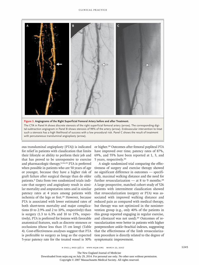
Figure 3. Angiograms of the Right Superficial Femoral Artery before and after Treatment.
The CTA in Panel A shows discrete stenosis of the right superficial femoral artery (arrow). The corresponding digi-tal-subtraction angiogram in Panel B shows stenosis of 99% of the artery (arrow). Endovascular intervention to treat such a stenosis has a high likelihood of success with a low procedural risk. Panel C shows the result of treatment with percutaneous transluminal angioplasty (arrow).
The New England Journal of Medicine Downloaded from nejm.org on July 29, 2014. For personal use only. No other uses without permission.
Copyright © 2007 Massachusetts Medical Society. All rights reserved.

T h e n e w e ng l a nd j o u r na l o f m e dic i n e
n engl j med 356;12 www.nejm.org march 22, 20071246
Data from studies directly comparing exer-cise therapy and PTA are scarce. In one small trial, walking distance at 6 years was better for patients treated with exercise therapy than for those treated with PTA.30 A randomized trial comparing the effects of exercise and PTA (with both groups receiving pharmacologic therapy) demonstrated similar quality-of-life outcomes, similar maximal walking distances, and similar ankle–brachial indexes, but fewer patients in the PTA group had occluded arteries (P = 0.004).31 An analysis of combined data from seven studies that compared exercise with PTA in patients with clau-dication showed that PTA resulted in a greater increase in the ankle–brachial index but with no significant difference in quality of life.32 How-ever, none of the studies included in the analysis compared the two interventions directly. A deci-sion-analysis model comparing the costs and qual-ity of life associated with PTA, bypass surgery, and exercise therapy showed that PTA, when fea-sible, was more effective than exercise therapy alone and was more cost-effective than bypass surgery ($38,000 vs. $311,000 per quality-adjusted year of life gained),33 but this conclusion, too, was not based on data from comparative clinical trials.
A r e a s of Uncerta in t y
Stents
The role of primary stent placement in revascu-larization of the superficial femoral artery re-mains controversial. In contrast to the coronary arteries, for which stent placement has largely
replaced angioplasty for revascularization, the su-perficial femoral artery is subject to longitudinal stretching, external compression, torsion, and flex-ion; these stresses may lead to stent fractures, which have been linked to restenosis.34 In early randomized trials comparing PTA with stent placement for the treatment of lesions in the su-perficial femoral artery, stent placement showed no advantage over angioplasty with bail-out stent placement.35,36
Technology has improved, however, and a re-cent randomized trial involving 104 patients with severe claudication showed significantly higher patency rates at 1 year for lesions in the superfi-cial femoral artery that were treated with stent placement than for lesions treated with PTA and bail-out stent placement (63% vs. 37%); the maxi-mal walking distance and the ankle–brachial in-dex were also significantly better in the stent group at 1 year. There were no major complica-tions in either group.37
A randomized trial of drug-coated (sirolimus) stents in the superficial femoral artery failed to show a reduction in the risk of restenosis as com-pared with the risk associated with bare-metal stents.38 Use of stents covered with polytetra-fluoroethylene (PTFE), developed to seal arterial perforations or to exclude aneurysmal segments, was compared with the use of PTA alone in a randomized trial, and there was no significant difference in 1-year patency rates (50% and 45%, respectively); however, major adverse events were more frequent in the group that received PTFE-covered stents (8.2% vs. 4.0%).39
Table 2. Characteristics of Imaging Methods Used to Diagnose Peripheral Arterial Disease.
Characteristic Duplex UltrasoundDigital-Subtraction
Angiography
Magnetic Resonance
AngiographyComputed Tomographic
Angiography
Advantages Noninvasive, can be used to visualize and quantitate severity of lesion
Gold standard, high resolu-tion, can be used to guide intervention
Noninvasive, no radiation or iodinated contrast material used, three- dimensional
Noninvasive, higher spatial resolution than with mag-netic resonance angiogra-phy, three-dimensional
Disadvantages Operator-dependent, imag-ing limited by dense cal-cification
Invasive, ionizing radiation and iodinated contrast material used, two- dimensional
Lower spatial resolution than with computed tomographic angiogra-phy, contraindicated if patient has claustro-phobia, image artifact if stent present
Ionizing radiation (25% of dose with digital-subtrac-tion angiography) and io-dinated contrast material used, imaging limited by dense calcification
Charge (Medicare)* $252.14 $474.07 $973.75 $530.45
* The charges shown represent the allowable total charge by Medicare in the New Orleans area.
The New England Journal of Medicine Downloaded from nejm.org on July 29, 2014. For personal use only. No other uses without permission.
Copyright © 2007 Massachusetts Medical Society. All rights reserved.

clinical pr actice
n engl j med 356;12 www.nejm.org march 22, 2007 1247
Adjunctive Angioplasty
Adjunctive angioplasty techniques such as ather-ectomy, cryotherapy, and the use of a cutting bal-loon (a balloon with microtomes attached to its surface, which make shallow incisions in the sur-face of the lesion when the balloon is inflated) have not been tested in meaningful comparative trials in patients with femoral artery disease; the only available data that show the benefit of these approaches is from single-center studies and un-controlled registries.40-42 Laser angioplasty has not been shown to be superior to conventional PTA or stent placement.43-45 Given the substan-tial additional expense of these approaches, more evidence of their efficacy is needed before wide-spread adoption can be justified.
Trials of brachytherapy (use of a catheter to deliver radiation to the lesion) for preventing restenosis in the superficial femoral artery after percutaneous PTA have had inconsistent re-sults.46,47 In one trial, the use of external-beam radiation (at a dose of 14 Gy) after PTA signifi-cantly reduced the rate of restenosis at 1 year after PTA,48 but these results require confirmation. The use of gene or cellular therapies in peripheral arte-rial disease has not resulted in a clear clinical
benefit in small studies49; larger, controlled clini-cal trials are needed to establish whether there is any role for these therapies in patients with clau-dication.
Follow-Up after Percutaneous Interventions
It is common to measure the ankle–brachial in-dex within a week after a percutaneous interven-tion has been performed in order to establish a new baseline. Patients are often reevaluated at 3-month intervals for the first year (when the risk of restenosis is highest); these assessments in-clude a detailed history taking, examination, and ankle–brachial index measurements; the value of a walking program and risk-factor modification should be reinforced during these visits. Howev-er, data on the optimal approach to follow-up are lacking.
Guidel ines
Comprehensive guidelines for the management of peripheral arterial disease have recently been published by an expert multidisciplinary com-mittee organized by the American College of Cardiology (ACC) and the American Heart Asso-
Table 3. AHA–ACC Guidelines for Pharmacologic Management of Claudication.*
Medication and Class of Evidence
Level of Evidence Dose Side Effects
Class I
Cilostazol A 100 mg two times/day Contraindicated in heart failure; headache, diarrhea, palpitations, dizziness
Class IIb
Pentoxifylline A 400 mg three times/day Sore throat, dyspepsia, nausea, diarrhea
Arginine B 3 g three times/day Gastrointestinal distress, drop in hematocrit
Propionyl levocarnitine B 1–2 g two times/ day None or mild
Ginkgo biloba B 120–160 mg/day None or mild
Class III
Prostaglandins A Beraprost: 40 µg three times/day Headache, flushing gastrointestinal distress
Vitamin E C 50 mg/day None or mild
Chelation EDTA A 1.5–3 g intravenously two times/wk Hypocalcemia, renal failure, proteinuria, gastrointestinal distress
* Information is from the American Heart Association and the American College of Cardiology (Hirsch et al.9). Class I ev-idence is defined as evidence, general agreement, or both that the treatment is beneficial, useful, and effective; class IIb as conflicting evidence or divergence of opinion about efficacy or usefulness (or efficacy that is less well established by evidence or opinion); and class III as evidence, general agreement, or both that the treatment is not beneficial, useful, and effective. Levels of evidence are classified as follows: level A, data derived from multiple randomized trials or meta-analyses; level B, data derived from a single randomized trial or from nonrandomized studies, and level C, the consen-sus opinion of experts, data from case studies, or the standard of care.
The New England Journal of Medicine Downloaded from nejm.org on July 29, 2014. For personal use only. No other uses without permission.
Copyright © 2007 Massachusetts Medical Society. All rights reserved.

T h e n e w e ng l a nd j o u r na l o f m e dic i n e
n engl j med 356;12 www.nejm.org march 22, 20071248
ciation (AHA).9 The TransAtlantic Inter-Society Consensus, representing multiple international specialty societies, has also published guidelines for the management of peripheral arterial disease that are in general agreement with the ACC–AHA document (Table 4).27
The ACC–AHA guidelines recommend endo-vascular therapy for patients whose condition in-terferes with their job performance or lifestyle and who have had an inadequate response to ex-ercise or pharmacologic therapy, as long as the clinical features suggest a reasonable likelihood of symptomatic improvement and the risk–ben-efit ratio is very high.9 The recommendations in this article are in agreement with the ACC–AHA guidelines.
Summ a r y a nd R ecommendations
The mail carrier described in the vignette has right-calf claudication that interferes with his ability to perform his job, and physical examination shows that his condition is consistent with stenosis or occlusion of the superficial femoral artery. The initial assessment should include measurement of his ankle–brachial index. Risk factors for ath-erosclerosis should be assessed, and appropriate modification instituted, including smoking ces-sation, dietary adjustment, and pharmacotherapy as warranted for dyslipidemia, hypertension, or hyperglycemia. He also should be treated with aspirin (75 mg to 325 mg per day).
Management in this case should include a supervised exercise program and a trial of cilo-stazol for claudication, an approach that is con-sistent with the AHA–ACC guidelines. Revascu-larization is warranted if the condition does not improve with conservative therapy and if the le-sion has anatomical features that are associated with a good outcome of revascularization. PTA is preferred over surgery and may be considered up front if there is a high probability of success and a low procedural risk (Table 4).9 An imaging study (duplex ultrasonography, CTA, or MRA, with the choice guided by the physician’s preference and the availability of local expertise) is indicated in patients who are candidates for revasculariza-tion in order to determine the location and mor-phologic characteristics of the obstructive lesion (or lesions). If a percutaneous intervention is con-sidered to be likely, one can proceed directly to digital-subtraction angiography, with a plan to perform the percutaneous intervention immedi-ately if the angiographic anatomy is suitable. Regardless of the initial therapy for this patient’s claudication, follow-up care should involve on-going attention to control of atherosclerotic risk factors, antiplatelet therapy with aspirin, and en-couragement of the patient to engage in regular exercise.
No potential conflict of interest relevant to this article was reported.
Table 4. TransAtlantic Inter-Society Consensus on Classification of Femoral Lesions and Recommended Approaches When Revascularization Is Planned.*
Lesion Type Characteristics Recommended Treatment
A Single stenosis ≤10 cm longSingle occlusion ≤5 cm long
Percutaneous transluminal angioplasty strong-ly preferred
B Multiple lesions, each ≤5 cm in lengthSingle lesion ≤15 cm long, not involving the popliteal
artery below the kneeSingle or multiple lesions in the absence of continu-
ous tibial vessels for distal bypassHeavily calcified occlusion ≤5 cm longSingle popliteal stenosis
Percutaneous transluminal angioplasty gener-ally preferred
C Multiple lesions >15 cm longRecurrent lesions after two endovascular interventions
Percutaneous transluminal angioplasty or sur-gery, depending on risk–benefit ratio
D Occlusion >20 cm longOcclusion of the popliteal or tibial–peroneal vessels
Surgery generally preferred
* Information is from Norgren et al.27
The New England Journal of Medicine Downloaded from nejm.org on July 29, 2014. For personal use only. No other uses without permission.
Copyright © 2007 Massachusetts Medical Society. All rights reserved.

clinical pr actice
n engl j med 356;12 www.nejm.org march 22, 2007 1249
References
Selvin E, Erlinger TP. Prevalence of and risk factors for peripheral arterial disease in the United States: results from the National Health and Nutrition Exami-nation Survey, 1999-2000. Circulation 2004; 110:738-43.
Pasternak RC, Criqui MH, Benjamin EJ, et al. Atherosclerotic Vascular Disease Conference: Writing Group I: epidemiol-ogy. Circulation 2004;109:2605-12.
Hiatt WR. Medical treatment of pe-ripheral arterial disease and claudication. N Engl J Med 2001;344:1608-21.
McDermott MM, Kerwin DR, Liu K, et al. Prevalence and significance of unrec-ognized lower extremity peripheral arterial disease in general medicine practice. J Gen Intern Med 2001;16:384-90.
McDermott MM, Liu K, Greenland P, et al. Functional decline in peripheral ar-terial disease: associations with the ankle brachial index and leg symptoms. JAMA 2004;292:453-61.
Kannel WB, Skinner JJ Jr, Schwartz MJ, Shurtleff D. Intermittent claudication: incidence in the Framingham Study. Cir-culation 1970;41:875-83.
Muluk SC, Muluk VS, Kelley ME, et al. Outcome events in patients with claudica-tion: a 15-year study in 2777 patients. J Vasc Surg 2001;33:251-7.
Aquino R, Johnnides C, Makaroun M, et al. Natural history of claudication: long-term serial follow-up study of 1244 claudicants. J Vasc Surg 2001;34:962-70.
Hirsch AT, Haskal ZJ, Hertzer NR, et al. ACC/AHA 2005 guidelines for the management of patients with peripheral arterial disease (lower extremity, renal, mesenteric, and abdominal aortic): execu-tive summary a collaborative report from the American Association for Vascular Surgery/Society for Vascular Surgery, So-ciety for Cardiovascular Angiography and Interventions, Society for Vascular Medi-cine and Biology, Society of Interventional Radiology, and the ACC/AHA Task Force on Practice Guidelines (Writing Commit-tee to Develop Guidelines for the Manage-ment of Patients with Peripheral Arterial Disease) endorsed by the American Asso-ciation of Cardiovascular and Pulmonary Rehabilitation; National Heart, Lung, and Blood Institute; Society for Vascular Nurs-ing; TransAtlantic Inter-Society Consensus; and Vascular Disease Foundation. J Am Coll Cardiol 2006;47:1239-312.
Schmieder FA, Comerota AJ. Intermit-tent claudication: magnitude of the prob-lem, patient evaluation, and therapeutic strategies. Am J Cardiol 2001;87:12A:3D-13D.
Ouwendijk R, de Vries M, Pattynama PM, et al. Imaging peripheral arterial dis-ease: a randomized controlled trial com-paring contrast-enhanced MR angiography and multi-detector row CT angiography. Radiology 2005;236:1094-103.
1.
2.
3.
4.
5.
6.
7.
8.
9.
10.
11.
Gradinscak DJ, Young N, Jones Y, O’Neil D, Sindhusake D. Risks of outpa-tient angiography and interventional pro-cedures: a prospective study. AJR Am J Roentgenol 2004;183:377-81.
Singh H, Cardella JF, Cole PE, et al. Quality improvement guidelines for diag-nostic arteriography. J Vasc Interv Radiol 2002;13:1-6.
Chobanian AV, Bakris GL, Black HR, et al. The Seventh Report of the Joint Na-tional Committee on Prevention, Detec-tion, Evaluation, and Treatment of High Blood Pressure: the JNC 7 report. JAMA 2003;289:2560-72. [Erratum, JAMA 2003; 290:197.]
Hankey GJ, Norman PE, Eikelboom JW. Medical treatment of peripheral arte-rial disease. JAMA 2006;295:547-53.
Yusuf S, Sleight P, Pogue J, Bosch J, Davies R, Dagenais G. Effects of an an-giotensin-converting-enzyme inhibitor, ramipril, on cardiovascular events in high-risk patients. N Engl J Med 2000; 342:145-53. [Errata, N Engl J Med 2000; 342:748, 1376.]
Antithrombotic Trialists’ Collabora-tion. Collaborative meta-analysis of ran-domised trials of antiplatelet therapy for prevention of death, myocardial infarc-tion, and stroke in high risk patients. BMJ 2002;324:71-86. [Erratum, BMJ 2002;324: 141.]
CAPRIE Steering Committee. A ran-domised, blinded, trial of clopidogrel ver-sus aspirin in patients at risk of ischaemic events (CAPRIE). Lancet 1996;348:1329-39.
Strandness DE Jr, Dalman RL, Panian S, et al. Effect of cilostazol in patients with intermittent claudication: a random-ized, double-blind, placebo-controlled study. Vasc Endovascular Surg 2002;36: 83-91.
Dawson DL, Cutler BS, Hiatt WR, et al. A comparison of cilostazol and pentox-ifylline for treating intermittent claudica-tion. Am J Med 2000;109:523-30.
Leng GC, Fowler B, Ernst E. Exercise for intermittent claudication. Cochrane Database Syst Rev 2000;2:CD000990.
Bendermacher BL, Willigendael EM, Teijink JA, Prins MH. Supervised exercise therapy versus non-supervised exercise therapy for intermittent claudication. Cochrane Database Syst Rev 2006;2:CD005263.
Gardner AW, Poehlman ET. Exercise rehabilitation programs for the treatment of claudication pain: a meta-analysis. JAMA 1995;274:975-80.
Holm J, Arfvidsson B, Jivegard L, et al. Chronic lower limb ischaemia: a prospec-tive randomised controlled study compar-ing the 1-year results of vascular surgery and percutaneous transluminal angio-plasty (PTA). Eur J Vasc Surg 1991;5:517-22.
12.
13.
14.
15.
16.
17.
18.
19.
20.
21.
22.
23.
24.
Wilson SE, Wolf GL, Cross AP. Percu-taneous transluminal angioplasty versus operation for peripheral arteriosclerosis: report of a prospective randomized trial in a selected group of patients. J Vasc Surg 1989;9:1-9.
Clark TW, Groffsky JL, Soulen MC. Predictors of long-term patency after fem-oropopliteal angioplasty: results from the STAR registry. J Vasc Interv Radiol 2001; 12:923-33.
Norgren L, Hiatt WR, Dormandy JA, Nehler MR, Harris KA, Fowkes FGR. In-ter-Society Consensus for the Manage-ment of Peripheral Arterial Disease (TASC II). Eur J Vasc Endovasc Surg 2007;33:Suppl:S1-S75.
Lundgren F, Dahllof AG, Lundholm K, Schersten T, Volkmann R. Intermittent claudication — surgical reconstruction or physical training? A prospective random-ized trial of treatment efficiency. Ann Surg 1989;209:346-55.
Feinglass J, McCarthy WJ, Slavensky R, Manheim LM, Martin GJ. Functional status and walking ability after lower ex-tremity bypass grafting or angioplasty for intermittent claudication: results from a prospective outcomes study. J Vasc Surg 2000;31:93-103.
Perkins JM, Collin J, Creasy TS, Fletcher EW, Morris PJ. Exercise training versus angioplasty for stable claudication: long and medium term results of a pro-spective, randomised trial. Eur J Vasc En-dovasc Surg 1996;11:409-13.
Whyman MR, Fowkes FG, Kerracher EM, et al. Is intermittent claudication im-proved by percutaneous transluminal an-gioplasty? A randomized controlled trial. J Vasc Surg 1997;26:551-7.
Spronk S, Bosch JL, Veen HF, den Hoed PT, Hunink MG. Intermittent clau-dication: functional capacity and quality of life after exercise training or percuta-neous transluminal angioplasty — system-atic review. Radiology 2005;235:833-42.
de Vries SO, Visser K, de Vries JA, Wong JB, Donaldson MC, Hunink MG. Intermittent claudication: cost-effective-ness of revascularization versus exercise therapy. Radiology 2002;222:25-36.
Scheinert D, Scheinert S, Sax J, et al. Prevalence and clinical impact of stent fractures after femoropopliteal stenting. J Am Coll Cardiol 2005;45:312-5.
Vroegindeweij D, Vos LD, Tielbeek AV, Buth J, Bosch HC. Balloon angioplasty combined with primary stenting versus balloon angioplasty alone in femoropop-liteal obstructions: a comparative ran-domized study. Cardiovasc Intervent Ra-diol 1997;20:420-5.
Muradin GS, Bosch JL, Stijnen T, Hunink MG. Balloon dilation and stent implantation for treatment of femoropop-liteal arterial disease: meta-analysis. Ra-diology 2001;221:137-45.
25.
26.
27.
28.
29.
30.
31.
32.
33.
34.
35.
36.
The New England Journal of Medicine Downloaded from nejm.org on July 29, 2014. For personal use only. No other uses without permission.
Copyright © 2007 Massachusetts Medical Society. All rights reserved.

n engl j med 356;12 www.nejm.org march 22, 20071250
clinical pr actice
Schillinger M, Sabeti S, Loewe C, et al. Balloon angioplasty versus implanta-tion of nitinol stents in the superficial femoral artery. N Engl J Med 2006;354: 1879-88.
Duda SH, Bosiers M, Lammer J, et al. Sirolimus-eluting versus bare nitinol stent for obstructive superficial femoral artery disease: the SIROCCO II trial. J Vasc In-terv Radiol 2005;16:331-8.
Food and Drug Adminstration, Center for Devices and Radiological Health. Gore Viabahn endoprosthesis: summary of safety and effectiveness. PMA application no. P040037. (Accessed February 21, 2007, at http://www.fda.gov/cdrh/pdf4/P040037b. pdf.)
Suri R, Wholey MH, Postoak D, Hagi-no RT, Toursarkissian B. Distal embolic protection during femoropopliteal ather-ectomy. Catheter Cardiovasc Interv 2006; 67:417-22.
Zeller T, Rastan A, Schwarzwalder U, et al. Midterm results after atherectomy-
37.
38.
39.
40.
41.
assisted angioplasty of below-knee arter-ies with use of the Silverhawk device. J Vasc Interv Radiol 2004;15:1391-7.
Laird JR, Biamino G, McNamara T, et al. Cryoplasty for the treatment of femo-ropopliteal arterial disease: extended fol-low-up results. J Endovasc Ther 2006;13:Suppl 2:II-52–II-59. abstract.
Visona A, Perissinotto C, Lusiani L, et al. Percutaneous excimer laser angioplas-ty of lower limb vessels: results of a pro-spective 24-month follow-up. Angiology 1998;49:91-8.
Scheinert D, Laird JR Jr, Schroder M, Steinkamp H, Balzer JO, Biamino G. Ex-cimer laser-assisted recanalization of long, chronic superficial femoral artery occlu-sions. J Endovasc Ther 2001;8:156-66.
Wissgott C, Scheinert D, Rademaker J, Werk M, Schedel H, Steinkamp HJ. Treatment of long superficial femoral ar-tery occlusions with excimer laser angio-plasty: long-term results after 48 months. Acta Radiol 2004;45:23-9.
42.
43.
44.
45.
Diehm N, Silvestro A, Do DD, et al. Endovascular brachytherapy after femo-ropopliteal balloon angioplasty fails to show robust clinical benefit over time. J Endovasc Ther 2005;12:723-30.
Wolfram RM, Budinsky AC, Pokrajac B, Potter R, Minar E. Endovascular brachytherapy: restenosis in de novo ver-sus recurrent lesions of femoropopliteal artery — the Vienna experience. Radiolo-gy 2005;236:338-42. [Erratum, Radiology 2006;239:613.]
Therasse E, Donath D, Lesperance J, et al. External beam radiation to prevent restenosis after superficial femoral artery balloon angioplasty. Circulation 2005;111: 3310-5.
Lederman RJ, Mendelsohn FO, Ander-son RD, et al. Therapeutic angiogenesis with recombinant fibroblast growth fac-tor-2 for intermittent claudication (the TRAFFIC study): a randomised trial. Lan-cet 2002;359:2053-8.Copyright © 2007 Massachusetts Medical Society.
46.
47.
48.
49.
electronic access to the journal’s cumulative index
At the Journal’s site on the World Wide Web (www.nejm.org), you can search an index of all articles published since January 1975
(abstracts 1975–1992, full text 1993–present). You can search by author, key word, title, type of article, and date. The results will include the citations
for the articles plus links to the full text of articles published since 1993. For nonsubscribers, time-limited access to single articles and 24-hour site access can also be ordered for a fee through the Internet (www.nejm.org).
The New England Journal of Medicine Downloaded from nejm.org on July 29, 2014. For personal use only. No other uses without permission.
Copyright © 2007 Massachusetts Medical Society. All rights reserved.
Copyright © 2022 FDOKUMEN