Social behavioral changes in MPTP-treated monkey model of Parkinson's disease
19
ORIGINAL RESEARCH ARTICLE published: 26 February 2015 doi: 10.3389/fnbeh.2015.00042 Social behavioral changes in MPTP-treated monkey model of Parkinson’s disease Elodie Durand 1 *, Odile Petit 2 , Léon Tremblay 3 , Cédric Zimmer 2 , Véronique Sgambato-Faure 3 , Carine Chassain 4 , Marlène Laurent 1 , Bruno Pereira 4 , Céline Silberberg 1 and Franck Durif 1 1 Université d’Auvergne Clermont 1, UFR Médecine, EA 7280 (NPsy-Sydo), Clermont-Ferrand, France 2 Département Ecologie, Physiologie et Ethologie, Institut Pluridisciplinaire Hubert Curien, UMR 7178, CNRS-UDS, Strasbourg, France 3 Centre de Neurosciences Cognitives, UMR 5229 CNRS-Université Lyon 1, Bron, France 4 CHU Clermont Ferrand, Gabriel Montpied, Clermont-Ferrand, France Edited by: Angela Roberts, University of Cambridge, UK Reviewed by: Michael Arthur Van Der Kooij, Ecole Polytèchnique Féderale de Lausanne, Switzerland Julien Vezoli, Ernst Strüngmann Institute for Neuroscience in Cooperation with Max Planck Society, Germany *Correspondence: Elodie Durand, CHU Clermont Ferrand/Service Neurologie Pr Durif, 58 rue Montalembert, 63003 Clermont-Ferrand, France e-mail: edurand2@ chu-clermontferrand.fr Parkinsonian patients experience not only the physical discomfort of motor disorders but also the considerable psychological distress caused by cognitive deficits and behavioral disorders. These two factors can result in a disruption of social relationships during the symptomatic and even the presymptomatic motor states of the disease. However, it remains difficult, if not impossible, to evaluate social relationships in presymptomatic patients. The present study focused on the evaluation of social relationships within a group of female long-tailed macaques during presymptomatic and symptomatic motor states induced by Chronic Low-Dose (CLD) and then Chronic High-Dose (CHD) systemic administration of 1-methyl-4-phenyl-l,2,3,6-tetrahydropyridine (MPTP). Dopaminergic denervation within basal ganglia and cortical areas was evaluated using Positron Emission Tomography (PET) scans with 18 F-DOPA (6-[18F]-fluoro-L-3,4-dihydroxyphenylalanine) radiotracer. Interestingly, social behavioral changes could be identified in the presymptomatic motor state before any motor and/or cognitive impairment occurred. Stronger effects were observed in subordinate animals compared to dominant animals. From baseline state to CLD-presymptomatic motor state, the frequency of emitted affiliative and aggressive behaviors increased. From CLD-presymptomatic to CHD-presymptomatic motor states, the frequency of the three categories of social behaviors (aggressive, submissive and affiliative) decreased. At this time, quantitative data analysis in PET scans highlighted a dopaminergic denervation in the insula and the posterior caudate nucleus. Finally, the frequency of the three categories of social behaviors decreased during the stable-symptomatic motor state compared to baseline and presymptomatic motor states; this was also associated with motor and cognitive disorders and a dopaminergic denervation in all the evaluated cortical and subcortical structures. Keywords: Parkinson’s disease, social behavior, non-human primate model, MPTP, PET imaging INTRODUCTION Idiopathic Parkinson’s disease (PD) is characterized by a loss of dopaminergic neurons in the substantia nigra pars compacta (SNc), resulting in decreased levels about 60% of dopamine release in the striatum and hence causing motor symptoms (bradykinesia, tremor and rigidity) (Kish et al., 1988). However, it is clear that PD is not only a “motor” but also a “cognitive” and “neuropsychiatric” disease (Thobois et al., 2010; Gallagher and Schrag, 2012). Psychiatric disorders include apathetic state (Pedersen et al., 2009; Thobois et al., 2010), anxiety (Gallagher and Schrag, 2012) and depression (Martínez-Martín and Damián, 2010) as well as hypomania, psychosis and impulse control dis- orders observed in patients receiving dopaminergic treatment (Weintraub et al., 2006; Ulla et al., 2012). The pathophysiology of such disorders has not yet been completely understood but includes lesions of the dopaminergic, serotoninergic, and nora- drenergic systems involved in Parkinson’s disease (Hirsch et al., 2003; Kish et al., 2008). However, a change in the mesocorticol- imbic dopaminergic system could play a role in the behavioral disorders of PD (Remy et al., 2005). Indeed, in addition to the lesion of the dopaminergic nigrostriatal system, other dopamin- ergic systems are also damaged in PD, namely those originating in the ventral tegmental area (VTA) that project to the limbic sys- tem, which is involved in the reward circuit (mesolimbic system), and also to the prefrontal cortex, which is involved in person- ality traits (mesocortical system) (Tzschentke, 2001; Haber and Knutson, 2010). The aforementioned non-motor symptoms could have an early impact on the social life of parkinsonian patients, leading some of them to become socially isolated both from themselves and from society. The symptoms impact the quality of life of parkinsonian patients, namely affecting social interactions, com- munication and/or emotion recognition (Schrag et al., 2000; Yoshimura et al., 2005; Pell et al., 2006). Frontiers in Behavioral Neuroscience www.frontiersin.org February 2015 | Volume 9 | Article 42 | 1 BEHAVIORAL NEUROSCIENCE
-
Upload
independent -
Category
Documents
-
view
1 -
download
0
Transcript of Social behavioral changes in MPTP-treated monkey model of Parkinson's disease

ORIGINAL RESEARCH ARTICLEpublished: 26 February 2015
doi: 10.3389/fnbeh.2015.00042
Social behavioral changes in MPTP-treated monkey modelof Parkinson’s diseaseElodie Durand1*, Odile Petit2, Léon Tremblay3, Cédric Zimmer2, Véronique Sgambato-Faure3,
Carine Chassain4, Marlène Laurent1, Bruno Pereira4, Céline Silberberg1 and Franck Durif1
1 Université d’Auvergne Clermont 1, UFR Médecine, EA 7280 (NPsy-Sydo), Clermont-Ferrand, France2 Département Ecologie, Physiologie et Ethologie, Institut Pluridisciplinaire Hubert Curien, UMR 7178, CNRS-UDS, Strasbourg, France3 Centre de Neurosciences Cognitives, UMR 5229 CNRS-Université Lyon 1, Bron, France4 CHU Clermont Ferrand, Gabriel Montpied, Clermont-Ferrand, France
Edited by:
Angela Roberts, University ofCambridge, UK
Reviewed by:
Michael Arthur Van Der Kooij, EcolePolytèchnique Féderale deLausanne, SwitzerlandJulien Vezoli, Ernst StrüngmannInstitute for Neuroscience inCooperation with Max PlanckSociety, Germany
*Correspondence:
Elodie Durand, CHU ClermontFerrand/Service Neurologie Pr Durif,58 rue Montalembert, 63003Clermont-Ferrand, Francee-mail: [email protected]
Parkinsonian patients experience not only the physical discomfort of motor disorders butalso the considerable psychological distress caused by cognitive deficits and behavioraldisorders. These two factors can result in a disruption of social relationships during thesymptomatic and even the presymptomatic motor states of the disease. However, itremains difficult, if not impossible, to evaluate social relationships in presymptomaticpatients. The present study focused on the evaluation of social relationships within agroup of female long-tailed macaques during presymptomatic and symptomatic motorstates induced by Chronic Low-Dose (CLD) and then Chronic High-Dose (CHD) systemicadministration of 1-methyl-4-phenyl-l,2,3,6-tetrahydropyridine (MPTP). Dopaminergicdenervation within basal ganglia and cortical areas was evaluated using Positron EmissionTomography (PET) scans with 18F-DOPA (6-[18F]-fluoro-L-3,4-dihydroxyphenylalanine)radiotracer. Interestingly, social behavioral changes could be identified in thepresymptomatic motor state before any motor and/or cognitive impairment occurred.Stronger effects were observed in subordinate animals compared to dominantanimals. From baseline state to CLD-presymptomatic motor state, the frequency ofemitted affiliative and aggressive behaviors increased. From CLD-presymptomatic toCHD-presymptomatic motor states, the frequency of the three categories of socialbehaviors (aggressive, submissive and affiliative) decreased. At this time, quantitativedata analysis in PET scans highlighted a dopaminergic denervation in the insula andthe posterior caudate nucleus. Finally, the frequency of the three categories of socialbehaviors decreased during the stable-symptomatic motor state compared to baseline andpresymptomatic motor states; this was also associated with motor and cognitive disordersand a dopaminergic denervation in all the evaluated cortical and subcortical structures.
Keywords: Parkinson’s disease, social behavior, non-human primate model, MPTP, PET imaging
INTRODUCTIONIdiopathic Parkinson’s disease (PD) is characterized by a lossof dopaminergic neurons in the substantia nigra pars compacta(SNc), resulting in decreased levels about 60% of dopaminerelease in the striatum and hence causing motor symptoms(bradykinesia, tremor and rigidity) (Kish et al., 1988). However,it is clear that PD is not only a “motor” but also a “cognitive”and “neuropsychiatric” disease (Thobois et al., 2010; Gallagherand Schrag, 2012). Psychiatric disorders include apathetic state(Pedersen et al., 2009; Thobois et al., 2010), anxiety (Gallagherand Schrag, 2012) and depression (Martínez-Martín and Damián,2010) as well as hypomania, psychosis and impulse control dis-orders observed in patients receiving dopaminergic treatment(Weintraub et al., 2006; Ulla et al., 2012). The pathophysiologyof such disorders has not yet been completely understood butincludes lesions of the dopaminergic, serotoninergic, and nora-drenergic systems involved in Parkinson’s disease (Hirsch et al.,
2003; Kish et al., 2008). However, a change in the mesocorticol-imbic dopaminergic system could play a role in the behavioraldisorders of PD (Remy et al., 2005). Indeed, in addition to thelesion of the dopaminergic nigrostriatal system, other dopamin-ergic systems are also damaged in PD, namely those originatingin the ventral tegmental area (VTA) that project to the limbic sys-tem, which is involved in the reward circuit (mesolimbic system),and also to the prefrontal cortex, which is involved in person-ality traits (mesocortical system) (Tzschentke, 2001; Haber andKnutson, 2010).
The aforementioned non-motor symptoms could have anearly impact on the social life of parkinsonian patients, leadingsome of them to become socially isolated both from themselvesand from society. The symptoms impact the quality of life ofparkinsonian patients, namely affecting social interactions, com-munication and/or emotion recognition (Schrag et al., 2000;Yoshimura et al., 2005; Pell et al., 2006).
Frontiers in Behavioral Neuroscience www.frontiersin.org February 2015 | Volume 9 | Article 42 | 1
BEHAVIORAL NEUROSCIENCE

Durand et al. Social behavior in MPTP-treated monkeys
It is therefore legitimate to ask whether a lesion of thedopaminergic system could affect social interactions. What typesof interactions could be affected by a dopaminergic lesion?Finally, which brain regions (basal ganglia and/or cortical areas)may be involved in social interactions changes? Answering thesequestions in patients is problematic, as it would be difficult ifnot impossible to accurately evaluate their social interactions ona daily basis and understand the real social impact of this dis-ease. In this context, the use of a non-human primate modeltherefore appears useful to study the social impact of dopamineneuron loss in Parkinson’s disease. In the wild, Macaca fascicularisfemales remain within their native group throughout their livesand therefore form clans of related individuals. Their ranks aremore stable than those of males and are transmitted from motherto daughter forming a matrilineage that rarely changes over gen-erations (Van Noordwijk and Van Schaik, 1987, 1999; Gumert,2010). Any change in social behavior in a group of Macaca fas-cicularis might therefore impact the balance of inter-individualsocial relationships.
Several previous studies in non-human primates have shownthe important role of several monoamines, such as dopamine,in hierarchical status and the expression of social behaviors inmacaques (Redmond et al., 1971; Kaplan et al., 2002; Morganet al., 2002; Riddick et al., 2009; Nader et al., 2012). Furthermore,neurobiological studies in non-human and human primates havealso shown the role of cortico-limbic regions (namely the amyg-dala, the orbitofrontal cortex, the anterior cingular cortex andthe insula) in the social behavioral network and in social cog-nition generally, which is heavily dependent upon the expres-sion and recognition of emotions (Amaral, 2002; Machado andBachevalier, 2006; Rushworth et al., 2007; Machado et al., 2009).
Currently, the primate model produced with the neurotoxin1-methyl-4-phenyl-l,2,3,6-tetrahydropyridine (MPTP) is consid-ered as the gold standard animal model of PD because of its closeresemblance to PD. Indeed, this model, particularly with systemicMPTP administration in macaques, characteristically replicatesmarked cellular loss in the SNc, the cardinal motor symptomsof PD (including abnormalities in axial movements and pos-tures), and the full extent of motor complications associated withchronic dopaminergic treatment (Jenner, 2003). Furthermore,the primate MPTP model can reproduce non-motor symptomsof PD including cognitive, sleep and gastrointestinal dysfunction(Barraud et al., 2009; Chaumette et al., 2009; Schneider et al.,2010). The chronic low-dose (CLD)-MPTP model of parkin-sonism in non-human primates was specifically developed tostudy the presymptomatic motor state in early PD (Schneiderand Kovelowski, 1990). This model induces a dopaminergicdenervation in the SN/VTA (Schneider, 1990), dopaminergicfrontostriatal cognitive deficits and deficits in spatial delayedresponse, set shifting, planning and impulsivity (Schneider andKovelowski, 1990; Schneider and Roeltgen, 1993; Schneider andPope-Coleman, 1995; Decamp and Schneider, 2004; Schneider,2006; Vezoli et al., 2011) and recently sleep-wake disorders(Videnovic et al., 2014). Moreover, this CLD-MPTP model hasbeen shown to induce a striatal denervation pattern more sim-ilar to the one observed in PD patients (Gibb and Lees, 1991;Perez-Otano et al., 1994) than the one induced by chronic
high-dose or acute MPTP intoxication. Most previous studiesusing chronic high-dose administration of MPTP (CHD-MPTP),mainly focused on motor and cognitive disorders related toneurochemical, imaging or pharmacological findings (Chassainet al., 2001; Madras et al., 2006; Blesa et al., 2012; Neumaneet al., 2012; Kortekaas et al., 2013). Interestingly, a longitudi-nal behavioral study in MPTP-hemiparkinsonian vervet monkeysalso showed a link between changes in social behaviors (aggres-sive and affiliative) and striatal dopamine levels measured by18F-DOPA (6-[18F]-fluoro-L-3,4-dihydroxyphenylalanine) PET(Positron Emission Tomography) scanning (Melega et al., 1996).
Therefore, the aim of the present study was to iden-tify the impact of chronic exposure to low and then tohigh doses of MPTP on the social behavior of six femalelong-tailed macaques living in a social group, and to assessthe time course evolution of cognitive and motor disabili-ties and social behavior disorder(s). PET (Positron EmissionTomography) scanning was used to assess dopaminergic den-ervation in basal ganglia and cortical areas using 18F-DOPA(6-[18F]-fluoro-L-3,4-dihydroxyphenylalanine).
MATERIALS AND METHODSETHICS STATEMENTAll procedures were carried out according to National Instituteof Health and the European Directive 2010/63/EU guidelines andthe Department of Veterinary Services (DDSV Clermont Ferrand,France). These experiments were also carried out according toguidelines published in the Guide for the Care and Use ofLaboratory Animals of the National Institutes of Health. Specificauthorization covering this study was delivered by the regionalanimal ethical committee (Comité d’Ethique en ExpérimentationAnimale Auvergne, C2EA-02) under Permit Number: CE19-08.
ANIMALSExperiments were conducted on six female long-tailed macaques(Macaca fascicularis) (5.2–6.6 years old, weighing 3.6–5.6 kg atthe beginning of the study): monkeys A, B, C, D, E, and F.This number of animals was sufficient for statistical analysisof social behaviors. They were housed together throughout thestudy, had free access to water and received food twice a day.No animals were sacrificed during the study. Standard condi-tions of humidity (55 ± 10%), temperature (24 ± 2◦C) andlight (12-h light/dark cycles) were respected. The housing con-sisted of two rooms connected by a trap door that was left openat all times, each room consisted of three 1.8 m3 cages (1 × 1 ×1.8 m). Each animal therefore had access to a minimum of 1.8 m3,as required by National Institute of Health and the EuropeanDirective 2010/63/EU guidelines.
CONTRACEPTIVE IMPLANTSeveral studies have already demonstrated an effect of menstrualcycle on social behavior (Adams et al., 1985; Michael and Zumpe,1993; Czoty et al., 2009). In order to remove any such influence,all animals (n = 6) were fitted with the subdermal contraceptiveimplant (half an implant/animal), Implanon® (Schering-Plough,USA) primarily used in humans (Isley, 2010) prior to all testingsessions and the evaluation of baseline conditions. This implant
Frontiers in Behavioral Neuroscience www.frontiersin.org February 2015 | Volume 9 | Article 42 | 2

Durand et al. Social behavior in MPTP-treated monkeys
overcomes the effects of menstrual cycles on observed behaviorsand monoamines. Each Implanon® rod consists of an ethylenevinylacetate copolymer core, containing 68 mg of the syntheticprogestin etonogestrel and produces an effect for 3 years.
TOXIN ADMINISTRATIONAll animals (n = 6) were exposed to MPTP hydrochloride(MPTP-HCl) (dissolved in saline, Sigma, St. Louis, USA) bysystemic intramuscular (i.m.) administration. Firstly, animalsreceived 0.1 mg/kg every 4–5 days for 58 weeks (10.7–12.3 mg/kg),i.e., chronic low dose CLD-MPTP protocol, to study the CLD-presymptomatic motor state. A second protocol was then used(6 days after the final CLD-MPTP injection), in which MPTPwas injected once per week at 0.4 mg/kg under light anesthesiausing ketamine (0.5 mg/kg) (Neumane et al., 2012), i.e., chronichigh-dose CHD-MPTP protocol, to study the stable-symptomaticmotor state, although a short CHD-presymptomatic motor statehad still been studied. During this second protocol, animalswere administered 3–17 doses for a period of 4–24 weeks (1.0–6.5 mg/kg) (Table 1; Figure 1).
EXPERIMENTAL DESIGNAnimals had been previously trained for cognitive testing, andtheir social and motor behaviors were assessed over the 5months preceding MPTP administration in order to have aglobal representation of the baseline state. One testing session
was carried out at least 36 h after MPTP injection at 2–3 weekintervals during the CLD-presymptomatic period and weeklyduring the CHD-presymptomatic and stable-symptomatic peri-ods. Thus, 20 testing sessions were performed during the CLD-presymptomatic motor state. During the CHD-presymptomaticmotor state, according to the animals, 2–10 testing sessions wereperformed and during the stable-symptomatic motor state 3 test-ing sessions were assessed. These social, motor and cognitiveevaluations were carried out over 4-day testing sessions (Day1 = 1 h motor behavior assessment (2 animals) + cognitive test-ing, Day 2 = 1 h motor behavior assessment (2 animals) + 6 hsocial behavior assessment, Day 3 = 1 h motor behavior assess-ment (2 animals) + cognitive testing, Day 4 = 6h social behaviorassessment) (Figure 1).
BEHAVIORAL STUDYNo experimenters were present in the testing room [three 1.8 m3
cages (1 × 1 × 1.8 m) with separating grids removed to obtainan aviary system] during social and motor assessments. This wasachieved through the use of three black and white wall cam-eras connected to a server and equipped with a Smart DigitalVideo Recorder for Life Sciences, also known as “Numeriscope”(View Point, Lyon, France). In addition, a color camera fixed tothe ceiling and rotating 360◦ (View Point, Lyon, France) trackedanimal movements in real-time and specifically zoomed in on thefaces of the animals to discriminate different facial expressions.
Table 1 | Individual sensitivity to MPTP during the CLD-MPTP and the CHD-MPTP protocols (number of MPTP injections, total amount of MPTP
administered (mg/kg) and related clinical score).
Dominant Subordinate
Monkey A Monkey B Monkey C Monkey D Monkey E Monkey F
CLD-presymptomatic period Number of injections during theCLD-MPTP protocol
110 110 110 110 110 110
Total amount of MPTP (mg/kg)administered during theCLD-MPTP protocol
11.0 11.0 11.0 11.0 11.0 11.0
Clinical score during theCLD-presymptomatic motor state
0.3 0.5 1.7 1 1.6 0.3
CHD-presymptomatic period Number of MPTP injection duringthe CHD-MPTP protocol for thepresymptomatic motor state
10 2 2 2 6 2
Total amount of MPTP (mg/kg)administered during theCHD-MPTP protocol for thepresymptomatic motor state
4.0 0.8 0.8 0.8 2.4 0.8
Clinical score during theCHD-presymptomatic motor state
2.2 2.5 4.7 2.8 3.6 3.0
Stable-symptomatic period Number of MPTP injection duringthe CHD-MPTP protocol for thestable-symptomatic motor state
7 3 3 1 1 1
Total amount of MPTP (mg/kg)administered during theCHD-MPTP protocol for thestable-symptomatic motor state
2.8 1.2 1.2 0.4 0.4 0.4
Clinic score during thestable-symptomatic motor state
6.5 7.4 7.6 7.9 8 7.6
Frontiers in Behavioral Neuroscience www.frontiersin.org February 2015 | Volume 9 | Article 42 | 3

Durand et al. Social behavior in MPTP-treated monkeys
FIGURE 1 | Experimental design. Animals had been previously trained forcognitive testing, and their social and motor behaviors were assessed overthe 5 months preceding MPTP administration in order to have a globalrepresentation of the baseline state. Then, all animals (n = 6) were exposedto MPTP hydrochloride (MPTP-HCl) by systemic intramuscular (i.m.)administration. Firstly, animals received 0.1 mg/kg every 4–5 days for 58weeks, i.e., chronic low dose CLD-MPTP protocol, to study theCLD-presymptomatic motor state. A second protocol was then used (6 daysafter the final CLD-MPTP injection), in which MPTP was injected once perweek at 0.4 mg/kg under light anesthesia, i.e., chronic high-dose CHD-MPTPprotocol, to study the stable-symptomatic motor state, although a shortCHD-presymptomatic motor state had still been studied. During this secondprotocol, animals were administered 3–17 doses for a period of 4–24 weeksto develop the first parkinsonian-like motor symptoms, depending onindividual body weight and the individual variability of sensitivity to MPTP. Tostabilize the parkinsonian-like motor symptoms (≥1 month), animals received
no or several additional MPTP injections according to fluctuations of theparkinsonian-like symptoms. One testing session was carried out at least36 h after MPTP injection at 2–3 week intervals during theCLD-presymptomatic period and weekly during the CHD-presymptomaticand stable-symptomatic periods. These social, motor and cognitiveevaluations were carried out over 4-day testing sessions (Day 1 = 1 h motorbehavior assessment (2 animals) + cognitive testing, Day 2 = 1 h motorbehavior assessment (2 animals) + 6 h social behavior assessment, Day 3 =1 h motor behavior assessment (2 animals) + cognitive testing, Day 4 = 6 hsocial behavior assessment). Three PET scans were performed: the first 18weeks before the end of the CLD-MPTP protocol (CLD PET), the second 2weeks after CHD-MPTP protocol (CHD PET) and the third during thestable-symptomatic motor state (Sympto PET). All scanned animals were inthe CLD-presymptomatic motor state during the CLD PET and also in theCHD-presymptomatic motor state during the CHD PET. All PET scans wereperformed at least 5 days after the MPTP injection.
Each evaluation session was recorded to allow evaluations by twoexperimenters with no previous knowledge of the state of theanimal.
Motor assessmentVideo observation was used to evaluate animals, and motorbehavior was scored five times during the 1-h protocol in orderto have an average score for each motor test. Two animals, housedseparately in adjacent cages (1 × 1 × 1.8 m for each cage), couldbe evaluated simultaneously and independently.
Clinical rating scale. Clinical rating was inspired from theCanadian rating scale (Gomez-Mancilla et al., 1993; Chassainet al., 2001). Clinical symptoms data presented the total scorefor posture (0 = normal; 1 = intermittent flexion of trunk andlimbs; 2 = constant flexion of trunk and limbs; 3 = crouch posi-tion), mobility (0 = normal; 1 = mild decrease; 2 = moderatedecrease; 3 = severe decrease), gait (0 = normal; 1 = slow; 2 =very slow; 3 = very slow with freezing) and tremor (0 = absent;1 = mild postural tremor; 2 = moderate postural tremor; 3 =resting tremor) (maximum total score of 12). A clinical score > 0and = 6, was defined as the presymptomatic motor state; finallyfor a clinical score > 6, the individual was considered to be devel-oping parkinsonian-like motor symptoms in stable-symptomaticmotor state (Chassain et al., 2001).
Locomotor activity assessment. Locomotor activity was assessedusing a Vigie Primates® image analyzer system (View Point, Lyon,
France) (Chassain et al., 2001). The system was comprised of avideo camera connected to a video image analyzer system thatcalculated the quantity and quality of the locomotor activity inreal time. The images were digitized with a 800 × 600 pixel def-inition on 256 gray levels, and the changes in gray level in pixelsfrom one image to the next were counted every 80 ms to plot araw activity curve (Chassain et al., 2001). It was possible to changethe following parameters: (i) the detection sensitivity determiningthe threshold from which a pixel is considered to have changedfrom one image to the next, (ii) the duration of data acquisi-tion before obtaining a summary of the activities of the animal inquestion during that period, and (iii) the duration of the experi-ment, which could range from one second to several days (a 1-htesting session during this study). From the raw curve, the activ-ity of each animal could be separated into three states. The firststate corresponded to inactivity of the animal, the second to nor-mal activity and the third to hyperactivity. Thresholds betweenthe three states could be adjusted to discriminate between themovements of the animal. During this study the time spent inan inactive state was specifically evaluated to ensure a good corre-lation between increased periods of inactivity and clinical motordisorders (Chassain et al., 2001).
Social assessmentDominance hierarchy. Hierarchical stability was verified overthe 5 months preceding the first MPTP injection. It was mea-sured via the tabulation of unidirectional conflicts and avoidancesinto a matrix of agonistic behaviors (aggressive and submissive
Frontiers in Behavioral Neuroscience www.frontiersin.org February 2015 | Volume 9 | Article 42 | 4

Durand et al. Social behavior in MPTP-treated monkeys
behaviors) which were then reorganized into a dominance matrix(Angst, 1975; Bentley-Condit and Smith, 1999; Riddick et al.,2009).
Hierarchical status was assessed by the hierarchical steepnessvalue, which shows the size of the absolute differences in over-all dominance success between individuals of adjacent ranks.In other words, the steeper the slope between two individu-als, the stronger the difference between two dominance rankswill be. The slope was measured using SOCPROG.6 software(Whitehead, 2009). Individual hierarchical status ranking wasdetermined using the David’s score for each animal (Gammellet al., 2003). The most dominant animal (monkey A) presentedthe highest David’s score and the most subordinate animal (mon-key F) presented the lowest value. The dominance hierarchy ofthe social group was linear and organized as followed: mon-key A > monkey B > monkey C > monkey D > monkey E >
monkey F.
Social observations. In this study, 19 social behaviors wereassessed and were categorized as aggressive, submissive or
affiliative behaviors (Van Hooff, 1967; Morgan et al., 2000;Brent and Veira, 2002; Kaplan et al., 2002; Camus et al., 2013)(Figure 2). All behaviors required animal movement (displace-ment, stiff approach, lunge, chase, avoid) and some required facialexpressions (open mouth display, stare, silent bared teeth display,teeth chatter, lipsmack display). For each animal, all social behav-iors were recorded for 2 h using the focal animal sampling method(Altmann, 1974) and the LabWatcher software (View Point, Lyon,France). In these focal group sessions, each behavior was recordedin terms of emission and reception. The average frequency ofthe three categories of behavior over a period of 2 h was shownfor each of the three motor states (normal, CLD-presymptomaticfollowing CLD-MPTP protocol, CHD-presymptomatic follow-ing CHD-MPTP protocol and stable-symptomatic). The assess-ment order of each individual social behavior was randomlydefined using the Kendall and BB Smith Table. All obser-vations were carried out between 8.00 a.m. and 2.00 p.m.Animals were fed with fruit and vegetables at 7.30 am, and hadad libitum access to pellets and water during social behaviorevaluation.
FIGURE 2 | Social behaviors of long-tailed macaques. Social behaviors oflong-tailed macaques are split into three categories: Aggression, Submissionand Affiliation. In this study, 19 social behaviors were assessed, some of which
required motor abilities (i.e., all facial expressions: open mouth display, stare,silent bared teeth display, teeth chatter, lipsmack display), and all of whichrequired animal movement (displacement, stiff approach, lunge, chase, avoid).
Frontiers in Behavioral Neuroscience www.frontiersin.org February 2015 | Volume 9 | Article 42 | 5

Durand et al. Social behavior in MPTP-treated monkeys
COGNITIVE STUDY: ORDT (OBJECT RETRIEVAL DETOUR TASK)The ORDT was used to assess the ability of the animals to retrievean object (a piece of fruit) from inside a transparent box that onlyopened on one side (Taylor et al., 1990; Roeltgen and Schneider,1991; Schneider and Pope-Coleman, 1995; Palfi et al., 1996; Vezoliet al., 2011). The Plexiglas box (15 × 15 × 5 cm) was fixed on atray that was adapted to the home cage of the animal. The exper-imenter modified the cognitive level and motor skills required tosolve the task and retrieve the piece of fruit by varying the locationof the box in relation to the subject, the location of the rewardinside the box, and finally the orientation of the open side ofthe box in relation to the subject. As shown in SupplementaryMaterial, each testing session consisted of 35 trials that were ran-domly presented to the animals. This testing session contained“difficult” or “detour” trials in which the animal had to make adetour around a closed side of the box to reach the reward (detourto reach the reward: see configurations 1–20 in SupplementaryMaterial), and “easy” trials which were defined as trials in whichthe opening of the box was facing the animal (no detour to reachthe reward: see configurations 21–35 in Supplementary Material).Subjects were allowed 1 min to retrieve the reward, and then thebox was set up for the next trial. The movements and responsesof the animal involving the tray or the box were not restrained inany way.
Measures of performance included the number of successesand errors on detour or easy trials. Successes (retrieval of rewardon the first reach) were expressed as a percentage of the totalnumber of detour and easy trials. Errors (barrier hits, i.e., hit-ting a transparent side of the box) were expressed as percentageof responses observed (there could be several responses per trialexcept in the case of success) for easy or detour trials.
Four of the six animals were trained to come and work inthe testing cage, without being forced in any way. Animals weretrained during 11–18 sessions to achieve the cognitive task with aminimum of 75% successfully trials during three consecutive ses-sions. The other 2 animals (the third dominant animal: monkeyC and the second subordinate animal: monkey E) refused to workon this test despite several weeks of training.
IMAGING STUDY: PET SCANSPET scan designOwing to limited access to the PET facility, only 4 animals of thesocial group were selected. On the one hand, the 3 animals withthe greatest behavioral social changes, namely the three most sub-ordinate animals, were selected for the imaging part of the study,waiting for variation of 18F-DOPA uptake (Ki values) in the brainstructures explored. On the other hand, the most dominant ani-mal was also selected because of its essential role in the stability ofthe social group (Petit and Thierry, 1992). Three PET scans wereperformed: the first 18 weeks before the end of the CLD-MPTPprotocol (CLD PET), the second 2 weeks after CHD-MPTP pro-tocol (CHD PET) and the third during the stable-symptomaticmotor state (Sympto PET). All scanned animals were in the CLD-presymptomatic motor state during the CLD PET and also in theCHD-presymptomatic motor state during the CHD PET. All PETscans were performed at least 5 days after the MPTP injection.Since no PET scan could be performed during the baseline state,
all data obtained from the three PET scans for the four MPTP-treated animals were compared to data from PET scan performedin five randomly selected healthy animals (Control PET), consid-ered as basal levels data. Finally, we compared the three PET scansperformed for the four MPTP-treated animals.
It was important to note here that the measurement of 18F-DOPA uptake in the control healthy animas showed less than10% variability, except in the subtantia nigra (13%), althoughno information was available regarding their hierarchical status.Moreover, previous PET studies with test-retest data have shown agood reproducibility of radiotracer binding measurement (Costeset al., 2007; Ballanger et al., 2013).
AcquisitionsAnimals were fasted overnight prior to MRI (Magnetic ResonanceImaging) and PET exams. On the day of the experiment, ani-mals were pretreated with Atropine (0.05 mg/kg i.m.) and wereanesthetized with Zoletil (15 mg/kg i.m.) 15 min later. LactatedRinger’s solution was continuously infused through a saphenousvein catheter. Animals were then transported to the ImagingCenter (CERMEP, Lyon, France) where they were placed on astereotaxic apparatus. Respiratory frequency, pO2 and heart ratewere monitored throughout the experiment. One hour after theanesthesia, PET scans were performed in a three-dimensional(3D) mode using a Siemens CTI HR+ tomograph, with an axialfield of view of 15.2 cm, yielding 63 planes and a nominal in-plane resolution of 4.1 mm full width at half maximum (FWHM).Before the tracer injection, a transmission scan (68Ge rotatingrod sources; 10 min) was acquired to correct for tissular 511 keVgamma attenuation. Dynamic acquisition started with the intra-venous (i.v.) injection of 18F-DOPA (138 ± 7.4 MBq) (Neumaneet al., 2012).
PET data processing and analysisThe 3D emission data were reconstructed with attenuationand scatter correction by a 3D filtered backprojection algo-rithm (Hamming filter; cut-off frequency, 0.5cycles/pixel) and azoom factor of three, giving a transaxial resolution of 6.5 mmFWHM. Reconstructed volumes were 128 × 128 matrices of0.32 × 0.32 mm2 pixels in sixty-three 2.42 mm spaced planes. Theextraction of tissue time-activity concentration curves from auto-mated delineated regions of interest was made possible by thespecific Macaca fascicularis brain atlas (Ballanger et al., 2013).The influx rate constant of 18F-DOPA (Ki) was calculated bylinearization of the graphical Patlak plot (Patlak et al., 1983)over 90 min post-injection using the cerebellum as the referenceregion.
During this study, a limited number of areas with signifi-cant 18F-DOPA uptake (Ki values) were assessed. Areas withinthe Basal Ganglia were the Anterior and Posterior CaudateNucleus (Ant CdN/Post CdN), the Anterior Putamen (Ant Put),the ventral striatum (V Str), the Posterior Dorsal and VentralPutamen (Post D Put/Post V Put) and the Substantia Nigra(SN). Areas assessed at the cortical level were the Insula (Ins),the Orbitofrontal Cortex (OFC), the Anterior Cingulate Cortex(ACC) and the Amygdala (Amyg). The cerebellum was the brainregion reference.
Frontiers in Behavioral Neuroscience www.frontiersin.org February 2015 | Volume 9 | Article 42 | 6

Durand et al. Social behavior in MPTP-treated monkeys
STATISTICAL ANALYSISStatistical analyses were performed with Statistica statistical soft-ware (version 10; StatSoft, Inc. Tulsa, USA). After verifying thatthe data followed a normal distribution, we carried out Analysis ofVariance with Repeated Measures followed by the Newman-Keulspost-hoc test for social, cognitive and locomotor activity data.Clinical motor score data were analyzed using a paired Studentt-test. PET scans data analysis between groups were performedwith Random Effects Models, making it possible to consider boththe group as fixed effect and the intra-subject variability with sub-ject as random effects. PET scans intra-group data analysis inthe MPTP-treated animals were performed with Chi2-test. Thecorrelations between measures were calculated using Pearson’scorrelation. In all cases, significance was accepted at the 95% ofconfidence level (p < 0.05).
RESULTSMOTOR BEHAVIOR ANALYSESNo animals developed any parkinsonian-like motor symptomsduring the CLD-MPTP protocol. The clinical score was below thethreshold score of 6. No changes were observed in the time ofinactivity from the baseline to the CLD-presymptomatic motorstates (Figures 3A,B).
During the CHD-MPTP protocol following the CLD-MPTPprotocol, two periods were observed. The first period corre-sponded to the CHD-presymptomatic motor state, and includeda significant increase in the clinical score compared to the CLD-presymptomatic motor state (3.1 ± 0.9 vs. 0.9 ± 0.6; t = 11.28,p < 0.001), although the score stayed below the threshold valueof 6. No significant increase in the time of inactivity was observedduring this state. The second period corresponded to the appear-ance of stable parkinsonian-like motor symptoms, and a signif-icant increase of clinical score was observed in comparison tothe CHD-presymptomatic (7.5 ± 0.6 vs. 3.1 ± 0.9; t = 13.26,p < 0.001) and the CLD-presymptomatic (0.9 ± 0.6; t = 29.97,p < 0.001) motor states. At this state, all animals had a clinicalscore > 6. The time spent in inactivity during this period was pos-itively correlated with clinical score (r = 0.69, p < 0.001) and wassignificantly higher than the CHD-presymptomatic [3510.9 ±81.1 s vs. 2853.6 ± 435.0 s; F(3, 20) = 9.69, p < 0.001; post-hoctest, p < 0.01] and the CLD-presymptomatic [2633.2 ± 644.5 s;F(3, 20) = 9.69, p < 0.001; post-hoc test, p < 0.01] motor states(Figures 3A,B). No significant difference was observed betweenthe two subgroups (Dominant vs. Subordinate). However, highindividual variability was observed in the sensitivity to MPTP,with varying numbers of CHD-MPTP injections and amounts ofMPTP required to develop parkinsonian-like motor symptoms.Indeed, monkeys B, C, D, and F seemed to be more sensitive toMPTP than monkeys A and E (Table 1).
SOCIAL ANALYSESBaseline stateThe baseline frequency of agonistic behaviors, i.e., aggressivebehaviors (emitted: 24.2 ± 23.3; received: 14.2 ± 11.3) and sub-missive behaviors (emitted: 34.1 ± 27.2; received: 32.3 ± 37.8),reflected the formal dominance within the social group, i.e., eachanimal recognized its own social status and that of its congeners
FIGURE 3 | Motor abilities from baseline to presymptomatic and
symptomatic motor states (n = 6). (A) Clinical score: values weremeans ± SD (n = 6). ¤¤¤p < 0.001: CHD-presymptomatic andstable-symptomatic motor states vs. CLD-presymptomatic motor state;###p < 0.001: stable-symptomatic motor state vs. CHD-presymptomaticmotor state (Student t-test). (B) Time spent in inactivity: values weremeans ± SD (n = 6). ∗∗∗p < 0.001: stable-symptomatic motor state vs.baseline state; ¤¤p < 0.01: symptomatic motor state vs.CLD-presymptomatic motor state; ##p < 0.01: stable-symptomatic motorstate vs. CHD-presymptomatic motor state (ANOVA and post-hoc test).
(Figures 4A,B). The affiliative behaviors category showed thehighest frequency of appearance (emitted: 59.7 ± 26.7; received:54.3 ± 26.2) (Figure 4C).
CLD-presymptomatic motor stateThere was no significant change in the frequency of emittedand received aggressive behaviors from the CLD-presymptomaticmotor state compared to the baseline state, although a markedincrease was observed (emitted: 53.5 ± 32.2 vs. 24.2 ± 23.3;received: 35.7 ± 46.2 vs. 14.2 ± 11.3) (Figure 4A). No sig-nificant changes were seen in the frequency of emitted andreceived submissive behaviors (Figure 4B). The frequency ofemitted affiliative behaviors significantly increased during theCLD-presymptomatic motor state compared to the baseline state[83.1 ± 27.7 vs. 59.7 ± 26.7; F(3, 20) = 4.02, p < 0.05; post-hoctest, p < 0.05] (Figure 4C).
In subgroup social analyses, there was no significant changein the frequency of emitted and received aggressive behav-iors. However, a marked increase in the frequency of emittedand received aggressive behaviors in subordinate animals wasobserved during the CLD-presymptomatic motor state compared
Frontiers in Behavioral Neuroscience www.frontiersin.org February 2015 | Volume 9 | Article 42 | 7

Durand et al. Social behavior in MPTP-treated monkeys
FIGURE 4 | Evolution of social behaviors from baseline to presymptomatic
and symptomatic motor states within the social group (n = 6). (A)
Aggressive behaviors were recorded in terms of emission and reception usingthe focal animal sampling method for each monkey. Values were means ± SD(n = 6). §p < 0.05: CLD-presymptomatic motor state vs. stable-symptomaticmotor state (ANOVA and post-hoc test). (B) Submissive behaviors were
recorded in terms of emission and reception using the focal animal samplingmethod for each monkey. (C) Affiliative behaviors were recorded in terms ofemission and reception using the focal animal sampling method for eachmonkey. Values were means ± SD (n = 6). ∗p < 0.05: CLD-presymptomaticmotor state vs. baseline state; §§ p < 0.01 § p < 0.05: CLD-presymptomaticmotor state vs. stable-symptomatic motor state (ANOVA and post-hoc test).
to the baseline state (emitted: 60.2 ± 47.6 vs. 9.8 ± 11.6; received:62.6 ± 55.0 vs. 24.0 ± 3.1) (Figure 5A). No significant changeswere seen in the frequency of emitted and received submissivebehaviors whatever the social subgroup (Figure 5B). No changeoccurred in the frequency of emitted affiliative behaviors of dom-inant animals. Unlike dominant animals, the frequency of emittedaffiliative behaviors of subordinate animals significantly increasedduring the CLD-presymptomatic motor state compared to thebaseline state [102.6 ± 13.4 vs. 63.8 ± 41.1; F(3, 8) = 9.43, p <
0.05; post-hoc test, p < 0.05]. No significant change occurred inthe frequency of received affiliative behaviors of dominant andsubordinate animals (Figure 5C).
Individual data (Table 2) were used to determine the inter-individual involvement of each animal in these marked changesobserved during the CLD-presymptomatic motor state comparedto the baseline state. Monkey A, the most dominant animal, pre-sented no major variation in its frequency of emitted aggressivebehaviors in terms of emission or reception. Moreover, whilst itsfrequency of emitted affiliative behaviors increased almost two-fold, no variation was noted in its frequency of received affiliativebehaviors. Monkey B, the second most dominant animal, pre-sented no major variation in its frequency of emitted aggressivebehaviors but the reception of aggressive behaviors was half ashigh as the baseline values. Moreover, it did not present anymajor variation in its frequency of affiliative behaviors in termsof emission or reception. The four others animals in the socialgroup showed a higher variation in the frequency of aggressive
behaviors. Indeed, the frequency of emitted aggressive behaviorsin monkey C increased almost three-fold, whilst its frequency ofreceived aggressive behaviors increased six-fold. Moreover, it didnot show any major variation of its frequency of affiliative behav-iors, in terms of emission and reception. For monkey D, its fre-quency of emitted aggressive behaviors decreased about 3.5 timeswhilst the frequency of received aggressive behaviors increasedalmost five-fold. Moreover its frequency of emitted affiliativebehaviors increased almost two-fold and no major variation wasobserved in its frequency of received affiliative behaviors. MonkeyE presented a marked increase of about 14.5 times its frequencyof emitted aggressive behaviors and did not show any variationin its frequency of received aggressive behaviors. Moreover, it didnot shown any major variation in its frequency of emitted affil-iative behaviors but its frequency of received affiliative behaviorsincreased almost two-fold. Finally, the most subordinate animal,monkey F, surprisingly emitted aggressive behaviors that had notbeen observed during the baseline state, and did not show anymajor variation in its frequency of received aggressive behaviors.Moreover, it showed a three-fold increase in its frequency of emit-ted affiliative behaviors and its frequency of received affiliativebehaviors increased almost two-fold.
Interestingly, the dominance hierarchy within the socialgroup was modified during the CLD-presymptomatic motorstate (A>B>C>E>F>D) compared to the baseline state(A>B>C>D>E>F). Thus, monkey D presented the lowest hier-archical status.
Frontiers in Behavioral Neuroscience www.frontiersin.org February 2015 | Volume 9 | Article 42 | 8

Durand et al. Social behavior in MPTP-treated monkeys
FIGURE 5 | Evolution of social behaviors from baseline to
presymptomatic and symptomatic motor states within the two social
subgroups (n = 3 in each subgroup). (A) Aggressive behaviors wererecorded in terms of emission and reception using the focal animal samplingmethod for each monkey. (B) Submissive behaviors were recorded in termsof emission and reception using the focal animal sampling method for eachmonkey. (C) Affiliative behaviors were recorded in terms of emission andreception using the focal animal sampling method for each monkey. Values
were means ± SD (Dominant group: n = 3; Subordinate group: n = 3).∗p < 0.05: Subordinate CLD-presymptomatic motor state vs. baseline state;#p < 0.05: Subordinate CLD-presymptomatic motor state vs.CHD-presymptomatic motor state; §§p < 0.01: SubordinateCLD-presymptomatic motor state vs. stable-symptomatic motor state;∗p < 0.05: Dominant stable-symptomatic motor state vs. baseline state;¤p < 0.05: Dominant stable-symptomatic motor state vs.CLD-presymptomatic motor state (ANOVA and post-hoc test).
CHD-presymptomatic motor stateThere was no significant change in the frequency of emit-ted and received aggressive behaviors compared to the baselineand the CLD-presymptomatic motor states, although a markeddecrease was observed during the CHD-presymptomatic motorstate compared to the CLD-presymptomatic motor state (emit-ted: 21.7 ± 20.7 vs. 53.5 ± 32.2; received: 12.4 ± 10.0 vs. 35.7 ±46.2) (Figure 4A). No significant changes were seen in the fre-quency of emitted and received submissive behaviors. However,a decrease in the frequency of emitted submissive was observedduring the CHD-presymptomatic motor state compared to theCLD-presymptomatic motor state (21.1 ± 17.5 vs. 35.8 ± 36.3)(Figure 4B). There was no significant change in the frequencyof emitted and received affiliative behaviors compared to thebaseline and the CLD-presymptomatic motor states, although adecrease was observed during the CHD-presymptomatic motorstate compared to the CLD-presymptomatic motor state (emit-ted: 59.4 ± 27.1 vs. 83.1 ± 27.7; received: 48.9 ± 16.6 vs. 66.3 ±20.6) (Figure 4C).
In subgroup social analyses, there was no significant changein the frequency of emitted and received aggressive behav-iors. However, a marked decrease in the frequency of emittedand received aggressive behaviors in subordinate animals wasobserved during the CHD-presymptomatic motor state com-pared to CLD-presymptomatic motor state (emitted: 15.7 ±26.7 vs. 60.2 ± 47.6; received: 21.0 ± 3.8 vs. 62.6 ± 55.0). No
significant change in the frequency or emitted and receive sub-missive behaviors was observed. However, a marked decrease inthe frequency of emitted submissive behaviors in subordinate ani-mals was observed during the CHD-presymptomatic motor statecompared to the CLD-presymptomatic motor state (36.7 ± 4.1 vs.62.6 ± 32.3). No significant change in the frequency or emittedand receive affiliative behaviors was observed. However, a markeddecrease in the frequency of emitted affiliative behaviors in sub-ordinate animals was observed during the CHD-presymptomaticmotor state compared to the CLD-presymptomatic motor state(70.2 ± 23.4 vs. 102.6 ± 13.4).
Individual data (Table 2) were used to determine the inter-individual involvement of each animal in these marked changesobserved during the CHD-presymptomatic motor state com-pared to the CLD-presymptomatic motor state. Monkey A, themost dominant animal, presented a decrease about 3 times inits frequency of emitted aggressive behaviors and did not showany variation in its frequency of received aggressive behaviors. Novariation in its frequency of emitted submissive behaviors wasobserved, whilst its frequency of received submissive behaviorsdecreased almost two-fold. Finally, monkey A did not presentany variation in its frequency of affiliative behaviors in termsof emission and reception. Monkey B, the second most domi-nant animal, presented no major variation in its frequency ofaggressive and submissive behaviors in terms of emission andreception. Moreover, its frequency of emitted affiliative behaviors
Frontiers in Behavioral Neuroscience www.frontiersin.org February 2015 | Volume 9 | Article 42 | 9

Durand et al. Social behavior in MPTP-treated monkeys
Table 2 | Individual frequency of the three categories of social behaviors during the baseline state, the CLD and CHD-presymptomatic motor
states and the stable-symptomatic motor state.
Dominant Subordinate
Monkey A Monkey B Monkey C Monkey D Monkey E Monkey F
Frequency of EMITTEDAGGRESSIVE BEHAVIORS
Baseline state 58.0 46.7 11.3 22.7 6.7 0.0
CLD-presymptomatic motor state 47.8 60.0 32.5 6.5 97.0 77.2
CHD-presymptomatic motor state 17.1 46.0 19.7 46.5 0.6 0.0
Stable-symptomatic motor state 26.3 6.0 8.3 25.3 0.3 0.0
Frequency of RECEIVEDAGGRESSIVE BEHAVIORS
Baseline state 0.0 9.3 4.0 24.7 26.7 20.7
CLD-presymptomatic motor state 0.1 4.2 22.0 126.1 28.5 33.3
CHD-presymptomatic motor state 0.1 7.0 4.3 21.5 17.0 24.5
Stable-symptomatic motor state 0.0 2.7 2.7 15 11.3 20.7
Frequency of EMITTEDSUBMISSIVE BEHAVIORS
Baseline state 1.3 17.3 14.7 44.7 72.0 54.7
CLD-presymptomatic motor state 0.6 6.7 19.7 99.3 39.1 49.3
CHD-presymptomatic motor state 1.1 8.0 7.3 38.0 32.1 40.0
Stable-symptomatic motor state 0.0 4.7 4.7 20.7 22.0 28.3
Frequency of RECEIVEDSUBMISSIVE BEHAVIORS
Baseline state 96.7 58.7 10.0 22.7 5.3 0.7
CLD-presymptomatic motor state 52.8 48.6 12.4 8.9 35.3 17.5
CHD-presymptomatic motor state 21.9 46.0 9.3 46.5 0.4 0.0
Stable-symptomatic motor state 38.3 10.7 8.7 29.3 0.0 0.0
Frequency of EMITTEDAFFILIATIVE BEHAVIORS
Baseline state 49.3 62.7 54.7 54.7 108.7 28.0
CLD-presymptomatic motor state 87.6 64.2 38.9 99.7 117.3 91.0
CHD-presymptomatic motor state 84.0 30.5 31.3 76.0 90.1 44.5
Stable-symptomatic motor state 96.3 22.7 20.3 49.0 69.7 29.0
Frequency of RECEIVEDAFFILIATIVE BEHAVIORS
Baseline state 101.3 66.0 50.0 43.3 30.7 34.7
CLD-presymptomatic motor state 90.5 78.7 43.8 46.1 54.5 84.3
CHD-presymptomatic motor state 77.4 52.5 42.7 51.5 27.5 42.0
Stable-symptomatic motor state 78.0 30.7 26.7 33.0 33.3 33.0
decreased almost two-fold, whilst no major variation in its fre-quency of received affiliative behaviors was observed. The fourothers animals in the social group showed a higher variation inthe frequency of social behaviors. Indeed, the frequency of emit-ted aggressive behaviors in monkey C decreased almost two-fold,whilst its frequency of received aggressive behaviors decreasedabout 5 times. Its frequency of emitted submissive behaviorsdecreased almost three-fold whilst no major variation in its fre-quency of received submissive behaviors was observed. Finally,monkey C did not present any variation in its frequency of affil-iative behaviors in terms of emission and reception. For monkeyD, its frequency of emitted aggressive behaviors increased about 7times and its frequency of received aggressive behaviors decreasedalmost six-fold. Its frequency of emitted submissive behaviorsdecreased almost three-fold, whilst its frequency of received sub-missive behaviors increased about 5 times. Finally, monkey D didnot present any variation in its frequency of affiliative behaviors interms of emission and reception. Monkey E presented a markeddecrease of about 162 times in its frequency of emitted aggres-sive behaviors and did not show any variation in its frequency ofreceived aggressive behaviors. Its frequency of emitted submissivebehaviors did not present any variation, whilst its frequency of
received submissive behaviors decreased about 88 times. Finally,monkey E did not present any variation in its frequency of emit-ted affiliative behaviors, whilst its frequency of received affiliativebehaviors decreased almost two-fold. The most subordinate ani-mal, monkey F presented no emission of aggressive behaviorsduring the CHD-presymptomatic motor state as observed duringthe baseline state and did not show any major variation in its fre-quency of received aggressive behaviors. Moreover, its frequencyof emitted submissive behaviors did not present any variation,whilst any reception of submissive behaviors was observed as dur-ing the baseline state. Finally, monkey F presented a decrease inits frequencies of emitted and received affiliative behaviors almosttwo-fold.
The dominance hierarchy within the social group wasmodified during the CHD-presymptomatic motor state(A>B>C>D>E>F) compared to the CLD-presymptomaticmotor state (A>B>C>E>F>D). Thus, monkey D recovered itsinitial hierarchical status.
Stable-symptomatic motor stateA significant difference was observed for the emitted aggressivebehaviors during the stable-symptomatic motor state compared
Frontiers in Behavioral Neuroscience www.frontiersin.org February 2015 | Volume 9 | Article 42 | 10

Durand et al. Social behavior in MPTP-treated monkeys
to the CLD-presymptomatic motor state [53.5 ± 32.2 vs. 11.1 ±11. 9; F(3, 20) = 3.27, p = 0.05; post-hoc test, p < 0.05] but notthe CHD-presymptomatic motor state and the baseline state(Figure 4A). No significant changes were seen in the frequencyof emitted and received submissive behaviors, although thesefrequencies seemed to be lower during the stable-symptomaticmotor state than during the three others states (Figure 4B).A significant difference was observed for the emitted affilia-tive behaviors during the stable-symptomatic motor state com-pared to the CLD-presymptomatic motor state [83.1 ± 27.7 vs.47.8 ± 30.3; F(3, 20) = 4.02, p < 0.05; post-hoc test, p < 0.05]but not the CHD-presymptomatic motor state and the baselinestate. The frequency of received affiliative behaviors also sig-nificantly decreased during the stable-symptomatic motor statecompared to the CLD-presymptomatic motor state [39.1 ± 19.2vs. 66.3 ± 20.6; F(3, 20) = 5.75, p < 0.01; post-hoc test, p < 0.01](Figure 4C).
In subgroup social analyses, there was no significant changesin the frequency of emitted and received aggressive behaviorsduring the stable-symptomatic motor state compared to thethree others states. However, the frequency of emitted aggres-sive behaviors in dominant animals seemed to be lower duringthe stable-symptomatic motor state than during the three oth-ers states. In subordinate animals, the frequency of aggressivebehaviors in terms of emission and reception seemed to belower during the stable-symptomatic motor state compared to theCLD-presymptomatic motor state (emitted: 8.6 ± 14.5 vs. 60.2 ±47.6; received: 15.7 ± 4.7 vs. 62.6 ± 55.0) but not the CHD-presymptomatic motor state and the baseline state (Figure 5A).No significant changes were seen in the frequency of emittedand received submissive behaviors in subgroup social analyses.However, the frequency of emitted submissive behaviors in subor-dinate animals seemed to be lower during the stable-symptomaticmotor state than during the three others states (Figure 5B). Nochange occurred in the frequency of emitted affiliative behaviorsof dominant animals whereas the frequency of received affiliativebehaviors was significantly lower during the stable-symptomaticmotor state compared to the baseline [45.11 ± 28.55 vs. 72.44 ±26.27; F(3, 8) = 6.33, p < 0.05; post-hoc test, p < 0.05] and theCLD-presymptomatic motor states [71.0 ± 24.3; F(3, 8) = 6.33,p < 0.05; post-hoc test, p < 0.05]. In subordinate animals, thefrequency of affiliative behaviors in terms of emission and recep-tion seemed to be lower during the stable-symptomatic motorstate compared to the CLD-presymptomatic motor state (emit-ted: 49.2 ± 20.3 vs. 102.6 ± 13.4; received: 33.1 ± 0.2 vs. 61.6 ±20.1) but not the CHD-presymptomatic motor state and thebaseline state (Figure 5C).
Individual data (Table 2) showed that a global decrease wasobserved in the frequency of emission and reception of allsocial behaviors during the stable-symptomatic motor state com-pared to the CHD-presymptomatic motor state, except for themost dominant animal. Moreover, any variation of the domi-nance hierarchy within the social group was observed duringthe stable-symptomatic motor state compared to the CHD-presymptomatic motor state. Thus, the dominance hierarchywas similar to the dominance hierarchy during the baselinestate.
Correlation analysesA positive correlation was observed between the frequencyof emitted aggressive behaviors and the frequency of receivedsubmissive behaviors (r = 0.69, p < 0.001). A positive corre-lation was observed between the frequency of emitted aggres-sive behaviors and the frequency of received affiliative behav-iors (r = 0.66, p < 0.001). A positive correlation was observedbetween the frequency of received aggressive behaviors andthe frequency of emitted submissive behaviors (r = 0.86, p <
0.001). A negative correlation was observed between the fre-quency of emitted submissive behaviors and the frequency ofreceived submissive behaviors (r = −0.41, p < 0.05). A posi-tive correlation was observed between the frequency of receivedsubmissive behaviors and the frequency of received affilia-tive behaviors (r = 0.77, p < 0.001). Finally, a negative cor-relation was observed between the time spent in inactivityand the frequency of emitted aggressive behaviors (r = −0.51,p < 0.05).
ORDT PERFORMANCEThe success rate for detour trials was negatively correlatedwith the clinical scores of animals (r = −0.80, p < 0.001)and significantly decreased during the stable-symptomatic statecompared to the baseline [55.4 ± 11.8% vs. 81.7 ± 5.9%;F(3, 12) = 10.52, p < 0.01; post-hoc test, p < 0.01], the CLD-presymptomatic motor [80.4 ± 6.0%; F(3, 12) = 10.52, p <
0.01; post-hoc test, p < 0.01] and the CHD-presymptomaticmotor states [83.7 ± 8.4%; F(3, 12) = 10.52, p < 0.01; post-hoctest, p < 0.01] (Figure 6A). The percentage of errors responsesobserved in the detour trials was positively correlated withthe time spent in inactivity (r = 0.60, p < 0.01) and signifi-cantly increased during the stable-symptomatic state comparedto the baseline [40.3 ± 10.3% vs. 23.2 ± 9.8%; F(3, 12) =9.83, p < 0.01; post-hoc test, p < 0.01], the CLD-presymptomaticmotor [22.1 ± 5.0%; F(3, 12) = 9.83, p < 0.01; post-hoc test, p <
0.01] and the CHD-presymptomatic motor states [17.2 ± 9.3%;F(3, 12) = 9.83, p < 0.01; post-hoc test, p < 0.01] (Figure 6B).Slight changes were seen in the percentage of successes inthe easy trials, with a significant decrease in values duringthe stable-symptomatic motor state compared to the CLD-presymptomatic motor state [92.1 ± 4.7% vs. 97.8 ± 2.6%;F(3, 12) = 4.52, p < 0.05; post-hoc test, p < 0.05] (Figure 6C).Any significant differences between the four states (baseline stateand the three motor states) was observed for the percentageof errors in the easy trials (Figure 6D). No significant differ-ence was observed between the two subgroups (Dominant vs.Subordinate).
A negative correlation between the success rate for detour tri-als and the percentage of errors responses in the detour trialswas observed (r = 0.92, p < 0.001). Similarly, a negative corre-lation was observed between the success rate for easy trials andthe percentage of errors responses in the easy trials (r = 0.93,p < 0.001).
Furthermore, the percentage of errors responses inthe detour trials was negatively correlated with the fre-quency of received affiliative behaviors (r = −0.52,p < 0.05).
Frontiers in Behavioral Neuroscience www.frontiersin.org February 2015 | Volume 9 | Article 42 | 11
Durand et al. Social behavior in MPTP-treated monkeys
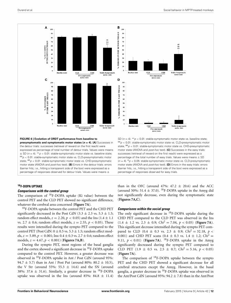
FIGURE 6 | Evolution of ORDT performance from baseline to
presymptomatic and symptomatic motor states (n = 4). (A) Successes inthe detour trials: successes (retrieval of reward on the first reach) wereexpressed as percentage of total number of detour trials. Values were means± SD (n = 4). ∗∗p < 0.01: stable-symptomatic motor state vs. baseline state;¤¤p < 0.01: stable-symptomatic motor state vs. CLD-presymptomatic motorstate; ##p < 0.01: stable-symptomatic motor state vs. CHD-presymptomaticmotor state (ANOVA and post-hoc test). (B) Errors in the detour trials: errors(barrier hits, i.e., hitting a transparent side of the box) were expressed as apercentage of responses observed for detour trials. Values were means ±
SD (n = 4). ∗∗p < 0.01: stable-symptomatic motor state vs. baseline state;¤¤p < 0.01: stable-symptomatic motor state vs. CLD-presymptomatic motorstate; ##p < 0.01: stable-symptomatic motor state vs. CHD-presymptomaticmotor state (ANOVA and post-hoc test). (C) Successes in the easy trials:successes (retrieval of reward on the first reach) were expressed as apercentage of the total number of easy trials. Values were means ± SD(n = 4). ¤p < 0.05: stable-symptomatic motor state vs. CLD-presymptomaticmotor state (ANOVA and post-hoc test). (D) Errors in the easy trials: errors(barrier hits, i.e., hitting a transparent side of the box) were expressed as apercentage of responses observed for easy trials.
18F-DOPA UPTAKEComparisons with the control groupThe comparison of 18F-DOPA uptake (Ki value) between thecontrol PET and the CLD PET showed no significant difference,whatever the cerebral area concerned (Figure 7A).
18F-DOPA uptake between the control PET and the CHD PETsignificantly decreased in the Post CdN (3.3 ± 2.3 vs. 5.3 ± 1.5;random effect models, z = 2.20, p < 0.05) and the Ins (1.4 ± 1.1vs. 2.7 ± 0.6; random effect models, z = 2.55, p < 0.05). Theseresults were intensified during the sympto PET compared to thecontrol PET (Post CdN: 0 ± 0.5 vs. 5.3 ± 1.5; random effect mod-els, z = 5.89, p < 0.001; Ins: 0.4 ± 0.3 vs. 2.7 ± 0.6; random effectmodels, z = 4.67, p < 0.001) (Figures 7A,B).
During the sympto PET, most regions of the basal gangliaand the cortex showed a significant decrease in 18F-DOPA uptakecompared to the control PET. However, a greater decrease wasobserved in 18F-DOPA uptake in Ant / Post CdN (around 95%:94.7 ± 5.7) than in Ant / Post Put (around 80%: 80.2 ± 10.5),the V Str (around 55%: 55.3 ± 14.6) and the SN (around38%: 37.6 ± 31.6). Similarly, a greater decrease in 18F-DOPAuptake was observed in the Ins (around 85%: 84.8 ± 11.4)
than in the OFC (around 47%: 47.2 ± 20.6) and the ACC(around 50%: 51.4 ± 37.0). 18F-DOPA uptake in the Amyg didnot significantly decrease, even during the symptomatic state(Figures 7A,C).
Comparisons within the social groupThe only significant decrease in 18F-DOPA uptake during theCHD PET compared to the CLD PET was observed in the Ins(1.4 ± 1.2 vs. 2.3 ± 0.9; Chi2 = 7.04, p < 0.05) (Figure 7A).This significant decrease intensified during the sympto PET com-pared to CLD (0.4 ± 0.3 vs. 2.3 ± 0.9; Chi2 = 32.18, p <
0.001) and CHD PET scans (0.4 ± 0.3 vs. 1.4 ± 1.2; Chi2 =9.11, p < 0.01) (Figure 7A). 18F-DOPA uptake in the Amygsignificantly decreased during the sympto PET compared toCLD PET (1.9 ± 0.5 vs. 2.5 ± 0.7; Chi2 = 5.16, p < 0.05)(Figure 7A).
The comparison of 18F-DOPA uptake between the symptoPET and the CHD PET showed a significant decrease for allcerebral structures except the Amyg. However, in the basalganglia, a greater decrease in 18F-DOPA uptake was observed inthe Ant/Post CdN (around 95%: 94.2 ± 7.8) than in the Ant/Post
Frontiers in Behavioral Neuroscience www.frontiersin.org February 2015 | Volume 9 | Article 42 | 12

Durand et al. Social behavior in MPTP-treated monkeys
FIGURE 7 | Comparisons of 18F-DOPA uptake in PET imaging. (A) The rateof specific uptake (Ki values) was assessed within basal ganglia in relation to18F-DOPA uptake in the reference brain region (cerebellum). The basal gangliaincludes the Anterior and Posterior Caudate Nucleus (Ant CdN/Post CdN), theAnterior Putamen (Ant Put), the Posterior Dorsal and Ventral Putamen (Post D/VPut), the Ventral Striatum (V Str) and the Substantia Nigra (SN), as well as uptakein cortical regions such as the Anterior Cingulate Cortex (ACC), the OrbitofrontalCortex (OFC), the Amygdala (Amyg) and the Insula (Ins). One PET scan wascarried out for the control group (n = 5) and was considered the Control PET.Three PET scans were carried out for four animals of the social group (n = 4) ofthe study: the first at the end of the presymptomatic motor state followingCLD-MPTP protocol (CLD PET), the second during the presymptomatic motorstate following CHD-MPTP protocol (CHD PET) period and the third during the
symptomatic motor state, when animals developed stable parkinsonian-likemotor symptoms (≥1 month) (Sympto PET). Comparison with the controlgroup: values of the Control PET scan were means ± SD (n = 5). Values of thethree other PET scans were means ± SD (n = 4). φφφp < 0.001, φφp < 0.01,φp < 0.05; CHD PET, Sympto PET vs. Control PET (random effects models).Comparison within the social group: values of the three PET scans weremeans ± SD (n = 4). ¤¤¤p < 0.001, ¤p < 0.05: CHD PET, Sympto PET vs. CLDPET; ###p < 0.001, ##p < 0.01, #p < 0.05: Sympto PET vs. CHD PET (Chi2-test).(B) Subtraction images between the average 18F-DOPA uptake of control PETand the average 18F-DOPA uptake of CHD PET. 18F-DOPA uptake decreases inthe Post CdN and the insula were illustrated in frontal sections (black arrows).(C) Subtraction images between the average 18F-DOPA uptake of control PETand the average 18F-DOPA uptake of sympto PET.
putamen (around 75%: 74.1 ± 7.4), the V Str (around 50%:48.3 ± 14.1) and the SN (around 25%: 24.0 ± 36.2). Similarly,in the cortical areas, a greater decrease of 18F-DOPA uptakewas observed in the Ins (around 72%: 72.5 ± 24.7) than in theOFC (around 24%: 23.4 ± 28.6) and the ACC (around 63%:63.1 ± 25.7).
Individual comparisonsOverall, the results showed a stronger inter-animal variabilityof the Ki uptake during the CLD and the CHD PET, withthe subordinate monkey D showing the lowest Ki value inall structures (apart in the ACC and the Ins during the CLD
PET) (Figures 8A,B). 18F-DOPA uptake decreased linearly inPost Put, Post CdN and Ins in all three subordinate animals,whereas the Ki uptake in the dominant animal only decreasedduring the symptomatic PET. A stronger individual variabilityof Ki was observed in ACC. However, the dynamic of changewas comparable between the 4 animals apart for the subordi-nate monkey D, whose Ki uptake was not detectable duringthe sympto PET. Finally, 18F-DOPA uptake in V Str and Amygdid not change between CLD and CHD PETs, except for thesubordinate monkey D. The only change involving a decreasein 18F-DOPA uptake in V Str and Amyg occurred during thesympto PET.
Frontiers in Behavioral Neuroscience www.frontiersin.org February 2015 | Volume 9 | Article 42 | 13

Durand et al. Social behavior in MPTP-treated monkeys
FIGURE 8 | Individual evolution of 18F-DOPA uptake in (A) 3 striatal
(posterior caudate nucleus, posterior putamen and ventral striatum) and
(B) 3 cortical (insula, amygdala and anterior cingulate cortex) areas
(n = 4). The 18F-DOPA uptake value in the different motor states
(presymptomatic and symptomatic) is represented for the most dominant(red line) and the three most subordinate animals (green, blue and orangelines). The 18F-DOPA uptake value for the control group is also shown (dottedline). The scale showed a difference and the lower threshold was 0.001.
Correlation analysesThere was a negative correlation between the clinical score andthe 18F-DOPA uptake in Ant CdN (r = −0.88, p < 0.001), PostCdN (r = −0.88, p < 001), Ant Put (r = −0.86, p < 0.001), PostPut (r = −0.79, p < 0.01), V Str (r = −0.75, p < 0.01), and Ins(r = −0.74, p < 0.01). Similarly, there was a negative correlationbetween the time spent in inactivity and the 18F-DOPA uptakein Ant CdN (r = −0.88, p < 0.05), Post CdN (r = −0.64, p <
0.05), Ant Put (r = −0.71, p < 0.05), Post Put (r = −0.62, p <
0.05).The success rate on detour trials was positively correlated to
the 18F-DOPA uptake in Ant CdN (r = 0.77, p < 0.05), Post CdN(r = 0.74, p < 0.05), Ant Put (r = 0.79, p < 0.05), Post Put (r =0.76, p < 0.05), ACC (r = 0.77, p < 0.05) and Ins (r = 0.69,p < 0.05). Moreover, there was a negative correlation betweenthe percentage of errors responses in the detour trials and the18F-DOPA uptake in Ant CdN (r = 0.70, p < 0.05), Ant Put(r = 0.74, p < 0.01), Post Put (r = 0.74, p < 0.01), Ins (r =0.69, p < 0.05) and ACC (r = 0.77, p < 0.01). Finally, a posi-tive correlation was observed between the frequency of emittedaffiliative behaviors and the 18F-DOPA uptake in Ins (r = 0.67,p < 0.05).
DISCUSSIONThe aim of this study was to focus on social behavioral changeswithin a group of female long-tailed macaques after chronicadministration of low (CLD) and then high (CHD) doses ofMPTP. These MPTP administration protocols made it possi-ble to evaluate each individual in three motor states (normal,presymptomatic and symptomatic).
Following the CLD-MPTP protocol, a significant increase washighlighted in the frequency of aggressive and affiliative behaviorsfrom the baseline state to the CLD-presymptomatic motor state.Indeed, a higher number of conflicts followed by reconciliationevents were observed. More precisely, these conflicts occurringduring the CLD-MPTP protocol were very intense with slapping,biting and other aggressive behaviors involving direct contactbetween individuals. None of these aggressive behaviors wereobserved in the baseline state, where the few conflicts observedwere of low intensity and mainly limited to visual threats withthe extensive use of facial expressions. Interestingly, these socialbehavioral changes were only shown by the subordinate animals.Moreover, a temporary change of the dominance hierarchy wasobserved; thus, monkey D presented the lowest hierarchical sta-tus compared to monkeys E and F. Individual data within this
Frontiers in Behavioral Neuroscience www.frontiersin.org February 2015 | Volume 9 | Article 42 | 14

Durand et al. Social behavior in MPTP-treated monkeys
subgroup were too heterogeneous to show any significant vari-ations in the three categories of social behaviors. Indeed, only thefrequency of emitted affiliative behaviors increased significantlyin the CLD-presymptomatic motor state compared to the base-line state. A clearer picture could be obtained by increasing thenumber of animals in this study, taking the hierarchical status intoconsideration.
From the CLD-presymptomatic to the CHD-presymptomaticmotor states, the frequency of social behaviors decreased. Thedominance hierarchy was similar to the baseline state; thus mon-key D recovered its initial hierarchical status. At this time, quan-titative data analysis in PET scans made it possible to highlighta dopaminergic denervation in two brain structures, the insulaand the posterior caudate nucleus, which could therefore beinvolved in the social behavioral changes observed from the CLD-presymptomatic motor state to the CHD-presymptomatic motorstate.
Finally, during the symptomatic motor state, the frequencyof social behaviors was lower across all categories, than thefrequency observed in the CLD and the CHD-presymptomaticmotor states. The dominance hierarchy was not modified andwas similar to that observed during the baseline state. Motor andcognitive disorders were also observed in this stable-symptomaticstate. PET scans results then showed a dopaminergic denervationin all the evaluated cortical and subcortical structures.
In the present study, the choice was made to intoxicate not justone animal with MPTP, but all the animals in the group. Thisdecision was made in order to increase the probability of observ-ing social behavioral changes due to inter-individual variability ofsensitivity to MPTP in monkeys. Moreover, this study did not usea control group to compare the distribution of interactions overtime in the present study group. This is explained by the fact thatthe stability of primate groups can vary despite similar sizes andsimilar ecological conditions (Sueur et al., 2011) and the distribu-tion of grooming can differ considerably between groups (Perry,1996; Manson et al., 1999; Dufour et al., 2011). Consequently, itis more efficient to analyze social relationships of a single groupover time when investigating network stability or instability. Forexample, this enables us to compare different observation periodswhere breeding conditions have changed, and study how individ-uals within one same group cope with perturbation (Dufour et al.,2011). The present study examines the evolution of social interac-tions over time in order to understand how our group of femalemacaques coped socially with disease-related changes.
CHRONIC LOW- AND HIGH-DOSE PROTOCOL IS EFFECTIVE TOOBSERVE SOCIAL BEHAVIORAL CHANGES AND DISSOCIATE THEMFROM COGNITIVE AND MOTOR IMPAIRMENTSThis is the first study describing social behavioral changes dur-ing the presymptomatic motor state following MPTP adminis-tration before the onset of cognitive and motor disorders. Onthe one hand, a previous study has also shown social behav-iors changes (aggressive and affiliative) during the symptomaticmotor state induced by MPTP administration (hemiparkinsonianmodel in vervet monkey model) (Melega et al., 1996). However,this study was performed during the symptomatic motor state.Thus, it is difficult to dissociate the effect of motor disorders
on social behavioral changes. On the other hand, it is impor-tant to note that two previous studies in the CLD-MPTP-treatedmacaque model showed cognitive impairment in the presymp-tomatic motor state (Schneider and Pope-Coleman, 1995; Vezoliet al., 2011), which was not the case in the present study.Indeed, cognitive and motor disturbances appeared only duringthe symptomatic motor state following the CHD-MPTP proto-col, and their occurrence was almost simultaneous. Thus, it wasdifficult to really dissociate the impact of motor disorders oncognitive abilities. Several hypotheses could explain the differ-ences in cognition results between this study and the two studiesmentioned above. Firstly, the dose of MPTP administered toanimals was different: 0.1 mg/kg in the present study vs. 0.05–0.075 mg/kg in Schneider’s study and 0.2 mg/kg in Vezoli’s study.Secondly, MPTP was administered every 4–5 days for more than50 weeks in the present study vs. 2–3 times per week for 24weeks in Schneider’s study and every 3–4 days for 5–25 weeksin Vezoli’s study. Thirdly, the social housing could also have animpact (Prescott et al., 2010), as a possible protective effect againstdevelopment of cognitive deficits. Finally, the sex and age of ani-mals differ between studies (Prescott et al., 2010; Darusman et al.,2014). Moreover, over-training might have an impact on suchsimple task as it was shown to be associated with less engage-ment of cognitive control areas, which is specifically tested withthis task (Patel et al., 2013). In the present study, there was anapparent gain in performance in CLD-presymptomatic motorstate compared to baseline state; this might indicate that ani-mals maintained learning capacities on the task with the MPTPregimen used and the task chosen in the present study.
HETEROGENEITY OF SOCIAL BEHAVIORAL CHANGES ACCORDING TOHIERARCHICAL STATUSIn the present study, social behavioral changes were only observedin the subordinate subgroup when compared to the dominantsubgroup. It was also important to note that the most domi-nant individual was also involved in post-conflicts reconciliationevents, suggesting that it retained its regulatory role within thesocial group (Petit and Thierry, 1992).
Hierarchical status was assessed using methods that have beenvalidated and widely used in ethology (David, 1987, 1988; Kaplanet al., 2002). Several studies have already demonstrated the spe-cific role played by dopamine in hierarchical status even if otherneurotransmitters such as noradrenalin and serotonin have beenimplicated in behavioral disorders (Raleigh et al., 1991; Siever,2008; Krämer et al., 2011). A study by Kaplan et al. (Kaplanet al., 2002) has shown that in non-human primates, a higherconcentration of homovanillic acid (dopamine’s catabolite) wasfound in the cerebrospinal fluid of dominant animals than insubordinate animals, whatever their gender. PET imaging stud-ies with a specific D2/D3 receptor radiotracer also highlighteddifferences in radiotracer uptake according to hierarchical statusin male long-tailed monkeys (Morgan et al., 2002; Nader et al.,2012) and in humans (Martinez et al., 2010). Interestingly, levelsof D2/D3 receptor availability appeared to be sensitive to changesin housing conditions. This sensitivity was such that the transitionfrom individual to social housing resulted in significant increasesin D2/D3 levels in dominant male animals, whilst subordinate
Frontiers in Behavioral Neuroscience www.frontiersin.org February 2015 | Volume 9 | Article 42 | 15

Durand et al. Social behavior in MPTP-treated monkeys
animals showed no change (Morgan et al., 2002). Nader et al.recently extended their earlier work in male long-tailed mon-keys to female long-tailed monkeys, with the additional use ofa dopamine transporter (DAT) radiotracer (Nader et al., 2012).Their results showed that although neither DAT nor D2/D3 recep-tor availability in the caudate nucleus and putamen was predictiveof social rank, both significantly changed after the formationof social hierarchies in female long-tailed monkeys. DopamineD2/D3 receptor availability significantly increased in females thatbecame dominant, whereas DAT availability decreased in subor-dinate females. Otherwise, an association between the dopamineD4 receptor and a personality trait such as novelty seeking hasbeen demonstrated in both humans (Benjamin et al., 1996;Ebstein et al., 1996) and non-human primates (Bailey et al.,2007). In the present study, the role of dopamine in the behavioralchanges observed can be evaluated through the use of 18F-Doparadiotracer, a specific radiotracer of dopaminergic innervation,during PET scans.
A POSSIBLE ASSOCIATION BETWEEN DOPAMINERGIC DENERVATIONAND SOCIAL BEHAVIORAL CHANGESDuring the CLD-presymptomatic motor state following the CLD-MPTP protocol, whereas a significant increase of the frequency ofsocial behaviors compared to the baseline state was observed, nosignificant difference was identified between the Ki values of 18F-Dopa uptake in the MPTP-treated animals compared to that ofthe control animals. However, as MPTP-treated animals were nottheir own control, it was impossible to know if they really presentany variation of their Ki values of 18F-Dopa uptake. Nevertheless,it was interesting to note that two subordinate animals (monkeysD and F) had lower Ki uptake values in the insula and dor-sal putamen than those observed in control individuals. Recentstudies have shown that neural circuits (Noonan et al., 2014)and dopaminergic innervation (Nader et al., 2012) could differfrom dominant to subordinate individuals. Thus, a brain struc-ture with initially low Ki values would probably be more sensitiveto MPTP intoxication and would therefore show dopaminergicdenervation earlier than other brain structures. However, MPTPsensitivity was not seen to differ according to hierarchical statusin this study. Finally, the use of non-dopaminergic radiotracer,including namely serotoninergic radiotracers, would have proba-bly shown interesting and complementary results to the presentstudy (Raleigh et al., 1991; Siever, 2008; Krämer et al., 2011).Indeed, a study in male vervet monkeys has shown that therewas a distinction between dominance and aggression and hasstrongly suggested that when hierarchical relationships are uncer-tain, serotonergic mechanisms may mediate the behaviors whichpermit a male to attain high dominance status (Raleigh et al.,1991). Moreover, whereas previous reports have suggested aninverse relationship between serotonin level and aggressive behav-ior with low levels of serotonin leading to higher aggression andvice versa, such a simple relationship seemed to be inconsistentwith the current data obtained in the study Krämer et al. (2011).
During the CHD-presymptomatic motor state following theCHD-MPTP protocol, a significant decrease of the frequency ofsocial behaviors compared to the CLD-presymptomatic motorstate was observed. Moreover, a dopaminergic denervation in the
insula and the posterior caudate was identified, only in the threesubordinate animals but not in the dominant animal.
The insula and the caudate nucleus are both involved in theorganization of the social behavioral network or “social brain net-work,” which also includes the prefrontal cortex, the amygdala,the hippocampus, the medial preoptic area, the hypothalamus,the anterior cingulate cortex and the basal ganglia as a whole(Newman, 1999; Skuse and Gallagher, 2009). This network is alsoclosely linked to the reward circuit (O’Connell and Hofmann,2011). The two abovementioned networks are involved in the reg-ulation of emotions and aggression (Davidson et al., 2000; Siever,2008).
More specifically, the insula plays a role in the regulation andexpression of several emotions, including disgust (Lamm andSinger, 2010; Jezzini et al., 2012), an emotion that is particu-larly impaired in parkinsonian patients (Suzuki et al., 2006), aswell as the expression of social behavior. A recent study in rhe-sus macaques used intracortical microstimulations in differentregions of the insula highlight its involvement in the expressionof disgust and in the expression of an essential affiliative facialexpression in social relationships in macaques, the “lipsmackingdisplay” (Caruana et al., 2011). Thus, an impairment of the insuladuring the CHD-presymptomatic motor state could be associatedwith the impaired expression of affiliative behaviors, as suggestedby the positive correlation observed in our study between the fre-quency of emitted affiliative behaviors and the 18F-DOPA uptakein the insula.
The caudate nucleus is particularly involved in the human“trust network” (Baumgartner et al., 2008), and its activ-ity increases throughout cooperation between protagonists (DeDreu, 2012). An impairment of the caudate nucleus during thepresymptomatic motor state could therefore be associated withan impairment of social relationships. This would explain why thefrequency of social behaviors decreased from the CLD to the CHDpresymptomatic motor states. This decrease could be associatedwith a “loss of emotional interest,” and could also correspondto an “apathetic state” during the presymptomatic motor state.Indeed, apathy is characterized in humans by a loss of interestand a lower participation in usual activities, a lack of initiative,diminished initiated activities, an emotional indifference and aflat affect. Moreover, apathy can be described in 3 dimensions(Levy and Dubois, 2006): cognitive, emotional and behavioral.However, the lack of effect on the ORDT performance during theCHD-presymptomatic motor state excluded the cognitive dimen-sion (Brown et al., 2012) and argued to possible emotional and/orbehavioral attempt.
Finally, and surprisingly, other cortical structures belonging tothe social behavior network, including the orbitofrontal cortex,the anterior cingulate cortex, amygdala, and a limbic structureof the basal ganglia, the ventral striatum, did not seem to beinvolved in behavioral changes observed during the presymp-tomatic motor states. Indeed, these structures were only impairedduring the stable-symptomatic motor state and could be asso-ciated not only with social behavioral changes but also withcognitive and motor disorders, as suggested by the correlationsobserved between the Ki value of several cerebral structures andmotor/cognitive disorders. This suggests that the dopaminergic
Frontiers in Behavioral Neuroscience www.frontiersin.org February 2015 | Volume 9 | Article 42 | 16

Durand et al. Social behavior in MPTP-treated monkeys
innervations of these brain structures are less sensitive to MPTPin our experimental conditions.
CONCLUSIONIn conclusion, the present study of the MPTP-treated non-humanprimate model of Parkinson’s disease showed that behavioralchanges appeared early, before the onset of motor and cognitivedisorders. This could be compared with clinical data obtainedin parkinsonian patients showing a high incidence of behav-ioral changes in the early stages of the disease (Aarsland et al.,2009). Furthermore, these behavioral changes were most com-mon among subordinate individuals. Finally, the results of thepresent study revealed the early sensitivity of cortical structuressuch as the insula to MPTP. However, additional studies includ-ing more animals to limit the inter-individual variability wouldbe necessary to clarify the role of hierarchical status on behav-ioral changes. Likewise, the use of non-dopaminergic radiotracer,including namely serotoninergic radiotracers, would have proba-bly shown interesting and complementary results to the presentstudy.
ACKNOWLEDGMENTThe authors are very grateful to Nicolas Costes and Didier Le Barsfor PET acquisitions.
SUPPLEMENTARY MATERIALThe Supplementary Material for this article can be found onlineat: http://www.frontiersin.org/journal/10.3389/fnbeh.2015.
00042/abstract
REFERENCESAarsland, D., Marsh, L., and Schrag, A. (2009). Neuropsychiatric symptoms in
Parkinson’s disease. Mov. Disord. 24, 2175–2186. doi: 10.1002/mds.22589Adams, M. R., Kaplan, J. R., and Koritnik, D. R. (1985). Psychosocial influences on
ovarian endocrine and ovulatory function in Macaca fascicularis. Physiol. Behav.35, 935–940. doi: 10.1016/0031-9384(85)90262-8
Altmann, J. (1974). Observational study of behavior: sampling methods. Behaviour49, 227–267. doi: 10.1163/156853974X00534
Amaral, D. G. (2002). The primate amygdala and the neurobiology of social behav-ior: implications for understanding social anxiety. Biol. Psychiatr. 51, 11–17. doi:10.1016/S0006-3223(01)01307-5
Angst, W. (1975). “Basic data and concepts on the social organization of MacacaFascicularis,” in Primate Behavior, ed L. A. Rosenblum (New York, NY: AcademicPress), 325–388.
Bailey, J. N., Breidenthal, S. E., Jorgensen, M. J., McCracken, J. T., andFairbanks, L. A. (2007). The association of DRD4 and novelty seeking isfound in a nonhuman primate model. Psychiatr. Genet. 17, 23–27. doi:10.1097/YPG.0b013e32801140f2
Ballanger, B., Tremblay, L., Sgambato-Faure, V., Beaudoin-Gobert, M., Lavenne, F.,Le Bars, D., et al. (2013). A multi-atlas based method for automated anatom-ical Macaca fascicularis brain MRI segmentation and PET kinetic extraction.Neuroimage 77, 26–43. doi: 10.1016/j.neuroimage.2013.03.029
Barraud, Q., Lambrecq, V., Forni, C., McGuire, S., Hill, M., Bioulac, B.,et al. (2009). Sleep disorders in Parkinson’s disease: the contribution ofthe MPTP non-human primate model. Exp. Neurol. 219, 574–582. doi:10.1016/j.expneurol.2009.07.019
Baumgartner, T., Heinrichs, M., Vonlanthen, A., Fischbacher, U., and Fehr, E.(2008). Oxytocin shapes the neural circuitry if trust and trust adaptation inhumans. Neuron 58, 639–650. doi: 10.1016/j.neuron.2008.04.009
Benjamin, J., Li, L., Patterson, C., Greenberg, B. D., Murphy, D. L., and Hamer,D. H. (1996). Population and familial association between the D4 dopaminereceptor gene and measures of Novelty Seeking. Nat. Genet. 12, 81–84. doi:10.1038/ng0196-81
Bentley-Condit, V. K., and Smith, E. O. (1999). Female dominance and femalesocial relationships among yellow baboons (Papio hamadryas cynocephalus).Am. J. Primatol. 47, 321–334.
Blesa, J., Pifl, C., Sánchez-González, M. A., Juri, C., García-Cabezas, M. A., Adánez,R., et al. (2012). The nigrostriatal system in the presymptomatic and symp-tomatic stages in the MPTP monkey model: a PET, histological and biochemicalstudy. Neurobiol. Dis. 48, 79–91. doi: 10.1016/j.nbd.2012.05.018
Brent, L., and Veira, Y. (2002). Social behavior of captive Indochinese and Insularlong-tailed macaques (Macaca fascicularis) following transfer to a new facility.Int. J. Primatol. 23, 147–159. doi: 10.1023/A:1013206125884
Brown, C. A., Campbell, M. C., Karimi, M., Tabbal, S. D., Loftin, S. K., Tian, L. L.,et al. (2012). Dopamine pathway loss in nucleus accumbens and ventral tegmen-tal area predicts apathetic behavior in MPTP-lesioned monkeys. Exp. Neurol.236, 190–197. doi: 10.1016/j.expneurol.2012.04.025
Camus, S. M. J., Blois-Heulin, C., Li, Q., Hausberger, M., and Bezard, E.(2013). Behavioural profiles in captive-bred cynomolgus macaques: towardsmonkey models of mental disorders? PLoS ONE 8:e62141. doi: 10.1371/jour-nal.pone.0062141
Caruana, F., Jezzini, A., Sbriscia-Fioretti, B., Rizzolatti, G., and Gallese, V. (2011).Emotional and social behaviors elicited by electrical stimulation of the insula inthe macaque monkey. Curr. Biol. 21, 195–199. doi: 10.1016/j.cub.2010.12.042
Chassain, C., Eschalier, A., and Durif, F. (2001). Assessment of motor behav-ior using a video system and a clinical rating scale in parkinsonianmonkeys lesioned by MPTP. J. Neurosci. Met. 111, 9–16. doi: 10.1016/S0165-0270(01)00425-3
Chaumette, T., Derkinderen, P., Bruley des Varannes, S., Bezard, E., and Accary,D. (2009). Neurochemical plasticity in the enteric nervous system of a pri-mate animal model of experimental Parkinsonism. Neurogastroenterol. Motil.21, 215–222. doi: 10.1111/j.1365-2982.2008.01226.x
Costes, N., Zimmer, L., Reilhac, A., Lavenne, F., Ryvlin, P., and Le Bars, D. (2007).Test-retest reproducibility of 18 F-MPPF PET in healthy humans: a reliabilitystudy. J. Nucl. Med. 48, 1279–1288. doi: 10.2967/jnumed.107.041905
Czoty, P. W., Riddick, N. V., Gage, H. D., Sandridge, M., Nader, S. H., Garg, S.,et al. (2009). Effect of menstrual cycle phase on dopamine D2 receptor avail-ability in female cynomolgus monkeys. Neuropsychopharmacology 34, 548–554.doi: 10.1038/npp.2008.3
Darusman, H. S., Call, J., Sajuthi, D., Schapiro, S. J., Gjedde, A., Kalliokoski, O., et al.(2014). Delayed response task performance as a function of age in cynomolgusmonkeys (Macaca fascicularis). Primates 55, 259–267. doi: 10.1007/s10329-013-0397-8
David, H. A. (1987). Ranking from unbalanced paired-comparison data.Biometrika 74, 432–436. doi: 10.1093/biomet/74.2.432
David, H. A. (1988). The Method of Paired Comparisons. London: Charles Griffin.Davidson, R. J., Putnam, K. M., and Larson, C. L. (2000). Dysfunction in the neu-
ral circuitry of emotion regulation-a possible prelude to violence. Science 289,591–594. doi: 10.1126/science.289.5479.591
Decamp, E., and Schneider, J. S. (2004). Attention and executive function deficitsin chronic low-dose MPTP-treated non-human primates. Eur. J. Neurosci. 20,1371–1378. doi: 10.1111/j.1460-9568.2004.03586.x
De Dreu, C. K. (2012). Oxytocin modulates cooperation within and competitionbetween groups: an integrative review and research agenda. Horm. Behav. 61,419–428. doi: 10.1016/j.yhbeh.2011.12.009
Dufour, V., Sueur, C., Whiten, A., and Buchanan-Smith, H. M. (2011). The impactof moving to a novel environment on social networks, activity and wellbeing intwo New World primates. Am. J. Primatol. 73, 802–811. doi: 10.1002/ajp.20943
Ebstein, R. P., Novick, O., Umansky, R., Priel, B., Osher, Y., Blaine, D., et al.(1996). Dopamine D4 receptor (D4DR) Exon III polymorphism associated withthe human personality trait of Novelty Seeking. Nat. Genet. 12, 78–80. doi:10.1038/ng0196-78
Gallagher, D. A., and Schrag, A. (2012). Psychosis, apathy, depression and anxiety inParkinson’s disease. Neurobiol. Dis. 46, 581–589. doi: 10.1016/j.nbd.2011.12.041
Gammell, M. P., De Vries, H., Jennings, D. J., Carlin, C. M., and Hayden,T. J. (2003). David’s score: a more appropriate dominance rankingmethod than Clutton-Brock et al.’s index. Anim. Behav. 66, 601–605. doi:10.1006/anbe.2003.2226
Gibb, W. R., and Lees, A. J. (1991). Anatomy, pigmentation, ventral and dorsal sub-populations of the substantia nigra, and differential cell death in Parkinson’sdisease. J. Neurol. Neurosurg. Psychiatry 54, 388–396. doi: 10.1136/jnnp.54.5.388
Frontiers in Behavioral Neuroscience www.frontiersin.org February 2015 | Volume 9 | Article 42 | 17

Durand et al. Social behavior in MPTP-treated monkeys
Gomez-Mancilla, B., Boucher, R., Gagnon, C., Di Paolo, T., Markstein, R., andBédard, P. J. (1993). Effect of adding the D1 agonist CY 208-243 to chronicbromocriptine treatment. I: evaluation of motor parameters in relation to stri-atal catecholamine content and dopamine receptors. Mov. Disord. 8, 144–150.doi: 10.1002/mds.870080205
Gumert, M. D. (2010). “Dominance and reciprocity in the grooming relation-ships of female long-tailed macaques (Macaca fascicularis) in Indonesia,” inIndonesian Primates (Developments in Primatology: Progress and Prospects), edsS. Gursky-Doyen and J. Supriatna (Springer Science+Business Media) Chapter18, 309–341.
Haber, S. N., and Knutson, B. (2010). The reward circuit: linking primateanatomy and human imaging. Neuropsychopharmacology 35, 4–26. doi:10.1038/npp.2009.129
Hirsch, E. C., Orieux, G., Muriel, M. P., Francois, C., and Feger, J. (2003).Nondopaminergic neurons in Parkinson’s disease. Adv. Neurol. 91, 29–37.
Isley, M. (2010). Implanon: the subdermal contraceptive implant. J. Pediatr.Adolesc. Gynecol. 23, 364–367. doi: 10.1016/j.jpag.2010.03.006
Jenner, P. (2003). The MPTP-treated primate as a model of motor complicationsin PD: primate model of motor complications. Neurology 61, S4–S11. doi:10.1212/WNL.61.6_suppl_3.S4
Jezzini, A., Caruana, F., Stoianov, I., Gallese, V., and Rizzolatti, G. (2012).Functional organization of the insula and inner perisylvian regions.Proc. Natl. Acad. Sci. U.S.A. 109, 10077–10082. doi: 10.1073/pnas.1200143109
Kaplan, J. R., Manuck, S. B., Fontenot, M. B., and Mann, J. J. (2002). Centralnervous system monoamine correlates of social dominance in cynomolgusmonkeys (Macaca fascicularis). Neuropsychopharmacology 26, 431–443. doi:10.1016/S0893-133X(01)00344-X
Kish, S. J., Shannak, K., and Hornykiewicz, O. (1988). Uneven pattern ofdopamine loss in the striatum of patients with idiopathic Parkinson’s disease.Pathophysiologic and clinical implications. N. Engl. J. Med. 318, 876–880. doi:10.1056/NEJM198804073181402
Kish, S. J., Tong, J., Hornykiewicz, O., Rajput, A., Chang, L. J., Guttman,M., et al. (2008). Preferential loss of serotonin markers in caudate ver-sus putamen in Parkinson’s disease. Brain 131, 120–131. doi: 10.1093/brain/awm239
Kortekaas, R., Eshuis, S. A., Andringa, G., Cools, A. R., and Leenders, K. L.(2013). Motor behavior correlates with striatal [18F]-DOPA uptake in MPTP-lesioned primates. Neurochem. Int. 62, 349–353. doi: 10.1016/j.neuint.2013.01.021
Krämer, U. M., Riba, J., Richter, S., and Münte, T. F. (2011). An fMRI studyon the role of serotonin in reactive aggression. PLoS ONE 6:e27668. doi:10.1371/journal.pone.0027668
Lamm, C., and Singer, T. (2010). The role of anterior insular cortex in socialemotions. Brain Struct. Funct. 214, 579–591. doi: 10.1007/s00429-010-0251-3
Levy, R., and Dubois, B. (2006). Apathy and the functional anatomy of the pre-frontal cortex-basal ganglia circuits. Cereb. Cortex 16, 916–928. doi: 10.1093/cer-cor/bhj043
Machado, C. J., and Bachevalier, J. (2006). The impact of selective amygdala, orbitalfrontal cortex, or hippocampal formation lesions on established social relation-ships in rhesus monkeys (Macaca mulatta). Behav. Neurosci. 120, 761–786. doi:10.1037/0735-7044.120.4.761
Machado, C. J., Kazama, A. M., and Bachevalier, J. (2009). Impact of amygdala,orbital frontal, or hippocampal lesions on threat avoidance and emotionalreactivity in nonhuman primates. Emotion 9, 147–163. doi: 10.1037/a0014539
Madras, B. K., Fahey, M. A., Goulet, M., Lin, Z., Bendor, J., Goodrich, C., et al.(2006). Dopamine transporter (DAT) inhibitors alleviate specific parkinsoniandeficits in monkeys: association with DAT occupancy in vivo. J. Pharmacol. Exp.Ther. 319, 570–585. doi: 10.1124/jpet.106.105312
Manson, J. H., Rose, L., Perry, S., and Gros-Louis, J. (1999). Dynamics of female-female social relationships in wild Cebus capucinus: data from two Costa Ricansites. Int. J. Primatol. 20, 679–706. doi: 10.1023/A:1020700718458
Martinez, D., Orlowska, D., Narendran, R., Slifstein, M., Liu, F., Kumar, D.,et al. (2010). Dopamine type 2/3 receptor availability in the striatumand social status in human volunteers. Biol. Psychiatr. 67, 275–278. doi:10.1016/j.biopsych.2009.07.037
Martínez-Martín, P., and Damián, J. (2010). Parkinson disease: depression andanxiety in Parkinson disease. Nat. Rev. Neurol. 6, 243–245. doi: 10.1038/nrneu-rol.2010.49
Melega, W. P., Raleigh, M. J., Stout, D. B., DeSalles, A. A., Cherry, S. R.,Blurton-Jones, M., et al. (1996). Longitudinal behavioral and 6-[18F]fluoro-L-DOPA-PET assessment in MPTP-hemiparkinsonian monkeys. Exp. Neurol. 141,318–329. doi: 10.1006/exnr.1996.0167
Michael, R. P., and Zumpe, D. (1993). A review of hormonal factors influencing thesexual and aggressive behavior of macaques. Am. J. Primatol. 30, 213–241. doi:10.1002/ajp.1350300306
Morgan, D., Grant, K. A., Gage, H. D., Mach, R. H., Kaplan, J. R., Prioleau, O. A.,et al. (2002). Social dominance in monkeys: dopamine D2 receptors and cocaineself-administration. Nat. Neurosci. 5, 169–174. doi: 10.1038/nn798
Morgan, D., Grant, K. A., Prioleau, O. A., Nader, S. H., Kaplan, J. R., andNader, M. A. (2000). Predictors of social status in cynomolgus monkeys(Macaca fascicularis) after group formation. Am. J. Primatol. 52, 115–131. doi:10.1002/1098-2345(200011)52:3<115::AID-AJP1>3.0.CO;2-Z
Nader, M. A., Nader, S. H., Czoty, P. W., Riddick, N. V., Gage, H. D., Gould,R. W., et al. (2012). Social dominance in female monkeys: dopamine recep-tor function and cocaine reinforcement. Biol. Psychiatr. 72, 414–421. doi:10.1016/j.biopsych.2012.03.002
Neumane, S., Mounayar, S., Jan, C., Epinat, J., Ballanger, B., Costes, N., et al. (2012).Effects of dopamine and serotonin antagonist injections into the striatopallidalcomplex of asymptomatic MPTP-treated monkeys. Neurobiol. Dis. 48, 27–39.doi: 10.1016/j.nbd.2012.06.002
Newman, S. W. (1999). The medial extended amygdala in male reproductive behav-ior. A node in the mammalian social behavior network. Ann. N.Y. Acad. Sci. 877,242–257. doi: 10.1111/j.1749-6632.1999.tb09271.x
Noonan, M. P., Sallet, J., Mars, R. B., Neubert, F. X., O’Reilly, J. X., Andersson, J. L.,et al. (2014). A neural circuit covarying with social hierarchy in macaque. PLoSBiol. 12:e1001940. doi: 10.1371/journal.pbio.1001940
O’Connell, L. A., and Hofmann, H. A. (2011). The vertebrate mesolimbic rewardsystem and social behavior network: a comparative synthesis. J. Comp. Neurol.519, 3599–3639. doi: 10.1002/cne.22735
Palfi, S., Ferrante, R. J., Brouillet, E., Beal, M. F., Dolan, R., Guyot, M. C., et al.(1996). Chronic 3-nitropropionic acid treatment in baboons replicates thecognitive and motor deficits of Huntington’s disease. J. Neurosci. 16, 3019–3025.
Patel, R., Spreng, R. N., and Turner, G. R. (2013). Functional brain changesfollowing cognitive and motor skills training. A quantitative meta-analysis.Neurorehab. Neural Repair 27, 187–199. doi: 10.1177/1545968312461718
Patlak, C. S., Blasberg, R. G., and Fenstermacher, J. D. (1983). Graphical evaluationof blood-to-brain transfer constants from multiple-time uptake data. J. Cereb.Blood Flow Metab. 3, 1–7. doi: 10.1038/jcbfm.1983.1
Pedersen, K. F., Alves, G., Aarsland, D., and Larsen, J. P. (2009). Occurrence and riskfactors for apathy in Parkinson disease: a 4-year prospective longitudinal study.J. Neurol. Neurosurg. Psychiatr. 80, 1279–1282. doi: 10.1136/jnnp.2008.170043
Pell, M. D., Cheang, H. S., and Leonard, C. L. (2006). The impact of Parkinson’s dis-ease on vocal-prosodic communication from the perspective of listeners. BrainLang. 97, 123–134. doi: 10.1016/j.bandl.2005.08.010
Perez-Otano, I., Oset, C., Luquin, M. R., Herrero, M. T., Obeso, J. A., and Del Rio, J.(1994). MPTP-induced parkinsonism in primates: pattern of striatal dopamineloss following acute and chronic administration. Neurosci. Lett. 175, 121–125.doi: 10.1016/0304-3940(94)91094-4
Perry, S. (1996). Female-female relationships in wild white-faced capuchin mon-keys, Cebus capucinus. Am. J. Primatol. 40, 167–182.
Petit, O., and Thierry, B. (1992). Affiliative function of the silent bared-teeth displayin moor macaques (Macaca maurus): further evidence for the particular statusof Sulawesi macaques. Int. J. Primatol. 13, 97–105. doi: 10.1007/BF02547729
Prescott, M. J., Brown, V. J., Flecknell, P. A., Gaffan, D., Garrod, K., Lemon, R.N., et al. (2010). Refinement of the use of food and fluid control as motiva-tional tools for macaques used in behavioural neuroscience research: reportof a Working Group of the NC3Rs. J. Neurosci. Met. 193, 167–188. doi:10.1016/j.jneumeth.2010.09.003
Raleigh, M. J., McGuire, M. T., Brammer, G. L., Pollack, D. B., and Yuwiler, A.(1991). Serotonergic mechanisms promote dominance acquisition in adult malevervet monkeys. Brain Res. 559, 181–190. doi: 10.1016/0006-8993(91)90001-C
Redmond, D. E., Maas, J. W., Kling, A., Graham, C. W., and Dekirmenjian, H.(1971). Social behavior of monkeys selectively depleted of monoamines. Science174, 428–431. doi: 10.1126/science.174.4007.428
Remy, P., Doder, M., Lees, A., Turjanski, N., and Brooks, D. (2005). Depressionin Parkinson’s disease: loss of dopamine and noradrenaline innervation in thelimbic system. Brain 128, 1314–1322. doi: 10.1093/brain/awh445
Frontiers in Behavioral Neuroscience www.frontiersin.org February 2015 | Volume 9 | Article 42 | 18

Durand et al. Social behavior in MPTP-treated monkeys
Riddick, N. V., Czoty, P. W., Gage, H. D., Kaplan, J. R., Nader, S. H., Icenhower,M., et al. (2009). Behavioral and neurobiological characteristics influencingsocial hierarchy formation in female cynomolgus monkeys. Neuroscience 158,1257–1265. doi: 10.1016/j.neuroscience.2008.11.016
Roeltgen, D. P., and Schneider, J. S. (1991). Chronic low-dose MPTP in nonhu-man primates: a possible model for attention deficit disorder. J. Child. Neurol.6, 82–89.
Rushworth, M. F., Behrens, T. E., Rudebeck, P. H., and Walton, M. E. (2007).Contrasting roles for cingulate and orbitofrontal cortex in decisions and socialbehaviour. Trends Cogn. Sci. 11, 168–176. doi: 10.1016/j.tics.2007.01.004
Schneider, J. S. (1990). Chronic exposure to low doses of MPTP. II. Neurochemicaland pathological consequences in cognitively-impaired, motor asymptomaticmonkeys. Brain Res. 534, 25–36. doi: 10.1016/0006-8993(90)90108-N
Schneider, J. S. (2006). “Modeling cognitive deficits associated with Parkinsonismin the chronic-low-dose MPTP-treated monkey,” in Animal Models of CognitiveImpairment, eds E. D. Levin and J. J. Buccafusco (Boca Raton, FL: CRC Press),Chapter 9, Frontiers in Neuroscience.
Schneider, J. S., and Kovelowski, C. J. II. (1990). Chronic exposure to low doses ofMPTP. I. Cognitive deficits in motor asymptomatic monkeys. Brain Res. 519,122–128. doi: 10.1016/0006-8993(90)90069-N
Schneider, J. S., and Pope-Coleman, A. (1995). Cognitive deficits precede motordeficits in a slowly progressing model of parkinsonism in the monkey.Neurodegeneration 4, 245–255. doi: 10.1016/1055-8330(95)90014-4
Schneider, J. S., and Roeltgen, D. P. (1993). Delayed matching-to-sample, objectretrieval, and discrimination reversal deficits in chronic low dose MPTP-treatedmonkeys. Brain Res. 615, 351–354. doi: 10.1016/0006-8993(93)90049-S
Schneider, J. S., Tinker, J. P., and Decamp, E. (2010). Clonidine improves atten-tional and memory components of delayed response performance in a modelof early Parkinsonism. Behav. Brain Res. 211, 236–239. doi: 10.1016/j.bbr.2010.03.040
Schrag, A., Jahanshahi, M., and Quinn, N. (2000). How does Parkinson’s diseaseaffect quality of life? A comparison with quality of life in the general population.Mov. Dis. 15, 1112–1118. doi: 10.1002/1531-8257(200011)15:6<1112::AID-MDS1008>3.0.CO;2-A
Siever, L. J. (2008). Neurobiology of aggression and violence. Am. J. Psychiatr. 165,429–442. doi: 10.1176/appi.ajp.2008.07111774
Skuse, D. H., and Gallagher, L. (2009). Dopaminergic-neuropeptide interactions inthe social brain. Trends Cogn. Sci. 13, 27–35. doi: 10.1016/j.tics.2008.09.007
Sueur, C., Jacobs, A., King, A. J., Petit, O., and Amblard, F. (2011). How can socialnetwork analysis improve the study of primate behaviour? Am. J. Primatol. 73,703–719. doi: 10.1002/ajp.20915
Suzuki, A., Hoshino, T., Shigemasu, K., and Kawamura, M. (2006). Disgust-specificimpairment of facial expression recognition in Parkinson’s disease. Brain 129,707–717. doi: 10.1093/brain/awl011
Taylor, J. R., Roth, R. H., Sladek, J. R., and Redmond, D. E. (1990). Cognitive andmotor deficits in the performance of an object retrieval task with a barrier-detour in monkeys (Cercopithecus aethiops sabaeus) treated with MPTP: long-term performance and effect of transparency of the barrier. Behav. Neurosci.104, 564–576. doi: 10.1037/0735-7044.104.4.564
Thobois, S., Ardouin, C., Lhommée, E., Klinger, H., Lagrange, C., Xie, J.,et al. (2010). Non-motor dopamine withdrawal syndrome after surgery forParkinson’s disease: predictors and underlying mesolimbic denervation. Brain133, 1111–1127. doi: 10.1093/brain/awq032
Tzschentke, T. M. (2001). Pharmacology and behavioral pharmacology ofthe mesocortical dopamine system. Prog. Neurobiol. 63, 241–320. doi:10.1016/S0301-0082(00)00033-2
Ulla, M., Thobois, S., Llorca, P. M., Derost, P., Lemaire, J. J., Chereau-Boudet,I., et al. (2012). Contact dependent reproducible hypomania induced by deepbrain stimulation in Parkinson’s disease: clinical, anatomical and functionalimaging study. Behav. Neurol. 82, 607–614. doi: 10.1136/jnnp.2009.199323
Van Hooff, J. A. R. A. M. (1967). “The facial displays of the catarrhine monkeys andapes,” in Primate Ethology, ed D. Morris (London: Weidenfeld and Nicolson),7–68.
Van Noordwijk, M. A., and Van Schaik, C. P. (1987). Competition among femalelong-tailed macaques, Macaca fascicularis. Anim. Behav. 35, 577–589. doi:10.1016/S0003-3472(87)80284-1
Van Noordwijk, M. A., and Van Schaik, C. P. (1999). The effects of dominancerank and group size on female lifetime reproductive success in wild long-tailed macaques, Macaca fascicularis. Primates 40, 105–130. doi: 10.1007/BF02557705
Vezoli, J., Fifel, K., Leviel, V., Dehay, C., Kennedy, H., Cooper, H. M., et al. (2011).Early presymptomatic and long-term changes of rest activity cycles and cog-nitive behavior in a MPTP-monkey model of Parkinson’s disease. PLoS ONE6:e23952. doi: 10.1371/journal.pone.0023952
Videnovic, A., Lazar, A. S., Barker, R. A., and Overeem, S. (2014). ‘The clocks thattime us’-circadian rhythms in neurodegenerative disorders. Nat. Rev. Neurol. 10,683–693. doi: 10.1038/nrneurol.2014.206
Weintraub, D., Siderowf, A. D., Potenza, M. N., Goveas, J., Morales, K. H., Duda,J. E., et al. (2006). Association of dopamine agonist use with impulse controldisorders in Parkinson disease. Arch. Neurol. 63, 969–973. doi: 10.1001/arch-neur.63.7.969
Whitehead, H. (2009). SOCPROG programs: analyzing animal social structures.Behav. Ecol. Sociobiol. 63, 765–778. doi: 10.1007/s00265-008-0697-y
Yoshimura, N., Kawamura, M., Masaoka, Y., and Homma, I. (2005). The amygdalaof patients with Parkinson’s disease is silent in response to fearful facialexpressions. Neuroscience 131, 523–534. doi: 10.1016/j.neuroscience.2004.09.054
Conflict of Interest Statement: The authors declare that the research was con-ducted in the absence of any commercial or financial relationships that could beconstrued as a potential conflict of interest.
Received: 17 November 2014; accepted: 05 February 2015; published online: 26February 2015.Citation: Durand E, Petit O, Tremblay L, Zimmer C, Sgambato-Faure V, Chassain C,Laurent M, Pereira B, Silberberg C and Durif F (2015) Social behavioral changes inMPTP-treated monkey model of Parkinson’s disease. Front. Behav. Neurosci. 9:42. doi:10.3389/fnbeh.2015.00042This article was submitted to the journal Frontiers in Behavioral Neuroscience.Copyright © 2015 Durand, Petit, Tremblay, Zimmer, Sgambato-Faure, Chassain,Laurent, Pereira, Silberberg and Durif. This is an open-access article distributed underthe terms of the Creative Commons Attribution License (CC BY). The use, distribu-tion or reproduction in other forums is permitted, provided the original author(s)or licensor are credited and that the original publication in this journal is cited, inaccordance with accepted academic practice. No use, distribution or reproduction ispermitted which does not comply with these terms.
Frontiers in Behavioral Neuroscience www.frontiersin.org February 2015 | Volume 9 | Article 42 | 19