Sleep cycling alternating pattern (CAP) expression is associated with hypersomnia and GH secretory...
7
Original article Sleep cycling alternating pattern (CAP) expression is associated with hypersomnia and GH secretory pattern in Prader–Willi syndrome Lorenzo Priano a, * , Graziano Grugni b , Giacinta Miscio a , Giulia Guastamacchia a,c , Lorenzo Toffolet a , Alessandro Sartorio b , Alessandro Mauro a,c a Divisione di Neurologia e Neuroriabilitazione, Department of Neurology, IRCCS Istituto Auxologico Italiano, Ospedale S.Giuseppe, Casella postale 1, Intra, 28921 Piancavallo (VB), Verbania, Italy b Divisione di Auxologia, IRCCS Istituto Auxologico Italiano, Piancavallo (VB), Verbania, Italy c Dipartimento di Neuroscienze, Universita ` di Torino, Torino, Italy Received 22 June 2005; received in revised form 28 November 2005; accepted 1 December 2005 Available online 4 October 2006 Abstract Background and purpose: Hypersomnia, sleep-disordered breathing and narcoleptic traits such as rapid eye movement (REM) sleep onset periods (SOREMPs) have been reported in Prader–Willi syndrome (PWS). In a group of young adult patients with genetically confirmed PWS we evaluated sleep and breathing polysomnographically, including cycling alternating pattern (CAP), and we ana- lyzed the potential interacting role of sleep variables, sleep-related breathing abnormalities, hypersomnia, severity of illness variables and growth hormone (GH) secretory pattern. Patients and methods: Eleven males and 7 females (mean age: 27.5±5.5 years) were submitted to a full night of complete polysom- nography and the multiple sleep latency test (MSLT). GH secretory pattern was evaluated by a standard GH-releasing hormone plus arginine test. Sixteen non-obese healthy subjects without sleep disturbances were recruited as controls. Results: Compared to controls PWS patients showed reduced mean MSLT score (P < 0.001), reduced mean latency of sleep (P = 0.03), increased REM sleep periods (P = 0.01), and increased mean CAP rate/non-rapid eye movement (NREM) (P < 0.001). Only four PWS patients had apnea/hypopnea index (AHI) 10. Conversely, significant nocturnal oxygen desaturation was frequent (83% of patients) and independent from apneas or hypopneas. In the PWS group, CAP rate/NREM showed a signif- icant negative correlation with MSLT score (P = 0.02) independently from arousals, respiratory disturbance variables, severity of illness measured by Holm’s score or body mass index (BMI). PWS patients with CAP expression characterized by higher proportion of A1 subtypes presented less severe GH deficiency (P = 0.01). Conclusions: Our study suggests a relationship between hypersomnia and CAP rate, and between CAP expression and GH secretory pattern in PWS, possibly reflecting underlying central dysfunctions. Ó 2005 Elsevier B.V. All rights reserved. Keywords: Prader–Willi syndrome; Cycling alternating pattern; Hypersomnia; GH deficiency; Apnea/hypopnea index; Multiple sleep latency test 1. Introduction Prader–Willi syndrome (PWS) is a complex genetic disorder with an estimated incidence of one in 10,000– 30,000 live births [1]. Most PWS patients (70–75%) carry a deletion in the paternally derived chromosome 15q11– q13, 25% have maternal uniparental disomy, and 2–5% show abnormal methylation of the imprinting center or a balanced translocation [2–6]. Clinical findings in PWS suggest an involvement of the hypothalamus–pituitary axis [7] and include hypogonadism, marked obesity, short stature, mental retardation, hypotonia and 1389-9457/$ - see front matter Ó 2005 Elsevier B.V. All rights reserved. doi:10.1016/j.sleep.2005.12.004 * Corresponding author. Tel.: + 39 0323 514273; fax: + 39 0323 587694. E-mail addresses: [email protected], [email protected] (L. Priano). www.elsevier.com/locate/sleep Sleep Medicine 7 (2006) 627–633
-
Upload
auxologico -
Category
Documents
-
view
1 -
download
0
Transcript of Sleep cycling alternating pattern (CAP) expression is associated with hypersomnia and GH secretory...

www.elsevier.com/locate/sleep
Sleep Medicine 7 (2006) 627–633
Original article
Sleep cycling alternating pattern (CAP) expression is associated withhypersomnia and GH secretory pattern in Prader–Willi syndrome
Lorenzo Priano a,*, Graziano Grugni b, Giacinta Miscio a, Giulia Guastamacchia a,c,Lorenzo Toffolet a, Alessandro Sartorio b, Alessandro Mauro a,c
a Divisione di Neurologia e Neuroriabilitazione, Department of Neurology, IRCCS Istituto Auxologico Italiano, Ospedale S.Giuseppe,
Casella postale 1, Intra, 28921 Piancavallo (VB), Verbania, Italyb Divisione di Auxologia, IRCCS Istituto Auxologico Italiano, Piancavallo (VB), Verbania, Italy
c Dipartimento di Neuroscienze, Universita di Torino, Torino, Italy
Received 22 June 2005; received in revised form 28 November 2005; accepted 1 December 2005Available online 4 October 2006
Abstract
Background and purpose: Hypersomnia, sleep-disordered breathing and narcoleptic traits such as rapid eye movement (REM) sleeponset periods (SOREMPs) have been reported in Prader–Willi syndrome (PWS). In a group of young adult patients with geneticallyconfirmed PWS we evaluated sleep and breathing polysomnographically, including cycling alternating pattern (CAP), and we ana-lyzed the potential interacting role of sleep variables, sleep-related breathing abnormalities, hypersomnia, severity of illness variablesand growth hormone (GH) secretory pattern.Patients and methods: Eleven males and 7 females (mean age: 27.5±5.5 years) were submitted to a full night of complete polysom-nography and the multiple sleep latency test (MSLT). GH secretory pattern was evaluated by a standard GH-releasing hormoneplus arginine test. Sixteen non-obese healthy subjects without sleep disturbances were recruited as controls.Results: Compared to controls PWS patients showed reduced mean MSLT score (P < 0.001), reduced mean latency of sleep(P = 0.03), increased REM sleep periods (P = 0.01), and increased mean CAP rate/non-rapid eye movement (NREM)(P < 0.001). Only four PWS patients had apnea/hypopnea index (AHI) � 10. Conversely, significant nocturnal oxygen desaturationwas frequent (83% of patients) and independent from apneas or hypopneas. In the PWS group, CAP rate/NREM showed a signif-icant negative correlation with MSLT score (P = 0.02) independently from arousals, respiratory disturbance variables, severity ofillness measured by Holm’s score or body mass index (BMI). PWS patients with CAP expression characterized by higher proportionof A1 subtypes presented less severe GH deficiency (P = 0.01).Conclusions: Our study suggests a relationship between hypersomnia and CAP rate, and between CAP expression and GH secretorypattern in PWS, possibly reflecting underlying central dysfunctions.� 2005 Elsevier B.V. All rights reserved.
Keywords: Prader–Willi syndrome; Cycling alternating pattern; Hypersomnia; GH deficiency; Apnea/hypopnea index; Multiple sleep latency test
1. Introduction
Prader–Willi syndrome (PWS) is a complex geneticdisorder with an estimated incidence of one in 10,000–
1389-9457/$ - see front matter � 2005 Elsevier B.V. All rights reserved.
doi:10.1016/j.sleep.2005.12.004
* Corresponding author. Tel.: + 39 0323 514273; fax: + 39 0323587694.
E-mail addresses: [email protected], [email protected](L. Priano).
30,000 live births [1]. Most PWS patients (70–75%) carrya deletion in the paternally derived chromosome 15q11–q13, 25% have maternal uniparental disomy, and 2–5%show abnormal methylation of the imprinting center ora balanced translocation [2–6]. Clinical findings in PWSsuggest an involvement of the hypothalamus–pituitaryaxis [7] and include hypogonadism, marked obesity,short stature, mental retardation, hypotonia and

628 L. Priano et al. / Sleep Medicine 7 (2006) 627–633
impaired growth hormone (GH) secretory pattern [1,8–10]. Hypersomnia during the day is common in PWS (2/3 of patients), but the pathogenesis of this symptom hasnot been fully clarified [11–13]. Obstructive and centralsleep apneas, abnormal ventilatory responses to hypoxiaand hypercapnia, sleep-related alveolar hypoventilationmay be present [14–17], but they can be considered suf-ficient explanations for hypersomnia in only a minorityof cases [18–21].
Classical sleep analysis and scoring according to Rec-htschaffen and Kales, which reflect the macrostructurepattern of sleep did not show specific findings in PWS[18,22]. The occurrence of increased number of rapideye movement (REM) episodes and REM sleep onsetperiods (SOREMPs), together with the findings ofreduced cerebrospinal fluid hypocretin levels, suggesteda possible relationship with narcolepsy and a possibleinvolvement of a central dysfunction in the genesis ofhypersomnia [23–25]. Transient electroencephalographic(EEG) phenomena lasting less than the scoring epochand cycling alternating pattern (CAP) [26–28], describ-ing what is known as the microstructure of sleep, havenot been studied in PWS. In particular, CAP analysiscould add important information as the percentage ofCAP to total NREM sleep (CAP rate/NREM), reflect-ing arousal instability and appearing as a valid indicatorof sleep quality, may be related to daytime sleepiness[29–32].
In a group of young adult PWS patients we evaluatedsleep, including CAP, and we analyzed the potentialinteracting role of sleep variables, sleep-disorderedbreathing, hypersomnia, severity of illness variablesand GH secretory pattern.
Table 1Clinical and laboratory data of PWS patients
Prader–Willi patients Sex Age (years) BMI WHR
MA M 18.1 37.8 0.85MI M 22.3 43.5 0.96CL M 29.2 40.0 1.02ZE F 26.0 38.9 0.79PE M 23.0 44.5 1.02GS M 27.1 69.0 0.72SV M 28.9 42.1 0.98SS F 31.2 60.3 0.86BM F 33.4 50.8 0.86PA F 25.9 46.8 0.95BA F 21.2 44.2 0.92SR F 32.0 46.3 0.91RD M 19.6 50.5 1.03GS M 28.9 42.5 0.83CG M 28.3 53.1 0.91BM F 33.1 63.0 0.98BP M 40.1 42.0 0.94BF M 27.1 46.8 0.92Mean ±SD 27.5±5.5 47.9±8.6 0.93±0.1
GH secretion is expressed as peak response to GHRH + arginine. Holm’s sUPD, uniparental maternal disomy of chromosome 15; Del 15, deletion of thindex (kg/m2); WHR, waist/hip ratio; ESS, Epworth sleepiness scale.
2. Patients and methods
Eighteen PWS patients (7 females and 11 males)aged 18.1–40.1 years (mean age: 27.5±5.5 years) wererecruited for the study (Table 1). The entire study pro-tocol was approved by the local ethical committee,and written informed consent was obtained from par-ents and, when applicable, from patients. Cytogeneticstudies were performed in all patients and severity ofillness was determined by Holm’s diagnostic criteria[33]. This scoring system comprises both major andminor criteria. Major criteria are weighted at onepoint each and include neonatal and infantile centralhypotonia, feeding problems in infancy with poorweight gain, excessive or rapid weight gain after 12months but before six years of age, characteristicfacial features, hypogonadism, mild to moderate men-tal retardation, hyperphagia, cytogenetic/molecularabnormality of the PWS chromosome region. Minorcriteria are weighted at one half-point and includedecreased fetal movement or infantile lethargy, charac-teristic behavior problems, daytime hypersomnolenceor sleep apnea, short stature for genetic background,hypopigmentation, acromicria, narrow hands withstraight ulnar borders, eye abnormalities, thick viscoussaliva, speech articulation defects and skin picking. Inadulthood a total score of 8 is necessary for the diag-nosis, and major criteria must comprise five or morepoints of the total score. We defined as obese thosepatients with a body mass index (BMI: kg/m2) higherthan 30 in males and 28.6 in females [34]. All PWSsubjects in our study were markedly obese (meanBMI: 47.9±8.6) and had typical clinical features with
ESS Holm’s score Karyotype GH peak (ng/ml)
8 11.5 Del 15 16.418 10.5 Del 15 8.811 12.0 Del 15 12.94 11.5 Del 15 7.38 11.5 Del 15 5.0
14 12.0 Del 15 3.816 11.5 Del 15 3.09 11.5 Del 15 3.49 10.5 Del 15 9.1
13 11.5 UPD 2.512 11.5 Del 15 6.210 10.0 Del 15 12.811 11.5 Del 15 6.312 11.0 Del 15 3.813 12.0 Del 15 7.57 11.5 Del 15 4.0
15 11.5 Del 15 4.411 11.0 Del 15 10.611.2±3.4 11.2±0.7 7.1±3.9
core according to Consensus Diagnostic Criteria (Holm et al., 1993).e proximal long arm of chromosome 15 (15q11–q13); BMI, body mass

Table 2Main nocturnal polysomnographic measures in Prader–Willi patientsand control group
Prader–Willi(18 patients)
Control group(16 subjects)
TST (min) 374.5±67.8 412±113.4SE (%) 85.5±8.6 87.2±13.3Sleep latency (min) 9.8±4.5a 13.3±4.5a
REM latency (min) 114.0±60.1 93.5±47.2WASO (%) 11.3±6.2 12.8±8.3NREM I (%) 10.7±5.4 11.5±9.2NREM II (%) 39.4±9.9 35.4±18.2NREM III–IV (%) 23.0±10.3 28.2±15.8REM (%) 15.3±5.3 18.2±8.2REM periods for each sleep (n) 6.8±3.8b 3.1±4.4b
REM periods after WASO (n) 7 in 4 patients 0Arousal index (n/h) 22.5±10.2 18.9±8.4MSLT (min) 7.2±2.9c 15.3±7.2c
Values expressed as mean±SD. TST, total sleep time; WASO, wakeafter sleep onset; SE, sleep efficiency = TST/time in bed; Arousalindex, number of arousals/hours of sleep; MSLT, multiple sleeplatency test. Unpaired t-test: ap = 0.03, bp = 0.01, cp < 0.001.
L. Priano et al. / Sleep Medicine 7 (2006) 627–633 629
Holm’s score equal to or higher than 10. No PWSpatient was undergoing hormonal replacement, withthe exception of case 13, with diabetes mellitus andtreated with insulin. Patients together with parentalhelp completed the Epworth Sleepiness Scale (ESS)to measure sleep propensity. All patients underwentan adaptation night and then a full-night polysomnog-raphy (PSG) in the sleep laboratory in a quiet roomwith video monitoring, and a multiple sleep latencytest (MSLT) the day after PSG. Patients were allowedto maintain their usual sleep habits and schedule.Therapy with benzodiazepines and neuroleptics takenby two and four patients, respectively, was discontin-ued 1 week before PSG. The following parameterswere recorded: electroencephalogram (EEG) usingC3–A2, C4–A1, O2–A1, O1–A2 derivations integratedby bipolar montages Fp2–F4, F4–C4, C4–P4, P4–02;Fp1–F3, F3–C3, C3–P3, P3–01; Fz–Cz, Cz–Pz of the10–20 international placement system; electro-oculo-gram (EOG) (bipolar montage: right ocular cantus–leftocular cantus); electrocardiogram (EKG); respiratoryeffort by thoracic and abdominal strain gauges, nasalair-flow by nasal cannula, snoring nose by a micro-phone, arterial oxyhaemoglobin (SaO2) using a pulseoximeter with finger probe; electromyogram (EMG)at submental and tibialis anterior muscles. Subjectswere not allowed to sleep during the daytime priorto polysomnographic examination. MSLT was per-formed the day after nocturnal PSG at 10:00, 12:00,14:00, 16:00 h. Sleep latency was measured from thetime lights were switched off until the onset of stage1 sleep of at least one-min duration, or the onset ofeither stage 2 or REM sleep. The early onset ofREM sleep was indicated by the occurrence of REMsleep within 20 min of sleep onset. Caretakers madesure that patients did not fall asleep between MSLTnaps. Conventional sleep analysis, arousals, and CAPanalysis were performed independently by two evalua-tors experienced in sleep staging according to Rechts-chaffen and Kales’ criteria, American Sleep DisordersAssociation (ASDA) rules for arousal classification[29], and on the basis of the guidelines defined byTerzano and colleagues for CAP scoring [35,36]. Con-sidering the age of our patients, respiratory patternswere scored and interpreted according to the availablecriteria for adults. Abnormal breathing events duringsleep were defined according to the criteria of com-plete cessation of airflow lasting at least 10 s with ven-tilatory effort maintained (obstructive apnea) orinterrupted (central apnea), or a discernible reductionin respiratory airflow accompanied by a decrease of4% or more in oxyhemoglobin saturation (hypopneas).When characteristics of both central and obstructiveapnea were present, we considered it mixed apnea.Apnea/hypopnea index (AHI) was calculated as num-ber of all apneas and hypopneas/hours of sleep.
Sixteen non-obese healthy subjects without sleep dis-turbances (8 males, 8 females, mean age: 25.2±7.3 years,range 20–32 years, mean BMI: 23.1±4.5) were recruitedas controls.
All PWS subjects (Table 1) underwent a standardGH-releasing hormone (GHRH) plus arginine (ARG)test after an overnight fast. According to the literature[37], we considered a GH peak response toGHRH + ARG lower than 9 and 16.5 ng/ml for severeand partial GH deficiency (GHD), respectively. Mostpatients showed severe GHD (72%) and five subjectshad a partial GHD (28%).
Statistical analysis of polysomnography variablesbetween groups was performed by means of unpairedStudent t-test. In order to analyze the relationshipsamong variables, Pearson correlation tests were comput-ed. A commercially available statistical software pack-age (StatView, SAS Institute, Inc.�) was used forstatistics.
3. Results
Main nocturnal polysomnographic measures arereported in Tables 2 and 3. Compared to controls,PWS group presented reduced mean sleep latency(P = 0.03), increased REM sleep periods (P = 0.01)and REM sleep following wake after sleep onset(WASO) in four patients. Prolonged periods of oxygendesaturation, most of them occurring during REM sleepand independent from apneas or hypopneas, were foundin 15 patients. REM abnormalities were independent ofthe presence of oxygen desaturations. Only four PWSpatients had AHI � 10, while two patients presentedAHI between 5 and 10, and 12 patients had AHI � 5.
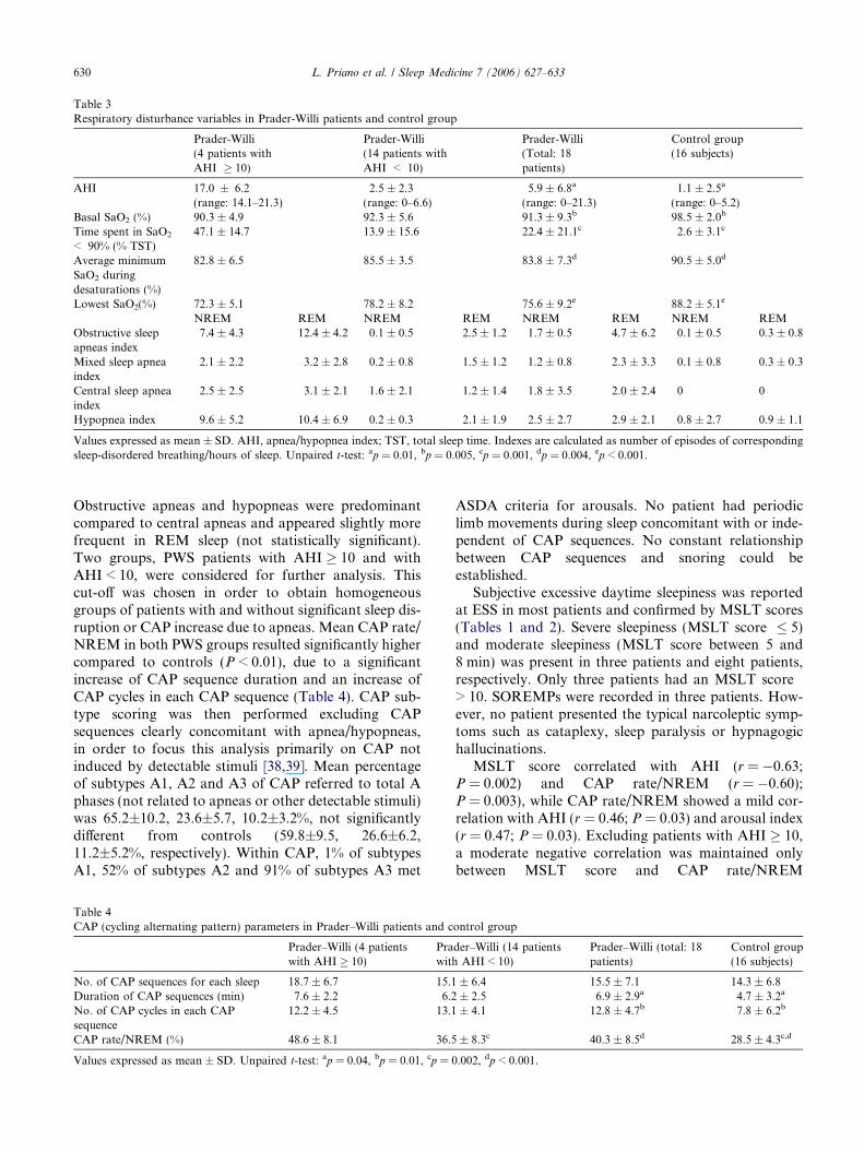
Table 3Respiratory disturbance variables in Prader-Willi patients and control group
Prader-Willi(4 patients withAHI � 10)
Prader-Willi(14 patients withAHI < 10)
Prader-Willi(Total: 18patients)
Control group(16 subjects)
AHI 17.0 ± 6.2(range: 14.1–21.3)
2.5 ± 2.3(range: 0–6.6)
5.9 ± 6.8a
(range: 0–21.3)1.1 ± 2.5a
(range: 0–5.2)Basal SaO2 (%) 90.3 ± 4.9 92.3 ± 5.6 91.3 ± 9.3b 98.5 ± 2.0b
Time spent in SaO2
< 90% (% TST)47.1 ± 14.7 13.9 ± 15.6 22.4 ± 21.1c 2.6 ± 3.1c
Average minimumSaO2 duringdesaturations (%)
82.8 ± 6.5 85.5 ± 3.5 83.8 ± 7.3d 90.5 ± 5.0d
Lowest SaO2(%) 72.3 ± 5.1 78.2 ± 8.2 75.6 ± 9.2e 88.2 ± 5.1e
NREM REM NREM REM NREM REM NREM REMObstructive sleepapneas index
7.4 ± 4.3 12.4 ± 4.2 0.1 ± 0.5 2.5 ± 1.2 1.7 ± 0.5 4.7 ± 6.2 0.1 ± 0.5 0.3 ± 0.8
Mixed sleep apneaindex
2.1 ± 2.2 3.2 ± 2.8 0.2 ± 0.8 1.5 ± 1.2 1.2 ± 0.8 2.3 ± 3.3 0.1 ± 0.8 0.3 ± 0.3
Central sleep apneaindex
2.5 ± 2.5 3.1 ± 2.1 1.6 ± 2.1 1.2 ± 1.4 1.8 ± 3.5 2.0 ± 2.4 0 0
Hypopnea index 9.6 ± 5.2 10.4 ± 6.9 0.2 ± 0.3 2.1 ± 1.9 2.5 ± 2.7 2.9 ± 2.1 0.8 ± 2.7 0.9 ± 1.1
Values expressed as mean ± SD. AHI, apnea/hypopnea index; TST, total sleep time. Indexes are calculated as number of episodes of correspondingsleep-disordered breathing/hours of sleep. Unpaired t-test: ap = 0.01, bp = 0.005, cp = 0.001, dp = 0.004, ep < 0.001.
630 L. Priano et al. / Sleep Medicine 7 (2006) 627–633
Obstructive apneas and hypopneas were predominantcompared to central apneas and appeared slightly morefrequent in REM sleep (not statistically significant).Two groups, PWS patients with AHI � 10 and withAHI < 10, were considered for further analysis. Thiscut-off was chosen in order to obtain homogeneousgroups of patients with and without significant sleep dis-ruption or CAP increase due to apneas. Mean CAP rate/NREM in both PWS groups resulted significantly highercompared to controls (P < 0.01), due to a significantincrease of CAP sequence duration and an increase ofCAP cycles in each CAP sequence (Table 4). CAP sub-type scoring was then performed excluding CAPsequences clearly concomitant with apnea/hypopneas,in order to focus this analysis primarily on CAP notinduced by detectable stimuli [38,39]. Mean percentageof subtypes A1, A2 and A3 of CAP referred to total Aphases (not related to apneas or other detectable stimuli)was 65.2±10.2, 23.6±5.7, 10.2±3.2%, not significantlydifferent from controls (59.8±9.5, 26.6±6.2,11.2±5.2%, respectively). Within CAP, 1% of subtypesA1, 52% of subtypes A2 and 91% of subtypes A3 met
Table 4CAP (cycling alternating pattern) parameters in Prader–Willi patients and c
Prader–Willi (4 patientswith AHI � 10)
Prawith
No. of CAP sequences for each sleep 18.7 ± 6.7 15.1Duration of CAP sequences (min) 7.6 ± 2.2 6.2No. of CAP cycles in each CAPsequence
12.2 ± 4.5 13.1
CAP rate/NREM (%) 48.6 ± 8.1 36.5
Values expressed as mean ± SD. Unpaired t-test: ap = 0.04, bp = 0.01, cp = 0
ASDA criteria for arousals. No patient had periodiclimb movements during sleep concomitant with or inde-pendent of CAP sequences. No constant relationshipbetween CAP sequences and snoring could beestablished.
Subjective excessive daytime sleepiness was reportedat ESS in most patients and confirmed by MSLT scores(Tables 1 and 2). Severe sleepiness (MSLT score � 5)and moderate sleepiness (MSLT score between 5 and8 min) was present in three patients and eight patients,respectively. Only three patients had an MSLT score> 10. SOREMPs were recorded in three patients. How-ever, no patient presented the typical narcoleptic symp-toms such as cataplexy, sleep paralysis or hypnagogichallucinations.
MSLT score correlated with AHI (r = �0.63;P = 0.002) and CAP rate/NREM (r = �0.60);P = 0.003), while CAP rate/NREM showed a mild cor-relation with AHI (r = 0.46; P = 0.03) and arousal index(r = 0.47; P = 0.03). Excluding patients with AHI � 10,a moderate negative correlation was maintained onlybetween MSLT score and CAP rate/NREM
ontrol group
der–Willi (14 patientsAHI < 10)
Prader–Willi (total: 18patients)
Control group(16 subjects)
± 6.4 15.5 ± 7.1 14.3 ± 6.8± 2.5 6.9 ± 2.9a 4.7 ± 3.2a
± 4.1 12.8 ± 4.7b 7.8 ± 6.2b
± 8.3c 40.3 ± 8.5d 28.5 ± 4.3c,d
.002, dp < 0.001.
02468
1012141618
20% 30% 40% 50% 60%
CAP rate/NREM
GH
pea
k (n
g/m
l) Group 1
Group 2
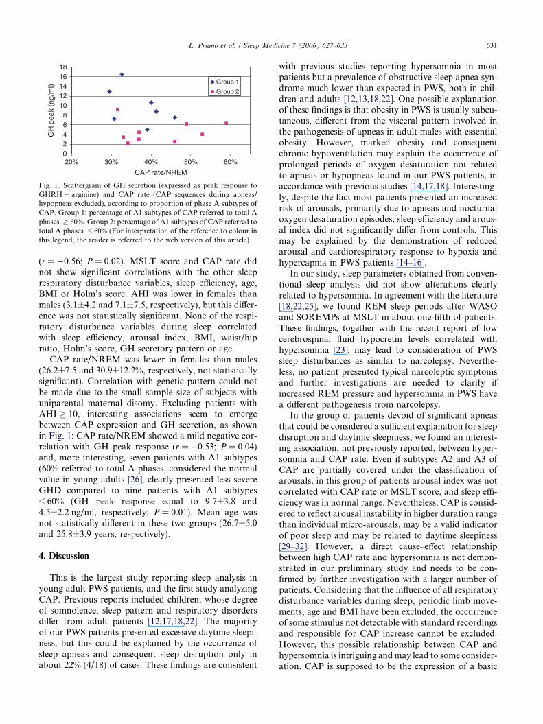
Fig. 1. Scattergram of GH secretion (expressed as peak response toGHRH + arginine) and CAP rate (CAP sequences during apneas/hypopneas excluded), according to proportion of phase A subtypes ofCAP. Group 1: percentage of A1 subtypes of CAP referred to total Aphases � 60%. Group 2: percentage of A1 subtypes of CAP referred tototal A phases < 60%.(For interpretation of the reference to colour inthis legend, the reader is referred to the web version of this article)
L. Priano et al. / Sleep Medicine 7 (2006) 627–633 631
(r = �0.56; P = 0.02). MSLT score and CAP rate didnot show significant correlations with the other sleeprespiratory disturbance variables, sleep efficiency, age,BMI or Holm’s score. AHI was lower in females thanmales (3.1±4.2 and 7.1±7.5, respectively), but this differ-ence was not statistically significant. None of the respi-ratory disturbance variables during sleep correlatedwith sleep efficiency, arousal index, BMI, waist/hipratio, Holm’s score, GH secretory pattern or age.
CAP rate/NREM was lower in females than males(26.2±7.5 and 30.9±12.2%, respectively, not statisticallysignificant). Correlation with genetic pattern could notbe made due to the small sample size of subjects withuniparental maternal disomy. Excluding patients withAHI � 10, interesting associations seem to emergebetween CAP expression and GH secretion, as shownin Fig. 1: CAP rate/NREM showed a mild negative cor-relation with GH peak response (r = �0.53; P = 0.04)and, more interesting, seven patients with A1 subtypes(60% referred to total A phases, considered the normalvalue in young adults [26], clearly presented less severeGHD compared to nine patients with A1 subtypes< 60% (GH peak response equal to 9.7±3.8 and4.5±2.2 ng/ml, respectively; P = 0.01). Mean age wasnot statistically different in these two groups (26.7±5.0and 25.8±3.9 years, respectively).
4. Discussion
This is the largest study reporting sleep analysis inyoung adult PWS patients, and the first study analyzingCAP. Previous reports included children, whose degreeof somnolence, sleep pattern and respiratory disordersdiffer from adult patients [12,17,18,22]. The majorityof our PWS patients presented excessive daytime sleepi-ness, but this could be explained by the occurrence ofsleep apneas and consequent sleep disruption only inabout 22% (4/18) of cases. These findings are consistent
with previous studies reporting hypersomnia in mostpatients but a prevalence of obstructive sleep apnea syn-drome much lower than expected in PWS, both in chil-dren and adults [12,13,18,22]. One possible explanationof these findings is that obesity in PWS is usually subcu-taneous, different from the visceral pattern involved inthe pathogenesis of apneas in adult males with essentialobesity. However, marked obesity and consequentchronic hypoventilation may explain the occurrence ofprolonged periods of oxygen desaturation not relatedto apneas or hypopneas found in our PWS patients, inaccordance with previous studies [14,17,18]. Interesting-ly, despite the fact most patients presented an increasedrisk of arousals, primarily due to apneas and nocturnaloxygen desaturation episodes, sleep efficiency and arous-al index did not significantly differ from controls. Thismay be explained by the demonstration of reducedarousal and cardiorespiratory response to hypoxia andhypercapnia in PWS patients [14–16].
In our study, sleep parameters obtained from conven-tional sleep analysis did not show alterations clearlyrelated to hypersomnia. In agreement with the literature[18,22,25], we found REM sleep periods after WASOand SOREMPs at MSLT in about one-fifth of patients.These findings, together with the recent report of lowcerebrospinal fluid hypocretin levels correlated withhypersomnia [23], may lead to consideration of PWSsleep disturbances as similar to narcolepsy. Neverthe-less, no patient presented typical narcoleptic symptomsand further investigations are needed to clarify ifincreased REM pressure and hypersomnia in PWS havea different pathogenesis from narcolepsy.
In the group of patients devoid of significant apneasthat could be considered a sufficient explanation for sleepdisruption and daytime sleepiness, we found an interest-ing association, not previously reported, between hyper-somnia and CAP rate. Even if subtypes A2 and A3 ofCAP are partially covered under the classification ofarousals, in this group of patients arousal index was notcorrelated with CAP rate or MSLT score, and sleep effi-ciency was in normal range. Nevertheless, CAP is consid-ered to reflect arousal instability in higher duration rangethan individual micro-arousals, may be a valid indicatorof poor sleep and may be related to daytime sleepiness[29–32]. However, a direct cause–effect relationshipbetween high CAP rate and hypersomnia is not demon-strated in our preliminary study and needs to be con-firmed by further investigation with a larger number ofpatients. Considering that the influence of all respiratorydisturbance variables during sleep, periodic limb move-ments, age and BMI have been excluded, the occurrenceof some stimulus not detectable with standard recordingsand responsible for CAP increase cannot be excluded.However, this possible relationship between CAP andhypersomnia is intriguing and may lead to some consider-ation. CAP is supposed to be the expression of a basic

632 L. Priano et al. / Sleep Medicine 7 (2006) 627–633
arousal modulator oscillating between greater (phase A)and lesser (phase B) arousal levels. Moreover, A1 sub-types, characterized by predominant high-voltage slowwaves, contribute to the build-up and maintenance ofEEG synchronization during NREM sleep, while A2and A3 subtypes, characterized by larger amount oflow-amplitude fast rhythms, may be related to the onsetand consolidation of REM sleep [26]. There is evidencethat CAP components corresponding to EEG synchronyrepresent the cortical expression of cortical–subcorticalinteractions mediated by thalamocortical pathways,while the high-frequency component of CAP appears tobe the expression of the activity of the same structuresinvolved in the generation of alpha waves, comprisingpyramidal neurons in layer IV and V within the cerebralcortex, with surface-parallel intracortical neuronsinvolved in its spread [40–42]. Altered CAP expression,similar to REM abnormalities and central apneas as sug-gested in the literature [22,25], may reflect a primary dys-function of central nervous system, and the hypothalamusmay play an important role [7]. In accordance with thishypothesis is the relationship between CAP and GHsecretion found in our study. Patients with more severeGHD have a higher CAP rate and a pattern of CAP char-acterized by lower proportion of A1 subtypes, indepen-dently from age, sex or BMI. This suggests that asubgroup of patients with more severe central dysfunc-tions might be defined. Further studies considering poten-tial interacting role of other factors, age, sex, geneticpattern and cerebrospinal fluid hypocretin level are need-ed to confirm our data and to corroborate the hypothesisof common central dysfunctions responsible for hyper-somnia, endocrine and CAP alterations in PWS.
References
[1] Donaldson MDC, Chu CE, Cooke A, et al. The Prader–Willisyndrome. Arch Dis Child 1994;70:58–63.
[2] Simonoff E, Bolton P, Rutter M. Mental retardation: geneticfindings, clinical implications and research agenda. J ChildPsychol Psychiatry 1996;3:259–80.
[3] State MW, Dykens EM. Genetics of childhood disorders; XV.Prader–Willi syndrome: genes, brain and behaviour. J Am AcadChild Adolesc Psychiatry 2000;39:797–800.
[4] Reed ML, Leff SE. Maternal imprinting of human SNRPN, agene deleted in Prader–Willi syndrome. Nat Genet 1994;6:163–7.
[5] Mac Donald HR, Wevrick R. The necdin gene is deleted inPrader–Willi syndrome and is imprinted in human and mouse.Hum Mol Genet 1997;6:1873–8.
[6] Muscatelli F, Abrous DN, Massacrier A, et al. Disruption of themouse necdin gene results in hypothalamic and behaviouralalteration reminiscent of the human Prader–Willi syndrome. HumMol Genet 2000;39:797–800.
[7] Swaab DF. Prader–Willi syndrome and the hypothalamus. ActaPaediatr Suppl 1997;423:50–4.
[8] Brandt BR, Rosen I. Impaired peripheral somatosensory functionin children with Prader–Willi syndrome. Neuropediatrics1998;29:124–6.
[9] Grugni G, Guzzaloni G, Morabito F. Impairment of GHresponsiveness to GH-releasing hexapeptide (GHRP-6) in Prad-er–Willi syndrome. J Endocrinol Invest 2001;24:340–8.
[10] Grugni G, Morabito F, Crino A. Gonadal function and itsdisorders in simple obesity and in Prader–Willi syndrome. In:Eiholzer U, l’Allemand D, Zipf WB, editors. Prader–Willisyndrome as a model for obesity. Basel: Karger; 2003. p. 140–55.
[11] Harris JC, Allen RP. Is excessive daytime sleepiness characteristicof Prader–Willi syndrome? The effects of weight change. ArchPediatr Adolesc Med 1996;150:1288–93.
[12] Clift S, Dahlitz M, Parkes JD. Sleep apnoea in the Prader–Willisyndrome. J Sleep Res 1994;3:121–6.
[13] Vela-Bueno A, Kales A, Soldatos CR, et al. Sleep in the Prader–Willi syndrome. Clinical and polygraphic findings. Arch Neurol1984;41:294–6.
[14] Menendez AA. Abnormal ventilatory responses in patients withPrader–Willi syndrome. Eur J Pediatr 1999;158:941–2.
[15] Arens R, Gozal D, Burrell BC, et al. Arousal and cardiorespi-ratory responses to hypoxia in Prader–Willi syndrome. Am JRespir Crit Care Med 1996;153:283–7.
[16] Livingston FR, Arens R, Bailey SL, et al. Hypercapnic arousalresponses in Prader–Willi syndrome. Chest 1995;108:1627–31.
[17] Nixon GM, Brouillette RT. Sleep and breathing in Prader–Willisyndrome. Pediatr Pulmonol 2002;34:209–17.
[18] Hertz G, Cataletto M, Feinsilver SH, Angulo M. Sleep andbreathing patterns in patients with Prader–Willi Sindrome (PWS):effects of age and gender. Sleep 1993;16:366–71.
[19] Sforza E, Krieger J, Geisert J, Kurtz D. Sleep and breathingabnormalities in a case of Prader–Willi syndrome. The effects ofacute continuous positive airway pressure treatment. Acta Pae-diatr Scand 1991;80:80–5.
[20] Vgontzas AN, Bixler EO, Kales A, Vela-Bueno A. Prader–Willisyndrome: effects of weight loss on sleep-disordered breathing inPrader–Willi syndrome: the role of obesity. Am J Med Genet1995;56:188–90.
[21] Hiroe Y, Inoue Y, Higami S, et al. Relationship betweenhypersomnia and respiratory disorder during sleep in Prader–Willi syndrome. Psychiatry Clin Neurosci 2000;54:323–5.
[22] Manni R, Politini L, Nobili L, et al. Hypersomnia in the PraderWilli syndrome: clinical–electrophysiological features and under-lyng factors. Clin Neurophysiol 2001;112:800–5.
[23] Nevsimalova S, Vankova J, Stepanova I, et al. Hypocretindeficiency in Prader–Willi syndrome. Eur J Neurol 2005;12:70–2.
[24] Dauvilliers Y, Baumann CR, Carlander B, et al. CSF hypocretin-1 levels in narcolepsy, Kleine–Levin syndrome, and other hyper-somnias and neurological conditions. J Neurol NeurosurgPsychiatry 2003;74:1667–73.
[25] Vgontzas AN, Bixler EO, Kales A, et al. Daytime sleepiness andREM abnormalities in Prader–Willi syndrome: evidence ofgeneralized hypoarousal. Int J Neurosci 1996;87:127–39.
[26] Terzano MG, Parrino L. Origin and significance of the cyclingalternating pattern (CAP). Sleep Med Rev 2000;4:101–23.
[27] Terzano MG, Mancia D, Salati MR, et al. The cyclic alternatingpattern as a physiologic component of normal NREM sleep. Sleep1985;8(2):137–45.
[28] Terzano MG, Parrino L, Rosa A, et al. CAP and arousals in thestructural development of sleep: an integrative perspective. SleepMed 2002;3:221–9.
[29] American Sleep Disorders Association (ASDA). EEG arousals:scoring rules and examples. A preliminary report from the SleepDisorders Atlas Task Force of the American Sleep DisordersAssociation. Sleep 1992;15:174–84.
[30] Terzano MG, Parrino L. Clinical application of cycling alternat-ing pattern. Physiol Behav 1993;54:807–13.
[31] Zucconi M, Oldani A, Ferini-Strambi L, Smirne S. Arousalfluctuation in non-rapid eye movement parasomnias: the role of

L. Priano et al. / Sleep Medicine 7 (2006) 627–633 633
cycling alternating pattern as a measure of arousal instability. JClin Neurophysiol 1995;12:147–54.
[32] Droste DW, Knauss JK, HAgerdon G, Kaps M. Periodic legmovements are part of B-wave rhythm and the cycling alternatingpattern. Acta Neurol Scand 1996;94:347–52.
[33] Holm VA, Cassidy SB, Butler MG, et al. Prader–Willi syndrome:consensus diagnostic criteria. Pediatrics 1993;91:398–402.
[34] Joint FAO/WHO/UNU: expert consultation. Energy and proteinrequirements. WHO technical report series, no. 724. Geneve:WHO; 1985.
[35] Terzano MG, Parrino L, Smerieri A, et al. Atlas, rules, andrecording techniques for the scoring of cyclic alternating pattern(CAP) in human sleep. Sleep Med 2002;3:187–99.
[36] Parrino L, Smerieri A, Rossi M, Terzano MG. Relationship ofslow and rapid EEG components of CAP to ASDA arousals innormal sleep. Sleep 2001;24:881–5.
[37] Aimaretti G, Corneli G, Razzore P, et al. Comparison betweeninsulin-induced hypoglycemia and growth hormone(GH)-releas-
ing hormone + arginine as provocative tests for the diagnosis ofGH deficiency in adults. J Clin Endocrinol Metab1998;83:1615–8.
[38] Hirshkowitz M, Moore CA, Hamilton CR, et al. Polysomnog-raphy of adults and elderly: sleep architecture, respiration and legmovement. J Clin Neurophysiol 1992;9:56–62.
[39] Terzano MG, Parrino L, Boselli M, et al. Polysomnographicanalysis of arousal responses in OSAS by means of the cyclingalternating pattern (CAP). J Clin Neurophysiol 1996;13:145–55.
[40] Amzica F, Steriade M. Electrophysiological correlates of sleepdelta waves. Electroencephalogr Clin Neurophysiol1998;107:69–83.
[41] Terzano MG, Parrino L, Boselli M, et al. CAP components andEEG synchronization in the first 3 sleep cycles. Clin Neurophysiol2000;111:283–90.
[42] Ferri R, Bruni O, Miano S, Terzano MG. Topographic mappingof the spectral components of the cycling alternating pattern(CAP). Sleep Med 2005;6:29–36.