Simulating movement of tRNA into the ribosome during decoding
6
Simulating movement of tRNA into the ribosome during decoding Kevin Y. Sanbonmatsu* † , Simpson Joseph ‡ , and Chang-Shung Tung* *Department of Theoretical Biology and Biophysics, Theoretical Division, Los Alamos National Laboratory, Los Alamos, NM 87545; and ‡ Department of Chemistry and Biochemistry, University of California at San Diego, La Jolla, CA 92093 Edited by Olke C. Uhlenbeck, Northwestern University, Evanston, IL, and approved August 19, 2005 (received for review April 30, 2005) Decoding is the key step during protein synthesis that enables information transfer from RNA to protein, a process critical for the survival of all organisms. We have used large-scale (2.64 10 6 atoms) all-atom simulations of the entire ribosome to understand a critical step of decoding. Although the decoding problem has been studied for more than four decades, the rate-limiting step of cognate tRNA selection has only recently been identified. This step, known as accommodation, involves the movement inside the ribosome of the aminoacyl-tRNA from the partially bound ‘‘AT’’ state to the fully bound ‘‘AA’’ state. Here, we show that a corridor of 20 universally conserved ribosomal RNA bases interacts with the tRNA during the accommodation movement. Surprisingly, the tRNA is impeded by the A-loop (23S helix 92), instead of enjoying a smooth transition to the AA state. In particular, universally conserved 23S ribosomal RNA bases U2492, C2556, and C2573 act as a 3D gate, causing the acceptor stem to pause before allowing entrance into the peptidyl transferase center. Our simulations demonstrate that the flexibility of the acceptor stem of the tRNA, in addition to flexibility of the anticodon arm, is essential for tRNA selection. This study serves as a template for simulating confor- mational changes in large (>10 6 atoms) biological and artificial molecular machines. proofreading protein synthesis molecular dynamics simulations RNA high-performance computing A ccurate and rapid selection of tRNAs by the ribosome is critical for cell viability. Selection of cognate tRNAs occurs in two stages (initial selection and proofreading), which are separated by hydrolysis of GTP in the aminoacyl- tRNAGTPelongation factor Tu (EF-Tu) ternary complex (1– 4). During initial selection, the ternary complex binds to the aminoacyl site (A site) of the small (30S) ribosomal subunit and the GTPase activation center (GAC) of the large (50S) ribo- somal subunit producing the ‘‘AT’’ state complex (5–7). It has also been suggested that codon–anticodon interactions at the exit site can affect selection (8). During the proofreading step, the EF-TuGDP dissociates, and the aminoacyl-tRNA moves from the AT to the fully bound AA state, where the tRNA occupies the 30S and 50S A sites (accommodation). The ribosome must adjust its conformation to accommodate this change in the aminoacyl-tRNA (6). This accommodation step is rate-limiting for cognate tRNA (9). The mechanism of this important step is currently unknown. Because the ribosome performs the complex operation of transforming a 4-letter alphabet sequence into a 20-letter alphabet sequence, the decoding mechanism has implications for artificial informa- tion-processing molecular machines. Structural biologists have made a significant contribution, determining the lower-resolution structures of the ribosome in the AA (10, 11) and AT states (12, 13), as well as the interactions of cognate tRNA analogs with the 30S and 50S subunits in atomic detail (14–17). More recently, higher- resolution cryo-EM data have shown that the aminoacyl-tRNA in the AT state is kinked with respect to the AA state (hereby referred to as the AT kink), suggesting that the accommodation change involves a relaxation of the kinked tRNA that preserves the decoding-center interactions (6, 7, 18). The only significant change in the AT state ribosome is the movement of the GAC (6), which occurs in two steps, corresponding to before and after GTP hydrolysis (7, 19). The structures of the AA (10) and AT (6) states of the 70S ribosome have established the initial and final states of the accommodation step; however, important questions remain concerning the accommodation pathway. In particular, (i) which proteins and 23S rRNA nucleotides does the tRNA interact with during accommodation? and (ii) how is it possible for the acceptor stem of the tRNA to move into the peptidyl transferase center, given the tight packing (20) of 23S rRNA in this region of the large subunit? Molecular dynamics simulations and modeling studies have explored the decoding center (21–24), isolated tRNA (25), and the inner core of the 30S ribosomal subunit (26, 27). Adaptive- mesh refinement Poisson–Boltzmann studies based on phos- phateC structures have examined the electrostatic potential of the subunits (28). Coarse-grained studies have investigated the motions of the ribosome within the approximations of the Gaussian network and bead models (28–31). Real-space refine- ment has been combined with cryo-EM structures to study the beginning and end states of a conformational change that occurs during translocation (32). Explicit solvent molecular dynamics simulations have previously helped elucidate functional mech- anisms of biomolecular systems (33–38) and been validated with experiments (39). The Advanced Simulation and Computing (ASCI) Q ma- chine at Los Alamos National Laboratory, together with the NAMD program (40), has enabled us to simulate the accom- modation of tRNA into the 70S ribosome in explicit solvent (2.64 10 6 atoms), yielding significantly more accurate dy- namics than implicit solvent methods (41) and the coarse- grained calculations described above. Computationally, the simulations represent a step forward in that the largest pre- vious biomolecular dynamics simulations have been on the order of 4.2 10 5 atoms (42, 43). Because the accommo- dation rate of cognate tRNAs is 7s (9), even enhanced sampling molecular dynamics simulation techniques yield in- sufficient sampling to observe spontaneous accommodation. Therefore, to examine accommodation, we have implemented the targeted molecular dynamics algorithm (35, 44, 45), which gradually reduces the root mean square deviation between the simulated structure and the final target structure (AA state) while allowing thermal fluctuations at a given root mean square deviation. Targeted molecular dynamics simulates a single barrier-crossing event. Although the accommodation barrier-crossing time cannot be measured experimentally, it is This paper was submitted directly (Track II) to the PNAS office. Freely available online through the PNAS open access option. Abbreviations: A site, aminoacyl site; GAC, GTPase activation center; EF-Tu, elongation factor Tu; SRL, sarcin-ricin loop. † To whom correspondence should be addressed. E-mail: [email protected]. © 2005 by The National Academy of Sciences of the USA 15854 –15859 PNAS November 1, 2005 vol. 102 no. 44 www.pnas.orgcgidoi10.1073pnas.0503456102
-
Upload
independent -
Category
Documents
-
view
2 -
download
0
Transcript of Simulating movement of tRNA into the ribosome during decoding

Simulating movement of tRNA into the ribosomeduring decodingKevin Y. Sanbonmatsu*†, Simpson Joseph‡, and Chang-Shung Tung*
*Department of Theoretical Biology and Biophysics, Theoretical Division, Los Alamos National Laboratory, Los Alamos, NM 87545; and ‡Departmentof Chemistry and Biochemistry, University of California at San Diego, La Jolla, CA 92093
Edited by Olke C. Uhlenbeck, Northwestern University, Evanston, IL, and approved August 19, 2005 (received for review April 30, 2005)
Decoding is the key step during protein synthesis that enablesinformation transfer from RNA to protein, a process critical for thesurvival of all organisms. We have used large-scale (2.64 � 106
atoms) all-atom simulations of the entire ribosome to understanda critical step of decoding. Although the decoding problem hasbeen studied for more than four decades, the rate-limiting step ofcognate tRNA selection has only recently been identified. This step,known as accommodation, involves the movement inside theribosome of the aminoacyl-tRNA from the partially bound ‘‘A�T’’state to the fully bound ‘‘A�A’’ state. Here, we show that a corridorof 20 universally conserved ribosomal RNA bases interacts with thetRNA during the accommodation movement. Surprisingly, thetRNA is impeded by the A-loop (23S helix 92), instead of enjoyinga smooth transition to the A�A state. In particular, universallyconserved 23S ribosomal RNA bases U2492, C2556, and C2573 actas a 3D gate, causing the acceptor stem to pause before allowingentrance into the peptidyl transferase center. Our simulationsdemonstrate that the flexibility of the acceptor stem of the tRNA,in addition to flexibility of the anticodon arm, is essential for tRNAselection. This study serves as a template for simulating confor-mational changes in large (>106 atoms) biological and artificialmolecular machines.
proofreading � protein synthesis � molecular dynamicssimulations � RNA � high-performance computing
Accurate and rapid selection of tRNAs by the ribosome iscritical for cell viability. Selection of cognate tRNAs occurs
in two stages (initial selection and proofreading), which areseparated by hydrolysis of GTP in the aminoacyl-tRNA�GTP�elongation factor Tu (EF-Tu) ternary complex (1–4). During initial selection, the ternary complex binds to theaminoacyl site (A site) of the small (30S) ribosomal subunit andthe GTPase activation center (GAC) of the large (50S) ribo-somal subunit producing the ‘‘A�T’’ state complex (5–7). It hasalso been suggested that codon–anticodon interactions at theexit site can affect selection (8).
During the proofreading step, the EF-Tu�GDP dissociates,and the aminoacyl-tRNA moves from the A�T to the fully boundA�A state, where the tRNA occupies the 30S and 50S A sites(accommodation). The ribosome must adjust its conformation toaccommodate this change in the aminoacyl-tRNA (6). Thisaccommodation step is rate-limiting for cognate tRNA (9). Themechanism of this important step is currently unknown. Becausethe ribosome performs the complex operation of transforming a4-letter alphabet sequence into a 20-letter alphabet sequence,the decoding mechanism has implications for artificial informa-tion-processing molecular machines.
Structural biologists have made a significant contribution,determining the lower-resolution structures of the ribosome inthe A�A (10, 11) and A�T states (12, 13), as well as theinteractions of cognate tRNA analogs with the 30S and 50Ssubunits in atomic detail (14–17). More recently, higher-resolution cryo-EM data have shown that the aminoacyl-tRNAin the A�T state is kinked with respect to the A�A state (herebyreferred to as the A�T kink), suggesting that the accommodation
change involves a relaxation of the kinked tRNA that preservesthe decoding-center interactions (6, 7, 18). The only significantchange in the A�T state ribosome is the movement of the GAC(6), which occurs in two steps, corresponding to before and afterGTP hydrolysis (7, 19). The structures of the A�A (10) and A�T(6) states of the 70S ribosome have established the initial andfinal states of the accommodation step; however, importantquestions remain concerning the accommodation pathway. Inparticular, (i) which proteins and 23S rRNA nucleotides does thetRNA interact with during accommodation? and (ii) how is itpossible for the acceptor stem of the tRNA to move into thepeptidyl transferase center, given the tight packing (20) of 23SrRNA in this region of the large subunit?
Molecular dynamics simulations and modeling studies haveexplored the decoding center (21–24), isolated tRNA (25), andthe inner core of the 30S ribosomal subunit (26, 27). Adaptive-mesh refinement Poisson–Boltzmann studies based on phos-phate�C� structures have examined the electrostatic potential ofthe subunits (28). Coarse-grained studies have investigated themotions of the ribosome within the approximations of theGaussian network and bead models (28–31). Real-space refine-ment has been combined with cryo-EM structures to study thebeginning and end states of a conformational change that occursduring translocation (32). Explicit solvent molecular dynamicssimulations have previously helped elucidate functional mech-anisms of biomolecular systems (33–38) and been validated withexperiments (39).
The Advanced Simulation and Computing (ASCI) Q ma-chine at Los Alamos National Laboratory, together with theNAMD program (40), has enabled us to simulate the accom-modation of tRNA into the 70S ribosome in explicit solvent(2.64 � 106 atoms), yielding significantly more accurate dy-namics than implicit solvent methods (41) and the coarse-grained calculations described above. Computationally, thesimulations represent a step forward in that the largest pre-vious biomolecular dynamics simulations have been on theorder of �4.2 � 105 atoms (42, 43). Because the accommo-dation rate of cognate tRNAs is �7�s (9), even enhancedsampling molecular dynamics simulation techniques yield in-sufficient sampling to observe spontaneous accommodation.Therefore, to examine accommodation, we have implementedthe targeted molecular dynamics algorithm (35, 44, 45), whichgradually reduces the root mean square deviation between thesimulated structure and the final target structure (A�A state)while allowing thermal f luctuations at a given root meansquare deviation. Targeted molecular dynamics simulates asingle barrier-crossing event. Although the accommodationbarrier-crossing time cannot be measured experimentally, it is
This paper was submitted directly (Track II) to the PNAS office.
Freely available online through the PNAS open access option.
Abbreviations: A site, aminoacyl site; GAC, GTPase activation center; EF-Tu, elongationfactor Tu; SRL, sarcin-ricin loop.
†To whom correspondence should be addressed. E-mail: [email protected].
© 2005 by The National Academy of Sciences of the USA
15854–15859 � PNAS � November 1, 2005 � vol. 102 � no. 44 www.pnas.org�cgi�doi�10.1073�pnas.0503456102

thought to be several orders of magnitude smaller than theaccommodation rate. Kinetic experiments have suggested thataccommodation follows EF-Tu dissociation (1). Here, weassume that EF-Tu has already dissociated from the ribosome.
Rather than providing an exact energy landscape of thetransition, our simulations produce stereochemically feasiblepathways that can be tested experimentally. In particular, thegoal of this work is to predict 23S rRNA bases and ribosomalprotein residues that are important for accommodation. Thisapproach is complementary to recent x-ray crystallography(10, 11, 14, 16, 17), cryo-EM (7, 12, 18), kinetic (1), andsingle-molecule (19) studies and helps establish a logicalpicture of the mechanism of tRNA selection. We tested thesensitivity of our results to initial conditions by performingseven 2-ns simulations with different initial velocities and thesensitivity to the simulated time scale by doubling and halvingthe simulation time, giving a total sampling of 20 ns forproduction simulations.
MethodsThe A�T-state model structure is similar to the previous A�A-state model (46), with the exception of the tRNA and the GAC(23S rRNA helices 43–44 and L11), whose structures are basedon the corresponding cryo-EM map (6). The solvated systemswere minimized and equilibrated (NAMD2.5) for 1.6 ns. Perfor-mance studies demonstrated near-linear scaling to 768 proces-sors (see Supporting Text, which is published as supporting
information on the PNAS web site, for details of the methodsused). The figures were created with VMD (47).
ResultsThe accommodation of the aminoacyl-tRNA in all simulationsdisplays a bulk motion of the tRNA body into the large subunitA site (Movie 1 A, which is published as supporting informationon the PNAS web site, and Fig. 1). The motion involves therelaxation of the A�T kink about tRNA positions 44–45 and 26,combined with a �45° rotation around the anticodon stem loopaxis. Each simulation also shows the subsequent accommodationof the universally conserved 3�-CCA end into the peptidyltransferase center and convergence to the target structure. Theentrance of the 3�-CCA end into the peptidyl transferase centeris impeded with respect to the motion of the tRNA body becauseof the interaction with the 23S A-loop (positions 2552–2561).The codon–anticodon interactions, and the interactions of 16SrRNA G530, A1492, and A1493 with the tRNA and mRNA,remained intact throughout the accommodation simulations(Fig. 5, which is published as supporting information on thePNAS web site).
The interactions between the tRNA and the ribosome (Movie1B and Fig. 2) are divided into four stages, describing A�Tinteractions (stage 1), relaxation of the tRNA body (stage 2),relaxation of the tRNA acceptor stem (stage 3), and A�Ainteractions (stage 4). The tRNA movement is characterized bythe parameters �1, �1, and �2 (defined in the Fig. 2 legend).
Fig. 1. Accommodation simulation overview and aminoacyl-tRNA–50S interactions. (a) Time evolution of cognate tRNA accommodation into the ribosome forone of seven 2-ns simulations. Explicit waters, ions, and the top portion of the 70S ribosome are not shown so that the tRNAs are visible. The 23S rRNA (white),50S proteins (light green), 16S rRNA (purple), 30S proteins (pink), mRNA (dark green), aminoacyl-tRNA (yellow) with Phe amino acid (dark green), peptidyl-tRNA(cyan), and 23S rRNA 2553 (red) are shown. The schematics are similar to that of Moazed and Noller (5) and depict the process of accommodation. A, aminoacylsite; P, peptidyl site. Stage 1 is represented by the initial production structure, after 1.6 ns of equilibration (t � 0 ns). Stage 2 shows the relaxation of theaminoacyl-tRNA body (i.e., all but the 3�-CCA portion) (t � 0.292 ns). In stage 3, the aminoacyl-tRNA body is accommodated (t � 0.65 ns). During stage 4, theaminoacyl-tRNA 3�-CCA end is accommodated into the peptidyl transferase center (2.2 ns). (b) Secondary structure diagrams color-coded by the region of tRNAthat participates in the aminoacyl-tRNA–50S interaction. Green, interactions with T�C loop; blue, interactions with D loop; magenta, interactions with 3�-CCAend. ASL, anticodon stem loop.
Sanbonmatsu et al. PNAS � November 1, 2005 � vol. 102 � no. 44 � 15855
BIO
PHYS
ICS

Stage 1. During the initial stage of accommodation, the 3�-CCAend of the tRNA remains relatively unencumbered by theribosome, apart from a slight deflection (��2 � 5°) because ofinteraction with ribosomal protein L14 (residues 52, 54, 89, and90) (Fig. 2 and Fig. 6, which is published as supporting infor-mation on the PNAS web site). There is also a transientinteraction between the acceptor arm and ribosomal proteinS12. Initially, the phosphate of tRNA base U69 interacts withHis-80 of S12. As the acceptor arm moves away from theA�T-state position, the phosphate of tRNA U68 briefly interactswith Gln-78.
The elbow region (tRNA bases 52–58) interacts with theGAC at base A1067, whose N6 position interacts consecutivelywith elbow bases (tRNA bases 55, 54, 53, 52, and 51) as thetRNA moves toward the A site (Figs. 2 and 3 and Fig. 7, whichis published as supporting information on the PNAS web site).The elbow region also interacts with 23S rRNA helix 89 U2473,which makes consecutive interactions with tRNA bases 62, 63,and 64 as they pass by helix 89 (Fig. 3). The tRNA also interactswith 23S rRNA A2660–G2661 in the sarcin-ricin loop (SRL)at early times as it moves toward the A site (Fig. 3). Largesubunit rRNA bases U2473, G2661, and A1067 appear to‘‘monitor’’ the tRNA by interacting continuously with thebackbone of the tRNA as the tRNA moves toward the A�Astate.
Stage 2. In stage 2 (Figs. 1b and 2), helix 89 continues to interactwith the tRNA. We see that at the end of stage 2 the tRNA body
is nearly accommodated, with �1 and �1 within 10% of their finalvalues (Fig. 2). The 3�-CCA end of the tRNA begins its inter-action with the A-loop in this stage (Figs. 1b and 3); however, itis not accommodated by the end of stage 2 (Movie 1B and Fig.2). Instead, it is impeded by the 23S A-loop, whose interactioncauses a significant deflection (��2 � �25°) in the 3�-CCA end(Fig. 2).
Stage 3. In stage 3, after first interacting with the elbow of thetRNA, the relative orientation of H89 with respect to thetRNA causes the interaction point to move on the tRNAprogressively toward the 3�-CCA end (Figs. 2 and 3). In thesense that H89 sterically restricts the motion of the tRNA,helix 89 appears to ‘‘guide’’ the aminoacyl-tRNA toward the50S A site. When the tRNA reaches the A-loop, the 3�-CCAend moves along the A-loop and H90 toward the peptidyltransferase center, where the 3�-CCA end interacts consecu-tively with respect to time with 2560, 2559, 2558, 2557, 2556,2555, 2554, and 2553 (Fig. 3).
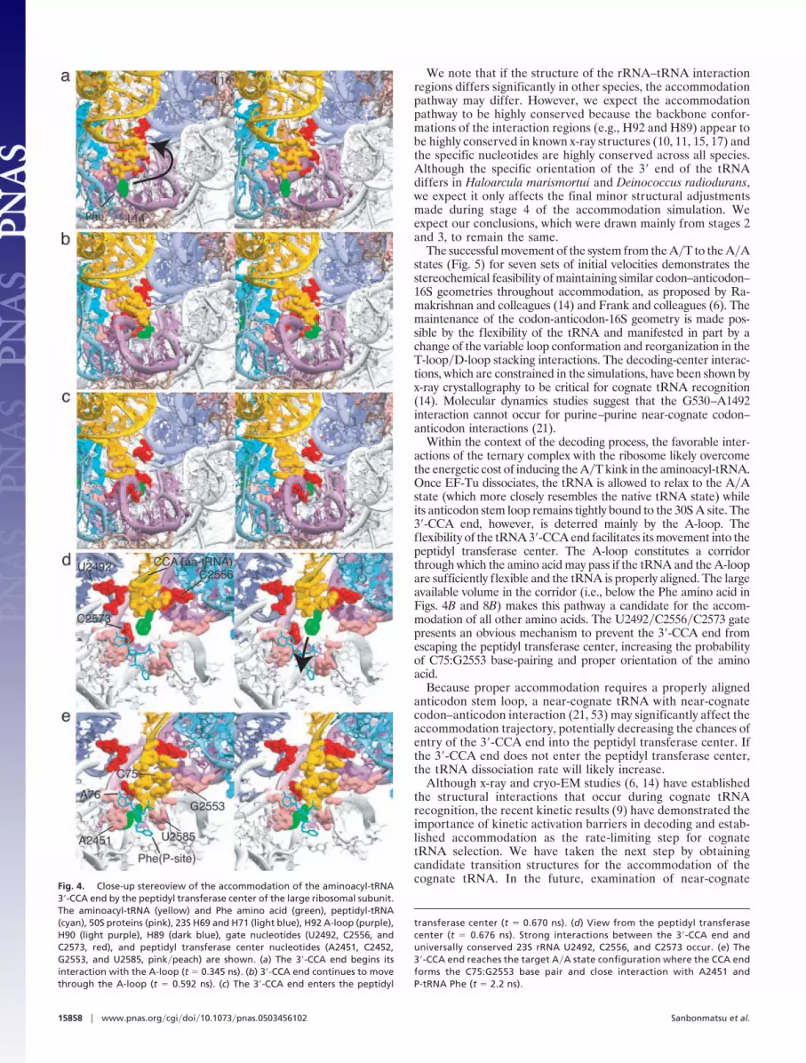
During stage 3, both the 3�-CCA end and the A-loop flexslightly to allow accommodation of the 3�-CCA end through theA-loop toward the peptidyl transferase center (Movie 1D, Fig. 4a–c, and Fig. 8 A–C, which is published as supporting informa-tion on the PNAS web site). The mutual f lexibility between thetRNA 3�-CCA end and the 23S rRNA A-loop resolves theapparent steric clash present during accommodation.
Strong interactions (probability density, p � 1, defined in theFig. 7 legend) occur between the 3�-CCA end and universally
Fig. 2. Stages of accommodation. (a) Aminoacyl-tRNA interaction regions (within 3.5 Å) colored according to the accommodation stage. (Inset) The contextof the accommodation wall on the 50S ribosomal subunit. (b) Time evolution of parameters that describe the deformation of aminoacyl-tRNA. Average values(black) of �1, �1, and �2 (averaged over seven trajectories) and corresponding variances (yellow) are shown. �1 is the angle between O3� atoms at positions 33,40, and 55 of the tRNA. �1 is the dihedral angle between O3� atoms at positions 33, 40, 55 and 1. �2 is the dihedral angle between O3� atoms at positions 69,71, and 73 and the O of the Phe. (c) Snapshots from stage 1 (t � 0 ns), stage 2 (t � 0.488 ns), stage 3 (t � 0.650 ns), and stage 4 (t � 2.2 ns).
15856 � www.pnas.org�cgi�doi�10.1073�pnas.0503456102 Sanbonmatsu et al.

conserved bases U2492, C2556, and C2573 (Movie 1 C and Dand Fig. 3). Interaction of the 3�-CCA end with these threenucleotides coincides with the pausing of the 3�-CCA-Pheportion of the tRNA (i.e., the center of mass velocity of thisportion decreases dramatically) just before entrance into thepeptidyl transferase center (Movie 1C and Fig. 9, which ispublished as supporting information on the PNAS web site). Inparticular, the 3�-CCA end pauses upon interaction withU2492 and C2556 until the U2492�C2556 gate opens (i.e., theU2492 O2�–C2556 O2� distance increases significantly) atwhich point the CCA end proceeds through (Fig. 9). The CCAend then immediately interacts with C2573, causing it to pausea second time (Fig. 9). These characteristics are consistent witha 3-nt, 3D gate that impedes the 3�-CCA-Phe portion beforeentrance into the peptidyl transferase center.
Finally, L16, H38, and H69 are positioned to prevent theaminoacyl-tRNA from overshooting its target position andinteracting with the peptidyl-tRNA. Thus, during stage 3, thetRNA is sterically guided, paused, and stopped by 23S rRNA.
Stage 4. By stage 4, the tRNA body has been accommodated bythe ribosome, and the 3�-CCA end enters the peptidyl trans-ferase center (Figs. 4 D and E and 8 D and E). During this stage,the 3�-CCA end moves into its final configuration [based on thehigher resolution x-ray data (16)], forming the C75:G2553 basepair and proper interactions with peptidyl transferase basesA2451, C2452 and U2585 (Movie 1E and Figs. 4 and 8). Thephosphate root mean square deviation values between the finalsimulated structure and the A�A-state x-ray structures are 1.21,1.27, and 2.41 Å for the 30S A and peptidyl (P) sites (14), the 50SA and P sites (16), and the 70S complex (10), respectively (A andP sites are defined by all rRNA nucleotides and protein residueswithin 4.5 Å of the tRNA analogs in the higher-resolution x-raystructures). The larger root mean square deviation for the 70Sis likely caused by the lower resolution of the 70S x-ray structureupon which our 70S A�A state is based.
DiscussionOf the 68 nucleotides in the 23S rRNA that were observed tointeract with the aminoacyl-tRNA during our simulations, 28
nucleotides (41%) are �95% conserved. Eighteen nucleotidesare ‘‘universally conserved’’ in the sense that they are as highlyconserved (�99%) or more highly conserved than 16S rRNAbase A1493 (48). The universal 23S rRNA nucleotides thatinteract with the aminoacyl-tRNA are 1943, 1953, and 1955 (H71region); 2492 (H89); 2506, 2451, 2452, 2583, 2584, and 2585(peptidyl transferase center); 2552, 2553, and 2556 (A-loopH92); 2508 and 2573 (H90); 2602 (H93); and 2662–3 (SRL). Theremaining 10 bases that are �95% conserved are 1074 (GAC);2469, 2470, 2472, 2482, and 2493 (H89); 2554, 2555, and 2559(A-loop H92); and 2660 (SRL).
The focus of this investigation is the transition from the A�Tto the A�A state, rather than the A�T and A�A statesthemselves. Of the tRNA interactions that occur betweenthese two states, the A-loop and H89 display the strongestinteractions (Fig. 3) and also have the largest number of highlyconserved nucleotides. In H89, purine mutations of U2492 willchange the U2492:U2460 pair [present in the x-ray structure(16) and in our simulations] to the more stable G:U or A:Upairs; however, the larger purine nucleotide will cause thebackbone of the 2492 region to protrude into the pathway ofthe tRNA, narrowing the accommodation corridor and, thus,increasing the steric barrier. The mutation of C2573 to a purinemay also result in increased steric inhibition and correspondinghyperaccurate phenotypes.
Previous studies showed that mutations of 23S U2492 suppressframeshifting (49), even though U2492 does not interact with thetRNA in the A�T or A�A states. This fact is consistent withU2492 interacting with the tRNA during accommodation, butnot in A�T or A�A states, as observed in our simulations.Mutations of U2555 exhibit a ribosomal ambiguity phenotype(49, 50), whereas mutations in G2583 result in increased trans-lational accuracy (51). Interestingly, both of these nucleotidesdisplay strong interactions with the tRNA in our simulations.Finally, changes in the conformation of the D-loop are thoughtto play a key role in decoding as shown by rapid kinetics andD-loop mutations (52). Performing simulations of availablemutants in addition to WT systems may provide greater insightinto experimental data.
Fig. 3. Time evolution of probability densities, p, of interactions (blue, no interaction; red, strong interaction). p is the number of interactions normalized bythe number of simulations (defined in Fig. 7). Nucleotide�residue positions for the aminoacyl-tRNA, 23S rRNA, and 50S proteins participating in aminoacyl-tRNAinteractions are shown along the x axis. Diagonal contours for the A-loop correspond to the aminoacyl-tRNA 3�-CCA end interacting with consecutive A-loopnucleotides as a function of time. Distinct regions are separated by vertical black bars.
Sanbonmatsu et al. PNAS � November 1, 2005 � vol. 102 � no. 44 � 15857
BIO
PHYS
ICS
We note that if the structure of the rRNA–tRNA interactionregions differs significantly in other species, the accommodationpathway may differ. However, we expect the accommodationpathway to be highly conserved because the backbone confor-mations of the interaction regions (e.g., H92 and H89) appear tobe highly conserved in known x-ray structures (10, 11, 15, 17) andthe specific nucleotides are highly conserved across all species.Although the specific orientation of the 3� end of the tRNAdiffers in Haloarcula marismortui and Deinococcus radiodurans,we expect it only affects the final minor structural adjustmentsmade during stage 4 of the accommodation simulation. Weexpect our conclusions, which were drawn mainly from stages 2and 3, to remain the same.
The successful movement of the system from the A�T to the A�Astates (Fig. 5) for seven sets of initial velocities demonstrates thestereochemical feasibility of maintaining similar codon–anticodon–16S geometries throughout accommodation, as proposed by Ra-makrishnan and colleagues (14) and Frank and colleagues (6). Themaintenance of the codon-anticodon-16S geometry is made pos-sible by the flexibility of the tRNA and manifested in part by achange of the variable loop conformation and reorganization in theT-loop�D-loop stacking interactions. The decoding-center interac-tions, which are constrained in the simulations, have been shown byx-ray crystallography to be critical for cognate tRNA recognition(14). Molecular dynamics studies suggest that the G530–A1492interaction cannot occur for purine–purine near-cognate codon–anticodon interactions (21).
Within the context of the decoding process, the favorable inter-actions of the ternary complex with the ribosome likely overcomethe energetic cost of inducing the A�T kink in the aminoacyl-tRNA.Once EF-Tu dissociates, the tRNA is allowed to relax to the A�Astate (which more closely resembles the native tRNA state) whileits anticodon stem loop remains tightly bound to the 30S A site. The3�-CCA end, however, is deterred mainly by the A-loop. Theflexibility of the tRNA 3�-CCA end facilitates its movement into thepeptidyl transferase center. The A-loop constitutes a corridorthrough which the amino acid may pass if the tRNA and the A-loopare sufficiently flexible and the tRNA is properly aligned. The largeavailable volume in the corridor (i.e., below the Phe amino acid inFigs. 4B and 8B) makes this pathway a candidate for the accom-modation of all other amino acids. The U2492�C2556�C2573 gatepresents an obvious mechanism to prevent the 3�-CCA end fromescaping the peptidyl transferase center, increasing the probabilityof C75:G2553 base-pairing and proper orientation of the aminoacid.
Because proper accommodation requires a properly alignedanticodon stem loop, a near-cognate tRNA with near-cognatecodon–anticodon interaction (21, 53) may significantly affect theaccommodation trajectory, potentially decreasing the chances ofentry of the 3�-CCA end into the peptidyl transferase center. Ifthe 3�-CCA end does not enter the peptidyl transferase center,the tRNA dissociation rate will likely increase.
Although x-ray and cryo-EM studies (6, 14) have establishedthe structural interactions that occur during cognate tRNArecognition, the recent kinetic results (9) have demonstrated theimportance of kinetic activation barriers in decoding and estab-lished accommodation as the rate-limiting step for cognatetRNA selection. We have taken the next step by obtainingcandidate transition structures for the accommodation of thecognate tRNA. In the future, examination of near-cognate
transferase center (t � 0.670 ns). (d) View from the peptidyl transferasecenter (t � 0.676 ns). Strong interactions between the 3�-CCA end anduniversally conserved 23S rRNA U2492, C2556, and C2573 occur. (e) The3�-CCA end reaches the target A�A state configuration where the CCA endforms the C75:G2553 base pair and close interaction with A2451 andP-tRNA Phe (t � 2.2 ns).
Fig. 4. Close-up stereoview of the accommodation of the aminoacyl-tRNA3�-CCA end by the peptidyl transferase center of the large ribosomal subunit.The aminoacyl-tRNA (yellow) and Phe amino acid (green), peptidyl-tRNA(cyan), 50S proteins (pink), 23S H69 and H71 (light blue), H92 A-loop (purple),H90 (light purple), H89 (dark blue), gate nucleotides (U2492, C2556, andC2573, red), and peptidyl transferase center nucleotides (A2451, C2452,G2553, and U2585, pink�peach) are shown. (a) The 3�-CCA end begins itsinteraction with the A-loop (t � 0.345 ns). (b) 3�-CCA end continues to movethrough the A-loop (t � 0.592 ns). (c) The 3�-CCA end enters the peptidyl
15858 � www.pnas.org�cgi�doi�10.1073�pnas.0503456102 Sanbonmatsu et al.

tRNAs will be critical to uncover the mechanism by whichribosomes are able to reject incorrect tRNAs, which is anessential part of the translation of genetic messages by theribosome.
We thank Steve Harvey, Angel Garcia, Chris Jarzynski, Jason Feinberg, andSteve Phelps for useful discussions and critical reading of the manuscript;
and Andy White, Manuel Vigil, and Hal Marshal for their support and input.This work was performed under the auspices of the U.S. Department ofEnergy under Contract W-7405-ENG-36. K.Y.S. was supported by NationalInstitutes of Health Grant R01-GM072686, and S.J. was supported byNational Science Foundation Grant MCB-0078322. The Ribosome Projectis generously supported by the Los Alamos National Laboratory Institu-tional Computing Program.
1. Rodnina, M. V. & Wintermeyer, W. (2001) Annu. Rev. Biochem. 70, 415–435.2. Ninio, J. (1974) J. Mol. Biol. 84, 297–313.3. Hopfield, J. J. (1974) Proc. Natl. Acad. Sci. USA 71, 4135–4139.4. Thompson, R. C. & Stone, P. J. (1977) Proc. Natl. Acad. Sci. USA 74, 198–202.5. Moazed, D. & Noller, H. F. (1989) Nature 342, 142–148.6. Valle, M., Zavialov, A., Li, W., Stagg, S. M., Sengupta, J., Nielsen, R. C., Nissen,
P., Harvey, S. C., Ehrenberg, M. & Frank, J. (2003) Nat. Struct. Biol. 10, 899–906.7. Frank, J., Sengupta, J., Gao, H., Li, W., Valle, M., Zavialov, A. & Ehrenberg,
M. (2005) FEBS Lett. 579, 959–962.8. Nierhaus, K. H. (1990) Biochemistry 29, 4997–5008.9. Gromadski, K. B. & Rodnina, M. V. (2004) Mol. Cell 13, 191–200.
10. Yusupov, M. M., Yusupova, G. Z., Baucom, A., Lieberman, K., Earnest, T. N.,Cate, J. H. D. & Noller, H. F. (2001) Science 292, 883–896.
11. Vila-Sanjurjo, A., Ridgeway, W. K., Seymaner, V., Zhang, W., Santoso, S., Yu,K. & Cate-Doudna, J. H. (2003) Proc. Natl. Acad. Sci. USA 100, 8682–8687.
12. Stark, H., Rodnina, M. V., RinkeAppel, J., Brimacombe, R., Wintermeyer, W.& vanHeel, M. (1997) Nature 389, 403–406.
13. Frank, J., Heagle, A. B. & Agrawal, R. K. (1999) J. Struct. Biol. 128, 15–18.14. Ogle, J. M., Brodersen, D. E., Clemons, W. M., Tarry, M. J., Carter, A. P. &
Ramakrishnan, V. (2001) Science 292, 897–902.15. Schmeing, T. M., Seila, A. C., Hansen, J. L., Freeborn, B., Soukup, J. K.,
Scaringe, S. A., Strobel, S. A., Moore, P. B. & Steitz, T. A. (2002) Nat. Struct.Biol. 9, 225–230.
16. Hansen, J. L., Schmeing, T. M., Moore, P. B. & Steitz, T. A. (2002) Proc. Natl.Acad. Sci. USA 99, 11670–11675.
17. Bashan, A., Agmon, I., Zarivach, R., Schluenzen, F., Harms, J., Berisio, R.,Bartels, H., Franceschi, F., Auerbach, T., Hansen, H. A., et al. (2003) Mol. Cell11, 91–102.
18. Valle, M., Sengupta, J., Swami, N. K., Grassucci, R. A., Burkhardt, N.,Nierhaus, K. H., Agrawal, R. & Frank, J. (2002) EMBO J. 21, 3557–3567.
19. Blanchard, S. C., Gonzalez, R. L., Kim, H. D., Chu, S. & Puglisi, J. D. (2004)Nat. Struct. Mol. Biol. 11, 1008–1014.
20. Ban, N., Nissen, P., Hansen, J., Moore, P. B. & Steitz, T. A. (2000) Science 289,905–920.
21. Sanbonmatsu, K. Y. & Joseph, S. (2003) J. Mol. Biol. 328, 33–47.22. Lim, V. I. & Curran, J. F. (2001) RNA 7, 942–957.23. VanLoock, M. S., Agrawal, R. K., Gabashvili, I. S., Qi, L., Frank, J. & Harvey,
S. C. (2000) J. Mol. Biol. 304, 507–515.24. VanLoock, M. S., Easterwood, T. R. & Harvey, S. C. (1999) J. Mol. Biol. 285,
2069–2078.25. Auffinger, P., LouiseMay, S. & Westhof, E. (1999) Biophys. J. 76, 50–64.26. Li, W., Ma, B. & Shapiro, B. (2003) Nucleic Acids Res. 31, 629–638.
27. Mears, J. A., Cannone, J. J., Stagg, S. M., Gutell, R. R., Agrawal, R. K. &Harvey, S. C. (2002) J. Mol. Biol. 321, 215–234.
28. Trylska, J., Konecny, R., Tama, F., Brooks, C. L., 3rd & McCammon, J. A.(2004) Biopolymers 74, 423–431.
29. Chacon, P., Tama, F. & Wriggers, W. (2003) J. Mol. Biol. 326, 485–492.30. Tama, F., Valle, M., Frank, J. & Brooks, C. L., 3rd (2003) Proc. Natl. Acad. Sci.
USA 100, 9319–9323.31. Trylska, J., Tozzini, V. & McCammon, J. A. (2005) Biophys. J. 89, 1455–1463.32. Gao, H., Sengupta, J., Valle, M., Korostelev, A., Eswar, N., Stagg, S. M., Van
Roey, P., Agrawal, R. K., Harvey, S. C., Sali, A., et al. (2003) Cell 113, 789–801.33. Karplus, M. & McCammon, J. A. (2002) Nat. Struct. Biol. 9, 646–652.34. Berneche, S. & Roux, B. (2001) Nature 414, 73–77.35. Young, M. A., Gonfloni, S., Superti-Furga, G., Roux, B. & Kuriyan, J. (2001)
Cell 105, 115–126.36. Bockmann, R. A. & Grubmuller, H. (2002) Nat. Struct. Biol. 9, 198–202.37. McCammon, J. A., Gelin, B. R. & Karplus, M. (1977) Nature 267, 585–590.38. Tajkhorshid, E., Nollert, P., Jensen, M. O., Miercke, L. J., O’Connell, J., Stroud,
R. M. & Schulten, K. (2002) Science 296, 525–530.39. Sporlein, S., Carstens, H., Satzger, H., Renner, C., Behrendt, R., Moroder, L.,
Tavan, P., Zinth, W. & Wachtveitl, J. (2002) Proc. Natl. Acad. Sci. USA 99,7998–8002.
40. Kale, L., Skeel, R., Bhandarkar, M., Brunner, R., Gursoy, A., Krawetz, N.,Phillips, J., Shinozaki, A., Varadarajan, K. & Schulten, K. (1999) J. Comput.Phys. 151, 283–312.
41. Nymeyer, H. & Garcia, A. (2003) Proc. Natl. Acad. Sci. USA 100, 13934–13939.42. Tieleman, D. P. (2004) BMC Biochem. 5, 10.43. Sotomayor, M., Corey, D. P. & Schulten, K. (2005) Structure (London) 13,
669–682.44. Ma, J., Sigler, P. B., Xu, Z. & Karplus, M. (2000) J. Mol. Biol. 302, 303–313.45. Schlitter, J., Engels, M. & Kruger, P. (1994) J. Mol. Graphics 12, 84–89.46. Tung, C. S. & Sanbonmatsu, K. Y. (2004) Biophys. J. 87, 2714–2722.47. Humphrey, W., Dalke, A. & Schulten, K. (1996) J. Mol. Graphics 14, 33–38.48. Cannone, J. J., Subramanian, S., Schnare, M. N., Collett, J. R., D’Souza, L. M.,
Du, Y., Feng, B., Lin, N., Madabusi, L. V., Muller, K. M., et al. (2002) BMCBioinformatics 3, 2.
49. O’Connor, M. & Dahlberg, A. E. (1995) J. Mol. Biol. 254, 838–847.50. O’Connor, M. & Dahlberg, A. E. (1993) Proc. Natl. Acad. Sci. USA 90,
9214–9218.51. Saarma, U. & Remme, J. (1992) Nucleic Acids Res. 20, 3147–3152.52. Yarus, M., Valle, M. & Frank, J. (2003) RNA 9, 384–385.53. Ogle, J. M., Murphy, F. V., Tarry, M. J. & Ramakrishnan, V. (2002) Cell 111,
721–732.
Sanbonmatsu et al. PNAS � November 1, 2005 � vol. 102 � no. 44 � 15859
BIO
PHYS
ICS