Simple and sensitive liquid chromatography-tandem mass spectrometric (LCMS/MS) method for estimation...
6
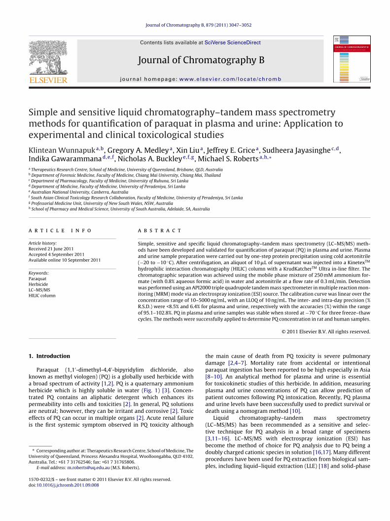
Journal of Chromatography B, 879 (2011) 3047–3052 Contents lists available at SciVerse ScienceDirect Journal of Chromatography B j ourna l ho me page: www.elsevier.com/locate/chromb Simple and sensitive liquid chromatography–tandem mass spectrometry methods for quantification of paraquat in plasma and urine: Application to experimental and clinical toxicological studies Klintean Wunnapuk a,b , Gregory A. Medley a , Xin Liu a , Jeffrey E. Grice a , Sudheera Jayasinghe c,d , Indika Gawarammana d,e,f , Nicholas A. Buckley e,f,g , Michael S. Roberts a,h,∗ a Therapeutics Research Centre, School of Medicine, University of Queensland, Brisbane, QLD, Australia b Department of Forensic Medicine, Faculty of Medicine, Chiang Mai University, Chiang Mai, Thailand c Department of Pharmacology, Faculty of Medicine, University of Ruhuna, Sri Lanka d Department of Medicine, Faculty of Medicine, University of Peradeniya, Sri Lanka e Australian National University, Canberra, Australia f South Asian Clinical Toxicology Research Collaboration, Faculty of Medicine, University of Peradeniya, Sri Lanka g Professorial Medicine Unit, University of New South Wales, NSW, Australia h School of Pharmacy and Medical Science, University of South Australia, Adelaide, SA, Australia a r t i c l e i n f o Article history: Received 21 June 2011 Accepted 4 September 2011 Available online 10 September 2011 Keywords: Paraquat Herbicide LC–MS/MS HILIC column a b s t r a c t Simple, sensitive and specific liquid chromatography–tandem mass spectrometry (LC–MS/MS) meth- ods have been developed and validated for quantification of paraquat (PQ) in plasma and urine. Plasma and urine sample preparation were carried out by one-step protein precipitation using cold acetonitrile (−20 to −10 ◦ C). After centrifugation, an aliquot of 10 L of supernatant was injected into a Kinetex TM hydrophilic interaction chromatography (HILIC) column with a KrudKatcher TM Ultra in-line filter. The chromatographic separation was achieved using the mobile phase mixture of 250 mM ammonium for- mate (with 0.8% aqueous formic acid) in water and acetonitrile at a flow rate of 0.3 mL/min. Detection was performed using an API2000 triple quadrupole tandem mass spectrometer in multiple reaction mon- itoring (MRM) mode via an electrospray ionization (ESI) source. The calibration curve was linear over the concentration range of 10–5000 ng/mL, with an LLOQ of 10 ng/mL. The inter- and intra-day precision (% R.S.D.) were <8.5% and 6.4% for plasma and urine, respectively with the accuracies (%) within the range of 95.1–102.8%. PQ in plasma and urine samples was stable when stored at −70 ◦ C for three freeze–thaw cycles. The methods were successfully applied to determine PQ concentration in rat and human samples. © 2011 Elsevier B.V. All rights reserved. 1. Introduction Paraquat (1,1 -dimethyl-4,4 -bipyridylim dichloride, also known as methyl viologen) (PQ) is a globally used herbicide with a broad spectrum of activity [1,2]. PQ is a quaternary ammonium herbicide which is highly soluble in water (Fig. 1) [3]. Concen- trated PQ contains an aliphatic detergent which enhances its permeability into cells and toxicities [2]. In general, PQ solutions are neutral; however, they can be irritant and corrosive [2]. Toxic effects of PQ can occur in multiple organs [2]. Acute renal failure is the first systemic symptom observed in PQ toxicity although ∗ Corresponding author at: Therapeutics Research Centre, School of Medicine, The University of Queensland, Princess Alexandra Hospital, Woolloongabba, QLD 4102, Australia. Tel.: +61 7 31762546; fax: +61 7 31765806. E-mail address: [email protected] (M.S. Roberts). the main cause of death from PQ toxicity is severe pulmonary damage [2,4–7]. Mortality rate from accidental or intentional paraquat ingestion has been reported to be high especially in Asia [8–10]. An analytical method for plasma and urine is essential for toxicokinetic studies of this herbicide. In addition, measuring plasma and urine concentrations of PQ can allow prediction of patient outcomes following PQ intoxication. Recently, PQ plasma and urine levels have been successfully used to predict survival or death using a nomogram method [10]. Liquid chromatography–tandem mass spectrometry (LC–MS/MS) has been recommended as a sensitive and selec- tive technique for PQ analysis in a broad range of specimens [3,11–16]. LC–MS/MS with electrospray ionization (ESI) has become the method of choice for PQ analysis due to PQ being a doubly charged cationic species in solution [16,17]. Many different procedures have been used for PQ extraction from biological sam- ples, including liquid–liquid extraction (LLE) [18] and solid-phase 1570-0232/$ – see front matter © 2011 Elsevier B.V. All rights reserved. doi:10.1016/j.jchromb.2011.09.008
-
Upload
independent -
Category
Documents
-
view
2 -
download
0
Transcript of Simple and sensitive liquid chromatography-tandem mass spectrometric (LCMS/MS) method for estimation...
Sme
KIa
b
c
d
e
f
g
h
a
ARAA
KPHLH
1
kahtpaei
UA
1d
Journal of Chromatography B, 879 (2011) 3047– 3052
Contents lists available at SciVerse ScienceDirect
Journal of Chromatography B
j ourna l ho me page: www.elsev ier .com/ locate /chromb
imple and sensitive liquid chromatography–tandem mass spectrometryethods for quantification of paraquat in plasma and urine: Application to
xperimental and clinical toxicological studies
lintean Wunnapuka,b, Gregory A. Medleya, Xin Liua, Jeffrey E. Gricea, Sudheera Jayasinghec,d,ndika Gawarammanad,e,f, Nicholas A. Buckleye,f,g, Michael S. Robertsa,h,∗
Therapeutics Research Centre, School of Medicine, University of Queensland, Brisbane, QLD, AustraliaDepartment of Forensic Medicine, Faculty of Medicine, Chiang Mai University, Chiang Mai, ThailandDepartment of Pharmacology, Faculty of Medicine, University of Ruhuna, Sri LankaDepartment of Medicine, Faculty of Medicine, University of Peradeniya, Sri LankaAustralian National University, Canberra, AustraliaSouth Asian Clinical Toxicology Research Collaboration, Faculty of Medicine, University of Peradeniya, Sri LankaProfessorial Medicine Unit, University of New South Wales, NSW, AustraliaSchool of Pharmacy and Medical Science, University of South Australia, Adelaide, SA, Australia
r t i c l e i n f o
rticle history:eceived 21 June 2011ccepted 4 September 2011vailable online 10 September 2011
eywords:araquaterbicideC–MS/MS
a b s t r a c t
Simple, sensitive and specific liquid chromatography–tandem mass spectrometry (LC–MS/MS) meth-ods have been developed and validated for quantification of paraquat (PQ) in plasma and urine. Plasmaand urine sample preparation were carried out by one-step protein precipitation using cold acetonitrile(−20 to −10 ◦C). After centrifugation, an aliquot of 10 �L of supernatant was injected into a KinetexTM
hydrophilic interaction chromatography (HILIC) column with a KrudKatcherTM Ultra in-line filter. Thechromatographic separation was achieved using the mobile phase mixture of 250 mM ammonium for-mate (with 0.8% aqueous formic acid) in water and acetonitrile at a flow rate of 0.3 mL/min. Detectionwas performed using an API2000 triple quadrupole tandem mass spectrometer in multiple reaction mon-
ILIC column itoring (MRM) mode via an electrospray ionization (ESI) source. The calibration curve was linear over theconcentration range of 10–5000 ng/mL, with an LLOQ of 10 ng/mL. The inter- and intra-day precision (%R.S.D.) were <8.5% and 6.4% for plasma and urine, respectively with the accuracies (%) within the rangeof 95.1–102.8%. PQ in plasma and urine samples was stable when stored at −70 ◦C for three freeze–thawcycles. The methods were successfully applied to determine PQ concentration in rat and human samples.
. Introduction
Paraquat (1,1′-dimethyl-4,4′-bipyridylim dichloride, alsonown as methyl viologen) (PQ) is a globally used herbicide with
broad spectrum of activity [1,2]. PQ is a quaternary ammoniumerbicide which is highly soluble in water (Fig. 1) [3]. Concen-rated PQ contains an aliphatic detergent which enhances itsermeability into cells and toxicities [2]. In general, PQ solutions
re neutral; however, they can be irritant and corrosive [2]. Toxicffects of PQ can occur in multiple organs [2]. Acute renal failures the first systemic symptom observed in PQ toxicity although∗ Corresponding author at: Therapeutics Research Centre, School of Medicine, Theniversity of Queensland, Princess Alexandra Hospital, Woolloongabba, QLD 4102,ustralia. Tel.: +61 7 31762546; fax: +61 7 31765806.
E-mail address: [email protected] (M.S. Roberts).
570-0232/$ – see front matter © 2011 Elsevier B.V. All rights reserved.oi:10.1016/j.jchromb.2011.09.008
© 2011 Elsevier B.V. All rights reserved.
the main cause of death from PQ toxicity is severe pulmonarydamage [2,4–7]. Mortality rate from accidental or intentionalparaquat ingestion has been reported to be high especially in Asia[8–10]. An analytical method for plasma and urine is essentialfor toxicokinetic studies of this herbicide. In addition, measuringplasma and urine concentrations of PQ can allow prediction ofpatient outcomes following PQ intoxication. Recently, PQ plasmaand urine levels have been successfully used to predict survival ordeath using a nomogram method [10].
Liquid chromatography–tandem mass spectrometry(LC–MS/MS) has been recommended as a sensitive and selec-tive technique for PQ analysis in a broad range of specimens[3,11–16]. LC–MS/MS with electrospray ionization (ESI) has
become the method of choice for PQ analysis due to PQ being adoubly charged cationic species in solution [16,17]. Many differentprocedures have been used for PQ extraction from biological sam-ples, including liquid–liquid extraction (LLE) [18] and solid-phase
3048 K. Wunnapuk et al. / J. Chromatog
eeasn
taoptsNo
oagmahcso[
dpew
2
2
mMgS
2
cmBw
2.6.1. Linearity and lower limit of quantification (LLOQ)
Fig. 1. Chemical structure of paraquat and ethyl viologen and their fragment ions.
xtraction (SPE) [3,14,19]. Disadvantages of LLE include low recov-ry and lengthy procedures [3]. While cation exchangers [3,11,16]nd silica C18 Sep-Paks [14,19] have been successfully used forolid phase extraction of PQ, they are impractical for high sampleumbers as they are time consuming and costly.
Recently, ion-pair reagents, such as trifluoroacetic acid (TFA),etrabutylammonium hydroxide (TBAOH) and heptafluorobutyriccid (HFBA), have been used with reverse-phase silica-based C8r C18 columns to separate the highly polar PQ in biological sam-les [3,11–14,20]. The addition of salts like ammonium acetate tohe mobile phase containing the ion-pair reagents has also beenhown to improve the separation and resolution of PQ [3,13,16].evertheless, in the presence of ion-pair reagents, the sensitivityf the methods decreases due to ion suppression [15,16].
The HILIC column is an alternative approach to the separationf highly polar compounds [21]. Typical HILIC stationary phasesre bare silica and silica embedded with different polar functionalroups including amine, cyano and diol [22]. An interaction of polaroieties such as hydroxyl groups in the analytes with the station-
ry phase, as well as with the organic and aqueous mobile phaseas been suggested as a retention mechanism [21,23]. The HILIColumn allows the separation of hydrophilic compounds on polartationary phases [24] and has been previously used for the analysisf PQ analysis in urine, following SPE under isocratic LC conditions16].
In this work, we developed an efficient and simple method foretermining PQ in rat and human samples by LC–MS/MS. The sam-le preparation was simple, employing only a one-step acetonitrilextraction. The analytical separation using the HILIC silica columnas sensitive and precise without the use of ion-pair reagents.
. Materials and methods
.1. Materials and reagents
Paraquat dichloride hydrate (PQ) and ethyl viologen dibro-ide (EV) were both purchased from Sigma–Aldrich (St. Louis,O, USA). Acetonitrile and formic acid were mass spectroscopy
rade and were obtained from Merck (Kilsyth, VIC, Australia) andigma–Aldrich (St. Louis, MO, USA), respectively.
.2. Equipment
The LC–MS/MS system consisted of an SLC-10AVP systemontroller, two LC-10AD pumps, an SIL-20AC-HT autosampler (Shi-
adzu, Kyoto, Japan) and an API2000 triple quadrupole (Appliediosystems Inc., Foster City, CA, USA) mass spectrometer coupledith an electrospray ionization (ESI) source and a divert valve.
r. B 879 (2011) 3047– 3052
2.3. Chromatographic and mass spectrometric conditions
2.3.1. Chromatographic conditionsThe chromatographic separation was modified from Whitehead
et al. [16]. Separation was achieved using a KinetexTM 2.6 �m HILICcolumn (50 mm × 2.10 mm, ID, Phenomenex, Torrance, USA) cou-pled with KrudKatcherTM Ultra in-line filter. The mobile phase was amixture of solvent A (250 mM ammonium formate containing 0.8%formic acid in water) and solvent B (acetonitrile) and was deliveredwith a gradient at a flow rate of 0.3 mL/min. The gradient pro-gramme commenced at 60 (%B), followed by direct switch to 20 (%B)at 0.1 min, maintained for 3.5 min for plasma and 5 min for urine,followed by a direct switch back to 60 (%B) at 3.6 min for plasmaand 5.1 min for urine and equilibrium for another 4 (for plasma)or 5 min (for urine) before stopping the run. The total chromato-graphic analysis time for plasma and urine were 7 and 10 min persample. The eluents for the first 2 min and the last 5.5 (for plasma)and 7 min (for urine) were diverted to waste via the valve.
2.3.2. Mass spectrometric conditionsDetection was performed with electrospray ionization oper-
ating in positive ion mode and the tandem spectrometer wasoperated in the multiple reactions monitoring (MRM) mode. Themass spectra for PQ and EV were first obtained in a full scan (Q1scan) mode by infusion of each compound (1 �g/mL) at 10 �L/min.From these spectra, the precursor ions were selected and subjectedto product ion scan. The dominant product ions were selected andthe most abundant precursor/product ion pair was chosen for MRM.The dwell times were set at 400 ms for PQ and 100 ms for EV. Condi-tions for mass spectrometric detection were as follows: nebulizergas, 70 psi; turbo gas, 70 psi; curtain gas, 20 psi; focusing poten-tial, 370 V; ion spray voltage, 5000 V; collision gas, 2; collision cellentrance potential, 19 V; collision energy, 20 V; declustering poten-tial, 70 V for PQ and 60 V for EV; entrance potential, 9.5 V for PQ and10 V for EV; collision cell exit potential, 32 V for PQ and 15 V for EV.
2.4. Preparation of standards and quality control (QC) samples
The stock solutions of PQ and EV were prepared by weighingout approximately 2.5 mg of each compound in 25 mL of deionisedwater. A 20 �g/mL PQ working solution was prepared by diluting5 mL of the stock solution with 20 mL of water. The internal stan-dard working solution (10 �g/mL of EV) was prepared by 10 timesdilution of the stock solution with water. The working standardsolutions were stored at 4 ◦C.
Nine PQ calibration standards were made by adding the workingsolutions to blank human plasma and urine covering a range from10 to 5000 ng/mL (10, 20, 50, 100, 200, 500, 1000, 2000 and 5000).QC samples at three concentration levels (30, 600 and 1500 ng/mL)were prepared in the same way.
2.5. Sample preparation
Two hundred microliters of cold acetonitrile (−20 to −10 ◦C)were added to 100 �L of plasma or urine and mixed with 10 �L ofinternal standard (EV, 10 �g/mL). The mixture was vortexed for 10 sand centrifuged at 14,500 rpm for 15 min. The supernatant was thentransferred to an HPLC vial and 10 �L of the sample was injectedinto the LC–MS/MS system.
2.6. Method validation
The linearity of the method was determined by analysing cali-bration standards ranging from 10 to 5000 ng/mL. The calibrationcurve was constructed using a weighted (1/�2) linear least-squares

K. Wunnapuk et al. / J. Chromatogr. B 879 (2011) 3047– 3052 3049
Table 1Mass spectra obtained by LC–MS/MS.
Compound Molecular weight Precursor ion m/z (relative intensity (%)) Assignment Product ion m/z (relative intensity (%))
PQ 186186 (22) [M]+ 186 (11), 171 (100)185 (22) [M−H]+ 185 (100), 170 (39), 169 (85), 158 (39), 144 (50)
93a (100) [M]2+ 171a (100), 169 (25), 142 (29), 93 (12)
EV 214
214 (9) [M]+ 185 (100), 213 (62)213 (23) [M−H]+ 185 (100), 213 (68)185 (45) [M−C H ]+ 185 (63), 157 (100)
rccac
2
sao(6cv
2
aladceTe
2
mtfaaiTti(d5
2
2
dsvpmso
each matrix were within 2.0%. The linear regression of the ratioof the areas of the analyte and internal standard peaks versus theconcentration was weighted by 1/x2. The LLOQs were established at10 ng/mL of PQ with the accuracy and precision of 109.2 and 5.9%
Table 2Linearity validation data.
107a (100)
a Ion selected for quantitation.
egression of the peak area ratios of PQ to EV obtained against theorresponding concentrations. The LLOQ was defined as the lowestoncentration in the calibration curve based on visual evaluationt which the analyte can be reliably detected with acceptable pre-ision and accuracy [25,26].
.6.2. Matrix effectSix batches of blank plasma and urine were screened for pos-
ible endogenous interferences. Matrix effects were evaluated bynalysing responses of PQ solution spiked into six different batchesf blank human plasma and urine at low (20 ng/mL) and high2000 ng/mL) concentrations. The PQ concentrations detected in
different batches were compared to those of the nominal con-entrations, and the matrix effect was reported as the percentagealues.
.6.3. Accuracy and precisionThe intra-day accuracy and precision were evaluated by
nalysing eight replicates of QC samples at three concentrationevels, in a single run. In order to determine inter-day accuracynd precision, five replicates of QC samples were analysed on fiveifferent validation days. Precision was represented by the per-entage of relative standard deviations (%R.S.D) while accuracy wasxpressed as (mean concentration)/(spiked concentration) × 100%.he acceptable tolerance for precision and accuracy was within 15%xcept at LLOQ, where it should not exceed 20% [25,26].
.6.4. Stability testThe stability of the analyte in the matrix was determined by
onitoring the degradation of samples when exposed to arbi-rary conditions of storage and handling. It was tested after threereeze/thaw cycles with eight replicates of QC samples at 30, 600nd 1500 ng/mL in both plasma and urine. The samples were storedt −70 ◦C between freeze/thaw cycles, and were thawed by allow-ng them to stand at room temperature for approximately 30 min.he QC samples were also analysed one month after preparationo determine the long-term storage stability. Short-term stabil-ty in the matrix during 3 h at ambient laboratory temperature24 ± 3 ◦C) was also determined. The post-preparative stability wasetermined by injection of prepared samples (kept at 4 ◦C) for 4 or
days after the initial injection.
.7. Application to animal and human toxicological studies
.7.1. Animal studyMale Wistar rats (200–250 g) received PQ orally at 3 different
oses (15, 30 and 60 mg/kg), with one rat per dose group. Bloodamples were collected at 24 h and the urine was collected at inter-als 0–4, 4–8, 8–12 and 12–24 h. The concentrations of PQ in rat
lasma and urine samples were determined using the validatedethods. The animal experiments were approved by the Univer-ity Animal Ethics Committee (Health Sciences) of the Universityf Queensland (AEC approval number: PAH/370/10/NHMRC).
2 5
[M]2+ 184 (17), 157a (100), 130 (17)
2.7.2. Human studyThese established methods were applied to analyse 150 plasma
samples from patients admitted to hospitals in Sri Lanka with acutePQ toxicity. This study was coordinated by the South Asian ClinicalToxicology Research Collaboration (SACTRC; www.sactrc.org). Thehuman study was approved by the Human Research Ethics Com-mittees of the Australian National University, the University of NewSouth Wales and the Committee on Research and Ethical Review ofthe Faculty of Medicine, University of Peradeniya.
3. Results and discussion
3.1. Chromatographic and mass spectrometric conditions
The precursor and product quantification ion pairs obtained byLC–MS/MS are summarized in Table 1. The most abundant ions ofPQ and EV in our mobile phase containing ammonium formate withformic acid are the doubly charged molecular ions [M]2+, at m/z93 and 107, respectively. A multiple reaction monitoring (MRM)mode was used for quantification with the mass transitions from93 [M]2+ to 171 [M−CH3]+ and 107 [M]2+ to 157 [MH−(C2H5)2]+ forPQ and EV, respectively. MS/MS product ion scan and the proposedfragmentation pathways of PQ are illustrated in Fig. 1.
The liquid chromatography was optimized on a HILIC columnunder gradient conditions. Good separation and retention of theanalyte and the internal standard were achieved in a total run timeof 7 and 10 min in plasma and urine, respectively. For highly polarcompounds like PQ, separation on the HILIC column is by partition-ing and weak cation exchange [16]. Representative chromatogramsof blank plasma and urine samples and spiked samples at LLOQ and1 �g/mL of PQ are shown in Fig. 2.
Diversion of the early and late eluent to waste reduced thebackground noise and kept the ion source clean. One step pro-tein precipitation with acetonitrile was suitable for both plasmaand urine sample preparation without any effect on separation andionization.
3.2. Method validation
3.2.1. Linearity of calibration and LLOQThe standard curve was linear over a concentration range of
10–5000 ng/mL in both plasma and urine (Table 2). The relativestandard deviation (R.S.D.) values of the retention times for PQ in
Sample tR (min)Mean ± SD
Linearregression (R2)
R.S.D. (%) Accuracy (%)
Plasma 3.58 ± 2.0 0.9947–0.9991 1.9–9.2 92.8–105.6Urine 3.85 ± 1.4 0.9910–0.9974 2.7–10.1 97.8–106.9

3050 K. Wunnapuk et al. / J. Chromatogr. B 879 (2011) 3047– 3052
F , (C) anp
av
3
al
ig. 2. MRM of the ion chromatography of PQ: (A) and (B) blank plasma and urinelasma and urine.
nd 102.9 and 7.3% in plasma and urine, respectively. The LLOQalues were comparable to those in previous reports [11,14].
.2.2. Matrix effectSix batches of urine and plasma samples were spiked with PQ
nd analysed and the variations due to matrix effects were calcu-ated. As shown in Table 3, there were no significant matrix effects
d (D) 10 ng/mL (LLOQ) of PQ in plasma and urine, (E) and (F) 1000 ng/mL of PQ in
for PQ at low (20 ng/mL) and high (2000 ng/mL) concentrations.The variations (% R.S.D.) among different matrices were between4.4 and 9.7%.
3.2.3. Accuracy and precisionThe intra- and inter-day precision in plasma and urine were
within 10% range and the accuracy of PQ determination in plasma

K. Wunnapuk et al. / J. Chromatogr. B 879 (2011) 3047– 3052 3051
Table 3Matrix effect.
Sample Nominal concentration(ng/mL)
Matrix effect (%) Mean ± SD (%)
Plasma 20 107.50 103.71± 5.402000 99.92
Urine 20 105.75 105.04± 1.002000 104.33
Fcq
ad
3
ttaatfsc
3
3
ssT
Fig. 4. Cumulative urine excretion of PQ in rats after oral administration of 3 differ-ent doses.
TI
ig. 3. The stability of PQ in matrices, U: urine, P: plasma, LQC: low level qualityontrol (30 ng/mL), MQC: medium level quality control (600 ng/mL), HQC: high leveluality control (1500 ng/mL), F/T: freeze/thaw, ACN: acetonitrile.
nd urine were between 95.1 and 103.2%. A summary of the intra-ay and inter-day precision and accuracy is presented in Table 4.
.2.4. Stability testThe short-term stability of PQ in plasma and urine at ambient
emperatures was assessed over at least 3 h by comparison withhe initial measured concentrations, as shown in Fig. 3. The plasmand urine standard samples were stable after storage at −70 ◦C fort least one month. Freeze/thaw stability experiments indicatedhat PQ was stable in human plasma and urine for at least threereeze/thaw cycles. After preparation, the samples were found to betable for at least 5 days at 4 ◦C. The relative deviations of detectedoncentrations were within 15% for all levels of QC samples.
.3. Application to animal and human specimens
.3.1. Animal samples
These methods were successfully applied in toxicokinetictudies. PQ was detected and quantified in plasma and urineamples taken at 3 different time points after oral administration.he identity of PQ was confirmed by the retention time and the
able 4ntra- and inter-day precision and accuracy for PQ in human plasma and urine.
Sample Nominal concentration(ng/mL)
Intra-day
Mean detected(ng/mL)
R.S.D. (%)
Plasma 30 29.6 8.5
600 579 3.7
1500 1509 5.2
Urine 30 40.0 5.7
600 598 1.9
1500 1,504 2.6
Fig. 5. Plasma concentration–time profile of a representative PQ intoxicationpatient.
identity of the parent ion ([M]2+) at m/z = 93 and the major ionicfragment ([M−CH3]+) at m/z = 171, as shown in the spectrum ofstandard PQ. The cumulative excretion curve of PQ in urine isshown in Fig. 4. The concentrations of PQ in plasma and urineafter oral administration ranged from less than 0.01–5135 �g/mL(Table 5).
3.3.2. Human samplesThe developed method was also used to analyse 150 clinical
human plasma samples. The retention time and precursor andproduct ion spectra of PQ in the sample matched well with thoseof the authentic standard. Those clinical samples with PQ concen-tration above the upper limit of quantification (5000 ng/ml) werereanalysed with a dilution of 10 or 100 times as appropriate withwater before sample preparation. Plasma concentrations of PQ var-
ied from less than 0.01 to 461.5 �g/mL. A representative plasmaconcentration–time profile of a PQ intoxication patient is shown inFig. 5.Inter-day
Accuracy (%) Mean detected(ng/mL)
R.S.D. (%) Accuracy (%)
98.7 29.8 3.6 99.396.5 571 2.7 95.1
101 1472 4.4 98.1103 29.8 3.1 99.5
99.6 585 3.8 97.6100 1,542 6.4 103

3052 K. Wunnapuk et al. / J. Chromatog
Table 5The concentrations of PQ in plasma and urine after oral administration (rat study).
Dose (mg/kg) Plasma (�g/mL) Urine (�g/mL)
24 h 4 h 8 h 12 h 24 h
4
taiputapt
A
igRU
R
[
[
[[
[
[
[
[[
[
[[[[
[[
toryInformation/Guidances/ucm070107.pdf, U.S. Department of Health andHuman Services, Food and Drug Administration, 2001.
[26] FDA, in: http://www.fda.gov/downloads/Regulator%20yInformation/ Guid-ances/UCM128049.pdf, U.S. Department of Health and Human Services, Food
15 0.01 10.03 11.75 8.10 5.2530 0.01 287.00 52.15 30.00 3.0360 0.05 108.50 49.30 59.30 10.11
. Conclusions
The proposed methods have been successfully applied to quan-ify PQ in plasma and urine samples. These methods provideccuracy and precision for determination of PQ in animal exper-ments and acute PQ poisoning cases, are simple to perform androvide sensitivity comparable to previous literature reports. These of the HILIC column for LC separation of PQ, followed by elec-rospray ionization–tandem mass spectrometry (ESI–MS/MS) with
single step extraction enabled us to accurately quantify PQ inlasma and urine without a solid phase extraction procedure orhe use of ion-pair agents.
cknowledgements
The authors are grateful to Mrs. Camilla Thomson for her helpn animal experimentation. This study was supported by projectrants (268053 and 1011772) from the National Health and Medicalesearch Committee of Australia. KW was supported by Chiang Mainiversity Scholarship.
eferences
[1] E. Bairaktari, K. Katopodis, K.C. Siamopoulos, O. Tsolas, Clin. Chem. 44 (1998)1256.
[2] R.J. Dinis-Oliveira, J.A. Duarte, A. Sanchez-Navarro, F. Remiao, M.L. Bastos, F.Carvalho, Crit. Rev. Toxicol. 38 (2008) 13.
r. B 879 (2011) 3047– 3052
[3] K.C. Wang, S.M. Chen, J.F. Hsu, S.G. Cheng, C.K. Lee, J. Chromatogr. B Analyt.Technol. Biomed. Life Sci. 876 (2008) 211.
[4] D.A. Purser, M.S. Rose, Toxicology 15 (1979) 31.[5] G.M. Hawksworth, P.N. Bennett, D.S. Davies, Toxicol. Appl. Pharmacol. 57 (1981)
139.[6] E.A. Lock, J. Ishmael, Toxicol. Appl. Pharmacol. 50 (1979) 67.[7] E.A. Lock, Toxicol. Appl. Pharmacol. 48 (1979) 327.[8] D.M. Roberts, M.F. Wilks, M.S. Roberts, R. Swaminathan, F. Mohamed, A.H.
Dawson, N.A. Buckley, Toxicol. Lett. 202 (2011) 69.[9] I.B. Gawarammana, N.A. Buckley, Br. J. Clin. Pharmacol. (2011).10] L. Senarathna, M. Eddleston, M.F. Wilks, B.H. Woollen, J.A. Tomenson, D.M.
Roberts, N.A. Buckley, QJM 102 (2009) 251.11] M.M. Ariffin, R.A. Anderson, J. Chromatogr. B Analyt. Technol. Biomed. Life Sci.
842 (2006) 91.12] R. Castro, E. Moyano, M.T. Galceran, J. Chromatogr. A 830 (1999) 145.13] M. Ito, Y. Hori, M. Fujisawa, A. Oda, S. Katsuyama, Y. Hirose, T. Yoshioka, Biol.
Pharm. Bull. 28 (2005) 725.14] X.P. Lee, T. Kumazawa, M. Fujishiro, C. Hasegawa, T. Arinobu, H. Seno, A. Ishii,
K. Sato, J. Mass Spectrom. 39 (2004) 1147.15] C.R. Mallet, Z. Lu, J.R. Mazzeo, Rapid Commun. Mass Spectrom. 18 (2004)
49.16] R.D. Whitehead Jr., M.A. Montesano, N.K. Jayatilaka, B. Buckley, B. Winnik, L.L.
Needham, D.B. Barr, J. Chromatogr. B Analyt. Technol. Biomed. Life Sci. 878(2010) 2548.
17] R. Castro, E. Moyano, M.T. Galceran, J. Chromatogr. A 914 (2001) 111.18] S.K. Baeck, Y.S. Shin, H.S. Chung, M.Y. Pyo, Arch. Pharm. Res. 30 (2007)
235.19] C. Fuke, T. Arao, Y. Morinaga, H. Takaesu, K. Ameno, T. Miyazaki, Leg. Med.
(Tokyo) 4 (2002) 156.20] B.B. Sadi, A.P. Vonderheide, J.A. Caruso, J. Chromatogr. A 1050 (2004) 95.21] V.V. Tolstikov, O. Fiehn, Anal. Biochem. 301 (2002) 298.22] Z. Hao, B. Xiao, N. Weng, J. Sep. Sci. 31 (2008) 1449.23] A.J. Alpert, M. Shukla, A.K. Shukla, L.R. Zieske, S.W. Yuen, M.A. Ferguson, A.
Mehlert, M. Pauly, R. Orlando, J. Chromatogr. A 676 (1994) 122.24] Y. Guo, S. Gaiki, J. Chromatogr. A 1074 (2005) 71.25] FDA, in: http://www.fda.gov/downloads/Drugs/GuidanceCompliance Regula-
and Drug Administration, 1996.