TSGA10 prevents nuclear localization of the hypoxia-inducible factor (HIF)-1α

Signaling pathway involved in hypoxia-inducible factor-1αregulation in hypoxic-ischemic cortical neurons in vitro
Li Zhang1, Yi Qu1, Chunlei Yang2, Jun Tang1, Xiaolan Zhang1, Meng Mao1,*, DezhiMu1,3,*, and Donna Ferriero3,41Department of Pediatrics, West China Second University Hospital, Sichuan University, Chengdu610041, China2Department of Cell Biology, Sichuan University, Chengdu 610041, China3Department of Neurology, University of California, San Francisco, California, USA4Department of Pediatrics, University of California San Francisco California, USA
AbstractHypoxia-inducible factor-1α (HIF-1α) is a key transciptional regulator of cellular and systemicoxygen homeostasis. Previous studies have shown that the regulation of HIF-1α is involved in theactivation of PI3K/Akt pathway in some cells. However, whether this pathway plays a role inmodulating HIF-1α in cultured cortical neurons during hypoxia-ischemia (HI) remains unclear. Wetherefore investigated the relationship between phosphoinositid 3-kinase/Akt (PI3K/Akt) pathwayand HIF-1α expression in cultured neurons using an oxygen and glucose deprivation (OGD) model.In this study, cortical neurons cultured in vitro were subjected to OGD for 3 h followed by reperfusion.The expressions of HIF-1α, VEGF, total Akt and phosphorelated-Akt (p-Akt) were detected by RT-PCR, Western blot and immunocytochemistry. We found that HIF-1α and VEGF were increased at4 h and peaked at 8 h after OGD. Meanwhile, p-Akt increased and peaked at 4h after reperfusion,preceding HIF-1α expression. Pre-treatment with wortmannin, a PI3K/Akt pathway inhibitor,significantly inhibited p-Akt expression and further attenuated both transcription and translation ofHIF-1α and VEGF. Collectively, our findings suggested that PI3K/Akt signaling pathway might beinvolved in HIF-1α regulation after OGD in cultured cortical neurons.
KeywordsHIF-1α; neurons; hypoxia-ischemia; signaling pathway
HIF-1, a key regulator of oxygen homeostasis, is a heterodimeric protein composed of the twosubunits, HIF-1α and HIF-1β. These two subunits belong to the basic helix-loop-helix (bHLH)-PAS protein family. HIF-1α is an inducible subunit regulated by hypoxia, HIF-1β, aconstitutive subunit which is not regulated by oxygen concentration [7]. In addition to hypoxia,
© 2009 Published by Elsevier Ireland Ltd.*Corresponding authors Meng Mao MD Department of Pediatrics, West China Second University Hospital, Sichuan University, Chengdu610041, China [email protected]. Dezhi Mu MD, PhD Department of Pediatrics, West China Second University Hospital, SichuanUniversity, Chengdu 610041, China [email protected] Fax: 86-28-85559065 Tel: 86-28-85503185.Publisher's Disclaimer: This is a PDF file of an unedited manuscript that has been accepted for publication. As a service to our customerswe are providing this early version of the manuscript. The manuscript will undergo copyediting, typesetting, and review of the resultingproof before it is published in its final citable form. Please note that during the production process errors may be discovered which couldaffect the content, and all legal disclaimers that apply to the journal pertain.
NIH Public AccessAuthor ManuscriptNeurosci Lett. Author manuscript; available in PMC 2010 September 11.
Published in final edited form as:Neurosci Lett. 2009 September 11; 461(1): 1–6. doi:10.1016/j.neulet.2009.03.091.
NIH
-PA Author Manuscript
NIH
-PA Author Manuscript
NIH
-PA Author Manuscript
HIF-1α has been shown to be induced by other stimuli, such as deferoxamine (DFO), insulin,and epidermal growth factor [4,9]. Under normoxia, HIF-1α is rapidly degraded by ubiquitin-proteasome pathway which is mediated by hydroxylation of the prolines 564 and 462 residues.This process is regulated by an O2-sensitive enzyme called HIF-1α prolyl-hydroxylase [5].Under hypoxia, the degradation of HIF-1α by hydroxylation and ubiquitination is inhibited andthus stabilized. Stabilized HIF-1α, a cytosolic protein then translocates into the nucleus, whereHIF-1α subunit is phosphorylated and dimerizes with HIF-1β subunit, a nuclear protein, toform functional HIF-1 [18]. Activated HIF-1 binds to hypoxia response elements (HRE) of itstarget genes and then triggers the transcription of the target genes such as vascular endothelialgrowth factor (VEGF) and erythropoietin (EPO) [12,14]. Protective effects of VEGF includepromoting angiogenesis, neurogenesis, and neurite outgrowth in brain after HI [19].
Although the factors modulating HIF-1α expression have been studied extensively, thesignaling pathways for HIF-1α regulation remain controversial. In the central nervous system,Akt is suggested to be necessary for neuronal survival, neurogenesis, angiogenesis, axon ordendrite formation, synaptogenesis, and synaptic transmission [1,11]. Recent studies in HepG2cells show that HIF-1α is regulated by various signaling pathways including PI3K/Akt [13].However, this regulation seems to be cell type specific. In HEK293T cells, the activation ofPI3K/Akt pathway does not affect the transcriptional activity of HIF-1α under either normoxicor hypoxic condition [2]. Whether PI3K/Akt pathway is involved in HIF-1α regulation incultured cortical neurons exposed to HI remains unclear. Therefore, we aimed to elucidate therelationship between PI3K/Akt and HIF-1α to address this question.
All animal research was approved by Sichuan University Committee on Animal Research.Pregnant Sprague-Dawley rats were sacrificed and embryos at 16-18 days of gestation wereremoved and kept in 4°C phosphate buffered saline (PBS, pH 7.4). The whole cerebral cortexwas isolated from the fetuses and cells were dissociated in a trypsin solution (1.25mg/mL inHank's Buffer salt solution) for 10min at 37°C. The cortex cell suspension was centrifuged andresuspended, then seeded in 6-well plates precoated with poly-D-lysine (Sigma), and grownin Neurobasal medium (NB) (Gibco) with 2% B27 supplement (Gibco) and 500 μM glutamine(Gibco) in a humidified incubator with 5% CO2 at 37°C. The following experiments wereperformed at day in vitro 5.
Cultures were exposed to HI for 3h followed by reoxygenation. To induce OGD, cultured cellswere gently washed twice with PBS, and then placed in DMEM without glucose. Cells wereexposed to hypoxia (95% N2/5% CO2) at 37°C in the airtight chamber for 3h. After 3h of OGDtreatment, the cell culture medium was changed back to normal NB medium and then returnedto 5% CO2, 37°C incubator. To elucidate whether PI3K is involved in HIF-1α regulation underOGD, cells were pretreated with wortmannin (an inhibitor of PI3K) at the concentration of 100nM for 30 min before OGD treatment.
Total RNA of cortical neurons was extracted by using Tissue/Cell RNA Mini Kit (WatsonBiotechnologies, INC). Complementary DNA (cDNA) was synthesized from isolated RNAusing reverse transcription reagents (RevertAid First Strand cDNA Synthesis Kit, Fermentas).Reverse transcription was performed using random hexamer primer at 25°C for 10 min, 42°Cfor 60 min, and 70°C for 10 min. cDNA was stored at −20°C until use. PCR was conductedusing PCR Master Mix (2×) (Fermentas) and the following primers: (a) HIF-1α: sense primer5′-AAGTCTAGGGATGCAGCAC-3′, antisence primer 5′-CAAGATCACCAGCATCTAG-3′; (b) VEGF: sense primer 5′-GCTCTCTTGGGTGCACTGGA-3′, antisense primer 5′-CACCGCCTTGGCTTGTCACA-3′; (c) β-actin: sense primer 5′-ACACTGTGCCCATCTAGGAGG-3′, antisense primer 5′-AGGGGCCGGACTCGTCATACT-3′.
Zhang et al. Page 2
Neurosci Lett. Author manuscript; available in PMC 2010 September 11.
NIH
-PA Author Manuscript
NIH
-PA Author Manuscript
NIH
-PA Author Manuscript

The PCR for HIF-1α was performed in 37 cycles with following cycle profile: 94°C for 30 sec,55°C for 30 sec and 72°C for 1.5 min. The PCR for VEGF was performed in 33 cycles withfollowing cycle profile: 94°C for 1 min, 55°C for 1 min and 72°C for 1.5 min. The PCR productswere separated on a 2% agarose gel and stained with ethidium bromide using 100bp ladderDNA maker as a size reference (BioTeke Corporation) and photographed under ultraviolettransillumination.
Western blot was performed as previously described [10]. Cells were lysed in CHAP lysisbuffer. Equal amounts of protein samples (100 μg) were loaded per lane and electrophoresedon 8% SDS/PAGE gels. After blocked in nonfat milk buffer, the membranes were separatelyincubated with mouse HIF-1α monoclonal antibody (Santa Cruz Biotechnology, diluted1:200), rabbit VEGF polyclonal antibody (Santa Cruz Biotechnology, diluted 1:300), rabbitAkt polyclonal antibody or mouse phospho-Akt (Ser473) monoclonal antibody (Cell Signaling,diluted 1:500) overnight at 4°C. Mouse β-actin monoclonal antibody (Santa CruzBiotechnology, diluted 1:1000) was detected as a loading control. Following washes, themembranes were incubated with horseradish peroxidase conjugated secondary antibody, goatanti-mouse or goat anti-rabbit IgG (Santa Cruz Biotechnology, 1:3000) in blocking solutionat room temperature (RT) for 1 h. The bands were developed using enhancedchemiluminescence (Millipore Corporation). NIH image was used to measure the densities ofthe protein signals.
For immunocytochemistry analysis, cultured cells were fixed with 4% paraformaldehyde andtreated with 0.3% hydrogen peroxide for 10 min, then blocked with 10% serum at 37°C for 30min. Cells were incubated with mouse HIF-1α monoclonal antibody (1:100 dilution) in PBS,or rabbit VEGF (1:200), rabbit Akt (1:400) or mouse p-Akt (1:400) at 37°C for 1 h andovernight at 4°C. After washes in PBS, cells were then incubated with biotin-conjugated goatanti-mouse or goat anti-rabbit IgG (Santa Cruz Biotechnology, 1:200) at 37 °C for 1 h, andavidin-biotin complex for 1 h. DAB was used to visualize the positive signals, and then cellswere counterstained with hematoxylin. After washing, cultures were mounted on gelatin-coated slides, dried, and coverslipped. As negative controls, alternate cultures were incubatedwithout primary antibody. Immages were observed using microscope (Leica, CM 2000).
Data were represented as mean ± standard deviation (SD). One-way ANOVA with Bonferroni/Dunnett post-hoc tests was performed for multiple comparisons. p<0.05 was consideredstatistically significant.
To study whether HIF-1α and VEGF expression were induced by OGD in cultured corticalneurons, an OGD model of exposure of cultured neurons without glucose in the media tohypoxia for 3h was used. HIF-1α and VEGF mRNA levels were measured in both normoxicneurons and OGD treated neurons at 0, 2, 4, 8, 12 and 24 h after reperfusion using RT-PCR.HIF-1α mRNA was induced at 2 h, peaked at 4 h, and gradually declined at 12 h. Two bandscorresponding to VEGF isoforms, VEGF164 (563 bp) and VEGF120 (431 bp) were maximallyincreased at 2 h, maintained at 4 h and decreased at 8 h. VEGF164 was the most dominanttranscript (Fig.1A).
To test HIF-1α and VEGF protein expression, Western blot analysis was performed in thisstudy. HIF-1α expression (110 kDa) was very weak in normoxic cultured neurons, but was up-regulated in response to OGD. HIF-1α and VEGF (42 kDa) expression were elevated at 4 h,peaked at 8 h and decreased at 12 h after reperfusion (Fig.1B). After quantification with β-actin, HIF-1α and VEGF protein levels were respectively increased approximately 4-fold and3-fold at 8 h compared to that in normoxia (p<0.001) (Fig.1C, Table 1).
The expression of Akt and p-Akt protein at the same time points as indicated above was alsodetected using Western blot. There was no significant difference in Akt (60 kDa) expression
Zhang et al. Page 3
Neurosci Lett. Author manuscript; available in PMC 2010 September 11.
NIH
-PA Author Manuscript
NIH
-PA Author Manuscript
NIH
-PA Author Manuscript

between normoxic and OGD treated neurons. However, OGD induced a rapid increase in p-Akt (60 kDa). The maximal induction of p-Akt appeared at 4 h, started to decrease at 8 h, andreturned to baseline at 24h (Fig.2A), which was earlier than that in HIF-1α and VEGF. Afternormalization with β-actin, an approximate 7-fold, 3.5-fold, and 3-fold increase of p-Aktinduction was respectively observed at 4 h, 8 h, and 12 h after reperfusion (p<0.001) (Fig.2B,Table 1). Since the maximum induction of p-Akt expression (at 4 h ) preceded HIF-1α and itstarget gene VEGF (at 8 h) in cortical neurons with OGD, we hypothesized that PI3K/Aktpathway might be involved in the regulation of HIF-1α and VEGF. To address this question,a PI3K inhibitor, wortmannin (100 nM), was used to pretreat cortical neurons for 30 min,followed by OGD for 3 h. P-Akt but not total Akt was significantly inhibited by wortmannincompared to DMSO treated control neurons at 4 h and 8 h after reperfusion (p<0.001) (Fig.3A, B, Table 2). Immunocytochemistry showed the similar findings as found by Western blots(Fig. 3C) and verified that the positive immunoreactions were localized in the cytoplasm ofthe neurons (Fig. 3C).
HIF-1α and VEGF expression after inhibition of p-Akt were measured. Pretreatment withwortmannin attenuated OGD induced HIF-1α and VEGF mRNA levels (Fig.4A). Similarfindings were observed with protein levels by Western blot detection (Fig.4B). Afterquantification, there was 60% and 70% of HIF-1α protein, 35% and 40% of VEGF proteindecrease respectively at 4 h and 8 h after reperfusion compared to control groups (p<0.001)(Fig.4C, Table 2). These findings suggest that Akt phosphorylation is required for OGDinduced HIF-1α and VEGF expression. Furthermore, immunocytochemistry also showed thatthe expression of HIF-1α and VEGF is attenuated by wortmannin treatment, which is consistentwith the findings of Western blot (Fig.4D).
In this study, HI significantly increased the expression of HIF-1α and VEGF both attranscriptional and translational levels. HIF-1α mRNA was increased at 2 h, peaked at 4 h, andgradually declined at 12 h. HIF-1α protein were elevated at 4 h, peaked at 8 h and decreasedat 12 h after reperfusion which agreed with previous reports that HIF-1α protein peaked at 8 hand declined at 24 h in injured cortex in a rat neonatal stroke model [15]. Our findings thatboth HIF-1α mRNA and protein are upregulated in cultured neurons treated with OGD,suggests that HI regulates HIF-1α on both the transcriptional and translational levels.
Previous reports suggested that hypoxic stimulation could induce HIF-1α expression via PI3K/Akt signaling pathway [17,8]. In this study, p-Akt peaked at 4 h after reperfusion from OGD,earlier than HIF-1α following OGD, suggesting that HIF-1α may be one of the downstreameffectors of PI3K/Akt signaling pathway. Studies on astrocytes and rat brains show that PI3K/Akt pathway is a positive regulator of HIF-1α activation [17,6,8]. However, these findings arenot consistent with other reports in vitro and in vivo [2,3]. These opposite findings suggest thatthe effects of PI3K/Akt on HIF-1α regulation are cell and tissue type specific. Wortmanninstrongly inhibited p-Akt as well as HIF-1α and VEGF. These findings further support thehypothesis that PI3K/Akt pathway plays an important role in regulating HIF-1α and its targetgenes in cultured neurons following OGD.
Interestingly, we found that wortmannin treatment inhibited HIF-1α transcription sinceHIF-1α mRNA was also decreased in this study, which is not in agreement with our previousstudy in an HI in vivo model. We previously found that wortmannin treatment down-regulatedHIF-1α protein but not the mRNA transcript [8]. These different findings may be due to thedifference between cultured neurons in vitro and animal models in vivo since there might bemore complicated mechanisms for HIF-1α regulation in vivo compared to in vitro, such asregulation by other cell types like astrocytes [16,20].
Zhang et al. Page 4
Neurosci Lett. Author manuscript; available in PMC 2010 September 11.
NIH
-PA Author Manuscript
NIH
-PA Author Manuscript
NIH
-PA Author Manuscript

Taken together, these findings suggest that PI3K/Akt pathway might play an important role inthe regulation of HIF-1α on both transcriptional and translational levels in cultured neuronsfollowing HI. Elucidating HIF-1α regulatory mechanisms may lead to new therapy strategiesbased on ischemic tolerance.
AcknowledgmentsThis work was supported by grants from National Natural Science Foundation of China (No.30825039, No.30770748),China Medical Board of New York (00-722), Ministry of Education of China (2006331-11-7 and 20070610092), andScience and Technology Bureau of Sichuan province (JY029-067).
References1. Akama KT, McEwen BS. Estrogen stimulates postsynaptic density-95 rapid protein synthesis via the
Akt/protein kinase B pathway. J Neurosci 2003;23:2333–2339. [PubMed: 12657692]2. Alvarez-Tejado M, Alfranca A, Aragonés J, Vara A, Landázuri MO, del Peso L. Lack of evidence for
the involvement of the phosphoinositide 3-kinase/Akt pathway in the activation of hypoxia-induciblefactors by low oxygen tension. J Biol Chem 2002;277:13508–13517. [PubMed: 11815624]
3. Arsham AM, Plas DR, Thompson CB, Simon MC. Phosphatidylinositol 3-kinase/Akt signaling isneither required for hypoxic stabilization of HIF-1α nor sufficient for HIF-1-dependent target genetranscription. J Biol Chem 2002;277:15162–15170. [PubMed: 11859074]
4. Hamrick SE, McQuillen PS, Jiang X, Mu D, Madan A, Ferriero DM. A role for hypoxia-induciblefactor-1alpha in desferoxamine neuroprotection. Neurosci Lett 2005;379:96–100. [PubMed:15823423]
5. Ivan M, Kondo K, Yang H, Kim W, Valiando J, Ohh M, Salic A, Asara JM, Lane WS, Kaelin WG Jr.HIFalpha targeted for VHL-mediated destruction by proline hydroxylation: implications for O2sensing. Science 2001;292(5516):464–468. [PubMed: 11292862]
6. Jiang BH, Jiang G, Zheng JZ, Lu Z, Hunter T, Vogt PK. Phosphatidylinositol 3-kinase signalingcontrols levels of hypoxia-inducible factor 1. Cell Growth Differ 2001;12:363–369. [PubMed:11457733]
7. Lee JW, Bae SH, Jeong JW, Kim SH, Kim KW. Hypoxia-inducible factor (HIF-1) alpha: its proteinstability and biological functions. Exp Mol Med 2004;36:1–12. [PubMed: 15031665]
8. Li L, Qu Y, Mao M, Mu D. The involvement of phosphoinositid 3-kinase/Akt pathway in the activationof hypoxia-inducible factor-1alpha in the developing rat brain after hypoxia-ischemia. Brain Res2008;1197:152–158. [PubMed: 18241842]
9. Li Y, Bi Z, Yan B, Wan Y. UVB radiation induces expression of HIF-1alpha and VEGF through theEGFR/PI3K/DEC1 pathway. Int J Mol Med 2006;18:713–719. [PubMed: 16964427]
10. Mao M, Hua Y, Jiang X, Li L, Zhang L, D Mu. Expression of tumor necrosis factor alpha and neuronalapoptosis in the developing rat brain after neonatal stroke. Neurosci Lett 2006;403:227–232.[PubMed: 16797840]
11. Markus A, Zhong J, Snider WD. Raf and akt mediate distinct aspects of sensory axon growth. Neuron2002;35:65–76. [PubMed: 12123609]
12. Milosevic J, Maisel M, Wegner F, Leuchtenberger J, Wenger RH, Gerlach M, Storch A, Schwarz J.Lack of hypoxia-inducible factor-1 alpha impairs midbrain neural precursor cells involving vascularendothelial growth factor signaling. J Neurosci 2007;27:412–421. [PubMed: 17215402]
13. Mottet D, Dumont V, Deccache Y, Demazy C, Ninane N, Raes M, Michiels C. Regulation of hypoxia-inducible factor-1alpha protein level during hypoxic conditions by the phosphatidylinositol 3-kinase/Akt/glycogen synthase kinase 3beta pathway in HepG2 cells. J Biol Chem 2003;278:31277–31285.[PubMed: 12764143]
14. Mu D, Chang YS, Vexler ZS, Ferriero DM. Hypoxia-inducible factor 1alpha and erythropoietinupregulation with deferoxamine salvage after neonatal stroke. Exp Neurol 2005;195:407–415.[PubMed: 16023639]
Zhang et al. Page 5
Neurosci Lett. Author manuscript; available in PMC 2010 September 11.
NIH
-PA Author Manuscript
NIH
-PA Author Manuscript
NIH
-PA Author Manuscript

15. Mu D, Jiang X, Sheldon RA, Fox CK, Hamrick SE, Vexler ZS, Ferriero DM. Regulation of hypoxia-inducible factor 1 alpha and induction of vascular endothelial growth factor in a rat neonatal strokemodel. Neurobiol Dis 2003;14:524–534. [PubMed: 14678768]
16. Oh YT, Lee JY, Yoon H, Lee EH, Baik HH, Kim SS, Ha J, Yoon KS, Choe W, Kang I.Lipopolysaccharide induces hypoxia-inducible factor-1 alpha mRNA expression and activation viaNADPH oxidase and Sp1-dependent pathway in BV2 murine microglial cells. Neurosci Lett2008;431:155–160. [PubMed: 18164813]
17. Pore N, Jiang Z, Shu HK, Bernhard E, Kao GD, Maity A. Akt1 activation can augment hypoxia-inducible factor-1alpha expression by increasing protein translation through a mammalian target ofrapamycin-independent pathway. Mol Cancer Res 2006;4:471–479. [PubMed: 16849522]
18. Salceda S, Caro J. Hypoxia-inducible factor 1alpha (HIF-1alpha) protein is rapidly degraded by theubiquitin-proteasome system under normoxic conditions. Its stabilization by hypoxia depends onredox-induced changes. J Biol Chem 1997;272:22642–22647. [PubMed: 9278421]
19. Shen F, Fan Y, Su H, Zhu Y, Chen Y, Liu W, Young WL, Yang GY. Adeno-associated viral vector-mediated hypoxia-regulated VEGF gene transfer promotes angiogenesis following focal cerebralischemia in mice. Gene Ther 2008;15:30–39. [PubMed: 17960159]
20. Vangeison G, Carr D, Federoff HJ, Rempe DA. The good, the bad, and the cell type-specific roles ofhypoxia inducible factor-1 alpha in neurons and astrocytes. J Neurosci 2008;28:1988–1993.[PubMed: 18287515]
Zhang et al. Page 6
Neurosci Lett. Author manuscript; available in PMC 2010 September 11.
NIH
-PA Author Manuscript
NIH
-PA Author Manuscript
NIH
-PA Author Manuscript
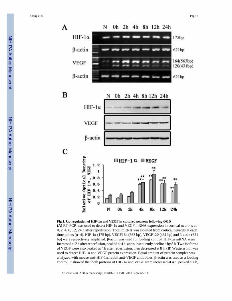
Fig.1. Up-regulation of HIF-1α and VEGF in cultured neurons following OGD(A) RT-PCR was used to detect HIF-1α and VEGF mRNA expression in cortical neurons at0, 2, 4, 8, 12, 24 h after reperfusion. Total mRNA was isolated from cortical neurons at eachtime points (n=4). HIF-1α (175 bp), VEGF164 (563 bp), VEGF120 (431 bp) and β-actin (621bp) were respectively amplified. β-actin was used for loading control. HIF-1α mRNA wereincreased at 2 h after reperfusion, peaked at 4 h, and subsequently declined by 8 h. Two isoformsof VEGF were also peaked at 4 h after reperfusion, then decreased at 8 h. (B) Western blot wasused to detect HIF-1α and VEGF protein expression. Equal amount of protein samples wasanalyzed with mouse anti-HIF-1α, rabbit anti-VEGF antibodies. β-actin was used as a loadingcontrol. It showed that both proteins of HIF-1α and VEGF were increased at 4 h, peaked at 8h,
Zhang et al. Page 7
Neurosci Lett. Author manuscript; available in PMC 2010 September 11.
NIH
-PA Author Manuscript
NIH
-PA Author Manuscript
NIH
-PA Author Manuscript

and decreased at 12 h. (C) The relative expression levels of proteins were calculated as relativeoptical density normalized to β-actin internal controls and represented as mean ± SD (n=4).(**P < 0.001 vs. normoxia)
Zhang et al. Page 8
Neurosci Lett. Author manuscript; available in PMC 2010 September 11.
NIH
-PA Author Manuscript
NIH
-PA Author Manuscript
NIH
-PA Author Manuscript

Fig.2. Activation of PI3K/Akt pathway in cultured neurons subjected to OGD(A) Western blot showed that total Akt protein was not changed after OGD, but p-Akt wasincreased at 4 h, gradually declined at 8 and 12 h, and returned to baseline at 24h. (B) Therelative expression levels of proteins were calculated as relative optical density normalized toβ-actin internal controls and represented as mean ± SD (n=4).(**P < 0.001 vs. normoxia)
Zhang et al. Page 9
Neurosci Lett. Author manuscript; available in PMC 2010 September 11.
NIH
-PA Author Manuscript
NIH
-PA Author Manuscript
NIH
-PA Author Manuscript

Fig.3. Expression of Akt and p-Akt in cultured neurons with wortmannin treatment(A) Western blot showed that p-Akt but not Akt was significantly decreased at 4 h and 8 h afterreperfusion from OGD in wortmannin-treated neurons compared to the DMSO control treatedneurons. (B) Akt and p-Akt relative expression levels were calculated as relative optical densitynormalized to β-actin internal controls and represented as mean ± SD (n=4). (**p<0.001 vs.control) (C) Immunostaining for Akt and p-Akt in OGD neurons with wortmannin treatment.It showed that positive immunoreactions were localized to the cytoplasm of neurons. P-Aktbut not total Akt protein was significantly down-regulated in wortmannin-treated neuronscompared with DMSO treated neurons. ×400.(W, wortmannin; HI, hypoxia-ischemia).
Zhang et al. Page 10
Neurosci Lett. Author manuscript; available in PMC 2010 September 11.
NIH
-PA Author Manuscript
NIH
-PA Author Manuscript
NIH
-PA Author Manuscript

Fig.4. Expression of HIF-1α and VEGF with wortmannin treatment(A) RT-PCR showed that HIF-1α (Left) and VEGF (Right) mRNA levels were decreased inwortmannin-treated neurons at 4 h after reperfusion. (B) Western blot showed HIF-1α andVEGF protein were also decreased at 4 h and 8 h after reperfusion in wortmannin-treatedneurons compared to DMSO treated neurons (C) The relative expression levels of proteinswere calculated as relative optical density using β-actin as internal controls, n=4. (**p<0.001vs. control) (D) Immunoreactivity for HIF-1α and VEGF was much weaker in wortmannintreated neurons compared to DMSO treated neurons. ×400.(HI 4h, hypoxia-ischemia after reperfusion at 4h; D, DMSO; W, wortmannin)
Zhang et al. Page 11
Neurosci Lett. Author manuscript; available in PMC 2010 September 11.
NIH
-PA Author Manuscript
NIH
-PA Author Manuscript
NIH
-PA Author Manuscript

NIH
-PA Author Manuscript
NIH
-PA Author Manuscript
NIH
-PA Author Manuscript
Zhang et al. Page 12
Table 1
Expression of HIF-1α, VEGF, Akt, p-Akt Protein in Cultured Neurons with OGD at Different Times afterReperfusion (n=4)
Time points(h)
Protein (OD)
HIF-1α VEGF Akt p-Akt
N 0.14±0.01 0.24±0.03 0.83±0.10 0.05±0.01
0 0.16±0.02 0.26±0.05 0.83±0.10 0.06±0.01
2 0.21±0.01 0.33±0.04 0.82±0.08 0.07±0.01
4 0.58±0.03** 0.65±0.10** 0.84±0.08 0.38±0.06**
8 0.72±0.03** 0.91±0.11** 0.84±0.10 0.22±0.02**
12 0.60±0.01** 0.62±0.10** 0.84±0.07 0.18±0.02**
24 0.36±0.04** 0.59±0.07** 0.83±0.09 0.04±0.01
OD: Relative optical density; N, normoxia
**p<0.001 vs. normoxia.
Neurosci Lett. Author manuscript; available in PMC 2010 September 11.

NIH
-PA Author Manuscript
NIH
-PA Author Manuscript
NIH
-PA Author Manuscript
Zhang et al. Page 13
Table 2
Expression of Akt, p-Akt, HIF-1α and VEGF in cultured neurons with wortmannin treatment (n=4)
Time Points(h)
Protein (OD)
Akt p-Akt HIF-1α VEGF
HI4 0.92±0.18 0.41±0.02 0.41±0.09 0.76±0.06
W/ HI4 0.92±0.17 0.12±0.01** 0.16±0.04** 0.50±0.05**
HI8 0.90±0.15 0.27±0.01 0.54±0.08 0.84±0.07
W/ HI8 0.91±0.17 0.09±0.01** 0.16±0.04** 0.51±0.08**
OD: Relative optical density; HI, hypoxia-ischemia; W, wortmannin
**p<0.001 vs. control.
Neurosci Lett. Author manuscript; available in PMC 2010 September 11.
Copyright © 2022 FDOKUMEN