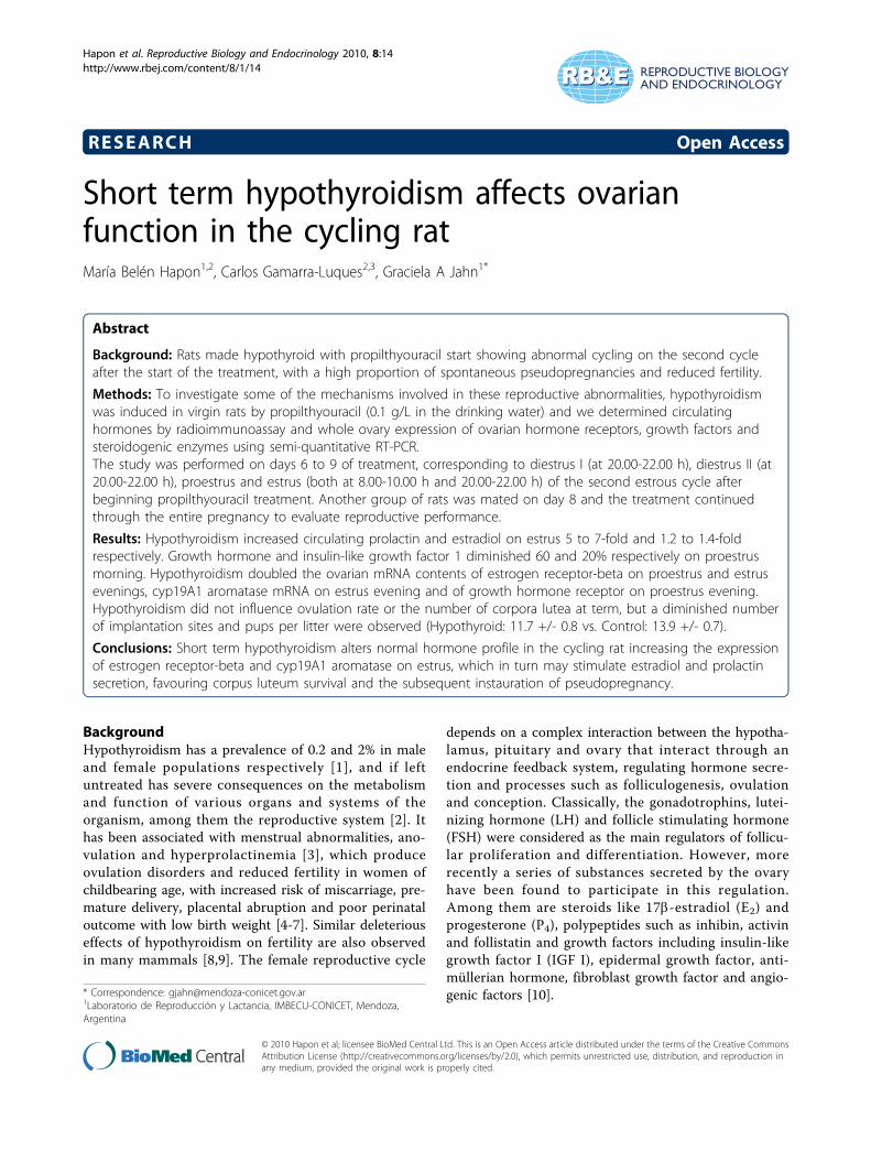
Short term hypothyroidism affects ovarian function in the cycling rat
11
RESEARCH Open Access Short term hypothyroidism affects ovarian function in the cycling rat María Belén Hapon 1,2 , Carlos Gamarra-Luques 2,3 , Graciela A Jahn 1* Abstract Background: Rats made hypothyroid with propilthyouracil start showing abnormal cycling on the second cycle after the start of the treatment, with a high proportion of spontaneous pseudopregnancies and reduced fertility. Methods: To investigate some of the mechanisms involved in these reproductive abnormalities, hypothyroidism was induced in virgin rats by propilthyouracil (0.1 g/L in the drinking water) and we determined circulating hormones by radioimmunoassay and whole ovary expression of ovarian hormone receptors, growth factors and steroidogenic enzymes using semi-quantitative RT-PCR. The study was performed on days 6 to 9 of treatment, corresponding to diestrus I (at 20.00-22.00 h), diestrus II (at 20.00-22.00 h), proestrus and estrus (both at 8.00-10.00 h and 20.00-22.00 h) of the second estrous cycle after beginning propilthyouracil treatment. Another group of rats was mated on day 8 and the treatment continued through the entire pregnancy to evaluate reproductive performance. Results: Hypothyroidism increased circulating prolactin and estradiol on estrus 5 to 7-fold and 1.2 to 1.4-fold respectively. Growth hormone and insulin-like growth factor 1 diminished 60 and 20% respectively on proestrus morning. Hypothyroidism doubled the ovarian mRNA contents of estrogen receptor-beta on proestrus and estrus evenings, cyp19A1 aromatase mRNA on estrus evening and of growth hormone receptor on proestrus evening. Hypothyroidism did not influence ovulation rate or the number of corpora lutea at term, but a diminished number of implantation sites and pups per litter were observed (Hypothyroid: 11.7 +/- 0.8 vs. Control: 13.9 +/- 0.7). Conclusions: Short term hypothyroidism alters normal hormone profile in the cycling rat increasing the expression of estrogen receptor-beta and cyp19A1 aromatase on estrus, which in turn may stimulate estradiol and prolactin secretion, favouring corpus luteum survival and the subsequent instauration of pseudopregnancy. Background Hypothyroidism has a prevalence of 0.2 and 2% in male and female populations respectively [1], and if left untreated has severe consequences on the metabolism and function of various organs and systems of the organism, among them the reproductive system [2]. It has been associated with menstrual abnormalities, ano- vulation and hyperprolactinemia [3], which produce ovulation disorders and reduced fertility in women of childbearing age, with increased risk of miscarriage, pre- mature delivery, placental abruption and poor perinatal outcome with low birth weight [4-7]. Similar deleterious effects of hypothyroidism on fertility are also observed in many mammals [8,9]. The female reproductive cycle depends on a complex interaction between the hypotha- lamus, pituitary and ovary that interact through an endocrine feedback system, regulating hormone secre- tion and processes such as folliculogenesis, ovulation and conception. Classically, the gonadotrophins, lutei- nizing hormone (LH) and follicle stimulating hormone (FSH) were considered as the main regulators of follicu- lar proliferation and differentiation. However, more recently a series of substances secreted by the ovary have been found to participate in this regulation. Among them are steroids like 17b-estradiol (E 2 ) and progesterone (P 4 ), polypeptides such as inhibin, activin and follistatin and growth factors including insulin-like growth factor I (IGF I), epidermal growth factor, anti- müllerian hormone, fibroblast growth factor and angio- genic factors [10]. * Correspondence: [email protected] 1 Laboratorio de Reproducción y Lactancia, IMBECU-CONICET, Mendoza, Argentina Hapon et al. Reproductive Biology and Endocrinology 2010, 8:14 http://www.rbej.com/content/8/1/14 © 2010 Hapon et al; licensee BioMed Central Ltd. This is an Open Access article distributed under the terms of the Creative Commons Attribution License (http://creativecommons.org/licenses/by/2.0), which permits unrestricted use, distribution, and reproduction in any medium, provided the original work is properly cited.
-
Upload
independent -
Category
Documents
-
view
0 -
download
0
Transcript of Short term hypothyroidism affects ovarian function in the cycling rat
RESEARCH Open Access
Short term hypothyroidism affects ovarianfunction in the cycling ratMaría Belén Hapon1,2, Carlos Gamarra-Luques2,3, Graciela A Jahn1*
Abstract
Background: Rats made hypothyroid with propilthyouracil start showing abnormal cycling on the second cycleafter the start of the treatment, with a high proportion of spontaneous pseudopregnancies and reduced fertility.
Methods: To investigate some of the mechanisms involved in these reproductive abnormalities, hypothyroidismwas induced in virgin rats by propilthyouracil (0.1 g/L in the drinking water) and we determined circulatinghormones by radioimmunoassay and whole ovary expression of ovarian hormone receptors, growth factors andsteroidogenic enzymes using semi-quantitative RT-PCR.The study was performed on days 6 to 9 of treatment, corresponding to diestrus I (at 20.00-22.00 h), diestrus II (at20.00-22.00 h), proestrus and estrus (both at 8.00-10.00 h and 20.00-22.00 h) of the second estrous cycle afterbeginning propilthyouracil treatment. Another group of rats was mated on day 8 and the treatment continuedthrough the entire pregnancy to evaluate reproductive performance.
Results: Hypothyroidism increased circulating prolactin and estradiol on estrus 5 to 7-fold and 1.2 to 1.4-foldrespectively. Growth hormone and insulin-like growth factor 1 diminished 60 and 20% respectively on proestrusmorning. Hypothyroidism doubled the ovarian mRNA contents of estrogen receptor-beta on proestrus and estrusevenings, cyp19A1 aromatase mRNA on estrus evening and of growth hormone receptor on proestrus evening.Hypothyroidism did not influence ovulation rate or the number of corpora lutea at term, but a diminished numberof implantation sites and pups per litter were observed (Hypothyroid: 11.7 +/- 0.8 vs. Control: 13.9 +/- 0.7).
Conclusions: Short term hypothyroidism alters normal hormone profile in the cycling rat increasing the expressionof estrogen receptor-beta and cyp19A1 aromatase on estrus, which in turn may stimulate estradiol and prolactinsecretion, favouring corpus luteum survival and the subsequent instauration of pseudopregnancy.
BackgroundHypothyroidism has a prevalence of 0.2 and 2% in maleand female populations respectively [1], and if leftuntreated has severe consequences on the metabolismand function of various organs and systems of theorganism, among them the reproductive system [2]. Ithas been associated with menstrual abnormalities, ano-vulation and hyperprolactinemia [3], which produceovulation disorders and reduced fertility in women ofchildbearing age, with increased risk of miscarriage, pre-mature delivery, placental abruption and poor perinataloutcome with low birth weight [4-7]. Similar deleteriouseffects of hypothyroidism on fertility are also observedin many mammals [8,9]. The female reproductive cycle
depends on a complex interaction between the hypotha-lamus, pituitary and ovary that interact through anendocrine feedback system, regulating hormone secre-tion and processes such as folliculogenesis, ovulationand conception. Classically, the gonadotrophins, lutei-nizing hormone (LH) and follicle stimulating hormone(FSH) were considered as the main regulators of follicu-lar proliferation and differentiation. However, morerecently a series of substances secreted by the ovaryhave been found to participate in this regulation.Among them are steroids like 17b-estradiol (E2) andprogesterone (P4), polypeptides such as inhibin, activinand follistatin and growth factors including insulin-likegrowth factor I (IGF I), epidermal growth factor, anti-müllerian hormone, fibroblast growth factor and angio-genic factors [10].* Correspondence: [email protected]
1Laboratorio de Reproducción y Lactancia, IMBECU-CONICET, Mendoza,Argentina
Hapon et al. Reproductive Biology and Endocrinology 2010, 8:14http://www.rbej.com/content/8/1/14
© 2010 Hapon et al; licensee BioMed Central Ltd. This is an Open Access article distributed under the terms of the Creative CommonsAttribution License (http://creativecommons.org/licenses/by/2.0), which permits unrestricted use, distribution, and reproduction inany medium, provided the original work is properly cited.

Thyroid hormones (THs) act at all the levels of regu-lation within the reproductive system. Triiodothyronine(T3) and thyroxine (T4) are present in follicular fluid[11]. Granulosa cells of antral preovulatory follicles andovarian stromal cells express thyroid hormone receptorsa and b messenger RNA (mRNA) and protein. Thus, itis conceivable that THs play an essential role in ovarianphysiology [12].In adult rats, hypothyroidism apparently does not
directly produce sterility but during the first half ofgestation it increases embryonic reabsorption, resultingin smaller litters and augmentation of fetal mortality,although it does not seem to affect implantation [13-16].On the other hand, hypothyroidism reduces fertility,causing estrous cycle irregularities and spontaneous con-secutive pseudopregnancies that are considered to be aconsequence of the hyper secretion of prolactin (PRL)during proestrus (P) and estrus (E) [17,14].The abnormal estrous cycles seen in hypothyroid
(HypoT) rats have been associated with diminished cir-culating growth hormone (GH), IGFs and E2, andadministration of T4 to HypoT rats reverts the effect oncirculating hormones and normalizes the estrous cycle[18]. However, the administration of GH to thyroidecto-mized rats did not improve the timing of gestation, thenumber of reabsorbed fetuses nor the number of pupsper litter [19]. Thus, the effect of hypothyroidism onfemale sexual cycles may no be due solely to the altera-tion of the GH/IGF axis.Recently we have shown that virgin rats treated with
the antithyroid propilthyouracil (PTU) presented irregu-lar cycles, spontaneous pseudopregnancies and alteredcirculating ovarian hormones and PRL after the thirdestrous cycle that resulted in mammary developmentsimilar to that of midpregnancy. Furthermore, when therats were mated 8 days after the start of antithyroidtreatment they produced smaller litters [14].To investigate the biochemical mechanisms involved
in the reproductive alterations (smaller litters and spon-taneous pseudopregnacies) observed in HypoT rats, westudied the early effects of PTU-induced hypothyroidismon the female reproductive axis analyzing its effect onbasic reproductive parameters and ovarian function invirgin rats on the second cycle after beginning antithyr-oid treatment, that is the cycle when the rats weremated on our previous study [14]. We evaluated theearly effect of hypothyroidism on the pattern of circulat-ing hormones, the ovarian expression of several hor-mone receptors, growth factors and steroidogenicenzymes. We also measured the ovulation rate in virginrats and the number of corpora lutea, implantation sitesand pups at term to determine if the reduced litter size
of HypoT rats is associated with ovulation failure orwith postovulatory phenomena.
MethodsAnimalsAdult female Wistar rats bred in our laboratory, 2-3months old, weighing 200-230 g at the onset of treatmentand with regular 4 day cycles were used. Vaginal smearsfrom all animals were obtained daily to monitor theestrous cycle. The rats were kept in a light (lights on 6.00- 20.00 h) and temperature (22-24°C) controlled room.Rat chow (Cargill, Cordoba, Argentina) and tap water orPTU solution were available ad libitum. Animal mainte-nance and handling was performed according to the NIHguide for the Care and Use of Laboratory Animals (NIHpublication N8 86-23, revised 1985 and 1991) and theUK requirements for ethics of animal experimentation(Animals Scientific Procedures, Act 1986).
Experimental designHypothyroidism was induced by administration of PTUat a concentration of 0.1 g/l in the drinking water. Thetreatment was started on E day. To determine the pat-tern of hormonal secretion and the function of theovary, groups of 8-10 HypoT or 8-10 control rats werekilled by decapitation during the second cycle afterinitiation of the treatment, starting at diestrus (D) I andat times selected so as to cover most of the stages of theestrous cycle, such as the initiation (D I at 20.00-22.00h) and end of the luteal phase (D II at 20.00-22.00 h),the peak of estrogen surge on P morning (8.00-10.00 h),the preovulatory peak on P evening (20.00-22.00 h) andthe postovulatory phase (E morning at 8.00-10.00 h andevening at 20.00-22.00 h). The denominations of thegroups correspond to the time which the animals weresacrificed. Trunk blood was collected and serum sepa-rated by centrifugation and stored at -20°C until usedfor subsequent determination of hormonal parameters.The ovaries were rapidly removed, washed in a cold sal-ine solution, snap-frozen in liquid nitrogen and storedat -80°C until they were used for RNA preparation. Todetermine the ovulation rate, the oviducts were alsoremoved from the rats sacrificed on E morning and theampullae were isolated. The oocytes were harvested byapplying gentle pressure to both ends of the ampulla,placed on a slide in phosphate buffer saline and countedunder a stereomicroscope.Another group of 8 rats were mated on the second P
(8 days) after starting the treatment. The presence ofspermatozoa in the vaginal smears the morning aftercaging with a fertile male in P night was indicative ofmating and this day was counted as day 0 of pregnancy.
Hapon et al. Reproductive Biology and Endocrinology 2010, 8:14http://www.rbej.com/content/8/1/14
Page 2 of 11
Two or three days before delivery the rats were cagedindividually. The day and hour of delivery and the num-ber of pups were recorded. Another group of gestatingrats were killed by decapitation on day 21 of gestationand the number of implantation sites of pups in uteroand of corpora lutea, in the ovaries, were recorded.
Hormone determinationsPRL, LH, FSH, GH and thyroid stimulating hormone(TSH) were measured by double antibody radioimmu-noassay using materials generously provided by Dr Par-low and the NHPP (National Hormone and PituitaryProgram, Harbor-UCLA Medical Center, Torrance, CA,USA) as previously described [14]. For each determina-tion, all the samples were measured by duplicate in thesame assay, and the intra-assay coefficient of variancewas less than 10%E2, P4, T3, T4 and IGF 1 concentrations in sera were
measured in duplicate by radioimmunoassay using com-mercial kits for total hormones DSL-4800, DSL-3400,DSL-3100, DSL-3200 and DSL 2900 double antibodyradioimmunoassay, respectively; all from Diagnostic Sys-tems Laboratories, Webster, TX). Inter- and intra-assaycoefficients of variation were less than 10%.
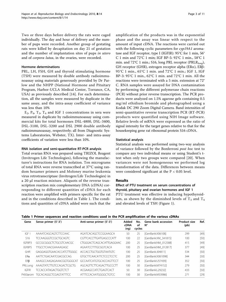
RNA isolation and semi-quantitative RT-PCR analysisTotal ovarian RNA was prepared using TRIZOL Reagent(Invitrogen Life Technologies), following the manufac-turer’s instructions for RNA isolation. Ten microgramsof total RNA were reverse transcribed at 37°C using ran-dom hexamer primers and Moloney murine leukemiavirus retrotranscriptase (Invitrogen/Life Technologies) ina 20 μl reaction mixture. Aliquots of the reverse tran-scription reaction mix complementary DNA (cDNA) cor-responding to different quantities of cDNA for eachreaction were amplified with primers specific for the ratand in the conditions described in Table 1. The condi-tions and quantities of cDNA added were such that the
amplification of the products was in the exponentialphase and the assay was linear with respect to theamount of input cDNA. The reactions were carried outwith the following cyclic parameters for cyp19A1 aroma-tase and IGF receptor, type I (IGFIR): 95°C for 1 min, 56°C 1 min and 72°C 1 min; IGF BP-5: 92°C 1 min., 58°C 1min. and 72°C 1 min.; S16, long PRL receptor (PRLRlong),GH receptor (GHR), estrogen receptor alpha (ERa), ERb:95°C 1 min., 65°C 1 min. and 72°C 1 min.; IGF 1, IGFBP-3: 95°C 1 min., 62°C 1 min. and 72°C 1 min. All thereactions were terminated with a 5 min. extension at 72°C. RNA samples were assayed for DNA contaminationby performing the different polymerase chain reactions(PCR) without prior reverse transcription. The PCR pro-ducts were analyzed on 1.5% agarose gels containing 0.5mg/ml ethidium bromide and photographed using aKodak DC 290 Zoom Digital Camera. Band intensities ofsemi-quantitative reverse transcription PCR (RT-PCR)products were quantified using NIH Image software.Relative levels of mRNA were expressed as the ratio ofsignal intensity for the target genes relative to that for thehousekeeping gene rat ribosomal protein S16 cDNA.
Statistical analysisStatistical analysis was performed using two-way analysisof variance followed by the Bonferroni post hoc test tocompare any two individual means or using Student’s t-test when only two groups were compared [20]. Whenvariances were not homogeneous we performed logtransformation of the data. Differences between meanswere considered significant at the P < 0.05 level.
ResultsEffect of PTU treatment on serum concentrations ofthyroid, pituitary and ovarian hormones and IGF 1PTU treatment was effective in inducing hypothyroid-ism, as shown by the diminished levels of T3 and T4
and elevated levels of TSH (Figure 1).
Table 1 Primer sequences and reaction conditions used in the PCR amplification of the various cDNAs
Gene Sense primer (5’-3’) Anti-sense primer (5’-3’) AddedcDNA(ng)
No.of
cycles
Gene bank accessionN°
Product size(pb)
Ref.
IGF I AAAATCAGCAGTCTTCCAAC AGATCACAGCTCCGGAAGCA 50 25 [GenBank:X06108] 299 [49]
S16 TCCAAGGGTCCGCTGCAGTC CGTTCACCTTGATGAGCCCATT 100 22 [GenBank:XM_341815] 100 [50]
IGFBP3 GCCGCGGGCTCTGCGTCAACGC CTGGGACTCAGCACATTGAGGAAC 200 25 [GenBank:NM_012588] 415 [49]
IGFBP5 TTGCCTCAACGAAAAGAGC AGAATCCTTTGCGGTCACA 100 25 [GenBank:NM_012817] 377 [49]
GHR GAGGAGGTGAACACCATCTTGGGC ACCACCTGCTGGTGTAATGTC 100 25 [GenBank:J04811] 534 [50]
ERa AATTCTGACAATCGACGCCAG GTGCTTCAACATTCTCCCTCCTC 200 25 [GenBank:X061098] 344 [50]
ERb AAAGCCAAGAGAAACGGTGGGCAT GCCAATCATGTGCACCAGTTCCT 100 25 [GenBank:U57439] 352 [50]
PRLLong AAAGTATCTTGTCCAGACTCGCTG AGCAGTTCTTCAGACTTGCCCTT 100 30 [GenBank:M74152] 279 [50]
IGFIR TCCACCATAGACTGGTCTCT ACGAAGCCATCTGAGTCACT 50 30 [GenBank:L29232] 433 [50]
P450arom TGCACAGGCTCGAGTATTTCC ATTTCCACAATGGGGCTGTCC 100 30 [GenBank:M33986] 271 [29]
Hapon et al. Reproductive Biology and Endocrinology 2010, 8:14http://www.rbej.com/content/8/1/14
Page 3 of 11
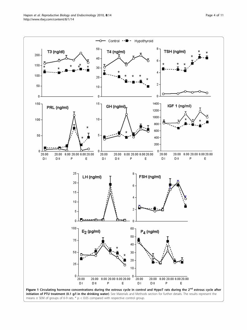
Figure 1 Circulating hormone concentrations during the estrous cycle in control and HypoT rats during the 2nd estrous cycle afterinitiation of PTU treatment (0.1 g/l in the drinking water). See Materials and Methods section for further details. The results represent themeans ± SEM of groups of 6-9 rats. * p < 0.05 compared with respective control group.
Hapon et al. Reproductive Biology and Endocrinology 2010, 8:14http://www.rbej.com/content/8/1/14
Page 4 of 11
A classical preovulatory pattern of gonadotrophinsecretion was observed in both groups and no differ-ences between groups were detected during the estrouscycle in LH or FSH (Figure 1), suggesting that PTUtreatment did not affect the pituitary secretion of thesehormones. On the other hand, although the preovula-tory peak of PRL was normal in the HypoT rats, weobserved a significant increase in PRL concentrationsduring E (Figure 1). Since GH secretion is also influ-enced by THs, we also measured circulating GH duringthe estrous cycle. Control rats showed a GH peak on Pmorning. This peak was not observed in the HypoT rats(Figure 1). Similarly, circulating IGF 1 in control ratspeaked on P and E mornings, and hypothyroidismannulled these increases.To evaluate the effect of hypothyroidism on the ovary
we also measured circulating E2 and P4 during theestrous cycle in the rat. Hypothyroidism did not modifyserum P4 pattern (Figure 1). As expected both groupsshowed increased levels during D I (luteal phase) andduring the preovulatory surge at P 20.00 h. E2 showed aprogressive increase from D I until P morning and thendiminished after ovulation. In HypoT rats the postovula-tory decrease was less prominent, resulting in signifi-cantly increased concentrations during E.
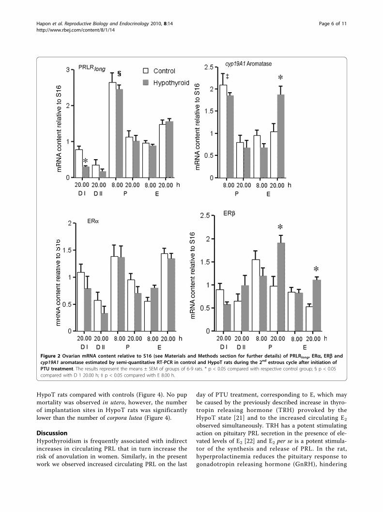
RT-PCR analysis of the relative expression of factorsrelated to the ovarian response to E2, PRL andgonadotrophins during the estrous cycleSince in the HypoT rats PRL and E2 levels wereincreased during E (Figure 1), we evaluated the ovarianresponse to these hormones determining the expressionof PRLRlong, ERa and ERb during the estrous cycle.A surge in PRLRlong expression was observed (Figure 2)
on P 8.00 h, followed by a second increase on E 20.00 h.The only effect of hypothyroidism was a diminishedexpression on D I.Expression of ERa showed a pattern similar to that
observed for PRLRlong, with two peaks on P morning andE afternoon and a decrease on E 8.00 h. Hypothyroidismdid not modify this pattern significantly (Figure 2).In control rats, ERb mRNA content (Figure 2) was
elevated on P 8 h, decreased progressively during Pafternoon and E and remained low on both D days. InHypoT rats, the highest expression was observed on P20.00 h, being significantly higher with respect to con-trols, and a second increase with respect to the controlswas observed on E 20.00 h (Figure 2).In order to determine the ovarian response to the cir-
culating FSH and LH and to explore the cause of theincreased circulating E2 observed in E (Figure 1), wemeasured cyp19A1 aromatase expression on P and E. Incontrol rats the maximum expression was observed onP 8.00 h, followed by low values on P evening and on E
(Figure 2). In HypoT rats the pattern was similar on Pand E morning, but on E 20.00 h cyp19A1 aromataseexpression was significantly higher compared tocontrols.
RT-PCR analysis of GH/IGF 1 system relative expression inthe ovary during the estrous cycleBased on the HypoT effects observed on circulating GH,IGF 1 and E2 we evaluated the expression, relative toS16, of several components of the GH/IGF axis duringthe estrous cycle at ovarian level. IGF 1 content did notvary during the estrous cycle and was not affected byhypothyroidism (Figure 3). In contrast, there were signif-icant differences in the expression of IGF binding pro-teins (IGF BPs). IGF BP-3 content was highest on P 8.00h (Figure 3), and showed the lowest values on both Ddays. The same pattern was amplified in the HypoT ratsthat had significantly higher values than controls on P8.00 h and significantly lower values on D I and II (Fig-ure 3).IGF BP-5 showed a pattern similar to that of IGF BP-
3, with the highest values on P morning and lowest onD days (Figure 3). The only effect of hypothyroidism onIGF BP-5 content was a diminution on D I (Figure 3).The expression of IGFIR in control rats (Figure 3) was
low on the D days, increased on P 8.00 h, diminishedthe same day at 20.00 h, and then rose progressivelyreaching maximum values on E 20.00 h. PTU treatedrats had a similar pattern, but showed diminishedexpression on both D days.In control rats GHR expression was stable during
most of the estrous cycle, except on D II, where therewas a marked decrease. HypoT rats had a similar pat-tern, except for an increase in the expression on P 20.00h (Figure 3).
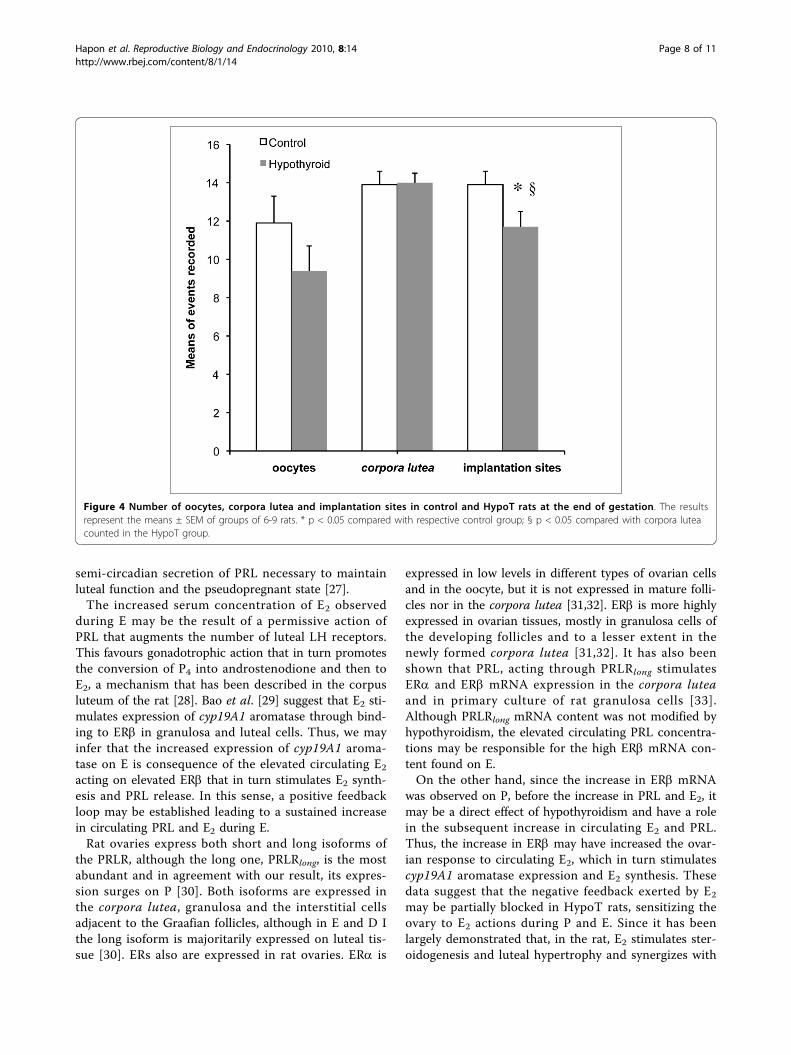
Effect of PTU treatment on the ovulation rate in virginrats and the number of corpora lutea and implantationsites at the end of gestationWe have previously shown that HypoT rats have smallerlitters [14]. To determine if this is a consequence of anovulatory deficit, the ovulation rate (measured as num-ber of oocytes per rat in the oviduct on E morning) wasrecorded in control and HypoT rats after 9 days of PTUtreatment. HypoT rats had a slightly lower ovulationrate compared to controls, but the difference was notstatistically significant (Figure 4).Next, we evaluated if the diminished number of pups
per litter was due to a defect in implantation or gesta-tional development, by counting the implantation sites,pups and corpora lutea in a group of HypoT and con-trol rats at term. There were no differences in the num-ber of corpora lutea but the number of implantationsites and of pups per rat was significantly lower in
Hapon et al. Reproductive Biology and Endocrinology 2010, 8:14http://www.rbej.com/content/8/1/14
Page 5 of 11
HypoT rats compared with controls (Figure 4). No pupmortality was observed in utero, however, the numberof implantation sites in HypoT rats was significantlylower than the number of corpora lutea (Figure 4).
DiscussionHypothyroidism is frequently associated with indirectincreases in circulating PRL that in turn increase therisk of anovulation in women. Similarly, in the presentwork we observed increased circulating PRL on the last
day of PTU treatment, corresponding to E, which maybe caused by the previously described increase in thyro-tropin releasing hormone (TRH) provoked by theHypoT state [21] and to the increased circulating E2
observed simultaneously. TRH has a potent stimulatingaction on pituitary PRL secretion in the presence of ele-vated levels of E2 [22] and E2 per se is a potent stimula-tor of the synthesis and release of PRL. In the rat,hyperprolactinemia reduces the pituitary response togonadotropin releasing hormone (GnRH), hindering
Figure 2 Ovarian mRNA content relative to S16 (see Materials and Methods section for further details) of PRLRlong, ERa, ERb andcyp19A1 aromatase estimated by semi-quantitative RT-PCR in control and HypoT rats during the 2nd estrous cycle after initiation ofPTU treatment. The results represent the means ± SEM of groups of 6-9 rats. * p < 0.05 compared with respective control group; § p < 0.05compared with D 1 20.00 h; ‡ p < 0.05 compared with E 8.00 h.
Hapon et al. Reproductive Biology and Endocrinology 2010, 8:14http://www.rbej.com/content/8/1/14
Page 6 of 11

preovulatory LH secretion [23] and makes the hypotha-lamus more susceptible to stimuli capable of inducingpseudopregnancies [24].The effects of hypothyroidism on the gonadotrophic
axis are contradictory, some authors have found thatPTU treated rats had diminished concentration of LHon D and P but the preovulatory surge was conserved[25], while others describe a reduced proportion of ovu-lating rats, but increased preovulatory levels of LH inthose rats that ovulated after radiochemical thyroidect-omy with 131I [17]. We found no significant variationson preovulatory LH levels on P, the eighth day afterinitiation of PTU treatment. Most probably, the discre-pancies between these three studies may be a conse-quence of the different treatment paradigms used to
induce hypothyroidism. We did not find, either, anyeffect on the ovulation rate, indicating that at this earlystage of PTU treatment, the gonadotrophic axis doesnot seem to be affected.On the other hand, the increased serum PRL observed
in E may be responsible for the induction of sponta-neous pseudopregnancies that we observed after moreprolonged PTU treatments [14]. Other authors also havefound that hypothyroidism induces prolonged D accom-panied with elevated circulating P4 indicative of pseudo-pregnancy [17,25]. E2 regulates luteal functionfacilitating the uptake of cholesterol for P4 synthesis[26]. Thus, the elevated E2 and PRL observed in E maystimulate subsequent P4 synthesis and secretion in thenewly formed corpora lutea, which in turn triggers the
Figure 3 Ovarian mRNA content relative to S16 of IGF 1, IGF R, IGF BP-3, IGF BP-5 and GHR estimated by semi-quantitative RT-PCR incontrol and HypoT rats during the 2nd estrous cycle after initiation of PTU treatment. The results represent the means ± SEM of groupsof 6-9 rats.* p < 0.05 compared with respective control group; § p < 0.05 compared with D 1 20.00 h.
Hapon et al. Reproductive Biology and Endocrinology 2010, 8:14http://www.rbej.com/content/8/1/14
Page 7 of 11
semi-circadian secretion of PRL necessary to maintainluteal function and the pseudopregnant state [27].The increased serum concentration of E2 observed
during E may be the result of a permissive action ofPRL that augments the number of luteal LH receptors.This favours gonadotrophic action that in turn promotesthe conversion of P4 into androstenodione and then toE2, a mechanism that has been described in the corpusluteum of the rat [28]. Bao et al. [29] suggest that E2 sti-mulates expression of cyp19A1 aromatase through bind-ing to ERb in granulosa and luteal cells. Thus, we mayinfer that the increased expression of cyp19A1 aroma-tase on E is consequence of the elevated circulating E2acting on elevated ERb that in turn stimulates E2 synth-esis and PRL release. In this sense, a positive feedbackloop may be established leading to a sustained increasein circulating PRL and E2 during E.Rat ovaries express both short and long isoforms of
the PRLR, although the long one, PRLRlong, is the mostabundant and in agreement with our result, its expres-sion surges on P [30]. Both isoforms are expressed inthe corpora lutea, granulosa and the interstitial cellsadjacent to the Graafian follicles, although in E and D Ithe long isoform is majoritarily expressed on luteal tis-sue [30]. ERs also are expressed in rat ovaries. ERa is
expressed in low levels in different types of ovarian cellsand in the oocyte, but it is not expressed in mature folli-cles nor in the corpora lutea [31,32]. ERb is more highlyexpressed in ovarian tissues, mostly in granulosa cells ofthe developing follicles and to a lesser extent in thenewly formed corpora lutea [31,32]. It has also beenshown that PRL, acting through PRLRlong stimulatesERa and ERb mRNA expression in the corpora luteaand in primary culture of rat granulosa cells [33].Although PRLRlong mRNA content was not modified byhypothyroidism, the elevated circulating PRL concentra-tions may be responsible for the high ERb mRNA con-tent found on E.On the other hand, since the increase in ERb mRNA
was observed on P, before the increase in PRL and E2, itmay be a direct effect of hypothyroidism and have a rolein the subsequent increase in circulating E2 and PRL.Thus, the increase in ERb may have increased the ovar-ian response to circulating E2, which in turn stimulatescyp19A1 aromatase expression and E2 synthesis. Thesedata suggest that the negative feedback exerted by E2
may be partially blocked in HypoT rats, sensitizing theovary to E2 actions during P and E. Since it has beenlargely demonstrated that, in the rat, E2 stimulates ster-oidogenesis and luteal hypertrophy and synergizes with
Figure 4 Number of oocytes, corpora lutea and implantation sites in control and HypoT rats at the end of gestation. The resultsrepresent the means ± SEM of groups of 6-9 rats. * p < 0.05 compared with respective control group; § p < 0.05 compared with corpora luteacounted in the HypoT group.
Hapon et al. Reproductive Biology and Endocrinology 2010, 8:14http://www.rbej.com/content/8/1/14
Page 8 of 11
the effect of PRL in the ovary, these factors may pro-mote corpus luteum survival favouring the establishmentof pseudopregnancy [26,34-37].Since during E, cyp19A1 aromatase is almost exclu-
sively expressed in the corpus luteum [38], the increasedexpression observed in the HypoT rats may be responsi-ble for the increased levels of circulating E2 observed atthe same time.Hypothyroidism also diminishes synthesis and secre-
tion of GH in the rat [39]. In particular, the stimulationof GH release produced by E2depends on the participa-tion of THs [40]. The GH surge observed on P morningin the control rats may be due to the elevated circulat-ing E2, and its absence in the HypoT rats may bedirectly linked to the low levels of THs, that may haveoffset the stimulatory action of E2. In addition, the lowGH levels on P may play a part in the decreased circu-lating IGF 1 levels on P morning, since GH is the mainstimulus for hepatic IGF 1 production, and the liver isthe main source of this hormone [41]. The decreasedlevels of IGF 1 on E morning, in contrast, may be adirect effect of hypothyroidism.Numerous investigations have demonstrated actions of
GH on steroidogenesis, gametogenesis and gonadal dif-ferentiation. Many of these effects are mediated by localactions of IGF 1. It has been postulated that this growthfactor has an essential participation in follicular develop-ment [42] and ovarian steroidogenesis [10]. GH exertsmost of its effects in the ovary through binding to itsspecific receptor. For example, mice lacking GHR pre-sent a diminished ovulation rate, caused by the failureof the ovarian response to gonadotrophins [43]. GHpromotes FSH-induced LH receptor (LHR) synthesis,and the administration of IGF 1 does not improve theovulation rate, suggesting a direct participation of GH inthe ovulation process. IGF 1 promotes FSH receptor(FSHR) expression, and in turn FSH augments bothIGFIR and FSHR. Besides, this growth factor exerts itsaction through its own receptor, present in granulosa,thecal and luteal cells. IGFIR expression is positivelyregulated by FSH, LH and GH [10] and it is expressedin the oocyte, mediating its maturation by IGF 1 [44].Thus, the increase in GHR on P in the HypoT rats mayhave compensated for the absent GH surge, allowingfollicle maturation to culminate in ovulation.Bachelot et al. [43] reported that IGF 1 produced
locally in the ovary is independent of GH. Although ourpresent and previous results [14] showed that hypothyr-oidism reduces serum levels of this growth factor(mainly of hepatic origin) in virgin rats with prolongedPTU treatment, we did not observe differences betweencontrol and HypoT rats in ovarian IGF 1 mRNA, norbetween the days of the cycle. It is possible that IGF 1regulation by THs is tissue specific, but we cannot
dismiss the possibility that this lack of effect is due tothe short length of the PTU treatment.IGF BP-3 sequestrates circulating IGF 1, modulating
its action. IGF BP-3 is expressed preferentially in ovar-ian theca and interstitial cells and it may have an antigo-nadotrophic effect through sequestering of IGF 1. Ourresults suggest an altered gonadotrophic response by thediminished availability of IGF 1 on P, while on D I andD II, in contrast, the lower IGF BP-3 concentrationsmay allow IGF 1 to be more available. Fraser et al. [45]demonstrated that IGF BP-3 expression in corpusluteum is more intense during luteolysis. Thus, thediminished expression observed during D may promotecorpus luteum survival, favouring the establishment ofpseudopregnancy.IGF BP-5 is the most abundant IGF 1 binding protein
in the ovary, mainly expressed in the granulosa cells ofatretic follicles, interstitial cells, corpus luteum and alsoin the superficial epithelium [46,47]. Our results showthat IGF BP-5 expression was not affected on P, andsince in D IGF BP-5 is selectively expressed in superfi-cial epithelium [46], we may assume that its activity isnot physiologically relevant at this moment.Lastly, ovulation rate and the number of corpora lutea
were not affected significantly by the short term treat-ment with PTU, suggesting that ovarian failure is notresponsible for the diminished number of pups per litterpreviously observed [14]. However, the number ofimplantation sites and of pups at delivery was smallerthan the number of corpora lutea in HypoT rats, sug-gesting that the deleterious effects of hypothyroidismare exerted on events that occur after ovulation. Allthese data point to impairments in fertilization ordiminished capacity of the embryos to implant as aprobable cause of the reduced litters born to HypoTmothers.The factors affecting intrauterine fetal survival and/or
implantation that may be responsible for the diminishednumber of pups per litter remain unclear and needfurther investigation.Hypothyroid women have increased rates of ovarian
failure and ovulatory dysfunctions [4,48]. The data pre-sented in this investigation may contribute to the under-standing of the influence of thyroid hormone disorderson infertility and support the checking of thyroid statusin patients with reproductive alterations.
ConclusionsThe present results show that during the second estrouscycle after the start of PTU treatment the rats arefrankly HypoT and show some alterations in hormonesecretion, and in the expression of ovarian receptors,members of the GH/IGF family and cyp19A1 aromatasethat may stimulate luteal function and survival and thus
Hapon et al. Reproductive Biology and Endocrinology 2010, 8:14http://www.rbej.com/content/8/1/14
Page 9 of 11
account for the appearance of spontaneous pseudopreg-nancies previously observed [14,17,25]. On the otherhand, the observed effects of short-term hypothyroidismdo not seem to affect significantly the ovulation rate,indicating that the smaller litters born to HypoT ratsseem to be caused by postovulatory events not linked toovulation rate or corpus luteum formation.
List of abbreviationscDNA: DNA copy; E: estrus; E2: estradiol; ER: estrogenreceptor; D: diestrus; FSH: follicle stimulating hormone;FSHR: FSH receptor; GH: growth hormone; GHR: GHreceptor; GnRH: gonadotropin release hormone; HypoT:hypothyroid; IGFIR: IGF receptor, type I; IGFBP: IGFbinding protein; IGF: insulin-like growth factor; LH:luteinizing hormone; LHR: LH receptor; mRNA: mes-senger RNA; P: proestrus; P4: progesterone; PCR: poly-merase chain reaction; PRL: prolactin; PRLR: PRLreceptor; PRLRlong: long form of the PRLR; PTU: pro-pylthyouracil; RT-PCR: reverse transcription PCR; T3:triiodothyronine; T4: thyroxine; THs: thyroid hormones;TRH: thyrotropin releasing hormone; TSH: thyroid sti-mulating hormone
AcknowledgementsThis investigation was supported by grants PIPs 5795 and 2298 fromCONICET (National Investigation Council of Science and Technology,Argentina), PMT-PICT 32529 from the Agencia de Promoción Científica yTecnológica, Argentina, PROGRESAR and SeCTyP 06/M003 UniversidadNacional de Cuyo GAJ, MBH and CGL are Career Scientists from CONICET.We thank Norma Carreño for the excellent technical support.
Author details1Laboratorio de Reproducción y Lactancia, IMBECU-CONICET, Mendoza,Argentina. 2Instituto de Ciencias Básicas, Universidad Nacional de Cuyo,Mendoza, Argentina. 3Instituto de Embriología e Histología, IHEM-CONICET,Facultad de Ciencias Médicas, Universidad Nacional de Cuyo, Mendoza,Argentina.
Authors’ contributionsMBH designed and conceived the study, carried out the experimentalanimal model, immunoassays, RT-PCR, performed the data analysis anddrafted the manuscript. CGL collaborated in processing the samples, RT-PCRand helped draft the manuscript. GAJ participated in the study design andcoordination, data analysis and helped draft the manuscript. All authors readand approved the final manuscript.
Competing interestsThe authors declare that they have no competing interests.
Received: 11 October 2009Accepted: 11 February 2010 Published: 11 February 2010
References1. Elliott B: Diagnosing and treating hypothyroidism. Nurse Pract 2000,
25:99-105.2. Doufas AG, Mastorakos G: The hypothalamic-pituitary-thyroid axis and the
female reproductive system. Ann N Y Acad Sci 2000, 900:65-76.3. Poppe K, Velkeniers B, Glinoer D: Thyroid disease and female
reproduction. Clin Endocrinol (Oxf) 2007, 66:309-321.4. Eldar-Geva T, Shoham M, Rösler A, Margalioth EJ, Livne K, Meirow D:
Subclinical hypothyroidism in infertile women: the importance of
continuous monitoring and the role of the thyrotropin-releasinghormone stimulation test. Gynecol Endocrinol 2007, 23:332-337.
5. Becks GP, Burrow GN: Thyroid disease and pregnancy. Med Clin North Am1991, 75:121-150.
6. Glinoer D: The regulation of thyroid function in pregnancy: pathways ofendocrine adaptation from physiology to pathology. Endocr Rev 1997,18:404-433.
7. Glinoer D: Iodine supplementation during pregnancy: importance andbiochemical assessment. Exp Clin Endocrinol Diabetes 1998, 106:3-21.
8. Tohei A, Imai A, Watanabe G, Taya K: Influence of thiouracil-inducedhypothyroidism on adrenal and gonadal functions in adult female rats. JVet Med Sci 1998, 60:439-446.
9. Stradtman EW: Thyroid dysfunction and ovulatory disorders. Textbook ofReproductive Medicine Norwalk: AppletonCarr BR, Blackwell RE 1993, 297-321.
10. Giudice LC: Insulin-like growth factors and ovarian folliculardevelopment. Endocr Rev 1992, 13:641-669.
11. Wakim AN, Polizotto SL, Buffo MJ, Marrero MA, Burholt DR: Thyroidhormones in human follicular fluid and thyroid hormone receptors inhuman granulosa cells. Fertil Steril 1993, 59:1187-1190.
12. Wakim AN, Paljug WR, Jasnosz KM, Alhakim N, Brown AB, Burholt DR:Thyroid hormone receptor messenger ribonucleic acid in humangranulosa and ovarian stromal cells. Fertil Steril 1994, 62:531-534.
13. Rao PM, Panda JN: Effects of hypothyroidism on pregnancy of rats. IndianJ Physiol Pharmacol 1980, 24:126-130.
14. Hapon MB, Simoncini M, Via G, Jahn GA: Effect of hypothyroidism onhormone profiles in virgin, pregnant and lactating rats, and on lactation.Reproduction 2003, 126:371-382.
15. Bakke JL, Lawrence NL, Robinson S, Bennett J: Endocrine studies of theuntreated progeny of thyroidectomized rats. Pediatr Res 1975, 9:742-748.
16. Varma SK, Murray R, Stanbury JB: Effect of maternal hypothyroidism andtriiodothyronine on the fetus and newborn in rats. Endocrinology 1978,102:24-30.
17. Mattheij JA, Swarts JJ, Lokerse P, van Kampen JT, Heide Van der D: Effect ofhypothyroidism on the pituitary-gonadal axis in the adult female rat. JEndocrinol 1995, 146:87-94.
18. Osorio A, Ruiz E, Ortega E: Possible role of GH/IGF 1 in the ovarianfunction of adult hypothyroid rats. Mol Cell Biochem 1998, 179:7-11.
19. Hendrich CE, Porterfield SP: Serum growth hormone levels in hypothyroidand GH-treated thyroidectomized rats and their progenies. Proc Soc ExpBiol Med 1992, 201:296-302.
20. Snedecor GW, Cochran WG: Statistical Methods Ames: Iowa State UniversityPress 1967.
21. Malcolm FL: Hypothalamus and Pituitary Neuroendocrinology. WilliamsTextbook of Endocrinology Philadelphia: Saunders ElsevierKronenberg HM,Melmed S, Polonsky KS, Larsen PR , 11 2008, 101-102.
22. Pan JT, Chen CW: Increased plasma prolactin levels in ovariectomizedthyroidectomised rats treated with Estrogen. Endocrinology 1990,126:3146-3152.
23. Lu KH, Chen HI, Grandison L, Huang HH, Meites J: Reduced luteinizinghormone release by synthetic luteinizing hormone releasing hormone(LHRH) in postpartum lactating rats. Endocrinology 1976, 98:1235-1240.
24. Helena CV, McKee DT, Bertram R, Walker AM, Freeman ME: The RhythmicSecretion of Mating-Induced Prolactin Secretion Is Controlled byProlactin Acting Centrally. Endocrinology 2009, 150:3245-3251.
25. Tohei A: Studies on the functional relationship between thyroid, adrenaland gonadal hormones. J Reprod Dev 2004, 50:9-20.
26. Shetty G, Bhatnagar AS, Moudgal NR: Blockade of estrogen synthesis withan aromatase inhibitor affects luteal function of the pseudopregnant rat.J Steroid Biochem Mol Biol 1995, 55:347-353.
27. Gorospe WC, Freeman ME: An ovarian role in prolonging and terminatingthe two surges of prolactin in pseudopregnant rats. Endocrinology 1981,108:1293-1298.
28. Frasor J, Gibori G: Prolactin regulation of estrogen receptor expression.Trends Endocrinol Metab 2003, 14:118-123.
29. Bao B, Kumar N, Karp RM, Garverick HA, Sundaram K: Estrogen receptor-beta expression in relation to the expression of luteinizing hormonereceptor and cytochrome P450 enzymes in rat ovarian follicles. BiolReprod 2000, 63:1747-1755.
30. Clarke DL, Arey BJ, Linzer DI: Prolactin receptor messenger ribonucleicacid expression in the ovary during the rat estrous cycle. Endocrinology1993, 133:2594-2603.
Hapon et al. Reproductive Biology and Endocrinology 2010, 8:14http://www.rbej.com/content/8/1/14
Page 10 of 11

31. Wang H, Eriksson H, Sahlin L: Estrogen receptors alpha and beta in thefemale reproductive tract of the rat during the estrous cycle. Biol Reprod2000, 63:1331-1340.
32. Mowa CN, Iwanaga T: Differential distribution of oestrogen receptor-alpha and -beta mRNAs in the female reproductive organ of rats asrevealed by in situ hybridization. J Endocrinol 2000, 165:59-66.
33. Telleria CM, Zhong L, Deb S, Srivastava RK, Park KS, Sugino N, Park-Sarge OK, Gibori G: Differential expression of the estrogen receptorsalpha and beta in the rat corpus luteum of pregnancy: regulation byprolactin and placental lactogens. Endocrinology 1998, 139:2432-2442.
34. Khan I, Belanger A, Chen YD, Gibori G: Influence of high-densitylipoprotein on estradiol stimulation of luteal steroidogenesis. Biol Reprod1985, 32:96-104.
35. McLean MP, Puryear TK, Khan I, Azhar S, Billheimer JT, Orly J, Gibori G:Estradiol regulation of sterol carrier protein-2 independent ofcytochrome P450 side-chain cleavage expression in the rat corpusluteum. Endocrinology 1989, 125:1337-1344.
36. Rao MC, Palfrey HC, Nash NT, Greisman A, Jayatilak PG, Gibori G: Effects ofestradiol on calcium-specific protein phosphorylation in the rat corpusluteum. Endocrinology 1987, 120:1010-1018.
37. Gibori G, Richards JS, Keyes L: Synergistic Effects of Prolactin and Estradiolin the Luteotropic Process in the Pregnant Rat: Regulation of EstradiolReceptor by ftolactin. Biol Reprod 1979, 21:419-423.
38. El-Maasarany S, Brandt ME, Majercik MH, Zimniski SJ, Puett D:Immunocytochemical localization and enzymatic distribution of ratovarian aromatase. Biol Reprod 1991, 44:550-560.
39. Harvey S, Scanes CG, Daughaday WH: Growth hormone Florida: CRCPressHarvey S, Scanes CG, Daughaday WH 1955.
40. Sorrentino JM, Kirkland WL, Sirbasku DA: Control of cell growth. II.Requirement of thyroid hormones for the in vivo estrogen-dependentgrowth of rat pituitary tumor cells. J Natl Cancer Inst 1976, 56:1155-1158.
41. Reiter EO, Rosenfeld RG: Normal and Aberramt Growth. Williams Textbookof Endocrinology Philadelphia: Saunders ElsevierKronenberg HM, Melmed S,Polonsky KS, Larsen PR , 11 2008, 864-865.
42. Zhou J, Kumar TR, Matzuk MM, Bondy C: Insulin-like growth factor Iregulates gonadotropin responsiveness in the murine ovary. MolEndocrinol 1997, 11:1924-1933.
43. Bachelot A, Monget P, Imbert-Bollore P, Coshigano K, Kopchick JJ, Kelly PA,Binart N: Growth hormone is required for ovarian follicular growth.Endocrinology 2002, 143:4104-4112.
44. Zhao J, Taverne MA, Weijden van der GC, Bevers MM, Hurk van den R:Immunohistochemical localisation of growth hormone (GH), GH receptor(GHR), insulin-like growth factor I (IGF 1) and type I IGF 1 receptor, andgene expression of GH and GHR in rat pre-antral follicles. Zygote 2002,10:85-94.
45. Fraser HM, Lunn SF, Kim H, Duncan WC, Rodger FE, Illingworth PJ,Erickson GF: Changes in insulin-like growth factor-binding protein-3messenger ribonucleic acid in endothelial cells of the human corpusluteum: a possible role in luteal development and rescue. J ClinEndocrinol Metab 2000, 85:1672-1677.
46. Erickson GF, Nakatani A, Ling N, Shimasaki S: Localization of insulin-likegrowth factor-binding protein-5 messenger ribonucleic acid in ratovaries during the estrous cycle. Endocrinology 1992, 130:1867-1878.
47. Adashi EY: Insulin-like Growth Factors as Determinants of Follicular Fate.J Soc Gynecol Investig 1995, 2:721-726.
48. Abalovich M, Mitelberg L, Allami C, Gutierrez S, Alcaraz G, Otero P,Levalle O: Subclinical hypothyroidism and thyroid autoimmunity inwomen with infertility. Gynecol Endocrinol 2007, 23:279-283.
49. Rosato R, Lindenbergh-Kortleve D, van Neck J, Drop S, Jahn G: Effect ofchronic thyroxine treatment on IGF-I, IGF-II and IGF-binding proteinexpression in mammary gland and liver during pregnancy and earlylactation in rats. European Journal of Endocrinology 2002, 146:729-739.
50. Sugino N, Zilberstein M, Srivastava RK, Telleria CM, Nelson SE, Risk M,Chou JY, Gibori G: Establishment and characterization of a simian virus40-transformed temperature-sensitive rat luteal cell line. Endocrinology1998, 139:1936-1942.
doi:10.1186/1477-7827-8-14Cite this article as: Hapon et al.: Short term hypothyroidism affectsovarian function in the cycling rat. Reproductive Biology and Endocrinology2010 8:14.
Submit your next manuscript to BioMed Centraland take full advantage of:
• Convenient online submission
• Thorough peer review
• No space constraints or color figure charges
• Immediate publication on acceptance
• Inclusion in PubMed, CAS, Scopus and Google Scholar
• Research which is freely available for redistribution
Submit your manuscript at www.biomedcentral.com/submit
Hapon et al. Reproductive Biology and Endocrinology 2010, 8:14http://www.rbej.com/content/8/1/14
Page 11 of 11