shear bond strength of composite and compomer to biodentine
14
SHEAR BOND STRENGTH OF COMPOSITE AND COMPOMER TO BIODENTINE ® APPLIED WITH VARIOUS BONDING AGENTS: AN IN-VITRO STUDY Article type:Research Article Funding:This study did not use any financial support. Running Title: Shear bond strength of Biodentine ® SULTAN KELES, DDS, PhD, Assist. Prof. 1 SERA SIMSEK DERELIOGLU 2 , Assoc. Prof. 1 Department of Pediatric Dentistry, Faculty of Dentistry, Adnan Menderes University, Aydın, Turkey. 2 Department of Pediatric Dentistry, Faculty of Dentistry, Ataturk University, Erzurum, Turkey. Authors: Sultan KELES (Corresponding author): Department of Paediatric Dentistry, Faculty of Dentistry, Adnan Menderes University, Aydın, Turkey.e-mail: dtsultank@gmail.com (Tel.:0902562133939, Fax:0902562151918) ORCHID ID: http://orcid.org/0000-0001-7978- 8715. Sera SIMSEK DERELIOGLU, Department of Paediatric Dentistry, Faculty of Dentistry, Ataturk University, Erzurum, Turkey.e-mail: [email protected]. ORCHID ID: http://orcid.org/0000-0001-5192-923X
-
Upload
khangminh22 -
Category
Documents
-
view
7 -
download
0
Transcript of shear bond strength of composite and compomer to biodentine

SHEAR BOND STRENGTH OF COMPOSITE AND COMPOMER TO
BIODENTINE® APPLIED WITH VARIOUS BONDING AGENTS: AN IN-VITRO
STUDY
Article type:Research Article
Funding:This study did not use any financial support.
Running Title: Shear bond strength of Biodentine®
SULTAN KELES, DDS, PhD, Assist. Prof. 1
SERA SIMSEK DERELIOGLU2, Assoc. Prof.
1 Department of Pediatric Dentistry, Faculty of Dentistry, Adnan Menderes University, Aydın,
Turkey.
2 Department of Pediatric Dentistry, Faculty of Dentistry, Ataturk University, Erzurum,
Turkey.
Authors:
Sultan KELES (Corresponding author): Department of Paediatric Dentistry, Faculty of
Dentistry, Adnan Menderes University, Aydın, Turkey.e-mail: [email protected]
(Tel.:0902562133939, Fax:0902562151918) ORCHID ID: http://orcid.org/0000-0001-7978-
8715.
Sera SIMSEK DERELIOGLU, Department of Paediatric Dentistry, Faculty of Dentistry,
Ataturk University, Erzurum, Turkey.e-mail: [email protected]. ORCHID ID:
http://orcid.org/0000-0001-5192-923X

Shear bond strength of composite and compomer to Biodentine® applied with various
bonding agents: An in-vitro study
ABSTRACT
Aim: This study assessed the shear bond strength of a nanohybrid composite resin and a
compomer to Biodentine® using three bonding systems (total-etch one-bottle, one and two-
step self-etch).
Material and Methods. Ninety customized acrylic blocks were instrumented with a 4-mm
diameter × 2-mm deep hole that was then filled with Biodentine®
. Dividing the blocks into 6
groups, Groups 1–3 had compomer placed over the Biodentine® with Clearfil SE Bond
®,
Prime&Bond NT® universal testing machine, or Clearfil Universal Bond
®, respectively;
Groups 4–6 were restored with composite using the same adhesives. A universal testing
device determined the shear bond strength, and the fractures were examined with a
stereomicroscope. Obtained data were analyzed with a two-way ANOVA and Tukey post-hoc
tests.
Results: The composite’s mean shear bond strength to Biodentine® was significantly higher
(p <0.05) than the compomer’s. The bonding strength was found to be higher in using with
the two-step self-etch adhesive system for both restorative materials (p <0.05). The strongest
bond (14.10 ± 2.83 MPa) was achieved in Group 4, while Group 2 was the weakest (8.25 ±
0.97 MPa).
Conclusion: The bonding to Biodentine® was affected by both the restorative material and
adhesive system. Composite resins applied with the different adhesive systems had higher
shear bond strengths than did compomer with the same adhesives. Additionally, the two-step
self-etching adhesive system was more likely to obtain high shear bond strength irrespective
of the restorative material (compomer or composite).
Keywords: Tricalcium silicate, Composite resins, Compomers, Adhesives
Biodentinin® çeşitli bağlayıcı ajanlarla kompomer ve kompozite bağlanma dayanımı:
Bir in-vitro çalışma
ÖZET
Amaç: Bu çalışmada, Biodentinin® üç farklı çeşitteki dentin bağlayıcı ajan (total etch, tek ve
iki aşamalı self-etch adeziv sistemler) kullanılarak bir nanohibrit kompozit rezine ve bir
kompomere olan bağlanma dayanımının değerlendirilmesi amaçlanmıştır.
Gereç ve Yöntem: Doksan adet akrilik blok oluşturuldu be bu bloklarda her birinde çapı 4
mm, derinliği 2 mm olan boşluklar oluşturuldu. Bütün boşluklar Biodentinle® dolduruldu.
Örnekler 6 gruba ayrıldı. Grup 1’de biodentinin üzerine Clearfil SE bond ile kompomer; Grup
2’de biodentinin üzerine Prime&Bond NT ile kompomer; Grup 3’de biodentinin üzerine
Clearfil Universal bond ile kompomer; Grup 4’de biodentinin üzerine Clearfil SE bond ile
kompozit; Grup 5’de biodentinin üzerine Prime&Bond NT ile kompozit; Grup 6’da
biodentinin üzerine Clearfil Universal bond ile kompozit uydulandı. Bağlanma dayanımı
universal test cihazıyla belirlendi ve başarısızlığa uğramış yüzeyler steromikroskopla
incelendi. Veriler iki yönlü ANOVA ve Tukey Çoklu Karşılaştırma testleriyle analiz edildi.
(= 0.05).
Bulgular: Kompozit rezinin biodentine bağlanma dayanımı kompomerden daha yüksek
olarak bulunmuştur(p<0.05). İki aşamalı self-etch adeziv system ve biodentine daha yüksek
bağlanma dayanımı göstermiştir (p<0.05). Biodentine en yüksek bağlanma dayanımı
(14.10±2.83 MPa) G-4’den (kompozit ile Clearfil SE bond) ve en düşük bağlanma dayanımı
(8.25±0.97 MPa) G-2’den (kompomer ile Prime&Bond NT ) edilmiştir.
Sonuç: Biodentinin bağlanma dayanımı kullanılan bağlayıcı ajan ve restoratif materyalin
tipinden etkilenmiştir. Farklı çeşitteki bağlayıcı ajanlarla uygulanan kompozit rezin
biodentine kompomerden yüksek bağlanma dayanımı göstermiştir. İlaveten, iki aşamalı self-
etch adeziv sistem hem kompomer hem de kompozit rezinle uygulandığında biodentine
yüksek bağlanma dayanımı elde etmek için diğer adeziv sistemlerden daha verimli olmuştur.
Anahtar Kelimeler: Trikalsiyum silikat, kompozit rezinler, kompomerler, adezivler.
INTRODUCTION
In pediatric dentistry, treatment strategies have recently focused on the dental pulp protection
& preservation and the materials under development that might achieve these goals. Hydraulic
calcium silicate cements stimulate recruitment and differentiation of the pulp cells, upregulate
transformation factors, and promote dentinogenesis.1 Biodentine
® was developed as a new
tricalcium silicate-based inorganic restorative commercial cement and is advertised as a
“bioactive dentine substitute” .2 The main powder component of Biodentine is tricalcium
silicate supplemented with calcium carbonate and zirconium oxide. The liquid component
consists of calcium chloride solution with a water reducing agent, which is responsible for its
short setting time and early strength development.2
Biodentine is indicated for primary tooth pulpotomy, that is the amputation of infected
coronal pulp tissue to sustain the vitality and function of the radicular pulp.2,3
Compared to
the mineral trioxide aggregate (MTA) that can be used for pulpotomies, Biodentine has
greater biocompatibility, bioactivity, biomineralization, and improved antibacterial properties,
in addition to its low cytotoxic effect.4-5
Fernandez et al.6 have reported a high clinical and
radiographic progress using both Biodentine and MTA as pulp-dressing agents in primary
molar pulpotomies. A tomographic evaluation by Nowicka et al.7 demonstrated that
Biodentine provided thicker dentin bridges in human molars than MTA or other materials.

Thus, Biodentine has been used as an alternative to MTA for primary molar pulpotomies,
because it is also more viscous and has a reduced setting time of approximately 12 minutes.8
Stainless steel crowns are the restoration of choice for carious primary molars after
pulpotomy or pulpectomy procedures.9 However, parents are increasingly demanding esthetic
restorations for their children's teeth. As an alternative to stainless steel crowns, resin
composites and compomers are now used extensively in pediatric patients for the restoration
of pulpotomized primary molar teeth. However, they can’t be applied on late mixed MTA
because they may negatively affect the setting, additionally etching and rinsing procedures
might dislodge the material. Because Biodentine has a shorter setting time, it can be an
alternative to MTA that allows layering after 12 minutes, thus enabling single-visit
procedures.8 The quality of a coronal seal depends on the type of restorative material used and
the bonding system providing adhesion between the restorative material and tooth structure.
Currently, very few researches have assessed the bonding strength of restorative materials
applied to Biodentine®
with various adhesive systems.10,11
The aim of this study was to collate
the shear bond strengths of composite resins and compomers applied over Biodentine®
with
three different adhesive systems.
MATERIALS AND METHODS The materials used and their composition, modes of application, and manufacturer
information are listed in Table 1.
Preparation of Biodentine specimens Acrylic blocks (n = 90) were prepared to contain a 4-mm diameter × 2-mm deep central hole,
which was then fully filled with Biodentine mixed in accordance with the manufacturer
instructions. The specimens were stored at 37 °C (98.6 °F) in 100% humidity for 12 min. to
encourage setting and then randomly divided into 6 groups with 15 treated blocks in each, as
follows:
Group 1: Clearfil SE Bond® and compomer (Dyract XP
®)
Group 2: Prime&Bond NT® and compomer (Dyract XP
®)
Group 3: Clearfil Universal Bond® and compomer (Dyract XP
®)
Group 4: Clearfil SE Bond® and composite resin (Clearfil Majesty Posterior
®)
Group 5: Prime&Bond NT® and composite resin (Clearfil Majesty Posterior
®)
Group 6: Clearfil Universal Bond® and composite resin (Clearfil Majesty Posterior
®).
The adhesive systems were used over Biodentine® in accordance with the manufacturer
instructions and followed by the restorative materials, which were applied using a 2-mm long
× 2-mm diameter cylindrical-shaped plastic. Polymerization was accomplished with a light-
emitting diode light-curing unit (Monitex Ti-Lite GT-1500®), after which, the plastic tubes
were removed from the blocks and stored at 37 °C (98.6 °F) in 100% humidity for 48 h.

Shear bond strength testing Samples were fixed in a universal testing device (Instron, AGS-1000Kgw
®; Shimadzu Corp.,
Chiroda-Ku, Tokyo, Japan) and tested with a knife-edge blade at a crosshead speed of 0.5
mm/min. until the bond between Biodentine® and tested material failed. The force removal for
restorative material was observed in Newtons and converted into megapascals (MPa).
Fracture analysis Fractured specimens were examined under a stereomicroscope (Olympus
®, Tokyo, Japan) at
a magnification of x25 and classified as follows; cohesive failure only in Biodentine®,
adhesive failure at the Biodentine-restorative material interface, or a mixed failure where both
failure modes occurred simultaneously. A blinded investigator performed the fracture
analysis.
Statistical analysis SPSS 20.0 was used to perform statistical analyses (IBM Corp., Armonk, NY, USA). p <0.05
considered statistically significant. A two-way ANOVA was used to determine the effects of
the adhesive systems and restorative materials on shear bond strength and their interactions.
Post hoc comparisons were performed with a Tukey test (p <0.05).
RESULTS Data analysis with the two-way ANOVA demonstrated that effects of the adhesive systems
and restorative materials on the shear bond strengths to Biodentine® were significant (p
<0.001); nonetheless, interaction between the adhesive systems and restorative materials was
not significant (p = 0.38) (Table 2). The means, standard deviations and shear bond strength
analyses are compared in Table 3. Statistically significant differences were found among the
bonding systems and restorative materials. Regarding the restorative materials, peak shear
bond strength values were obtained in the composite resin groups whereas regarding the
adhesive systems, peak shear bond strength values were obtained in the Clearfil SE® groups
(p <0.05). Group 4 (composite resin + Clearfil SE Bond®) displayed the uppermost shear
bond strength to Biodentine® (p <0.05), while Group 1 (Compomer + Prime&Bond NT
®) had
the lowest bonding strength in the tested groups (p <0.05). Fig. 1 shows the distribution of the
fracture modes among the groups. Representative stereomicroscopic photo images of the
failure modes can be seen in Fig. 2.

DISCUSSION As an alternative to MTA, Biodentine can provide clinicians with strong advantages such as a
short setting time, good placement, and bioactivity.12
A short setting time is an especially
important issue for pediatric dentists, and that of Biodentine allows pediatric dentists to
complete the restorative procedure after primary molar pulpotomies in a single session.
Biodentine was introduced as a dentine substitute material, but ıt can also be used as pulp
dressing material for pulpotomy procedures. Thus, it can be used both as the pulp dressing
material and the dentin substitute material at the same time, with the restoration placed
directly over the Biodentine.2 The success of a pulp dressing material depends on the upper
restorative material’s adequate coronal sealling.13
The quality of the coronal seal is based on
the choice of material, and the adhesion between the restorative material and Biodentine®
is
an important factor for a successful treatment.14
This study revealed that the restorative material and type of adhesive system used affected the
shear bond strength of Biodentine®. An interaction between the adhesive system and
restorative material was also observed. Composite resin applied with a two-step self-etching
adhesive system achieved the uppermost bonding strength, while the lowest bonding strength
was observed in the compomer resin applied with a total-etch adhesive system.
Odabas et al.11
assessed the bonding strength of a composite resin to Biodentine® by using
three divergent adhesive systems at 2 time intervals. Similar to our study, they found the
group with the lowest bond strength used a total-etch adhesive, and the one with the highest
bond strength used a two-step self-etching adhesive. We also compared the bonding strength
of compomer resin to Biodentine® by using three adhesives and found the peak bond strength
in the two-step self-etching adhesive.
Cengiz and Ulusoy15
have suggested that the application of etch-and-rinse adhesives to
Biodentine may improve the adhesion of composite resins. However, in this study, the etch
&rinse adhesive application didn’t improve the composite resin’s shear bond strength to
Biodentine®. In contrast to our study, Al-Ashou et al.
10 reported that there were no
statistically significant differences between the shear bond strengths of composite and
compomer to Biodentine. However, they used a total-etch agent for the compomer and
composite groups and etched the Biodentine surfaces with 37% phosphoric acid. We only
used 37% phosphoric acid to etch the surfaces of the groups that had total-etch adhesive
applications. Aksoy and Unal16
also compared the shear bond strengths of different adhesive
systems to Biodentine and found no significant differences between the self-etching and
etch&rinse bonding modes of the self-etch adhesive systems.
We used three adhesive systems that have different application procedures, functional
monomers, and pH values: Clearfil SE Bond® (one-step self-etching adhesive) and Clearfil
Universal Bond®
(two-step self-etching adhesive), which have 10-MDP as the functional
monomer, and Prime&Bond NT® as the etch-and-rinse adhesive system, which does not
contain 10-MDP. Recent studies have revealed that 10-MDP may chemically bind to the
calcium ions in Biodentine®, improving the micromechanical attachment and chemical
adhesion between them.17,18
Similar to previous studies, we obtained higher shear bond
strength values in the groups using adhesives containing 10-MDP.

Self-etch adhesives have been classified according to their pH levels as mild (pH > 2),
moderate (1 < pH < 2), and aggressive (pH < 1). Aggressive self-etching adhesives have deep
demineralization effects on dentin and dentin-like materials due to their high acidity.19
The
adhesives used in this study had mild pH values, with Clearfil SE Bond®, Clearfil Universal
Bond®, and Prime&Bond NT
® having pHs of approximately 2.1, 2.3, and 2.7, respectively.
Compomer applied to Biodentine with Prime&Bond NT® had the lowest shear bond strength,
which can be explained by its higher pH. However, we used 37% phosphoric acid before
applying the Prime&Bond NT®. This result suggests the presence of the functional monomer
in the adhesive system had a greater effect on the bond strength than did the adhesive system
demineralization effect or acid etching.
The failure analysis showed the adhesive, cohesive, and/or mixed types of fractures
depending on the restorative material and adhesive system. Similar to previous studies 17,16
the samples with higher shear bond strengths tended to fail cohesively in the Biodentine®.
Cohesive failures in a restorative material could result due to the material’s low internal
resistance or higher bond strength than internal resistance of the material20
.
Deepa et al.21
concluded that cohesive failures occurred within Biodentine® when it was applied over resin
composite immediately following its setting time, indicating it is weak during its early setting
phase. In our study, the samples that had lower shear bond strengths tended to have adhesive
failures at the composite/compomer resin and Biodentine® interface.
CONCLUSION
This study shows that types of restorative material and adhesive systems used affect the shear
bond strength of Biodentine®. The application of composite resin over the Biodentine
® with a
two-step self-etching adhesive system provided a greater bonding strength than the compomer
resin regardless of adhesive system.
1. Bogen G, Chandler NP. Pulp preservation in immature permanent teeth. Endod Top.
2010;23(1):131–152.
2. Rajasekharan S, Martens LC, Cauwels RGEC, Verbeeck RMH. BiodentineTM
material
characteristics and clinical applications: a review of the literature. Eur Arch Paediatr
Dent. 2014;15(3):147–158.
3. Gokcek M, Hazar BE. Vital Pulpa Tedavilerinde Güncel Yaklaşimlar. Atatürk
Üniversitesi Diş Hekim Fakültesi Derg. 2015;25(1):118-129.
4. Koubi G, Colon P, Franquin JC, Hartmann A. Clinical evaluation of the performance

and safety of a new dentine substitute, Biodentine, in the restoration of posterior teeth -
a prospective study. Clin Oral Investig. 2013;17(1):243–249.
5. Shayegan A, Jurysta C, Atash R, Petein M, Abbeele A Vanden. Biodentine used as a
pulp-capping agent in primary pig teeth. Pediatr Dent. 2012;34(7):e202-8.
6. Cuadros-Fernández C, Lorente Rodríguez AI, Sáez-Martínez S, García-Binimelis J,
About I, Mercadé M. Short-term treatment outcome of pulpotomies in primary molars
using mineral trioxide aggregate and Biodentine: a randomized clinical trial. Clin Oral
Investig. 2016;20(7):1639–1645.
7. Nowicka A, Wilk G, Lipski M, Kołecki J, Buczkowska-Radlińska J. Tomographic
Evaluation of Reparative Dentin Formation after Direct Pulp Capping with
Ca(OH)<inf>2</inf>, MTA, Biodentine, and Dentin Bonding System in Human Teeth.
J Endod. 2015;41(8):1234–1240.
8. Prati C, Gandolfi MG. Calcium silicate bioactive cements: Biological perspectives and
clinical applications. Dent Mater. 2015;31(4):351–370.
9. Kindelan SA, Day P, Nichol R, Willmott N, Fayle SA. UK National Clinical
Guidelines in Paediatric Dentistry: stainless steel preformed crowns for primary
molars. Int J Paediatr Dent. 2008;18 Suppl 1:20–28.
10. Al-Ashou WMO, Nayif MM, Yahya MM. Shear bond strength of glass and resin based
restorative materials to calcium based cement (Biodentine). Int J Enhanc Res Sci
Technol Eng. 2014;3(1):400–404.
11. Odabaş ME, Bani M, Tirali RE. Shear bond strengths of different adhesive systems to
biodentine. Sci World J. 2013;2013.
12. Kaup M, Dammann CH einrich, Schäfer E, Dammaschke T. Shear bond strength of
Biodentine, ProRoot MTA, glass ionomer cement and composite resin on human
dentine ex vivo. Head Face Med. 2015;11:14.

13. Trope M. Regenerative Potential of Dental Pulp. J Endod. 2008;34(7 SUPPL.).
14. Pranov PD, Manoj C, Manvar NU, Anuja ID, Aditya PS JP. Comparative Evaluation of
Effect of Two Different Bonding Systems on Shear Bond Strength of Composite and
Compomer to Mineral Trioxide Aggregate: An In Vitro Study. J Int Oral Heal.
2015;7(10):93–95.
15. Cengiz E, Ulusoy N. Microshear bond strength of tri-calcium silicate-based cements to
different restorative materials. J Adhes Dent. 2016;18(3):1–7.
16. Aksoy S UM. Shear bond strength of universal adhesive systems to a bioactive dentin
substitute (Biodentine®) at different time intervals. Stomatol Dis Sci. 2017;1:166–122.
17. Çolak H, Tokay U, Uzgur R, Uzgur Z, Ercan E, Hamidi MM. The effect of different
adhesives and setting times on bond strength between Biodentine and composite. J
Appl Biomater Funct Mater. 2016;14(2):217–222.
18. Hashem DF, Foxton R, Manoharan A, Watson TF, Banerjee A. The physical
characteristics of resin composite-calcium silicate interface as part of a
layered/laminate adhesive restoration. Dent Mater. 2014;30(3):343–349.
19. Tosun G, Koyuturk AE, Sener Y, Sengun A. Bond strength of two total-etching
bonding systems on caries-affected and sound primary teeth dentin. Int J Paediatr
Dent. 2008;18(1):62–69.
20. El-Kalla IH, García-Godoy F. Bond strength and interfacial micromorphology of
compomers in primary and permanent teeth. Int J Paediatr Dent. 1998;8(2):103–114.
21. Deepa V, Dhamaraju B, Bollu I, Balaji T. Shear bond strength evaluation of resin
composite bonded to three different liners: TheraCal LC, Biodentine, and resin-
modified glass ionomer cement using universal adhesive: An in vitro study. J Conserv
Dent. 2016;19(2):166.

Table 1. Materials used in the study Material Composition Steps of Application
Biodentine® (Septodont, Saint
Maur des Fosses,
France)
Powder: Tricalcium silicate, dicalcium silicate, calcium
carbonate and oxide, iron oxide and zirconium oxide
Liquid: Calcium chloride and hydrosoluble polymer
Mixing premeasured unit dose
capsules in a high-speed
amalgamator for 30
seconds
Composite ( Clearfil Majesty,
Kuraray Noritake
Dental Inc., Okayama,
Japan)
Silanated barium glass filler, prepolimerised organic filler,
bisphenol A-glycidyl methacrylate (bis-GMA),
hydrophobic aromatic dimethacrylate and
dicamphorquinone
Light cure for 20 s
Compomer ( Dyract XP,
Dentsply IH Ltd, United
Kingdom)
Urethane Dimethacrylate, Ethoxylated Bisphenol A
Dimethacrylate, strontium fluoride, Butanedioic acid,
1,4-bis[2-[(2-methyl-1-oxo-2-propen-1-yl)oxy]ethyl]
ester 2,3 dicarboxylic acid, Trimethylolpropane
Trimethacrylate (TMPTMA), 2,2'-
Ethylendioxydiethyldimethacrylat
Light cure for 10 s
Clearfil SE Bond (Kuraray
Noritake Dental Inc.,
Okayama, Japan)
Primer:10-Methacryloyloxydecyl dihydrogen phosphate
(MDP), HEMA,
hydrophilic aliphatic dimethacrylate, dicamphoroquinone,
N-diethyl-p-toluidine, and water
Bond:10-Methacryloyloxydecyldihy drogenphosphate
(MDP), bisphenol
A-glycidyl methacrylate (bis-GMA), HEMA, hydrophobic
aliphatic dimethacrylate, dicamphoroquinone, N- diethyl-p-
toluidine and colloidal silica
1) Apply primer for 20 s.
2) Dry with mild air for 5 s
3) Apply bond for 10 s.
4) Apply air low gently
5) Light-cure for 10 s.
Prime & Bond NT
(Caulk/Dentsply
International Inc.,
Milford, DE, USA)
Di- and trimethacrylate resin, PENTA, functionalized
amorphous silica, photoinitiators, stabilizers, cetylamine,
hydroluoride, and acetone
Apply 35% phosphoric.
d etchant for 15 s.
Rinse and blot-dry.
Apply bond.
Allow gentle air stream

Light-cure for 10 s.
Clearfil Universal Bond (Kuraray Noritake
Dental Inc., Okayama,
Japan)
bisphenol A diglycidylmethacrylate, 2-hydroxyethyl
methacrylate, ethanol, 10-Methacryloyloxydecyl
dihydrogen phosphate
Hydrophilic aliphatic dimethacrylate
Colloidal silica, di-Camphorquinone, Silane coupling agent
Apply bond to the entire cavity
wall with the applicator
brush and rub it
in for 10 seconds, light cure
bond for 10 s
Table 2. Two-way ANOVA analysis
Variation source df Sum of squares Mean Square F p value
Bonding agent 2 110.037 67.035 15.364 0.000
Restorative material 1 143.809 144.992 18.740 0.000
Bonding agent
XRestorative material
2 349.073 4.261 0.977 0.381
Residual 84 366.494 4.363
Total 90 11194.201
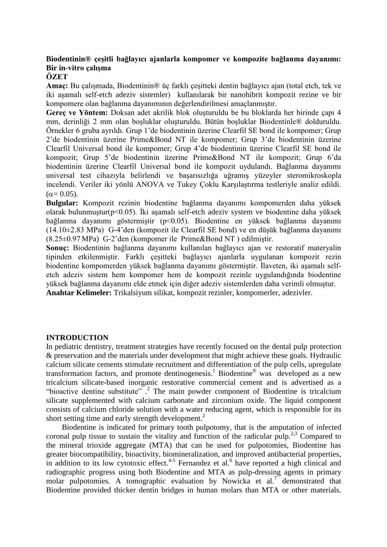
Table 3. Mean values and standard deviation (SD) of shear bond sterngth of tested material to
Biodentine® using three adhesive systems
Bonding system Restorative Material
Compomer resin Composite resin
Group/Mean±SD Group/Mean±SD
Prime&Bond NT G-2/8.25±0.97a
G-5/10.65±1.74b
Clearfil Universal Bond G-3/9.66±2.26b
G-6/11.52±2.77b
Clearfil SE Bond G-1/10.74±1.17b
G-4/14.10±2.83c
p <0.05
Figures
Figure 1. Fracture mode distribution of the specimens according to groups.

Figure 2: Steriomicroscopic imaging of the failure modes. (a) Cohesive failure in Biodentine;
(b) Mix failure; (c )Adhesive failure.