No-bottle’ vs ‘multi-bottle’ dentin adhesives—a microtensile bond strength and morphological...
8
‘No-bottle’ vs ‘multi-bottle’ dentin adhesives—a microtensile bond strength and morphological study R. Frankenberger a , J. Perdiga ˜o b, * , B.T. Rosa c,1 , M. Lopes d a Policlinic for Operative Dentistry and Periodontology, University of Erlangen-Nuremberg, Glueckstrasse 11, D-91054 Erlangen, Germany b University of Minnesota, Division of Operative Dentistry and Minnesota Dental Research Center for Biomaterials and Biomechanics, 8-450 Moos Tower, 515 Delaware St. SE, Minneapolis, MN 55455, USA c Former Graduate Student, University of North Carolina at Chapel Hill, Department of Operative Dentistry, CB no. 7450 Brauer Hall, Chapel Hill, NC 27599- 7450, USA d Minnesota Dental Research Center for Biomaterials and Biomechanics, 16-212 Moos Tower, 515 Delaware St. SE, Minneapolis, MN 55455, USA Received 11 April 2000; revised 18 August 2000; accepted 30 August 2000 Abstract Purpose: To compare the adhesive capability of the new adhesive Prompt L-Pop (ESPE) with that of two total-etch adhesive systems— EBS Multi (ESPE) and Prime&Bond NT (Dentsply). Methods: Extracted human molars were bonded and prepared for microtensile dentin bond strength (mTBS) testing using Prompt L-Pop, EBS Multi, and Prime&Bond NT combined with Pertac II (composite) or Hytac Aplitip (compomer). Prompt L-Pop was applied using five different protocols: (1) as an ‘all-in-one’ self-conditioning adhesive, as per manufacturer’s instructions (LP); (2) as a self-etching primer combined with a separate bonding resin (LP/self-etch 2-step); (3) as a classical primer used upon etching dentin with phosphoric acid, followed by a bonding resin (LP/total-etch 3-step); (4) as a multi-application ‘all-in-one‘ self-conditioning adhesive (LP/multi-coat) to leave a visibly glossy dentin surface; and (5) as a filled adhesive, upon adding quartz fillers to its composition (LP/filled). After 24 h of storage in water at 378C the mTBS were measured in an Instron machine. Corresponding interfaces of the same specimens were micromorphologically analyzed using SEM and TEM. Results: When used with a composite resin, LP/filled and LP/multi-coat resulted in significantly higher mTBS than LP. The addition of an extra adhesive bonding resin (LP/self-etch 2-step) had no effect on bond strength. The use of Prompt L-Pop as a primer of a fourth-generation adhesive (LP/total-etch 3-step) replacing the EBS Multi primer, resulted in lower bond strengths than those of the original EBS Multi. LP/ multi-coat showed similar mTBS to Prime&Bond NT (P&BNT). When used with a compomer, LP exhibited higher bond strengths than when used with the resin composite and was as effective as the experimental groups LP/filled and LP/multi-coat and the control group P&BNT. The SEM evaluation showed an inconsistent hybrid layer for the LP specimens, whereas in both the LP/filled and LP/multi-coat specimens a hybrid layer was clearly evident. Under the TEM all groups displayed dentin hybridization with dissolved smear plugs in the specimens that had been conditioned with Prompt L-Pop without a separate etching step. In many tubules of specimens conditioned with LP (as per manufacturer’s instructions), fillers of the resin composite were present within the dentinal tubules. Conclusions: When combined with a resin composite, Prompt L-Pop resulted in statistically lower bond strengths when applied in one layer than when applied in multiple layers. Prompt L-Pop also resulted in higher bond strengths when used with a polyacid-modified composite resin than with a composite resin. When applied in multiple coats, Prompt L-Pop results in bond strengths that are not statistically different from those of Prime&Bond NT, a total-etch adhesive. q 2001 Academy of Dental Materials. Published by Elsevier Science Ltd. All rights reserved. 1. Introduction The increasing demand for esthetic restorations has generated intensive research on adhesive materials with special focus on amalgam alternatives [1–3]. Successful adhesion to dental hard tissues is a fundamental requirement prior to the insertion of tooth-colored materials such as direct resin composites [4–7]. The polymerization shrink- age of resin composites produces stress between bonded restorations and tooth walls, potentially resulting in gap formation [8–10]. Only a perfect adaptation is capable of preventing microleakage and, consequently, recurrent caries and pulpal irritation [5,10]. Bonding to enamel has been known as being clinically Dental Materials 17 (2001) 373–380 dental materials 0109-5641/01/$20.00 + 0.00 q 2001 Academy of Dental Materials. Published by Elsevier Science Ltd. All rights reserved. PII: S0109-5641(00)00084-1 www.elsevier.com/locate/dental * Corresponding author. Tel.: 11-612-625-8486; fax: 11-612-625-7440. E-mail address: [email protected] (J. Perdiga ˜o). 1 Currently in Private Practice in Londrina, Parana ´, Brazil.
Transcript of No-bottle’ vs ‘multi-bottle’ dentin adhesives—a microtensile bond strength and morphological...

`No-bottle' vs `multi-bottle' dentin adhesivesÐa microtensile bondstrength and morphological study
R. Frankenbergera, J. PerdigaÄob,*, B.T. Rosac,1, M. Lopesd
aPoliclinic for Operative Dentistry and Periodontology, University of Erlangen-Nuremberg, Glueckstrasse 11, D-91054 Erlangen, GermanybUniversity of Minnesota, Division of Operative Dentistry and Minnesota Dental Research Center for Biomaterials and Biomechanics, 8-450 Moos Tower,
515 Delaware St. SE, Minneapolis, MN 55455, USAcFormer Graduate Student, University of North Carolina at Chapel Hill, Department of Operative Dentistry, CB no. 7450 Brauer Hall, Chapel Hill, NC 27599-
7450, USAd Minnesota Dental Research Center for Biomaterials and Biomechanics, 16-212 Moos Tower, 515 Delaware St. SE, Minneapolis, MN 55455, USA
Received 11 April 2000; revised 18 August 2000; accepted 30 August 2000
Abstract
Purpose: To compare the adhesive capability of the new adhesive Prompt L-Pop (ESPE) with that of two total-etch adhesive systemsÐ
EBS Multi (ESPE) and Prime&Bond NT (Dentsply).
Methods: Extracted human molars were bonded and prepared for microtensile dentin bond strength (mTBS) testing using Prompt L-Pop,
EBS Multi, and Prime&Bond NT combined with Pertac II (composite) or Hytac Aplitip (compomer). Prompt L-Pop was applied using ®ve
different protocols: (1) as an `all-in-one' self-conditioning adhesive, as per manufacturer's instructions (LP); (2) as a self-etching primer
combined with a separate bonding resin (LP/self-etch 2-step); (3) as a classical primer used upon etching dentin with phosphoric acid,
followed by a bonding resin (LP/total-etch 3-step); (4) as a multi-application `all-in-one` self-conditioning adhesive (LP/multi-coat) to leave
a visibly glossy dentin surface; and (5) as a ®lled adhesive, upon adding quartz ®llers to its composition (LP/®lled). After 24 h of storage in
water at 378C the mTBS were measured in an Instron machine. Corresponding interfaces of the same specimens were micromorphologically
analyzed using SEM and TEM.
Results: When used with a composite resin, LP/®lled and LP/multi-coat resulted in signi®cantly higher mTBS than LP. The addition of an
extra adhesive bonding resin (LP/self-etch 2-step) had no effect on bond strength. The use of Prompt L-Pop as a primer of a fourth-generation
adhesive (LP/total-etch 3-step) replacing the EBS Multi primer, resulted in lower bond strengths than those of the original EBS Multi. LP/
multi-coat showed similar mTBS to Prime&Bond NT (P&BNT). When used with a compomer, LP exhibited higher bond strengths than when
used with the resin composite and was as effective as the experimental groups LP/®lled and LP/multi-coat and the control group P&BNT. The
SEM evaluation showed an inconsistent hybrid layer for the LP specimens, whereas in both the LP/®lled and LP/multi-coat specimens a
hybrid layer was clearly evident. Under the TEM all groups displayed dentin hybridization with dissolved smear plugs in the specimens that
had been conditioned with Prompt L-Pop without a separate etching step. In many tubules of specimens conditioned with LP (as per
manufacturer's instructions), ®llers of the resin composite were present within the dentinal tubules.
Conclusions: When combined with a resin composite, Prompt L-Pop resulted in statistically lower bond strengths when applied in one
layer than when applied in multiple layers. Prompt L-Pop also resulted in higher bond strengths when used with a polyacid-modi®ed
composite resin than with a composite resin. When applied in multiple coats, Prompt L-Pop results in bond strengths that are not statistically
different from those of Prime&Bond NT, a total-etch adhesive. q 2001 Academy of Dental Materials. Published by Elsevier Science Ltd. All
rights reserved.
1. Introduction
The increasing demand for esthetic restorations has
generated intensive research on adhesive materials with
special focus on amalgam alternatives [1±3]. Successful
adhesion to dental hard tissues is a fundamental requirement
prior to the insertion of tooth-colored materials such as
direct resin composites [4±7]. The polymerization shrink-
age of resin composites produces stress between bonded
restorations and tooth walls, potentially resulting in gap
formation [8±10]. Only a perfect adaptation is capable of
preventing microleakage and, consequently, recurrent caries
and pulpal irritation [5,10].
Bonding to enamel has been known as being clinically
Dental Materials 17 (2001) 373±380
dentalmaterials
0109-5641/01/$20.00 + 0.00 q 2001 Academy of Dental Materials. Published by Elsevier Science Ltd. All rights reserved.
PII: S0109-5641(00)00084-1
www.elsevier.com/locate/dental
* Corresponding author. Tel.: 11-612-625-8486; fax: 11-612-625-7440.
E-mail address: [email protected] (J. PerdigaÄo).1 Currently in Private Practice in Londrina, ParanaÂ, Brazil.

very reliable. Since its introduction in 1955, the acid-etch-
technique has provided an ideal surface morphology as a
result of the use of 30±40% phosphoric acid [2,11]. The
resulting etch pattern is characterized by abundant micro-
porosities which allow the penetration of polymerizable
monomers into those porosities to form resin tags that
provide micromechanical retention [2].
Successful attempts of bonding to dentin in a similar
fashion have been reported more recently [12±15]. Due to
the speci®c properties of human dentin, such as the tubular
structure and its intrinsic wetness, bonding to dentin is more
dif®cult to achieve than bonding to enamel [16±19]. The
bonding mechanism of recent dentin bonding agents is
based on the penetration of ambiphilic molecules into
acid-etched dentin [20±22]. Water-chasing solvents, such
as ethanol or acetone, are commonly utilized to facilitate
penetration of the monomers and to obtain a direct contact
of resin with the collagen ®bers, which results in a mixed
zone of polymerized resin and entangled collagen ®brils, the
hybrid layer [12,13,20]. Dentin bonding agents that rely on
dentin hybridization have been reported to result in higher
bond strengths especially when the dentin is left moist rather
than being desiccated with air [12,13].
A different philosophy is advocated by `self-etching
primers', which have been evaluated in several studies
[24±26]. Self-etching primers condition and prime enamel
and dentin simultaneously without rinsing. The mild acidity
of these materials is responsible for their inability to remove
the smear plugs upon conditioning. Nevertheless, self-etch-
ing primers hybridize dentin for up to 2 mm and have been
reported to withstand stresses from polymerization shrink-
age clinically [27]. These materials have the ability to
dissolve hydroxyapatite partially, both within the smear
layer and the dentin surface, resulting in a resin-in®ltrated
zone with entrapped minerals [10,17]. These attempts at
creating adhesion to dentin without a separate etching
step may be a noteworthy alternative to the total etch
technique.
The simultaneous treatment of enamel and dentin with one
material in one application is a major step towards simpli®ca-
tion of the clinical procedure with the ®nal goal of decreasing
the possibility of error during the application of current adhe-
sive materials. This ultimate simpli®cation is a new bonding
concept known as `all-in-one'. A new `all-in-one' adhesive,
(Prompt L-Pop, ESPE, Seefeld, Germany), has been reported
to result in enamel bond strengths comparable to those
obtained with total-etch multi-step adhesives [26,28]. Prompt
L-Pop is now recommended to be used with composites and
compomers, which represents a modi®cation of the ®rst
version recommended for compomers exclusively, without a
separate light-curing step.
The null hypothesis tested in this project is two-fold. (1)
The use of Prompt L-Pop as a multi-step adhesive system
does not result in higher bond strengths than when used as
per manufacturer's directions. (2) The bond strengths
obtained with Prompt L-Pop are not signi®cantly lower
than those obtained with either a water- or an acetone-
based total-etch adhesive.
2. Materials and methods
2.1. Specimen preparation and microtensile testing
A total of 32 caries-free extracted human third molars
were used in this investigation. The teeth were stored in a
0.1% thymol solution at room temperature for up to 4 weeks
after extraction and were consecutively debrided and exam-
ined to ensure that they were free of defects. The occlusal
enamel was removed and 700±900 mm thick enamel-dentin
discs were cut from the mid-coronal level of the tooth,
perpendicular to the tooth axis by slow-speed diamond-
saw sectioning (Isomet 1000, Buehler Ltd, Lake Bluff, IL,
USA) under water. A standardized smear layer was created
on both surfaces by wet sanding with 600-grit silicon
carbide abrasive paper (Buehler Ltd) for 60 s. Both surfaces
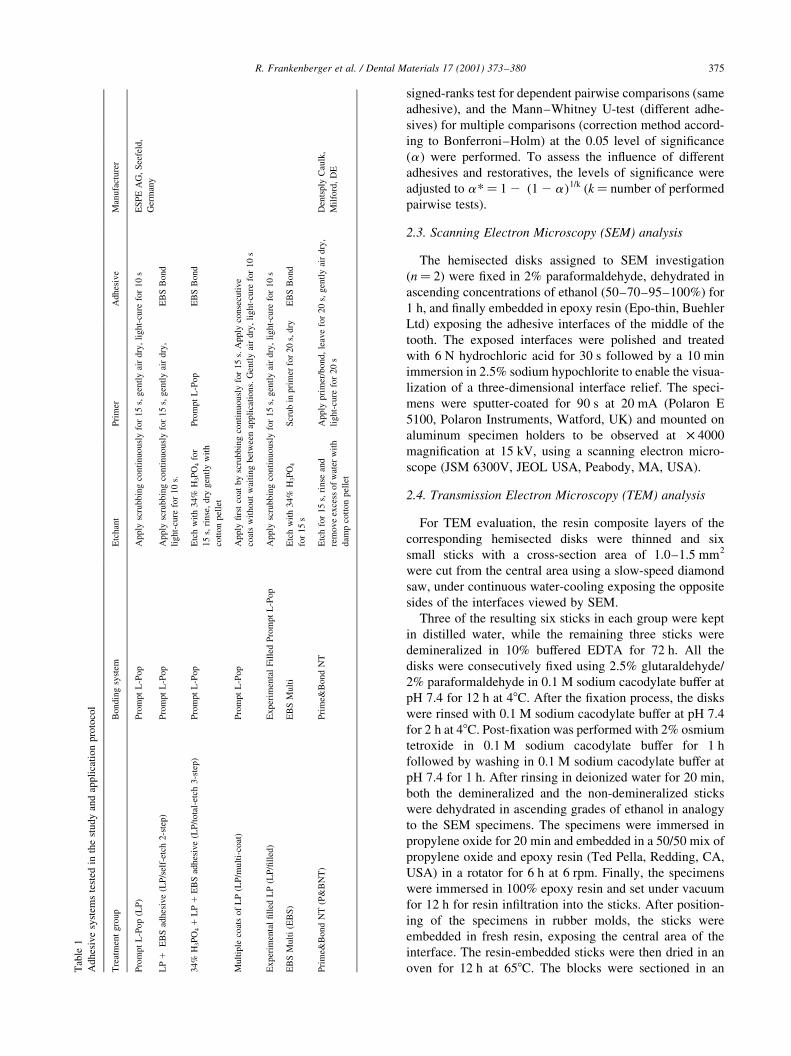
were treated with the dentin adhesive systems (n� 2)
displayed in Table 1. The crowns of the cut teeth were
reconstructed with four consecutive 1 mm-layers of a mini-
®lled hybrid resin composite or a compomer (Pertac II or
Hytac Aplitip, ESPE). Each layer was light-cured for 40 s
with a photocuring unit (Elipar Highlight, ESPE). The inten-
sity of the light was monitored periodically with a radio-
meter (Demetron/Kerr, Danbury, CT, USA) to ensure that
400 mW/cm2 were exceeded. The disks were then cut in
half and stored in distilled water to fabricate the SEM and
the TEM specimens.
The mTBS specimens were stored in distilled water for
24 h at 378C and then sectioned in 800 mm-disks parallel to
the tooth axis. The disks were modi®ed to obtain an hour-
glass shape using a diamond bur mounted in a high-speed
handpiece, under continuous water spray. The area of
the tested interface was adjusted to 1.00±1.25 mm2. The
hourglass shaped specimens were mounted in a special jig
(Bencor Multi-T, Danville Engineering, San Ramon, CA,
USA) with cyanoacrylate cement (Zapit, Dental Ventures
of America, Corona, CA, USA) and debonded using a
universal testing machine (Model 4411, Instron Co., Canton,
MA, USA) with a 50 N load cell traveling at a crosshead
speed of 1 mm/min. Microtensile bond strengths were deter-
mined by computing the ratio of maximum load (N) by the
adhesion area. After the mTBS test, the dentin sides of the
fractured interfaces were analyzed under a stereo light
microscope at 40 £ magni®cation to determine the fracture
mode.
2.2. Statistical analysis
The statistical analysis was performed using a software
package (SPSS 8.0 for Windows, SPSS Inc., Chicago, IL,
USA). The values of bond strength were non-normally
distributed (proved by Kolmogorov±Smirnov test), there-
fore non-parametric tests, the Wilcoxon matched-pairs
R. Frankenberger et al. / Dental Materials 17 (2001) 373±380374
signed-ranks test for dependent pairwise comparisons (same
adhesive), and the Mann±Whitney U-test (different adhe-
sives) for multiple comparisons (correction method accord-
ing to Bonferroni±Holm) at the 0.05 level of signi®cance
(a ) were performed. To assess the in¯uence of different
adhesives and restoratives, the levels of signi®cance were
adjusted to a*� 1 2 (1 2 a)1/k (k� number of performed
pairwise tests).
2.3. Scanning Electron Microscopy (SEM) analysis
The hemisected disks assigned to SEM investigation
(n� 2) were ®xed in 2% paraformaldehyde, dehydrated in
ascending concentrations of ethanol (50±70±95±100%) for
1 h, and ®nally embedded in epoxy resin (Epo-thin, Buehler
Ltd) exposing the adhesive interfaces of the middle of the
tooth. The exposed interfaces were polished and treated
with 6 N hydrochloric acid for 30 s followed by a 10 min
immersion in 2.5% sodium hypochlorite to enable the visua-
lization of a three-dimensional interface relief. The speci-
mens were sputter-coated for 90 s at 20 mA (Polaron E
5100, Polaron Instruments, Watford, UK) and mounted on
aluminum specimen holders to be observed at £ 4000
magni®cation at 15 kV, using a scanning electron micro-
scope (JSM 6300V, JEOL USA, Peabody, MA, USA).
2.4. Transmission Electron Microscopy (TEM) analysis
For TEM evaluation, the resin composite layers of the
corresponding hemisected disks were thinned and six
small sticks with a cross-section area of 1.0±1.5 mm2
were cut from the central area using a slow-speed diamond
saw, under continuous water-cooling exposing the opposite
sides of the interfaces viewed by SEM.
Three of the resulting six sticks in each group were kept
in distilled water, while the remaining three sticks were
demineralized in 10% buffered EDTA for 72 h. All the
disks were consecutively ®xed using 2.5% glutaraldehyde/
2% paraformaldehyde in 0.1 M sodium cacodylate buffer at
pH 7.4 for 12 h at 48C. After the ®xation process, the disks
were rinsed with 0.1 M sodium cacodylate buffer at pH 7.4
for 2 h at 48C. Post-®xation was performed with 2% osmium
tetroxide in 0.1 M sodium cacodylate buffer for 1 h
followed by washing in 0.1 M sodium cacodylate buffer at
pH 7.4 for 1 h. After rinsing in deionized water for 20 min,
both the demineralized and the non-demineralized sticks
were dehydrated in ascending grades of ethanol in analogy
to the SEM specimens. The specimens were immersed in
propylene oxide for 20 min and embedded in a 50/50 mix of
propylene oxide and epoxy resin (Ted Pella, Redding, CA,
USA) in a rotator for 6 h at 6 rpm. Finally, the specimens
were immersed in 100% epoxy resin and set under vacuum
for 12 h for resin in®ltration into the sticks. After position-
ing of the specimens in rubber molds, the sticks were
embedded in fresh resin, exposing the central area of the
interface. The resin-embedded sticks were then dried in an
oven for 12 h at 658C. The blocks were sectioned in an
R. Frankenberger et al. / Dental Materials 17 (2001) 373±380 375T
able
1
Ad
hes
ive
syst
ems
test
edin
the
stu
dy
and
app
lica
tio
np
roto
col
Tre
atm
ent
gro
up
Bondin
gsy
stem
Etc
han
tP
rim
erA
dhes
ive
Man
ufa
cture
r
Pro
mpt
L-P
op
(LP
)P
rom
pt
L-P
op
Apply
scru
bbin
gco
nti
nuousl
yfo
r15
s,gen
tly
air
dry
,li
ght-
cure
for
10
sE
SP
EA
G,
See
feld
,
Ger
man
y
LP
1E
BS
adhes
ive
(LP
/sel
f-et
ch2-s
tep)
Pro
mpt
L-P
op
Apply
scru
bbin
gco
nti
nuousl
yfo
r15
s,gen
tly
air
dry
,
light-
cure
for
10
s.
EB
SB
ond
34%
H3P
O4
1L
P1
EB
Sad
hes
ive
(LP
/tota
l-et
ch3-s
tep)
Pro
mpt
L-P
op
Etc
hw
ith
34%
H3P
O4
for
15
s,ri
nse
,dry
gen
tly
wit
h
cott
on
pel
let
Pro
mpt
L-P
op
EB
SB
ond
Mult
iple
coat
sof
LP
(LP
/mult
i-co
at)
Pro
mpt
L-P
op
Apply
®rs
tco
atby
scru
bbin
gco
nti
nuousl
yfo
r15
s.A
pply
conse
cuti
ve
coat
sw
ithout
wai
ting
bet
wee
nap
pli
cati
ons.
Gen
tly
air
dry
,li
ght-
cure
for
10
s
Exper
imen
tal
®ll
edL
P(L
P/®
lled
)E
xper
imen
tal
Fil
led
Pro
mpt
L-P
op
Apply
scru
bbin
gco
nti
nuousl
yfo
r15
s,gen
tly
air
dry
,li
ght-
cure
for
10
s
EB
SM
ult
i(E
BS
)E
BS
Mult
iE
tch
wit
h34%
H3P
O4
for
15
s
Scr
ub
inpri
mer
for
20
s,dry
EB
SB
ond
Pri
me&
Bond
NT
(P&
BN
T)
Pri
me&
Bond
NT
Etc
hfo
r15
s,ri
nse
and
rem
ove
exce
ssof
wat
erw
ith
dam
pco
tton
pel
let
Apply
pri
mer
/bond,
leav
efo
r20
s,gen
tly
air
dry
,
light-
cure
for
20
s
Den
tsply
Cau
lk,
Mil
ford
,D
E

ultra-microtome (MT 2-B Ultramicrotome, Ivan Sorvall
Inc., Norwalk, CT, USA), and the demineralized sections
were additionally stained with 2% uranyl acetate for 10 min.
and 3% lead citrate for 5 min.
After drying at room temperature, the sections were
analyzed using a Transmission Electron Microscope
(Philips CM-12, Philips Electronic Instruments Inc.,
Mahwah, NJ, USA) at an accelerating voltage of 100 kV.
3. Results
3.1. Microtensile bond strength
When Prompt L-Pop was dried with air to evaporate the
water used as solvent, dull areas were often apparent on the
cut and smeared tooth surface, which, under magni®cation
glasses looked like dry spots. An overview of the results is
shown in Table 2. When used with composite, LP resulted in
lower mean bond strengths than when used with a compomer.
The use of an additional bonding resin (LP/self-etch 2-step)
revealed no positive effect on bond strength (P . 0.05),
whereas phosphoric acid etching prior to the application of
Prompt L-Pop as conventional primer (LP/total-etch 3-step)
followed by a bonding resin, resulted in an increase in bond
strengths (P , 0.05). Both LP/®lled and LP/multi-coat groups
resulted in signi®cantly higher mTBS than Prompt L-Pop
applied as per manufacturer's instructions (LP) at P , 0.05.
Prime & Bond NT (P&BNT) resulted in similar mean bond
strengths to LP/multi (P . 0.05). EBS Multi resulted in statis-
tically higher dentin bond strengths than any other experimen-
tal group.
For the groups restored with the compomer, Prompt L-
Pop behaved somewhat differently as compared to the
groups restored with composite. LP (material used as per
manufacturer's instructions) achieved statistically similar
mTBS to LP/multi-coat and LP/®lled at P . 0.05. L-Pop
resulted in higher mTBS than LP/self-etch 2-step (group
with additional application of the EBS Multi adhesive)
and LP with the resin composite at P , 0.05. LP/total-
etch 3-step with the compomer showed lower m-TBS than
with the resin composite. Experimental applications of
Prompt L-Pop alone (LP/multi-coat, LP/®lled) revealed
similar bonding behavior compared with P&BNT at
P . 0.05. EBS Multi resulted in signi®cantly higher bond
strengths than the specimens bonded with LP at P , 0.05.
All specimens exhibited adhesive interface failure.
3.2. Electron microscopy
The SEM evaluation showed no microscopical presence of
an acid resistant hybrid layer for the LP specimens (Fig. 1).
However, the specimens treated with Prompt L-Pop in multi-
ple layers (LP/multi-coat) exhibited a distinct hybrid zone
approximately 1 mm thick (Fig. 2). The ®lled experimental
version of the Prompt L-Pop adhesive (LP/®lled) resulted in
a thicker and more consistent hybrid layer of 1±2 mm thick-
ness (Fig. 3) and a visible layer of resin between the hybrid
zone and the restorative material (Fig. 3). The application of
one and multiple coats of Prompt L-Pop resulted in a barely
visible adhesive layer in the micrographs (Figs. 1 and 2).
P&BNT and EBS Multi exhibited similar morphology under
the SEM, showing hybrid layers approximately 3±4 mm thick
and the characteristic triangular peritubular hybridization
(Figs. 4 and 5).
The TEM investigation exhibited the formation of a
hybrid layer for all groups. The interfaces produced by the
use of Prompt L-Pop, in accordance with the manufacturer's
protocol, showed characteristic features. In some tubule
ori®ces the dissolved smear plugs appear as voids within
the resin-®lled tubule (Fig. 6). Intertubular and triangular
peritubular in®ltration of demineralized dentin appears thin-
ner compared with PBNT (not shown) (Fig. 6). An adhesive
layer between hybrid zone and onlying resin composite was
not detectable, therefore most of the funnel-shaped tubular
ori®ces were ®lled with ®llers of the restorative material to a
depth of up to 3 mm. The magni®cation of a hybridized
region at a tubule ori®ce of an LP specimen shows a super-
®cial area of lower electron density and a deeper zone with
more pronounced electron density (Fig. 7). The analysis of
the LP/multi-coat specimens revealed a superior saturation
of the upper half of the hybrid layer combined with visible
penetration of lateral branches (Fig. 8), and less penetration
of ®llers from the restoratives into the tubules. The control
groups showed thicker hybrid layers in analogy to the SEM
observations.
R. Frankenberger et al. / Dental Materials 17 (2001) 373±380376
Table 2
Results of microtensile bond strength test for different restorative materials [MPa] (SD). Asterisks show signi®cant differences between groups with the same
adhesive for bonding of resin composite and compomer (Wilcoxon test, P , 0.05). Groups with same superscript letters among one restorative material are not
statistically different (Mann±Whitney U-test, P . 0.05)
Pertac II Signi®cance (P , 0.05) Hytac ApliTip
LP 5.2 (6.1)D * 13.5 (6.3)c
LP/total-etch 2-step 11.3 (9.6)CD n.s. 6.9 (5.1)d
LP/total-etch 3-step 18.6 (6.9)B * 12.1 (3.3)c
LP/multi-coat 16.4 (5.0)BC n.s. 18.1 (6.3)bc
LP/®lled 12.8 (5.7)C n.s. 16.9 (6.9)bc
P&BNT 19.0 (3.9)B n.s. 19.4 (7.6)b
EBS 28.9 (8.7)A n.s. 23.8 (6.3)ab

4. Discussion
This in vitro study was design to clarify the effect of
alternative application protocols and ®ller content of a
new dentin-bonding agent on dentin bond strength and
micromorphological appearance of the resin±dentin inter-
face. Prompt L-Pop is a recent attempt to the simpli®cation
of the rather complicated methods for bonding to dentin.
This may be advantageous with respect to the technique
sensitivity claimed for modern dentin bonding agents
[29,30]. Almost all results presently available concerning
in vitro performance of dentin bonding systems were carried
out in strict accordance with the manufacturers' recommen-
dations. However, Ciucchi demonstrated that different
general dental practitioners achieved rather inconsistent
dentin shear bond strengths even when using identical
adhesive systems [30]. Therefore it is an obvious assump-
tion that incorrect application procedures may affect dentin
bonding behaviour, as reported by Peschke for the particular
adhesive system OptiBond FL (Kerr Co., Orange, CA) [31].
On the other hand, packing all required qualities of an
effective dentin adhesive such as wettability, acidity,
capability of penetration, and ®nally cohesive strength and
loading capacity into one chemical composition may be
R. Frankenberger et al. / Dental Materials 17 (2001) 373±380 377
Fig. 1. Scanning electron micrograph of interface bonded with Prompt
L-Pop in one layer (®nal magni®cation: £ 4000). Resin tags (T) are present
with lateral branches (asterisks). No acid-resistant hybrid layer is detect-
able. The resin composite (RC) lies directly on the dentin without an inter-
mediate adhesive layer (circle).
Fig. 2. Scanning electron micrograph of a specimen bonded with Prompt L-
Pop with multiple coats (®nal magni®cation: £ 4000). Note the thin 1mm
thick hybrid layer (H, arrowheads). Resin tags (T) are present revealing
lateral branches (asterisk).
Fig. 3. Scanning electron micrograph of interface bonded with Prompt L-
Pop in the experimentally ®lled version (®nal magni®cation: £ 4000). Note
the visible thickness of the adhesive layer (A, arrowheads) beneath the resin
composite (RC). The hybrid layer (H, arrowheads) is 2 mm thick. The
tubular resin tags (T) show lateral branches (asterisks).
Fig. 4. SEM resin±dentin interface formed by Prime&Bond NT (®nal
magni®cation: £ 4000) The hybrid layer (H, arrowheads) is approximately
4 mm thick. The resin tags show a triangular shape at the entrances (T) and
lateral branches (asterisks).

hard to accomplish. As reported for total-etch one-bottle
adhesives, the compromise of wettablity and stability
seems to result in an overall poorer adhesive performance
compared with multi-step systems. The latter have different
chemical compositions for priming and bonding the acid-
conditioned dentin [17,23,32]. Nevertheless, clinicians
prefer materials with an easy application technique when
performing adhesive dentistry in daily practice; therefore
all of these simpli®ed products are usually accepted with
enthusiasm by clinicians [33,34].
First results for Prompt L-Pop, initially marketed as a
simpli®ed bonding agent for compomers only, were very
promising [22,26,28,34,35]. All the reported high bond
strengths to enamel have been recognized as encouraging,
due to the fact that the resulting etch pattern in enamel
showed a nearly identical performance to that caused by
etching with phosphoric acid [34]. The version of Prompt
L-Pop used in the present project is now marketed in an
improved formula for bonding compomers and resin
composites as well, therefore, both restorative materials
were tested. The evaluation methods carried out in the
present study are commonly accepted. Microtensile bond
R. Frankenberger et al. / Dental Materials 17 (2001) 373±380378
Fig. 5. SEM interface produced by EBS Multi (®nal magni®cation: £4000).
The interface shows a pronounced appearance with a hybrid layer (H), resin
tags (T) and short lateral branches (asterisks).
Fig. 6. Transmission electron micrograph of interface bonded with Prompt
L-Pop in one layer (non-decalci®ed specimen, ®nal magni®cation:
£ 6300). The presence of ®llers within the tubule ori®ce is evident. The
resin-in®ltrated (H) zone with a thickness ,1 mm extends from the inter-
tubular to the peritubular area where the penetration is characteristically
triangular. D� dentin.
Fig. 7. Higher magni®cation of a hybridized zone at a tubule ori®ce produced
by Prompt L-Pop according to the manufacturer' recommendations
(decalci®ed specimen, £ 17,000). The upper half of the hybrid layer (H)
appears to be less saturated with adhesive compared to the bottom half of the
penetrated demineralized dentin with higher electron density. D� dentin.
Fig. 8. Transmission electron micrograph of interface bonded with Prompt
L-Pop in multiple coats (®nal magni®cation: £ 28,000). The hybrid layer
(H) is more saturated than in Fig. 7. Note the fully hybridized lateral (H)
branch below the bottom of the hybrid layer (asterik). D� dentin.

strength testing enables the investigation of interfacial
bonds with reduced probability of pulling out dentin from
the ¯at surface similar to that often recorded when testing in
shear or conventional tensile mode [36±38]. The analysis of
the debonded dentin samples clearly demonstrated that
particular advantage compared to other bond strength test-
ing methodologies. The interfacial analysis by use of SEM
and TEM represents an ideal complement to the mTBS
method allowing us to look into the clues of micro- and
ultra-morphological appearances of the different attempts
for successful bonding to human dentin [21,35]. To have a
valid control for the demineralized TEM samples, non-
decalci®ed specimens were also evaluated (Fig. 6) [39].
The behavior of Prompt L-Pop exhibited a non-uniform
characteristic. During the application of Prompt L-Pop on
¯at dentin surfaces a major problem became clearly evident.
After applying the material for 15 s, LP has to be dried with
air to evaporate the water used as solvent. If this procedure
is carried out carefully, dull areas are often apparent on the
cut and smeared tooth surface, representing dry spots where
the material is probably too thin for being successfully
photopolymerized. The dry spots may have been responsi-
ble for the areas without the interfacial characteristics of
hybridization under the SEM (Fig. 1). TEM investigation
disclosed lower resin saturation in the upper half of the
hybrid layer, which may be attributed to oxygen inhibition
of the thin resin layer after evaporating the water, prevent-
ing the adhesive from polymerizing. There have been
reports in the literature showing that additional coats of
the adhesive system may solve this particular problem
[32,33]. The observation of dry spots was indeed completely
prevented by applying multiple coats of the bonding mate-
rial after drying the solvent until the whole dentin surface
became thoroughly glossy. As a result of multiple coats and
immediate light curing, an adequate and apparently poly-
merizable resin layer is created, resulting in signi®canlty
higher mTBS for the LP/multi-coat groups, which also stres-
ses the need for light curing prior to inserting the restorative.
The same effect was evident when the adhesive was modi-
®ed with quartz ®llers. The noticeably higher viscosity of
the experimentally ®lled Prompt L-Pop may have prevented
the formation of dry spots on the surface and may have
formed an adhesive layer above the hybrid layer. The
SEM disclosed a hybrid layer thicker than that in the other
groups treated with Prompt L-Pop without ®llers. Addition-
ally, the TEM analysis of the interfaces bonded with Prompt
L-Pop applied in one layer (LP) demonstrated an extremely
thin appearance exhibiting ®ller particles of the resin compo-
site plugged into the dentinal tubules during the application of
the material onto the ¯at dentin surface (Fig. 6).
The application of the more hydrophilic polyacid-modi®ed
composite Hytac Aplitip [28] to dentin showed that LP bonds
better to compomers than to the more hydrophobic resins, such
as composites. This is demonstrated by the results of Prompt
L-Pop used according to the manufacturer's protocol (LP).
Although the same problems with dry spots occurred during
the application procedure, the effect seems to be compensated
by a possible greater chemical af®nity of Prompt L-Pop to
Hytac than to the corresponding composite [28]. This might
explain the fact that the experimentally modi®ed groups show
no increase in bond strength.
The use of the EBS un®lled resin resulted in detrimental
effects for Prompt L-Pop regardless of additional phospho-
ric acid etching. When used with EBS alone, the compomer
also exhibited lower bond strengths than when bonded to the
resin composite Pertac II. This might be attributed to some
kind of chemical incompatibility between the EBS bonding
agent and the polyacid-modi®ed material Hytac Aplitip.
This study clari®es the fact that total-etch adhesive
systems, in which a separate phosphoric acid etching step
is used prior to priming or combined priming and bonding,
still represent the dominant method to achieve high bond
strengths to dentin. The bene®cial effect of light-curing the
bonding for the adhesive performance of resin composites
was previously reported [23]. Prompt L-Pop is now used
with composites, which represents a modi®cation of the
®rst version recommended for compomers exclusively with-
out a separate light-curing step.
Altogether, the present mTBS and micromorphological
results point out low bond strengths and narrow ®lm thick-
ness of the all-in-one self-conditioning adhesive when
applied in one layer. However, either the addition of quartz
®llers or the additional application of layers onto the dentin
surface demonstrated to be capable of compensating the
tendency towards the formation of dry spots and consecu-
tively incomplete penetration of the dentin surface. The
ultramorphological analysis con®ms the assumptions gener-
ated by the results of the mTBS investigation. Among the
modi®ed application protocols carried out in the course of
this study, only multiple coating or ®ller addition showed
the desired effect on more saturation within the resin±dentin
interface (higher electron-density, as observed in Fig. 8) as
well as higher mTBS.
The null hypotheses were rejected. (1) The use of Prompt
L-Pop as a multi-step adhesive system resulted in higher
bond strengths than when used as per manufacturer's direc-
tions. (2) When applied in multiple coats, Prompt L-Pop
resulted in bond strengths that were not statistically different
from those of P&BNT, a total-etch adhesive. However, the
bond strengths obtained with EBS Multi, a water-one-based
total-etch adhesive, were signi®cantly higher than those
obtained with Prompt L-Pop.
Acknowledgements
This research project was supported by a grant sponsored
by ESPE, Seefeld, Germany, as the ESPE Dental Talent
Award 1998 `Young Lecturer of the Year'. We are grateful
to Wallace Ambrose at the Electron Microscopy Labora-
tory, Dental Research Center, University of North Carolina
at Chapel Hill for his helpful comments.
R. Frankenberger et al. / Dental Materials 17 (2001) 373±380 379

References
[1] PerdigaÄo J, Lambrechts P, Van Meerbeek B, Braem M, Yildiz E,
Yucel T, Vanherle G. The interaction of adhesive systems with
human dentin. Am J Dent 1996;9:167±73.
[2] Swift EJ, PerdigaÄo J, Heymann HO. Bonding to enamel and dentin: a
brief history and state of the art, 1995. Quintessence Int 1995;26:95±
110.
[3] Wilson NHF, Dunne SM, Gainsford ID. Current materials and tech-
niques for direct restorations in posterior teeth. Part 2: resin composite
systems. Int Dent J 1998;47:185±93.
[4] Eick JD, Cobb CM, Chappel P, Spencer P, Robinson SJ. The dentinal
surface: its in¯uence on dentinal adhesion. Part I. Quintessence Int
1991;22:967±77.
[5] Eick JD, Gwinnett AJ, Pashley DH, Robinson SJ. Current concepts on
adhesion to dentin. Crit Rev Oral Biol Medic 1997;8:306±35.
[6] Pashley DH, Sano H, Ciucchi B, Yoshiyama M, Carvalho RM.
Adhesion testing of dentin bonding agents: a review. Dent Mater
1995;11:117±25.
[7] PerdigaÄo J. An ultramorphological study of human dentin exposed to
adhesive systems. PhD thesis, Catholic University of Leuven,
Belgium, 1995.
[8] Davidson CL, de Gee AJ, Feilzer AJ. The competition between the
composite dentin bond strength and the polymerization contraction
stress. J Dent Res 1984;63:1396±9.
[9] Finger WJ, Fritz U. Laboratory evaluation of one-component enamel/
dentin bonding agents. Am J Dent 1996;9:206±10.
[10] Van Meerbeek B, PerdigaÄo J, Lambrechts P, Vanherle G. The clinical
performance of adhesives. J Dent 1998;26:120.
[11] Buonocore MG. A simple method of increasing the adhesion of
acrylic ®lling materials to enamel surfaces. J Dent Res
1955;34:849±54.
[12] Kanca J. Resin bonding to wet substrate. I. Bonding to dentin. Quin-
tessence Int 1992;23:39±41.
[13] Nakabayashi N, Kojima K, Masuhara E. The promotion of adhesion
by the in®ltration of monomers into tooth substrates. J Biomed Mater
Res 1992;16:265±73.
[14] PerdigaÄo J, Swift EJ, Denehy GE, Wefel JS, Donly KJ. In vitro bond
strengths and SEM evaluation of dentin bonding systems to different
dentin substrates. J Dent Res 1994;73:44±55.
[15] Sugizaki J. The effect of various primers on the dentin adhesion of
resin composites. SEM and TEM observations of the resin-impreg-
nated layer and adhesion promoting effects of the primers. Jap J Soc
Cons Dent 1991;34:228±65.
[16] Balooch M, Wu-Magidi IC, Balazs A, Lundkvist AS, Marshall SJ,
Marshall GW, Siekhaus WJ, Kinney JH. Viscoelastic properties of
demineralized human dentin measured in water with an atomic force
microscope (AFM) based indentation. J Biomed Mater Res
1998;40:539±44.
[17] Frankenberger R, KraÈmer N, Petschelt A. Fatigue behaviour of differ-
ent dentin adhesives. Clin Oral Invest 1999;3:11±17.
[18] Tay FR, Gwinnett JA, Wei SH. Micromorphological spectrum of
acidconditioned dentin following the application of a water-based
adhesive. Dent Mater 1998;14:329338.
[19] Walshaw PR, McComb D. Clinical considerations for optimal
dentinal bonding. Quintessence Int 1996;27:619±25.
[20] Nakabayashi N, Ashizawa M, Nakamura M. Identi®cation of a resin±
dentin hybrid layer in vital human dentin created in vivo: durable
bonding to vital dentin. Quintessence Int 1992;23:135±41.
[21] Van Meerbeek B, Yoshida Y, Snauwaert J, Hellemans L, Lambrechts P,
Vanherle G, Wakasa K, Pashley DH. Hybridization effectiveness of a
twostep versus a threestep smear layer removing adhesive system exam-
ined correlatively by TEM and AFM. J Adhes Dent 1999;1:7±23.
[22] Lopes M, PerdigaÄo J, Ambrose WW. Ultramorphological study of
dentin treated with a simpli®ed adhesive. J Dent Res 1999;78:475
(Abstr. no. 2955).
[23] Frankenberger R, Sindel J, KraÈmer N, Petschelt A. Dentin bond
strength and marginal adaptation: Direct composite resins vs. ceramic
inlays. Oper Dent 1999;24:147±55.
[24] Gordan VV, Vargas MA, Cobb DS, Denehy DE. Evaluation of adhe-
sive systems using acidic primers. Am J Dent 1997;10:219±23.
[25] PerdigaÄo J, Lopes L, Lambrechts P, LeitaÄo J, Van Meerbeek B,
Vanherle G. Effects of a self-etching primer on enamel shear bond
strengths and SEM morphology. Am J Dent 1997;10:141±6.
[26] Nunes MF, PerdigaÄo J, Rosa BT. The effect of an experimental one-
application self-conditioning adhesive on microleakage. J Dent Res
1999;78:306 (Abstr. no. 1602).
[27] Opdam NJM, Roeters JJM, Feilzer AJ, Verdonschot EH. Marginal
integrity and postoperative sensitivity in class 2 resin composite
restorations in vivo. J Dent 1998;26:555±62.
[28] Rosa BT, PerdigaÄo J. Bond strengths of nonrinsing adhesives. Quin-
tessence Int 2000;31:353±8.
[29] Ciucchi B, Bouillaguet S, Russell C. Dentin bonding by general prac-
titioners. J Dent Res 1997; 76:136 (Abstr. no. 981).
[30] Frankenberger R, KraÈmer N, Petschelt A. Technique sensitivity of
dentin bonding: effect of application mistakes on bond strength and
marginal adaptation. Oper Dent 2000:324±30.
[31] Peschke A, Blunck U, Roulet J-F. In¯uence of incorrect application of
Optibond FL on the marginal adaptation. J Dent Res 1998;77:680
(Abstr. no. 388).
[32] Swift EJ, Wilder AD, May KN, Waddell SL. Shear bond strengths of
one-bottle dentin adhesives using multiple applications. Oper Dent
1997;22:194±9.
[33] Vargas MA, Fortin D, Meckes M. Effect of primer coats on composite
bond strength to dentin. J Dent Res 1995;74:34 (Abstr. no. 182).
[34] PerdigaÄo J, Lopes M. Dentin bonding ± questions or the new millen-
nium. J Adhes Dent 1999;1:191±209.
[35] Blunck U, Roulet J-F. Marginal adaptation of compomer class V
restorations in vitro. J Adhes Dent 1999;1:153±8.
[36] Yoshiyama M, Sano H, Ebisu S, Tagami J, Ciucchi B, Carvalho RM,
Johnson MH, Pashley DH. Regional strengths of bonding agents to
cervical sclerotic root dentin. J Dent Res 1996;75:14041413.
[37] Van Noort R, Noroozi S, Howard IC, Cardew G. A critique of bond
strength measurements. J Dent 1989;17:61±67.
[38] Sano H, Shono T, Sonoda H, Takatsu T, Ciucchi B, Carvalho R,
Pashley DH. Relationship between surface area for adhesion and
tensile bond strengthÐevaluation of a micro-tensile bond test. Dent
Mater 1994;10:236240.
[39] Van Meerbeek B, Conn LJ, Duke ES, Eick JD, Robinson SJ, Guerrero
D. Correlative transmission electron microscopy examination of
nondemineralized and demineralized resin±dentin interfaces. J Dent
Res 1996;75:879888.
R. Frankenberger et al. / Dental Materials 17 (2001) 373±380380