New Single-bottle Ceramic Primer - The Journal of ...
8
The Journal of Contemporary Dental Practice, December 2016;17(12):1033-1039 1033 JCDP ABSTRACT Introduction: This article presents a 6-month case report and an in vitro evaluation of the performance of the new self-etching glass-ceramic monobond etch and prime (MEP) when applied in a lithium disilicate (LD). The MEP contains in the same bottle, along with acid conditioner and silanes. This simplifies the bonding procedures by reducing not only the number of steps, but also the working time. A 42-year-old female patient sought for esthetic treatment, and the main complaint was the darkened appearance of the upper lateral incisor. The esthetic treatment includes dental bleaching and gingivectomy, all-ceramic zirco- nia crowns, and all-ceramic crown and laminate veneer LD. After 6 months of esthetic treatment, marginal staining, gap, or chip fracturing damaging margins after sharp explorer in the margins was not observed. Furthermore, the inferior etching pattern of MEP was compared with traditional hydrofluoric acid (HF) conditioning. The microshear bond strength to the intaglio surface of LD was statistically similar when HF was compared with MEP. This new self-etching glass-ceramic showed good in vitro and 6 months clinical results in this case report. Future long-term clinical studies with more clinical case need to be done to confirm the performance of the use of this new conditioner. Clinical significance: The use of a new self-etching glass- ceramic showed a good in vitro and 6 months clinical results. This new product can be an easy, simple, and alternative approach for esthetics bonding procedure. New Single-bottle Ceramic Primer: 6-month Case Report and Laboratory Performance 1 Fabiana SF Siqueira, 2 Rodrigo S Alessi, 3 Andres FM Cardenas, 4 Carlos Kose, 5 Shelon C Souza Pinto 6 Matheus C Bandeca, 7 Alessandro D Loguercio, 8 João C Gomes 1,7,8 Department of Restorative Dentistry, School of Dentistry Ponta Grossa State University, Ponta Grosa, Paraná, Brazil 2,4 Department of Dentistry, School of Dentistry Ponta Grossa State University, Ponta Grossa, Paraná, Brazil 3 Department of Postgraduate Program in Dentistry, CEUMA University, São Luís, Maranhão, Brazil 5 Department of Dentistry, School of Dentistry, Ponta Grossa State University, Ponta Grosa, Paraná, Brazil 6 Department of Program in Dentistry, CEUMA University, São Luís, Maranhão, Brazil Corresponding Author: Alessandro D Loguercio, Department of Restorative Dentistry, School of Dentistry, Ponta Grossa State University, Ponta Grosa, Paraná, Brazil, e-mail: aloguercio@ hotmail.com Keywords: Dental veneer, Lithium disilicate, Microshear bond strength. How to cite this article: Siqueira FSF, Alessi RS, Cardenas AFM, Kose C, Souza Pinto SC, Bandeca MC, Loguercio AD, Gomes JC. New Single-bottle Ceramic Primer: 6-month Case Report and Laboratory Performance. J Contemp Dent Pract 2016; 17(12):1033-1039. Source of support: Nil Conflict of interest: None INTRODUCTION Contemporarily, for adhesive cementation procedures of silica-based ceramics, the enamel, dentin surfaces, and ceramic surfaces must be properly conditioned. The conventional bonding approach requires the etching preparation of the ceramic surface with hydrofluoric acid (HF) and chemical bond with silane coupling agent. 1 Hydrofluoric acid etching is commonly indicated to partially dissolve the glassy phase and to create microporosities. The HF exposure time depends on the ceramic microstructure and on the HF concentration, which usually ranges between 2.5 and 10%. 2 The HF reacts with the glass matrix, which contains silica, and forms hexafluorosilicates. This glassy matrix is etched, and the crystalline microstructure is revealed, resulting in an increase in the roughness for the ceramic surface, and this is beneficial for the micromechanical retention of the resin cements. 2 Furthermore, the application of a coupling agent (silane) into the preetched internal surface is required. This bifunctional adhesion enhancer creates a chemical interaction between the silica in the glassy phase of the ceramic and the methacrylate groups of the resin through siloxane bonds. 1,3 However, the acid etching with HF may also weaken the ceramic surface, with a progressive increase in CASE REPORT 10.5005/jp-journals-10024-1977
-
Upload
khangminh22 -
Category
Documents
-
view
2 -
download
0
Transcript of New Single-bottle Ceramic Primer - The Journal of ...
New Single-bottle Ceramic Primer: 6-month Case Report and Laboratory Performance
The Journal of Contemporary Dental Practice, December 2016;17(12):1033-1039 1033
JCDP
ABSTRACTIntroduction: This article presents a 6-month case report and an in vitro evaluation of the performance of the new self-etching glass-ceramic monobond etch and prime (MEP) when applied in a lithium disilicate (LD). The MEP contains in the same bottle, along with acid conditioner and silanes. This simplifies the bonding procedures by reducing not only the number of steps, but also the working time. A 42-year-old female patient sought for esthetic treatment, and the main complaint was the darkened appearance of the upper lateral incisor. The esthetic treatment includes dental bleaching and gingivectomy, all-ceramic zirco-nia crowns, and all-ceramic crown and laminate veneer LD. After 6 months of esthetic treatment, marginal staining, gap, or chip fracturing damaging margins after sharp explorer in the margins was not observed. Furthermore, the inferior etching pattern of MEP was compared with traditional hydrofluoric acid (HF) conditioning. The microshear bond strength to the intaglio surface of LD was statistically similar when HF was compared with MEP. This new self-etching glass-ceramic showed good in vitro and 6 months clinical results in this case report. Future long-term clinical studies with more clinical case need to be done to confirm the performance of the use of this new conditioner.
Clinical significance: The use of a new self-etching glass-ceramic showed a good in vitro and 6 months clinical results. This new product can be an easy, simple, and alternative approach for esthetics bonding procedure.
New Single-bottle Ceramic Primer: 6-month Case Report and Laboratory Performance1Fabiana SF Siqueira, 2Rodrigo S Alessi, 3Andres FM Cardenas, 4Carlos Kose, 5Shelon C Souza Pinto 6Matheus C Bandeca, 7Alessandro D Loguercio, 8João C Gomes
1,7,8Department of Restorative Dentistry, School of Dentistry Ponta Grossa State University, Ponta Grosa, Paraná, Brazil2,4Department of Dentistry, School of Dentistry Ponta Grossa State University, Ponta Grossa, Paraná, Brazil3Department of Postgraduate Program in Dentistry, CEUMA University, São Luís, Maranhão, Brazil5Department of Dentistry, School of Dentistry, Ponta Grossa State University, Ponta Grosa, Paraná, Brazil6Department of Program in Dentistry, CEUMA University, São Luís, Maranhão, Brazil
Corresponding Author: Alessandro D Loguercio, Department of Restorative Dentistry, School of Dentistry, Ponta Grossa State University, Ponta Grosa, Paraná, Brazil, e-mail: [email protected]
Keywords: Dental veneer, Lithium disilicate, Microshear bond strength.
How to cite this article: Siqueira FSF, Alessi RS, Cardenas AFM, Kose C, Souza Pinto SC, Bandeca MC, Loguercio AD, Gomes JC. New Single-bottle Ceramic Primer: 6-month Case Report and Laboratory Performance. J Contemp Dent Pract 2016; 17(12):1033-1039.
Source of support: Nil
Conflict of interest: None
INTRODUCTION
Contemporarily, for adhesive cementation procedures of silica-based ceramics, the enamel, dentin surfaces, and ceramic surfaces must be properly conditioned. The conventional bonding approach requires the etching preparation of the ceramic surface with hydrofluoric acid (HF) and chemical bond with silane coupling agent.1
Hydrofluoric acid etching is commonly indicated to partially dissolve the glassy phase and to create microporosities. The HF exposure time depends on the ceramic microstructure and on the HF concentration, which usually ranges between 2.5 and 10%.2 The HF reacts with the glass matrix, which contains silica, and forms hexafluorosilicates. This glassy matrix is etched, and the crystalline microstructure is revealed, resulting in an increase in the roughness for the ceramic surface, and this is beneficial for the micromechanical retention of the resin cements.2
Furthermore, the application of a coupling agent (silane) into the preetched internal surface is required. This bifunctional adhesion enhancer creates a chemical interaction between the silica in the glassy phase of the ceramic and the methacrylate groups of the resin through siloxane bonds.1,3
However, the acid etching with HF may also weaken the ceramic surface, with a progressive increase in
CASE REPORT10.5005/jp-journals-10024-1977
Fabiana SF Siqueira et al
1034
weakening as a function of the concentration used for etching.4-6 There is clear evidence detailing the modifica-tions of surface defects as a function of acid concentration and etching time.5
Besides, the use of HF has been questioned because it is poisonous and caustic, and represents a potential health hazard due to its toxicity and volatility, especially when contacting unprotected skin.7,8 This is a huge problem to intraoral ceramic repair.9 Therefore, the use of HF in lower concentrations or the elimination of this step can be considered advantageous not only in terms of patients’ health but also especially that of clinicians, who often deal with adhesive cementation.
Recently, manufacturer has released more versatile self-etching glass-ceramic primer that shortens the pretreatment of dental restorations, which include acid etching and silane in only one bottle [monobond etch and prime (MEP)], besides being less toxic, and offers less risk to health. Unfortunately, to the extent of authors’ knowledge, no full papers have been published or evalu-ated on MEP. Only abstracts presented in the last IADR Congress were found,10-13 and usually, these studies concluded that bond strength to lithium disilicate (LD) is similar when MEP was compared with conventional HF + silane.
Thus, the aim of this case report is to present the esthetic result of multiple all-ceramic restorations after 6 months of clinical evaluation using LD and the in vitro evaluation of the microshear bond strength and ceramic surface-etching pattern with a new self-etching glass ceramic.
TECHNIQUE DESCRIPTION
Case Description
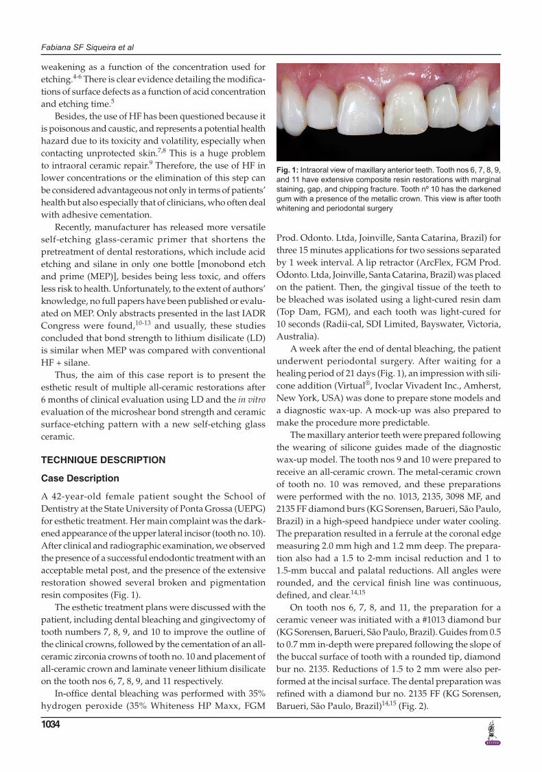
A 42-year-old female patient sought the School of Dentistry at the State University of Ponta Grossa (UEPG) for esthetic treatment. Her main complaint was the dark-ened appearance of the upper lateral incisor (tooth no. 10). After clinical and radiographic examination, we observed the presence of a successful endodontic treatment with an acceptable metal post, and the presence of the extensive restoration showed several broken and pigmentation resin composites (Fig. 1).
The esthetic treatment plans were discussed with the patient, including dental bleaching and gingivectomy of tooth numbers 7, 8, 9, and 10 to improve the outline of the clinical crowns, followed by the cementation of an all-ceramic zirconia crowns of tooth no. 10 and placement of all-ceramic crown and laminate veneer lithium disilicate on the tooth nos 6, 7, 8, 9, and 11 respectively.
In-office dental bleaching was performed with 35% hydrogen peroxide (35% Whiteness HP Maxx, FGM
Prod. Odonto. Ltda, Joinville, Santa Catarina, Brazil) for three 15 minutes applications for two sessions separated by 1 week interval. A lip retractor (ArcFlex, FGM Prod. Odonto. Ltda, Joinville, Santa Catarina, Brazil) was placed on the patient. Then, the gingival tissue of the teeth to be bleached was isolated using a light-cured resin dam (Top Dam, FGM), and each tooth was light-cured for 10 seconds (Radii-cal, SDI Limited, Bayswater, Victoria, Australia).
A week after the end of dental bleaching, the patient underwent periodontal surgery. After waiting for a healing period of 21 days (Fig. 1), an impression with sili-cone addition (Virtual®, Ivoclar Vivadent Inc., Amherst, New York, USA) was done to prepare stone models and a diagnostic wax-up. A mock-up was also prepared to make the procedure more predictable.
The maxillary anterior teeth were prepared following the wearing of silicone guides made of the diagnostic wax-up model. The tooth nos 9 and 10 were prepared to receive an all-ceramic crown. The metal-ceramic crown of tooth no. 10 was removed, and these preparations were performed with the no. 1013, 2135, 3098 MF, and 2135 FF diamond burs (KG Sorensen, Barueri, São Paulo, Brazil) in a high-speed handpiece under water cooling. The preparation resulted in a ferrule at the coronal edge measuring 2.0 mm high and 1.2 mm deep. The prepara-tion also had a 1.5 to 2-mm incisal reduction and 1 to 1.5-mm buccal and palatal reductions. All angles were rounded, and the cervical finish line was continuous, defined, and clear.14,15
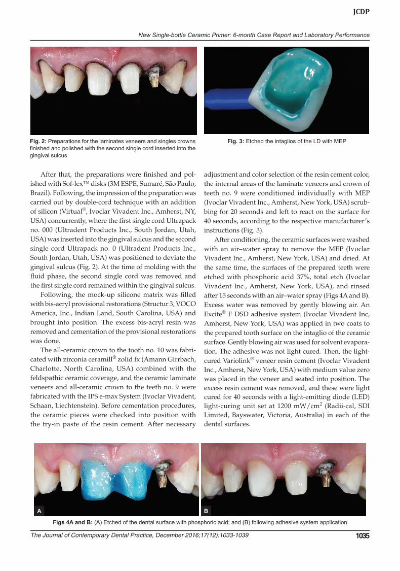
On tooth nos 6, 7, 8, and 11, the preparation for a ceramic veneer was initiated with a #1013 diamond bur (KG Sorensen, Barueri, São Paulo, Brazil). Guides from 0.5 to 0.7 mm in-depth were prepared following the slope of the buccal surface of tooth with a rounded tip, diamond bur no. 2135. Reductions of 1.5 to 2 mm were also per-formed at the incisal surface. The dental preparation was refined with a diamond bur no. 2135 FF (KG Sorensen, Barueri, São Paulo, Brazil)14,15 (Fig. 2).
Fig. 1: Intraoral view of maxillary anterior teeth. Tooth nos 6, 7, 8, 9, and 11 have extensive composite resin restorations with marginal staining, gap, and chipping fracture. Tooth nº 10 has the darkened gum with a presence of the metallic crown. This view is after tooth whitening and periodontal surgery
New Single-bottle Ceramic Primer: 6-month Case Report and Laboratory Performance
The Journal of Contemporary Dental Practice, December 2016;17(12):1033-1039 1035
JCDP
After that, the preparations were finished and pol-ished with Sof-lex™ disks (3M ESPE, Sumaré, São Paulo, Brazil). Following, the impression of the preparation was carried out by double-cord technique with an addition of silicon (Virtual®, Ivoclar Vivadent Inc., Amherst, NY, USA) concurrently, where the first single cord Ultrapack no. 000 (Ultradent Products Inc., South Jordan, Utah, USA) was inserted into the gingival sulcus and the second single cord Ultrapack no. 0 (Ultradent Products Inc., South Jordan, Utah, USA) was positioned to deviate the gingival sulcus (Fig. 2). At the time of molding with the fluid phase, the second single cord was removed and the first single cord remained within the gingival sulcus.
Following, the mock-up silicone matrix was filled with bis-acryl provisional restorations (Structur 3, VOCO America, Inc., Indian Land, South Carolina, USA) and brought into position. The excess bis-acryl resin was removed and cementation of the provisional restorations was done.
The all-ceramic crown to the tooth no. 10 was fabri-cated with zirconia ceramill® zolid fx (Amann Girrbach, Charlotte, North Carolina, USA) combined with the feldspathic ceramic coverage, and the ceramic laminate veneers and all-ceramic crown to the teeth no. 9 were fabricated with the IPS e-max System (Ivoclar Vivadent, Schaan, Liechtenstein). Before cementation procedures, the ceramic pieces were checked into position with the try-in paste of the resin cement. After necessary
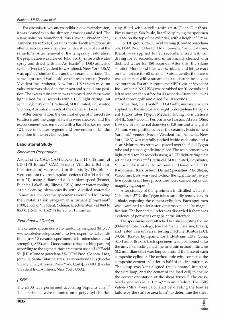
adjustment and color selection of the resin cement color, the internal areas of the laminate veneers and crown of teeth no. 9 were conditioned individually with MEP (Ivoclar Vivadent Inc., Amherst, New York, USA) scrub-bing for 20 seconds and left to react on the surface for 40 seconds, according to the respective manufacturer’s instructions (Fig. 3).
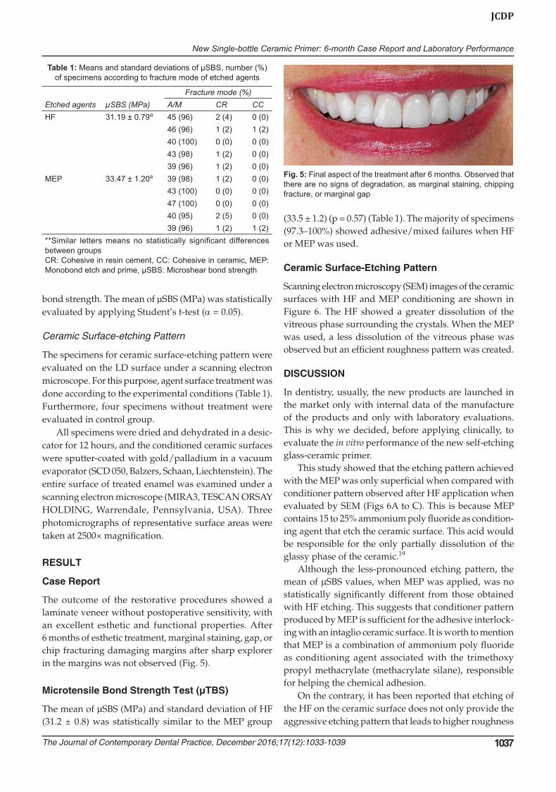
After conditioning, the ceramic surfaces were washed with an air–water spray to remove the MEP (Ivoclar Vivadent Inc., Amherst, New York, USA) and dried. At the same time, the surfaces of the prepared teeth were etched with phosphoric acid 37%, total etch (Ivoclar Vivadent Inc., Amherst, New York, USA), and rinsed after 15 seconds with an air–water spray (Figs 4A and B). Excess water was removed by gently blowing air. An Excite® F DSD adhesive system (Ivoclar Vivadent Inc, Amherst, New York, USA) was applied in two coats to the prepared tooth surface on the intaglio of the ceramic surface. Gently blowing air was used for solvent evapora-tion. The adhesive was not light cured. Then, the light-cured Variolink® veneer resin cement (Ivoclar Vivadent Inc., Amherst, New York, USA) with medium value zero was placed in the veneer and seated into position. The excess resin cement was removed, and these were light cured for 40 seconds with a light-emitting diode (LED) light-curing unit set at 1200 mW/cm2 (Radii-cal, SDI Limited, Bayswater, Victoria, Australia) in each of the dental surfaces.
Fig. 2: Preparations for the laminates veneers and singles crowns finished and polished with the second single cord inserted into the gingival sulcus
Fig. 3: Etched the intaglios of the LD with MEP
Figs 4A and B: (A) Etched of the dental surface with phosphoric acid; and (B) following adhesive system application
A B
Fabiana SF Siqueira et al
1036
For zirconia crown, after sandblasted with air abrasion, it was cleaned with the ultrasonic washes and dried. The silane solution Monobond Plus (Ivoclar Vivadent Inc., Amherst, New York, USA) was applied with a microbrush after 60 seconds and dispersed with a stream of air at the same time. After removal of the temporary restoration, the preparation was cleaned, followed by rinse with water spray and dried with air. An Excite® F DSD adhesive system (Ivoclar Vivadent Inc., Amherst, New York, USA) was applied similar than another ceramic surface. The same light-cured Variolink® veneer resin cement (Ivoclar Vivadent Inc, Amherst, New York, USA) with medium value zero was placed in the crown and seated into posi-tion. The excess resin cement was removed, and these were light cured for 60 seconds with a LED light curing unit set at 1200 mW/cm2 (Radii-cal, SDI Limited, Bayswater, Victoria, Australia) in each of the dental surfaces.
After cementation, the cervical edges of indirect res-torations and the gingival health were checked, and the excess cement was removed with a Bard-Parker number 12 blade for better hygiene and prevention of biofilm retention in the cervical region.
Laboratorial Study
Specimen Preparation
A total of 12 CAD/CAM blocks (12 × 14 × 18 mm) of LD (IPS E.max® CAD; Ivoclar Vivadent, Schaan, Liechtenstein) were used in this study. The blocks were cut into two rectangular sections (12 × 14 × 9 mm) (n = 24), using a diamond disk at slow speed (Isomet, Buehler, LakeBluff, Illinois, USA) under water cooling. After cleaning ultrasonically with distilled water for 15 minutes, the ceramic specimens were fired following the crystallization program in a furnace (Programat® P300, Ivoclar Vivadent, Schaan, Liechtenstein) at 840 to 850°C (1544° to 1562°F) for 20 to 31 minutes.
Experimental Design
The ceramic specimens were randomly assigned (http://www.sealedenvelope.com) into two experimental condi-tions [n = 10 ceramic specimens; 6 to microshear bond strength (µSBS), and 4 for ceramic surface-etching pattern] according to the agent surface treatment used: (1) HF acid 5% [HF (Condac porcelana 5%, FGM Prod. Odonto. Ltda, Joinville, Santa Catarina, Brazil) + Monobond Plus (Ivoclar Vivadent Inc., Amherst, New York, USA)]; (2) MEP (Ivoclar Vivadent Inc., Amherst, New York, USA).
µSBS
The µSBS was performed according Siqueira et al.16 The specimens were mounted on a polyvinyl chloride
ring filled with acrylic resin (AutoClear, DentBras, Pirassununga, São Paulo, Brazil) displaying the specimen surface on the top of the cylinder, with a height of 3 mm.
For HF group, 5% HF acid etching (Condac porcelana 5%, FGM Prod. Odonto. Ltda, Joinville, Santa Catarina, Brazil) was applied for 20 seconds, rinsed with air drying for 30 seconds, and ultrasonically cleaned with distilled water for 180 seconds. After this, the silane solution Monobond Plus was scrubbed and left to react on the surface for 60 seconds. Subsequently, the excess was dispersed with a stream of air to ensure the solvent evaporation. For other group, the MEP (Ivoclar Vivadent Inc., Amherst, NY, USA) was scrubbed for 20 seconds and left to react on the surface for 40 seconds. After that, it was rinsed thoroughly and dried for 10 seconds.
After that, the Excite® F DSD adhesive system was applied on the surface and eight polyethylene transpar-ent Tygon tubes (Tygon Medical Tubing Formulations 54-HL, Saint-Gobain Performance Plastics, Akron, Ohio, USA), with an internal diameter of 0.8 mm and a height of 0.5 mm, were positioned over the ceramic. Resin cement Variolink® veneer (Ivoclar Vivadent Inc., Amherst, New York, USA) was carefully packed inside each tube, and a clear Mylar matrix strip was placed over the filled Tygon tube and pressed gently into place. The resin cement was light-cured for 20 seconds using a LED light-curing unit set at 1200 mW/cm2 (Radii-cal, SDI Limited, Bayswater, Victoria, Australia). A radiometer (Demetron L.E.D. Radiometer, Kerr Sybron Dental Specialties, Middleton, Wisconsin, USA) was used to check the light intensity every five specimens. These procedures were carried out under magnifying loupes.17
After storage of the specimens in distilled water for 24 hours at 37°C, the Tygon tubes carefully removed with a blade, exposing the cement cylinders. Each specimen was examined under a stereomicroscope at 10× magni-fication. The bonded cylinder was discarded if there was evidence of porosities or gaps at the interface.
The specimens were attached to a shear-testing fixture (Odeme Biotechnology, Joaçaba, Santa Catarina, Brazil), and tested in a universal testing machine (Kratos IKCL 3-USB, Kratos Equipamentos Industriais Ltda, Cotia, São Paulo, Brazil). Each specimen was positioned onto the universal testing machine, and thin orthodontic wire (0.2 mm diameter) was looped around the base of each composite cylinder. The orthodontic wire contacted the composite cement cylinder in half of its circumference. The setup was kept aligned (resin–enamel interface, the wire loop, and the center of the load cell) to ensure the correct orientation of the shear forces.18 The cross-head speed was set at 1 mm/min until failure. The µSBS values (MPa) were calculated by dividing the load at failure by the surface area (mm2) to determine the shear
New Single-bottle Ceramic Primer: 6-month Case Report and Laboratory Performance
The Journal of Contemporary Dental Practice, December 2016;17(12):1033-1039 1037
JCDP
bond strength. The mean of µSBS (MPa) was statistically evaluated by applying Student’s t-test (α = 0.05).
Ceramic Surface-etching Pattern
The specimens for ceramic surface-etching pattern were evaluated on the LD surface under a scanning electron microscope. For this purpose, agent surface treatment was done according to the experimental conditions (Table 1). Furthermore, four specimens without treatment were evaluated in control group.
All specimens were dried and dehydrated in a desic-cator for 12 hours, and the conditioned ceramic surfaces were sputter-coated with gold/palladium in a vacuum evaporator (SCD 050, Balzers, Schaan, Liechtenstein). The entire surface of treated enamel was examined under a scanning electron microscope (MIRA3, TESCAN ORSAY HOLDING, Warrendale, Pennsylvania, USA). Three photomicrographs of representative surface areas were taken at 2500× magnification.
RESULT
Case Report
The outcome of the restorative procedures showed a laminate veneer without postoperative sensitivity, with an excellent esthetic and functional properties. After 6 months of esthetic treatment, marginal staining, gap, or chip fracturing damaging margins after sharp explorer in the margins was not observed (Fig. 5).
Microtensile Bond Strength Test (µTBS)
The mean of µSBS (MPa) and standard deviation of HF (31.2 ± 0.8) was statistically similar to the MEP group
(33.5 ± 1.2) (p = 0.57) (Table 1). The majority of specimens (97.3–100%) showed adhesive/mixed failures when HF or MEP was used.
Ceramic Surface-Etching Pattern
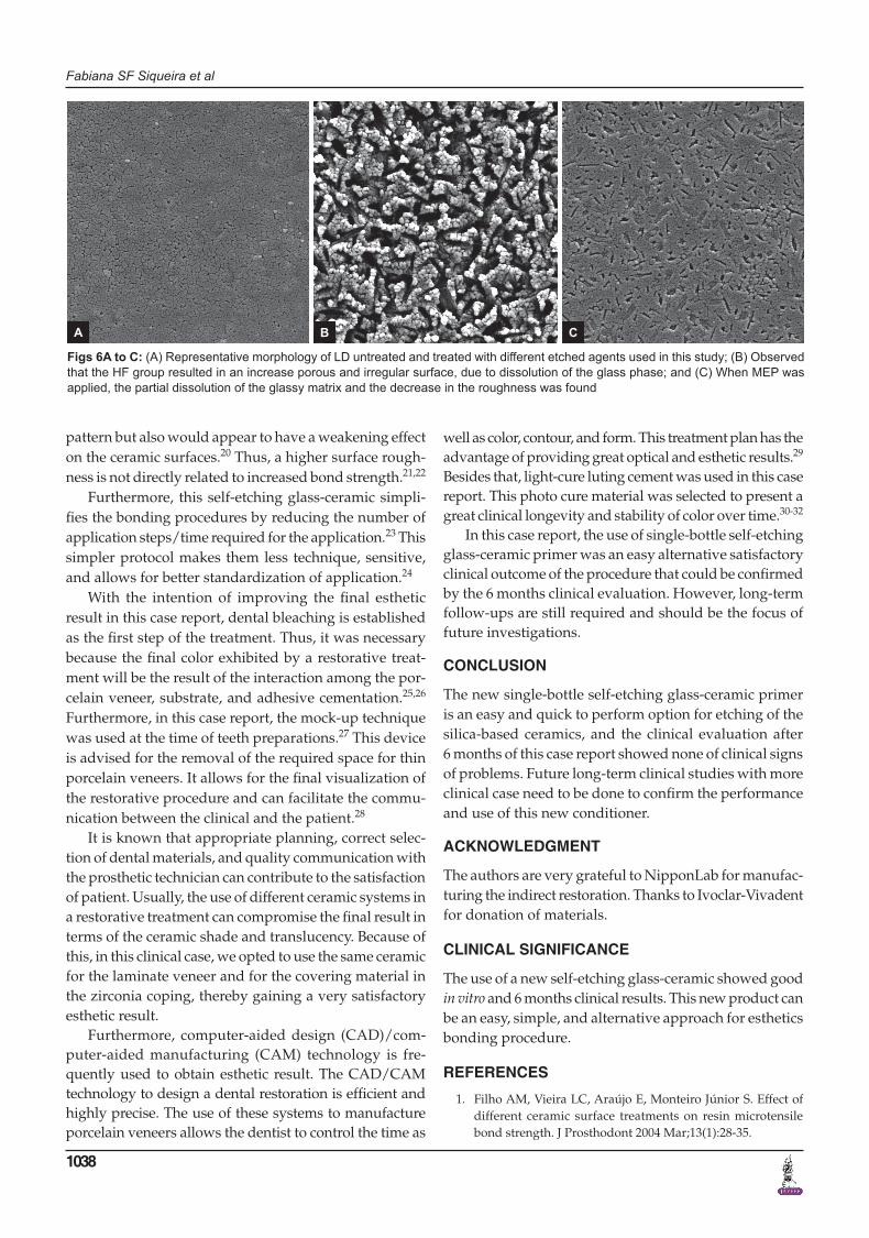
Scanning electron microscopy (SEM) images of the ceramic surfaces with HF and MEP conditioning are shown in Figure 6. The HF showed a greater dissolution of the vitreous phase surrounding the crystals. When the MEP was used, a less dissolution of the vitreous phase was observed but an efficient roughness pattern was created.
DISCUSSION
In dentistry, usually, the new products are launched in the market only with internal data of the manufacture of the products and only with laboratory evaluations. This is why we decided, before applying clinically, to evaluate the in vitro performance of the new self-etching glass-ceramic primer.
This study showed that the etching pattern achieved with the MEP was only superficial when compared with conditioner pattern observed after HF application when evaluated by SEM (Figs 6A to C). This is because MEP contains 15 to 25% ammonium poly fluoride as condition-ing agent that etch the ceramic surface. This acid would be responsible for the only partially dissolution of the glassy phase of the ceramic.19
Although the less-pronounced etching pattern, the mean of µSBS values, when MEP was applied, was no statistically significantly different from those obtained with HF etching. This suggests that conditioner pattern produced by MEP is sufficient for the adhesive interlock-ing with an intaglio ceramic surface. It is worth to mention that MEP is a combination of ammonium poly fluoride as conditioning agent associated with the trimethoxy propyl methacrylate (methacrylate silane), responsible for helping the chemical adhesion.
On the contrary, it has been reported that etching of the HF on the ceramic surface does not only provide the aggressive etching pattern that leads to higher roughness
Table 1: Means and standard deviations of µSBS, number (%) of specimens according to fracture mode of etched agents
Etched agents µSBS (MPa)Fracture mode (%)
A/M CR CCHF 31.19 ± 0.79a 45 (96) 2 (4) 0 (0)
46 (96) 1 (2) 1 (2)40 (100) 0 (0) 0 (0)43 (98) 1 (2) 0 (0)39 (96) 1 (2) 0 (0)
MEP 33.47 ± 1.20a 39 (98) 1 (2) 0 (0)43 (100) 0 (0) 0 (0)47 (100) 0 (0) 0 (0)40 (95) 2 (5) 0 (0)39 (96) 1 (2) 1 (2)
**Similar letters means no statistically significant differences between groupsCR: Cohesive in resin cement, CC: Cohesive in ceramic, MEP: Monobond etch and prime, µSBS: Microshear bond strength
Fig. 5: Final aspect of the treatment after 6 months. Observed that there are no signs of degradation, as marginal staining, chipping fracture, or marginal gap
Fabiana SF Siqueira et al
1038
pattern but also would appear to have a weakening effect on the ceramic surfaces.20 Thus, a higher surface rough-ness is not directly related to increased bond strength.21,22
Furthermore, this self-etching glass-ceramic simpli-fies the bonding procedures by reducing the number of application steps/time required for the application.23 This simpler protocol makes them less technique, sensitive, and allows for better standardization of application.24
With the intention of improving the final esthetic result in this case report, dental bleaching is established as the first step of the treatment. Thus, it was necessary because the final color exhibited by a restorative treat-ment will be the result of the interaction among the por-celain veneer, substrate, and adhesive cementation.25,26 Furthermore, in this case report, the mock-up technique was used at the time of teeth preparations.27 This device is advised for the removal of the required space for thin porcelain veneers. It allows for the final visualization of the restorative procedure and can facilitate the commu-nication between the clinical and the patient.28
It is known that appropriate planning, correct selec-tion of dental materials, and quality communication with the prosthetic technician can contribute to the satisfaction of patient. Usually, the use of different ceramic systems in a restorative treatment can compromise the final result in terms of the ceramic shade and translucency. Because of this, in this clinical case, we opted to use the same ceramic for the laminate veneer and for the covering material in the zirconia coping, thereby gaining a very satisfactory esthetic result.
Furthermore, computer-aided design (CAD)/com-puter-aided manufacturing (CAM) technology is fre-quently used to obtain esthetic result. The CAD/CAM technology to design a dental restoration is efficient and highly precise. The use of these systems to manufacture porcelain veneers allows the dentist to control the time as
well as color, contour, and form. This treatment plan has the advantage of providing great optical and esthetic results.29 Besides that, light-cure luting cement was used in this case report. This photo cure material was selected to present a great clinical longevity and stability of color over time.30-32
In this case report, the use of single-bottle self-etching glass-ceramic primer was an easy alternative satisfactory clinical outcome of the procedure that could be confirmed by the 6 months clinical evaluation. However, long-term follow-ups are still required and should be the focus of future investigations.
CONCLUSION
The new single-bottle self-etching glass-ceramic primer is an easy and quick to perform option for etching of the silica-based ceramics, and the clinical evaluation after 6 months of this case report showed none of clinical signs of problems. Future long-term clinical studies with more clinical case need to be done to confirm the performance and use of this new conditioner.
ACKNOWLEDGMENT
The authors are very grateful to NipponLab for manufac-turing the indirect restoration. Thanks to Ivoclar-Vivadent for donation of materials.
CLINICAL SIGNIFICANCE
The use of a new self-etching glass-ceramic showed good in vitro and 6 months clinical results. This new product can be an easy, simple, and alternative approach for esthetics bonding procedure.
REFERENCES
1. Filho AM, Vieira LC, Araújo E, Monteiro Júnior S. Effect of different ceramic surface treatments on resin microtensile bond strength. J Prosthodont 2004 Mar;13(1):28-35.
Figs 6A to C: (A) Representative morphology of LD untreated and treated with different etched agents used in this study; (B) Observed that the HF group resulted in an increase porous and irregular surface, due to dissolution of the glass phase; and (C) When MEP was applied, the partial dissolution of the glassy matrix and the decrease in the roughness was found
A B C
New Single-bottle Ceramic Primer: 6-month Case Report and Laboratory Performance
The Journal of Contemporary Dental Practice, December 2016;17(12):1033-1039 1039
JCDP
2. Chen JH, Matsumura H, Atsuta M. Effect of different etching periods on the bond strength of a composite resin to a machin-able porcelain. J Dent 1998 Jan;26(1):53-58.
3. Söderholm KJ, Shang SW. Molecular orientation of silane at the surface of colloidal silica. J Dent Res 1993 Jun;72(6):1050-1054.
4. Addison O, Fleming GJ. The influence of cement lute, thermocycling and surface preparation on the strength of a porcelain laminate veneering material. Dent Mater 2004 Mar;20(3):286-292.
5. Addison O, Marquis PM, Fleming GJ. The impact of hydro-fluoric acid surface treatments on the performance of a porcelain laminate restorative material. Dent Mater 2007 Apr;23(4):461-468.
6. Della Bona A, Anusavice KJ, Hood JA. Effect of ceramic surface treatment on tensile bond strength to a resin cement. Int J Prosthodont 2002 May-Jun;15(3):248-253.
7. Bertolini JC. Hydrofluoric acid: a review of toxicity. J Emerg Med 1992 Mar-Apr;10(2):163-168.
8. Ozcan M, Allahbeickaraghi A, Dündar M. Possible hazardous effects of hydrofluoric acid and recommendations for treat-ment approach: a review. Clin Oral Invest 2012 Feb;16(1):15-23.
9. Ozcan M. Evaluation of alternative intra-oral repair techniques for fractured ceramic-fused-to-metal restorations. J Oral Rehabil 2003 Feb;30(2):194-203.
10. Swank H, Bailey C, Motyka N, Vandewalle K. Bond Strength of Resin Cement to Lithium Disilicate with Pre-treatments. J Dent Res 2016;95(Special issue A):Abstract 569.
11. Chang B, Lawson N, Burgess J. Stability of Silane to Lithium Disilicate in Extreme Environmental Conditions. J Dent Res 2016;95(Special issue A):Abstract 571.
12. Heleba A, Hill T, Singhal S, McCabe P, Tysowsky G. Shear Bond Strength of Ceramic Primers with Lithium Disilicate. J Dent Res 2016;95(Special Issue A):Abstract 1083.
13. Tysowsky G, Heleba A, Hill T, Singhal S, McCabe P. Effect of Storage on Shear-bond Strength of Self-Etch Ceramic Primer. J Dent Res. 2016;95(Special issue A):Abstract 1067.
14. Gomes GM, Monte-Alto RV, Santos GO, Fai CK, Loguercio AD, Gomes OM, Gomes JC, Reis A. Use of a direct anatomic post in a flared root canal: a three-year follow-up. Oper Dent 2016 Jan-Feb;41(1):E23-28.
15. Cardenas A, Siqueira F, Davila-Sanchez A, Gomes GM, Reis A, Gomes JC. Four-year follow-up of a direct anatomical fiber post and esthetic procedures: a case report. Oper Dent 2016 Jul-Aug;41(4):363-369.
16. Siqueira F, Cardenas AM, Gutierrez MF, Malaquias P, Hass V, Reis A, et al. Laboratory performance of universal adhesive systems for luting CAD/CAM restorative materials. J Adhes Dent 2016;18(4):331-340.
17. Munoz MA, Baggio R, Mendes YB, Gomes GM, Luque-Martinez I, Loguercio AD, Reis A. The effect of the loading
method and cross-head speed on resin-dentin microshear bond strength. Int J Adhes Adhes 2014 Apr;50:136-141.
18. Shimada Y, Yamaguchi S, Tagami J. Micro-shear bond strength of dual-cured resin cement to glass ceramics. Dent Mater 2002 Jul;18(5):380-388.
19. Ivoclar-Vivadent. Monobond Etch & Prime:2015;2015. Available from: http://www.ivoclarvivadent.com/en/bonding-agents-luting-composites-to-restorations/monobond-etch-and-prime. [Last cited on April 18, 2016].
20. Hooshmand T, Parvizi S, Keshvad A. Effect of surface acid etching on the biaxial flexural strength of two hot-pressed glass ceramics. J Prosthodont 2008 Jul;17(5):415-419.
21. Xiaoping L, Dongfeng R, Silikas N. Effect of etching time and resin bond on the flexural strength of IPS e.max Press glass ceramic. Dent Mater 2014 Dec;30(12):e330-336.
22. Zogheib LV, Bona AD, Kimpara ET, McCabe JF. Effect of hydrofluoric acid etching duration on the roughness and flexural strength of a lithium disilicate-based glass ceramic. Braz Dent J 2011;22(1):45-50.
23. Van Meerbeek B, Van Landuyt K, De Munck J, Hashimoto M, Peumans M, Lambrechts P, Yoshida Y, Inoue S, Suzuki K. Technique-sensitivity of contemporary adhesives. Dent Mater J 2005 Mar;24(1):1-13.
24. Perdigão J. New developments in dental adhesion. Dent Clin North Am 2007 Apr;51(2):333-357, viii.
25. Alqahtani MQ, Aljurais RM, Alshaafi MM. The effects of dif-ferent shades of resin luting cement on the color of ceramic veneers. Dent Mater J 2012;31(3):354-361.
26. Meyer Filho A, Vieira LC, Baratieri LN, Lopes GC. Porcelain veneers as an alternative for the esthetic treatment of stained anterior teeth: clinical report. Quintessence Int 2005 Mar;36(3):191-196.
27. Gresnigt M, Ozcan M, Kalk W. Esthetic rehabilitation of worn anterior teeth with thin porcelain laminate veneers. Eur J Esthet Dent 2011 Autumn;6(3):298-313.
28. Rotoli BT, Lima DA, Pini NP, Aguiar FH, Pereira GD, Paulillo LA. Porcelain veneers as an alternative for esthetic treatment: clinical report. Oper Dent 2013 Sep-Oct;38(5): 459-466.
29. Giordano R. Materials for chairside CAD/CAM-produced restorations. J Am Dent Assoc 2006 Sep;137 Suppl:14S-21S.
30. Beier US, Kapferer I, Burtscher D, Dumfahrt H. Clinical per-formance of porcelain laminate veneers for up to 20 years. Int J Prosthodont 2012 Jan-Feb;25(1):79-85.
31. Burke FJ. Survival rates for porcelain laminate veneers with special reference to the effect of preparation in dentin: a litera-ture review. J Esthet Restor Dent 2012 Aug;24(4):257-265.
32. Karaagaclioglu L, Yilmaz B. Influence of cement shade and water storage on the final color of leucite-reinforced ceramics. Oper Dent 2008 Jul-Aug;33(4):386-391.