Direct comparison of the bond strength results of the different ...
16
dental materials 26 (2010) e78–e93 available at www.sciencedirect.com journal homepage: www.intl.elsevierhealth.com/journals/dema Review Direct comparison of the bond strength results of the different test methods: A critical literature review Susanne S. Scherrer a,∗ , Paulo F. Cesar b , Mike V. Swain c,d a University of Geneva, School of Dental Medicine, Dept. of Prosthodontics-Biomaterials, Geneva, Switzerland b University of São Paulo, School of Dentistry, Department of Dental Materials, São Paulo, Brazil c University of Sydney, Biomaterials Laboratory, Sydney Dental Hospital, Sydney, Australia d University of Otago, Biomaterials, Dunedin, New Zealand article info Article history: Received 15 September 2009 Received in revised form 1 December 2009 Accepted 1 December 2009 Keywords: Review Adhesion testing Adhesion to dentin Shear Microshear Tensile Microtensile abstract Objective. The goal of this paper is to undertake a literature search collecting all dentin bond strength data obtained for six adhesives with four tests (shear, microshear, tensile and microtensile) and to critically analyze the results with respect to average bond strength, coefficient of variation, mode of failure and product ranking. Method. A PubMed search was carried out for the years between 1998 and 2009 identifying publications on bond strength measurements of resin composite to dentin using four tests: shear, tensile, microshear and microtensile. The six adhesive resins were selected covering three step systems (OptiBond FL, Scotch Bond Multi-Purpose Plus), two-step (Prime & Bond NT, Single Bond, Clearfil SE Bond) and one step (Adper Prompt L Pop). Results. Pooling results from 147 references showed an ongoing high scatter in the bond strength data regardless which adhesive and which bond test was used. Coefficients of variation remained high (20–50%) even with the microbond test. The reported modes of failure for all tests still included high number of cohesive failures. The ranking seemed to be dependant on the test used. Significance. The scatter in dentin bond strength data remains regardless which test is used confirming Finite Element Analysis predicting non-uniform stress distributions due to a number of geometrical, loading, material properties and specimens preparation variables. This reopens the question whether, an interfacial fracture mechanics approach to analyze the dentin–adhesive bond is not more appropriate for obtaining better agreement among dentin bond related papers. © 2009 Academy of Dental Materials. Published by Elsevier Ltd. All rights reserved. Contents 1. Introduction to the review ................................................................................................... e79 2. Materials and methods ...................................................................................................... e79 ∗ Corresponding author. E-mail address: [email protected] (S.S. Scherrer). 0109-5641/$ – see front matter © 2009 Academy of Dental Materials. Published by Elsevier Ltd. All rights reserved. doi:10.1016/j.dental.2009.12.002
-
Upload
khangminh22 -
Category
Documents
-
view
1 -
download
0
Transcript of Direct comparison of the bond strength results of the different ...
d e n t a l m a t e r i a l s 2 6 ( 2 0 1 0 ) e78–e93
avai lab le at www.sc iencedi rec t .com
journa l homepage: www. int l .e lsev ierhea l th .com/ journa ls /dema
Review
Direct comparison of the bond strength results of thedifferent test methods: A critical literature review
Susanne S. Scherrera,∗, Paulo F. Cesarb, Mike V. Swainc,d
a University of Geneva, School of Dental Medicine, Dept. of Prosthodontics-Biomaterials, Geneva, Switzerlandb University of São Paulo, School of Dentistry, Department of Dental Materials, São Paulo, Brazilc University of Sydney, Biomaterials Laboratory, Sydney Dental Hospital, Sydney, Australiad University of Otago, Biomaterials, Dunedin, New Zealand
a r t i c l e i n f o
Article history:
Received 15 September 2009
Received in revised form
1 December 2009
Accepted 1 December 2009
Keywords:
Review
Adhesion testing
Adhesion to dentin
Shear
Microshear
Tensile
Microtensile
a b s t r a c t
Objective. The goal of this paper is to undertake a literature search collecting all dentin
bond strength data obtained for six adhesives with four tests (shear, microshear, tensile
and microtensile) and to critically analyze the results with respect to average bond strength,
coefficient of variation, mode of failure and product ranking.
Method. A PubMed search was carried out for the years between 1998 and 2009 identifying
publications on bond strength measurements of resin composite to dentin using four tests:
shear, tensile, microshear and microtensile. The six adhesive resins were selected covering
three step systems (OptiBond FL, Scotch Bond Multi-Purpose Plus), two-step (Prime & Bond
NT, Single Bond, Clearfil SE Bond) and one step (Adper Prompt L Pop).
Results. Pooling results from 147 references showed an ongoing high scatter in the bond
strength data regardless which adhesive and which bond test was used. Coefficients of
variation remained high (20–50%) even with the microbond test. The reported modes of
failure for all tests still included high number of cohesive failures. The ranking seemed to
be dependant on the test used.
Significance. The scatter in dentin bond strength data remains regardless which test is used
confirming Finite Element Analysis predicting non-uniform stress distributions due to a
number of geometrical, loading, material properties and specimens preparation variables.
This reopens the question whether, an interfacial fracture mechanics approach to analyze
the dentin–adhesive bond is not more appropriate for obtaining better agreement among
dentin bond related papers.
emy of Dental Materials. Published by Elsevier Ltd. All rights reserved.
© 2009 AcadContents
1. Introduction to the review . . . . . . . . . . . . . . . . . . . . . . . . . . . . . . . . . . . . .2. Materials and methods . . . . . . . . . . . . . . . . . . . . . . . . . . . . . . . . . . . . . . . .
∗ Corresponding author.E-mail address: [email protected] (S.S. Scherrer).
0109-5641/$ – see front matter © 2009 Academy of Dental Materials. Pudoi:10.1016/j.dental.2009.12.002
. . . . . . . . . . . . . . . . . . . . . . . . . . . . . . . . . . . . . . . . . . . . . . . . . . . . . . . . . . . . . . e79. . . . . . . . . . . . . . . . . . . . . . . . . . . . . . . . . . . . . . . . . . . . . . . . . . . . . . . . . . . . . . e79
blished by Elsevier Ltd. All rights reserved.
d e n t a l m a t e r i a l s 2 6 ( 2 0 1 0 ) e78–e93 e79
3. Results and discussion. . . . . . . . . . . . . . . . . . . . . . . . . . . . . . . . . . . . . . . . . . . . . . . . . . . . . . . . . . . . . . . . . . . . . . . . . . . . . . . . . . . . . . . . . . . . . . . . . . . . . . . e803.1. Variability in bond strength between tests for a same adhesive. . . . . . . . . . . . . . . . . . . . . . . . . . . . . . . . . . . . . . . . . . . . . . . . . . . . e803.2. Coefficient of variation of bond strength within tests . . . . . . . . . . . . . . . . . . . . . . . . . . . . . . . . . . . . . . . . . . . . . . . . . . . . . . . . . . . . . . e833.3. Mode of failure for each test . . . . . . . . . . . . . . . . . . . . . . . . . . . . . . . . . . . . . . . . . . . . . . . . . . . . . . . . . . . . . . . . . . . . . . . . . . . . . . . . . . . . . . . . . . e843.4. Ranking dependence by test . . . . . . . . . . . . . . . . . . . . . . . . . . . . . . . . . . . . . . . . . . . . . . . . . . . . . . . . . . . . . . . . . . . . . . . . . . . . . . . . . . . . . . . . . . e853.5. The use of Weibull statistics for bond strength data . . . . . . . . . . . . . . . . . . . . . . . . . . . . . . . . . . . . . . . . . . . . . . . . . . . . . . . . . . . . . . . . e853.6. Fracture mechanics approach for interfacial bond assessment . . . . . . . . . . . . . . . . . . . . . . . . . . . . . . . . . . . . . . . . . . . . . . . . . . . . e87
4. Conclusions and perspectives . . . . . . . . . . . . . . . . . . . . . . . . . . . . . . . . . . . . . . . . . . . . . . . . . . . . . . . . . . . . . . . . . . . . . . . . . . . . . . . . . . . . . . . . . . . . . . . e88. . . . . .. . . . .
1
Wjtpwapossonsatp1s(dftcalspsosltd
wmutaiWm[tfi
Acknowledgment . . . . . . . . . . . . . . . . . . . . . . . . . . . . . . . . . . . . . . . . .References . . . . . . . . . . . . . . . . . . . . . . . . . . . . . . . . . . . . . . . . . . . . . . . . .
. Introduction to the review
hen an assignment for a literature review involves the sub-ect of “dentin bonding” an instantaneous shiver runs downhe backbone as the amount of data is a knock out (over 6000apers just with the Keywords: “bonding” AND “dentin”) andhen combined with the sorting through testing parameters
nd variables, it becomes a nightmare. The reasons for such aopular topic of research is of course the rapid developmentf bonding adhesives to dentin and the fact that the productcreening test methods such as shear, tensile and microten-ile are inexpensive testing routines in most dental schoolr research laboratories. Dentin bonding however has beenotorious for high spread in the results, whether within theame laboratory or between laboratories using the same tests,nd even 15–20 years ago suggestions were raised to improvehe standardization of bond strength testing after reviewingossible reasons for such variability [1–7]. The introduction in994 of the microtensile bond strength test [8] allowing mea-urements of the tensile bond strength on very small surfaces∼1 mm2) opened the research to regional differences withinentin, and had the advantage of producing many specimensrom the same extracted tooth. It was thought that with thisest, the characteristic high bond strength spread obtained inonventional shear and tensile tests would diminish due tobetter stress distribution over a very small surface during
oading, generating more interface failures (i.e. fewer cohe-ive failures) in dentin [8]. In 1999, Pashley et al. [9] reviewedositively the microtensile bond test and summarized its ver-atile usage providing new insights into strength of adhesionf restorative materials to clinically relevant sites and sub-trates and advocated this test as a means for evaluating theong-term durability of resin-hard-tissue bonds. Since then,he microtensile bond strength test has become the most usedentin bond test.
The philosophical question however to be asked here ishat is the final goal of measuring bond strength? Is it toeasure interfacial bond strength? Is it to distinguish prod-
ct A from product B? Is it to rank the products according toheir results? Is it to understand localized degradation withinbonded surface? Is it a test to indicate reliability of the bond-
ng? Which test should be used? Are the results dubious?ell, considering the information gathered from Finite Ele-
ent Analyses (FEA) generated for shear [4,10–12], microshear11], tensile [4,7] and microtensile [13], almost every possibleesting variable (i.e. specimen’s geometry, loading condition,lm thickness, modulus of elasticity of the materials involved)
. . . . . . . . . . . . . . . . . . . . . . . . . . . . . . . . . . . . . . . . . . . . . . . . . . . . . . . . . . . . . e88. . . . . . . . . . . . . . . . . . . . . . . . . . . . . . . . . . . . . . . . . . . . . . . . . . . . . . . . . . . . . . e88
has a significant influence on the stress state and thus on thebond strength values.
Therefore, the goal of this paper is to perform a 10 year lit-erature search collecting dentin bond strength data obtainedfor six adhesives with four tests (shear, microshear, tensile andmicrotensile) and to critically analyze the results with respectto average bond strength, coefficient of variation, mode of fail-ure and product ranking. No attempt is made to undertakeadditional statistical analysis, as the major focus of this paperis to point out limitations of the most popular methods forevaluating adhesion. Alternative approaches to bond strengthevaluation including the use of Weibull statistics and fracturemechanics will be discussed.
2. Materials and methods
A PubMed search was carried out for the years between 1998and 2009 to identify publications on bond strength mea-surements of resin composite to dentin using shear, tensile,microshear and microtensile. The search would poke aroundKeywords: such as “(Clearfil SE bond) AND (bond) AND (dentin)NOT (bovine) NOT (primary) NOT (enamel) NOT (root) NOT(fiber)” as an example. The six adhesive resins selected were:(a) two three step systems where the etching (and rinsing);priming; and bonding are carried out separately (OptiBondFL; Kerr; Orange; CA; USA; and Scotch Bond Multi-PurposePlus; 3 M Espe; St. Paul; MN; USA); (b) two two-step self-primingsystems which include etching (and rising) followed by aself-bonding primer (Prime & Bond NT; Dentsply/De Trey; Kon-stanz; Germany; and Single Bond; 3 M Espe; St. Paul; MN; USA);(c) a two-step self-etching system where the etch and prime aretogether followed by a bonding step (Clearfil SE Bond; Kuraray;Osaka; Japan); and (d) a one step “all-in-one” system where theetching; priming; and bonding are combined into a single step(Adper Prompt L Pop; 3 M Espe; Seefeld; Germany).
The selected criteria for inclusion within the literaturesearch were: (1) the bonding substrate: human coronal to mid-coronal dentin of molars, premolar and central incisors; (2)the storage media for the extracted human teeth: formalin,thymol, chloramine, sodium azide or saline solution; (3) thepost-extraction storage time prior to sample preparation: 15days to 6 months; (4) the dentin surface preparation: sand-paper from 180 to 1200 grit SiC, fine or medium diamond
burs, tungsten carbide burs; (5) application of the adhesivesto the prepared dentin surfaces following the manufacturers’instructions; (6) the storage time and media of the bondedspecimens prior to testing: 10 min (immediate) or 24 h up to 14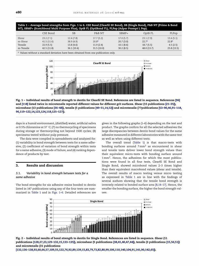
e80 d e n t a l m a t e r i a l s 2 6 ( 2 0 1 0 ) e78–e93
Table 1 – Average bond strengths from Figs. 1 to 6. CSE Bond (Clearfil SE Bond), SB (Single Bond), P&B NT (Prime & BondNT), SBMP+ (Scotchbond Multi Purpose Plus), OptB FL (OptiBond FL), PLPop (Adper Prompt L Pop).
CSE Bond SB P&B NT SBMP+ OptB FL PLPop
Shear 23.2 (7.1) 12.4 (7.8) 17.7 (5.2) 17.0 (5.7) 23.1 (7.9) 13.4 (5.1)m-Shear 41.5 (11.6) 38.9 (4.9) 20.8a 20.7 (3.0) 22.7a 22.8a
Tensile 22.9 (5.5) 13.8 (4.6) 11.9 (2.4) 10.1 (8.6) 18.7 (5.5) 4.5 (2.5)m-Tensile 42.5 (11.8) 36.1 (10.4) 31.5 (10.0) 30.2 (8.5) 48.0 (13.7) 25.8 (13.5)
a Values without a standard deviation have been obtained from one publication only.
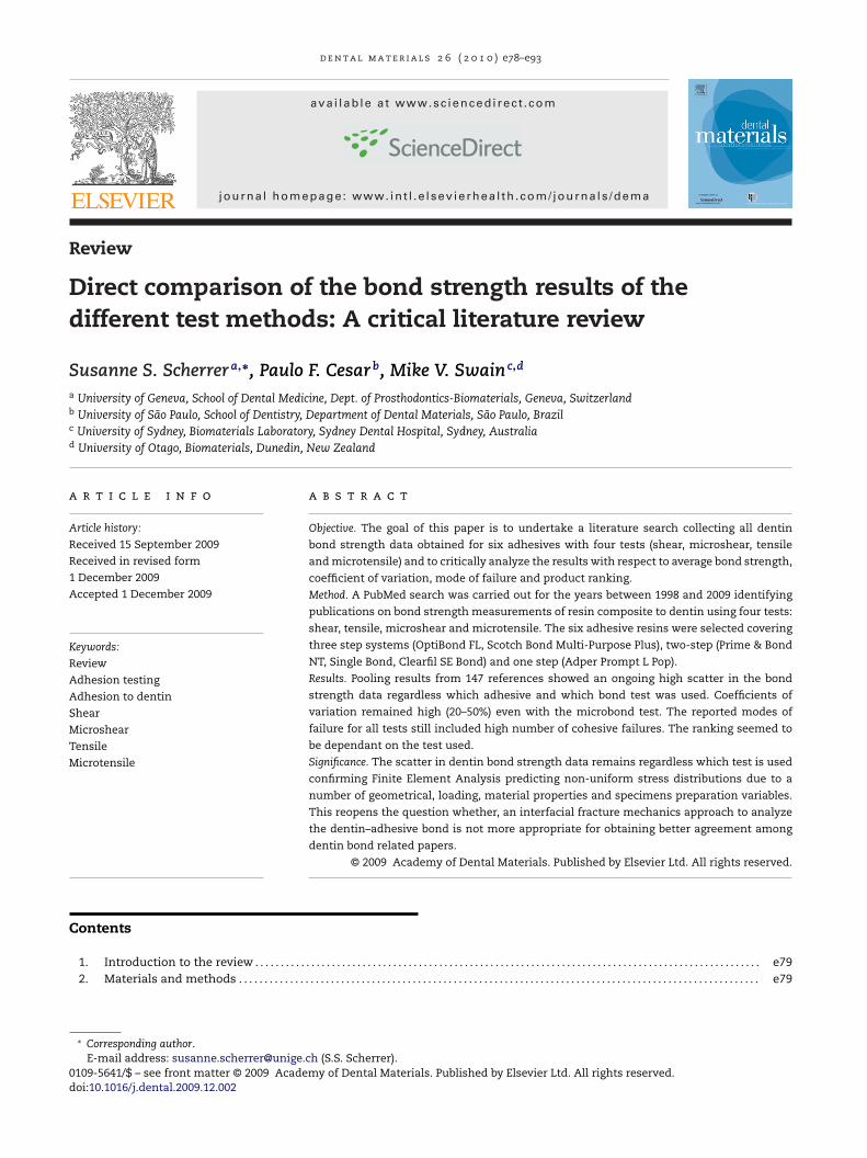
Fig. 1 – Individual results of bond strength to dentin for Clearfil SE Bond. References are listed in sequence. References [96]and [118] listed twice in microtensile reported different values for different grit surfaces. Shear (15 publications [21–35]),
49–5
microshear (13 publications [36–48]), tensile (5 publications [96,119–122,14,123,124,118,125–127]).days in a humid environment, (distilled) water, artificial salivaor 0.5% chloramine at 37 ◦C; (7) no thermocycling of specimensduring storage or thermocycling not beyond 1500 cycles; (8)specimens tested without pulp pressure.
The data were compiled in spreadsheets and analyzed for:(1) variability in bond strength between tests for a same adhe-sive, (2) coefficient of variation of bond strength within testsfor a same adhesive, (3) mode of failure, and (4) ranking depen-dence of products by test.
3. Results and discussion
3.1. Variability in bond strength between tests for asame adhesive
The bond strengths for six adhesive resins bonded to dentinlisted in 147 publications using any of the four tests are sum-marized in Table 1 and in Figs. 1–6. Detailed references are
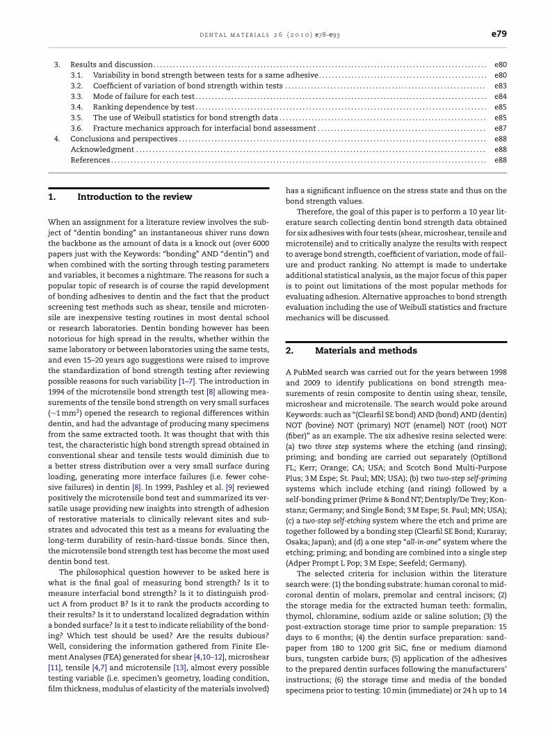
Fig. 2 – Individual results of bond strength to dentin for Single Bpublications [128,27,23,129–132,15,133–135]), microshear (5 publand microtensile (31 publications[132,136–138,93,60,66,57,109,55,122,76,92,89,139,15,83,79,73,82,8
1,14,52]) and microtensile (77publications [53–90,46,91–118,
given in the following graphs (1–6) depending on the test andproduct. The graphs confirm for all the selected adhesives thelarge discrepancies between dentin bond values for the sameadhesive measured in different laboratories with the same testas well as when using different tests.
The overall trend (Table 1) is that macro-tests withbonding surfaces around 7 mm2 as encountered in shearand tensile tests deliver lower bond strength values thantheir equivalent micro-tests with bonding surface around1 mm2. Hence, the adhesives for which the most publica-tions were found in all four tests, Clearfil SE Bond andSingle Bond, showed microbond values 2–3 times higherthan their equivalent macrobond values (shear and tensile).The overall results of macro testing versus micro testingas expressed in Table 1 are in line with the findings of
several authors showing that the tensile bond strength isinversely related to bonded surface area [8,14–17]. Hence, thesmaller the bonding surface, the higher the bond strength val-ues.ond. References are listed in sequence. Shear (11ications [38,41,40,47,44]), tensile (3 publications [15,50,51])
0,99,106,110,140,104,91,141,90,142,65]).
d e n t a l m a t e r i a l s 2 6 ( 2 0 1 0 ) e78–e93 e81
Fig. 3 – Individual results of bond strength to dentin for Prime & Bond NT. References are listed in sequence. Shear (7publications [28,24,143,25,144,145,32]), microshear (1 publication [36]), tensile (2 publications [52,50]) and microtensile (17publications [146,137,93,91,60,54,71,110,147,106,76,75,112,79,99,104,70]).
Fig. 4 – Individual results of bond strength to dentin for Scotchbond Multi Purpose Plus. References are listed in sequence.Reference [152] listed twice in microtensile reported different values for different dentin location. Shear (8 publications[ ]), tep
cept
ahhSmph
Ft[p
15,26,148–150,30,24,134]), microshear (2 publications [36,17ublications [152,17,149,152,72,15,60,57,82,80]).
The overall increase in bond strength for small surfacesan be explained by the Weibull distribution showing math-matically that an increase in specimen size increases therobability of encountering a strength-limiting flaw and thathe specimen’s flaws are size distributed [18].
Direct comparisons between tensile and microtensile forll six adhesives showed microtensile bond values 2–5 timesigher (Table 1). Direct comparison between shear and micros-ear could only be made for three adhesives (Clearfil SE Bond,
ingle Bond, Scotchbond Multi Purpose (MP) Plus) for whichinimum of two microshear values were reported. For theseroducts, the microshear bond strengths were 1.2–3 timesigher than their shear values.
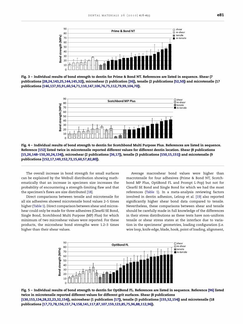
ig. 5 – Individual results of bond strength to dentin for OptiBondwice in microtensile reported different values for different grit s130,153,134,28,22,23,32,154]), microshear (1 publication [17]), tenublications [17,72,78,156,157,74,158,141,117,87,107,159,123,85,7
nsile (3 publications [150,15,151]) and microtensile (9
Average macroshear bond values were higher thanmacrotensile for four adhesives (Prime & Bond NT, Scotch-bond MP Plus, OptiBond FL and Prompt L-Pop) but not forClearfil SE Bond and Single Bond for which we had the mostreferences (Table 1). In a meta-analysis reviewing factorsinvolved in dentin adhesion, Leloup et al. [19] also reportedsignificantly higher shear bond data compared to tensile.Nevertheless, these comparisons between shear and tensileshould be carefully made in full knowledge of the differences
in their stress distributions as these tests have non-uniformtensile or shear stress states at the interface due to varia-tion in the specimens’ geometries, loading configuration (i.e.wire loop, knife edge, blade, hook, point of loading, alignment,FL. References are listed in sequence. Reference [96] listedurfaces. Shear (8 publicationssile (3 publications [155,52,154]) and microtensile (185,96,88,112,96]).
e82 d e n t a l m a t e r i a l s 2 6 ( 2 0 1 0 ) e78–e93
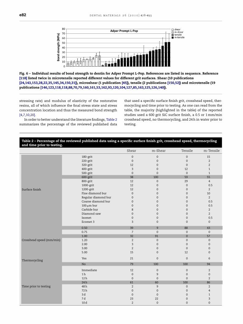
Fig. 6 – Individual results of bond strength to dentin for Adper Prompt L-Pop. References are listed in sequence. Reference[118] listed twice in microtensile reported different values for different grit surfaces. Shear (10 publications
n [4,104
[24,143,153,28,22,35,145,34,150,31]), microshear (1 publicatiopublications [146,123,118,118,88,70,79,160,161,53,162,93,120
stressing rate) and modulus of elasticity of the restorativeresins, all of which influence the final stress state and stress
concentration location and thus the measured bond strength[4,7,10,20].In order to better understand the literature findings, Table 2summarizes the percentage of the reviewed published data
Table 2 – Percentage of the reviewed published data using a speand time prior to testing.
Surface finish
180-grit220-grit320-grit400-grit500-grit600-grit800-grit1000-grit1200-gritFine diamond burRegular diamond burCoarse diamond bur100 �m burCarbide burDiamond sawIsometEcomet 3
Crosshead speed (mm/min)
0.500.751.001.202.003.005.00
ThermocyclingYes
No
Time prior to testing
Immediate1 h12 h24 h48 h72 h3 d7 d10 d
5]), tensile (2 publications [150,52]) and microtensile (19,127,85,163,125,126,140]).
that used a specific surface finish grit, crosshead speed, ther-mocycling and time prior to testing. As one can read from the
table, the majority (highlighted in the table) of the reportedstudies used a 600 grit SiC surface finish, a 0.5 or 1 mm/mincrosshead speed, no thermocycling, and 24 h in water prior totesting.cific surface finish grit, crosshead speed, thermocycling
Shear m-Shear Tensile m-Tensile
0 0 0 150 0 0 2
12 0 0 23 0 12 10 0 0 1
38 100 59 5512 0 29 412 0 0 0.512 0 0 2
0 0 0 100 0 0 20 0 0 0.50 0 0 0.58 0 0 20 0 0 20 0 0 0.53 0 0 0
39 9 88 437 0 0 0
32 91 0 572 0 0 03 0 0 02 0 0 0
15 0 12 0
21 0 0 6
79 100 100 94
12 0 0 20 9 0 00 0 0 2
61 60 100 862 9 0 20 0 0 40 0 0 1
23 22 0 32 0 0 0
d e n t a l m a t e r i a l s 2 6 ( 2 0 1 0 ) e78–e93 e83
Table 3 – Mean coefficient of variation (±standard deviation, in %) obtained from bond strength data as a function of typeof test and adhesive.
Adhesive Bond strength test
Shear m-Shear Tensile m-Tensile
Clearfil SE Bond 25 ± 13 15 ± 4 20 ± 10 23 ± 10Single Bond 42 ± 35 14 ± 6 22 ± 21 22 ± 10Prime & Bond NT 31 ± 22 9 ± 0a 28 ± 12 28 ± 14Scotchbond MP Plus 45 ± 18 20 ± 8 40 ± 6 22 ± 9OptiBond FL 24 ± 8 25 ± 0a 21 ± 11 26 ± 10
36
publi
3t
Tetcipa(mnst
ot
bsnlsv
fttf
F[[[o
Prompt L-Pop 33 ± 18
a Values without a standard deviation have been obtained from one
.2. Coefficient of variation of bond strength withinests
he large spread in the results of bond testing has beenxplained by many papers using FEA. These authors have con-inuously alerted the scientific community of massive stressoncentrations in all dentin bond tests. Major differencesn the stress states, non-uniform stress distributions wereointed out for shear [4,7,10–12], microshear [11], tensile [4,7]nd microtensile [13]. Almost every possible testing variablei.e. specimen’s geometry, loading condition, film thickness,
odulus of elasticity of the materials involved) has a sig-ificant influence on the stress state and thus on the bondtrength values and is responsible for the inconsistencies inhe results.
Table 3 shows the average coefficients of variation (CVs)btained from 147 publications on bond strength data for eachest and adhesive with the corresponding standard deviations.
Overall, for all six adhesives, the CV in microtensile rangedetween 22% and 49%, in tensile between 20% and 53%, inhear between 24% and 45%. Due to the lack of references,o comments can be made for the microshear test. Neverthe-
ess, the CV seemed to be rather product dependent as the onetep adhesive (Prompt L-Pop) systematically showed very highariation in all tests.
As expected shear test data showed a high CV. The dif-
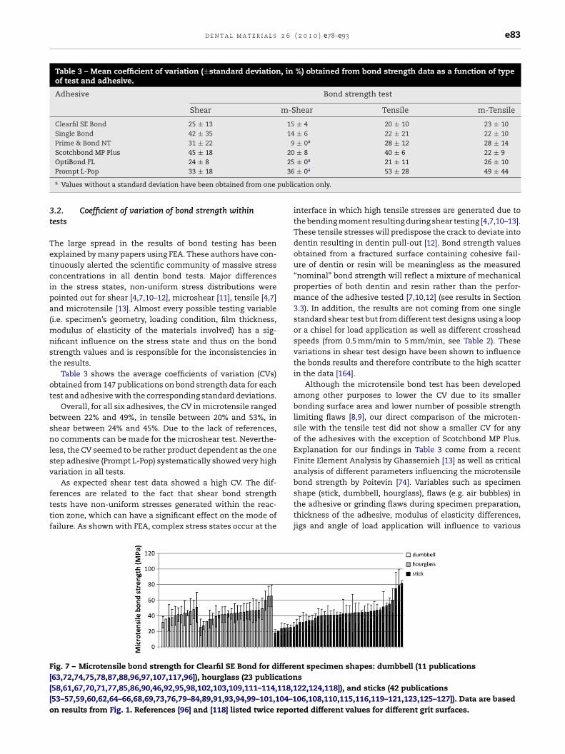
erences are related to the fact that shear bond strengthests have non-uniform stresses generated within the reac-ion zone, which can have a significant effect on the mode ofailure. As shown with FEA, complex stress states occur at theig. 7 – Microtensile bond strength for Clearfil SE Bond for differe63,72,74,75,78,87,88,96,97,107,117,96]), hourglass (23 publication58,61,67,70,71,77,85,86,90,46,92,95,98,102,103,109,111–114,118,153–57,59,60,62,64–66,68,69,73,76,79–84,89,91,93,94,99–101,104–1n results from Fig. 1. References [96] and [118] listed twice repor
± 0a 53 ± 28 49 ± 44
cation only.
interface in which high tensile stresses are generated due tothe bending moment resulting during shear testing [4,7,10–13].These tensile stresses will predispose the crack to deviate intodentin resulting in dentin pull-out [12]. Bond strength valuesobtained from a fractured surface containing cohesive fail-ure of dentin or resin will be meaningless as the measured“nominal” bond strength will reflect a mixture of mechanicalproperties of both dentin and resin rather than the perfor-mance of the adhesive tested [7,10,12] (see results in Section3.3). In addition, the results are not coming from one singlestandard shear test but from different test designs using a loopor a chisel for load application as well as different crossheadspeeds (from 0.5 mm/min to 5 mm/min, see Table 2). Thesevariations in shear test design have been shown to influencethe bonds results and therefore contribute to the high scatterin the data [164].
Although the microtensile bond test has been developedamong other purposes to lower the CV due to its smallerbonding surface area and lower number of possible strengthlimiting flaws [8,9], our direct comparison of the microten-sile with the tensile test did not show a smaller CV for anyof the adhesives with the exception of Scotchbond MP Plus.Explanation for our findings in Table 3 come from a recentFinite Element Analysis by Ghassemieh [13] as well as criticalanalysis of different parameters influencing the microtensilebond strength by Poitevin [74]. Variables such as specimen
shape (stick, dumbbell, hourglass), flaws (e.g. air bubbles) inthe adhesive or grinding flaws during specimen preparation,thickness of the adhesive, modulus of elasticity differences,jigs and angle of load application will influence to variousnt specimen shapes: dumbbell (11 publicationss22,124,118]), and sticks (42 publications06,108,110,115,116,119–121,123,125–127]). Data are basedted different values for different grit surfaces.
e84 d e n t a l m a t e r i a l s 2
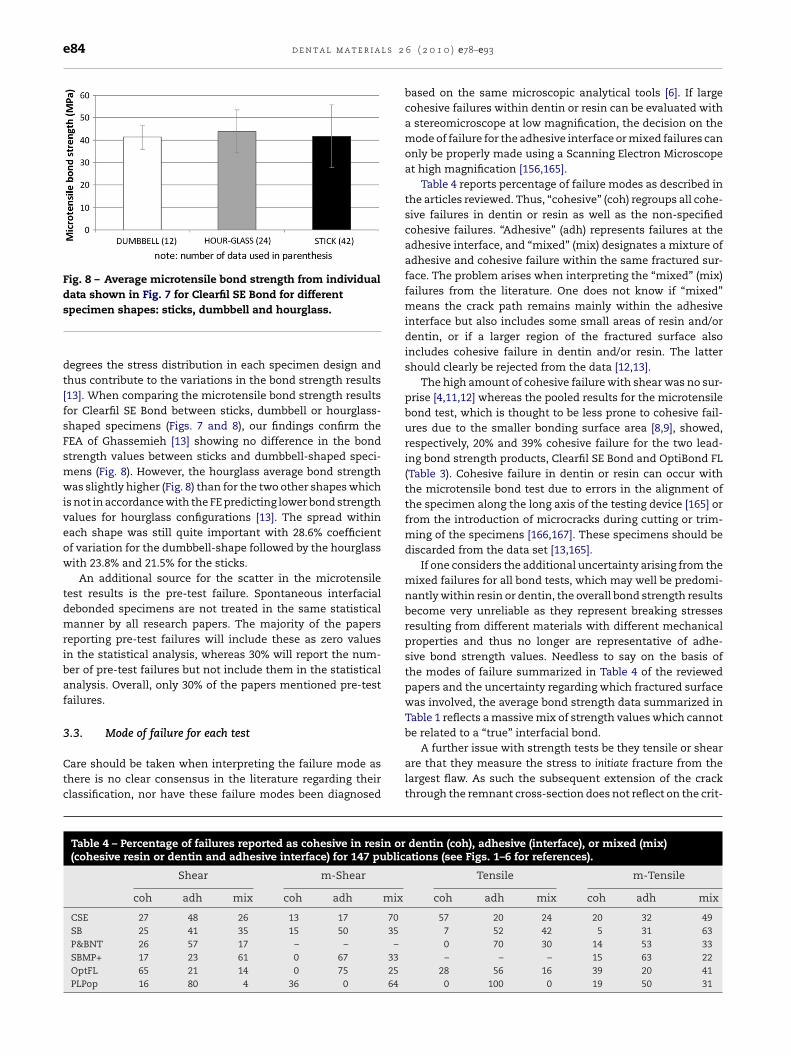
Fig. 8 – Average microtensile bond strength from individualdata shown in Fig. 7 for Clearfil SE Bond for different
specimen shapes: sticks, dumbbell and hourglass.degrees the stress distribution in each specimen design andthus contribute to the variations in the bond strength results[13]. When comparing the microtensile bond strength resultsfor Clearfil SE Bond between sticks, dumbbell or hourglass-shaped specimens (Figs. 7 and 8), our findings confirm theFEA of Ghassemieh [13] showing no difference in the bondstrength values between sticks and dumbbell-shaped speci-mens (Fig. 8). However, the hourglass average bond strengthwas slightly higher (Fig. 8) than for the two other shapes whichis not in accordance with the FE predicting lower bond strengthvalues for hourglass configurations [13]. The spread withineach shape was still quite important with 28.6% coefficientof variation for the dumbbell-shape followed by the hourglasswith 23.8% and 21.5% for the sticks.
An additional source for the scatter in the microtensiletest results is the pre-test failure. Spontaneous interfacialdebonded specimens are not treated in the same statisticalmanner by all research papers. The majority of the papersreporting pre-test failures will include these as zero valuesin the statistical analysis, whereas 30% will report the num-ber of pre-test failures but not include them in the statisticalanalysis. Overall, only 30% of the papers mentioned pre-testfailures.
3.3. Mode of failure for each test
Care should be taken when interpreting the failure mode asthere is no clear consensus in the literature regarding theirclassification, nor have these failure modes been diagnosed
Table 4 – Percentage of failures reported as cohesive in resin or(cohesive resin or dentin and adhesive interface) for 147 public
Shear m-Shear
coh adh mix coh adh mix
CSE 27 48 26 13 17 70SB 25 41 35 15 50 35P&BNT 26 57 17 – – –SBMP+ 17 23 61 0 67 33OptFL 65 21 14 0 75 25PLPop 16 80 4 36 0 64
6 ( 2 0 1 0 ) e78–e93
based on the same microscopic analytical tools [6]. If largecohesive failures within dentin or resin can be evaluated witha stereomicroscope at low magnification, the decision on themode of failure for the adhesive interface or mixed failures canonly be properly made using a Scanning Electron Microscopeat high magnification [156,165].
Table 4 reports percentage of failure modes as described inthe articles reviewed. Thus, “cohesive” (coh) regroups all cohe-sive failures in dentin or resin as well as the non-specifiedcohesive failures. “Adhesive” (adh) represents failures at theadhesive interface, and “mixed” (mix) designates a mixture ofadhesive and cohesive failure within the same fractured sur-face. The problem arises when interpreting the “mixed” (mix)failures from the literature. One does not know if “mixed”means the crack path remains mainly within the adhesiveinterface but also includes some small areas of resin and/ordentin, or if a larger region of the fractured surface alsoincludes cohesive failure in dentin and/or resin. The lattershould clearly be rejected from the data [12,13].
The high amount of cohesive failure with shear was no sur-prise [4,11,12] whereas the pooled results for the microtensilebond test, which is thought to be less prone to cohesive fail-ures due to the smaller bonding surface area [8,9], showed,respectively, 20% and 39% cohesive failure for the two lead-ing bond strength products, Clearfil SE Bond and OptiBond FL(Table 3). Cohesive failure in dentin or resin can occur withthe microtensile bond test due to errors in the alignment ofthe specimen along the long axis of the testing device [165] orfrom the introduction of microcracks during cutting or trim-ming of the specimens [166,167]. These specimens should bediscarded from the data set [13,165].
If one considers the additional uncertainty arising from themixed failures for all bond tests, which may well be predomi-nantly within resin or dentin, the overall bond strength resultsbecome very unreliable as they represent breaking stressesresulting from different materials with different mechanicalproperties and thus no longer are representative of adhe-sive bond strength values. Needless to say on the basis ofthe modes of failure summarized in Table 4 of the reviewedpapers and the uncertainty regarding which fractured surfacewas involved, the average bond strength data summarized inTable 1 reflects a massive mix of strength values which cannotbe related to a “true” interfacial bond.
A further issue with strength tests be they tensile or shearare that they measure the stress to initiate fracture from thelargest flaw. As such the subsequent extension of the crackthrough the remnant cross-section does not reflect on the crit-
dentin (coh), adhesive (interface), or mixed (mix)ations (see Figs. 1–6 for references).
Tensile m-Tensile
coh adh mix coh adh mix
57 20 24 20 32 497 52 42 5 31 630 70 30 14 53 33– – – 15 63 22
28 56 16 39 20 410 100 0 19 50 31
d e n t a l m a t e r i a l s 2 6
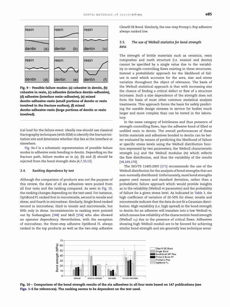
Fig. 9 – Possible failure modes: (a) cohesive in dentin, (b)cohesive in resin, (c) adhesive (interface dentin–adhesive),(d) adhesive (interface resin–adhesive), (e) mixeddentin–adhesive–resin (small portions of dentin or resininvolved in the fracture surface), (f) mixeddentin–adhesive–resin (large portions of dentin or resini
ifte
mfr
3
AtatOssfioaor
FF
nvolved).
cal load for the failure event. Ideally one should use classicalractography techniques (with SEM) to identify the fracture ini-iation site and determine whether this lies at the interface orlsewhere.
Fig. 9a–f is a schematic representation of possible failureodes in adhesive resin bonding to dentin. Depending on the
racture path, failure modes as in (a), (b) and (f) should beejected from the bond strength data [4,7,10,12].
.4. Ranking dependence by test
lthough the comparison of products was not the purpose ofhis review, the data of all six adhesives were pooled fromll four tests and the ranking compared. As seen in Fig. 10,he ranking changes depending on the test used. For instance,ptiBond FL ranked first in microtensile, second in tensile andhear, and fourth in microshear. Similarly, Single Bond rankedecond in microshear, third in tensile and microtensile, butfth only in shear. Inconsistencies in ranking were pointed
ut by Sudsangiam [168] and Moll [154] who also showedn operator dependency. Nevertheless, with the exceptionf microshear, the three-step adhesive OptiBond FL alwaysanked in the top products as well as the two-step adhesiveig. 10 – Comparison of the bond strength results of the six adheigs. 1–6 for references). The ranking seems to be dependent on
( 2 0 1 0 ) e78–e93 e85
Clearfil SE Bond. Similarly, the one-step Promp L-Pop adhesivealways ranked low.
3.5. The use of Weibull statistics for bond strengthdata
The strength of brittle materials such as ceramics, resincomposites and tooth structure (i.e. enamel and dentin)cannot be specified by a single value due to the variabil-ity in strength-controlling flaws existing in these structures.Instead a probabilistic approach for the likelihood of fail-ure is used which accounts for the area, size and stressvariation throughout the object of relevance. The basis ofthe Weibull statistical approach is that with increasing sizethe chance of finding a critical defect or flaw of a structureincreases. Such a size dependence of the strength does notform the basis of most other common statistical analysistreatments. This approach forms the basis for safely predict-ing the useable design stresses in service for bodies muchlarger and more complex than can be tested in the labora-tory.
In the same category of brittleness and thus presence ofstrength-controlling flaws, lays the adhesive bond of filled orunfilled resin to dentin. The overall performances of thesebrittle materials and adhesives bonded to dentin can be bet-ter evaluated by means of predicting the likelihood of failureat specific stress levels using the Weibull distribution func-tion expressed by two parameters, the Weibull characteristicstrength (�0) and the Weibull modulus (m) which reflectsthe flaw distribution, and thus the variability of the results[18,169,170].
The ISO/TS 11405:2003 [171] recommends the use of theWeibull distribution for the analysis of bond strengths that arenon-normally distributed. Unfortunately, most bond strengthspapers used means and standard deviation, rather than aprobabilistic failure approach which would provide insightsas to the reliability (Weibull m parameter) and the probabilityof failure for a given stress level. As indicated in Table 3, thehigh coefficient of variation of 20–50% for shear, tensile andmicrotensile indicate that the data do not fit a Gaussian distri-bution. High variability (i.e. high spread) in the bond strengthto dentin for an adhesive will translate into a low Weibull m,
which means low reliability of the characteristic bond strength(Weibull �0) due to the presence of critical flaws. Adhesivesshowing high Weibull moduli are to be favored for achievingsimilar bond strength and are generally less technique sensi-sives in all four tests based on 147 publications (seethe test used.
e86d
en
ta
lm
at
er
ial
s2
6(2
01
0)
e78–e93
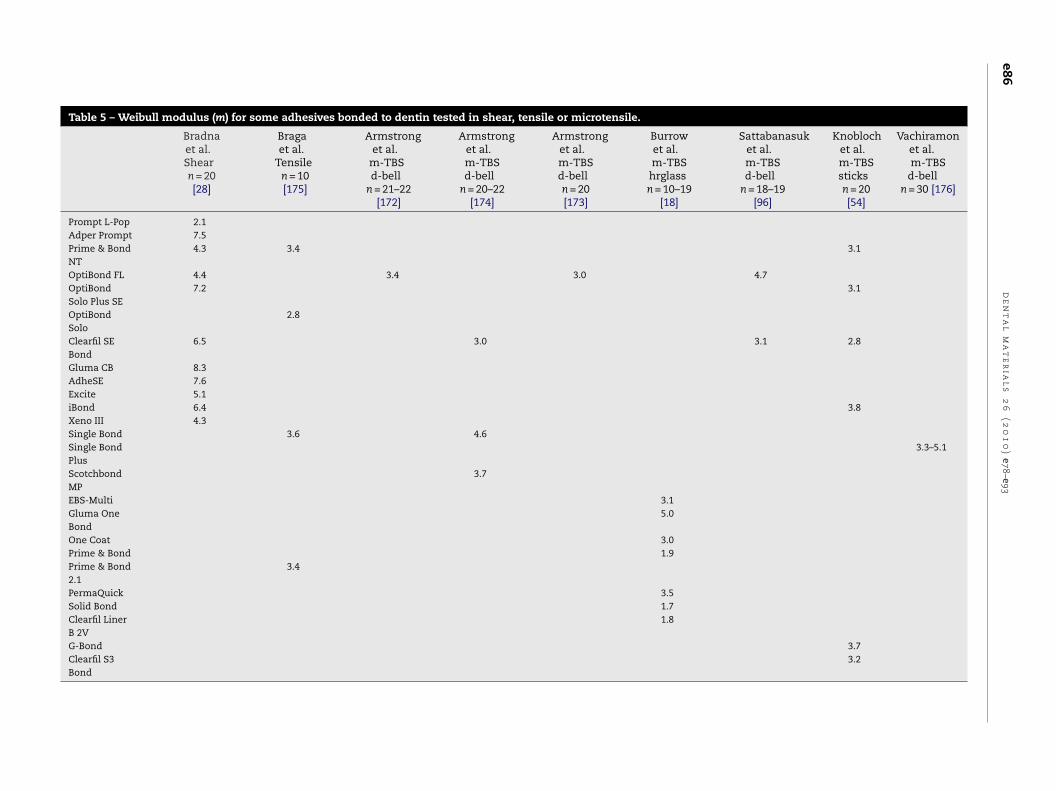
Table 5 – Weibull modulus (m) for some adhesives bonded to dentin tested in shear, tensile or microtensile.
Bradnaet al.Shearn = 20[28]
Bragaet al.
Tensilen = 10[175]
Armstronget al.
m-TBSd-bell
n = 21–22[172]
Armstronget al.m-TBSd-bell
n = 20–22[174]
Armstronget al.m-TBSd-belln = 20[173]
Burrowet al.m-TBShrglassn = 10–19
[18]
Sattabanasuket al.m-TBSd-bell
n = 18–19[96]
Knoblochet al.m-TBSsticksn = 20[54]
Vachiramonet al.m-TBS
d-belln = 30 [176]
Prompt L-Pop 2.1Adper Prompt 7.5Prime & BondNT
4.3 3.4 3.1
OptiBond FL 4.4 3.4 3.0 4.7OptiBondSolo Plus SE
7.2 3.1
OptiBondSolo
2.8
Clearfil SEBond
6.5 3.0 3.1 2.8
Gluma CB 8.3AdheSE 7.6Excite 5.1iBond 6.4 3.8Xeno III 4.3Single Bond 3.6 4.6Single BondPlus
3.3–5.1
ScotchbondMP
3.7
EBS-Multi 3.1Gluma OneBond
5.0
One Coat 3.0Prime & Bond 1.9Prime & Bond2.1
3.4
PermaQuick 3.5Solid Bond 1.7Clearfil LinerB 2V
1.8
G-Bond 3.7Clearfil S3Bond
3.2

d e n t a l m a t e r i a l s 2 6 ( 2 0 1 0 ) e78–e93 e87
Table 6 – Interfacial fracture toughness (KIc) or strain energy release rate (GIc) reported for some adhesives bonded todentin. Legend: CVSrod = chevron short rod, CVSbar = chevron short bar, SENB = single edge notched beam,NTP = notchless triangular prism, SB2 = Scotchbond 2, SBMP = Scotchbond Multipurpose, SBMP+ = ScotchbondMultipurpose Plus, AB2 = All-Bond 2, CLB2 = Clearfil Liner Bond 2.
Author Test specimen Product KIc (MPa√
m) GIc (J/m2) Shear (MPa) Tensile (MPa)
Tam and Pilliar [179] CVSrod SB2 0.20 (0.14) 1.7 (0.9)SBMP 0.34 (0.21) 2.7 (1.9)AB2 0.69 (0.41) 8.4 (4.0)
Lin and Douglas [182] CVSbar SB2 42.8 (7.8)SBMP 75.0 (10.5)
Armstrong et al. [181] CVSbar AB2 0.88 (0.24)Toparli and Aksoy [180] SENB SBMP 0.74 (0.04)
Tantbirojn et al. [183] CVSrod SB 107.0 (26.0) 14.8 (3.9)SBMP+ 93.0 (24.0) 14.6 (2.3)
Tam et al. [178] CVSrod AB2 0.64 (0.41)0.43
0.94
ti
Weui[tsf2yTsu
aa7tfdmor�
a�
fttclao7asi
CLB2
Far and Ruse [177] NTP SB
ive. As a reference, typical Weibull m value for stainless steels 100, for engineering ceramics 10 and for chalk 5 [18].
During the review, only a few papers were found that usedeibull statistics for commercial adhesives bond strength
valuation [18,28,54,96,172–176]. The reported m parametersing shear, tensile or microtensile tests is summarized
n Table 5. Six studies using the microtensile bond test18,54,172–174,176] reported Weibull m values ranging from 1.7o 5 for 16 adhesives bonded to dentin indicating high disper-ion and a rather poor reliability (low m). Similarly, m data ofour adhesives using a tensile bond test [175] ranged between.8 and 3.6. Recently, Bradna et al. [28] applied Weibull anal-sis to 11 adhesives bonded to dentin and tested in shear.he reported Weibull moduli ranged between 2.1 and 8.2howing significant reliability differences among the prod-cts.
Another indication of the significance of the Weibullpproach is that most tests have been conducted with rel-tively small bonded areas, 1 mm2 for the microtensile andmm2 for the tensile tests. These areas are often much smaller
han clinically considered which may be greater than 70 mm2
or a large MOD molar cavity. Based on the ratio of the pooledata in Fig. 10 for the microtensile and tensile results for all theaterials considered the tensile values were approximately
nly half of the microtensile. If we then use the simple Weibullelationship for the strengths being related to the areas, that is
0 = � (A/A0)1/m, where �0 and A0 are the microtensile strengthnd area, � and A are the tensile strength and area. The ratio of
0/� is approximately 2 (see Fig. 10). Ratio of A/A0 is 7. There-ore from �0/� = (A/A0)1/m we have 2 = (7)1/m. Thus m = 2.8. Onhis basis one can then predict the consequence for the reduc-ion in strength associated with the area of “real” large bondedavity say 70 mm2 or 25 mm2 for a medium sized cavity. So,et us assume �0 = 40 MPa, A0 = 1 mm2, A(tooth cavity) = 70 mm2
nd m = 2.8, we would expect a bond strength �(tooth cavity)
f 8.8 MPa which would be approximately half that of the
mm2 results. With the same assumption as before, if were dealing with a medium size cavity of 25 mm2, the bondtrength would be of 12.7 MPa. These values are very low andf the bonded composite resin were to develop appreciable(0.14)
(0.12)
shrinkage or thermo-elastic stresses the likelihood of inter-face failure would be very high.
The question that remains is how different would theseWeibull scale (�0) and shape (m) parameters look if all thespecimens showing cohesive failures were rejected from thedata.
Overall, the reliability performances of bond strength dataas reported in Table 5 remained low, confirming the limitationsof bond tests prone to high scatter from non-uniform stressstates as already discussed as well as a variety of strength-controlling flaws present in the specimen.
3.6. Fracture mechanics approach for interfacial bondassessment
Considering the still high discrepancies in the bond strengthresults found in this literature review (1998–2009) regardingthe same adhesive tested by different laboratories and by dif-ferent tests, one should consider other testing approaches [10].The main question is what is the final goal of measuring bondstrength? If we are interested in evaluating bonding differ-ences among products, or overall degradation susceptibilitywith time we should move to fracture mechanics approachesand use the potential power of stable crack propagation withinan interface using either; (1) KIc, the fracture toughness, whichis the material’s resistance to crack propagation [177–181]or, (2) its related sister, GIc, the strain energy release rate orwork to separate the adhesive resin from its bond to dentin[182,183]. The concept is to initiate and propagate in a sta-ble manner a crack through the bonded interface using eitherthe chevron notch short rod or bar design [178,179,181–183],or a modification of the chevron notched short rod known asthe Notchless Triangular Prism [177,184], or the single edgenotched beam [180].
Table 6 summarizes reports of interfacial KIc or GIc for someadhesives bonded to dentin [177–183]. Regardless of which test
method was used, all authors reported true interfacial failurewith minimal cohesive fractures in dentin or resin, thus test-ing the adhesive’s ability to resist crack propagation or peelingresistance from the substrate.
l s 2
(
r
e88 d e n t a l m a t e r i a
In our view, the interfacial fracture mechanics approachshould gain more recognition among the “dentin bondingcommunity” and be promoted as it has been for other bi-materials interfaces such as the ceramic–metal [185–189], theceramic–resin [190,191] or the resin–metal interface [191–193].Miniaturization of chevron notches [177–179,183,190] can bereadily used on human dentin as these are small sized spec-imens. Larger specimens in beam shape for the bi-materialsinterface energy release calculations [185–190] can be obtainedfrom using bovine dentin.
It would be very interesting to see comparisons fromfracture mechanics tests of the interfacial bond for three-step, two-step and one-step adhesives to dentin from variousresearch groups and correlate the results with in vivo findings.
4. Conclusions and perspectives
This literature review compiling and comparing bond strengthdata from 1998 to 2009 for six adhesives bonded to dentinusing four tests has pointed out that:
(a) despite similar sample preparation description and test-ing, the scatter in bond strength is present regardless ofwhich test and which adhesive has been used.
(b) the high scatter and coefficients of variation are due inpart to the inclusion of cohesive failures in dentin andcomposite as well as pre-testing failures into the statisticalanalyses.
(c) the modes of failure as described in the reviewed litera-ture are often only evaluated with low power microscopemagnification which add to the errors of interpretation ofthe materials involved at the fractured surface and to thedistinction of failure modes.
d) the reported “mixed” failure modes are often not describ-ing the percentage of cohesive failure and within whichmaterial (dentin, adhesive resin or restorative compos-ite). A high percentage of cohesive failure (in dentin andcomposite) will have an impact on the scatter of the bondstrength data.
(e) the statistical analysis most often includes all brokenspecimens whether they are cohesively, adhesively or pre-test failed, which adds to the scatter of the bond strengthdata.
It seems that as a first step, there is a real need for a con-sensus regarding a thorough screening to determine whichspecimen should be used before and after testing (i.e. dis-card predamaged specimens, discard fractures that are mainlycohesive), which statistical analysis to apply and how todescribe failure modes. In addition, the researcher has to beaware of the known problems arising from non-uniform ten-sile or shear stress states at the interface due to variation inthe specimens’ geometries, loading configuration and modu-lus of elasticity of the restorative resins as well as the existenceof various strength-controlling flaws.
In light of the above remarks and findings of this literaturereview, a few recommendations can be suggested.
Recommendation 1: If traditional bond strength tests (shear,microshear, tensile, microtensile) are to be used in full
6 ( 2 0 1 0 ) e78–e93
knowledge of the difficulties of interpretation of the bond-ing performance of adhesion to dentin, all broken specimensthat show cohesive failure in dentin or resin composite shouldbe discarded as these data are not representative of an inter-face bond strength, but rather reflect a mixture of mechanicalproperties of the different materials involved (i.e. dentin,restorative resin). Only adhesive failures or mixed failureswith small (<10%) resin or dentin involvement should be con-sidered for the bond strength calculation (Fig. 9). This requiresthorough microscopic evaluation (stereo and SEM) of the frac-tured surface.
Recommendation 2: The use of Weibull statistics should besystematically applied to evaluate bond strength data to pro-vide more relevant information regarding failure probabilityas a function of stress level as well as reliability informa-tion of the bond. A minimum of 30 non-cohesively failedspecimens (see Fig. 9 for rejection criteria) should be madeavailable. Improvement will come from higher Weibull m val-ues when several adhesives or different surface preparationsare involved. So, for the sake of showing bond strength degra-dation by introducing an aging or a fatigue variable to thestudy, it is critical to have a significant amount of valid base-line specimens (≥30) and to thoroughly evaluate the Weibullscale (�0) and shape (m) parameters.
Recommendation 3: The authors would like to encourage theadhesion community to move to a more fracture mechan-ics approach when evaluating the adhesive bonded interface.Fracture toughness (KIc) or the strain energy release rates (GIc)are tests that are considered more meaningful to measure theenergy or work to separate the adhesive resin from its bond todentin.
The findings of this literature review emphasize the needto approach bond strength tests with the awareness of somecurrent deficiencies and then strive to eliminate these in thefuture.
Acknowledgment
This paper was presented as an assignment at the Academyof Dental Materials annual meeting in Portland, Oregon, USA,October 29–31, 2009.
e f e r e n c e s
[1] Fowler CS, Swartz ML, Moore BK, Rhodes BF. Influence ofselected variables on adhesion testing. Dent Mater1992;8:265–9.
[2] Retief DH. Standardizing laboratory adhesion tests. Am JDent 1991;4:231–6.
[3] Stanley HR. An urgent plea for a standardized bonding(adhesion) test. J Dent Res 1993;72:1362–3.
[4] Van Noort R, Noroozi S, Howard IC, Cardew G. A critique ofbond strength measurements. J Dent 1989;17:61–7.
[5] Rueggeberg FA. Substrate for adhesion testing to toothstructure—review of the literature. Dent Mater 1991;7:2–10.
[6] Pashley DH, Sano H, Ciucchi B, Yoshiyama M, Carvalho RM.Adhesion testing of dentin bonding agents: a review. DentMater 1995;11:117–25.
[7] Van Noort R, Cardew GE, Howard IC, Noroozi S. The effectof local interfacial geometry on the measurement of the

2 6
d e n t a l m a t e r i a l stensile bond strength to dentin. J Dent Res 1991;70:889–93.
[8] Sano H, Shono T, Sonoda H, Takatsu T, Ciucchi B, CarvalhoR, Pashley DH. Relationship between surface area foradhesion and tensile bond strength—evaluation of amicro-tensile bond test. Dent Mater 1994;10:236–40.
[9] Pashley DH, Carvalho RM, Sano H, Nakajima M, YoshiyamaM, Shono Y, Fernandes CA, Tay F. The microtensile bondtest: a review. J Adhes Dent 1999;1:299–309.
[10] DeHoff PH, Anusavice KJ, Wang Z. Three-dimensional finiteelement analysis of the shear bond test. Dent Mater1995;11:126–31.
[11] Placido E, Meira JB, Lima RG, Muench A, de Souza RM,Ballester RY. Shear versus micro-shear bond strength test: afinite element stress analysis. Dent Mater 2007;23:1086–92.
[12] Versluis A, Tantbirojn D, Douglas WH. Why do shear bondtests pull out dentin? J Dent Res 1997;76:1298–307.
[13] Ghassemieh E. Evaluation of sources of uncertainties inmicrotensile bond strength of dental adhesive system fordifferent specimen geometries. Dent Mater 2008;24:536–47.
[14] Abdalla AI. Microtensile and tensile bond strength ofsingle-bottle adhesives: a new test method. J Oral Rehabil2004;31:379–84.
[15] Cardoso PE, Braga RR, Carrilho MR. Evaluation ofmicro-tensile, shear and tensile tests determining the bondstrength of three adhesive systems. Dent Mater1998;14:394–8.
[16] Escribano NI, Del-Nero MO, de la Macorra JC. Inverserelationship between tensile bond strength anddimensions of bonded area. J Biomed Mater Res B ApplBiomater 2003;66:419–24.
[17] Phrukkanon S, Burrow MF, Tyas MJ. Effect of cross-sectionalsurface area on bond strengths between resin and dentin.Dent Mater 1998;14:120–8.
[18] Burrow MF, Thomas D, Swain MV, Tyas MJ. Analysis oftensile bond strengths using Weibull statistics.Biomaterials 2004;25:5031–5.
[19] Leloup G, D’Hoore W, Bouter D, Degrange M, Vreven J.Meta-analytical review of factors involved in dentinadherence. J Dent Res 2001;80:1605–14.
[20] van Noort R. Clinical relevance of laboratory studies ondental materials: strength determination—a personal view.J Dent 1994;22(Suppl. 1):S4–8.
[21] Brulat N, Rocca JP, Leforestier E, Fiorucci G, Nammour S,Bertrand MF. Shear bond strength of self-etching adhesivesystems to Er:YAG-laser-prepared dentin. Lasers Med Sci2009;24:53–7.
[22] Peutzfeldt A, Asmussen E. Influence of eugenol-containingtemporary cement on bonding of self-etching adhesives todentin. J Adhes Dent 2006;8:31–4.
[23] Hagge MS, Lindemuth JS. Shear bond strength of anautopolymerizing core buildup composite bonded todentin with 9 dentin adhesive systems. J Prosthet Dent2001;86:620–3.
[24] Courson F, Bouter D, Ruse ND, Degrange M. Bond strengthsof nine current dentine adhesive systems to primary andpermanent teeth. J Oral Rehabil 2005;32:296–303.
[25] Bulucu B, Yesilyurt C, Cakir S, Meydan AD. Influence ofradiation on bond strength. J Adhes Dent 2006;8:217–21.
[26] Rosin C, Arana-Chavez VE, Netto NG, Luz MA. Effects ofcleaning agents on bond strength to dentin. Braz Oral Res2005;19:127–33.
[27] Hosoya Y, Shinkawa H, Suefiji C, Nozaka K, Garcia-Godoy F.
Effects of diamond bur particle size on dentin bondstrength. Am J Dent 2004;17:359–64.[28] Bradna P, Vrbova R, Dudek M, Roubickova A, Housova D.Comparison of bonding performance of self-etching and
( 2 0 1 0 ) e78–e93 e89
etch-and-rinse adhesives on human dentin using reliabilityanalysis. J Adhes Dent 2008;10:423–9.
[29] Ermis RB, Gokay N. Effect of fluorosis on dentine shearbond strength of a self-etching bonding system. J OralRehabil 2003;30:1090–4.
[30] Besnault C, Attal JP. Influence of a simulated oralenvironment on dentin bond strength of two adhesivesystems. Am J Dent 2001;14:367–72.
[31] Naughton WT, Latta MA. Bond strength of composite todentin using self-etching adhesive systems. QuintessenceInt 2005;36:259–62.
[32] Bonilla ED, Stevenson 3rd RG, Yashar M, Caputo AA. Effectof application technique and dentin bonding agentinteraction on shear bond strength. Oper Dent2003;28:568–73.
[33] Sengun A, Unlu N, Ozer F, OztUrk B. Bond strength of fivecurrent adhesives to caries-affected dentin. J Oral Rehabil2002;29:777–81.
[34] Koyuturk AE, Sengun A, Ozer F, Sener Y, Gokalp A. Shearbond strengths of self-etching adhesives to caries-affecteddentin on the gingival wall. Dent Mater J 2006;25:59–65.
[35] Thomsen KB, Peutzfeldt A. Resin composites: strength ofthe bond to dentin versus mechanical properties. Clin OralInvestig 2007;11:45–9.
[36] Silva EM, Duarte PB, Poskus LT, Barcellos AA, Guimaraes JG.Nanoleakage and microshear bond strength indeproteinized human dentin. J Biomed Mater Res B ApplBiomater 2007;81:336–42.
[37] Adebayo OA, Francis Burrow M, John Tyas M. Bonding ofone-step and two-step self-etching primer adhesives todentin with different tubule orientations. Acta OdontolScand 2008;66:159–68.
[38] Carvalho CN, de Oliveira Bauer JR, Loguercio AD, Reis A.Effect of ZOE temporary restoration on resin-dentin bondstrength using different adhesive strategies. J Esthet RestorDent 2007;19:144–52 [discussion 153].
[39] Kuphasuk W, Harnirattisai C, Senawongse P, Tagami J. Bondstrengths of two adhesive systems to dentin contaminatedwith a hemostatic agent. Oper Dent 2007;32:399–405.
[40] Senawongse P, Harnirattisai C, Shimada Y, Tagami J.Effective bond strength of current adhesive systems ondeciduous and permanent dentin. Oper Dent2004;29:196–202.
[41] Toba S, Veerapravati W, Shimada Y, Nikaido T, Tagami J.Micro-shear bond strengths of adhesive resins to coronaldentin versus the floor of the pulp chamber. Am J Dent2003;16(Spec. No.):51A–6A.
[42] Sadr A, Shimada Y, Tagami J. Effects of solvent drying timeon micro-shear bond strength and mechanical propertiesof two self-etching adhesive systems. Dent Mater2007;23:1114–9.
[43] de Carvalho RC, de Freitas PM, Otsuki M, de Eduardo CP,Tagami J. Micro-shear bond strength ofEr:YAG-laser-treated dentin. Lasers Med Sci 2008;23:117–24.
[44] Wei S, Sadr A, Shimada Y, Tagami J. Effect of caries-affecteddentin hardness on the shear bond strength of currentadhesives. J Adhes Dent 2008;10:431–40.
[45] Sasakawa W, Nakaoki Y, Nagano F, Horiuchi S, Ikeda T,Tanaka T, Noda M, Inoue S, Sano H, Sidhu SK. Micro-shearbond strength of five single-step adhesives to dentin. DentMater J 2005;24:617–27.
[46] Ishikawa A, Shimada Y, Foxton RM, Tagami J. Micro-tensileand micro-shear bond strengths of current self-etchadhesives to enamel and dentin. Am J Dent 2007;20:161–6.
[47] Hiraishi N, Kitasako Y, Nikaido T, Nomura S, Burrow MF,Tagami J. Effect of artificial saliva contamination on pHvalue change and dentin bond strength. Dent Mater2003;19:429–34.

l s 2
e90 d e n t a l m a t e r i a[48] Sattabanasuk V, Shimada Y, Tagami J. The bond of resin todifferent dentin surface characteristics. Oper Dent2004;29:333–41.
[49] Omae M, Inoue M, Itota T, Finger WJ, Tanaka K, YamamotoK, Yoshiyama M. Effect of a desensitizing agent containingglutaraldehyde and HEMA on bond strength to Er:YAGlaser-irradiated dentine. J Dent 2007;35:398–402.
[50] Cardoso MV, Moretto SG, de Carvalho RC, Russo EM.Influence of intrapulpal pressure simulation on the bondstrength of adhesive systems to dentin. Braz Oral Res2008;22:170–5.
[51] Ramos RP, Chinelatti MA, Chimello DT, Borsatto MC, PecoraJD, Palma-Dibb RG. Bonding of self-etching and total-etchsystems to Er:YAG laser-irradiated dentin. Tensile bondstrength and scanning electron microscopy. Braz Dent J2004;15(Spec. No.):SI9–20.
[52] Moll K, Schuster B, Haller B. Dentin bonding of light- andself-curing resin composites using simplified total- andself-etch adhesives. Quintessence Int 2007;38:e27–35.
[53] Albuquerque M, Pegoraro M, Mattei G, Reis A, Loguercio AD.Effect of double-application or the application of ahydrophobic layer for improved efficacy of one-stepself-etch systems in enamel and dentin. Oper Dent2008;33:564–70.
[54] Knobloch LA, Gailey D, Azer S, Johnston WM, Clelland N,Kerby RE. Bond strengths of one- and two-step self-etchadhesive systems. J Prosthet Dent 2007;97:216–22.
[55] Hebling J, Castro FL, Costa CA. Adhesive performance ofdentin bonding agents applied in vivo and in vitro. Effect ofintrapulpal pressure and dentin depth. J Biomed Mater ResB Appl Biomater 2007;83:295–303.
[56] Omar H, El-Badrawy W, El-Mowafy O, Atta O, Saleem B.Microtensile bond strength of resin composite bonded tocaries-affected dentin with three adhesives. Oper Dent2007;32:24–30.
[57] de Silva AL, Lima DA, de Souza GM, dos Santos CT, PaulilloLA. Influence of additional adhesive application on themicrotensile bond strength of adhesive systems. Oper Dent2006;31:562–8.
[58] Cavalcanti AN, Mitsui FH, Ambrosano GM, Mathias P,Marchi GM. Dentin bonding on different walls of a class IIpreparation. J Adhes Dent 2008;10:17–23.
[59] Bagis B, Turkarslan S, Tezvergil-Mutluay A, Uctasli S,Vallittu PK, Lassila LV. Effect of ultrasonic agitation on bondstrength of self-etching adhesives to dentin. J Adhes Dent2008;10:441–5.
[60] de Menezes MJ, Arrais CA, Giannini M. Influence oflight-activated and auto- and dual-polymerizing adhesivesystems on bond strength of indirect composite resin todentin. J Prosthet Dent 2006;96:115–21.
[61] Nikaido T, Kitasako Y, Burrow MF, Umino A, Maruoka R,Ikeda M, Tagami J. Effect of resin coating on dentin bonddurability of a resin cement over 1 year. Am J Dent2008;21:64–8.
[62] Rocha PI, Borges AB, Rodrigues JR, Arrais CA, Giannini M.Effect of dentinal surface preparation on bond strength ofself-etching adhesive systems. Braz Oral Res 2006;20:52–8.
[63] Burrow MF, Bokas J, Tanumiharja M, Tyas MJ. Microtensilebond strengths to caries-affected dentine treated withCarisolv. Aust Dent J 2003;48:110–4.
[64] Franca FM, dos Santos AJ, Lovadino JR. Influence of airabrasion and long-term storage on the bond strength ofself-etching adhesives to dentin. Oper Dent 2007;32:217–24.
[65] Jacques P, Hebling J. Effect of dentin conditioners on themicrotensile bond strength of a conventional and aself-etching primer adhesive system. Dent Mater2005;21:103–9.
6 ( 2 0 1 0 ) e78–e93
[66] De Goes MF, Giannini M, Di Hipolito V, Carrilho MR, DaronchM, Rueggeberg FA. Microtensile bond strength of adhesivesystems to dentin with or without application of anintermediate flowable resin layer. Braz Dent J 2008;19:51–6.
[67] Yazici AR, Akca T, Ozgunaltay G, Dayangac B. Bond strengthof a self-etching adhesive system to caries-affected dentin.Oper Dent 2004;29:176–81.
[68] Lodovici E, Reis A, Geraldeli S, Ferracane JL, Ballester RY,Rodrigues Filho LE. Does adhesive thickness affectresin–dentin bond strength after thermal/load cycling?Oper Dent 2009;34:58–64.
[69] Donmez N, Belli S, Pashley DH, Tay FR. Ultrastructuralcorrelates of in vivo/in vitro bond degradation in self-etchadhesives. J Dent Res 2005;84:355–9.
[70] Ceballos L, Camejo DG, Victoria Fuentes M, Osorio R,Toledano M, Carvalho RM, Pashley DH. Microtensile bondstrength of total-etch and self-etching adhesives tocaries-affected dentine. J Dent 2003;31:469–77.
[71] O’Keefe KL, Powers JM. Adhesion of resin composite corematerials to dentin. Int J Prosthodont 2001;14:451–6.
[72] Guzman-Armstrong S, Armstrong SR, Qian F. Relationshipbetween nanoleakage and microtensile bond strength atthe resin–dentin interface. Oper Dent 2003;28:60–6.
[73] Abdalla AI, Elsayed HY, Garcia-Godoy F. Effect of hydrostaticpulpal water pressure on microtensile bond strength ofself-etch adhesives to dentin. Am J Dent 2008;21:233–8.
[74] Poitevin A, De Munck J, Van Landuyt K, Coutinho E,Peumans M, Lambrechts P, Van Meerbeek B. Criticalanalysis of the influence of different parameters on themicrotensile bond strength of adhesives to dentin. J AdhesDent 2008;10:7–16.
[75] De Munck J, Van Meerbeek B, Satoshi I, Vargas M, Yoshida Y,Armstrong S, Lambrechts P, Vanherle G. Microtensile bondstrengths of one- and two-step self-etch adhesives tobur-cut enamel and dentin. Am J Dent 2003;16:414–20.
[76] de Castro FL, de Andrade MF, Duarte Junior SL, Vaz LG, AhidFJ. Effect of 2% chlorhexidine on microtensile bond strengthof composite to dentin. J Adhes Dent 2003;5:129–38.
[77] Nayif MM, Nakajima M, Foxton RM, Tagami J. Bond strengthand ultimate tensile strength of resin composite filled intodentine cavity; effect of bulk and incremental fillingtechnique. J Dent 2008;36:228–34.
[78] Ikeda T, De Munck J, Shirai K, Hikita K, Inoue S, Sano H,Lambrechts P, Van Meerbeek B. Effect of fracture strength ofprimer–adhesive mixture on bonding effectiveness. DentMater 2005;21:413–20.
[79] Osorio R, Pisani-Proenca J, Erhardt MC, Osorio E, AguileraFS, Tay FR, Toledano M. Resistance of ten contemporaryadhesives to resin–dentine bond degradation. J Dent2008;36:163–9.
[80] Reis A, Grandi V, Carlotto L, Bortoli G, Patzlaff R, RodriguesAccorinte Mde L, Dourado Loguercio A. Effect of smear layerthickness and acidity of self-etching solutions on early andlong-term bond strength to dentin. J Dent 2005;33:549–59.
[81] Paranhos MP, Spohr AM, Marcondes M, Oshima HM, MotaEG, Burnett Jr LH. Influence of Nd:YAG laser irradiation onmicrotensile bond strength of adhesive systems to soundor carious dentin. Quintessence Int 2009;40:145–53.
[82] Kenshima S, Reis A, Uceda-Gomez N, Tancredo Lde L, FilhoLE, Nogueira FN, Loguercio AD. Effect of smear layerthickness and pH of self-etching adhesive systems on thebond strength and gap formation to dentin. J Adhes Dent2005;7:117–26.
[83] Toledano M, Osorio R, Osorio E, Aguilera FS, Yamauti M,Pashley DH, Tay F. Durability of resin–dentin bonds: effectsof direct/indirect exposure and storage media. Dent Mater2007;23:885–92.
2 6
d e n t a l m a t e r i a l s[84] Abdalla AI, Feilzer AJ. Four-year water degradation of atotal-etch and two self-etching adhesives bonded todentin. J Dent 2008;36:611–7.
[85] Moll K, Park HJ, Haller B. Effect of simulated pulpal pressureon dentin bond strength of self-etching bonding systems.Am J Dent 2005;18:335–9.
[86] Doi J, Itota T, Torii Y, Nakabo S, Yoshiyama M. Micro-tensilebond strength of self-etching primer adhesive systems tohuman coronal carious dentin. J Oral Rehabil2004;31:1023–8.
[87] De Munck J, Shirai K, Yoshida Y, Inoue S, Van Landuyt K,Lambrechts P, Suzuki K, Shintani H, Van Meerbeek B. Effectof water storage on the bonding effectiveness of 6adhesives to Class I cavity dentin. Oper Dent2006;31:456–65.
[88] Cardoso MV, Coutinho E, Ermis RB, Poitevin A, Van LanduytK, De Munck J, Carvalho RC, Van Meerbeek B. Influence ofdentin cavity surface finishing on micro-tensile bondstrength of adhesives. Dent Mater 2008;24:492–501.
[89] de Oliveira MT, de Freitas PM, de Paula Eduardo C,Ambrosano GM, Giannini M. Influence of diamondsono-abrasion, air-abrasion and Er:YAG laser irradiation onbonding of different adhesive systems to dentin. Eur J Dent2007;1:158–66.
[90] Arrais CA, Giannini M, Nakajima M, Tagami J. Effects ofadditional and extended acid etching on bonding tocaries-affected dentine. Eur J Oral Sci 2004;112:458–64.
[91] Perdigao J, Eiriksson S, Rosa BT, Lopes M, Gomes G. Effect ofcalcium removal on dentin bond strengths. QuintessenceInt 2001;32:142–6.
[92] Jayasooriya PR, Pereira PN, Nikaido T, Tagami J. Efficacy of aresin coating on bond strengths of resin cement to dentin. JEsthet Restor Dent 2003;15:105–13 [discussion 113].
[93] Proenca JP, Polido M, Osorio E, Erhardt MC, Aguilera FS,Garcia-Godoy F, Osorio R, Toledano M. Dentin regional bondstrength of self-etch and total-etch adhesive systems. DentMater 2007;23:1542–8.
[94] Hurmuzlu F, Ozdemir AK, Hubbezoglu I, Coskun A, Siso SH.Bond strength of adhesives to dentin involving total andself-etch adhesives. Quintessence Int 2007;38:e206–12.
[95] Nishimura K, Nikaido T, Foxton RM, Tagami J. Effect ofair-powder polishing on dentin adhesion of a self-etchingprimer bonding system. Dent Mater J 2005;24:59–65.
[96] Sattabanasuk V, Vachiramon V, Qian F, Armstrong SR.Resin–dentin bond strength as related to different surfacepreparation methods. J Dent 2007;35:467–75.
[97] Cardoso MV, Coutinho E, Ermis RB, Poitevin A, Van LanduytK, De Munck J, Carvalho RC, Lambrechts P, Van Meerbeek B.Influence of Er,Cr:YSGG laser treatment on the microtensilebond strength of adhesives to dentin. J Adhes Dent2008;10:25–33.
[98] Pangsrisomboon B, Harnirattisai C, Nilsri K, Burrow MF.Microtensile bond strength of self-etching adhesivesystems to differently prepared dentin. Am J Dent2007;20:259–62.
[99] Toledano M, Osorio R, Ceballos L, Fuentes MV, FernandesCA, Tay FR, Carvalho RM. Microtensile bond strength ofseveral adhesive systems to different dentin depths. Am JDent 2003;16:292–8.
[100] Erhardt MC, Osorio E, Aguilera FS, Proenca JP, Osorio R,Toledano M. Influence of dentin acid-etching andNaOCl-treatment on bond strengths of self-etch adhesives.Am J Dent 2008;21:44–8.
[101] Toledano M, Osorio R, Moreira MA, Cabrerizo-Vilchez MA,
Gea P, Tay FR, Pashley DH. Effect of the hydration status ofthe smear layer on the wettability and bond strength of aself-etching primer to dentin. Am J Dent 2004;17:310–4.( 2 0 1 0 ) e78–e93 e91
[102] Hosaka K, Nakajima M, Monticelli F, Carrilho M, Yamauti M,Aksornmuang J, Nishitani Y, Tay FR, Pashley DH, Tagami J.Influence of hydrostatic pulpal pressure on themicrotensile bond strength of all-in-one self-etchingadhesives. J Adhes Dent 2007;9:437–42.
[103] Nakajima M, Okuda M, Ogata M, Pereira PN, Tagami J,Pashley DH. The durability of a fluoride-releasing resinadhesive system to dentin. Oper Dent 2003;28:186–92.
[104] Yesilyurt C, Bulucu B. Bond strength of total-etch andself-etch dentin adhesive systems on peripheral andcentral dentinal tissue: a microtensile bond strength test. JContemp Dent Pract 2006;7:26–36.
[105] Frankenberger R, Pashley DH, Reich SM, Lohbauer U,Petschelt A, Tay FR. Characterisation of resin–dentineinterfaces by compressive cyclic loading. Biomaterials2005;26:2043–52.
[106] Toledano M, Osorio R, Albaladejo A, Aguilera FS, Osorio E.Differential effect of in vitro degradation on resin–dentinbonds produced by self-etch versus total-etch adhesives. JBiomed Mater Res A 2006;77:128–35.
[107] Van Meerbeek B, De Munck J, Mattar D, Van Landuyt K,Lambrechts P. Microtensile bond strengths of an etch&rinseand self-etch adhesive to enamel and dentin as a functionof surface treatment. Oper Dent 2003;28:647–60.
[108] Osorio R, Toledano M, Osorio E, Aguilera FS, Tay FR. Effectof load cycling and in vitro degradation on resin–dentinbonds using a self-etching primer. J Biomed Mater Res A2005;72:399–408.
[109] Waidyasekera PG, Nikaido T, Weerasinghe DD, Tagami J.Bonding of acid-etch and self-etch adhesives to humanfluorosed dentine. J Dent 2007;35:915–22.
[110] Toledano M, Osorio R, Albaladejo A, Aguilera FS, Tay FR,Ferrari M. Effect of cyclic loading on the microtensile bondstrengths of total-etch and self-etch adhesives. Oper Dent2006;31:25–32.
[111] Takahashi A, Sato Y, Uno S, Pereira PN, Sano H. Effects ofmechanical properties of adhesive resins on bond strengthto dentin. Dent Mater 2002;18:263–8.
[112] Inoue S, Vargas MA, Abe Y, Yoshida Y, Lambrechts P,Vanherle G, Sano H, Van Meerbeek B. Microtensile bondstrength of eleven contemporary adhesives to dentin. JAdhes Dent 2001;3:237–45.
[113] Ogata M, Harada N, Yamaguchi S, Nakajima M, Pereira PN,Tagami J. Effects of different burs on dentin bond strengthsof self-etching primer bonding systems. Oper Dent2001;26:375–82.
[114] Nakajima M, Hosaka K, Yamauti M, Foxton RM, Tagami J.Bonding durability of a self-etching primer system tonormal and caries-affected dentin under hydrostaticpulpal pressure in vitro. Am J Dent 2006;19:147–50.
[115] Yiu CK, Hiraishi N, King NM, Tay FR. Effect of dentinalsurface preparation on bond strength of self-etchingadhesives. J Adhes Dent 2008;10:173–82.
[116] Botta SB, da Ana PA, Zezell DM, Powers JM, Matos AB.Adhesion after erbium,chromium:yttrium-scandium-gallium-garnet laserapplication at three different irradiation conditions. LasersMed Sci 2009;24:67–73.
[117] Shirai K, De Munck J, Yoshida Y, Inoue S, Lambrechts P,Suzuki K, Shintani H, Van Meerbeek B. Effect of cavityconfiguration and aging on the bonding effectiveness of sixadhesives to dentin. Dent Mater 2005;21:110–24.
[118] Semeraro S, Mezzanzanica D, Spreafico D, Gagliani M, Re D,Tanaka T, Sidhu SK, Sano H. Effect of different bur grinding
on the bond strength of self-etching adhesives. Oper Dent2006;31:317–23.[119] Shinohara MS, Yamauti M, Inoue G, Nikaido T, Tagami J,Giannini M, de Goes MF. Evaluation of antibacterial and
l s 2
e92 d e n t a l m a t e r i afluoride-releasing adhesive system on dentin–microtensilebond strength and acid–base challenge. Dent Mater J2006;25:545–52.
[120] Sadek FT, Calheiros FC, Cardoso PE, Kawano Y, Tay F, FerrariM. Early and 24-hour bond strength and degree ofconversion of etch-and-rinse and self-etch adhesives. Am JDent 2008;21:30–4.
[121] Cheong C, King NM, Pashley DH, Ferrari M, Toledano M, TayFR. Incompatibility of self-etch adhesives withchemical/dual-cured composites: two-step vs one-stepsystems. Oper Dent 2003;28:747–55.
[122] Dias WR, Pereira PN, Swift Jr EJ. Effect of bur type onmicrotensile bond strengths of self-etching systems tohuman dentin. J Adhes Dent 2004;6:195–203.
[123] Ermis RB, De Munck J, Cardoso MV, Coutinho E, Van LanduytKL, Poitevin A, Lambrechts P, Van Meerbeek B. Bondstrength of self-etch adhesives to dentin prepared withthree different diamond burs. Dent Mater 2008;24:978–85.
[124] Sidhu SK, Omata Y, Tanaka T, Koshiro K, Spreafico D,Semeraro S, Mezzanzanica D, Sano H. Bondingcharacteristics of newly developed all-in-one adhesives. JBiomed Mater Res B Appl Biomater 2007;80:297–303.
[125] Spreafico D, Semeraro S, Mezzanzanica D, Re D, Gagliani M,Tanaka T, Sano H, Sidhu SK. The effect of the air-blowingstep on the technique sensitivity of four different adhesivesystems. J Dent 2006;34:237–44.
[126] Perdigao J, Gomes G, Gondo R, Fundingsland JW. In vitrobonding performance of all-in-one adhesives. Part I.Microtensile bond strengths. J Adhes Dent 2006;8:367–73.
[127] Brackett WW, Tay FR, Looney SW, Ito S, Haisch LD, PashleyDH. Microtensile dentin and enamel bond strengths ofrecent self-etching resins. Oper Dent 2008;33:89–95.
[128] Barbosa CM, Sasaki RT, Florio FM, Basting RT. Influence oftime on bond strength after bleaching with 35% hydrogenperoxide. J Contemp Dent Pract 2008;9:81–8.
[129] Sen D, Akgungor G. Shear bond strengths of two compositecore materials after using all-in-one and single-bottledentin adhesives. J Prosthodont 2005;14:97–103.
[130] Prati C, Chersoni S, Pashley DH. Effect of removal of surfacecollagen fibrils on resin–dentin bonding. Dent Mater1999;15:323–31.
[131] Basting RT, Freitas PM, Pimenta LA, Serra MC. Shear bondstrength after dentin bleaching with 10% carbamideperoxide agents. Braz Oral Res 2004;18:162–7.
[132] da Silva BM, Florio FM, Basting RT. Shear bond strength ofresin composite to enamel and dentin submitted to acarbamide peroxide dentifrice. Am J Dent 2007;20:319–23.
[133] Giannini M, Seixas CA, Reis AF, Pimenta LA. Six-monthstorage-time evaluation of one-bottle adhesive systems todentin. J Esthet Restor Dent 2003;15:43–8 [discussion 49].
[134] Pecora N, Yaman P, Dennison J, Herrero A. Comparison ofshear bond strength relative to two testing devices. JProsthet Dent 2002;88:511–5.
[135] Staninec M, Gardner AK, Le CQ, Sarma AV, Fried D.Adhesion of composite to enamel and dentin surfacesirradiated by IR laser pulses of 0.5–35 micros duration. JBiomed Mater Res B Appl Biomater 2006;79:193–201.
[136] Phrukkanon S, Burrow MF, Tyas MJ. The effect of dentinelocation and tubule orientation on the bond strengthsbetween resin and dentine. J Dent 1999;27:265–74.
[137] Burrow MF, Nopnakeepong U, Phrukkanon S. A comparisonof microtensile bond strengths of several dentin bondingsystems to primary and permanent dentin. Dent Mater
2002;18:239–45.[138] Carrilho MR, Tay FR, Pashley DH, Tjaderhane L, CarvalhoRM. Mechanical stability of resin–dentin bond components.Dent Mater 2005;21:232–41.
6 ( 2 0 1 0 ) e78–e93
[139] Carrilho MR, Carvalho RM, Tay FR, Yiu C, Pashley DH.Durability of resin–dentin bonds related to water and oilstorage. Am J Dent 2005;18:315–9.
[140] Pereira PN, Nunes MF, Miguez PA, Swift Jr EJ. Bond strengthsof a 1-step self-etching system to caries-affected andnormal dentin. Oper Dent 2006;31:677–81.
[141] Nikolaenko SA, Lohbauer U, Roggendorf M, Petschelt A,Dasch W, Frankenberger R. Influence of c-factor andlayering technique on microtensile bond strength todentin. Dent Mater 2004;20:579–85.
[142] Yoshiyama M, Tay FR, Doi J, Nishitani Y, Yamada T, Itou K,Carvalho RM, Nakajima M, Pashley DH. Bonding ofself-etch and total-etch adhesives to carious dentin. J DentRes 2002;81:556–60.
[143] Kazemi RB, Meiers JC, Peppers K. Effect of caries disclosingagents on bond strengths of total-etch and self-etchingprimer dentin bonding systems to resin composite. OperDent 2002;27:238–42.
[144] Bertrand MF, Semez G, Leforestier E, Muller-Bolla M,Nammour S, Rocca JP. Er:YAG laser cavity preparation andcomposite resin bonding with a single-componentadhesive system: relationship between shear bond strengthand microleakage. Lasers Surg Med 2006;38:615–23.
[145] El-Araby AM, Talic YF. The effect of thermocycling on theadhesion of self-etching adhesives on dental enamel anddentin. J Contemp Dent Pract 2007;8:17–24.
[146] Frankenberger R, Perdigao J, Rosa BT, Lopes M. “No-bottle”vs “multi-bottle” dentin adhesives—a microtensile bondstrength and morphological study. Dent Mater2001;17:373–80.
[147] Tanumiharja M, Burrow MF, Tyas MJ. Microtensile bondstrengths of seven dentin adhesive systems. Dent Mater2000;16:180–7.
[148] Schneider BT, Baumann MA, Watanabe LG, Marshall Jr GW.Dentin shear bond strength of compomers and composites.Dent Mater 2000;16:15–9.
[149] Schreiner RF, Chappell RP, Glaros AG, Eick JD. Microtensiletesting of dentin adhesives. Dent Mater 1998;14:194–201.
[150] Maurin JC, Lagneau C, Durand M, Lissac M, Seux D. Tensileand shear bond strength evaluation of a total-etchthree-step and two self-etching one-step dentin bondingsystems. J Adhes Dent 2006;8:27–30.
[151] Koh SH, Powers JM, Bebermeyer RD, Li D. Tensile bondstrengths of fourth- and fifth-generation dentin adhesiveswith packable resin composites. J Esthet Restor Dent2001;13:379–86.
[152] Cekic-Nagas I, Ergun G, Nagas E, Tezvergil A, Vallittu PK,Lassila LV. Comparison between regional micropush-outand microtensile bond strength of resin composite todentin. Acta Odontol Scand 2008;66:73–81.
[153] Asmussen E, Peutzfeldt A. Resin composites: strength ofthe bond to dentin versus surface energy parameters. DentMater 2005;21:1039–43.
[154] Moll K, Fritzenschaft A, Haller B. In vitro comparison ofdentin bonding systems: effect of testing method andoperator. Quintessence Int 2004;35:845–52.
[155] Geitel B, Wischnewski R, Jahn KR, Barthel RC, Zimmer S,Roulet JF. Tensile bond strength of composite to air-abradeddentin. Oper Dent 2004;29:69–74.
[156] Armstrong SR, Boyer DB, Keller JC. Microtensile bondstrength testing and failure analysis of two dentinadhesives. Dent Mater 1998;14:44–50.
[157] Poitevin A, De Munck J, Van Landuyt K, Coutinho E,Peumans M, Lambrechts P, Van Meerbeek B. Influence of
three specimen fixation modes on the micro-tensile bondstrength of adhesives to dentin. Dent Mater J 2007;26:694–9.[158] Mine A, De Munck J, Van Landuyt KL, Poitevin A, Kuboki T,Yoshida Y, Suzuki K, Lambrechts P, Van Meerbeek B.

2 6
d e n t a l m a t e r i a l sBonding effectiveness and interfacial characterization of aHEMA/TEGDMA-free three-step etch&rinse adhesive. JDent 2008;36:767–73.
[159] Magne P, Mahallati R, Bazos P, So WS. Direct dentin bondingtechnique sensitivity when using air/suction drying steps. JEsthet Restor Dent 2008;20:130–8 [discussion 139–140].
[160] Goracci C, Sadek FT, Monticelli F, Cardoso PE, Ferrari M.Microtensile bond strength of self-etching adhesives toenamel and dentin. J Adhes Dent 2004;6:313–8.
[161] do Amaral RC, Stanislawczuk R, Zander-Grande C, MichelMD, Reis A, Loguercio AD. Active application improves thebonding performance of self-etch adhesives to dentin. JDent 2009;37:82–90.
[162] Reis A, Albuquerque M, Pegoraro M, Mattei G, Bauer JR,Grande RH, Klein-Junior CA, Baumhardt-Neto R, LoguercioAD. Can the durability of one-step self-etch adhesives beimproved by double application or by an extra layer ofhydrophobic resin? J Dent 2008;36:309–15.
[163] Foxton RM, Melo L, Stone DG, Pilecki P, Sherriff M, WatsonTF. Long-term durability of one-step adhesive-compositesystems to enamel and dentin. Oper Dent 2008;33:651–7.
[164] Dickens SH, Milos MF. Relationship of dentin shear bondstrengths to different laboratory test designs. Am J Dent2002;15:185–92.
[165] Cho BH, Dickens SH. Effects of the acetone content ofsingle solution dentin bonding agents on the adhesivelayer thickness and the microtensile bond strength. DentMater 2004;20:107–15.
[166] Sadek FT, Cury AH, Monticelli F, Ferrari M, Cardoso PE. Theinfluence of the cutting speed on bond strength andintegrity of microtensile specimens. Dent Mater2005;21:1144–9.
[167] Sadek FT, Monticelli F, Muench A, Ferrari M, Cardoso PE. Anovel method to obtain microtensile specimensminimizing cut flaws. J Biomed Mater Res B Appl Biomater2006;78:7–14.
[168] Sudsangiam S, van Noort R. Do dentin bond strength testsserve a useful purpose? J Adhes Dent 1999;1:57–67.
[169] McCabe JF, Carrick TE. A statistical approach to themechanical testing of dental materials. Dent Mater1986;2:139–42.
[170] McCabe JF, Walls AW. The treatment of results for tensilebond strength testing. J Dent 1986;14:165–8.
[171] ISO-TS-11405. Testing of adhesion to tooth structure; 2003.[172] Armstrong SR, Keller JC, Boyer DB. The influence of water
storage and C-factor on the dentin–resin compositemicrotensile bond strength and debond pathway utilizing afilled and unfilled adhesive resin. Dent Mater2001;17:268–76.
[173] Armstrong SR, Vargas MA, Chung I, Pashley DH, CampbellJA, Laffoon JE, Qian F. Resin–dentin interfacialultrastructure and microtensile dentin bond strength afterfive-year water storage. Oper Dent 2004;29:705–12.
[174] Armstrong SR, Vargas MA, Fang Q, Laffoon JE. Microtensilebond strength of a total-etch 3-step, total-etch 2-step,self-etch 2-step, and a self-etch 1-step dentin bonding
system through 15-month water storage. J Adhes Dent2003;5:47–56.[175] Braga RR, Cesar PF, Gonzaga CC. Tensile bond strength offilled and unfilled adhesives to dentin. Am J Dent2000;13:73–6.
( 2 0 1 0 ) e78–e93 e93
[176] Vachiramon V, Vargas MA, Pashley DH, Tay FR, Geraldeli S,Qian F, Armstrong SR. Effects of oxalate on dentin bondafter 3-month simulated pulpal pressure. J Dent2008;36:178–85.
[177] Far C, Ruse ND. Effect of bleaching on fracture toughness ofcomposite-dentin bonds. J Adhes Dent 2003;5:175–82.
[178] Tam LE, Khoshand S, Pilliar RM. Fracture resistance ofdentin–composite interfaces using different adhesive resinlayers. J Dent 2001;29:217–25.
[179] Tam LE, Pilliar RM. Fracture toughness ofdentin/resin-composite adhesive interfaces. J Dent Res1993;72:953–9.
[180] Toparli M, Aksoy T. Fracture toughness determination ofcomposite resin and dentin/composite resin adhesiveinterfaces by laboratory testing and finite element models.Dent Mater 1998;14:287–93.
[181] Armstrong SR, Boyer DB, Keller JC, Park JB. Effect of hybridlayer on fracture toughness of adhesively bondeddentin–resin composite joint. Dent Mater 1998;14:91–8.
[182] Lin CP, Douglas WH. Failure mechanisms at the humandentin–resin interface: a fracture mechanics approach. JBiomech 1994;27:1037–47.
[183] Tantbirojn D, Cheng YS, Versluis A, Hodges JS, Douglas WH.Nominal shear or fracture mechanics in the assessment ofcomposite-dentin adhesion? J Dent Res 2000;79:41–8.
[184] Ruse ND, Troczynski T, MacEntee MI, Feduik D. Novelfracture toughness test using a notchless triangular prism(NTP) specimen. J Biomed Mater Res 1996;31:457–63.
[185] Charalambides PG, Lund J, Evans AG, McMeeking RM. A testspecimen for determining the fracture resistarim ofbimaterial interfaces. Journal of AppliedMechanics-Transactions of the ASME 1989;56:77–82.
[186] Homann F, Waddell JN, Swain MV. Influence of water,loading rate and bonder on the adhesion of porcelain totitanium. J Dent 2006;34:485–90.
[187] Suansuwan N, Swain MV. New approach for evaluatingmetal–porcelain interfacial bonding. Int J Prosthodont1999;12:547–52.
[188] Tholey MJ, Waddell JN, Swain MV. Influence of the bonderon the adhesion of porcelain to machined titanium asdetermined by the strain energy release rate. Dent Mater2007;23:822–8.
[189] Yamada K, Onizuka T, Endo K, Ohno H, Swain MV. Theinfluence of Goldbonder and pre-heat treatment on theadhesion of titanium alloy and porcelain. J Oral Rehabil2005;32:213–20.
[190] Moharamzadeh K, Hooshmand T, Keshvad A, Van Noort R.Fracture toughness of a ceramic–resin interface. DentMater 2008;24:172–7.
[191] Sun R, Suansuwan N, Kilpatrick N, Swain M.Characterisation of tribochemically assisted bonding ofcomposite resin to porcelain and metal. J Dent2000;28:441–5.
[192] Asmussen E, Attal JP, Degrange M. Adherence of
resin-based luting agents assessed by the energy offracture. Acta Odontol Scand 1993;51:235–40.[193] Moulin P, Degrange M, Picard B. Influence of surfacetreatment on adherence energy of alloys used in bondedprosthetics. J Oral Rehabil 1999;26:413–21.