Severity of Retinopathy Parallels the Degree of Parasite Sequestration in the Eyes and Brains of...
10
MAJOR ARTICLE Severity of Retinopathy Parallels the Degree of Parasite Sequestration in the Eyes and Brains of Malawian Children With Fatal Cerebral Malaria Valentina Barrera, 1 Paul Stephenson Hiscott, 1 Alister Gordon Craig, 2 Valerie Ann White, 4,5 Danny Arnold Milner, 6,7 Nicholas Alexander Venton Beare, 1,3 Ian James Callum MacCormick, 1,9 Steve Kamiza, 10 Terrie Ellen Taylor, 8,11 Malcolm Edward Molyneux, 2,7 and Simon Peter Harding 1 1 Department of Eye and Vision Science, Institute of Ageing and Chronic Disease, University of Liverpool, 2 Liverpool School of Tropical Medicine, and 3 St. Paul’s Eye Unit, Royal Liverpool University Hospital, United Kingdom; 4 Department of Pathology and Laboratory Medicine, and 5 Department of Ophthalmology and Visual Science, University of British Columbia and Vancouver General Hospital, Canada; 6 Anatomic and Clinical Pathology, Brigham and Women’s Hospital, and 7 Immunology and Infectious Diseases, Harvard School of Public Health, Boston, Massachusetts; 8 Department of Osteopathic Medical Specialties, College of Osteopathic Medicine, Michigan State University, East Lansing; 9 Malawi-Liverpool-Wellcome Trust Clinical Research Programme, 10 Department of Histopathology, and 11 Blantyre Malaria Project, College of Medicine, University of Malawi, Blantyre Background. Malarial retinopathy (MR) has diagnostic and prognostic value in children with Plasmodium fal- ciparum cerebral malaria (CM). A clinicopathological correlation between observed retinal changes during life and the degree of sequestration of parasitized red blood cells was investigated in ocularand cerebral vessels at autopsy. Methods. In 18 Malawian children who died from clinically defined CM, we studied the intensity of sequestra- tion and the maturity of sequestered parasites in the retina, in nonretinal ocular tissues, and in the brain. Results. Five children with clinically defined CM during life had other causes of death identified at autopsy, no MR, and scanty intracerebral sequestration. Thirteen children had MR and died from CM. MR severity correlated with percentage of microvessels parasitized in the retina, brain, and nonretinal tissues with some neuroectodermal components (all P < .01). In moderate/severe MR cases (n = 8), vascular congestion was more intense (ρ = 0.841; P < .001), sequestered parasites were more mature, and the quantity of extraerythrocytic hemozoin was higher, com- pared with mild MR cases (n = 5). Conclusions. These data provide a histopathological basis for the known correlation between degrees of retino- pathy and cerebral dysfunction in CM. In addition to being a valuable tool for clinical diagnosis, retinal observations give important information about neurovascular pathophysiology in pediatric CM. Keywords. malarial retinopathy; cerebral malaria; Plasmodium falciparum malaria; pediatric coma; clinicopath- ological correlation; neurovasculature; parasite sequestration; microvascular congestion; vascular pathology; histopathology. Cerebral malaria (CM) is a severe complication of in- fection with Plasmodium falciparum that has a dis- proportionate impact on children in sub-Saharan Africa. CM is defined clinically as peripheral parasite- mia with coma not directly attributable to convul- sions, hypoglycemia, concomitant infections, or any other identi fiable cause [ 1, 2]. This definition is broad, and where malaria is endemic it may misclas- sify CM in up to 25% of cases, when postmortem his- topathological analysis of the brain is the reference standard [2]. Received 17 July 2014; accepted 10 October 2014. Presented in part: 9th Annual Biology and Pathology of the Malaria Parasite Con- ference, Heidelberg, Germany, 13 May 2013 [abstract 53]; 10th Annual Biology and Pathology of the Malaria Parasite Conference, Heidelberg, Germany, 14 May 2014 [abstract 34]; American Society of Tropical Medicine and Hygiene 62nd Annual Meeting, Washington, D. C., 17 November 2013 [abstract 155]. Correspondence: Valentina Barrera, Department Eye and Vision Science, University of Liverpool, Daulby St, Liverpool L69 3GA, UK (vbarrera@liverpool. ac.uk). The Journal of Infectious Diseases ® © The Author 2014. Published by Oxford University Press on behalf of the Infectious Diseases Society of America. This is an Open Access article distributed under the terms of the Creative Commons Attribution License (http://creativecommons.org/licenses/ by/4.0/), which permits unrestricted reuse, distribution, and reproduction in any medium, provided the original work is properly cited. DOI: 10.1093/infdis/jiu592 Retinopathy Predicts P. falciparum Sequestration • JID • 1 Journal of Infectious Diseases Advance Access published November 27, 2014 at Liverpool University Library on December 3, 2014 http://jid.oxfordjournals.org/ Downloaded from
Transcript of Severity of Retinopathy Parallels the Degree of Parasite Sequestration in the Eyes and Brains of...

M A J O R A R T I C L E
Severity of Retinopathy Parallels the Degreeof Parasite Sequestration in the Eyes and Brainsof Malawian Children With Fatal CerebralMalaria
Valentina Barrera,1 Paul Stephenson Hiscott,1 Alister Gordon Craig,2 Valerie Ann White,4,5 Danny Arnold Milner,6,7
Nicholas Alexander Venton Beare,1,3 Ian James Callum MacCormick,1,9 Steve Kamiza,10 Terrie Ellen Taylor,8,11
Malcolm Edward Molyneux,2,7 and Simon Peter Harding1
1Department of Eye and Vision Science, Institute of Ageing and Chronic Disease, University of Liverpool, 2Liverpool School of Tropical Medicine, and3St. Paul’s Eye Unit, Royal Liverpool University Hospital, United Kingdom; 4Department of Pathology and Laboratory Medicine, and 5Department ofOphthalmology and Visual Science, University of British Columbia and Vancouver General Hospital, Canada; 6Anatomic and Clinical Pathology, Brighamand Women’s Hospital, and 7Immunology and Infectious Diseases, Harvard School of Public Health, Boston, Massachusetts; 8Department of OsteopathicMedical Specialties, College of Osteopathic Medicine, Michigan State University, East Lansing; 9Malawi-Liverpool-Wellcome Trust Clinical ResearchProgramme, 10Department of Histopathology, and 11Blantyre Malaria Project, College of Medicine, University of Malawi, Blantyre
Background. Malarial retinopathy (MR) has diagnostic and prognostic value in children with Plasmodium fal-ciparum cerebral malaria (CM). A clinicopathological correlation between observed retinal changes during life andthe degree of sequestration of parasitized red blood cells was investigated in ocular and cerebral vessels at autopsy.
Methods. In 18 Malawian children who died from clinically defined CM, we studied the intensity of sequestra-tion and the maturity of sequestered parasites in the retina, in nonretinal ocular tissues, and in the brain.
Results. Five children with clinically defined CM during life had other causes of death identified at autopsy, noMR, and scanty intracerebral sequestration. Thirteen children had MR and died from CM. MR severity correlatedwith percentage of microvessels parasitized in the retina, brain, and nonretinal tissues with some neuroectodermalcomponents (all P < .01). In moderate/severe MR cases (n = 8), vascular congestion was more intense (ρ = 0.841;P < .001), sequestered parasites were more mature, and the quantity of extraerythrocytic hemozoin was higher, com-pared with mild MR cases (n = 5).
Conclusions. These data provide a histopathological basis for the known correlation between degrees of retino-pathy and cerebral dysfunction in CM. In addition to being a valuable tool for clinical diagnosis, retinal observationsgive important information about neurovascular pathophysiology in pediatric CM.
Keywords. malarial retinopathy; cerebral malaria; Plasmodium falciparummalaria; pediatric coma; clinicopath-ological correlation; neurovasculature; parasite sequestration; microvascular congestion; vascular pathology;histopathology.
Cerebral malaria (CM) is a severe complication of in-fection with Plasmodium falciparum that has a dis-proportionate impact on children in sub-SaharanAfrica. CM is defined clinically as peripheral parasite-mia with coma not directly attributable to convul-sions, hypoglycemia, concomitant infections, or anyother identifiable cause [1, 2]. This definition isbroad, and where malaria is endemic it may misclas-sify CM in up to 25% of cases, when postmortem his-topathological analysis of the brain is the referencestandard [2].
Received 17 July 2014; accepted 10 October 2014.Presented in part: 9th Annual Biology and Pathology of the Malaria Parasite Con-
ference, Heidelberg, Germany, 13 May 2013 [abstract 53]; 10th Annual Biology andPathology of the Malaria Parasite Conference, Heidelberg, Germany, 14 May 2014[abstract 34]; American Society of Tropical Medicine and Hygiene 62nd AnnualMeeting, Washington, D. C., 17 November 2013 [abstract 155].
Correspondence: Valentina Barrera, Department Eye and Vision Science,University of Liverpool, Daulby St, Liverpool L69 3GA, UK ([email protected]).
The Journal of Infectious Diseases®
© The Author 2014. Published by Oxford University Press on behalf of the InfectiousDiseasesSocietyofAmerica. This isanOpenAccessarticle distributedunder the termsof the Creative Commons Attribution License (http://creativecommons.org/licenses/by/4.0/), which permits unrestricted reuse, distribution, and reproduction in anymedium, provided the original work is properly cited.DOI: 10.1093/infdis/jiu592
Retinopathy Predicts P. falciparum Sequestration • JID • 1
Journal of Infectious Diseases Advance Access published November 27, 2014 at L
iverpool University L
ibrary on Decem
ber 3, 2014http://jid.oxfordjournals.org/
Dow
nloaded from
Pediatric CM is associated with several retinal signs collective-ly known as malarial retinopathy (MR) [3–5]. These include ret-inal whitening, vessel discoloration, retinal hemorrhages, andpapilledema [6]. MR is an important finding in children withsuspected CM because it is the only clinical sign recognized dur-ing life that distinguishes between histopathologically confirmedCM and cases that meet the broad clinical definition of CM butactually have another cause of death [2]. The severity of retinop-athy in CM is associated with death [7], and the number of hem-orrhages in the retina before death is correlated with the densityof hemorrhages in the brain at autopsy [8]. Since the retina andbrain share many features, including their neuroectodermal ori-gin, these empirical associations suggest that retinal observationsin CM may approximate similar but unseen cerebral pathology[4]. However, a direct quantitative comparison of the degree ofsequestration in retina and brain has not been carried out.
P. falciparum grows in human red blood cells (RBCs) over a48-hour cycle [9].At 18–20 hours after invasion, the parasite ma-tures and parasitized RBCs (pRBCs) sequester in the microvas-culature of various tissues, including neural tissue. In CM,intracerebral accumulation of sequestered pRBCs is linked to vas-cular pathology [10], with changes potentially causing moderateblood brain barrier dysfunction [11]. The pigment hemozoin iscreated by the consumption of hemoglobin by parasites [12],and because it becomes unambiguously visible microscopicallyat 30–34 hours [13], it is a useful indicator of the stage of parasitedevelopment. Hemozoin becomes extraerythrocytic when is re-leased into the vessel lumen after schizont rupture [14–18].
Vascular congestion is a pathological process occurring in ves-sels because of increased accumulation of blood cells and impairedoutflow from the tissue, and it has recently been identified as apotential mechanism of coma during CM. Intense sequestrationof pRBCs, enhanced by adherence of noninfected RBCs topRBCs (known as rosetting), may lead to vascular congestion inthe brain, which in adults has been associated with deeper levelsof premortem coma and a shorter time to death [19].
To further elucidate the relationship between and the parasi-tological basis for MR and CM, we conducted a clinicopatho-logical study of intact retinas from eyes obtained as part of aprevious long-term autopsy study of Malawian children whodied with coma and P. falciparum parasitemia. We postulatedthat the presence of sequestration in retinal vessels would distin-guish between CM and non-CM coma and that it would parallelneurovascular sequestration in the brain. Moreover, we hypoth-esized that there may be an association between the severity ofretinopathy diagnosed during life and the degree of parasite se-questration, the latter including levels (percentage of parasitizedvessels), intensity (number of pRBCs sequestered), and matura-tion stage of sequestered pRBCs. In addition to neural tissue, theeye contains tissues derived from nonneuroectodermal sources,as well as tissues with some neuroectodermal components, inaddition to those derived from the mesoderm and ectoderm
(Supplementary Materials). We hypothesized that the densityof sequestration would be greatest in tissues that have thesame embryological origin as the brain and least in tissuesfully derived from nonneural progenitors. If correct, this findingwould support the concept that neuroectodermal origin is a fac-tor in the pathogenesis of pediatric CM and that clinically ob-served signs of malarial retinopathy are likely to reflect cerebralpathology, because the retina and brain share a common em-bryological heritage [4, 20].
MATERIALS AND METHODS
Study Subjects and Clinical Eye ExaminationIndividuals in the autopsy study [2] were recruited from amongchildren admitted to the Queen Elizabeth Central Hospital inBlantyre, Malawi, between 1996 and 2010 with a clinical diag-nosis of CM and whose parent/guardian gave informed consent.The definition of CM was presence of coma (Blantyre comascore ≤2 [21]) and P. falciparum parasitemia in the absenceof any other identifiable cause of coma (including meningitis,hypoglycemia, or postictal state of ≤2 hours).
Subjects for our study of MR were patients from the archiveof this autopsy study who had had a full clinical eye examinationduring life. Based on our previously published findings of theprognostic significance of severity of MR [7], cases were avail-able from 3 groups: no MR, mild MR, and moderate/severeMR. pRBC counts in the brain, determined by histological anal-ysis, were available for the same patients, for whom parasite se-questration in cerebral capillaries was quantified [16] (a detaileddescription of CM classification and cerebral pathology is avail-able in Supplementary Table 1). Clinical eye examinations wereperformed by indirect ophthalmoscopy before death. MR wasgraded according to a previously published system on standard-ized charts [6] with records made of retinal whitening (centraland peripheral), retinal vessel discoloration, retinal hemorrhag-es, and papilledema. Mild MR was defined as the presence of themildest positive category for any retinal sign(s) during clinicalexamination (vessel changes ≤2 quadrants, hemorrhages ≤5,composite central retinal whitening ≤2, and peripheral retinalwhitening ≤1). Moderate/severe MR was defined as MR inwhich ≥1 retinal sign exceeded the mild category [7]. The pres-ence of papilledema was also scored because it is the highestpredictor of death in patients with MR [7]. If a patient died, au-topsy was performed to international standards and eyes wereremoved. The last measurement of parasitemia was performedwithin 6 hours of death, and the patient’s human immunodefi-ciency virus type 1 serological status was determined as previ-ously described [2].
Tissue Processing and Pathologic MicroscopyThe eye specimens were coded and fixed in 10% v/v neutralbuffered formalin and then processed and embedded in paraffin
2 • JID • Barrera et al
at Liverpool U
niversity Library on D
ecember 3, 2014
http://jid.oxfordjournals.org/D
ownloaded from
wax as previously described [22]. Tissue sections 3–4 µm thickwere cut and stained with standard hematoxylin-eosin or Giem-sa stains. All observations were performed by individuals whowere masked to the patient’s disease status. Eye histopath-ological analysis was conducted to quantify sequestration inmicrovessels (arterioles, venules, and capillaries) in the neuralretina, optic nerve, choroid, ciliary body, iris, episclera/Tenoncapsule, and extraocular muscles. Three different malaria para-site elements were scored after death in the microvasculature:unpigmented pRBCs (without hemozoin; known as immatureforms), pigmented pRBCs (with intraerythrocytic hemozoin;known as mature forms), and extraerythrocytic hemozoin.These evaluations were made per vessel cross-section, across aminimum of 5 tissue sections stepped through a depth of at least200 µm of tissue thickness for each case, in a standardized fash-ion as described elsewhere (Supplementary Materials) [2, 16,22]. Parasite counts were reported for at least 100 vessels foreach vessel subtype examined in each specimen for each oculartissue. Both levels (expressed as a percentage of parasitized ves-sels) and intensity (expressed as the number of pRBCs seques-tered) were reported for parasite sequestration. Microvascularcongestion was measured using a method modified from thatdescribed by Ponsford et al [19] by summing the number ofpRBCs and nonparasitized RBCs (npRBCs) in vessel subtypes(diameter range, 8–50 µm). Counted cells were recorded indi-vidually, and data were normalized and reported as pRBCsplus npRBCs per 100 parasitized microvessels.
Statistical AnalysisAfter the quantitative evaluations were completed, specimencodes were broken, and the results were compared to the clinicaldata, using SPSS Statistics 20 (Supplementary Materials). Means( ± SDs) are reported. The Student t test was used to compareparametric data between 2 groups. One-way analysis of variance(with the Bonferroni post-hoc modification) and the Kruskal–Wallis test were used to compare data across the 3 MR classifica-tion groups, for parametric and nonparametric data, respectively.Values of P ≤ .05 were considered statistically significant.Spearman bivariate analysis (with statistical significance de-fined as a P value of < .01) was used to correlate the percentageof vessels parasitized or the number of RBCs (npRBCs andpRBCs) with the MR ranking score, based on the sum of scoresassigned to grading of vessel changes, hemorrhages, centraland peripheral whitening, and papilledema (papilledema, ifpresent, was weighted at a score of 2 to reflect the high oddsof death associated with this factor [7]). To further investigatecongestion in parasitized vessels, linear regression analysis wasperformed.
Ethics StatementThe study was approved by the research ethics committees atthe University of Malawi College of Medicine, Michigan State
University, and the Liverpool School of Tropical Medicineand was performed in accordance with the Declaration of Hel-sinki. Written consent for the clinical eye examination wassought in English or in the language of the parent/guardianwho gave the permission on the patient’s behalf. If a patientdied, additional informed written consent for autopsy wassought from the parent/guardian (as approved by the researchethics committees at the University of Malawi).
RESULTS
A total of 18 patients from the archive met the clinical and con-sent criteria for inclusion in the study. Of these 18 patients, 5had no MR, 5 had mild MR, and 8 had moderate/severe MR.Full details of the retinal clinical examinations are presentedin Table 1, and additional clinical information is presented inSupplementary Table 1. The 5 children in the no MR groupwere found to have died from a nonmalaria cause (pneumoniaor hepatitis) and had a low proportion of parasitized capillariesin the brain (Supplementary Table 1) [2, 16]. Although thesechildren had peripheral P. falciparum parasitemia on admis-sion, none of them had MR. In the 13 MR cases, no other causesof death were identified at autopsy, and in each case the amountof intracerebral sequestration of pRBCs exceeded 21% of capil-laries, consistent with a diagnosis of CM (SupplementaryTable 1) [2, 16]. The clinical signs of MR are shown in Figure 1and Supplementary Figure 1.
Percentage of Retinal Vessels With Parasite SequestrationFive children had causes of death other than CM, had no MRand had sequestration of pRBCs in <10% of their retinal capil-laries and in <20% retinal venules (mean [±SD], 5% ± 3% and9% ± 8%, respectively; Supplementary Table 2). These findingswere consistent with the paucity of sequestration seen in thebrains of the same patients (Supplementary Table 1). By con-trast, all 13 children with histologically proven CM had MR andexhibited pRBC sequestration in a mean (±SD) of 69% ± 21%and 81% ± 16% of retinal capillaries and venules, respectively(P < .001; data not shown).
The percentage of vessels with pRBC sequestration in the ret-ina measured after death was correlated positively with thegrade of MR severity observed before death. This was true forall retinal vessel types (ρ = 0.800 for capillaries [Figure 2A];ρ = 0.823 for arterioles and ρ = 0.851 for venules [data notshown]; P < .001 for all). Cases were distributed in 3 clusters,corresponding to the severity of MR. Cases with moderate/se-vere MR had the highest percentage of parasitized capillaries(mean [±SD], 84% ± 10%; Figure 2B and Figure 3A–C), com-pared with mild MR (mean [±SD] 45% ± 6%; SupplementaryFigure 1). Arterioles were generally less affected than other mi-crovessels in patients with no MR or mild MR (mean [±SD],2% ± 2% and 15% ± 11% parasitized arterioles, respectively),
Retinopathy Predicts P. falciparum Sequestration • JID • 3
at Liverpool U
niversity Library on D
ecember 3, 2014
http://jid.oxfordjournals.org/D
ownloaded from
except in patients with moderate/severe MR, in which the se-questration levels were higher (mean [±SD], 66% ± 24% parasit-ized arterioles; Figure 2B and 3D and Supplementary Table 2).Intense sequestration in venules was sometimes associated withretinal hemorrhages (Figure 3E).
Sequestration in Tissues With Neuroectodermal andNonneuroectodermal OriginsTo further explore the association between clinically observedMR and pRBC sequestration in neural tissue, we analyzed mea-surements of sequestration in vessels from the brain and from arange of ocular tissues in the same cohort of patients. We founda positive correlation between the percentages of parasitizedcapillaries in the retina and brain (Figure 2C), and those inthe brain were also associated with the MR severity score(ρ = 0.606; P < .001; data not shown). The 5 patients without ev-idence of MR showed sequestration levels of <21% of cerebralcapillaries, a threshold above which cerebral pathology is seenand clinical CM is diagnosed [2] (Supplementary Table 1). Thedistribution of sequestered pRBCs in multiple ocular tissues isshown in Figure 4. The most intense sequestration was seen intissues containing or closely juxtaposed to a neuroectodermal
component (ie, optic nerve head, ciliary body, and iris; Supple-mentary Materials), similar to that observed in the retina (Fig-ure 4A–D). Choroidal vessels showed significantly less pRBCsequestration than the retina (Figure 4E ). Tissues with a non-neuroectodermal origin, such as the episclera/Tenon capsuleand extraocular muscles, had the lowest amount of pRBCsof the groups. A positive correlation was found between the per-centage of parasitized vessels in ocular vasculature and MR se-verity grade (both capillaries and venules; P < .05; data notshown), except for choroidal capillaries and episcleral capillariesand venules. The maturation stage of sequestered pRBCs in theoptic nerve, iris, and ciliary body was the same as that found inthe retina (a representative image of pigmented pRBCs is shownin Figure 4H).
Vascular Congestion in Parasitized Retinal VesselsThe intensity (calculated as the number of sequestered pRBCs/100 venules) and level (reported as the percentage of microves-sels with pRBCs) of sequestration were positively correlatedwith each other in the retina (ρ = 0.829; P < .001; Figure 5A).Microvascular congestion, measured as the total number ofRBCs in the retinal vasculature (pRBCs plus npRBCs), assessed
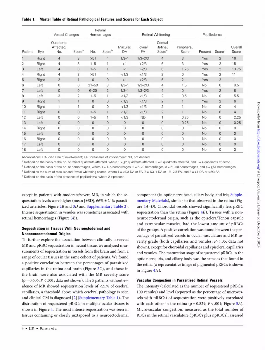
Table 1. Master Table of Retinal Pathological Features and Scores for Each Subject
Patient Eye
Vessel ChangesRetinal
Hemorrhages Retinal Whitening Papilledema
OverallScore
QuadrantsAffected,
No. Scorea No. ScorebMacular,
DAFoveal,FA
CentralRetinal,Scorec
Peripheral,Score Present Scored
1 Right 4 3 ≥51 4 1/3–1 1/3–2/3 4 3 Yes 2 162 Right 4 3 1–5 1 >1 >2/3 6 3 Yes 2 15
3 Left 4 3 1–5 1 >1 >2/3 6 1.75 Yes 2 13.75
4 Right 4 3 ≥51 4 <1/3 <1/3 2 0 Yes 2 115 Right 2 1 0 0 >1 >2/3 6 2 Yes 2 11
6 Left 0 0 21–50 3 1/3–1 1/3–2/3 4 1.5 No 0 8.5
7 Left 0 0 6–20 2 1/3–1 1/3–2/3 4 0 Yes 2 88 Left 3 2 1–5 1 <1/3 <1/3 2 0.5 No 0 5.5
9 Right 1 1 0 0 <1/3 <1/3 2 1 Yes 2 6
10 Right 1 1 0 0 <1/3 <1/3 2 1 No 0 411 Right 0 0 1–5 1 <1/3 <1/3 2 1 No 0 4
12 Left 0 0 1–5 1 <1/3 ND 1 0.25 No 0 2.25
13 Left 0 0 0 0 0 0 0 0.25 No 0 0.2514 Right 0 0 0 0 0 0 0 0 No 0 0
15 Left 0 0 0 0 0 0 0 0 No 0 0
16 Right 0 0 0 0 0 0 0 0 No 0 017 Left 0 0 0 0 0 0 0 0 No 0 0
18 Left 0 0 0 0 0 0 0 0 No 0 0
Abbreviations: DA, disc area of involvement; FA, foveal area of involvement; ND, not defined.a Defined on the basis of the no. of retinal quadrants affected, where 1 =≤2 quadrants affected, 2 = 3 quadrants affected, and 3 = 4 quadrants affected.b Defined on the basis of the no. of hemorrhages, where 1 = 1–5 hemorrhages, 2 = 6–20 hemorrhages, 3 = 21–50 hemorrhages, and 4 =≥51 hemorrhages.c Defined as the sum of macular and foveal whitening scores, where 1 = <1/3 DA or FA, 2 = 1/3–1 DA or 1/3–2/3 FA, and 3 = >1 DA or >2/3 FA.d Defined on the basis of the presence of papilledema, where 2 = present.
4 • JID • Barrera et al
at Liverpool U
niversity Library on D
ecember 3, 2014
http://jid.oxfordjournals.org/D
ownloaded from
per 100 parasitized venules, was increased in patients with MR,compared with those without MR (mean [±SD], 843 ± 337 and352 ± 222, respectively; P = .004; data not shown) and positivelyassociated with sequestration levels (ρ = 0.841; P < .001; datanot shown). We compared means across the 3 MR severity
classification groups and found significantly greater congestionin individuals with moderate/severe MR, compared with thosewithout MR (P = .008).
A descriptive model using linear regression analysis con-firmed the relationship between vascular congestion and levels
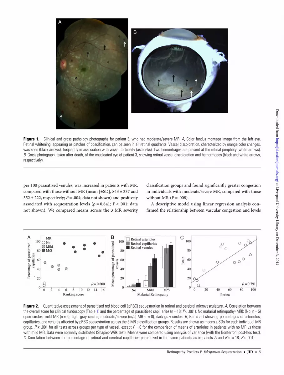
Figure 1. Clinical and gross pathology photographs for patient 3, who had moderate/severe MR. A, Color fundus montage image from the left eye.Retinal whitening, appearing as patches of opacification, can be seen in all retinal quadrants. Vessel discoloration, characterized by orange color changes,was seen (black arrows), frequently in association with vessel tortuosity (asterisks). Two hemorrhages are present at the retinal periphery (white arrows).B, Gross photograph, taken after death, of the enucleated eye of patient 3, showing retinal vessel discoloration and hemorrhages (black and white arrows,respectively).
Figure 2. Quantitative assessment of parasitized red blood cell ( pRBC) sequestration in retinal and cerebral microvasculature. A, Correlation betweenthe overall score for clinical fundoscopy (Table 1) and the percentage of parasitized capillaries (n = 18; P < .001). No malarial retinopathy (MR); (No; n = 5)open circles; mild MR (n = 5), light gray circles; moderate/severe (m/s) MR (n = 8), dark gray circles. B, Bar chart showing percentages of arterioles,capillaries, and venules affected by pRBC sequestration across the 3 MR classification groups. Results are shown as means ± SDs for each individual MRgroup. P ≤ .001 for all tests across groups per type of vessel, except P = .8 for the comparison of means of arterioles in patients with no MR vs thosewith mild MR. Data were normally distributed (Shapiro-Wilk test). Means were compared using analysis of variance (with the Bonferroni post-hoc test).C, Correlation between the percentage of retinal and cerebral capillaries parasitized in the same patients as in panels A and B (n = 18; P < .001).
Retinopathy Predicts P. falciparum Sequestration • JID • 5
at Liverpool U
niversity Library on D
ecember 3, 2014
http://jid.oxfordjournals.org/D
ownloaded from

of sequestration (Figure 5B). The number of npRBCs was notincreased in vessels without parasite sequestration (ρ = − 0.401;P = .11; data not shown), confirming that vascular congestion isrestricted to affected venules and is not attributable to postmor-tem artifacts. Histologically, the lumen of congested venules inmoderate/severe MR cases appeared to be packed with bothpRBCs and npRBCs (Figure 5C).
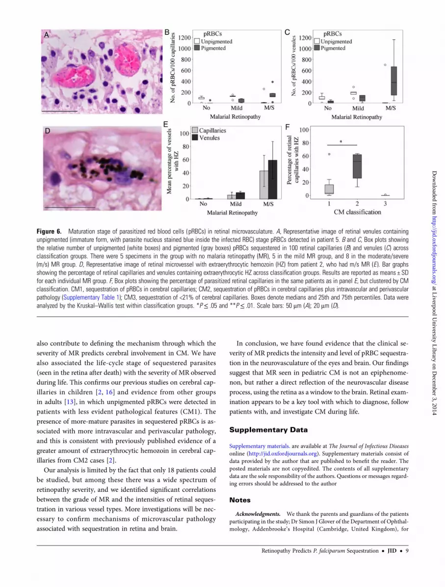
Parasite Stage in Relation to MR and CM SeverityIn moderate/severe MR cases, pigmented pRBCs were the mostpredominant forms, with unpigmented pRBCs composing<10% of the total amount of pRBCs sequestered, except patient5 (Figure 6A). Unpigmented pRBCs were more commonly ob-served in retinopathy-negative and mild MR cases (Figure 6Band 6C). We used hemozoin as an additional marker of parasite
Figure 3. Photomicrographs of parasitized retinal capillaries and venules. A, Representative image of retina cross-sections from a moderate/severe MRspecimen (patient 3) containing pigmented (mature-form) parasitized red blood cells (pRBCs). Cross-section showing capillaries and venules affected byparasite sequestration are shown (arrows, intense sequestration; arrowheads, low or no sequestration). B, High-power image of 1 representative venule withnumerous pRBCs. A prominent endothelial cell nucleus is shown (arrow). C, A Giemsa-stained venule illustrating pigmented (mature-form) pRBCs (patient7). D, Parasitized arteriole in a cross-section stained by hematoxylin-eosin, with a lower number of pRBCs (arrow; patient 8). The vessel wall is characterizedby a thick, pink-stained muscle layer. E, Highly parasitized venules with adjacent superficial retinal hemorrhage ( patient 4). Section was stained byhematoxylin-eosin. Nonparasitized RBCs stain red, while pigmented pRBCs contain hemozoin, which appears as dark brown spots. Scale bars: 100 μm(A); 20 μm (B–D), and 50 μm (E ).
6 • JID • Barrera et al
at Liverpool U
niversity Library on D
ecember 3, 2014
http://jid.oxfordjournals.org/D
ownloaded from
maturation across classification groups (Figure 6D–E ). In pa-tients with no MR and those with mild MR, extraerythrocytichemozoin was seen in <2% of retinal capillaries and <15% ofretinal venules. Mean percentages (±SD) of retinal capillariesand venules containing extraerythrocytic hemozoin were highin the moderate/severe MR group (43% ± 24% and 59% ± 28%,respectively). The percentage of retinal capillaries with extra-erythrocytic hemozoin was significantly greater in patientswith cerebral sequestration plus additional vascular pathologyin the brain (CM2) than in patients with cerebral sequestrationalone (CM1; P < .05; Figure 6F ), confirming previously pub-lished evidence for hemozoin in cerebral capillaries [2].
DISCUSSION
Clinical observation of MR in children with CM is predictive ofcerebral sequestration and death [2]. Our results add importantnew histological evidence to this finding by showing that the
degree of sequestration of pRBCs in the retinal microvasculaturediscriminates between patients with MR and those without MR,reflecting the severity of sequestration and pathology in thebrain. We also provide quantitative evidence for the first timethat the severity of MR is correlated with the intensity of seques-tration and the parasite maturation stage in retinal microvascu-lature. Intense sequestration was found in almost all the oculartissues with neuroectodermal components, and the proportionof retinal capillaries with extraerythrocytic hemozoin predictsCM1 and CM2, 2 histological categories in the brain. The par-allels between ocular pathology and brain pathology stronglysuggest that features of clinically observed MR closely reflectthe CM process, which is taking place throughout the neurovas-culature, and that the severity of retinopathy is directly relatedto the intensity of sequestration.
In all patients without MR, <20% of retinal microvessels con-tained parasites, which were in smaller numbers and at earlierstages than in MR-positive patients, hypothetically reflecting a
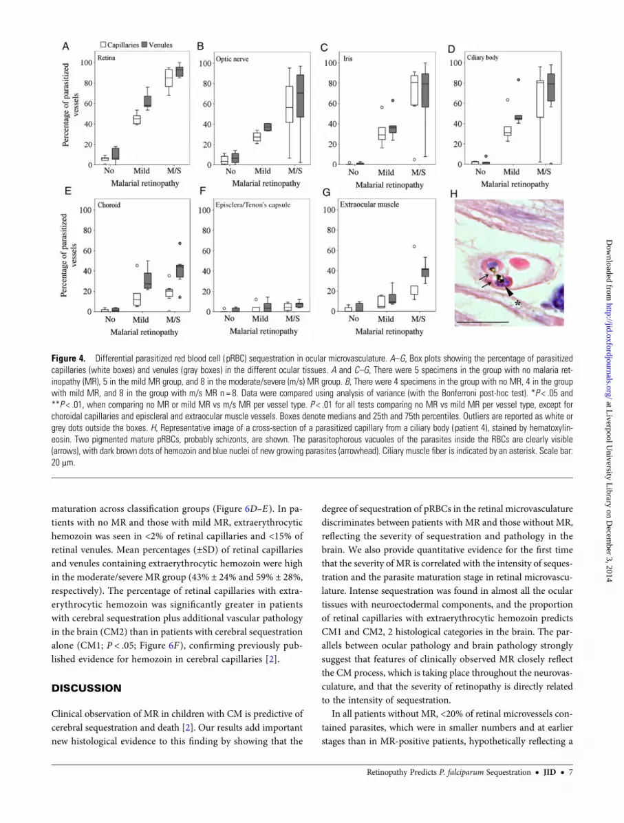
Figure 4. Differential parasitized red blood cell ( pRBC) sequestration in ocular microvasculature. A–G, Box plots showing the percentage of parasitizedcapillaries (white boxes) and venules (gray boxes) in the different ocular tissues. A and C–G, There were 5 specimens in the group with no malaria ret-inopathy (MR), 5 in the mild MR group, and 8 in the moderate/severe (m/s) MR group. B, There were 4 specimens in the group with no MR, 4 in the groupwith mild MR, and 8 in the group with m/s MR n = 8. Data were compared using analysis of variance (with the Bonferroni post-hoc test). *P < .05 and**P < .01, when comparing no MR or mild MR vs m/s MR per vessel type. P < .01 for all tests comparing no MR vs mild MR per vessel type, except forchoroidal capillaries and episcleral and extraocular muscle vessels. Boxes denote medians and 25th and 75th percentiles. Outliers are reported as white orgrey dots outside the boxes. H, Representative image of a cross-section of a parasitized capillary from a ciliary body (patient 4), stained by hematoxylin-eosin. Two pigmented mature pRBCs, probably schizonts, are shown. The parasitophorous vacuoles of the parasites inside the RBCs are clearly visible(arrows), with dark brown dots of hemozoin and blue nuclei of new growing parasites (arrowhead). Ciliary muscle fiber is indicated by an asterisk. Scale bar:20 μm.
Retinopathy Predicts P. falciparum Sequestration • JID • 7
at Liverpool U
niversity Library on D
ecember 3, 2014
http://jid.oxfordjournals.org/D
ownloaded from
nonmalarial coma with incidental parasitemia. These values arecomparable with the 21% cutoff of parasitized capillaries in thebrain described previously [2], which separated children whosedeath appeared to be due to CM from those with another causeof death and incidental parasitemia. Among patients with MR,there were significantly different levels (expressed as a percent-age of parasitized vessels), as well as intensity (expressed as thenumber of pRBCs) of parasite sequestration between mild MRand moderate/severe MR.
Parasite sequestration can lead to vessel engorgement [23] andalterations of the vessel wall [24], histologically described as dis-tension of vessels [25] or dilation [19],with impaired venous out-flow resulting in vascular congestion. We found an increasednumber of pRBCs sequestered in retinal venules from retinopa-thy-positive patients. We have shown histopathological evidenceof vascular congestion as a measure of the density of accumulatedcells (pRBCs plus npRBCs [19]) and abnormal alterations in ret-inal venules packed with pRBCs plus npRBCs; a representativeimage of distended vessel wall and rounded large endothelial nu-clei is shown in Figure 5C. In a postmortem histopathology studyof fatal CM in Vietnamese adults, congestion of heavily parasit-ized cerebral microvessels was associated with length of coma be-fore death occurred (ie, shorter time to death) [19].
Vascular congestion may be an important part of the retinaldisease process leading to MR, possibly reflecting the abnormalaccumulation of pRBCs in the cerebral vasculature evolvingwith CM. Increased vascular congestion may be due to the ro-setting process occurring in malaria, by which pRBC can bindto multiple npRBCs, and potentially contributing to microvas-cular obstruction [26] and CM [27].When evolving with vessel
obstruction, congestion is likely to induce local hypoxia, intra-vascular thrombosis [22, 28], increased transmural and shearstress, and pathological changes to endothelial cells [23, 29], al-ready described in abnormal vessels observed in children withMR [4, 6].
We are the first to have directly correlated sequestration levelsin neurovasculature from the retina and brain, 2 organs of neu-roectodermal origin, in children dying of CM. To explore fur-ther the association between MR and pRBC sequestration inneural tissues, we analyzed ocular tissues closest to neuroecto-dermal origin, namely the optic nerve head, ciliary body, andiris (Supplementary Materials), and found that sequestrationwas as intense as seen in the retina. This similarity is likelydue to similar vascular endothelial surface receptor profiles, in-duced by the tissue in which the vessels are situated and medi-ating the adhesion of pRBCs within the vessel. The choroid,despite its proximity to the retinal pigment epithelium, whichis derived from the neuroectoderm, contained less intense se-questration than other intraocular tissues. Other mechanisms,such as ocular hemodynamics, may play an important role indetermining the degree of sequestration in microvessels; thechoroid has a high rate of blood flow, roughly 20 times thatof the retina [4].Our observation of greater sequestration in ret-inal venules and capillaries than in arterioles is consistent withthe hypothesis that pRBC sequestration is favored by lowerblood velocity, with consequent lower shear stress [4]. Thesedata may reflect how hemodynamics can impact differential se-questration rates in central nervous system, compared withother organs, and confirm the important diagnostic value ofMR investigation during life. For this reason, our study may
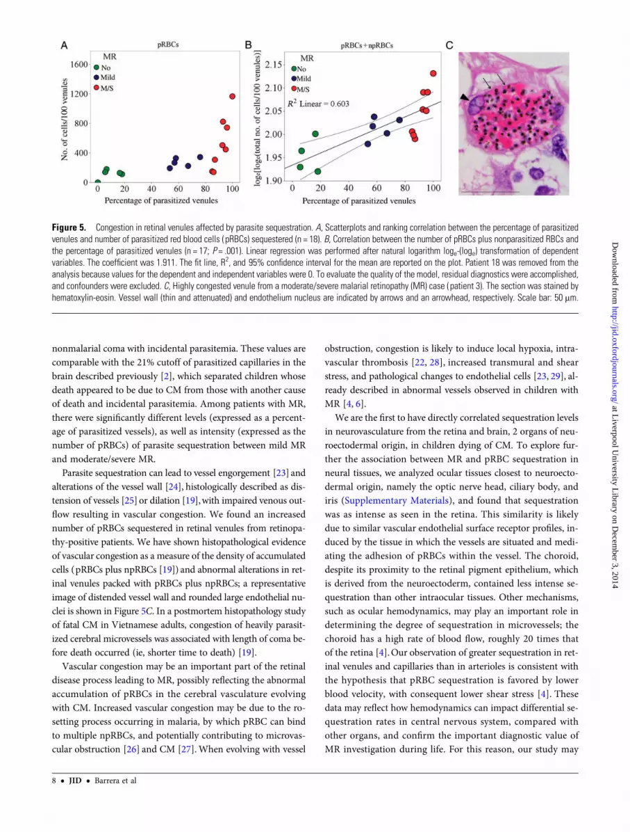
Figure 5. Congestion in retinal venules affected by parasite sequestration. A, Scatterplots and ranking correlation between the percentage of parasitizedvenules and number of parasitized red blood cells ( pRBCs) sequestered (n = 18). B, Correlation between the number of pRBCs plus nonparasitized RBCs andthe percentage of parasitized venules (n = 17; P = .001). Linear regression was performed after natural logarithm loge-(loge) transformation of dependentvariables. The coefficient was 1.911. The fit line, R2, and 95% confidence interval for the mean are reported on the plot. Patient 18 was removed from theanalysis because values for the dependent and independent variables were 0. To evaluate the quality of the model, residual diagnostics were accomplished,and confounders were excluded. C, Highly congested venule from a moderate/severe malarial retinopathy (MR) case (patient 3). The section was stained byhematoxylin-eosin. Vessel wall (thin and attenuated) and endothelium nucleus are indicated by arrows and an arrowhead, respectively. Scale bar: 50 μm.
8 • JID • Barrera et al
at Liverpool U
niversity Library on D
ecember 3, 2014
http://jid.oxfordjournals.org/D
ownloaded from
also contribute to defining the mechanism through which theseverity of MR predicts cerebral involvement in CM. We havealso associated the life-cycle stage of sequestered parasites(seen in the retina after death) with the severity of MR observedduring life. This confirms our previous studies on cerebral cap-illaries in children [2, 16] and evidence from other groupsin adults [13], in which unpigmented pRBCs were detected inpatients with less evident pathological features (CM1). Thepresence of more-mature parasites in sequestered pRBCs is as-sociated with more intravascular and perivascular pathology,and this is consistent with previously published evidence of agreater amount of extraerythrocytic hemozoin in cerebral cap-illaries from CM2 cases [2].
Our analysis is limited by the fact that only 18 patients couldbe studied, but among these there was a wide spectrum ofretinopathy severity, and we identified significant correlationsbetween the grade of MR and the intensities of retinal seques-tration in various vessel types. More investigations will be nec-essary to confirm mechanisms of microvascular pathologyassociated with sequestration in retina and brain.
In conclusion, we have found evidence that the clinical se-verity of MR predicts the intensity and level of pRBC sequestra-tion in the neurovasculature of the eyes and brain. Our findingssuggest that MR seen in pediatric CM is not an epiphenome-non, but rather a direct reflection of the neurovascular diseaseprocess, using the retina as a window to the brain. Retinal exam-ination appears to be a key tool with which to diagnose, followpatients with, and investigate CM during life.
Supplementary Data
Supplementary materials. are available at The Journal of Infectious Diseasesonline (http://jid.oxfordjournals.org). Supplementary materials consist ofdata provided by the author that are published to benefit the reader. Theposted materials are not copyedited. The contents of all supplementarydata are the sole responsibility of the authors. Questions or messages regard-ing errors should be addressed to the author
Notes
Acknowledgments. We thank the parents and guardians of the patientsparticipating in the study; Dr Simon J Glover of the Department of Ophthal-mology, Addenbrooke’s Hospital (Cambridge, United Kingdom), for
Figure 6. Maturation stage of parasitized red blood cells (pRBCs) in retinal microvasculature. A, Representative image of retinal venules containingunpigmented (immature form, with parasite nucleus stained blue inside the infected RBC) stage pRBCs detected in patient 5. B and C, Box plots showingthe relative number of unpigmented (white boxes) and pigmented (gray boxes) pRBCs sequestered in 100 retinal capillaries (B) and venules (C) acrossclassification groups. There were 5 specimens in the group with no malaria retinopathy (MR), 5 in the mild MR group, and 8 in the moderate/severe(m/s) MR group. D, Representative image of retinal microvessel with extraerythrocytic hemozoin (HZ) from patient 2, who had m/s MR (E ). Bar graphsshowing the percentage of retinal capillaries and venules containing extraerythrocytic HZ across classification groups. Results are reported as means ± SDfor each individual MR group. F, Box plots showing the percentage of parasitized retinal capillaries in the same patients as in panel E, but clustered by CMclassification. CM1, sequestration of pRBCs in cerebral capillaries; CM2, sequestration of pRBCs in cerebral capillaries plus intravascular and perivascularpathology (Supplementary Table 1); CM3, sequestration of <21% of cerebral capillaries. Boxes denote medians and 25th and 75th percentiles. Data wereanalyzed by the Kruskal–Wallis test within classification groups. *P≤ .05 and **P≤ .01. Scale bars: 50 μm (A); 20 μm (D).
Retinopathy Predicts P. falciparum Sequestration • JID • 9
at Liverpool U
niversity Library on D
ecember 3, 2014
http://jid.oxfordjournals.org/D
ownloaded from
assistance with retinal examinations; Dr Macpherson Mallewa and the nurs-es of the Pediatric Department Research Ward, Queen Elizabeth CentralHospital (Blantyre, Malawi), for caring for the patients; Dr Gabriela Czan-ner of the Biostatistics Department, University of Liverpool (Liverpool,United Kingdom), for assistance with statistical analysis; Dr Richard Carrof the Pathology Laboratory, South Warwickshire General Hospitals NHSTrust (Warwick, United Kingdom), for his support with parasite stage eval-uation in histological sections; Simon Biddolph, Thomas Whitehead, andDuaha Ghafouri of the Royal Liverpool University Hospital and Universityof Liverpool and laboratory technologists at Vancouver General Hospital(Vancouver, Canada), for assistance with histopathological quantitativeanalysis (see data analysis in Supplementary Materials); and Dr YalinZheng and Man Ting Kwong, for clinical photographic montages.V. B. and D. A. M. performed the histopathological experiments on ocu-
lar and brain tissues, respectively. V. B., P. S. H., A. G. C., N. A. V. B., andS. P. H. conceived and designed the experiments. V. B. and P. S. H. analyzedthe data. V. B., P. S. H., V. A. W., D. A. M., N. A. V. B., and S. P. H. con-tributed to reagents, materials, and/or analysis tools. V. B. wrote the firstdraft of the manuscript. V. B., P. S. H., A. G. C., V. A. W., D. A. M.,N. A. V.B, I. J. C. M., S. K., T. E. T., M. E. M., and S. P. H. contributed tothe writing and agreed with manuscript results and conclusions.Disclaimer. The funders had no role in study design, data collection
and analysis, decision to publish, or preparation of the manuscript.Financial support. This work was supported by the Wellcome Trust
(grant 092668/Z/10/Z to S. P. H., grant 084679/Z/08/Z, and grant 074125to M. E. M.) and the National Institutes of Health (grant 5R01AI034969-11 to T. E. T.), the last two for The Blantyre Autopsy Study (access to ocularand brain specimens).Potential conflicts of interest. All authors: No reported conflicts.All authors have submitted the ICMJE Form for Disclosure of Potential
Conflicts of Interest. Conflicts that the editors consider relevant to the con-tent of the manuscript have been disclosed.
References
1. Newton CR, Taylor TE, Whitten RO. Pathophysiology of fatal falci-parum malaria in African children. Am J Trop Med Hyg 1998;58:673–83.
2. Taylor TE, Fu WJ, Carr RA, et al. Differentiating the pathologies ofcerebral malaria by postmortem parasite counts. Nat Med 2004; 10:143–5.
3. Hero M, Harding SP, Riva CE, Winstanley PA, Peshu N, Marsh K.Photographic and angiographic characterization of the retina ofKenyan children with severe malaria. Arch Ophthalmol 1997; 115:997–1003.
4. Maccormick IJ, Beare NA, Taylor TE, et al. Cerebral malaria in children:using the retina to study the brain. Brain 2014; 137(Pt 8):2119–42.
5. Essuman VA, Ntim-Amponsah CT, Astrup BS, et al. Retinopathy in se-vere malaria in Ghanaian children–overlap between fundus changes incerebral and non-cerebral malaria. Malar J 2010; 9:232.
6. Harding SP, Lewallen S, Beare NA, Smith A, Taylor TE, Molyneux ME.Classifying and grading retinal signs in severe malaria. Trop Doct 2006;36(suppl 1):1–13.
7. Beare NA, Southern C, Chalira C, Taylor TE, Molyneux ME, HardingSP. Prognostic significance and course of retinopathy in children withsevere malaria. Arch Ophthalmol 2004; 122:1141–7.
8. White VA, Lewallen S, Beare N, Kayira K, Carr RA, Taylor TE. Corre-lation of retinal haemorrhages with brain haemorrhages in childrendying of cerebral malaria in Malawi. Trans R Soc Trop Med Hyg2001; 95:618–21.
9. Bannister L, Mitchell G. The ins, outs and roundabouts of malaria.Trends Parasitol 2003; 19:209–13.
10. Grau GE, Craig AG. Cerebral malaria pathogenesis: revisiting parasiteand host contributions. Future Microbiol 2012; 7:291–302.
11. Brown H, Rogerson S, Taylor T, et al. Blood-brain barrier function incerebral malaria in Malawian children. Am J Trop Med Hyg 2001;64:207–13.
12. Hempelmann E, Motta C, Hughes R, Ward SA, Bray PG. Plasmodiumfalciparum: sacrificing membrane to grow crystals? Trends Parasitol2003; 19:23–6.
13. Silamut K, Phu NH, Whitty C, et al. A quantitative analysis of the mi-crovascular sequestration of malaria parasites in the human brain. Am JPathol 1999; 155:395–410.
14. Armah H, Dodoo AK, Wiredu EK, et al. High-level cerebellar expres-sion of cytokines and adhesion molecules in fatal, paediatric, cerebralmalaria. Ann Trop Med Parasitol 2005; 99:629–47.
15. Dorovini-Zis K, Schmidt K, Huynh H, et al. The neuropathology offatal cerebral malaria in malawian children. Am J Pathol 2011; 178:2146–58.
16. Milner DA, Valim C, Carr RA, et al. A histological method for quanti-fying Plasmodium falciparum in the brain in fatal paediatric cerebralmalaria. Malar J 2013; 12:191.
17. Nguansangiam S, Day NP, Hien TT, et al. A quantitative ultrastructuralstudy of renal pathology in fatal Plasmodium falciparum malaria. TropMed Int Health 2007; 12:1037–50.
18. White NJ, Turner GD, Day NP, Dondorp AM. Lethal malaria: March-iafava and Bignami were right. J Infect Dis 2013; 208:192–8.
19. Ponsford MJ, Medana IM, Prapansilp P, et al. Sequestration and micro-vascular congestion are associated with coma in human cerebral malar-ia. J Infect Dis 2012; 205:663–71.
20. Maude RJ, Dondorp AM, Abu Sayeed A, Day NP, White NJ, Beare NA.The eye in cerebral malaria: what can it teach us? Trans R Soc Trop MedHyg 2009; 103:661–4.
21. Molyneux ME, Taylor TE, Wirima JJ, Borgstein A. Clinical features andprognostic indicators in paediatric cerebral malaria: a study of 131 co-matose Malawian children. QJM 1989; 71:441–59.
22. White V, Lewallen S, Beare N, Molyneux M, Taylor T. Retinal pathologyof pediatric cerebral malaria in Malawi. PLoS One 2009; 4:e4317.
23. Dondorp AM, Pongponratn E, White NJ. Reduced microcirculatoryflow in severe falciparummalaria: pathophysiology and electron-micro-scopic pathology. Acta Trop 2004; 89:309–17.
24. Mitchell RN. Hemodynamic disorders, thromboembolism and shock.In Kumar V, Abbas A, Aster J, eds. Robbins basic pathology. 9th ed.Philadelphia, Pennsylvania: Elsevier, 2012:75–98.
25. Mathan VI, Penny GR, Mathan MM, Rowley D. Bacterial lipopolysac-charide-induced intestinal microvascular lesions leading to acute diar-rhea. J Clin Invest 1988; 82:1714–21.
26. Kaul DK, Roth EF Jr, Nagel RL, Howard RJ, Handunnetti SM. Rosettingof Plasmodium falciparum-infected red blood cells with uninfected redblood cells enhances microvascular obstruction under flow conditions.Blood 1991; 78:812–9.
27. Carlson J, Helmby H, Hill AV, Brewster D, Greenwood BM, WahlgrenM. Human cerebral malaria: association with erythrocyte rosetting andlack of anti-rosetting antibodies. Lancet 1990; 336:1457–60.
28. Moxon CA, Wassmer SC, Milner DA, et al. Loss of endothelial pro-tein C receptors links coagulation and inflammation to parasite se-questration in cerebral malaria in African children. Blood 2013;122:842–51.
29. Popel AS, Johnson PC. Microcirculation and hemorheology. Ann RevFluid Mech 2005; 37:43–69.
10 • JID • Barrera et al
at Liverpool U
niversity Library on D
ecember 3, 2014
http://jid.oxfordjournals.org/D
ownloaded from