Mobile Telemedicine Screening for Diabetic Retinopathy ...
13
Citation: Charlot, A.; Baudin, F.; Tessier, M.; Lebrize, S.; Hurand, V.; Megroian, D.; Arnould, L.; Ben-Ghezala, I.; Bron, A.M.; Gabrielle, P.-H.; et al. Mobile Telemedicine Screening for Diabetic Retinopathy Using Nonmydriatic Fundus Photographs in Burgundy: 11 Years of Results. J. Clin. Med. 2022, 11, 1318. https://doi.org/10.3390/ jcm11051318 Academic Editor: Alberto Barcelo Received: 25 January 2022 Accepted: 25 February 2022 Published: 27 February 2022 Publisher’s Note: MDPI stays neutral with regard to jurisdictional claims in published maps and institutional affil- iations. Copyright: © 2022 by the authors. Licensee MDPI, Basel, Switzerland. This article is an open access article distributed under the terms and conditions of the Creative Commons Attribution (CC BY) license (https:// creativecommons.org/licenses/by/ 4.0/). Journal of Clinical Medicine Article Mobile Telemedicine Screening for Diabetic Retinopathy Using Nonmydriatic Fundus Photographs in Burgundy: 11 Years of Results Anthony Charlot 1 , Florian Baudin 1,2,3, * ,Mélanie Tessier 1 , Sarah Lebrize 1 , Victoire Hurand 1 , Déborah Megroian 1 , Louis Arnould 1 , Inès Ben-Ghezala 1,2 , Alain Marie Bron 1,4 , Pierre-Henry Gabrielle 1,4 and Catherine Creuzot-Garcher 1,4 1 Ophthalmology Department, University Hospital, 14 Rue Paul Gaffarel, 21079 Dijon, France; [email protected] (A.C.); [email protected] (M.T.); [email protected] (S.L.); [email protected] (V.H.); [email protected] (D.M.); [email protected] (L.A.); [email protected] (I.B.-G.); [email protected] (A.M.B.); [email protected] (P.-H.G.); [email protected] (C.C.-G.) 2 INSERM—Institut National de la Santé et de la Recherche Médicale, CIC 1432, Clinical Investigation Center, Clinical Epidemiology/Clinical Trials Unit, Burgundy University, 21000 Dijon, France 3 Physiopathologie et Epidémiologie Cérébro-Cardiovasculaires (PEC2, EA 7460), Burgundy University, 21000 Dijon, France 4 Eye and Nutrition Research Group, CSGA, UMR 1324 INRA, 6265 CNRS, Burgundy University, 21000 Dijon, France * Correspondence: fl[email protected]; Tel.: +33-380-293-536; Fax: +33-380293879 Abstract: We analyzed the results of mobile screening for diabetic retinopathy (DR) using retinal photographs, comparing these results between rural and periurban areas, and before and after the first national COVID-19 pandemic lockdown. The Burgundy Union Régionale des Professionnels de Santé (URPS) has organized an annual DR screening since 2004. The examination, performed by an orthoptist, consisted of taking the patient 0 s history, intraocular pressure measurement, and taking retinal photographs. After remote transmission, the examinations were interpreted by participating ophthalmologists at the Dijon University Hospital. In September 2016, the screening was open to periurban townships. In 11 years, 10,220 patients were screened: 1420 patients (13.9%) had DR of any type, with an average age of 68.5 (±11.3) years, and 59.2% were men. These patients had a statistically significantly higher glycated hemoglobin level (7.4% vs. 7.0%) and a longer duration of diabetes (13.8 vs. 9.3 years) than patients without DR. When comparing rural and periurban areas and periods before and after the beginning of the COVID-19 pandemic, we did not find any significant difference in the screening results. The results of this study are in line with the average findings of similar studies comparing screening strategies for DR. The early detection of DR can benefit from mobile telemedicine screening, identifying a considerable number of patients at an elevated risk, especially in rural areas where access to ophthalmological care is limited. Keywords: diabetic retinopathy; telemedicine; screening 1. Introduction Diabetes mellitus is a multifactorial disease with genetic and environmental causes whose prevalence continues to increase. Between 2000 and 2013, the rate of patients treated for diabetes in France increased by 5.4% per year [1]. In France, according to the International Diabetes Federation (IDF), the number of people between 20 and 79 years of age suffering from diabetes was estimated to be 3.48 million in 2019 (4.8% of the population). This number is expected to increase to 3.76 million in 2030 (5.5%) [2]. Worldwide, the number of people with diabetes is expected to grow from 463 million in 2019 to 642 million J. Clin. Med. 2022, 11, 1318. https://doi.org/10.3390/jcm11051318 https://www.mdpi.com/journal/jcm
-
Upload
khangminh22 -
Category
Documents
-
view
0 -
download
0
Transcript of Mobile Telemedicine Screening for Diabetic Retinopathy ...

�����������������
Citation: Charlot, A.; Baudin, F.;
Tessier, M.; Lebrize, S.; Hurand, V.;
Megroian, D.; Arnould, L.;
Ben-Ghezala, I.; Bron, A.M.;
Gabrielle, P.-H.; et al. Mobile
Telemedicine Screening for Diabetic
Retinopathy Using Nonmydriatic
Fundus Photographs in Burgundy:
11 Years of Results. J. Clin. Med. 2022,
11, 1318. https://doi.org/10.3390/
jcm11051318
Academic Editor: Alberto Barcelo
Received: 25 January 2022
Accepted: 25 February 2022
Published: 27 February 2022
Publisher’s Note: MDPI stays neutral
with regard to jurisdictional claims in
published maps and institutional affil-
iations.
Copyright: © 2022 by the authors.
Licensee MDPI, Basel, Switzerland.
This article is an open access article
distributed under the terms and
conditions of the Creative Commons
Attribution (CC BY) license (https://
creativecommons.org/licenses/by/
4.0/).
Journal of
Clinical Medicine
Article
Mobile Telemedicine Screening for Diabetic Retinopathy UsingNonmydriatic Fundus Photographs in Burgundy: 11 Yearsof ResultsAnthony Charlot 1, Florian Baudin 1,2,3,* , Mélanie Tessier 1, Sarah Lebrize 1, Victoire Hurand 1,Déborah Megroian 1, Louis Arnould 1 , Inès Ben-Ghezala 1,2 , Alain Marie Bron 1,4 ,Pierre-Henry Gabrielle 1,4 and Catherine Creuzot-Garcher 1,4
1 Ophthalmology Department, University Hospital, 14 Rue Paul Gaffarel, 21079 Dijon, France;[email protected] (A.C.); [email protected] (M.T.); [email protected] (S.L.);[email protected] (V.H.); [email protected] (D.M.);[email protected] (L.A.); [email protected] (I.B.-G.); [email protected] (A.M.B.);[email protected] (P.-H.G.); [email protected] (C.C.-G.)
2 INSERM—Institut National de la Santé et de la Recherche Médicale, CIC 1432, Clinical Investigation Center,Clinical Epidemiology/Clinical Trials Unit, Burgundy University, 21000 Dijon, France
3 Physiopathologie et Epidémiologie Cérébro-Cardiovasculaires (PEC2, EA 7460), Burgundy University,21000 Dijon, France
4 Eye and Nutrition Research Group, CSGA, UMR 1324 INRA, 6265 CNRS, Burgundy University,21000 Dijon, France
* Correspondence: [email protected]; Tel.: +33-380-293-536; Fax: +33-380293879
Abstract: We analyzed the results of mobile screening for diabetic retinopathy (DR) using retinalphotographs, comparing these results between rural and periurban areas, and before and after thefirst national COVID-19 pandemic lockdown. The Burgundy Union Régionale des Professionnels deSanté (URPS) has organized an annual DR screening since 2004. The examination, performed by anorthoptist, consisted of taking the patient′s history, intraocular pressure measurement, and takingretinal photographs. After remote transmission, the examinations were interpreted by participatingophthalmologists at the Dijon University Hospital. In September 2016, the screening was open toperiurban townships. In 11 years, 10,220 patients were screened: 1420 patients (13.9%) had DR of anytype, with an average age of 68.5 (±11.3) years, and 59.2% were men. These patients had a statisticallysignificantly higher glycated hemoglobin level (7.4% vs. 7.0%) and a longer duration of diabetes(13.8 vs. 9.3 years) than patients without DR. When comparing rural and periurban areas and periodsbefore and after the beginning of the COVID-19 pandemic, we did not find any significant differencein the screening results. The results of this study are in line with the average findings of similarstudies comparing screening strategies for DR. The early detection of DR can benefit from mobiletelemedicine screening, identifying a considerable number of patients at an elevated risk, especiallyin rural areas where access to ophthalmological care is limited.
Keywords: diabetic retinopathy; telemedicine; screening
1. Introduction
Diabetes mellitus is a multifactorial disease with genetic and environmental causeswhose prevalence continues to increase. Between 2000 and 2013, the rate of patientstreated for diabetes in France increased by 5.4% per year [1]. In France, according to theInternational Diabetes Federation (IDF), the number of people between 20 and 79 years ofage suffering from diabetes was estimated to be 3.48 million in 2019 (4.8% of the population).This number is expected to increase to 3.76 million in 2030 (5.5%) [2]. Worldwide, thenumber of people with diabetes is expected to grow from 463 million in 2019 to 642 million
J. Clin. Med. 2022, 11, 1318. https://doi.org/10.3390/jcm11051318 https://www.mdpi.com/journal/jcm

J. Clin. Med. 2022, 11, 1318 2 of 13
in 2040 [2]. This pathology causes multiple systemic complications, often of vascular origin,mainly linked to the duration of diabetes and glycemic control [3].
The most common ophthalmologic complication is diabetic retinopathy (DR): a mi-crovascular complication that can lead to retinal ischemia, neovascular glaucoma, and thusfunctional or even anatomical loss of the eyeball [4,5]. Diabetes is the world’s leading causeof blindness in the working population [6]. The prevalence of DR of any type is estimated ataround 30% of diabetic patients, increasing as the duration of diabetes, glycated hemoglobinlevel, blood pressure, and type 1 diabetes increases [7,8]. The screening of diabetic patientsis essential for diagnosing DR at an early stage, setting up appropriate treatment, andpreventing irreversible vision loss [9]. Annual screening with dilated fundus examinationis recommended, but it remains insufficient in France. According to the Haute Autorité deSanté (HAS, French National Authority for Health) in its 2010 report, only 50% of diabeticpatients received an annual ophthalmological consultation and 72% within 2 years. Asa result, screening with nonmydriatic retinal photographs and remote interpretation hasbeen considered [10]. Various standardized recruitment protocols have been implementedto increase annual screenings, whether via a general practitioner or an endocrinologist, inhospitals [11–13] or drugstores [14], or even set up by local authorities [15]. Mobile screen-ing is justified in Burgundy because of its vast territory and a density of ophthalmologiststhat is lower than the national average, at 6.65 ophthalmologists per 100,000 inhabitants,versus 8.8 per 100,000 in France (French Ministry of Health).
The main objective of this study is to describe the prevalence and characteristics ofDR in a mobile screening campaign in Burgundy, France. The secondary objectives are todescribe the prevalence of ophthalmologic pathologies not related to diabetes, to compareresults between rural and periurban populations, and describe the results before and afterthe COVID-19 pandemic.
2. Materials and Methods
This was a retrospective, cross-sectional, and descriptive study based on screeningdata analysis performed between September 2009 and November 2020 in Burgundy, France.The study complied with the Declaration of Helsinki, and the locally appointed ethicscommittee gave approval for the research protocol.
2.1. Recruitment and Procedure
This itinerant screening for DR has been organized every year since 2004 by theBurgundy Union Régionale des Professionnels de Santé—Médecins Libéraux (URPS-ML,Regional Union of Health Professionals–Private Physicians). Each screening campaignbegins in September and runs through June of the following year. A break is usually takenin December and January. In 2020, due to the COVID-19 pandemic and the first nationallockdown in France, screening was suspended between 20 February and 6 October. Inevery visited township, departmental healthcare organizations identify diabetic patientswho did not have any ophthalmologic acts coded in the previous 2 years. Then, a letter issent to inform and invite patients to this free screening. Before the COVID-19 pandemic,patients were invited to participate in the screening without an appointment. Since 2020,patients have been required to make an appointment beforehand to optimize the numberof people. Sometimes, practitioners asked the patient to bring the mobile screening deviceto the medical offices, but this took place for a minority of the screenings. The Burgundyregion was visited in 2 years, resulting in a lapse of 2 years before the same location wasrevisited. Since September 2016, screening campaigns have been open to periurban areas,whereas only the most rural townships were previously screened. We compared periurbanand rural areas between September 2016 and June 2019, according to a classification ofthe Agence Régionale de Santé de Bourgogne Franche Comté (Regional Health Agencyof Burgundy Franche Comté), initially in 5 categories on 52 social and health criteria(including socioeconomic level, healthcare supply, general and premature mortality rates,and the age of the population). We classed townships into two main categories: rural and

J. Clin. Med. 2022, 11, 1318 3 of 13
periurban townships. Because of the COVID-19 pandemic, we decided not to include theperiod beginning in September 2019 in this analysis to avoid recruitment bias.
A trained orthoptist performed the screening with mobile equipment. It consistedin collecting information about the administrative characteristics, the patient′s medicalfollow-up (referring ophthalmologist, last ophthalmological check-up, referring physician,and endocrinological follow-up), and personal history (type and duration of diabetes, an-tidiabetic and antihypertensive treatment, last glycated hemoglobin, and ophthalmologicalhistory). Then, an ophthalmological examination was performed. Intraocular pressuremeasurements were taken using the Canon TX-20P air tonometer pachymeter (Canon,Tokyo, Japan), and fundus retinal photographs were taken using a Canon NM CR-2 PlusFundus Camera (Canon, Tokyo, Japan). For each patient, two photographs were taken:the first one was centered on the posterior pole and the second on the optic nerve. Incase of poor-quality photographs, mydriatic drops were instilled in both eyes, and thephotographs were retaken. Then, the images were transmitted via a web platform to theophthalmology department of the Dijon University Hospital for remote interpretation. Theinterpretation was based on the ALFEDIAM classification (Association de langue françaisepour l’Étude du diabète et des maladies métaboliques (French Language Association for theStudy of Diabetes and Metabolic Diseases), derived from the ETDRS classification (EarlyTreatment Diabetic Retinopathy Study) [16,17]. Ophthalmologists classified the fundusexaminations in a standardized form: no DR, mild non-proliferative DR (NPDR), moderateNPDR, severe NPDR, or proliferative DR (PDR) [17]. Other suspected ophthalmologicdiseases (e.g., macular edema, ocular hypertension (OHT), optic nerve head anomaly,choroidal nevus, age-related macular degeneration (AMD), central retinal vein occlusion(CRVO), and cataracts) were noted. OHT was defined as an intraocular pressure measuredover 21 mmHg. The results were transmitted to the patients’ general practitioner andthe declared endocrinologist. The patient received only advice on when to consult anophthalmologist if needed. The ophthalmologist chosen by the patient received the reportbefore the consultation.
2.2. Statistical Analysis
The worst eye for DR status and other pathologies was considered for the analysis.First, data were described with descriptive parameters (percentages, mean, and standarddeviation). Comparative analyses were performed using a modified Student t-test forthe quantitative variables. For the qualitative variables, the chi-square test was used asappropriate. Otherwise, the Fisher test was used. The level of significance was set at0.05. Multivariate analyses for rural versus periurban comparisons were performed usinglogistic regressions.
3. Results3.1. Description of the Study Population
Between September 2009 and November 2020, 10,220 patients received at least onescreening examination, and 12,723 examinations were performed. At the time of their firstscreening examination, patients were on average 68.5 (±11.2) years old and reported theirdiabetes was ongoing for a mean 10.0 (±9.4) years. The mean glycated hemoglobin levelwas 7.1% (±1.2), based on the self-report of 9145 patients (89.48%). Other characteristicsof these patients at their first screening examination are summarized in Table 1. Only43.4% (4438 people) of the patients had not had an ophthalmologic check-up over theprevious 2 years, a percentage rising to 95.1% of patients who had not had a check-upover the previous year (9723 patients). The mean time since the last consultation was 3.7(±3.6) years based on the declaration of 9198 patients (90.0%) who were able to date theirlast consultation.

J. Clin. Med. 2022, 11, 1318 4 of 13
Table 1. Characteristics of patients at their first screening examination.
Gender
Female 4154 (40.7)Male 6066 (59.4)
Age (years) a 68.5 (±11.2)Attending ophthalmologist 3744 (36.5)
Type of diabetesType 1 355 (3.5)Type 2 8993 (88.0)Unknown 877 (8.5)
Treatment of diabetesOral antidiabetics 9063 (88.7)Insulin 1762 (17.2)
Treatment of high blood pressure 6557 (64.2)Progression ≥ 1 year 1146 (11.2)Last check-up more than 2 years before 4438 (43.4)
Number of screenings per patient1 8321 (81.4)2 1450 (14.2)3 or more 449 (4.4)
Data are qualitative, expressed as number (percentage), except for (a) a quantitative data, expressed as mean(±standard deviation).
The number of new patients between 2010 and 2020 is shown in Figure 1, with themean value of 891.7 (±295.1) of new patients per year. Data from 2009 with only 411 newpatients are not shown in the graph because they cover screening only from September toDecember of that year. The year with the most patients screened was 2019, with 1540 newpatients. Due to the COVID-19 pandemic, only 489 new patients were recruited in 2020,which is the lowest number in this study for a full year.
J. Clin. Med. 2022, 11, 1318 4 of 14
Table 1. Characteristics of patients at their first screening examination.
Gender Female 4154 (40.7) Male 6066 (59.4) Age (years) a 68.5 (± 11.2) Attending ophthalmologist 3744 (36.5) Type of diabetes Type 1 355 (3.5) Type 2 8993 (88.0) Unknown 877 (8.5) Treatment of diabetes Oral antidiabetics 9063 (88.7) Insulin 1762 (17.2) Treatment of high blood pressure 6557 (64.2) Progression ≥ 1 year 1146 (11.2) Last check-up more than 2 years before 4438 (43.4) Number of screenings per patient 1 8321 (81.4) 2 1450 (14.2) 3 or more 449 (4.4) Data are qualitative, expressed as number (percentage), except for (a) a quantitative data, ex-pressed as mean (± standard deviation).
The number of new patients between 2010 and 2020 is shown in Figure 1, with the mean value of 891.7 (±295.1) of new patients per year. Data from 2009 with only 411 new patients are not shown in the graph because they cover screening only from September to December of that year. The year with the most patients screened was 2019, with 1540 new patients. Due to the COVID-19 pandemic, only 489 new patients were recruited in 2020, which is the lowest number in this study for a full year.
Figure 1. Newly screened patients per year.
783 747 693
1060 1026
666 662
10451098
1540
489
0
200
400
600
800
1000
1200
1400
1600
1800
2010 2011 2012 2013 2014 2015 2016 2017 2018 2019 2020
New patients Mean
Figure 1. Newly screened patients per year.

J. Clin. Med. 2022, 11, 1318 5 of 13
Between September 2009 and November 2020, 12,723 examinations were performed.Due to recurrent visits in townships, 8321 patients (81.4%) had only one examination, while1899 (18.6%) had two or more examinations during the period studied. For 11 years ofscreening, an average of 1060 (±431) exams per year were performed.
3.2. Screening Results
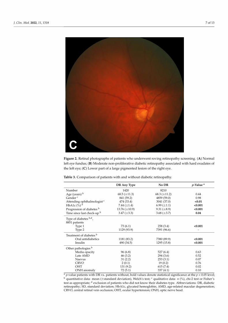
The results of analysis regarding only the first exam of each patient are shown inTable 2 (see Figure 2A,B for retinal photographs example). Over the 11 years of the study,the rate of DR in at least one eye in all stages was 13.9% (1420 patients out of 10,220). Thisrate rises to 14.8% after excluding 590 patients whose photographs were unreadable (5.8%of the patients excluded, 1420 patients with DR out of 9630 patients). At the same time, wefound other ophthalmologic conditions in at least one eye other than DR: media opacity(692 patients, 6.8%); late AMD (347 patients, 3.4%); choroidal nevus (287 patients, 2.8%; seeFigure 2C); CRVO (20 patients, 0.2%); optic nerve head anomaly (418 patients, 4.1%); andOHT (799 patients, 7.8%).
Table 2. Results of screening at the patients’ first examination.
DR Status
No DR 8209 (80.3)All stages 1420 (13.9)Mild NPDR 948 (9.3)Moderate NPDR 366 (3.6)Severe NPDR 84 (0.8)PDR 22 (0.2)
Unreadable 590 (5.8)
Other pathologiesMedia opacity 692 (6.8)Late AMD 347 (3.4)Nevus 287 (2.8)CRVO 20 (0.2)OHT 799 (7.8)Papillary anomaly 418 (4.1)
Data are qualitative, expressed as number (percentage). Abbreviations: DR, diabetic retinopathy; NPDR, non-proliferative DR; PDR, proliferative DR; AMD, age-related macular degeneration; CRVO, central retinal veinocclusion; OHT, ocular hypertension.
3.3. Comparison of Patients with and without DR of Any Type
We compared patients with DR of any type with those without, and we obtained twogroups of 1420 and 8210 patients, respectively, after excluding 590 (5.8%) patients withunreadable photographs. The results are shown in Table 3. Patients with DR of any typehad significantly more type 1 diabetes (p < 0.001), higher glycated hemoglobin (p < 0.001),longer duration of diabetes (p < 0.001), more insulin treatment (p < 0.001), and fewer oralantidiabetics (p < 0.001). They also significantly had a shorter time since the last visit(p = 0.04) and more often reported an attending ophthalmologist (p < 0.01).
3.4. Differences between Rural and Periurban Areas
Regarding the comparison between rural and periurban areas, between September2016 and June 2019, 3783 patients underwent at least one examination: 2753 patients livingin rural townships and 1230 patients in urban or periurban townships according to thepatients’ home addresses. In the univariate analysis, patients living in rural areas weresignificantly younger (p < 0.001) than patients living in urban areas. In addition, there wasa significantly smaller proportion of mild non-proliferative DR in patients living in ruralareas (p = 0.04). There was no significant difference regarding other stages of retinopathyor nondiabetic ophthalmologic pathologies (Table 4).

J. Clin. Med. 2022, 11, 1318 6 of 13J. Clin. Med. 2022, 11, 1318 6 of 14
Figure 2. Cont.
J. Clin. Med. 2022, 11, 1318 7 of 13J. Clin. Med. 2022, 11, 1318 7 of 14
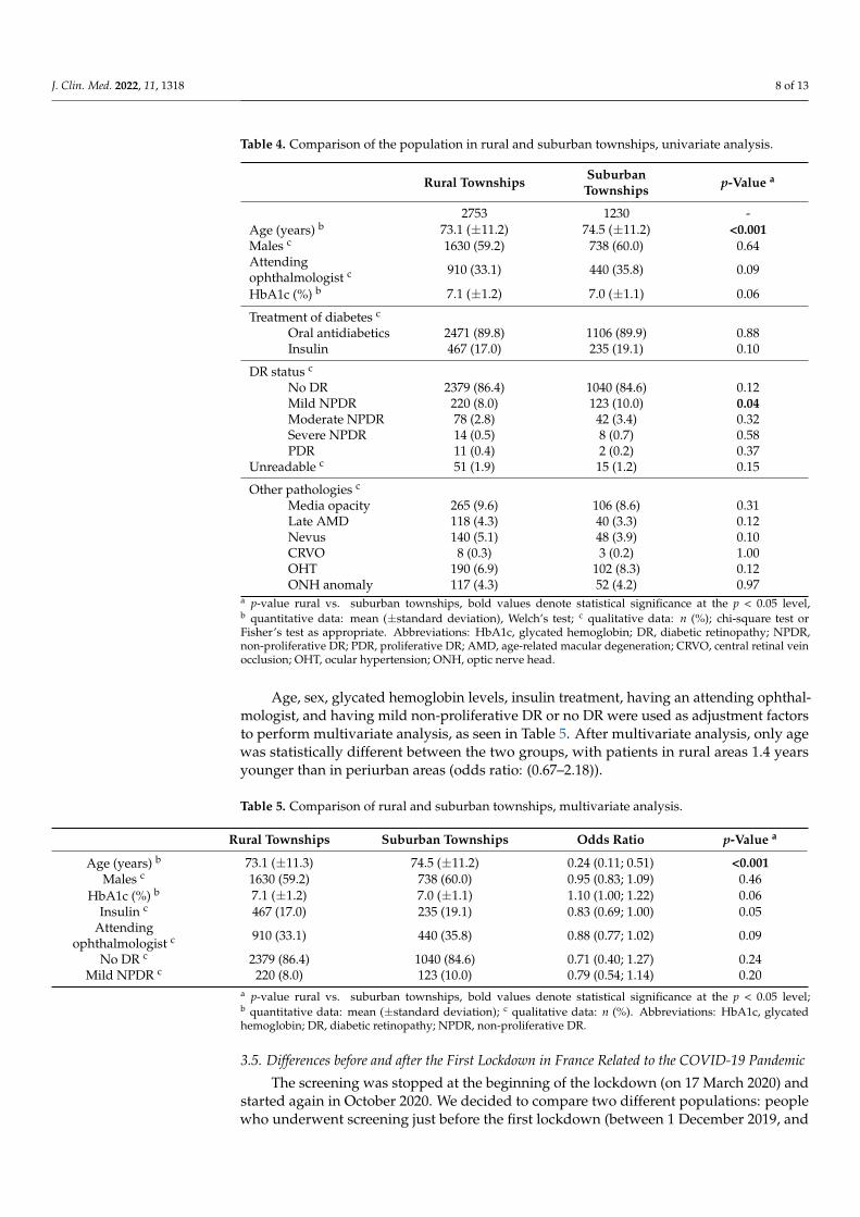
Figure 2. Retinal photographs of patients who underwent roving retinopathy screening. (A) Normal left eye fundus; (B) Moderate non-proliferative diabetic retinopathy associated with hard exudates of the left eye; (C) Lower part of a large pigmented lesion of the right eye.
3.3. Comparison of Patients with and without DR of any Type We compared patients with DR of any type with those without, and we obtained two
groups of 1420 and 8210 patients, respectively, after excluding 590 (5.8%) patients with unreadable photographs. The results are shown in Table 3. Patients with DR of any type had significantly more type 1 diabetes (p < 0.001), higher glycated hemoglobin (p < 0.001), longer duration of diabetes (p < 0.001), more insulin treatment (p < 0.001), and fewer oral antidiabetics (p < 0.001). They also significantly had a shorter time since the last visit (p = 0.04) and more often reported an attending ophthalmologist (p < 0.01).
Table 3. Comparison of patients with and without diabetic retinopathy.
DR any Type No DR p-Value a
Number 1420 8210 Age (years) b 68.5 (±11.3) 68.3 (±11.2) 0.44 Gender c 841 (59.2) 4859 (59.0) 0.98 Attending ophthalmologist c 474 (33.4) 3041 (37.0) <0.01 HbA1c (%) b 7.44 (±1.4) 6.99 (±1.1) <0.001 Progression of diabetes b 13.76 (±10.9) 9.31 (±8.9) <0.001 Time since last check-up b 3.47 (±3.3) 3.68 (±3.7) 0.04 Type of diabetes b,d, 8851 patients Type 1 73 (6.1) 258 (3.4) <0.001 Type 2 1129 (93.9) 7391 (96.6) - Treatment of diabetes b Oral antidiabetics 1181 (83.2) 7380 (89.9) <0.001 Insulin 490 (34.5) 1295 (15.8) <0.001
Figure 2. Retinal photographs of patients who underwent roving retinopathy screening. (A) Normalleft eye fundus; (B) Moderate non-proliferative diabetic retinopathy associated with hard exudates ofthe left eye; (C) Lower part of a large pigmented lesion of the right eye.
Table 3. Comparison of patients with and without diabetic retinopathy.
DR Any Type No DR p-Value a
Number 1420 8210Age (years) b 68.5 (±11.3) 68.3 (±11.2) 0.44Gender c 841 (59.2) 4859 (59.0) 0.98Attending ophthalmologist c 474 (33.4) 3041 (37.0) <0.01HbA1c (%) b 7.44 (±1.4) 6.99 (±1.1) <0.001Progression of diabetes b 13.76 (±10.9) 9.31 (±8.9) <0.001Time since last check-up b 3.47 (±3.3) 3.68 (±3.7) 0.04
Type of diabetes b,d,8851 patients
Type 1 73 (6.1) 258 (3.4) <0.001Type 2 1129 (93.9) 7391 (96.6) -
Treatment of diabetes b
Oral antidiabetics 1181 (83.2) 7380 (89.9) <0.001Insulin 490 (34.5) 1295 (15.8) <0.001
Other pathologies b
Media opacity 96 (6.8) 527 (6.4) 0.63Late AMD 46 (3.2) 294 (3.6) 0.52Naevus 31 (2.2) 253 (3.1) 0.07CRVO 2 (0.1) 19 (0.2) 0.76OHT 131 (9.2) 615 (7.4) 0.02ONH anomaly 72 (5.1) 337 (4.1) 0.10
a p-value patients with DR vs. patients without, bold values denote statistical significance at the p < 0.05 level;b quantitative data: mean (±standard deviation), Welch’s test; c qualitative data: n (%), chi-2 test or Fisher’stest as appropriate; d exclusion of patients who did not know their diabetes type. Abbreviations: DR, diabeticretinopathy; SD, standard deviation; HbA1c, glycated hemoglobin; AMD, age-related macular degeneration;CRVO, central retinal vein occlusion; OHT, ocular hypertension; ONH, optic nerve head.
J. Clin. Med. 2022, 11, 1318 8 of 13
Table 4. Comparison of the population in rural and suburban townships, univariate analysis.
Rural Townships SuburbanTownships p-Value a
2753 1230 -Age (years) b 73.1 (±11.2) 74.5 (±11.2) <0.001Males c 1630 (59.2) 738 (60.0) 0.64Attendingophthalmologist c 910 (33.1) 440 (35.8) 0.09
HbA1c (%) b 7.1 (±1.2) 7.0 (±1.1) 0.06
Treatment of diabetes c
Oral antidiabetics 2471 (89.8) 1106 (89.9) 0.88Insulin 467 (17.0) 235 (19.1) 0.10
DR status c
No DR 2379 (86.4) 1040 (84.6) 0.12Mild NPDR 220 (8.0) 123 (10.0) 0.04Moderate NPDR 78 (2.8) 42 (3.4) 0.32Severe NPDR 14 (0.5) 8 (0.7) 0.58PDR 11 (0.4) 2 (0.2) 0.37
Unreadable c 51 (1.9) 15 (1.2) 0.15
Other pathologies c
Media opacity 265 (9.6) 106 (8.6) 0.31Late AMD 118 (4.3) 40 (3.3) 0.12Nevus 140 (5.1) 48 (3.9) 0.10CRVO 8 (0.3) 3 (0.2) 1.00OHT 190 (6.9) 102 (8.3) 0.12ONH anomaly 117 (4.3) 52 (4.2) 0.97
a p-value rural vs. suburban townships, bold values denote statistical significance at the p < 0.05 level,b quantitative data: mean (±standard deviation), Welch’s test; c qualitative data: n (%); chi-square test orFisher’s test as appropriate. Abbreviations: HbA1c, glycated hemoglobin; DR, diabetic retinopathy; NPDR,non-proliferative DR; PDR, proliferative DR; AMD, age-related macular degeneration; CRVO, central retinal veinocclusion; OHT, ocular hypertension; ONH, optic nerve head.
Age, sex, glycated hemoglobin levels, insulin treatment, having an attending ophthal-mologist, and having mild non-proliferative DR or no DR were used as adjustment factorsto perform multivariate analysis, as seen in Table 5. After multivariate analysis, only agewas statistically different between the two groups, with patients in rural areas 1.4 yearsyounger than in periurban areas (odds ratio: (0.67–2.18)).
Table 5. Comparison of rural and suburban townships, multivariate analysis.
Rural Townships Suburban Townships Odds Ratio p-Value a
Age (years) b 73.1 (±11.3) 74.5 (±11.2) 0.24 (0.11; 0.51) <0.001Males c 1630 (59.2) 738 (60.0) 0.95 (0.83; 1.09) 0.46
HbA1c (%) b 7.1 (±1.2) 7.0 (±1.1) 1.10 (1.00; 1.22) 0.06Insulin c 467 (17.0) 235 (19.1) 0.83 (0.69; 1.00) 0.05
Attendingophthalmologist c 910 (33.1) 440 (35.8) 0.88 (0.77; 1.02) 0.09
No DR c 2379 (86.4) 1040 (84.6) 0.71 (0.40; 1.27) 0.24Mild NPDR c 220 (8.0) 123 (10.0) 0.79 (0.54; 1.14) 0.20
a p-value rural vs. suburban townships, bold values denote statistical significance at the p < 0.05 level;b quantitative data: mean (±standard deviation); c qualitative data: n (%). Abbreviations: HbA1c, glycatedhemoglobin; DR, diabetic retinopathy; NPDR, non-proliferative DR.
3.5. Differences before and after the First Lockdown in France Related to the COVID-19 Pandemic
The screening was stopped at the beginning of the lockdown (on 17 March 2020) andstarted again in October 2020. We decided to compare two different populations: peoplewho underwent screening just before the first lockdown (between 1 December 2019, and

J. Clin. Med. 2022, 11, 1318 9 of 13
29 February 2020; 367 patients) and those who had screening after screening resumed(between 1 October 2020, and 31 November 2020; 441 patients). Due to the short durationof this period, none of the patients had multiple examinations. The results of these analysesare shown in Table 6. Patients exhibited the same characteristics apart from age: peoplewere significantly older before lockdown (70.3 ± 11.7 years old before vs. 69.1 ± 10.1 yearsold after, p = 0.04). There was no significant difference concerning the stage of the DRbetween the groups (p = 0.56).
Table 6. Comparison of the study population, before and after the first lockdown.
Before Lockdown After Lockdown p-Value a
367 441Age (years) b 70.3 (±11.7) 69.1 (±10.2) 0.04Males c 212 (57.8) 271 (61.5) 0.31HbA1c (%) b 7.1 (±1.2) 6.9 (±1.0) 0.28Progression ofdiabetes (years) b 11.9 (±10.4) 11.2 (±9.6) 0.37
Insulin-dependent c 71 (20.5) 69 (16.8) 0.22
DR status c 0.56No DR 325 (88.5) 393 (89.1) -Mild NPDR 29 (7.9) 37 (8.4) -Moderate
NPDR 7 (1.9) 9 (2.0) -
Severe NPDR 5 (1.4) 2 (0.5) -PDR 1 (0.3) 0 -
a p-value before vs. after the lockdown, bold values denote statistical significance at the p < 0.05 level; b quantitativedata: mean (±standard deviation); c qualitative data: n (%). Abbreviations: HbA1c, glycated hemoglobin; DR,diabetic retinopathy; NPDR, non-proliferative DR; PDR, proliferative DR.
4. Discussion
In this study, the prevalence of DR at all stages combined was 13.9% at the firstexamination of each patient. Moreover, the DR rate was lower than the disease’s knownprevalence in the literature, around 30% (34.6% in the meta-analysis of Yau et al. in 2012) [7].However, this rate is related to a diagnostic approach using a fundus exam performed byan ophthalmologist or ETDRS standard seven-field imaging [17]. The lower rate may alsobe explained by the regular in-office ophthalmologic follow-up of patients with knownDR or diabetes with general complications, and therefore at greater risk of DR. It is alsoconceivable that patients who take the initiative to attend such screening are those whohave more rigorous medical monitoring of their diabetes and better-controlled diabetes,and therefore fewer risk factors for DR, producing a “healthy worker effect”. We also canimagine that patients with severe diabetic ophthalmologic damage leading to symptoms,such as decreased visual acuity will more readily visita an ophthalmologic consultation oremergency department and will not participate in this type of screening campaign. Finally,our screening technique with two-field imaging is less precise than a fundus examination orstandard seven-field imaging and may misdiagnose some cases of mild to moderate NPDR.Moreover, recent studies using ultra-wide field (UWF) imaging did not show significantdifferences between ETDRS standard seven-field imaging and UWF imaging. However, inscreening, it is easier and more precise to use UWF imaging with only one nonmydriaticphotograph than multiple photograph imaging [18].
Comparing populations with or without DR, patients with DR were significantlymore likely to have type 1 diabetes, had a longer course of diabetes, had higher glycatedhemoglobin, and mainly were treated with insulin. These characteristics are well known asrisk factors for developing DR [7,19]. Patients with DR were less likely to be treated withoral antidiabetics: this may be due to the greater proportion of type 1 diabetic people inDR who are not treated with this type of medication. We found no differences in otherdiscussed risk factors because it was not statistically significant (gender) or because data
J. Clin. Med. 2022, 11, 1318 10 of 13
were not collected (smoking, blood pressure, and cholesterol levels) [19]. Surprisingly,patients with DR reported an attending ophthalmologist more often and had a shorter timesince the last visit than patients without DR.
We found some results consistent with other French telemedical screening studies:in the Haut-Rhin study reported by Lenoble et al., the rate of all stages of DR was 18.0%(vs. 13.9% in the current study) [13]. However, in this study, patients were recruiteddifferently, referred by their physicians, whereas they were self-referred in our study.Additionally, the duration of diabetes was longer (16.6 vs. 10.0 years), and the glycatedhemoglobin level was higher (9.3% vs. 7.1%). In contrast, the 2012 study of Schulze-Döboldet al. in Ile-de-France showed a higher DR rate (24.2%) [20]. However, the populationwas recruited mostly in hospitals: 17 of the 29 retinal photographers used were fromhospitals, and hospital recruitment was described as having a significantly higher numberof DRs because of more severe disease in these patients. The study conducted by Soulié-Strougar et al. in 2007 analyzed one of the first DR screening campaigns in Burgundyand found a DR rate of 8.6% [21]. This rate was lower than ours with the same patientrecruitment method, but at this time the screening campaign was less well known andselected only those patients most motivated in monitoring their disease, with the bestdiabetes management, and with a lower DR rate. Finally, in the Italian multicenter NO-BLIND study, in nine public out-patient clinics, a trained diabetologist in each center tookphotographs of the patients′ fundus oculi using a portable digital ophthalmoscope (MiiSHorus Scope DEC 100) [22]. The prevalence of DR observed was similar to ours (15.5%).These results confirm that telemedicine can rely on adequately trained diabetologists, andit can be performed with portable and less expensive instruments directly at diabetescenters, with results comparable to traditional retinography performed by highly qualifiedpersonnel. Therefore, telemedicine is the only cost-saving alternative compared withtraditional ophthalmologist examination, regardless of geographical setting, and is wellaccepted by both patients and providers.
The analysis of the retinal photographs showed pathologies not directly relatedto diabetes. The prevalence of choroidal nevi is variable, depending on the studies.For example, a 2.1% prevalence of choroidal nevi was found in one Australian study,5.0% retro-equatorial nevi in an American study, while another Australian study found a6.5% prevalence [23–25]. The current study found the presence of a nevus in 2.8% of thepatients. However, we believe that this proportion remains underestimated: photographswere centered on the posterior pole. The prevalence of late AMD in our study was esti-mated at 3.4%. A 2017 European meta-analysis found the prevalence of late AMD between0.2% and 5.6% for all age groups [26]. The present study found a proportion within theaverage range. However, these figures remain challenging to interpret because diabeticpatients with known AMD are probably regularly followed up and have no reason tobe screened in this way. Here, the prevalence of OHT was 7.8%. A 2006 French cohortstudy found an 8.9% OHT rate; the two are quite similar [27]. Conversely, it is difficult toestimate the prevalence of glaucoma based only on a photograph of the optic disc, sincean abnormality on the optic nerve head is not sufficient to diagnose glaucoma, whereas anormal papilla does not exclude this pathology.
This study is one of the first to compare patients from rural and urban areas in thesame cohort. After multivariate analysis, the only difference we found was the age ofthe patients, who were significantly younger in rural areas. Despite the same duration ofdiabetes, it is conceivable that patients in rural areas are younger when diagnosed withdiabetes, due to poorer access to care and poorer control of diabetes risk factors. Thissuggests that screening for DR by telemedicine is of interest in rural and periurban areas. Itwould be interesting to reexamine these figures in a few years when the number of patientsin periurban areas will be greater.
As pointed out by Galiero et al. in their recent review of the literature, it is importantto be able to maintain screening and follow-up processes in at-risk populations duringperiods of pandemic and restriction, such as during COVID-19 [28]. The opportunity to

J. Clin. Med. 2022, 11, 1318 11 of 13
safely proceed with the management of diabetic patients by limiting their contact has notonly allowed for better management of this disabling complication, but also for inclusionin on-going studies on comorbidities associated with DR in real-life situations [29,30]. Dueto the COVID-19 pandemic and the first lockdown, we expect a post-COVID-19 populationwith poorer diabetes control characteristics or more DR, but this was not the case in thepresent study, with comparable populations before and after the first lockdown. This lack ofa difference may be related to the small sample sizes. This situation has led us to adapt thefollow-up of our patients and, most particularly, to develop teleconsultations or therapeuticeducation protocols [31].
The main strength of this study is the significant number of patients over a long period.Another strength is that it has been around since 2004, resulting in well-organized andstandardized recruitment. Moreover, various types of population can participate in thisscreening: at the beginning, only rural townships were visited, it opened to periurbantownships in 2016, and screening in urban townships started in 2021.
The study also has a number of weaknesses, notably a declarative bias from patients:some of them were unable to tell us about their glycated hemoglobin, type of treatment,type of diabetes, or duration of disease, which leads to a lack of information. Over thelast year, we have also been impacted by the COVID-19 pandemic, decreasing the numberof screened patients. Another of the study’s weaknesses is that this screening campaignonly recruits patients voluntarily, which makes it possible that the most assiduous patientswere selected.
Areas of improvement can be explored, such as questioning general practitioners(last glycated hemoglobin, treatment), combining visual acuity measurement, performingoptical coherence tomography (OCT) of the macula, or using UWF imaging. However,while all of these areas of improvement are important for the early detection and treatmentof anomalies, they can be responsible for a significant loss of time and money. The useof automated algorithms is being studied for our screening campaign. We are discussingthe possibility of using such an algorithm to identify abnormal photographs that will thenbe read by an ophthalmologist, although this artificial intelligence technology will allowphotographs without noticeable abnormalities to be classified as normal without havingbeen read by an ophthalmologist. Automated detection is now increasingly used and canbe combined with the use of UWF imaging to allow easier and faster screening [32].
In conclusion, our screening technique is a meaningful way to detect earlier curablediseases, whether or not they are related to diabetes mellitus, in a population that does nothave sufficient ophthalmologic control. However, it has weaknesses and can be improved.This screening campaign has a significant interest in rural and periurban areas. It has beendescribed as cost effective, particularly in rural areas, limiting the transport of patients overlong distances, which is reimbursed by healthcare providers in France for patients sufferingfrom diabetes mellitus [33].
Author Contributions: Conceptualization, A.C. and F.B.; Methodology, A.C., F.B. and I.B.-G.; Soft-ware, F.B.; Formal Analysis, I.B.-G. and F.B.; Investigation, A.C., S.L., V.H. and D.M.; Validation,P.-H.G., L.A., C.C.-G. and A.M.B.; Writing—Original Draft Preparation, A.C. and F.B.; Writing—Review and Editing, I.B.-G., M.T., P.-H.G., L.A., C.C.-G. and A.M.B.; Supervision, C.C.-G. and A.M.B.All authors contributed to this manuscript and have approved the submitted version and agree to bepersonally accountable for their contribution and for ensuring that questions related to the accuracyor integrity of any part of the work, even ones in which the author was not personally involved, areappropriately investigated, resolved, and documented in the literature. All authors have read andagreed to the published version of the manuscript.
Funding: This research received no external funding.
Institutional Review Board Statement: This study complied with the Declaration of Helsinki, andthe locally appointed ethics committee gave approval for the research protocol. Dijon UniversityHospital Ethics Committee, retrospective study on DR screening-06/09/2021.
J. Clin. Med. 2022, 11, 1318 12 of 13
Informed Consent Statement: Because all patients received standard medical care and because thedataset contained no information that could enable patient identification, the study was exempt frominformed consent requirements.
Conflicts of Interest: F.B. is a consultant for: Novartis, Théa; P.-H.G. is a consultant for: Allergan,Bayer, Novartis; L.A. is a consultant for: Novartis; A.M.B. is a consultant for: Aerie, Allergan, Bauschand Lomb, Santen Pharmaceutical, Théa; C.C.-G. is a consultant for: Allergan, Bayer, Horus Pharma,Novartis, Roche, Théa. The following authors declare no conflict of interests: A.C., M.T., S.L., V.H.,D.M., I.B.-G.
References1. Franc, C. Epidemiology of diabetes: Frightening predictions. Med. Sci. 2013, 29, 711–714.2. Aiello, L.P.; Avery, R.L.; Arrigg, P.G.; Keyt, B.A.; Jampel, H.D.; Shah, S.T.; Pasquale, L.R.; Thieme, H.; Iwamoto, M.A.; Park, J.E.;
et al. Vascular Endothelial Growth Factor in Ocular Fluid of Patients with Diabetic Retinopathy and Other Retinal Disorders. N.Engl. J. Med. 1994, 331, 1480–1487. [CrossRef] [PubMed]
3. Forbes, J.M.; Cooper, M.E. Mechanisms of Diabetic Complications. Physiol. Rev. 2013, 93, 137–188. [CrossRef] [PubMed]4. Wang, W.; Lo, A.C.Y. Diabetic Retinopathy: Pathophysiology and Treatments. Int. J. Mol. Sci. 2018, 19, 1816. [CrossRef]5. Simó-Servat, O.; Hernández, C.; Simó, R. Diabetic Retinopathy in the Context of Patients with Diabetes. Ophthalmic Res. 2019, 62,
211–217. [CrossRef]6. Klein, B.E.K. Overview of Epidemiologic Studies of Diabetic Retinopathy. Ophthalmic Epidemiol. 2007, 14, 179–183. [CrossRef]7. Yau, J.W.Y.; Rogers, S.L.; Kawasaki, R.; Lamoureux, E.L.; Kowalski, J.W.; Bek, T.; Chen, S.-J.; Dekker, J.M.; Fletcher, A.; Grauslund, J.;
et al. Global Prevalence and Major Risk Factors of Diabetic Retinopathy. Diabetes Care 2012, 35, 556–564. [CrossRef]8. Vujosevic, S.; Midena, E. Diabetic Retinopathy in Italy: Epidemiology Data and Telemedicine Screening Programs. J. Diabetes Res.
2016, 2016, 3627465. [CrossRef]9. Vujosevic, S.; Aldington, S.; Silva, P.; Hernández, C.; Scanlon, P.; Peto, T.; Simó, R. Screening for diabetic retinopathy: New
perspectives and challenges. Lancet Diabetes Endocrinol. 2020, 8, 337–347. [CrossRef]10. Rosenfeld, P.J.; Brown, D.M.; Heier, J.S.; Boyer, D.S.; Kaiser, P.K.; Chung, C.Y.; Kim, R.Y.; for the MARINA Study Group.
Ranibizumab for neovascular age-related macular degeneration. N. Engl. J. Med. 2006, 355, 1419–1431. [CrossRef]11. Massin, P.; Aubert, J.-P.; Erginay, A.; Bourovitch, J.; BenMehidi, A.; Audran, G.; Bernit, B.; Jamet, M.; Collet, C.; Laloi-Michelin, M.;
et al. Screening for diabetic retinopathy: The first telemedical approach in a primary care setting in France. Diabetes Metab. 2004,30, 451–457. [CrossRef]
12. Massin, P.; Aubert, J.; Eschwege, E.; Erginay, A.; Bourovitch, J.; BenMehidi, A.; Nougarède, M.; Bouée, S.; Fagnani, F.; Tcherny, M.;et al. Evaluation of a screening program for diabetic retinopathy in a primary care setting Dodia (Dépistage ophtalmologique dudiabète) study. Diabetes Metab. 2005, 31, 153–162. [CrossRef]
13. Lenoble, P.; Kheliouen, M.; Bourderont, D.; Klinger, V.; Nasica, X.; Benseddik, Y.; Holl, P. Dépistage de la rétinopathie diabétiquepar télédiagnostic dans le Haut-Rhin. J. Fr. Ophtalmol. 2009, 32, 91–97. [CrossRef] [PubMed]
14. Brown, D.M.; Kaiser, P.; Michels, M.; Soubrane, G.; Heier, J.S.; Kim, R.Y.; Sy, J.P.; Schneider, S.; for the ANCHOR Study Group.Ranibizumab versus Verteporfin for Neovascular Age-Related Macular Degeneration. N. Engl. J. Med. 2006, 355, 1432–1444.[CrossRef]
15. Massin, P.; Chabouis, A.; Erginay, A.; Viens-Bitker, C.; Lecleire-Collet, A.; Meas, T.; Guillausseau, P.-J.; Choupot, G.; André, B.;Denormandie, P. OPHDIAT©: A telemedical network screening system for diabetic retinopathy in the Île-de-France. DiabetesMetab. 2008, 34, 227–234. [CrossRef]
16. Massin, P.; Angioi-Duprez, K.; Bacin, F.; Cathelineau, B.; Cathelineau, G.; Chaine, G.; Coscas, G.; Flament, J.; Sahel, J.-A.; Turut, P.;et al. Recommendations of the ALFEDIAM (French Association for the Study of Diabetes and Metabolic Diseases) for thescreening and surveillance of diabetic retinopathy. J. Fr. Ophtalmol. 1997, 20, 302–310.
17. Early Treatment Diabetic Retinopathy Study Research Group. Grading diabetic retinopathy from stereoscopic color fundusphotographs—An extension of the modified Airlie House classification. ETDRS report number 10. In Ophthalmology; 1991; 98,(Suppl. S5), pp. 786–806. [CrossRef]
18. Aiello, L.P.; Odia, I.; Glassman, A.R.; Melia, M.; Jampol, L.M.; Bressler, N.M.; Kiss, S.; Silva, P.S.; Wykoff, C.C.; Sun, J.K.;et al. Comparison of Early Treatment Diabetic Retinopathy Study Standard 7-Field Imaging with Ultrawide-Field Imaging forDetermining Severity of Diabetic Retinopathy. JAMA Ophthalmol. 2019, 137, 65–73. [CrossRef]
19. Wat, N.; Wong, R.L.; Wong, I.Y. Associations between diabetic retinopathy and systemic risk factors. Hong Kong Med. J. 2016, 22,589–599. [CrossRef]
20. Schulze-Döbold, C.; Erginay, A.; Robert, N.; Chabouis, A.; Massin, P. Ophdiat®: Five-year experience of a telemedical screeningprogramme for diabetic retinopathy in Paris and the surrounding area. Diabetes Metab. 2012, 38, 450–457. [CrossRef]
21. Soulié-Strougar, M.; Charles, A.; Métral, P.; Quercia, P.; Souchier, M.; Chirpaz, L.; Bron, A.; Creuzot-Garcher, C. Screening diabeticretinopathy in Burgundy with an itinerant nonmydriatic camera. J. Fr. Ophtalmol. 2007, 30, 121–126. [CrossRef]

J. Clin. Med. 2022, 11, 1318 13 of 13
22. Sasso, F.C.; Pafundi, P.C.; Gelso, A.; Bono, V.; Costagliola, C.; Marfella, R.; Sardu, C.; Rinaldi, L.; Galiero, R.; Acierno, C.; et al.Telemedicine for screening diabetic retinopathy: The NO BLIND Italian multicenter study. Diabetes Metab. Res. Rev. 2019, 35,e3113. [PubMed]
23. Keel, S.; Xie, J.; Foreman, J.; Taylor, H.R.; Dirani, M. Prevalence and characteristics of choroidal nevi: The Australian National EyeHealth Survey. Clin. Exp. Ophthalmol. 2018, 46, 777–782. [CrossRef]
24. Chien, J.L.; Sioufi, K.; Surakiatchanukul, T.; Shields, J.A.; Shields, C.L. Choroidal nevus: A review of prevalence, features, genetics,risks, and outcomes. Curr. Opin. Ophthalmol. 2017, 28, 228–237. [CrossRef]
25. Sumich, P.; Mitchell, P.; Wang, J.J. Choroidal nevi in a white population: The Blue Mountains Eye Study. Arch. Ophthalmol. 1998,116, 645–650. [CrossRef] [PubMed]
26. Colijn, J.M.; Buitendijk, G.H.S.; Prokofyeva, E.; Alves, D.; Cachulo, M.L.; Khawaja, A.P.; Cougnard-Gregoire, A.; Merle, B.M.J.;Korb, C.; Erke, M.G.; et al. Prevalence of Age-Related Macular Degeneration in Europe: The Past and the Future. Ophthalmology2017, 124, 1753–1763. [CrossRef]
27. Bron, A.; Baudouin, C.; Nordmann, J.P.; Rouland, J.F.; Thomas, F.; Bean, K.; De Clercq, B.; Bénétos, A.; de Gendre, A.S.; Lefebvre, S.Prevalence of intraocular hypertension and glaucoma in a nonselected French population. J. Fr. Ophtalmol. 2006, 29, 635–641.[CrossRef]
28. Galiero, R.; Pafundi, P.C.; Nevola, R.; Rinaldi, L.; Acierno, C.; Caturano, A.; Salvatore, T.; Adinolfi, L.E.; Costagliola, C.; Sasso, F.C.The Importance of Telemedicine during COVID-19 Pandemic: A Focus on Diabetic Retinopathy. J. Diabetes Res. 2020, 2020,9036847. [CrossRef]
29. Sasso, F.C.; Pafundi, P.C.; Gelso, A.; Bono, V.; Costagliola, C.; Marfella, R.; Sardu, C.; Rinaldi, L.; Galiero, R.; Acierno, C.; et al.High HDL cholesterol: A risk factor for diabetic retinopathy? Findings from NO BLIND study. Diabetes Res. Clin. Pract. 2019, 150,236–244. [CrossRef]
30. Sasso, F.; Pafundi, P.C.; Gelso, A.; Bono, V.; Costagliola, C.; Marfella, R.; Sardu, C.; Rinaldi, L.; Galiero, R.; Acierno, C.; et al.Relationship between albuminuric CKD and diabetic retinopathy in a real-world setting of type 2 diabetes: Findings from Noblind study. Nutr. Metab. Cardiovasc. Dis. 2019, 29, 923–930. [CrossRef]
31. Mukona, D.M.; Zvinavashe, M. Self- management of diabetes mellitus during the Covid-19 pandemic: Recommendations for aresource limited setting. Diabetes Metab. Syndr. Clin. Res. Rev. 2020, 14, 1575–1578. [CrossRef] [PubMed]
32. Wang, K.; Jayadev, C.; Nittala, M.G.; Velaga, S.B.; Ramachandra, C.A.; Bhaskaranand, M.; Bhat, S.; Solanki, K.; Sadda, S.R.Automated detection of diabetic retinopathy lesions on ultrawidefield pseudocolour images. Acta Ophthalmol. 2018, 96, e168–e173.[CrossRef] [PubMed]
33. Sharafeldin, N.; Kawaguchi, A.; Sundaram, A.; Campbell, S.; Rudnisky, C.; Weis, E.; Tennant, M.T.S.; Damji, K.F. Review ofeconomic evaluations of teleophthalmology as a screening strategy for chronic eye disease in adults. Br. J. Ophthalmol. 2018, 102,1485–1491. [CrossRef] [PubMed]