Reevaluating the Definition of Intraretinal Microvascular Abnormalities and Neovascularization...
11
Reevaluating the Definition of Intraretinal Microvascular Abnormalities and Neovascularization Elsewhere in Diabetic Retinopathy Using Optical Coherence Tomography and Fluorescein Angiography CECILIA S. LEE, AARON Y. LEE, DAWN A. SIM, PEARSE A. KEANE, HEMAL MEHTA, JAVIER ZARRANZ-VENTURA, MARCUS FRUTTIGER, CATHERINE A. EGAN, AND ADNAN TUFAIL PURPOSE: To evaluate the agreement between clinical examination, spectral-domain ocular coherence tomogra- phy (SD OCT), and fluorescein angiography (FA) in diag- nosing intraretinal microvascular abnormality (IRMA) and neovascularization elsewhere (NVE) and define the SD OCT features that differentiate NVEs from IRMAs. DESIGN: Retrospective study. METHODS: Data were collected from 23 lesions from 8 diabetic patients, seen from July 2012 through October 2013 at Moorfields Eye Hospital, United Kingdom. Main outcomes were SD OCT features and FA leakage of IRMA and neovascular complex. The agreement be- tween 3 evaluations was analyzed by Fleiss’ kappa. RESULTS: The following 5 SD OCT features signifi- cantly differentiated IRMAs from NVEs: (1) hyperreflec- tive dots in superficial inner retina (P [ .002); (2) the outpouching of internal limiting membrane (ILM) (P [ .004); (3) the breach of ILM (P [ .004); (4) the breach of posterior hyaloid (P [ .0005); (5) hyperreflective dots in vitreous (P [ .008). The agreement was moderate between 3 evaluations (k [ 0.48, P [ 7.11 3 10 L5 ) but substantial between clinical and SD OCT evaluation (k [ 0.72, P [ .00055). There was no significant agreement between OCT evaluation and FA leakage (k [ 0.249, P [ .232). CONCLUSIONS: SD OCT will be a valuable adjunct in evaluating IRMA and NVE, since it can verify the his- topathologic correlate. SD OCT provides subtle anatomic insights and may be more accurate than clinical examina- tion or leakage on FA, our current method of diagnosing this important endpoint, which has implications in future trial design for proliferative diabetic retinopathy prevention. (Am J Ophthalmol 2014;-:-–-. Ó 2014 by Elsevier Inc. All rights reserved.) D IABETIC RETINOPATHY (DR) IS THE LEADING causes of blindness in working-age populations worldwide. 1 Initially described by Jaeger in 1855, 2 DR was mainly categorized into nonproliferative vs proliferative disease. Proliferative diabetic retinopathy or ‘‘diabetic retinitis proliferans’’ was first reported by Manz in 1876, 3,4 and the initial descriptions of diabetic neovascularizations have been largely based on histo- pathologic description of new blood vessels that grow into the vitreous through a break of the internal limiting membrane (ILM). 5–8 The term intraretinal microvascular abnormality (IRMA) arose much later as a clinical definition in 1968 from the Airlie Classification of Diabetic Retinopathy. 9 His- topathologic description of IRMA predates the clinical defi- nition, 10 but Airlie classification provided an early framework for a ‘‘common language’’ in staging DR in clin- ical practice and trials. 9,11 In 1981, Diabetic Retinopathy Study report No. 7 12 provided standard photographs 8A and 8B and subsequently, IRMA became defined as tortuous intraretinal vascular segments in fields 4–7, varying in caliber from barely visible to 31 mm per Early Treatment of Diabetic Retinopathy Study (ETDRS). 13 As histopathol- ogy is limited to examining a single time point of a lesion’s evolution, whether IRMAs are a direct precursor lesion of neovascularization elsewhere (NVE) has not been estab- lished, but the severity of IRMA was shown to be a risk factor for the progression into proliferative diabetic retinopathy (PDR). 14 In fact, IRMA became one of the defining charac- teristics of end-stage nonproliferative diabetic retinopathy and therefore an important clinical endpoint. 15 During the landmark trials of the Diabetic Retinopathy Study and the ETDRS, IRMA and NVE were differentiated based on color stereoscopic photographs. 12,13 Although fluorescein angiography (FA) was used in ETDRS to evaluate the degree of macular edema and the severity of DR, it was not employed for the definition of IRMAs. 16 However, ETDRS did identify that the source of Accepted for publication Sep 25, 2014. From the Medical Retina Service (C.S.L., A.Y.L., P.A.K., H.M., C.A.E., A.T.) and National Institute for Health Research Biomedical Research Centre for Ophthalmology (D.A.S., P.A.K., J.Z.-V., C.A.E., A.T.), Moorfields Eye Hospital National Health Service Foundation Trust; and University College London, Institute of Ophthalmology (A.Y.L., D.A.S., P.A.K., M.F., A.T.), London, United Kingdom. Current affiliations of the authors: C.S. Lee, University of Washington, Seattle, Washington; A.Y. Lee, University of British Columbia, Vancou- ver, Canada. Inquiries to Adnan Tufail, Moorfields Eye Hospital National Health Service Foundation Trust, 162 City Road, London EC1V 2PD, United Kingdom; e-mail: adnan.tufail@moorfields.nhs.uk 0002-9394/$36.00 http://dx.doi.org/10.1016/j.ajo.2014.09.041 1 Ó 2014 BY ELSEVIER INC.ALL RIGHTS RESERVED.
-
Upload
moorfields -
Category
Documents
-
view
2 -
download
0
Transcript of Reevaluating the Definition of Intraretinal Microvascular Abnormalities and Neovascularization...
Reevaluating the Definition of Intraretinal MicrovascularAbnormalities and Neovascularization Elsewhere inDiabetic Retinopathy Using Optical Coherence
Tomography and Fluorescein Angiography
CECILIA S. LEE, AARONY. LEE, DAWNA. SIM, PEARSE A. KEANE, HEMALMEHTA, JAVIER ZARRANZ-VENTURA,MARCUS FRUTTIGER, CATHERINE A. EGAN, AND ADNAN TUFAIL
� PURPOSE: To evaluate the agreement between clinical
examination, spectral-domain ocular coherence tomogra-
phy (SDOCT), and fluorescein angiography (FA) in diag-
nosing intraretinal microvascular abnormality (IRMA)
and neovascularization elsewhere (NVE) and define the
SD OCT features that differentiate NVEs from IRMAs.� DESIGN: Retrospective study.� METHODS: Data were collected from 23 lesions from 8
diabetic patients, seen from July 2012 through October
2013 at Moorfields Eye Hospital, United Kingdom.
Main outcomes were SD OCT features and FA leakage
of IRMA and neovascular complex. The agreement be-
tween 3 evaluations was analyzed by Fleiss’ kappa.� RESULTS: The following 5 SD OCT features signifi-
cantly differentiated IRMAs fromNVEs: (1) hyperreflec-
tive dots in superficial inner retina (P [ .002); (2) the
outpouching of internal limiting membrane (ILM) (P[
.004); (3) the breach of ILM (P[ .004); (4) the breach
of posterior hyaloid (P [ .0005); (5) hyperreflective
dots in vitreous (P[ .008). The agreement was moderate
between 3 evaluations (k[ 0.48, P[ 7.113 10L5) but
substantial between clinical and SDOCTevaluation (k[
0.72, P[ .00055). There was no significant agreement
between OCT evaluation and FA leakage (k [ 0.249,
P[ .232).� CONCLUSIONS: SD OCT will be a valuable adjunct
in evaluating IRMA and NVE, since it can verify the his-
topathologic correlate. SDOCT provides subtle anatomic
insights and may be more accurate than clinical examina-
tion or leakage on FA, our current method of diagnosing
this important endpoint, which has implications in future
trial design for proliferative diabetic retinopathy
prevention. (Am J Ophthalmol 2014;-:-–-.
� 2014 by Elsevier Inc. All rights reserved.)
DIABETIC RETINOPATHY (DR) IS THE LEADING
causes of blindness in working-age populations
worldwide.1 Initially described by Jaeger in
1855,2 DR was mainly categorized into nonproliferative
vs proliferative disease. Proliferative diabetic retinopathy
or ‘‘diabetic retinitis proliferans’’ was first reported by
Manz in 1876,3,4 and the initial descriptions of diabetic
neovascularizations have been largely based on histo-
pathologic description of new blood vessels that grow
into the vitreous through a break of the internal limiting
membrane (ILM).5–8
The term intraretinal microvascular abnormality
(IRMA) arose much later as a clinical definition in 1968
from theAirlieClassification ofDiabetic Retinopathy.9His-
topathologic description of IRMApredates the clinical defi-
nition,10 but Airlie classification provided an early
framework for a ‘‘common language’’ in staging DR in clin-
ical practice and trials.9,11 In 1981, Diabetic Retinopathy
Study report No. 712 provided standard photographs 8A
and 8B and subsequently, IRMAbecame defined as tortuous
intraretinal vascular segments in fields 4–7, varying in
caliber from barely visible to 31 mm per Early Treatment
of Diabetic Retinopathy Study (ETDRS).13As histopathol-
ogy is limited to examining a single time point of a lesion’s
evolution, whether IRMAs are a direct precursor lesion of
neovascularization elsewhere (NVE) has not been estab-
lished, but the severity of IRMAwas shown to be a risk factor
for the progression into proliferative diabetic retinopathy
(PDR).14 In fact, IRMA became one of the defining charac-
teristics of end-stage nonproliferative diabetic retinopathy
and therefore an important clinical endpoint.15
During the landmark trials of the Diabetic Retinopathy
Study and the ETDRS, IRMA andNVEwere differentiated
based on color stereoscopic photographs.12,13 Although
fluorescein angiography (FA) was used in ETDRS to
evaluate the degree of macular edema and the severity of
DR, it was not employed for the definition of IRMAs.16
However, ETDRS did identify that the source of
Accepted for publication Sep 25, 2014.From the Medical Retina Service (C.S.L., A.Y.L., P.A.K., H.M.,
C.A.E., A.T.) and National Institute for Health Research BiomedicalResearch Centre for Ophthalmology (D.A.S., P.A.K., J.Z.-V., C.A.E.,A.T.), Moorfields Eye Hospital National Health Service FoundationTrust; and University College London, Institute of Ophthalmology(A.Y.L., D.A.S., P.A.K., M.F., A.T.), London, United Kingdom.
Current affiliations of the authors: C.S. Lee, University of Washington,Seattle, Washington; A.Y. Lee, University of British Columbia, Vancou-ver, Canada.
Inquiries to Adnan Tufail, Moorfields Eye Hospital National HealthService Foundation Trust, 162 City Road, London EC1V 2PD, UnitedKingdom; e-mail: [email protected]
0002-9394/$36.00http://dx.doi.org/10.1016/j.ajo.2014.09.041
1� 2014 BY ELSEVIER INC. ALL RIGHTS RESERVED.
‘‘fluorescein leakage’’ in DR included microaneurysms,
dilated capillaries, and other evident vascular abnormal-
ities such as IRMA and neovascularization.16 Furthermore,
it was revealed that diffuse leakage in the retina was predic-
tive of progression of DR.17 Thus, in the time of Airlie clas-
sification and consequent landmark trials, FA findings were
to be used only as an adjunct to clinical examination and
color photography, rather than as the source of defining
stages of DR.9,16 However, textbooks often state that
IRMAs have no or minimal leakage on FA and that this
is often how they are differentiated from NVEs.18–20
Optical coherence tomography (OCT) is a noninvasive
imaging modality that allows the evaluation of the vitreous
cavity, retinal layers, retinal pigment epithelium, and
choroid.21 The advent of spectral-domain (SD) OCT has
allowed better sensitivity, increased depth of penetration,
and higher resolution of each image obtained.22 Current
commercially available SD OCT provides high-resolution
images with an axial resolution of <5 mm.22,23 As a
result, OCT parameters are increasingly used in various
clinical trials.24–26 With commercially available OCT, it
is now possible to evaluate the disruption of the ILM and
the breach of the posterior hyaloid associated with NVE
or neovascularization of disc (NVD).27,28 However,
whether IRMA and NVE can be distinguished on SD
OCT has not been established.
In this study, we perform detailed characterization of the
SD OCT features of IRMA and NVE/NVD, with compar-
ison to clinical and FA findings. In particular, we evaluate
the ability of SD OCT to show breach of the posterior hy-
aloid in support of previous histopathologic descriptions of
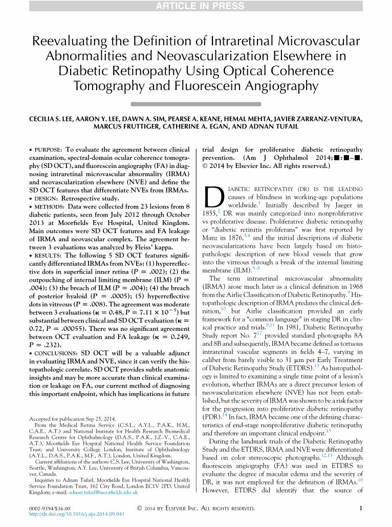
TABLE 1. Demographics and Clinical Characteristics of Study Patients With Diabetic Retinopathy and the Diagnoses of Their Retinal
Lesions Defined by Clinical Evaluation, Spectral-Domain Optical Coherence Tomography, and Fluorescein Angiography
Patient Age Sex Type DM Lesion No. Laterality Clinical Dx OCT Dx FA Leakagea Previous PRP
1 47 M 2 1 OD NVE NVE þ Yes
2 OS IRMA IRMA � Yes
2 57 M 2 3 OS IRMA IRMA � No
4 OS NVE NVE þ No
5 OS NVE NVE þ No
6 OS NVE NVE þ No
7 OS NVE NVE þ No
3 25 M 1 8 OD NVE NVE þ No
9 OS IRMA IRMA þ No
10 OS IRMA IRMA þ No
11 OS IRMA IRMA þ No
12 OS IRMA IRMA þ No
13 OS IRMA IRMA � No
4 26 F 1 14 OD IRMA NVE � No
15 OD NVE NVE þ No
16 OS NVE NVE þ No
5 44 M 2 17 OD NVD NVD � Yes
18 OD NVE NVE þ Yes
19 OS NVE NVE þ Yes
6 65 M 2 20 OS IRMA IRMA � No
7 62 F 2 21 OD NVE NVE þ No
22 OD NVD NVD þ No
8 43 M 2 23 OS IRMA NVE þ No
DM ¼ diabetes mellitus; Dx ¼ diagnosis; FA ¼ fluorescein angiography; IRMA ¼ intraretinal microvascular abnormality; NVD ¼ neovascula-
rization of disc; NVE ¼ neovascularization elsewhere; OCT ¼ optical coherence tomography; PRP ¼ panretinal photocoagulation.aþ indicates FA leakage; - indicates no FA leakage.
TABLE 2. Analysis of Spectral-Domain Optical Coherence
Tomography Features in Evaluating and Distinguishing
Clinically Diagnosed Intraretinal Microvascular Abnormality
vs Neovascularization Elsewhere in Diabetic Retinopathy
IRMA
(n)
NVE
(n) P Value
Adjusted
P Value
Hyperreflective dots in inner retina 7 0 .00049 .0024
ILM outpouching 8 1 .00073 .0036
ILM breach 2 12 .00073 .0036
Posterior hyaloid breach 2 13 .000092 .00046
Vitreous dots 1 9 .0017 .0084
ILM ¼ internal limiting membrane; IRMA ¼ intraretinal micro-
vascular abnormality; NVE ¼ neovascularization elsewhere.
2 --- 2014AMERICAN JOURNAL OF OPHTHALMOLOGY
NVE, with the objective of refining disease feature defini-
tions for use as clinical endpoints.
SUBJECTS AND METHODS
� INCLUSION CRITERIA AND DATA COLLECTION: Clin-
ical and imaging data were collected retrospectively from
patients attending medical retinal clinics at Moorfields
Eye Hospital, London, United Kingdom from July 1,
2012 to October 31, 2013. All patients were assessed by
medical retina specialists in the same institution. Approval
for data collection and analysis were obtained from the
Institutional Review Board at Moorfields Eye Hospital,
London, United Kingdom and adhered to the tenets set
forth in the Declaration of Helsinki.
Eight patients with a diagnosis of type 1 or 2 diabetes
mellitus who had undergone concurrent FA and SD
OCT scanning (Spectralis; Heidelberg Engineering, Hei-
delberg, Germany) for evaluation of PDR were included
in the study. Patients with angiographic and SD OCT im-
age sets of insufficient quality to allow grading of DM
severity and segmentation of retinal and posterior hyaloid
boundaries were excluded. No image manipulation was
performed. Classification of IRMA and NVE were based
on a clinical diagnosis using color and red-free photographs
as part of the patients’ standard of care.
� ACQUISITION AND ANALYSIS OF FLUORESCEIN
ANGIOGRAPHY: All angiographic images were acquired
with a digital retinal camera system (Topcon TRC 50IX;
Topcon Medical Systems, Inc, Paramus, New Jersey,
USA). Macular centered FAs with peripheral sweeps
were obtained.
� QUALITATIVEANALYSISOFFLUORESCEINANGIOGRAPHY
IMAGES: FA and any available fundus images were reviewed
independently by 2masked graders (C.L., A.L.). FAwas inter-
preted as leakage or no leakage.
� ACQUISITION AND ANALYSIS OF SPECTRAL-DOMAIN
OCULAR COHERENCE TOMOGRAPHY IMAGE SETS: SD
OCT images sets were obtained using a standard, commer-
cially available SDOCT device. In each case, both macular
and extramacular raster scan acquisition protocol were
performed, centered on the fovea and the NVE, respec-
tively. SD OCT images at the NVEs were selected either
with the vertical or horizontal scanning plane bisecting
the NVE, and the image set size was adjusted accordingly
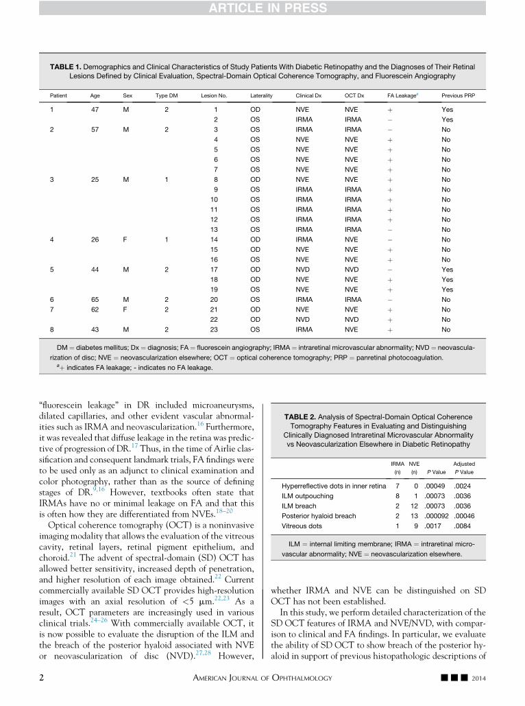
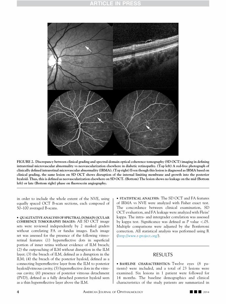
FIGURE 1. Spectral-domain optical coherence tomography characteristics found in intraretinal microvascular abnormalities and/or
neovascularization elsewhere in diabetic retinopathy. The top row illustrates different characteristics of intraretinal microvascular
abnormalities on spectral-domain optical coherence tomography and the bottom row shows those of neovascularization elsewhere.
(Top left) There are hyperreflective dots (arrow) in the inner retina without breach of the internal limiting membrane (ILM).
(Top middle) There is an outpouching of the ILM without disruption of the layer (arrow). The contour of the ILM remains smooth.
(Top right) There are 2 areas of ILM breach (arrows) without the breach of posterior hyaloid or further growth into the core vitreous.
The posterior hyaloid membrane is placed over 2 lesions. (Bottom left) There is a breach of posterior hyaloid and the lesion grows into
the core vitreous. (Bottom middle) The lesion shows multiple breaches of posterior hyaloid and linear growth along the horizontal
plane of the vitreous cortex. (Bottom right) There are multiple hyperreflective dots in the vitreous near the neovascularization else-
where lesion (arrow).
VOL. -, NO. - 3REDEFINING THE CRITERIA OF NEOVASCULARIZATION IN DIABETES
in order to include the whole extent of the NVE, using
equally spaced OCT B-scan sections, each composed of
50–100 averaged B-scans.
� QUALITATIVEANALYSISOFSPECTRAL-DOMAINOCULAR
COHERENCE TOMOGRAPHY IMAGES: All SD OCT image
sets were reviewed independently by 2 masked graders
without correlating FA or fundus images. Each image
set was assessed for the presence of the following vitreo-
retinal features: (1) hyperreflective dots in superficial
portion of inner retina without evidence of ILM breach;
(2) the outpouching of ILM without disruption in the ILM
layer; (3) the breach of ILM, defined as a disruption in the
ILM; (4) the breach of the posterior hyaloid, defined as a
connecting hyperreflective layer from the ILM to posterior
hyaloid/vitreous cavity; (5) hyperreflective dots in the vitre-
ous cavity; (6) presence of posterior vitreous detachment
(PVD), defined as a fully detached posterior hyaloid seen
as a thin hyperreflective layer above the ILM.
� STATISTICAL ANALYSIS: The SD OCT and FA features
of IRMA vs NVE were analyzed with Fisher exact test.
The concordance between clinical examination, SD
OCT evaluation, and FA leakage were analyzed with Fleiss’
kappa. The intra- and intergrader correlation was assessed
by kappa test. Significance was defined as P value <.05.
Multiple comparisons were adjusted by the Bonferroni
correction. All statistical analysis was performed using R
(http://www.r-project.org/).
RESULTS
� BASELINE CHARACTERISTICS: Twelve eyes (8 pa-
tients) were included, and a total of 23 lesions were
examined. Six lesions in 1 patient were followed for
14 months. The baseline demographics and clinical
characteristics of the study patients are summarized in
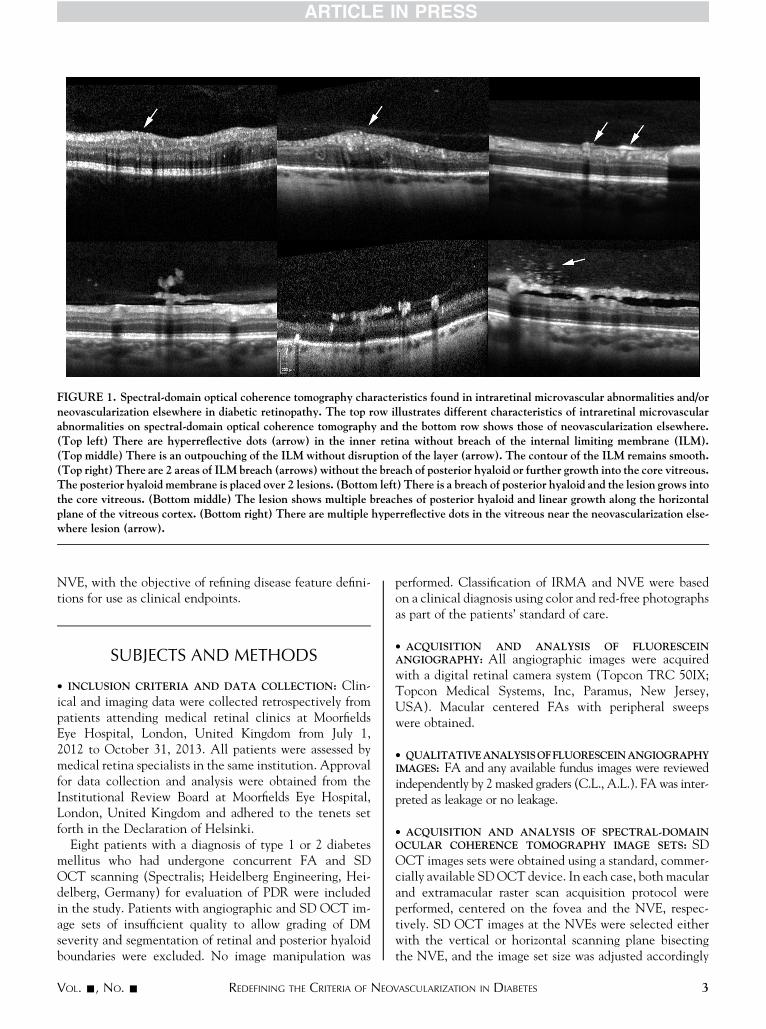
FIGURE 2. Discrepancy between clinical grading and spectral-domain optical coherence tomography (SD OCT) imaging in defining
intraretinal microvascular abnormality vs neovascularization elsewhere in diabetic retinopathy. (Top left) A red-free photograph of
clinically defined intraretinal microvascular abnormality (IRMA). (Top right) Even though this lesion is diagnosed as IRMA based on
clinical grading, the same lesion on SD OCT shows disruption of the internal limiting membrane and growth into the posterior
hyaloid. Thus, this is defined as neovascularization elsewhere on SDOCT. (Bottom) The lesion shows no leakage on the mid (Bottom
left) or late (Bottom right) phase on fluorescein angiography.
4 --- 2014AMERICAN JOURNAL OF OPHTHALMOLOGY
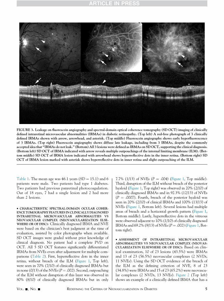
Table 1. The mean age was 46.1 years (SD ¼ 15.1) and 6
patients were male. Two patients had type 1 diabetes.
Two patients had previous panretinal photocoagulation.
Out of 18 eyes, 7 had a single lesion and 2 had more
than 2 lesions.
� CHARACTERISTIC SPECTRAL-DOMAIN OCULAR COHER-
ENCETOMOGRAPHYFEATURES INCLINICALLYDIAGNOSED
INTRARETINAL MICROVASCULAR ABNORMALITIES VS
NEOVASCULAR COMPLEX (NEOVASCULARIZATION ELSE-
WHERE OR OF DISC): Clinically diagnosed IRMA and NVE
were based on the clinician’s best judgment at the time of
evaluation, assisted by color photographs when available.
SD OCT images were graded without prior knowledge of
clinical diagnosis. No patient had a complete PVD on
OCT. All 5 SD OCT features significantly differentiated
IRMAs from NVEs even after adjustment for multiple com-
parisons (Table 2). First, hyperreflective dots in the inner
retina, without breach of the ILM (Figure 1, Top left),
were seen in 70% (7/10) of clinically diagnosed IRMAs but
in none (0/13) of theNVEs (P¼ .002). Second, outpouching
of the ILM without disruption of this layer was observed in
80% (8/10) of clinically diagnosed IRMAs but in only
7.7% (1/13) of NVEs (P ¼ .004) (Figure 1, Top middle).
Third, disruption of the ILM without breach of the posterior
hyaloid (Figure 1, Top right) was observed in 20% (2/10) of
clinically diagnosed IRMAs and in 92.3% (12/13) of NVEs
(P ¼ .0007). Fourth, breach of the posterior hyaloid was
seen in 20% (2/10) of clinical IRMAs and 100% (13/13) of
NVEs (Figure 1, Bottom left). Several lesions had multiple
areas of breach and a horizontal growth pattern (Figure 1,
Bottom middle). Lastly, hyperreflective dots in the vitreous
were observed adjacent to 10% (1/10) of clinically diagnosed
IRMAs and 69.2% (9/13) of NVEs (P¼ .002) (Figure 1, Bot-
tom right).
� ASSESSMENT OF INTRARETINAL MICROVASCULAR
ABNORMALITIES VS NEOVASCULAR COMPLEX (NEOVAS-
CULARIZATION ELSEWHERE OR OF DISC): Based on clin-
ical examination, 10 of 23 lesions (43.5%) were IRMAs
and 13 of 23 (56.5%) neovascular complexes (2 NVDs,
11 NVEs). Using the SD OCT evidence of the breach of
the ILM as the defining criterion of NVE, 8 of 23
(34.8%) were IRMAs and 15 of 23 (65.2%) were neovascu-
lar complexes (2 NVDs, 13 NVEs). Figure 2 (Top left)
shows an example of a clinically defined IRMA that has a
FIGURE 3. Leakage on fluorescein angiography and spectral-domain optical coherence tomography (SD OCT) imaging of clinically
defined intraretinal microvascular abnormalities (IRMAs) in diabetic retinopathy. (Top left) A red-free photograph of 3 clinically
defined IRMAs shown with arrow, arrowhead, and asterisk. (Top middle) Fluorescein angiography shows early hyperfluorescence
of 3 IRMAs. (Top right) Fluorescein angiography shows diffuse late leakage, including from 3 IRMAs, despite the commonly
accepted idea that ‘‘IRMAs do not leak.’’ (Bottom)All 3 lesions were defined as IRMAs on SDOCT, supporting the clinical diagnosis.
(Bottom left) SD OCT of IRMA indicated with arrow reveals multiple outpouchings of the internal limiting membrane (ILM). (Bot-
tom middle) SD OCT of IRMA lesion indicated with arrowhead shows hyperreflective dots in the inner retina. (Bottom right) SD
OCT of IRMA lesion marked with asterisk shows hyperreflective dots in inner retina and slight outpouching of the ILM.
VOL. -, NO. - 5REDEFINING THE CRITERIA OF NEOVASCULARIZATION IN DIABETES
6 --- 2014AMERICAN JOURNAL OF OPHTHALMOLOGY

clear disruption of the ILM on OCT (Figure 2, Top right),
but that did not leak on FA (Figure 2, Bottom). Five out
of 10 clinically defined IRMAs (50%) and 12 out of 13
clinically defined NVDs or NVEs (92.3%) showed leakage
on FA. Figure 3 shows 3 clinically defined IRMAs and their
SD OCTs, respectively (Figure 3, Top left and Bottom
row). There is diffuse leakage from all 3 lesions on FA
(Figure 3, Top middle and Right).
� AGREEMENT BETWEEN CLINICAL EXAMINATION,
SPECTRAL-DOMAIN OCULAR COHERENCE TOMOGRAPHY
EVALUATION, AND FLUORESCEIN ANGIOGRAPHY
RESULTS: The agreement between 3 evaluations (clin-
ical, SD OCT, FA) was moderate, with kappa value
(k) of 0.48 (P ¼ 7.11 3 10�5). There was substantial
agreement between clinical examination and SD OCT
evaluation of NVE, with the highest k of 0.72 (P ¼
.00055). The agreement between clinical examination
and FA leakage in evaluation of NVE was fair (k ¼
0.25 P ¼ .042). There was no significant agreement be-
tween SD OCT evaluation of NVE and FA leakage
(k ¼ 0.249, P ¼ .232). Reproducibility of grading of all
images between 2 graders was substantial, with a
weighted k ¼ 0.87 (SE ¼ 0.09).
� CASE STUDY: A 25-year-old white man with type I dia-
betes was referred to our medical retina clinic from the
United Kingdom national diabetic retinopathy screening
program. On initial examination, he had proliferative
changes in both eyes and several IRMAs in both eyes.
There were a total of 5 IRMAs that were clinically noted
in the left eye and the diagnosis was supported by the
absence of the ILM breach on SD OCT (Figure 4, First
row, left). Four out of 5 IRMAs showed severe leakage on
the FA. This patient underwent panretinal photocoagula-
tion in both eyes and was followed every 2–3 months for
18 months, during which he received 2 additional fill-in
laser therapies.
His infrared and OCT images of the initial evaluation
showed an IRMA located superotemporal to the disc in
the left eye (Figure 4, First row). The initial outpouching
of the ILM became more prominent 4 and 9 months later
(Figure 4, Second row and Third row). Eventually, this
IRMA progressed into an NVE 14 months after the initial
evaluation (Figure 4, Fourth row). During his follow-up, 3
out of 5 IRMAs breached the ILM and became NVEs in
a similar fashion.
DISCUSSION
THIS STUDYHASREVIEWEDTHE INITIALHISTOPATHOLOGIC
descriptions of neovascularization in DR, and demon-
strated the potential use of SD OCT in evaluating the
vitreoretinal characteristics of aberrant neovascular struc-
tures for the purpose of distinguishing between NVEs and
IRMAs. FA is an important imaging modality and is useful
for assessing macular edema and the DR severity. Although
helpful in determining the activity of the NVEs, leakage in
FA alone may not be sufficient for differentiating NVEs
from IRMAs. This is because FA leakage can occur in other
settings, such as in dilated capillaries and vascular abnor-
malities other than with NVE. Furthermore, FA is an inva-
sive test and is time consuming, making it less than ideal for
frequent use in routine disease monitoring.
The current gold standard of differentiating IRMA from
NVE is by clinical examination. IRMAs were defined
by the tortuosity and the caliber of vessels on standard pho-
tographs.9,12,13 The histopathologic definition of NVE
states that breach of the ILM and growth into posterior
hyaloid should occur only in NVEs and never in IRMAs.
With current SD OCT technology, it is possible to
noninvasively evaluate a cross-section of the vitreous and
retinal layers, thereby confirming or refuting the breach
of ILM or posterior hyaloid. In accordance with early histo-
pathologic definitions, posterior hyaloid breach on SD
OCT was used as the defining diagnostic criterion for
NVE in our study. The SD OCT diagnosis of neovasculari-
zation was in agreement with clinical diagnosis in 84.6% of
our cohort. It is interesting that in 15.4% of cases, there was
disagreement between both methods. Although the gold-
standard clinical definition of IRMA and NVE (with stan-
dard photographs) is what has been used in daily clinical
settings and in major clinical trials such as ETDRS, it is
the SD OCT characteristics that more parallel the original
definition of NVE. Therefore, it is difficult to determine
which diagnosis to accept when 2 modalities do not agree.
It may be that SD OCT findings should be incorporated
into current definitions of both IRMA and NVE. However,
we recognize that the validity of this additional test can
only be answered in large-scale studies.
The identity of hyperreflective dots in the inner retina
has been suggested to be either microglia-activated cells29
or new vessels.30 It has been hypothesized that there may
be inflammation around retinal capillaries mediated by
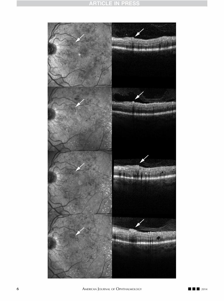
FIGURE 4. Temporal progression of intraretinal microvascular abnormality (IRMA) to neovascularization elsewhere (NVE) of a
single lesion in diabetic retinopathy over a 14-month period. (First row, left) Infrared image shows an IRMA. (First row, right) Con-
current spectral-domain optical coherence tomography (SDOCT) shows slight outpouching of the internal limiting membrane (ILM)
with hyperreflective dots in the inner retina. (Second row, left) Infrared image taken 4 months after image in first row. (Second row,
right) On concurrent SDOCT image, there is more distinctive outpouching of the ILMwithout disruption. (Third row, left) Infrared
image taken 5 months after image in second row. (Third row, right) The area of outpouching is larger without disruption of the ILM.
(Fourth row, left) Infrared image taken 5 months after image in third row shows fine vessels characteristic of neovascularization.
(Fourth row, right) SD OCT image shows evidence of NVE with a breach of the ILM and growth into the posterior hyaloid.
VOL. -, NO. - 7REDEFINING THE CRITERIA OF NEOVASCULARIZATION IN DIABETES
the surrounding microglia.31 Furthermore, these dots have
been described in both inflammatory retinal conditions and
other retinal vascular pathology.32 In our study, they were
present in IRMAs and active NVEs but not in inactive
NVE that was fibrosed and did not leak on FA. We think
that hyperreflective dots may represent initial changes in
the earliest stage of IRMA that persist until the IRMAs
or NVEs are no longer active. Although nonspecific, the
presence of hyperreflective dots on SD OCT in the area
of suspicious vascular lesions may be used in clinical prac-
tice to indicate that the patient requires close monitoring.
Further studies with comparative histopathology would be
useful to validate these features.
It is well known that moderate to severe IRMA increases
the risk of developing PDR.14 However, the notion that
IRMAs are direct precursors of NVE is controversial. In
our study, only 3 IRMAs progressed to NVEs while they
were being followed longitudinally (Figure 4).We observed
that the transition from IRMA to NVE commenced with
an initial outpouching of the ILM without the disruption
of this layer. It has been suggested that once there is a
disruption of the ILM, the early neovascular complex grows
into the potential space between the ILM and the posterior
hyaloid.33,34 The underlying mechanism is thought to be
attributable to leakage from the vessels that creates a
focal detachment of the vitreous into which new vessels
can grow.33,34 In our study, we similarly observed that
once there was a breach into the posterior hyaloid, NVE
grew across the horizontal plane of the posterior hyaloid.
Once this potential space was created, it was common to
observe multiple breaches across the posterior hyaloid
face as the NVE became larger. Given our small cohort,
whether IRMA and NVE originate from the same
pathologies is still unanswered. However, to our
knowledge, this is the first time that the possible
progression from IRMA to NVE has been shown on SD
OCT. Further studies with larger cohorts with serial
imaging will be needed to better understand the
pathophysiology of neovascular progression.
In light of the findings from this study, we propose the
following SD OCT features, which defined the different
stages of IRMA and NVE, in Table 3. Stage I of IRMA is
defined by early vascular or inflammatory changes noted
as hyperreflective dots in inner retina, but no breach or
outpouching of the ILM. They may correspond to
microglia-activated retinal capillary changes indicative of
the activity of retinopathy.29 Stage II of IRMA is defined
by the outpouching of the ILM (Figure 4, First row). As
the size of vascular abnormalities enlarges, the outpouching
area enlarges accordingly (Figure 4, Second row). Finally, if
these vessels are observed to grow outward towards the vit-
reous—the site of least resistance—this distinguishes an
IRMA from a stage I NVE. At this point, although there
is a breach of the ILM, the lesion does not extend into
the vitreous cavity, and the hyperreflective posterior hya-
loid layer appears intact despite the presence of the NVE
through the break of the ILM (Figure 1, Top right). Stage
II NVE is defined by the growth along the posterior hyaloid.
The time period between stages I and II of NVE is likely
minimal, given that it was rare to find an NVE lesion
that had only ILM breach without the breach of the poste-
rior hyaloid in our study. Stage III of NVE appears to
involve multiple areas of breach (Figure 1, Bottom middle)
and linear growth along the horizontal plane of the poste-
rior hyaloid. However, someNVEsmay grow vertically into
the vitreous cavity (Figure 1, Bottom left), and the signifi-
cance of different growth patterns is unclear. Once the
NVE is firmly established in connection with vitreous, it
appears to cause some contraction and cleavage in the vit-
reous cavity.
The vitreous appears to provide a scaffolding for NVE’s
growth35 and a complete PVD has been shown to lower
the risk of PDR.35 Indeed, none of our patients had a com-
plete PVD. It has been suggested that an iatrogenic PVD,
either surgical or chemical, may decrease the risk of devel-
oping PDR.35 However, it is noteworthy that a developing
PVD is also an important factor in precipitating a vitreous
hemorrhage by the disruption of both active and inactive
neovascular complexes. In fact, hyperreflective vitreous
dots, as seen in SD OCT, may represent a previous vitreous
hemorrhage27 or be related to the increased vascular
permeability of active neovascular complex lesions. There-
fore, SD OCT could not only aid in the diagnosis of low-vs
high-risk PDR, but may also be used to evaluate any suspi-
cious vascular lesions before the use of a vitreolysis agent.
There are several limitations of the study inherent to a
retrospective study of a small cohort. However, the study
poses important questions regarding current methods of
defining IRMA and NVE. The study advocates for SD
OCT features as adjunct criteria for IRMA and NVE. Clin-
ical examination with or without standard or stereo
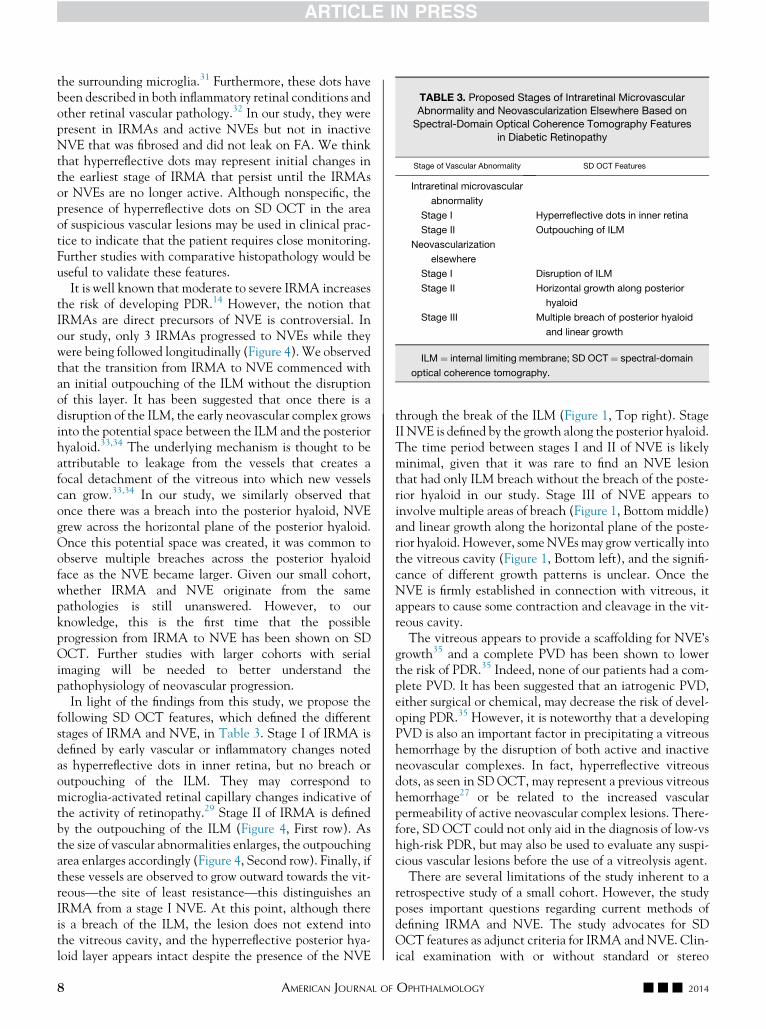
TABLE 3. Proposed Stages of Intraretinal Microvascular
Abnormality and Neovascularization Elsewhere Based on
Spectral-Domain Optical Coherence Tomography Features
in Diabetic Retinopathy
Stage of Vascular Abnormality SD OCT Features
Intraretinal microvascular
abnormality
Stage I Hyperreflective dots in inner retina
Stage II Outpouching of ILM
Neovascularization
elsewhere
Stage I Disruption of ILM
Stage II Horizontal growth along posterior
hyaloid
Stage III Multiple breach of posterior hyaloid
and linear growth
ILM ¼ internal limiting membrane; SD OCT ¼ spectral-domain
optical coherence tomography.
8 --- 2014AMERICAN JOURNAL OF OPHTHALMOLOGY

photographs can be difficult, and the agreement in inter-
preting the photographs and the leakage on FA can vary
between the examiners and the readers in reading cen-
ters.16 Firstly, our study did not include stereo photographs
used in the original Airlie classification. However, stereo
imaging for the assessment of ‘‘depth’’ in the retina has all
but disappeared from clinical practice since the advent of
OCT, which provides detailed cross-section information
from the vitreous and retina. Secondly, this study excluded
patients who had low-quality images and therefore cannot
assess the feasibility of obtaining adequate images in busy
clinic settings. In fact, peripheral retinal NVEs are not
easily scanned with OCT. However, this may change
with the use of ‘‘swept-source’’ OCT systems with ‘‘wide-
field’’ image acquisition. In addition, we note that the den-
sity of B-scans when reviewing NVE using the SD OCT
is critical. It is possible to misinterpret the OCT definitions
of NVE or IRMA if only viewing a single scan—that is,
one scan may only show the outpouching of the ILM
(OCT—definition of a stage II IRMA) while the adjacent
scan may reveal a breach of the ILM (OCT—definition of a
stage I NVE). Finally, the small number of patients may
overestimate the power of the significance shown in our
study. Future studies with larger cohorts are needed to bet-
ter study the importance of the OCT characterizations.
In this study, we identified several OCT-derived param-
eters that distinguish IRMAs and NVEs. We suggest that
current clinical and FA definitions of IRMA vs NVE
should be supported with SDOCT findings. If these param-
eters are successfully validated in a prospective cohort, it
may serve as important endpoints to clinical trials and di-
rection of future studies, in particular with the introduction
of vitreolysis agents and its possible role in the prevention
of DR progression. Further investigations on the utility of
SD OCT evaluation may also enable clinicians to more
closely monitor patients without the need for FA and to
consequently tailor management decisions according to
the individual’s response to treatment.
THEAUTHORSHAVE COMPLETEDAND SUBMITTED THE ICMJE FORM FORDISCLOSUREOF POTENTIAL CONFLICTSOF INTEREST.Drs Sim, Zarranz-Ventura, Keane, Egan, and Tufail have received travel grants from the Allergan European Retina Panel, London, United Kingdom; DrEgan has been on advisory boards for Novartis (Basel, Switzerland); Dr Tufail has been on advisory boards for Novartis (Basel, Switzerland), Pfizer (Surrey,United Kingdom), GlaxoSmithKline (Middlesex, United Kingdom), Thrombogenics (Leuven, Belgium), Bayer (Leverkusen, Germany), Allergan (Irvine,California, USA), and Heidelberg Engineering (Heidelberg, Germany). Drs Keane, Egan, Sim, and Tufail have received a proportion of their funding fromthe Department of Health’s National Institute for Health Research Biomedical Research Centre for Ophthalmology at Moorfields Eye Hospital and Uni-versity of College London Institute of Ophthalmology. The views expressed in the publication are those of the author and not necessarily those of theDepartment of Health. Drs Sim, Keane, and Fruttiger receive funding from Fight For Sight United Kingdom, Grant 1987. Dr Zarranz-Ventura is a grantrecipient of the Spanish Retina & Vitreous Society (Sociedad Espanola de Retina y Vitreo). Contributions of authors: conception and design (C.S.L.,A.Y.L., D.A.S., H.M., C.A.E., A.T.); analysis and interpretation (C.S.L., A.Y.L., D.A.S., P.A.K., H.M.); writing the article (C.S.L.); critical revisionof the article (A.Y.L., D.A.S., P.A.K., H.M., C.A.E., A.T.); final approval of the article (C.S.L., A.Y.L., D.A.S., P.A.K., H.M., J.Z.-V., M.F., C.A.E.,A.T.); data collection (C.S.L., A.Y.L., D.A.S., H.M., J.Z.-V.); statistical expertise (A.Y.L.); literature search (C.S.L.).
REFERENCES
1. Yau JW, Rogers SL, Kawasaki R, et al. Global prevalence and
major risk factors of diabetic retinopathy. Diabetes Care 2012;
35(3):556–564.
2. Jaeger E. Beitrage zur Pathologie des Auges.Wien: Kaiserlich-
konigliche Hof-und Staatsdruckerei; 1855:33–36.
3. ManzW. Retinitis Proliferans.Graefes Arch Clin Exp Ophthal-
mol 1876;22(3):229–275.
4. Diseases of the retina. In: Duke-Elder S, ed. System of
Ophthalmology. Vol. 10 London: Kimpton; 1967:410–445.
5. Weeks JE. Retinitis proliferans. Trans Am Ophthalmol Soc
1897;8:158–180.
6. Flemming P. A case of retinitis proliferans in which the eye
was examined after death. Trans Ophthalmol Soc U K 1898;
18:154–164.
7. Ballantyne AJ. The state of the retina in diabetes mellitus.
Trans Ophthalmol Soc U K 1946;66:503–543.
8. Archer DB. Neovascularization of the retina. Trans Ophthal-
mol Soc U K 1976;96(4):471–493.
9. Goldberg MF, Fine SL. Symposium on the treatment of
diabetic retinopathy. Washington, DC: US Govt Printing
Office; 1969:7–22.
10. Ashton P. Arteriolar involvement in diabetic retinopathy. Br
J Ophthalmol 1953;37(5):282–292.
11. GoldbergMF, Jampol LM. Knowledge of diabetic retinopathy
before and 18 years after the Airlie House Symposium on
Treatment of Diabetic Retinopathy. Ophthalmology 1987;
94(7):741–746.
12. Diabetic retinopathy study. Report Number 6. Design,
methods, and baseline results. Report Number 7. A modifica-
tion of the Airlie House classification of diabetic retinopathy.
Invest Ophthalmol Vis Sci 1981;21(1 Pt 2):1–226.
13. Early Treatment Diabetic Retinopathy Study Research
Group. Grading diabetic retinopathy from stereoscopic color
fundus photographs–an extension of the modified Airlie
House classification. ETDRS report number 10. Ophthal-
mology 1991;98(5 Suppl):786–806.
14. Early Treatment Diabetic Retinopathy Study Research
Group. Fundus photographic risk factors for progression of
diabetic retinopathy. ETDRS report number 12. Ophthal-
mology 1991;98(5 Suppl):823–833.
15. Early Treatment Diabetic Retinopathy Study Research
Group. Early Treatment Diabetic Retinopathy Study
design and baseline patient characteristics. ETDRS report
number 7. Ophthalmology 1991;98(5 Suppl):741–756.
16. Early Treatment Diabetic Retinopathy Study Research
Group. Classification of diabetic retinopathy from fluorescein
angiograms. ETDRS report number 11. Ophthalmology 1991;
98(5 Suppl):807–822.
VOL. -, NO. - 9REDEFINING THE CRITERIA OF NEOVASCULARIZATION IN DIABETES
17. Early Treatment Diabetic Retinopathy Study Research
Group. Fluorescein angiographic risk factors for progression
of diabetic retinopathy. ETDRS report number 13. Ophthal-
mology 1991;98(5 Suppl):834–840.
18. Davis MD, Blodi BA. Proliferative diabetic retinopathy.
In: Ryan SJ, ed. Retina. 4th ed. Vol 2. Philadelphia:
Elsevier/Mosby; 2006:1285–1322.
19. Lee C. Diabetic retinopathy. Spaide RF, ed. Diseases of the
Retina and Vitreous. Philadelphia: W.B. Saunders Company;
1999:129–143.
20. Kim JW, Ai E. Diabetic retinopathy. BrownGC, Regillo CD,
Flynn HW, eds. Vitreoretinal Disease. The Essentials. New
York: Thieme; 1999:133–159.
21. Huang D, Swanson EA, Lin C-P, et al. Optical coherence to-
mography. Science 1991;254(5035):1178–1181.
22. Keane PA, Patel PJ, Liakopoulos S, Heussen FM, Sadda SR,
Tufail A. Evaluation of age-related macular degeneration
with optical coherence tomography. Surv Ophthalmol 2012;
57(5):389–414.
23. Lee DH, Kim JT, Jung DW, Joe SG, Yoon YH. The relation-
ship between foveal ischemia and spectral-domain optical
coherence tomography findings in ischemic diabetic macular
edema. Invest Ophthalmol Vis Sci 2013;54(2):1080–1085.
24. Davis MD, Bressler SB, Aiello LP, et al. Comparison of time-
domain OCT and fundus photographic assessments of retinal
thickening in eyes with diabetic macular edema. Invest
Ophthalmol Vis Sci 2008;49(5):1745–1752.
25. Brown DM, Nguyen QD, Marcus DM, et al. Long-term out-
comes of ranibizumab therapy for diabetic macular edema:
the 36-month results from two phase III trials: RISE and
RIDE. Ophthalmology 2013;120(10):2013–2022.
26. Rofagha S, Bhisitkul RB, Boyer DS, Sadda SR, Zhang K,
Group S-US. Seven-year outcomes in ranibizumab-treated
patients in ANCHOR, MARINA, and HORIZON: a multi-
center cohort study (SEVEN-UP). Ophthalmology 2013;
120(11):2292–2299.
27. Muqit MM, Stanga PE. Fourier-domain optical coherence to-
mography evaluation of retinal and optic nerve head neovas-
cularisation in proliferative diabetic retinopathy. Br J
Ophthalmol 2014;98(1):65–72.
28. Cho H, Alwassia AA, Regiatieri CV, et al. Retinal neovascu-
larization secondary to proliferative diabetic retinopathy
characterized by spectral domain optical coherence tomogra-
phy. Retina 2013;33(3):542–547.
29. Vujosevic S, Bini S, Midena G, Berton M, Pilotto E,
Midena E. Hyperreflective intraretinal spots in diabetics
without and with nonproliferative diabetic retinopathy: an
in vivo study using spectral domain OCT. J Diabetes Res
2013;2013:491835.
30. Garner A. Developments in the pathology of diabetic reti-
nopathy: a review. J R Soc Med 1981;74(6):427–431.
31. Zeng HY, Green WR, Tso MO. Microglial activation in hu-
man diabetic retinopathy. Arch Ophthalmol 2008;126(2):
227–232.
32. Ogino K, Murakami T, Tsujikawa A, et al. Characteristics of
optical coherence tomographic hyperreflective foci in retinal
vein occlusion. Retina 2012;32(1):77–85.
33. Constable IJ. Pathology of vitreous membranes and the effect
of haemorrhage and new vessels on the vitreous. Trans
Ophthalmol Soc U K 1975;95(3):382–386.
34. Davis MD. Vitreous contraction in proliferative diabetic reti-
nopathy. Arch Ophthalmol 1965;74(6):741–751.
35. Ono R, Kakehashi A, Yamagami H, et al. Prospective assess-
ment of proliferative diabetic retinopathy with observations
of posterior vitreous detachment. Int Ophthalmol 2005;
26(1-2):15–19.
10 --- 2014AMERICAN JOURNAL OF OPHTHALMOLOGY

Biosketch
Cecilia S. Lee, MD, is currently an Acting Instructor of Ophthalmology at University of Washington in Seattle,
Washington. She completed her residency at Emory University in Atlanta, Georgia, followed by a fellowship in uveitis
at Washington University in St. Louis and in medical retina at Moorfields Eye Hospital in London, UK. Her research
interests include non-invasive imaging in medical retina and uveitis and the ocular surface microbiome.
VOL. -, NO. - 10.e1REDEFINING THE CRITERIA OF NEOVASCULARIZATION IN DIABETES