Perinatal Systemic Inflammatory Response Syndrome and Retinopathy of Prematurity
19
Perinatal Systemic Inflammatory Response Syndrome and Retinopathy of Prematurity Beena G. Sood, Department of Pediatrics Wayne State University, Detroit, MI 48201 Ashima Madan, Department of Pediatrics Stanford University School of Medicine, Palo Alto, CA 94305 Shampa Saha, Statistics and Epidemiology Unit RTI International, Research Triangle Park, NC 27709 Diana Schendel, Centers for Disease Control and Prevention Atlanta, GA 30329 Poul Thorsen, Copyright © 2009 International Pediatric Research Foundation, Inc. All rights reserved. Corresponding author: Beena Gaind Sood, MD Children’s Hospital of Michigan 3901 Beaubien Blvd., 4H42 Detroit, MI 48201 Tel (313) 745-5638 Fax (313) 745-5867 [email protected] . The results reported in this manuscript were presented in part at the Pediatric Academic Societies’ Annual Meeting on May 4, 2008 at Honolulu, Hawaii. The following investigators participated in this study: Alan Jobe, University of Cincinnati. William Oh, Abbot R. Laptook, Lewis P. Rubin, Angelita M. Hensman, Brown University. Avroy A. Fanaroff, Michele C. Walsh, Nancy S. Newman, Bonnie S. Siner, Case Western Reserve University. Diana E. Schendel, Centers for Disease Control and Prevention. Edward F. Donovan, Vivek Narendran, Barbara Alexander, Cathy Grisby, Marcia Worley Mersmann, Holly L. Mincey, Jody Hessling, Cincinnati Children’s Hospital Medical Center. Ronald N. Goldberg, C. Michael Cotten, Kathy J. Auten, Duke University. Barbara J. Stoll, Ira Adams-Chapman, Ellen C. Hale, Emory University. Linda L. Wright, Rosemary D. Higgins, Sumner J. Yaffe, Elizabeth M. McClure, Eunice Kennedy Shriver National Institute of Child Health and Human Development. James A. Lemons, Brenda B. Poindexter, Diana D. Appel, Dianne E. Herron, Leslie D. Wilson, Indiana University. W. Kenneth Poole, Abhik Das, Scott A. McDonald, Betty Hastings, Kristin Zaterka-Baxter, Jeanette O’Donnell Auman, RTI International. David K. Stevenson, Krisa P. Van Meurs, M. Bethany Ball, Stanford University. Kristin Skogstrand, David M. Hougaard, Statens Serum Institut. Poul Thorsen, University of Aarhus, Denmark. Namasivayam Ambalavanan, Monica V. Collins, Shirley S. Cosby, University of Alabama at Birmingham. Neil N. Finer, Maynard R. Rasmussen, David Kaegi, Kathy Arnell, Clarence Demetrio, Wade Rich, University of California – San Diego. Charles R. Bauer, Shahnaz Duara, Ruth Everett-Thomas, University of Miami. Lu-Ann Papile, Conra Backstrom Lacy, University of New Mexico. Sheldon B. Korones, Henrietta S. Bada, Tina Hudson, University of Tennessee. Abbot R. Laptook, Walid A. Salhab, R. Sue Broyles, Susie Madison, Jackie F. Hickman, Sally S. Adams, Linda A. Madden, Elizabeth Heyne, Cristin Dooley, University of Texas Southwestern Medical Center at Dallas. Jon E. Tyson, Kathleen Kennedy, Brenda H. Morris, Esther G. Akpa, Patty A. Cluff, Claudia Y. Franco, Anna E. Lis, Georgia E. McDavid, Patti L. Tate, University of Texas Health Science Center at Houston. T. Michael O’Shea, Robert G. Dillard, Lisa K. Washburn, Barbara G. Jackson, Nancy J. Peters, Wake Forest University. G. Ganesh Konduri, Geraldine Muran, Rebecca Bara, Wayne State University. Richard A. Ehrenkranz, Patricia Gettner, Monica Konstantino, Elaine Romano, Yale University. This is a PDF file of an unedited manuscript that has been accepted for publication. As a service to our customers we are providing this early version of the manuscript. The manuscript will undergo copyediting, typesetting, and review of the resulting proof before it is published in its final citable form. Please note that during the production process errors may be discovered which could affect the content, and all legal disclaimers that apply to the journal pertain. NIH Public Access Author Manuscript Pediatr Res. Author manuscript; available in PMC 2011 April 1. Published in final edited form as: Pediatr Res. 2010 April ; 67(4): 394–400. doi:10.1203/PDR.0b013e3181d01a36. NIH-PA Author Manuscript NIH-PA Author Manuscript NIH-PA Author Manuscript
Transcript of Perinatal Systemic Inflammatory Response Syndrome and Retinopathy of Prematurity

Perinatal Systemic Inflammatory Response Syndrome andRetinopathy of Prematurity
Beena G. Sood,Department of Pediatrics Wayne State University, Detroit, MI 48201
Ashima Madan,Department of Pediatrics Stanford University School of Medicine, Palo Alto, CA 94305
Shampa Saha,Statistics and Epidemiology Unit RTI International, Research Triangle Park, NC 27709
Diana Schendel,Centers for Disease Control and Prevention Atlanta, GA 30329
Poul Thorsen,
Copyright © 2009 International Pediatric Research Foundation, Inc. All rights reserved.Corresponding author: Beena Gaind Sood, MD Children’s Hospital of Michigan 3901 Beaubien Blvd., 4H42 Detroit, MI 48201 Tel (313)745-5638 Fax (313) 745-5867 [email protected] .The results reported in this manuscript were presented in part at the Pediatric Academic Societies’ Annual Meeting on May 4, 2008 atHonolulu, Hawaii.The following investigators participated in this study: Alan Jobe, University of Cincinnati.William Oh, Abbot R. Laptook, Lewis P. Rubin, Angelita M. Hensman, Brown University.Avroy A. Fanaroff, Michele C. Walsh, Nancy S. Newman, Bonnie S. Siner, Case Western Reserve University.Diana E. Schendel, Centers for Disease Control and Prevention.Edward F. Donovan, Vivek Narendran, Barbara Alexander, Cathy Grisby, Marcia Worley Mersmann, Holly L. Mincey, Jody Hessling,Cincinnati Children’s Hospital Medical Center.Ronald N. Goldberg, C. Michael Cotten, Kathy J. Auten, Duke University.Barbara J. Stoll, Ira Adams-Chapman, Ellen C. Hale, Emory University.Linda L. Wright, Rosemary D. Higgins, Sumner J. Yaffe, Elizabeth M. McClure, Eunice Kennedy Shriver National Institute of ChildHealth and Human Development.James A. Lemons, Brenda B. Poindexter, Diana D. Appel, Dianne E. Herron, Leslie D. Wilson, Indiana University.W. Kenneth Poole, Abhik Das, Scott A. McDonald, Betty Hastings, Kristin Zaterka-Baxter, Jeanette O’Donnell Auman, RTIInternational.David K. Stevenson, Krisa P. Van Meurs, M. Bethany Ball, Stanford University.Kristin Skogstrand, David M. Hougaard, Statens Serum Institut.Poul Thorsen, University of Aarhus, Denmark.Namasivayam Ambalavanan, Monica V. Collins, Shirley S. Cosby, University of Alabama at Birmingham.Neil N. Finer, Maynard R. Rasmussen, David Kaegi, Kathy Arnell, Clarence Demetrio, Wade Rich, University of California – SanDiego.Charles R. Bauer, Shahnaz Duara, Ruth Everett-Thomas, University of Miami.Lu-Ann Papile, Conra Backstrom Lacy, University of New Mexico.Sheldon B. Korones, Henrietta S. Bada, Tina Hudson, University of Tennessee.Abbot R. Laptook, Walid A. Salhab, R. Sue Broyles, Susie Madison, Jackie F. Hickman, Sally S. Adams, Linda A. Madden, ElizabethHeyne, Cristin Dooley, University of Texas Southwestern Medical Center at Dallas.Jon E. Tyson, Kathleen Kennedy, Brenda H. Morris, Esther G. Akpa, Patty A. Cluff, Claudia Y. Franco, Anna E. Lis, Georgia E. McDavid,Patti L. Tate, University of Texas Health Science Center at Houston.T. Michael O’Shea, Robert G. Dillard, Lisa K. Washburn, Barbara G. Jackson, Nancy J. Peters, Wake Forest University.G. Ganesh Konduri, Geraldine Muran, Rebecca Bara, Wayne State University.Richard A. Ehrenkranz, Patricia Gettner, Monica Konstantino, Elaine Romano, Yale University.This is a PDF file of an unedited manuscript that has been accepted for publication. As a service to our customers we are providing thisearly version of the manuscript. The manuscript will undergo copyediting, typesetting, and review of the resulting proof before it ispublished in its final citable form. Please note that during the production process errors may be discovered which could affect the content,and all legal disclaimers that apply to the journal pertain.
NIH Public AccessAuthor ManuscriptPediatr Res. Author manuscript; available in PMC 2011 April 1.
Published in final edited form as:Pediatr Res. 2010 April ; 67(4): 394–400. doi:10.1203/PDR.0b013e3181d01a36.
NIH
-PA Author Manuscript
NIH
-PA Author Manuscript
NIH
-PA Author Manuscript

Department of Epidemiology and Social Medicine University of Aarhus, DK-8000 Denmark; RollinsSchool of Public Health Emory University, Atlanta, GA 30322
Kristin Skogstrand,Department of Clinical Biochemistry and Immunology Statens Serum Institut, DK-2300,Copenhagen, Denmark
David Hougaard,Department of Clinical Biochemistry and Immunology Statens Serum Institut, DK-2300,Copenhagen, Denmark
Seetha Shankaran, andDepartment of Pediatrics Wayne State University, Detroit, MI 48201
Wally CarloDepartment of Pediatrics University of Alabama at Birmingham, Birmingham, AL 35233
On behalf of the NICHD Neonatal Research Network
AbstractFetal and neonatal inflammation is associated with several morbidities of prematurity. Its relationshipto retinopathy of prematurity (ROP) has not been investigated. Our objective was to determine therelationship between cytokine levels and ROP in the first three postnatal weeks. Data for this studywere derived from the NICHD Cytokine Study. Dried blood spots were obtained from infants <1000gon days 0-1, 3±1, 7±2, 14±3, and 21±3. Infants were classified into three groups – No, Mild, andSevere ROP. Multiplex Luminex assay was used to quantify 20 cytokines. Temporal profiles ofcytokines were evaluated using mixed effects models after controlling for covariates. Of 1074 infantsenrolled, 890 were examined for ROP and 877 included in the analysis. ROP was associated withseveral clinical characteristics on unadjusted analyses. Eight cytokines remained significantlydifferent across ROP groups in adjusted analyses. IL-6 and IL-17 showed significant effects in earlytime periods (D0-3); TGF-β, brain derived neurotrophic factor (BDNF), and regulated uponactivation, normal T cell expressed and secreted (RANTES) in later time periods (D7-21) and IL-18,C-reactive protein (CRP) and neurotrophin-4 (NT-4) in both early and later time periods. Weconclude that perinatal inflammation may be involved in the pathogenesis of ROP.
Retinopathy of prematurity (ROP), a vasoproliferative disorder of the developing retina, is amajor cause of blindness in infancy. ROP is a biphasic disease consisting of an initial phaseof blunted vascular growth followed by a second phase of vaso- proliferation that is recognizedon ophthalmoscopy four to six weeks after birth. Angiogenesis, the fundamental processinvolved in retinal vascular development, is tightly regulated by a complex network ofcytokines, extracellular matrix components, and growth factors the action of which varies ina time-dependent fashion. Although inflammatory cytokines have the ability to modulateangiogenesis, their role in triggering the dysregulated angiogenesis in ROP has not beeninvestigated (1).
Antenatal intrauterine infection with the resulting fetal inflammatory response syndrome(FIRS) is important in the pathogenesis of preterm birth and its associated morbidities includingsepsis, periventricular leukomalacia (PVL), intraventricular hemorrhage (IVH), necrotizingenterocolitis (NEC), and bronchopulmonary dysplasia (2,3). It is not known whether FIRS alsopredisposes to the development of ROP. Recently it has been reported that chorioamnionitismay be a risk factor for ROP occurrence and progression; however this association was notsignificant on adjusted analyses in this small retrospective study (4). Similarly, although severalreports suggest that postnatal severity of illness and Candida infection contribute to ROPfrequency and/or severity, the role of the associated systemic inflammatory response syndrome
Sood et al. Page 2
Pediatr Res. Author manuscript; available in PMC 2011 April 1.
NIH
-PA Author Manuscript
NIH
-PA Author Manuscript
NIH
-PA Author Manuscript

(SIRS) as evaluated by cytokine levels in the pathogenesis of ROP has not been investigated(5). Neonatal non-infectious inflammation might further increase the inflammatory burden(6). In this study, we present for the first time, epidemiological data from a large cohort ofpreterm babies to support the role of perinatal inflammation in the pathogenesis of ROP.
We hypothesized that inflammatory mediators released during SIRS in the pre-/peri-natalperiod lead to disruption of angiogenesis resulting in ROP. The aim of the current study wasto investigate the relationship between concentrations of fetal/neonatal inflammatory markersand growth factors in dried blood spots (DBS) from preterm infants collected over the firstthree weeks after birth and ROP.
MethodsWe conducted a secondary analysis using clinical and biologic data collected as part of themulticenter NICHD Cytokine study, a prospective study assessing biomarkers and risk factorsfor adverse neurodevelopmental outcomes in preterm infants. This study was approved by theInstitutional Review Boards at all 17 participating centers, and written informed consent wasobtained from the parent(s).
Preterm neonates with birth weight 401-1000 g who were enrolled in the NICHD CytokineStudy and had available DBS and results of ROP diagnostic examinations were eligible forinclusion in the study. Information from ROP diagnostic examinations that was available inthis dataset included highest stage and lowest zone of ROP, presence of plus disease, diagnosisof threshold disease, and need for retinal ablative therapy. In addition, presence of regressingROP was recorded at 120 days postnatal age, discharge, or death, whichever came first. Infantswho died prior to eye exam for ROP or 36 weeks postmenstrual age (PMA) were excluded.
Whole blood was collected on filter paper and frozen at five time periods (days): 0-1 [D0], 3± 1 [D3], 7 ± 2 [D7], 14 ± 3 [D14], and 21 ± 3 [D21]. The treatment of blood samples wasstandardized. After the filter paper blood sample had dried, it was wrapped in stock paper coverand placed in a plastic bag which was stored in a −20°C freezer as soon as possible after thespecimen was dry but no later than 24 hours. Clinical data were collected by trained researchcoordinators, and all analyses were performed at a central data coordinating center. Theoutcome variable was ROP which was classified into three categories – No ROP, Mild ROP(ROP other than Severe) and Severe ROP. Severe ROP included Type 1 and Type 2 ROP asdefined by the Early Treatment for Retinopathy of Prematurity trial [ETROP] (7). Confoundingvariables included center, gestational age, birth weight, race, gender, premature prolongedrupture of membranes, sepsis (early and late), antenatal steroids, postnatal steroids, days inO2, IVH (Grades 3, 4), cystic PVL, surgically treated patent ductus arteriosus, and NEC (Bells’stage ≥2).
The stored blood spots were analyzed using a multiplex Luminex assay (Luminex Corp.,Austin, TX) as described previously (8,9). This assay has low intra- (<10%) and inter-assay(7-23%) variation. It has been shown that cytokine measurements on DBS stored at −24 °C for>20 years are stable over time. The first part of our study included a hypothesis driveninvestigation of 11 cytokines and growth factors reported to be important in the pathogenesisof inflammation, preterm birth, angiogenesis and or proliferative retinopathies (IL-1β, IL-6,IL-8, sIL-6R, TNF-α, IFN-γ, MIP-1α, monocyte chemoattractant protein-1 [MCP-1], MMP-9,TGF-β, regulated upon activation, normal T cell expressed and secreted [RANTES]);subsequently we broadened our investigation to include nine other markers that were availablein the subjects of the primary study (IL-2, IL-4, IL-10, IL-17, IL-18, granulocyte-macrophagecolony stimulating factor [GM-CSF], CRP, BDNF, NT-4) based on exploratory analysis.
Sood et al. Page 3
Pediatr Res. Author manuscript; available in PMC 2011 April 1.
NIH
-PA Author Manuscript
NIH
-PA Author Manuscript
NIH
-PA Author Manuscript

Statistical AnalysesStatistical analyses were performed using SAS System 9.1 (SAS, Cary, NC). Unadjustedanalysis was performed using chi-square tests for categorical outcomes and analysis of variancetest (F-test) for continuous outcomes to study the association of several maternal and neonatal,variables with ROP (no, mild, and severe ROP). Medians and inter-quartile ranges werecomputed for all cytokines at each of the five time points and the Kruskal-Wallis nonparametricanalysis of variance used to assess differences across ROP groups. For each cytokine, levelswere plotted against time for each ROP group to assess their temporal profiles over the firstthree postnatal weeks. Differences in the temporal profiles across the three ROP groups werefurther evaluated using mixed effect models for each cytokine independently to account forthe multicenter design, repeated measurement of cytokine levels, significant clinical covariates,and interaction of postnatal age and ROP group. A p value of <0.05 was considered statisticallysignificant.
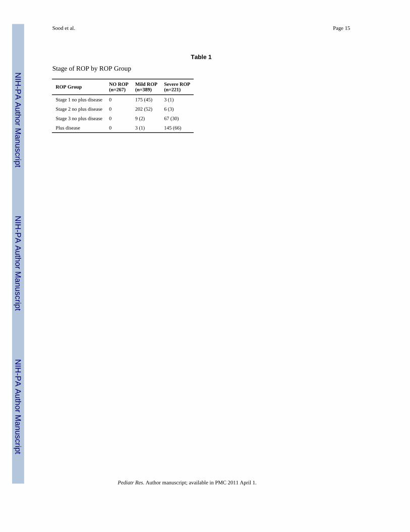
ResultsAmong 1074 patients enrolled, 890 had a screening eye exam for ROP. Thirteen infants whodied before 36 weeks PMA were excluded from the analysis as it has been shown that themedian (5%, 95%) age at the time of development pre-threshold ROP is 36.1 (32.1, 42.1) weeksPMA (10). Of these 13 infants, ten had no ROP, two had Mild ROP, and one infant had SevereROP (Type 2 ROP). None of these infants had Type 1 or aggressive posterior ROP. Of theremaining 877 infants, 610 (69.6%) were diagnosed with ROP. The numbers of infants thatdeveloped each stage of ROP are reported in Table 1. Fifteen infants died before 42 weeksPMA and had potentially incomplete ROP outcome data. Of these infants, five had SevereROP (Type 1 ROP treated with laser photocoagulation [n=2], Type 2 ROP [n=3]), five hadMild ROP, and five had no ROP. DBS were available for 560 infants on D0, 709 infants onD3, 811 infants on D7, 789 infants on D14 and 765 infants on D21 (Figure 1).
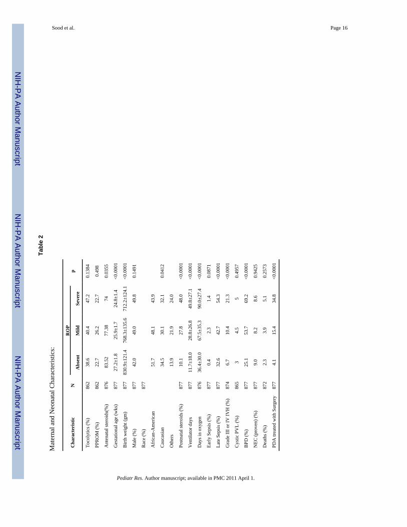
Maternal and Neonatal CharacteristicsThe mean±SD birthweight of infants included in this study was 773±129 grams. Infants withany ROP were less likely to have received antenatal steroids (p<0.05), and had a lowergestational age and birth weight (p<0.0001) (Table 2). There was a statistically significantassociation between race and development of ROP (p=0.04). Infants who developed ROP weremore likely to have received postnatal steroids, longer duration of assisted ventilation andoxygen therapy, late onset sepsis, Grade III-IV IVH, surgically treated patent ductus arteriosusand bronchopulmonary dysplasia (p<0.0001). Although infants with ROP had a higherincidence of early sepsis, the difference was not statistically significant (p 0.09). Similarlyalthough mothers of infants with ROP were more likely to have received tocolytics, thisdifference was not statistically significant (p>0.1). There were no differences in gender,incidence of NEC, cystic PVL, PPROM and mortality between infants who developed ROPcompared to those who did not.
Comparison of Cytokines by ROP group at Each Time PointKruskal-Wallis analysis indicated no difference in median cytokine levels between ROP groupsfor IL-4, IL-6, TNF-α, MIP-1α at any of the five time points. Significant association of severalother cytokines with ROP group was observed on D0 (IL-1β, IL-8, IL-10, IL-17, IL-18, sIL-6R,GM-CSF, CRP, TGF-β, and RANTES), D3 (IL-8, IL-10, IL-17, sIL-6R, IFN-γ, GM-CSF,NT-4, MMP-9, and CRP), D7 (IL-1β, IL-2, IL-8, IL-10, IL-17, IL-18, sIL-6R, BDNF, GM-CSF, NT-4, MMP-9, and CRP), D14 (IL-1β, IL-2, IL-8, IL-17, IL-18, BDNF, GM-CSF, NT-4,MCP-1, MMP-9, CRP and RANTES), and D21 (IL-8, IL-10, IL-17, IL-18, sIL-6R, BDNF,GM-CSF, NT-4, MCP-1, MMP-9, CRP, TGF-β and RANTES). Mixed effects model analysisafter controlling for important covariates further indicated significant differences across ROP
Sood et al. Page 4
Pediatr Res. Author manuscript; available in PMC 2011 April 1.
NIH
-PA Author Manuscript
NIH
-PA Author Manuscript
NIH
-PA Author Manuscript

groups for IL-6, IL-17, IL-18, sIL-6R, TGF-β, BDNF, NT-4, CRP, and RANTES (main effectof ROP group on longitudinal data analysis, p<0.05 for all) (Figures 2 and 3).
Time Trends of Various CytokinesMixed effect model analysis by ROP group indicated that the majority of the cytokines showedsignificant time trends (p<0.05 for all) during the first three weeks of life after controlling forimportant covariates including center, gestational age, gender, late sepsis, antenatal steroids,postnatal steroids, days in oxygen, race, NEC, severe IVH, and PDA treated with surgery.Cytokines that showed a decreasing trend over time included IL-1β, IL-2, IL-4, IL-5, IL-6,IL-10, IL-12, IL-17, TNF-B, GM-CSF, NT-4. Cytokines that showed an increasing trend overtime included IL-18, BDNF, MMP-9, TGF-β, MIP-1α, RANTES, and CRP.
Interaction of ROP Group and Cytokines over timeSix cytokines showed significant main effects for ROP group and time with a significantinteraction of ROP group with time on mixed effect model analysis after controlling forimportant covariates (p<0.05) [IL-6, IL-17, IL-18, TGF-β, BDNF, RANTES] (Figure 2). Twocytokines showed significant main effects for ROP group without a significant interaction ofROP group with time [CRP, NT-4] (Figure 3). Although sIL-6R had a significant associationwith ROP (p=0.0425); the association was significant only on D14 and the direction of changeof sIL-6R levels on univariate and adjusted analyses was contradictory. Therefore, this has notbeen described further.
The Least Squares Means, group comparisons, and significant covariates for cytokines withand without significant ROP-time interaction on adjusted analyses are presented in Tables 3and 4 respectively. Table 5 summarizes the direction of change in cytokine levels showingsignificant association with various ROP groups at different time periods on adjusted analyses.
Two cytokines (IL-6, IL-17) showed significant association with ROP in the early time periods(D0, D3) (Table 5) and three cytokines (TGF-β, BDNF, RANTES) showed significantassociation in the later time periods (D7, D14, D21). IL-18, CRP, and NT-4 showed significantassociation in both the early and later time periods (D0, D3, D7, D14, D21). IL-18 was uniquein showing biphasic time trends with lower levels in infants with ROP on D0 and higher levelson D7, D14, and D21 compared to infants without ROP.
DiscussionThis is the first population study of preterm infants evaluating the association of perinatalinflammation as evaluated by cytokine levels and ROP. In preterm infants, development ofROP was associated with elevated levels of IL-6 and lower levels of IL-17 and IL-18 on D0suggesting a relationship between FIRS and ROP. IL-17 continued to be significantly lowerin infants with mild ROP on D3. Association between ROP group and elevated levels of CRPand lower levels of NT-4 was observed as early as D3; NT-4 continued to be low on D7 andCRP continued to be higher in infants with ROP on D14and D21. ROP was associated withhigher levels of IL-18 and lower levels of TGF-β, BDNF, and RANTES on days 7 to 21suggesting that postnatal systemic inflammatory response syndrome is also an important factorin the pathogenesis of ROP.
Cytokines have pleiotropic actions including recruitment and activation of immune cells,regulation of angiogenesis, cell proliferation, and apoptosis. Cytokines can have both anti- andpro-inflammatory and anti- and pro-angiogenic action depending on the target tissue, dose ofcytokine, its timing, and duration. In addition to being the site of angiogenesis, the retina isalso a neural tissue in which local autocrine loops between neurotrophic, angiogenic and
Sood et al. Page 5
Pediatr Res. Author manuscript; available in PMC 2011 April 1.
NIH
-PA Author Manuscript
NIH
-PA Author Manuscript
NIH
-PA Author Manuscript

inflammatory mediators play an important role in normal development; disruption of thesemay result in pathologic states. Cytokine levels at birth are reflective of perinatal eventswhereas subsequent levels are reflective of postnatal experiences of the infant (11). Principalpro-inflammatory cytokines are TNF-α, IL-1, IL-6, IL-8, IL-12, IL-17, IL-18, soluble IL-6receptor-α (sIL-6rα), IFN- γ, GM-CSF, MCP-1, MIP 1 α, and RANTES. Key anti-inflammatory cytokines include IL-2, IL-4, IL-10, IL-13, IL-11 and TGF-β. BDNF and NT-4are neurotrophic factors that also play a role in the regulation of intraocular inflammation.
In this study, we found a panel of six inflammatory markers and two growth factors to besignificantly associated with ROP in the 1st three weeks of life. In contrast to reports of elevatedplasma IL-1β, TNF-α, MMP-9 IL-2, MCP-1 in FIRS and proliferative retinopathies, we didnot find these cytokines significantly associated with ROP severity in our large populationbased study (12-14). An important factor that may explain this difference is that these studiesmeasured one or a few cytokines in smaller groups of patients.
IL-6 is involved in the regulation of the immune system, in acute phase reaction andinflammation. IL-6 has been shown to play an important role in the pathogenesis of diabeticretinopathy, FIRS, and neonatal sepsis (2,13,15). In the present study, significantly higher IL-6levels were noted on D0 in infants with mild ROP compared to those with severe or no ROP.However, because of the lack of increase seen in infants with severe ROP, it is difficult toconclude that IL-6 has an association with the development of ROP.
C-reactive protein (CRP), a biochemical marker of inflammation, was elevated in infants withROP both in early and late time periods in this study. Elevated levels of C-reactive protein areassociated with systemic vascular disorders, diabetic retinopathy, age-related maculardegeneration, amniotic fluid infection, congenital neonatal sepsis, funisitis, and late neonatalsepsis (1,16,17). The association of elevated CRP levels and ROP in our study persisted evenafter controlling for clinical covariates suggesting that a coexistent systemic inflammatoryresponse syndrome may account for the association of elevated levels of CRP and ROP.
Infants with severe ROP tended to have lower levels of cytokines IL-17 and IL-18 on D0 andTGF-β and RANTES on D7-21. These results can be explained by varying pro- and anti-inflammatory actions of the same cytokine in a time, dose, and tissue dependent manner.Another possible explanation for these results could be immune tolerance following chronicfetal exposure to inflammation as has been described previously (18,19). IL-17 and IL-18 arecytokines that have been described recently and whose role in FIRS has not yet beeninvestigated. IL-17 is a pro-inflammatory cytokine that promotes angiogenesis. It is implicatedin intraocular inflammation in uveitis (20). IL-17 has an important role in induction ofprotective immune response against extracellular bacterial or fungal pathogens such asKlebsiella pneumonia and Candida albicans (21). Based on the known biological actions ofIL-17, the lower IL-17 levels in infants with ROP in early time points may be responsible forarrest of angiogenesis in early ROP and also predispose these infants to late sepsis that wasstrongly associated with ROP group.
IL-18 is a pleiotropic proinflammatory cytokine that has immunoregulatory activity. It can actas either an angiogenic or an angiostatic factor (22). High levels of IL-18 have been reportedin patients with type 1 diabetes mellitus; however their relationship to microvascularcomplications in diabetes is controversial (23). In contrast, in our study the biphasic and robustassociation of IL-18 with ROP may be consistent with its role as an immunoregulator andmodulator of angiogenesis with time sensitive expression.
TGF-β is a multifunctional protein that regulates cell growth, differentiation, migration, andextracellular matrix production and plays an important role in embryonic development, woundhealing, immune responses, and vascular development (24). TGF-β has been described as a
Sood et al. Page 6
Pediatr Res. Author manuscript; available in PMC 2011 April 1.
NIH
-PA Author Manuscript
NIH
-PA Author Manuscript
NIH
-PA Author Manuscript

cytokine with bipolarity that can both trigger and inhibit the immune system and angiogenesis(25). Either overproduction or underproduction of TGF-β has been shown to cause ocularabnormalities. In the present study, infants with severe ROP had lower levels of TGF-β in thelater time periods in this study. These findings are consistent with the role of TGF-β as aregulator of angiogenesis.
The chemokine RANTES plays a significant role in innate immunity, which is particularlyimportant in the neonatal period (26). RANTES cord blood concentrations have been reportedto be lower in healthy preterm than in term neonates (27). The increasing levels of RANTESover the 1st three weeks in the participants of this study is consistent with the trends describedfor term infants (26). RANTES has been implicated in the pathogenesis of diabetic retinopathy;its role in ROP has not been investigated to date (14). However, in contrast to elevated levelsassociated with diabetic retinopathy, RANTES levels were lower with increasing ROP severityin our study.
Clinicians are most familiar with the vascular changes associated with ROP as these are easilydistinguishable on ophthalmoscopy (13). Nevertheless, the retina is not only a vascular tissuebut also a neural tissue with glial, microglial and neuronal cells. Direct retinal injury that maynot be detectable ophthalmoscopically, has been demonstrated early in diabetic retinopathyand in animal models of ROP (28). Astrocytes are involved in the formation of the glia limitansof the retinal vessels, and are more sensitive to hypoxia than retinal neurons. Astrocytessubsequently recolonize the retina after a delay that matches the period of leakiness of theproliferative vasculature. BDNF and NT-4 are neurotrophins that have been demonstrated topromote retinal ganglion cell survival after injury (29). Although neurons are the major cellularsource of BDNF, BDNF can also be secreted by vascular endothelial cells and immune cellsthus representing a crucial link between the nervous and immune system (30,31). Pro-inflammatory cytokines regulate the secretion of BDNF and NT-4 which in turn supportneuronal survival and further influence the secretion of these same cytokines setting up positivefeedback autocrine loops. Serum levels of BDNF have been reported to be higher in adults ascompared to newborns, and in full term neonates compared to those delivered pretermsuggesting a developmental appearance of neurotrophins in humans. The increase in BDNFand decrease in NT4 levels with age in our cohort are consistent with the trends described inliterature for term neonates (32,33). NT-4 levels were lower in infants with ROP compared tocontrols in our study with significant differences on D3 and D7. BDNF levels were significantlylower in infants with severe ROP at the later time points. A similar trend of BDNF levels hasbeen reported recently in a small study of preterm infants (33). We speculate that lower serumconcentrations of BDNF and NT-4 in infants with ROP compared to infants without ROP inour study may reflect glial and neuronal loss associated with ROP.
The majority of clinical characteristics that were strongly associated with ROP in our studyhave been previously reported in literature (34,35). Center difference was a significantcovariate in most of the analyses. This could be related to differing ethnic populations, inborn/outborn infant proportions, clinical practices, or other unidentified factors. Importantly, evenafter controlling for these covariates, the association between ROP and eight cytokinesremained significant suggesting their role in the pathogenesis of ROP.
Strengths of our study include a large sample size; prospectively collected data permitting athorough investigation of the association of perinatal inflammation and ROP, standardizedcollection of repeated, timed blood samples for three weeks starting from birth; assay ofmultiple cytokines from small volumes of blood using the Luminex assay; and multivariateadjustment for control of clinical variables.
Sood et al. Page 7
Pediatr Res. Author manuscript; available in PMC 2011 April 1.
NIH
-PA Author Manuscript
NIH
-PA Author Manuscript
NIH
-PA Author Manuscript
Despite the important findings of this study, there are limitations. First, this study is a secondaryanalysis of the NICHD Cytokine study. Although detailed information regarding serial eyeexaminations was not available, all infants underwent retinal examinations using aninternational classification with documentation of highest stage and lowest zone of ROP,presence of plus disease and need for treatment. Fifteen infants died before 42 weeks PMAand had potentially incomplete ROP data. Secondly, it is difficult to distinguish betweenprimary and secondary mediators in the cytokines described. Systemic levels evaluated in ourstudy may not be representative of ocular levels and it is not possible to ascertain whethersystemic levels are secondary to ocular levels or induce ocular changes. However, the fact thatchanges in cytokine and growth factor concentrations preceded the clinical diagnosis of ROPand that positive association persisted after rigorous adjustment for multiple clinically relevantcovariates make the case for causal association plausible. Thirdly, although we included 20biomarkers representing inflammation and angiogenesis, other markers (VEGF, IGF-I) thathave been shown to be important in retinal neovascularization were not included. Lastly, wedid not correct for multiple comparisons since the posthoc subgroup analysis in this study isexploratory in nature and hypothesis generating. Therefore, reliance should be placed on theobserved effect size rather than on statistical significance testing as a basis for decision making(36).
In summary, we have shown a coordinated pattern of increased and decreased levels of eightcytokines in the first three weeks of life in infants who were diagnosed with ROP. Additionalrigorously designed prospective studies are needed to further investigate the preliminaryevidence presented in this study. Cytokine dysregulation in ROP may offer a window ofopportunity to diagnose and treat ROP medically earlier than the current standards of ROPscreening examinations and retinal ablative therapy allow.
AcknowledgmentsFinancial support: The National Institutes of Health (General Clinical Research Center grants M01 RR30, M01 RR32,M01 RR39, M01 RR70, M01 RR80, M01 RR633, M01 RR750, M01 RR997, M01 RR6022, M01 RR7122, M01RR8084, M01 RR16587,), the Eunice Kennedy Shriver National Institute of Child Health and Human Development(grants U01 HD36790, U10 HD21364, U10 HD21373, U10 HD21385, U10 HD21397, U10 HD21415, U10 HD27851,U10 HD27853, U10 HD27856, U10 HD27871, U10 HD27880, U10 HD27881, U10 HD27904, U10 HD34216, U10HD40461, U10 HD40492, U10 HD40498, U10 HD40689), and the Centers for Disease Control and Prevention(Interagency Agreement Y1-HD-5000-01) provided grant support for recruitment for 1999-2001 and data analysis forthe Neonatal Research Network’s Cytokines Study. The funding agencies provided overall oversight for study conduct,but all data analyses and interpretation were independent of the funding agencies.
ABBREVIATIONS
BDNF brain derived neurotrophic factor
CRP C-reactive protein
FIRS fetal inflammatory response syndrome
GM-CSF granulocyte-macrophage colony stimulating factor
IVH intraventricular hemorrhage
MCP-1 monocyte chemoattractant protein-1
MIP-1α macrophage inflammatory protein-1α
MMP-9 matrix metalloproteinase-9
NEC necrotizing enterocolitis
NT-4 neurotrophin-4
Sood et al. Page 8
Pediatr Res. Author manuscript; available in PMC 2011 April 1.
NIH
-PA Author Manuscript
NIH
-PA Author Manuscript
NIH
-PA Author Manuscript

PVL periventricular leukomalacia
RANTES regulated upon activation normal T cell expressed and secreted
ROP retinopathy of prematurity
sIL-6R soluble IL-6 receptor
REFERENCES1. van Hecke MV, Dekker JM, Nijpels G, Moll AC, Heine RJ, Bouter LM, Polak BC, Stehouwer CD.
Inflammation and endothelial dysfunction are associated with retinopathy: the Hoorn Study.Diabetologia 2005;48:1300–1306. [PubMed: 15918015]
2. Gomez R, Romero R, Ghezzi F, Yoon BH, Mazor M, Berry SM. The fetal inflammatory responsesyndrome. Am J Obstet Gynecol 1998;179:194–202. [PubMed: 9704787]
3. Paananen R, Husa AK, Vuolteenaho R, Herva R, Kaukola T, Hallman M. Blood cytokines during theperinatal period in very preterm infants: relationship of inflammatory response and bronchopulmonarydysplasia. J Pediatr 2009;154:39–43. e33. [PubMed: 18760808]
4. Dammann O, Brinkhaus M-J, Bartels DB, Dördelmann M, Dressler F, Kerk J, Dörk T, Dammann CE.Immaturity, perinatal inflammation, and retinopathy of prematurity: A multi-hit hypothesis. EarlyHum Dev 2009;85:325–329. [PubMed: 19217727]
5. Manzoni P, Maestri A, Leonessa M, Mostert M, Farina D, Gomirato G. Fungal and bacterial sepsisand threshold ROP in preterm very low birth weight neonates. J Perinatol 2006;26:23–30. [PubMed:16355104]
6. Dammann O, Leviton A. Inflammation, brain damage and visual dysfunction in preterm infants. SeminFetal Neonatal Med 2006;11:363–368. [PubMed: 16581321]
7. Early Treatment For Retinopathy Of Prematurity Cooperative Group. Revised indications for thetreatment of retinopathy of prematurity: results of the early treatment for retinopathy of prematurityrandomized trial. Arch Ophthalmol 2003;121:1684–1694. [PubMed: 14662586]
8. Skogstrand K, Ekelund CK, Thorsen P, Vogel I, Jacobsson B, Norgaard-Pedersen B, Hougaard DM.Effects of blood sample handling procedures on measurable inflammatory markers in plasma, serumand dried blood spot samples. J Immunol Methods 2008;336:78–84. [PubMed: 18495149]
9. Skogstrand K, Thorsen P, Norgaard-Pedersen B, Schendel DE, Sorensen LC, Hougaard DM.Simultaneous measurement of 25 inflammatory markers and neurotrophins in neonatal dried bloodspots by immunoassay with xMAP technology. Clin Chem 2005;51:1854–1866. [PubMed: 16081507]
10. Good WV, Hardy RJ, Dobson V, Palmer EA, Phelps DL, Quintos M, Tung B. The incidence andcourse of retinopathy of prematurity: findings from the early treatment for retinopathy of prematuritystudy. Pediatrics 2005;116:15–23. [PubMed: 15995025]
11. Viscardi RM, Muhumuza CK, Rodriguez A, Fairchild KD, Sun CC, Gross GW, Campbell AB, WilsonPD, Hester L, Hasday JD. Inflammatory markers in intrauterine and fetal blood and cerebrospinalfluid compartments are associated with adverse pulmonary and neurologic outcomes in preterminfants. Pediatr Res 2004;55:1009–1017. [PubMed: 15155869]
12. Mysliwiec M, Balcerska A, Zorena K, Mysliwska J, Lipowski P, Raczynska K. The role of vascularendothelial growth factor, tumor necrosis factor alpha and interleukin-6 in pathogenesis of diabeticretinopathy. Diabetes Res Clin Pract 2008;79:141–146. [PubMed: 17716775]
13. Gardner TW, Antonetti DA, Barber AJ, LaNoue KF, Levison SW. Diabetic retinopathy: more thanmeets the eye. Surv Ophthalmol 2002;47:S253–S262. [PubMed: 12507627]
14. Meleth AD, Agron E, Chan CC, Reed GF, Arora K, Byrnes G, Csaky KG, Ferris FL 3rd, Chew EY.Serum inflammatory markers in diabetic retinopathy. Invest Ophthalmol Vis Sci 2005;46:4295–4301. [PubMed: 16249511]
15. Kurt AN, Aygun AD, Godekmerdan A, Kurt A, Dogan Y, Yilmaz E. Serum IL-1beta, IL-6, IL-8, andTNF-alpha levels in early diagnosis and management of neonatal sepsis. Mediators Inflamm2007;2007:31397. [PubMed: 18274637]
Sood et al. Page 9
Pediatr Res. Author manuscript; available in PMC 2011 April 1.
NIH
-PA Author Manuscript
NIH
-PA Author Manuscript
NIH
-PA Author Manuscript

16. Gotsch F, Romero R, Kusanovic JP, Mazaki-Tovi S, Pineles BL, Erez O, Espinoza J, Hassan SS. Thefetal inflammatory response syndrome. Clin Obstet Gynecol 2007;50:652–683. [PubMed: 17762416]
17. Khassawneh M, Hayajneh WA, Kofahi H, Khader Y, Amarin Z, Daoud A. Diagnostic markers forneonatal sepsis: comparing C-reactive protein, interleukin-6 and immunoglobulin M. Scand JImmunol 2007;65:171–175. [PubMed: 17257222]
18. Kallapur SG, Jobe AH, Ball MK, Nitsos I, Moss TJ, Hillman NH, Newnham JP, Kramer BW.Pulmonary and Systemic Endotoxin Tolerance in Preterm Fetal Sheep Exposed to Chorioamnionitis.J Immunol 2007;179:8491–8499. [PubMed: 18056396]
19. Kramer BW, Ikegami M, Moss TJ, Nitsos I, Newnham JP, Jobe AH. Endotoxin-inducedChorioamnionitis Modulates Innate Immunity of Monocytes in Preterm Sheep. Am J Respir CritCare Med 2005;171:73–77. [PubMed: 15466254]
20. Amadi-Obi A, Yu CR, Liu X, Mahdi RM, Clarke GL, Nussenblatt RB, Gery I, Lee YS, EgwuaguCE. TH17 cells contribute to uveitis and scleritis and are expanded by IL-2 and inhibited by IL-27/STAT1. Nat Med 2007;13:711–718. [PubMed: 17496900]
21. Matsuzaki G, Umemura M. Interleukin-17 as an Effector Molecule of Innate and Acquired Immunityagainst Infections. Microbiol Immunol 2007;51:1139–1147. [PubMed: 18094532]
22. Qiao H, Sonoda KH, Ikeda Y, Yoshimura T, Hijioka K, Jo YJ, Sassa Y, Tsutsumi-Miyahara C, HataY, Akira S, Ishibashi T. Interleukin-18 regulates pathological intraocular neovascularization. JLeukoc Biol 2007;81:1012–1021. [PubMed: 17234681]
23. Altinova AE, Yetkin I, Akbay E, Bukan N, Arslan M. Serum IL-18 levels in patients with type 1diabetes: relations to metabolic control and microvascular complications. Cytokine 2008;42:217–221. [PubMed: 18359638]
24. Zhao S, Overbeek PA. Elevated TGFbeta signaling inhibits ocular vascular development. Dev Biol2001;237:45–53. [PubMed: 11518504]
25. Wahl SM. Transforming growth factor-beta: innately bipolar. Curr Opin Immunol 2007;19:55–62.[PubMed: 17137775]
26. Sarafidis K, Diamanti E, Taparkou A, Tzimouli V, Drossou-Agakidou V, Kanakoudi-Tsakalidou F.Plasma RANTES increase during the first month of life independently of the feeding mode. Eur JPediatr 2007;166:819–823. [PubMed: 17102972]
27. Sullivan SE, Staba SL, Gersting JA, Hutson AD, Theriaque D, Christensen RD, Calhoun DA.Circulating concentrations of chemokines in cord blood, neonates, and adults. Pediatr Res2002;51:653–657. [PubMed: 11978892]
28. Fulton AB, Reynaud X, Hansen RM, Lemere CA, Parker C, Williams TP. Rod photoreceptors ininfant rats with a history of oxygen exposure. Invest Ophthalmol Vis Sci 1999;40:168–174. [PubMed:9888440]
29. Perez MT, Caminos E. Expression of brain-derived neurotrophic factor and of its functional receptorin neonatal and adult rat retina. Neurosci Lett 1995;183:96–99. [PubMed: 7746496]
30. Kerschensteiner M, Gallmeier E, Behrens L, Leal VV, Misgeld T, Klinkert WE, Kolbeck R, HoppeE, Oropeza-Wekerle RL, Bartke I, Stadelmann C, Lassmann H, Wekerle H, Hohlfeld R. Activatedhuman T cells, B cells, and monocytes produce brain-derived neurotrophic factor in vitro and ininflammatory brain lesions: a neuroprotective role of inflammation? J Exp Med 1999;189:865–870.[PubMed: 10049950]
31. Malamitsi-Puchner A, Economou E, Rigopoulou O, Boutsikou T. Perinatal changes of brain-derivedneurotrophic factor in pre- and fullterm neonates. Early Hum Dev 2004;76:17–22. [PubMed:14729159]
32. Nikolaou KE, Malamitsi-Puchner A, Boutsikou T, Economou E, Boutsikou M, Puchner KP, Baka S,Hassiakos D. The varying patterns of neurotrophin changes in the perinatal period. Ann N Y AcadSci 2006;1092:426–433. [PubMed: 17308169]
33. Rao R, Mashburn CB, Mao J, Wadhwa N, Smith GM, Desai NS. Brain-derived neurotrophic factorin infants <32 weeks gestational age: correlation with antenatal factors and postnatal outcomes.Pediatr Res 2009;65:548–552. [PubMed: 19190539]
34. Karna P, Muttineni J, Angell L, Karmaus W. Retinopathy of prematurity and risk factors: a prospectivecohort study. BMC Pediatr 2005;5:18. [PubMed: 15985170]
Sood et al. Page 10
Pediatr Res. Author manuscript; available in PMC 2011 April 1.
NIH
-PA Author Manuscript
NIH
-PA Author Manuscript
NIH
-PA Author Manuscript

35. Akkoyun I, Oto S, Yilmaz G, Gurakan B, Tarcan A, Anuk D, Akgun S, Akova YA. Risk factors inthe development of mild and severe retinopathy of prematurity. J AAPOS 2006;10:449–453.[PubMed: 17070481]
36. Lagakos SW. The challenge of subgroup analyses--reporting without distorting. N Engl J Med2006;354:1667–1669. [PubMed: 16625007]
Sood et al. Page 11
Pediatr Res. Author manuscript; available in PMC 2011 April 1.
NIH
-PA Author Manuscript
NIH
-PA Author Manuscript
NIH
-PA Author Manuscript

Figure 1. Flow Diagram of Preterm Neonates with Available DBS and Results of ROP DiagnosticExaminationsDBS=Dried blood spots
Sood et al. Page 12
Pediatr Res. Author manuscript; available in PMC 2011 April 1.
NIH
-PA Author Manuscript
NIH
-PA Author Manuscript
NIH
-PA Author Manuscript

Figure 2. Median Concentrations (pg/ml) of Cytokines With Significant ROP Group TimeInteractionA. IL-6, B. IL-17, C. IL-18, D. TGB-β, E. BDNF, F. RANTES. ◆: no ROP; □: mild ROP;△: severe ROP
Sood et al. Page 13
Pediatr Res. Author manuscript; available in PMC 2011 April 1.
NIH
-PA Author Manuscript
NIH
-PA Author Manuscript
NIH
-PA Author Manuscript

Figure 3. Median Concentrations (pg/ml) of Cytokines Without Significant ROP Group TimeInteractionA. CRP, B. NT-4. ◆: no ROP; □: mild ROP; △: severe ROP
Sood et al. Page 14
Pediatr Res. Author manuscript; available in PMC 2011 April 1.
NIH
-PA Author Manuscript
NIH
-PA Author Manuscript
NIH
-PA Author Manuscript
NIH
-PA Author Manuscript
NIH
-PA Author Manuscript
NIH
-PA Author Manuscript
Sood et al. Page 15
Table 1
Stage of ROP by ROP Group
ROP Group NO ROP(n=267)
Mild ROP(n=389)
Severe ROP(n=221)
Stage 1 no plus disease 0 175 (45) 3 (1)
Stage 2 no plus disease 0 202 (52) 6 (3)
Stage 3 no plus disease 0 9 (2) 67 (30)
Plus disease 0 3 (1) 145 (66)
Pediatr Res. Author manuscript; available in PMC 2011 April 1.
NIH
-PA Author Manuscript
NIH
-PA Author Manuscript
NIH
-PA Author Manuscript
Sood et al. Page 16
Tabl
e 2
Mat
erna
l and
Neo
nata
l Cha
ract
eris
tics:
Cha
ract
eris
ticN
RO
Pp
Abs
ent
Mild
Seve
re
Toco
lytic
s (%
)86
238
.640
.447
.20.
1384
PPR
OM
(%)
862
22.7
26.2
22.7
0.49
8
Ant
enat
al st
eroi
ds(%
)87
683
.52
77.3
874
0.03
55
Ges
tatio
nal a
ge (w
ks)
877
27.2
±1.8
25.9
±1.7
24.8
±1.4
<0.0
001
Birt
h w
eigh
t (gm
)87
783
0.9±
121.
476
8.3±
135.
671
2.2±
124.
1<0
.000
1
Mal
e (%
)87
742
.049
.049
.80.
1491
Rac
e (%
)87
7
Afr
ican
-Am
eric
an51
.748
.143
.9
Cau
casi
an34
.530
.132
.10.
0412
Oth
ers
13.9
21.9
24.0
Post
nata
l ste
roid
s (%
)87
710
.127
.848
.0<0
.000
1
Ven
tilat
or d
ays
877
11.7
±18.
028
.8±2
6.8
49.8
±27.
1<0
.000
1
Day
s in
oxyg
en87
636
.4±3
0.0
67.5
±35.
390
.0±2
7.4
<0.0
001
Early
Sep
sis (
%)
877
0.4
2.3
1.4
0.08
71
Late
Sep
sis (
%)
877
32.6
42.7
54.3
<0.0
001
Gra
de II
I or I
V IV
H (%
)87
46.
710
.421
.3<0
.000
1
Cys
tic P
VL
(%)
865
34.
55
0.49
57
BPD
(%)
877
25.1
53.7
69.2
<0.0
001
NEC
(pro
ven)
(%)
877
9.0
8.2
8.6
0.94
25
Dea
ths (
%)
872
2.3
3.9
5.1
0.25
73
PDA
trea
ted
with
Sur
gery
877
4.1
15.4
34.8
<0.0
001
Pediatr Res. Author manuscript; available in PMC 2011 April 1.

NIH
-PA Author Manuscript
NIH
-PA Author Manuscript
NIH
-PA Author Manuscript
Sood et al. Page 17
Tabl
e 3
Mix
ed M
odel
Ana
lysi
s for
Cyt
okin
es S
how
ing
Sign
ifica
nt R
OP
Gro
up a
nd T
ime
Inte
ract
ion
Afte
r Con
trolli
ng fo
r Sig
nific
ant C
ovar
iate
s:
Cyt
okin
eE
ffect
– F
Val
ue (P
r>F)
Tim
ePe
riod
Adj
uste
d M
eans
by
RO
PG
roup
(pg/
ml)
RO
P G
roup
Com
pari
sons
(p)
Sign
ifica
nt C
ovar
iate
s (p)
RO
PPo
stna
tal
Age
RO
P*A
geN
oM
ildSe
vere
No
vsM
ildN
o vs
Seve
reM
ild v
sSe
vere
IL-6
3.57
(0.0
284)
23.3
2(<
0.00
01)
2.62
(0.0
073)
D0
D3
377
149
844
139
499
103
<.00
01 NS
NS
NS
0.00
10 NS
Seve
re IV
H (0
.044
4)
IL-1
73.
09(0
.045
8)24
.28
(<0.
0001
)2.
31(0
.018
2)D
0D
328
622
820
018
421
118
7<.
0001
0.03
310.
0024 NS
NS
NS
Cen
ter (
<0.0
001)
Seve
re IV
H (0
.030
7)
IL-1
83.
45(0
.032
3)87
.08
(<0.
0001
)6.
68(<
0.00
01)
D0
D3
D7
D14
D21
1,72
21,
847
1,90
82,
047
2,16
3
1,67
81,
874
2,04
72,
361
2,53
4
1,45
31,
787
2,15
22,
521
2,71
2
NS
NS
NS
0.00
06<.
0001
0.01
83 NS
0.01
87<.
0001
<.00
01
0.03
24 NS
NS
NS
NS
Cen
ter (
<0.0
001)
TGF-
â3.
08(0
.046
6)10
8.58
(<0.
0001
)4.
17(<
0.00
01)
D7
D14
D21
1,65
42,
356
2,59
0
1,61
32,
141
2,43
6
1,40
71,
937
2,00
2
NS
0.04
49 NS
0.04
190.
0007
<.00
01
NS
NS
0.00
01C
ente
r (<.
0001
)N
EC (0
.003
9)
BD
NF
3.84
(0.0
219)
55.9
1(<
.000
1)3.
52(0
.000
5)D
7D
14D
21
6,69
412
,530
12,7
89
5,56
910
,052
12,5
05
4,38
08,
108
8,53
0
NS
0.00
65 NS
0.02
59<.
0001
0.00
01
NS
0.04
01<.
0001
Cen
ter (
<.00
01)
Mal
e ge
nder
(<.0
001)
PDA
(0.0
056)
RA
NTE
S4.
04(0
.017
9)58
.14
(<.0
001)
4.20
(<.0
001)
D7
D14
D21
130
192
181
120
152
166
108
144
147
NS
<.00
01 NS
0.02
53<.
0001
0.00
11
NS
NS
0.03
73C
ente
r (<.
0001
)La
te o
nset
seps
is (0
.001
0)
RO
P*A
ge –
indi
cate
s int
erac
tion
of R
OP
grou
p an
d ag
e
Pediatr Res. Author manuscript; available in PMC 2011 April 1.

NIH
-PA Author Manuscript
NIH
-PA Author Manuscript
NIH
-PA Author Manuscript
Sood et al. Page 18
Tabl
e 4
Mix
ed M
odel
Ana
lysi
s for
Cyt
okin
es W
ithou
t Sig
nific
ant R
OP
Gro
up a
nd T
ime
Inte
ract
ion
Afte
r Con
trolli
ng fo
r Sig
nific
ant C
ovar
iate
s:
Cyt
okin
eE
ffect
– F
Val
ue (P
r>F)
Tim
ePe
riod
Adj
uste
d M
eans
by
RO
P G
roup
(pg/
ml)
RO
P G
roup
Com
pari
sons
(p)
Sign
ifica
nt C
ovar
iate
s (p)
RO
PA
geR
OP*
Age
No
Mild
Seve
reN
o vs
Mild
No
vsSe
vere
Mild
vs
Seve
re
NT-
44.
64(0
.009
9)17
7.58
(<0.
0001
)1.
89(0
.057
)D
3D
711
6.49
85.1
899
.81
75.7
690
.48
66.6
90.
0014 NS
<0.0
001
0.00
17N
SN
SC
ente
r (<0
.000
1)PN
Ste
roid
s (<0
.005
5)
CR
P6.
07(0
.002
4)32
.30
(<0.
0001
)0.
77(0
.633
3)
D0
D3
D7
D14
D21
0.61
121.
2603
0.82
700.
8904
0.92
56
0.86
521.
5552
0.96
291.
1121
1.36
00
0.84
161.
5306
0.94
551.
1748
1.31
90
NS
0.01
6N
SN
S0.
0003
NS
NS
NS
0.03
410.
0039
NS
NS
NS
NS
NS
Cen
ter (
<0.0
001)
Mal
e ge
nder
(0.0
023)
AN
ster
oids
(0.0
374)
Bla
ck ra
ce (0
.007
9)
RO
P*A
ge –
indi
cate
s int
erac
tion
of R
OP
grou
p an
d ag
e
Pediatr Res. Author manuscript; available in PMC 2011 April 1.

NIH
-PA Author Manuscript
NIH
-PA Author Manuscript
NIH
-PA Author Manuscript
Sood et al. Page 19
Tabl
e 5
Sum
mar
y of
cyt
okin
es si
gnifi
cant
ly d
iffer
ent b
etw
een
RO
P gr
oups
by
Tim
e Pe
riod
Afte
r Con
trolli
ng fo
r Sig
nific
ant C
ovar
iate
s:
RO
P G
roup
Com
pari
sons
D0
D3
D7
D14
D21
No
vs M
ildIL
-6 ↑
IL-1
7 ↓
IL-1
7 ↓
NT-
4 ↓
CR
P ↑
IL-1
8 ↑
TGF-
â ↓
BD
NF
↓
RA
NTE
S ↓
IL-1
8 ↑
CR
P ↑
No
vs S
ever
eIL
-17
↓
IL-1
8 ↓
NT-
4 ↓
IL-1
8 ↑
TGF-
â ↓
BD
NF
↓
RA
NTE
S ↓
NT-
4 ↓
IL-1
8 ↑
TGF-
â ↓
BD
NF
↓
RA
NTE
S ↓
CR
P ↑
IL-1
8 ↑
TGF-
â ↓
BD
NF
↓
RA
NTE
S ↓
CR
P ↑
Mild
vs S
ever
eIL
-6 ↑
IL-1
8 ↓
BD
NF
↓
TGF-
â ↓
BD
NF
↓
RA
NTE
S ↓
Arr
ows i
ndic
ate
dire
ctio
n of
cha
nge
in m
arke
r in
the
mor
e ab
norm
al R
OP
grou
p.
Pediatr Res. Author manuscript; available in PMC 2011 April 1.