The effects of vanadium treatment on bone in diabetic and non-diabetic rats
Upload
khangminh22Category
view
0download
0
Citation: Munjral, S.; Maindarkar, M.;
Ahluwalia, P.; Puvvula, A.; Jamthikar,
A.; Jujaray, T.; Suri, N.; Paul, S.;
Pathak, R.; Saba, L.; et al.
Cardiovascular Risk Stratification in
Diabetic Retinopathy via
Atherosclerotic Pathway in
COVID-19/Non-COVID-19
Frameworks Using Artificial
Intelligence Paradigm: A Narrative
Review. Diagnostics 2022, 12, 1234.
https://doi.org/10.3390/
diagnostics12051234
Academic Editor: Shang-Ming Zhou
Received: 14 April 2022
Accepted: 11 May 2022
Published: 14 May 2022
Publisher’s Note: MDPI stays neutral
with regard to jurisdictional claims in
published maps and institutional affil-
iations.
Copyright: © 2022 by the authors.
Licensee MDPI, Basel, Switzerland.
This article is an open access article
distributed under the terms and
conditions of the Creative Commons
Attribution (CC BY) license (https://
creativecommons.org/licenses/by/
4.0/).
diagnostics
Review
Cardiovascular Risk Stratification in Diabetic Retinopathy viaAtherosclerotic Pathway in COVID-19/Non-COVID-19Frameworks Using Artificial Intelligence Paradigm:A Narrative ReviewSmiksha Munjral 1 , Mahesh Maindarkar 1,2, Puneet Ahluwalia 3, Anudeep Puvvula 1,4, Ankush Jamthikar 1,Tanay Jujaray 1,5, Neha Suri 6, Sudip Paul 2 , Rajesh Pathak 7, Luca Saba 8, Renoh Johnson Chalakkal 9 ,Suneet Gupta 10, Gavino Faa 11 , Inder M. Singh 1, Paramjit S. Chadha 1, Monika Turk 12, Amer M. Johri 13,Narendra N. Khanna 14, Klaudija Viskovic 15 , Sophie Mavrogeni 16, John R. Laird 17, Gyan Pareek 18,Martin Miner 19, David W. Sobel 20 , Antonella Balestrieri 8, Petros P. Sfikakis 20, George Tsoulfas 21 ,Athanasios Protogerou 22 , Durga Prasanna Misra 23, Vikas Agarwal 23 , George D. Kitas 24,25, Raghu Kolluri 26,Jagjit Teji 27, Mustafa Al-Maini 28, Surinder K. Dhanjil 1, Meyypan Sockalingam 29, Ajit Saxena 14,Aditya Sharma 30, Vijay Rathore 31, Mostafa Fatemi 32 , Azra Alizad 33, Vijay Viswanathan 34,Padukode R. Krishnan 35, Tomaz Omerzu 36, Subbaram Naidu 37 , Andrew Nicolaides 38, Mostafa M. Fouda 39
and Jasjit S. Suri 1,*
1 Stroke Monitoring and Diagnostic Division, AtheroPoint™, Roseville, CA 95661, USA;[email protected] (S.M.); [email protected] (M.M.);[email protected] (A.P.); [email protected] (A.J.); [email protected] (T.J.);[email protected] (I.M.S.); [email protected] (P.S.C.); [email protected] (S.K.D.)
2 Department of Biomedical Engineering, North Eastern Hill University, Shillong 793022, India;[email protected]
3 Max Institute of Cancer Care, Max Super Specialty Hospital, New Delhi 110017, India; [email protected] Annu’s Hospitals for Skin and Diabetes, Nellore 524101, India5 Department of Molecular, Cell and Developmental Biology, University of California,
Santa Cruz, CA 95616, USA6 Mira Loma High School, Sacramento, CA 95821, USA; [email protected] Department of Computer Science Engineering, Rawatpura Sarkar University, Raipur 492015, India;
[email protected] Department of Radiology, Azienda Ospedaliero Universitaria, 40138 Cagliari, Italy;
[email protected] (L.S.); [email protected] (A.B.)9 oDocs Eye Care Research Laboratory, Dunedin 9013, New Zealand; [email protected] CSE Department, Bennett University, Greater Noida 201310, India; [email protected] Department of Pathology, Azienda Ospedaliero Universitaria, 09124 Cagliari, Italy; [email protected] The Hanse-Wissenschaftskolleg Institute for Advanced Study, 27753 Delmenhorst, Germany;
[email protected] Department of Medicine, Division of Cardiology, Queen’s University, Kingston, ON K7L 3N6, Canada;
[email protected] Department of Cardiology, Indraprastha APOLLO Hospitals, New Delhi 110001, India;
[email protected] (N.N.K.); [email protected] (A.S.)15 Department of Radiology and Ultrasound, University Hospital for Infectious Diseases, 10 000 Zagreb, Croatia;
[email protected] Cardiology Clinic, Onassis Cardiac Surgery Centre, 17674 Athens, Greece; [email protected] Heart and Vascular Institute, Adventist Health St. Helena, St. Helena, CA 94574, USA; [email protected] Minimally Invasive Urology Institute, Brown University, Providence, RI 02912, USA;
[email protected] Men’s Health Centre, Miriam Hospital Providence, Providence, RI 02906, USA; [email protected] Rheumatology Unit, National Kapodistrian University of Athens, 15772 Athens, Greece;
[email protected] (D.W.S.); [email protected] (P.P.S.)21 Department of Surgery, Aristoteleion University of Thessaloniki, 54124 Thessaloniki, Greece;
[email protected] Cardiovascular Prevention and Research Unit, Department of Pathophysiology, National & Kapodistrian
University of Athens, 15772 Athens, Greece; [email protected] Department of Immunology, Sanjay Gandhi Postgraduate Institute of Medical Sciences,
Lucknow 226014, India; [email protected] (D.P.M.); [email protected] (V.A.)
Diagnostics 2022, 12, 1234. https://doi.org/10.3390/diagnostics12051234 https://www.mdpi.com/journal/diagnostics

Diagnostics 2022, 12, 1234 2 of 39
24 Academic Affairs, Dudley Group NHS Foundation Trust, Dudley DY1 2HQ, UK; [email protected] Arthritis Research UK Epidemiology Unit, Manchester University, Manchester M13 9PL, UK26 OhioHealth Heart and Vascular, Columbus, OH 43214, USA; [email protected] Ann and Robert H. Lurie Children’s Hospital of Chicago, Chicago, IL 60611, USA; [email protected] Allergy, Clinical Immunology and Rheumatology Institute, Toronto, ON L4Z 4C4, Canada;
[email protected] MV Centre of Diabetes, Chennai 600013, India; [email protected] Division of Cardiovascular Medicine, University of Virginia, Charlottesville, VA 22904, USA;
[email protected] Nephrology Department, Kaiser Permanente, Sacramento, CA 95119, USA; [email protected] Department of Physiology & Biomedical Engineering, Mayo Clinic College of Medicine and Science,
Rochester, MN 55905, USA; [email protected] Department of Radiology, Mayo Clinic College of Medicine and Science, Rochester, MN 55905, USA;
[email protected] MV Hospital for Diabetes and Professor MVD Research Centre, Chennai 600013, India;
[email protected] Neurology Department, Fortis Hospital, Bangalore 560076, India; [email protected] Department of Neurology, University Medical Centre Maribor, 1262 Maribor, Slovenia;
[email protected] Electrical Engineering Department, University of Minnesota, Duluth, MN 55812, USA; [email protected] Vascular Screening and Diagnostic Centre, University of Nicosia Medical School, Nicosia 2408, Cyprus;
[email protected] Department of Electrical and Computer Engineering, Idaho State University, Pocatello, ID 83209, USA;
[email protected]* Correspondence: [email protected]; Tel.: +1+916-749-5628
Abstract: Diabetes is one of the main causes of the rising cases of blindness in adults. This mi-crovascular complication of diabetes is termed diabetic retinopathy (DR) and is associated with anexpanding risk of cardiovascular events in diabetes patients. DR, in its various forms, is seen to bea powerful indicator of atherosclerosis. Further, the macrovascular complication of diabetes leadsto coronary artery disease (CAD). Thus, the timely identification of cardiovascular disease (CVD)complications in DR patients is of utmost importance. Since CAD risk assessment is expensive forlow-income countries, it is important to look for surrogate biomarkers for risk stratification of CVD inDR patients. Due to the common genetic makeup between the coronary and carotid arteries, low-cost,high-resolution imaging such as carotid B-mode ultrasound (US) can be used for arterial tissue char-acterization and risk stratification in DR patients. The advent of artificial intelligence (AI) techniqueshas facilitated the handling of large cohorts in a big data framework to identify atherosclerotic plaquefeatures in arterial ultrasound. This enables timely CVD risk assessment and risk stratification ofpatients with DR. Thus, this review focuses on understanding the pathophysiology of DR, retinaland CAD imaging, the role of surrogate markers for CVD, and finally, the CVD risk stratificationof DR patients. The review shows a step-by-step cyclic activity of how diabetes and atheroscleroticdisease cause DR, leading to the worsening of CVD. We propose a solution to how AI can help in theidentification of CVD risk. Lastly, we analyze the role of DR/CVD in the COVID-19 framework.
Keywords: diabetic retinopathy; atherosclerosis; cardiovascular disease; surrogate biomarkers;artificial intelligence; risk stratification; risk assessment
1. Introduction
The mortality of 17.9 million people every year in the world is due to cardiovasculardiseases (CVD) [1]. Meanwhile, atherosclerosis is considered one of the main leading causesof CVD [2,3], and several other factors, such as prolonged diabetes mellitus (DM) andlifestyle factors, are attributed to it as well. Diabetes is a chronic disease and is responsiblefor 1.5 million deaths worldwide [4,5]. The number of people suffering from diabetesis rising every year. A 2018 consensus report suggested that 34.2 million people haddiabetes, out of which 7.3 million remained undiagnosed [6]. The poor healthcare system

Diagnostics 2022, 12, 1234 3 of 39
in developing countries has led to a further worsening of the condition. There are currently285 million people in the world who have diabetes mellitus (DM). By 2030, the number ofpeople suffering from DM is expected to rise to 439 million [7]. Apart from being a leadingfactor in causing CVD, diabetes also doubles the threat of stroke [8,9]. Uncontrolled DMleads to blindness, renal failure, myocardial infarction, and lower limb amputation [10].Specifically, it often leads to diabetic retinopathy (DR), a pathological condition affectingthe vision. Globally, DR affects 93 million people [11,12]. Therefore, it is important to knowthe causes of diabetes that lead to the formation of DR and the role of retinal imaging inevaluating the stages of DR.
DM develops due to increased levels of blood sugar in our bodies, causing damage toblood vessels as time progresses [13,14]. This affects the tiny network of blood vessels inthe eye [15]. Since the retina of the eye is a sensory membrane that requires a regular supplyof blood [16], a person suffering from diabetes may experience vision loss after sufferingdamage to these blood vessels. This well-known condition is DR [17,18]. Furthermore, poorglycemic control, high cholesterol, microalbuminuria, smoking, and vasoconstriction fromhigh blood pressure all contribute to some of the risk factors associated with DR [19–21].
Many investigations have found a link between DR and CVD [22,23]. DR is an indica-tion of active and uncontrolled diabetes and thereby increases the risk of CVD [24,25]. Theycan also trigger inflammatory responses that can contribute to atherosclerosis also causesCAD and worsening CVD [3,24,25]. Obesity, hypertension, and hyperlipidemia are majorrisk factors for both DR and CVD [26]. Several studies link more severe AtheroscleroticCardiovascular Disease (ASCVD) to more advanced DR stages [27–30]. Therefore, DRpromotes CVD it is imperative to understand the association between DR and CVD tominimize heart attacks, cardiovascular events (CVE), and the risk of stroke [31,32].
Retinal imaging is a vital practice in DR investigation with increasing DR, alterationssuch as hard exudate development and hemorrhage development elevate the risk ofCVD [33]. Using retinal imaging to track DR changes is critical to determining the severityof DR [34]. When assessing CVD risk, coronary imaging is recommended [35]. Furthermore,coronary artery imaging is required to observe plaque in CAD. Effective imaging methodsfor detecting coronary plaque include intravascular ultrasonography and optical coherencetomography [36]. There are various previous studies [22,26,37–39] that do not clearlyexplain the details, which includes (i) direct studies of DR-CVD using AI (ii) granular riskprediction using AI (iii) studies of bias in AI (iv) DR-CVD in a surrogate framework and(v) effect of COVID-19 on DR-CVD. Thus, there is a clear need for (i) reliable and automatedcarotid plaque risk assessment, (ii) CVD risk stratification, and (iii) early monitoring ofatherosclerotic disease in DR patients. These three elements are critical in the detection ofhigh-risk CVD in DR patients from worsening.
The healthcare industry has been revolutionized by Artificial Intelligence (AI). Manymedical applications have utilized Machine Learning (ML) and Deep Learning (DL) algo-rithms [40,41]. AI-based solutions are data-driven, using database information to makejudgments. It finds non-linear relationships between input predictors and cardiovascularoutcomes [42]. ML-based algorithms may employ complicated, non-linear relationshipsamong several input risk predictors (or attributes) at once, unlike existing statistical risk pre-diction models [42,43]. To create predictions, DL algorithms extract features directly fromthe input data, for example, carotid wall tissue characterization, picture segmentation, andCVD risk stratification [44]. The use of DL algorithms (CNNs) to extract features and thentrain/test an ML classifier to gain final classification has also been demonstrated [45,46].Recently, retinal pictures have been used to predict coronary artery calcium scores andestimate CVD risk [47,48]. ML and DL-based algorithms have been used to predict diabeticretinopathy [49–52]. Therefore, it is possible by reducing the requirement for human in-tervention, AI-based solutions enable the examination of image-based retinal inputs [53].Several carotid ultrasonography applications utilizing AI-based algorithms have shownpromise [54,55]. Thus, these AI-based algorithms may be used to concurrently handle CVDand DR diseases in patient risk assessment.
Diagnostics 2022, 12, 1234 4 of 39
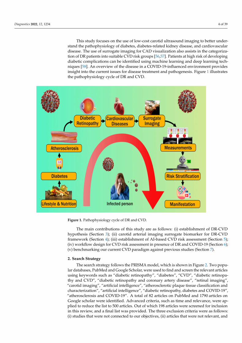
This study focuses on the use of low-cost carotid ultrasound imaging to better under-stand the pathophysiology of diabetes, diabetes-related kidney disease, and cardiovasculardisease. The use of surrogate imaging for CAD visualization also assists in the categoriza-tion of DR patients into suitable CVD risk groups [56,57]. Patients at high risk of developingdiabetic complications can be identified using machine learning and deep learning tech-niques [58]. An overview of the disease in a COVID-19-influenced environment providesinsight into the current issues for disease treatment and pathogenesis. Figure 1 illustratesthe pathophysiology cycle of DR and CVD.
Diagnostics 2022, 12, 1234 4 of 41
man intervention, AI-based solutions enable the examination of image-based retinal in-puts [53]. Several carotid ultrasonography applications utilizing AI-based algorithms have shown promise [54,55]. Thus, these AI-based algorithms may be used to concur-rently handle CVD and DR diseases in patient risk assessment.
This study focuses on the use of low-cost carotid ultrasound imaging to better un-derstand the pathophysiology of diabetes, diabetes-related kidney disease, and cardiovas-cular disease. The use of surrogate imaging for CAD visualization also assists in the cate-gorization of DR patients into suitable CVD risk groups [56,57]. Patients at high risk of developing diabetic complications can be identified using machine learning and deep learning techniques [58]. An overview of the disease in a COVID-19-influenced environ-ment provides insight into the current issues for disease treatment and pathogenesis. Fig-ure 1 illustrates the pathophysiology cycle of DR and CVD.
The main contributions of this study are as follows: (i) establishment of DR-CVD hy-pothesis (Section 3); (ii) carotid arterial imaging surrogate biomarker for DR-CVD frame-work (Section 4); (iii) establishment of AI-based CVD risk assessment (Section 5); (iv) workflow design for CVD risk assessment in presence of DR and COVID-19 (Section 6); (v) benchmarking our current CVD paradigm against previous studies (Section 7).
Figure 1. Pathophysiology cycle of DR and CVD.
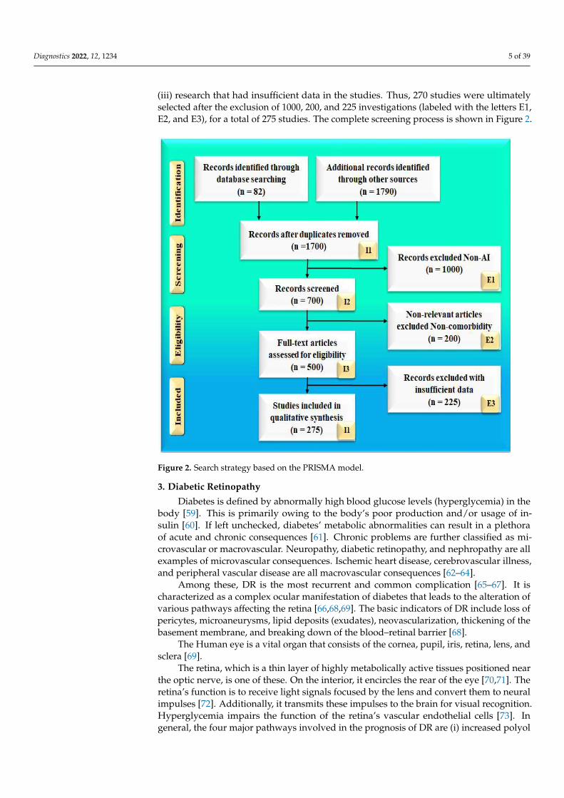
2. Search Strategy The search strategy follows the PRISMA model, which is shown in Figure 2. Two
popular databases, PubMed and Google Scholar, were used to find and screen the relevant articles using keywords such as “diabetic retinopathy”, “diabetes”, “CVD”, “diabetic ret-inopathy and CVD”, “diabetic retinopathy and coronary artery disease”, “retinal imag-ing”, “carotid imaging”, “artificial intelligence”, “atherosclerotic plaque tissue classifica-tion and characterization”, “artificial intelligence”, “diabetic retinopathy, diabetes and COVID-19”, “atherosclerosis and COVID-19”. A total of 82 articles on PubMed and 1790 articles on Google scholar were identified. Advanced criteria, such as time and relevance,
Figure 1. Pathophysiology cycle of DR and CVD.
The main contributions of this study are as follows: (i) establishment of DR-CVDhypothesis (Section 3); (ii) carotid arterial imaging surrogate biomarker for DR-CVDframework (Section 4); (iii) establishment of AI-based CVD risk assessment (Section 5);(iv) workflow design for CVD risk assessment in presence of DR and COVID-19 (Section 6);(v) benchmarking our current CVD paradigm against previous studies (Section 7).
2. Search Strategy
The search strategy follows the PRISMA model, which is shown in Figure 2. Two popu-lar databases, PubMed and Google Scholar, were used to find and screen the relevant articlesusing keywords such as “diabetic retinopathy”, “diabetes”, “CVD”, “diabetic retinopa-thy and CVD”, “diabetic retinopathy and coronary artery disease”, “retinal imaging”,“carotid imaging”, “artificial intelligence”, “atherosclerotic plaque tissue classification andcharacterization”, “artificial intelligence”, “diabetic retinopathy, diabetes and COVID-19”,“atherosclerosis and COVID-19”. A total of 82 articles on PubMed and 1790 articles onGoogle scholar were identified. Advanced criteria, such as time and relevance, were ap-plied to reduce the list to 500 articles. Out of which 198 articles were screened for inclusionin this review, and a final list was provided. The three exclusion criteria were as follows:(i) studies that were not connected to our objectives, (ii) articles that were not relevant, and
Diagnostics 2022, 12, 1234 5 of 39
(iii) research that had insufficient data in the studies. Thus, 270 studies were ultimatelyselected after the exclusion of 1000, 200, and 225 investigations (labeled with the letters E1,E2, and E3), for a total of 275 studies. The complete screening process is shown in Figure 2.
Diagnostics 2022, 12, 1234 5 of 41
were applied to reduce the list to 500 articles. Out of which 198 articles were screened for inclusion in this review, and a final list was provided. The three exclusion criteria were as follows: (i) studies that were not connected to our objectives, (ii) articles that were not relevant, and (iii) research that had insufficient data in the studies. Thus, 270 studies were ultimately selected after the exclusion of 1000, 200, and 225 investigations (labeled with the letters E1, E2, and E3), for a total of 275 studies. The complete screening process is shown in Figure 2.
Figure 2. Search strategy based on the PRISMA model.
3. Diabetic Retinopathy Diabetes is defined by abnormally high blood glucose levels (hyperglycemia) in the
body [59]. This is primarily owing to the body’s poor production and/or usage of insulin [60]. If left unchecked, diabetes’ metabolic abnormalities can result in a plethora of acute and chronic consequences [61]. Chronic problems are further classified as microvascular or macrovascular. Neuropathy, diabetic retinopathy, and nephropathy are all examples of microvascular consequences. Ischemic heart disease, cerebrovascular illness, and pe-ripheral vascular disease are all macrovascular consequences [62–64].
Among these, DR is the most recurrent and common complication [65–67]. It is char-acterized as a complex ocular manifestation of diabetes that leads to the alteration of var-ious pathways affecting the retina [66,68,69]. The basic indicators of DR include loss of pericytes, microaneurysms, lipid deposits (exudates), neovascularization, thickening of the basement membrane, and breaking down of the blood–retinal barrier [68].
The Human eye is a vital organ that consists of the cornea, pupil, iris, retina, lens, and sclera [69].
The retina, which is a thin layer of highly metabolically active tissues positioned near the optic nerve, is one of these. On the interior, it encircles the rear of the eye [70,71]. The retina’s function is to receive light signals focused by the lens and convert them to neural impulses [72]. Additionally, it transmits these impulses to the brain for visual recognition.
Figure 2. Search strategy based on the PRISMA model.
3. Diabetic Retinopathy
Diabetes is defined by abnormally high blood glucose levels (hyperglycemia) in thebody [59]. This is primarily owing to the body’s poor production and/or usage of in-sulin [60]. If left unchecked, diabetes’ metabolic abnormalities can result in a plethoraof acute and chronic consequences [61]. Chronic problems are further classified as mi-crovascular or macrovascular. Neuropathy, diabetic retinopathy, and nephropathy are allexamples of microvascular consequences. Ischemic heart disease, cerebrovascular illness,and peripheral vascular disease are all macrovascular consequences [62–64].
Among these, DR is the most recurrent and common complication [65–67]. It ischaracterized as a complex ocular manifestation of diabetes that leads to the alteration ofvarious pathways affecting the retina [66,68,69]. The basic indicators of DR include loss ofpericytes, microaneurysms, lipid deposits (exudates), neovascularization, thickening of thebasement membrane, and breaking down of the blood–retinal barrier [68].
The Human eye is a vital organ that consists of the cornea, pupil, iris, retina, lens, andsclera [69].
The retina, which is a thin layer of highly metabolically active tissues positioned nearthe optic nerve, is one of these. On the interior, it encircles the rear of the eye [70,71]. Theretina’s function is to receive light signals focused by the lens and convert them to neuralimpulses [72]. Additionally, it transmits these impulses to the brain for visual recognition.Hyperglycemia impairs the function of the retina’s vascular endothelial cells [73]. Ingeneral, the four major pathways involved in the prognosis of DR are (i) increased polyol
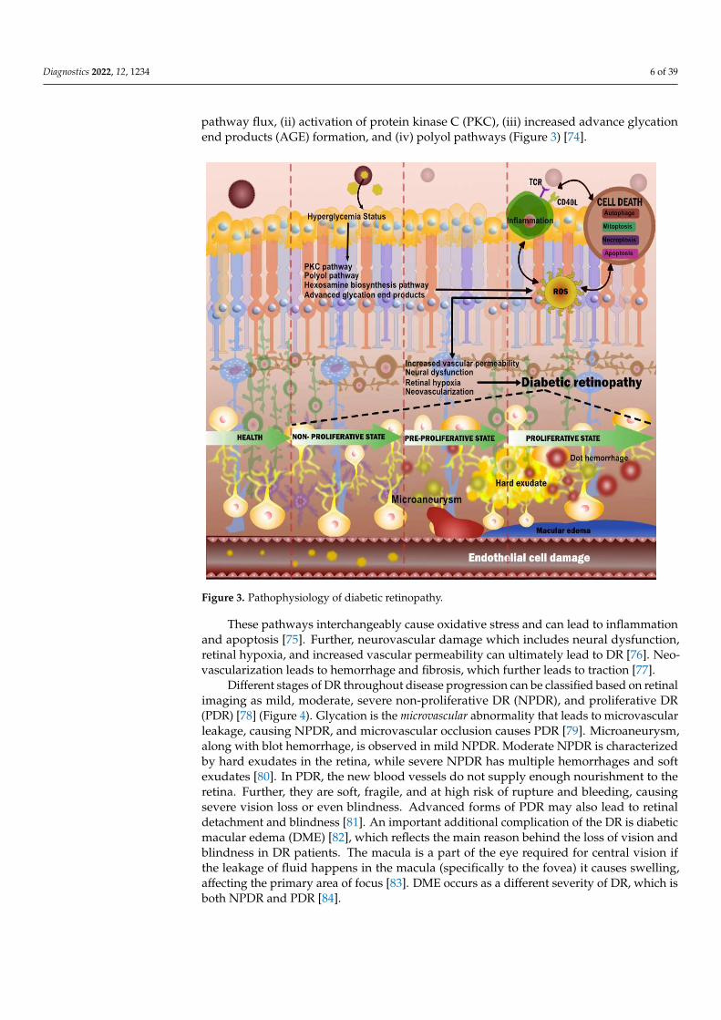
Diagnostics 2022, 12, 1234 6 of 39
pathway flux, (ii) activation of protein kinase C (PKC), (iii) increased advance glycationend products (AGE) formation, and (iv) polyol pathways (Figure 3) [74].
Diagnostics 2022, 12, 1234 6 of 41
Hyperglycemia impairs the function of the retina’s vascular endothelial cells [73]. In gen-eral, the four major pathways involved in the prognosis of DR are (i) increased polyol pathway flux, (ii) activation of protein kinase C (PKC), (iii) increased advance glycation end products (AGE) formation, and (iv) polyol pathways (Figure 3) [74].
Figure 3. Pathophysiology of diabetic retinopathy.
These pathways interchangeably cause oxidative stress and can lead to inflammation and apoptosis [75]. Further, neurovascular damage which includes neural dysfunction, retinal hypoxia, and increased vascular permeability can ultimately lead to DR [76]. Neo-vascularization leads to hemorrhage and fibrosis, which further leads to traction [77].
Different stages of DR throughout disease progression can be classified based on ret-inal imaging as mild, moderate, severe non-proliferative DR (NPDR), and proliferative DR (PDR) [78] (Figure 4). Glycation is the microvascular abnormality that leads to micro-vascular leakage, causing NPDR, and microvascular occlusion causes PDR [79]. Microan-eurysm, along with blot hemorrhage, is observed in mild NPDR. Moderate NPDR is char-acterized by hard exudates in the retina, while severe NPDR has multiple hemorrhages and soft exudates [80]. In PDR, the new blood vessels do not supply enough nourishment to the retina. Further, they are soft, fragile, and at high risk of rupture and bleeding, caus-ing severe vision loss or even blindness. Advanced forms of PDR may also lead to retinal detachment and blindness [81]. An important additional complication of the DR is dia-betic macular edema (DME) [82], which reflects the main reason behind the loss of vision and blindness in DR patients. The macula is a part of the eye required for central vision if the leakage of fluid happens in the macula (specifically to the fovea) it causes swelling, affecting the primary area of focus [83]. DME occurs as a different severity of DR, which is both NPDR and PDR [84].
Figure 3. Pathophysiology of diabetic retinopathy.
These pathways interchangeably cause oxidative stress and can lead to inflammationand apoptosis [75]. Further, neurovascular damage which includes neural dysfunction,retinal hypoxia, and increased vascular permeability can ultimately lead to DR [76]. Neo-vascularization leads to hemorrhage and fibrosis, which further leads to traction [77].
Different stages of DR throughout disease progression can be classified based on retinalimaging as mild, moderate, severe non-proliferative DR (NPDR), and proliferative DR(PDR) [78] (Figure 4). Glycation is the microvascular abnormality that leads to microvascularleakage, causing NPDR, and microvascular occlusion causes PDR [79]. Microaneurysm,along with blot hemorrhage, is observed in mild NPDR. Moderate NPDR is characterizedby hard exudates in the retina, while severe NPDR has multiple hemorrhages and softexudates [80]. In PDR, the new blood vessels do not supply enough nourishment to theretina. Further, they are soft, fragile, and at high risk of rupture and bleeding, causingsevere vision loss or even blindness. Advanced forms of PDR may also lead to retinaldetachment and blindness [81]. An important additional complication of the DR is diabeticmacular edema (DME) [82], which reflects the main reason behind the loss of vision andblindness in DR patients. The macula is a part of the eye required for central vision ifthe leakage of fluid happens in the macula (specifically to the fovea) it causes swelling,affecting the primary area of focus [83]. DME occurs as a different severity of DR, which isboth NPDR and PDR [84].

Diagnostics 2022, 12, 1234 7 of 39Diagnostics 2022, 12, 1234 7 of 41
Figure 4. Stages of diabetic retinopathy (courtesy of AtheroPoint, Roseville, CA, USA; permission granted).
A change in the structure and cellular content of the microvasculature such as ather-osclerosis is a sign of early DR [85,86]. Thus, the pathogenesis of DR has several contrib-uting factors that are driven by atherosclerosis. Several studies have shown that endothe-lial permeability, neo-angiogenesis, and plaque micro-vascularization are all influenced by a blood vessel’s vasa vasorum (a network of small blood vessels that supply the walls of larger blood vessels) [87]. Recent pieces of evidence suggest that in patients with dia-betes, vasa vasorum shows evolutionary changes [88]. It is the same as the beginning stage of the retina, in which endothelial dysfunction and loss of capillaries predominate [88]. This results in an unstable plaque and favors plaque rupture. As discussed earlier, hard exudates present in the moderate stages of NPDR also account for their association with CVD and plaque formation [84]. Hard exudates appear in the retina due to leakage of lipids and proteinaceous material through the endothelial barrier. It is also responsible for the appearance of plaque in large arteries [89]. Elevated blood pressure has also been as-sociated with DR and is a significant biomarker in atherosclerotic disease [90].
According to another study [88], there is a link between diabetes and CVD, high plasma LDL cholesterol, and proteinuria. The development and progression of retinopa-thy may be more severe in patients with diabetes and signs of atherosclerosis, necessitat-ing more frequent examinations and therapies in these patients. As a result, it is necessary to develop appropriate treatment choices.
3.1. The Biological Link between DR and CVD A vascular relationship cause exists between DR and CVD, it is observed that reti-
nopathy is a microvascular dysfunction caused due to endothelial dysfunction that results in arteriolar wall leakage [91]. These small arteriolar and capillary bed leakage causes ret-inopathy and nephropathy. However, large arterial wall leakage causes lipid accumula-tion, consequently leading to a pathogenic cascade of atherosclerosis [92]. Hyperglycemia causes inflammation by releasing reactive oxygen species, advanced glycation end prod-ucts, cytokines, and chemokines [93]. These collectively cause oxidative stress and endo-thelial dysfunction that facilitates the entry of monocytes and macrophages. Sequentially, endothelial dysfunction also helps low-density lipoprotein (LDL) particles penetrate the intimal wall of the vessel in a process called transcytosis [94]. Further, the LDL particles get oxidized and form OxLDL, due to the inflammatory markers process [95–97]. Addi-tionally, endothelial dysfunction activates the scavenger receptors (SRc) known as SR-AI/II, SR-BI, and the cluster of differentiation 36 (CD36). These results in intracellular up-take of oxLDL by macrophages in the arterial intima and help in the formation of foam cells [98,99] (see Figure 5). Over time, the foam cells die, contributing to the production of
Figure 4. Stages of diabetic retinopathy (courtesy of AtheroPoint, Roseville, CA, USA;permission granted).
A change in the structure and cellular content of the microvasculature such as atheroscle-rosis is a sign of early DR [85,86]. Thus, the pathogenesis of DR has several contributingfactors that are driven by atherosclerosis. Several studies have shown that endothelialpermeability, neo-angiogenesis, and plaque micro-vascularization are all influenced by ablood vessel’s vasa vasorum (a network of small blood vessels that supply the walls oflarger blood vessels) [87]. Recent pieces of evidence suggest that in patients with diabetes,vasa vasorum shows evolutionary changes [88]. It is the same as the beginning stageof the retina, in which endothelial dysfunction and loss of capillaries predominate [88].This results in an unstable plaque and favors plaque rupture. As discussed earlier, hardexudates present in the moderate stages of NPDR also account for their association withCVD and plaque formation [84]. Hard exudates appear in the retina due to leakage oflipids and proteinaceous material through the endothelial barrier. It is also responsiblefor the appearance of plaque in large arteries [89]. Elevated blood pressure has also beenassociated with DR and is a significant biomarker in atherosclerotic disease [90].
According to another study [88], there is a link between diabetes and CVD, highplasma LDL cholesterol, and proteinuria. The development and progression of retinopathymay be more severe in patients with diabetes and signs of atherosclerosis, necessitatingmore frequent examinations and therapies in these patients. As a result, it is necessary todevelop appropriate treatment choices.
3.1. The Biological Link between DR and CVD
A vascular relationship cause exists between DR and CVD, it is observed that retinopa-thy is a microvascular dysfunction caused due to endothelial dysfunction that resultsin arteriolar wall leakage [91]. These small arteriolar and capillary bed leakage causesretinopathy and nephropathy. However, large arterial wall leakage causes lipid accumula-tion, consequently leading to a pathogenic cascade of atherosclerosis [92]. Hyperglycemiacauses inflammation by releasing reactive oxygen species, advanced glycation end products,cytokines, and chemokines [93]. These collectively cause oxidative stress and endothe-lial dysfunction that facilitates the entry of monocytes and macrophages. Sequentially,endothelial dysfunction also helps low-density lipoprotein (LDL) particles penetrate theintimal wall of the vessel in a process called transcytosis [94]. Further, the LDL particles getoxidized and form OxLDL, due to the inflammatory markers process [95–97]. Addition-ally, endothelial dysfunction activates the scavenger receptors (SRc) known as SR-AI/II,SR-BI, and the cluster of differentiation 36 (CD36). These results in intracellular uptake ofoxLDL by macrophages in the arterial intima and help in the formation of foam cells [98,99](see Figure 5). Over time, the foam cells die, contributing to the production of intersti-tial collagen and elastin inside the foam cells resulting in the formation of the necrotic

Diagnostics 2022, 12, 1234 8 of 39
core [100]. Collectively, these overall sequential steps initiate the platelet aggregation andadhesion favors the atherosclerotic plaque formation causing micro and macrovascularcomplications [101].
Diagnostics 2022, 12, 1234 8 of 41
interstitial collagen and elastin inside the foam cells resulting in the formation of the ne-crotic core [100]. Collectively, these overall sequential steps initiate the platelet aggrega-tion and adhesion favors the atherosclerotic plaque formation causing micro and macro-vascular complications [101].
Figure 5. The biological link between DR and CVD (courtesy of AtheroPoint, Roseville, CA, USA; permission granted).
Coronary artery disease is the most common type of heart disease and is often known as cardiovascular disease as well as coronary heart disease. It has been shown that diabe-tes can cause blood vessels to thicken, which can progress to CHD [102]. Thus, a person with DR should be at an elevated risk of CHD/CAD. To study this, we went through re-cent literature and found several interesting attributes. In a study by Barlovic et al. [103], it was observed that 416 CVD events occurred during 12,872 person-years of follow-up. Severe diabetic retinopathy (SDR) was seen to increase CVD risk, particularly for periph-eral artery disease (PAD) in long-standing type 1 diabetes [104].
Hecke et al. [105] examined a cohort of 2237 type 1 diabetic patients in their study. After 7.9 years of follow-up, 64 people had died and 128 people had new CVD. People who had nonproliferative and proliferative retinopathy were more likely to die from any cause and have a higher risk of having a heart attack or suffering from stroke than people who did not have retinopathy [106]. They found that people with type 1 diabetes who have non-proliferative or proliferative retinopathy have an increased risk of all-cause death and new CVD. Another study by Pradeepa et al. [107] showed that in South Indian patients with type 2 diabetes, the prevalence of CAD was significantly higher in patients with DR compared to those without. In subjects with glycated hemoglobin (HbA1c) levels
Figure 5. The biological link between DR and CVD (courtesy of AtheroPoint, Roseville, CA, USA;permission granted).
Coronary artery disease is the most common type of heart disease and is often knownas cardiovascular disease as well as coronary heart disease. It has been shown that diabetescan cause blood vessels to thicken, which can progress to CHD [102]. Thus, a person withDR should be at an elevated risk of CHD/CAD. To study this, we went through recentliterature and found several interesting attributes. In a study by Barlovic et al. [103], it wasobserved that 416 CVD events occurred during 12,872 person-years of follow-up. Severediabetic retinopathy (SDR) was seen to increase CVD risk, particularly for peripheral arterydisease (PAD) in long-standing type 1 diabetes [104].
Hecke et al. [105] examined a cohort of 2237 type 1 diabetic patients in their study.After 7.9 years of follow-up, 64 people had died and 128 people had new CVD. People whohad nonproliferative and proliferative retinopathy were more likely to die from any causeand have a higher risk of having a heart attack or suffering from stroke than people whodid not have retinopathy [106]. They found that people with type 1 diabetes who havenon-proliferative or proliferative retinopathy have an increased risk of all-cause death andnew CVD. Another study by Pradeepa et al. [107] showed that in South Indian patientswith type 2 diabetes, the prevalence of CAD was significantly higher in patients with DRcompared to those without. In subjects with glycated hemoglobin (HbA1c) levels > 7%
Diagnostics 2022, 12, 1234 9 of 39
(p = 0.002), a significant association was observed between DR and CAD. Some people whohad eye-bleeding or microaneurysms had a higher risk of having a heart attack, and thosewho had cotton wool spots had an increased chance that they would have a heart attackor have another stroke. The same study by Kawasaki et al. [108] concluded that type 2diabetic patients with even a mild stage of DR, such as dot hemorrhages are already atrisk of CHD. Ellis et al. [109] in their study suggested that understanding the link betweenDR and CVD would lead to refined treatment strategies leading to personalized treatmentstrategies. In another study, by Cheung et al. [110], out of 214 participants that had DR,there were 209 CHD events. The presence of DR was linked to a two-fold increase in theincidence of CHD events and a three-fold increase in fatal CHD events. Table 1 presentsthe link between DR and CHD.
Table 1. The link between DR and CHD.
Citations Year PDR a CVD b RI c CHD d CI e AI f RS g DR-CVDLink
SOC h
Hecke et al. [105] 2005 X X 5 X 5 5 5 X XCheung et al. [110] 2007 5 X 5 X 5 5 5 X X
Kawasaki et al. [108] 2013 X X 5 X 5 5 X X XEllis et al. [109] 2013 X X 5 X 5 5 5 X X
Pradeepa et al. [107] 2015 5 X X X 5 5 5 X XUm et al. [111] 2015 5 X X X 5 5 5 X X
Barlovic et al. [103] 2018 5 X 5 X 5 5 5 X XXu et al. [112] 2020 5 X 5 X 5 5 5 X X
PDR a: Pathophysiology of Diabetic Retinopathy, CVD b: Cardiovascular Diseases, RI c: Retinal Imaging, CHD d:Coronary Heart Disease, CI e: Carotid Imaging, AI f: Artificial Intelligence, RS g: Risk Stratification, SOC h:Strength of Correlation.
Um et al. [111] explained that people with type 2 diabetes and PDR had a more severecoronary artery calcification and both were more likely to have CHD, compared to patientswithout DR. Thus, in asymptomatic patients with type 2 diabetes, PDR can be a predictor ofCHD. Xu et al. [79] concluded that DR was a risk marker for CVD. Their findings indicatedthat DR predicts a doubled mortality of CVD in diabetes. This clearly showed that DR wasstrongly related to CVD. All the above studies demonstrate our hypothesis holds that DRis responsible for the worsening of CVD.
Diabetic Retinopathy Imaging and Cardiovascular Disease: Establishing the Hypothesis
The human eye is a vital organ that helps in the direct and non-invasive visualiza-tion of DR changes. Direct visualization of neurovasculature of the eye can be done vianon-invasive imaging modalities [26]. Several studies indicate an increased risk of CVDassociated with DR patients. A study reported that retinopathy signs are associated withcoronary artery calcification and may be markers for atherosclerotic disease [113]. The reti-nal arteriolar narrowing was observed as a marker of coronary microvascular disease [35].
Alonso et al. [37] identified that Type 2 diabetic patients with DR had more atheroscle-rosis in their carotid arteries. Another study reported a significant association of increasedcarotid intima-media thickness (cIMT) with DR and peripheral vascular disease (PVD) [114].Concomitant diabetic cardiomyopathy was indicated in those suffering from advancedDR. These people usually have or will have chronic or recurrent heart failure [115]. Thus,we hypothesize that the condition of the heart could be altered during different stages ofDR. Furthermore, patients with macular edema (ME) and proliferative diabetic retinopathy(PDR) are at the highest risk of developing CVD [84]. To classify CVD risk for differentstages of DR, it is necessary to generate an output of these stages in the form of retinalscans. Therefore, retinal or ocular imaging modalities have an increasingly vital role in themanagement of diabetes, diabetic retinopathy, and the prognostication of associated events.
Retinal imaging is a diagnostic tool primarily used in the diagnosis of retinal diseasesas well as in monitoring retinal conditions with time. The three principal technologies used

Diagnostics 2022, 12, 1234 10 of 39
in retinal imaging are (i) fundus camera imaging, (ii) scanning laser ophthalmoscopy, and(iii) optical coherence tomography (OCT).
3.2. Fundus Camera Imaging
Thirty- to fifty-degree field-of-view images were provided by standard fundus pho-tography. This includes the macula and optic nerve [116]. 3D, semi-transparent, and retinaltissues projected onto the imaging plane are acted upon by reflected light to generate two-dimensional (2D) representations [117]. Fundus camera imaging has been used extensivelyin DR imaging [33,118]. It provides imaging of blood vessels, lesions, hemorrhages, andexudates that are pre-symptomatic stages of retinopathy. The standard ultra-wide field,fundus autofluorescence, and smartphone-based fundus photography are several typesof fundus imaging techniques [119] that are quick and simple. It covers a larger retinalfield and has high patient compliance (see Figure 6). However, 3D layer visualization is notpossible for some pathologies like macular edema and age-related macular degeneration,but using such 3D scans provides a quick diagnosis. Fundus imaging has advantageson the cost side, being usually a quarter of the price of an OCT scanner. Recently, cost-effective smartphone-based fundus imaging cameras have been developed that can providegood-quality retinal images.
Diagnostics 2022, 12, 1234 10 of 41
ent stages of DR. Furthermore, patients with macular edema (ME) and proliferative dia-betic retinopathy (PDR) are at the highest risk of developing CVD [84]. To classify CVD risk for different stages of DR, it is necessary to generate an output of these stages in the form of retinal scans. Therefore, retinal or ocular imaging modalities have an increasingly vital role in the management of diabetes, diabetic retinopathy, and the prognostication of associated events.
Retinal imaging is a diagnostic tool primarily used in the diagnosis of retinal diseases as well as in monitoring retinal conditions with time. The three principal technologies used in retinal imaging are (i) fundus camera imaging, (ii) scanning laser ophthalmos-copy, and (iii) optical coherence tomography (OCT).
3.2. Fundus Camera Imaging Thirty- to fifty-degree field-of-view images were provided by standard fundus pho-
tography. This includes the macula and optic nerve [116]. 3D, semi-transparent, and reti-nal tissues projected onto the imaging plane are acted upon by reflected light to generate two-dimensional (2D) representations [117]. Fundus camera imaging has been used ex-tensively in DR imaging [33,118]. It provides imaging of blood vessels, lesions, hemor-rhages, and exudates that are pre-symptomatic stages of retinopathy. The standard ultra-wide field, fundus autofluorescence, and smartphone-based fundus photography are sev-eral types of fundus imaging techniques [119] that are quick and simple. It covers a larger retinal field and has high patient compliance (see Figure 6). However, 3D layer visualiza-tion is not possible for some pathologies like macular edema and age-related macular de-generation, but using such 3D scans provides a quick diagnosis. Fundus imaging has ad-vantages on the cost side, being usually a quarter of the price of an OCT scanner. Recently, cost-effective smartphone-based fundus imaging cameras have been developed that can provide good-quality retinal images.
Figure 6. (A) Retinal images were taken using an IR camera [120]; (B) imaging using nun IR portable fundus camera [120] (Courtesy of oDocs Eye Care, Dunedin, New Zealand, reproduced with per-mission).
Figure 6. (A) Retinal images were taken using an IR camera [120]; (B) imaging using nun IRportable fundus camera [120] (Courtesy of oDocs Eye Care, Dunedin, New Zealand, reproducedwith permission).
3.3. Optical Coherence Tomography
OCT is a reproducible, non-invasive imaging modality that allows easy detection [121].It has provided new areas of understanding in ophthalmology. This optical scanningtechnique uses near-infrared light and can be thought of as “optical ultrasound” wheninterpreting these scans [35]. OCT images are high-resolution scans (1–15 mm) with a pene-tration depth of 2 to 3 mm in human tissue [36] (see Figure 7). It gives a cross-sectional viewof internal retinal structures and helps in detecting possible markers of neurodegeneration.

Diagnostics 2022, 12, 1234 11 of 39
Diagnostics 2022, 12, 1234 11 of 41
3.3. Optical Coherence Tomography OCT is a reproducible, non-invasive imaging modality that allows easy detection
[121]. It has provided new areas of understanding in ophthalmology. This optical scan-ning technique uses near-infrared light and can be thought of as “optical ultrasound” when interpreting these scans [35]. OCT images are high-resolution scans (1–15 mm) with a penetration depth of 2 to 3 mm in human tissue [36] (see Figure 7). It gives a cross-sectional view of internal retinal structures and helps in detecting possible markers of neurodegeneration.
Figure 7. (A) HRA + OCT imaging with a Spectralis HRA+ device Binarized optical coherence to-mography pictures with varying degrees of DR severity, as well as non-segmented angiograms, are shown in (B). (a) There is no DR. (b) Mild NPDR if any. (c) NPDR of a moderate level. (d) A very bad case of NPDR. (e) It is a PDR and it is important to note that the following CDI and FD values are the same: CDI is 0.358 and FD is 1.56, CDI is 0.351 and FD is 1.57, CDI is 0.342 and FD is 1.59, CDI is 0.340 and FD is 1.60 and CDI is 0.335 and FD is 1.61. Nonproliferative diabetes retinopathy is referred to as NPDR, while proliferative diabetic retinopathy is referred to as PDR.
Figure 7. (A) HRA + OCT imaging with a Spectralis HRA+ device Binarized optical coherencetomography pictures with varying degrees of DR severity, as well as non-segmented angiograms, areshown in (B). (a) There is no DR. (b) Mild NPDR if any. (c) NPDR of a moderate level. (d) A very badcase of NPDR. (e) It is a PDR and it is important to note that the following CDI and FD values are thesame: CDI is 0.358 and FD is 1.56, CDI is 0.351 and FD is 1.57, CDI is 0.342 and FD is 1.59, CDI is0.340 and FD is 1.60 and CDI is 0.335 and FD is 1.61. Nonproliferative diabetes retinopathy is referredto as NPDR, while proliferative diabetic retinopathy is referred to as PDR.
3.4. Optical Coherence Tomography and Angiography
OCT does not directly measure blood flow velocity, distinguish arteries and veins, ordetect vascular permeability changes. In addition, hence ICGA and FA remain commonmethods to visualize blood flow [116]. However, FA and ICGA are slow and cannot producetopographic 3D images. The advent of OCTA in 2012 revolutionized ophthalmology [117].It aids in the non-invasive examination of retinal and choroid anatomy and vasculature [26].It helps in the non-invasive evaluation of the structure and vasculature within the retinaand choroid [26]. Thus, with help of these imaging techniques, we can effectively accessDR and associated CVD risk. Table 2. Shows the difference between FI and OCT.

Diagnostics 2022, 12, 1234 12 of 39
Table 2. Difference between FI and OCT.
Modality Image Formation RF # Features of Interest Limitations
FIColour photograph
of the retinalsurface.
7–20 Blood vessels, lesions,exudates, hemorrhages.
Dilation ofpupils is often
needed.
OCT Near-infrared lightpenetrates the retina. 4
The internal retinalstructure is shown in
cross-section, includingchanges in the nerve
fiber layer.
Susceptible tomedia opacities,
does notvisualize blood.
#: Resolution factor; FI: fundus imaging; OCT: optical coherence tomography.
3.5. DR and CVD: Does Our Hypothesis Hold True?
The advances in retinal imaging technology and its power to diagnose DR give a verystrong edge for risk stratification in DR. The question, therefore, arises if patients sufferingfrom DR can be used for direct CVD evaluation and risk stratification, or do all the grades of DRincrease the risk of CVD?
A recent study, called Action for Health in Diabetes (AHEAD) between DR and CVD,was conducted on type 2 diabetes patients having a cohort size of 4098 participants [122].The authors showed that there was an increase in CVD composite that affected the mi-crovascular disease (MVD) having the following statistics [(HR 1.34, 95% CI 1.11–1.61),CAD (HR 1.24, 95% CI 1.01–1.52), stroke (HR 1.55, 95% CI 1.03–2.33), and cardiovascularmortality (HR 1.26, 95% CI 0.72–2.22)]. This was clear evidence of worsening CVD dueto DR progression. There was another recent study that showed a reduction in LV func-tion due to diabetes-related microvascular complications, which was evaluated using theglobal longitudinal strain (GLS) [29]. Asymptomatic patients with DM had reduced GLSand were independent of other cardiovascular risk factors. Microvascular problems weremore prevalent in patients with non-obstructive coronary artery disease. Furthermore, theburden of microvascular problems was linked to a higher load of coronary artery plaqueburden (CAPB).
In another study, conducted on a cohort from China having type 2 diabetes mellitus(T2DM), it was shown that ASCVD was strongly associated with DR [27]. The authorsshowed that the association of DR with ASCVD was significantly higher compared topatients with non-ASCVD (ChiSquare: χ2 = 5.805, p-value = 0.016). The authors furtherdemonstrated that DR was an independent statistical indicator in the presence of ASCVDhaving the odds ratio (OR) (95% CI): 2.321 (1.152–4.678), p-value = 0.018.
Further, only PDR was linked with ASCVD [OR (95% CI): 8.333 (1.813–38.304),p-value = 0.006]. The connection remained after adjusting for ASCVD risk variables[OR (95% CI): 7.466 (1.355–41.137), p = 0.021]. Future risk of CVD, MI, and CHF [30], wasassociated to DR severity in 2020. DR enhanced the incidence of CVD, MI, and CHF mor-tality. In their study, there were 77,376 patients, including 59.8% men, 31.28% non-HispanicWhites, and 41.48% Hispanics. Minimal NPDR increased the likelihood of CVA (1.31;95% CI, 1.18–1.46), MI (1.30; 95% CI, 1.15–1.46), and death (1.29; 95 percent CI, 1.19–1.40).HR 1.15; 95% CI 1.05–1.25. Patients with symptomatic NPDR and proliferative diabeticretinopathy showed increased mortality (HR, 1.55; 95% CI, 1.32–1.82); (HR, 1.92; 95 percentCI, 1.57–2.34); CHF: HR, 1.96; 95% CI, 1.47–2.59; and death: HR, 1.87; 95% CI, 1.36–2.56.
3.6. Descriptive Analysis Validating the DR-CVD Hypothesis
Mimoun et al. [123] showed the relationship between the retinal microvasculaturechallenges and (a) white matter lesions in the brain leading to stroke and (b) coronarycalcification leading to heart failure. These retinal microvasculature challenges consistedof reduced (i) arteriolar diameter, (ii) venular dilatation, and (iii) retinopathy lesions. Theauthors showed ventricular dilation was due to the presence of diabetes, obesity, andmetabolic disorders. The authors further presented that retinopathy is correlated with
Diagnostics 2022, 12, 1234 13 of 39
cerebral white matter lesions in the brain leading to stroke. This was evidenced by an MRIof the brain. On the heart side, the authors showed that these microvasculature challengeswere related to coronary calcification leading to heart failure. From this study, we concludethat there is a direct relationship between retinal damage due to diabetes and coronaryartery calcification. Thus, since carotid artery disease is a surrogate marker of coronaryartery disease, we can adopt carotid artery disease biomarkers for CVD risk assessment indiabetic retinopathy patients.
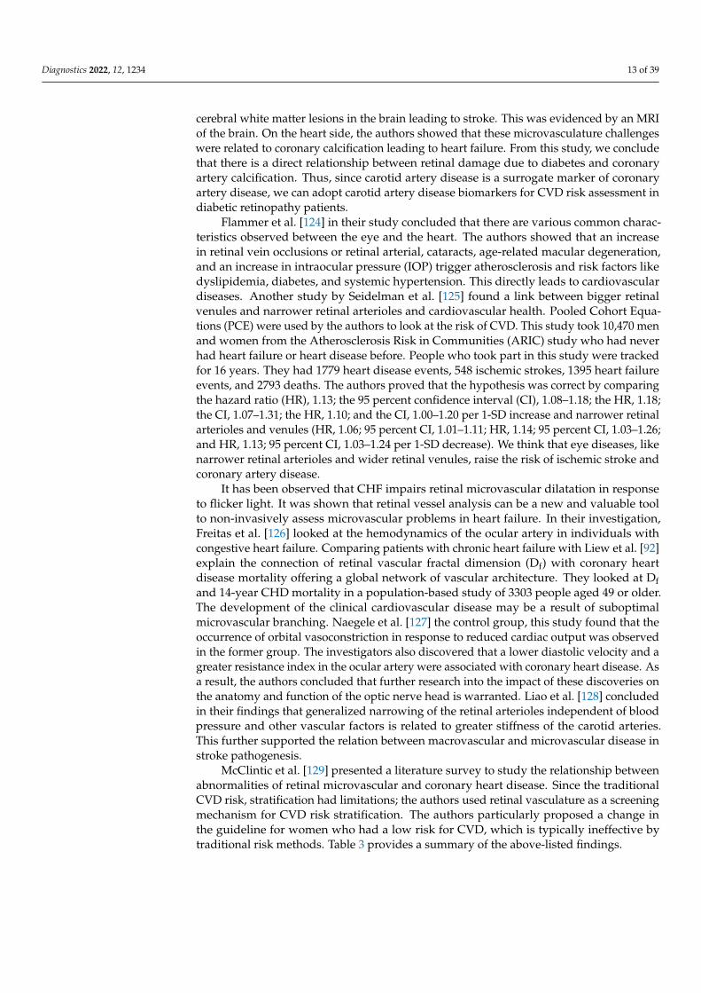
Flammer et al. [124] in their study concluded that there are various common charac-teristics observed between the eye and the heart. The authors showed that an increasein retinal vein occlusions or retinal arterial, cataracts, age-related macular degeneration,and an increase in intraocular pressure (IOP) trigger atherosclerosis and risk factors likedyslipidemia, diabetes, and systemic hypertension. This directly leads to cardiovasculardiseases. Another study by Seidelman et al. [125] found a link between bigger retinalvenules and narrower retinal arterioles and cardiovascular health. Pooled Cohort Equa-tions (PCE) were used by the authors to look at the risk of CVD. This study took 10,470 menand women from the Atherosclerosis Risk in Communities (ARIC) study who had neverhad heart failure or heart disease before. People who took part in this study were trackedfor 16 years. They had 1779 heart disease events, 548 ischemic strokes, 1395 heart failureevents, and 2793 deaths. The authors proved that the hypothesis was correct by comparingthe hazard ratio (HR), 1.13; the 95 percent confidence interval (CI), 1.08–1.18; the HR, 1.18;the CI, 1.07–1.31; the HR, 1.10; and the CI, 1.00–1.20 per 1-SD increase and narrower retinalarterioles and venules (HR, 1.06; 95 percent CI, 1.01–1.11; HR, 1.14; 95 percent CI, 1.03–1.26;and HR, 1.13; 95 percent CI, 1.03–1.24 per 1-SD decrease). We think that eye diseases, likenarrower retinal arterioles and wider retinal venules, raise the risk of ischemic stroke andcoronary artery disease.
It has been observed that CHF impairs retinal microvascular dilatation in responseto flicker light. It was shown that retinal vessel analysis can be a new and valuable toolto non-invasively assess microvascular problems in heart failure. In their investigation,Freitas et al. [126] looked at the hemodynamics of the ocular artery in individuals withcongestive heart failure. Comparing patients with chronic heart failure with Liew et al. [92]explain the connection of retinal vascular fractal dimension (Df) with coronary heartdisease mortality offering a global network of vascular architecture. They looked at Dfand 14-year CHD mortality in a population-based study of 3303 people aged 49 or older.The development of the clinical cardiovascular disease may be a result of suboptimalmicrovascular branching. Naegele et al. [127] the control group, this study found that theoccurrence of orbital vasoconstriction in response to reduced cardiac output was observedin the former group. The investigators also discovered that a lower diastolic velocity and agreater resistance index in the ocular artery were associated with coronary heart disease. Asa result, the authors concluded that further research into the impact of these discoveries onthe anatomy and function of the optic nerve head is warranted. Liao et al. [128] concludedin their findings that generalized narrowing of the retinal arterioles independent of bloodpressure and other vascular factors is related to greater stiffness of the carotid arteries.This further supported the relation between macrovascular and microvascular disease instroke pathogenesis.
McClintic et al. [129] presented a literature survey to study the relationship betweenabnormalities of retinal microvascular and coronary heart disease. Since the traditionalCVD risk, stratification had limitations; the authors used retinal vasculature as a screeningmechanism for CVD risk stratification. The authors particularly proposed a change inthe guideline for women who had a low risk for CVD, which is typically ineffective bytraditional risk methods. Table 3 provides a summary of the above-listed findings.

Diagnostics 2022, 12, 1234 14 of 39
Table 3. Studies showing evidence for the DR-CVD hypothesis.
SN Author Year ImagingDevice Comorbidity DR-CVD
Link Conclusion
1. Liao et al. [128] 2004 Retinal imaginghypertension,
dyslipidemia, anddiabetes mellitus
XMacro and microvascular
disease support strokeprognosis.
2. Minmoun et al. [123] 2009 Laser Dopplerflowmetry
Retinalmicrovascularabnormalities
X
retinopathy is correlatedwith white matter lesionsin the brain and coronary
calcification
3. McClintic et al. [129] 2010 Retinal imaging Type 2 diabetes X
Retinal vasculatureabnormalities were
related to coronary heartdisease
4. Liew et al. [130] 2010 Retinal imaging CHD XFractal analysis onmicrovasculature
predicted CHD mortality
5. Freitas et al. [126] 2011 Color Dopplerimaging CHF X
Abnormalities in theoptic nerve head in the
eyes were related to CHF
6. Flammer et al. [124] 2012 Color Dopplerimaging
dyslipidemia, DM,or systemic
hypertension X
CVD was found to beassociated with macular
degeneration andimpaired autoregulation
in the eyes.
7. Seidelmann et al. [125] 2016 Retinal vesselimaging
ASCVE or heartfailure (HF) X
Reduction in retinalarterioles and
enlargement of retinalvenules showed stroke
and CHD
8. Naegele et al. [127] 2017Dynamic
Retinal VesselAnalyzer
Smoking,hypertension,
dyslipidemia, anddiabetes mellitus
X
In patients with CHF, theresponsiveness of theretinal microvasculardilatation to flickering
light was reduced.
4. Carotid Imaging for CVD Risk Assessment in DR Patients
DR can act as an indicator not just for cardiovascular or coronary artery diseases, butalso for cerebrovascular diseases. Figure 8 shows how the link between carotid ultrasoundand coronary artery disease, both having the common thread of atherosclerosis. Figure 8ashows the visualization of the carotid and coronary scans using radiation-free ultrasoundscans. Figure 8b shows the typical low-cost and portable ultrasound measurement deviceused for screening the carotid arteries.
Thus, there are two reasons for studying the surrogate markers for CVD. First, it hasalready been proven that DR is associated with carotid artery disease. Second, DR is associ-ated with coronary artery disease which in turn is associated with a carotid atheroscleroticdisease that acts as a surrogate marker for coronary artery disease [131,132].
In support of the former case, several studies have been published that links DR tocarotid artery disease [27,30]. Similarly, several important studies have been publishedthat link carotid artery disease to coronary artery disease [131–133]. Thus, this section isfocused on (a) the relationship between DR and carotid artery disease and (b) carotid arteryas a surrogate marker for coronary artery disease.

Diagnostics 2022, 12, 1234 15 of 39
Diagnostics 2022, 12, 1234 15 of 41
of retinal ven-ules showed
stroke and CHD
8. Naegele et al.
[127] 2017
Dynamic Retinal Ves-sel Analyzer
Smoking, hyperten-sion, dyslipidemia, and diabetes melli-
tus
In patients with CHF, the re-
sponsiveness of the retinal mi-
crovascular dila-tation to flicker-ing light was re-
duced.
4. Carotid Imaging for CVD Risk Assessment in DR Patients DR can act as an indicator not just for cardiovascular or coronary artery diseases, but
also for cerebrovascular diseases. Figure 8 shows how the link between carotid ultrasound and coronary artery disease, both having the common thread of atherosclerosis. Figure 8a shows the visualization of the carotid and coronary scans using radiation-free ultrasound scans. Figure 8b shows the typical low-cost and portable ultrasound measurement device used for screening the carotid arteries.
Thus, there are two reasons for studying the surrogate markers for CVD. First, it has already been proven that DR is associated with carotid artery disease. Second, DR is asso-ciated with coronary artery disease which in turn is associated with a carotid atheroscle-rotic disease that acts as a surrogate marker for coronary artery disease [131,132].
In support of the former case, several studies have been published that links DR to carotid artery disease [27,30]. Similarly, several important studies have been published that link carotid artery disease to coronary artery disease [131–133]. Thus, this section is focused on (a) the relationship between DR and carotid artery disease and (b) carotid ar-tery as a surrogate marker for coronary artery disease.
Figure 8. (a) The carotid artery is employed as a proxy for coronary artery disease. (b) Imaging gadget with a linear ultrasound probe scanning the carotid artery. (Courtesy of AtheroPoint, Rose-ville, CA, USA; produced with permission).
Figure 8. (a) The carotid artery is employed as a proxy for coronary artery disease. (b) Imaging gadgetwith a linear ultrasound probe scanning the carotid artery. (Courtesy of AtheroPoint, Roseville, CA,USA; produced with permission).
4.1. DR and Cerebrovascular/Carotid Artery Disease
A recent study investigated whether minute retinal microvascular changes indicateDR, and if this could cause arterial stiffening [28]. The authors hypothesized that retinalmicrovascular dysfunction may be seen in patients with carotid stiffness. Their findingsindicated that stiffness was associated with a decreased ability of the retina to dilate inresponse to flickering light. Additionally, this link was shown to be greater in personshaving type 2 diabetes. A further study by Lee et al. [134] explained the disruption ofthe common carotid artery (CCA) could lead to retinal ischemia. This is because of theophthalmic artery (OpA), which is a retinal artery supplying blood from the internal carotidartery (ICA). The retinal blood supplying vessel is the OpA.
Several studies by Drinkwater et al. showed that carotid artery disease is indepen-dently associated with the retinal microvascular disease as assessed by OCTA in type 2diabetes [135–137]. In another study, by Lu et al. [138], a link between time in range (TIR)and macrovascular disease was suggested after seeing an increase in cIMT associated withTIR. Meanwhile, DR was seen to be an independent predictor of subclinical cardiovasculardisease [38]. With the risk of carotid artery disease, it is also vital to look at coronary arterydisease. Since both these diseases hold a link, this will cause the condition of DR to worsen.
4.2. Carotid Artery Disease—A Surrogate of Coronary Artery Disease or Cardiovascular Disease
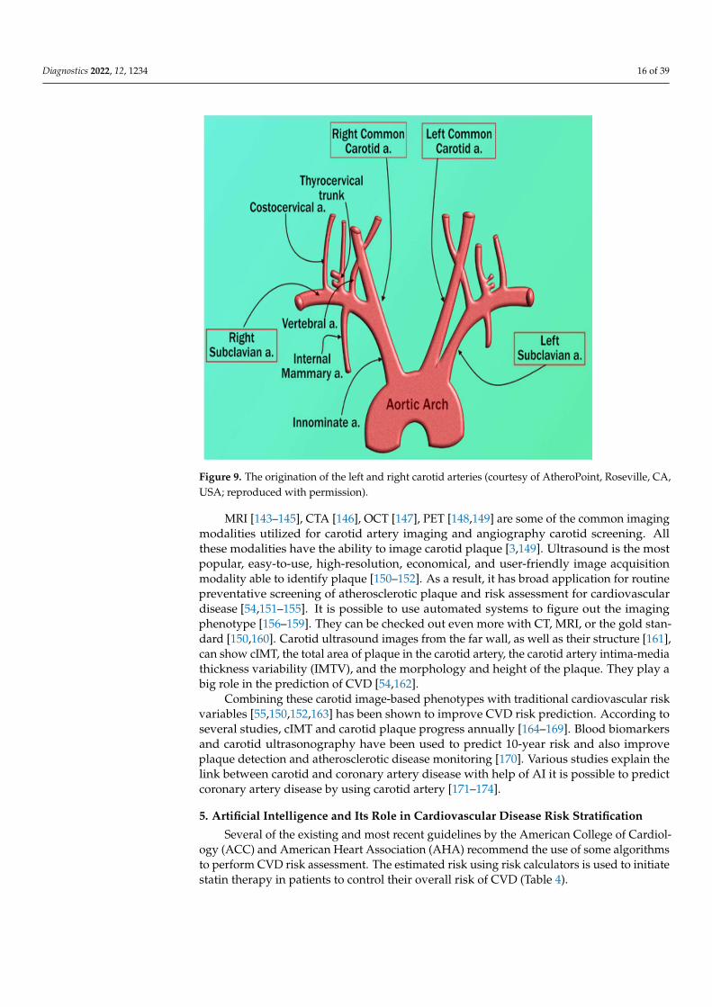
There is a risk to the heart and brain due to vascular diseases [139,140]. Both carotidand coronary arteries have a common genetic makeup. This link is well established dueto the similarity of the structure between the aortic arch, coronary artery, and carotidartery. Even though these arteries stem from a different major artery, they follow symmet-rical courses (see Figure 9). Thus, carotid artery disease could be considered a surrogatebiomarker for CAD [141,142].
Diagnostics 2022, 12, 1234 16 of 39
Diagnostics 2022, 12, 1234 16 of 41
4.1. DR and Cerebrovascular/Carotid Artery Disease A recent study investigated whether minute retinal microvascular changes indicate
DR, and if this could cause arterial stiffening [28]. The authors hypothesized that retinal microvascular dysfunction may be seen in patients with carotid stiffness. Their findings indicated that stiffness was associated with a decreased ability of the retina to dilate in response to flickering light. Additionally, this link was shown to be greater in persons having type 2 diabetes. A further study by Lee et al. [134] explained the disruption of the common carotid artery (CCA) could lead to retinal ischemia. This is because of the oph-thalmic artery (OpA), which is a retinal artery supplying blood from the internal carotid artery (ICA). The retinal blood supplying vessel is the OpA.
Several studies by Drinkwater et al. showed that carotid artery disease is inde-pendently associated with the retinal microvascular disease as assessed by OCTA in type 2 diabetes [135–137]. In another study, by Lu et al. [138], a link between time in range (TIR) and macrovascular disease was suggested after seeing an increase in cIMT associated with TIR. Meanwhile, DR was seen to be an independent predictor of subclinical cardiovascu-lar disease [38]. With the risk of carotid artery disease, it is also vital to look at coronary artery disease. Since both these diseases hold a link, this will cause the condition of DR to worsen.
4.2. Carotid Artery Disease—A Surrogate of Coronary Artery Disease or Cardiovascular Disease There is a risk to the heart and brain due to vascular diseases [139,140]. Both carotid
and coronary arteries have a common genetic makeup. This link is well established due to the similarity of the structure between the aortic arch, coronary artery, and carotid ar-tery. Even though these arteries stem from a different major artery, they follow symmet-rical courses (see Figure 9). Thus, carotid artery disease could be considered a surrogate biomarker for CAD [141,142].
Figure 9. The origination of the left and right carotid arteries (courtesy of AtheroPoint, Roseville, CA, USA; reproduced with permission).
Figure 9. The origination of the left and right carotid arteries (courtesy of AtheroPoint, Roseville, CA,USA; reproduced with permission).
MRI [143–145], CTA [146], OCT [147], PET [148,149] are some of the common imagingmodalities utilized for carotid artery imaging and angiography carotid screening. Allthese modalities have the ability to image carotid plaque [3,149]. Ultrasound is the mostpopular, easy-to-use, high-resolution, economical, and user-friendly image acquisitionmodality able to identify plaque [150–152]. As a result, it has broad application for routinepreventative screening of atherosclerotic plaque and risk assessment for cardiovasculardisease [54,151–155]. It is possible to use automated systems to figure out the imagingphenotype [156–159]. They can be checked out even more with CT, MRI, or the gold stan-dard [150,160]. Carotid ultrasound images from the far wall, as well as their structure [161],can show cIMT, the total area of plaque in the carotid artery, the carotid artery intima-mediathickness variability (IMTV), and the morphology and height of the plaque. They play abig role in the prediction of CVD [54,162].
Combining these carotid image-based phenotypes with traditional cardiovascular riskvariables [55,150,152,163] has been shown to improve CVD risk prediction. According toseveral studies, cIMT and carotid plaque progress annually [164–169]. Blood biomarkersand carotid ultrasonography have been used to predict 10-year risk and also improveplaque detection and atherosclerotic disease monitoring [170]. Various studies explain thelink between carotid and coronary artery disease with help of AI it is possible to predictcoronary artery disease by using carotid artery [171–174].
5. Artificial Intelligence and Its Role in Cardiovascular Disease Risk Stratification
Several of the existing and most recent guidelines by the American College of Cardiol-ogy (ACC) and American Heart Association (AHA) recommend the use of some algorithmsto perform CVD risk assessment. The estimated risk using risk calculators is used to initiatestatin therapy in patients to control their overall risk of CVD (Table 4).

Diagnostics 2022, 12, 1234 17 of 39
Table 4. CVD risk stratification thresholds for statin initiation.
Guidelines Risk Score Cut-Off with Statin Initiation
ACC/AHA 2013 [175] Risk Score for PooledCohorts
7.5% cutoff for starting amoderate to high-intensity statin
NICE 2014 [176–178] QRISK2 risk engine Offers atorvastatin 20mg dailywho have a score ≥10%
Canadian 2012 [179] FRS cardiovascular diseaserisk score
Offers atorvastatin 20mg daily ascore of 10%
U.S. Preventive ServicesTask Force [180]
Risk Score for PooledCohorts
Low-to-Moderate Statin Dose inRisk > 10%
Several CVD risk calculators have been developed based on this concept, all within astatistical framework.
The Framingham risk score [181], the systematic coronary risk evaluation score(SCORE) [182], QRISK3 [183], and the pooled cohort risk equation created by the AmericanCollege of Cardiology and the American Heart Association [175] are the most frequentlyused CVD risk calculators.
Because they were developed based on specific ethnic cohorts, when applied to diverseethnic populations, they can either underestimate or inflate the risk of cardiovasculardisease. These calculators were built using regression-based techniques that can handle alimited set of risk predictors.
• Because these CVD risk calculators were developed through the use of regression-based approaches, they assume that there is a linear relationship between the riskpredictors and the endpoints. Because of this constraint, a complicated non-linear asso-ciation between the risk predictors and the endpoints is not taken into consideration.
• The final and most significant difficulty is that such conventional risk factors areexclusively reliant on traditional risk variables, which do not provide any informationon atherosclerotic plaque burden in the first place. It is possible to overcome thisdifficulty by utilizing low-cost imaging methods.
Due to these issues, a more reliable and precise CVD risk prediction model is required.Traditional risk calculators can be improved by including image-based phenotypes inCVD risk prediction models. Suri et al. [153,184–187] have attempted this by combiningautomated carotid ultrasonography image-based phenotypes with traditional risk variablesto produce integrated CVD risk. Ten-year CVD risk was calculated utilizing carotid ultra-sound image-based phenotypes and traditional risk variables (Figure 10). The pie figureshows the independent contribution of conventional risk variables and carotid imagingphenotypes to 10-year CVD risk. To improve overall accuracy and address other issues,improved risk prediction algorithms are required.
AI-based algorithms have proved themselves to be superior to the existing CVD riskcalculators [188–190]. This is the reason for the growing interest of clinicians in exploringthe potential of AI in dealing with several healthcare problems, including the CVD riskassessment. AI is primarily categorized into two types of algorithms: machine learningand deep learning algorithms. Both of these algorithms require large datasets under a bigdata framework to build their internal models and provide accurate risk assessment [191].Machine learning algorithms require a series of pre-processing steps that involve datacleaning, noise reduction [192,193], feature extraction, and feature selection. Several goodexamples can be seen for different disease characterization such as diabetes [194], lung [195],thyroid [196,197], liver [198,199], breast [200], and coronary [195]. Figure 11 shows thegeneral framework of any ML-based algorithm.
The generalized architecture is commonly divided into two parts that are comprisedof an offline model and an online model. The offline model deals with the training ofan ML-based algorithm using the risk factors and endpoints and generates the offlinecoefficients. This will then be used under the online model to transform the unseen risk

Diagnostics 2022, 12, 1234 18 of 39
predictors into final CVD risk labels. Both of these offline and online models requirehandcrafted features for the training and prediction of labels. In CVD risk assessment,such features can be derived from the patients’ demographic and clinical parameters,including laboratory-based blood tests, electronic health records, and imaging modalities.Compared to the existing CVD risk assessment, calculators such as Framingham risk score(FRS), Pooled cohort risk equation (PCRE), and QRISK3 calculators that can handle onlya limited set of risk factors, ML-based algorithms can deal with a much larger numberof risk predictors at the same time. Figure 11 shows the generalized architecture of theML-based system.
Diagnostics 2022, 12, 1234 19 of 41
Figure 10. Risk predictors make up a big part of a person’s 10-year CVD risk profile when they’re looked at for the left common carotid artery (a,b), right common carotid artery (c,d), and the average of left and right common carotid artery (AtheroEdge 2.0) (e). This figure was made with permission [201] by AtheroPoint, USA. (Courtesy of AtheroPoint, Roseville, CA, USA; reproduced with per-mission).
Figure 10. Risk predictors make up a big part of a person’s 10-year CVD risk profile when they’relooked at for the left common carotid artery (a,b), right common carotid artery (c,d), and the average ofleft and right common carotid artery (AtheroEdge 2.0) (e). This figure was made with permission [201]by AtheroPoint, USA. (Courtesy of AtheroPoint, Roseville, CA, USA; reproduced with permission).

Diagnostics 2022, 12, 1234 19 of 39Diagnostics 2022, 12, 1234 20 of 41
Figure 11. The generalized architecture of the ML-based system.
ML-based algorithms make the final prediction based on several linear and non-lin-ear patterns available within the input risk predictors. This is a key specialty of AI-driven algorithms, which makes them distinct from several conventional CVD risk calculators. Commonly used and popular ML-based algorithms are the support vector machine, ran-dom forest, decision tree, and extreme gradient boosting [202].
Nearly all ML-based algorithms can efficiently distinguish between the low-risk CVD patients and the high-CVD-risk patients [203]. In terms of multiclass endpoints, the ML-based algorithms have provided better risk stratification compared to the conven-tional CVD risk calculators [185]. Besides this, the ML-based algorithms also efficiently differentiate symptomatic and asymptomatic carotid atherosclerotic plaques [204,205]. Recently attempts were made to combine the traditional CVD risk calculators with the carotid atherosclerotic plaque-based phenotypes [206]. This combination is referred to as integrated risk predictors. Such integrated risk predictors have shown high CVD risk pre-diction ability under the ML framework compared to using the traditional risk factors alone [154]. When compared against the 13 existing CVD risk calculators, such integrated feature-based ML systems reported superior performance [207]. This can be seen in Figure 12, which shows the comparison between the ML algorithms and statistical calculators.
It was found that AI-based algorithms had a better overall risk-strategy accuracy of 92.52 percent than the 13 types of CCVRC. This was more than any of the 13 types. Other people have shown that machine learning can be used to make better risk predictions. They used carotid ultrasound plaque attributes to enhance the risk prediction precision [202,208]. One more ML-based study, by Kakadiaris et al. [190] and Weng et al. [188], also found that ML-based algorithms were better than conventional CVD risk calculators based on statistics.
Figure 11. The generalized architecture of the ML-based system.
ML-based algorithms make the final prediction based on several linear and non-linearpatterns available within the input risk predictors. This is a key specialty of AI-drivenalgorithms, which makes them distinct from several conventional CVD risk calculators.Commonly used and popular ML-based algorithms are the support vector machine, ran-dom forest, decision tree, and extreme gradient boosting [202].
Nearly all ML-based algorithms can efficiently distinguish between the low-riskCVD patients and the high-CVD-risk patients [203]. In terms of multiclass endpoints,the ML-based algorithms have provided better risk stratification compared to the conven-tional CVD risk calculators [185]. Besides this, the ML-based algorithms also efficientlydifferentiate symptomatic and asymptomatic carotid atherosclerotic plaques [204,205]. Re-cently attempts were made to combine the traditional CVD risk calculators with the carotidatherosclerotic plaque-based phenotypes [206]. This combination is referred to as integratedrisk predictors. Such integrated risk predictors have shown high CVD risk prediction abilityunder the ML framework compared to using the traditional risk factors alone [154]. Whencompared against the 13 existing CVD risk calculators, such integrated feature-based MLsystems reported superior performance [207]. This can be seen in Figure 12, which showsthe comparison between the ML algorithms and statistical calculators.
Diagnostics 2022, 12, 1234 21 of 41
Figure 12. Comparing the ML-based CVD risk assessment using AtheroEdge™ 3.0ML with (A) 13 types of CCVRC and (B) the standard-of-care ASCVD calculator (produced with permission [207]).
Machine learning has proved to be a boon not only in CVD risk stratification, but also in several other areas, including benign and malignant tumor identification [209], charac-terization of intra-nodular vascularization of thyroid lesions [210], psoriasis identification [211], and so on.
In addition to ML, DL-based algorithms are also powerful in making an accurate and reliable diagnosis. DL techniques are the extension of a classical artificial neural network and can efficiently be used for medical image analysis, including feature extraction and classification [212]. Unlike ML-based algorithms, DL algorithms extract features by them-selves and perform classification or prediction tasks [213]. In medical imaging, a popular algorithm called the convolutional neural network (CNN) has been getting a lot of atten-tion. This algorithm is based on Deep Learning. CNN can find more high-level features than artisanal ones, and it can use them to make medical diagnoses [45,214]. In Figure 13, the input image is convolved using a set of kernels (also called filters) that extract multiple high-level patterns from the image.
A pooling operation selects the meaningful and dominant features. During CNN training, the backpropagation algorithm learns the overall coefficients of all kernels. Leka-dir et al. [215] recently employed CNN to classify carotid ultrasound pictures into lipid, fibrous, and calcified plaque. CNNs have also been used to assess carotid phenotypes such as intima-media thickness and lumen diameter [216–218].
Recently, Rim et al. [47] used the DL-based algorithm to predict the CAC score from retinal images. The authors proved that the CAC score measured using retinal images was comparable to the CT-derived CAC scores. Cheung et al. [48] used the DL-based algo-rithm to assess the CVD risk via the measurement of retinal vessel caliber. Besides this, DL-based algorithms have been widely adopted for the screening of DR patients [51,52,219]. Thus, it is very evident that both AI-based algorithms could be used for accu-rate CVD risk assessment as well as for automatic DR detection. Furthermore, along with traditional risk factors, the integration of carotid ultrasound plaque phenotypes could be used for preventive screening of patients in atherosclerosis and risk estimation.
Figure 12. Comparing the ML-based CVD risk assessment using AtheroEdge™ 3.0ML with (A) 13 typesof CCVRC and (B) the standard-of-care ASCVD calculator (produced with permission [207]).

Diagnostics 2022, 12, 1234 20 of 39
It was found that AI-based algorithms had a better overall risk-strategy accuracy of92.52 percent than the 13 types of CCVRC. This was more than any of the 13 types. Otherpeople have shown that machine learning can be used to make better risk predictions. Theyused carotid ultrasound plaque attributes to enhance the risk prediction precision [202,208].One more ML-based study, by Kakadiaris et al. [190] and Weng et al. [188], also foundthat ML-based algorithms were better than conventional CVD risk calculators basedon statistics.
Machine learning has proved to be a boon not only in CVD risk stratification, but alsoin several other areas, including benign and malignant tumor identification [209], characteri-zation of intra-nodular vascularization of thyroid lesions [210], psoriasis identification [211],and so on.
In addition to ML, DL-based algorithms are also powerful in making an accurateand reliable diagnosis. DL techniques are the extension of a classical artificial neuralnetwork and can efficiently be used for medical image analysis, including feature extractionand classification [212]. Unlike ML-based algorithms, DL algorithms extract features bythemselves and perform classification or prediction tasks [213]. In medical imaging, apopular algorithm called the convolutional neural network (CNN) has been getting a lotof attention. This algorithm is based on Deep Learning. CNN can find more high-levelfeatures than artisanal ones, and it can use them to make medical diagnoses [45,214]. InFigure 13, the input image is convolved using a set of kernels (also called filters) that extractmultiple high-level patterns from the image.
A pooling operation selects the meaningful and dominant features. During CNNtraining, the backpropagation algorithm learns the overall coefficients of all kernels. Lekadiret al. [215] recently employed CNN to classify carotid ultrasound pictures into lipid, fibrous,and calcified plaque. CNNs have also been used to assess carotid phenotypes such asintima-media thickness and lumen diameter [216–218].
Recently, Rim et al. [47] used the DL-based algorithm to predict the CAC score fromretinal images. The authors proved that the CAC score measured using retinal imageswas comparable to the CT-derived CAC scores. Cheung et al. [48] used the DL-basedalgorithm to assess the CVD risk via the measurement of retinal vessel caliber. Besides this,DL-based algorithms have been widely adopted for the screening of DR patients [51,52,219].Thus, it is very evident that both AI-based algorithms could be used for accurate CVD riskassessment as well as for automatic DR detection. Furthermore, along with traditionalrisk factors, the integration of carotid ultrasound plaque phenotypes could be used forpreventive screening of patients in atherosclerosis and risk estimation.
Diagnostics 2022, 12, 1234 22 of 41
Figure 13. A general architecture of CNN used in medical image analysis application (courtesy of AtheroPoint, Roseville, CA, USA).
6. DR/CVD in the COVID-19 Framework COVID-19 has caused massive global death [219]. It has caused over 5.7 million fa-
talities globally [220]. The coronavirus disease that originated in 2019 is named COVID-19. It is triggered by SARS-CoV-2 [221] and damages numerous routes [222]. Diabetes type 1 and 2 patients had an increased incidence of COVID-19 [223,224]. Individuals with ex-isting CVD are also at risk of COVID-19-related problems [225–229]. Several investiga-tions have linked (SARS-CoV-2) and ocular symptoms [227,230,231]. COVID-19 symp-toms range from mild to severe; one of these symptoms includes anosmia (loss of smell). Various studies suggest that anosmia is due to the effect of the virus on the olfactory bulbs [230]. It has been very well established that olfactory impairment is associated with dia-betes and related microvascular complications [231]. In COVID-19, patients with DR have an elevated risk of unfavorable conditions [232]. Costa et al. [233] recently examined indi-viduals who had recovered from the COVID-19 acute phase, 15.6% of whom had DR.
With the ongoing pandemic, it is critical to evaluate these patients’ situations. Several studies have reported the difficulties that several countries face in doing DR screening amid the pandemic [234,235]. Telemedicine was reported to be a useful solution for DR [236]. Telemedicine platforms specific to ophthalmology practice have also been devel-oped [158,237,238]. It is, therefore, vital to opt for a low-cost, preventive screening tool to provide effective treatment strategies and risk stratification for these patients as soon as possible. These patients are risking life and limb in the ongoing pandemic [201,224].
6.1. Adverse Effects of COVID-19 on DR Patients This section shows how DR was affected during the COVID-19 period. The im-
portance of DR during pandemic times has risen several folds. DR management has been suspended because of SARS-CoV-2, which has hampered access to diabetes-related med-ical consultations and retinal tests [237]. Many DR screening services and referral initia-tives have been negatively impacted by the COVID-19 pandemic [238].
In the COVID-19 period, diabetes was related to a greater probability of unfavorable DR outcomes due to the worsening of preexisting pulmonary microcirculatory abnormal-ities [239].
Ahmed et al. [240] noted that the COVID-19 pandemic has altered real-world practice patterns in DR management. Due to a change in these patterns, intravitreal injections for
Figure 13. A general architecture of CNN used in medical image analysis application (courtesy ofAtheroPoint, Roseville, CA, USA).

Diagnostics 2022, 12, 1234 21 of 39
6. DR/CVD in the COVID-19 Framework
COVID-19 has caused massive global death [219]. It has caused over 5.7 million fatali-ties globally [220]. The coronavirus disease that originated in 2019 is named COVID-19. It istriggered by SARS-CoV-2 [221] and damages numerous routes [222]. Diabetes type 1 and 2patients had an increased incidence of COVID-19 [223,224]. Individuals with existing CVDare also at risk of COVID-19-related problems [225–229]. Several investigations have linked(SARS-CoV-2) and ocular symptoms [227,230,231]. COVID-19 symptoms range from mildto severe; one of these symptoms includes anosmia (loss of smell). Various studies suggestthat anosmia is due to the effect of the virus on the olfactory bulbs [230]. It has beenvery well established that olfactory impairment is associated with diabetes and relatedmicrovascular complications [231]. In COVID-19, patients with DR have an elevated riskof unfavorable conditions [232]. Costa et al. [233] recently examined individuals who hadrecovered from the COVID-19 acute phase, 15.6% of whom had DR.
With the ongoing pandemic, it is critical to evaluate these patients’ situations. Sev-eral studies have reported the difficulties that several countries face in doing DR screen-ing amid the pandemic [234,235]. Telemedicine was reported to be a useful solutionfor DR [236]. Telemedicine platforms specific to ophthalmology practice have also beendeveloped [158,237,238]. It is, therefore, vital to opt for a low-cost, preventive screeningtool to provide effective treatment strategies and risk stratification for these patients as soonas possible. These patients are risking life and limb in the ongoing pandemic [201,224].
6.1. Adverse Effects of COVID-19 on DR Patients
This section shows how DR was affected during the COVID-19 period. The impor-tance of DR during pandemic times has risen several folds. DR management has beensuspended because of SARS-CoV-2, which has hampered access to diabetes-related medicalconsultations and retinal tests [237]. Many DR screening services and referral initiativeshave been negatively impacted by the COVID-19 pandemic [238].
In the COVID-19 period, diabetes was related to a greater probability of unfavorableDR outcomes due to the worsening of preexisting pulmonary microcirculatoryabnormalities [239].
Ahmed et al. [240] noted that the COVID-19 pandemic has altered real-world practicepatterns in DR management. Due to a change in these patterns, intravitreal injections for thetreatment and management of DR were reduced significantly. Similar findings were seen ina study conducted by Chatziralli et al. [241], which concluded that there was a significantpostponement in patient care that further led to worsening of visual acuity outcomes inpatients with DR. Thus, these studies clearly show the health hazard for DR patients duringCOVID-19 period. Das et al. [242] found a rise in patients with proliferative DR (56%)and sight-threatening DR (79%) as well as vitreoretinal operations (31%), and intravitrealinjections (19%). During the lockdown, the number of blind patients grew. This highlightsthe need for proper DR treatment during COVID-19. Dwairi et al. [243] found that delayingor interrupting a key operation for DR patients could harm their visual prognosis. Theauthors also advised residential monitoring, “treat and extend” strategy, portable OCTtesting, and more long-acting anti-VEGF (vascular endothelial growth factor) medicines.
COVID-19 individuals had an intraocular infection, according to Nayak et al. [244].This infection was mostly fungal; the authors concluded that routine eye exams wererequired during COVID-19. Due to the relevance of DR during pandemics, mobile appli-cations were developed. Khurana et al. [245] compared the use of a mobile applicationcalled Checkup Vision Assessment System to routine VA reference tests in the clinic. Theauthors demonstrated the need for such a smartphone device, especially during the globalpandemic. Saxena et al. [246] highlighted the presence of vitamin D supplementation byevaluating serum vitamin D levels during the COVID-19 period. When Vitamin D levelsare below 10 ng/mL, the study found a relationship between diabetes and COVID-19.Several research has come out with new treatments to overcome resource and geographicconstraints. Walsh et al. [247] stated that investment in national strategic alliances and

Diagnostics 2022, 12, 1234 22 of 39
technology can assist in promoting health and ophthalmic care. Teleophthalmology willthus be vital in the development of eye care. SFI is a technique developed by Kumariet al. [248] that allows patients to take photos of their retinas, addressing the barriers toavailability and affordability in an era of the pandemic.
Thus, we conclude that DR and COVID-19 run side by side and cannot be ignored.Therefore, we need the CVD estimation of patients who are DR affected during COVID-19.Further, it should be noted that efforts toward treatment and manifestations on DR patientswere very active during the COVID-19 period. This demonstrates the importance of DRduring the COVID-19 pandemic, which is still active. Our group has been very closelymonitoring the comorbidities during the long COVID-19 period [249]. This further assertsour assumption that DR cannot be ignored, and we must study the relationship betweenDR-CVD during the COVID-19 period. In the next section, we present the link betweenDR and CVD during COVID-19 times. From the above discussions, we finally concludethat during the COVID-19 period, diabetes and DR were both severely affected. This has adirect bearing on the heart and CVD.
6.2. Relationship of DR and CVD during the COVID-19 Period
Diabetes has a strong relationship with COVID-19. Very recently, our group publishedan international journal showing the relationship between Diabetes and COVID-19, whichinspired us to work on Diabetes-DR-CVD and COVID-19 [9]. This is the first journalpaper that links the “bidirectional nature of diabetes” with COVID-19, called the process of“diabetes ketoacidosis (DKA)”. We show here how COVID-19 increases the HbA1c andhow the viral entry of COVID-19 increases in the presence of diabetes [250]. This can beseen in Figure 14 below, showing DKA.
Patients with COVID-19 who had no previous history of diabetes developed significantconsequences such as DKA. DKA arises due to the overproduction of opposing regulators,which favors ketones [251]. Low insulin levels also cause it. DKA is more common intype 1 DM patients [40]. It can also happen in people with type 2 diabetes [252]. The threepaths of new-onset DM or progression of pre-existing DM after COVID-19 infection areshown in Figure 14 (pathways IV, V, and VI). As shown in Figure 14, the diabetes–CVDlink leads to CVD and stroke [173]. This is the most important figure in the DR-CVDinvestigation. This shows the importance of CVD work during COVID-19 times and thelink between uncontrolled diabetes, CVD, and COVID-19. COVID-19 and CVD haveanother significant component [41]. Thus, it is very important to link COVID-19 with DRand CVD. COVID-19 has serious impacts, especially on patients with underlying conditionslike CVD, diabetes mellitus, and hypertension. In individuals with the underlying disease,cardiac events worsen [253]. Many hospitalized patients reported heart damage in therange of 12–26%. The infection’s cytokines may alter the patients’ intramural coronaryarteries. Cardiovascular disorders affect ARDS in COVID-19 afflicted patients [254].
SARS-CoV-2 can cause complex severe ruptured plaque [255]. The enhanced instabilityof coronary and carotid plaques may also enhance the risk of cerebral ischemic strokesand myocardial infarctions in SARS-CoV-2 positive asymptomatic individuals [9,256]. Theabove discussion is a clear pathway between COVID-19 and CVD. Further note that theprocess of DR accelerates the worsening of CVD in the COVID-19 framework.
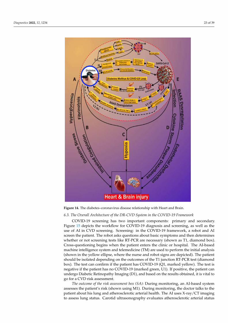
Diagnostics 2022, 12, 1234 23 of 39Diagnostics 2022, 12, 1234 24 of 41
Figure 14. The diabetes–coronavirus disease relationship with Heart and Brain.
Patients with COVID-19 who had no previous history of diabetes developed signifi-cant consequences such as DKA. DKA arises due to the overproduction of opposing reg-ulators, which favors ketones [251]. Low insulin levels also cause it. DKA is more common in type 1 DM patients [40]. It can also happen in people with type 2 diabetes [252]. The three paths of new-onset DM or progression of pre-existing DM after COVID-19 infection are shown in Figure 14 (pathways IV, V, and VI). As shown in Figure 14, the diabetes–CVD link leads to CVD and stroke [173]. This is the most important figure in the DR-CVD investigation. This shows the importance of CVD work during COVID-19 times and the link between uncontrolled diabetes, CVD, and COVID-19. COVID-19 and CVD have an-other significant component [41]. Thus, it is very important to link COVID-19 with DR and CVD. COVID-19 has serious impacts, especially on patients with underlying condi-tions like CVD, diabetes mellitus, and hypertension. In individuals with the underlying disease, cardiac events worsen [253]. Many hospitalized patients reported heart damage
Figure 14. The diabetes–coronavirus disease relationship with Heart and Brain.
6.3. The Overall Architecture of the DR-CVD System in the COVID-19 Framework
COVID-19 screening has two important components: primary and secondary.Figure 15 depicts the workflow for COVID-19 diagnosis and screening, as well as theuse of AI in CVD screening. Screening: in the COVID-19 framework, a robot and AIscreen the patient. The robot asks questions about basic symptoms and then determineswhether or not screening tests like RT-PCR are necessary (shown as T1, diamond box).Cross-questioning begins when the patient enters the clinic or hospital. The AI-basedmachine intelligence system and telemedicine (TM) are used to perform the initial analysis(shown in the yellow ellipse, where the nurse and robot signs are depicted). The patientshould be isolated depending on the outcomes of the T1 junction RT-PCR test (diamondbox). The test can confirm if the patient has COVID-19 (Q1, marked yellow). The test isnegative if the patient has no COVID-19 (marked green, U1). If positive, the patient canundergo Diabetic Retinopathy Imaging (D1), and based on the results obtained, it is vital togo for a CVD risk assessment.
The outcome of the risk assessment box (SA): During monitoring, an AI-based systemassesses the patient’s risk (shown using M1). During monitoring, the doctor talks to thepatient about his lung and atherosclerotic arterial health. The AI uses X-ray/CT imagingto assess lung status. Carotid ultrasonography evaluates atherosclerotic arterial status

Diagnostics 2022, 12, 1234 24 of 39
for CVD risk. (I) deteriorating lung condition and the need for ICU; (II) worsening CVDcondition and requirement for CVD medications; (III) untouched.
Diagnostics 2022, 12, 1234 25 of 41
in the range of 12–26%. The infection’s cytokines may alter the patients’ intramural coro-nary arteries. Cardiovascular disorders affect ARDS in COVID-19 afflicted patients [254].
SARS-CoV-2 can cause complex severe ruptured plaque [255]. The enhanced insta-bility of coronary and carotid plaques may also enhance the risk of cerebral ischemic strokes and myocardial infarctions in SARS-CoV-2 positive asymptomatic individuals [9,256]. The above discussion is a clear pathway between COVID-19 and CVD. Further note that the process of DR accelerates the worsening of CVD in the COVID-19 frame-work.
6.3. The Overall Architecture of the DR-CVD System in the COVID-19 Framework COVID-19 screening has two important components: primary and secondary. Figure
15 depicts the workflow for COVID-19 diagnosis and screening, as well as the use of AI in CVD screening. Screening: in the COVID-19 framework, a robot and AI screen the pa-tient. The robot asks questions about basic symptoms and then determines whether or not screening tests like RT-PCR are necessary (shown as T1, diamond box). Cross-questioning begins when the patient enters the clinic or hospital. The AI-based machine intelligence system and telemedicine (TM) are used to perform the initial analysis (shown in the yel-low ellipse, where the nurse and robot signs are depicted). The patient should be isolated depending on the outcomes of the T1 junction RT-PCR test (diamond box). The test can confirm if the patient has COVID-19 (Q1, marked yellow). The test is negative if the pa-tient has no COVID-19 (marked green, U1). If positive, the patient can undergo Diabetic Retinopathy Imaging (D1), and based on the results obtained, it is vital to go for a CVD risk assessment.
The outcome of the risk assessment box (SA): During monitoring, an AI-based system assesses the patient’s risk (shown using M1). During monitoring, the doctor talks to the patient about his lung and atherosclerotic arterial health. The AI uses X-ray/CT imaging to assess lung status. Carotid ultrasonography evaluates atherosclerotic arterial status for CVD risk. (I) deteriorating lung condition and the need for ICU; (II) worsening CVD con-dition and requirement for CVD medications; (III) untouched.
Figure 15. COVID-19 risk assessment for DR and CVD.
Figure 15. COVID-19 risk assessment for DR and CVD.
The risk evaluation box has three outcomes (SA). (I) Ground Glass Opacities (GGO) inthe patient (from the output of the monitor). When there are no atherosclerotic lesions inthe carotid arteries, the person is considered severely symptomatic and is assessed in theICU with ventilation. The lungs are slowly filling up with fluid, therefore this is a must (seeFigure 15). In the ICU, the patient is constantly observed (shown by monitoring functionM2). To treat CVD (II) if the patient’s lungs are stable (extremely low GGO) and the carotidarteries are plaque-burdened (C). In outcome (III), the individual has neither lung norarterial damage, making him uninfected (U2). Since the individual was quarantined (Q1)and the risk assessment (A) was done using AI-based imaging, the doctor must assessthe patient based on clinical judgment (C) (marked as J). Finally, after the individual isuninfected (U2), the extensive COVID process begins (secondary screening, marked SS).If positive, the quarantine feedback loop starts again (Q1). They are COVID-19-free ifnegative (U3, marked green).
6.4. Role of AI in CVD Risk Assessment for COVID-19 Screening
Using a machine learning technique, imaging data can be used to predict CVD risk atthe vascular scale [208,257–259]. The goal is to obtain OCT scans to see if the person hasDiabetic Retinopathy (DR). Further, if positive, the patient is at risk for CVD and shouldbe monitored for lung and arterial function. Plaque image-based characteristics can beobtained from patient carotid scans and risk estimated using machine learning. The AIcomponent simply requires the green trained model labeled Imaging-based AI with arobot AI logo on it. The machine learning algorithm tells the patient’s clinician (M1) if therelative risk is low, medium, or high. The patient’s CVD risk is assessed using this color-coded system (marked as SA). This can include evaluating the CT lungs for ground-glassopacity [260] or pulmonary embolism [261]. Thus, the proposal depicts AI-based CVDmonitoring for pandemic diagnosis.
7. Critical Discussion
This review provides insight into the link between DR and CVD. The impact of DRon vision is well understood. Further, after a thorough analysis of various studies, it is
Diagnostics 2022, 12, 1234 25 of 39
evident that DR patients are at increased risk for CVD conditions. Therefore, along withDR screening, it is optimal to adopt a low-cost system to prevent the worsening of the CVDconditions of a patient. With the help of AtheroEdge™ 2.0 and 3.0, effective monitoringcan be done for these patients and long-term complications can be prevented with thehelp of AI-based interventions in the model. Deep learning and Machine learning helpsin calculating CVD risk with greater sensitivity and specificity. The model can be trainedin such a way that it does not need any human intervention and results can be measuredquickly. This proves to be a breakthrough in the current healthcare systems. With the helpof this information, clinicians can counsel patients with vision-threatening DR and furtherguide them about associated CVD risk.
7.1. Benchmarking
After a perusal of various studies, several articles discussed the link between DRand CVD (Table 5). However, there was no such article found which was addressing allthe components in our review. Alonso et al. [37] showed that DR in T2D patients withnormal renal function and without CVD risk was associated with a higher plaque burden(≥2 carotid plaques) in carotid arteries. The authors conducted a cross-sectional studyon 312 patients (51% men), with 153 patients with DR. The authors demonstrated thepercentage of carotid plaques in DR was higher than without DR (68% vs. 52%, p = 0.0045).The authors used multivariate logistic regression that DR was independently associatedwith cIMT (p = 0.0176). In another study, Simó et al. [38] made an interesting remarkrelating to DR and CVD suggesting that, while staging diabetes, DR and CVD should neverbe considered as separate entities. The authors further suggested DR to be equivalent tohypertension and dyslipidemia in terms of deteriorating CVD. The authors suggested thatif diabetes is not considered when evaluating CVD, the results could be biased. Finally, theyconcluded that all DR patients must undergo CVD screening for better risk stratification.Ting et al. [26] discussed in their chapter that retinal venules which are wider and havenarrower arterioles were associated with an increased risk of CVD. The authors discussedthe role of narrower retinal arteriolar caliber were significantly associated with low eGFRin CKD patients.
Table 5. Comparing the proposed review against previous reviews on joint DR and CVD.
Citations Year DR a CVD b RI c CI d AI e RS f COV-19 g
Son et al. [22] 2010 X X 5 X X X 5
Alonso et al. [37] 2015 X X 5 5 5 5 5
Ting et al. [26] 2019 X X X X X X 5
Simó et al. [38] 2019 X X 5 X 5 5 5
Gupta et al. [39] 2021 X 5 X 5 X 5 5
Proposed Review 2022 X X X X X X XDR a: Diabetic Retinopathy: CVD b: Cardiovascular Diseases, RI c: Retinal Imaging, CI d: Carotid Imaging AI e:Artificial Intelligence, RS f: Risk Stratification, COV-19 g: COVID-19.
The authors used deep learning on fundus images and correlated them with 5-yearCVD risk. Further, Gupta et al. [39] presented in their review the role of examining andworking on the heart and predicting the macrovascular changes based on microvascularfeatures and functions derived from retinal imaging. The authors showed a link betweenretinal imaging and the diagnosis of CVD. Since the usage of retinal scanning helps in thediagnosis of DR, age-related macular degeneration, and glaucoma, the authors linked thiswith heart conditions. Son et al. [22] showed the relationship between DR and subclinicalatherosclerosis by using 142 subjects with T2D who are free from CVD.
The methodology consisted of (a) cIMT measurement using carotid ultrasound and(b) CVD risk using UK Prospective Diabetic Study (UKPDS) calculator. The authors ob-served that patients with subclinical atherosclerosis had a higher rate of hypertension andDR. Further, CVD risk was higher in subjects with subclinical atherosclerosis. The studyconcluded that DR is an independent risk biomarker for subclinical atherosclerosis in T2D.Hence, to our understanding, no other article has provided such valuable insights about

Diagnostics 2022, 12, 1234 26 of 39
different approaches towards the diseases that are necessary to study DR and CVD as wellas provide necessary interventions in their treatment strategies.
Our previous study, Integration of cardiovascular risk assessment with COVID-19using artificial intelligence [262], the so-called “RCM study”, also used a surrogate markerfor CVD risk assessment; however, the differences between RCM study and the currentstudy, the so-called DR study, should be noted. While the block of surrogate biomarkersremains the same in both scenarios, the fundamental difference is the key applicationscovered in the RCM study vs. the current study, i.e., the COVID-19 cohort vs. the DiabeticRetinopathy cohort. The patient selection criteria are based on the COVID-19 symptomsvs. diabetes symptoms. The second fundamental difference lies in the biological drivefor the diseases, about acute respiratory distress syndrome (ARDS) and cytokine storm inthe COVID-19 paradigm vs. atherosclerosis formation in the arterial vessels, leading todamage of coronary arteries. Another secondary difference between the two approachesis the usage of the imaging modality for image data acquisition, such as MRI/CT inRCM study vs. intravascular ultrasound (IVUS) for coronary artery imaging in DR study.While the imaging is conducted differently, the measurement criteria also subsequentlychange by computing 3D straining imaging for the left ventricle (LV) chamber in RCMstudy vs. quantification of the plaque burden in either IVUS study or carotid arterialplaque measurements or plaque burden in carotid arteries. Please note that the surrogatebiomarkers for both studies utilize the B-model carotid ultrasound for plaque burdenmeasurements, which are surrogate biomarkers for CVD/Stroke imaging. Finally, since theapplication is different, the workflow changes entirely between the two applications. Inthe RCM study, we followed CVD/Stroke risk assessment during COVID-19 times usingCT imaging for monitoring the lung followed by carotid scanning for plaque build-upmeasurements, the surrogate marker for CVD/Stroke risk, while in the DR study, theworkflow consisted of the following steps: (i) DR imaging, (ii) Lung CT imaging, andcarotid arterial imaging. Thus, the COVID-19 severity report and CVD risk assessmentreports were generated, followed by the manifestations.
7.2. Recommendations
It is clear from our review that DR patients must opt for CVD risk assessment tech-niques, especially the patients suffering from high risk, vision-threatening DR. Carotidimaging could be a boon for patients undergoing retinal imaging that have DR. Ultrasound-based imaging modalities are the most convenient modalities for carotid imaging. Forretinal imaging, optical coherence tomography and angiography proved to be revolution-ary in ophthalmology [263]. However, the use of fundus imaging is widely followed,with further possibilities explored using smartphone-based retinal imaging modalities.Further, for risk assessment, artificial intelligence-based algorithms are the most highlyrecommended. It is, therefore, recommended that all these factors be followed to identifyand treat the disease at its earliest.
7.3. A Special Note on DR and Monitoring of CVD Risk
With our current understanding of DR and the existing interventions associated withit, we believe that the link between DR and CVD is not well established in the generalpopulation. This is the reason for the worsening statistics of both diseases. It is thusimportant to educate DR patients about their risk of CVD and provide the necessarymonitoring options. It is necessary to provide this pipeline as an intervention in healthcaresystems. With the help of robust imaging and monitoring techniques, several lives can besaved, and disease can be treated.
7.4. The Effects of COVID-19 on DR Patients
This section shows how DR got affected during the COVID-19 period. The importanceof DR during pandemic times has risen several folds. SARS-CoV-2 was seen as an importantbarrier to DR management and this has led to the suspension of primary healthcare services

Diagnostics 2022, 12, 1234 27 of 39
such as limited access to diabetes-related medical consultations and retinal screenings [237].Advocacy with the government is seen to be critical to facilitating continuous sustainableservices as many DR screening services and referral initiatives have been adversely im-pacted by the COVID-19 pandemic [238]. Please note that, in the presence of diabetes, dueto the aggravation of underlying pulmonary microcirculatory impairments, it has beenobserved that the underlying microvascular disease is associated with a higher risk ofadverse DR outcomes in the COVID-19 period [239].
7.5. Strengths, Weaknesses, and Future Extensions
By introducing this link, we provide additional support to existing healthcare systems.It is rightly said that prevention is better than cure. With the knowledge about the linkbetween DR and CVD as well as the low-cost screening using AI-based algorithms, not onlycan patients be treated, but disease can also be prevented from happening. One limitationthat we feel is that there are no pipelines developed for treating DR patients for CVD and itis important to put more light on this aspect. With the ongoing pandemic, it is important todiscuss how both the targeted diseases may be affected in the presence of the SARS-CoV-2virus. In the future, we would like to discuss the role of big data in reducing the bias ofcohort [264]. Further, we would focus on the role of different types of DL and ML-basedclassifiers like the ensemble machine learning (EML)-based algorithm and auto-encodersfor automated DR detection [265,266] and carotid wall segmentation [221,262,263] andquantification [267–274].
8. Conclusions
This narrative review presented the role of diabetic retinopathy and its association withcardiovascular risks in diabetes patients. We also showed the complications of diabetes thatlead to coronary heart disease. This review emphasized the hypothesis that DR can lead tothe worsening of CVD. Thus, it is of utmost importance to identify CVD complications inDR patients. We also demonstrated that carotid imaging can be used as a low-cost, non-invasive alternative to the existing imaging modalities for CVD screening in DR patients.This low-cost B-mode ultrasound will also be beneficial for plaque tissue characterization inDR patients to provide an additional and vital understanding of the CVD risk. Further, weobserved that AI-based solutions are powerful for risk stratification of CVD in DR patients.Lastly, we glance at the role of DR and CVD in the COVID-19 framework and observe howAI plays a role in this system.
Author Contributions: Conceptualization, J.S.S., S.M. (Smiksha Munjral), M.M. (Mahesh Main-darkar), A.J. and A.P. (Anudeep Puvvula); Methodology and software, J.S.S., M.M. (Mahesh Main-darkar), S.M. (Smiksha Munjral), L.S. and R.P. Validation, A.M.J., M.T., P.A., I.M.S., G.F., N.N.K. andS.M. (Smiksha Munjral); Investigation, R.J.C., P.S.C., K.V., J.R.L., G.P., M.S., S.M. (Sophie Mavrogeni),M.M. (Martin Miner), G.P., R.P. and J.S.S.; Resources, S.P.; Data curation, M.M. (Mahesh Maindarkar),L.S., N.S., S.G., T.O., G.D.K. and J.S.S.; Writing—original draft preparation, S.M. (Smiksha Munjral),M.M. (Mahesh Maindarkar), A.P. (Anudeep Puvvula), L.S., M.A.-M., D.W.S., M.M.F., S.N. and J.S.S.;Writing—review and editing, S.P., M.M. (Mahesh Maindarkar), S.M. (Smiksha Munjral), L.S., M.T.,M.S., A.B., V.V., P.P.S., A.M.J., P.R.K. and J.S.S.; Visualization, S.M. (Smiksha Munjral), D.P.M., A.A.,S.K.D., K.V., R.K., T.J., A.S. (Aditya Sharma), G.T., A.P. (Athanasios Protogerou), A.N. and J.S.S.;Supervision, S.M. (Sophie Mavrogeni), A.S. (Ajit Saxena), V.R., V.A., M.F. and J.S.S.; Project adminis-tration, N.N.K., J.T., P.P.S. and J.S.S. All authors have read and agreed to the published version ofthe manuscript.
Funding: This research received no external funding.
Institutional Review Board Statement: Not applicable.
Informed Consent Statement: Suri is with AtheroPoint™ LLC, Roseville, CA, USA, which doescardiovascular and stroke imaging.
Data Availability Statement: No data availability.
Conflicts of Interest: The authors declare no conflict of interest.

Diagnostics 2022, 12, 1234 28 of 39
Abbreviations
ACC American College of CardiologyAECRS AtheroEdge Composite Risk ScoreAGE Advance Glycation End ProductsAHA American Heart AssociationAHEAD Action for Health in DiabetesAI Artificial IntelligenceASCVD Atherosclerotic Cardiovascular DiseaseCAD Coronary Artery DiseaseCAPB Coronary Artery Plaque BurdenCCA Common Carotid ArteryCHF Congestive Heart FailureCI Confidence IntervalcIMT Carotid Intima-Media ThicknessCT Computerized TomographyCVA Cerebrovascular AccidentCVD Cardiovascular diseaseCVE Cardiovascular EventsCCVRC Conventional Cardiovascular Risk CalculatorDL Deep LearningDM Diabetes MellitusDME Diabetic Macular EdemaDR Diabetic RetinopathyEML Ensemble Machine LearningFA Fluorescein AngiographyFRS Framingham Risk ScoreGLS Global Longitudinal StrainHR Hazard RatioICA Internal carotid arteryICGA Indocyanine Green AngiographyIMTV Intima-Media Thickness VariabilityIVUS Intravascular UltrasoundMA Macular EdemaMI Myocardial InfarctionML Machine LearningMRI Magnetic Resonance ImagingMVD Microvascular DiseaseNICE National Institute for Health and Care ExcellenceNPDR Non-Proliferative Diabetic RetinopathyOCT Optical Coherence TomographyOpA Ophthalmic arteryOR Odds RatioPCRE Pooled cohort risk equationPDR Proliferative Diabetic RetinopathyPET Positron Emission TomographyPVD Peripheral Vascular DiseaseRRS Reynold Risk ScoreT2DM Type 2 Diabetes MellitusTIR Time in RangePKC Protein Kinase CUKPDS UK Prospective Diabetes ScoreUS UltrasoundUTC Ultrasound-based Tissue characterizationCOVID-19 Coronavirus-2019WHO World Health Organization

Diagnostics 2022, 12, 1234 29 of 39
References1. World Health Organization. Cardiovascular Diseases. Available online: https://www.who.int/health-topics/cardiovascular-
diseases/#tab=tab_1 (accessed on 22 February 2022).2. Frostegård, J. Immunity, atherosclerosis and cardiovascular disease. BMC Med. 2013, 11, 117. [CrossRef] [PubMed]3. Suri, J.S.; Kathuria, C.; Molinari, F. Atherosclerosis Disease Management; Springer Science & Business Media: Berlin, Germany, 2010.4. Diabetes. Available online: http://www.who.int/news-room/fact-sheets/detail/diabetes (accessed on 26 February 2022).5. Kushner, R.F.; Sorensen, K.W. Lifestyle medicine: The future of chronic disease management. J. Curr. Opin. Endocrinol. Diabetes
Obes. 2013, 20, 389–395. [CrossRef] [PubMed]6. National Diabetes Statistics Report. 2020. Available online: https://www.cdc.gov/diabetes/data/statistics-report/index.html
(accessed on 28 February 2022).7. Friedman, D.S.; Ali, F.; Kourgialis, N. Diabetic Retinopathy in the Developing World: How to Approach Identifying and Treating
Underserved Populations. Am. J. Ophthalmol. 2011, 151, 192–194.e1. [CrossRef] [PubMed]8. Kenny, H.C.; Abel, E.D. Heart failure in type 2 diabetes mellitus: Impact of glucose-lowering agents, heart failure therapies, and
novel therapeutic strategies. Circ. Res. 2019, 124, 121–141. [CrossRef]9. Viswanathan, V.; Puvvula, A.; Jamthikar, A.D.; Saba, L.; Johri, A.M.; Kotsis, V.; Khanna, N.N.; Dhanjil, S.K.; Majhail, M.; Misra,
D.P.; et al. Bidirectional link between diabetes mellitus and coronavirus disease 2019 leading to cardiovascular disease: Anarrative review. World J. Diabetes 2021, 12, 215–237. [CrossRef] [PubMed]
10. PAHO. About Diabetes. Available online: https://www3.paho.org/hq/index.php?option=com_content&view=article&id=6717:2012-about-diabetes&Itemid=39447&lang=en (accessed on 3 January 2022).
11. Yau, J.W.Y.; Rogers, S.L.; Kawasaki, R.; Lamoureux, E.L.; Kowalski, J.W.; Bek, T.; Chen, S.-J.; Dekker, J.M.; Fletcher, A.; Grauslund,J.; et al. Global Prevalence and Major Risk Factors of Diabetic Retinopathy. Diabetes Care 2012, 35, 556–564. [CrossRef] [PubMed]
12. Roberts, K.C.; Rao, D.; Bennett, T.L.; Loukine, L.; Jayaraman, G.C. Prevalence and patterns of chronic disease multimorbidity andassociated determinants in Canada. Health Promot. Chronic Dis. Prev. Can. 2015, 35, 87–94. [CrossRef]
13. Kharroubi, A.T.; Darwish, H.M. Diabetes mellitus: The epidemic of the century. World J. Diabetes 2015, 6, 850. [CrossRef]14. Blair, M. Diabetes Mellitus Review. Urol. Nurs. 2016, 36, 27–36. [CrossRef]15. Queiroz, M.; Sena, C.M. Perivascular adipose tissue in age-related vascular disease. Ageing Res. Rev. 2020, 59, 101040. [CrossRef]16. Mahabadi, N.; al Khalili, Y. Neuroanatomy, Retina; StatPearls: Island, FL, USA, 2020.17. Wang, W.; Lo, A.C.Y. Diabetic Retinopathy: Pathophysiology and Treatments. Int. J. Mol. Sci. 2018, 19, 1816. [CrossRef] [PubMed]18. Stitt, A.W.; Curtis, T.; Chen, M.; Medina, R.J.; McKay, G.; Jenkins, A.; Gardiner, T.A.; Lyons, T.J.; Hammes, H.-P.; Simó, R.; et al.
The progress in understanding and treatment of diabetic retinopathy. Prog. Retin. Eye Res. 2016, 51, 156–186. [CrossRef] [PubMed]19. Paetkau, M.E.; Boyd, T.; Winship, B.; Grace, M. Cigarette smoking and diabetic retinopathy. Diabetes 1977, 26, 46–49. [CrossRef]
[PubMed]20. Amini, M.; Safaei, H.; Aminorroaya, A. The Incidence of Microalbuminuria and its Associated Risk Factors in Type 2 Diabetic
Patients in Isfahan, Iran. Rev. Diabet. Stud. 2007, 4, 242–248. [CrossRef]21. Wat, N.; Wong, R.L.; Wong, I.Y. Associations between diabetic retinopathy and systemic risk factors. Hong Kong Med. J. 2016, 22,
589–599. [CrossRef]22. Son, J.-W.; Jang, E.-H.; Kim, M.-K.; Kim, I.T.; Roh, Y.J.; Baek, K.-H.; Song, K.-H.; Yoon, K.-H.; Cha, B.-Y.; Lee, K.-W.; et al. Diabetic
retinopathy is associated with subclinical atherosclerosis in newly diagnosed type 2 diabetes mellitus. Diabetes Res. Clin. Pract.2011, 91, 253–259. [CrossRef]
23. Melo, L.G.N.; Morales, P.H.; Drummond, K.R.G.; Santos, D.C.; Pizarro, M.H.; Barros, B.S.V.; Mattos, T.C.L.; Pinheiro, A.A.;Mallmann, F.; Leal, F.S.L.; et al. Diabetic Retinopathy May Indicate an Increased Risk of Cardiovascular Disease in Patients withType 1 Diabetes—A Nested Case-Control Study in Brazil. Front. Endocrinol. 2019, 10, 689. [CrossRef]
24. Yun, D.Q.Q.; Cheung, N.D. Diabetic Retinopathy and Heart Disease. In Diabetic Retinopathy and Cardiovascular Disease; KargerPublishers: Basel, Switzerland, 2019; Volume 27, pp. 54–63.
25. Barrett, E.J.; Liu, Z.; Khamaisi, M.; King, G.L.; Klein, R.; Klein, B.E.K.; Hughes, T.M.; Craft, S.; Freedman, B.I.; Bowden, D.W.;et al. Diabetic Microvascular Disease: An Endocrine Society Scientific Statement. J. Clin. Endocrinol. Metab. 2017, 102, 4343–4410.[CrossRef]
26. Ting, D.S.; Peng, L.; Varadarajan, A.V.; Liu, T.Y.A. Novel Retinal Imaging in Assessment of Cardiovascular Risk Factors andSystemic Vascular Diseases. In Diabetic Retinopathy and Cardiovascular Disease; Karger Publishers: Basel, Switzerland, 2019;Volume 27, pp. 106–118.
27. Gao, L.; Zhao, W.; Yang, J.-K.; Qin, M.-Z. Proliferative diabetic retinopathy in patients with type 2 diabetes correlates with thepresence of atherosclerosis cardiovascular disease. Diabetol. Metab. Syndr. 2021, 13, 1–9. [CrossRef]
28. van der Heide, F.C.T.; Zhou, T.L.; Henry, R.M.A.; Houben, A.J.H.M.; Kroon, A.A.; Dagnelie, P.C.; van Dongen, M.C.J.M.; Eussen,S.J.P.M.; Berendschot, T.T.J.M.; Schouten, J.S.A.G.; et al. Carotid stiffness is associated with retinal microvascular dysfunction—TheMaastricht study. Microcirculation 2021, 28, e12702. [CrossRef]
29. Gubitosi-Klug, R.; Gao, X.; Pop-Busui, R.; de Boer, I.H.; White, N.; Aiello, L.P.; Miller, R.; Palmer, J.; Tamborlane, W.; Wallia, A.;et al. Associations of Microvascular Complications With the Risk of Cardiovascular Disease in Type 1 Diabetes. Diabetes Care2021, 44, 1499–1505. [CrossRef] [PubMed]

Diagnostics 2022, 12, 1234 30 of 39
30. Modjtahedi, B.S.; Wu, J.; Luong, T.Q.; Gandhi, N.K.; Fong, D.S.; Chen, W. Severity of diabetic retinopathy and the risk offuture cerebrovascular disease, cardiovascular disease, and all-cause mortality. Ophthalmology 2021, 128, 1169–1179. [CrossRef][PubMed]
31. Sugamura, K.; Keaney, J.F., Jr. Reactive oxygen species in cardiovascular disease. Free Radic. Biol. Med. 2011, 51, 978–992.[CrossRef] [PubMed]
32. Khurana, R.; Simons, M.; Martin, J.F.; Zachary, I.C. Role of angiogenesis in cardiovascular disease: A critical appraisal. J. Circ.2005, 112, 1813–1824. [CrossRef] [PubMed]
33. Chalakkal, R.J.; Abdulla, W.H.; Hong, S.C. Fundus Retinal Image Analyses for Screening and Diagnosing Diabetic Retinopathy,Macular Edema, and Glaucoma Disorders. In Diabetes and Fundus OCT; Elsevier: Amsterdam, The Netherlands, 2020; pp. 59–111.
34. Abràmoff, M.D.; Garvin, M.K.; Sonka, M. Retinal imaging and image analysis. IEEE Rev. Biomed. Eng. 2010, 3, 169–208. [CrossRef][PubMed]
35. MacGillivray, T.J.; Trucco, E.; Cameron, J.R.; Dhillon, B.; Houston, J.G.; Van Beek, E.J.R. Retinal imaging as a source of biomarkersfor diagnosis, characterization and prognosis of chronic illness or long-term conditions. Br. J. Radiol. 2014, 87, 20130832. [CrossRef]
36. Lamirel, C.; Newman, N.; Biousse, V. Optical coherence tomography (OCT) in optic neuritis and multiple sclerosis. Rev. Neurol.2010, 166, 978–986. [CrossRef]
37. Alonso, N.; Traveset, A.; Rubinat, E.; Ortega, E.; Alcubierre, N.; Sanahuja, J.; Hernández, M.; Betriu, A.; Jurjo, C.; Fernández, E.Type 2 diabetes-associated carotid plaque burden is increased in patients with retinopathy compared to those without retinopathy.Cardiovasc. Diabetol. 2015, 14, 1–9. [CrossRef]
38. Simó, R.; Bañeras, J.; Hernández, C.; Rodríguez-Palomares, J.; Valente, F.; Gutierrez, L.; González-Alujas, T.; Ferreira, I.; Aguadé-Bruix, S.; Montaner, J. Diabetic retinopathy as an independent predictor of subclinical cardiovascular disease: Baseline results ofthe PRECISED study. BMJ Open Diabetes Res. Care 2019, 7, e000845. [CrossRef]
39. Gupta, K.; Reddy, S. Heart, Eye, and Artificial Intelligence: A Review. Cardiol. Res. 2021, 12, 132. [CrossRef]40. Ayman, J.S.S.; El-Baz, S. Machine Learning in Medicine-Chapman & Hall/CRC Healthcare Informatics Series; CRC Press: Boca Raton,
FL, USA, 2021.41. Ayman El-Baz, J.S.S. Big Data in Multimodal Medical Imaging; Chapman and Hall: London, UK, 2019.42. Goldstein, B.A.; Navar, A.M.; Carter, R.E. Moving beyond regression techniques in cardiovascular risk prediction: Applying
machine learning to address analytic challenges. Eur. Heart J. 2016, 38, 1805–1814. [CrossRef] [PubMed]43. Ming, C.; Viassolo, V.; Probst-Hensch, N.; Chappuis, P.O.; Dinov, I.D.; Katapodi, M.C. Machine learning techniques for per-
sonalized breast cancer risk prediction: Comparison with the BCRAT and BOADICEA models. Breast Cancer Res. 2019, 21, 75.[CrossRef] [PubMed]
44. Biswas, M.; Kuppili, V.; Saba, L.; Edla, D.R.; Suri, H.S.; Cuadrado-Godia, E.; Laird, J.; Marinhoe, R.; Sanches, J.; Nicolaides, A.State-of-the-art review on deep learning in medical imaging. Front. Biosci. 2019, 24, 392–426.
45. Chalakkal, R.; Hafiz, F.; Abdulla, W.; Swain, A. An efficient framework for automated screening of Clinically Significant MacularEdema. Comput. Biol. Med. 2020, 130, 104128. [CrossRef]
46. Karbhari, Y.; Basu, A.; Geem, Z.W.; Han, G.-T.; Sarkar, R. Generation of Synthetic Chest X-ray Images and Detection of COVID-19:A Deep Learning Based Approach. Diagnostics 2021, 11, 895. [CrossRef] [PubMed]
47. Rim, T.H.; Lee, C.J.; Tham, Y.-C.; Cheung, N.; Yu, M.; Lee, G.; Kim, Y.; Ting, D.S.W.; Chong, C.C.Y.; Choi, Y.S.; et al. Deep-learning-based cardiovascular risk stratification using coronary artery calcium scores predicted from retinal photographs. Lancet Digit.Health 2021, 3, e306–e316. [CrossRef]
48. Cheung, C.Y.; Xu, D.; Cheng, C.-Y.; Sabanayagam, C.; Tham, Y.-C.; Yu, M.; Rim, T.H.; Chai, C.Y.; Gopinath, B.; Mitchell, P.; et al. Adeep-learning system for the assessment of cardiovascular disease risk via the measurement of retinal-vessel calibre. Nat. Biomed.Eng. 2020, 5, 498–508. [CrossRef]
49. Nielsen, K.B.; Lautrup, M.L.; Andersen, J.K.; Savarimuthu, T.R.; Grauslund, J. Deep Learning–Based Algorithms in Screening ofDiabetic Retinopathy: A Systematic Review of Diagnostic Performance. Ophthalmol. Retin. 2018, 3, 294–304. [CrossRef]
50. Chalakkal, R.J. Automatic Retinal Image Analysis to Triage Retinal Pathologies; University of Auckland: Auckland, New Zealand, 2019.51. Gayathri, S.; Gopi, V.P.; Palanisamy, P. Diabetic retinopathy classification based on multipath CNN and machine learning
classifiers. Phys. Eng. Sci. Med. 2021, 44, 639–653. [CrossRef]52. Dashtipour, K.; Taylor, W.; Ansari, S.; Zahid, A.; Gogate, M.; Ahmad, J.; Assaleh, K.; Arshad, K.; Imran, M.A.; Abbasi, Q.
Detecting Alzheimer’s Disease Using Machine Learning Methods. In Body Area Networks. Smart IoT and Big Data for IntelligentHealth Management. BODYNETS 2021. Lecture Notes of the Institute for Computer Sciences, Social Informatics and TelecommunicationsEngineering; Ur Rehman, M., Zoha, A., Eds.; Springer: Cham, Germany, 2022; Volume 420.
53. Li, J.-P.O.; Liu, H.; Ting, D.S.J.; Jeon, S.; Chan, R.V.P.; Kim, J.E.; Sim, D.A.; Thomas, P.B.M.; Lin, H.; Chen, Y.; et al. Digitaltechnology, tele-medicine and artificial intelligence in ophthalmology: A global perspective. Prog. Retin. Eye Res. 2021, 82, 100900.[CrossRef]
54. Khanna, N.N.; Jamthikar, A.; Gupta, D.; Nicolaides, A.; Araki, T.; Saba, L.; Cuadrado-Godia, E.; Sharma, A.; Omerzu, T.; Suri,H.S.; et al. Performance evaluation of 10-year ultrasound image-based stroke/cardiovascular (CV) risk calculator by comparingagainst ten conventional CV risk calculators: A diabetic study. Comput. Biol. Med. 2019, 105, 125–143. [CrossRef] [PubMed]
55. Molinari, F.; Liboni, W.; Giustetto, P.; Badalamenti, S.; Suri, J.S. Automatic computer-based tracings (act) in longitudinal 2-dultrasound images using different scanners. J. Mech. Med. Biol. 2009, 9, 481–505. [CrossRef]

Diagnostics 2022, 12, 1234 31 of 39
56. Bruder, O.; Wagner, A.; Jensen, C.J.; Schneider, S.; Ong, P.; Kispert, E.-M.; Nassenstein, K.; Schlosser, T.; Sabin, G.V.; Sechtem, U.;et al. Myocardial Scar Visualized by Cardiovascular Magnetic Resonance Imaging Predicts Major Adverse Events in PatientsWith Hypertrophic Cardiomyopathy. J. Am. Coll. Cardiol. 2010, 56, 875–887. [CrossRef] [PubMed]
57. Erbel, R.; Möhlenkamp, S.; Moebus, S.; Schmermund, A.; Lehmann, N.; Stang, A.; Dragano, N.; Grönemeyer, D.; Seibel, R.; Kälsch,H.; et al. Coronary Risk Stratification, Discrimination, and Reclassification Improvement Based on Quantification of SubclinicalCoronary Atherosclerosis: The Heinz Nixdorf Recall Study. J. Am. Coll. Cardiol. 2010, 56, 1397–1406. [CrossRef]
58. Zhu, T.; Li, K.; Herrero, P.; Georgiou, P. Deep Learning for Diabetes: A Systematic Review. IEEE J. Biomed. Health Inform. 2020, 25,2744–2757. [CrossRef]
59. Association, A.D. Diagnosis and classification of diabetes mellitus. Diabetes Care 2010, 33 (Suppl. S1), S62–S69. [CrossRef]60. Wilcox, G. Insulin and insulin resistance. Clin. Biochem. Rev. 2005, 26, 19.61. Nathan, D.M. Long-Term Complications of Diabetes Mellitus. N. Engl. J. Med. 1993, 328, 1676–1685. [CrossRef]62. Sayin, N.; Kara, N.; Pekel, G. Ocular complications of diabetes mellitus. World J. Diabetes 2015, 6, 92. [CrossRef]63. Papatheodorou, K.; Papanas, N.; Banach, M.; Papazoglou, D.; Edmonds, M. Complications of Diabetes 2016; Hindawi: London,
UK, 2016.64. Heydari, I.; Radi, V.; Razmjou, S.; Amiri, A. Chronic complications of diabetes mellitus in newly diagnosed patients. Int. J.
Diabetes Mellit. 2010, 2, 61–63. [CrossRef]65. Nentwich, M.M.; Ulbig, M.W. Diabetic retinopathy-ocular complications of diabetes mellitus. World J. Diabetes 2015, 6, 489.
[CrossRef] [PubMed]66. Ahsan, H. Diabetic retinopathy–Biomolecules and multiple pathophysiology. Diabetes Metab. Syndr. Clin. Res. Rev. 2015, 9, 51–54.
[CrossRef] [PubMed]67. Coyne, K.S.; Margolis, M.K.; Kennedy-Martin, T.; Baker, T.M.; Klein, R.; Paul, M.D.; Revicki, D.A. The impact of diabetic
retinopathy: Perspectives from patient focus groups. Fam. Pract. 2004, 21, 447–453. [CrossRef] [PubMed]68. Amin, J.; Sharif, M.; Yasmin, M. A Review on Recent Developments for Detection of Diabetic Retinopathy. Scientifica 2016, 2016.
[CrossRef]69. Ciulla, T.A.; Amador, A.G.; Zinman, B. Diabetic retinopathy and diabetic macular edema: Pathophysiology, screening, and novel
therapies. Diabetes Care 2003, 26, 2653–2664. [CrossRef]70. Hammes, H.-P.; Lin, J.; Renner, O.; Shani, M.; Lundqvist, A.; Betsholtz, C.; Brownlee, M.; Deutsch, U. Pericytes and the
Pathogenesis of Diabetic Retinopathy. Diabetes 2002, 51, 3107–3112. [CrossRef]71. Zhu, J.; Zhang, E.; del Rio-Tsonis, K. Eye Anatomy. In eLS; John Wiley & Sons, Ltd.: Hoboken, NJ, USA, 2012.72. Hildebrand, G.D.; Fielder, A.R. Anatomy and physiology of the retina. In Pediatric Retina; Springer: Berlin/Heidelberg,
Germany, 2011. [CrossRef]73. Maggs, D.; Miller, P.; Ofri, R. Slatter’s Fundamentals of Veterinary Ophthalmology E-Book; Elsevier Health Sciences: Amsterdam, The
Netherlands, 2017.74. Murugan, R. Implementation of Deep Learning Neural Network for Retinal Images. In Handbook of Research on Applications and
Implementations of Machine Learning Techniques; IGI Global: Dauphin, PA, USA, 2020; pp. 77–95.75. Shin, E.S.; Sorenson, C.M.; Sheibani, N. Diabetes and Retinal Vascular Dysfunction. J. Ophthalmic Vis. Res. 2015, 9, 362–373.
[CrossRef]76. Safi, S.Z.; Qvist, R.; Kumar, S.; Batumalaie, K.; Ismail, I.S.B. Molecular mechanisms of diabetic retinopathy, general preventive
strategies, and novel therapeutic targets. BioMed Res. Int. 2014, 2014, 801269. [CrossRef]77. Giacco, F.; Brownlee, M. Oxidative Stress and Diabetic Complications. Circ. Res. 2010, 107, 1058–1070. [CrossRef]78. Yumnamcha, T.; Guerra, M.; Singh, L.P.; Ibrahim, A.S. Metabolic Dysregulation and Neurovascular Dysfunction in Diabetic
Retinopathy. Antioxidants 2020, 9, 1244. [CrossRef]79. Wu, M.-Y.; Yiang, G.-T.; Lai, T.-T.; Li, C.-J. The Oxidative Stress and Mitochondrial Dysfunction during the Pathogenesis of
Diabetic Retinopathy. Oxidative Med. Cell. Longev. 2018, 2018, 3420187. [CrossRef] [PubMed]80. Singh, R.; Ramasamy, K.; Abraham, C.; Gupta, V.; Gupta, A. Diabetic retinopathy: An update. Indian J. Ophthalmol. 2008, 56, 179.81. Xu, J.; Chen, L.-J.; Yu, J.; Wang, H.-J.; Zhang, F.; Liu, Q.; Wu, J. Involvement of Advanced Glycation End Products in the
Pathogenesis of Diabetic Retinopathy. Cell. Physiol. Biochem. 2018, 48, 705–717. [CrossRef] [PubMed]82. Bandello, F.; Lattanzio, R.; Aragona, E.; Marchese, A.; Querques, G.; Zucchiatti, I. Nonproliferative Diabetic Retinopathy. In
Clinical Strategies in the Management of Diabetic Retinopathy; Springer: Berlin, Germany, 2019; pp. 21–95.83. Silva, P.A.; Cavallerano, J.D.; Sun, J.K.; Blodi, B.A.; Davis, M.D.; Aiello, L.M.; Aiello, L.P. Proliferative diabetic retinopathy. In
Retina; Elsevier: Amsterdam, The Netherlands, 2013; pp. 969–1000.84. Bresnick, G.H. Diabetic macular edema: A review. J. Ophthalmol. 1986, 93, 989–997. [CrossRef]85. Xie, J.; Ikram, M.K.; Cotch, M.F.; Klein, B.; Varma, R.; Shaw, J.E.; Klein, R.; Mitchell, P.; Lamoureux, E.L.; Wong, T.Y. Association of
diabetic macular edema and proliferative diabetic retinopathy with cardiovascular disease: A systematic review and meta-analysis.JAMA Ophthalmol. 2017, 135, 586–593. [CrossRef]
86. Barot, M.; Gokulgandhi, M.R.; Patel, S.; Mitra, A.K. Microvascular complications and diabetic retinopathy: Recent advances andfuture implications. Futur. Med. Chem. 2013, 5, 301–314. [CrossRef]

Diagnostics 2022, 12, 1234 32 of 39
87. Carbonell, M.; Castelblanco, E.; Valldeperas, X.; Betriu, À.; Traveset, A.; Granado-Casas, M.; Hernández, M.; Vázquez, F.; Martín,M.; Rubinat, E. Diabetic retinopathy is associated with the presence and burden of subclinical carotid atherosclerosis in type 1diabetes. Cardiovasc. Diabetol. 2018, 17, 1–10. [CrossRef]
88. Sedding, D.G.; Boyle, E.C.; Demandt, J.A.F.; Sluimer, J.C.; Dutzmann, J.; Haverich, A.; Bauersachs, J. Vasa Vasorum Angiogenesis:Key Player in the Initiation and Progression of Atherosclerosis and Potential Target for the Treatment of Cardiovascular Disease.Front. Immunol. 2018, 9, 706. [CrossRef]
89. Gerstein, H.C.; Nair, V.; Chaube, R.; Stoute, H.; Werstuck, G. Dysglycemia and the Density of the Coronary Vasa Vasorum. DiabetesCare 2019, 42, 980–982. [CrossRef]
90. Simó, R.; Stehouwer, C.D.A.; Avogaro, A. Diabetic retinopathy: Looking beyond the eyes. Diabetologia 2020, 63, 1662–1664.[CrossRef]
91. Petrie, J.; Guzik, T.J.; Touyz, R.M. Diabetes, Hypertension, and Cardiovascular Disease: Clinical Insights and Vascular Mechanisms.Can. J. Cardiol. 2017, 34, 575–584. [CrossRef] [PubMed]
92. Bertoluci, M.C.; Cé, G.V.; da Silva, A.M.; Wainstein, M.V.; Boff, W.; Puñales, M. Endothelial dysfunction as a predictor ofcardiovascular disease in type 1 diabetes. J. World J. Diabetes 2015, 6, 679. [CrossRef] [PubMed]
93. Cheung, N.; Liew, G.; Wong, T.Y. Current approaches to retinopathy as a predictor of cardiovascular risk. In ExperimentalApproaches to Diabetic Retinopathy; Karger Publishers: Basel, Switzerland, 2010; Volume 20, pp. 203–219.
94. Basta, G.; Schmidt, A.M.; De Caterina, R. Advanced glycation end products and vascular inflammation: Implications foraccelerated atherosclerosis in diabetes. Cardiovasc. Res. 2004, 63, 582–592. [CrossRef] [PubMed]
95. Khatana, C.; Saini, N.K.; Chakrabarti, S.; Saini, V.; Sharma, A.; Saini, R.V.; Saini, A.K. Mechanistic Insights into the OxidizedLow-Density Lipoprotein-Induced Atherosclerosis. Oxidative Med. Cell. Longev. 2020, 2020, 5245308. [CrossRef] [PubMed]
96. Skålén, K.; Gustafsson, M.; Rydberg, E.K.; Hultén, L.M.; Wiklund, O.; Innerarity, T.L.; Borén, J. Subendothelial retention ofatherogenic lipoproteins in early atherosclerosis. Nature 2002, 417, 750–754. [CrossRef]
97. Tabas, I.; Williams, K.J.; Borén, J. Subendothelial lipoprotein retention as the initiating process in atherosclerosis: Update andtherapeutic implications. Circulation 2007, 116, 1832–1844. [CrossRef]
98. Ribeiro, S.; Faria, M.D.S.; Silva, G.; Nascimento, H.; Rocha-Pereira, P.; Miranda, V.; Vieira, E.; Santos, R.; Mendonça, D.;Quintanilha, A.; et al. Oxidized low-density lipoprotein and lipoprotein(a) levels in chronic kidney disease patients underhemodialysis: Influence of adiponectin and of a polymorphism in the apolipoprotein(a) gene. Hemodial. Int. 2012, 16, 481–490.[CrossRef]
99. Kunjathoor, V.V.; Febbraio, M.; Podrez, E.A.; Moore, K.J.; Andersson, L.; Koehn, S.; Rhee, J.S.; Silverstein, R.; Hoff, H.F.; Freeman,M.W. Scavenger Receptors Class A-I/II and CD36 Are the Principal Receptors Responsible for the Uptake of Modified LowDensity Lipoprotein Leading to Lipid Loading in Macrophages. J. Biol. Chem. 2002, 277, 49982–49988. [CrossRef]
100. Bäck, M.; Yurdagul, A., Jr.; Tabas, I.; Öörni, K.; Kovanen, P.T. Inflammation and its resolution in atherosclerosis: Mediators andtherapeutic opportunities. Nat. Rev. Cardiol. 2019, 16, 389–406. [CrossRef]
101. Goossens, P.; Gijbels, M.J.; Zernecke, A.; Eijgelaar, W.; Vergouwe, M.N.; van der Made, I.; Vanderlocht, J.; Beckers, L.; Buurman,W.A.; Daemen, M.; et al. Myeloid Type I Interferon Signaling Promotes Atherosclerosis by Stimulating Macrophage Recruitmentto Lesions. Cell Metab. 2010, 12, 142–153. [CrossRef]
102. Badimon, L.; Vilahur, G. Thrombosis formation on atherosclerotic lesions and plaque rupture. J. Intern. Med. 2014, 276, 618–632.[CrossRef] [PubMed]
103. Yamasaki, Y.; Kodama, M.; Nishizawa, H.; Sakamoto, K.; Matsuhisa, M.; Kajimoto, Y.; Kosugi, K.; Shimizu, Y.; Kawamori, R.;Hori, M. Carotid intima-media thickness in Japanese type 2 diabetic subjects: Predictors of progression and relationship withincident coronary heart disease. Diabetes Care 2000, 23, 1310–1315. [CrossRef] [PubMed]
104. Barlovic, D.P.; Harjutsalo, V.; Gordin, D.; Kallio, M.; Forsblom, C.; King, G.; Groop, P.-H. The Association of Severe DiabeticRetinopathy With Cardiovascular Outcomes in Long-standing Type 1 Diabetes: A Longitudinal Follow-up. Diabetes Care 2018, 41,2487–2494. [CrossRef] [PubMed]
105. Bandello, F.; Toni, D.; Porta, M.; Varano, M. Diabetic retinopathy, diabetic macular edema, and cardiovascular risk: The importanceof a long-term perspective and a multidisciplinary approach to optimal intravitreal therapy. Geol. Rundsch. 2019, 57, 513–526.[CrossRef] [PubMed]
106. van Hecke, M.V.; Dekker, J.M.; Stehouwer, C.D.; Polak, B.C.; Fuller, J.H.; Sjolie, A.K.; Kofinis, A.; Rottiers, R.; Porta, M.; Chaturvedi,N. Diabetic retinopathy is associated with mortality and cardiovascular disease incidence: The EURODIAB prospective complica-tions study. Diabetes Care 2005, 28, 1383–1389. [CrossRef]
107. Khalil, C.A.; Roussel, R.; Mohammedi, K.; Danchin, N.; Marre, M. Cause-specific mortality in diabetes: Recent changes in trendmortality. Eur. J. Prev. Cardiol. 2011, 19, 374–381. [CrossRef]
108. Pradeepa, R.; Surendar, J.; Indulekha, K.; Chella, S.; Anjana, R.M.; Mohan, V. Relationship of Diabetic Retinopathy with CoronaryArtery Disease in Asian Indians with Type 2 Diabetes: The Chennai Urban Rural Epidemiology Study (CURES) Eye Study—3.Diabetes Technol. Ther. 2015, 17, 112–118. [CrossRef]
109. Kawasaki, R.; Tanaka, S.; Tanaka, S.; Abe, S.; Sone, H.; Yokote, K.; Ishibashi, S.; Katayama, S.; Ohashi, Y.; Akanuma, Y. Risk ofcardiovascular diseases is increased even with mild diabetic retinopathy: The Japan Diabetes Complications Study. Ophthalmology2013, 120, 574–582. [CrossRef]

Diagnostics 2022, 12, 1234 33 of 39
110. Ellis, T.P.; Choudhury, R.H.; Kaul, K.; Chopra, M.; Kohner, E.M.; Tarr, J.M.; Chibber, R. Diabetic retinopathy and atherosclerosis: Isthere a link? Curr. Diabetes Rev. 2013, 9, 146–160. [CrossRef]
111. Cheung, N.; Wang, J.J.; Klein, R.; Couper, D.J.; Sharrett, A.R.; Wong, T.Y. Diabetic retinopathy and the risk of coronary heartdisease: The Atherosclerosis Risk in Communities Study. Diabetes Care 2007, 30, 1742–1746. [CrossRef]
112. Um, T.; Lee, D.H.; Kang, J.-W.; Kim, E.Y.; Yoon, Y.H. The Degree of Diabetic Retinopathy in Patients with Type 2 DiabetesCorrelates with the Presence and Severity of Coronary Heart Disease. J. Korean Med. Sci. 2016, 31, 1292–1299. [CrossRef][PubMed]
113. Liu, Y.; Xue, L.; Zhang, Y.; Xie, A. Association Between Stroke and Parkinson’s Disease: A Meta-analysis. J. Mol. Neurosci. 2020,70, 1169–1176. [CrossRef] [PubMed]
114. Fisher, J.B.; Jacobs, D.A.; Markowitz, C.E.; Galetta, S.L.; Volpe, N.J.; Nano-Schiavi, M.L.; Baier, M.L.; Frohman, E.M.; Winslow, H.;Frohman, T.C. Relation of Visual Function to Retinal Nerve Fiber Layer Thickness in Multiple Sclerosis. Ophthalmology 2006, 113,324–332. [CrossRef] [PubMed]
115. Ambrish Avate, D.S.S., Dr. Relation of CIMT to various Micro & Macro-vascular complications of Diabetes. Int. J. Med. Res. 20182018, 3, 11–14.
116. Ohno, T.; Takamoto, S.; Motomura, N. Diabetic Retinopathy and Coronary Artery Disease From the Cardiac Surgeon’s Perspective.Ann. Thorac. Surg. 2008, 85, 681–689. [CrossRef]
117. Baumal, C.R.; Duker, J.S. Current Management of Diabetic Retinopathy; Elsevier Health Sciences: Amsterdam, The Netherlands, 2017.118. Abràmoff, M.D.; Magalhães, P.J.; Ram, S.J. Image processing with ImageJ. Biophotonics Int. 2004, 11, 36–42.119. Facey, K.; Cummins, E.; Macpherson, K.; Morris, A.; Reay, L.; Slattery, J. Health Technology Assessment Report 1: Organisations of
Services for Diabetic Retinopathy Screening; Health Technology Board: Glasgow, Scotland, 2002.120. Jain, A.B.; Prakash, V.J.; Bhende, M. Techniques of fundus imaging. Med. Vis. Res. Found. 2015, 33, 100.121. Kummer, B.R.; Diaz, I.; Wu, X.; Aaroe, A.E.; Ba, M.L.C.; Iadecola, C.; Kamel, H.; Navi, B.B. Associations between cerebrovascular
risk factors and parkinson disease. Ann. Neurol. 2019, 86, 572–581. [CrossRef]122. Karmel, M. Retinal Imaging: Choosing the Right Method. EyeNet Mag. 2014, 3, 169–208.123. Vitolins, M.Z.; Anderson, A.M.; Delahanty, L.; Raynor, H.; Miller, G.D.; Mobley, C.; Reeves, R.; Yamamoto, M.; Champagne, C.;
Wing, R.R.; et al. Action for Health in Diabetes (Look AHEAD) Trial: Baseline Evaluation of Selected Nutrients and Food GroupIntake. J. Am. Diet. Assoc. 2009, 109, 1367–1375. [CrossRef]
124. Mimoun, L.; Massin, P.; Steg, G. Retinal microvascularisation abnormalities and cardiovascular risk. Arch. Cardiovasc. Dis. 2009,102, 449–456. [CrossRef] [PubMed]
125. Flammer, J.; Konieczka, K.; Bruno, R.M.; Virdis, A.; Flammer, A.J.; Taddei, S. The eye and the heart. Eur. Heart J. 2013, 34,1270–1278. [CrossRef] [PubMed]
126. Seidelmann, S.B.; Claggett, B.; Bravo, P.E.; Gupta, A.; Farhad, H.; Klein, B.E.; Klein, R.; di Carli, M.; Solomon, S.D. Retinal vesselcalibers in predicting long-term cardiovascular outcomes: The atherosclerosis risk in communities study. Circulation 2016, 134,1328–1338. [CrossRef] [PubMed]
127. Almeida-Freitas, D.B.; Meira-Freitas, D.; de Melo, L.A.S., Jr.; Paranhos, A., Jr.; Iared, W.; Ajzen, S. Color Doppler imaging of theophthalmic artery in patients with chronic heart failure. Arq. Bras. De Oftalmol. 2011, 74, 326–329. [CrossRef]
128. Naegele, M.P.; Barthelmes, J.; Ludovici, V.; Cantatore, S.; von Eckardstein, A.; Enseleit, F.; Lüscher, T.F.; Ruschitzka, F.; Sudano, I.;Flammer, A.J. Retinal microvascular dysfunction in heart failure. Eur. Heart J. 2018, 39, 47–56. [CrossRef] [PubMed]
129. Liao, D.; Wong, T.Y.; Klein, R.; Jones, D.; Hubbard, L.; Sharrett, A.R. Relationship between Carotid Artery Stiffness and RetinalArteriolar Narrowing in Healthy Middle-Aged Persons. Stroke 2004, 35, 837–842. [CrossRef]
130. McClintic, B.R.; McClintic, J.I.; Bisognano, J.D.; Block, R.C. The Relationship between Retinal Microvascular Abnormalities andCoronary Heart Disease: A Review. Am. J. Med. 2010, 123, 374.e1–374.e7. [CrossRef]
131. Amato, M.; Montorsi, P.; Ravani, A.L.; Oldani, E.; Galli, S.; Ravagnani, P.M.; Tremoli, E.; Baldassarre, D. Carotid intima-mediathickness by B-mode ultrasound as surrogate of coronary atherosclerosis: Correlation with quantitative coronary angiographyand coronary intravascular ultrasound findings. Eur. Heart J. 2007, 28, 2094–2101. [CrossRef]
132. Bots, M.L. Carotid intima-media thickness as a surrogate marker for cardiovascular disease in intervention studies. Curr. Med.Res. Opin. 2006, 22, 2181–2190. [CrossRef]
133. Gudmundsson, E.F.; Bjornsdottir, G.; Sigurdsson, S.; Eiriksdottir, G.; Thorsson, B.; Aspelund, T.; Gudnason, V. Carotid Plaque is aStrong Surrogate Marker for CAC and Subclinical CHD in the General Population. Atheroscler. Suppl. 2018, 32, 14. [CrossRef]
134. Liew, G.; Mitchell, P.; Rochtchina, E.; Wong, T.Y.; Hsu, W.; Lee, M.L.; Wainwright, A.; Wang, J.J. Fractal analysis of retinalmicrovasculature and coronary heart disease mortality. Eur. Heart J. 2010, 32, 422–429. [CrossRef] [PubMed]
135. Lee, D.; Miwa, Y.; Jeong, H.; Ikeda, S.-I.; Katada, Y.; Tsubota, K.; Kurihara, T. A Murine Model of Ischemic Retinal Injury Inducedby Transient Bilateral Common Carotid Artery Occlusion. J. Vis. Exp. 2020, 165, e61865. [CrossRef] [PubMed]
136. Drinkwater, J.J.; Chen, F.K.; Brooks, A.M.; Davis, B.T.; Turner, A.W.; Davis, T.M.; Davis, W.A. Carotid Disease and Retinal OpticalCoherence Tomography Angiography Parameters in Type 2 Diabetes: The Fremantle Diabetes Study Phase II. Diabetes Care 2020,43, 3034–3041. [CrossRef] [PubMed]
137. Drinkwater, J.J.; Chen, F.K.; Brooks, A.M.; Davis, B.T.; Turner, A.W.; Davis, T.M.E.; Davis, W.A. The association between carotiddisease, arterial stiffness and diabetic retinopathy in type 2 diabetes: The Fremantle Diabetes Study Phase II. Diabet. Med. 2020,38, e14407. [CrossRef] [PubMed]

Diagnostics 2022, 12, 1234 34 of 39
138. Drinkwater, J.J.; Davis, T.M.E.; Davis, W. The relationship between carotid disease and retinopathy in diabetes: A systematicreview. Cardiovasc. Diabetol. 2020, 19, 1–16. [CrossRef]
139. Lu, J.; Ma, X.; Shen, Y.; Wu, Q.; Wang, R.; Zhang, L.; Mo, Y.; Lu, W.; Zhu, W.; Bao, Y.; et al. Time in Range Is Associated withCarotid Intima-Media Thickness in Type 2 Diabetes. Diabetes Technol. Ther. 2020, 22, 72–78. [CrossRef]
140. Hakim, A.M.; Ng, J.B.; Turek, M. Heart disease as a risk factor for dementia. Clin. Epidemiol. 2013, 5, 135–145. [CrossRef]141. Bruijn, R.F.A.G.D.; Ikram, M.A. Cardiovascular risk factors and future risk of Alzheimer’s disease. BMC Med. 2014, 12, 130.
[CrossRef]142. Ikeda, N.; Gupta, A.; Dey, N.; Bose, S.; Shafique, S.; Arak, T.; Godia, E.C.; Saba, L.; Laird, J.R.; Nicolaides, A.; et al. Improved
Correlation between Carotid and Coronary Atherosclerosis SYNTAX Score Using Automated Ultrasound Carotid Bulb PlaqueIMT Measurement. Ultrasound Med. Biol. 2015, 41, 1247–1262. [CrossRef]
143. Araki, T.; Ikeda, N.; Shukla, D.; Jain, P.K.; Londhe, N.D.; Shrivastava, V.K.; Banchhor, S.K.; Saba, L.; Nicolaides, A.; Shafique, S.;et al. PCA-based polling strategy in machine learning framework for coronary artery disease risk assessment in intravascularultrasound: A link between carotid and coronary grayscale plaque morphology. Comput. Methods Programs Biomed. 2016, 128,137–158. [CrossRef]
144. Skeoch, S.; Cristinacce, P.H.; Williams, H.; Pemberton, P.; Xu, D.; Sun, J.; James, J.; Yuan, C.; Hatsukami, T.; Hockings, P. Imagingatherosclerosis in rheumatoid arthritis: Evidence for increased prevalence, altered phenotype and a link between systemic andlocalised plaque inflammation. Sci. Rep. 2017, 7, 827. [CrossRef] [PubMed]
145. Kramer, C.M.; Anderson, J.D. MRI of atherosclerosis: Diagnosis and monitoring therapy. Expert Rev. Cardiovasc. Ther. 2007, 5,69–80. [CrossRef] [PubMed]
146. Yuan, C.; Kerwin, W.S. MRI of atherosclerosis. J. Magn. Reson. Imaging 2004, 19, 710–719. [CrossRef]147. Koelemay, M.J.W.; Nederkoorn, P.J.; Reitsma, J.B.; Majoie, C.B. Systematic Review of Computed Tomographic Angiography for
Assessment of Carotid Artery Disease. Stroke 2004, 35, 2306–2312. [CrossRef] [PubMed]148. Boi, A.; Jamthikar, A.D.; Saba, L.; Gupta, D.; Sharma, A.; Loi, B.; Laird, J.R.; Khanna, N.N.; Suri, J.S. A Survey on Coronary
Atherosclerotic Plaque Tissue Characterization in Intravascular Optical Coherence Tomography. Curr. Atheroscler. Rep. 2018,20, 33. [CrossRef]
149. Laine, A.; Sanches, J.M.; Suri, J.S. Ultrasound Imaging: Advances and Applications; Springer: Berlin, Germany, 2012.150. Suri, J.S. Advances in Diagnostic and Therapeutic Ultrasound Imaging; Artech House: London, UK, 2008.151. Saba, L.; Sanfilippo, R.; Sannia, S.; Anzidei, M.; Montisci, R.; Mallarini, G.; Suri, J.S. Association Between Carotid Artery Plaque
Volume, Composition, and Ulceration: A Retrospective Assessment With MDCT. Am. J. Roentgenol. 2012, 199, 151–156. [CrossRef]152. Beach, K.W. Principles of Ultrasonic Imaging and Instrumentation. In Ultrasound and Carotid Bifurcation Atherosclerosis; Nicolaides,
A., Beach, K.W., Kyriacou, E., Pattichis, C.S., Eds.; Springer: London, UK, 2012; pp. 67–96.153. Saba, L.; Jamthikar, A.; Gupta, D.; Khanna, N.N.; Viskovic, K.; Suri, H.S.; Gupta, A.; Mavrogeni, S.; Turk, M.; Laird, J.R.; et al.
Global perspective on carotid intima-media thickness and plaque: Should the current measurement guidelines be revisited? Int.Angiol. 2020, 38, 451–465. [CrossRef]
154. Kotsis, V.; Jamthikar, A.; Araki, T.; Gupta, D.; Laird, J.R.; Giannopoulos, A.A.; Saba, L.; Suri, H.S.; Mavrogeni, S.; Kitas, G.D.; et al.Echolucency-based phenotype in carotid atherosclerosis disease for risk stratification of diabetes patients. Diabetes Res. Clin. Pract.2018, 143, 322–331. [CrossRef]
155. Khanna, N.N.; Jamthikar, A.; Araki, T.; Gupta, D.; Piga, M.; Saba, L.; Carcassi, C.; Nicolaides, A.; Laird, J.R.; Suri, H.S.; et al.Nonlinear model for the carotid artery disease 10-year risk prediction by fusing conventional cardiovascular factors to carotidultrasound image phenotypes: A Japanese diabetes cohort study. Echocardiography 2019, 36, 345–361. [CrossRef]
156. Khanna, N.N.; Jamthikar, A.D.; Gupta, D.; Araki, T.; Piga, M.; Saba, L.; Carcassi, C.; Nicolaides, A.; Laird, J.R.; Suri, H.S.; et al.Effect of carotid image-based phenotypes on cardiovascular risk calculator: AECRS1.0. Med. Biol. Eng. Comput. 2019, 57,1553–1566. [CrossRef]
157. Jamthikar, A.; Gupta, D.; Khanna, N.N.; Saba, L.; Araki, T.; Viskovic, K.; Suri, H.S.; Gupta, A.; Mavrogeni, S.; Turk, M.; et al. Alow-cost machine learning-based cardiovascular/stroke risk assessment system: Integration of conventional factors with imagephenotypes. Cardiovasc. Diagn. Ther. 2019, 9, 420–430. [CrossRef] [PubMed]
158. Liu, K.; Suri, J.S. Automatic Vessel Indentification for Angiographic Screening. U.S. Patent 90800901-A, 17 July 2001.159. Molinari, F.; Zeng, G.; Suri, J.S. Intima-media thickness: Setting a standard for a completely automated method of ultrasound
measurement. IEEE Trans. Ultrason. Ferroelectr. Freq. Control 2010, 57, 1112–1124. [CrossRef] [PubMed]160. Molinari, F.; Pattichis, C.S.; Zeng, G.; Saba, L.; Acharya, U.R.; Sanfilippo, R.; Nicolaides, A.; Suri, J.S. Completely Automated
Multiresolution Edge Snapper—A New Technique for an Accurate Carotid Ultrasound IMT Measurement: Clinical Validationand Benchmarking on a Multi-Institutional Database. IEEE Trans. Image Process. 2011, 21, 1211–1222. [CrossRef] [PubMed]
161. Molinari, F.; Acharya, U.R.; Zeng, G.; Meiburger, K.M.; Suri, J.S. Completely automated robust edge snapper for carotid ultrasoundIMT measurement on a multi-institutional database of 300 images. Med. Biol. Eng. Comput. 2011, 49, 935–945. [CrossRef][PubMed]
162. Molinari, F.; Meiburger, K.M.; Zeng, G.; Acharya, U.R.; Liboni, W.; Nicolaides, A.; Suri, J.S. Carotid artery recognition system: Acomparison of three automated paradigms for ultrasound images. Med. Phys. 2012, 39, 378–391. [CrossRef]

Diagnostics 2022, 12, 1234 35 of 39
163. Molinari, F.; Meiburger, K.M.; Saba, L.; Acharya, U.R.; Ledda, G.; Zeng, G.; Ho, S.Y.S.; Ahuja, A.T.; Ho, S.C.; Nicolaides, A.; et al.Ultrasound IMT measurement on a multi-ethnic and multi-institutional database: Our review and experience using four fullyautomated and one semi-automated methods. Comput. Methods Programs Biomed. 2012, 108, 946–960. [CrossRef]
164. Molinari, F.; Acharya, U.R.; Saba, L.; Nicolaides, A.; Suri, J.S. Hypothesis validation of far wall brightness in carotid arteryultrasound for feature-based IMT measurement using a combination of level set segmentation and registration. In Multi-ModalityAtherosclerosis Imaging and Diagnosis; Springer: Berlin, Germany, 2014; pp. 255–267.
165. Mantella, L.E.; Colledanchise, K.N.; Hétu, M.-F.; Feinstein, S.B.; Abunassar, J.; Johri, A.M. Carotid intraplaque neovascularizationpredicts coronary artery disease and cardiovascular events. Eur. Heart J. Cardiovasc. Imaging 2019, 20, 1239–1247. [CrossRef]
166. Saba, L.; Agarwal, N.; Cau, R.; Gerosa, C.; Sanfilippo, R.; Porcu, M.; Montisci, R.; Cerrone, G.; Qi, Y.; Balestrieri, A.; et al. Reviewof imaging biomarkers for the vulnerable carotid plaque. JVS Vasc. Sci. 2021, 2, 149–158. [CrossRef]
167. Hansen, K.; Östling, G.; Persson, M.; Nilsson, P.M.; Melander, O.; Engström, G.; Hedblad, B.; Rosvall, M. The effect of smokingon carotid intima–media thickness progression rate and rate of lumen diameter reduction. Eur. J. Intern. Med. 2015, 28, 74–79.[CrossRef]
168. Rashid, S.A.; Mahmud, S.A. Correlation between Carotid Artery Intima-Media Thickness and Luminal Diameter with Body MassIndex and Other Cardiovascular Risk Factors in Adults. Sultan Qaboos Univ. Med. J. [SQUMJ] 2015, 15, e344–e350. [CrossRef]
169. Johnson, H.M.; Douglas, P.S.; Srinivasan, S.R.; Bond, M.G.; Tang, R.; Li, S.; Chen, W.; Berenson, G.S.; Stein, J.H. Predictors ofcarotid intima-media thickness progression in young adults: The Bogalusa Heart Study. Stroke 2007, 38, 900–905. [CrossRef]
170. Solomon, A.; Tsang, L.; Woodiwiss, A.J.; Millen, A.M.E.; Norton, G.R.; Dessein, P.H. Cardiovascular Disease Risk amongst AfricanBlack Patients with Rheumatoid Arthritis: The Need for Population Specific Stratification. BioMed Res. Int. 2014, 2014, 826095.[CrossRef] [PubMed]
171. Rosvall, M.; Persson, M.; Östling, G.; Nilsson, P.; Melander, O.; Hedblad, B.; Engström, G. Risk factors for the progression ofcarotid intima-media thickness over a 16-year follow-up period: The Malmö Diet and Cancer Study. Atherosclerosis 2015, 239,615–621. [CrossRef] [PubMed]
172. Ikeda, N.; Saba, L.; Molinari, F.; Piga, M.; Meiburger, K.M.; Sugi, K.; Porcu, M.; Bocchiddi, L.; Acharya, U.R.; Nakamura, M.;et al. Automated carotid intima-media thickness and its link for prediction of SYNTAX score in Japanese coronary artery diseasepatients. Int. Angiol. 2013, 32, 339–348. [PubMed]
173. Viswanathan, V.; Jamthikar, A.D.; Gupta, D.; Puvvula, A.; Khanna, N.N.; Saba, L.; Viskovic, K.; Mavrogeni, S.; Laird, J.R.; Pareek,G.; et al. Does the Carotid Bulb Offer a Better 10-Year CVD/Stroke Risk Assessment Compared to the Common Carotid Artery?A 1516 Ultrasound Scan Study. Angiology 2020, 71, 920–933. [CrossRef] [PubMed]
174. Araki, T.; Ikeda, N.; Molinari, F.; Dey, N.; Acharjee, S.; Saba, L.; Suri, J.S. Link between automated coronary calcium volumes fromintravascular ultrasound to automated carotid IMT from B-mode ultrasound in coronary artery disease population. Int. Angiol.2014, 33, 392–403. [PubMed]
175. Viswanathan, V.; Jamthikar, A.D.; Gupta, D.; Puvvula, A.; Khanna, N.N.; Saba, L.; Viskovic, K.; Mavrogeni, S.; Turk, M.; Laird,J.R.; et al. Integration of eGFR biomarker in image-based CV/Stroke risk calculator: A south Asian-Indian diabetes cohort withmoderate chronic kidney disease. Int. Angiol. A J. Int. Union Angiol. 2020, 39, 290–306.
176. Munjral, S.; Ahluwalia, P.; Jamthikar, A.D.; Puvvula, A.; Saba, L.; Faa, G.; Singh, I.M.; Chadha, P.S.; Turk, M.; Johri, A.M.; et al.Nutrition, atherosclerosis, arterial imaging, cardiovascular risk stratification, and manifestations in COVID-19 framework: Anarrative review. Front. Biosci. 2021, 26, 1312–1339. [CrossRef]
177. Araki, T.; Ikeda, N.; Shukla, D.; Londhe, N.D.; Shrivastava, V.K.; Banchhor, S.K.; Saba, L.; Nicolaides, A.; Shafique, S.; Laird, J.R. Anew method for IVUS-based coronary artery disease risk stratification: A link between coronary & carotid ultrasound plaqueburdens. Comput. Methods Programs Biomed. 2016, 124, 161–179.
178. Goff, D.C.; Lloyd-Jones, D.M.; Bennett, G.; Coady, S.; D’agostino, R.B.; Gibbons, R.; Greenland, P.; Lackland, D.T.; Levy, D.;O’donnell, C.J. 2013 ACC/AHA guideline on the assessment of cardiovascular risk: A report of the American College ofCardiology/American Heart Association Task Force on Practice Guidelines. J. Am. Coll. Cardiol. 2014, 63, 2935–2959. [CrossRef]
179. Cooper, A.; O’Flynn, N. Risk assessment and lipid modification for primary and secondary prevention of cardiovascular disease:Summary of NICE guidance. BMJ 2008, 336, 1246–1248. [CrossRef] [PubMed]
180. Duerden, M.; O’Flynn, N.; Qureshi, N. Cardiovascular risk assessment and lipid modification: NICE guideline. Br. J. Gen. Pract.2015, 65, 378–380. [CrossRef] [PubMed]
181. NICE. Cardiovascular Disease: Risk Assessment and Reduction, Including Lipid Modifification. 2014. Available online:https://www.nice.org.uk/guidance/cg181 (accessed on 13 April 2022).
182. Anderson, T.J.; Grégoire, J.; Pearson, G.J.; Barry, A.R.; Couture, P.; Dawes, M.; Francis, G.A.; Genest, J., Jr.; Grover, S.; Gupta, M.;et al. 2016 Canadian Cardiovascular Society Guidelines for the Management of Dyslipidemia for the Prevention of CardiovascularDisease in the Adult. Can. J. Cardiol. 2016, 32, 1263–1282. [CrossRef] [PubMed]
183. Lin, E.; Alessio, A. What are the basic concepts of temporal, contrast, and spatial resolution in cardiac CT? J. Cardiovasc. Comput.Tomogr. 2009, 3, 403–408. [CrossRef] [PubMed]
184. D’agostino, R.B.; Vasan, R.S.; Pencina, M.J.; Wolf, P.A.; Cobain, M.; Massaro, J.M.; Kannel, W.B. General cardiovascular risk profilefor use in primary care: The Framingham Heart Study. Circulation 2008, 117, 743–753. [CrossRef] [PubMed]

Diagnostics 2022, 12, 1234 36 of 39
185. Conroy, R.M.; Pyörälä, K.; Fitzgerald, A.P.; Sans, S.; Menotti, A.; De Backer, G.; De Bacquer, D.; Ducimetière, P.; Jousilahti, P.; Keil,U.; et al. Estimation of ten-year risk of fatal cardiovascular disease in Europe: The SCORE project. Eur. Heart J. 2003, 24, 987–1003.[CrossRef]
186. Hippisley-Cox, J.; Coupland, C.; Brindle, P. Development and validation of QRISK3 risk prediction algorithms to estimate futurerisk of cardiovascular disease: Prospective cohort study. BMJ 2017, 357, j2099. [CrossRef]
187. Jamthikar, A.D.; Gupta, D.; Johri, A.M.; Mantella, L.E.; Saba, L.; Kolluri, R.; Sharma, A.M.; Viswanathan, V.; Nicolaides, A.; Suri,J.S. Low-Cost Office-Based Cardiovascular Risk Stratification Using Machine Learning and Focused Carotid Ultrasound in anAsian-Indian Cohort. J. Med. Syst. 2020, 44, 208. [CrossRef]
188. Jamthikar, A.D.; Gupta, D.; Mantella, L.E.; Saba, L.; Laird, J.R.; Johri, A.M.; Suri, J.S. Multiclass machine learning vs. conventionalcalculators for stroke/CVD risk assessment using carotid plaque predictors with coronary angiography scores as gold standard:A 500 participants study. Int. J. Cardiovasc. Imaging 2020, 37, 1171–1187. [CrossRef]
189. Johri, A.M.; Mantella, L.E.; Jamthikar, A.D.; Saba, L.; Laird, J.R.; Suri, J.S. Role of artificial intelligence in cardiovascular riskprediction and outcomes: Comparison of machine-learning and conventional statistical approaches for the analysis of carotidultrasound features and intra-plaque neovascularization. Int. J. Cardiovasc. Imaging 2021, 37, 3145–3156. [CrossRef]
190. Kigka, V.I.; Sakellarios, A.I.; Mantzaris, M.D.; Tsakanikas, V.D.; Potsika, V.T.; Palombo, D.; Fotiadis, D.I. A Machine LearningModel for the Identification of High risk Carotid Atherosclerotic Plaques. In Proceedings of the 2021 43rd Annual InternationalConference of the IEEE Engineering in Medicine & Biology Society (EMBC), Virtual, 31 October–4 November 2021.
191. Weng, S.F.; Reps, J.M.; Kai, J.; Garibaldi, J.M.; Qureshi, N. Can machine-learning improve cardiovascular risk prediction usingroutine clinical data? PLoS ONE 2017, 12, e0174944. [CrossRef] [PubMed]
192. Alaa, A.M.; Bolton, T.; di Angelantonio, E.; Rudd, J.H.; van der Schaar, M. Cardiovascular disease risk prediction using automatedmachine learning: A prospective study of 423,604 UK Biobank participants. PLoS ONE 2019, 14, e0213653. [CrossRef] [PubMed]
193. Kakadiaris, I.A.; Vrigkas, M.; Yen, A.A.; Kuznetsova, T.; Budoff, M.; Naghavi, M. Machine Learning Outperforms ACC/AHACVD Risk Calculator in MESA. J. Am. Heart Assoc. 2018, 7, e009476. [CrossRef] [PubMed]
194. El-Baz, A.; Suri, J.S. Big Data in Multimodal Medical Imaging; CRC Press: Boca Raton, FL, USA, 2019.195. Sudeep, P.; Palanisamy, P.; Rajan, J.; Baradaran, H.; Saba, L.; Gupta, A.; Suri, J.S. Speckle reduction in medical ultrasound images
using an unbiased non-local means method. Biomed. Signal Process. Control 2016, 28, 1–8. [CrossRef]196. Poudel, P.; Illanes, A.; Ataide, E.J.; Esmaeili, N.; Balakrishnan, S.; Friebe, M. Thyroid ultrasound texture classification using
autoregressive features in conjunction with machine learning approaches. IEEE Access 2019, 7, 79354–79365. [CrossRef]197. Maniruzzaman, R.J.; Hasan, A.M.; Suri, H.S.; Abedin, M.; El-Baz, A.; Suri, J.S. Accurate Diabetes Risk Stratification Using Machine
Learning: Role of Missing Value and Outliers. J. Med. Syst. 2018, 42, 92. [CrossRef]198. Than, J.C.; Saba, L.; Noor, N.M.; Rijal, O.M.; Kassim, R.M.; Yunus, A.; Suri, H.S.; Porcu, M.; Suri, J.S. Lung disease stratification
using amalgamation of Riesz and Gabor transforms in machine learning framework. Comput. Biol. Med. 2017, 89, 197–211.[CrossRef]
199. Acharya, U.R.; Faust, O.; Sree, S.V.; Molinari, F.; Suri, J.S. ThyroScreen system: High resolution ultrasound thyroid imagecharacterization into benign and malignant classes using novel combination of texture and discrete wavelet transform. Comput.Methods Programs Biomed. 2011, 107, 233–241. [CrossRef]
200. Acharya, U.R.; Sree, S.V.; Mookiah, M.R.K.; Yantri, R.; Molinari, F.; Zieleznik, W.; Małyszek-Tumidajewicz, J.; Stepien, B.; Bardales,R.H.; Witkowska, A.; et al. Diagnosis of Hashimoto’s thyroiditis in ultrasound using tissue characterization and pixel classification.Proc. Inst. Mech. Eng. Part H J. Eng. Med. 2013, 227, 788–798. [CrossRef]
201. Acharya, U.R.; Sree, S.V.; Ribeiro, R.; Krishnamurthi, G.; Marinho, R.; Sanches, J.; Suri, J.S. Data mining framework for fatty liverdisease classification in ultrasound: A hybrid feature extraction paradigm. Med. Phys. 2012, 39, 4255–4264. [CrossRef]
202. Biswas, M.; Kuppili, V.; Edla, D.R.; Suri, H.S.; Saba, L.; Marinhoe, R.T.; Sanches, J.; Suri, J.S. Symtosis: A liver ultrasound tissuecharacterization and risk stratification in optimized deep learning paradigm. Comput. Methods Programs Biomed. 2018, 155,165–177. [CrossRef] [PubMed]
203. Singh, B.K.; Verma, K.; Thoke, A.; Suri, J.S. Risk stratification of 2D ultrasound-based breast lesions using hybrid feature selectionin machine learning paradigm. Measurement 2017, 105, 146–157. [CrossRef]
204. Suri, J.S.; Puvvula, A.; Majhail, M.; Biswas, M.; Jamthikar, A.D.; Saba, L.; Faa, G.; Singh, I.M.; Oberleitner, R.; Turk, M.; et al.Integration of cardiovascular risk assessment with COVID-19 using artificial intelligence. Rev. Cardiovasc. Med. 2020, 21, 541–560.[CrossRef] [PubMed]
205. Jamthikar, A.D.; Gupta, D.; Saba, L.; Khanna, N.N.; Viskovic, K.; Mavrogeni, S.; Laird, J.R.; Sattar, N.; Johri, A.M.; Pareek, G.;et al. Artificial intelligence framework for predictive cardiovascular and stroke risk assessment models: A narrative review ofintegrated approaches using carotid ultrasound. Comput. Biol. Med. 2020, 126, 104043. [CrossRef]
206. Hijazi, S.; Page, A.; Kantarci, B.; Soyata, T. Machine Learning in Cardiac Health Monitoring and Decision Support. Computer 2016,49, 38–48. [CrossRef]
207. Acharya, U.R.; Faust, O.; Sree, S.V.; Molinari, F.; Saba, L.; Nicolaides, A.; Suri, J.S. An Accurate and Generalized Approach toPlaque Characterization in 346 Carotid Ultrasound Scans. IEEE Trans. Instrum. Meas. 2011, 61, 1045–1053. [CrossRef]
208. Acharya, U.R.; Mookiah, M.R.K.; Sree, S.V.; Afonso, D.; Sanches, J.; Shafique, S.; Nicolaides, A.; Pedro, L.M.; e Fernandes, J.F.;Suri, J.S. Atherosclerotic plaque tissue characterization in 2D ultrasound longitudinal carotid scans for automated classification:A paradigm for stroke risk assessment. Med. Biol. Eng. Comput. 2013, 51, 513–523. [CrossRef]

Diagnostics 2022, 12, 1234 37 of 39
209. Yerly, P.; Rodondi, N.; Viswanathan, B.; Riesen, W.; Vogt, P.; Bovet, P. Association between conventional risk factors and differentultrasound-based markers of atherosclerosis at carotid and femoral levels in a middle-aged population. Int. J. Cardiovasc. Imaging2012, 29, 589–599. [CrossRef]
210. Jamthikar, A.; Gupta, D.; Saba, L.; Khanna, N.N.; Araki, T.; Viskovic, K.; Mavrogeni, S.; Laird, J.R.; Pareek, G.; Miner, M.; et al.Cardiovascular/stroke risk predictive calculators: A comparison between statistical and machine learning models. Cardiovasc.Diagn. Ther. 2020, 10, 919–938. [CrossRef]
211. Jamthikar, A.; Gupta, D.; Khanna, N.N.; Saba, L.; Laird, J.R.; Suri, J.S. Cardiovascular/stroke risk prevention: A new machinelearning framework integrating carotid ultrasound image-based phenotypes and its harmonics with conventional risk factors.Indian Heart J. 2020, 72, 258–264. [CrossRef]
212. Acharya, U.R.; Swapna, G.; Sree, S.V.; Molinari, F.; Gupta, S.; Bardales, R.H.; Witkowska, A.; Suri, J.S. A Review on Ultrasound-Based Thyroid Cancer Tissue Characterization and Automated Classification. Technol. Cancer Res. Treat. 2014, 13, 289–301.[CrossRef] [PubMed]
213. Molinari, F.; Mantovani, A.; Deandrea, M.; Limone, P.; Garberoglio, R.; Suri, J.S. Characterization of Single Thyroid Nodules byContrast-Enhanced 3-D Ultrasound. Ultrasound Med. Biol. 2010, 36, 1616–1625. [CrossRef] [PubMed]
214. Shrivastava, V.; Londhe, N.D.; Sonawane, R.S.; Suri, J.S. Computer-aided diagnosis of psoriasis skin images with HOS, texture andcolor features: A first comparative study of its kind. Comput. Methods Programs Biomed. 2016, 126, 98–109. [CrossRef] [PubMed]
215. Ebner, L.M.; Mougiakakou, S.; Christodoulidis, S.; Anthimopoulos, M.; Christe, A. Lung Pattern Classification for InterstitialLung Diseases Using a Deep Convolutional Neural Network. IEEE Trans. Med. Imaging 2016, 35, 1207–1216.
216. Chong, J.; Tjurin, P.; Niemelä, M.; Jämsä, T.; Farrahi, V. Machine-learning models for activity class prediction: A comparativestudy of feature selection and classification algorithms. Gait Posture 2021, 89, 45–53. [CrossRef] [PubMed]
217. Shen, D.; Wu, G.; Suk, H.-I. Deep learning in medical image analysis. Annu. Rev. Biomed. Eng. 2017, 19, 221–248. [CrossRef]218. Lekadir, K.; Galimzianova, A.; Betriu, À.; Vila, M.d.; Igual, L.; Rubin, D.L.; Fernández, E.; Radeva, P.; Napel, S. A Convolutional
Neural Network for Automatic Characterization of Plaque Composition in Carotid Ultrasound. IEEE J. Biomed. Health Inform.2017, 21, 48–55. [CrossRef]
219. Biswas, M.; Kuppili, V.; Araki, T.; Edla, D.R.; Godia, E.C.; Saba, L.; Suri, H.S.; Omerzu, T.; Laird, J.R.; Khanna, N.N.; et al. Deeplearning strategy for accurate carotid intima-media thickness measurement: An ultrasound study on Japanese diabetic cohort.Comput. Biol. Med. 2018, 98, 100–117. [CrossRef]
220. Muller, K.-R.; Mika, S.; Ratsch, G.; Tsuda, K.; Scholkopf, B. An introduction to kernel-based learning algorithms. IEEE Trans.Neural Netw. 2001, 12, 181–201. [CrossRef]
221. Jain, P.K.; Sharma, N.; Giannopoulos, A.A.; Saba, L.; Nicolaides, A.; Suri, J.S. Hybrid deep learning segmentation models foratherosclerotic plaque in internal carotid artery B-mode ultrasound. Comput. Biol. Med. 2021, 136, 104721. [CrossRef]
222. Dai, L.; Wu, L.; Li, H.; Cai, C.; Wu, Q.; Kong, H.; Liu, R.; Wang, X.; Hou, X.; Liu, Y.; et al. A deep learning system for detectingdiabetic retinopathy across the disease spectrum. Nat. Commun. 2021, 12, 3242. [CrossRef] [PubMed]
223. Chakraborty, I.; Maity, P. COVID-19 outbreak: Migration, effects on society, global environment and prevention. Sci. Total Environ.2020, 728, 138882. [CrossRef] [PubMed]
224. Coronavirus Worldometers [Internet]. 2022. Available online: https://www.worldometers.info/coronavirus/ (accessed on 6March 2022).
225. World Health Organization (WHO). Naming the Coronavirus Disease (COVID-19) and the Virus That Causes It. 2019. Availableonline: https://www.who.int/emergencies/diseases/novel-coronavirus-2019/technical-guidance/naming-the-coronavirus-disease-(covid-2019)-and-the-virus-that-causes-it (accessed on 22 June 2021).
226. Suri, J.S.; Puvvula, A.; Biswas, M.; Majhail, M.; Saba, L.; Faa, G.; Singh, I.M.; Oberleitner, R.; Turk, M.; Chadha, P.S.; et al.COVID-19 pathways for brain and heart injury in comorbidity patients: A role of medical imaging and artificial intelligence-basedCOVID severity classification: A review. Comput. Biol. Med. 2020, 124, 103960. [CrossRef] [PubMed]
227. McGurnaghan, S.J.; Weir, A.; Bishop, J.; Kennedy, S.; Blackbourn, L.A.K.; McAllister, D.A.; Hutchinson, S.; Caparrotta, T.M.;Mellor, J.; Jeyam, A.; et al. Risks of and risk factors for COVID-19 disease in people with diabetes: A cohort study of the totalpopulation of Scotland. Lancet Diabetes Endocrinol. 2020, 9, 82–93. [CrossRef]
228. Suri, J.S.; Agarwal, S.; Gupta, S.K.; Puvvula, A.; Viskovic, K.; Suri, N.; Alizad, A.; El-Baz, A.; Saba, L.; Fatemi, M.; et al. SystematicReview of Artificial Intelligence in Acute Respiratory Distress Syndrome for COVID-19 Lung Patients: A Biomedical ImagingPerspective. IEEE J. Biomed. Health Inform. 2021, 25, 4128–4139. [CrossRef] [PubMed]
229. Bae, S.; Kim, S.R.; Kim, M.-N.; Shim, W.J.; Park, S.-M. Impact of cardiovascular disease and risk factors on fatal outcomes inpatients with COVID-19 according to age: A systematic review and meta-analysis. Heart 2020, 107, 373–380. [CrossRef]
230. Nishiga, M.; Wang, D.W.; Han, Y.; Lewis, D.B.; Wu, J.C. COVID-19 and cardiovascular disease: From basic mechanisms to clinicalperspectives. Nat. Rev. Cardiol. 2020, 17, 543–558. [CrossRef]
231. Samidurai, A.; Das, A. Cardiovascular Complications Associated with COVID-19 and Potential Therapeutic Strategies. Int. J. Mol.Sci. 2020, 21, 6790. [CrossRef]
232. Guzik, T.J.; Mohiddin, S.A.; Dimarco, A.; Patel, V.; Savvatis, K.; Marelli-Berg, F.M.; Madhur, M.S.; Tomaszewski, M.; Maffia, P.;D’Acquisto, F. COVID-19 and the cardiovascular system: Implications for risk assessment, diagnosis, and treatment options.Cardiovasc. Res. 2020, 116, 1666–1687. [CrossRef]

Diagnostics 2022, 12, 1234 38 of 39
233. Cau, R.; Falaschi, Z.; Paschè, A.; Danna, P.; Arioli, R.; Arru, C.D.; Zagaria, D.; Tricca, S.; Suri, J.S.; Karla, M.K. Computedtomography findings of COVID-19 pneumonia in Intensive Care Unit-patients. J. Public Health Res. 2021, 22, 166.
234. Ho, D.; Low, R.; Tong, L.; Gupta, V.; Veeraraghavan, A.; Agrawal, R. COVID-19 and the Ocular Surface: A Review of Transmissionand Manifestations. Ocul. Immunol. Inflamm. 2020, 28, 726–734. [CrossRef] [PubMed]
235. Xie, H.-T.; Jiang, S.-Y.; Xu, K.-K.; Liu, X.; Xu, B.; Wang, L.; Zhang, M.-C. SARS-CoV-2 in the ocular surface of COVID-19 patients.Eye Vis. 2020, 7, 1–3. [CrossRef] [PubMed]
236. Xydakis, M.S.; Albers, M.W.; Holbrook, E.H.; Lyon, D.M.; Shih, R.Y.; Frasnelli, J.A.; Pagenstecher, A.; Kupke, A.; Enquist, L.W.;Perlman, S. Post-viral effects of COVID-19 in the olfactory system and their implications. Lancet Neurol. 2021, 20, 753–761.[CrossRef]
237. Kim, S.J.; Windon, M.J.; Lin, S.Y. The association between diabetes and olfactory impairment in adults: A systematic review andmeta-analysis. Laryngoscope 2019, 4, 465–475. [CrossRef] [PubMed]
238. Corcillo, A.; Cohen, S.; Li, A.; Crane, J.; Kariyawasam, D.; Karalliedde, J. Diabetic retinopathy is independently associated withincreased risk of intubation: A single centre cohort study of patients with diabetes hospitalised with COVID-19. Diabetes Res. Clin.Pract. 2020, 171, 108529. [CrossRef]
239. Costa, F.; Bonifácio, L.P.; Bellissimo-Rodrigues, F.; Rocha, E.M.; Jorge, R.; Bollela, V.R.; Antunes-Foschini, R. Ocular findingsamong patients surviving COVID-19. Sci. Rep. 2021, 11, 11085. [CrossRef]
240. Jamali, S.; Ashrafi, E.; Mohammadi, S.F. Personal Experience with COVID-19 and Community Screening of Diabetic Retinopathyin Iran. J. Diabetes Sci. Technol. 2020, 14, 737–738. [CrossRef]
241. Malerbi, F.K.; Morales, P.H.A.; Regatieri, C.V.S. Diabetic retinopathy screening and the COVID-19 pandemic in Brazil. Arq. Bras.de Oftalmol. 2020, 83. [CrossRef]
242. Galiero, R.; Pafundi, P.C.; Nevola, R.; Rinaldi, L.; Acierno, C.; Caturano, A.; Salvatore, T.; Adinolfi, L.E.; Costagliola, C.; Sasso,F.C. The Importance of Telemedicine during COVID-19 Pandemic: A Focus on Diabetic Retinopathy. J. Diabetes Res. 2020,2020, 9036847. [CrossRef]
243. MedicMind. Teleophthalmology. Available online: https://www.medicmind.tech/ (accessed on 10 March 2022).244. Care1. Pandemic Eyecare Response Canada (PERC). Available online: https://care1.ca/ (accessed on 14 March 2022).245. Wentzel, A.; Mchiza, Z.J.-R. Exploring Factors Associated with Diabetic Retinopathy Treatment Compliance Behaviour in Cape
Town, South Africa. Int. J. Environ. Res. Public Health 2021, 18, 12209. [CrossRef]246. Murthy, G.S. Situational analysis of diabetic retinopathy screening in India: How has it changed in the last three years? Indian J.
Ophthalmol. 2021, 69, 2944. [CrossRef] [PubMed]247. Basra, R.; Whyte, M.; Karalliedde, J.; Vas, P. What is the impact of microvascular complications of diabetes on severe COVID-19?
Microvasc. Res. 2021, 140, 104310. [CrossRef] [PubMed]248. Ahmed, I.; Liu, T.Y.A. The Impact of COVID-19 on Diabetic Retinopathy Monitoring and Treatment. Curr. Diabetes Rep. 2021, 21,
1–5. [CrossRef] [PubMed]249. Chatziralli, I.; Dimitriou, E.; Kazantzis, D.; Machairoudia, G.; Theodossiadis, G.; Theodossiadis, P. Effect of COVID-19-Associated
Lockdown on Patients With Diabetic Retinopathy. Cureus 2021, 13, e14831. [CrossRef]250. Das, A.V.; Narayanan, R.; Rani, P.K. Effect of COVID-19 Pandemic on Presentation of Patients with Diabetic Retinopathy in a
Multitier Ophthalmology Network in India. Cureus 2021, 13, e19148. [CrossRef]251. Al-Dwairi, R.A.; Rwashdeh, H.; Otoom, M. The Influence of COVID-19 Lockdown in Jordan on Patients with Diabetic Retinopathy:
A Case–Control Study. Ther. Clin. Risk Manag. 2021, 17, 1011–1022. [CrossRef]252. Nayak, S.; Das, T.; Parameswarappa, D.; Sharma, S.; Jakati, S.; Jalali, S.; Narayanan, R.; Basu, S.; Tyagi, M.; Dave, V.; et al.
Sight-threatening intraocular infection in patients with COVID-19 in India. Indian J. Ophthalmol. 2021, 69, 3664. [CrossRef]253. Khurana, R.N.; Hoang, C.; Khanani, A.M.; Steklov, N.; Singerman, L.J. A Smart Mobile Application to Monitor Visual Function
in Diabetic Retinopathy and Age-Related Macular Degeneration: The CLEAR Study. Am. J. Ophthalmol. 2021, 227, 222–230.[CrossRef]
254. Saxena, S. Vitamin D supplementation in diabetic retinopathy in the era of COVID-19. Indian J. Ophthalmol. 2021, 69, 483–484.[CrossRef]
255. Walsh, L.; Hong, S.C.; Chalakkal, R.J.; Ogbuehi, K.C. A systematic review of current teleophthalmology services in New Zealandcompared to the four comparable countries of the United Kingdom, Australia, United States of America (USA) and Canada. Clin.Ophthalmol. 2021, 15, 4015. [CrossRef]
256. Kumari, S.; Venkatesh, P.; Tandon, N.; Chawla, R.; Takkar, B.; Kumar, A. Selfie fundus imaging for diabetic retinopathy screening.Eye 2021, 1–6. [CrossRef] [PubMed]
257. Cau, R.; Faa, G.; Nardi, V.; Balestrieri, A.; Puig, J.; Suri, J.S.; SanFilippo, R.; Saba, L. Long-COVID diagnosis: From diagnostic toadvanced AI-driven models. Eur. J. Radiol. 2022, 148, 110164. [CrossRef] [PubMed]
258. Li, J.; Wang, X.; Chen, J.; Zuo, X.; Zhang, H.; Deng, A. COVID-19 infection may cause ketosis and ketoacidosis. Diabetes Obes.Metab. 2020, 22, 1935–1941. [CrossRef]
259. Chee, Y.J.; Ng, S.J.H.; Yeoh, E. Diabetic ketoacidosis precipitated by COVID-19 in a patient with newly diagnosed diabetesmellitus. Diabetes Res. Clin. Pract. 2020, 164, 108166. [CrossRef]
260. Misra, S.; Oliver, N.S. Diabetic ketoacidosis in adults. BMJ 2015, 351, h5660. [CrossRef] [PubMed]

Diagnostics 2022, 12, 1234 39 of 39
261. Sattar, Y.; Ullah, W.; Rauf, H. COVID-19 cardiovascular epidemiology, cellular pathogenesis, clinical manifestations and manage-ment. Int. J. Cardiol. Heart Vasc. 2020, 29, 100589. [CrossRef] [PubMed]
262. Jain, P.K.; Sharma, N.; Saba, L.; Paraskevas, K.I.; Kalra, M.K.; Johri, A.; Nicolaides, A.N.; Suri, J.S. Automated deep learning-basedparadigm for high-risk plaque detection in B-mode common carotid ultrasound scans: An asymptomatic Japanese cohort study.Int. Angiol. 2022, 41, 9–23. [CrossRef]
263. Saba, L.; Gerosa, C.; Fanni, D.; Marongiu, F.; La Nasa, G.; Caocci, G.; Barcellona, D.; Balestrieri, A.; Coghe, F.; Orru, G.; et al.Molecular pathways triggered by COVID-19 in different organs: ACE2 receptor-expressing cells under attack? A review. Eur. Rev.Med. Pharmacol. Sci. 2020, 24, 12609–12622.
264. Saba, L.; Gerosa, C.; Wintermark, M.; Hedin, U.; Fanni, D.; Suri, J.S.; Balestrieri, A.; Faa, G. Can COVID-19 trigger the plaquevulnerability—a Kounis syndrome warning for asymptomatic subjects. Cardiovasc. Diagn. Ther. 2020, 10, 1352. [CrossRef]
265. Bihri, H.; Nejjari, R.; Azzouzi, S.; Charaf, M.E.H. An Artificial Neural Network-Based System to Predict Cardiovascular Disease.In The International Conference on Information, Communication & Cybersecurity; Springer: Cham, Switzerland, 2021.
266. Faizal, A.S.M.; Thevarajah, T.M.; Khor, S.M.; Chang, S.W. A review of risk prediction models in cardiovascular disease: Conven-tional approach vs. artificial intelligent approach. Comput. Methods Programs Biomed. 2021, 207, 106190. [CrossRef]
267. Suri, J.S.; Paul, S.; Maindarkar, M.A.; Puvvula, A.; Saxena, S.; Saba, L.; Turk, M.; Laird, J.R.; Khanna, N.N.; Viskovic, K.;et al. Cardiovascular/Stroke Risk Stratification in Parkinson’s Disease Patients Using Atherosclerosis Pathway and ArtificialIntelligence Paradigm: A Systematic Review. Metabolites 2022, 12, 312. [CrossRef] [PubMed]
268. Cau, R.; Pacielli, A.; Fatemeh, H.; Vaudano, P.; Arru, C.; Crivelli, P.; Stranieri, G.; Suri, J.S.; Mannelli, L.; Conti, M.; et al.Complications in COVID-19 patients: Characteristics of pulmonary embolism. Clin. Imaging 2021, 77, 244–249. [CrossRef][PubMed]
269. Pillai, S.V.; Kumar, R.S. The role of data-driven artificial intelligence on COVID-19 disease management in public sphere: Areview. Decision 2021, 48, 375–389. [CrossRef]
270. Jelinek, H.; Cesar, R. Segmentation of Retinal Fundus Vasculature in Nonmydriatic Camera Images Using Wavelets. Angiogr.Plaque Imaging 2003, 193–224. [CrossRef]
271. Paul, S.; Maindarkar, M.; Saxena, S.; Saba, L.; Turk, M.; Kalra, M.; Krishnan, P.R.; Suri, J.S. Bias Investigation in ArtificialIntelligence Systems for Early Detection of Parkinson’s Disease: A Narrative Review. Diagnostics 2022, 12, 166. [CrossRef][PubMed]
272. Bora, A.; Balasubramanian, S.; Babenko, B.; Virmani, S.; Venugopalan, S.; Mitani, A.; Marinho, G.D.O.; Cuadros, J.; Ruamviboon-suk, P.; Corrado, G.S.; et al. Predicting the risk of developing diabetic retinopathy using deep learning. Lancet Digit. Health 2020,3, e10–e19. [CrossRef]
273. Poplin, R.; Varadarajan, A.V.; Blumer, K.; Liu, Y.; McConnell, M.V.; Corrado, G.S.; Peng, L.; Webster, D.R. Prediction ofcardiovascular risk factors from retinal fundus photographs via deep learning. Nat. Biomed. Eng. 2018, 2, 158–164. [CrossRef]
274. Jain, P.K.; Sharma, N.; Saba, L.; Paraskevas, K.I.; Kalra, M.K.; Johri, A.; Laird, J.R.; Nicolaides, A.N.; Suri, J.S. Unseen ArtificialIntelligence—Deep Learning Paradigm for Segmentation of Low Atherosclerotic Plaque in Carotid Ultrasound: A MulticenterCardiovascular Study. Diagnostics 2021, 11, 2257. [CrossRef]
Copyright © 2022 FDOKUMEN