Homeostasis of plasma membrane viscosity in fluctuating temperatures
Upload
independentCategory
view
0download
0
Research article
3322 TheJournalofClinicalInvestigation http://www.jci.org Volume 119 Number 11 November 2009
Selective modulation of TLR4-activated inflammatory responses by altered iron
homeostasis in miceLijian Wang,1,2 Lynne Harrington,1 Estela Trebicka,1 Hai Ning Shi,1,3 Jonathan C. Kagan,4
Charles C. Hong,5 Herbert Y. Lin,6 Jodie L. Babitt,6 and Bobby J. Cherayil1,3
1Mucosal Immunology Laboratory, Massachusetts General Hospital, Charlestown, Massachusetts, USA. 2Department of Nutrition, Harvard School of Public Health, Boston, Massachusetts, USA. 3Department of Pediatrics, Harvard Medical School, Boston, Massachusetts, USA.
4Department of Gastroenterology, Children’s Hospital Boston, Boston, Massachusetts, USA. 5Division of Cardiovascular Medicine, Vanderbilt University Medical Center, Nashville, Tennessee, USA. 6Program in Membrane Biology,
Division of Nephrology and Center for Systems Biology, Massachusetts General Hospital, Boston, Massachusetts, USA.
Micedeficientinthehemochromatosisgene,Hfe,haveattenuatedinflammatoryresponsestoSalmonellainfec-tionassociatedwithdecreasedmacrophageTNF-αandIL-6biosynthesisafterexposuretoLPS.Inthisstudy,weshowthattheabnormalcytokineproductionisrelatedtoimpairedTLR4signaling.DespitetheirabnormalresponsetoLPS,HfeKOmacrophagesproducedamountsofTNF-αsimilartothoseinWTcellsafterTLR2stimulation.Consistentwiththisfinding,LPS-inducedactivationofMal/MyD88-dependenteventswasnor-malinthemutantmacrophages.However,LPS-inducedIFN-βexpression,aTRAM/TRIF-dependentresponseactivatedbyTLR4,wasreducedbyHfedeficiency.ThisreductioncouldbereplicatedinWTmacrophageswiththeuseofironchelators.Incontrast,TLR3-activatedexpressionofIFN-β,aTRIF-dependentresponse,wasnormalinHfeKOmacrophagesandwasunaffectedbyironchelation.OurdatasuggestthatlowintracellularironselectivelyimpairssignalingviatheTLR4/TRAM/TRIFpathwayproximaltoTRIFandresultsinreducedLPS-inducedcytokineexpression.Furthermore,bymimickingthealteredironmetabolismassociatedwithHfedeficiency,wefoundthat3differentinhibitorsofhepcidinattenuatedSalmonella-inducedandnoninfec-tiousenterocolitis.Thus,manipulationofironhomeostasiscouldrepresentanewtherapeuticapproachtocontrollinginflammation.
IntroductionIron is an essential micronutrient for both microbial pathogens and their mammalian hosts (1). Consequently, disordered iron homeo-stasis in humans and experimental animal models is associated with alterations in the course of infectious disease. Conditions of iron overload have been shown to predispose to infections such as sal-monellosis and tuberculosis (2–6). Similarly, iron deficiency confers relative resistance to infection, whereas iron supplements can reverse this effect (7–10). The effects of altered iron metabolism on infec-tious disease have been attributed to changes in the host immune response, as well as direct effects of iron on microbial growth (1, 11), but the underlying mechanisms remain poorly understood.
Hereditary hemochromatosis is a genetically determined iron overload disease (12). The most common form, type I, is associated with variations in the hemochromatosis gene, HFE, which encodes an atypical class I MHC protein expressed on hepatocytes, macro-phages, and intestinal crypt cells (13). The HFE protein is involved in sensing circulating iron status and initiating signals that regu-late the expression of hepcidin, a secreted hepatocyte peptide that plays a key role in iron homeostasis (14–16). Hepcidin expression is upregulated by iron loading and inflammation, whereas it is inhibited by iron deficiency, anemia, and hypoxia (14, 15). It binds
to the iron exporter ferroportin (FPN), expressed on the surface of macrophages and duodenal enterocytes, and promotes lysosomal degradation of this protein (17). Hepcidin-induced downregula-tion of FPN thus inhibits cellular iron export from the intestinal epithelium and macrophages. The HFE/hepcidin/FPN axis is a major regulatory mechanism that maintains iron homeostasis in response to changing requirements. When HFE fails to function normally, FPN expression is elevated because of low circulating hepcidin levels; the resulting increase in iron absorption from the gut and release from phagocytes leads to pathologic deposition of the metal in various tissues (12). Mice with homozygous disrup-tion of the Hfe gene recapitulate several features of type I hemo-chromatosis, including low hepcidin, elevated macrophage FPN, and high serum and liver iron levels (18–21).
Type I hemochromatosis is associated with increased suscep-tibility to pathogens such as Yersinia enterocolitica and Vibrio vul-nificus (22–24). In keeping with these clinical observations, we found recently that mice deficient in Hfe have an abnormal innate immune response to oral infection with Salmonella typhimurium, characterized by attenuated intestinal inflammation and increased tissue pathogen burden (25). Investigation of the underlying mech-anism revealed that peritoneal macrophages from the mutant ani-mals had decreased intracellular free iron levels, consistent with elevated FPN expression. Moreover, the low intracellular iron had an inhibitory effect on Salmonella- and LPS-induced upregulation of the proinflammatory cytokines TNF-α and IL-6. In the present work, we elucidate how the abnormality of iron metabolism caused by Hfe deficiency leads to impaired innate immune responses. We
Conflictofinterest: H.Y. Lin and J.L. Babitt have ownership interest in Ferrumax Pharmaceuticals, which has licensed technology from Massachusetts General Hospi-tal based on their work. A patent application entitled “Methods and Composition to Regulate Iron Metabolism” has been submitted by Massachusetts General Hospital.
Citationforthisarticle: J. Clin. Invest. 119:3322–3328 (2009). doi:10.1172/JCI39939.

research article
TheJournalofClinicalInvestigation http://www.jci.org Volume 119 Number 11 November 2009 3323
also show that manipulation of iron homeostasis may represent a novel approach to controlling inflammation.
ResultsAbnormal inflammatory cytokine expression in Hfe KO macrophages is associated with intact Mal/MyD88-dependent responses. We previously showed that production of the proinflammatory cytokines TNF-α and IL-6 was significantly reduced in Hfe KO peritoneal macro-phages after Salmonella infection or LPS treatment (25), which indicates an abnormality in the response to TLR4 activation. To investigate the underlying mechanism, we examined responses to activation of other TLRs. In contrast to the significant reduction in LPS-induced TNF-α expression in the Hfe KO macrophages, we found that the response to TLR2 activation with the synthetic lipopeptide ligand Pam3CSK4 was normal in the mutant cells (Figure 1). In our previous work, we presented evidence indicat-ing that the abnormal response to TLR4 activation was related to the low intracellular iron levels in the Hfe KO macrophages. To further substantiate this idea, we used an assay based on quench-ing of calcein fluorescence by free iron (25) to evaluate peritoneal macrophages immediately after harvest and after overnight cul-ture in vitro. Freshly isolated Hfe KO macrophages had apprecia-bly higher calcein fluorescence than did WT cells (Supplemental Figure 1; supplemental material available online with this article; doi:10.1172/JCI39939DS1), indicative of lower intracellular iron levels and consistent with our previously published observations (25). After overnight incubation, however, the calcein fluorescence of the Hfe KO macrophages decreased to the level of the WT cells (Supplemental Figure 1), which indicates that the conditions of prolonged in vitro culture resulted in normalization of intracellular iron in the Hfe KO cells. Moreover, after overnight culture, there was no longer any difference between the WT and mutant macro-phages with respect to LPS-induced TNF-α production (Supple-mental Figure 2; compare with Figure 1). Overnight treatment of the macrophages with hepcidin at a dose that has previously been shown to downregulate FPN expression and elevate intracellular iron (17) led to a significant increase in the amount of TNF-α pro-duced in response to LPS by both WT and Hfe KO macrophages (Supplemental Figure 2), a finding consistent with our previously reported observation that iron loading of WT macrophages leads to increased LPS-induced TNF-α expression (25). Finally, we found
that, unlike freshly isolated peritoneal macrophages, there was no difference in LPS-induced TNF-α production between WT and Hfe KO bone marrow–derived macrophages (Supplemental Figure 3; compare with Figure 1). Thus, multiple lines of evidence support the notion that the abnormal TLR4-activated cytokine response of the Hfe KO macrophages is caused by the low intracellular iron in these cells rather than being a cell-autonomous consequence of Hfe deficiency. It should be noted that although we present herein only the observations on TNF-α expression, similar findings were obtained when IL-6 production was analyzed (data not shown).
TLR4 is connected to 2 major signal transduction pathways via 2 sets of adaptor proteins — myeloid differentiation primary response protein 88 (MyD88) and MyD88-adaptor like (Mal) or Toll/IL-1 receptor domain–containing adaptor inducing IFN-β (TRIF) and TRIF-related adaptor molecule (TRAM) — with each contributing to different cellular responses, whereas TLR2 acti-vates only the Mal/MyD88 pathway (26, 27). Our observation that the cytokine response to TLR2 stimulation was normal in the Hfe KO macrophages suggested that Mal/MyD88 signaling is intact in these cells. To address this issue further, we analyzed activation of the ERK and p38 kinases, an important Mal/MyD88-dependent response, in WT and Hfe KO macrophages following LPS stimula-tion. We found no obvious differences between the 2 types of cells with respect to the activation of either of the kinases (Figure 2). We also found no differences between WT and Hfe KO macrophages with respect to LPS-induced NF-κB activation, as measured by IκBα degradation (Supplemental Figure 4). These observations provide further support for the idea that Hfe deficiency does not impair Mal/MyD88-dependent signaling.
TRAM/TRIF-dependent responses activated by TLR4 are impaired in Hfe KO macrophages. The normal Mal/MyD88-dependent responses in the Hfe KO macrophages raise the possibility that the attenua-tion of LPS-induced TNF-α expression in these cells may be caused by an abnormality of the TRAM/TRIF pathway, an idea supported by prior work showing that both TRAM and TRIF are required for the production of proinflammatory cytokines in response to TLR4 activation (28, 29). To address this possibility directly, we analyzed LPS-induced upregulation of IFN-β, a response that is mediated
Figure 1Hfe KO macrophages have an abnormal cytokine response to stimula-tion with TLR4, but not TLR2. Peritoneal macrophages from WT and Hfe KO mice were stimulated with 100 ng/ml LPS (n = 4) or 1 μg/ml Pam3CSK4 (n = 3) for 6 hours. Supernatants were collected and ana-lyzed by ELISA for TNF-α. Data represent mean ± SD. Similar results were obtained in 2 separate experiments. *P < 0.0001.
Figure 2LPS-induced activation of ERK and p38 kinase pathways is intact in Hfe KO macrophages. WT and Hfe KO macrophages were stimu-lated for the indicated times with 100 ng/ml LPS. Cell lysates were prepared, and Western blotting was performed to determine levels of total and phosphorylated kinases. Similar results were obtained in 2 separate experiments.

research article
3324 TheJournalofClinicalInvestigation http://www.jci.org Volume 119 Number 11 November 2009
by TRAM/TRIF signals (26, 27). We found that the LPS-stimulated Hfe KO macrophages expressed significantly lower levels of IFN-β mRNA and protein than did WT cells (Figure 3A), which supports the idea that there is an impairment of TRAM/TRIF signaling in the mutant macrophages.
To further characterize the TRAM/TRIF signaling pathway in the Hfe KO macrophages, we examined the upregulation of IFN-β after stimulation of TLR3 with the synthetic ligand poly(I:C). This response involves recruitment of TRIF and activation of signaling events downstream of this adaptor, but it does not require TRAM (26, 27). There was no significant difference between WT and Hfe KO macrophages in the poly(I:C)-induced expression of IFN-β mRNA and protein (Figure 3B). This observation suggests that the functions of TRIF and signal transducing molecules distal to TRIF are unaffected by Hfe deficiency.
We previously showed that Hfe KO macrophages have a reduction of free intracellular iron as a result of elevated FPN expression (25). To determine whether the abnormality of LPS-induced TRAM/TRIF-dependent responses in the Hfe KO macrophages is related to the low levels of iron in these cells, we treated WT macrophages with the membrane-permeable iron chelator salicaldehyde isonic-otinoyl hydrazone (SIH) in order to reduce the intracellular pool of free iron (25, 30). SIH treatment had a clear inhibitory effect on LPS-induced IFN-β expression, but did not affect IFN-β induction in response to poly(I:C) (Figure 4). Thus, lowering intracellular iron levels in WT macrophages resulted in an impairment of TLR4-acti-vated, but not TLR3-activated, TRAM/TRIF-dependent responses, similar to the effects of Hfe deficiency. This finding provides fur-ther support for the idea that low intracellular iron contributes to the abnormal cytokine response of the Hfe KO macrophages.
Attenuated intestinal inflammation in WT mice after manipulation of iron homeostasis. Our results thus far indicated that Hfe deficiency
and the associated reduction of intracellular iron in macrophages impairs TRAM/TRIF-dependent cytokine responses activated by TLR4. These findings raise the possibility that deliberate lower-ing of iron levels in macrophages may offer a way to suppress inflammation. To test this idea, we took the approach of inhibit-ing hepcidin expression in order to mimic the alterations in iron metabolism associated with Hfe deficiency. For this purpose, we used the small molecule inhibitor dorsomorphin, which has been shown recently to block the bone morphogenetic protein (BMP) receptor–activated signals required for hepcidin upregulation in response to inflammatory stimuli (31–33).
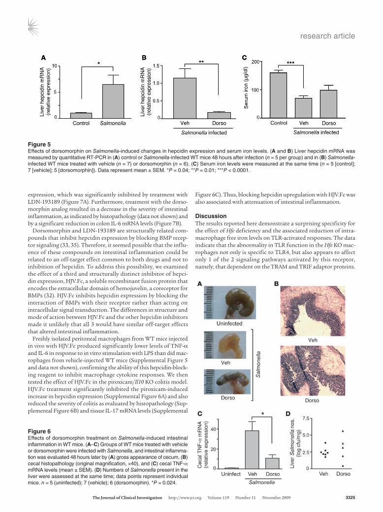
We found that oral infection of WT mice with S. typhimurium was associated with a significant increase in hepcidin expression in the liver (Figure 5A). Dorsomorphin treatment significantly inhibited the increase in liver hepcidin expression induced by Salmonella (Figure 5B) and also caused a partial reversal of the decrease in serum iron levels caused by the infection (Figure 5C). Most interestingly, dorsomorphin attenuated the severity of Sal-monella-induced intestinal inflammation, as indicated by multiple observations. As shown in Figure 6A, Salmonella infection of the vehicle-treated mice led to obvious shrinkage and edema of the cecum, whereas these changes were less severe in the dorsomor-phin-treated animals. In keeping with these differences, the cecal histopathology after Salmonella infection showed reduced inflam-matory cell infiltration and more normal-appearing epithelial architecture in the dorsomorphin-treated mice compared with the vehicle-treated animals (Figure 6B). Finally, dorsomorphin treatment significantly lowered levels of TNF-α mRNA in the ceca of the Salmonella-infected mice (Figure 6C). Dorsomorphin treatment did not have significant effects on the tissue Salmonella burden, although the number of bacteria recovered from the liver in the dorsomorphin-treated group was more variable than in the vehicle-treated animals (Figure 6D). Thus, inhibition of hepcidin upregulation by dorsomorphin was associated with a reduction in the in vivo inflammatory response to Salmonella without a major influence on bacterial replication.
The effect of inhibiting hepcidin expression on inflammation was not confined to acute infectious enterocolitis. We tested the effect of an analog of dorsomorphin, LDN-193189, in a model of chron-ic colitis induced in Il10 KO mice by administration of piroxicam (34). Piroxicam administration resulted in an increase in hepcidin
Figure 3Induction of IFN-β by activation of TLR4, but not TLR3, is abnormal in Hfe KO macrophages. WT and Hfe KO macrophages were stimulated with (A) 100 ng/ml LPS or (B) 10 μg/ml poly(I:C) for 6 hours (n = 3 per group). Supernatants were collected and analyzed by ELISA for IFN-β. Data represent mean ± SD. Similar results were obtained in 2 separate experiments. *P = 0.026; **P = 0.039.
Figure 4SIH inhibits LPS-induced, but not poly(I:C)-induced, expression of IFN-β in WT macrophages. WT macrophages were stimulated with 100 ng/ml LPS or 10 μg/ml poly(I:C) for 6 hours in the presence of the indicated concentrations of SIH (n = 3 per group). Supernatants were collected and analyzed by ELISA for IFN-β. Data represent mean ± SD. Similar results were obtained in 2 separate experiments. *P = 0.03.
research article
TheJournalofClinicalInvestigation http://www.jci.org Volume 119 Number 11 November 2009 3325
expression, which was significantly inhibited by treatment with LDN-193189 (Figure 7A). Furthermore, treatment with the dorso-morphin analog resulted in a decrease in the severity of intestinal inflammation, as indicated by histopathology (data not shown) and by a significant reduction in colon IL-6 mRNA levels (Figure 7B).
Dorsomorphin and LDN-193189 are structurally related com-pounds that inhibit hepcidin expression by blocking BMP recep-tor signaling (33, 35). Therefore, it seemed possible that the influ-ence of these compounds on intestinal inflammation could be related to an off-target effect common to both drugs and not to inhibition of hepcidin. To address this possibility, we examined the effect of a third and structurally distinct inhibitor of hepci-din expression, HJV.Fc, a soluble recombinant fusion protein that encodes the extracellular domain of hemojuvelin, a coreceptor for BMPs (32). HJV.Fc inhibits hepcidin expression by blocking the interaction of BMPs with their receptor rather than acting on intracellular signal transduction. The differences in structure and mode of action between HJV.Fc and the other hepcidin inhibitors made it unlikely that all 3 would have similar off-target effects that altered intestinal inflammation.
Freshly isolated peritoneal macrophages from WT mice injected in vivo with HJV.Fc produced significantly lower levels of TNF-α and IL-6 in response to in vitro stimulation with LPS than did mac-rophages from vehicle-injected WT mice (Supplemental Figure 5 and data not shown), confirming the ability of this hepcidin-block-ing reagent to inhibit macrophage cytokine responses. We then tested the effect of HJV.Fc in the piroxicam/Il10 KO colitis model. HJV.Fc treatment significantly inhibited the piroxicam-induced increase in hepcidin expression (Supplemental Figure 6A) and also reduced the severity of colitis as evaluated by histopathology (Sup-plemental Figure 6B) and tissue IL-17 mRNA levels (Supplemental
Figure 6C). Thus, blocking hepcidin upregulation with HJV.Fc was also associated with attenuation of intestinal inflammation.
DiscussionThe results reported here demonstrate a surprising specificity for the effect of Hfe deficiency and the associated reduction of intra-macrophage free iron levels on TLR-activated responses. The data indicate that the abnormality in TLR function in the Hfe KO mac-rophages not only is specific to TLR4, but also appears to affect only 1 of the 2 signaling pathways activated by this receptor, namely, that dependent on the TRAM and TRIF adaptor proteins.
Figure 5Effects of dorsomorphin on Salmonella-induced changes in hepcidin expression and serum iron levels. (A and B) Liver hepcidin mRNA was measured by quantitative RT-PCR in (A) control or Salmonella-infected WT mice 48 hours after infection (n = 5 per group) and in (B) Salmonella-infected WT mice treated with vehicle (n = 7) or dorsomorphin (n = 6). (C) Serum iron levels were measured at the same time (n = 5 [control]; 7 [vehicle]; 5 [dorsomorphin]). Data represent mean ± SEM. *P = 0.04; **P = 0.01; ***P < 0.0001.
Figure 6Effects of dorsomorphin treatment on Salmonella-induced intestinal inflammation in WT mice. (A–C) Groups of WT mice treated with vehicle or dorsomorphin were infected with Salmonella, and intestinal inflamma-tion was evaluated 48 hours later by (A) gross appearance of cecum, (B) cecal histopathology (original magnification, ×40), and (C) cecal TNF-α mRNA levels (mean ± SEM). (D) Numbers of Salmonella present in the liver were assessed at the same time; data points represent individual mice. n = 5 (uninfected); 7 (vehicle); 6 (dorsomorphin). *P = 0.024.

research article
3326 TheJournalofClinicalInvestigation http://www.jci.org Volume 119 Number 11 November 2009
Furthermore, based on what is known about TLR signaling and on our finding that upregulation of IFN-β in the Hfe KO macrophages was impaired in response to LPS but not poly(I:C), the simplest interpretation of the data is that Hfe deficiency and the associated decrease in intramacrophage iron levels negatively impact the MyD88-independent signaling pathway at a step unique to TLR4, that is, proximal to TRIF (Figure 8). Such an abnormality would also explain the reduction in Salmonella- and LPS-induced produc-tion of TNF-α and IL-6 in the Hfe KO macrophages reported here and in our earlier work (25), because both TRAM and TRIF are required for expression of these cytokines (28, 29).
Among the TLRs, TLR4 is unique in being connected to both Mal/MyD88 and TRAM/TRIF signaling pathways (26, 27). Upon activa-tion by ligand, TLR4 engages the 2 sets of adaptor proteins sequen-tially. Mal and MyD88 are recruited to the TLR4 cytoplasmic domain at the plasma membrane, but the interaction with TRAM and subse-quently TRIF requires endocytosis of the receptor (36). Thus, there are several aspects of the TLR4-specific activation of TRIF-dependent signals that could be adversely affected by the low intramacrophage iron levels associated with Hfe deficiency, including TLR4 endocyto-sis, TLR4/TRAM interactions, TRAM expression, and TRAM/TRIF interactions. The list of possibilities is further extended by the fact that TRAM is known to undergo posttranslational myristoylation, a modification that is required for correct subcellular localization and function (36, 37). Although the Hfe KO macrophages had a mod-est reduction in cell surface TLR4 expression (Supplemental Figure 7A), we believe this is unlikely to be a major factor in the abnormal signaling, since activation of the ERK and p38 kinases was as robust as in the WT cells. Expression of the TRAM and TRIF adaptors also appeared to be normal in the Hfe KO macrophages, at least at the mRNA level (Supplemental Figure 7B). Further work will be required to determine exactly how low intracellular iron inhibits TLR4-acti-vated signal transduction via the TRAM/TRIF pathway.
Regardless of the precise mechanism of action, it is clear that the low intramacrophage iron levels associated with Hfe defi-ciency have substantial impact on the TLR4-induced production of various inflammatory cytokines. Furthermore, the results of our experiments with the inhibitors of hepcidin expression indi-cate that the acute, hepcidin-mediated changes in iron homeo-stasis that occur during infection can also influence inflamma-tory responses. Specifically, the increase in intramacrophage iron caused by hepcidin-induced FPN downregulation could be considered to act as a proinflammatory signal that facilitates expression of cytokines such as TNF-α. This idea provides a teleologic explanation for the increase in hepcidin stimulated by inflammatory signals: the response may have protective value in terms of ramping up macrophage activation. Our findings also raise the possibility that blocking hepcidin expression may rep-resent a novel approach to controlling inflammation. Dorsomor-phin, LDN-193189, and HJV.Fc treatment reduced the severity of intestinal inflammation in models of acute infectious colitis and chronic noninfectious colitis. Although we cannot definitively exclude the possibility that the attenuation of inflammation represents the outcome of effects other than those on hepcidin expression, we think it is unlikely in the context of our observa-tions with the Hfe KO mice, and given that HJV.Fc and the other 2 inhibitors are structurally distinct and inhibit BMP-mediated hepcidin expression by quite different mechanisms (32, 33, 35).
Figure 7LDN-193189 suppresses hepcidin upregulation and intestinal inflam-mation in piroxicam-induced colitis. Il10 KO mice were treated with piroxicam for 2 weeks and then injected with vehicle or LDN-193189 for an additional week. The animals were sacrificed, and (A) liver hepcidin mRNA (n = 3 [untreated and vehicle]; 4 [LDN-193189]) and (B) colon IL-6 mRNA (n = 9 [vehicle]; 12 [LDN-193189]) were measured by quantitative RT-PCR. Data represent mean ± SEM. *P = 0.005; **P = 0.0007.
Figure 8Influence of low intracellular iron on LPS-induced cytokine expression. Based on the effects of Hfe deficiency and iron chelation on responses to TLR2, TLR3, and TLR4 stimulation, low intracellular iron is likely to impair TLR4 signaling at a step in the TRAM/TRIF pathway proximal to TRIF. IRF3, IFN regulatory factor 3.

research article
TheJournalofClinicalInvestigation http://www.jci.org Volume 119 Number 11 November 2009 3327
Iron chelation therapy with agents such as desferrioxamine has been tried previously for chronic inflammatory conditions, such as rheumatoid arthritis, and limited success has been reported in some studies (38). Inhibition of hepcidin expression or func-tion could have advantages over the nonspecific reduction of iron produced by chelators since its effects would be confined largely to FPN-expressing cells such as macrophages. In addition, the increase in FPN expression that would follow reduced circulat-ing hepcidin levels would increase iron availability and help to correct the anemia associated with many longstanding inflam-matory states (39). Our results suggest that this strategy may be worth further investigation.
The findings presented here, as well those we have reported previously (25), strongly support the idea that low intracellular iron levels play an important role in the abnormal TLR4 signaling and cytokine response of the Hfe KO macrophages. However, it has been shown recently that the hepcidin-FPN interaction leads to activation of the Jak2 kinase (40). This study implicated Jak2-dependent phosphorylation of FPN in downregulation of the receptor, but it is also possible that downstream signals activated by Jak2 could influence gene expression and, therefore, that defi-ciency of such signals could contribute to the abnormal cytokine production by macrophages from Hfe KO mice. This is a possibil-ity that deserves exploration.
In conclusion, we have shown that Hfe deficiency and the asso-ciated decrease in intramacrophage iron have a negative influ-ence on signaling through the TRAM/TRIF pathway activated by TLR4, probably at a step proximal to TRIF. Our findings reveal what we believe to be a novel, iron-dependent mechanism that modulates TLR4 signals; moreover, they provide an explanation for the attenuated in vitro and in vivo inflammatory responses of Hfe KO mice (25) and, potentially, for the increased suscepti-bility of individuals with hemochromatosis to certain bacterial infections (22–24). We have also shown that manipulation of iron homeostasis may represent a novel strategy for controlling inflammation. Further investigation of the interactions between iron metabolism and inflammatory responses will allow for refinement of this therapeutic approach.
MethodsAnimals. WT C57BL/6 mice were obtained from the Jackson Laboratory. Hfe KO mice on the C57BL/6 background were originally provided by N.C. Andrews (Duke University, Durham, North Carolina, USA; ref. 20). All mice were bred and housed in a specific pathogen–free facility at Massa-chusetts General Hospital. Animals were given water and standard labora-tory chow ad libitum and used at 7–12 weeks of age. All animal experiments were approved by the Massachusetts General Hospital Subcommittee on Research Animal Care.
Macrophage stimulation. Thioglycollate-elicited peritoneal macrophages were prepared as previously described (25, 41) and stimulated with 100 ng/ml LPS (Ultra-pure; List Biological Laboratories), 1 μg/ml of the synthetic bacterial lipopeptide Pam3CSK4, or 10 μg/ml poly(I:C) (both from Invi-vogen) for the indicated times in triplicate wells of a 24-well tissue culture plate. SIH was provided by P. Ponka (McGill University, Montreal, Quebec, Canada) and was added to macrophage cultures 30 minutes before addi-tion of TLR ligands. Cell supernatants were analyzed by ELISA for various cytokines using specific antibody pairs obtained from R&D Systems or BD Biosciences — Pharmingen. Total RNA was prepared from the cells using TRIzol reagent (Invitrogen), and quantitative RT-PCR was carried out to determine levels of cytokine mRNA according to previously described
methods (25, 41). Primers used for amplification of TNF-α, IL-6, and the housekeeping transcript 36B4 have been described previously (25). Other primers used were as follows: IFN-β sense, 5′-CAGCTCCAAGAAAGGAC-GAAC-3′; IFN-β antisense, 5′-GGCAGTGTAACTCTTCTGCAT-3′; TRAM sense, 5′-CGATCAAGACGGCCATGAGTC-3′; TRAM antisense, 5′-CTC-GTCGGTGTCATCTTCTGC-3′; TRIF sense, 5′-AACCTCCACATCCCCT-GTTTT-3′; TRIF antisense, 5′-GCCCTGGCATGGATAACCA-3′. Immu-noblotting procedures used for analysis of MAP kinase activation and IκBα degradation have been described in detail elsewhere (41). Bone mar-row–derived macrophages were prepared by growing bone marrow cells in medium containing 10% L-929–conditioned medium for 7 days. The cells were then stimulated and analyzed as described above.
Flow cytometry. Thioglycollate-elicited peritoneal macrophages were stained with a phycoerythrin-conjugated anti-TLR4 antibody (clone UT41; eBioscience) and subjected to flow cytometry on a FACScan (BD) as previ-ously described (42). The assay for intracellular iron based on iron-depen-dent quenching of calcein fluorescence has been described previously (25).
Salmonella-induced enterocolitis. Following the protocol described by Bar-thel et al. (43), mice were given a single 20-mg oral dose of streptomycin followed 24 hours later by oral infection with 108 cfu of the streptomy-cin-resistant, WT, invasion-competent SL1344 strain of S. typhimurium. Treatment with dorsomorphin (12.5 μg/g BW i.p.; Calbiochem) or an equivalent volume of the vehicle DMSO was started at the same time as the streptomycin was administered and was continued twice daily for the duration of the experiment. Animals were sacrificed 48 hours after infec-tion. At necropsy, intestinal inflammation was assessed by gross appear-ance of the cecum, cecal histopathology, and quantitative RT-PCR–based measurement of cecal TNF-α, all as previously described in detail (25). Liver hepcidin expression was determined by quantitative RT-PCR with the following primers: sense, 5′-AGAGCTGCAGCCTTTGCAC-3′, antisense, 5′-GAAGATGCAGATGGGGAAGT-3′. Bacterial burden was assessed by homogenizing weighed portions of liver in sterile 1% Triton X-100 and plating serial dilutions of the homogenates on LB agar contain-ing 50 μg/ml streptomycin.
Piroxicam-induced colitis in Il10 KO mice. The protocol described by Berg et al. was followed (34). In brief, 4- to 6-week-old Il10 KO mice (C57BL/6 background, breeders obtained from Jackson Laboratory) were fed with 60 mg piroxicam (Sigma-Aldrich) mixed with 250 g NIH-31M chow for 1 week, followed by 80 mg piroxicam plus 250 g chow for an additional 1 week. The animals were then placed on their regular diet and treated with either LDN-193189 or HJV.Fc over the course of the following week. LDN-193189 was custom synthesized, as described by Cuny et al. (35), by Shang-hai United Pharmatec and dissolved at a concentration of 0.25 mg/ml in 2% 2-hydroxypropyl-β-cyclodextrin. Mice were injected i.p. twice daily with 3 mg/kg BW LDN-193189 or with an equivalent volume of the vehicle. HJV.Fc (prepared as described in ref. 32) was injected i.p. 3 times over the course of a week at a dose of 15 mg/kg BW. Control animals were injected with a corresponding volume of PBS vehicle. The animals were sacrificed, and colonic inflammation was evaluated by histopathology. An investiga-tor blinded to the identity of the samples examined the hematoxylin and eosin–stained sections and graded the severity of inflammation based on a previously described scoring system (44). Liver hepcidin expression was determined by quantitative RT-PCR at the same time.
Statistics. Results were compared by 2-tailed Student’s t test or Mann-Whitney U test. A P value less than 0.05 was considered significant. n refers to number of individual mice or stimulations.
AcknowledgmentsThis work was supported by funds from the Broad Medical Research Program (grant no. IBD-0253) to Bobby J. Cherayil and from Wyeth

research article
3328 TheJournalofClinicalInvestigation http://www.jci.org Volume 119 Number 11 November 2009
Nutrition to L. Wang. C.C. Hong received support from the Center for Research in Fibrodysplasia Ossificans Progressiva and Related Disorders. H.Y. Lin was supported in part by grant R01-071837 from the National Institute of Diabetes and Digestive and Kidney Diseases, NIH. We are grateful to Nancy Andrews for providing the Hfe KO mice and to Prem Ponka for the gift of SIH. We thank Sarah A. Faasse for technical assistance with iron assays.
Received for publication May 21, 2009, and accepted in revised form August 19, 2009.
Address correspondence to: Bobby J. Cherayil, Mucosal Immunol-ogy Laboratory, Massachusetts General Hospital, Building 114, 16th Street, Charlestown, Massachusetts 02129, USA. Phone: (617) 726-4170; Fax: (617) 726-4172; E-mail: [email protected].
1. Schaible, U.E., and Kaufmann, S.H. 2004. Iron and microbial infection. Nat. Rev. Microbiol. 2:946–953.
2. Gangaidzo, I.T., et al. 2001. Association of pul-monary tuberculosis with increased dietary iron. J. Infect. Dis. 184:936–939.
3. Gordeuk, V.R., McLaren, C.E., MacPhail, A.P., Deichsel, G., and Bothwell, T.H. 1996. Associa-tions of iron overload in Africa with hepatocellular carcinoma and tuberculosis: Strachan’s 1929 thesis revisited. Blood. 87:3470–3476.
4. Magnus, S.A., Hambleton, I.R., Moosdeen, F., and Serjeant, G.R. 1999. Recurrent infections in homozygous sickle cell disease. Arch. Dis. Child. 80:537–541.
5. Moyo, V.M., Gangaidzo, I.T., Gordeuk, V.R., Kiire, C.F., and Macphail, A.P. 1997. Tuberculosis and iron overload in Africa: a review. Cent. Afr. J. Med. 43:334–339.
6. Wanachiwanawin, W. 2000. Infections in E-beta thalassemia. J. Pediatr. Hematol. Oncol. 22:581–587.
7. Murray, M.J., Murray, A.B., Murray, M.B., and Murray, C.J. 1978. The adverse effect of iron reple-tion on the course of certain infections. Br. Med. J. 2:1113–1115.
8. Nyakeriga, A.M., et al. 2004. Iron deficiency and malaria among children living on the coast of Kenya. J. Infect. Dis. 190:439–447.
9. Sazawal, S., et al. 2006. Effects of routine prophy-lactic supplementation with iron and folic acid on admission to hospital and mortality in preschool children in a high malaria transmission setting: community-based, randomized, placebo-controlled trial. Lancet. 367:133–143.
10. Puschmann, M., and Ganzoni, A.M. 1977. Increased resistance of iron-deficient mice to Salmonella infec-tion. Infect. Immun. 17:663–664.
11. Wang, L., and Cherayil, B.J. 2009. Ironing out the wrinkles in host defense: interactions between iron homeostasis and innate immunity. J. Innate Immun. 1:455–464.
12. Pietrangelo, A. 2006. Hereditary hemochromatosis. Annu. Rev. Nutr. 26:251–270.
13. Feder, J.N., et al. 1996. A novel MHC class I-like gene is mutated in patients with hereditary hae-mochromatosis. Nat. Genet. 13:399–408.
14. Andrews, N.C. 2007. Iron homeostasis. Annu. Rev. Physiol. 69:69–85.
15. Nemeth, E., and Ganz, T. 2006. Regulation of iron metabolism by hepcidin. Annu. Rev. Nutr. 26:323–342.
16. Goswami, T., and Andrews, N.C. 2006. Hereditary hemochromatosis protein, HFE, interaction with
transferrin receptor 2 suggests a molecular mecha-nism for mammalian iron sensing. J. Biol. Chem. 281:28494–28498.
17. Nemeth, E., et al. 2004. Hepcidin regulates cellular iron efflux by binding ferroportin and inducing its degradation. Science. 306:2090–2093.
18. Ahmad, K.A., et al. 2002. Decreased liver hepcidin expression in the Hfe knockout mouse. Blood Cells Mol. Dis. 29:361–366.
19. Bahram, S., et al. 1999. Experimental hemochroma-tosis due to MHC class I HFE deficiency: immune status and iron metabolism. Proc. Natl. Acad. Sci. U. S. A. 96:13312–13317.
20. Levy, J.E., Montross, L.K., Cohen, D.E., Fleming, M.D., and Andrews, N.C. 1999. The C282Y muta-tion causing hereditary hemochromatosis does not produce a null allele. Blood. 94:9–11.
21. Zhou, X.Y., et al. 1998. HFE gene knock-out pro-duces a mouse model of hereditary hemochroma-tosis. Proc. Natl. Acad. Sci. U. S. A. 95:2492–2497.
22. Bullen, J.J., Spalding, P.B., Ward, C.G., and Gutter-idge, J.M. 1991. Hemochromatosis, iron and septi-cemia caused by Vibrio vulnificus. Arch. Intern. Med. 151:1606–1609.
23. Doherty, C.P. 2007. Host-pathogen interactions: the role of iron. J. Nutr. 137:1341–1344.
24. Gerhard, G.S., et al. 2001. Vibrio vulnificus septicemia in a patient with the hemochromatosis HFE C282Y mutation. Arch. Pathol. Lab. Med. 125:1107–1109.
25. Wang, L., et al. 2008. Attenuated inflammatory responses in hemochromatosis reveal a role for iron in the regulation of macrophage cytokine translation. J. Immunol. 181:2723–2731.
26. O’Neill, L.A. 2008. The interleukin-1/Toll-like receptor superfamily:10 years of progress. Immunol. Rev. 226:10–18.
27. Kawai, T., and Akira, S. 2008. Toll-like receptor and RIG-I receptor signaling. Ann. N. Y. Acad. Sci. 1143:1–20.
28. Yamamoto, M., et al. 2003. Role of adaptor TRIF in the MyD88-independent Toll-like receptor signal-ing pathway. Science. 301:640–643.
29. Yamamoto, M., et al. 2003. TRAM is specifi-cally involved in the Toll-like receptor 4-medi-ated MyD88-independent signaling pathway. Nat. Immunol. 4:1144–1150.
30. Simunek, T., et al. 2005. SIH – a novel lipophilic iron chelator – protects H9c2 cardiomyoblasts from oxidative stress-induced mitochondrial injury and cell death. J. Mol. Cell. Cardiol. 39:345–354.
31. Babitt, J.L., et al. 2006. Bone morphogenetic pro-tein signaling by hemojuvelin regulates hepcidin
expression. Nat. Genet. 38:531–539. 32. Babitt, J.L., et al. 2007. Modulation of bone mor-
phogenetic protein signaling in vivo regulates sys-temic iron balance. J. Clin. Invest. 117:1933–1939.
33. Yu, P.B., et al. 2008. Dorsomorphin inhibits BMP signals required for embyrogenesis and iron metab-olism. Nat. Chem. Biol. 4:33–41.
34. Berg, D.J., et al. 2002. Rapid development of colitis in NSAID-treated IL10-deficient mice. Gastroenter-ology. 123:1527–1542.
35. Cuny, G.D., et al. 2008. Structure-activity relation-ship study of bone morphogenetic protein signaling inhibitors. Bioorg. Med. Chem. Lett. 18:4388–4392.
36. Kagan, J.C., et al. 2008. TRAM couples endocytosis of Toll-like receptor 4 to the induction of interfer-on-beta. Nat. Immunol. 9:361–368.
37. Rowe, D.C., et al. 2006. The myristoylation of TRIF-related adaptor molecule is essential for Toll-like receptor 4 signal transduction. Proc. Natl. Acad. Sci. U. S. A. 103:6299–6304.
38. Magaro, M., et al. 1990. Iron chelation in rheuma-toid arthritis: clinical and laboratory evaluation. Ann. Rheum. Dis. 49:268–269.
39. Weinstein, D.A., et al. 2002. Inappropriate expres-sion of hepcidin is associated with iron refractory anemia: implications for the anemia of chronic dis-ease. Blood. 100:3776–3781.
40. De Domenico, I., Lo, E., Ward, D.M., and Kaplan, J. 2009. Hepcidin-induced internalization of ferroportin requires binding and cooperative interaction with Jak2. Proc. Natl. Acad. Sci. U. S. A. 106:3800–3805.
41. Li, Q., and Cherayil, B.J. 2003. Role of Toll-like receptor 4 in macrophage activation and tolerance during Salmonella enterica serovar Typhimurium infection. Infect. Immun. 71:4873–4882.
42. Rhee, S.J., Walker, W.A., and Cherayil, B.J. 2005. Developmentally regulated intestinal expression of IFNγ and its target genes and the age-specific response to enteric Salmonella infection. J. Immunol. 175:1127–1136.
43. Barthel, M., et al. 2003. Pretreatment of mice with streptomycin provides a Salmonella enterica serovar Typhimurium colitis model that allows analysis of both pathogen and host. Infect. Immun. 71:2839–2858.
44. Chen, C.C., Louie, S., McCormick, B., Walker, W.A., and Shi, H.N. 2005. Concurrent infection with an intestinal helminth parasite impairs host resis-tance to enteric Citrobacter rodentium and enhances Citrobacter-induced colitis in mice. Infect. Immun. 73:5468–5481.
Copyright © 2022 FDOKUMEN