
d(GGGT)4 and r(GGGU)4 are both HIV-1 inhibitors and interleukin-6 receptor aptamers
Nucleic Acids Research, 2009, 1–16doi:10.1093/nar/gkp185
Selection, characterization and application of newRNA HIV gp 120 aptamers for facile delivery ofDicer substrate siRNAs into HIV infected cellsJiehua Zhou1, Piotr Swiderski2, Haitang Li1, Jane Zhang3, C. Preston Neff4,
Ramesh Akkina4 and John J. Rossi1,3,*
1Division of Molecular Biology, 2Shared Resource-DNA/RNA Peptide, 3Graduate School of Biological Sciences,Beckman Research Institute of City of Hope, City of Hope, Duarte, CA 91010 and 4Department of Microbiology,Immunology and Pathology, Colorado State University, Fort Collins, CO 80523-1619, USA
Received February 9, 2009; Revised March 5, 2009; Accepted March 8, 2009
ABSTRACT
The envelope glycoprotein of human immunodefi-ciency virus (HIV) consists of an exterior glycopro-tein (gp120) and a trans-membrane domain (gp41)and has an important role in viral entry into cells.HIV-1 entry has been validated as a clinically rele-vant anti-viral strategy for drug discovery. In thepresent work, several 2’-F substituted RNA apta-mers that bind to the HIV-1BaL gp120 protein withnanomole affinity were isolated from a RNA libraryby the SELEX (Systematic Evolution of Ligands byEXponential enrichment) procedure. From two ofthese aptamers we created a series of new dualinhibitory function anti-gp120 aptamer–siRNA chi-meras. The aptamers and aptamer–siRNA chimerasspecifically bind to and are internalized into cellsexpressing HIV gp160. The Dicer-substrate siRNAdelivered by the aptamers is functionally processedby Dicer, resulting in specific inhibition of HIV-1replication and infectivity in cultured CEM T-cellsand primary blood mononuclear cells (PBMCs).Moreover, we have introduced a ‘sticky’ sequenceonto a chemically synthesized aptamer which facil-itates attachment of the Dicer substrate siRNAs forpotential multiplexing. Our results provide a set ofnovel inhibitory agents for blocking HIV replicationand further validate the use of aptamers for deliveryof Dicer substrate siRNAs.
INTRODUCTION
The global epidemic of infection by HIV has createdan urgent need for new classes of antiretroviral agents.
HIV-1 infection is initiated by the interactions betweenthe external envelope glycoprotein gp120 of HIV and thehuman cell surface receptor CD4 (1–4), subsequently lead-ing to fusion of the viral membrane with the target cellmembrane (5,6). Thus, HIV-1 entry into its target cellsrepresents an attractive target for new anti-HIV-1 drugdevelopment (7–12).The most effective anti-HIV drugs used in the clinic
consist of combinations of compounds that block theearly reverse transcription step, and the late viral gag pro-tein maturation step (13–15). Preventing the virus frombinding to its primary receptor is one of the most obviousand direct ways to prevent infection. One rational antiviralapproach is to create some small ligands directed tothe virus surface protein gp120. Successes in discoveringnew classes of gp120-directed inhibitors targeted to theinitial step of HIV-1 entry have been reported in recentyears (8,16,17). Soluble CD4 (sCD4) has demonstratedefficacy against many laboratory strains (18,19), howeverdisappointing results from clinical trials were attributedto poor activity of this drug against primary isolates(20,21). Another entry inhibitor PRO-542, a CD4-IgGrecombinant fusion protein effectively neutralizes manyHIV-1 strains in cell culture (22,23) as well as clinical iso-lates (24,25), but unfortunately this drug has not enjoyedwidespread use in the clinical treatment of HIV infection.Other inhibitors bind gp120 less specifically through elec-trostatic or lectin–carbohydrate interactions [sulfatedpolymers, cyanovirin (26)]. These properties createchallenges for the practical implementation of these mole-cules as drugs. In 2003, Martin et al. (27) designed a 27-aaCD4 peptide mimic, CD4M33, which was shown to bindto gp120 and inhibit HIV-1 infection in vitro. The clinicalapplication of this peptide though has not yet takenplace. Aside from antibody or peptide mimics, nucleicacid aptamers with low to mid-range nanomole binding
*To whom correspondence should be addressed. Tel: +1 626 301 8360; Fax: +1 626 301 8271; Email: [email protected]
� 2009 The Author(s)This is an Open Access article distributed under the terms of the Creative Commons Attribution Non-Commercial License (http://creativecommons.org/licenses/by-nc/2.0/uk/) which permits unrestricted non-commercial use, distribution, and reproduction in any medium, provided the original work is properly cited.
Nucleic Acids Research Advance Access published March 21, 2009 by guest on D
ecember 15, 2013
http://nar.oxfordjournals.org/D
ownloaded from
affinities are an attractive class of therapeutic molecules(27–30). Aptamers which block the interaction ofgp120 and CD4 have been previously developed. Onesuch aptamer was shown to specifically interact withthe conserved coreceptor interacting region of R5 straingp120 (29).Although both aptamers and antibody or engineered
peptide mimics have great specificity and binding affinity,the nucleic acid-based aptamers offer more syntheticaccessibility, convenient modification, chemical versatility,stability and lack immunogenicity (31). Therefore,aptamers can be utilized for flexible applications rangingfrom diagnostic to therapeutic assay formats (32,33).Aptamers that target specific cell surface proteins areemployed as interesting delivery molecules to target adistinct cell type, hence reducing off-target effects orother unwanted side effects. The first example of aptamersused for siRNA delivery is an anti-PSMA aptamer (34)that binds tightly to the prostate-specific membrane anti-gen (PSMA), a prostate cancer cell-surface receptor.McNamara et al. (35) in 2006 reported the use of thisaptamer for receptor mediated siRNA delivery. In thisstudy, they selectively delivered a 21-mer siRNA intoPSMA expressing cells, resulting in silencing of targettranscripts both in cell culture and in vivo followingintra-tumoral delivery. Similarly, Chu et al. (36) used amodular streptavidin bridge to connect lamin A/C orGAPDH siRNAs to the biotinylated variants of thisanti-PSMA aptamer. Consequently, this system inducedsilencing of the targeted genes only in cells expressingthe PSMA receptor. The targeting properties of the anti-PSMA aptamer also can be exploited for localizingother therapeutic agents to tumors such as a toxin (37),doxorubicin (38) or nanoparticles (39–42). We recentlydescribed a novel dual inhibitory function anti-gp120aptamer-siRNA chimera, in which both the aptamer andthe siRNA portions have potent anti-HIV activities (43).Additionally, HIV gp120 expressed on the surface of HIVinfected cells was used for aptamer-mediated delivery ofan anti-HIV siRNA, resulting in pronounced inhibitionof HIV replication in cell culture.For treatment of HIV using aptamer–siRNA chimeras,
it is highly desirable to generate new aptamers toexpand the diversity of target recognition for potentialuse in vivo. New anti-gp120 aptamers and varioussiRNAs targeting different genes could be combined toavert viral resistance to a single aptamer–siRNA combina-tion. In the present study we successfully isolated sev-eral new 20-F substituted RNA aptamers that bind tothe HIV-1Ba-L gp120 protein. Two different types of dualinhibitory function anti-gp120 aptamer–siRNA conju-gates were constructed. One of these is a covalent apta-mer–siRNA chimera and the other is an aptamer witha GC-rich bridge that facilitates the interchange of differ-ent RNAs with the same aptamer. Although the aptameralone provided HIV inhibitory function, the aptamer–siRNA chimeras provided more potent inhibition ofHIV suggesting cooperativity between the siRNA and
aptamer portions in inhibiting HIV replication andspread. Our results demonstrate that our selected apta-mers not only provide lead inhibitors for potential anti-HIV therapeutic applications, but also act as effectivedelivery vehicles for anti-HIV siRNAs into HIV infectedcells. The aptamers studies here could also selectively deli-ver other small HIV inhibitory agents.
MATERIALS AND METHODS
Materials
Unless otherwise noted, all chemicals were purchasedfrom Sigma-Aldrich, all restriction enzymes wereobtained from New England BioLabs (NEB) and allcell culture products were purchased from GIBOC(Gibco BRL/Life Technologies, a division of Invitrogen).Sources for the other reagents were: DuraScribe T7 tran-scription Kit (EPICENTRE Biotechnologies); SilencersiRNA Labeling Kit (Ambion); Hoechst 33342 (nucleardye for live cells) (Molecular Probes, Invitrogen);Random primers (Invitrogen); Bio-Spin 30 Columns(Bio-Rad); Recombinant Human Dicer Enzyme Kit(Ambion); Lipofectamine 2000 (Invitrogen); CHO-EnvTransfectants (CHO-WT and CHO-EE), the HIV-1BaLgp120 protein and HIV-1BaL virus were obtained fromthe AIDS Research and Reference Reagent Program.
siRNAs
siRNAs and antisense strand RNAs were purchased fromIntegrated DNA Technologies (IDT).
Generation of aptamer and chimera RNAs byin vitro transcription
Aptamer and chimera RNAs were prepared as previouslydescribed (43). The sense strands of the chimeras areunderlined. The italic UU is the linker between the apta-mer and siRNA portions.
A-1 aptamer: 50-GGGAGGACGAUGCGGAAUUGAGGGACCACGCGCUGCUUGUUGUGAUAAGCAGUUUGUCGUGAUGGCAGACGACUCGCCCGA-30
B-68 aptamer: 50-GGGAGGACGAUGCGGACAUAGUAAUGACACGGAGGAUGGAGAAAAAACAGCCAUCUCUUGACGGUCAGACGACUCGCCCGA-30
Chimera A-1-sense strand: 50-GGGAGGACGAUGCGGAAUUGAGGGACCACGCGCUGCUUGUUGUGAUAAGCAGUUUGUCGUGAUGGCAGACGACUCGCCCGA UU GCGGAGACAGCGACGAAGAGCUCAUCA-30
Chimera B-68-sense strand: 50-GGGAGGACGAUGCGGACAUAGUAAUGACACGGAGGAUGGAGAAAAAACAGCCAUCUCUUGACGGUCAGACGACUCGCCCGA UU GCGGAGACAGCGACGAAGAGCUCAUCA-30
2 Nucleic Acids Research, 2009
by guest on Decem
ber 15, 2013http://nar.oxfordjournals.org/
Dow
nloaded from

Antisense strand: 50-UGAUGAGCUCUUCGUCGCUGUCUCCGCdTdT-30
Aptamer-Stick-siRNA
A-1-stick, Stick-sense and stick-antisense were chemicallysynthesized by the Synthetic and Biopolymer ChemistryCore in the City of Hope (the details shown in theSupplementary Material). The A-1-stick RNA wasrefolded in HBS buffer (10mM HEPES pH 7.4, 150mMNaCl, 1mM CaCl2, 1mM MgCl2, 2.7mM KCl), heatedto 958C for 3min and then slowly cooled to 378C. Theincubation was continued at 378C for 10min. The Sense-stick or Antisense-stick strand was annealed to thecomplementary partner using the same molar amountsas the corresponding partner strand to form the stick-siRNAs (NS-1 and NS-2). The same amount of therefolded A-1-stick was added and incubated at 378Cfor 10min in HBS buffer to form the A-1-stick-siRNA(N-1 and N-2).
A-1-stick: 50-GGGAGGACGAUGCGGAAUUGAGGGACCACGCGCUGCUUGUUGUGAUAAGCAGUUUGUCGUGAUGGCAGACGACUCGCCCGAXXXXXXX GUACAUUCUAGAUAGCC-30
Tat/rev N-1:Antisense-Stick: 50-UGAUGAGCUCUUCGUCGCUG
UCUCCGC XXXXX GGCUAUCUAGAAUGUAC-30
Sense strand: 50-GCGGAGACAGCGACGAAGAGCUCAUCAUU-30
Tat/rev N-2:Stick-sense: 50-GCGGAGACAGCGACGAAGAGCUC
AUCA XXXXX GGCUAUCUAGAAUGUAC-30
Antisense strand: 50-UGAUGAGCUCUUCGUCGCUGUCUCCGCUU-30
TNPO3N-1:Antisense-stick: 50-UACACGAGCUGCAAUGUCGG
CUUUGCU XXXXX GGCUAUCUAGAAUGUAC -30
Sense stand: 50-CAAAGCCGACAUUGCAGCUCGUGUAUU-30
CD4N-2:Stick-sense: 50-UCAAGAGACUCCUCAGUGAGAAG
AA XXXXX GGCUAUCUAGAAUGUAC-30
Antisense strand: 50-UUCUUCUCACUGAGGAGUCUCUUGAUU-30
The bold nucleotides indicate 20-Flourine modifiedsugars. The stick portion is underlined. This contains20-OMe modified A and G and 20-F modified U and C.The italic X indicates the three-carbon linker (C3) betweenthe aptamer/siRNA and stick sequences.
Preparation of the RNA library
The starting DNA library contained 50 nucleotides ofrandom sequences and was synthesized by IntegratedDNA Technologies (Coralville, Iowa). The randomregion is flanked by constant regions, which include
the T7 promoter for in vitro transcription and a 30 tagfor RT–PCR. The 50 and 30 constant sequences are50-TAA TAC GAC TCA CTA TAG GGA GGA CGATGC GG-30 (32-mer) and 50-TCG GGC GAG TCG TCTG-30 (16-mer), respectively. The DNA random library(0.4 mM) was amplified by PCR using 3 mM each of 50-and 30-primers, along with 2mM MgCl2 and 200 mM ofeach dNTP. In order to preserve the abundance of theoriginal DNA library, PCR was limited to 10 cycles.After the PCR reactions (10 reactions, 100ml per reaction),the amplified dsDNA pool was recovered using aQIAquick Gel purification Kit. The resulting dsDNAwas converted to an RNA library using theDuraScription Kit (Epicentre, Madison, WI, USA)according to the manufacturer’s instructions. In thetranscription reaction mixture, CTP and UTP werereplaced with 20-F-CTP and 20-F-UTP to produce ribo-nuclease resistant RNA. The reactions were incubatedat 378C for 6 h, and subsequently the template DNAwas removed by DNase I digestion. The transcribedRNA pool was purified in an 8% polyacrylamide/7Murea gel. The purified RNA library was quantified byUV spectrophotometry.
In vitro selection of RNA aptamers
The SELEX was performed principally as described byTuerk and Gold (44). In every round, the RNA poolswere refolded in HBS buffer (10mM HEPES pH 7.4,150mM NaCl, 1mM CaCl2, 1mM MgCl2, 2.7mMKCl), heated to 958C for 3min and then slowly cooledto 378C. Incubation was continued at 378C for 10min.Generally, in order to minimize nonspecific binding
with the nitrocellulose filters, the refolded RNA poolswere preadsorbed to a nitrocellulose filter (HAWP filter,0.45mm) for 30min, prior to incubation with the HIV-1Balgp120 protein. The precleared RNA pool was incubatedwith the target protein in low-salt RNA binding buffer(10mM HEPES pH 7.4, 50mM NaCl, 1mM CaCl2,1mM MgCl2, 2.7mM KCl, 10mM DTT, 0.01% BSAand tRNA) for 30min for SELEX rounds 1 to 4. Afterthe fourth round of SELEX, a high-salt RNA bindingbuffer (10mM HEPES pH 7.4, 150mM NaCl, 1mMCaCl2, 1mM MgCl2, 2.7mM KCl, 10mM DTT, 0.01%BSA and tRNA) was used. With the SELEX progress, theamount of gp120 protein was reduced and competitortRNA was increased in order to increase the stringencyof aptamer selection.For the first cycle of selection, the precleared random
RNA pool (40 mg, 1.5 nmol, 9� 1014 molecules) andHIV-1Bal gp120 protein (0.23 nmol, RNA/Protein ratio6.5/1) were incubated in 200 ml low-salt RNA bindingbuffer on a rotating platform at room temperature for30min. The reaction was passed through a prewettednitrocellulose filter and washed with 1ml binding buffer.The bound RNA was eluted from the filter with 200 mlelution buffer (7M urea and 5mM EDTA) at 958Cfor 5min, followed by phenol/chloroform extractionand concentration with a Microcon YM-30 column.
Nucleic Acids Research, 2009 3
by guest on Decem
ber 15, 2013http://nar.oxfordjournals.org/
Dow
nloaded from
The recovered RNA pool was reversed transcribed usingthe ThermoScript RT-PCR system (Invitrogen) andamplified for 15 cycles of PCR. After the amplifieddsDNA pool was purified using a QIAquick Gel purifi-cation Kit, it was transcribed as described above for thenext round of selection.After 12 rounds of SELEX, the resulting cDNA was
amplified by PCR cloned into the TA cloning vectorpCR 2.1 (Invitrogen). Individual clones were identifiedby DNA sequencing.
Gel shift assays and determination of dissociation constants
The gp120 protein was serially diluted to the desiredconcentrations (0–640 nM). A constant amount ofP32-end-labeled RNA (10 nM) was used. The binding reac-tion was performed as described above. After incubation,20 ml of binding reaction was loaded into a 5% nondena-turing polyacrylamide gel. Following electrophoresisthe gel was exposed to a Phosphor image screen and theradioactivity was quantified using a Typhoon scanner.The dissociation constants were calculated using non-linear curve regression with a Graph Pad Prism.
Cell culture
HEK 293 cells and CEM cells were purchased from ATCCand cultured in DMEM and RPMI 1640 supplementedwith 10% FBS. CHO-WT and CHO-EE cells wereobtained through the AIDS Research and ReferenceReagent Program and were grown in GMEM-S. Cellswere cultured in a humidified 5% CO2 incubator at 378C.
PBMCs. Peripheral blood mononuclear samples wereobtained from healthy donors from the City of hopeNational Medical Center. PBMCs were isolated fromwhole blood by centrifugation through a Ficoll-Hypaque solution (Histopaque-1077, Sigma). CD8 cells(T-cytotoxic/suppressor cells) were depleted from thePBMCs by CD8 Dynabeads (Invitrogen, CA) accordingto the manufacturer’s instructions. CD8+ T cell-depletedPBMCs were washed twice in PBS and resuspended inculture media (RPMI 1640 with 10% FBS, 1 (PenStrepand 100U/ml interleukin-2). Cells were cultured in ahumidified 5% CO2 incubator at 378C.
Cell-surface binding of experimental RNAs(Flow cytometry analysis)
CHO-WT gp160 or CHO-EE cells were washed withPBS, trypsinized and detached from the plates. Afterwashing cells twice with 500 ml binding buffer. Cell pelletswere resuspended in binding buffer and incubated at378C for 30min. Cells were then pelleted and resuspendedin 50 ml of prewarmed binding buffer containing 400 nMCy3-labeled experimental RNAs. After incubation at 378Cfor 40min, cells were washed three times with 500 ml ofprewarmed binding buffer, and finally resuspended in350ml of binding buffer prewarmed to 378C and analyzedby flow cytometry. For the aptamer–stick–siRNA
(N-1 and N-2), the Cy3-labeled aptamer–stick was incu-bated with the siRNA–stick to form the aptamer–stick–siRNA complex and the complexes were analyzed by flowcytometry.
Internalization and intracellular localization studies(Live-cell Confocal Microscopy analyses)
The CHO-WT gp160 and CHO-EE cells were grown in35mm plate (Glass Bottom Dish, MatTek, Ashland, MA,USA) with seeding at 0.3� 106 in GMEM-S medium toallow about 70% confluence in 24 h. On the day of theexperiments, cells were washed with 1ml of prewarmedPBS, and incubated with 1ml of prewarmed completegrowth medium for 30min at 378C.
Assay 1. The cells were first stained by treatment with0.15mg/ml Hoechst 33342 (nuclear dye for live cells,Molecular Probes, Invitrogen, CA, USA) according tothe manufacturer’s instructions. Subsequently, Cy3-labeled RNAs at a 100 nM final concentration wereadded into the media and incubated for live-cell confocalmicroscopy in a 5% CO2 microscopy incubator at 378C.The images were collected every 15min using a ZeissLSM 510 Meta Inverted 2 photon confocal microscopesystem under water immersion at 40� magnification.
Assay 2. Cy3-labeled RNAs at a 100 nM final concen-tration were added to media and incubated for live-cellconfocal microscopy in a 5% CO2 microscopy incubatorat 378C. The images were collected every 15min usinga Zeiss LSM 510 Meta Inverted 2 photon confocal micros-copy system under water immersion at 40� magnification.After 16 h of incubation and imaging, the cells werestained by treatment with 0.15mg/ml Hoechst 33342(nuclear dye for live cells, Molecular Probes, Invitrogen,CA, USA) according to the manufacturer’s instructions.The images were collected as described previously.
HIV-1 challenge and p24 antigen assays
CEM cells or human PBMCs were infected with HIV IIIBor NL4-3 or Bal for 5 days (MOI 0.001 or 0.005). Prior toRNA treatments the infected cells were gently washedwith PBS three times to remove free virus. Next, 2� 104
infected cells and 3� 104 uninfected cells were incubatedwith refolded RNAs at 400 nM final concentration in96-well plates at 378C. The culture supernatants werecollected at different times (3 d, 5 d, 7 d and 9 d or 11 d).The p24 antigen analyses were performed using a CoulterHIV-1 p24 Antigen Assay (Beckman Coulter) accordingto the manufacturer’s instructions.
qRT–PCR analysis
As previously described, the CEM cells or human PBMCswere infected with virus for 5 days. Prior to analyses, theinfected cells were gently washed 3 times to eliminatefree virus. The infected cells were treated directly withthe experimental RNA (400 nM). After 7 days of incuba-tion, total RNAs were isolated with STAT-60 (TEL-TEST
4 Nucleic Acids Research, 2009
by guest on Decem
ber 15, 2013http://nar.oxfordjournals.org/
Dow
nloaded from
‘B’, Friendswood, TX, USA). Expression of the tat/revcoding RNAs was analyzed by quantitative RT–PCRusing 2 (iQ SyberGreen Mastermix (BIO-RAD) and spe-cific primer sets at a final concentration of 400 nM. Primerswere as follows: IIIB and NL4-3 tat/rev forward primer:50-GGC GTT ACT CGA CAG AGG AG-30; IIIB andNL4-3 tat/rev reverse primer: 50-TGC TTT GAT AGAGAA GCT TGA TG-30; Bal tat/rev forward primer:50-GAA GCA TCC AGG AAG TCA GC-30; Bal tat/revReverse primer: 50-TGC TTTGATAGAGAAACT TGATGA-30; CD4 forward primer: 50-GCT GGA ATC CAACATCAAGG-30; CD4 reverse primer: 50-CTT CTGAAACCGGTGAGGAC-30. TNPO3 Forward primer: 50-CCTGGA AGG GAT GTG TGC-30; TNPO3 Reverse primer:50-AAA AAG GCA AAG AAG TCA CAT CA-30;GAPDH forward primer 1: 50-CAT TGA CCT CAACTA CAT G-30; GAPDH reverse primer 2: 50-TCT CCATGG TGG TGA AGA C-30.
RNA-Stat60 was used to extract total RNA accordingto the manufacturer’s instructions (Tel-Test). ResidualDNA was digested using the DNA-free kit per the manu-facturer’s instructions (Ambion, CA, USA). cDNA wasproduced using 2 mg of total RNA Moloney murine leuke-mia virus reverse transcriptase and random primers in a15 ml reaction according to the manufacturer’s instructions(Invitrogen). GAPDH expression was used for normal-ization of the qPCR data.
In vitro Dicer assays
The sense or antisense strands were end-labeled with T4polynucleotide kinase and g-32P-ATP. Subsequently, cor-responding antisense or sense strands were annealedwith equimolar amounts of 50-end-labeled sense or anti-sense strands in HBS buffer to form the chimeras. Theannealed stick–siRNA was incubated with refoldedA-1-stick at 378C for 10min. The experimental RNAs(1 pmol) were incubated at 378C for 40min in the presenceor in the absence of 1U of human recombinant Dicerenzyme following the manufacturer’s recommendations(Ambion, Austin, TX, USA). Reactions were stoppedby phenol/chloroform extraction and the resultingsolutions were electrophoresed in a denaturing 20% poly-acrylamide gel. The gels were subsequently exposed toX-ray film.
RESULTS
Selection and identification of RNA aptamersagainst HIV-1BaL gp120
An in vitro SELEX procedure (44–47) was used to select20-fluoropyrimidine modified RNA aptamers whichselectively bind the R5 strain HIV-1BaL gp120 envelopeprotein. To carry out the SELEX an RNA library contain-ing a central stretch of 50 random nucleotides wassynthesized by in vitro T7 transcription. Prior to selec-tion for binding to the gp120 protein, the initial random-ized RNA pool, which contained 1015 unique RNA
molecules (1.5 nmol), was preadsorbed to a nitrocellulosemembrane to remove nonspecific binders. The pre-clearedRNA pool was then incubated with 0.23 nmol ofHIV-1BaL gp120 protein at room temperature. Subse-quently, the RNA/protein complexes were isolated fromthe unbound RNA molecules via binding to a nitrocellu-lose filter. The selected RNAs were recovered from thefilter, reverse transcribed and the cDNA was amplifiedby PCR. Transcripts of the PCR product were used forthe next round of selection. The filter binding assay wasused to monitor the progress of selection after eachSELEX cycle. The binding affinity was evaluated as thepercent of the RNA retained on the filter in the total RNApool. When compared with the starting RNA pool(1-RNA) from which 0.1% of the input RNAs wereretained on the membrane, the ninth RNA library(9-RNA) had 9.72% of the input RNA bound. No fur-ther enrichment was seen following additional selectionrounds (Supplementary Figure S1), suggesting that max-imal binding of the RNA pool had been reached. Thebinding activities of the RNA pools were further con-firmed by gel shift assays (data not shown). These resultsindicated that the RNA pool was successively enrichedin ligands with high binding specificity for the targetprotein.The highly enriched aptamer pools (12-RNA) were
cloned and sequenced. The individual clones were classi-fied into six different groups based on the alignmentsof individual aptamer sequences (Table 1). About fortypercent of the clones (Group I and II aptamers) includeda conserved sequence, which is comprised of 13 nucleo-tides A(A/G)TTGAGGGACC(A/G). No common sec-ondary structural motifs in the six groups were foundusing secondary structure predictions based upon theRNA folding algorithms Mfold and Quickfold. One rep-resentative sequence from each group (A-1, A-5, A-9,A-12, A-28 and B-68) was chosen for further characteriza-tion because of their relative abundance within theirgroup. The dissociation constants (Kd) for selectedaptamers with the target protein were calculated from anative gel mobility shift assay (Figure 1A). Three of theaptamers showed good binding kinetics to gp120. Theapparent Kd values of A-1 and B-68 were about 52 nMand 97 nM (Figure 1B and Supplementary Figure S2),respectively. The filter binding assay (data not shown)also confirmed the binding activity of these individualaptamers. These aptamers also showed selective bindingwith the HIV-1BaL gp120 (data not shown) versus theHIV gp120 CM protein.For the sake of comparison, a 20-F modified RNA
aptamer B40t77 that is specific for the conservedco-receptor region of the gp120 of the R5 strain of HIV-1previously developed (28) by the James’ lab was also testedin the gel shift assays using the HIV-1BaL gp120 protein.Interestingly, the B40t77 aptamer did not show bindingaffinity to this target protein (data not shown), suggestingthat B40t77 and our selected aptamers recognize dif-ferent epitopes of the HIV-1BaL gp120 protein. Since the
Nucleic Acids Research, 2009 5
by guest on Decem
ber 15, 2013http://nar.oxfordjournals.org/
Dow
nloaded from
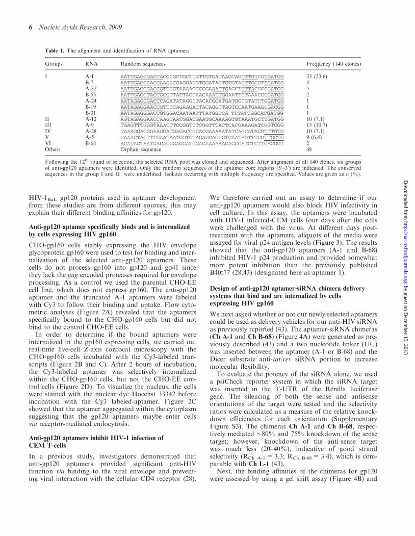
HIV-1BaL gp120 proteins used in aptamer developmentfrom these studies are from different sources, this mayexplain their different binding affinities for gp120.
Anti-gp120 aptamer specifically binds and is internalizedby cells expressing HIV gp160
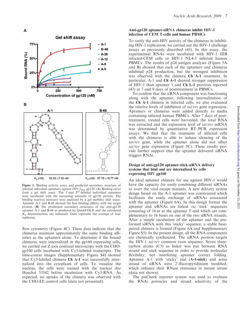
CHO-gp160 cells stably expressing the HIV envelopeglycoprotein gp160 were used to test for binding and inter-nalization of the selected anti-gp120 aptamers. Thesecells do not process gp160 into gp120 and gp41 sincethey lack the gag encoded proteases required for envelopeprocessing. As a control we used the parental CHO-EEcell line, which does not express gp160. The anti-gp120aptamer and the truncated A-1 aptamers were labeledwith Cy3 to follow their binding and uptake. Flow cyto-metric analyses (Figure 2A) revealed that the aptamersspecifically bound to the CHO-gp160 cells but did notbind to the control CHO-EE cells.In order to determine if the bound aptamers were
internalized in the gp160 expressing cells, we carried outreal-time live-cell Z-axis confocal microscopy with theCHO-gp160 cells incubated with the Cy3-labeled tran-scripts (Figure 2B and C). After 2 hours of incubation,the Cy3-labeled aptamer was selectively internalizedwithin the CHO-gp160 cells, but not the CHO-EE con-trol cells (Figure 2D). To visualize the nucleus, the cellswere stained with the nuclear dye Hoechst 33342 beforeincubation with the Cy3 labeled-aptamer. Figure 2Cshowed that the aptamer aggregated within the cytoplasmsuggesting that the gp120 aptamers maybe enter cellsvia receptor-mediated endocytosis.
Anti-gp120 aptamers inhibit HIV-1 infection ofCEM T-cells
In a previous study, investigators demonstrated thatanti-gp120 aptamers provided significant anti-HIVfunction via binding to the viral envelope and prevent-ing viral interaction with the cellular CD4 receptor (28).
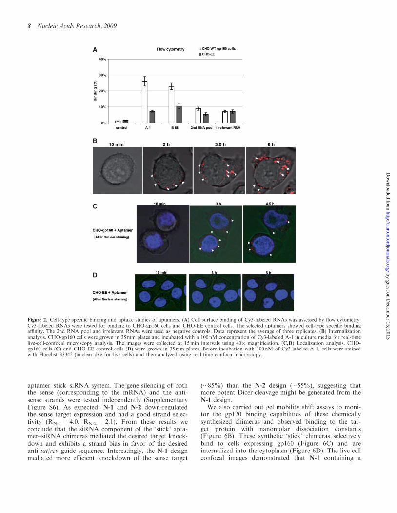
We therefore carried out an assay to determine if ouranti-gp120 aptamers would also block HIV infectivity incell culture. In this assay, the aptamers were incubatedwith HIV-1 infected-CEM cells four days after the cellswere challenged with the virus. At different days post-treatment with the aptamers, aliquots of the media wereassayed for viral p24 antigen levels (Figure 3). The resultsshowed that the anti-gp120 aptamers (A-1 and B-68)inhibited HIV-1 p24 production and provided somewhatmore potent inhibition than the previously publishedB40t77 (28,43) (designated here as aptamer 1).
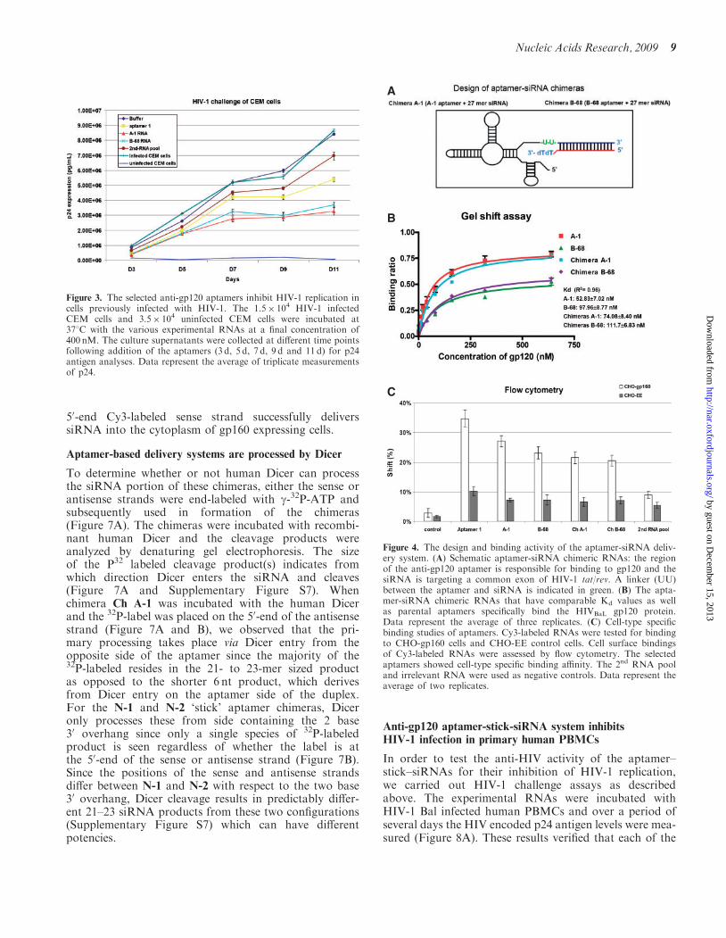
Design of anti-gp120 aptamer-siRNA chimera deliverysystems that bind and are internalized by cellsexpressing HIV gp160
We next asked whether or not our newly selected aptamerscould be used as delivery vehicles for our anti-HIV siRNAas previously reported (43). The aptamer-siRNA chimeras(Ch A-1 and Ch B-68) (Figure 4A) were generated as pre-viously described (43) and a two nucleotide linker (UU)was inserted between the aptamer (A-1 or B-68) and theDicer substrate anti-tat/rev siRNA portion to increasemolecular flexibility.
To evaluate the potency of the siRNA alone, we useda psiCheck reporter system in which the siRNA targetwas inserted in the 30-UTR of the Renilla luciferasegene. The silencing of both the sense and antisenseorientations of the target were tested and the selectivityratios were calculated as a measure of the relative knock-down efficiencies for each orientation (SupplementaryFigure S3). The chimeras Ch A-1 and Ch B-68, respec-tively mediated �80% and 75% knockdown of the sensetarget; however, knockdown of the anti-sense targetwas much less (20–40%), indicative of good strandselectivity (RCh A-1=3.3; RCh B-68=3.4), which is com-parable with Ch L-1 (43).
Next, the binding affinities of the chimeras for gp120were assessed by using a gel shift assay (Figure 4B) and
Table 1. The alignment and identification of RNA aptamers
Groups RNA Random sequences Frequency (140 clones)
I A-1 AATTGAGGGACCACGCGCTGCTTGTTGTGATAAGCAGTTTGTCGTGATGG 33 (23.6)B-7 AATTGAGGGACCAACGCGAGGATGTGGATAGTGTGTATTTGCGTTGATGG 3A-32 AATTGAGGGACCGTTGGTAAAAGCCGGAAATTGAGCTTTTACGGCGATGG 5B-55 AATTGAGGTACCGCGTTATTAGGAACAAATTGGAATTCTAAACGCGATGG 2A-24 AATAGAGGGACCCAGATATAGGCTACACGGATGATGGTGTATCTGGATGG 1B-19 AATAGAGGAACCGTTTCAGAAGACTACAGGTTAGTCCAATGAAGCGACGG 1B-31 AATAGAGGGACCGTGGACAATAATTTATGGTCA TTTATTGGCACGATGG 1
II A-12 AGTAGAGGAACCAAGCAATGGATGAATGCAAAAGTGTAAATGCTTGATGG 10 (7.1)III A-9 TGAGTTTGGGTAAATTTCCGGTTTCGGTTTACTCACGAAAGATCGGTCGG 15 (10.7)IV A-28 TAAAGGAGGGAAGGATGAGACCGCACGAAAAATATCAGCATACGTTTGTG 10 (7.1)V A-5 GAAACTAGTTTGAATAATGGTGTAGAGGAGGGTCAATAGTTTCGTTGGTG 9 (6.4)VI B-68 ACATAGTAATGACACGGAGGATGGAGAAAAAACAGCCATCTCTTGACGGT 2Others Orphan sequence 48
Following the 12th round of selection, the selected RNA pool was cloned and sequenced. After alignment of all 140 clones, six groupsof anti-gp120 aptamers were identified. Only the random sequences of the aptamer core regions (50–30) are indicated. The conservedsequences in the group I and II were underlined. Isolates occurring with multiple frequency are specified. Values are given as n (%).
6 Nucleic Acids Research, 2009
by guest on Decem
ber 15, 2013http://nar.oxfordjournals.org/
Dow
nloaded from
flow cytometry (Figure 4C). These data indicate that thechimeras maintain approximately the same binding affi-nities as the aptamers alone. To determine if the boundchimeras were internalized in the gp160 expressing cells,we carried out Z-axis confocal microscopy with the CHO-gp160 cells incubated with Cy3-labeled transcripts. Thetime-course images (Supplementary Figure S4) showedthat Cy3-labeled chimera Ch A-1 was successfully inter-nalized into the cytoplasm of cells. To visualize thenucleus, the cells were stained with the nuclear dyeHoechst 33342 before incubation with Cy3-RNA. Asexpected, no uptake of the chimera was observed withthe CHO-EE control cells (data not presented).
Anti-gp120 aptamer-siRNA chimeras inhibit HIV-1infection of CEM T-cells and human PBMCs
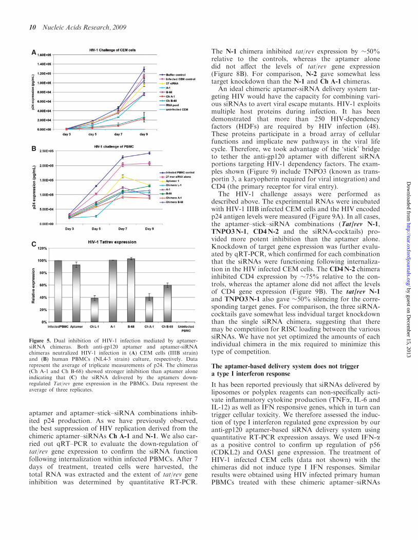
To verify the anti-HIV activity of the chimeras in inhibit-ing HIV-1 replication, we carried out the HIV-1 challengeassays as previously described (43). In this assay, theexperimental RNAs were incubated with HIV-1 IIIBinfected-CEM cells or HIV-1 NL4-3 infected humanPBMCs. The results of p24 antigen analyses (Figure 5Aand B) showed that each of the aptamers and chimerasinhibited p24 production, but the strongest inhibitionwas observed with the chimera Ch A-1 treatment. Inparticular, A-1 and Ch A-1 showed stronger suppressionof HIV-1 than aptamer 1 and Ch L-1 previous reported(43) at 7 and 9 days of posttreatment in PBMCs.To confirm that the siRNA component was functioning
along with the aptamer, following internalization ofthe Ch A-1 chimera in infected cells, we also evaluatedthe relative levels of inhibition of tat/rev gene expression.Aptamers or chimeras were added directly to mediacontaining infected human PBMCs. After 7 days of post-treatment, treated cells were harvested, the total RNAwas extracted and the expression level of tat/rev mRNAwas determined by quantitative RT–PCR expressionassays. We find that the treatment of infected cellswith the chimeras is able to induce silencing of thetat/rev gene, while the aptamer alone did not affecttat/rev gene expression (Figure 5C). These results pro-vide further support that the aptamer delivered siRNAtriggers RNAi.
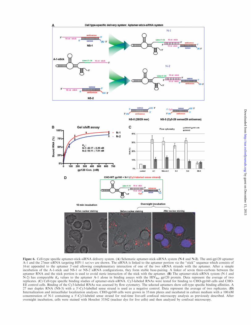
Design of anti-gp120 aptamer-stick-siRNA deliverysystems that bind and are internalized by cellsexpressing HIV gp160
An ideal aptamer chimera for use against HIV-1 wouldhave the capacity for easily combining different siRNAsto avert the viral escape mutants. A new delivery systemdesign based on the A-1 aptamer was constructed whichfacilitates the ready exchange of siRNAs associatedwith the aptamer (Figure 6A). In this design format theaptamer and siRNAs are linked via ‘stick’ sequencesconsisting of 16 nt at the aptamer 30-end which are com-plementary to 16 bases on one of the two siRNA strands.After a simple incubation of the aptamer and the pre-formed siRNA with this ‘sticky’ sequence, a stable base-paired chimera is formed (Figure 6A and SupplementaryFigure S5). In the present design, all the RNA componentsare chemically synthesized. The siRNA portion targetsthe HIV-1 tat/rev common exon sequence. Seven three-carbon atoms (C3) as linker was put between RNAstrand and stick sequence in order to provide molecularflexibility, not interfering aptamer correct folding.Aptamer A-1 with ‘sticky’ end (A-1-stick) and sensestrand of siRNA were 20-fluoropyridinium modified,which enhance their RNase resistance in mouse serum(data not shown).The psiCheck reporter system was used to evaluate
the RNAi potencies and strand selectivity of the
A
B
Figure 1. Binding activity assay and predicted secondary structure ofselected individual aptamers against HIV-1BaL gp120. (A) Binding curvefrom a gel shift assay. The 50-end P32-labeled individual aptamerswere incubated with the increasing amounts of gp120 protein. Thebinding reaction mixtures were analyzed by a gel mobility shift assay.Aptamer A-1 and B-68 showed the best binding affinity with the targetprotein. (B) The predicated secondary structures of the anti-gp120aptamer A-1 and B-68 as predicted by QuickFOLD and the calculatedKd determinations are indicated. Data represent the average of fourreplicates.
Nucleic Acids Research, 2009 7
by guest on Decem
ber 15, 2013http://nar.oxfordjournals.org/
Dow
nloaded from
aptamer–stick–siRNA system. The gene silencing of boththe sense (corresponding to the mRNA) and the anti-sense strands were tested independently (SupplementaryFigure S6). As expected, N-1 and N-2 down-regulatedthe sense target expression and had a good strand selec-tivity (RN-1=4.0; RN-2=2.1). From these results weconclude that the siRNA component of the ‘stick’ apta-mer–siRNA chimeras mediated the desired target knock-down and exhibits a strand bias in favor of the desiredanti-tat/rev guide sequence. Interestingly, the N-1 designmediated more efficient knockdown of the sense target
(�85%) than the N-2 design (�55%), suggesting thatmore potent Dicer-cleavage might be generated from theN-1 design.
We also carried out gel mobility shift assays to moni-tor the gp120 binding capabilities of these chemicallysynthesized chimeras and observed binding to the tar-get protein with nanomolar dissociation constants(Figure 6B). These synthetic ‘stick’ chimeras selectivelybind to cells expressing gp160 (Figure 6C) and areinternalized into the cytoplasm (Figure 6D). The live-cellconfocal images demonstrated that N-1 containing a
A
C
D
B
Figure 2. Cell-type specific binding and uptake studies of aptamers. (A) Cell surface binding of Cy3-labeled RNAs was assessed by flow cytometry.Cy3-labeled RNAs were tested for binding to CHO-gp160 cells and CHO-EE control cells. The selected aptamers showed cell-type specific bindingaffinity. The 2nd RNA pool and irrelevant RNAs were used as negative controls. Data represent the average of three replicates. (B) Internalizationanalysis. CHO-gp160 cells were grown in 35mm plates and incubated with a 100 nM concentration of Cy3-labeled A-1 in culture media for real-timelive-cell-confocal microscopy analysis. The images were collected at 15min intervals using 40� magnification. (C,D) Localization analysis. CHO-gp160 cells (C) and CHO-EE control cells (D) were grown in 35mm plates. Before incubation with 100 nM of Cy3-labeled A-1, cells were stainedwith Hoechst 33342 (nuclear dye for live cells) and then analyzed using real-time confocal microscopy.
8 Nucleic Acids Research, 2009
by guest on Decem
ber 15, 2013http://nar.oxfordjournals.org/
Dow
nloaded from
50-end Cy3-labeled sense strand successfully deliverssiRNA into the cytoplasm of gp160 expressing cells.
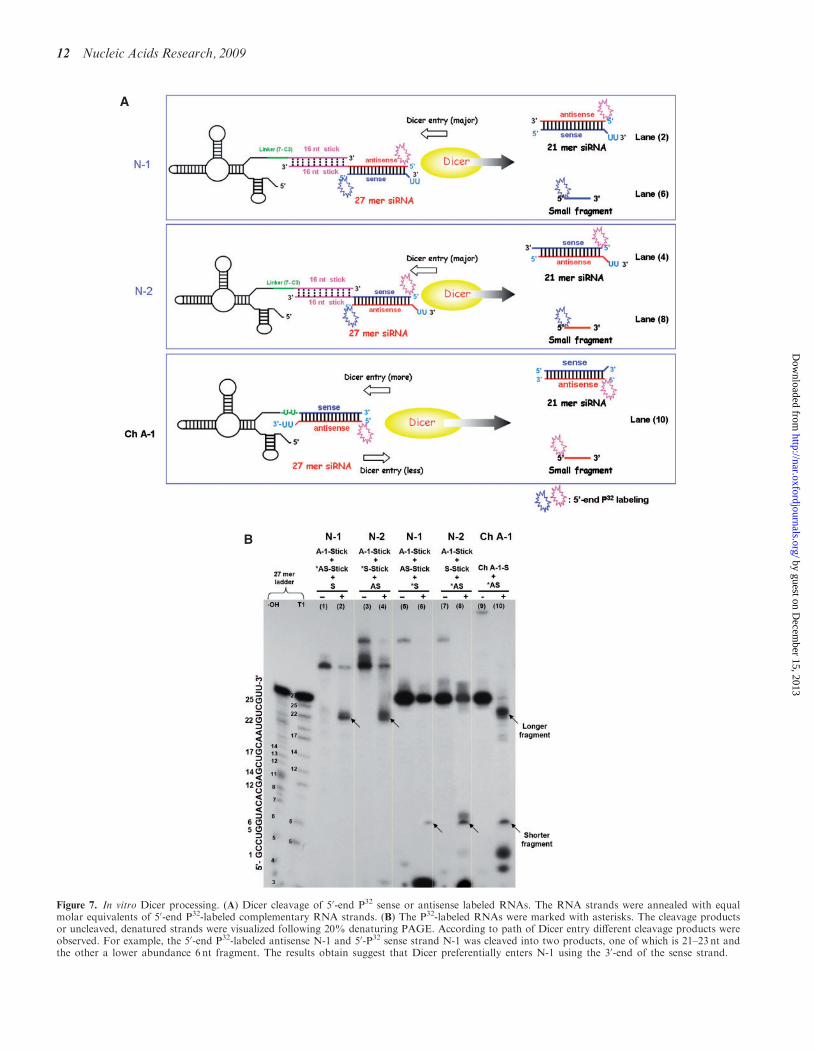
Aptamer-based delivery systems are processed by Dicer
To determine whether or not human Dicer can processthe siRNA portion of these chimeras, either the sense orantisense strands were end-labeled with g-32P-ATP andsubsequently used in formation of the chimeras(Figure 7A). The chimeras were incubated with recombi-nant human Dicer and the cleavage products wereanalyzed by denaturing gel electrophoresis. The sizeof the P32 labeled cleavage product(s) indicates fromwhich direction Dicer enters the siRNA and cleaves(Figure 7A and Supplementary Figure S7). Whenchimera Ch A-1 was incubated with the human Dicerand the 32P-label was placed on the 50-end of the antisensestrand (Figure 7A and B), we observed that the pri-mary processing takes place via Dicer entry from theopposite side of the aptamer since the majority of the32P-labeled resides in the 21- to 23-mer sized productas opposed to the shorter 6 nt product, which derivesfrom Dicer entry on the aptamer side of the duplex.For the N-1 and N-2 ‘stick’ aptamer chimeras, Diceronly processes these from side containing the 2 base30 overhang since only a single species of 32P-labeledproduct is seen regardless of whether the label is atthe 50-end of the sense or antisense strand (Figure 7B).Since the positions of the sense and antisense strandsdiffer between N-1 and N-2 with respect to the two base30 overhang, Dicer cleavage results in predictably differ-ent 21–23 siRNA products from these two configurations(Supplementary Figure S7) which can have differentpotencies.
Anti-gp120 aptamer-stick-siRNA system inhibitsHIV-1 infection in primary human PBMCs
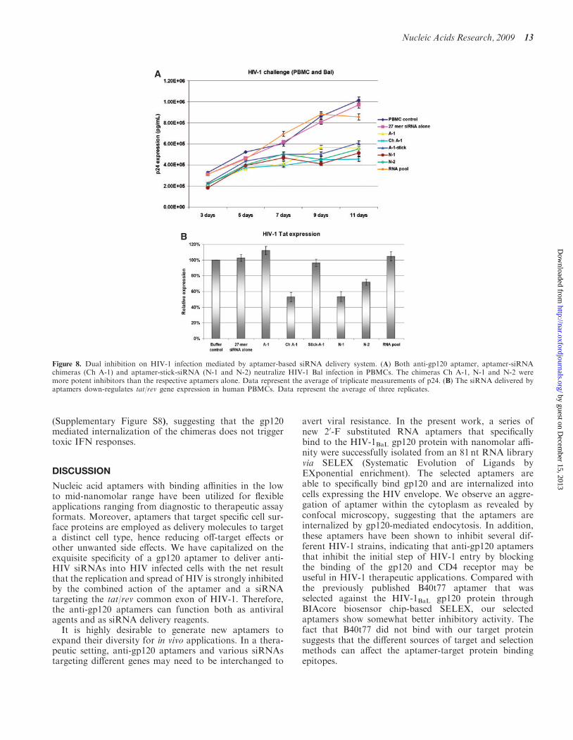
In order to test the anti-HIV activity of the aptamer–stick–siRNAs for their inhibition of HIV-1 replication,we carried out HIV-1 challenge assays as describedabove. The experimental RNAs were incubated withHIV-1 Bal infected human PBMCs and over a period ofseveral days the HIV encoded p24 antigen levels were mea-sured (Figure 8A). These results verified that each of the
A
B
C
Figure 4. The design and binding activity of the aptamer-siRNA deliv-ery system. (A) Schematic aptamer-siRNA chimeric RNAs: the regionof the anti-gp120 aptamer is responsible for binding to gp120 and thesiRNA is targeting a common exon of HIV-1 tat/rev. A linker (UU)between the aptamer and siRNA is indicated in green. (B) The apta-mer-siRNA chimeric RNAs that have comparable Kd values as wellas parental aptamers specifically bind the HIVBaL gp120 protein.Data represent the average of three replicates. (C) Cell-type specificbinding studies of aptamers. Cy3-labeled RNAs were tested for bindingto CHO-gp160 cells and CHO-EE control cells. Cell surface bindingsof Cy3-labeled RNAs were assessed by flow cytometry. The selectedaptamers showed cell-type specific binding affinity. The 2nd RNA pooland irrelevant RNA were used as negative controls. Data represent theaverage of two replicates.
Figure 3. The selected anti-gp120 aptamers inhibit HIV-1 replication incells previously infected with HIV-1. The 1.5� 104 HIV-1 infectedCEM cells and 3.5� 104 uninfected CEM cells were incubated at378C with the various experimental RNAs at a final concentration of400 nM. The culture supernatants were collected at different time pointsfollowing addition of the aptamers (3 d, 5 d, 7 d, 9 d and 11 d) for p24antigen analyses. Data represent the average of triplicate measurementsof p24.
Nucleic Acids Research, 2009 9
by guest on Decem
ber 15, 2013http://nar.oxfordjournals.org/
Dow
nloaded from
aptamer and aptamer–stick–siRNA combinations inhib-ited p24 production. As we have previously observed,the best suppression of HIV replication derived from thechimeric aptamer–siRNAs Ch A-1 and N-1. We also car-ried out qRT–PCR to evaluate the down-regulation oftat/rev gene expression to confirm the siRNA functionfollowing internalization within infected PBMCs. After 7days of treatment, treated cells were harvested, thetotal RNA was extracted and the extent of tat/rev geneinhibition was determined by quantitative RT-PCR.
The N-1 chimera inhibited tat/rev expression by �50%relative to the controls, whereas the aptamer alonedid not affect the levels of tat/rev gene expression(Figure 8B). For comparison, N-2 gave somewhat lesstarget knockdown than the N-1 and Ch A-1 chimeras.
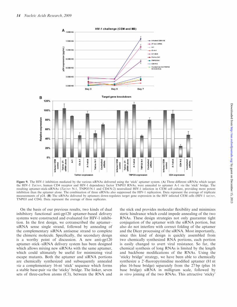
An ideal chimeric aptamer-siRNA delivery system tar-geting HIV would have the capacity for combining vari-ous siRNAs to avert viral escape mutants. HIV-1 exploitsmultiple host proteins during infection. It has beendemonstrated that more than 250 HIV-dependencyfactors (HDFs) are required by HIV infection (48).These proteins participate in a broad array of cellularfunctions and implicate new pathways in the viral lifecycle. Therefore, we took advantage of the ‘stick’ bridgeto tether the anti-gp120 aptamer with different siRNAportions targeting HIV-1 dependency factors. The exam-ples shown (Figure 9) include TNPO3 (known as trans-portin 3, a karyopherin required for viral integration) andCD4 (the primary receptor for viral entry).
The HIV-1 challenge assays were performed asdescribed above. The experimental RNAs were incubatedwith HIV-1 IIIB infected CEM cells and the HIV encodedp24 antigen levels were measured (Figure 9A). In all cases,the aptamer–stick–siRNA combinations (Tat/rev N-1,TNPO3N-1, CD4N-2 and the siRNA-cocktails) pro-vided more potent inhibition than the aptamer alone.Knockdown of target gene expression was further evalu-ated by qRT-PCR, which confirmed for each combinationthat the siRNAs were functioning following internaliza-tion in the HIV infected CEM cells. The CD4N-2 chimerainhibited CD4 expression by �75% relative to the con-trols, whereas the aptamer alone did not affect the levelsof CD4 gene expression (Figure 9B). The tat/rev N-1and TNPO3N-1 also gave �50% silencing for the corre-sponding target genes. For comparison, the three siRNA-cocktails gave somewhat less indvidual target knockdownthan the single siRNA chimera, suggesting that theremay be competition for RISC loading between the varioussiRNAs. We have not yet optimized the amounts of eachindividual chimera in the mix required to minimize thistype of competition.
The aptamer-based delivery system does not triggera type I interferon response
It has been reported previously that siRNAs delivered byliposomes or polyplex reagents can non-specifically acti-vate inflammatory cytokine production (TNFa, IL-6 andIL-12) as well as IFN responsive genes, which in turn cantrigger cellular toxicity. We therefore assessed the induc-tion of type I interferon regulated gene expression by ouranti-gp120 aptamer-based siRNA delivery system usingquantitative RT-PCR expression assays. We used IFN-aas a positive control to confirm up regulation of p56(CDKL2) and OAS1 gene expression. The treatment ofHIV-1 infected CEM cells (data not shown) with thechimeras did not induce type I IFN responses. Similarresults were obtained using HIV infected primary humanPBMCs treated with these chimeric aptamer–siRNAs
A
B
C
Figure 5. Dual inhibition of HIV-1 infection mediated by aptamer-siRNA chimeras. Both anti-gp120 aptamer and aptamer-siRNAchimeras neutralized HIV-1 infection in (A) CEM cells (IIIB strain)and (B) human PBMCs (NL4-3 strain) culture, respectively. Datarepresent the average of triplicate measurements of p24. The chimeras(Ch A-1 and Ch B-68) showed stronger inhibition than aptamer aloneindicating that (C) the siRNA delivered by the aptamers down-regulated Tat/rev gene expression in the PBMCs. Data represent theaverage of three replicates.
10 Nucleic Acids Research, 2009
by guest on Decem
ber 15, 2013http://nar.oxfordjournals.org/
Dow
nloaded from
A
B C
D
Figure 6. Cell-type specific aptamer-stick-siRNA delivery system. (A) Schematic aptamer-stick-siRNA system (N-1 and N-2). The anti-gp120 aptamerA-1 and the 27mer siRNA targeting HIV-1 tat/rev are shown. The siRNA is linked to the aptamer portion via the ‘‘stick’’ sequence which consists of16 nt appended to the aptamer 30-end allowing complementary interaction of one of the two siRNA strands with the aptamer. After a simpleincubation of the A-1-stick and NS-1 or NS-2 siRNA configurations, they form stable base-pairing. A linker of seven three-carbons between theaptamer RNA and the stick portion is used to avoid steric interaction of the stick with the aptamer. (B) The aptamer-stick-siRNA system (N-1 andN-2) has comparable Kd values to the aptamer A-1 alone in binding assays with the HIVBal gp120 protein. Data represent the average of tworeplicates. (C) Cell-type specific binding studies of aptamer-stick-siRNA. Cy3-labeled RNAs were tested for binding to CHO-gp160 cells and CHO-EE control cells. Binding of the Cy3-labeled RNAs was assessed by flow cytometry. The selected aptamers show cell-type specific binding affinities. A27 mer duplex RNA (NS-3) with a 50-Cy3-labelled sense strand is used as a negative control. Data represent the average of two replicates. (D)Internalization and intracellular localization analyses. CHO-gp160 cells were grown in 35mm plates and incubated in culture medium with a 100 nMconcentration of N-1 containing a 50-Cy3-labeled sense strand for real-time live-cell confocal microscopy analysis as previously described. Afterovernight incubation, cells were stained with Hoechst 33342 (nuclear dye for live cells) and then analyzed by confocal microscopy.
by guest on Decem
ber 15, 2013http://nar.oxfordjournals.org/
Dow
nloaded from
A
B
Figure 7. In vitro Dicer processing. (A) Dicer cleavage of 50-end P32 sense or antisense labeled RNAs. The RNA strands were annealed with equalmolar equivalents of 50-end P32-labeled complementary RNA strands. (B) The P32-labeled RNAs were marked with asterisks. The cleavage productsor uncleaved, denatured strands were visualized following 20% denaturing PAGE. According to path of Dicer entry different cleavage products wereobserved. For example, the 50-end P32-labeled antisense N-1 and 50-P32 sense strand N-1 was cleaved into two products, one of which is 21–23 nt andthe other a lower abundance 6 nt fragment. The results obtain suggest that Dicer preferentially enters N-1 using the 30-end of the sense strand.
12 Nucleic Acids Research, 2009
by guest on Decem
ber 15, 2013http://nar.oxfordjournals.org/
Dow
nloaded from
(Supplementary Figure S8), suggesting that the gp120mediated internalization of the chimeras does not triggertoxic IFN responses.
DISCUSSION
Nucleic acid aptamers with binding affinities in the lowto mid-nanomolar range have been utilized for flexibleapplications ranging from diagnostic to therapeutic assayformats. Moreover, aptamers that target specific cell sur-face proteins are employed as delivery molecules to targeta distinct cell type, hence reducing off-target effects orother unwanted side effects. We have capitalized on theexquisite specificity of a gp120 aptamer to deliver anti-HIV siRNAs into HIV infected cells with the net resultthat the replication and spread of HIV is strongly inhibitedby the combined action of the aptamer and a siRNAtargeting the tat/rev common exon of HIV-1. Therefore,the anti-gp120 aptamers can function both as antiviralagents and as siRNA delivery reagents.
It is highly desirable to generate new aptamers toexpand their diversity for in vivo applications. In a thera-peutic setting, anti-gp120 aptamers and various siRNAstargeting different genes may need to be interchanged to
avert viral resistance. In the present work, a series ofnew 20-F substituted RNA aptamers that specificallybind to the HIV-1BaL gp120 protein with nanomolar affi-nity were successfully isolated from an 81 nt RNA libraryvia SELEX (Systematic Evolution of Ligands byEXponential enrichment). The selected aptamers areable to specifically bind gp120 and are internalized intocells expressing the HIV envelope. We observe an aggre-gation of aptamer within the cytoplasm as revealed byconfocal microscopy, suggesting that the aptamers areinternalized by gp120-mediated endocytosis. In addition,these aptamers have been shown to inhibit several dif-ferent HIV-1 strains, indicating that anti-gp120 aptamersthat inhibit the initial step of HIV-1 entry by blockingthe binding of the gp120 and CD4 receptor may beuseful in HIV-1 therapeutic applications. Compared withthe previously published B40t77 aptamer that wasselected against the HIV-1BaL gp120 protein throughBIAcore biosensor chip-based SELEX, our selectedaptamers show somewhat better inhibitory activity. Thefact that B40t77 did not bind with our target proteinsuggests that the different sources of target and selectionmethods can affect the aptamer-target protein bindingepitopes.
A
B
Figure 8. Dual inhibition on HIV-1 infection mediated by aptamer-based siRNA delivery system. (A) Both anti-gp120 aptamer, aptamer-siRNAchimeras (Ch A-1) and aptamer-stick-siRNA (N-1 and N-2) neutralize HIV-1 Bal infection in PBMCs. The chimeras Ch A-1, N-1 and N-2 weremore potent inhibitors than the respective aptamers alone. Data represent the average of triplicate measurements of p24. (B) The siRNA delivered byaptamers down-regulates tat/rev gene expression in human PBMCs. Data represent the average of three replicates.
Nucleic Acids Research, 2009 13
by guest on Decem
ber 15, 2013http://nar.oxfordjournals.org/
Dow
nloaded from
On the basis of our previous results, two kinds of dualinhibitory functional anti-gp120 aptamer-based deliverysystems were constructed and evaluated for HIV-1 inhibi-tion. In the first design, we cotranscribed the aptamer–siRNA sense single strand, followed by annealing ofthe complementary siRNA antisense strand to completethe chimeric molecule. Specifically, the secondary designis a worthy point of discussion. A new anti-gp120aptamer–stick–siRNA delivery system has been designedwhich allows mixing new siRNAs with the same aptamer,which could ultimately be useful for minimizing viralescape mutants. Both the aptamer and siRNA portionsare chemically synthesized and subsequently annealedvia a complementary 16-nt ‘stick’ sequence which formsa stable base-pair via the ‘sticky’ bridge. The linker, sevensets of three-carbon atoms (C3), between the RNA and
the stick end provides molecular flexibility and minimizessteric hindrance which could impede annealing of the twoRNAs. These design strategies not only guarantee tightconjugation of the aptamer with the siRNA portion, butalso do not interfere with correct folding of the aptamerand the Dicer processing of the siRNA. Most importantly,since this kind of design is quickly assembled fromtwo chemically synthesized RNA portions, each portionis easily changed to avert viral resistance. So far, thechemical synthesis of long RNAs is limited by the lengthand backbone modifications of the RNAs. Using the‘sticky bridge’ strategy, we have been able to chemicallysynthesize a 20-fluoropyrimidine modified aptamer (81 ntplus 16-base bridge) separately from the 27 bp (plus 16base bridge) siRNA in milligram scale, followed byin vitro joining of the two RNAs. This attractive ‘sticky’
A
B
Figure 9. The HIV-1 inhibition mediated by the various siRNAs delivered using the ‘stick’ aptamer system. (A) Three different siRNAs which targetthe HIV-1 Tat/rev, human CD4 receptor and HIV-1 dependency factor TNPO3 RNAs, were annealed to aptamer A-1 via the ‘stick’ bridge. Theresulting aptamer-stick-siRNAs (Tat/rev N-1, TNPO3N-1 and CD4N-2) neutralized HIV-1 infection in CEM cell culture, providing more potentinhibition than the aptamer alone. The combination of three siRNAs also suppressed the HIV-1 replication. Data represent the average of triplicatemeasurements of p24. (B) The siRNAs delivered by aptamers down-regulates target gene expression in the HIV infected CEM cells (HIV-1 tat/rev,TNPO3 and CD4). Data represent the average of three replicates.
14 Nucleic Acids Research, 2009
by guest on Decem
ber 15, 2013http://nar.oxfordjournals.org/
Dow
nloaded from
bridge-based approach can potentially be used for mixingdifferent siRNAs with a single aptamer to multiplex tar-get down regulation, and in the case of HIV infection, toavert resistance to the siRNA component.
We demonstrate that our delivery systems specificallybind to the surface of cells expressing gp160 and are inter-nalized, allowing functional processing of the siRNAinto RISC, resulting in specific inhibition of HIV-1 repli-cation and infectivity in cell culture. Both of theanti-gp120-based siRNA delivery systems serve as dualfunction inhibitors and therefore provide greater efficacythan either the aptamer or siRNA applied alone.
Interestingly, even though our designs (Ch A-1, N-1 andN-2) are able to down-regulate the target expression andshow good strand selectivity, their RNAi potencies aredifferent. Previous studies of Dicer substrate siRNAs(49,50) used substrates in which Dicer entry on the bluntend of the DsiRNAs was completely blocked by placingtwo deoxy nucleotides at the 30-end of the desired passen-ger strand, creating an absolute polarity for Dicer entryfrom the two base 30 overhang. The present studies didnot employ deoxys so entry from the blunt end of theCh A-1 siRNA takes place relatively efficiently relativeto the entry from the 2 base 30 overhang, which is adja-cent to the aptamer that creates a steric hindrance forDicer entry (Figure 7B). Also, in contrast to our previousobservations, (49,50) the preference for guide strandselection in our chimeras does not necessarily favour thestrand with the two base 30 overhang (SupplementaryFigures S6 and S7), since the more potent configurationN-1 has the guide strand positioned with the 50-end atthe side of Dicer entry. In this context it should benoted that the strand selectivity of N-1 is not as good asthat of N-2 (Supplementary Figure S6). The difference inpotency between N-1 and N-2 most likely resides in thespecies of 21- to 23-mer produced, which differ betweenthe two forms. Interestingly, Dicer does not process the‘stick’ aptamer siRNAs at all from the blunt end side asit does with the Ch A-1. We attribute this to the lack of afree 30OH in these chimeras, in which the end of thesiRNA is covalently attached to the ‘stick’ sequencewhich in turn forms a 20OMe modified duplex with theaptamer. The 20OMe backbone modifications throughoutthe ‘stick’ also block Dicer cleavage (51). Since thestrand selectivity of Dicer generally favors the strandwith the 2 base 30 overhang (49,50), it is important todesign the Dicer substrates such that a potent siRNAagainst the desired target generated from the cleavageevent. This will minimize the off target potential of thepassenger strand.
In summary, we have demonstrated that the anti-gp120aptamers not only provide a potential new drug therapyapproach for combating HIV infection, but also act asdelivery vehicles for siRNAs and perhaps other smallRNA inhibitors. The ‘sticky’ bridge-base approach offersa major advantage in both chemical synthesis and theopportunity to mix and match the aptamer with differentsiRNAs in a non-covalent fashion. For HIV clinical
applications, the mixing approach may be a requirementfor averting viral escape mutants.
SUPPLEMENTARY DATA
Supplementary Data are available at NAR Online.
ACKNOWLEDGEMENTS
We thank Britta Hoehn, Guihua Sun, Harris Soifer andLisa Scherer for helpful discussions. The followingreagents were obtained through the NIH AIDSResearch and Reference Reagent Program, Division ofAIDS, NIAID, NIH: The CHO-EE and CHO-gp160 cellline (52,53); the pNL4-3 luc vector; HIV-1BaL gp120 fromDAIDS, NIAID.
FUNDING
National Institutes of Health (AI29329, HL07470 toJ.J.R). Funding for open access charge: NationalInstitutes of Health (AI29329).
Conflict of interest statement. None declared.
REFERENCES
1. Dalgleish,A.G., Beverley,P.C., Clapham,P.R., Crawford,D.H.,Greaves,M.F. and Weiss,R.A. (1984) The CD4 (T4) antigen isan essential component of the receptor for the AIDS retrovirus.Nature, 312, 763–767.
2. Klatzmann,D., Champagne,E., Chamaret,S., Gruest,J., Guetard,D.,Hercend,T., Gluckman,J.C. and Montagnier,L. (1984)T-lymphocyte T4 molecule behaves as the receptor forhuman retrovirus LAV. Nature, 312, 767–768.
3. Sattentau,Q.J. andMoore,J.P. (1993) The role of CD4 in HIV bindingand entry. Philos. Trans. R Soc Lond. B Biol. Sci., 342, 59–66.
4. Kwong,P.D., Wyatt,R., Robinson,J., Sweet,R.W., Sodroski,J. andHendrickson,W.A. (1998) Structure of an HIV gp120 envelopeglycoprotein in complex with the CD4 receptor and aneutralizing human antibody. Nature, 393, 648–659.
5. Ugolini,S., Mondor,I. and Sattentau,Q.J. (1999) HIV-1 attachment:another look. Trends Microbiol., 7, 144–149.
6. Wyatt,R. and Sodroski,J. (1998) The HIV-1 envelope glycoproteins:fusogens, antigens, and immunogens. Science, 280, 1884–1888.
7. Chan,D.C. and Kim,P.S. (1998) HIV entry and its inhibition. Cell,93, 681–684.
8. Blair,W.S., Lin,P.F., Meanwell,N.A. and Wallace,O.B. (2000)HIV-1 entry - an expanding portal for drug discovery. Drug Discov.Today, 5, 183–194.
9. Cooley,L.A. and Lewin,S.R. (2003) HIV-1 cell entry and advancesin viral entry inhibitor therapy. J. Clin. Virol., 26, 121–132.
10. Kilby,J.M. and Eron,J.J. (2003) Novel therapies based onmechanisms of HIV-1 cell entry. N. Engl. J. Med., 348, 2228–2238.
11. Pomerantz,R.J. and Horn,D.L. (2003) Twenty years of therapy forHIV-1 infection. Nat. Med., 9, 867–873.
12. Moore,J.P. and Doms,R.W. (2003) The entry of entry inhibitors: afusion of science and medicine. Proc. Natl Acad. Sci. USA, 100,10598–10602.
13. Jiang,S., Zhao,Q. and Debnath,A.K. (2002) Peptide and non-peptide HIV fusion inhibitors. Curr. Pharm. Des., 8, 563–580.
14. Pierson,T.C. and Doms,R.W. (2003) HIV-1 entry inhibitors: newtargets, novel therapies. Immunol. Lett., 85, 113–118.
15. Meanwell,N.A. and Kadow,J.F. (2003) Inhibitors of the entry ofHIV into host cells. Curr. Opin. Drug Discov. Devel., 6, 451–461.
Nucleic Acids Research, 2009 15
by guest on Decem
ber 15, 2013http://nar.oxfordjournals.org/
Dow
nloaded from

16. Lin,P.F., Blair,W., Wang,T., Spicer,T., Guo,Q., Zhou,N.,Gong,Y.F., Wang,H.G., Rose,R., Yamanaka,G. et al. (2003) Asmall molecule HIV-1 inhibitor that targets the HIV-1 envelope andinhibits CD4 receptor binding. Proc. Natl Acad. Sci. USA, 100,11013–11018.
17. Ryser,H.J. and Fluckiger,R. (2005) Progress in targeting HIV-1entry. Drug Discov. Today, 10, 1085–1094.
18. Smith,D.H., Byrn,R.A., Marsters,S.A., Gregory,T., Groopman,J.E.and Capon,D.J. (1987) Blocking of HIV-1 infectivity by a soluble,secreted form of the CD4 antigen. Science, 238, 1704–1707.
19. Hussey,R.E., Richardson,N.E., Kowalski,M., Brown,N.R.,Chang,H.C., Siliciano,R.F., Dorfman,T., Walker,B., Sodroski,J.and Reinherz,E.L. (1988) A soluble CD4 protein selectively inhibitsHIV replication and syncytium formation. Nature, 331, 78–81.
20. Daar,E.S., Li,X.L., Moudgil,T. and Ho,D.D. (1990) Highconcentrations of recombinant soluble CD4 are required toneutralize primary human immunodeficiency virus type 1 isolates.Proc. Natl Acad. Sci. USA, 87, 6574–6578.
21. Schacker,T., Coombs,R.W., Collier,A.C., Zeh,J.E., Fox,I., Alam,J.,Nelson,K., Eggert,E. and Corey,L. (1994) The effects of high-doserecombinant soluble CD4 on human immunodeficiency virus type 1viremia. J. Infect. Dis., 169, 37–40.
22. Trkola,A., Pomales,A.B., Yuan,H., Korber,B., Maddon,P.J.,Allaway,G.P., Katinger,H., Barbas,C.F. III, Burton,D.R., Ho,D.D.et al. (1995) Cross-clade neutralization of primary isolates ofhuman immunodeficiency virus type 1 by human monoclonalantibodies and tetrameric CD4-IgG. J. Virol., 69, 6609–6617.
23. Allaway,G.P., Davis-Bruno,K.L., Beaudry,G.A., Garcia,E.B.,Wong,E.L., Ryder,A.M., Hasel,K.W., Gauduin,M.C., Koup,R.A.,McDougal,J.S. et al. (1995) Expression and characterization ofCD4-IgG2, a novel heterotetramer that neutralizes primary HIVtype 1 isolates. AIDS Res. Hum. Retroviruses, 11, 533–539.
24. Shearer,W.T., Israel,R.J., Starr,S., Fletcher,C.V., Wara,D.,Rathore,M., Church,J., DeVille,J., Fenton,T., Graham,B. et al.(2000) Recombinant CD4-IgG2 in human immunodeficiencyvirus type 1-infected children: phase 1/2 study. The PediatricAIDS Clinical Trials Group Protocol 351 Study Team.J. Infect. Dis., 182, 1774–1779.
25. Jacobson,J.M., Lowy,I., Fletcher,C.V., O’Neill,T.J., Tran,D.N.,Ketas,T.J., Trkola,A., Klotman,M.E., Maddon,P.J., Olson,W.C.et al. (2000) Single-dose safety, pharmacology, and antiviralactivity of the human immunodeficiency virus (HIV) type 1 entryinhibitor PRO 542 in HIV-infected adults. J. Infect. Dis., 182,326–329.
26. Dey,B., Lerner,D.L., Lusso,P., Boyd,M.R., Elder,J.H. andBerger,E.A. (2000) Multiple antiviral activities of cyanovirin-N:blocking of human immunodeficiency virus type 1 gp120 interactionwith CD4 and coreceptor and inhibition of diverse envelopedviruses. J. Virol., 74, 4562–4569.
27. Martin,L., Stricher,F., Misse,D., Sironi,F., Pugniere,M., Barthe,P.,Prado-Gotor,R., Freulon,I., Magne,X., Roumestand,C. et al. (2003)Rational design of a CD4 mimic that inhibits HIV-1 entryand exposes cryptic neutralization epitopes. Nat. Biotechnol., 21,71–76.
28. Khati,M., Schuman,M., Ibrahim,J., Sattentau,Q., Gordon,S. andJames,W. (2003) Neutralization of infectivity of diverse R5 clinicalisolates of human immunodeficiency virus type 1 by gp120-binding2’F-RNA aptamers. J. Virol., 77, 12692–12698.
29. Dey,A.K., Khati,M., Tang,M., Wyatt,R., Lea,S.M. and James,W.(2005) An aptamer that neutralizes R5 strains of humanimmunodeficiency virus type 1 blocks gp120-CCR5 interaction.J. Virol., 79, 13806–13810.
30. Dey,A.K., Griffiths,C., Lea,S.M. and James,W. (2005) Structuralcharacterization of an anti-gp120 RNA aptamer that neutralizesR5 strains of HIV-1. RNA, 11, 873–884.
31. Nimjee,S.M., Rusconi,C.P. and Sullenger,B.A. (2005) Aptamers: anemerging class of therapeutics. Annu. Rev. Med., 56, 555–583.
32. Pestourie,C., Tavitian,B. and Duconge,F. (2005) Aptamersagainst extracellular targets for in vivo applications. Biochimie, 87,921–930.
33. Hicke,B.J. and Stephens,A.W. (2000) Escort aptamers: a deliveryservice for diagnosis and therapy. J. Clin. Invest., 106, 923–928.
34. Lupold,S.E., Hicke,B.J., Lin,Y. and Coffey,D.S. (2002)Identification and characterization of nuclease-stabilized RNAmolecules that bind human prostate cancer cells via the prostate-specific membrane antigen. Cancer Res., 62, 4029–4033.
35. McNamara,J.O. II, Andrechek,E.R., Wang,Y., Viles,K.D.,Rempel,R.E., Gilboa,E., Sullenger,B.A. and Giangrande,P.H.(2006) Cell type-specific delivery of siRNAs with aptamer-siRNAchimeras. Nat. Biotechnol., 24, 1005–1015.
36. Chu,T.C., Twu,K.Y., Ellington,A.D. and Levy,M. (2006) Aptamermediated siRNA delivery. Nucleic Acids Res., 34, e73.
37. Chu,T.C., Marks,J.W. III, Lavery,L.A., Faulkner,S.,Rosenblum,M.G., Ellington,A.D. and Levy,M. (2006)Aptamer:toxin conjugates that specifically target prostate tumorcells. Cancer Res., 66, 5989–5992.
38. Bagalkot,V., Farokhzad,O.C., Langer,R. and Jon,S. (2006) Anaptamer-doxorubicin physical conjugate as a novel targeteddrug-delivery platform. Angew. Chem. Int. Ed. Engl., 45, 8149–8152.
39. Farokhzad,O.C., Jon,S., Khademhosseini,A., Tran,T.N.,Lavan,D.A. and Langer,R. (2004) Nanoparticle-aptamerbioconjugates: a new approach for targeting prostate cancer cells.Cancer Res., 64, 7668–7672.
40. Farokhzad,O.C., Cheng,J., Teply,B.A., Sherifi,I., Jon,S.,Kantoff,P.W., Richie,J.P. and Langer,R. (2006) Targetednanoparticle-aptamer bioconjugates for cancer chemotherapyin vivo. Proc. Natl Acad. Sci. USA, 103, 6315–6320.
41. Farokhzad,O.C., Karp,J.M. and Langer,R. (2006) Nanoparticle-aptamer bioconjugates for cancer targeting. Expert Opin. DrugDeliv., 3, 311–324.
42. Engels,F.K., Mathot,R.A. and Verweij,J. (2007) Alternativedrug formulations of docetaxel: a review. Anticancer Drugs, 18,95–103.
43. Zhou,J., Li,H., Li,S., Zaia,J. and Rossi,J.J. (2008) Novel dualinhibitory function aptamer-siRNA delivery system for HIV-1therapy. Mol. Ther., 16, 1481–1489.
44. Tuerk,C. and Gold,L. (1990) Systematic evolution of ligands byexponential enrichment: RNA ligands to bacteriophage T4 DNApolymerase. Science, 249, 505–510.
45. Ellington,A.D. and Szostak,J.W. (1990) In vitro selection of RNAmolecules that bind specific ligands. Nature, 346, 818–822.
46. Tuerk,C., MacDougal,S. and Gold,L. (1992) RNA pseudoknotsthat inhibit human immunodeficiency virus type 1 reversetranscriptase. Proc. Natl Acad. Sci. USA, 89, 6988–6992.
47. Fitzwater,T. and Polisky,B. (1996) A SELEX primer. MethodsEnzymol., 267, 275–301.
48. Brass,A.L., Dykxhoorn,D.M., Benita,Y., Yan,N., Engelman,A.,Xavier,R.J., Lieberman,J. and Elledge,S.J. (2008) Identification ofhost proteins required for HIV infection through a functionalgenomic screen. Science, 319, 921–926.
49. Rose,S.D., Kim,D.H., Amarzguioui,M., Heidel,J.D.,Collingwood,M.A., Davis,M.E., Rossi,J.J. and Behlke,M.A. (2005)Functional polarity is introduced by Dicer processing of shortsubstrate RNAs. Nucleic Acids Res., 33, 4140–4156.
50. Amarzguioui,M., Lundberg,P., Cantin,E., Hagstrom,J.,Behlke,M.A. and Rossi,J.J. (2006) Rational design and in vitro andin vivo delivery of Dicer substrate siRNA. Nat. Protocol, 1,508–517.
51. Collingwood,M.A., Rose,S.D., Huang,L., Hillier,C.,Amarzguioui,M., Wiiger,M.T., Soifer,H.S., Rossi,J.J. andBehlke,M.A. (2008) Chemical modification patterns compatible withhigh potency dicer-substrate small interfering RNAs.Oligonucleotides, 18, 187–200.
52. Weiss,C.D. and White,J.M. (1993) Characterization of stableChinese hamster ovary cells expressing wild-type, secreted, andglycosylphosphatidylinositol-anchored human immunodeficiencyvirus type 1 envelope glycoprotein. J. Virol., 67, 7060–7066.
53. Vodicka,M.A., Goh,W.C., Wu,L.I., Rogel,M.E., Bartz,S.R.,Schweickart,V.L., Raport,C.J. and Emerman,M. (1997) Indicatorcell lines for detection of primary strains of human and simianimmunodeficiency viruses. Virology, 233, 193–198.
16 Nucleic Acids Research, 2009
by guest on Decem
ber 15, 2013http://nar.oxfordjournals.org/
Dow
nloaded from
Copyright © 2022 FDOKUMEN