![Intrinsically radiolabelled [(59)Fe]-SPIONs for dual MRI/radionuclide detection](https://static.fdokumen.com/doc/165x107/6335c40d379741109e00c5c6/intrinsically-radiolabelled-59fe-spions-for-dual-mriradionuclide-detection.jpg)
Intrinsically radiolabelled [(59)Fe]-SPIONs for dual MRI/radionuclide detection

Caught in Action: Selecting PeptideAptamers Against IntrinsicallyDisordered Proteins in Live CellsJacqueline D. Cobbert1, Christopher DeMott1, Subhabrata Majumder1, Eric A. Smith2, Sergey Reverdatto1,David S. Burz1, Kathleen A. McDonough2 & Alexander Shekhtman1
1Department of Chemistry, University at Albany, Albany, NY, 2Wadsworth Center, NY State Department of Health, Albany, NY.
Intrinsically disordered proteins (IDPs) or unstructured segments within proteins play an important role incellular physiology and pathology. Low cellular concentration, multiple binding partners, frequentpost-translational modifications and the presence of multiple conformations make it difficult tocharacterize IDP interactions in intact cells. We used peptide aptamers selected by using theyeast-two-hybrid scheme and in-cell NMR to identify high affinity binders to transiently structured IDP andunstructured segments at atomic resolution. Since both the selection and characterization of peptideaptamers take place inside the cell, only physiologically relevant conformations of IDPs are targeted. Themethod is validated by using peptide aptamers selected against the prokaryotic ubiquitin-like protein, Pup,of the mycobacterium proteasome. The selected aptamers bind to distinct sites on Pup and have vastlydifferent effects on rescuing mycobacterial proteasome substrate and on the survival of theBacille-Calmette-Guerin, BCG, strain of M. bovis. This technology can be applied to study the elusive actionof IDPs under near physiological conditions.
On average 4% of prokaryotic and 33% of eukaryotic genomes encode proteins or large segments ofproteins that lack a stable secondary or tertiary structure1. These intrinsically disordered proteins(IDPs) or disordered regions can be highly conserved between species, and are often functional2–4.
IDPs have the ability to bind to multiple proteins and play a role in the assembly of macromolecular arrays5,6.IDPs are implicated in neuropathology, autoimmune diseases and cancers, and are considered to be promisingtargets for drug therapy7. Despite wide recognition of IDPs and the nascent secondary structures that exist in theunbound state, the molecular mechanisms underlying the functions of IDPs are not widely understood.
There is a limited arsenal of molecular tools that can be applied to study interactions of IDPs in a physiologicalmilieu5,8,9. Due to the intrinsic flexibility of IDPs, the powerful methodology of using small molecules to blockprotein-protein interaction surfaces are applied to well-folded IDP binding partners; there is only one example ofsmall molecule inhibitor to IDP, c-Myc10. Since many IDPs fold upon binding to their cognate targets4,5,11, site-directed mutagenesis, which creates local changes in the chemical structure of the IDP, cannot be effectively usedto interrogate the usually large IDP-target interaction surfaces5,12.
Recently we introduced a Combinatorial Library of Improved Peptide aptamers (CLIPs), containing more than3*1010 peptide aptamers, as a tool to help isolate site-specific binders to select cellular targets13. Peptide apta-mers14,15 (PA) are small ,12 kDa proteins that consist of a randomized 8 amino acid peptide sequence insertedinto the loop of a modified thioredoxin platform. The modification of the thioredoxin platform13 improvessolubility and decreases the tendency of PAs to oligomerize, creating molecules with drug-like binding abilities16.
PAs are selected by using the yeast-two-hybrid (Y2H) technique14,15, which facilitates binding to a broad rangeof cellular, viral and bacterial target proteins with high specificity and strong affinity16. CLIPs technology is anattractive molecular tool to analyze the functional consequences of nascent structures present in free IDPs due tothe in vivo nature of target selection and comprehensive coverage of the potential binding surfaces.
In-cell NMR is used to analyze nascent structures of IDPs inside live cells at atomic resolution8,17–21. STINT-NMR is an in-cell technique developed to study protein-protein structural interactions inside live cells22,23. Weused STINT-NMR to define the interaction surfaces between a small IDP, the prokaryotic ubiquitin-like protein,Pup24, and selected PAs, in the crowded cytosol. Unlike its eukaryotic counterpart, Pup is unstructured25. Sinceboth selection and structural characterization of PA-IDP complexes are made inside cells, we expect that thistechnology will be highly relevant to identify functional complexes between IDPs and its binding targets.
OPEN
SUBJECT AREAS:
MICROBIOLOGYTECHNIQUES
INTRACELLULAR SIGNALLINGPEPTIDES AND PROTEINS
CHEMICAL TOOLS
Received29 December 2014
Accepted3 March 2015
Published24 March 2015
Correspondence andrequests for materials
should be addressed toA.S. (ashekhtman@
albany.edu)
SCIENTIFIC REPORTS | 5 : 9402 | DOI: 10.1038/srep09402 1

The Mycobacterium tuberculosis (Mtb) proteasomal system is anattractive target for drug therapy against latent Mtb infection since itprovides defense against the reactive nitrogen intermediates gener-ated to counter bacteria inside phagosomes26,27. Indeed, the potentproteasome inhibitors, Bortezomib28 (Btz) and GL527, increase thesensitivity of Mtb to nitro-oxidative stress27. The Mtb proteasomesystem consists of the Mtb core particle, CP, a proteasomal ATPase,Mpa, and prokaryotic ubiquitin-like protein, Pup. Mpa binds to theproteasome CP and Pup binding to Mpa directs the pupylatedsubstrate into the proteasome for degradation. We used the Mtbproteasome system to illustrate how the combination of CLIPs andSTINT-NMR technologies allows the selection of specific binders toPup and how these molecules can be used to analyze the functionalinteractions of Pup inside mycobacteria.
ResultsSelection of peptide aptamers that interact with Pup. Yeast two-hybrid selection (Fig. 1a) was used to identify PAs that bindspecifically to Pup in vivo. Pup fused to the Gal4 DNA bindingdomain (BD) was used as bait, and a randomized PA library,CLIPs13, was the prey. E. coli thioredoxin, fused to the GAL4transcriptional activation domain (AD), served as the scaffold toconstrain the octa-peptides of randomized sequence in an aptamerloop13,14. Approximately 1.5*107 transformants were screened onAde2 plates and out of fifty selected, three clones, designated PA-1,PA-3, and PA-7, were found to activate all Y2H markers, ADE2,HIS3, MEL1, and AUR1-C, indicating molecular interactionsbetween PAs and Pup.
DNA encoding the interacting PAs was PCR amplified from theyeast and sequenced (Fig. 1b). PA-1 contains primarily nonpolaraliphatic and two aromatic residues, tryptophan and histidine. PA-3 and PA-7 are similar in sequence, and contain positively chargedand aromatic residues. To estimate the binding affinity of each PA forPup in vitro, fluorescence titrations were conducted using bacteriallyover-expressed and purified components. Fluorescence signals werequenched as Pup was added (Fig. 1c). In contrast, the thioredoxinscaffold13, which was used as a negative control, did not show fluor-
escence quenching upon adding Pup. The resolved Kd’s are in thenanomolar range.
Peptide aptamers bind to the a-helix and N-terminal region of Pup.We used in-cell NMR (STINT-NMR) to identify the Pup residuesaffected by PA binding22. [U-15N]-Pup was overexpressed in E. coliBL21(DE3) for two hours, followed by overexpression of unlabeledPA for up to 16 hours; spectra were collected at time intervalscorresponding to increasing PA concentrations. After collectingthe sample, the cells were subjected to in-cell NMR analysiswithout prior freezing. The in-cell 1H{15N}-HSQC NMR spectrumof free Pup was used as a control (Fig. 2 and Supplementary Fig. 1).The acquisition time for an in-cell NMR experiment was less thantwo hours. To verify that the protein signals originated from cellularprotein, after each in-cell NMR experiment, the cells were recoveredfrom the tube, centrifuged, and the NMR spectrum of thesupernatant was recorded; no protein signals were detected.
Both the chemical shifts and peak intensities of Pup residues wereaffected by ligand binding (Fig. 2 and Supplementary Fig. 1).Conventional analyses of interacting proteins, which compare thein-cell NMR spectra of the protein prior to and following full over-expression of the interactor, ignores changes in the in-cell NMRspectra that arise over the time course of the experiment fromnon-interactor and concentration-independent binding processes29.To unambiguously resolve the principal binding mode between Pupand PAs against the dynamic cellular background, we analyzed mat-rices composed of in-cell NMR intensity changes, MIC, and chemicalshift changes, MCSC, of [U-15N]-Pup peaks over the time course ofPA overexpression by using single-value decomposition, SVD29. Inthis approach, the principal binding mode between Pup and PAs isidentified by an abrupt drop in the singular values (SV) of the screeplot of either MIC or MCSC (Fig. 3 and Supplementary Fig. 2). Thedrop results in a poor goodness of fit of linear regression, R2 , 0.8.Random binding of Pup to cytosol contributes to a gradual decreasein SVs, and results in R2 . 0.9.
The scree plot of Pup-PA-1 MCSC shows a clear drop after the firstSV indicating the presence of a single principal binding mode (Fig. 3and Supplementary Fig. 2). The corresponding plot of Pup-PA-1
Figure 1 | Y2H selected PAs bind to Pup with nanomolar affinity. (a) Yeast-Two-Hybrid selection of PAs. 1) Pup, fused to the Gal4 DNA-binding
domain (BD), is expressed in yeast containing several reporter genes that possess sites for the BD in their promoters. The reporter genes are not expressed
if the activation domain (AD) is not present. 2) Pup, fused to the BD, is expressed concurrently with a randomized PA loop in a thioredoxin (Thio)
scaffold13, fused to the AD. Binding between Pup and a PA activates transcription of reporter genes in cells, while reporter gene transcription is suppressed
in cells where Pup and a PA do not interact. (b) PA sequences selected through Y2H screening and the Pup-binding affinity of each sequence. (c) Pup-PA
binding isotherms. Changes in native PA fluorescence intensity are plotted vs. the concentration of Pup. Thio scaffold13 was used as a negative control and
showed no changes in fluorescence upon titration with Pup. The data represent mean values and are fitted as described in Methods. All experiments were
performed in triplicate.
www.nature.com/scientificreports
SCIENTIFIC REPORTS | 5 : 9402 | DOI: 10.1038/srep09402 2
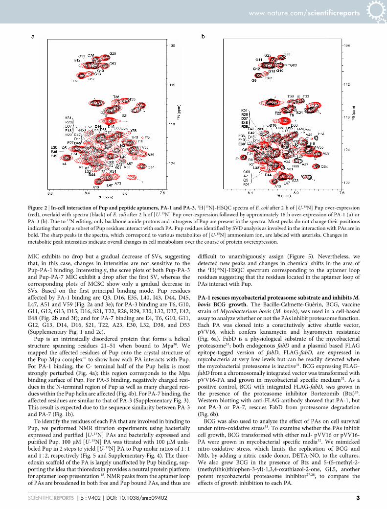
MIC exhibits no drop but a gradual decrease of SVs, suggestingthat, in this case, changes in intensities are not sensitive to thePup-PA-1 binding. Interestingly, the scree plots of both Pup-PA-3and Pup-PA-7 MIC exhibit a drop after the first SV, whereas thecorresponding plots of MCSC show only a gradual decrease inSVs. Based on the first principal binding mode, Pup residuesaffected by PA-1 binding are Q3, D16, E35, L40, I43, D44, D45,L47, A51 and V59 (Fig. 2a and 3e); for PA-3 binding are T6, G10,G11, G12, G13, D15, D16, S21, T22, R28, R29, E30, L32, D37, E42,E48 (Fig. 2b and 3f); and for PA-7 binding are E4, T6, G10, G11,G12, G13, D14, D16, S21, T22, A23, E30, L32, D38, and D53(Supplementary Fig. 1 and 2c).
Pup is an intrinsically disordered protein that forms a helicalstructure spanning residues 21–51 when bound to Mpa30. Wemapped the affected residues of Pup onto the crystal structure ofthe Pup-Mpa complex30 to show how each PA interacts with Pup.For PA-1 binding, the C- terminal half of the Pup helix is moststrongly perturbed (Fig. 4a); this region corresponds to the Mpabinding surface of Pup. For PA-3 binding, negatively charged resi-dues in the N-terminal region of Pup as well as many charged resi-dues within the Pup helix are affected (Fig. 4b). For PA-7 binding, theaffected residues are similar to that of PA-3 (Supplementary Fig. 3).This result is expected due to the sequence similarity between PA-3and PA-7 (Fig. 1b).
To identify the residues of each PA that are involved in binding toPup, we performed NMR titration experiments using bacteriallyexpressed and purified [U-15N] PAs and bacterially expressed andpurified Pup. 100 mM [U-15N] PA was titrated with 100 mM unla-beled Pup in 2 steps to yield [U-15N] PA to Pup molar ratios of 151and 152, respectively (Fig. 5 and Supplementary Fig. 4). The thior-edoxin scaffold of the PA is largely unaffected by Pup binding, sup-porting the idea that thioredoxin provides a neutral protein platformfor aptamer loop presentation 15. NMR peaks from the aptamer loopof PAs are broadened in both free and Pup bound PAs, and thus are
difficult to unambiguously assign (Figure 5). Nevertheless, wedetected new peaks and changes in chemical shifts in the area ofthe 1H{15N}-HSQC spectrum corresponding to the aptamer loopresidues suggesting that the residues located in the aptamer loop ofPAs interact with Pup.
PA-1 rescues mycobacterial proteasome substrate and inhibits M.bovis BCG growth. The Bacille-Calmette-Guerin, BCG, vaccinestrain of Mycobacterium bovis (M. bovis), was used in a cell-basedassay to analyze whether or not the PAs inhibit proteasome function.Each PA was cloned into a constitutively active shuttle vector,pVV16, which confers kanamycin and hygromycin resistance(Fig. 6a). FabD is a physiological substrate of the mycobacterialproteasome31; both endogenous fabD and a plasmid based FLAGepitope-tagged version of fabD, FLAG-fabD, are expressed inmycobacteria at very low levels but can be readily detected whenthe mycobacterial proteasome is inactive31. BCG expressing FLAG-fabD from a chromosomally integrated vector was transformed withpVV16-PA and grown in mycobacterial specific medium32. As apositive control, BCG with integrated FLAG-fabD, was grown inthe presence of the proteasome inhibitor Bortezomib (Btz)28.Western blotting with anti-FLAG antibody showed that PA-1, butnot PA-3 or PA-7, rescues FabD from proteasome degradation(Fig. 6b).
BCG was also used to analyze the effect of PAs on cell survivalunder nitro-oxidative stress33. To examine whether the PAs inhibitcell growth, BCG transformed with either null- pVV16 or pVV16-PA were grown in mycobacterial specific media32. We mimickednitro-oxidative stress, which limits the replication of BCG andMtb, by adding a nitric oxide donor, DETA-NO, to the cultures.We also grew BCG in the presence of Btz and 5-(5-methyl-2-(methylthio)thiophen-3-yl)-1,3,4-oxathiazol-2-one, GL5, anotherpotent mycobacterial proteasome inhibitor27,28, to compare theeffects of growth inhibition to each PA.
Figure 2 | In-cell interaction of Pup and peptide aptamers, PA-1 and PA-3. 1H{15N}-HSQC spectra of E. coli after 2 h of [U-15N] Pup over-expression
(red), overlaid with spectra (black) of E. coli after 2 h of [U-15N] Pup over-expression followed by approximately 16 h over-expression of PA-1 (a) or
PA-3 (b). Due to 15N editing, only backbone amide protons and nitrogens of Pup are present in the spectra. Most peaks do not change their positions
indicating that only a subset of Pup residues interact with each PA. Pup residues identified by SVD analysis as involved in the interaction with PAs are in
bold. The sharp peaks in the spectra, which correspond to various metabolites of [U-15N] ammonium ion, are labeled with asterisks. Changes in
metabolite peak intensities indicate overall changes in cell metabolism over the course of protein overexpression.
www.nature.com/scientificreports
SCIENTIFIC REPORTS | 5 : 9402 | DOI: 10.1038/srep09402 3

BCG cultures were grown with shaking for four days, and aliquotswere plated to determine the live bacteria count. Cultures were grownfor an additional three days to accumulate sufficient amounts ofprotein for Western blot analyses. Expression of each PA was verifiedby Western blotting by using an anti-thioredoxin monoclonal anti-
body. Native thioredoxin was not detected under control conditions(Supplementary Fig. 5).
The biological log reduction of the relative number of livemicrobes is expressed in terms of colony forming units, CFU/mL.After incubating plates for 3 weeks, we observed a dramatic, 100-fold
Figure 3 | SVD analyses of in-cell Pup-PA-1 and Pup-PA-3 interactions. Matrices consisting of chemical shift changes, MCSC, and intensity changes,
MIC, in in-cell [U-15N] Pup peaks over the time course of PA-1 and PA-3 overexpression were analyzed29 to identify Pup residues involved in PA
binding. The scree plots show the distribution of singular values that define the relative contribution of each binding mode to the MCSC or MIC,
respectively. (a) and (d) The first binding mode contributes to 92.6% of the MCSC for Pup-PA-1 and 86.3% of the MIC for Pup-PA-3, indicating that it is
a principal binding mode. A clear drop in the progression of singular values is evident after the first singular value and R2 values for the linear regressions
are poor, 0.75 and 0.65 for (a) and (d), respectively. (b) and (c) The first binding mode contributes to 62% of the MCSC for Pup-PA-1 and 64% of the MIC
for Pup-PA-3. No clear drop in singular values is evident and the R2 values for the linear regressions are high, 0.91 and 0.93 for (b) and (c), respectively,
precluding the data from identifying principal binding modes in these cases. (e) and (f) The contribution of Pup residues to the first and second binding
mode with PAs is shown in red and blue bars, respectively. There are two bars per residue. The maximum contribution of Pup residues to the second mode
is used as a threshold to identify Pup residues affected by PA binding.
www.nature.com/scientificreports
SCIENTIFIC REPORTS | 5 : 9402 | DOI: 10.1038/srep09402 4

or a 2 log10, reduction in CFU/mL for BCG expressing PA-1 grownunder DETA-NO stress (Fig. 6c and 6d). Nitro-oxidative stress alone,provided by DETA-NO, had no effect on the growth of BCG(Fig. 6d). Cell growth was inhibited by the presence of proteasomeinhibitors GL5 (1.0 log) and Btz (1.0–1.5 log), and, importantly, BCGviability was further reduced by the combination of GL5 and DETA-NO (2.5–3.0 log) or Btz and DETA-NO (2.0–2.5 log). These resultscorroborate previous studies that proteasome inhibitors limit Mtbgrowth under nitro- oxidative stress27.
Importantly, cell viability in the presence of PA-1 and DETA-NOwas comparable to that of Btz and DETA-NO, suggesting that both
Btz and PA-1 lead to enhancement of BCG sensitivity to nitro-oxidative stress. In contrast, PA-3 and PA-7, which interact with asegment of Pup that does not bind Mpa, in combination withDETA-NO, had only minimal effects on growth (0.5 log reduc-tion) (Fig. 6d). Overexpression of PA-1 in the presence of Btz andDETA-NO led to no further growth inhibition beyond that of Btzand DETA-NO alone, which is consistent with PA-1 and Btzaffecting the same pathway. These assays demonstrate that despitethe uniformly high binding affinity of the selected PAs (Fig. 1b),the specificity of the interaction with Pup leads to distinct physio-logical outcomes.
Figure 4 | PA-1 and PA-3 bind to distinct sites on Pup. Pup residues (shown in red) identified by SVD analysis to be affected by PA-1 (a) and PA-3 (b)
binding are mapped onto the Pup-Mpa complex30. Some labeled residues in (a) and (b) are obscured due to image orientation. The image of the
Pup-Mpa structure (PDB code 3M9D) was constructed by using Modeller45.
Figure 5 | Only residues in the PA loop bind Pup. 1H{15N}-HSQC spectra of purified [U-15N] PA (red), overlaid with spectra (black) obtained after
titrating with purified Pup, (a) PA-1 and (b) PA-3. Each insert shows the residues from the PA loop. Well-resolved peaks of the thioredoxin scaffold are
labeled. Most peaks do not change their position or intensities, reflecting the fact that thioredoxin is a neutral PA scaffold. Only a subset of PA residues,
from the PA loop (right insets), exhibit substantial broadening and chemical shift changes, indicating Pup-PA loop interactions. The chemical shifts of
indole protons of tryptophans located both near and inside the PA loop of PA-1 (Figure 1) change due to the interaction with Pup (left inset). Free and
Pup bound PA-3 indole protons are completely broadened and, thus, unavailable for analysis. Due to15N editing, only backbone amide protons and
nitrogens of PA are present in the spectra.
www.nature.com/scientificreports
SCIENTIFIC REPORTS | 5 : 9402 | DOI: 10.1038/srep09402 5

DiscussionUsing CLIPs technology we isolated three peptide aptamers that bindto Pup with nanomolar affinity, PA-1, PA-3 and PA-7. The bindingsites of the selected PAs are close to and even overlap each other. Thisfact highlights a critical advantage of Y2H selection13,14. PAs selectedto bind to a given target do so without competition between themolecules in the library. These PAs would not be selected by usingin vitro selection schemes, such as phage, yeast, or ribosomal display,where all molecules in the library compete for the same target and,consequently, only the highest affinity aptamers remain after mul-tiple cycles of selection34.
Both Pup and PAs have to be over-expressed well above theirphysiological concentrations to be visible by in-cell NMR. Underthese conditions, the cellular binding partners of Pup do not play asignificant role in complex formation and can be largely ignored35.We also expect that Pup folding in the crowded cytosol of E. coli willbe similar to that which occurs in mycobacteria. Most importantly,the functional assays allowed us to test the effects of PA binding to theIDP in a native physiological milieu.
In spite of similar binding affinities, the peptide aptamers iden-tified in this screen have vastly different functional effects onrescuing a known proteasome substrate31, FabD, from degradationand on the survival of M. bovis BCG under nitro-oxidative stress.
Only PA-1, but not PA-3 or PA-7, inhibits proteasome degrada-tion of FabD in BCG (Fig. 6b). Expression of PA-1 leads to a 100-fold inhibition of growth, but PA-3 and PA-7 lead to less than a5-fold decrease in the survival rate of DETA-NO treated cells(Fig. 6d). These effects are due to the different ways in whichthe PAs engage Pup, which is known to form a helical structurewhen bound to its target, Mpa30. Only the PA-1 interaction sur-face strongly overlaps with the Mpa helical binding surface onPup, whereas binding of the positively charged PA-3 and PA-7aptamers affects two highly charged segments of Pup: the N-ter-minal region, which is not involved in Mpa binding, and a seg-ment of the Mpa helical binding surface on Pup. (Fig. 4 andSupplementary Fig. 3) We hypothesize that both PA-3 and PA-7 interact with Pup electrostatically with high affinity but lowspecificity, and can not effectively block the Pup helix from bind-ing to Mpa.
Both of the proteasome inhibitors used in this study, Btz and GL-5,were shown to substantially increase Mtb sensitivity to nitro-oxid-ative stress27. The growth reduction caused by PA-1 with DETA-NOwas comparable to that observed using Btz and DETA-NO and only0.5 log lower than cells grown in the presence of GL5 and DETA-NO.This result suggests that PAs can be as potent as small molecules atinhibiting Mtb growth.
Figure 6 | PA-1, but not PA-3 or PA-7, rescues proteasome substrate FabD from degradation and inhibits the growth of BCG. (a) Diagram of the
expression shuttle vector, pVV16, used in the BCG assay. The plasmid contains an E. coli origin derived from pUC19, a mycobacterial origin derived from
pAL500046, and a constitutively active BCG hsp60 promoter. (b) BCG-FLAG-FabD was transformed with pVV16-PA-1, -PA-3, and -PA-7 to examine the
steady-state level of FLAG-tagged FabD in untreated cells. As a control, BCG was incubated with and without 50 mM Btz. Anti-groEL2 was used as a
loading control. (c) BCG plates after 3 weeks of growth show a 100-fold reduction in colonies due to expression of PA-1. (d) Inhibition of BCG growth
under conditions that reduce proteasome function. 50 mM Btz, GL5, and DETA- NO were used as indicated. Error bars represent standard deviations and
asterisks indicate p- values ,0.05 (Prism 5 Graphpad Inc.). The arrow indicates the strength of the initial inoculum. The dashed line is the lower limit of
detection. All experiments were performed in triplicate.
www.nature.com/scientificreports
SCIENTIFIC REPORTS | 5 : 9402 | DOI: 10.1038/srep09402 6

Drugs are widely used to inhibit enzymatic activities27 and it is veryimportant to know whether these drugs have off-target effects36. Herewe showed that known inhibitors of the Mtb proteasome in com-bination with PA-1 do not synergistically inhibit growth, suggestingthat they act along the same biological pathway. At the same time,both GL5 and Btz are more effective than PA-1 alone in reducing thesurvival rate. One possible explanation is different stability of smallmolecular drugs compared to PA-1. Alternatively, this suggests thatbesides the proteasome, GL5 and Btz inhibit other enzymes import-ant for Mtb survival.
The technology demonstrated in this work permits both screeningand structural characterization of protein interactions in thecrowded environment of the cytosol. This is important for IDPs,for which local structures may be partially stabilized due to macro-molecular crowding21,37. Thus, selected PAs are already directedagainst physiologically relevant conformations, which may not besignificantly populated during in vitro selection.
Functionally, IDPs and unstructured segments within proteinscan facilitate protein-protein interactions, serve as flexible linkersbetween folded domains and provide convenient sites for post-trans-lational modifications3. Site- and conformation-specific binding ofPAs to IDPs or unstructured segments uniquely affect these func-tions, either by blocking access to the protein interaction sites and thesites of post-translational modification, or by decreasing the flexibil-ity of critical sites in the linkers. The availability of PAs selected tobind to specific sites on IDPs allows unprecedented opportunity toanalyze the functional consequences of IDP interactions with atomicprecision in live cells.
MethodsReagents and chemicals. Restriction enzymes, Taq DNA Polymerase, and PhusionDNA Polymerase were from New England BioLabs. Pfu DNA Polymerase was fromAgilent Technologies. All other chemicals used were reagent grade or better.
Yeast strains. Matchmaker Gold yeast-hybrid system 2 and vectors, pGBKT7 andpGADT7, were obtained from Clontech. Yeast strain Y2HGold (MATa, trp1-901,leu2-3, 112, ura3-52, his3-200, gal4D, gal80D, LYS2:: GAL1UAS–Gal1TATA–His3,GAL2UAS–Gal2TATA–Ade2, URA3:: MEL1UAS– Mel1TATA, AUR1-C MEL1) andY187 (MATa, ura3-52, his3-200, ade2-101, trp1-901, leu2-3, 112, gal4D, gal80D, met-, URA3:: Gal1UAS–Gal1TATA–LacZ, MEL1) were used for peptide screening andgrown in yeast peptone dextrose adenine (YPDA) growth medium. Four reportergenes, AUR1-C, ADE2, HIS3, and MEL1, under the control of GAL4 upstreamactivating sequences (UASs) and TATA boxes, were used to select PAs and eliminatefalse positives. AUR1-C confers strong resistance to the highly toxic antibiotic,Aureobasidin A (Aur A). ADE2 and HIS3 provide metabolic selection, and MEL1,which encodes a-galactosidase, is used for X- a-galactosidase (X-a-gal) selection.
Plasmid Construction. The peptide aptamer (PA) library generated in our lab wasused to screen against Pup13. The library contains approximately 3*1010 peptideaptamers cloned into pGADT7 to yield a pGADT7-thioredoxin-peptide aptamerlibrary construct in which an 8-amino-acid randomized peptide is inserted into aconstrained loop of D26A, K57Q thioredoxin13. The thioredoxin is fused in framewith amino acids 768–881 of the GAL4 activating domain (AD). The plasmid has ahemagglutinin (HA) epitope tag, a 2 m origin, and confers ampicillin resistance forselection in E. coli and LEU2 for selection in yeast.
DNA encoding Pup (64 aa) was PCR amplified from Mtb genomic DNA using TaqDNA Polymerase and oligonucleotides 59-TTTTTTCATATGGCGCAAGAGC-AGACCAAGC-39 and 59- TTTTTTGTCGACTCACTGTCCGCCCTTTTGG-39,which contain flanking 59-NdeI and 39-SalI restriction sites. The restriction-digestedPCR product was ligated into expression vector pGBKT7 to yield pGBKT7-Pup inwhich Pup is fused in frame with amino acids 1–147 of the GAL4 DNA bindingdomain (DNA-BD). The expressed protein contains an N-terminal c-Myc epitope tagand confers kanamycin resistance for selection in E. coli and TRP3 for selection inyeast.
DNA encoding Mpa (613 aa) was PCR amplified from Mycobacterium smegmatisgenomic DNA using Phusion DNA Polymerase and oligonucleotides 59- AAAAA-AGGTACCATGAGTGAGTCAGA-39 and 59-AAAAAGGTACCCAGGTACTG-GCCCAG-39, which contains flanking 59- and 39-KpnI restriction sites. The restric-tion-digested PCR product was ligated into pGADT7 to yield pGADT7-Mpa in whichMpa is fused to a GAL4 activating domain. The expressed protein contains a C-terminal hemagglutinin (HA) epitope tag and ampicillin resistance for selection in E.coli and LEU2 for selection in yeast.
DNA encoding each individual peptide aptamer was PCR amplified from pGADT7using Taq Polymerase and oligonucleotides 59-CGGGGCCATCCTCGTCGCTT-TCTGG-39 and 59- GGTTTTGATCGATGTTCAGTTGTGCA-39. Pfu DNA
Polymerase and PCR products were used to clone peptide aptamers into a modifiedpBAD expression vector, which confers ampicillin resistance. The resulting plasmids,pBAD-PA, express N-terminal 6xHis-tagged scaffold protein, thioredoxin.
DNA encoding each peptide aptamer was PCR amplified using Phusion DNAPolymerase and oligonucleotides 59-TTTTTTCATATGGGCCATAAAATTATT-39
and 59- CCCAAGCTTACGCCAGGTTAGCGTCG-39, which contain flanking 59-NdeI and 39-HindIII restriction sites. The restriction-digested PCR product wasligated into pVV16 (BEI Resources) to yield pVV16-PA. The pVV16 affords het-erologous protein expression under constitutively active hsp60 promoter and conferskanamycin and hygromycin resistance for selection in E. coli and mycobacterialstrains. Each aptamer was handled in the same manner.
To make the FLAG-tagged mycobacterial proteasome substrate, fabD was amp-lified by PCR from BCG genomic DNA using primers encoding the FLAG epitope(MDYKDDDDKI), 59-GGGTGCGAGCAACGCAATCATCTTATCGTCGTC-ATCCTTGTAATCCATGGTACC-39 and 59-GGATCCTTATAGGTTTGCCAGC-TCGTCCAGGTC-39, which contain flanking 59- KpnI and 39-BamHI restrictionsites. The restriction-digested PCR product was ligated into the BCG plasmidpMBC1260. pMBC1260 is a single copy plasmid that integrates into the attB site onthe BCG chromosome and affords heterologous protein expression off of the con-stitutively active Rv0805 promoter and confers kanamycin resistance for selection inE. coli and mycobacterial strains.
Verification of reporter gene activation. Plasmid pGBKT7-Pup (bait) wastransformed into yeast strain Y2HGold and plasmid pGADT7-Mpa (prey) wastransformed into yeast strain Y187. Transformants were mated on YPDA agar platesand mated transformants were selected on synthetic dropout media (SDM) lackingtryptophan and leucine, SDM lacking tryptophan, leucine, and histidine, SDMlacking tryptophan, leucine, and adenine, SDM lacking tryptophan and leucinesupplemented with X-a- gal, and SDM lacking tryptophan and leucine supplementedwith Aur A, to verify the activation of reporter genes by these constructs.
Library transformation and screening. Plasmid pGBKT7-Pup was used as bait toscreen CLIPs13 by using the Matchmaker Gold yeast-hybrid system 2 protocol. TheCLIPs was transformed into yeast strain Y2HGold harboring pGBKT7-Pup by usingthe lithium acetate method. Transformants were selected initially for growth on ade2
trp2 leu2 plates. Positive clones were streaked onto trp2 leu2 his2 plates; ade2 trp2
leu2 plates; trp2 leu2 plates supplemented with X-a-gal; and trp2 leu2 platessupplemented with Aur A, to test the stringency of the interaction with Pup.
Expression and purification of aptamers for in vitro assays. For fluorescencetitration experiments, E. coli strain DH10B was transformed with pBAD- PA andgrown in M-50538 auto-inducing medium containing 0.25% L-arabinose, and150 mg/L of carbenicillin. All PA plasmids were treated in the same manner. For[U-15N] labeling of PAs, E. coli strain Origami B (Novagen) was transformed withpBAD-PA and grown in N-50538 auto- inducing medium containing 2.66 g/L of[U-15N] ammonium chloride as the sole nitrogen source, 0.25% L-arabinose, and150 mg/L of carbenicillin. Cultures were grown at 37uC for 20–26 h. Cells wereharvested, re-suspended in extraction buffer (50 mM sodium phosphate, pH 7.0,300 mM sodium chloride, 6 M guanidine hydrochloride) and sonicated. The lysatewas cleared by centrifugation, and the supernatant was loaded onto a pre-equilibratedTALON column (Clontech). The column was washed with 10 column volumes (CV)of extraction buffer, 10 CV of extraction buffer containing 0.4% octyl phenolethoxylate (Triton X-100, Baker), and again with 10 CV of extraction buffer. Proteinwas eluted with 10 CV of 13 imidazole elution buffer (45 mM sodium phosphate, pH7.0, 270 mM sodium chloride, 5.4 M guanidine hydrochloride, 150 mM imidazole).Eluted protein was concentrated to 2–2.5 mL by using an Amicon Ultra-15centrifugal filter (Millipore) with a 3 kDa molecular weight cutoff (MWCO), andsupplemented with 50 mL of 0.5 M EDTA, 6 mL of TSP buffer (20 mM sodiumphosphate, pH 7.5, 100 mM sodium thiosulfate) and 1 mL of glycerol. Proteinsamples were dialyzed against 1 L of 50 mM sodium phosphate, pH 8.0, 100 mMsodium chloride, 0.5 M guanidine hydrochloride, 2 mM EDTA, and 10% glycerol, for5–10 hours at 4uC in a 1 kDa MWCO dialysis bag (Spectrum Laboratories, Inc.).Samples were further dialyzed against 2 L of TSP buffer supplemented with 10%glycerol, for 10 hours at 4uC. Finally, peptide aptamers were concentrated by using anAmicon Ultra-15 centrifugal filter with a 3 kDa MWCO, exchanged into TSP bufferand stored on ice. The peptide aptamers were estimated to be .95% pure byCoomassie-stained SDS- PAGE.
Expression and purification of Pup for in vitro assays. E. coli strain BL21(DE3)Codon1 (Novagen) was transformed with pTM-Pup, which encodes a tryptophanleader sequence (trpL) before Pup39. Cells were grown to an OD595 of 0.5 in minimalmedium (M9) containing 35 mg/L of kanamycin, induced with 0.5 mM isopropyl 1-thio-b-d-galactopyranoside (IPTG), and grown overnight at 37uC. Cells wereharvested, re- suspended in 20 mM HEPES-Na, pH 7.0 buffer containing 8 M urea,and sonicated. The lysate was cleared by centrifugation, and the supernatant wasloaded onto a nickel-nitrilotriacetic acid- agarose (Ni-NTA) column (Qiagen). Thecolumn was washed with 10 CV of wash buffer (20 mM HEPES-Na buffer, pH 7.0,containing 8 M urea), and the protein was eluted with elution buffer (20 mMphosphate buffer, pH 4.0 containing 8 M urea). Eluted protein was concentrated to 2–2.5 mL by using an Amicon Ultra-15 centrifugal filter (Millipore), with a 3 kDaMWCO. The concentrated sample was dialyzed against 1 L of 10 mM sodiumphosphate buffer, pH 6.5, for 3–4 h at 4uC in a 1 kDa MWCO dialysis bag (Spectrum
www.nature.com/scientificreports
SCIENTIFIC REPORTS | 5 : 9402 | DOI: 10.1038/srep09402 7

Laboratories, Inc.). Formic acid was added to the resultant sample to a finalconcentration of 70% and the N-terminal 6xHis-tag and trpL39 were removed bycyanogen bromide cleavage at room temperature for 1 h. The sample was cleared bycentrifugation and placed in a 1 kDa MWCO dialysis bag (Spectrum Laboratories,Inc.) for dialysis against 4 L of 10 mM potassium phosphate buffer, pH 7.0, at roomtemperature. The dialysis buffer was changed every 3–4 hours or until a precipitateformed. The sample was cleared by centrifugation, concentrated by using an AmiconUltra-15 centrifugal filter with a 3 kDa MWCO and loaded onto an anion exchangecolumn (Amersham Biosciences). The protein was eluted with 30 mL of 10 mMpotassium phosphate buffer, pH 7.0, using a 0–1 M NaCl gradient. The fractionscontaining eluted protein were concentrated by using an Amicon Ultra-4 centrifugalfilter with a 3 kDa MWCO and the final protein was stored on ice. Protein wasestimated to be .95% pure by Coomassie-stained SDS-PAGE.
Fluorescence titrations. Native tryptophan fluorescence experiments wereconducted using a Horiba Jobin Yvon Fluorolog spectrofluorometer equipped with aPerkin Elmer 4 3 4 mm quartz cuvette. 100 nM aptamer solutions were individuallytitrated from 0.01–2 mM with Pup in 500 mL of TSP buffer. The excitation andemission wavelengths were 280 nm and 352 nm, respectively. Dissociation constants,Kd, were estimated from the changes in peak fluorescence intensities as a function ofPup concentration by using Prism 5 software (GraphPad). Data were fit to theequation, (F- F0)/Fmax 5 [Pup]/(Kd1[Pup]) where F is the fluorescence intensity at agiven Pup concentration, F0 is the fluorescence intensity of the blank, and Fmax is themaximum fluorescence intensity.
Sequential over-expression of 15N-Pup and aptamers for STINT-NMR. PlasmidspRSF-Pup and pBAD-PA were co-transformed into E. coli strain BL21(DE3)Codon1 (Novagen) for sequential over-expression. To evaluate the interactionbetween Pup and the peptide aptamers, [U-15N] Pup was overexpressed for two hoursfollowed by 2, 4, 6, and 16 hours over-expression of unlabeled PA. Each PA washandled in the same manner.
For [U-15N] labeling, cells were grown in minimal medium (M9) containing100 mg/L of ampicillin, 35 mg/L of kanamycin, and 1 g/L of [U-15N] ammoniumchloride as the sole nitrogen source. Pup expression was induced with 0.5 mM IPTGat an OD595 of 0.5 and grown for 2 h at 37uC. A 100 mL sample of culture wascollected, centrifuged, washed twice with 50 mL of 10 mM potassium phosphatebuffer, pH 6.5, re-suspended in 500 mL of NMR buffer and 10% D2O, and usedimmediately for NMR analysis. A control sample of [U-15N] Pup was collected toassess the extent of over-expression. The remainder of the culture was centrifuged,washed twice with M9 salts, re-suspended in fresh unlabeled minimal mediumcontaining 100 mg/L of ampicillin and 35 mg/L of kanamycin. PA expression wasinduced with 0.25% L-arabinose (w/v) and grown overnight at 37uC. A 100 mLsample of culture was collected, centrifuged, washed twice with 50 mL of 10 mMpotassium phosphate buffer, pH 6.5, re-suspended in 500 mL of NMR buffer and 10%D2O, and used immediately for subsequent NMR analysis.
NMR Spectroscopy. NMR experiments were performed on Bruker Avance IIIspectrometers equipped with a cryoprobe, operating at 1H frequencies of 500 MHzand 700 MHz. All spectra were collected at 298 K, which yielded high quality spectraof Pup. We used a Watergate version of the 1H{15N}- edited heteronuclear singlequantum coherence (HSQC) experiment recorded with 64 transients as 512x64complex points in proton and nitrogen dimensions, respectively, apodized with asquared cosine-bell window function and zero-filled to 1024 3 128 points prior toFourier transformation. The corresponding sweep widths were 12 and 35 ppm in the1H and 15N dimensions, respectively. The spectra were processed by using theprogram TOPSPIN (Bruker, Inc) and the program CARA40,41 was used for spectralanalysis. Chemical shifts of in-cell [U-15N] Pup were assigned42. Thioredoxin scaffoldchemical shifts were obtained from Reverdatto et al13. To reassign the [U-15N] Puppeaks that changed position due to complex formation we assumed minimumchemical shift changes43, calculated as DV 5 ((DVHN)2 1 (0.25 * DVN)2)1/2, whereVNH and VN represent amide hydrogen and nitrogen chemical shifts, respectively.Peak intensity changes were calculated as: (I/Iref)free - (I/Iref)complex, where I is anindividual peak intensity and Iref is the peak intensity of a glutamine at 7.45 ppm and112.5 ppm in the proton and nitrogen dimensions, respectively, that does not shiftduring titration.
The same peak served as Iref in the spectra of free [U-15N] Pup and in peptideaptamer complexes. The data were represented by an m 3 n matrix of either chemicalshift changes, MCSC, or peak intensities changes, MIC, in which the column index, n,represents different time points in the overexpression of PA, corresponding toincreasing concentration, and the row index, m, represents the chemical shift orintensity changes of amino acid peaks on Pup, respectively. SVD analyses of thematrices were performed as described29 without modification to identify principal in-cell binding modes of PAs to Pup. The significance of a particular mode was assessedby its singular value. The maximum contribution of Pup residues to the second modewas used as a threshold to identify Pup residues affected by PA binding.
M. bovis proteasome substrate inhibition and growth assay. Plasmid pMBC1260-FLAG-fabD was transformed into M. bovis BCG (Pasteur strain, Trudeau Institute).Approximately 1.5 mg of DNA was added to 50 mL of cells in a 0.2 cm electroporationcuvette and DNA was introduced using a Gene Pulser (Bio-Rad) set at 2.5 kV, 25 mF,with the pulse controller resistance set at infinity. Following electroporation, thesample was immediately diluted with 2 mL of mycomedium32, Middlebrook 7H9
medium (Difco), supplemented with 0.5% (v/v) glycerol, 10% (v/v) oleic acid-albumin-dextrose-catalase (OADC) and 0.05% (v/v) Tween 80), and incubated at37uC overnight. Cells were plated on Middlebrook 7H10 (Difco) plates supplementedwith 10% OADC, 0.01% (w/v) cycloheximide, and 25 mg/mL of kanamycin andincubated for 3 weeks at 37uC. Colonies were grown in 5 mL of mycomedium for 5–7days at 37uC for PCR screening and seed stocks. Each pVV16-PA was introduced intoBCG under 25 mg/mL of kanamycin selection in the same manner. Each pVV16- PAwas introduced into BCG with integrated FLAG-fabD under 100 mg/mL ofhygromycin selection in the same manner.
To monitor the growth of BCG during the expression of each PA, cultures con-taining 2.5*106 CFU/mL were grown for 4 days under various conditions at 37uC, inthe presence of 25 mg/mL of kanamycin. Vector only BCG was used as a negativecontrol. 50 mM bortezomib (Btz) (Sigma-Aldrich) and 50 mM 5-(5-methyl-2-(methylthio)thiophen-3-yl)-1,3,4-oxathiazol-2-one (GL5) were used as positivecontrols for inhibition. BCG was assayed in the presence of 50 mM 2,2-(hydroxyni-trosohydrazino)-bis-ethanamine (DETA-NO), 50 mM GL5 with and without 50 mMDETA-NO, 50 mM Btz with and without 50 mM DETA-NO, and 50 /M Btz, 50 /M DETA- NO and PAs. Each culture was replenished with GL5, Btz, and DETA-NOafter two days of growth44. Triplicates of each culture were grown and serial dilutionsranging from 1022 to 1025 were plated in triplicate and incubated for 3 weeks at 37uCto enumerate the surviving CFU. Mean 6S.D. values are reported. Statistical com-parisons between untreated BCG and treated BCG were performed by using Student’st test.
Western blotting.. BCG cultures were grown for seven days to late log phase forWestern blot analyses. To inhibit the BCG proteasome, cultures were grown for 5days without and 2 days with 50 mM Btz. Cells were collected, washed with phosphatebuffered saline (PBS) buffer containing protease inhibitor, re-suspended at 0.25 mg/mL in Tris-sodium dodecyl sulfate (Tris/SDS) buffer, 0.3% (w/v) SDS, 50 mM Tris-HCl [pH 8.0], and sonicated. Sonicated sample were frozen and thawed 10 times andthe sonication was repeated to lyse the bacteria. Lysates were heated for 5 min at 37uCafter adding 1% SDS and 0.0125% b-mercaptoethanol. Lysates were cleared bycentrifugation and stored at 280uC for subsequent analysis. Lysates wereimmunoblotted and probed for FLAG-FabD or thioredoxin with an anti-FLAGhorseradish peroxidase (HRP)- conjugated polyclonal antibody (Cell Signaling) oranti-thioredoxin monoclonal antibody (Novagen), respectively. For anti-thioredoxin,HRP-conjugated goat anti-mouse IgG (Amersham Pharmacia Biotechnology) wasused to identify sites of binding by the primary antibody. Blots were incubated inSuperSignal West Pico Chemiluminescent Substrate (Thermo Scientific) for 15 minand imaged on a ChemiDoc XRS1 System (Bio-Rad) by using Quantity Onesoftware. Membranes were stripped of bound antibodies and re-probed with anti-groEL2 (Abcam) for loading controls.
1. van der Lee, R. et al. Classification of intrinsically disordered regions and proteins.Chem Rev 114, 6589–631 (2014).
2. Wright, P. E. & Dyson, H. J. Intrinsically unstructured proteins: re-assessing theprotein structure- function paradigm. J Mol Biol 293, 321–31 (1999).
3. Dunker, A. K., Cortese, M. S., Romero, P., Iakoucheva, L. M. & Uversky, V. N.Flexible nets. FEBS Journal 272, 5129–5148 (2005).
4. Sugase, K., Dyson, H. J. & Wright, P. E. Mechanism of coupled folding andbinding of an intrinsically disordered protein. Nature 447, 1021–5 (2007).
5. Tompa, P. Intrinsically unstructured proteins. TIBS 27, 527–533 (2002).6. Fong, J. H. et al. Intrinsic disorder in protein interactions: insights from a
comprehensive structural analysis. PLoS Comput Biol 5, e1000316 (2009).7. Uversky, V. N. Intrinsically disordered proteins and novel strategies for drug
discovery. Expert Opin Drug Discov 7, 475–488 (2012).8. Felli, I. C. & Pierattelli, R. Recent progress in NMR spectroscopy: Toward the
study of intrinsically disordered proteins of increasing size and complexity.IUBMB Life 64, 473–481 (2012).
9. Van Roey, K. et al. Short linear motifs: ubiquitous and functionally diverse proteininteraction modules directing cell regulation. Chem Rev 114, 6733–78 (2014).
10. Hammoudeh, D. I., Follis, A. V., Prochownik, E. V. & Metallo, S. J. Multipleindependent binding sites for small-molecule inhibitors on the oncoprotein c-Myc. J Am Chem Soc 131, 7390–401 (2009).
11. Demarest, S. J. et al. Mutual synergistic folding in recruitment of CBP/p300 byp160 nuclear receptor coactivators. Nature 415, 549–53 (2002).
12. Muller-Spath, S. et al. Charge interactions can dominate the dimensions ofintrinsically disordered proteins. PNAS 107, 14609–14614 (2010).
13. Reverdatto, S. et al. Combinatorial Library of Improved Peptide Aptamers(CLIPs) to inhibit RAGE signal transduction in mammalian cells. PLoS One 8,e65180 (2013).
14. Colas, P. et al. Genetic selection of peptide aptamers that recognize and inhibitcyclin- dependent kinase 2. Nature 380, 548–50 (1996).
15. Bickle, M. B., Dusserre, E., Moncorge, O., Bottin, H. & Colas, P. Selection andcharacterization of large collections of peptide aptamers through optimized yeasttwo- hybrid procedures. Nat Protoc 1, 1066–91 (2006).
16. Colas, P. Combinatorial protein reagents to manipulate protein function. CurrOpin Chem Biol 4, 54–9 (2000).
17. Serber, Z. et al. Investigating macromolecules inside cultured and injected cells byin-cell NMR spectroscopy. Nat Protoc 1, 2701–9 (2006).
www.nature.com/scientificreports
SCIENTIFIC REPORTS | 5 : 9402 | DOI: 10.1038/srep09402 8
18. Pielak, G. J. et al. Protein nuclear magnetic resonance under physiologicalconditions. Biochemistry 48, 226–34 (2009).
19. Arnesano, F. et al. Probing the Interaction of Cisplatin with the Human CopperChaperone Atox1 by Solution and In-Cell NMR Spectroscopy. J Am Chem Soc133, 18361–18369 (2011).
20. Theillet, F. X. et al. Physicochemical properties of cells and their effects onintrinsically disordered proteins (IDPs). Chem Rev 114, 6661–714 (2014).
21. Banci, L. et al. Atomic-resolution monitoring of protein maturation in live humancells by NMR. Nat Chem Biol 9, 297–9 (2013).
22. Burz, D. S., Dutta, K., Cowburn, D. & Shekhtman, A. Mapping structuralinteractions using in-cell NMR spectroscopy (STINT-NMR). Nat Methods 3,91–3 (2006).
23. Burz, D. S., Dutta, K., Cowburn, D. & Shekhtman, A. In-cell NMR for protein-protein interactions (STINT-NMR). Nat Protoc 1, 146–52 (2006).
24. Pearce, M. J., Mintseris, J., Ferreyra, J., Gygi, S. P. & Darwin, K. H. Ubiquitin-likeprotein involved in the proteasome pathway of Mycobacterium tuberculosis.Science 322, 1104–7 (2008).
25. Chen, X. et al. Prokaryotic ubiquitin-like protein pup is intrinsically disordered.J Mol Biol 392, 208–17 (2009).
26. Darwin, K. H., Ehrt, S., Gutierrez-Ramos, J. C., Weich, N. & Nathan, C. F. Theproteasome of Mycobacterium tuberculosis is required for resistance to nitricoxide. Science 302, 1963–6 (2003).
27. Lin, G. et al. Inhibitors selective for mycobacterial versus human proteasomes.Nature 461, 621–6 (2009).
28. Hu, G. et al. Structure of the Mycobacterium tuberculosis proteasome andmechanism of inhibition by a peptidyl boronate. Mol Microbiol 59, 1417–28(2006).
29. Majumder, S., DeMott, C. M., Burz, D. S. & Shekhtman, A. Using singular valuedecomposition to characterize protein-protein interactions by in-cell NMRspectroscopy. Chembiochem 15, 929–33 (2014).
30. Wang, T., Darwin, K. H. & Li, H. Binding-induced folding of prokaryoticubiquitin-like protein on the Mycobacterium proteasomal ATPase targetssubstrates for degradation. Nat Struct Mol Biol 17, 1352–7 (2010).
31. Pearce, M. J. et al. Identification of substrates of the Mycobacterium tuberculosisproteasome. EMBO J 25, 5423–32 (2006).
32. Florczyk, M. A. et al. A family of acr-coregulated Mycobacterium tuberculosisgenes shares a common DNA motif and requires Rv3133c (dosR or devR) forexpression. Infect Immun 71, 5332–43 (2003).
33. Hondalus, M. K. et al. Attenuation of and Protection Induced by a LeucineAuxotroph of Mycobacterium tuberculosis. Infection and Immunity 68,2888–2898 (2000).
34. Colas, P. & Brent, R. The impact of two-hybrid and related methods onbiotechnology. Trends Biotechnol 16, 355–63 (1998).
35. Maldonado, A. Y., Burz, D. S. & Shekhtman, A. In-cell NMR spectroscopy. ProgNucl Magn Reson Spectrosc 59, 197–212 (2011).
36. MacDonald, M. L. et al. Identifying off-target effects and hidden phenotypes ofdrugs in human cells. Nat Chem Biol 2, 329–37 (2006).
37. Hong, J. & Gierasch, L. M. Macromolecular crowding remodels the energylandscape of a protein by favoring a more compact unfolded state. J Am Chem Soc132, 10445–52 (2010).
38. Studier, F. W. Protein production by auto-induction in high density shakingcultures. Protein Expr Purif 41, 207–34 (2005).
39. Staley, J. P. & Kim, P. S. Formation of a native-like subdomain in a partially foldedintermediate of bovine pancreatic trypsin inhibitor. Protein Sci 3, 1822–32 (1994).
40. Wuthrich, K. NMR of proteins and nucleic acids, 293 (John Wiley&Sons, NewYork, 1986).
41. Masse, J. E., Keller, R. & Pervushin, K. SideLink: automated side-chain assignmentof biopolymers from NMR data by relative-hypothesis-prioritization-basedsimulated logic. J Magn Reson 181, 45–67 (2006).
42. Maldonado, A. Y., Burz, D. S., Reverdatto, S. & Shekhtman, A. Fate of Pup insidethe Mycobacterium proteasome studied by in-cell NMR. PLoS One 8, e74576(2013).
43. Farmer, B. T. 2nd et al. Localizing the NADP1 binding site on the MurB enzymeby NMR. Nat Struct Biol 3, 995–7 (1996).
44. Feelisch, M. The use of nitric oxide donors in pharmacological studies. NaunynSchmiedebergs Arch Pharmacol 358, 113–22 (1998).
45. Eswar, N., Eramian, D., Webb, B., Shen, M. Y. & Sali, A. Protein structuremodeling with MODELLER. Methods Mol Biol 426, 145–59 (2008).
46. Ranes, M. G., Rauzier, J., Lagranderie, M., Gheorghiu, M. & Gicquel, B. Functionalanalysis of pAL5000, a plasmid from Mycobacterium fortuitum: construction of a‘‘mini’’ mycobacterium-Escherichia coli shuttle vector. J Bacteriol 172, 2793–7(1990).
AcknowledgmentsThis work was supported by NIH grants GM085006 to A.S. and R01AI063499 to K.M.Naked Plasmid pVV16 for Expression in Mycobacterium smegmatis, NR-13402, wasobtained through BEI Resources, NIAID, NIH.
Author contributionsA.S. and K.M. designed the experiments. J.C., D.B. and A.S. wrote the main manuscript text.J.C., C.D., S.M., S.R. and E.S. performed the experiments and prepared the figures. Allauthors reviewed the manuscript.
Additional informationSupplementary information accompanies this paper at http://www.nature.com/scientificreports
Competing financial interests: The authors declare no competing financial interests.
How to cite this article: Cobbert, J.D. et al. Caught in Action: Selecting Peptide AptamersAgainst Intrinsically Disordered Proteins in Live Cells. Sci. Rep. 5, 9402; DOI:10.1038/srep09402 (2015).
This work is licensed under a Creative Commons Attribution 4.0 InternationalLicense. The images or other third party material in this article are included in thearticle’s Creative Commons license, unless indicated otherwise in the credit line; ifthe material is not included under the Creative Commons license, users will needto obtain permission from the license holder in order to reproduce the material. Toview a copy of this license, visit http://creativecommons.org/licenses/by/4.0/
www.nature.com/scientificreports
SCIENTIFIC REPORTS | 5 : 9402 | DOI: 10.1038/srep09402 9
Copyright © 2022 FDOKUMEN


![Intrinsically Conductive Organo-Silver Linear Chain Polymers [– S–Ag–S–biphenyl–]n assembled on Roughened Elemental Silver](https://static.fdokumen.com/doc/165x107/632805dd6989153a060bbeb3/intrinsically-conductive-organo-silver-linear-chain-polymers-sagsbiphenyln.jpg)


![[B0700DP.C] Intrinsically Safe I/O Subsystem User's Guide](https://static.fdokumen.com/doc/165x107/6337673477f831aefd0294e9/b0700dpc-intrinsically-safe-io-subsystem-users-guide.jpg)