Screening for Lung Cancer With Low-Dose Computed ... - uspstf
567
Evidence Synthesis Number 198 Screening for Lung Cancer With Low-Dose Computed Tomography: An Evidence Review for the U.S. Preventive Services Task Force Prepared for: Agency for Healthcare Research and Quality U.S. Department of Health and Human Services 5600 Fishers Lane Rockville, MD 20857 www.ahrq.gov Contract No. HHSA-290-2015-00011-I, Task Order No. 11 Prepared by: RTI International–University of North Carolina at Chapel Hill Evidence-based Practice Center Research Triangle Park, NC 27709 Investigators: Daniel E. Jonas, MD, MPH Daniel S. Reuland, MD, MPH Shivani M. Reddy, MD, MSc Max Nagle, MD, MPH Stephen D. Clark, MD, MPH Rachel Palmieri Weber, PhD Chineme Enyioha, MD, MPH Teri Malo, PhD, MPH Alison T. Brenner, PhD, MPH Charli Armstrong, BA Manny Coker-Schwimmer, MPH Jennifer Cook Middleton, PhD Christiane Voisin, MSLS Russell P. Harris, MD, MPH AHRQ Publication No. 20-05266-EF-1 July 2020
-
Upload
khangminh22 -
Category
Documents
-
view
1 -
download
0
Transcript of Screening for Lung Cancer With Low-Dose Computed ... - uspstf

Evidence Synthesis
Number 198 Screening for Lung Cancer With Low-Dose Computed Tomography: An Evidence Review for the U.S. Preventive Services Task Force Prepared for:
Agency for Healthcare Research and Quality
U.S. Department of Health and Human Services
5600 Fishers Lane
Rockville, MD 20857
www.ahrq.gov
Contract No. HHSA-290-2015-00011-I, Task Order No. 11
Prepared by:
RTI International–University of North Carolina at Chapel Hill Evidence-based Practice Center
Research Triangle Park, NC 27709
Investigators:
Daniel E. Jonas, MD, MPH
Daniel S. Reuland, MD, MPH
Shivani M. Reddy, MD, MSc
Max Nagle, MD, MPH
Stephen D. Clark, MD, MPH
Rachel Palmieri Weber, PhD
Chineme Enyioha, MD, MPH
Teri Malo, PhD, MPH
Alison T. Brenner, PhD, MPH
Charli Armstrong, BA
Manny Coker-Schwimmer, MPH
Jennifer Cook Middleton, PhD
Christiane Voisin, MSLS
Russell P. Harris, MD, MPH
AHRQ Publication No. 20-05266-EF-1
July 2020

Screening for Lung Cancer With LDCT ii RTI–UNC EPC
This report is based on research conducted by the RTI International–University of North
Carolina at Chapel Hill Evidence-based Practice Center (EPC) under contract to the Agency for
Healthcare Research and Quality (AHRQ), Rockville, MD (Contract No. HHSA-290-2015-
00011-I, Task Order No. 11). The findings and conclusions in this document are those of the
authors, who are responsible for its contents, and do not necessarily represent the views of
AHRQ. Therefore, no statement in this report should be construed as an official position of
AHRQ or of the U.S. Department of Health and Human Services.
The information in this report is intended to help health care decision makers—patients and
clinicians, health system leaders, and policymakers, among others—make well-informed
decisions and thereby improve the quality of health care services. This report is not intended to
be a substitute for the application of clinical judgment. Anyone who makes decisions concerning
the provision of clinical care should consider this report in the same way as any medical
reference and in conjunction with all other pertinent information (i.e., in the context of available
resources and circumstances presented by individual patients).
The final report may be used, in whole or in part, as the basis for development of clinical practice
guidelines and other quality enhancement tools, or as a basis for reimbursement and coverage
policies. AHRQ or U.S. Department of Health and Human Services endorsement of such
derivative products may not be stated or implied.
None of the investigators have any affiliations or financial involvement that conflicts with the
material presented in this report.
Acknowledgments The authors gratefully acknowledge the following individuals for their contributions to this
project and deeply appreciate their support, commitment, and contributions: Howard Tracer,
MD, AHRQ Medical Officer; Tracy Wolff, MD, MPH, Scientific Director, USPSTF Division,
AHRQ; M. Patricia Rivera, MD, Professor of Medicine, Division of Pulmonary Diseases and
Critical Care Medicine at University of North Carolina at Chapel Hill for her expert input and
her review of an earlier draft of this report; expert reviewers Deni Aberle, MD, University of
California, Los Angeles Medical Center; Peter Bach, MD, MAPP, Memorial Sloan-Kettering
Cancer Center; Tanner Caverly, MD, University of Michigan; Michael Jaklitsch, MD, Brigham
and Women’s Hospital; and Renda Soylemez Wiener, MD, MPH, U.S. Department of Veteran’s
Affairs, Boston University School of Medicine; federal partners from the Centers for Disease
Control and Prevention, the National Cancer Institute, and the National Institute of Nursing
Research; Sharon Barrell, MA, editor, Loraine Monroe, publications specialist; and Carol
Woodell, EPC Program Manager.

Screening for Lung Cancer With LDCT iii RTI–UNC EPC
Structured Abstract Purpose: To systematically review the evidence on effectiveness, accuracy, and harms of
screening for lung cancer with low-dose computed tomography (LDCT) for populations and
settings relevant to primary care in the United States.
Data Sources: PubMed/MEDLINE, the Cochrane Library, and trial registries through May 28,
2019; reference lists of retrieved articles; outside experts; and reviewers, with surveillance of the
literature through March 2020.
Study Selection: English-language controlled trials of screening for lung cancer with LDCT;
studies evaluating LDCT screening accuracy; studies of risk prediction models comparing
benefits and harms of screening vs. the use of trial eligibility criteria or 2013 U.S. Preventive
Services Task Force recommendations; trials and prospective cohort studies of treatment for
Stage I lung cancer with surgery or stereotactic body radiotherapy reporting at least 5-year
survival; prospective cohort and case-control studies reporting harms.
Data Extraction: One investigator extracted data and a second checked accuracy. Two
reviewers independently rated quality for all included studies using predefined criteria.
Data Synthesis: This review included 223 publications. Seven randomized, controlled trials
(RCTs) (described in 26 articles; 86,486 participants) evaluated lung cancer screening with
LDCT; the National Lung Screening Trial (NLST) and Nederlands-Leuvens Longkanker
Screenings Onderzoek (NELSON) were the only RCTs that were adequately powered. The
NLST found a reduction in lung cancer mortality (calculated incidence rate ratio [IRR], 0.85
[95% confidence interval {CI}, 0.75 to 0.96]) and all-cause mortality (calculated IRR, 0.93 [95%
CI, 0.88 to 0.99]) with three rounds of annual LDCT screening compared with chest X-ray for
high-risk current and former smokers ages 55 to 74 years. These findings indicate a number
needed to screen (NNS) to prevent one lung cancer death of 323 over 6.5 years of followup.
NELSON found a reduction in lung cancer mortality (calculated IRR, 0.75 [95% CI, 0.61 to
0.90]) but not all-cause mortality (calculated IRR, 1.01 [95% CI, 0.92 to 1.11]) with four rounds
of LDCT screening with increasing intervals (at baseline, 1 year, 3 years, and 5.5 years)
compared with no screening for high-risk current and former smokers ages 50 to 74 years. These
findings indicate an NNS to prevent one lung cancer death of 130 over 10 years of followup. The
sensitivity of LDCT ranged from 59 to 100 percent (13 studies; n=76,856) and was over 80
percent in most studies. The specificity ranged from 26.4 to 99.7 percent (13 studies; n=75,819)
and was over 75 percent in most studies. The positive predictive value (PPV) ranged from 3.3 to
43.5 percent. The negative predictive value ranged from 97.7 to 100 percent. Evidence suggests
that using the Lung-RADS™ classification system in the NLST would have increased specificity
while decreasing sensitivity and increasing nodule size threshold for a positive screening result
would increase PPV. Harms of screening included radiation-induced cancer (0.26 to 0.81 major
cancers for every 1,000 people screened with 10 annual LDCTs), false-positive results leading to
unnecessary tests and invasive procedures, overdiagnosis, incidental findings, and short-term
increases in distress because of indeterminate results. For every 1,000 persons screened in the
NLST, false-positive results led to 17 invasive procedures (number needed to harm, 59),
resulting in less than one major complication. Using Lung-RADS reduces false-positive results

Screening for Lung Cancer With LDCT iv RTI–UNC EPC
compared with the NLST criteria; using Lung-RADS would have prevented about 23 percent of
all invasive procedures for false positives in the NLST. Overdiagnosis estimates ranged from a 0
to 67 percent chance that a screen-detected lung cancer was overdiagnosed. The NLST data
indicate approximately four cases of overdiagnosis (and 3 lung cancer deaths prevented) per
1,000 people screened over 6.5 years. Incidental findings were common and variably defined
with a wide range reported across studies (4.4% to 40.7% of people screened).
Modeling studies estimated that using risk prediction models would increase the number of
screen-preventable deaths, reduce the number of participants needed to screen to prevent one
lung cancer death, and reduce the number of false positive selections (i.e., selecting persons to be
screened who did not have or develop lung cancer or death from lung cancer) per prevented lung
cancer death compared with risk factor–based screening, when NLST-like cancer detection and
mortality reductions were assumed, but the strength of evidence was low because it was largely
derived from post hoc application to trial data and modeling.
Limitations: NLST and NELSON participants were younger, more highly educated, and less
likely to be current smokers than the U.S. screening-eligible population, and they had limited
racial and ethnic diversity. The general U.S. population eligible for lung cancer screening may be
less likely to benefit from early detection compared with the NLST and NELSON participants
because they face a higher risk of death from competing causes and the NLST and NELSON
were mainly conducted at large academic centers, potentially limiting applicability to
community-based practice. Most studies reviewed in this report (including the NLST) did not use
current nodule evaluation protocols such as Lung-RADS.
Conclusions: Screening high-risk persons with LDCT can reduce lung cancer mortality and may
reduce all-cause mortality but also causes false-positive results leading to unnecessary tests and
invasive procedures, overdiagnosis, incidental findings, short-term increases in distress (from
indeterminate results), and, rarely, radiation-induced cancers. The evidence for benefits comes
from two RCTs that enrolled participants who were more likely to benefit than the U.S.
screening-eligible population and that were mainly conducted at large academic centers,
potentially limiting applicability to community-based practice. Application of lung cancer
screening with current nodule management protocols (e.g., Lung-RADS) might improve the
balance of benefits and harms. Use of risk prediction models might improve the balance of
benefits and harms, although there remains considerable uncertainty about how such approaches
would perform in actual practice because current evidence does not include prospective clinical
utility studies.

Screening for Lung Cancer With LDCT v RTI–UNC EPC
Table of Contents
Chapter 1. Introduction................................................................................................................ 1 Scope and Purpose ...................................................................................................................... 1
Condition Definition ................................................................................................................... 1
Etiology and Natural History ...................................................................................................... 1
Risk Factors ................................................................................................................................ 2
Prevalence and Burden ............................................................................................................... 2
Rationale for Screening and Screening Strategies ...................................................................... 3
Treatment Approaches ................................................................................................................ 4
Clinical Practice in the United States .......................................................................................... 4
Recommendations of Other Organizations ................................................................................. 5
Chapter 2. Methods ...................................................................................................................... 6 Key Questions and Analytic Framework .................................................................................... 6
Data Sources and Searches ......................................................................................................... 7
Study Selection ........................................................................................................................... 8
Quality Assessment and Data Abstraction .................................................................................. 8
Data Synthesis and Analysis ....................................................................................................... 9
USPSTF Involvement ................................................................................................................. 9
Expert Review and Public Comment .......................................................................................... 9
Chapter 3. Results ....................................................................................................................... 11 Literature Search ....................................................................................................................... 11
Results by Key Question ........................................................................................................... 11
Key Question 1. .................................................................................................................... 11
Key Question 2. Does the Use of Risk Prediction Models for Identifying Adults at Higher
Risk of Lung Cancer Mortality Improve the Balance of Benefits and Harms of Screening
Compared With the Use of Trial Eligibility Criteria (e.g., NLST Criteria) or the 2014
USPSTF Recommendations? ................................................................................................ 15
Key Question 3. Accuracy .................................................................................................... 20
Key Questions 4 and 5. Harms of Screening, Workup, or Surveillance ............................... 23
Incidental Findings Leading to Additional Tests and Subsequent Harms ...................... 34
Key Question 6a. How Effective is Surgical Resection or SBRT for the Treatment of Early
(Stage I) NSCLC? ................................................................................................................. 36
Key Question 6b. Does Effectiveness Differ for Subgroups Defined by Age, Sex,
Race/Ethnicity, or Presence of Comorbid Conditions? ........................................................ 36
Key Question 7a. What Are the Harms Associated With Surgical Resection or SBRT for the
Treatment of Early (Stage I) NSCLC? .................................................................................. 42
Key Question 7b. Do the Harms Differ for Subgroups Defined by Age, Sex, Race/Ethnicity,
or Presence of Comorbid Conditions? .................................................................................. 42
Key Question 8. What Is the Magnitude of Change in All-Cause and Lung Cancer Mortality
That Results From a Specified Change in Lung Cancer Incidence (and Change in
Distribution of Lung Cancer Stages [i.e., Stage Shift]) After Screening? ............................ 47
Chapter 4. Discussion ................................................................................................................. 48 Summary of Evidence ............................................................................................................... 48
Evidence for Benefit and Harms of Screening ..................................................................... 48
Accuracy of Screening With LDCT ..................................................................................... 52

Screening for Lung Cancer With LDCT vi RTI–UNC EPC
Benefits and Harms of Surgery and SBRT for Stage I NSCLC ........................................... 52
Limitations ................................................................................................................................ 53
Future Research Needs ............................................................................................................. 54
Conclusion ................................................................................................................................ 55
References .................................................................................................................................... 56
Figures
Figure 1. Analytic Framework
Figure 2. Summary of Evidence Search and Selection
Figure 3. Trial Results for Lung Cancer Incidence (KQ 1)
Figure 4. Trial Results for Incidence of Early (I-II) and Late (III-IV) Stage Lung Cancer (KQ 1)
Figure 5. Trial Results for Lung Cancer Mortality (KQ 1)
Figure 6. Trial Results for All-Cause Mortality (KQ 1)
Tables
Table 1. Non-Small Cell Lung Cancer Staging Overview, Typical 5-Year Survival, and
Treatment Approaches
Table 2. Characteristics of Included RCTs Evaluating Screening With LDCT Compared With
CXR or With No Screening
Table 3. Predictors Used in Risk Prediction Models for Identifying Adults at Higher Risk of
Lung Cancer Mortality and Model Applicability
Table 4. PLCOm2012 Model Estimated Benefits and Harms Over 6 Years Compared With
USPSTF or NLST Criteria
Table 5. Summary of Modeling Studies Evaluating Screen-Prevented Lung Cancer Deaths and
Number Needed to Screen to Prevent One Lung Cancer Death
Table 6. Accuracy of LDCT for Lung Cancer Screening in Randomized, Controlled Trials
Table 7. Accuracy of LDCT for Lung Cancer Screening in Nonrandomized Studies
Table 8. LDCT Parameters, by Study Type
Table 9. Number and Percentage of False-Positive Results After Screening With LDCT
Table 10. False-Positive Evaluations
Table 11. Summary of Evidence on Screening for Lung Cancer With LDCT
Appendixes
Appendix A. Additional Background and Contextual Questions
Appendix B. Additional Methods Information
Appendix C. Excluded Studies
Appendix D. Quality Assessments
Appendix E. Additional Tables and Results

Screening for Lung Cancer With LDCT 1 RTI–UNC EPC
Chapter 1. Introduction
Scope and Purpose
The U.S. Preventive Services Task Force (USPSTF) will use this report to inform an update of
its recommendation on the topic of lung cancer screening. In 2013, the USPSTF recommended
annual screening for lung cancer with low-dose computed tomography (LDCT) in adults ages 55
to 80 years who have a 30 pack-year smoking history and currently smoke or have quit within
the past 15 years (B recommendation).1 The USPSTF recommended that screening should be
discontinued once a person has not smoked for 15 years or develops a health problem that
substantially limits life expectancy or the ability or willingness to have curative lung surgery.
This report systematically evaluates the current evidence on screening for lung cancer with
LDCT for populations and settings relevant to primary care in the United States. This report also
summarizes the main benefits and harms of surgical resection or stereotactic body radiotherapy
(SBRT) for the treatment of early (Stage I) non-small cell lung cancer (NSCLC).
Condition Definition
Lung cancer is an abnormal proliferation of cells that originate in the lung tissues. Lung cancer
has traditionally been classified into two major categories based on cell type and incorporation of
immunohistochemical and molecular characteristics: (1) NSCLC, which collectively comprises
adenocarcinoma, squamous cell carcinoma, and large cell carcinoma, and (2) small-cell lung
cancer (SCLC), which is more aggressive and has worse survival rates.2 The Tumor Node
Metastasis staging system is used to characterize the extent of disease and determine lung cancer
stage, treatment, and prognosis. Table 1 shows an overview of staging for NSCLC. Persons with
Stage I disease have lung tumors less than or equal to 4 cm, no lymph node or metastatic
involvement, and the best prognosis for survival.3 For SCLC, a simpler staging designating
limited and extensive disease is used.
Etiology and Natural History
Smoking is the number one cause of lung cancer, but secondhand smoke and environmental
exposures also increase risk.4 Trends in lung cancer incidence and mortality rates have closely
reflected historical patterns of smoking (but with a delay of decades).4 In general, the prognosis
for persons with lung cancer is poor; the 5-year survival rate for all stages combined was about
16 percent from 1995 to 2001, with rates varying significantly by stage at diagnosis.5 From 2008
to 2014, reported 5-year survival rates were better, 18.6 percent for all stages combined.3 Most
patients (79%) diagnosed with lung cancer present with distant or metastatic disease; only 16
percent are diagnosed with localized (i.e., Stage 1) disease.3

Screening for Lung Cancer With LDCT 2 RTI–UNC EPC
Risk Factors
The risk of developing lung cancer is largely driven by age and smoking status. The incidence of
lung cancer increases with every additional decade of life; the median age of lung cancer
diagnosis is 70 years.6, 7 Smoking is estimated to account for nearly 90 percent of all lung
cancers.8 The relative risk of lung cancer in smokers is approximately 20-fold that of
nonsmokers, and risk increases with cumulative quantity and duration of smoking.9 Secondhand
smoke is also an established cause of lung cancer, in which patients are exposed to the same
components of tobacco smoke at lower concentrations.10
Other risk factors for lung cancer include environmental exposures, radiation therapy, other
(noncancer) lung diseases, race/ethnicity,11 and family history. Environmental exposures account
for a proportionately smaller burden of lung cancer compared with tobacco (approximately 10%)
and include the carcinogens radon, asbestos, polycyclic aromatic hydrocarbons (i.e., tar, soot),
arsenic, and metals (e.g., beryllium, cadmium, chromium, nickel).12, 13 Patients treated with
radiation therapy are also at an increased risk of developing a primary lung cancer. In a
systematic review that included 21 studies of patients with Hodgkin’s lymphoma, radiation
therapy was associated with an approximately five-fold increase in secondary lung cancer; the
percentage of patients who received radiation therapy ranged from 48 to 100 percent in the
included studies.14 Similarly, in a meta-analysis of breast cancer patients (N=631,021), those
treated with radiation therapy had a higher risk of a second lung cancer (relative risk [RR], 1.23;
95% confidence interval [CI], 1.07 to 1.43), which increased with duration of time following
diagnosis.15 Lung diseases, such as chronic obstructive pulmonary disease (COPD) and
pulmonary fibrosis, are associated with an increased risk of lung cancer, independent of age and
smoking history.16 In a subcohort analysis of the National Lung Screening Trial (NLST), lung
cancer incidence increased linearly with increasing severity of COPD.17 Cigarette smoking
potentiates the risk of lung cancer in persons with other risk factors like environmental
exposures, radiation therapy, and lung disease.17-19 Finally, a family history of lung cancer is
associated with a 1.7-fold increased risk of developing lung cancer (95% CI, 1.6 to 1.9), an
association that is greater with two or more relatives with lung cancer and weaker in nonsmokers
(odds ratio [OR], 3.6 [95% CI, 1.6 to 83] and OR, 1.4 [95% CI, 1.2 to 1.7], respectively).20
Prevalence and Burden
Lung cancer is the second most common cancer and the leading cause of cancer-related death in
both men and women in the United States.21 In 2017 (the most recent year with complete data)
222,500 persons in the United States were diagnosed with lung cancer, and 155,870 persons died
from lung cancer, of which 84,590 were men and 71,280 were women.21 A large majority
(approximately 85%) of lung cancers are NSCLC, about 10 to 15 percent of lung cancers are
SCLC, and fewer than 5 percent are lung carcinoid tumors.22 Lung cancer incidence increases
with age, and the risk is greater in men than in women. Among men, black men have the highest
incidence rate of getting lung cancer, followed by white, Asian/Pacific Islander, American
Indian/Alaska Native, and Hispanic men.3 Among women, the rate is highest among white
women, followed by black, American Indian/Alaska Native, Asian/Pacific Islander, and Hispanic
women.3 Lung cancer incidence and death rates have decreased since the 1990s in both men and

Screening for Lung Cancer With LDCT 3 RTI–UNC EPC
women because of lower rates of smoking.3, 21
Regarding the preventable burden of disease, a 2013 study using National Health and Nutrition
Examination Survey and National Health Interview Survey (NHIS) data estimated that
approximately 8.6 million Americans were eligible for lung cancer screening in 2010 according
to NLST eligibility criteria (ages 55 to 74 years with at least a 30 pack-year smoking history who
currently smoke or used to smoke). The study stated that if the NLST were fully implemented
among this screening-eligible population, a total of 12,250 lung cancer deaths would be averted
each year.23 Others have estimated fewer lung cancer deaths would be averted. A study using
data from the 2012 Health and Retirement Study evaluated comorbidities, life expectancy,
smoking history, and other characteristics in the screening-eligible population and in NLST
participants; it reported a lower 5-year survival rate and life expectancy in the screening-eligible
persons compared with NLST participants. The authors concluded that the general U.S.
population eligible for lung cancer screening is probably less likely to benefit from early
detection compared with NLST participants because they face a high risk of death from
competing causes, such as heart disease, diabetes, or stroke.24
Rationale for Screening and Screening Strategies
Lung cancer has a high prevalence, high morbidity and mortality, and better survival rates if
diagnosed at an earlier stage.3 The main rationale for screening is that it could lead to earlier
detection of lung cancer when treatment is more likely to be effective. Screening is aimed at
early detection of NSCLC rather than SCLC because the latter is much less common and
typically spreads too quickly to be reliably detected by intermittent screening. The screening
modality used in current clinical practice is LDCT. Other screening modalities that have been
studied, but have not found to be beneficial, include sputum cytology, chest X-ray (CXR), and
biomarkers.25, 26
Findings from LDCT can range from incidental pulmonary nodules to lesions suspicious for lung
cancer. Multiple approaches to nodule classification that guide additional testing or surveillance
are available. For example, in an effort to standardize LDCT screening results reporting, the
American College of Radiology developed and endorses the Lung-RADS™ classification system
(Appendix A Table 1 and Appendix A Table 2).27 Lung-RADS provides guidance to clinicians
on which findings are suspicious for cancer and suggested management. Briefly, lesions in Lung-
RADS categories 1 and 2 are considered benign, whereas category 3 lesions (probably benign)
warrant more frequent surveillance, and category 4 lesions (suspicious) require more aggressive
evaluation.
For patients with suspected lung cancer, diagnosis by the least invasive method is
recommended.28 Choosing a method to establish a diagnosis of lung cancer depends on the
location of the primary lesion and potential metastatic lesions. Diagnostic techniques and
procedures include sputum cytology; flexible bronchoscopy, preferred for central lesions;
endobronchial ultrasound, preferred for peripheral lesions; trans-thoracic tissue needle aspiration
for lesions not accessible by bronchoscopy; pleural fluid cytology or biopsy for pleural lesions;
and surgery. If results from any method are negative and clinical suspicion is high, more invasive

Screening for Lung Cancer With LDCT 4 RTI–UNC EPC
testing is recommended.
Treatment Approaches
Lung cancer can be treated with surgery, chemotherapy, radiation therapy, newer targeted
immunotherapies, and combinations of these treatments.29 Management is determined by the
presenting stage of disease and the patient’s functional status, pulmonary function, medical
comorbidities, and values (Table 1 for NSCLC; Appendix A Table 3 for SCLC). Surgical
resection, lobectomy, is the treatment of choice for eligible patients with Stage I or II NSCLC
and can be performed via open thoracotomy or video-assisted thoracoscopic surgery (VATS).30
For nonsurgical candidates, SBRT is a treatment option. In the NLST and NELSON trials, 50 to
62 percent of diagnosed cancers in the LDCT screening group were Stage I and 6 to 7 percent
were Stage II (Appendix A Table 4).31, 32
Clinical Practice in the United States
Several recent studies have described the uptake of lung cancer screening in the United States
since the USPSTF B recommendation was issued. An analysis of data from the Cancer Control
Module of the NHIS data from 2010 (before the most recent USPSTF guidelines were issued)
and 2015 (after the guidelines were issued) gives some idea of screening uptake.33 The NHIS
survey used the following item as a proxy for lung cancer screening with chest computed
tomography (CT): “Were any of the CAT scans of your chest area done to check for lung cancer,
rather than for some other reason?” Overall, the percentage of U.S. adults older than age 40 who
received CT scans for lung cancer screening was very low, although it increased from 2010 to
2015 (1.3% vs. 2.1%). Among respondents who met USPSTF-recommended age and smoking
criteria, screening increased from 2.1 to 6.0 percent (p<0.001). The survey also found a temporal
increase in screening from 2.1 to 3.8 percent among 55- to 74-year-olds who were at lower risk
for lung cancer because they did not meet the eligibility criteria for smoking (p<0.001). Overall,
the findings suggest an increase, which was large in relative terms but small in absolute terms, in
use of CT screening in the U.S. population meeting eligibility criteria for lung cancer screening
as well as some “unintended spillover” of screening to lower risk populations. An analysis using
a 20 percent national sample of Medicare enrollees ages 55 to 77 years from January 2010
through December 2016 estimated even lower rates of LDCT screening than those estimated
from NHIS.34 More recently, however, a study using data for 10 states from the 2017 Behavioral
Risk Factor Surveillance System survey found that uptake of LDCT was up to 14.4 percent, with
variation across the 10 states (from 6.5% to 18.1%) and higher rates for those with insurance or
COPD.35
A recent survey of medical directors of Federally Qualified Health Centers that serve low-
income populations found that 43 percent of clinics had implemented lung cancer screening,
although most reported low volume. Respondents noted that substantial implementation
challenges include lack of staff time, lack of resources to systematically collect tobacco use data
and track screened populations, and substantial patient financial barriers to initial screening (for
those uninsured) and followup procedures.36

Screening for Lung Cancer With LDCT 5 RTI–UNC EPC
A description of implementation of lung cancer screening in the Veterans Health Administration
found that 2,106 patients underwent screening over 2 years.37 The authors noted that screening
registry data collection was labor intensive and required manual abstraction of medical record
information. Of all patients screened, 56 percent had nodules that required tracking; 2 percent
required further evaluation, but the findings were not cancer; and 1.5 percent had lung cancer.
Incidental findings (e.g., emphysema, coronary artery calcification) were noted on LDCT scans
of 40.7 percent of patients.37, 38
The Centers for Medicare & Medicaid Services (CMS) covers lung cancer screening, albeit with
several stipulations.39 Among these stipulations is a requirement for a written order from a
provider during a lung cancer screening counseling and shared decision making (SDM) visit.
Specific required elements of this visit included the use of one or more decision aids, to include
benefits, harms, followup diagnostic testing, overdiagnosis, false positive rate, and total radiation
exposure. Another stipulation was that CMS would cover screening only by radiologists and
imaging facilities that meet certain quality standards and that collect and submit required data
elements to a CMS-approved national registry for each LDCT lung cancer screening performed.
Virtually all guidelines that recommend lung cancer screening, including those issued by the
USPSTF, recommend that providers conduct a rigorous process of informed and SDM about the
benefits and harms of lung cancer screening before initiating screening. However, given the
complex nature of benefits and harms associated with screening, there is some concern that
robust SDM is impractical to implement in actual practice.36, 40, 41 Contextual Question 1
(Appendix A) further describes the barriers to implementing lung cancer screening and
surveillance in clinical practice in the United States.
Recommendations of Other Organizations
Most guidelines on lung cancer screening now recommend screening in high-risk persons. The
American Cancer Society, along with several specialty societies including the American
Thoracic Society, the American College of Chest Physicians, and the American Lung
Association, have issued recommendations that are similar to those of the USPSTF (Appendix A
Table 5). The definition of high risk varies somewhat in terms of age range, smoking history,
and other factors but is generally overlapping across guidelines. The National Comprehensive
Cancer Network (NCCN) recommends expansion of the screening-eligible population beyond
the USPSTF criteria by beginning at age 50 in those with 20 or more pack-years if they also have
an additional risk factor, including a cancer history, family history, chronic lung disease
(including COPD), or occupational/environmental exposures (e.g., asbestos, radon, silica). The
NCCN also notes that it is reasonable to consider using the PLCOm2012 lung cancer risk
calculator to assist in quantifying risk, considering a 1.3 percent threshold of lung cancer risk
(over 6 years).42 Of note, the American Academy of Family Physicians (AAFP) reviewed the
USPSTF recommendation and concluded that evidence was insufficient43 to recommend for or
against screening.44 They determined that screening cannot be recommended on the basis of a
single study conducted in major medical centers.

Screening for Lung Cancer With LDCT 6 RTI–UNC EPC
Chapter 2. Methods
Key Questions and Analytic Framework
The scope and key questions (KQs) were developed by the Evidence-based Practice Center
(EPC) investigators, USPSTF members, and Agency for Healthcare Research and Quality
(AHRQ) Medical Officers. The analytic framework and KQs that guided the review are shown in
Figure 1. Eight KQs were developed for this review:
1. a. Does screening for lung cancer with LDCT change the incidence of lung cancer and the
distribution of lung cancer types and stages (i.e., stage shift)?
b. Does screening for lung cancer with LDCT change all-cause mortality, lung cancer
mortality, or quality of life?
c. Does the effectiveness of screening for lung cancer with LDCT differ for subgroups
defined by age, sex, race/ethnicity, presence of comorbid conditions, or other lung
cancer risk factors?
d. Does the effectiveness of screening for lung cancer with LDCT differ by the number or
frequency of LDCT scans (e.g., annual screening for 3 years, the protocol used in the
NLST vs. other approaches)?
2. Does the use of risk prediction models for identifying adults at higher risk of lung cancer
mortality improve the balance of benefits and harms of screening compared with the use of
trial eligibility criteria (e.g., NLST criteria) or the 2013 USPSTF recommendations?
3. a. What is the accuracy of screening for lung cancer with LDCT?
b. Does the accuracy of screening for lung cancer with LDCT differ for subgroups defined
by age, sex, race/ethnicity, presence of comorbid conditions, or other lung cancer risk
factors?
c. Does the accuracy of screening for lung cancer with LDCT differ for various approaches
to nodule classification (i.e., those based on nodule size and characteristics)?
4. a. What are the harms associated with screening for lung cancer with LDCT?
b. Do the harms of screening for lung cancer with LDCT differ with the use of Lung-
RADS, International Early Lung Cancer Action Program (I-ELCAP), or similar
approaches (e.g., to reduce false-positive results)?
c. Do the harms of screening for lung cancer with LDCT differ for subgroups defined by
age, sex, race/ethnicity, presence of comorbid conditions, or other lung cancer risk
factors?
5. a. What are the harms associated with workup or surveillance of nodules?
b. Do the harms of workup or surveillance of nodules differ with the use of Lung-RADS, I-
ELCAP, or similar approaches (e.g., to reduce false-positive results)?
c. Do the harms of workup or surveillance of nodules differ for subgroups defined by age,
sex, race/ethnicity, presence of comorbid conditions, or other lung cancer risk factors?
6. a. What is the effectiveness of surgical resection or SBRT for the treatment of early (Stage
I) non-small cell lung cancer?
b. Does the effectiveness of surgical resection or SBRT differ for subgroups defined by
age, sex, race/ethnicity, or presence of comorbid conditions?

Screening for Lung Cancer With LDCT 7 RTI–UNC EPC
7. a. What are the harms associated with surgical resection or SBRT for the treatment of early
(Stage I) non-small cell lung cancer?
b. Do the harms of surgical resection or SBRT differ for subgroups defined by age, sex,
race/ethnicity, or presence of comorbid conditions?
8. What is the magnitude of change in all-cause and lung cancer mortality that results from a
specified change in lung cancer incidence (and change in distribution of lung cancer stages
[i.e., stage shift]) after screening?
In addition to addressing the KQs, this review also looked for evidence related to four contextual
questions (CQs) that focused on barriers to implementing lung cancer screening and surveillance
in clinical practice in the United States; the representativeness of participants, settings, and
providers in randomized, controlled trials (RCTs) of lung cancer screening to corresponding
individuals and institutions in the United States.; the comparability of 5-year survival rates and
life expectancy of screening-eligible adults (based on NSLT criteria or USPSTF
recommendations) to those of NLST participants; unintended benefits of LDCT screening for
lung cancer from detecting incidental findings; and the effectiveness of smoking cessation
interventions among patients receiving LDCT screening. These CQs were not a part of this
systematic review. They are intended to provide additional background information. Literature
addressing these questions is summarized in Appendix A.
Data Sources and Searches
PubMed/MEDLINE and the Cochrane Library were searched for English-language articles
published from January 1, 2012, through May 28, 2019. A predefined list of search terms and
Medical Subject Headings (MeSH) focused on terms that describe relevant populations, tests,
interventions, outcomes, and study designs was used when applicable. The search relied
primarily on the previous systematic review for the USPSTF to identify potentially relevant
studies published before 2012 (we reassessed all articles included in that systematic review using
the eligibility criteria).45, 46 Complete search terms and limits are listed in Appendix B.
ClinicalTrials.gov and the World Health Organization International Clinical Trials Registry
Platform (WHO ICTRP) were searched for unpublished literature. To supplement electronic
searches, reference lists of relevant articles, systematic reviews, and studies meeting the
inclusion criteria were reviewed. Studies suggested by peer reviewers or public comment
respondents were reviewed and, if appropriate, incorporated into the final review. Since May 28,
2019, ongoing surveillance was conducted through article alerts and targeted searches of journals
to identify major studies published in the interim that may affect the conclusions or
understanding of the evidence and the related USPSTF recommendation. The last surveillance
was conducted on March 20, 2020, and we identified one study that used the LCDRAT risk
prediction model using the NHIS 2013-2015. Findings were similar to those reported by other
studies assessing the LCDRAT that are included in this review and would not change
conclusions or the strength of evidence.47 The study also estimated life-years gained by screening
by developing another model for risk prediction of mortality; however, the model was not
externally validated in a non-NHIS cohort and was therefore not eligible for this review. All
literature search results were managed using EndNoteTM version 7.4 (Thomson Reuters, New
York, NY).

Screening for Lung Cancer With LDCT 8 RTI–UNC EPC
Study Selection
Inclusion and exclusion criteria for populations, interventions, comparators, outcomes, timing,
settings, and study designs were developed with input from the USPSTF (Appendix B2).
English-language studies of adults age 18 years or older conducted in countries categorized as
“very high” on the 2016 Human Development Index48 and published in or after 2001 were
included. For KQs 1 through 5 and 8 (screening and risk prediction), studies of asymptomatic
adults with at least 1,000 participants were included. For KQs 6 and 7 (benefits and harms of
treatment), studies among adults with Stage I NSCLC treated with surgery or SBRT (sometimes
referred to as stereotactic ablative radiation, or SABR) were included. For all KQs, controlled
clinical trials were eligible. Prospective cohort studies (i.e., cohort studies based on prospectively
collected data that were intended to be used for evaluations relevant to this review) were also
eligible for KQs on harms of screening or workup (KQs 4 and 5) and treatment (KQs 6 and 7);
case-control studies were eligible for KQs on harms (KQs 4, 5, and 7).
For KQ 1 (direct evidence for health outcomes), studies that compared LDCT with CXR, no
screening, or usual care were eligible. For KQ 2 (on risk prediction), externally validated models
aimed at identifying persons at increased risk of lung cancer using multiple variables, including
at least age and smoking history, were included. Eligible risk prediction models had to be
compared to either the 2013 USPSTF recommendations or criteria used by trials showing benefit
(e.g., NLST). Eligible outcomes included estimated screen-preventable lung cancer deaths or all-
cause mortality, estimated screening effectiveness (e.g., number needed to screen [NNS]), and
estimated screening harms. For KQ 3 (on accuracy), eligible outcomes included sensitivity,
specificity, and predictive value. Because there is no single gold standard for assessing accuracy
of LDCT for the diagnosis of lung cancer, comparators of subsequent diagnosis of lung cancer
within 1 year (likely from repeat imagining and biopsy), biopsy, or subsequent imaging were
eligible. For KQs on the harms of screening (KQ 4) or workup and surveillance (KQ 5), studies
that evaluated LDCT (KQ 4) or other tests used after screening (KQ 5) were eligible; a
comparison group was not required. For KQs on benefits (KQ 6) and harms (KQ 7) of treatment,
studies that reported survival over at least 5 years of followup or harms were eligible.
Titles and abstracts were independently reviewed by two investigators; those marked for
potential inclusion by either reviewer were retrieved for evaluation of the full text. The full texts
were then independently reviewed by two investigators to determine final inclusion or exclusion.
Disagreements were resolved by discussion and consensus.
Quality Assessment and Data Abstraction
Quality assessments were conducted using instruments devised for each of the included study
designs and adapted for this topic. Criteria developed by the USPSTF49 were used to evaluate
randomized studies, while Cochrane’s ROBINS-I tool50 was used for nonrandomized studies, the
QUADAS-2 instrument51 was used to assess studies of diagnostic accuracy (KQ 3), and the
CHARMS checklist52 was used to assess risk prediction models (KQ 2) (Appendix B). Each
study was evaluated by two independent reviewers using the instrument(s) described above.
Risk-of-bias ratings were translated into overall quality ratings of good, fair, or poor, using

Screening for Lung Cancer With LDCT 9 RTI–UNC EPC
USPSTF criteria.49 Disagreements were resolved by discussion. Only studies rated as having
good or fair quality were included.
For each included study, one investigator extracted pertinent information about the methods,
populations, interventions, comparators, outcomes, timing, settings, and study designs. All data
extractions were checked by a second investigator for completeness and accuracy.
Data Synthesis and Analysis
Findings for each KQ were summarized in tabular and narrative format. For KQs 4 and 5, it was
often unclear whether harms were directly from LDCT screening or were part of the downstream
workup that follows screening. Therefore, this review reports the harms of screening and the
cascade of events that follows within a combined section for KQs 4 and 5 that was stratified by
outcome (e.g., radiation, overdiagnosis), specifying, when possible, if harms were directly from a
particular part of the cascade. The overall strength of the evidence for each KQ was assessed as
high, moderate, low, or insufficient based on the overall quality of the studies, consistency of
results between studies, precision of findings, risk of reporting bias, and limitations of the body
of evidence, using methods developed for the USPSTF (and the EPC program).49 Additionally,
the applicability of the findings to U.S. primary care populations and settings was assessed.
Discrepancies were resolved through consensus discussion.
To determine whether meta-analyses were appropriate, the clinical and methodological
heterogeneity of the studies was assessed according to established guidance.53 The populations,
tests, treatments, comparators, outcomes, and study designs were assessed qualitatively, looking
for similarities and differences. The authors of this review did not conduct meta-analyses
because of substantial clinical and methodological heterogeneity. For example, the trials of lung
cancer screening differed in eligibility criteria (e.g., age, pack-years of smoking, years since
quitting), number of screening rounds (from 2 to 5), screening intervals (e.g., annual, biennial, or
escalating), thresholds for a positive screen (e.g., 4 mm, 5 mm, or based on volume), and
comparators (CXR or no screening). For KQ 1, the authors of this review created forest plots to
display the findings of each study by calculating incidence rate ratios (IRR), using number of
events and person-years, for lung cancer incidence, lung cancer mortality, and all-cause
mortality. Quantitative analyses were conducted using Stata version 14 (Stata Corp).
USPSTF Involvement
This review was funded by AHRQ. AHRQ staff and USPSTF members participated in
developing the scope of the work and reviewed draft reports, but the authors are solely
responsible for the content.
Expert Review and Public Comment
A draft Research Plan was posted for public comment on the USPSTF Web site from May 3,

Screening for Lung Cancer With LDCT 10 RTI–UNC EPC
2018 to May 30, 2018. In response to public comments, the USPSTF expanded the eligibility
criteria to include SBRT and clarified terminology related to screening tests, comparators, and
outcomes in the Research Plan. A final Research Plan was posted on the USPSTF’s Web site on
August 16, 2018. A draft report was reviewed by content experts, representatives of Federal
partners, USPSTF members, and AHRQ Medical Officers. Reviewer comments were presented
to the USPSTF during its deliberations and subsequently addressed in revisions of this report
when appropriate. The draft report will also be posted for public comment. Revisions will be
made based on comments received, and any references suggested by expert or public reviewers
will be evaluated for inclusion/exclusion.

Screening for Lung Cancer With LDCT 11 RTI–UNC EPC
Chapter 3. Results
Literature Search
We identified 11,541 unique records and assessed 2,212 full-text articles for eligibility (Figure
2). We excluded 1,989 articles for various reasons, detailed in Appendix C, and included 223
publications. Of these, 26 publications reported eligible outcomes for the overarching question,
KQ 1. Details of quality assessments of included studies and studies excluded because of poor
quality are in Appendix D Tables 1-11.
Results by Key Question
Key Question 1 KQ 1a. Does screening for lung cancer with LDCT change the incidence of lung cancer and the
distribution of lung cancer types and stages (i.e., stage shift)?
b. Does screening for lung cancer with LDCT change all-cause mortality, lung cancer
mortality, or quality of life?
c. Does the effectiveness of screening for lung cancer with LDCT differ for subgroups
defined by age, sex, race/ethnicity, presence of comorbid conditions, or other lung
cancer risk factors?
d. Does the effectiveness of screening for lung cancer with LDCT differ by the number or
frequency of LDCT scans (e.g., annual screening for 3 years, the protocol used in the
NLST, vs. other approaches)?
Summary of Included Trials
We included seven randomized, clinical trials (described in 26 articles) that evaluated lung
cancer screening with LDCT (Table 2): NLST, Detection and Screening of Early Lung Cancer
with Novel Imaging Technology and Molecular Essays (DANTE), Danish Lung Cancer
Screening Trial (DLCST), Italian Lung Cancer Screening Trial (ITALUNG), Lung Screening
Study (LSS), the German Lung Cancer Screening Intervention Trial (LUSI), and the Nederlands-
Leuvens Longkanker Screenings Onderzoek (NELSON) study.31, 32, 54-77 All seven trials reported
data on lung cancer incidence, lung cancer mortality, and all-cause mortality. Two trials
conducted in the United States compared LDCT with CXR (LSS and NLST), and five trials
conducted in Europe compared LDCT with no screening (DANTE, DLCST, ITALUNG, LUSI,
and NELSON). Only the NLST (53,454 participants) and NELSON (15,792 participants) were
adequately powered to assess for a lung cancer mortality benefit.31, 74 Sample sizes ranged from
2,472 (DANTE) to 4,104 (DLCST) for the other five trials.57, 60, 63, 67, 69 The age range for
eligibility was similar across trials, with all ranges falling within 50 to 74 years of age. Current
smokers ranged from 48 to 77 percent of the participants in each trial. The majority of
participants were male in all trials (range, 56% to 100%); the DANTE trial enrolled 100 percent
male participants and NELSON enrolled 84 percent males. The majority of participants were
white in all trials; in the NLST, 91 percent were white, less than 5 percent were black, and less

Screening for Lung Cancer With LDCT 12 RTI–UNC EPC
than 2 percent were Hispanic or Latino. Six of the included trials evaluated annual screening,
although the number of screening rounds varied, ranging from two (LSS) to five (DANTE,
DLCST, and LUSI). NELSON evaluated four rounds of screening with increasing intervals for
each round (baseline and after 1, 3, and 5.5 years). NELSON was also unique in using
volumetric measurements of detected nodules and calculating volume-doubling time to define
positive screening results (see KQ 3 for further details about definitions of positive tests for all
trials).
Trials varied in their definition of a positive screen and in the followup evaluation process (see
KQ 3 and KQs 4/5 section on false positives for details). All trials began enrollment between
2000 and 2007. Median duration of followup for lung cancer mortality (including publications
describing long-term post-trial followup) ranged from 5.2 (LSS) to 12.3 (NLST) years.
Compared with the prior systematic review conducted for the USPSTF, longer followup or more
complete endpoint verification was available from DANTE,59 DLCST,65 LSS,73 and the NLST,61,
72 and three additional trials—NELSON,74 ITALUNG,60 and LUSI57, 71—reported data relevant
to this KQ.
The NLST was rated as good quality for the main trial outcomes.31, 55, 56 The extended post-trial
followup of NLST was rated as fair quality because of using different ascertainment methods
during trial years (with a verification committee) than for post-trial years (relying on registries
and without a verification committee); lack of information on any post-trial screening with
LDCT that may have taken place in either the LDCT or the CXR group; missing data for lung
cancer incidence for 11 out of 33 centers (representing 12.4% of trial participants) that did not
have a home state cancer registry available for linkages (this was not a concern for mortality
outcomes because linkage to the National Death Index was available for all but 2.2% of trial
participants); and, for estimates of overdiagnosis, risk of biasing estimates toward the null
because the comparison group received CXR (rather than no screening test).
The main methodological limitations of the NELSON trial included risk of ascertainment bias
and lacking details on potential harms of screening (e.g., further testing after screening, such as
biopsies, and related harms). Ascertainment included a blinded review of medical files for 296
out of 426 (69.5%) of deceased Dutch patients with known lung cancer; the ascertainment
therefore lacked blinded review for over 30 percent of deceased Dutch patients with known lung
cancer and for over 80 percent of all 1,728 deaths that occurred. The limited blinded review
revealed concordance of 86.1 percent among members of the independent expert committee and
a sensitivity and specificity of 92.6 percent and 98.8 percent of the official death certificate for
the study’s primary outcome (lung cancer mortality). It was not reported whether the 296 blinded
medical files reviewed were equally divided between the screening and control groups, raising
additional concerns for differential bias in ascertainment. Methods used by the various registries
for ascertainment were not reported. For females, there was limited reporting of some
information, such as recruitment and selection for the study and adherence to the screening
intervention.
We excluded one trial (the Multi-centric Italian Lung Detection [MILD] study) for poor
quality;78, 79 sensitivity analyses in Appendix E show results of that trial for lung cancer
mortality and all-cause mortality. As in the prior report for the USPSTF, MILD was considered

Screening for Lung Cancer With LDCT 13 RTI–UNC EPC
to have a high risk of bias because of significant differences between the LDCT and no-
screening groups at baseline, raising concerns about inadequate randomization, differential
followup between groups (with less followup among the control group), high risk of
measurement bias, and inability to reach its planned sample size (of 10,000 participants).
Incidence of Lung Cancer and Distribution of Lung Cancer Types and Stages
Overall, the cumulative incidence of lung cancer was higher in LDCT screening groups than in
control groups for all studies except for the ITALUNG study (Figure 3 and Appendix E Table
2. Adenocarcinomas were the most commonly diagnosed lung cancer type in both arms of all
trials (ranging from 35% [NLST] to 68% [LUSI] of lung cancers diagnosed in LDCT arms)
(Appendix E Table 2). All included trials reported more Stage I cancers in LDCT groups than in
control groups (Appendix E Table 2). Most trials reported between 45 and 50 percent Stage I
lung cancers in the LDCT groups; absolute between-group differences for Stage I lung cancers
ranged from 8 (LSS) to 48 percent (LUSI). Figure 4 shows the increases in early stage (I-II) and
decreases in late stage (III-IV) lung cancer incidence, representing stage shift. At 6.5 years
followup, the NLST reported a higher incidence of lung cancer among LDCT participants (4.1%;
1,089 lung cancers; 662 per 100,000 person-years) than among CXR participants (3.6%; 969
lung cancers; 558 per 100,000 person-years). The calculated incidence rate ratio was 1.12 (95%
CI, 1.02 to 1.22). The LDCT and CXR groups had similar proportions of adenocarcinomas (35%
vs. 34% of incident cancers), squamous cell carcinomas (22% vs. 22%), small cell carcinoma
(13% vs. 16%), and other lung cancer types (Appendix E Table 2). For stage distribution, the
trial reported more Stage I lung cancers in the LDCT group than the CXR group (520 vs. 289
Stage I lung cancers; 50% vs. 31% of incident lung cancers) and fewer Stage IV lung cancers
(226 vs. 335; 22% vs. 36%, respectively). An extended followup of the NLST reported no
statistically significant difference between groups for overall lung cancer incidence (1,701 lung
cancers for the LDCT group vs. 1,681 for the CXR group; calculated incidence rate ratio of 1.01
[0.95, 1.08] Figure 3). For stage distribution after 11.3 years, the extended followup identified
more Stage I lung cancers in the LDCT group than in the CXR group (40% vs. 27% of incident
lung cancers) and fewer Stage IV lung cancers (28% vs. 36%, respectively) (Appendix E Table
2). The extended followup used linkages to state cancer registries and the National Death Index
to ascertain outcomes beyond the original trial (rather than the same ascertainment methods used
for the original trial).
Lung Cancer Mortality
Figure 5 shows the calculated IRRs for the trials that reported lung cancer mortality. Only the
NLST and NELSON had sufficiently large sample sizes to detect a difference between groups.
The original publication of the main results from the NLST reported a relative risk reduction in
lung cancer mortality of 20.0 percent (95% CI, 6.8 to 26.7);31 a subsequent publication with
additional endpoint verification for lung cancer deaths (with approximately an additional year of
followup covered) reported a relative reduction of 16 percent (95% CI, 5 to 25).61 Over almost 7
years of followup, and over 140,000 person-years of followup in each group, the NLST found a
significant reduction in lung cancer mortality with three rounds of annual LDCT screening
compared with CXR (469 vs. 552 lung cancer deaths;61 280 per 100,000 person-years vs. 332 per
100,000 person-years; calculated IRR, 0.85 [95% CI, 0.75 to 0.96]). These findings indicate an

Screening for Lung Cancer With LDCT 14 RTI–UNC EPC
NNS to prevent one lung cancer death of 323 over 6.5 years of followup. This calculated NNS is
similar to the NNS reported by the initial NLST results publication (i.e., NNS 320 among those
undergoing ≥1 screens; intention-to-screen analysis, NNS of 310 [95% CI, 190 to 840]) but is
slightly different because of the incorporation of the additional endpoint verification. Analysis of
extended followup data of NLST participants at 12.3 years after randomization found a similar
absolute difference between groups (1,147 vs. 1,236 lung cancer deaths; RR, 0.92 [95% CI, 0.85
to 1.00]; absolute difference between groups of 3.3 [95% CI, -0.2 to 6.8] lung cancer deaths per
1,000 participants). The NELSON trial reported a reduction in lung cancer mortality for four
rounds of screening with increasing intervals between LDCTs (at baseline, 1 year, 3 years, and
5.5 years). Combining NELSON data for males and females, there were 181 lung cancer deaths
among participants in the screening group and 242 in the control group (calculated IRR, 0.75
[95% CI, 0.61 to 0.90]) over 10 years of followup. These findings indicate a NNS to prevent one
lung cancer death of 130 over 10 years of followup. Results of the other trials were very
imprecise and did not show statistically significant differences between screening with LDCT
and no screening (Figure 5).
All-Cause Mortality
Figure 6 shows the calculated IRRs for the trials that reported all-cause mortality. The NLST
found a reduction in all-cause mortality with LDCT screening compared with CXR (1,912 vs.
2,039 deaths; 1,141 per 100,000 person-years vs. 1,225 per 100,000 person-years; calculated IRR
of 0.93 [95% CI, 0.88 to 0.99]). To prevent one death from any cause, the NNS from the NLST
was 219 (95% CI, 112 to 5,000). The other trials found no statistically significant differences
between screening with LDCT and no screening, but results were imprecise (Figure 6). In the
NELSON trial, there were more all-cause deaths in the LDCT screening group than in the control
group (868 vs. 860), although the difference between groups was not statistically significant.
Quality of Life
None of the included trials assessed for potential benefits of LDCT screening on quality of life
(some evaluated short-term quality of life to assess for possible psychosocial harms of screening,
as described in KQ 4, but none evaluated quality of life over the longer course of the trial).
Subgroups
All included trials enrolled participants at high risk for lung cancer (based on age and smoking
history). Seven publications using DLCST, LUSI, NELSON, or NLST data described subgroup
analyses for at least one of the following; age, sex, race/ethnicity, smoking status and pack-years,
history of COPD, and other pulmonary conditions.61, 62, 64, 65, 71, 72, 74 A post hoc analysis of NLST
data reported that 88 percent of the mortality benefit was achieved by screening the 60 percent of
participants at highest risk for lung cancer death.54 The 20 percent of participants at lowest risk
accounted for just 1 percent of prevented lung-cancer deaths.54 Other post hoc analyses of NLST
data reported lung cancer mortality by sex (RR 0.73 for women vs. 0.92 for men, p=0.08), age
(RR 0.82 for <65 vs. 0.87 for ≥65, p=0.60), race/ethnicity (hazard ratio [HR] 0.61 for black
individuals vs. 0.86 for whites, p=0.29), and smoking status (RR 0.81 for current smokers vs.
0.91 for former smokers, p=0.40), and did not identify statistically significant differences
Screening for Lung Cancer With LDCT 15 RTI–UNC EPC
between groups.61, 62, 64 A long-term followup of NLST participants at 12.3 years reported similar
results for subgroups and did not identify statistically significant interactions by sex, age, or
smoking status (sex: RR 0.86 for women vs. 0.97 for men, p=0.17; age: RR 0.86 for <65 years
vs. 1.01 for ≥65 years, p=0.051; smoking status: RR 0.88 for current smokers vs. 1.01 for former
smokers, p=0.12).72 Both LUSI and NELSON reported a similar pattern for subgroups by sex as
found in NLST that was not statistically significantly different between groups (LUSI: women,
HR=0.31 [95% CI, 0.10 to 0.96] vs. men, HR=0.94 [95% CI, 0.54 to 1.61], p=0.09) or without
reporting an interaction test (NELSON: women, RR 0.67 [95% CI, 0.38 to 1.14] vs. men, RR
0.76 [95% CI, 0.61 to 0.94] at 10 years of followup).71, 74 NELSON reported analyses by age
group among the men in the trial (not including the women in those analyses) but did not report
interaction tests for subgroups defined by age (RRs ranged from 0.59 [95% CI, 0.35 to 0.98] for
persons aged 65 to 69 years at randomization to 0.85 [95% CI, 0.48 to 1.50] for persons aged 50
to 54 years at randomization).74 In a post hoc analysis of the DLSCT trial, age and having both
COPD and greater than or equal to 35 pack-years of smoking were associated with an increased
risk of death from lung cancer.65
Difference in Effectiveness by the Number or Frequency of LDCT Scans
Only the MILD study, which was excluded for poor quality, had a direct comparison by
frequencies, comparing annual screening, biennial screening, and no screening.80 No good- or
fair-quality studies directly compared number or frequency of LDCT scans. Screening intervals
were similar for all trials except for NELSON (which used increasing intervals between tests for
each of its four screening rounds), with screening done annually. The number of screening
rounds varied across studies; the NLST had three annual scans. Reported participation rates
across studies varied somewhat but were 90 percent or greater for all studies except for
ITALUNG (adherence to screening of 81% across all rounds of screening) and LSS (77% at year
1 among participants with positive baseline screen).
Key Question 2. Does the Use of Risk Prediction Models for Identifying Adults at Higher Risk of Lung Cancer Mortality Improve the Balance of Benefits and Harms of Screening Compared With the Use of Trial Eligibility Criteria (e.g., NLST Criteria) or the 2013 USPSTF Recommendations? Summary
For benefits, four studies of three different risk prediction models (a modified version of a model
developed from participants of the Prostate, Lung, Colorectal, and Ovarian Cancer Screening
Trial [PLCOm2012], the Lung Cancer Death Risk Assessment Tool [LCDRAT], and Kovalchik
model) estimating outcomes in four different cohorts reported increased screen-preventable
deaths compared with the risk factor–based criteria used by the NLST or USPSTF (in the 2013
recommendations). Three studies demonstrated improved screening efficiency (determined by
the NNS) of risk prediction models compared with risk factor−based screening, while one study
showed mixed results. For harms, eight studies of 13 different risk prediction models
(PLCOm2012, simplified PLCOm2012, Bach, Liverpool Lung Project [LLP], simplified LLP,

Screening for Lung Cancer With LDCT 16 RTI–UNC EPC
Knoke, Two-Stage Clonal Expansion [TSCE] incidence, TSCE Cancer Prevention Study [CPS]
death, TSCE Nurses’ Health Study [NHS]/Health Professionals Followup Study [HPFS] death),
the Hunt Lung Cancer model, LCDRAT, COPD-LUCSS, Kovalchik model) estimating
outcomes in four different cohorts reported similar numbers of false-positive selections from risk
prediction with respect to lung cancer events (i.e., the risk prediction model selected people to be
screened who did not have or develop lung cancer or death from lung cancer) and mixed findings
for rates of false-positive selections with respect to lung cancer events when comparing risk
prediction models with the risk factor–based criteria used by the NLST or USPSTF. In general,
estimates of benefits and harms were consistent but imprecise, primarily because of a lack of an
established risk threshold to apply the model.
Description of Included Studies
Nine good- or fair-quality studies were included, which evaluated 13 different risk prediction
models.54, 81-86 Table 3 summarizes the predictors included in each model. The PLCOm2012
model was the most commonly evaluated model compared with risk factor–based criteria in five
studies;81, 83-85, 87 the LCDRAT model was evaluated by two studies;82, 87 the other models were
evaluated by one study each. Risk models included personal history, smoking history, family
history of cancer, occupational exposures like asbestos, and lung conditions (e.g., COPD,
emphysema).
The PLCOm2012 model was developed in ever-smokers in the PLCO control arm. Compared
with USPSTF criteria, the PLCOm2012 model includes more personal factors (e.g., history of
malignancy), more detailed smoking history, family history, and a personal history of COPD.83
The Lung Cancer Risk Assessment Tool (LCRAT) and LCDRAT are risk models developed and
validated in the control and CXR arms of the PLCO, respectively.82 Additional eligible models
for this systematic review included the Kovalchik model, the Bach model, the LLP model,
simplified LLP model, the Knoke model, the Hunt Lung Cancer model, and three TSCE models
predicting lung cancer incidence and death. Models included a variety of additional risk factors,
such as smoking intensity (cigarettes per day);54, 82, 83, 88, 89 occupational asbestos exposure;88, 90
lung conditions of emphysema, COPD, and pneumonia;54, 82, 83, 86, 90 and family history of lung
cancer.54, 82, 83, 90
The models included in the evidence review were developed across several cohorts: smokers in
the PLCO control arm,82, 83 NLST control arm,54 the Pittsburg Lung Screening Study,86 the
Carotene and Retinol Efficacy Trial,88 the Liverpool Lung Project case-control study,90 the NHS,
HPFS,91 the American Cancer Society’s first Cancer Prevention Study (CPS-I), the American
Cancer Society’s second Cancer Prevention Study (CPS-II),92 and the HUNT study.93 The
models were externally validated in four cohorts in the United States,54, 81-83 one in Spain,86 one
in Norway,93 and one in Australia.84 Specifically, these cohorts included the NLST control
(CXR) arm or pooled arms,82, 83 smokers from the CXR and control arms of the PLCO Screening
Trial 2003-2009,54, 81-83 the NHIS 2010-2012,82 NHIS 2015,87 the Australian 45 and Up Study
(cohort of 267,017 Medicare-eligible individuals 2006-2009),84 the CONOR database in
Norway,93 and the Pamplona-International Early Lung Cancer Detection Program (572
individuals 2001-2013).86 Models predicted lung cancer incidence,83 lung cancer death,54, 92, 94 or
both.88 The time horizon of the predictions was 1 year for the Bach model and TSCE models,88,

Screening for Lung Cancer With LDCT 17 RTI–UNC EPC
91, 92 although to obtain predictions for longer time frames, investigators repeated the risk
prediction for multiple years: 5 years for the LLP model,90 the Katki model (LCDRAT and
LCRAT),82 and the Kovalchik model;54 6 years for the PLCOm2012 model, the HUNT model,93
and the Knoke model;94 or were not reported.86
Outcomes were estimated by applying each risk model to the cohort (or cohorts) used for
external validation. There are currently no consensus risk thresholds to deploy risk prediction
models for lung cancer screening. In other words, there is not a particular 5- or 6-year calculated
risk for lung cancer incidence or lung cancer death that is agreed upon as the threshold for
recommending screening. Individual study investigators employed one or more of the following
strategies to determine a risk threshold, which could be used to estimate benefit or harm
outcomes of using a risk-based approach to screening compared with NLST or USPSTF criteria:
1. Fixed-USPSTF (or NLST) population size: select model risk threshold such that the
number screened matches the number of USPSTF (or NLST) screen-eligible smokers in
the United States81-85
2. Fixed-USPSTF effectiveness estimate: select model risk threshold such that the NNS
matches the NNS of USPSTF-eligible smokers in the United States82
3. Stratification by risk quantiles or quintiles54, 93
4. Comparable or improved mortality compared with the NLST: select risk threshold at
which lung cancer mortality rates were consistently lower in the CT arm vs. CXR arm of
the NLST84, 85
5. Optimal classification based on receiver operator curve84
6. Risk threshold ≥2% absolute risk84
Twelve models demonstrated fair to good discrimination for both lung cancer incidence and lung
cancer mortality. Area under the curve [AUC] ranged from 0.62 to 0.89 for eligible studies with
better discrimination for lung cancer mortality than for lung cancer incidence and better
discrimination in PLCO cohorts compared with the NLST or other cohorts (Table 3). For lung
cancer mortality, the Katki model, Kovalchik model, PLCOm2012 model (full and simplified),
and Bach models generally had better discrimination (and satisfactory calibration) than the other
risk prediction models.54, 81-84 For one model (COPD-Lung Cancer Screening Score [LUCSS]),
the included study did not report discrimination or calibration.86 Studies reporting discrimination
or calibration for these models that did not also report eligible KQ 2 outcomes are not included
in this summary.
Results of Included Studies
Studies of the PLCOm2012 Model, the Most Commonly Evaluated Model
Five studies evaluated the PLCOm2012 model using five different risk thresholds estimating
outcomes over 6 years (Table 4).81, 83-85, 87 Additionally, a simplified version of the PLCOm2012
model was evaluated that included age, and smoking history only.81
Two studies of the PLCOm2012 model calculated an increase in screen-prevented lung cancer
deaths compared with the NLST criteria over 6 years using assumptions of NLST-like reduction

Screening for Lung Cancer With LDCT 18 RTI–UNC EPC
in lung cancer mortality (20%) among smokers in the CXR arm of the PLCO.83, 87 These two
studies also evaluated NNS to prevent one lung cancer death. One study found a reduction in
NNS to prevent one lung cancer death (174 vs. 203).83 The other study evaluated three risk
thresholds (1.3%, 1.51%, and 2.19%) with NNS decreasing as the risk threshold increased (222,
207, and 169) such that the NNS was higher when using a risk prediction model for the two
lowest risk thresholds compared with risk factor−based screening.87
Across studies of the PLCOm2012 model using a fixed-population approach to setting a risk
threshold, there were a similar percentage of false-positive selections for screening and similar
rates of false-positive selections for screening with respect to lung cancer deaths when compared
with the USPSTF or NLST criteria (range 96.0 to 97.9%, and 37.1 to 38.1 rates, respectively). In
the 45 and Up Study, the rate of false-positive selections for screening with respect to lung
cancer incidence was lower compared with PLCO cohorts, but similar to false-positive rates for
the risk prediction model and risk factor–based screening criteria. Additionally, a simplified
version of the PLCOm2012 model including age, and smoking history only was evaluated using
fixed-NLST population risk thresholds (1.19% to 1.20%) and similarly found no difference
between number of false positive selections for screening or rates of false-positive selections for
screening with respect to lung cancer incidence or death when compared with the NLST
criteria.81
Using the risk threshold of at least 2 percent yielded a lower number of false-positive selections
for screening and rates of false-positive selections for screening with respect to lung cancer
incidence compared with USPSTF criteria in one study84 and a lower number of false-positive
selections per prevented deaths in another.87 Studies using a risk threshold based on or close to
the NLST mortality benefit (1.51%, 1.49% for optimal receiver operating characteristic curve
classification) generally had similar numbers of false-positive selections, but mixed results with
respect to rates of false-positive selections, depending on the cohort that was used to estimate
outcomes. Two studies applied the PLCOm2012 model to cohorts of ever-smokers where the risk
prediction model yielded mixed rates of false-positive selections with respect to lung cancer
incidence compared with risk factor–based criteria: in the PLCO-CXR cohort, 33.8 (risk
threshold ≥ 1.51%) vs. 37.3 (USPSTF criteria) and the 45 and Up Study, 28.0 for risk threshold ≥
1.51 percent, 28.2 for risk threshold ≥ 1.49 percent, 23.7 for USPSTF criteria.84, 85 Neither of
these two studies evaluated the effect of risk prediction models on screen-prevented lung cancer
deaths or NNS.
Studies of Risk Prediction Models Reporting Benefits and Harms
For the LCRAT and LCDRAT, fixed-USPSTF-population, fixed-USPSTF effectiveness
strategies, and comparable mortality benefit to NLST were used to select risk thresholds to apply
the model to the NHIS 2010-2012 and NHIS 2015.82, 87 Study investigators assumed NLST-like
increases in lung cancer incidence and 20 percent reduction in lung cancer mortality to estimate
screen-preventable deaths, NNS, false-positive selections per prevented death (also called
“screening efficiency”), and overdiagnosed lung cancer per prevented death. Kovalchik et al
developed a risk model predicting lung cancer death in the NLST control arm and applied the
model to the NLST-CT arm to estimate the outcomes above.54 Several risk thresholds were

Screening for Lung Cancer With LDCT 19 RTI–UNC EPC
evaluated in Kovalchik et al based on risk quintiles; results for quintiles 3-5 and 4-5
corresponding to risk thresholds of 0.84 percent and 1.23 percent are shown in Table 5.54
Studies of the PLCOm2012 model and LCDRAT model estimated a greater number of screen-
preventable lung cancer deaths than with the NLST criteria. Calculations for some studies
yielded a much higher total number of estimated screen-preventable lung cancer deaths because
larger national samples of smokers were used (size of sample greater than 9 million) compared
with the samples used to estimate outcomes for the other models (i.e., PLCO and NLST trial
arms that included ~20,000-30,000 persons).82, 87 Kovalchik et al reported outcomes for high-risk
subsets of the NLST CT screening arm, so screen-preventable lung cancer deaths were
intrinsically smaller for the subset compared with the whole trial arm.
Most studies of the three risk prediction models estimated a lower NNS than screening with the
NLST criteria, ranging from 29 to 136 fewer subjects screened per lung cancer death prevented.
Exceptions included a study of the LCDRAT that used the PLCO-fixed effectiveness threshold,
which intentionally sets the NNS equal to that achieved by the NLST criteria,82 and one study
that used a fixed-population risk threshold (1.3%) and NLST-like mortality benefit threshold
(1.51%) in a more modern cohort (NHIS 2015) in which NNS was higher using the risk
prediction model compared with risk factor−based criteria (222 and 207, respectively, vs. 194).87
Screening efficiency also improved in most cases when a risk-based approach was applied
compared with the NLST criteria (range of false-positive selections per prevented lung cancer
death: 64-167 for risk models vs. 108-196 for the NLST criteria). The exception was application
of the PLCOm2012 model to the 2015 NHIS cohort in which risk thresholds of 1.35 percent and
1.51 percent were used; false-positive selections per prevented deaths ranged from 207 to 222
compared with 194 for USPSTF criteria.87 For the two thresholds of LCDRAT evaluated,
overdiagnosis was similar for risk-based screening and screening using the NLST criteria.
Other Studies of Risk Prediction Models Reporting Only Harms
For the remaining models included in the systematic review—the Bach, LLP, simplified LLP,
Knoke, TSCE model, HUNT Lung Cancer Model, estimates of false-positive selections and
rates of false-positive selections with respect to lung cancer incidence or death were compared
with the NLST criteria using PLCO cohorts (fixed-NLST population-based risk threshold).81, 93
In general, false-positive percentages and rates using risk-based screening were similar to
screening using the NLST criteria (range of false-positive selections 97 to 98%; range of rates of
false-positive selections 21 to 38%).
The COPD-LUCSS score predicts lung cancer incidence in subjects with COPD, including risk
factors of age, body mass index, smoking in pack-years, and radiologic emphysema.86 Using a
risk threshold of COPD-LUCSS score of 7 to 10, this score had a lower number of false-positive
selections for screening with respect to lung cancer incidence than the NLST criteria (86% vs.
91%).

Screening for Lung Cancer With LDCT 20 RTI–UNC EPC
Key Question 3. Accuracy
a. What is the accuracy of screening for lung cancer with LDCT?
b. Does the accuracy of screening for lung cancer with LDCT differ for subgroups defined
by age, sex, race/ethnicity, presence of comorbid conditions, or other lung cancer risk
factors?
c. Does the accuracy of screening for lung cancer with LDCT differ for various approaches
to nodule classification (i.e., those based on nodule size and characteristics)?
Summary
Fifty-two articles were eligible for this KQ.31, 32, 37, 55-59, 62, 68, 69, 74-77, 80, 95-130 Of those, many
reported information from the same study (i.e., redundant data) or preliminary data that were
later updated in another publication. Therefore, we describe the results from the 23 publications
with the most complete data.57, 59, 62, 74, 80, 95, 97-99, 101, 102, 106, 109, 111, 113, 117, 119-121, 124, 125, 128, 129
Sensitivity of LDCT from 13 studies (76,856 total participants) ranged from 59 to 100 percent;
all but three studies reported sensitivity over 80 percent. Specificity of LDCT from 13 studies
(75,819 total participants) ranged from 26.4 to 99.7 percent; all but three reported specificity
over 75 percent. Positive predictive value (13 studies, 56,704 participants) ranged from 3.3 to
43.5 percent. Negative predictive value (9 studies, 47,496 participants) ranged from 97.7 to 100
percent. Variability in accuracy was mainly attributed to heterogeneity of eligibility criteria,
heterogeneity of screening protocols (e.g., number of screening rounds, screening intervals),
heterogeneity and completeness of followup length (e.g., to identify false-negative screens), and
heterogeneity in the definitions (e.g., of positive tests, indeterminate tests, false-positive test,
false-negative tests). Three studies (2,211 observations) reported on reliability, finding fair to
moderate reliability among radiologists.101, 106, 111 Regarding subgroups, one study demonstrated
that LDCT had higher sensitivity and lower specificity for persons 65 or older than for younger
persons.62 Two studies (52,268 participants) compared various approaches to nodule
classification (Lung-RADS or I-ELCAP) using the NLST protocol as the basis for comparison.98,
102 These demonstrated that using Lung-RADS in the NLST would have increased specificity
while decreasing sensitivity, and that increases in positive predictive value (PPV) are seen with
increasing nodule size thresholds.
Detailed Results: Accuracy
RCTs and nonrandomized studies that reported on sensitivity, specificity, or predictive values (or
provided the data that allowed us to calculate measures of accuracy) are summarized in Tables 6
and 7, respectively. Six RCTs57, 59, 74, 80, 97, 99 and seven nonrandomized studies113, 119, 121, 124, 125,
128, 129 provided sensitivity data. Sensitivity in the RCTs ranged from 59 to 95 percent. Sensitivity
in the nonrandomized studies ranged from 87.7 to 100 percent, with five of the nonrandomized
studies having sensitivity greater than 90 percent. Six RCTs57, 59, 74, 80, 97, 99 and seven
nonrandomized studies109, 119, 121, 124, 125, 128, 129 provided specificity data. Specificity in the RCTs
ranged from 26.4 to 99.2 percent, and specificity in the nonrandomized studies ranged from 34.0
to 99.7 percent. All but two of the nonrandomized studies109, 128 had specificity greater than 90
percent. Nine RCTs57, 59, 74, 80, 97, 99, 117, 120 and four nonrandomized studies109, 121, 128, 129 provided
PPV data. PPV ranged from 3.3 to 43.5 percent in the RCTs and from 4.6 to 20.9 percent in the

Screening for Lung Cancer With LDCT 21 RTI–UNC EPC
nonrandomized studies. Six RCTs57, 59, 74, 80, 97, 99 and three nonrandomized studies121, 128, 129
provided negative predictive value (NPV) data. NPV ranged from 97.7 to 99.9 percent in the
RCTs and from 99.2 to 100 percent in the nonrandomized studies.
Among the trials that reported a reduction in lung cancer mortality, NLST and NELSON, the
reported sensitivities were 93.1 and 59 percent and reported specificities were 76.5 and 95.8
percent, respectively.74, 99 Although the NPVs were similar for the NLST and NELSON (99.9%
and 97.7%, respectively), the PPVs were vastly different (3.3% and 43.5%, respectively). This
difference could potentially be accounted for by the difference in screening protocols—
NELSON used a volumetric approach and provided for an indeterminate nodule result category
and the NLST used an approach of maximum diameter without an indeterminate category—or
possibly by the prevalence of lung cancer in each of the trial settings. Alternatively, these data
could represent two different positions on the same ROC curve, illustrative of tradeoffs between
sensitivity and specificity.
Numerous factors may account for the variability in accuracy across studies. There was
heterogeneity in the screening protocols, particularly for the number of screening LDCT scans
performed, the interval between screening rounds, and threshold for a positive test. The studies
also varied in terms of their followup lengths, and some had incomplete followup data. For
instance, most of the nonrandomized studies did not report the length of followup after the last
screening scan. As a result of differential and incomplete followup data, some studies may not
have adequately captured false-positive and false-negative screens, perhaps because of an
inability to ascertain complete data on the workup of screen-positive nodules or the development
of interval cancers after a negative screen. As well, the definitions for positive test, indeterminate
test, false-positive test, and false-negative test varied across studies. The three most common
methods for defining a positive test were similar to those used by the NLST (NCN ≥4 mm
maximum diameter), I-ELCAP (NCN ≥5 mm average of maximum length and width), or
NELSON protocols (e.g., volume of NCN ≥500 mm3).
Reliability
Three studies (2,211 observations) conducted analyses of RCT data to report reliability
outcomes.101, 106, 111 Two of these studies calculated kappa values among radiologists; all average
outcomes either had fair (kappa 0.21 to 0.40) or moderate (kappa 0.41 to 0.60) agreement levels.
One study using data from three NELSON trial sites evaluated agreement among radiologists for
a set of 160 nodules equally distributed across solid, part-solid with large solid component, part-
solid with small solid component, and ground glass nodule definitions, finding moderate
agreement (kappa 0.51 [95% CI, 0.30 to 0.68]).111 Another study using 1990 scans from the
DLCST focused on identifying emphysema (and other outcomes not eligible for this review) but
also reported some outcomes eligible for this review. Specifically, it reported moderate
agreement in identifying pleural nodules (kappa 0.53), centrilobular nodules (kappa 0.41), and
masses (kappa 0.42), with fair agreement for subpleural/paraseptal nodules (kappa 0.24).106
Finally, a study of data from the NELSON trial found that 22 of the 61 interval or post-screen
cancers diagnosed in NELSON were, in retrospect, visible on the prior LDCT.101 It was
determined that 20 of these 22 were detection errors (i.e., the radiologic abnormality was not
detected), and the other two were detected but were misinterpreted. The study did not report
Screening for Lung Cancer With LDCT 22 RTI–UNC EPC
kappa statistics.
Variation by Subgroups
Two studies (44,792 participants) assessed how the accuracy of LDCT varied by subgroups.62, 124
An analysis of NLST data stratified by age of Medicare eligibility (age ≥65) demonstrated
increased sensitivity (94.3% vs. 93.2%), decreased specificity (72.3% vs. 78.0%), and increased
PPV (4.9% vs. 3.0%, p < 0.001) for Medicare-eligible participants. The increased PPV was
attributed to higher cancer prevalence in this population.62 Data from the Osaka Cancer Registry
Database were stratified by sex and smoking status, finding no statistically significant
differences between women and men for sensitivity (84.6% [95% CI, 65.0 to 100] vs. 90.6%
[95% CI, 80.5 to 100]) or specificity (93.5% [95% CI, 92.6 to 94.4] vs. 92.1% [95% CI, 91.3 to
92.9]) or by smoking status for sensitivity (current 84.0% [95% CI, 69.6 to 98.4], former 85.7%
[95% CI, 59.8 to 100], nonsmoker 100% [95% CI, NR]), or specificity (current 92.4% [95% CI,
91.6 to 93.3], former 91.5% [95% CI, 89.9 to 93.1], nonsmoker 93.5% [95% CI, 92.5 to
94.4]).124
Variation by Approaches to Nodule Classification
Two retrospective studies compared how various approaches to nodule classification would alter
the accuracy of LDCT, both using data from the NLST.98, 102 The first study (26,722 participants)
was a retrospective analysis that applied Lung-RADS criteria to NLST data and found that using
Lung-RADS (with Lung-RADS categories 1 and 2 considered negative results) was estimated to
increase the specificity of LDCT (from 73.4% to 87.2% at baseline, p<0.001; from 78.2% to
94.7% after baseline, p<0.001) but decrease the sensitivity (from 93.5% to 84.9% at baseline,
p<0.001; from 93.8% to 78.6% after baseline, p<0.001) compared with using the NLST
criteria.98 The second study (5,848 NLST participants with positive LDCT screens) evaluated
how using I-ELCAP criteria and other thresholds for a positive test (e.g., 5 mm average
diameter, 6mm, etc.) alters the frequency of positive results and related outcomes compared with
the NLST criteria (4 mm longest diameter).102 The study did not report measures of accuracy, but
the data reported allow for calculation of PPV and show that applying I-ELCAP criteria (5 mm
average diameter) to NLST data increases the PPV (from 4% to 5.7%), as does increasing the
threshold beyond I-ELCAP criteria (e.g., PPV 8.5% for 6 mm, PPV 12.2% for 7 mm). However,
this analysis did not calculate other test characteristics (sensitivity, specificity, NPV) and
excluded 848 nonsolid noncalcified nodules that would have otherwise met the criteria for a
positive screen.102
Comparing volumetric and nonvolumetric (i.e., maximum diameter or average maximum length
and width) approaches indicates that the PPV in trials using volumetric approaches to nodule
classification tends to be higher than in those using nonvolumetric approaches. However,
because there are no direct comparisons of these approaches, differences in study populations
(e.g., lung cancer incidence) and other contributors to heterogeneity across studies may account
for the differences in PPV. The NPVs are universally high using both approaches, and no trends
in sensitivities or specificities are apparent.

Screening for Lung Cancer With LDCT 23 RTI–UNC EPC
Key Questions 4 and 5. Harms of Screening, Workup, or Surveillance KQ 4a. What are the harms associated with screening for lung cancer with LDCT?
b. Do the harms of screening for lung cancer with LDCT differ with the use of Lung-
RADS, I-ELCAP, or similar approaches (e.g., to reduce false-positive results)?
c. Do the harms of screening for lung cancer with LDCT differ for subgroups defined by
age, sex, race/ethnicity, presence of comorbid conditions, or other lung cancer risk
factors?
KQ 5a. What are the harms associated with workup or surveillance of nodules?
b. Do the harms of workup or surveillance of nodules differ with the use of Lung-RADS, I-
ELCAP, or similar approaches (e.g., to reduce false-positive results)?
c. Do the harms of workup or surveillance of nodules differ for subgroups defined by age,
sex, race/ethnicity, presence of comorbid conditions, or other lung cancer risk factors?
Radiation Exposure
Nine publications reported on radiation associated with LDCT (Table 8).31, 65, 109, 115, 122, 128, 131-133
Most of those reported the radiation associated with one LDCT, with ranges from 0.65 mSv to
2.36 mSv. Two of the studies evaluated the cumulative radiation exposure for participants
undergoing screening with LDCT.132, 134 Using the results of those two studies to estimate the
cumulative radiation exposure for 25 years of annual screening (i.e., annual screening from age
55 to 80 as recommended by the USPSTF in 2013) yields 20.8 mSv to 32.5 mSv.
One of the two studies describing cumulative exposure reported that screened participants in the
ITALUNG trial had cumulative radiation exposure of 3.3 mSv for multidetector CT (MDCT).134
The authors estimated this would result in a lifetime risk of fatal cancer of 0.11 per 1,000
subjects for MDCT after the four screening rounds.
The other evaluated the Continuing Observation of Smoking Subjects (COSMOS) study and
reported a cumulative radiation dose from LDCT and positron emission tomography (PET) CT
scans (individual PET CTs had a median radiation dose 4.0 mSv) to be 13.0 mSv for women and
9.3 mSv for men after 10 years of annual screening.132 This study also noted cumulative dosing
by interval years and sex, with men averaging 3.0 mSv (range 1.9 to 27.4) after 3 years and 5.2
mSv (range 2.9 to 39.6) after 5 years and women averaging 4.2 mSv (range 2.9 to 23.3) after 3
years and 7.2 mSv (range 4.1 to 26.8) after 5 years (p values for comparison by sex not reported).
This study additionally estimated lifetime attributable risk of cancer estimated with the National
Research Council’s Biological Effects of Ionizing Radiation VII report, which estimated the
lifetime attributable risk of cancer incidence after radiation exposure for specific organs. Using
this report, the estimated lifetime risk of cancer from radiation of 10 annual LDCTs was 2.6 to
8.1 major cancers per 10,000 people screened (converting to every 1,000 people screened: 0.26
to 0.81 major cancers). The study reported that men and women starting at an earlier age (50-54
years old) will have a higher number of radiation-induced major cancers (males 3.7 and females
8.1 cancers per 10,000 screened) than older (≥65 years old) participants (males 2.6 and females
5.1 cancers per 10,000 screened); no statistical testing for differences was reported. Projected
risk specifically for radiation-induced lung cancer was similar, with younger patients (beginning
at ages 50-54) having a higher risk than those beginning screening at age 65 years or older

Screening for Lung Cancer With LDCT 24 RTI–UNC EPC
(males 2.1 and females 5.5 cancers per 10,000 screened vs. 1.4 and 3.8 cancers per 10,000
screened for those 65 years or older, respectively). The authors estimated that there will be one
major radiation-induced cancer (lung, stomach, colon, liver, bladder, thyroid, breast, ovaries,
uterus, or leukemia) for approximately every 100 lung cancers detected by screening during the
10 years of the study.
None of the included studies provided estimates for the lifetime risk of radiation-induced cancers
or fatal cancers from continuing annual screening up to age 80.
False-Positive Results and Followup Evaluations
Twenty seven publications reported false-positive rates or enough information to determine the
rate of false positives, defined as any result leading to additional evaluation (repeat LDCT scan
prior to next annual screening, PET scan, biopsy, etc.) that did not result in a diagnosis of
cancer.31, 37, 55-57, 62, 63, 67, 68, 74, 80, 100, 102, 105, 109, 115, 118-121, 126, 128-130, 135-137 False-positive rates varied
widely across studies, most likely due to differences in definitions of positive results, such as
cut-offs for nodule size (e.g., 4 mm vs. 5 mm vs. 6 mm), use of volume doubling time, and
various nodule characteristics considered. We determined the false-positive rate by dividing the
number of false positives by the number of individuals screened with LDCT. The range of false-
positive rates overall was 7.9 to 49.3 percent for baseline screening and 0.6 to 28.6 percent for
individual incidence screening rounds, although rates for some subgroups were higher (e.g., age
≥65 years) (Table 9). For trials, rates ranged from 7.9 to 26.9 percent for baseline screening and
0.6 to 27.2 percent for incidence screening.31, 55, 57, 62, 74, 115, 118, 120 For cohort studies, false-
positive rates ranged from 9.6 to 49.3 percent for baseline screening and 5.0% to 28.6 percent for
incident screening.37, 80, 105, 119, 126, 128, 129, 135 False-positive rates generally declined with each
screening round.62, 80, 118, 119, 126, 129
Among the trials that found lung cancer screening mortality benefit and cohort studies based in
the United States, false-positive rates were 9.6 percent to 28.9 percent for baseline and 5.0
percent to 28.6 percent for incident rounds. The NLST reported false-positive rates for baseline,
year 1, and year 2 of 26.3, 27.2, and 15.9 percent, respectively.31 The NELSON trial noted false-
positive rates of 19.8 percent at baseline, 7.1 percent at year 1, 9.0 percent for males at year 3,
and 3.9 percent for males at year 5.5 of screening.74, 118 One study of 112 radiologists from 32
screening centers who each interpreted 100 or more NLST scans reported a mean false-positive
rate of 28.7 percent (standard deviation 13.7, range 3.8% to 69.0%).100 Mean rates were similar
for academic (25 centers) and nonacademic (7 centers) centers (27.9% vs. 26.7%,
respectively).100 An implementation study through the Veterans Administration revealed a false-
positive rate of 28.9 percent of veterans eligible for screening (58% of those who were actually
screened) at baseline screening.37 False-positive rates varied across eight study sites, ranging
from 12.6 to 45.8 percent of veterans eligible for screening.37
Regarding whether harms of screening differ with the use of Lung-RADS, I-ELCAP, or similar
approaches (KQs 4b and 5b), we found no eligible studies that directly compared Lung-RADS
vs. I-ELCAP within a common set of participants. Three studies assessed how use of Lung-
RADS would have affected false-positive result rates.98, 130, 137 One found a false-positive rate
among baseline results for Lung-RADS of 12.8 percent (95% CI, 12.4% to 13.2%) vs. 26.6
Screening for Lung Cancer With LDCT 25 RTI–UNC EPC
percent (95% CI, 26.1% to 27.1%) for the NLST approach. Another study used NLST baseline
data to evaluate whether Lung-RADS category 4X improves prediction of malignancy in
subsolid nodules.130 It reported false-positive rates (i.e., upgrade of a benign nodule to category
4X) for nodules in category 3 of 7 percent (95% CI, 5% to 9%), category 4A of 7 percent (95%
CI, 4% to 10%), and category 4B of 19 percent (95% CI, 13% to 24%).130 The third stratified
NLST participants by risk (using the Tammemagi lung cancer risk prediction model) and found
increasing false-positive rates for increasing risk, ranging from 8.3 to 17.6 percent for baseline
rates and 12.9 to 25.9 percent for cumulative rates.137 Among studies using I-ELCAP criteria, the
false-positive rate ranged from 9.6 to 16.6 percent for baseline screening to 5.0 to 28.6 percent
for incident screening.119, 126, 129
For subgroups, one study evaluating NLST data on two annual rounds of LDCT scans found a
cumulative risk of at least one false-positive test to be 33 percent.117 It reported that after a
second round of screening, smokers with more pack-years had 1.5 times the odds of a false-
positive result (OR, 1.53 [95% CI, 1.08 to 2.18]). Another subgroup analysis of the NLST data
found higher false-positive rates in those older than 65 years (23.5% for all participants; 22.0%
vs. 27.7% for those <65 years vs. ≥65 years for all rounds, p=0.001).62
False-Positive Evaluations
The most detrimental harms of false-positive results occur in the workup of these nodules, which
can include further imaging (LDCT, CT, or PET), biopsy, or surgical procedures. Fourteen
studies reported on the evaluation of false-positive results.31, 55, 58, 62, 97, 115-117, 119, 125, 128, 131, 133, 138
Definitions of procedures and groupings of procedures varied among studies. Among all patients
screened, the percentage who had a needle biopsy for a false-positive result ranged from 0.09 to
0.56 percent (Table 10). Complication rates from needle biopsy for false positives ranged from
0.03 to 0.07 percent of all those screened. Surgical procedures (and surgical resections) for false
positives were reported in 0.5 to 1.3 percent (0.1% to 0.5%) of all screened participants.
In the NLST, false-positive results led to invasive procedures (needle biopsy, thoracotomy,
thoracoscopy, mediastinoscopy, and bronchoscopy) in 1.7 percent of those screened.
Complications occurred in 0.1 percent of those screened, with major, intermediate, and minor
complications occurring in 0.03 percent, 0.05 percent, and 0.01 percent, respectively, of those
screened. Death in the 60 days following the most invasive procedure performed occurred in
0.007 percent of those screened.31
No studies directly compared the workup of nodules identified by I-ELCAP or Lung-RADS, but
rates of biopsy for false positives ranged from 0.09 to 0.42 percent of all persons screened in I-
ELCAP studies.116, 119 The one study using Lung-RADS found a rate for surgical procedures
(e.g., mediastinoscopy, video-assisted thoracoscopic [VATS], or thoracotomy) of 0.3 percent for
false positives among all those screened.131 An evaluation using NLST data estimated that 117
invasive procedures for false positives (23.4% of all invasive procedures for false positives from
the NLST) would be prevented by using Lung-RADS criteria (preventing an invasive procedure
for a false positive screening result for 0.44% of all persons screened).98
For subgroups, a study using the NLST data evaluating age differences for invasive procedures

Screening for Lung Cancer With LDCT 26 RTI–UNC EPC
after false positives reported a rate of 3.3 percent of all LDCT screens for those 65 years or older
and 2.7 percent of all LDCT screens in those younger than 65 (p=0.039).62
Overdiagnosis
Five studies specifically examined overdiagnosis,133, 139-142 and we examined seven trials for
differences in cancer incidence between LDCT and comparison groups.31, 60, 68, 70, 74, 78, 143
Overdiagnosis is the detection of a cancer in a patient that would not have become clinically
apparent in the patient’s lifetime. In addition to the psychological consequences of being
diagnosed with cancer, the major harm of this detection is unnecessary treatment (e.g.,
chemotherapy, radiation, and/or surgical resection) of something that would never have caused a
problem. The presence of overdiagnosis is supported by multiple trials demonstrating an excess
of early-stage cancers in the screening group without eventual catch-up of cancer cases in the
comparison group in the followup period.31, 68, 70, 78, 143 In the initial publication of NLST results,
there were an excess of 119 lung cancers after three screening rounds and 6.5 years of followup
(total cancers: 1,060 from the LDCT group and 941 from the CXR group).31 The post-trial
followup of NLST reported that there was no significant overall increase in lung cancer
incidence at a median of 11.3 years of followup (1,701 vs. 1,681, respectively, RR, 1.01 [95%
CI, 0.95 to 1.09]).72 However, the extended post-trial followup of NLST had some important
methodologic limitations for ascertaining lung cancer incidence and overdiagnosis. These
included using different methods during trial years (with a verification committee) than for post-
trial years (relying on registries and without a verification committee); lack of information on
any post-trial screening with LDCT that may have taken place in either the LDCT or the CXR
group; missing data for lung cancer incidence for 11 out of 33 centers (representing 12.4% of
trial participants) that did not have a home state cancer registry available for linkages; and risk of
biasing overdiagnosis estimates toward the null because the comparison group received CXR
(rather than no screening test). In the NELSON trial, there were an excess of 40 lung cancers
after 10 years of followup since randomization, the a priori planned followup duration (total
cancers 648: 344 from LDCT group and 304 from the control group; after 11 years of followup
there was an excess of 14 cancers).74 The ITALUNG trial reported a catch-up of lung cancers in
the 5 years following the end of five rounds of annual screening.60 However, inadequate duration
of followup and heterogeneity of followup duration across trials limit the evaluation of
overdiagnosis.
Determining the rate of overdiagnosis in screening is challenging because calculations of excess
cancers are influenced by followup periods. One modeling study using the NLST data, limited by
6.5 years of followup, reported a probability of 18.5 percent (95% CI, 5.4% to 30.6%) that any
detected lung cancer by screening is overdiagnosis (for NSCLC specifically, probability 22.5%
[95% CI, 9.7% to 34.3%]).140 The study reported 1.38 cases of overdiagnosis in every 320
patients needed to screen to prevent one death from lung cancer. This study additionally modeled
risk of overdiagnosis with lifetime followup after five annual screens, finding an overdiagnosis
rate of 12 percent (95% CI, 7% to 15%) for all NSCLC after five annual LDCT scans with
lifetime followup compared with no screening. A study using data from DLCST revealed an
excess of 43 cancers (96 cancers overall and 64 screen-detected in the LDCT group vs. 53 in
control group) after five annual LDCT scans and 5 years of followup, placing the estimate of
overdiagnosis at 67.2 percent (95% CI, 37.1% to 95.4%) (absolute difference of cancers divided

Screening for Lung Cancer With LDCT 27 RTI–UNC EPC
by screen-detected cancers).139
One study sought to determine characteristics of potential overdiagnosis cases by evaluating
volume doubling time (VDT),133 finding about 25% of cancers are slow growing or indolent. The
authors acknowledge, however, that it has also been reported that previously stable nodules can
increase their rate of growth rapidly.144 A review of the Pittsburgh Lung Screening Study
(PLuSS) trial cancer cases found 17/93 (18.5%) of prevalent cancers were indolent using a cut-
off VDT of >400 days and a standardized uptake value of ≤1 on the PET scan.141 Sixteen out of
the 17 (94.1%) were histologically adenocarcinomas, representing potential histologic shift.
To better determine populations at greater risk of overdiagnosis, one study evaluated
overdiagnosis by COPD status in a subgroup of the NLST and found an excess of 26
adenocarcinoma-associated cancers in the COPD absent group. The authors argue that an excess
of this histologic group, which is predominantly early stage, may represent a histologic shift to
more indolent cancers identified by screening and not a clinically significant stage shift.142
Smoking Behavior
One RCT (DLCST, 4,075 participants) reported in two publications, three publications reporting
on studies of participants from RCTs (NELSON, NLST, LSS, 19,426 total participants), and
three cohort studies (ELCAP, Mayo Lung Project, and PLuSS, 5,537 total participants) included
an evaluation of the impact of LDCT screening or screening results on smoking cessation and
relapse. Evidence comparing LDCT vs. controls (no screening or CXR, depending on study) for
smoking cessation or abstinence outcomes does not indicate that screening leads to false
reassurance. Abnormal or indeterminate screening test results may increase cessation and
continued abstinence, but normal screening test results had no influence. Regarding smoking
intensity, evidence was minimal, and no study showed any influence of screening or test result
on smoking intensity. Regarding smoking cessation and continued abstinence, studies showed
that study participation, which could be a proxy for participation in a lung screening program,
may have influenced smoking cessation. Below, we describe evidence showing the (1) impact of
LDCT vs. CXR or no screening on smoking cessation and intensity (using data from RCTs); (2)
impact of abnormal (true positive and false positive) or indeterminate screening results vs.
normal results on smoking cessation, abstinence, and relapse (using data from RCTs and
uncontrolled studies); and (3) potential impact of study participation (regardless of arm
assignment or treatment) on cessation, abstinence, and relapse.
One RCT (DLCST; described in two publications) and one report of participants from an RCT
(NELSON) showed mixed results regarding smoking cessation when comparing participants
who were screened with those who were not screened.143, 145, 146 In a report of the 4,075
participants from the DLCST, the quit rate at year 1 among baseline current smokers was almost
identical for the LDCT and no screening groups (11.9% vs. 11.8%, p=0.95).143 The annual
proportion of nonsmokers increased in each of the five study years but was not different across
study arms (LDCT vs. no screening: baseline: 25% vs. 23%, p=0.213; year 2: 31% vs. 30%,
p=0.537; year 3: 36% vs. 37%, p=0.599; year 4: 40% vs. 40%, p=0.827; year 5: 43% vs. 43%,
p=0.909).145 Conversely, in a paper reporting on 1,284 participants from the NELSON trial, both
study arms showed relatively high abstinence rates (compared with general adult population rates

Screening for Lung Cancer With LDCT 28 RTI–UNC EPC
of 3% to 7%), but the control arm was somewhat higher (LDCT vs. no screening on smoking
abstinence at 2 years–no smoking in past 7 days: 15.1% vs. 19.8%, p=0.04; fewer than five
cigarettes within 2 weeks of quit date: 14.5% vs. 19.1%, p=0.04; fewer than five cigarettes since
quit date: 13.9% vs. 18.7%, p=0.03).146 This same analysis of NELSON trial participants showed
no influence of LDCT screening on smoking intensity compared with no screening (reduced
intensity: 53.1% vs. 53.8%, p=0.23; increased intensity: 17.7% vs. 13.8%, p=NR; remained
stable: 29.2% vs. 32.4%, p=NR).146
One RCT (DLCST, N=3,745) and one paper reporting on screening arm participants from the
NLST showed some evidence that screening results (positive or indeterminate vs. normal) may
increase smoking cessation and decrease relapse.143, 147 From the analysis of the 16,964 screening
arm participants from the NLST, any false-positive result was associated with a greater point
abstinence (first report of no longer smoking: HR, 1.23 [95% CI, 1.13 to 1.35]) and sustained
abstinence (for at least 6 months: HR, 1.28 [95% CI, 1.15 to 1.43]) among smokers. In addition,
recent quitters with at least one false-positive result were less likely to relapse than those with
negative results (HR, 0.72 [95% CI, 0.54 to 0.96]).147 Among the 3,745 DLCST participants with
complete data on smoking habits, baseline smokers with positive results were more likely to quit
than those with negative results (17.7% vs. 11.4%, p=0.04) and baseline ex-smokers were with
positive results were less likely to relapse than those with negative results (4.7% vs. 10.6%,
p<0.01).143
In four uncontrolled studies that compared positive or indeterminate vs. normal screening results,
outcomes for smoking cessation and relapse were mixed.148-151 A study of 2,078 ELCAP
participants reported that those with negative results had higher cumulative point abstinence than
those with any positive result (HR, 1.39 [95% CI, 1.01 to 1.90]; p<0.05) but did not have higher
prolonged abstinence (HR, 1.34 [95% CI, 0.90 to 1.99]).148 From the NELSON trial, a random
sample of 990 male smokers with indeterminate results made more quit attempts than those with
negative test results (1.9 +/- 2.7 attempts vs. 1.5 +/- 2.0 attempts, p=0.016), but there was no
difference in point (12.2% vs. 10.4%, p=0.39) or prolonged (11.5% vs. 8.9%, p=0.19)
abstinence.151 Among 1,365 participants from the Mayo Lung Study, an abnormal result among
baseline smokers was predictive of smoking abstinence (OR, 1.37 [95% CI, 1.12 to 1.67];
p=0.002) but not among baseline ex-smokers.150 Among a cohort of 2,094 baseline active
smokers from the PLuSS study, those who received a referral to further evaluation (e.g.,
additional scans) as a result of any non-normal initial LDCT result, compared with those with no
referral, reported more smoking cessation. The most pronounced difference compared those with
referral for results with moderate to high suspicion for cancer (delta, reported quit attempts
18.8% [95% CI, 11.1% to 26.5%]; reported quit more than 30 days, 17.7% [95% CI, 9.4% to
26.0%]; reported quit more than 30 days without relapse at 1 year 12.2% [95% CI, 4.9% to
19.5%]).149
Two uncontrolled studies152, 153 reported the impact of screening (or study) participation on
cessation, relapse, or motivation to quit. Among 1,473 baseline current smokers in the Mayo
Lung Study, 14.9% reported abstinence at 1 year of followup, compared with 5 to 7 percent in
the general population.152 Finally, a description of reasons for study participation among 144
LSS participants and 169 NLST participants suggests that those willing to participate in a
screening program might be more open to receiving cessation counseling.153 Both studies

Screening for Lung Cancer With LDCT 29 RTI–UNC EPC
concluded that LDCT screening may be a “teachable moment” with regard to smoking cessation.
One RCT (DLCST, N=4,075)145 and one sample of RCT participants (NELSON, N=1,284)146
also suggested that study participation, which could represent participation in a screening
program, may, in and of itself, increase smoking cessation rates.
We did not find eligible studies reporting whether smoking behavior after LDCT differs for
subgroups defined by age, sex, race/ethnicity, presence of comorbid conditions, or other lung
cancer risk factors.
Psychosocial Harms
Four RCTs (DLCST, NELSON, NLST, and UK Lung Cancer Screening [UKLS] trial, 12,096
total participants), reported in six publications,115, 154-158 one uncontrolled cohort study (PLuSS,
400 participants),159 and two studies of participants from the screening arm of an RCT
(NELSON, 630 participants;160 UKLS, 1,589 participants161) included an evaluation of potential
psychosocial consequences of undergoing LDCT screening for lung cancer. These studies
evaluated general health-related quality of life (HRQoL; 3 studies),154, 157, 160 anxiety (8
studies),115, 154-160 depression (2 studies),115, 155 distress (3 studies),115, 157, 160 and other
psychosocial consequences of LDCT screening (5 studies).115, 156, 158, 159, 161 Taken together, there
is moderate evidence to suggest that, compared with no screening, individuals who receive
LDCT screening do not have worse general HRQoL, anxiety, or distress over two years of
followup. Some evidence suggests differential consequences by screening result such that
general HRQoL and anxiety were worse, at least in the short term, for individuals who received
true-positive results compared with other screening results; distress was worse for participants
who received an indeterminate screening result compared with other results. The strength of
evidence is low for other psychosocial consequences, largely because of unknown consistency,
imprecision, and only one or two studies assessed outcomes. The following paragraphs describe
evidence for LDCT vs. no screening or CXR and for comparisons of people with different LDCT
screening results (e.g., comparing those with false-positive results vs. negative results), on
general HRQoL, anxiety and depression, distress, and other psychosocial consequences.
General Quality of Life
To measure general HRQoL, the NELSON trial used the SF-12 and EuroQol visual analog scale
[EQ-5D VAS] questionnaires.157 The SF-12 consists of a Physical Component Score [PCS] and a
Mental Component Score [MCS]; scores range from 0 (lower level of health) to 100 (higher
level of health). The EQ-5D VAS asks participants to rate their health on a scale of 0 (worst
imaginable status) to 100 (best imaginable status). Regarding general HRQoL, the NELSON trial
reported no statistically significant differences over 2 years of followup between individuals who
had LDCT screening for lung cancer and those who were assigned to a no-screening control arm
(mean PCS from the SF-12: 49.95 screening arm vs. 49.07 control arm; mean MCS from the SF-
12: 52.50 screening arm vs. 51.69 control arm; mean EQ-5D VAS: 79.53 screening arm vs.
77.45 control arm; 931 participants).157 The authors used a minimal important difference (MID)
threshold of at least half of a standard deviation of the mean to determine whether the differences
between assessment points were clinically relevant. Moreover, no differences in HRQoL were
observed for individuals with a negative or indeterminate result from baseline to 6 months after

Screening for Lung Cancer With LDCT 30 RTI–UNC EPC
the second-round screening (mean PCS: 50.20 negative result vs. 49.24 indeterminate result;
mean MCS: 52.70 negative result vs. 51.82 indeterminate result; mean EQ-5D VAS: 80.12
negative result vs. 78.22 indeterminate result). Similarly, findings from the NLST suggest no
statistically significant differences in general HRQoL (measured using a PCS and MCS derived
from the SF-36) from baseline to 6 months followup between individuals with false-positive,
positive for significant incidental findings, or negative screen results.154 Compared with those
receiving negative results and after adjusting for potential confounders (e.g., baseline age, sex,
race/ethnicity), regression estimates were not statistically significantly different for PCS or MCS
from baseline to short-term (1 month) and long-term (6 months) followup for those receiving
false-positive results (PCS baseline to 1 month: 0.46, 95% confidence limit [CL], -0.04 to 0.97;
PCS baseline to 6 months: 0.30, 95% CL, -0.27 to 0.87; MCS baseline to 1 month: -0.22, 95%
CL, -0.82 to 0.37; MCS baseline to 6 months: 0.03, 95% CL, -0.65 to 0.70) or significant
incidental findings results (PCS baseline to 1 month: 0.13, 95% CL, -0.62 to 0.88; PCS baseline
to 6 months: -0.16, 95% CL, -1.01 to 0.69; MCS baseline to 1 month: -0.04, 95% CL, -0.93 to
0.84; MCS baseline to 6 months: 0.29, 95% CL, -0.72 to 1.31). However, short-term and long-
term HRQoL were worse for individuals receiving true-positive results compared with those
receiving other screening results. Regression analyses revealed statistically significant changes
for those receiving true-positive results compared with those receiving negative results from
baseline to 1 month for MCS (-3.95, 95% CL, -5.87 to -2.04) and baseline to 6 months for PCS
(-7.02, 95% CL, -8.80 to -5.24) and MCS (-4.15, 95% CL, -6.27 to -2.03) but not for baseline to
1 month for PCS (-1.18, 95% CL, -2.81 to 0.45). These findings should be interpreted with the
awareness that participants in this trial received extensive counseling as part of the consent
process, including information about the high risk of a false-positive screen and related followup.
General HRQoL did not differ between those receiving LDCT screening and those receiving a
CXR. Compared with participants randomized to receive a CXR, those who were randomized to
LDCT screening did not exhibit better or worse general HRQoL (PCS baseline to 1 month: 0.07,
95% CL, -0.44 to 0.59; PCS baseline to 6 months: 0.50, 95% CL, -0.06 to 1.07; MCS baseline to
1 month: 0.23, 95% CL, -0.37 to 0.83; MCS baseline to 6 months: 0.07, 95% CL, -0.61 to
0.74).154
Anxiety and Depression
Some evidence suggests individuals experience short-term increases in anxiety after undergoing
LDCT screening for lung cancer, but these increases tend to diminish over time. In an
uncontrolled cohort study, the PLuSS,159 participants who had an indeterminate screening result
had increased state anxiety (i.e., anxiety about an event, measured using the State-Trait Anxiety
Inventory [STAI]) at 1 to 2 weeks postscreen (mean [M]=37.7, standard deviation [SD]=13.8)
and 6 months (M=37.3, SD=12.6) compared with baseline (M=34.4, SD=12.3), but state anxiety
returned to baseline levels 12 months after screening (M=35.3, SD=13.5). For reference, a score
of 39 to 40 or 54 to 55 for older adults, has been suggested for detecting clinically meaningful
symptoms of state anxiety.162 In multivariable analysis, the regression coefficient for the
interaction between an indeterminate screening result and survey time (7.50; standard error [SE],
2.00) and the interaction between an indeterminate screening result and survey time squared (-
1.41; SE, 0.39) were both statistically significant at p < .001. Analyses for trait anxiety (i.e.,
anxiety as a personal characteristic) did not yield any statistically significant associations for the
survey time or screening result (negative, indeterminate, or suspicious result) variables. Findings

Screening for Lung Cancer With LDCT 31 RTI–UNC EPC
from the NLST suggest differential anxiety levels (measured using STAI Form Y-1) by
screening result such that anxiety was substantially higher (worse) among individuals who
received a true-positive result (1-month score: M=41.06, SD=15.10; 6-month score: M=37.69,
SD=12.04) compared with those who received false-positive (1-month score: M=34.34,
SD=12.58; 6-month score: M=33.92, SD=12.77), significant incidental findings (1-month score:
M=33.83, SD=12.68; 6-month score: M=33.19, SD=12.41), or negative screen results (1-month
score: M=32.67, SD=11.97; 6-month score: M=32.76, SD=12.36).154 Anxiety did not differ by
screening arm; compared with participants randomized to receive a CXR, those who were
randomized to LDCT screening did not exhibit better or worse anxiety (STAI ratio at 1 month:
1.01, 95% CL, 0.93 to 1.10; STAI ratio at 6 months: 1.02, 95% CL, 0.93 to 1.12). Conversely,
data from the DLCST did not indicate that undergoing LDCT screening for lung cancer increases
the risk of receiving prescription medications for anxiety or depression during the period from
baseline to 3 years followup compared with the control arm (adjusted HR: 1.00 [95% CI, 0.90 to
1.12]).155 As the authors note, the use of prescriptions would likely identify only more severe
anxiety and depression.
Distress
Research also suggests short-term increased distress levels following LDCT screening for lung
cancer for individuals receiving an indeterminate result.160 In the NELSON trial, the 15-item
Impact of Events Scale (IES) was tailored to measure lung cancer–specific distress. In addition
to producing a total summary score (range: 0-75), the IES yields scores for the intrusive subscale
(e.g., having trouble staying asleep because pictures or thoughts about the event came to mind;
range: 0-35) and avoidance subscale (e.g., trying to remove the event from memory; range: 0-
40). In the short term (2 months after a baseline scan), the NELSON trial data revealed that
distress levels were higher (worse) among individuals who received an indeterminate result (IES
total score: M=8.3, SD=11.3) compared with those who received a negative result (IES total
score: M=2.4, SD=5.5). These differences were both statistically significant (p<.01) and
considered clinically relevant by the authors (using a MID threshold of at least half of a standard
deviation of the mean),160 although the effect was small because the average IES total score for
those with indeterminate results was just 8.3 on a scale that ranges from 0 to 75. For those who
received an indeterminate result, distress levels returned to near-baseline levels 2 years after
baseline screening.157 Similarly, findings from the UKLS Trial suggest higher levels of distress
among individuals who undergo LDCT screening for lung cancer compared with no screening,
but these effects were short term and were only among individuals with low scores at baseline
(intervention arm: M=8.54 [95% CI, 8.44 to 8.64]; control arm: M=8.26 [95% CI, 8.16 to
8.36]).115 Data from this trial also suggest differential distress levels by screening result;
individuals who received a multidisciplinary team referral (indicating a major lung abnormality)
reported the highest distress.
Other Psychosocial Consequences
Participants in the DLCST were assessed for other potential psychosocial consequences of
LDCT screening, measured using the Consequences of Screening (COS) and Consequences of
Screening in Lung Cancer (COS-LC).156, 158 COS scales included anxiety (range of values: 0-18),
behavior (range: 0-21), dejection (range: 0-18), and sleep (range: 0-12); single items included

Screening for Lung Cancer With LDCT 32 RTI–UNC EPC
busy to take mind off things (range: 0-3), less interest in sex (range: 0-3), and self-rated health
(range: 0-4). COS-LC scales included self-blame (range: 0-15), focus on (airway) symptoms
(range: 0-24), stigmatization (range: 0-12), introvert (range: 0-18), harm of smoking (range: 0-6),
and anxiety (anxiety for COS-LC was the same scale used in COS plus an extra item: shocked;
range: 0-21); single items included busy to take mind off things (range: 0-3), less interest in sex
(range: 0-3), and self-rated health (range: 0-4). For reference, higher scores indicate more
negative psychosocial consequences. Among participants with negative screening results in the
LDCT screening arm and those in the control arm, mean scores significantly worsened from the
prevalence round (prerandomization to study arm) to the incidence round (postrandomization) on
the behavior scale (mean increase: 1.0535 screen arm, 1.1962 control arm), dejection scale
(mean increase: 0.4076 screen arm, 0.5371 control arm), and sleep scale (mean increase: 1.0271
screen arm, 1.1025 control arm) and on two single items: busy to take mind off things (mean
increase: 0.0539 screen arm, 0.0760 control arm) and less interest in sex (mean increase: 0.2253
screen arm, 0.1811 control arm; all p < .01).158 The significantly worse scores for the three scales
persisted for another three rounds of screening. At the incidence round, scores were worse for the
control arm than for the LDCT screening arm for three COS scales: anxiety (M=1.50, SD=2.52
screen arm vs. M=1.71, SD=2.79 control arm), behavior (M=1.76, SD=2.85 screen arm vs.
M=2.02, SD=3.04 control arm), and dejection (M=1.61, SD=2.71 screen arm vs. M=1.88,
SD=2.98 control arm). Scores were also worse for the control arm for four COS-LC scales: self-
blame (M=2.32, SD=3.53 screen arm vs. M=2.62, SD=3.75 control arm), focus on (airway)
symptoms (M=3.30, SD=3.58 screen arm vs. M=3.80, SD=3.93 control arm), introvert (M=1.89,
SD=1.76 screen arm vs. M=2.22, SD=2.96 control arm), and anxiety (M=1.55, SD=2.67 screen
arm vs. M=1.77, SD=2.93 control arm). The authors note that one possible explanation for the
worse psychosocial consequences in the control arm is that compared with participants in the
LDCT screening arm control arm participants did not benefit from the reassurance that a normal
screening result may offer. Although these differences meet the threshold for statistical
significance, it is unclear whether they are clinically meaningful. Using at least a half of a
standard deviation of the mean as a threshold for determining the MID,163 we found that none of
the statistically significant differences would be considered clinically meaningful.
The UKLS Trial assessed participants’ satisfaction with their decision to participate in an LDCT
trial using the Satisfaction with Decision Scale.115 This six-item scale has five response
categories that span from strongly disagree to strongly agree; items are summed and averaged for
a total possible score ranging from 1 to 5. The authors dichotomized this score such that a score
less than 5 is considered “not very satisfied” and a score of 5 is considered “very satisfied.”
Findings suggest decision satisfaction varied by LDCT screening result. In the short term (2
weeks after receiving scan results), 57 percent of participants who were positive for
multidisciplinary team referral were very satisfied with their decision to participate in the trial,
whereas 46 percent with a negative result, 44 percent with a negative result who also had an
incidental finding, and 36 percent with a positive for repeat scan result were very satisfied with
their decision. In the long term (10 to 27 months after recruitment), 71 percent of participants
with a true-positive result were very satisfied with their decision to participate in the trial
compared with 39 percent with a true-negative result, 45 percent with an incidental finding, and
41 percent with a false-positive result.
The UKLS Trial also assessed perceived concern about the LDCT scan result, which was used to

Screening for Lung Cancer With LDCT 33 RTI–UNC EPC
represent perceived threat.161 The authors examined whether there was an association between
perceived concern and expectation-result congruence. Two weeks after they received their LDCT
scan result, participants completed a questionnaire that included a single-item measure of
perceived concern: “How concerned were you by your CT scan result?” Participants responded
by selecting “not at all concerned,” “not very concerned,” “fairly concerned,” or “very
concerned.” At baseline, participants were asked to report their expected scan result:
“normal/clear scan result” (renamed “negative”) or “unclear or abnormal scan result.” Actual
scan results were categorized as negative or positive for a repeat scan or MDT referral. Four
expectation-result congruence groups were formed: (1) expected negative, (2) unexpected
followup, (3) unexpected negative, and (4) expected followup. Findings indicate that although
most (82%) of the 1,589 participants expected a negative result, 48 percent actually had a
negative result. There was a statistically significant association between perceived concern about
the LDCT scan result and expectation-result congruence (p<.001). Participants who received an
expected negative result were statistically significantly less concerned (57% not at all concerned)
about their scan result compared with those who did not have an expected negative result
(p<.001). Participants who received an unexpected followup result reported more concern (54%
fairly or very concerned) compared with those with an expected negative result (22% fairly or
very concerned) and those with an unexpected negative result (36% fairly or very
concerned)(p<.001). Among those who expected a followup result, 65 percent reported they
were fairly or very concerned. Younger age, those in the most deprived group (vs. the most
affluent, measured using the Index of Multiple Deprivation), and those with an experience of
lung cancer were more concerned about the result (all p=.01).
The PLuSS assessed fear of lung cancer and perceived risk of lung cancer among participants
who had LDCT screening.159 Three questions, adapted from the Psychological Consequences
Questionnaire, were used to assess the effects of screening on fear. The five-point response scale
ranged from “never” to “most of the time.” Scores were summed to obtain a total score; higher
scores suggested greater fear of cancer. Average fear of lung cancer scores varied by LDCT
screening result. Fear of lung cancer scores remained fairly level over time for participants with
negative screen results (M=7.0, SD=2.5 initial; M=7.0, SD=2.4 at postscreen; M=6.5, SD=2.4 at
6-month followup; M=6.7, SD=2.3 at 12-month followup) or indeterminate screen results
(M=7.2, SD=2.8 initial; M=7.5, SD=2.7 at postscreen; M=7.1, SD=2.6 at 6-month followup;
M=7.1, SD=2.7 at 12-month followup). Among participants with a suspicious screen result, fear
of cancer increased after screening. This increase diminished over time but did not return to
baseline levels by the 12-month followup survey (M=6.4, SD=2.3 initial; M=8.5, SD=2.6 at
postscreen; M=7.4, SD=3.0 at 6-month followup; M=7.1, SD=2.5 at 12-month followup). The
authors also highlighted that fear of lung cancer did not diminish over time for participants with
a negative screen result, as might be expected, and that perhaps a negative result does not bring
peace of mind. Perceived risk of lung cancer was measured by asking participants how likely
they believed it was that they had or will get lung cancer. Participants indicated their risk on a
scale from no chance (0%) to certain (100%). As for perceived risk of lung cancer, average
scores also varied by LDCT screening result. Perceived risk of lung cancer decreased after
screening for those with a negative screen result (M=17.1, SD=20.4 initial; M=11.2, SD=20.2 at
postscreen; M=13.1, SD=20.8 at 6-month followup; M=13.1, SD=19.9 at 12-month followup).
For those with an indeterminate result, perceived risk increased at postscreen (M=20.1, SD=25.0
compared with M=18.9, SD=22.9 initial), decreased at 6 months (M=14.8, SD=19.7), and

Screening for Lung Cancer With LDCT 34 RTI–UNC EPC
increased to baseline levels at 12-month followup (M=18.9, SD=25.2). For those with a
suspicious screening result, perceived risk nearly doubled at postscreen (M=34.5, SD=28.0
compared with M=18.6, SD=15.7 initial), then decreased at 6 months (M=30.3, SD=28.0), and
increased at 12-month followup (M=31.2, SD=28.9).
Subgroups
We did not identify studies reporting whether psychosocial consequences of screening for lung
cancer with LDCT differ for subgroups defined by age, sex, race/ethnicity, presence of comorbid
conditions, or other lung cancer risk factors.
Incidental Findings Leading to Additional Tests and Subsequent Harms Summary
Studies reported a wide range of screening-related incidental findings (4.4% to 40.7%) that were
deemed significant and/or requiring further evaluation (Appendix E Table 3). Rates varied
considerably in part because there was no consistent definition of what constitutes an incidental
finding nor which findings were “actionable” or “clinically significant.” Older age was
associated with a greater likelihood of incidental findings. Common incidental findings included
coronary artery calcification, aortic aneurysms, emphysema, and infectious and inflammatory
processes. Other common findings were masses, nodules, or cysts of the kidney, breast, adrenal,
liver, thyroid, pancreas, spine, and lymph nodes. Cancers involving these organs were ultimately
diagnosed in 0.39 percent of NLST participants in the LDCT arm during the 4-year screening
period. Incidental findings led to downstream evaluation including consultations, additional
imaging, and invasive procedures with associated costs and burdens. The benefits of incidental
detection of nonlung cancer conditions are uncertain.
Detailed Results
Evidence From Uncontrolled Studies
Most of the current evidence regarding incidental findings comes from uncontrolled studies
because incidental findings are not easily defined for an unscreened (control) population. We
found six fair-quality uncontrolled studies (n=27,237 total participants) that described rates of
“significant” incidental findings in LDCT-screened populations.37, 115, 135, 164-166 Two of these
used data from trials (NLST and UKLS).115, 164 The other four were U.S.-based cohort studies.
Some of these studies reported additional data regarding followup evaluations and findings.
A study of NLST participants assigned to the LDCT screening arm (three rounds) who were
enrolled at American College of Radiology Imaging Network centers (n=17,309) found that 58.7
percent of participants had one or more extrapulmonary findings, including 19.6 percent with
findings categorized by radiologists as “potentially significant.”164 The frequency of these
“potentially significant” abnormalities was highest for cardiovascular findings (e.g.,
atherosclerotic calcifications and aortic aneurysms) (8.5%), followed by renal (2.4%),

Screening for Lung Cancer With LDCT 35 RTI–UNC EPC
hepatobiliary (2.1%), adrenal (1.2%), and thyroid (0.6%). These findings led to additional
specialty consultations, imaging, invasive testing, and surgery. Extra-thoracic cancers, including
kidney, thyroid, and liver cancers, were diagnosed in 67 (0.39%) participants during the 4-year
screening period. By organ type, the ratio of malignancy to incidental LDCT lesion was highest
for the thyroid (1 cancer per 14 findings) followed by kidneys (1 cancer per 37 findings).
In the United Kingdom Lung Cancer Screening Trial, among 1,994 participants screened with a
single round of LDCT, the rate of significant incidental findings not related to thoracic
malignancy that were referred back to the participant’s general practitioner was 6.4 percent.115
The Veterans Health Administration Lung Cancer Screening Demonstration Project reported
incidental findings at eight demonstration sites after a single round of screening. They found that
40.7 percent of participants (n=2,452) had one or more incidental findings deemed likely to
require followup or further evaluation.37 The most common findings included coronary artery
calcification, emphysema, abdominal abnormalities and masses (14%), aortic dilation (8.3%),
inflammatory or interstitial processes (25.4%), and thyroid nodules (2.4%). The rate of incidental
findings deemed likely to need followup varied widely across the eight demonstration sites from
20.0 to 63.4 percent.
A study of 320 patients undergoing one round of LDCT screening at a tertiary U.S. lung cancer
screening program reported the frequency and types of incidental findings along with additional
data on subsequent evaluation that was driven by prespecified care paths.165 If using a broad
definition of incidental findings, the vast majority (94%) of the 320 patients had some type of
incidental abnormality noted by radiologists in the LDCT report. These types of incidental
abnormalities included calcification of coronary arteries (56%) or the aorta (21%), emphysema
(50.6%), aortic dilation (8.1%), adrenal nodules (3.8%), renal cysts (2.5%), and thyroid nodules
(4.7%). Using a narrower definition, we see that 15 percent of participants had incidental
findings categorized as “concerning” and underwent further evaluation that included a variety of
nonpulmonary subspecialty consultations, lab tests, imaging studies, and invasive procedures.
Five fine-needle aspirations of thyroid nodules were performed. One patient had a total
thyroidectomy that revealed a (benign) hyperplastic nodule and multinodular goiter. Evaluation
of two suspicious renal masses led to diagnosis of two renal cell carcinomas (grade 3).
Another U.S. cohort study found that 14 percent of 1,520 patients assigned to three rounds of
annual screening had incidental nonpulmonary findings of significance that required further
evaluation.166 The most common nonpulmonary findings (with frequency >1%) were abdominal
aortic aneurysm (3.4%), adrenal masses (2.3%), indeterminate renal masses (2.2%), renal calculi
(1.6%), and breast nodules (1.1%). Several nonlung cancers were eventually diagnosed including
two carcinoid tumors, four renal cell cancers, three breast cancers, two lymphomas, two gastric
tumors, and one pheochromocytoma.
PLuSS enrolled 3,642 participants assigned to two rounds of annual LDCT screening and
followup. A total of 4.4 percent had “significant” incidental findings, which were not otherwise
characterized.135

Screening for Lung Cancer With LDCT 36 RTI–UNC EPC
Evidence From RCTs
We identified one eligible controlled trial.167 Because of concerns that LDCT could lead to
overdiagnosis of thyroid cancer through increased incidental detection, the study used data from
the NLST (n=53,248) to examine the association of LDCT screening and thyroid cancer risk.167
It reported a total of 60 thyroid cancers (37 in the LDCT group vs. 23 in the CXR group), finding
a significant increase in thyroid cancer incidence in the LDCT arm compared with the CXR arm
during the 3 years of active screening (HR, 2.19 [95% CI, 1.07 to 4.47]) but not during
subsequent years of nonimaging observation (HR, 1.08 [95% CI, 0.49 to 2.37]).
Subgroup Differences
We identified one study that examined age differences in incidental findings. In this study of
26,722 participants in the LDCT screening arm of the NLST, negative screening results with
“clinically significant abnormalities” were more common in the screened cohort over age 65
years compared with those under age 65 (9.2% vs. 6.9%, p< 0.0001).62
Key Question 6a. How Effective Is Surgical Resection or SBRT for the Treatment of Early (Stage I) NSCLC?
Key Question 6b. Does Effectiveness Differ for Subgroups Defined by Age, Sex, Race/Ethnicity, or Presence of Comorbid Conditions? Summary
No RCTs comparing surgical resection or SBRT with no treatment for stage I NSCLC were
identified. Twenty-seven uncontrolled studies evaluating surgical resection (n=147,837 patients
with stage I NSCLC),168-194 including 6 from the prior review,188-193 (Appendix E Tables 4 and
5) and 13 uncontrolled studies evaluating SBRT (n=8,697 patients with stage I NSCLC)183, 194-205
(Appendix E Tables 4 and 6) for the treatment of stage I NSCLC were included for KQ 6 for
presentation to the USPSTF; additional studies were subsequently identified in update searches
and literature surveillance and are described below in the Update Search Summary sections.
Results of those studies were similar to what was identified by the original search yield. The
studies from the original search yield were uncontrolled analyses of prospectively collected data
from registries or databases (e.g., National Cancer Database) or primary studies conducted at one
or more institutions. Five surgical resection studies175, 177, 179, 183, 192 and one SBRT study183 were
rated as good quality; the remaining studies were rated fair quality (Appendix D Table 1). Seven
surgery studies170, 179, 182, 184, 185, 189, 190 and 1 SBRT202 study reported survival outcomes among
subgroups.
The strength of evidence for the effectiveness of surgical resection and SBRT for the treatment
of stage I NSCLC is moderate and low for benefit, respectively, downgrading primarily because
the evidence came from uncontrolled cohort studies and for imprecision. Clinical characteristics
of the NSCLC diagnoses and operability of tumors, surgical approaches, and SBRT treatment
characteristics among studies and over time resulted in imprecise results, despite an overall
substantial sample size for the question related to surgical resection.

Screening for Lung Cancer With LDCT 37 RTI–UNC EPC
Surgical Resection
Description of Included Studies
Twenty-seven studies evaluated the effectiveness of surgical resection for the treatment of stage I
NSCLC. Sample sizes ranged from 540175 to 54,350.176 Of the 27 studies, 14 were primary
studies conducted between 1983 and 2012 in the United States,168, 170, 172, 174, 179, 180 Japan,171, 177,
182, 189, 191, 193 the United Kingdom,175, 179 and Italy169 (n=16,671 stage I NSCLC patients). The
remaining 13 studies were analyses of 131,166 stage I NSCLC patients in the Surveillance,
Epidemiology, and End Results (SEER) database between 1988 and 2012 (k=5 studies);173, 178,
181, 186, 190 the National Cancer Database (NCDB) between 2003 and 2012 (k=5 studies);176, 183-185,
187 the Veteran’s Affairs Informatic and Computing Infrastructure (VINCI) database between
2006 and 2015; and cancer registries in Norway (1993 to 2002),192 and Japan (2004).188 The
SEER program and database, initiated in 1973 by the National Cancer Institute, includes data
from a network of cancer registries that represent approximately one-third of the U.S.
population,206 and the NCDB is a nationwide oncology outcomes database for more than 1,500
Commission on Cancer–accredited cancer programs in the United States and Puerto Rico. The
NCDB is a joint effort by the American College of Surgeons and the American Cancer Society,
captures approximately 70 percent of all newly diagnosed cases of cancer in the United States,
and includes over 34 million records.207 Six of the studies188-193 were included in the prior
review.46
Most studies included patients with mean or median ages between 63 and 69. Exceptions
included two studies of SEER data focused on patients with stage IA NSCLC who were 75 years
or older178 or who received sublobar resection (SLR),186 and a study of patients from the United
Kingdom who received wedge resection;175 limited resections, rather than lobectomy, are often
indicated for elderly patients with comorbidities or poor pulmonary reserve. The percentage of
male patients in most studies ranged from 36 percent184 to 72 percent.169 Ninety-six percent of
the patients in the analysis of VINCI data (i.e., veterans) were male.194 Four studies had mean or
median followup times of less than 3 years,175, 183, 186, 208 five had more than 5 years of followup
on average,172, 174, 179, 185, 191 and 11 did not report mean or median length of followup.168, 173, 176,
178, 180, 184, 187, 189, 190, 192, 193 The other 7 studies had median followup between three and five years.
Patients were enrolled based on both clinical (k=12 studies) and pathologic (k=11 studies) stage.
Study populations were restricted to patients with stage I NSCLC, or they presented results for
subgroups of patients defined by stage. One study did not specify type of staging,190 two studies
did not specify staging at enrollment but provided results for both clinical and pathologic
staging,188, 193 and one study categorized patients by pathologic stage when available (clinical
stage, otherwise).176 Eight of the 27 studies included only patients with stage IA NSCLC.177, 178,
181, 182, 185-187, 190 Most studies included multiple histologic subtypes of stage I NSCLC. Four
studies included only patients with adenocarcinoma, the most common subtype; three of the four
studies were further restricted to stage IA adenocarcinoma NSCLC,177, 181, 182 and the fourth was
restricted to adenocarcinoma NSCLCs with lepidic features (i.e., well-differentiated, noninvasive
tumor growth).184 Three studies included only patients who received a lobectomy,171, 173, 183 one
included only patients who received SLR,186 and one included only patients who received wedge
resection.175 All other studies included multiple surgical approaches. In one study, patients were

Screening for Lung Cancer With LDCT 38 RTI–UNC EPC
categorized by whether they received video-assisted thoracoscopic (VATS) or open
lobectomy.180
Detailed Results
Long-term survival rates varied across study populations, overall and among subgroups defined
by various surgical approaches and tumor characteristics in 27 studies. One fair quality study
was conducted among a highly selected population of patients 75 years of age or older with stage
IA NSCLC178 and a good quality study only presented results for patients with pathologic stage I
NSCLC by cardiac risk score category179; both are described in the subgroups section below. Of
the remaining 25 studies, 14 report results for stage I NSCLC (n=139,562),168-170, 172-176, 180, 183,
184, 189, 192, 194 12 report results for stage IA NSCLC (n=49,741),171, 177, 181, 182, 185-188, 190-193 and 4
report results for pathologic stage IB NSCLC (n=4,852).171, 188, 192, 193
Across all surgical approaches in 14 studies of stage I NSCLC, the 5-year overall survival (OS)
ranged from 51 percent in a good quality Norwegian database study of 1,375 patients from 1993
to 2002192 to 86 percent in a fair quality Japanese study of 713 patients from 1994 to 2003;189
both studies evaluated surgical resection as the intervention (rather than specific surgical
approaches). Among 54,350 patients in the NCDB from 2003 to 2006, the 5-year OS for surgical
resection was 61 percent for pathologic and 57 percent for clinical stage I NSCLC.176 In a fair
quality analysis of SEER data from 2004 to 2010, the 5-year OS ranged from 53 percent to 75
percent among 16,315 stage I NSCLC patients who received lobectomy, depending on tumor
size and visceral pleural invasion (VPI) status;173 three other studies reported 5-year OS for
lobectomy of 59 percent in 1,781 healthy patients matched to healthy patients who received
SBRT183 and 70 percent in both the NCDB (2003-2006; n=1,991)184 and VA (2006-2015;
n=3,620)194 databases. Except for one analysis of SEER data from 1988 to 1997, where the 5-
year OS was 58 percent among 10,761 patients, all other studies that evaluated surgical resection
for stage IA NSCLC were conducted in Japan177, 182, 188, 193 or Norway192 where the 5-year OS
rates ranged from 65 percent192 to 86 percent.188 The 5-year OS rates for lobectomy among
11,990 patients in the NCDB (2003-2006) and 7,989 patients in SEER (2004-2012) with stage
IA NSCLC were 66 percent185 and 71 percent,181 respectively. All of the studies that evaluated
surgical resection for pathologic stage IB NSCLC were conducted in Japan171, 188, 193 and
Norway;192 the 5-year OS rates ranged from 42 percent among 816 Norwegian patients (1993-
2002)192 to 69 percent among 2,398 Japanese patients in 2004.188
Ten studies reported survival rates for different types of or approaches to surgical resection. In
one US study of lobectomy for clinical stage I NSCLC (n=963) in 2002-2011, the 5-year OS rate
was significantly higher among patients who received VATS (78%) than patients who received
open lobectomy (68%), but the difference decreased in a propensity score–matched analysis of
the data.180 In five studies, 5-year OS rates were statistically similar between surgical
approaches, although rates were generally numerically higher for lobectomy compared with SLR
approaches.174, 177, 181, 184, 191 In one of the studies (n=614), there was also no difference between
lobectomy and segmentectomy with respect to 5-year recurrence-free survival rates (71% [95%
CI, 64% to 78%] and 70% [95% CI, 63% to 78%], respectively).174 The 5-year lung cancer-
specific survival (LCSS) was 84 and 81 percent for lobectomy and segmentectomy, respectively,
in a SEER study of 7,989 pathologic stage IA patients in 2004-2012.181 Lobectomy outperformed

Screening for Lung Cancer With LDCT 39 RTI–UNC EPC
SLR in two studies; the 5-year OS rates were 66 percent185 and 70194 percent for lobectomy and
51 percent185 and 56194 percent for SLR. In one of those studies (n=3,620 VA patients), the 5-
year incidence of cancer death was 23 percent for patients who received lobectomy and 32
percent for patients who received SLR.194 Lobectomy resulted in significantly higher 5-year OS
rates than specific types of SLR in two additional studies. In one study among patients 75 years
and older, the 5-year OS was 50 percent for lobectomy compared with 44 percent and 39 percent
for segmentectomy and wedge resection, respectively; the 5-year cancer-specific survival (CSS)
was also different by surgical approach (65%, 59%, and 53%, respectively).178 In the other study
of 7,034 patients in the NCDB (2003-2011), the 5-year OS rates were 70 percent, 60 percent, and
55 percent for lobectomy, segmentectomy, and wedge resection, respectively.187
The 5-year OS rates were higher among stage IA than stage IB NSCLC patients in four
studies,171, 188, 192, 193 regardless of whether the tumor staging was clinical or pathologic, and
ranged from 64 percent192 to 86 percent188 for stage IA and 42 percent192 to 69 percent188 for
stage IB. Three studies reported 5-year survival rates by tumor size, but each study used different
categories (<2 vs. 2-3 vs. 3-5 cm;173 ≤1 vs. 1-2 cm;187 and ≤3 vs. 3-5 vs. >5 cm192), making it
difficult to compare them directly. However, survival rates decreased as tumor size increased in
all three studies. In one of the studies (which used SEER data from 16,315 patients from 2004 to
2010), investigators further stratified by VPI status.173 Both the 5-year OS and LCSS rates were
higher among patients without VPI than patients with VPI. Among patients with tumors <2 cm,
the 5-year OS and LCSS rates were 75 percent and 88 percent, respectively, for patients without
VPI; the rates were lower (70 percent and 84 percent, respectively) for patients with VPI.
Similarly, among patients with tumors 3-5 cm, the 5-year OS and LCSS rates were 60 percent
and 72 percent, respectively, for patients without VPI; the rates were lower (53% and 66%,
respectively) for patients with VPI.173 Finally, in a multisite study of 618 patients in Japan, 5-
year OS was higher among patients who met node negative criteria post-surgery (96%) than
patients who did not (83%), as was 5-year recurrence-free survival (97% and 76%,
respectively).177
Subgroups
Seven studies evaluated the effectiveness of surgical resection among subgroups of patients with
stage I NSCLC; one additional study included a highly selected population based on age 75 years
or older.178 Overall survival was higher among females,182, 184, 185, 189, 190 younger patients,182, 184,
185, 189, 190 white patients,184 patients without comorbidities,170, 179, 184, 185 and non- or light
smokers182, 189 than among males, older patients, black patients, patients with comorbidities, and
smokers or heavy smokers, respectively.
Five-year OS rates were higher among females than males in three studies (91% vs. 83%,189 85%
vs.74%,182 and 63% vs. 53%,190 respectively); 10-year overall survival was also higher among
females (85%) than males (77%) in one Japanese study between 1994 and 2003.189 In the NCDB
from 2003 to 2006, the multivariable-adjusted HRs for females compared with males were 0.78
(95% CI, 0.67 to 0.90) among 1,991 patients with lepidic adenocarcinoma184 and 0.76 (95% CI,
0.72 to 0.80) among 11,990 patients with clinical stage IA.185 Five- and 10-year OS rates were
higher among younger patients (i.e., <67 years of age) than older patients in both Japan182, 189 and
the United States and Europe.179, 190 In a study restricted to 1,640 patients 75 years or older with

Screening for Lung Cancer With LDCT 40 RTI–UNC EPC
stage IA NSCLC, the 5-year OS was 50 percent for lobectomy, 44 percent for segmentectomy,
and 39 percent for wedge resection.178 In the NCDB from 2003 to 2006, there was a 46 percent
increased risk of death for every 10 years of age (adjusted HR, 1.46; 95% CI, 1.35 to 1.59).184 In
the NDCB study of 1,991 patients with lepidic adenocarcinoma (2003-2006), black patients had
a 45 percent increased risk of death compared with white patients (adjusted HR, 1.45; 95% CI,
1.07 to 1.96); risk of death was nonsignificantly lower among other nonwhite patients (adjusted
HR, 0.90; 95% CI, 0.60 to 1.35) than white patients.184
The Charleson-Deyo Comorbidity Index is a validated method of predicting mortality by
weighting comorbid conditions.209 In an analysis of 11,990 clinical stage IA patients in the
NCDB diagnosed between 2003 and 2006, the multivariable-adjusted HRs for patients with
Charleson-Deyo Comorbidity Index scores of 1 and ≥2 were 1.21 (95% CI, 1.14 to 1.29) and
1.56 (95% CI, 1.44 to 1.68), respectively, when compared with patients with a score of 0.185
Adjusted HRs were similar in the 2003-2006 analysis of patients with lepidic adenocarcinoma
who had Charleson-Deyo Comorbidity Index scores of 1 and 2 (compared with 0).184 Five-year
OS among patients with and without COPD was similar (73% and 74%, respectively) in a U.S.
study of 724 patients conducted from 1992 to 2010.170 As another proxy for comorbidity, the
Thoracic Revised Cardiac Risk Index (ThRCRI) is a prognostic tool that aims to identify patients
at increased risk of major cardiac events after surgical resection for lung cancer.179, 210, 211 A
study of 1,370 patients with pathologic stage I NSCLC who underwent surgical resection in three
U.S. and European thoracic surgery units from 2000 to 2011 were evaluated according to their
ThRCRI class (A: score 0 to 1; B: score 1.5 to 2.5; and C: score > 2.5).179 Five-year OS and CSS
rates decreased as ThRCRI scores increased (class A: 66% and 77%, respectively; class B: 53%
and 75%, respectively; class C: 35% and 55%, respectively). Likewise, median survival
decreased with ThRCRI scores (98, 68, and 60 months for classes A, B, and C, respectively).179
Finally, 5- and 10-year OS rates were higher among nonsmokers (5-year OS: 91%; 10-year OS:
86%)189 and patients reporting 0 to 20 pack-years of smoking (5-year OS: 86%)182 than among
smokers (5-year OS: 83%; 10-year OS: 76%)189 and patients reporting more than 20 pack-years
of smoking (5-year OS: 71%)182 in two Japanese studies, one of which was restricted to patients
with stage 1 adenocarcinoma NSCLC.182
Update Search Summary: Surgery Results
Nine fair-quality studies212-220 identified through the update search evaluated the effectiveness of
surgical resection for the treatment of stage I NSCLC (Appendix E Tables 7 and 8). Four
studies analyzed 40,288 patients from the SEER database between 2000 and 2014, ensuring at
least some overlap of patients among analyses;212, 213, 217, 219 one study analyzed 14,545 patients
from the California Cancer Registry between 2007 and 2013;214 and one study analyzed 6,905
patients from the Polish National Lung Cancer Registry between 2007 and 2013.215 Long-term
survival rates varied substantially across study populations (5-year OS: 33% to 84.6%) but were
similar to what was reported by the original search yield (5-year OS: 51% to 86%). Limited
evidence based on a single study220 also supported findings from the original search yield that
sicker patients (i.e., those with clinically relevant comorbidities) generally did not fare as well in
terms of survival as patients without comorbidities.

Screening for Lung Cancer With LDCT 41 RTI–UNC EPC
Stereotactic Body Radiotherapy
Description of Included Studies
Thirteen studies evaluated the effectiveness of SBRT, also known as stereotactic ablative
radiotherapy (SABR), for the treatment of stage I NSCLC (Appendix E Tables 4 and 6);183, 194-
205 all studies, except one good quality study,183 were rated as fair quality. Sample sizes ranged
from 39 to 4,454. Of the 13 studies, four studies analyzed data from the NCDB for patients
diagnosed and treated with SBRT from 2003 through 2014.183, 194, 197, 198, 200 In the largest NCDB
analysis, 4,454 patients were treated with SBRT. The median followup time was 50 months
(95% CI, 49 to 52 months) in the entire cohort, which also included 335 radiofrequency ablation
patients; 46 percent of the cohort was male and the mean age was 74 years.200 The three other
NCDB analyses were among 1,781 otherwise healthy patients with operable tumors (i.e., surgery
was not contraindicated because of patient risk factors),183 498 patients with inoperable
tumors,198 and 127 patients who were nonagenarians (i.e., ≥90 years old) at diagnosis.197 The
mean age of the healthy patients was 76 years, and the proportion of males ranged from 43 to 46
percent in the three analyses. One additional database study included 449 patients diagnosed
between 2006 and 2015 from VINCI. A majority of patients were age 60 to 79 years at
diagnosis, a majority were diagnosed between 2011 and 2015, and 97 percent were male.194 In
addition to the database studies, eight primary studies were conducted between 2003 and 2014 in
the United States,195, 202 Denmark,201 Japan,196, 199, 203 The Netherlands,204 and Scandinavia.205
Sample sizes ranged from 39204 to 772,202 and the reported percentage of male patients ranged
from 45 in Denmark201 to 72 in Japan.196 The mean age of patients ranged from age 72 to 79
years; one study grouped patients by age at diagnosis (<75 years, ≥75 years) where the mean
ages were age 67 and 81 years, respectively and is further described in the subgroups section
below.202 One study each included patients with only operable199 or only inoperable205 tumors;
the operability of tumors was mixed (range of percent inoperable: 62% to 85%) or not
described196, 202 in the remaining studies.
Detailed Results
The 5-year OS was 33 percent among more than 4,000 patients in the NCDB200 but was lower
among patients with inoperable tumors (n=498) (30%)198 or who were 90 years of age or older at
diagnosis (n=127) (20%).197 In a propensity score–matched analysis of otherwise healthy patients
with operable tumors receiving lobectomy or SBRT in the NCDB that was rated as good quality,
the 5-year OS was 29 percent among 1,781 patients receiving SBRT. In the same study, 235
SBRT patients who refused surgery were propensity score matched to lobectomy patients, and
the 5-year OS was 40 percent.183 Among 449 veterans in the VINCI database who received
SBRT, the 5-year OS was 44 percent and the 5-year unadjusted cumulative incidence of cancer
death was 45 percent.194
The median followup time among eight primary studies evaluating SBRT for stage I NSCLC
ranged from 3 years (reported as 38 months204) in a Dutch study of 39 patients to 7 years in a
U.S. study of 65 patients.195 Among 57 patients in Scandinavia and 100 patients in Japan with
inoperable tumors, the 5-year OS ranged from 30 percent (95% CI, 18% to 42%)205 to 42 percent
(95% CI, 33% to 52%),203 respectively. Among Japanese patients with operable tumors, the 5-

Screening for Lung Cancer With LDCT 42 RTI–UNC EPC
year OS ranged from 54 percent (95% CI, 41% to 65%) among 65 patients203 to 67 percent (95%
CI, 50% to 79%) among 40 patients.199 From studies with mixed or unknown patient populations
in terms of operability, the 5-yr OS ranged from 35 percent among 136 patients in Denmark201 to
66 percent among 65 patients in the United States.195 In the U.S. study with median followup of
7 years and a mixed patient population, the 7-year OS was 47.5 percent and the 5- and 7-year
progression-free survival rates were 49.5 percent and 38.2 percent, respectively.195 The 5-year
progression-free survival was similar in a Scandinavian study (52% [95% CI, 33% to 70%]).205
Subgroups
One study of 772 patients treated with SABR between 2004 and 2014 at The University of Texas
MD Anderson Cancer Center compared survival between patients less than 75 years of age with
patients 75 years or older.202 The median overall survival was significantly higher among
younger patients (61.2 months [95% CI, 53.2 to 69.2 months]) than among older patients (47.7
months [95% CI, 39.6 to 55.9 months]). Five-year OS rates decreased with increasing age in two
separate analyses; among patients with mean ages in the 70s, 80s,202 and 90s,197 the 5-year OS
rates were 52, 40, and 20 percent, respectively.
Update Search Summary: SBRT Results
Fourteen studies (13 fair-quality studies221-233 and 1 good-quality study234) identified through the
update search evaluated the effectiveness of SBRT for the treatment of Stage I NSCLC
(Appendix E Tables 7 and 9). Two studies analyzed 27,795 patients from the NCDB between
2004 and 2014;226, 230 and one study analyzed 378 patients from the Netherlands Cancer
Registry.229 Long-term survival rates varied substantially across study populations (5-year OS:
26% to 80%) but were similar to what was reported by the original search yield (5-year OS: 20%
to 67%). Although survival varied by subgroups defined by clinical and patient characteristics,
differences between subgroups based on sex, age, or NSCLC T-stage were not statistically
significant.
Key Question 7a. What Are the Harms Associated With Surgical Resection or SBRT for the Treatment of Early (Stage I) NSCLC?
Key Question 7b. Do the Harms Differ for Subgroups Defined by Age, Sex, Race/Ethnicity, or Presence of Comorbid Conditions? Summary
No RCTs comparing surgical resection or SBRT with no treatment for stage I NSCLC were
identified. Twenty-five uncontrolled studies evaluating surgical resection (n=737,775 patients
with stage I NSCLC)168, 171, 174, 175, 177, 180, 183, 187, 194, 235-250 for the treatment of stage I NSCLC
were included for KQ 7 (Appendix E Tables 4 and 5) for presentation to the USPSTF;
additional studies were subsequently identified in update searches and literature surveillance and
are described below in the Update Search Summary sections. Results of those studies were
similar to what was identified by the original search yield. Nine of the studies from the original
search yield were previously included for KQ 6,168, 171, 175, 177, 180, 183, 187, 194, 251 three were rated

Screening for Lung Cancer With LDCT 43 RTI–UNC EPC
good quality,175, 177, 183 and 22 were rated fair quality (Appendix D Table 1).168, 171, 174, 180, 187, 194,
235-250 Nine of the studies were uncontrolled analyses of prospectively collected data from the
NCDB from 1998 to 2010, ensuring at least some overlap of patients among analyses.187, 237, 238,
241-243, 248-250 Likewise, there were three analyses of SEER data from 1992 to 2009 that likely
included some overlap of patients.235, 239, 245 The remaining studies were from other registries or
databases (e.g., VINCI) or primary studies conducted at one or more institutions. Five surgery
studies reported harms outcomes among subgroups of patients defined by age, sex, and
comorbidities.187, 237, 240, 246, 249
An additional 31 studies (32 articles) evaluating SBRT (n=17,353 patients with stage I
NSCLC)194-196, 200-205, 235, 239, 244, 249, 252-270 were also included for KQ 7 (Appendix E Tables 4
and 6) for presentation to the USPSTF; additional studies were subsequently identified in update
searches and literature surveillance and are described below in the Update Search Summary
sections; results of those studies were similar to what was identified by the original search yield.
One of the studies was an RCT comparing two dosing regimens of SBRT (34 Gy in 1 fraction vs.
48 Gy in four consecutive daily fractions);261 the remaining studies were uncontrolled. All 31
studies were rated fair quality (Appendix D Tables 1-4). Two studies analyzed data from the
NCDB from 2004 to 2014,200, 249 and two studies analyzed data from SEER from 2001 to
2009,235, 239 likely resulting in some patient overlap in analyses. Four SBRT studies reported
harms outcomes among subgroups defined by age, sex, and comorbidities.202, 249, 253, 259
The strength of evidence for harms from treatment of stage I NSCLC is moderate for surgical
resection and low for SBRT/SABR. Estimates of low 30- and 90-day mortality rates are
reasonably consistent and precise for surgical resection, as are the estimates for specific adverse
effects. For SBRT/SABR, estimates for 30- and 90-day mortality are reasonably consistently low
and for specific adverse events are consistently mild to moderate. However, a majority of the
SBRT/SABR studies enrolled fewer than 200 patients (i.e., are imprecise) and are clinically
heterogeneous in terms of patients and treatment details. Both bodies of evidence are primarily
uncontrolled studies of fair quality and may be affected by selective reporting of specific adverse
events.
Harms From Surgical Resection
Description of Included Studies
Twenty-five uncontrolled studies of mostly fair quality evaluated the harms of surgical resection
for the treatment of stage I NSCLC between 1983 and 2015 and were included in KQ 7
(Appendix E Tables 4 and 5).168, 171, 174, 175, 177, 180, 183, 187, 194, 235-250 Sample sizes ranged from 540
patients in a good-quality study conducted in the United Kingdom from 2011 to 2012175 to
146,908 patients in an analysis of the NCDB from 2004 to 2013.243 Most of the studies were
conducted in the United States with the exceptions of one study from Denmark,247 one from the
United Kingdom,175 and two from Japan.171, 177 A total of 737,775 patients with stage I NSCLC
were included in the 25 studies, but there is likely overlap of indeterminant extent of patients
among studies that used data from the NCDB (k=9 studies)187, 237, 238, 241-243, 248-250 and SEER
databases (k=3).235, 239, 245 The mean or median age ranged from 65 to 69 years in most studies.
One good quality study, which enrolled only patients who received wedge resection, was

Screening for Lung Cancer With LDCT 44 RTI–UNC EPC
conducted among patients with a median age of 72 years (interquartile ratio [IQR]: 64 to 77
years).175 Two studies of SEER data reported median ages of 75 years, but the entire patient
population also included patients receiving SBRT (who tend to be older; see below).235, 239
Ninety-six percent of the patients in the analysis of VINCI data (i.e., veterans) were male;194 the
remaining studies were relatively balanced between males and females (range of % male: 43% to
56%). While some studies focused solely on patients who received lobectomy,171, 180, 183, 236, 245,
247, 248 most studies included multiple types of surgical resection including lobectomy,
segmentectomy, wedge resection, and pneumonectomy.
Detailed Results
The 30-day mortality rates ranged from zero in a good-quality Japanese study of 618 clinical
stage IA patients177 to 3.6 percent (95% CI, 3.06% to 4.1%) in an analysis of almost 5,000
patients in the SEER database from 1992 to 2002 who were 65 years of age or older.245 The 30-
day mortality rate for 1,386 patients receiving a pneumonectomy (i.e., surgical removal of one
lung) in the NCDB from 2004 to 2013 was an outlier at 7.8 percent compared with rates of 2
percent and 1.8 percent for lobectomy (i.e., removal of a single lobe) and SLR (i.e., removal of
less than a full lobe, such as wedge resection or segmentectomy), respectively.249 The 30-day
mortality rate among patients in the NCDB who delayed surgery 8 or more weeks after diagnosis
was higher (2.9%) than patients who did not delay surgery (2.4%, p=0.01).238 One study in
Denmark reported a higher 30-day mortality rate among lobectomy patients who received a
thoracotomy (2.9%) than patients who received VATS (1.1%, p=0.02).247 There were no
significant differences among various surgical approaches in other studies.174, 194, 235, 236, 246, 248
Ninety-day mortality rates ranged from 2 percent in a study of VATS vs. open lobectomy180 to
4.8 percent (95% CI, 2.7% to 7.8%) among lobectomy patients in another study of lobectomy
and segmentectomy.174 The 90-day mortality rate for 1,386 patients receiving a pneumonectomy
in the NCDB from 2004 to 2013 was another outlier at 11.9 percent compared with rates of 3.5
percent and 3.3 percent for lobectomy and SLR, respectively.249 In an analysis of over 145,000
patients in the NCDB from 2004 to 2013, the 30- and 90-day mortality rates were significantly
higher (but still <4%) among patients who did not meet quality measures that included
anatomical resection, surgery within 8 weeks of diagnosis, resection for cure or complete
remission, or sampling of 10 or more lymph nodes.243
The overall perioperative morbidity (categorized as pulmonary, cardiac, neurological, and renal,
but not otherwise defined) in one study comparing VATS to open lobectomy among 963 patients
was 19 percent and 34 percent, respectively (p=0.0001)180 and in another study was 46 percent
and 36 percent for patients (n=899) receiving lobectomy and segmentectomy, respectively
(p=0.01) (Appendix E Table 5).246 Less than 30 percent of patients experienced any
perioperative morbidity in a study of 800 patients (28%)168 or acute toxicity within 60 days of
surgery in 1,183 patients in a National Comprehensive Cancer Network analysis (23%).244 Rates
of specific adverse events attributed to surgical resection were generally low. The percentage of
patients experiencing infection or pneumonia ranged from 3.3 percent236 to 7 percent;180 patients
with delayed surgery experienced significantly higher rates of infection (11%) than patients
without delayed surgery (6%, p=0.006).238 Patients undergoing delayed surgery,238 VATS
lobectomy (compared with robotic lobectomy236 or segmentectomy246), or who were pathologic
stage IB (compared with pathologic stage IA171) experienced higher blood loss (sometimes

Screening for Lung Cancer With LDCT 45 RTI–UNC EPC
defined by need for transfusion or a return to the operating room). Three studies reported rates of
bronchopleural fistulas of less than 0.5 percent.168, 180, 236 Greater than 10 percent of patients in
some studies reported cardiac arrhythmias168, 180, 236, 238, 240 or pulmonary morbidities,180, 236, 238,
245, 246 including air leaks.236, 238
Subgroups
Five studies evaluated harms of surgical resection among subgroups of patients.187, 237, 240, 246, 249
Thirty-day mortality rates increased with increasing age in four studies. Compared with patients
under 75 years, patients 75 years or older were 165 percent more likely to die within 30 days of
surgery (OR, 2.65, 95% CI, 2.38 to 2.95) in a multivariable analysis of NCDB data from 2003 to
2011.237 In another NCDB analysis, the 30- and 90-day unadjusted mortality rates for patients 55
years or younger were 0.97 percent and 1.55 percent, respectively, compared with patients 80
years or older (3.94% and 7.30%, respectively).249 Mortality rates were higher among males than
females in two studies.187, 237 Finally, the risk of death within 30 days of surgery increased as the
Charleson-Deyo Comorbidity Score increased in two studies.187, 237 One study found no
difference in mortality between “normal”- and “high”-risk patients;240 high-risk patients were
primarily identified as having predicted forced expiratory volume in 1 second and predicted
diffusion capacity of the lung for carbon monoxide of 50 percent or less.
Update Search Summary: Surgery Results
Four fair-quality studies215, 218, 271, 272 identified through the update search evaluated harms of
surgical resection for the treatment of stage I NSCLC between 2017 and 2018 and were included
for KQ7 (Appendix E Tables 7 and 8). One study analyzed 6,905 patients from the Polish
National Lung Cancer Registry from 2007 to 2013,215 and another analyzed 9,508 patients from
the SEER database from 2000 to 2009.271 Estimates of 30- and 90-day mortality and
perioperative morbidity (when reported) were reasonably consistent and precise, and they were
similar to the original search yield’s findings for surgical resection.
Harms From SBRT
Description of Included Studies
Thirty-one fair quality studies described in 32 articles evaluated SBRT/SABR (n=17,353 patients
with stage I NSCLC)194-196, 200-205, 235, 239, 244, 249, 252-270 for the treatment of stage I NSCLC
between 1998 and 2015 and were included for KQ 7 (Appendix E Tables 4 and 6). Sample sizes
ranged from 30 patients in a single institution study in Italy260 to 8,216 in an analysis of the
NCDB from 2004 to 2013;273 most studies enrolled fewer than 200 patients. One of the studies
was a fair-quality RCT comparing two dosing regimens of SBRT (34 Gy in one fraction vs. 48
Gy in four consecutive daily fractions);261 the remaining studies were uncontrolled. While most
studies were conducted in North America, there were a few conducted in Europe201, 204, 205, 252, 254,
260, 265, 268 and Asia.196, 203, 253, 266 The mean or median age of patients receiving SBRT/SABR was
between 70 and 79, and the percentage of male patients ranged from 37 percent to 97 percent.

Screening for Lung Cancer With LDCT 46 RTI–UNC EPC
Treatment-related toxicity and adverse events were evaluated using the Common Toxicity
Criteria for Adverse Events version 3 or 4 in 18 studies. Adverse events were graded according
to severity (grade 1: mild; grade 2: moderate; grade 3: severe or medically significant; grade 4:
life-threatening consequences; grade 5: death related to adverse event).274 Clinical toxicities were
also graded using Radiation Therapy Oncology Group (RTOG) criteria275 in two studies.260, 261
Detailed Results
Nine studies reported 30-day mortality rates of 0 to 2 percent;194, 196, 201, 235, 244, 249, 256, 263, 264 90-
day mortality rates were similar (range: 0% to 3%) in nine studies.194, 196, 201, 235, 239, 249, 256, 263, 264
The most commonly reported adverse events were radiological toxicity, pulmonary toxicity and
respiratory disorders, fatigue, pain, and dermatologic adverse events. The RCT comparing two
SBRT dosing regimens reported that a majority of reported adverse events were grade 2 (i.e.,
moderate);261 the incidence of grade 2 toxicities ranged from 9 percent269 to 31 percent204in the
other studies, and the common toxicities were dyspnea, esophageal pain, chest wall pain, and
coughing. The range of grade 3 (i.e., severe) toxicities was 0 percent to 13 percent in 13 studies;
the most common grade 3 toxicities reported were pulmonary toxicities, fatigue, chest wall pain,
and dermatitis.195, 201, 203, 204, 253, 258, 260-262, 266, 267, 269, 270 Seven studies reported no grade 4 (i.e., life
threatening) adverse events.195, 204, 205, 253, 262, 265, 267, 269 Among six studies that reported patients
who experienced a grade 4 adverse event,203, 205, 260, 261, 263, 266, 270 the highest incidence rate was 5
percent (dyspnea among patients with medically inoperable tumors)266 and the most commonly
reported toxicity was pulmonary in nature. One study reported a death due to hemoptysis in a
patient older than 75 years of age,202 and two studies each reported a single death due radiation
pneumonitis.254, 258
Thirteen studies reported data related to rib fractures;195, 202, 205, 252, 254, 256, 258, 259, 261, 262, 265, 266, 269
most were grade 1 (i.e., mild) or 2 (i.e., moderate) according to the Common Toxicity Criteria
for Adverse Events or RTOG criteria. The overall incidence of any rib fracture ranged from 0269
to 37 percent.259 In the study reporting the highest overall incidence, 17 of 46 patients reported
41 fractured ribs and the median time to a fractured rib after SBRT was 21 months (range: 7 to
40 months).259 In the RCT comparing dosing regimens, 18 percent of patients receiving 34 Gy in
one fraction and only 2 percent of patients receiving 48 Gy in four fractions at 12 Gy per fraction
experienced an “injury” that included fracture.261 Nineteen studies reported data related to
radiation pneumonitis.195, 202, 203, 205, 235, 244, 252-254, 256-258, 261, 262, 264, 265, 268-270 As many as 75% of
patients experienced grade 1 radiation pneumonitis195 and as described above, only two patients
experienced grade 5 (i.e., fatal) radiation pneumonitis.254, 258 The rate of grade 2, 3, or 4 (i.e.,
moderate severity to life-threatening) radiation pneumonitis in all of the studies was less than 12
percent.
Subgroups
Four SBRT studies reported harms outcomes among subgroups defined by age, sex, and
comorbidities.202, 249, 253, 259 Thirty- and 90-day unadjusted mortality rates did not substantially
differ by age in one study of over 8,000 patients in the NCDB,249 and rates of grade 2 or 3 (i.e.,
moderate or severe) adverse events did not differ by age (<75 years, ≥75 years) in a study of 772
U.S.-based patients.202 In one Japanese study, females experienced numerically higher rates of

Screening for Lung Cancer With LDCT 47 RTI–UNC EPC
grade 2 or higher radiation pneumonitis than males (16% vs. 13%, respectively; adjusted OR
1.30 [95% CI, 0.53 to 3.10]).253 In a small study of 46 patients, females were significantly more
likely to experience rib fractures (adjusted OR 4.43), but the CI was very wide (1.68 to 11.69).259
In that same study, patients with diabetes or COPD were less likely to experience rib fractures
(OR 0.51 [95% CI, 0.09 to 2.88) for diabetes and OR 0.97 [95% CI, 0.28 to 3.39] for COPD) but
not significantly so.259
Update Search Summary: SBRT Results
Twenty-nine studies (28 fair-quality studies222-228, 230-232, 234, 276-293 and 1 good-quality study234)
identified through the update search evaluated the effectiveness of SBRT for the treatment of
stage I NSCLC between 2006 and 2019 (Appendix E Tables 7 and 9). Two studies analyzed
27,795 patients from the NCDB from 2004 to 2014,226, 230 one analyzed 99 patients from the
Amsterdam Cancer Registry from 2002 to 2007,287 one study analyzed 55 patients from the
RTOG 0236 uncontrolled clinical trial,277 and one RCT (the CHISEL trial) analyzed 66
patients.291 Estimates of 30- and 90-day mortality from the update search yield were reasonably
consistently low and for specific adverse events were consistently mild to moderate. The most
commonly reported adverse events were radiological toxicity, pulmonary toxicity and respiratory
disorders, fatigue, chest wall pain, and dermatologic adverse events. These findings matched
those of the original search yield, and studies from the update search yield were subject to the
same limitations. Studies identified in the update search did not report enough information to
determine whether most included patients experienced adverse events.
Key Question 8. What Is the Magnitude of Change in All-Cause and Lung Cancer Mortality That Results From a Specified Change in Lung Cancer Incidence (and Change in Distribution of Lung Cancer Stages [i.e., Stage Shift]) After Screening? The NLST results indicate that an absolute increase in lung cancer incidence of 0.5 percent
(4.1% vs. 3.6% of participants) and the associated absolute increase in Stage I lung cancers of 19
percent (50% vs. 31% of incident lung cancers) and absolute decrease in Stage IV lung cancers
of 14 percent (22% vs. 36% of incident lung cancers) after three annual rounds of screening with
LDCT (compared with CXR) were associated with 52 fewer lung cancer deaths and 84 fewer all-
cause deaths per 100,000 person-years.31, 61 Attributing the changes in lung cancer and all-cause
mortality to this particular change in lung cancer incidence assumes the approach to workup of
lung cancers and subsequent treatments (surgical interventions) used in the NLST.
The NELSON results indicate that an absolute increase in lung cancer incidence of 0.6 percent
(5.2% vs. 4.6% of participants) and the associated absolute increase in Stage I lung cancers of 27
percent (41% vs. 14% of incident lung cancers) and absolute decrease in Stage IV lung cancers
of 19 percent (27% vs. 46% of incident lung cancers) after four rounds of screening with LDCT
using a volumetric method (compared with no screening) were associated with 83 fewer lung
cancer deaths per 100,000 person-years, but not fewer all-cause deaths.74 Attributing the changes
in lung cancer to this particular change in lung cancer incidence assumes the approach to workup
of lung cancers and subsequent treatments (surgical interventions) used in NELSON.

Screening for Lung Cancer With LDCT 48 RTI–UNC EPC
Chapter 4. Discussion
Summary of Evidence
Table 11 provides a summary of the main findings in this evidence review organized by KQ
along with a description of consistency, precision, quality, limitations, strength of evidence, and
applicability.
Evidence for Benefit and Harms of Screening For benefits of screening, the good-quality NLST demonstrated a reduction in lung cancer
mortality and all-cause mortality with three rounds of annual LDCT screening compared with
CXR. Its results indicate an NNS of 323 to prevent one lung cancer death over 6.5 years of
followup. The fair-quality NELSON trial also demonstrated a reduction in lung cancer mortality,
but not all-cause mortality, with four rounds of LDCT screening with increasing intervals; its
results indicate a NNS of 130 to prevent one lung cancer death over 10 years of followup.
Harms of screening include false-positive results leading to unnecessary tests and invasive
procedures, overdiagnosis, incidental findings, short-term increases in distress because of
indeterminate results, and, rarely, radiation-induced cancer (estimated 0.26 to 0.81 major cancers
for every 1,000 people screened with 10 annual LDCTs). For every 1,000 persons screened in the
NLST, false-positive results led to 17 invasive procedures. Overdiagnosis estimates ranged from
a 0 to 67 percent chance that a screen-detected lung cancer was overdiagnosed. The NLST data
indicate approximately four cases of overdiagnosis (and 3 lung cancer deaths prevented) per
1,000 people screened (for 3 rounds of annual screening and 6.5 years of followup). Incidental
findings were common and variably defined with a wide range reported across studies (4.4% to
40.7%). Common incidental findings were coronary artery calcification; aortic aneurysms;
emphysema; infectious and inflammatory processes; and masses, nodules, or cysts of the kidney,
breast, adrenal, liver, thyroid, pancreas, spine, and lymph nodes. Incidental findings led to
consultations, additional imaging, and invasive procedures. To further underscore the
downstream impact of incidental findings, a study of patients undergoing one round of LDCT
screening in the Cleveland Clinic screening program estimated a 1-year cost of screening based
on Medicare reimbursement of $817 per patient, of which 46 percent was attributed to evaluation
and treatment of incidental findings.165
The NLST and NELSON results are generally applicable to high-risk current and former
smokers ages 50 to 74 years, but participants were younger, more highly educated, less likely to
be current smokers than the U.S. screening-eligible population, and had limited racial and ethnic
diversity (91% white; <5% black; <2% Hispanic or Latino). The general U.S. population eligible
for lung cancer screening may be less likely to benefit from early detection compared with the
NLST and NELSON participants because they face a high risk of death from competing causes,
such as heart disease, diabetes, or stroke.24 A study using data from the 2012 Health and
Retirement Study (a national survey of adults 50 years or older) evaluated comorbidities, life
expectancy, smoking history, and other characteristics in the screening-eligible population and in

Screening for Lung Cancer With LDCT 49 RTI–UNC EPC
NLST participants; it reported a lower 5-year survival rate and life expectancy in the screening-
eligible persons compared with NLST participants (87% vs. 93%, p<0.001 and 18.7 years vs.
21.2 years, respectively).24 NELSON did not allow people with any of the following to be
enrolled in the trial: moderate or severe health problems and an inability to climb two flights of
stairs; weight over 140 kg; or current or past renal cancer, melanoma, or breast cancer.
The NLST was mainly conducted at large academic centers, potentially limiting its applicability
to community-based practice (e.g., because of challenges with implementation [Contextual
Question 1 in Appendix A], level of multidisciplinary expertise). Many of the trial centers are
well recognized for expertise in thoracic radiology as well as cancer diagnosis and treatment.31
Community centers may be less equipped for screening programs and for treatment of lung
cancers identified by screening. For example, the NLST publication noted that mortality
associated with surgical resection of lung cancer was much lower in the trial than that reported
for the U.S. population (1% vs. 4%).31, 294
Regarding pack-years of smoking among trial participants, NLST required a minimum of 30
pack-years for enrollment, whereas NELSON had a lower threshold for eligibility. Specifically,
it required that participants smoked either (1) more than 15 cigarettes a day for more than 25
years or (2) more than 10 cigarettes a day for over 30 years, which roughly translate to about 19
pack-years and 15 pack-years, respectively. Among participants enrolled in the study, the median
number of pack-years smoked was 38 (interquartile ratio 29.7 to 49.5). The trials enrolled current
smokers or those who had quit within 10 years (NELSON) or 15 years (NLST).
Most studies reviewed in this report (including NLST) did not use current nodule evaluation
protocols such as Lung-RADS (endorsed by the American College of Radiology). A study
included in this review estimated that Lung-RADS would reduce false-positive results compared
with NLST criteria and that about 23 percent of all invasive procedures for false-positive results
from the NLST would have been prevented by using Lung-RADS criteria.98 A recent publication
developed an infographic to show the outcomes of screening 1,000 persons (with 3 annual
screens) if Lung-RADS had been used in the NLST:295
779 persons would have normal results
180 persons would have at least one abnormal result requiring a followup LDCT at 3 or 6
months but no lung cancer diagnosis (false-positive screens)
o 13 of those 180 would require an invasive procedure to rule out lung cancer
o 0.4 (1 in 2,500 screened) would have a major complication from an invasive
procedure
o 0.2 (1 in 5,000 screened) would die within 60 days of an invasive procedure from any
cause
41 persons would be diagnosed with lung cancer
o 4 cases represent overdiagnosis
o 3 cases represent lung cancer deaths prevented because of screening
The infographic did not address some important harms, including those from incidental findings.
Application of lung cancer screening with (1) current nodule management protocols and (2) the
use of risk prediction models might improve the balance of benefits and harms, although the

Screening for Lung Cancer With LDCT 50 RTI–UNC EPC
strength of evidence supporting this possibility was low. There remains considerable uncertainty
about how such approaches would perform in actual practice because the evidence was largely
derived from post hoc application of criteria to trial data (for Lung-RADS) and from modeling
studies (for risk prediction) and does not include prospective clinical utility studies. When
applied to current clinical practice, lung cancer screening programs have demonstrated
significant variation, even within a single institution type (e.g., the Veterans Health
Administration demonstration project reported a wide range of false-positive rates [12.6% to
45.8% of veterans eligible for screening] and incidental findings deemed likely to need followup
[20.0% to 63.4%] across eight study sites).37
Risk prediction models are an alternative to risk factor–based selection of participants for lung
cancer screening and aim to improve identification of those most likely to benefit and to avoid
screening those least likely to develop and die from lung cancer. Several models have been
developed that incorporate multiple risk factors into regression-based models that predict an
absolute risk of lung cancer incidence or mortality. Subjects meeting a specified risk threshold
could be offered screening.
The 2013 USPSTF recommendations for lung cancer screening identify subjects appropriate for
screening using risk factors of age and smoking history. Some studies suggested that even among
persons meeting these criteria there is a broad range of risk of lung cancer incidence and
mortality. An analysis of NLST data reported that about 90 percent of the mortality benefit was
achieved by screening the highest 60th percentile at risk.54 Additionally, some studies have noted
that persons not meeting USPSTF criteria (due to age or lower cumulative pack-years) may
benefit from lung cancer screening, in part due to loss of information from dichotomizing
smoking history and not accounting for other known risk factors for lung cancer such as African
American race, COPD, radiation treatment, family history, and occupational exposures.296, 297
Studies included in this evidence review found that risk prediction models increased the number
of screen-preventable deaths. In most cases, they also reduced the number of participants needed
to screen to prevent one lung cancer death (i.e., increased efficiency of screening), and reduced
the number of false-positive selections for screening per prevented lung cancer death compared
with risk factor–based screening, when NLST-like cancer detection and mortality reductions
were assumed. The exception is one study of the PLCOm2012 model applied to a more
contemporary cohort (NHIS 2015) where risk thresholds of 1.3 percent and 1.51 percent result in
a higher NNS and number of false-positive selections for screening per prevented death.87 These
risk thresholds were developed using the PLCO study, which enrolled patients from 1993 to
2001. The number of smokers in the United States has decreased since that time, which is
reflected in the NHIS dataset, suggesting fixed population methods can lead to different
thresholds across different cohorts due to underlying differences in patient demographics,
smoking behavior, and other risk factors. Overall, the results of the risk prediction studies
suggest that lung cancer screening benefits may be improved and harms might be reduced if
participants could be selected based on risk prediction calculations,54, 82, 83 with re-evaluation of
risk thresholds over time.
The studies comparing risk prediction model–guided screening with risk factor–based screening
have limitations. First, studies reporting increased screen-preventable deaths and reduced NNS

Screening for Lung Cancer With LDCT 51 RTI–UNC EPC
with risk prediction models assumed NLST-like benefits from screening to estimate outcomes.82,
83 Related to the aforementioned applicability issues, lung cancer screening in routine clinical
practice and screening that targets persons who would not have been eligible for the NLST may
not result in similar detection of screen-preventable cancers and mortality benefits as found in
the trial. Second, no studies included in this systematic review evaluated life-years gained by
using risk prediction models; only screen-prevented deaths were reported. At older ages, while
screening may increase the number of deaths averted, the competing risk of death from other
conditions may attenuate improvements in life-years gained. The collaborative decision analysis
that is being conducted for the USPSTF addresses this issue. Third, almost all risk prediction
models were studied by retrospectively applying models to previously conducted cohort studies
or trials.
An important challenge related to the use and evaluation of risk prediction models is the lack of
established risk thresholds to implement individualized risk prediction–based screening in
practice. The decision to offer LDCT screening to an individual would be contingent on whether
the absolute risk of lung cancer incidence or mortality falls above a prespecified cut-off. The
included studies used a variety of approaches to estimating risk thresholds, most commonly a
USPSTF- or NLST-fixed population screening size. With this approach, the risk threshold is set
where the same number of persons would undergo LDCT as those who would be identified by a
risk factor–based approach, implying that the absolute number of participants screened by
USPSTF criteria is considered an acceptable number of persons to screen.
Another approach was to determine the risk threshold above which there was evidence of
mortality benefit from the NLST trial. Two studies of the PLCOm2012 models using this risk
threshold (≥1.51%) reported the number of false-positive selections for screening and
specificities from which rates of false-positive selections were calculated. It is important to note
that “false positive” for KQ 2 refers to the model performance with respect to the models
selecting persons to be screened who did not have or develop lung cancer events (diagnosis or
death), not with respect to LDCT results. While the overall percentage of false-positive
selections for screening was similar for risk prediction model- and risk factor–based screening
approaches, the PLCOm2012 model had a lower rate of false-positive selections than the
USPSTF criteria in the U.S.-based PLCO cohort (33.8% vs. 37.3%) compared with an Australian
study in which the model has a higher rate of false-positive selections vs. USPSTF criteria
(28.0% vs. 23.7%). A greater percentage of the U.S. study had a 6-year lung cancer incidence
≥1.51% than the Australian study (35% vs. 25%), suggesting that the underlying risk of the
population may affect evaluation of the model and model performance in different populations.
The accompanying decision analysis evaluates three risk prediction models captured by the
systematic review that are publicly available and accessible: the PLCOm2012, LCDRAT, and
Bach models.298 The decision analysis uses simplified versions of all three of these models
restricted to age, sex, and smoking covariates because jointly simulating other risk factors (e.g.,
race/ethnicity, family history, medical comorbidities) was not possible due to the lack of well-
calibrated and validated lung cancer natural history models incorporating all covariates,
accounting for their correlation and time trends. While the CISNET group has extended the
Smoking History Generator to consider other covariates, the new Risk Factor Generator is still
being evaluated and validated.

Screening for Lung Cancer With LDCT 52 RTI–UNC EPC
Accuracy of Screening With LDCT The previous evidence review for the USPSTF included one trial and five cohort studies
reporting sensitivity (from 80 to 100%) and two trials and five cohort studies reporting
specificity (from 28 to 100%).45 This review includes the studies from the prior review in
addition to more recently published studies. In this review, the vast majority of studies reported
sensitivity over 80 percent and specificity over 75 percent. NPVs were universally high (range:
97.7% to 100%), but PPVs showed more variation across studies (range: 3.3% to 43.5%).
Variability in accuracy was mainly attributed to heterogeneity of eligibility criteria, screening
protocols (e.g., number of screening rounds, screening intervals), heterogeneity and
completeness of followup length (e.g., to identify false-negative screens), and heterogeneity in
the definitions (e.g., of positive tests, indeterminate tests, false-positive test, false-negative tests).
Some studies focused on the number of positive scans or nodules rather than on the number of
participants with a positive scan, making it challenging to calculate accuracy metrics.
Few studies used the nodule classification approach recommended by American College of
Radiology (i.e., Lung-RADS). Two studies (52,268 participants) compared various approaches
to nodule classification (Lung-RADS or I-ELCAP) using the NLST protocol as the basis for
comparison.98, 102 These reported that using Lung-RADS in the NLST would have increased
specificity while decreasing sensitivity and that increases in PPV are seen with increasing nodule
size thresholds. The included studies provide limited evidence on whether volumetric or
nonvolumetric approaches yield greater accuracy because there are no direct comparisons of
these approaches; differences in study populations (e.g., lung cancer incidence) and other
contributors to heterogeneity across studies may account for the higher PPVs that tend to be
reported in studies using volumetric approaches.
Benefits and Harms of Surgery and SBRT for Stage I NSCLC The effectiveness of screening for lung cancer with LDCT relies on identification of Stage I
NSCLC and subsequent successful surgical removal. This review found a range of 5-year OS
across studies from 33 to 86 percent for Stage I NSCLC. The included studies indicate that OS
may be higher for lobectomy than SLR surgical approaches; Stage IA than Stage IB tumors;
smaller than larger tumors; and for patients who are female, younger, nonsmokers, or have fewer
comorbidities than patients who are male, older, smokers, or sicker. Harms of surgery include
mortality (30-day mortality rates: 4% or less in most studies; 90-day mortality: 2% to 5% in most
studies). Less than one-third of patients in most studies experienced treatment-related adverse
events. Common adverse events included pulmonary events (e.g., air leak, pleural effusion) and
cardiac arrhythmias.
Across the included studies there was substantial clinical heterogeneity of factors that are related
to outcomes. NSCLC staging has changed over time (including definition of Stage I and tumor
size criteria) and varied across studies, and studies varied in use of clinical or pathologic
requirements for eligibility (i.e., some identified/enrolled participants based on clinical staging
and others based on pathologic staging). Among studies that collected data on both clinical and
pathologic staging, some upstaging after surgical resection often occurred (e.g., 20% of patients
were upstaged in SEER194). Variation in surgical approaches over time may also be associated

Screening for Lung Cancer With LDCT 53 RTI–UNC EPC
with patient outcomes, with worse outcomes for open surgery than for minimally invasive
approaches such as VATS resection. Use of lobectomy vs. limited/sublobar resection may be
associated with patient outcomes, but patients who receive limited resections are often older and
sicker.
SBRT is an emerging treatment technology that has not yet been standardized in terms of
treatment protocols related to dose, frequency, and duration. Studies reported a wide range of 5-
year OS (from 20% to 80%) and harms. Harms included 30- and 90-day mortality (rates ranged
from 0% to 3%), pulmonary toxicities, respiratory disorders (including dyspnea), chest wall pain,
fatigue, dermatologic reactions, rib fractures, and others. Adverse events were experienced by a
majority of those treated with SBRT, but most were of mild or moderate severity. Variation in 5-
year OS was likely related to clinical characteristics, such as age, comorbidities, and operability
of tumors.
Limitations of the SBRT evidence include small sample sizes, often reporting only short-term
survival outcomes (e.g., 2- or 3-year OS), lack of pathologic confirmation of lung cancer
diagnosis and stage, and lack of comparison groups. Some studies of SBRT that were included
for KQ 7 (harms) were excluded from KQ 6 because they only reported survival outcomes at
timepoints less than 5 years.235, 239, 252-256, 258, 261, 262, 264-266, 269, 270 We excluded additional short-
term studies that would have been eligible for KQ 6 if they had longer followup; these studies
were not eligible for KQ 7 either (because they did not report on harms).299-311 Regarding
pathologic confirmation of diagnosis and stage, it was often lacking in studies of SBRT because
patients had not undergone surgical resection.
The evidence summarized in this review for surgery and SBRT generally comes from
uncontrolled studies. No RCTs compared surgical resection with SBRT (the STARs, ROSEL,
and ROG 1021 RCTs were all stopped early due to poor accrual). Investigators acknowledged
how difficult it is to compare surgical resection with SBRT, primarily because SBRT was
typically performed when surgery was contraindicated, and many performed propensity-score
matched analyses. We did not include the evidence from comparative analyses, however,
because it was beyond the scope of this review and instead reported on the absolute rates for
eligible outcomes reported by the studies, which are not necessarily comparable across groups or
studies.
Limitations
This review has limitations. The limitations of the included studies are discussed above in
Results and Discussion. Here we focus on limitations of this review. We excluded non-English
language articles. We excluded studies with sample size less than 500 or 1,000 for some KQs to
focus on the best evidence. Doing so omitted some smaller studies that reported on harms of
screening. For example, a study of 351 participants in the NELSON trial examined discomfort of
LDCT scanning and waiting for the LDCT results.312 Most participants (88% to 99%) reported
experiencing no discomfort related to the LDCT scan, but about half reported at least some
discomfort from waiting for the result (46%) and dreading the result (51%).

Screening for Lung Cancer With LDCT 54 RTI–UNC EPC
The KQ on risk prediction models (KQ 2) was focused on how well risk prediction models
perform vs. current recommended risk factor–based criteria for lung cancer screening, with
respect to estimated screen-preventable deaths or all-cause mortality, screening effectiveness
(e.g., number needed to screen), and screening harms (e.g., false-positive screens). To be
included in this review, a risk prediction model was required to be externally validated, include
known lung cancer risk factors of age and smoking history, and compare outcomes with either
USPSTF or screening criteria from a trial showing benefit (e.g., NLST). KQ 2 complements the
decision analysis report298 by evaluating previously published studies that apply risk prediction
models to cohorts or representative samples of the U.S. population rather than simulated
populations.
For accuracy, some included studies did not report accuracy metrics; rather, when sufficient data
were reported, we calculated sensitivity, specificity, PPV, and NPV from the study data. This
approach introduces uncertainty into these statistics and may account for variability (e.g.,
because it was sometimes uncertain whether data were number of nodules, number of LDCTs, or
number of people).
Future Research Needs
The NLST and NELSON used different approaches to screening (for both screening intervals
and definitions of positive tests). Additional research evaluating effectiveness and
implementation of the volumetric approach used in NELSON vs. the approach used in the NLST,
Lung-RADS, and other nodule management approaches could be useful to inform screening
programs.
The optimal screening intervals for LDCT screening and optimal ages to start and stop screening
could be important areas of future research. No good- or fair-quality trials directly compared
different screening intervals. The 2013 USPSTF recommendation to screen every year from age
55 to 80 for everyone who meets risk-based criteria is relatively intensive. Longer intervals
between LDCTs could be considered (e.g., perhaps longer intervals or stopping completely after
some number of normal scans). The NELSON trial provides some empirical evidence of lung
cancer mortality benefit with a less than annual screening interval.
Studies on how current nodule management approaches and risk prediction performs in clinical
practice are needed. Possible next steps in evaluating risk prediction models for lung cancer
screening include prospective evaluation compared with risk factor–based criteria, further
research into appropriate risk thresholds, and implementation studies of lung cancer risk
prediction models in clinical practice. The recently published CHEST guidelines on lung cancer
screening noted that it is uncertain whether applying risk prediction models would lead to
changes in patient or cancer phenotype that would affect the balance of benefits and harms of
screening because the risk models include variables that affect nodule presence, risk of nodule
evaluation, risk of lung cancer treatment, survival after lung cancer treatment, and overall
survival.313
Research into biomarkers combined with LDCT could potentially improve the efficiency of lung

Screening for Lung Cancer With LDCT 55 RTI–UNC EPC
cancer screening. Biomarkers related to detection of lung cancer could include protein antigens
or antibodies, cell-free DNA, mRNA, and miRNA (noncoding RNA that regulates translation or
degradation).25 Biomarkers could potentially be used to identify high-risk candidates for
screening with LDCT, as is currently under study in the Early Cancer detection test-Lung cancer
Scotland (ECLS) study.314 Biomarkers are in early stages of development, with work being done
on evaluating the ability of biomarkers to discriminate between persons with and without the
disease, rather than prospectively detecting persons with early disease.25
Three ongoing trials conducted in Japan, China, and the United Kingdom were identified in this
review.115, 315, 316 The Japanese randomized trial for evaluating the efficacy of low-dose thoracic
CT screening for lung cancer in people with a smoking history of less than 30 pack-years (JECS
study) plans to include 17,500 subjects in each arm.315 Participants will be randomized to LDCT
in Years 1 and 6 or to CXR in Year 1. Participants in both arms are also encouraged to have
annual CXR for lung cancer screening. The primary outcomes are the sensitivity and specificity
of the screening modalities in the first year, and secondary outcomes include the lung cancer
stage and incidence, harms of screening, and mortality over 10 years. An RCT in China
randomized 6,717 participants with at least 20 pack-years of smoking to LDCT screening every
2 years for three rounds or to standard care.316 The primary aim is to evaluate detection of lung
cancer, and the secondary aim is to evaluate lung cancer–specific mortality. The UKLS pilot
randomized 4,055 people; the full trial is expected to randomize another 28,000 participants from
seven centers.115 Enrollment into UKLS was based on a risk questionnaire (Liverpool Lung
Project risk model version 2) for people 50 to 75 years of age, to identify those at high risk of
developing lung cancer (≥5% over 5 years). Although the UKLS has reported some preliminary
findings from its pilot phase that are described in this evidence report (e.g., for accuracy, false-
positive results, and possible psychosocial harms), assessment of health and mortality outcomes
is ongoing and will be reported after followup of 10 years.
Conclusion
Screening high-risk persons with LDCT can reduce lung cancer mortality and may reduce all-
cause mortality, but it also causes false-positive results leading to unnecessary tests and invasive
procedures, overdiagnosis, incidental findings, short-term increases in distress (from
indeterminate results), and, rarely, radiation-induced cancers. The evidence for benefits comes
from two RCTs that enrolled participants who were more likely to benefit than the U.S.
screening-eligible population and that were mainly conducted at large academic centers,
potentially limiting applicability to community-based practice. Application of lung cancer
screening with current nodule management protocols (e.g., Lung-RADS) might improve the
balance of benefits and harms. Use of risk prediction models might improve the balance of
benefits and harms, although there remains considerable uncertainty about how such approaches
would perform in actual practice because current evidence does not include prospective clinical
utility studies.

Screening for Lung Cancer With LDCT 56 RTI–UNC EPC
References 1. Moyer VA. Screening for lung cancer: U.S. Preventive Services Task Force
recommendation statement. Ann Intern Med. 2014 Mar 04;160(5):330-8. doi:
10.7326/m13-2771. PMID: 24378917.
2. Travis WD, Brambilla E, Nicholson AG, et al. The 2015 World Health Organization
classification of lung tumors: impact of genetic, clinical and radiologic advances since
the 2004 classification. J Thorac Oncol. 2015 Sep;10(9):1243-60. doi:
10.1097/JTO.0000000000000630. PMID: 26291008.
3. National Cancer Institute Surveillance, Epidemiology, and End Results Program. Cancer
stat facts: lung and bronchus cancer. National Cancer Institute.
https://seer.cancer.gov/statfacts/html/lungb.html. Accessed March 7, 2019.
4. National Cancer Institute. A snapshot of lung cancer. National Cancer Institute.
https://www.cancer.gov/types/lung. Accessed Oct 19, 2016.
5. National Cancer Institute. Non-small cell lung cancer treatment (PDQ®)–health
professional version. National Cancer Institute; 2016.
https://www.cancer.gov/types/lung/hp/non-small-cell-lung-treatment-pdq. Accessed Oct
19, 2016.
6. American Cancer Society Medical and Editorial Team. Key statistics for lung cancer.
Atlanta, GA American Cancer Society; 2017. https://www.cancer.org/cancer/non-small-
cell-lung-cancer/about/key-statistics.html. Accessed October 23, 2017.
7. Siegel RL, Miller KD, Jemal A. Cancer statistics, 2015. CA Cancer J Clin. 2015 Jan-
Feb;65(1):5-29. doi: 10.3322/caac.21254. PMID: 25559415.
8. Alberg AJ, Brock MV, Ford JG, et al. Epidemiology of lung cancer: diagnosis and
management of lung cancer, 3rd ed: American College of Chest Physicians evidence-
based clinical practice guidelines. Chest. 2013 May;143(5 Suppl):e1S-e29S. doi:
10.1378/chest.12-2345. PMID: 23649439.
9. Samet JM. Health benefits of smoking cessation. Clin Chest Med. 1991 Dec;12(4):669-
79. PMID: 1747986.
10. Hackshaw AK, Law MR, Wald NJ. The accumulated evidence on lung cancer and
environmental tobacco smoke. BMJ. 1997 Oct 18;315(7114):980-8. PMID: 9365295.
11. Aldrich MC, Mercaldo SF, Sandler KL, et al. Evaluation of USPSTF lung cancer
screening guidelines among African American adult smokers. JAMA Oncol. 2019 Jun
27doi: 10.1001/jamaoncol.2019.1402. PMID: 31246249.
12. Krewski D, Lubin JH, Zielinski JM, et al. Residential radon and risk of lung cancer: a
combined analysis of 7 North American case-control studies. Epidemiology. 2005
Mar;16(2):137-45. PMID: 15703527.
13. Straif K, Benbrahim-Tallaa L, Baan R, et al. A review of human carcinogens--Part C:
metals, arsenic, dusts, and fibres. Lancet Oncol. 2009 May;10(5):453-4. PMID:
19418618.
14. Ibrahim EM, Kazkaz GA, Abouelkhair KM, et al. Increased risk of second lung cancer in
Hodgkin's lymphoma survivors: a meta-analysis. Lung. 2013 Feb;191(1):117-34. doi:
10.1007/s00408-012-9418-4. PMID: 23053567.

Screening for Lung Cancer With LDCT 57 RTI–UNC EPC
15. Grantzau T, Overgaard J. Risk of second non-breast cancer after radiotherapy for breast
cancer: a systematic review and meta-analysis of 762,468 patients. Radiother Oncol.
2015 Jan;114(1):56-65. doi: 10.1016/j.radonc.2014.10.004. PMID: 25454172.
16. Brenner DR, Boffetta P, Duell EJ, et al. Previous lung diseases and lung cancer risk: a
pooled analysis from the International Lung Cancer Consortium. Am J Epidemiol. 2012
Oct 01;176(7):573-85. doi: 10.1093/aje/kws151. PMID: 22986146.
17. Hopkins RJ, Duan F, Chiles C, et al. Reduced expiratory flow rate among heavy smokers
increases lung cancer risk. Results from the National Lung Screening Trial-American
College of Radiology Imaging Network Cohort. Ann Am Thorac Soc. 2017
Mar;14(3):392-402. doi: 10.1513/AnnalsATS.201609-741OC. PMID: 28076701.
18. Saracci R. The interactions of tobacco smoking and other agents in cancer etiology.
Epidemiol Rev. 1987;9:175-93. PMID: 3315716.
19. American Cancer Society Medical and Editorial Content Team. Non-small cell lung
cancer risk factors. Atlanta, GA: American Cancer Society; 2016.
https://www.cancer.org/cancer/non-small-cell-lung-cancer/causes-risks-prevention/risk-
factors.html. Accessed October 23, 2017.
20. Lissowska J, Foretova L, Dabek J, et al. Family history and lung cancer risk: international
multicentre case-control study in Eastern and Central Europe and meta-analyses. Cancer
Causes Control. 2010 Jul;21(7):1091-104. doi: 10.1007/s10552-010-9537-2. PMID:
20306329.
21. Siegel RL, Miller KD, Jemal A. Cancer statistics, 2017. CA Cancer J Clin. 2017
Jan;67(1):7-30. doi: 10.3322/caac.21387. PMID: 28055103.
22. American Cancer Society Medical and Editorial Team. About non-small cell lung cancer.
Atlanta, GA: American Cancer Society. https://www.cancer.org/cancer/non-small-cell-
lung-cancer/about/what-is-non-small-cell-lung-cancer.html. Accessed 3/8/2019, 2019.
23. Ma J, Ward EM, Smith R, et al. Annual number of lung cancer deaths potentially
avertable by screening in the United States. Cancer. 2013 Apr 01;119(7):1381-5. doi:
10.1002/cncr.27813. PMID: 23440730.
24. Howard DH, Richards TB, Bach PB, et al. Comorbidities, smoking status, and life
expectancy among individuals eligible for lung cancer screening. Cancer. 2015 Dec
15;121(24):4341-7. doi: 10.1002/cncr.29677. PMID: 26372542.
25. Hirales Casillas CE, Flores Fernandez JM, Padilla Camberos E, et al. Current status of
circulating protein biomarkers to aid the early detection of lung cancer. Future Oncol.
2014 Jun;10(8):1501-13. doi: 10.2217/fon.14.21. PMID: 25052758.
26. Oken MM, Hocking WG, Kvale PA, et al. Screening by chest radiograph and lung cancer
mortality: the Prostate, Lung, Colorectal, and Ovarian (PLCO) randomized trial. JAMA.
2011 Nov 02;306(17):1865-73. doi: 10.1001/jama.2011.1591. PMID: 22031728.
27. American College of Radiology. Lung CT screening reporting & data system ACR.org:
American College of Radiology. https://www.acr.org/Clinical-Resources/Reporting-and-
Data-Systems/Lung-Rads. Accessed March 7, 2019.
28. Rivera MP, Mehta AC, Wahidi MM. Establishing the diagnosis of lung cancer: diagnosis
and management of lung cancer, 3rd ed: American College of Chest Physicians evidence-
based clinical practice guidelines. Chest. 2013 May;143(5 Suppl):e142S-e65S. doi:
10.1378/chest.12-2353. PMID: 23649436.
29. Ettinger DS, Akerley W, Borghaei H, et al. Non-small cell lung cancer, version 2.2013. J
Natl Compr Canc Netw. 2013 Jun 01;11(6):645-53; quiz 53. PMID: 23744864.

Screening for Lung Cancer With LDCT 58 RTI–UNC EPC
30. Yan TD, Black D, Bannon PG, et al. Systematic review and meta-analysis of randomized
and nonrandomized trials on safety and efficacy of video-assisted thoracic surgery
lobectomy for early-stage non-small-cell lung cancer. J Clin Oncol. 2009 May
20;27(15):2553-62. doi: 10.1200/jco.2008.18.2733. PMID: 19289625.
31. Aberle DR, Adams AM, Berg CD, et al. Reduced lung-cancer mortality with low-dose
computed tomographic screening. N Engl J Med. 2011 Aug 04;365(5):395-409. doi:
10.1056/NEJMoa1102873. PMID: 21714641.
32. Horeweg N, Scholten ET, de Jong PA, et al. Detection of lung cancer through low-dose
CT screening (NELSON): a prespecified analysis of screening test performance and
interval cancers. Lancet Oncol. 2014 Nov;15(12):1342-50. doi: 10.1016/s1470-
2045(14)70387-0. PMID: 25282284.
33. Huo J, Shen C, Volk RJ, et al. Use of CT and chest radiography for lung cancer screening
before and after publication of screening guidelines: intended and unintended uptake.
JAMA Intern Med. 2017 Mar 1;177(3):439-41. doi: 10.1001/jamainternmed.2016.9016.
PMID: 28135349.
34. Nishi S, Zhou J, Kuo YF, et al. Use of lung cancer screening with low-dose computed
tomography in the Medicare population. Mayo Clin Proc Innov Qual Outcomes. 2019
Mar;3(1):70-7. doi: 10.1016/j.mayocpiqo.2018.12.003. PMID: 30899910.
35. Zahnd WE, Eberth JM. Lung cancer screening utilization: a behavioral risk factor
surveillance system analysis. Am J Prev Med. 2019;57(2):250-5. doi:
10.1016/j.amepre.2019.03.015.
36. Zeliadt SB, Hoffman RM, Birkby G, et al. Challenges implementing lung cancer
screening in federally qualified health centers. Am J Prev Med. 2018 Apr;54(4):568-75.
doi: 10.1016/j.amepre.2018.01.001. PMID: 29429606.
37. Kinsinger LS, Anderson C, Kim J, et al. Implementation of lung cancer screening in the
Veterans Health Administration. JAMA Intern Med. 2017 Mar 01;177(3):399-406. doi:
10.1001/jamainternmed.2016.9022. PMID: 28135352.
38. Redberg RF, O'Malley PG. Important questions about lung cancer screening programs
when incidental findings exceed lung cancer nodules by 40 to 1. JAMA Intern Med. 2017
Mar 1;177(3):311-2. doi: 10.1001/jamainternmed.2016.9446. PMID: 28135358.
39. Centers for Medicare & Medicaid Services. Proposed decision memo for screening for
lung cancer with low dose computed tomography (LDCT) (CAG-00439N). Baltimore,
MD: U.S. Centers for Medicare & Medicaid Services; 2014.
https://www.cms.gov/medicare-coverage-database/details/nca-proposed-decision-
memo.aspx?NCAId=274 Accessed Nov 20, 2017.
40. Brenner A, James S, Malo TL, et al. Analysis of patient-provider conversations to assess
quality of shared decision making about lung cancer screening. SciAbstracts: Lineberger
Comprehensive Cancer Center, University of North Carolina at Chapel Hill; 2018.
41. Reuland DS, Cubillos L, Brenner AT, et al. A pre-post study testing a lung cancer
screening decision aid in primary care. BMC Med Inform Decis Mak. 2018 Jan
12;18(1):5. doi: 10.1186/s12911-018-0582-1. PMID: 29325548.
42. Network NCC.Network NCC, ed Clinical Practice Guidelines in Oncology: Lung Cancer
Screening, Version 1.2020. 2019.
https://www.nccn.org/professionals/physician_gls/pdf/lung_screening.pdf. Accessed June
19, 2019.

Screening for Lung Cancer With LDCT 59 RTI–UNC EPC
43. American Academy of Family Physicians. Clinical Preventive Service recommendation:
lung cancer. American Academy of Family Physicians; 2017.
http://www.aafp.org/patient-care/clinical-recommendations/all/lung-cancer.html.
Accessed Nov 20, 2017.
44. Seehusen DA. Should family physicians routinely screen for lung cancer in high-risk
populations? No: the USPSTF's recommendation for lung cancer screening is
overreaching. Am Fam Physician. 2014 Jul 15;90(2):73d-4d. PMID: 25077590.
45. Humphrey L, Deffebach M, Pappas M, et al. Screening for lung cancer: systematic
review to update the U.S. Preventive Services Task Force Recommendation Evidence
Syntheses, No. 105. Report No.: 13-05188-EF-1. Rockville, MD: Agency for Healthcare
Research and Quality; July 2013. PMID: 24027793.
46. Humphrey LL, Deffebach M, Pappas M, et al. Screening for lung cancer with low-dose
computed tomography: a systematic review to update the US Preventive Services Task
Force recommendation. Ann Intern Med. 2013 Sep 17;159(6):411-20. doi: 10.7326/0003-
4819-159-6-201309170-00690. PMID: 23897166.
47. Cheung LC, Berg CD, Castle PE, et al. Life-gained-based versus risk-based selection of
smokers for lung cancer screening. Ann Intern Med. 2019 Oct 22doi: 10.7326/M19-1263.
PMID: 31634914.
48. Jahan S. Human development report 2016: human development for everyone The United
Nations Development Programme. New York, NY: 2016.
http://hdr.undp.org/sites/default/files/2016_human_development_report.pdf
49. U.S. Preventive Services Task Force. Appendix VI. Criteria for assessing internal validity
of individual studies. 2015. In Procedure Manual [cited 5/17/2017]. Rockville, MD: U.S.
Preventive Services Task Force. 2015. Available from:
https://www.uspreventiveservicestaskforce.org/Page/Name/appendix-vi-criteria-for-
assessing-internal-validity-of-individual-studies. ClinicalTrials.gov Identifier:
50. Sterne JA, Hernan MA, Reeves BC, et al. ROBINS-I: a tool for assessing risk of bias in
non-randomised studies of interventions. BMJ. 2016 Oct 12;355:i4919. doi:
10.1136/bmj.i4919. PMID: 27733354.
51. Whiting PF, Rutjes AW, Westwood ME, et al. QUADAS-2: a revised tool for the quality
assessment of diagnostic accuracy studies. Ann Intern Med. 2011 Oct 18;155(8):529-36.
doi: 10.7326/0003-4819-155-8-201110180-00009. PMID: 22007046.
52. Moons KG, de Groot JA, Bouwmeester W, et al. Critical appraisal and data extraction for
systematic reviews of prediction modelling studies: the CHARMS checklist. PLoS Med.
2014 Oct;11(10):e1001744. doi: 10.1371/journal.pmed.1001744. PMID: 25314315.
53. West SL, Gartlehner G, Mansfield AJ, et al. Comparative effectiveness review methods:
clinical heterogeneity Agency for Healthcare Research and Quality. 10-EHC070-EF.
Rockville (MD): 2010. PMID: 21433337.
https://www.ncbi.nlm.nih.gov/pubmed/21433337
54. Kovalchik SA, Tammemagi M, Berg CD, et al. Targeting of low-dose CT screening
according to the risk of lung-cancer death. N Engl J Med. 2013 Jul 18;369(3):245-54. doi:
10.1056/NEJMoa1301851. PMID: 23863051.
55. Church TR, Black WC, Aberle DR, et al. Results of initial low-dose computed
tomographic screening for lung cancer. N Engl J Med. 2013 May 23;368(21):1980-91.
doi: 10.1056/NEJMoa1209120. PMID: 23697514.

Screening for Lung Cancer With LDCT 60 RTI–UNC EPC
56. Aberle DR, DeMello S, Berg CD, et al. Results of the two incidence screenings in the
National Lung Screening Trial. N Engl J Med. 2013 Sep 05;369(10):920-31. doi:
10.1056/NEJMoa1208962. PMID: 24004119.
57. Becker N, Motsch E, Gross ML, et al. Randomized study on early detection of lung
cancer with MSCT in Germany: results of the first 3 years of follow-up after
randomization. J Thorac Oncol. 2015 Jun;10(6):890-6. doi:
10.1097/jto.0000000000000530. PMID: 25783198.
58. Becker N, Motsch E, Gross ML, et al. Randomized study on early detection of lung
cancer with MSCT in Germany: study design and results of the first screening round. J
Cancer Res Clin Oncol. 2012 Sep;138(9):1475-86. doi: 10.1007/s00432-012-1228-9.
PMID: 22526165.
59. Infante M, Cavuto S, Lutman FR, et al. Long-term follow-up results of the DANTE Trial,
a randomized study of lung cancer screening with spiral computed tomography. Am J
Respir Crit Care Med. 2015 May 15;191(10):1166-75. doi: 10.1164/rccm.201408-
1475OC. PMID: 25760561.
60. Paci E, Puliti D, Lopes Pegna A, et al. Mortality, survival and incidence rates in the
ITALUNG randomised lung cancer screening trial. Thorax. 2017 Sep;72(9):825-31. doi:
10.1136/thoraxjnl-2016-209825. PMID: 28377492.
61. Pinsky PF, Church TR, Izmirlian G, et al. The National Lung Screening Trial: results
stratified by demographics, smoking history, and lung cancer histology. Cancer. 2013
Nov 15;119(22):3976-83. doi: 10.1002/cncr.28326. PMID: 24037918.
62. Pinsky PF, Gierada DS, Hocking W, et al. National Lung Screening Trial findings by
age: Medicare-eligible versus under-65 population. Ann Intern Med. 2014 Nov
04;161(9):627-33. doi: 10.7326/m14-1484. PMID: 25199624.
63. Saghir Z, Dirksen A, Ashraf H, et al. CT screening for lung cancer brings forward early
disease. The randomised Danish Lung Cancer Screening Trial: status after five annual
screening rounds with low-dose CT. Thorax. 2012 Apr;67(4):296-301. doi:
10.1136/thoraxjnl-2011-200736. PMID: 22286927.
64. Tanner NT, Gebregziabher M, Hughes Halbert C, et al. Racial differences in outcomes
within the National Lung Screening Trial. Implications for widespread implementation.
Am J Respir Crit Care Med. 2015 Jul 15;192(2):200-8. doi: 10.1164/rccm.201502-
0259OC. PMID: 25928649.
65. Wille MM, Dirksen A, Ashraf H, et al. Results of the randomized Danish Lung Cancer
Screening Trial with focus on high-risk profiling. Am J Respir Crit Care Med. 2016 Mar
01;193(5):542-51. doi: 10.1164/rccm.201505-1040OC. PMID: 26485620.
66. Schabath MB, Massion PP, Thompson ZJ, et al. Differences in patient outcomes of
prevalence, interval, and screen-detected lung cancers in the CT arm of the national lung
screening trial. PLoS One. 2016;11(8):e0159880. doi: 10.1371/journal.pone.0159880.
PMID: 27509046.
67. Gohagan JK, Marcus PM, Fagerstrom RM, et al. Final results of the Lung Screening
Study, a randomized feasibility study of spiral CT versus chest X-ray screening for lung
cancer. Lung Cancer. 2005 Jan;47(1):9-15. doi: 10.1016/j.lungcan.2004.06.007. PMID:
15603850.
68. Gohagan J, Marcus P, Fagerstrom R, et al. Baseline findings of a randomized feasibility
trial of lung cancer screening with spiral CT scan vs chest radiograph: the Lung

Screening for Lung Cancer With LDCT 61 RTI–UNC EPC
Screening Study of the National Cancer Institute. Chest. 2004 Jul;126(1):114-21. doi:
10.1378/chest.126.1.114. PMID: 15249451.
69. Infante M, Cavuto S, Lutman FR, et al. A randomized study of lung cancer screening
with spiral computed tomography: three-year results from the DANTE trial. Am J Respir
Crit Care Med. 2009 Sep 1;180(5):445-53. doi: 10.1164/rccm.200901-0076OC. PMID:
19520905.
70. Infante M, Lutman FR, Cavuto S, et al. Lung cancer screening with spiral CT: baseline
results of the randomized DANTE trial. Lung Cancer. 2008 Mar;59(3):355-63. doi:
10.1016/j.lungcan.2007.08.040. PMID: 17936405.
71. Becker N, Motsch E, Trotter A, et al. Lung cancer mortality reduction by LDCT
screening - results from the randomised German LUSI trial. Int J Cancer. 2019 Jun 4doi:
10.1002/ijc.32486. PMID: 31162856.
72. Black WC, Chiles C, Church TR, et al. Lung cancer incidence and mortality with
extended follow-up in the National Lung Screening Trial National Lung Screening Trial
Writing Team (1). J Thorac Oncol. 2019 Jun 13doi: 10.1016/j.jtho.2019.05.044. PMID:
31260833.
73. Doroudi M, Pinsky P, Marcus P. Lung cancer mortality in the Lung Screening Study
Feasibility Trial. JNCI Cancer Spectrum. 2018;2(3)doi:
https://doi.org/10.1093/jncics/pky042.
74. de Koning HJ, van der Aalst CM, de Jong PA, et al. Reduced lung-cancer mortality with
volume CT screening in a randomized trial. N Engl J Med. 2020 Jan 29doi:
10.1056/NEJMoa1911793. PMID: 31995683.
75. Ru Zhao Y, Xie X, de Koning HJ, et al. NELSON lung cancer screening study. Cancer
Imaging. 2011 Oct 03;11 Spec No A:S79-84. doi: 10.1102/1470-7330.2011.9020. PMID:
22185865.
76. Walter JE, Heuvelmans MA, de Jong PA, et al. Occurrence and lung cancer probability
of new solid nodules at incidence screening with low-dose CT: analysis of data from the
randomised, controlled NELSON trial. Lancet Oncol. 2016 Jul;17(7):907-16. doi:
10.1016/s1470-2045(16)30069-9. PMID: 27283862.
77. Xu DM, Gietema H, de Koning H, et al. Nodule management protocol of the NELSON
randomised lung cancer screening trial. Lung Cancer. 2006 Nov;54(2):177-84. doi:
10.1016/j.lungcan.2006.08.006. PMID: 16989922.
78. Pastorino U, Rossi M, Rosato V, et al. Annual or biennial CT screening versus
observation in heavy smokers: 5-year results of the MILD trial. Eur J Cancer Prev. 2012
May;21(3):308-15. doi: 10.1097/CEJ.0b013e328351e1b6. PMID: 22465911.
79. Pastorino U, Silva M, Sestini S, et al. Prolonged lung cancer screening reduced 10-year
mortality in the MILD trial: new confirmation of lung cancer screening efficacy. Ann
Oncol. 2019 Jun 5doi: 10.1093/annonc/mdz169. PMID: 31168572.
80. Sverzellati N, Silva M, Calareso G, et al. Low-dose computed tomography for lung
cancer screening: comparison of performance between annual and biennial screen. Eur
Radiol. 2016 Nov;26(11):3821-9. doi: 10.1007/s00330-016-4228-3. PMID: 26868497.
81. ten Haaf K, Jeon J, Tammemagi MC, et al. Risk prediction models for selection of lung
cancer screening candidates: a retrospective validation study. PLoS Med. 2017
Apr;14(4):e1002277. doi: 10.1371/journal.pmed.1002277. PMID: 28376113.

Screening for Lung Cancer With LDCT 62 RTI–UNC EPC
82. Katki HA, Kovalchik SA, Berg CD, et al. Development and validation of risk models to
select ever-smokers for CT lung cancer screening. JAMA. 2016 Jun 07;315(21):2300-11.
doi: 10.1001/jama.2016.6255. PMID: 27179989.
83. Tammemagi MC, Katki HA, Hocking WG, et al. Selection criteria for lung-cancer
screening. N Engl J Med. 2013 Feb 21;368(8):728-36. doi: 10.1056/NEJMoa1211776.
PMID: 23425165.
84. Weber M, Yap S, Goldsbury D, et al. Identifying high risk individuals for targeted lung
cancer screening: independent validation of the PLCOm2012 risk prediction tool. Int J
Cancer. 2017 Jul 15;141(2):242-53. doi: 10.1002/ijc.30673. PMID: 28249359.
85. Tammemagi MC, Church TR, Hocking WG, et al. Evaluation of the lung cancer risks at
which to screen ever- and never-smokers: screening rules applied to the PLCO and NLST
cohorts. PLoS Med. 2014 Dec;11(12):e1001764. doi: 10.1371/journal.pmed.1001764.
PMID: 25460915.
86. de-Torres JP, Wilson DO, Sanchez-Salcedo P, et al. Lung cancer in patients with chronic
obstructive pulmonary disease. Development and validation of the COPD Lung Cancer
Screening Score. Am J Respir Crit Care Med. 2015 Feb 1;191(3):285-91. doi:
10.1164/rccm.201407-1210OC. PMID: 25522175.
87. Landy R, Cheung LC, Berg CD, et al. Contemporary implications of U.S. Preventive
Services Task Force and risk-based guidelines for lung cancer screening eligibility in the
United States. Ann Intern Med. 2019 Jun 4doi: 10.7326/M18-3617. PMID: 31158854.
88. Bach PB, Kattan MW, Thornquist MD, et al. Variations in lung cancer risk among
smokers. J Natl Cancer Inst. 2003 Mar 19;95(6):470-8. PMID: 12644540.
89. Khera N, Holland JC, Griffin JM. Setting the stage for universal financial distress
screening in routine cancer care. Cancer. 2017 Nov 01;123(21):4092-6. doi:
10.1002/cncr.30940. PMID: 28817185.
90. Cassidy A, Myles JP, van Tongeren M, et al. The LLP risk model: an individual risk
prediction model for lung cancer. Br J Cancer. 2008 Jan 29;98(2):270-6. doi:
10.1038/sj.bjc.6604158. PMID: 18087271.
91. Meza R, Hazelton WD, Colditz GA, et al. Analysis of lung cancer incidence in the
Nurses' Health and the Health Professionals' Follow-Up Studies using a multistage
carcinogenesis model. Cancer Causes Control. 2008 Apr;19(3):317-28. doi:
10.1007/s10552-007-9094-5. PMID: 18058248.
92. Hazelton WD, Clements MS, Moolgavkar SH. Multistage carcinogenesis and lung cancer
mortality in three cohorts. Cancer Epidemiol Biomarkers Prev. 2005 May;14(5):1171-81.
doi: 10.1158/1055-9965.EPI-04-0756. PMID: 15894668.
93. Markaki M, Tsamardinos I, Langhammer A, et al. A validated clinical risk prediction
model for lung cancer in smokers of all ages and exposure types: a HUNT study.
EBioMedicine. 2018 May;31:36-46. doi: 10.1016/j.ebiom.2018.03.027. PMID:
29678673.
94. Knoke JD, Burns DM, Thun MJ. The change in excess risk of lung cancer attributable to
smoking following smoking cessation: an examination of different analytic approaches
using CPS-I data. Cancer Causes Control. 2008 Mar;19(2):207-19. doi: 10.1007/s10552-
007-9086-5. PMID: 17992575.
95. Field JK, Duffy SW, Baldwin DR, et al. UK Lung Cancer RCT Pilot Screening Trial:
baseline findings from the screening arm provide evidence for the potential

Screening for Lung Cancer With LDCT 63 RTI–UNC EPC
implementation of lung cancer screening. Thorax. 2016 Feb;71(2):161-70. doi:
10.1136/thoraxjnl-2015-207140. PMID: 26645413.
96. Horeweg N, van der Aalst CM, Vliegenthart R, et al. Volumetric computed tomography
screening for lung cancer: three rounds of the NELSON trial. Eur Respir J. 2013
Dec;42(6):1659-67. doi: 10.1183/09031936.00197712. PMID: 23845716.
97. Lopes Pegna A, Picozzi G, Falaschi F, et al. Four-year results of low-dose CT screening
and nodule management in the ITALUNG trial. J Thorac Oncol. 2013 Jul;8(7):866-75.
doi: 10.1097/JTO.0b013e31828f68d6. PMID: 23612465.
98. Pinsky PF, Gierada DS, Black W, et al. Performance of Lung-RADS in the National
Lung Screening Trial: a retrospective assessment. Ann Intern Med. 2015 Apr
07;162(7):485-91. doi: 10.7326/m14-2086. PMID: 25664444.
99. Pinsky PF, Gierada DS, Nath H, et al. ROC curves for low-dose CT in the National Lung
Screening Trial. J Med Screen. 2013;20(3):165-8. doi: 10.1177/0969141313500666.
PMID: 24009092.
100. Pinsky PF, Gierada DS, Nath PH, et al. National lung screening trial: variability in nodule
detection rates in chest CT studies. Radiology. 2013 Sep;268(3):865-73. doi:
10.1148/radiol.13121530. PMID: 23592767.
101. Scholten ET, Horeweg N, de Koning HJ, et al. Computed tomographic characteristics of
interval and post screen carcinomas in lung cancer screening. Eur Radiol. 2015
Jan;25(1):81-8. doi: 10.1007/s00330-014-3394-4. PMID: 25187382.
102. Yip R, Henschke CI, Yankelevitz DF, et al. CT screening for lung cancer: alternative
definitions of positive test result based on the national lung screening trial and
international early lung cancer action program databases. Radiology. 2014
Nov;273(2):591-6. doi: 10.1148/radiol.14132950. PMID: 24955929.
103. Yankelevitz DF, Yip R, Smith JP, et al. CT screening for lung cancer: Nonsolid nodules
in baseline and annual repeat rounds. Radiology. 2015 Nov;277(2):555-64. doi:
10.1148/radiol.2015142554. PMID: 26101879.
104. Henschke CI, Yip R, Smith JP, et al. CT screening for lung cancer: part-solid nodules in
baseline and annual repeat rounds. AJR Am J Roentgenol. 2016 Dec;207(6):1176-84. doi:
10.2214/ajr.16.16043. PMID: 27726410.
105. Henschke CI, Yip R, Yankelevitz DF, et al. Definition of a positive test result in
computed tomography screening for lung cancer: a cohort study. Ann Intern Med. 2013
Feb 19;158(4):246-52. doi: 10.7326/0003-4819-158-4-201302190-00004. PMID:
23420233.
106. Wille MM, Thomsen LH, Dirksen A, et al. Emphysema progression is visually detectable
in low-dose CT in continuous but not in former smokers. Eur Radiol. 2014
Nov;24(11):2692-9. doi: 10.1007/s00330-014-3294-7. PMID: 25038853.
107. Zhao YR, Heuvelmans MA, Dorrius MD, et al. Features of resolving and nonresolving
indeterminate pulmonary nodules at follow-up CT: the NELSON study. Radiology. 2014
Mar;270(3):872-9. doi: 10.1148/radiol.13130332. PMID: 24475806.
108. Gierada DS, Pinsky PF, Duan F, et al. Interval lung cancer after a negative CT screening
examination: CT findings and outcomes in National Lung Screening Trial participants.
Eur Radiol. 2017 Aug;27(8):3249-56. doi: 10.1007/s00330-016-4705-8. PMID:
28050695.

Screening for Lung Cancer With LDCT 64 RTI–UNC EPC
109. Crucitti P, Gallo IF, Santoro G, et al. Lung cancer screening with low dose CT:
experience at Campus Bio-Medico of Rome on 1500 patients. Minerva Chir. 2015
Dec;70(6):393-9. PMID: 25700151.
110. Wang Y, van Klaveren RJ, de Bock GH, et al. No benefit for consensus double reading at
baseline screening for lung cancer with the use of semiautomated volumetry software.
Radiology. 2012 Jan;262(1):320-6. doi: 10.1148/radiol.11102289. PMID: 22106357.
111. van Riel SJ, Sanchez CI, Bankier AA, et al. Observer variability for classification of
pulmonary nodules on low-dose CT images and its effect on nodule management.
Radiology. 2015 Dec;277(3):863-71. doi: 10.1148/radiol.2015142700. PMID: 26020438.
112. Heuvelmans MA, Oudkerk M, de Bock GH, et al. Optimisation of volume-doubling time
cutoff for fast-growing lung nodules in CT lung cancer screening reduces false-positive
referrals. Eur Radiol. 2013 Jul;23(7):1836-45. doi: 10.1007/s00330-013-2799-9. PMID:
23508275.
113. Tammemagi MC, Schmidt H, Martel S, et al. Participant selection for lung cancer
screening by risk modelling (the Pan-Canadian Early Detection of Lung Cancer [PanCan]
study): a single-arm, prospective study. Lancet Oncol. 2017 Nov;18(11):1523-31. doi:
10.1016/s1470-2045(17)30597-1. PMID: 29055736.
114. McWilliams A, Tammemagi MC, Mayo JR, et al. Probability of cancer in pulmonary
nodules detected on first screening CT. N Engl J Med. 2013 Sep 5;369(10):910-9. doi:
10.1056/NEJMoa1214726. PMID: 24004118.
115. Field JK, Duffy SW, Baldwin DR, et al. The UK Lung Cancer Screening Trial: a pilot
randomised controlled trial of low-dose computed tomography screening for the early
detection of lung cancer. Health Technol Assess. 2016 May;20(40):1-146. doi:
10.3310/hta20400. PMID: 27224642.
116. Wagnetz U, Menezes RJ, Boerner S, et al. CT screening for lung cancer: implication of
lung biopsy recommendations. AJR Am J Roentgenol. 2012 Feb;198(2):351-8. doi:
10.2214/ajr.11.6726. PMID: 22268177.
117. Croswell JM, Baker SG, Marcus PM, et al. Cumulative incidence of false-positive test
results in lung cancer screening: a randomized trial. Ann Intern Med. 2010 Apr
20;152(8):505-12, w176-80. doi: 10.7326/0003-4819-152-8-201004200-00007. PMID:
20404381.
118. van Klaveren RJ, Oudkerk M, Prokop M, et al. Management of lung nodules detected by
volume CT scanning. N Engl J Med. 2009 Dec 3;361(23):2221-9. doi:
10.1056/NEJMoa0906085. PMID: 19955524.
119. Menezes RJ, Roberts HC, Paul NS, et al. Lung cancer screening using low-dose
computed tomography in at-risk individuals: the Toronto experience. Lung Cancer. 2010
Feb;67(2):177-83. doi: 10.1016/j.lungcan.2009.03.030. PMID: 19427055.
120. Pedersen JH, Ashraf H, Dirksen A, et al. The Danish randomized lung cancer CT
screening trial--overall design and results of the prevalence round. J Thorac Oncol. 2009
May;4(5):608-14. doi: 10.1097/JTO.0b013e3181a0d98f. PMID: 19357536.
121. Tsushima K, Sone S, Hanaoka T, et al. Radiological diagnosis of small pulmonary
nodules detected on low-dose screening computed tomography. Respirology. 2008
Nov;13(6):817-24. doi: 10.1111/j.1440-1843.2008.01379.x. PMID: 18811880.
122. Veronesi G, Bellomi M, Scanagatta P, et al. Difficulties encountered managing nodules
detected during a computed tomography lung cancer screening program. J Thorac

Screening for Lung Cancer With LDCT 65 RTI–UNC EPC
Cardiovasc Surg. 2008 Sep;136(3):611-7. doi: 10.1016/j.jtcvs.2008.02.082. PMID:
18805261.
123. Lopes Pegna A, Picozzi G, Mascalchi M, et al. Design, recruitment and baseline results
of the ITALUNG trial for lung cancer screening with low-dose CT. Lung Cancer. 2009
Apr;64(1):34-40. doi: 10.1016/j.lungcan.2008.07.003. PMID: 18723240.
124. Toyoda Y, Nakayama T, Kusunoki Y, et al. Sensitivity and specificity of lung cancer
screening using chest low-dose computed tomography. Br J Cancer. 2008 May
20;98(10):1602-7. doi: 10.1038/sj.bjc.6604351. PMID: 18475292.
125. Veronesi G, Bellomi M, Mulshine JL, et al. Lung cancer screening with low-dose
computed tomography: a non-invasive diagnostic protocol for baseline lung nodules.
Lung Cancer. 2008 Sep;61(3):340-9. doi: 10.1016/j.lungcan.2008.01.001. PMID:
18308420.
126. Henschke CI, Yankelevitz DF, Libby DM, et al. Survival of patients with stage I lung
cancer detected on CT screening. N Engl J Med. 2006 Oct 26;355(17):1763-71. doi:
10.1056/NEJMoa060476. PMID: 17065637.
127. Henschke CI, Yip R, Miettinen OS. Women's susceptibility to tobacco carcinogens and
survival after diagnosis of lung cancer. JAMA. 2006 Jul 12;296(2):180-4. doi:
10.1001/jama.296.2.180. PMID: 16835423.
128. Swensen SJ, Jett JR, Hartman TE, et al. CT screening for lung cancer: five-year
prospective experience. Radiology. 2005 Apr;235(1):259-65. doi:
10.1148/radiol.2351041662. PMID: 15695622.
129. Henschke CI, Yankelevitz DF, Smith JP, et al. CT screening for lung cancer assessing a
regimen's diagnostic performance. Clin Imaging. 2004 Sep-Oct;28(5):317-21. doi:
10.1016/j.clinimag.2004.05.001. PMID: 15471661.
130. Chung K, Jacobs C, Scholten ET, et al. Lung-RADS category 4X: Does it improve
prediction of malignancy in subsolid nodules? Radiology. 2017 Jul;284(1):264-71. doi:
10.1148/radiol.2017161624. PMID: 28339311.
131. Walker BL, Williamson C, Regis SM, et al. Surgical outcomes in a large, clinical, low-
dose computed tomographic lung cancer screening program. Ann Thorac Surg. 2015
Oct;100(4):1218-23. doi: 10.1016/j.athoracsur.2015.04.112. PMID: 26209493.
132. Rampinelli C, De Marco P, Origgi D, et al. Exposure to low dose computed tomography
for lung cancer screening and risk of cancer: secondary analysis of trial data and risk-
benefit analysis. BMJ. 2017 Feb 8;356:j347. doi: 10.1136/bmj.j347. PMID: 28179230.
133. Veronesi G, Maisonneuve P, Bellomi M, et al. Estimating overdiagnosis in low-dose
computed tomography screening for lung cancer: a cohort study. Ann Intern Med. 2012
Dec 4;157(11):776-84. doi: 10.7326/0003-4819-157-11-201212040-00005. PMID:
23208167.
134. Mascalchi M, Belli G, Zappa M, et al. Risk-benefit analysis of X-ray exposure associated
with lung cancer screening in the Italung-CT trial. AJR Am J Roentgenol. 2006
Aug;187(2):421-9. doi: 10.2214/ajr.05.0088. PMID: 16861547.
135. Wilson DO, Weissfeld JL, Fuhrman CR, et al. The Pittsburgh Lung Screening Study
(PLuSS): outcomes within 3 years of a first computed tomography scan. Am J Respir Crit
Care Med. 2008 Nov 1;178(9):956-61. doi: 10.1164/rccm.200802-336OC. PMID:
18635890.

Screening for Lung Cancer With LDCT 66 RTI–UNC EPC
136. van 't Westeinde SC, Horeweg N, Vernhout RM, et al. The role of conventional
bronchoscopy in the workup of suspicious CT scan screen-detected pulmonary nodules.
Chest. 2012 Aug;142(2):377-84. doi: 10.1378/chest.11-2030. PMID: 22302298.
137. Pinsky PF, Bellinger CR, Miller DP, Jr. False-positive screens and lung cancer risk in the
National Lung Screening Trial: implications for shared decision-making. J Med Screen.
2018 Jun;25(2):110-2. doi: 10.1177/0969141317727771. PMID: 28929865.
138. Infante M, Chiesa G, Solomon D, et al. Surgical procedures in the DANTE trial, a
randomized study of lung cancer early detection with spiral computed tomography:
comparative analysis in the screening and control arm. J Thorac Oncol. 2011
Feb;6(2):327-35. doi: 10.1097/JTO.0b013e318200f523. PMID: 21178639.
139. Heleno B, Siersma V, Brodersen J. Estimation of overdiagnosis of lung cancer in low-
dose computed tomography screening. JAMA Intern Med. 2018 Aug 13doi:
10.1001/jamainternmed.2018.3056. PMID: 30105380.
140. Patz EF, Jr., Pinsky P, Gatsonis C, et al. Overdiagnosis in low-dose computed
tomography screening for lung cancer. JAMA Intern Med. 2014 Feb 01;174(2):269-74.
doi: 10.1001/jamainternmed.2013.12738. PMID: 24322569.
141. Thalanayar PM, Altintas N, Weissfeld JL, et al. Indolent, potentially inconsequential lung
cancers in the Pittsburgh Lung Screening Study. Ann Am Thorac Soc. 2015
Aug;12(8):1193-6. doi: 10.1513/AnnalsATS.201412-577OC. PMID: 26107281.
142. Young RP, Duan F, Chiles C, et al. Airflow limitation and histology shift in the National
Lung Screening Trial. The NLST-ACRIN Cohort Substudy. Am J Respir Crit Care Med.
2015 Nov 1;192(9):1060-7. doi: 10.1164/rccm.201505-0894OC. PMID: 26199983.
143. Ashraf H, Tonnesen P, Holst Pedersen J, et al. Effect of CT screening on smoking habits
at 1-year follow-up in the Danish Lung Cancer Screening Trial (DLCST). Thorax. 2009
May;64(5):388-92. doi: 10.1136/thx.2008.102475. PMID: 19052048.
144. Hasegawa M, Sone S, Takashima S, et al. Growth rate of small lung cancers detected on
mass CT screening. Br J Radiol. 2000 Dec;73(876):1252-9. doi:
10.1259/bjr.73.876.11205667. PMID: 11205667.
145. Ashraf H, Saghir Z, Dirksen A, et al. Smoking habits in the randomised Danish Lung
Cancer Screening Trial with low-dose CT: final results after a 5-year screening
programme. Thorax. 2014 Jun;69(6):574-9. doi: 10.1136/thoraxjnl-2013-203849. PMID:
24443174.
146. van der Aalst CM, van den Bergh KA, Willemsen MC, et al. Lung cancer screening and
smoking abstinence: 2 year follow-up data from the Dutch-Belgian randomised
controlled lung cancer screening trial. Thorax. 2010 Jul;65(7):600-5. doi:
10.1136/thx.2009.133751. PMID: 20627916.
147. Clark MA, Gorelick JJ, Sicks JD, et al. The relations between false positive and negative
screens and smoking cessation and relapse in the National Lung Screening Trial:
implications for public health. Nicotine Tob Res. 2016 Jan;18(1):17-24. doi:
10.1093/ntr/ntv037. PMID: 25746779.
148. Anderson CM, Yip R, Henschke CI, et al. Smoking cessation and relapse during a lung
cancer screening program. Cancer Epidemiol Biomarkers Prev. 2009 Dec;18(12):3476-
83. doi: 10.1158/1055-9965.epi-09-0176. PMID: 19959698.
149. Styn MA, Land SR, Perkins KA, et al. Smoking behavior 1 year after computed
tomography screening for lung cancer: Effect of physician referral for abnormal CT

Screening for Lung Cancer With LDCT 67 RTI–UNC EPC
findings. Cancer Epidemiol Biomarkers Prev. 2009 Dec;18(12):3484-9. doi:
10.1158/1055-9965.epi-09-0895. PMID: 19959699.
150. Townsend CO, Clark MM, Jett JR, et al. Relation between smoking cessation and
receiving results from three annual spiral chest computed tomography scans for lung
carcinoma screening. Cancer. 2005 May 15;103(10):2154-62. doi: 10.1002/cncr.21045.
PMID: 15825210.
151. van der Aalst CM, van Klaveren RJ, van den Bergh KA, et al. The impact of a lung
cancer computed tomography screening result on smoking abstinence. Eur Respir J. 2011
Jun;37(6):1466-73. doi: 10.1183/09031936.00035410. PMID: 21148233.
152. Cox LS, Clark MM, Jett JR, et al. Change in smoking status after spiral chest computed
tomography scan screening. Cancer. 2003 Dec 1;98(11):2495-501. doi:
10.1002/cncr.11813. PMID: 14635086.
153. Taylor KL, Cox LS, Zincke N, et al. Lung cancer screening as a teachable moment for
smoking cessation. Lung Cancer. 2007 Apr;56(1):125-34. doi:
10.1016/j.lungcan.2006.11.015. PMID: 17196298.
154. Gareen IF, Duan F, Greco EM, et al. Impact of lung cancer screening results on
participant health-related quality of life and state anxiety in the National Lung Screening
Trial. Cancer. 2014 Nov 01;120(21):3401-9. doi: 10.1002/cncr.28833. PMID: 25065710.
155. Kaerlev L, Iachina M, Pedersen JH, et al. CT-Screening for lung cancer does not increase
the use of anxiolytic or antidepressant medication. BMC Cancer. 2012 May 23;12:188.
doi: 10.1186/1471-2407-12-188. PMID: 22621716.
156. Rasmussen JF, Siersma V, Pedersen JH, et al. Psychosocial consequences in the Danish
randomised controlled lung cancer screening trial (DLCST). Lung Cancer. 2015
Jan;87(1):65-72. doi: 10.1016/j.lungcan.2014.11.003. PMID: 25433982.
157. van den Bergh KA, Essink-Bot ML, Borsboom GJ, et al. Long-term effects of lung
cancer computed tomography screening on health-related quality of life: the NELSON
trial. Eur Respir J. 2011 Jul;38(1):154-61. doi: 10.1183/09031936.00123410. PMID:
21148229.
158. Aggestrup LM, Hestbech MS, Siersma V, et al. Psychosocial consequences of allocation
to lung cancer screening: a randomised controlled trial. BMJ Open. 2012;2(2):e000663.
doi: 10.1136/bmjopen-2011-000663. PMID: 22382119.
159. Byrne MM, Weissfeld J, Roberts MS. Anxiety, fear of cancer, and perceived risk of
cancer following lung cancer screening. Med Decis Making. 2008 Nov-Dec;28(6):917-
25. doi: 10.1177/0272989x08322013. PMID: 18725404.
160. van den Bergh KA, Essink-Bot ML, Borsboom GJ, et al. Short-term health-related
quality of life consequences in a lung cancer CT screening trial (NELSON). Br J Cancer.
2010 Jan 5;102(1):27-34. doi: 10.1038/sj.bjc.6605459. PMID: 19935789.
161. Dunn CE, Edwards A, Carter B, et al. The role of screening expectations in modifying
short-term psychological responses to low-dose computed tomography lung cancer
screening among high-risk individuals. Patient Educ Couns. 2017 Aug;100(8):1572-9.
doi: 10.1016/j.pec.2017.02.024. PMID: 28274672.
162. Julian LJ. Measures of anxiety: State‐Trait Anxiety Inventory (STAI), Beck Anxiety
Inventory (BAI), and Hospital Anxiety and Depression Scale‐Anxiety (HADS‐A).
Arthritis Care Res (Hoboken). 2011 November;63(S11 ):S467-S72. doi:
10.1002/acr.20561|.

Screening for Lung Cancer With LDCT 68 RTI–UNC EPC
163. Norman GR, Sloan JA, Wyrwich KW. Interpretation of changes in health-related quality
of life: the remarkable universality of half a standard deviation. Med Care. 2003
May;41(5):582-92. doi: 10.1097/01.MLR.0000062554.74615.4C. PMID: 12719681.
164. Nguyen XV, Davies L, Eastwood JD, et al. Extrapulmonary findings and malignancies in
participants screened with chest CT in the National Lung Screening Trial. J Am Coll
Radiol. 2017 Mar;14(3):324-30. doi: 10.1016/j.jacr.2016.09.044. PMID: 28259326.
165. Morgan L, Choi H, Reid M, et al. Frequency of incidental findings and subsequent
evaluation in low-dose computed tomographic scans for lung cancer screening. Ann Am
Thorac Soc. 2017 Sep;14(9):1450-6. doi: 10.1513/AnnalsATS.201612-1023OC. PMID:
28421812.
166. Swensen SJ, Jett JR, Sloan JA, et al. Screening for lung cancer with low-dose spiral
computed tomography. Am J Respir Crit Care Med. 2002 Feb 15;165(4):508-13. doi:
10.1164/ajrccm.165.4.2107006. PMID: 11850344.
167. O'Grady TJ, Kitahara CM, DiRienzo AG, et al. Randomization to screening for prostate,
lung, colorectal and ovarian cancers and thyroid cancer incidence in two large cancer
screening trials. PLoS One. 2014;9(9):e106880. doi: 10.1371/journal.pone.0106880.
PMID: 25192282.
168. Scheel PJ, 3rd, Crabtree TD, Bell JM, et al. Does surgeon experience affect outcomes in
pathologic stage I lung cancer? J Thorac Cardiovasc Surg. 2015 Apr;149(4):998-
1004.e1. doi: 10.1016/j.jtcvs.2014.12.032. PMID: 25636526.
169. Guerrera F, Errico L, Evangelista A, et al. Exploring stage I non-small-cell lung cancer:
development of a prognostic model predicting 5-year survival after surgical
resectiondagger. Eur J Cardiothorac Surg. 2015 Jun;47(6):1037-43. doi:
10.1093/ejcts/ezu410. PMID: 25391390.
170. Zhai R, Yu X, Shafer A, et al. The impact of coexisting COPD on survival of patients
with early-stage non-small cell lung cancer undergoing surgical resection. Chest. 2014
Feb;145(2):346-53. doi: 10.1378/chest.13-1176. PMID: 24008835.
171. Nakamura H, Saji H, Kurimoto N, et al. Impact of intraoperative blood loss on long-term
survival after lung cancer resection. Ann Thorac Cardiovasc Surg. 2015;21(1):18-23. doi:
10.5761/atcs.oa.13-00312. PMID: 24583702.
172. Su S, Scott W, Allen M, et al. Patterns of survival and recurrence after surgical treatment
of early stage non-small cell lung carcinoma in the ACOSOG Z0030 (ALLIANCE) trial.
J Thorac Cardiovasc Surg; 2014. p. 747-52: Discussion 52-3.
173. Lakha S, Gomez JE, Flores RM, et al. Prognostic significance of visceral pleural
involvement in early-stage lung cancer. Chest. 2014 Dec;146(6):1619-26. doi:
10.1378/chest.14-0204. PMID: 25032521.
174. Landreneau RJ, Normolle DP, Christie NA, et al. Recurrence and survival outcomes after
anatomic segmentectomy versus lobectomy for clinical stage I non-small-cell lung
cancer: a propensity-matched analysis. J Clin Oncol. 2014 Aug 10;32(23):2449-55. doi:
10.1200/jco.2013.50.8762. PMID: 24982447.
175. Mediratta N, Shackcloth M, Page R, et al. Should males ever undergo wedge resection
for stage 1 non-small-cell lung cancer? A propensity analysis. Eur J Cardiothorac Surg.
2014 Aug;46(2):267-73; discussion 73. doi: 10.1093/ejcts/ezt603. PMID: 24420371.
176. Khullar OV, Gillespie T, Nickleach DC, et al. Socioeconomic risk factors for long-term
mortality after pulmonary resection for lung cancer: an analysis of more than 90,000

Screening for Lung Cancer With LDCT 69 RTI–UNC EPC
patients from the National Cancer Data Base. J Am Coll Surg. 2015 Feb;220(2):156-
68.e4. doi: 10.1016/j.jamcollsurg.2014.10.009. PMID: 25488349.
177. Tsutani Y, Miyata Y, Nakayama H, et al. Sublobar resection for lung adenocarcinoma
meeting node-negative criteria on preoperative imaging. Ann Thorac Surg. 2014
May;97(5):1701-7. doi: 10.1016/j.athoracsur.2014.02.024. PMID: 24679941.
178. Razi SS, John MM, Sainathan S, et al. Sublobar resection is equivalent to lobectomy for
T1a non-small cell lung cancer in the elderly: a surveillance, epidemiology, and end
results database analysis. J Surg Res. 2016 Feb;200(2):683-9. doi:
10.1016/j.jss.2015.08.045. PMID: 26490227.
179. Brunelli A, Ferguson MK, Salati M, et al. Thoracic revised cardiac risk index is
associated with prognosis after resection for stage I lung cancer. Ann Thorac Surg. 2015
Jul;100(1):195-200. doi: 10.1016/j.athoracsur.2015.03.103. PMID: 26004925.
180. Stephens N, Rice D, Correa A, et al. Thoracoscopic lobectomy is associated with
improved short-term and equivalent oncological outcomes compared with open
lobectomy for clinical Stage I non-small-cell lung cancer: a propensity-matched analysis
of 963 cases. Eur J Cardiothorac Surg. 2014 Oct;46(4):607-13. doi:
10.1093/ejcts/ezu036. PMID: 24603446.
181. Zhao ZR, Situ DR, Lau RWH, et al. Comparison of segmentectomy and lobectomy in
Stage IA adenocarcinomas. J Thorac Oncol. 2017 May;12(5):890-6. doi:
10.1016/j.jtho.2017.01.012. PMID: 28111235.
182. Maeda R, Yoshida J, Ishii G, et al. Influence of cigarette smoking on survival and tumor
invasiveness in clinical stage IA lung adenocarcinoma. Ann Thorac Surg. 2012
May;93(5):1626-32. doi: 10.1016/j.athoracsur.2012.01.005. PMID: 22421592.
183. Rosen JE, Salazar MC, Wang Z, et al. Lobectomy versus stereotactic body radiotherapy
in healthy patients with stage I lung cancer. J Thorac Cardiovasc Surg. 2016
Jul;152(1):44-54.e9. doi: 10.1016/j.jtcvs.2016.03.060. PMID: 27131846.
184. Cox ML, Yang CJ, Speicher PJ, et al. The role of extent of surgical resection and lymph
node assessment for clinical stage I pulmonary lepidic adenocarcinoma: an analysis of
1991 patients. J Thorac Oncol. 2017 Apr;12(4):689-96. doi: 10.1016/j.jtho.2017.01.003.
PMID: 28082103.
185. Speicher PJ, Gu L, Gulack BC, et al. Sublobar resection for clinical stage IA non-small-
cell lung cancer in the United States. Clin Lung Cancer. 2016 Jan;17(1):47-55. doi:
10.1016/j.cllc.2015.07.005. PMID: 26602547.
186. Fernandez FG, Crabtree TD, Liu J, et al. Sublobar resection versus definitive radiation in
patients with stage IA non-small cell lung cancer. Ann Thorac Surg. 2012 Aug;94(2):354-
60; discussion 60-1. doi: 10.1016/j.athoracsur.2011.12.092. PMID: 22429673.
187. Khullar OV, Liu Y, Gillespie T, et al. Survival after sublobar resection versus lobectomy
for clinical stage IA lung cancer: an analysis from the national cancer data base. J Thorac
Oncol. 2015 Nov;10(11):1625-33. doi: 10.1097/jto.0000000000000664. PMID:
26352534.
188. Sawabata N, Miyaoka E, Asamura H, et al. Japanese lung cancer registry study of 11,663
surgical cases in 2004: demographic and prognosis changes over decade. J Thorac Oncol.
2011 Jul;6(7):1229-35. doi: 10.1097/JTO.0b013e318219aae2. PMID: 21610521.
189. Maeda R, Yoshida J, Ishii G, et al. Long-term survival and risk factors for recurrence in
stage I non-small cell lung cancer patients with tumors up to 3 cm in maximum
Screening for Lung Cancer With LDCT 70 RTI–UNC EPC
dimension. Chest. 2010 Aug;138(2):357-62. doi: 10.1378/chest.09-3046. PMID:
20435660.
190. Chang MY, Mentzer SJ, Colson YL, et al. Factors predicting poor survival after resection
of stage IA non-small cell lung cancer. J Thorac Cardiovasc Surg. 2007 Oct;134(4):850-
6. doi: 10.1016/j.jtcvs.2007.03.044. PMID: 17903494.
191. Okada M, Koike T, Higashiyama M, et al. Radical sublobar resection for small-sized
non-small cell lung cancer: a multicenter study. J Thorac Cardiovasc Surg. 2006
Oct;132(4):769-75. doi: 10.1016/j.jtcvs.2006.02.063. PMID: 17000286.
192. Strand TE, Rostad H, Moller B, et al. Survival after resection for primary lung cancer: a
population based study of 3211 resected patients. Thorax. 2006 Aug;61(8):710-5. doi:
10.1136/thx.2005.056481. PMID: 16601091.
193. Goya T, Asamura H, Yoshimura H, et al. Prognosis of 6644 resected non-small cell lung
cancers in Japan: a Japanese lung cancer registry study. Lung Cancer. 2005
Nov;50(2):227-34. doi: 10.1016/j.lungcan.2005.05.021. PMID: 16061304.
194. Bryant AK, Mundt RC, Sandhu AP, et al. Stereotactic body radiation therapy versus
surgery for early lung cancer among US veterans. Ann Thorac Surg. 2018
Feb;105(2):425-31. doi: 10.1016/j.athoracsur.2017.07.048. PMID: 29198624.
195. Sun B, Brooks ED, Komaki RU, et al. 7-year follow-up after stereotactic ablative
radiotherapy for patients with stage I non-small cell lung cancer: results of a phase 2
clinical trial. Cancer. 2017 Aug 15;123(16):3031-9. doi: 10.1002/cncr.30693. PMID:
28346656.
196. Matsuo Y, Chen F, Hamaji M, et al. Comparison of long-term survival outcomes between
stereotactic body radiotherapy and sublobar resection for stage I non-small-cell lung
cancer in patients at high risk for lobectomy: A propensity score matching analysis. Eur J
Cancer. 2014 Nov;50(17):2932-8. doi: 10.1016/j.ejca.2014.09.006. PMID: 25281527.
197. Arnold BN, Thomas DC, Rosen JE, et al. Effectiveness of local therapy for stage I non-
small-cell lung cancer in nonagenarians. Surgery. 2017 Sep;162(3):640-51. doi:
10.1016/j.surg.2017.04.025. PMID: 28697883.
198. Koshy M, Malik R, Weichselbaum RR, et al. Increasing radiation therapy dose is
associated with improved survival in patients undergoing stereotactic body radiation
therapy for stage I non-small-cell lung cancer. Int J Radiat Oncol Biol Phys. 2015 Feb
1;91(2):344-50. doi: 10.1016/j.ijrobp.2014.10.002. PMID: 25636759.
199. Eba J, Nakamura K, Mizusawa J, et al. Stereotactic body radiotherapy versus lobectomy
for operable clinical stage IA lung adenocarcinoma: comparison of survival outcomes in
two clinical trials with propensity score analysis (JCOG1313-A). Jpn J Clin Oncol. 2016
Aug;46(8):748-53. doi: 10.1093/jjco/hyw058. PMID: 27174959.
200. Lam A, Yoshida EJ, Bui K, et al. A National Cancer Database analysis of radiofrequency
ablation versus stereotactic body radiotherapy in early-stage non-small cell lung cancer. J
Vasc Interv Radiol. 2018 Jul 27doi: 10.1016/j.jvir.2018.04.029. PMID: 30061058.
201. Jeppesen SS, Hansen NCG, Schytte T, et al. Survival of localized NSCLC patients
without active treatment or treated with SBRT. Acta Oncol. 2018 Feb;57(2):219-25. doi:
10.1080/0284186x.2017.1374558. PMID: 28885080.
202. Brooks ED, Sun B, Zhao L, et al. Stereotactic ablative radiation therapy is highly safe
and effective for elderly patients with early-stage non-small cell lung cancer. Int J Radiat
Oncol Biol Phys. 2017 Jul 15;98(4):900-7. doi: 10.1016/j.ijrobp.2016.12.022. PMID:
28258887.

Screening for Lung Cancer With LDCT 71 RTI–UNC EPC
203. Nagata Y, Hiraoka M, Shibata T, et al. Prospective trial of stereotactic body radiation
therapy for both operable and inoperable T1N0M0 non-small cell lung cancer: japan
clinical oncology group study JCOG0403. Int J Radiat Oncol Biol Phys. 2015 Dec
1;93(5):989-96. doi: 10.1016/j.ijrobp.2015.07.2278. PMID: 26581137.
204. Ubels RJ, Mokhles S, Andrinopoulou ER, et al. Quality of life during 5 years after
stereotactic radiotherapy in stage I non-small cell lung cancer. Radiat Oncol. 2015 Apr
22;10:98. doi: 10.1186/s13014-015-0405-9. PMID: 25896787.
205. Lindberg K, Nyman J, Riesenfeld Kallskog V, et al. Long-term results of a prospective
phase II trial of medically inoperable stage I NSCLC treated with SBRT - the Nordic
experience. Acta Oncol. 2015;54(8):1096-104. doi: 10.3109/0284186x.2015.1020966.
PMID: 25813471.
206. National Cancer Institute. Surveillance, epidemiology, and end results (SEER). Bethesda,
MD: National Cancer Institute, Division of Cancer Control and Population Sciences,
Surveillance Research Program; 2018.
https://seer.cancer.gov/about/factsheets/SEER_Overview.pdf. Accessed May 14, 2019.
207. American College of Surgeons. About the National Cancer Database. Chicago, IL:
American College of Surgeons; 2019. Accessed May 15, 2019.
208. Numata T, Araya J, Yoshii Y, et al. Clinical efficacy of anti-glycopeptidolipid-core IgA
test for diagnosing Mycobacterium avium complex infection in lung. Respirology. 2015
Nov;20(8):1277-81. doi: 10.1111/resp.12640. PMID: 26338466.
209. Charlson ME, Pompei P, Ales KL, et al. A new method of classifying prognostic
comorbidity in longitudinal studies: development and validation. J Chronic Dis.
1987;40(5):373-83. PMID: 3558716.
210. Brunelli A, Varela G, Salati M, et al. Recalibration of the revised cardiac risk index in
lung resection candidates. Ann Thorac Surg. 2010 Jul;90(1):199-203. doi:
10.1016/j.athoracsur.2010.03.042. PMID: 20609775.
211. Thomas DC, Blasberg JD, Arnold BN, et al. Validating the thoracic revised cardiac risk
index following lung resection. Ann Thorac Surg. 2017 Aug;104(2):389-94. doi:
10.1016/j.athoracsur.2017.02.006. PMID: 28499655.
212. Moon MH, Moon YK, Moon SW. Segmentectomy versus lobectomy in early non-small
cell lung cancer of 2 cm or less in size: a population-based study. Respirology. 2018
Jul;23(7):695-703. doi: 10.1111/resp.13277. PMID: 29465766.
213. Zhou H, Zhang Y, Qiu Z, et al. Nomogram to predict cause-specific mortality in patients
with surgically resected stage I non-small-cell lung cancer: a competing risk analysis.
Clin Lung Cancer. 2018 Mar;19(2):e195-e203. doi: 10.1016/j.cllc.2017.10.016. PMID:
29153966.
214. Berry MF, Canchola AJ, Gensheimer MF, et al. Factors associated with treatment of
clinical stage I non-small-cell lung cancer: a population-based analysis. Clin Lung
Cancer. 2018 Sep;19(5):e745-e58. doi: 10.1016/j.cllc.2018.05.009. PMID: 30149883.
215. Dziedzic R, Zurek W, Marjanski T, et al. Stage I non-small-cell lung cancer: long-term
results of lobectomy versus sublobar resection from the Polish National Lung Cancer
Registry. Eur J Cardiothorac Surg. 2017 Aug 1;52(2):363-9. doi: 10.1093/ejcts/ezx092.
PMID: 28402455.
216. Handa Y, Tsutani Y, Ikeda T, et al. Reassessment of right middle lobe lung cancer:
comparison of segments 4 and 5 tumors. Ann Thorac Surg. 2018 May;105(5):1543-50.
doi: 10.1016/j.athoracsur.2017.12.007. PMID: 29329989.

Screening for Lung Cancer With LDCT 72 RTI–UNC EPC
217. Liu Y, Shen J, Liu L, et al. Impact of examined lymph node counts on survival of patients
with stage IA non-small cell lung cancer undergoing sublobar resection. J Thorac Dis.
2018 Dec;10(12):6569-77. doi: 10.21037/jtd.2018.11.49. PMID: 30746202.
218. Lutz JA, Seguin-Givelet A, Grigoroiu M, et al. Oncological results of full thoracoscopic
major pulmonary resections for clinical Stage I non-small-cell lung cancer. Eur J
Cardiothorac Surg. 2019 Feb 1;55(2):263-70. doi: 10.1093/ejcts/ezy245. PMID:
30052990.
219. Lv X, Cao J, Dai X, et al. Survival rates after lobectomy versus sublobar resection for
early-stage right middle lobe non-small cell lung cancer. Thorac Cancer. 2018
Aug;9(8):1026-31. doi: 10.1111/1759-7714.12782. PMID: 29927089.
220. Sekihara K, Aokage K, Oki T, et al. Long-term survival after complete resection of non-
small-cell lung cancer in patients with interstitial lung disease. Interact Cardiovasc
Thorac Surg. 2018 Apr 1;26(4):638-43. doi: 10.1093/icvts/ivx400. PMID: 29272403.
221. Jeppesen SS, Schytte T, Jensen HR, et al. Stereotactic body radiation therapy versus
conventional radiation therapy in patients with early stage non-small cell lung cancer: an
updated retrospective study on local failure and survival rates. Acta Oncol. 2013
Oct;52(7):1552-8. doi: 10.3109/0284186x.2013.813635. PMID: 23902274.
222. Inoue T, Katoh N, Onimaru R, et al. Stereotactic body radiotherapy using gated
radiotherapy with real-time tumor-tracking for stage I non-small cell lung cancer. Radiat
Oncol. 2013 Mar 21;8:69. doi: 10.1186/1748-717x-8-69. PMID: 23518013.
223. Lagerwaard FJ, Verstegen NE, Haasbeek CJ, et al. Outcomes of stereotactic ablative
radiotherapy in patients with potentially operable stage I non-small cell lung cancer. Int J
Radiat Oncol Biol Phys. 2012 May 1;83(1):348-53. doi: 10.1016/j.ijrobp.2011.06.2003.
PMID: 22104360.
224. Onishi H, Shirato H, Nagata Y, et al. Hypofractionated stereotactic radiotherapy
(HypoFXSRT) for stage I non-small cell lung cancer: updated results of 257 patients in a
Japanese multi-institutional study. J Thorac Oncol. 2007 Jul;2(7 Suppl 3):S94-100. doi:
10.1097/JTO.0b013e318074de34. PMID: 17603311.
225. Baumann P, Nyman J, Lax I, et al. Factors important for efficacy of stereotactic body
radiotherapy of medically inoperable stage I lung cancer. A retrospective analysis of
patients treated in the Nordic countries. Acta Oncol. 2006;45(7):787-95. doi:
10.1080/02841860600904862. PMID: 16982541.
226. Uhlig J, Ludwig JM, Goldberg SB, et al. Survival rates after thermal ablation versus
stereotactic radiation therapy for stage 1 non-small cell lung cancer: a national cancer
database study. Radiology. 2018 Dec;289(3):862-70. doi: 10.1148/radiol.2018180979.
PMID: 30226453.
227. Karasawa K, Hayakawa S, Machitori Y, et al. Accelerated hypofractionated radiotherapy
versus stereotactic body radiotherapy for the treatment of stage I nonsmall cell lung
cancer-a single institution experience with long-term follow-up. Technol Cancer Res
Treat. 2018 Jan 1;17:1533033818806318. doi: 10.1177/1533033818806318. PMID:
30317929.
228. Manyam BV, Videtic GMM, Verdecchia K, et al. Effect of tumor location and dosimetric
predictors for chest wall toxicity in single-fraction stereotactic body radiation therapy for
stage I non-small cell lung cancer. Pract Radiat Oncol. 2019 Mar;9(2):e187-e95. doi:
10.1016/j.prro.2018.11.011. PMID: 30529796.

Screening for Lung Cancer With LDCT 73 RTI–UNC EPC
229. Detillon D, Aarts MJ, De Jaeger K, et al. Video-assisted thoracic lobectomy versus
stereotactic body radiotherapy for stage I non-small cell lung cancer in elderly patients: a
propensity matched comparative analysis. Eur Respir J. 2019 Mar 28doi:
10.1183/13993003.01561-2018. PMID: 30923188.
230. Baine MJ, Sleightholm R, Neilsen BK, et al. Stereotactic body radiation therapy versus
nonradiotherapeutic ablative procedures (laser/cryoablation and electrocautery) for early-
stage non-small cell lung cancer. J Natl Compr Canc Netw. 2019 May 1;17(5):450-8. doi:
10.6004/jnccn.2018.7269. PMID: 31085762.
231. Cummings MA, Ma SJ, Hermann G, et al. Comparison of single- and five-fraction
regimens of stereotactic body radiation therapy for peripheral early-stage non-small-cell
lung cancer: a two-institution propensity-matched analysis. Clin Lung Cancer. 2018
Nov;19(6):511-7. doi: 10.1016/j.cllc.2018.07.006. PMID: 30131257.
232. Lee S, Song SY, Kim SS, et al. Feasible optimization of stereotactic ablative radiotherapy
dose by tumor size for stage I non-small-cell lung cancer. Clin Lung Cancer. 2018
Mar;19(2):e253-e61. doi: 10.1016/j.cllc.2017.11.001. PMID: 29196082.
233. Wink KCJ, Lock S, Rossi M, et al. Contact of a tumour with the pleura is not associated
with regional recurrence following stereotactic ablative radiotherapy for early stage non-
small cell lung cancer. Radiother Oncol. 2019 Feb;131:120-6. doi:
10.1016/j.radonc.2018.11.024. PMID: 30773178.
234. Shibamoto Y, Hashizume C, Baba F, et al. Stereotactic body radiotherapy using a
radiobiology-based regimen for stage I nonsmall cell lung cancer: a multicenter study.
Cancer. 2012 Apr 15;118(8):2078-84. doi: 10.1002/cncr.26470. PMID: 22009495.
235. Shirvani SM, Jiang J, Chang JY, et al. Comparative effectiveness of 5 treatment
strategies for early-stage non-small cell lung cancer in the elderly. Int J Radiat Oncol
Biol Phys. 2012 Dec 1;84(5):1060-70. doi: 10.1016/j.ijrobp.2012.07.2354. PMID:
22975611.
236. Louie BE, Wilson JL, Kim S, et al. Comparison of video-assisted thoracoscopic surgery
and robotic approaches for clinical Stage I and Stage II non-small cell lung cancer using
the Society of Thoracic Surgeons Database. Ann Thorac Surg. 2016 Sep;102(3):917-24.
doi: 10.1016/j.athoracsur.2016.03.032. PMID: 27209613.
237. Husain ZA, Kim AW, Yu JB, et al. Defining the high-risk population for mortality after
resection of early stage NSCLC. Clin Lung Cancer. 2015 Nov;16(6):e183-7. doi:
10.1016/j.cllc.2015.04.007. PMID: 25979646.
238. Samson P, Patel A, Garrett T, et al. Effects of delayed surgical resection on short-term
and long-term outcomes in clinical stage I non-small cell lung cancer. Ann Thorac Surg.
2015 Jun;99(6):1906-12; discussion 13. doi: 10.1016/j.athoracsur.2015.02.022. PMID:
25890663.
239. Shirvani SM, Jiang J, Chang JY, et al. Lobectomy, sublobar resection, and stereotactic
ablative radiotherapy for early-stage non-small cell lung cancers in the elderly. JAMA
Surg. 2014 Dec;149(12):1244-53. doi: 10.1001/jamasurg.2014.556. PMID: 25321323.
240. Puri V, Crabtree TD, Bell JM, et al. National cooperative group trials of "high-risk"
patients with lung cancer: are they truly "high-risk"? Ann Thorac Surg. 2014
May;97(5):1678-83; discussion 83-5. doi: 10.1016/j.athoracsur.2013.12.028. PMID:
24534644.
241. Melvan JN, Sancheti MS, Gillespie T, et al. Nonclinical factors associated with 30-day
mortality after lung cancer resection: an analysis of 215,000 patients using the National

Screening for Lung Cancer With LDCT 74 RTI–UNC EPC
Cancer Data Base. J Am Coll Surg. 2015 Aug;221(2):550-63. doi:
10.1016/j.jamcollsurg.2015.03.056. PMID: 26206651.
242. Rosen JE, Hancock JG, Kim AW, et al. Predictors of mortality after surgical management
of lung cancer in the National Cancer Database. Ann Thorac Surg. 2014 Dec;98(6):1953-
60. doi: 10.1016/j.athoracsur.2014.07.007. PMID: 25443003.
243. Samson P, Crabtree T, Broderick S, et al. Quality measures in clinical stage I non-small
cell lung cancer: improved performance is associated with improved survival. Ann
Thorac Surg. 2017 Jan;103(1):303-11. doi: 10.1016/j.athoracsur.2016.07.003. PMID:
27665480.
244. Valle LF, Jagsi R, Bobiak SN, et al. Variation in definitive therapy for localized non-
small cell lung cancer among national comprehensive cancer network institutions. Int J
Radiat Oncol Biol Phys. 2016 Feb 1;94(2):360-7. doi: 10.1016/j.ijrobp.2015.10.030.
PMID: 26853344.
245. Shapiro M, Mhango G, Kates M, et al. Extent of lymph node resection does not increase
perioperative morbidity and mortality after surgery for stage I lung cancer in the elderly.
Eur J Surg Oncol. 2012 Jun;38(6):516-22. doi: 10.1016/j.ejso.2011.12.018. PMID:
22244908.
246. Schuchert MJ, Awais O, Abbas G, et al. Influence of age and IB status after resection of
node-negative non-small cell lung cancer. Ann Thorac Surg. 2012 Mar;93(3):929-35;
discussion 35-6. doi: 10.1016/j.athoracsur.2011.09.047. PMID: 22364984.
247. Licht PB, Jorgensen OD, Ladegaard L, et al. A national study of nodal upstaging after
thoracoscopic versus open lobectomy for clinical stage I lung cancer. Ann Thorac Surg.
2013 Sep;96(3):943-9; discussion 9-50. doi: 10.1016/j.athoracsur.2013.04.011. PMID:
23684159.
248. Yang CF, Sun Z, Speicher PJ, et al. Use and outcomes of minimally invasive lobectomy
for stage I non-small cell lung cancer in the national cancer data base. Ann Thorac Surg.
2016 Mar;101(3):1037-42. doi: 10.1016/j.athoracsur.2015.11.018. PMID: 26822346.
249. Stokes WA, Bronsert MR, Meguid RA, et al. Post-treatment mortality after surgery and
stereotactic body radiotherapy for early-stage non-small-cell lung cancer. J Clin Oncol.
2018 Mar 1;36(7):642-51. doi: 10.1200/jco.2017.75.6536. PMID: 29346041.
250. Puri V, Crabtree TD, Bell JM, et al. Treatment outcomes in stage I lung cancer: a
comparison of surgery and stereotactic body radiation therapy. J Thorac Oncol. 2015
Dec;10(12):1776-84. doi: 10.1097/jto.0000000000000680. PMID: 26334753.
251. Brown LM, Louie BE, Jackson N, et al. Recurrence and survival after segmentectomy in
patients with prior lung resection for early-stage non-small cell lung cancer. Ann Thorac
Surg. 2016 Oct;102(4):1110-8. doi: 10.1016/j.athoracsur.2016.04.037. PMID: 27350237.
252. Bibault JE, Mirabel X, Lacornerie T, et al. Adapted prescription dose for Monte Carlo
algorithm in lung SBRT: clinical outcome on 205 patients. PLoS One.
2015;10(7):e0133617. doi: 10.1371/journal.pone.0133617. PMID: 26207808.
253. Katoh N, Soda I, Tamamura H, et al. Clinical outcomes of stage I and IIA non-small cell
lung cancer patients treated with stereotactic body radiotherapy using a real-time tumor-
tracking radiotherapy system. Radiat Oncol. 2017 Jan 5;12(1):3. doi: 10.1186/s13014-
016-0742-3. PMID: 28057036.
254. Grills IS, Hope AJ, Guckenberger M, et al. A collaborative analysis of stereotactic lung
radiotherapy outcomes for early-stage non-small-cell lung cancer using daily online cone-

Screening for Lung Cancer With LDCT 75 RTI–UNC EPC
beam computed tomography image-guided radiotherapy. J Thorac Oncol. 2012
Sep;7(9):1382-93. doi: 10.1097/JTO.0b013e318260e00d. PMID: 22843086.
255. Haidar YM, Rahn DA, 3rd, Nath S, et al. Comparison of outcomes following stereotactic
body radiotherapy for non-small cell lung cancer in patients with and without
pathological confirmation. Ther Adv Respir Dis. 2014 Feb;8(1):3-12. doi:
10.1177/1753465813512545. PMID: 24334338.
256. Rosen LR, Fischer-Valuck BW, Katz SR, et al. Helical image-guided stereotactic body
radiotherapy (SBRT) for the treatment of early-stage lung cancer: a single-institution
experience at the Willis-Knighton Cancer Center. Tumori. 2014 Jan-Feb;100(1):42-8.
doi: 10.1700/1430.15814. PMID: 24675490.
257. Allibhai Z, Taremi M, Bezjak A, et al. The impact of tumor size on outcomes after
stereotactic body radiation therapy for medically inoperable early-stage non-small cell
lung cancer. Int J Radiat Oncol Biol Phys. 2013 Dec 1;87(5):1064-70. doi:
10.1016/j.ijrobp.2013.08.020. PMID: 24210082.
258. Mathieu D, Campeau MP, Bahig H, et al. Long-term quality of life in early-stage non-
small cell lung cancer patients treated with robotic stereotactic ablative radiation therapy.
Pract Radiat Oncol. 2015 Jul-Aug;5(4):e365-73. doi: 10.1016/j.prro.2014.12.002. PMID:
25680997.
259. Taremi M, Hope A, Lindsay P, et al. Predictors of radiotherapy induced bone injury
(RIBI) after stereotactic lung radiotherapy. Radiat Oncol. 2012 Sep 17;7:159. doi:
10.1186/1748-717x-7-159. PMID: 22985910.
260. Ferrero C, Badellino S, Filippi AR, et al. Pulmonary function and quality of life after
VMAT-based stereotactic ablative radiotherapy for early stage inoperable NSCLC: a
prospective study. Lung Cancer. 2015 Sep;89(3):350-6. doi:
10.1016/j.lungcan.2015.06.019. PMID: 26164208.
261. Videtic GM, Hu C, Singh AK, et al. A randomized phase 2 study comparing 2
stereotactic body radiation therapy schedules for medically inoperable patients with stage
I peripheral non-small cell lung cancer: NRG oncology RTOG 0915 (NCCTG N0927).
Int J Radiat Oncol Biol Phys. 2015 Nov 15;93(4):757-64. doi:
10.1016/j.ijrobp.2015.07.2260. PMID: 26530743.
262. Taremi M, Hope A, Dahele M, et al. Stereotactic body radiotherapy for medically
inoperable lung cancer: prospective, single-center study of 108 consecutive patients. Int J
Radiat Oncol Biol Phys. 2012 Feb 1;82(2):967-73. doi: 10.1016/j.ijrobp.2010.12.039.
PMID: 21377293.
263. Crabtree T, Puri V, Timmerman R, et al. Treatment of stage I lung cancer in high-risk
and inoperable patients: comparison of prospective clinical trials using stereotactic body
radiotherapy (RTOG 0236), sublobar resection (ACOSOG Z4032), and radiofrequency
ablation (ACOSOG Z4033). J Thorac Cardiovasc Surg. 2013 Mar;145(3):692-9. doi:
10.1016/j.jtcvs.2012.10.038. PMID: 23174176.
264. Robinson CG, DeWees TA, El Naqa IM, et al. Patterns of failure after stereotactic body
radiation therapy or lobar resection for clinical stage I non-small-cell lung cancer. J
Thorac Oncol. 2013 Feb;8(2):192-201. doi: 10.1097/JTO.0b013e31827ce361. PMID:
23287852.
265. Nyman J, Hallqvist A, Lund JA, et al. SPACE - A randomized study of SBRT vs
conventional fractionated radiotherapy in medically inoperable stage I NSCLC.

Screening for Lung Cancer With LDCT 76 RTI–UNC EPC
Radiother Oncol. 2016 Oct;121(1):1-8. doi: 10.1016/j.radonc.2016.08.015. PMID:
27600155.
266. Jeon W, Ahn SJ, Kim YC, et al. Correlation of biologically effective dose and the tumor
control in Stage I (<5 cm) non-small cell lung cancer with stereotactic ablative
radiotherapy: a single institutional cohort study. Jpn J Clin Oncol. 2018 Feb 1;48(2):144-
52. doi: 10.1093/jjco/hyx172. PMID: 29194510.
267. Ma SJ, Serra LM, Syed YA, et al. Comparison of single- and three-fraction schedules of
stereotactic body radiation therapy for peripheral early-stage non-small-cell lung cancer.
Clin Lung Cancer. 2018 Mar;19(2):e235-e40. doi: 10.1016/j.cllc.2017.10.010. PMID:
29153897.
268. Badellino S, Muzio JD, Schivazappa G, et al. No differences in radiological changes after
3D conformal vs VMAT-based stereotactic radiotherapy for early stage non-small cell
lung cancer. Br J Radiol. 2017 Oct;90(1078):20170143. doi: 10.1259/bjr.20170143.
PMID: 28749172.
269. Videtic GM, Stephans KL, Woody NM, et al. 30 Gy or 34 Gy? Comparing 2 single-
fraction SBRT dose schedules for stage I medically inoperable non-small cell lung
cancer. Int J Radiat Oncol Biol Phys. 2014 Sep 1;90(1):203-8. doi:
10.1016/j.ijrobp.2014.05.017. PMID: 25015198.
270. Timmerman R, Paulus R, Galvin J, et al. Stereotactic body radiation therapy for
inoperable early stage lung cancer. JAMA. 2010 Mar 17;303(11):1070-6. doi:
10.1001/jama.2010.261. PMID: 20233825.
271. Ezer N, Kale M, Sigel K, et al. Outcomes after video-assisted thoracoscopic lobectomy
versus open lobectomy for early-stage lung cancer in older adults. Ann Am Thorac Soc.
2018 Jan;15(1):76-82. doi: 10.1513/AnnalsATS.201612-980OC. PMID: 29121474.
272. Ueda T, Suzuki K, Matsunaga T, et al. Postoperative atrial fibrillation is less frequent in
pulmonary segmentectomy compared with lobectomy. Gen Thorac Cardiovasc Surg.
2018 Feb;66(2):95-100. doi: 10.1007/s11748-017-0858-x. PMID: 29134538.
273. Weiss E, Wijesooriya K, Dill SV, et al. Tumor and normal tissue motion in the thorax
during respiration: analysis of volumetric and positional variations using 4D CT. Int J
Radiat Oncol Biol Phys. 2007 Jan 1;67(1):296-307. doi: 10.1016/j.ijrobp.2006.09.009.
PMID: 17189078.
274. National Cancer Institute. Common terminology criteria for adverse events (CTCAE).
Version 4.0 U.S. Department of Health and Human Services, National Institutes of
Health and the National Cancer Institute. Washington, DC: June 14 2010.
https://www.eortc.be/services/doc/ctc/CTCAE_4.03_2010-06-
14_QuickReference_5x7.pdf
275. RTOG Foundation. Cooperative group common toxicity criteria Philadelphia, PA: NRG
Oncology Philadelphia West Research Center; 2019. Accessed May 15, 2019.
276. Factor OB, Vu CC, Schneider JG, et al. Stereotactic body radiation therapy for stage I
non-small cell lung cancer: a small academic hospital experience. Front Oncol.
2014;4:287. doi: 10.3389/fonc.2014.00287. PMID: 25368843.
277. Stanic S, Paulus R, Timmerman RD, et al. No clinically significant changes in pulmonary
function following stereotactic body radiation therapy for early- stage peripheral non-
small cell lung cancer: an analysis of RTOG 0236. Int J Radiat Oncol Biol Phys. 2014
Apr 1;88(5):1092-9. doi: 10.1016/j.ijrobp.2013.12.050. PMID: 24661663.

Screening for Lung Cancer With LDCT 77 RTI–UNC EPC
278. Guckenberger M, Allgauer M, Appold S, et al. Safety and efficacy of stereotactic body
radiotherapy for stage 1 non-small-cell lung cancer in routine clinical practice: a patterns-
of-care and outcome analysis. J Thorac Oncol. 2013 Aug;8(8):1050-8. doi:
10.1097/JTO.0b013e318293dc45. PMID: 23817193.
279. Fischer-Valuck BW, Durci M, Katz SR, et al. Influence of patient characteristics on
survival following treatment with helical stereotactic body radiotherapy (SBRT) in stage
I non-small-cell lung cancer. Thorac Cancer. 2013 Feb;4(1):27-34. doi: 10.1111/j.1759-
7714.2012.00137.x. PMID: 28920325.
280. Chang JY, Liu H, Balter P, et al. Clinical outcome and predictors of survival and
pneumonitis after stereotactic ablative radiotherapy for stage I non-small cell lung cancer.
Radiat Oncol. 2012 Sep 10;7:152. doi: 10.1186/1748-717x-7-152. PMID: 22963661.
281. Lagerwaard FJ, Aaronson NK, Gundy CM, et al. Patient-reported quality of life after
stereotactic ablative radiotherapy for early-stage lung cancer. J Thorac Oncol. 2012
Jul;7(7):1148-54. doi: 10.1097/JTO.0b013e318252cfef. PMID: 22610256.
282. Westover KD, Seco J, Adams JA, et al. Proton SBRT for medically inoperable stage I
NSCLC. J Thorac Oncol. 2012 Jun;7(6):1021-5. doi: 10.1097/JTO.0b013e31824de0bf.
PMID: 22551902.
283. Matsuo Y, Shibuya K, Nakamura M, et al. Dose--volume metrics associated with
radiation pneumonitis after stereotactic body radiation therapy for lung cancer. Int J
Radiat Oncol Biol Phys. 2012 Jul 15;83(4):e545-9. doi: 10.1016/j.ijrobp.2012.01.018.
PMID: 22436782.
284. Bongers EM, Haasbeek CJ, Lagerwaard FJ, et al. Incidence and risk factors for chest wall
toxicity after risk-adapted stereotactic radiotherapy for early-stage lung cancer. J Thorac
Oncol. 2011 Dec;6(12):2052-7. doi: 10.1097/JTO.0b013e3182307e74. PMID: 22052227.
285. Mutter RW, Liu F, Abreu A, et al. Dose-volume parameters predict for the development
of chest wall pain after stereotactic body radiation for lung cancer. Int J Radiat Oncol
Biol Phys. 2012 Apr 1;82(5):1783-90. doi: 10.1016/j.ijrobp.2011.03.053. PMID:
21868173.
286. Olsen JR, Robinson CG, El Naqa I, et al. Dose-response for stereotactic body
radiotherapy in early-stage non-small-cell lung cancer. Int J Radiat Oncol Biol Phys.
2011 Nov 15;81(4):e299-303. doi: 10.1016/j.ijrobp.2011.01.038. PMID: 21477948.
287. Palma D, Visser O, Lagerwaard FJ, et al. Impact of introducing stereotactic lung
radiotherapy for elderly patients with stage I non-small-cell lung cancer: a population-
based time-trend analysis. J Clin Oncol. 2010 Dec 10;28(35):5153-9. doi:
10.1200/jco.2010.30.0731. PMID: 21041709.
288. Barriger RB, Forquer JA, Brabham JG, et al. A dose-volume analysis of radiation
pneumonitis in non-small cell lung cancer patients treated with stereotactic body
radiation therapy. Int J Radiat Oncol Biol Phys. 2012 Jan 1;82(1):457-62. doi:
10.1016/j.ijrobp.2010.08.056. PMID: 21035956.
289. Haasbeek CJ, Lagerwaard FJ, Antonisse ME, et al. Stage I nonsmall cell lung cancer in
patients aged > or =75 years: outcomes after stereotactic radiotherapy. Cancer. 2010 Jan
15;116(2):406-14. doi: 10.1002/cncr.24759. PMID: 19950125.
290. Lagerwaard FJ, Haasbeek CJ, Smit EF, et al. Outcomes of risk-adapted fractionated
stereotactic radiotherapy for stage I non-small-cell lung cancer. Int J Radiat Oncol Biol
Phys. 2008 Mar 1;70(3):685-92. doi: 10.1016/j.ijrobp.2007.10.053. PMID: 18164849.

Screening for Lung Cancer With LDCT 78 RTI–UNC EPC
291. Ball D, Mai GT, Vinod S, et al. Stereotactic ablative radiotherapy versus standard
radiotherapy in stage 1 non-small-cell lung cancer (TROG 09.02 CHISEL): a phase 3,
open-label, randomised controlled trial. Lancet Oncol. 2019 Apr;20(4):494-503. doi:
10.1016/S1470-2045(18)30896-9. PMID: 30770291.
292. Ye L, Xu F, Shi S, et al. A SUVmax-based propensity matched analysis of stereotactic
body radiotherapy versus surgery in stage I non-small cell lung cancer: unveiling the role
of 18F-FDG PET/CT in clinical decision-making. Clin Transl Oncol. 2018
Aug;20(8):1026-34. doi: 10.1007/s12094-017-1819-7. PMID: 29327241.
293. Ackerson BG, Tong BC, Hong JC, et al. Stereotactic body radiation therapy versus
sublobar resection for stage I NSCLC. Lung Cancer. 2018 Nov;125:185-91. doi:
10.1016/j.lungcan.2018.09.020. PMID: 30429018.
294. Bach PB, Cramer LD, Schrag D, et al. The influence of hospital volume on survival after
resection for lung cancer. N Engl J Med. 2001 Jul 19;345(3):181-8. doi:
10.1056/nejm200107193450306. PMID: 11463014.
295. Robbins HA, Callister M, Sasieni P, et al. Benefits and harms in the National Lung
Screening Trial: expected outcomes with a modern management protocol. Lancet
Respiratory Medicine. 2019doi: 10.1016/S2213-2600(19)30136-5.
296. Rachet B, Siemiatycki J, Abrahamowicz M, et al. A flexible modeling approach to
estimating the component effects of smoking behavior on lung cancer. J Clin Epidemiol.
2004 Oct;57(10):1076-85. doi: 10.1016/j.jclinepi.2004.02.014. PMID: 15528059.
297. Remen T, Pintos J, Abrahamowicz M, et al. Risk of lung cancer in relation to various
metrics of smoking history: a case-control study in Montreal. BMC Cancer. 2018 Dec
19;18(1):1275. doi: 10.1186/s12885-018-5144-5. PMID: 30567516.
298. Meza R, de Koning HJ, Kong C-Y, et al. Evaluation of the benefits and harms of lung
cancer screening with with low-dose CT: a collaborative modeling study for the U.S.
Preventive Services Task Force Prepared by the Cancer Intervention and Surveillance
Modeling Network (CISNET), Lung Cancer Working Group under Contract No. HHSA-
290-2015-00011-I, Task Order 11. Rockville, MD: Agency for Healthcare Research and
Quality; in process.
299. Lovinfosse P, Janvary ZL, Coucke P, et al. FDG PET/CT texture analysis for predicting
the outcome of lung cancer treated by stereotactic body radiation therapy. Eur J Nucl
Med Mol Imaging. 2016 Jul;43(8):1453-60. doi: 10.1007/s00259-016-3314-8. PMID:
26830299.
300. Woody NM, Stephans KL, Andrews M, et al. A histologic basis for the efficacy of SBRT
to the lung. J Thorac Oncol. 2017 Mar;12(3):510-9. doi: 10.1016/j.jtho.2016.11.002.
PMID: 28017592.
301. Horner-Rieber J, Bernhardt D, Dern J, et al. Histology of non-small cell lung cancer
predicts the response to stereotactic body radiotherapy. Radiother Oncol. 2017
Nov;125(2):317-24. doi: 10.1016/j.radonc.2017.08.029. PMID: 28919006.
302. Shultz DB, Trakul N, Abelson JA, et al. Imaging features associated with disease
progression after stereotactic ablative radiotherapy for stage I non-small-cell lung cancer.
Clin Lung Cancer. 2014 Jul;15(4):294-301.e3. doi: 10.1016/j.cllc.2013.12.011. PMID:
24594400.
303. Atallah S, Cho BC, Allibhai Z, et al. Impact of pretreatment tumor growth rate on
outcome of early-stage lung cancer treated with stereotactic body radiation therapy. Int J

Screening for Lung Cancer With LDCT 79 RTI–UNC EPC
Radiat Oncol Biol Phys. 2014 Jul 1;89(3):532-8. doi: 10.1016/j.ijrobp.2014.03.003.
PMID: 24929163.
304. Marwaha G, Stephans KL, Woody NM, et al. Lung stereotactic body radiation therapy:
regional nodal failure is not predicted by tumor size. J Thorac Oncol. 2014
Nov;9(11):1693-7. doi: 10.1097/jto.0000000000000313. PMID: 25185531.
305. Hamamoto Y, Kataoka M, Yamashita M, et al. Lung-cancer related chest events detected
by periodical follow-up CT after stereotactic body radiotherapy for stage I primary lung
cancer: retrospective analysis of incidence of lung-cancer related chest events and
outcomes of salvage treatment. Jpn J Radiol. 2012 Oct;30(8):671-5. doi:
10.1007/s11604-012-0107-2. PMID: 22836906.
306. Mak RH, Hermann G, Lewis JH, et al. Outcomes by tumor histology and KRAS mutation
status after lung stereotactic body radiation therapy for early-stage non-small-cell lung
cancer. Clin Lung Cancer. 2015 Jan;16(1):24-32. doi: 10.1016/j.cllc.2014.09.005. PMID:
25450872.
307. Kishi T, Matsuo Y, Ueki N, et al. Pretreatment Modified Glasgow Prognostic Score
predicts clinical outcomes after stereotactic body radiation therapy for early-stage non-
small cell lung cancer. Int J Radiat Oncol Biol Phys. 2015 Jul 1;92(3):619-26. doi:
10.1016/j.ijrobp.2015.02.018. PMID: 26068494.
308. Paul S, Lee PC, Mao J, et al. Long term survival with stereotactic ablative radiotherapy
(SABR) versus thoracoscopic sublobar lung resection in elderly people: national
population based study with propensity matched comparative analysis. BMJ. 2016 Jul
8;354:i3570. doi: 10.1136/bmj.i3570. PMID: 27400862.
309. Roach MC, Rehman S, DeWees TA, et al. It's never too late: smoking cessation after
stereotactic body radiation therapy for non-small cell lung carcinoma improves overall
survival. Pract Radiat Oncol. 2016 Jan-Feb;6(1):12-8. doi: 10.1016/j.prro.2015.09.005.
PMID: 26598909.
310. Sun V, Kim JY, Williams AC, et al. Quality of life and symptoms following stereotactic
body radiotherapy in early-stage lung cancer patients. J Community Support Oncol. 2014
Nov;12(11):407-14. PMID: 25789332.
311. Koshy M, Malik R, Mahmood U, et al. Stereotactic body radiotherapy and treatment at a
high volume facility is associated with improved survival in patients with inoperable
stage I non-small cell lung cancer. Radiother Oncol. 2015 Feb;114(2):148-54. doi:
10.1016/j.radonc.2014.12.004. PMID: 25586951.
312. van den Bergh KA, Essink-Bot ML, Bunge EM, et al. Impact of computed tomography
screening for lung cancer on participants in a randomized controlled trial (NELSON
trial). Cancer. 2008 Jul 15;113(2):396-404. doi: 10.1002/cncr.23590. PMID: 18484588.
313. Mazzone PJ, Silvestri GA, Patel S, et al. Screening for Lung Cancer: CHEST Guideline
and Expert Panel Report. Chest. 2018 Jan 24doi: 10.1016/j.chest.2018.01.016. PMID:
29374513.
314. Sullivan FM, Farmer E, Mair FS, et al. Detection in blood of autoantibodies to tumour
antigens as a case-finding method in lung cancer using the EarlyCDT(R)-Lung Test
(ECLS): study protocol for a randomized controlled trial. BMC Cancer. 2017 Mar
11;17(1):187. doi: 10.1186/s12885-017-3175-y. PMID: 28284200.
315. Sagawa M, Nakayama T, Tanaka M, et al. A randomized controlled trial on the efficacy
of thoracic CT screening for lung cancer in non-smokers and smokers of <30 pack-years

Screening for Lung Cancer With LDCT 80 RTI–UNC EPC
aged 50-64 years (JECS study): research design. Jpn J Clin Oncol. 2012
Dec;42(12):1219-21. doi: 10.1093/jjco/hys157. PMID: 23042771.
316. Yang W, Qian F, Teng J, et al. Community-based lung cancer screening with low-dose
CT in China: results of the baseline screening. Lung Cancer. 2018 Mar;117:20-6. doi:
10.1016/j.lungcan.2018.01.003. PMID: 29496251.
317. Detterbeck FC, Boffa DJ, Kim AW, et al. The eighth edition lung cancer stage
classification. Chest. 2017 Jan;151(1):193-203. doi: 10.1016/j.chest.2016.10.010. PMID:
27780786.
318. Reck M, Rabe KF. Precision diagnosis and treatment for advanced non-small-cell lung
cancer. N Engl J Med. 2017 Aug 31;377(9):849-61. doi: 10.1056/NEJMra1703413.
PMID: 28854088.
319. Morrow DA. Chest discomfort. In Harrison's principles of internal medicine. [cited
1/22/18]. New York, NY: McGraw-Hill. 2014. Available from:
http://accessmedicine.mhmedical.com/content.aspx?bookid=1130§ionid=79724231.
ClinicalTrials.gov Identifier:
320. Ettinger DS, Wood DE, Aisner DL, et al. Non-small cell lung cancer, version 5.2017,
NCCN Clinical Practice Guidelines in Oncology. J Natl Compr Canc Netw. 2017
Apr;15(4):504-35. PMID: 28404761.
321. American Cancer Society Medical and Editorial Content Team. Non-small cell lung
cancer survival rates, by stage. Atlanta, GA: American Cancer Society; 2017.
https://www.cancer.org/cancer/non-small-cell-lung-cancer/detection-diagnosis-
staging/survival-rates.html. Accessed January 22, 2018.
322. Hazelton WD, Jeon J, Meza R, et al. Chapter 8: the FHCRC lung cancer model. Risk
Anal. 2012 Jul;32 Suppl 1:S99-S116. doi: 10.1111/j.1539-6924.2011.01681.x. PMID:
22882896.
323. Heuvelmans MA, Oudkerk M, de Jong PA, et al. The impact of radiologists' expertise on
screen results decisions in a CT lung cancer screening trial. Eur Radiol. 2015
Mar;25(3):792-9. doi: 10.1007/s00330-014-3467-4. PMID: 25366707.
324. National Comprehensive Cancer Network. NCCN clinical practice guidelines in
oncology: small cell lung cancer Version 2.2017. Fort Washington, PA: National
Comprehensive Cancer Network; September 15 2016. https://www.tri-
kobe.org/nccn/guideline/lung/english/small.pdf
325. Chen VW, Ruiz BA, Hsieh MC, et al. Analysis of stage and clinical/prognostic factors
for lung cancer from SEER registries: AJCC staging and collaborative stage data
collection system. Cancer. 2014 Dec 1;120 Suppl 23:3781-92. doi: 10.1002/cncr.29045.
PMID: 25412390.
326. Wender R, Fontham ET, Barrera E, Jr., et al. American Cancer Society lung cancer
screening guidelines. CA Cancer J Clin. 2013 Mar-Apr;63(2):107-17. doi:
10.3322/caac.21172. PMID: 23315954.
327. Smith RA, Andrews K, Brooks D, et al. Cancer screening in the United States, 2016: a
review of current American Cancer Society guidelines and current issues in cancer
screening. CA Cancer J Clin. 2016 Mar-Apr;66(2):96-114. doi: 10.3322/caac.21336.
PMID: 26797525.
328. Detterbeck FC, Mazzone PJ, Naidich DP, et al. Screening for lung cancer: diagnosis and
management of lung cancer, 3rd ed: American College of Chest Physicians evidence-
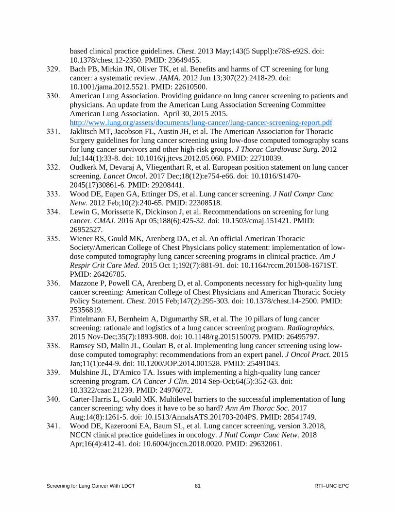
Screening for Lung Cancer With LDCT 81 RTI–UNC EPC
based clinical practice guidelines. Chest. 2013 May;143(5 Suppl):e78S-e92S. doi:
10.1378/chest.12-2350. PMID: 23649455.
329. Bach PB, Mirkin JN, Oliver TK, et al. Benefits and harms of CT screening for lung
cancer: a systematic review. JAMA. 2012 Jun 13;307(22):2418-29. doi:
10.1001/jama.2012.5521. PMID: 22610500.
330. American Lung Association. Providing guidance on lung cancer screening to patients and
physicians. An update from the American Lung Association Screening Committee
American Lung Association. April 30, 2015 2015.
http://www.lung.org/assets/documents/lung-cancer/lung-cancer-screening-report.pdf
331. Jaklitsch MT, Jacobson FL, Austin JH, et al. The American Association for Thoracic
Surgery guidelines for lung cancer screening using low-dose computed tomography scans
for lung cancer survivors and other high-risk groups. J Thorac Cardiovasc Surg. 2012
Jul;144(1):33-8. doi: 10.1016/j.jtcvs.2012.05.060. PMID: 22710039.
332. Oudkerk M, Devaraj A, Vliegenthart R, et al. European position statement on lung cancer
screening. Lancet Oncol. 2017 Dec;18(12):e754-e66. doi: 10.1016/S1470-
2045(17)30861-6. PMID: 29208441.
333. Wood DE, Eapen GA, Ettinger DS, et al. Lung cancer screening. J Natl Compr Canc
Netw. 2012 Feb;10(2):240-65. PMID: 22308518.
334. Lewin G, Morissette K, Dickinson J, et al. Recommendations on screening for lung
cancer. CMAJ. 2016 Apr 05;188(6):425-32. doi: 10.1503/cmaj.151421. PMID:
26952527.
335. Wiener RS, Gould MK, Arenberg DA, et al. An official American Thoracic
Society/American College of Chest Physicians policy statement: implementation of low-
dose computed tomography lung cancer screening programs in clinical practice. Am J
Respir Crit Care Med. 2015 Oct 1;192(7):881-91. doi: 10.1164/rccm.201508-1671ST.
PMID: 26426785.
336. Mazzone P, Powell CA, Arenberg D, et al. Components necessary for high-quality lung
cancer screening: American College of Chest Physicians and American Thoracic Society
Policy Statement. Chest. 2015 Feb;147(2):295-303. doi: 10.1378/chest.14-2500. PMID:
25356819.
337. Fintelmann FJ, Bernheim A, Digumarthy SR, et al. The 10 pillars of lung cancer
screening: rationale and logistics of a lung cancer screening program. Radiographics.
2015 Nov-Dec;35(7):1893-908. doi: 10.1148/rg.2015150079. PMID: 26495797.
338. Ramsey SD, Malin JL, Goulart B, et al. Implementing lung cancer screening using low-
dose computed tomography: recommendations from an expert panel. J Oncol Pract. 2015
Jan;11(1):e44-9. doi: 10.1200/JOP.2014.001528. PMID: 25491043.
339. Mulshine JL, D'Amico TA. Issues with implementing a high-quality lung cancer
screening program. CA Cancer J Clin. 2014 Sep-Oct;64(5):352-63. doi:
10.3322/caac.21239. PMID: 24976072.
340. Carter-Harris L, Gould MK. Multilevel barriers to the successful implementation of lung
cancer screening: why does it have to be so hard? Ann Am Thorac Soc. 2017
Aug;14(8):1261-5. doi: 10.1513/AnnalsATS.201703-204PS. PMID: 28541749.
341. Wood DE, Kazerooni EA, Baum SL, et al. Lung cancer screening, version 3.2018,
NCCN clinical practice guidelines in oncology. J Natl Compr Canc Netw. 2018
Apr;16(4):412-41. doi: 10.6004/jnccn.2018.0020. PMID: 29632061.

Screening for Lung Cancer With LDCT 82 RTI–UNC EPC
342. Goodwin JS, Nishi S, Zhou J, et al. Use of the shared decision-making visit for lung
cancer screening among Medicare enrollees. JAMA Intern Med. 2019 Jan 14doi:
10.1001/jamainternmed.2018.6405. PMID: 30640388.
343. Brenner AT, Malo TL, Margolis M, et al. Evaluating shared decision making for lung
cancer screening. JAMA Intern Med. 2018 Aug 13doi:
10.1001/jamainternmed.2018.3054. PMID: 30105393.
344. Couet N, Desroches S, Robitaille H, et al. Assessments of the extent to which health-care
providers involve patients in decision making: a systematic review of studies using the
OPTION instrument. Health Expect. 2015 Aug;18(4):542-61. doi: 10.1111/hex.12054.
PMID: 23451939.
345. Crothers K, Kross EK, Reisch LM, et al. Patients' attitudes regarding lung cancer
screening and decision aids. A survey and focus group study. Ann Am Thorac Soc. 2016
Nov;13(11):1992-2001. doi: 10.1513/AnnalsATS.201604-289OC. PMID: 27652509.
346. Henderson LM, Jones LM, Marsh MW, et al. Opinions, practice patterns, and perceived
barriers to lung cancer screening among attending and resident primary care physicians.
Risk Manag Healthc Policy. 2017;10:189-95. doi: 10.2147/RMHP.S143152. PMID:
29403320.
347. Hoffman RM, Sussman AL, Getrich CM, et al. Attitudes and beliefs of primary care
providers in New Mexico about lung cancer screening using low-dose computed
tomography. Prev Chronic Dis. 2015 Jul 9;12:E108. doi: 10.5888/pcd12.150112. PMID:
26160294.
348. Simmons VN, Gray JE, Schabath MB, et al. High-risk community and primary care
providers knowledge about and barriers to low-dose computed topography lung cancer
screening. Lung Cancer. 2017 Apr;106:42-9. doi: 10.1016/j.lungcan.2017.01.012. PMID:
28285693.
349. Lewis JA, Petty WJ, Tooze JA, et al. Low-Dose CT lung cancer screening practices and
attitudes among primary care providers at an academic medical center. Cancer Epidemiol
Biomarkers Prev. 2015 Apr;24(4):664-70. doi: 10.1158/1055-9965.Epi-14-1241. PMID:
25613118.
350. Hoffmann TC, Del Mar C. Patients' expectations of the benefits and harms of treatments,
screening, and tests: a systematic review. JAMA Intern Med. 2015 Feb;175(2):274-86.
doi: 10.1001/jamainternmed.2014.6016. PMID: 25531451.
351. Hoffmann TC, Del Mar C. Clinicians' expectations of the benefits and harms of
treatments, screening, and tests: a systematic review. JAMA Intern Med. 2017 Mar
1;177(3):407-19. doi: 10.1001/jamainternmed.2016.8254. PMID: 28097303.
352. Hoffman RM, Couper MP, Zikmund-Fisher BJ, et al. Prostate cancer screening decisions:
results from the National Survey of Medical Decisions (DECISIONS study). Arch Intern
Med. 2009 Sep 28;169(17):1611-8. doi: 10.1001/archinternmed.2009.262. PMID:
19786681.
353. Dobler CC, Midthun DE, Montori VM. Quality of shared decision making in lung cancer
screening: the right process, with the right partners, at the right time and place. Mayo Clin
Proc. 2017 Nov;92(11):1612-6. doi: 10.1016/j.mayocp.2017.08.010. PMID: 29101930.
354. Goodson JD. POINT: Should only primary care physicians provide shared decision-
making services to discuss the risks/benefits of a low-dose chest CT scan for lung cancer
screening? Yes. Chest. 2017 Jun;151(6):1213-5. doi: 10.1016/j.chest.2016.11.057. PMID:
28041890.

Screening for Lung Cancer With LDCT 83 RTI–UNC EPC
355. Dobler CC. Shared decision making rarely happens for lung cancer screening. JAMA
Intern Med. 2019 Jan 1;179(1):122. doi: 10.1001/jamainternmed.2018.6983. PMID:
30615081.
356. Reuland DS, Malo TL, Brenner AT. Shared decision making rarely happens for lung
cancer screening-reply. JAMA Intern Med. 2019 Jan 1;179(1):122-3. doi:
10.1001/jamainternmed.2018.6986. PMID: 30615085.
357. Dharod A, Bellinger C, Foley K, et al. The reach and feasibility of an interactive lung
cancer screening decision aid delivered by patient portal. Appl Clin Inform. 2019
Jan;10(1):19-27. doi: 10.1055/s-0038-1676807. PMID: 30625501.
358. Mazzone PJ, Tenenbaum A, Seeley M, et al. Impact of a lung cancer screening
counseling and shared decision-making visit. Chest. 2017 Mar;151(3):572-8. doi:
10.1016/j.chest.2016.10.027. PMID: 27815154.
359. Brenner AT, Cubillos L, Birchard K, et al. Improving the implementation of lung cancer
screening guidelines at an academic primary care practice. J Healthc Qual. 2018
Jan/Feb;40(1):27-35. doi: 10.1097/jhq.0000000000000089. PMID: 28885238.
360. Eberth JM, Qiu R, Adams SA, et al. Lung cancer screening using low-dose CT: the
current national landscape. Lung Cancer. 2014 Sep;85(3):379-84. doi:
10.1016/j.lungcan.2014.07.002. PMID: 25088660.
361. Eberth JM, Bozorgi P, Lebron LM, et al. Geographic availability of low-dose computed
tomography for lung cancer screening in the United States, 2017. Prev Chronic Dis. 2018
Oct 4;15:E119. doi: 10.5888/pcd15.180241. PMID: 30289107.
362. American College of Radiology. Accredited facility search. Reston, VA: ACR
Accreditation; n.d.
363. Iaccarino JM, Clark J, Bolton R, et al. A national survey of pulmonologists' views on
low-dose computed tomography screening for lung cancer. Ann Am Thorac Soc. 2015
Nov;12(11):1667-75. doi: 10.1513/AnnalsATS.201507-467OC. PMID: 26368003.
364. Carter-Harris L, Brandzel S, Wernli KJ, et al. A qualitative study exploring why
individuals opt out of lung cancer screening. Fam Pract. 2017 Apr 01;34(2):239-44. doi:
10.1093/fampra/cmw146. PMID: 28122849.
365. Cardarelli R, Roper KL, Cardarelli K, et al. Identifying community perspectives for a
lung cancer screening awareness campaign in Appalachia Kentucky: the Terminate Lung
Cancer (TLC) Study. J Cancer Educ. 2017 Mar;32(1):125-34. doi: 10.1007/s13187-015-
0914-0. PMID: 26411308.
366. Heny J. Kaiser Family Foundation. 2017 Employer Health Benefits Survey. San
Francisco, CA: Kaiser Family Foundation; 2017. Accessed May 20, 2019.
367. Aberle DR, Adams AM, Berg CD, et al. Baseline characteristics of participants in the
randomized national lung screening trial. J Natl Cancer Inst. 2010 Dec 1;102(23):1771-9.
doi: 10.1093/jnci/djq434. PMID: 21119104.
368. Curry SJ, Krist AH, Owens DK, et al. Risk assessment for cardiovascular disease with
nontraditional risk factors: US Preventive Services Task Force recommendation
statement. JAMA. 2018 Jul 17;320(3):272-80. doi: 10.1001/jama.2018.8359. PMID:
29998297.
369. Siu AL, Bibbins-Domingo K, Grossman DC, et al. Screening for chronic obstructive
pulmonary disease: US Preventive Services Task Force recommendation statement.
JAMA. 2016 Apr 5;315(13):1372-7. doi: 10.1001/jama.2016.2638. PMID: 27046365.

Screening for Lung Cancer With LDCT 84 RTI–UNC EPC
370. Lin JS, Bowles EJA, Williams SB, et al. Thyroid cancer: screening Evidence Synthesis
No. 151. AHRQ Publication No. 15-05221-EF-1. Rockville, MD: Quality AfHRa; May
2017.
371. LeFevre ML, U. S. Preventive Services Task Force. Screening for abdominal aortic
aneurysm: U.S. Preventive Services Task Force recommendation statement. Ann Intern
Med. 2014 Aug 19;161(4):281-90. doi: 10.7326/M14-1204. PMID: 24957320.
372. Guirguis-Blake JM, Beil TL, Sun X, et al. Primary care screening for abdominal aortic
aneurysm: a systematic evidence review for the U.S. Preventive Services Task Force
Evidence Synthesis No. 109. AHRQ Publication No. 14-05202-EF-1. Rockville, MD:
Agency for Healthcare Research and Quality; January 2014.
373. Tanner NT, Kanodra NM, Gebregziabher M, et al. The association between smoking
abstinence and mortality in the National Lung Screening Trial. Am J Respir Crit Care
Med. 2016 Mar 01;193(5):534-41. doi: 10.1164/rccm.201507-1420OC. PMID:
26502000.
374. Syrek JT, Chin J, Ashby L, et al. Center for Medicare & Medicaid Services Decision
memo for screening for lung cancer with low dose computed tomography (LDCT) (CAG-
00439N). Baltimore, MD: Centers for Medicare & Medicaid Services; 2015.
http://www.cms.gov/medicare-coverage-database/details/nca-decision-
memo.aspx?NCAId=274. Accessed May 20, 2019.
375. Siu AL. Behavioral and pharmacotherapy interventions for tobacco smoking cessation in
adults, including pregnant women: U.S. Preventive Services Task Force recommendation
statement. Ann Intern Med. 2015 Oct 20;163(8):622-34. doi: 10.7326/m15-2023. PMID:
26389730.
376. Pineiro B, Simmons VN, Palmer AM, et al. Smoking cessation interventions within the
context of low-dose computed tomography lung cancer screening: a systematic review.
Lung Cancer. 2016 Aug;98:91-8. doi: 10.1016/j.lungcan.2016.05.028. PMID: 27393513.
377. Iaccarino JM, Duran C, Slatore CG, et al. Combining smoking cessation interventions
with LDCT lung cancer screening: a systematic review. Prev Med. 2019 Apr;121:24-32.
doi: 10.1016/j.ypmed.2019.02.016. PMID: 30753860.
378. Shea BJ, Reeves BC, Wells G, et al. AMSTAR 2: a critical appraisal tool for systematic
reviews that include randomised or non-randomised studies of healthcare interventions,
or both. BMJ. 2017 Sep 21;358:j4008. doi: 10.1136/bmj.j4008. PMID: 28935701.
379. Clark MM, Cox LS, Jett JR, et al. Effectiveness of smoking cessation self-help materials
in a lung cancer screening population. Lung Cancer. 2004 Apr;44(1):13-21. doi:
10.1016/j.lungcan.2003.10.001. PMID: 15013579.
380. van der Aalst CM, de Koning HJ, van den Bergh KA, et al. The effectiveness of a
computer-tailored smoking cessation intervention for participants in lung cancer
screening: a randomised controlled trial. Lung Cancer. 2012 May;76(2):204-10. doi:
10.1016/j.lungcan.2011.10.006. PMID: 22054915.
381. Ferketich AK, Otterson GA, King M, et al. A pilot test of a combined tobacco
dependence treatment and lung cancer screening program. Lung Cancer. 2012
May;76(2):211-5. doi: 10.1016/j.lungcan.2011.10.011. PMID: 22088938.
382. Pozzi P, Munarini E, Bravi F, et al. A combined smoking cessation intervention within a
lung cancer screening trial: a pilot observational study. Tumori. 2015 May-
Jun;101(3):306-11. doi: 10.5301/tj.5000282. PMID: 25838249.

Screening for Lung Cancer With LDCT 85 RTI–UNC EPC
383. Filippo L, Principe R, Cesario A, et al. Smoking cessation intervention within the
framework of a lung cancer screening program: preliminary results and clinical
perspectives from the "Cosmos-II" Trial. Lung. 2015 Feb;193(1):147-9. doi:
10.1007/s00408-014-9661-y. PMID: 25323328.
384. Taylor KL, Hagerman CJ, Luta G, et al. Preliminary evaluation of a telephone-based
smoking cessation intervention in the lung cancer screening setting: A randomized
clinical trial. Lung Cancer. 2017 Jun;108:242-6. doi: 10.1016/j.lungcan.2017.01.020.
PMID: 28216065.
385. Marshall HM, Courtney DA, Passmore LH, et al. Brief tailored smoking cessation
counseling in a lung cancer screening population is feasible: a pilot randomized
controlled trial. Nicotine Tob Res. 2016 Jul;18(7):1665-9. doi: 10.1093/ntr/ntw010.
PMID: 26834052.
386. Dhanasopon AP, Salazar MC, Hoag JR, et al. Fate of pneumonectomy patients variably
captured by non-small cell lung cancer staging system. Ann Thorac Surg. 2017
Dec;104(6):1829-36. doi: 10.1016/j.athoracsur.2017.06.073. PMID: 29074151.
387. Lee SY, Hong MJ, Jeon HS, et al. Functional intronic ERCC1 polymorphism from
regulomeDB can predict survival in lung cancer after surgery. Oncotarget. 2015 Sep
15;6(27):24522-32. doi: 10.18632/oncotarget.4083. PMID: 26056042.
388. Miura H, Inoue T, Shiomi H, et al. Differences in rates of radiation-induced true and false
rib fractures after stereotactic body radiation therapy for Stage I primary lung cancer. J
Radiat Res. 2015 Mar;56(2):332-7. doi: 10.1093/jrr/rru107. PMID: 25504640.
389. Yousaf-Khan U, van der Aalst C, de Jong PA, et al. Final screening round of the
NELSON lung cancer screening trial: the effect of a 2.5-year screening interval. Thorax.
2017 Jan;72(1):48-56. doi: 10.1136/thoraxjnl-2016-208655. PMID: 27364640.
390. Zhou H, Tapias LF, Gaissert HA, et al. Lymph node assessment and impact on survival
in video-assisted thoracoscopic lobectomy or segmentectomy. Ann Thorac Surg. 2015
Sep;100(3):910-6. doi: 10.1016/j.athoracsur.2015.04.034. PMID: 26165483.
391. Brain K, Lifford KJ, Carter B, et al. Long-term psychosocial outcomes of low-dose CT
screening: results of the UK Lung Cancer Screening randomised controlled trial. Thorax.
2016 Nov;71(11):996-1005. doi: 10.1136/thoraxjnl-2016-208283. PMID: 27471048.
392. Brain K, Carter B, Lifford KJ, et al. Impact of low-dose CT screening on smoking
cessation among high-risk participants in the UK Lung Cancer Screening Trial. Thorax.
2017 Oct;72(10):912-8. doi: 10.1136/thoraxjnl-2016-209690. PMID: 28710339.
393. Pinsky PF, Marcus PM, Kramer BS, et al. Diagnostic procedures after a positive spiral
computed tomography lung carcinoma screen. Cancer. 2005 Jan 1;103(1):157-63. doi:
10.1002/cncr.20746. PMID: 15529306.
394. Li K, Husing A, Sookthai D, et al. Selecting high-risk individuals for lung cancer
screening: a prospective evaluation of existing risk models and eligibility criteria in the
German EPIC Cohort. Cancer Prev Res (Phila). 2015 Sep;8(9):777-85. doi:
10.1158/1940-6207.capr-14-0424. PMID: 26076698.
395. McKee BJ, Regis SM, McKee AB, et al. Performance of ACR Lung-RADS in a clinical
CT lung screening program. J Am Coll Radiol. 2015 Mar;12(3):273-6. doi:
10.1016/j.jacr.2014.08.004. PMID: 25176499.
396. Colinet B, Jacot W, Bertrand D, et al. A new simplified comorbidity score as a prognostic
factor in non-small-cell lung cancer patients: description and comparison with the

Screening for Lung Cancer With LDCT 86 RTI–UNC EPC
Charlson's index. Br J Cancer. 2005 Nov 14;93(10):1098-105. doi:
10.1038/sj.bjc.6602836. PMID: 16234816.

Figure 1. Analytic Framework
Screening for Lung Cancer With LDCT 87 RTI–UNC EPC
Figure 1. Analytic Framework
* The evaluation of evidence on treatment will be limited to studies of surgical resection or stereotactic body radiotherapy for
stage I NSCLC.
Abbreviations: NSCLC=non-small cell lung cancer.

Figure 2. Summary of Evidence Search and Selection
Screening for Lung Cancer With LDCT 88 RTI–UNC EPC
Figure 2. Summary of Evidence Search and Selection
Abbreviations: KQ=key question; SBRT/SABR=SBRT=stereotactic body radiotherapy/SABR= stereotactic ablative radiation.
# of records identified through database
searching
9,228
PubMed: 9,038
Cochrane: 190
# of additional records identified through other sources
2,313
Included in last review for USPSTF: 90
Handsearch: 230
WHO ICTRP: 380
ClinicalTrials.Gov: 1,613
# of records screened
11,541
# of records excluded
9,329
# of full-text articles assessed for eligibility
2,212
# of full-text articles excluded, with reasons
1,989
Wrong language/Non-English: 3
Abstract only: 21
Ineligible population: 934
Ineligible risk prediction model: 51
Ineligible screening modality: 91
Ineligible intervention (tx): 103
Ineligible comparator: 74
Ineligible outcome(s): 179
Ineligible study design (systematic review): 20
Ineligible study design: 162
Ineligible sample size: 290
Ineligible study duration for KQ 6 (surgery studies only): 8
Ineligible study duration for KQ 6 (SBRT/SABR): 28
Eligible, except for country setting: 8
Ineligible for publication before 2001: 0
Irretrievable: 6
Poor quality: 11
# of articles included in qualitative
synthesis of systematic review
223*
*Because many articles contribute to ≥1
Key Question, the number of articles
listed per-Key Question in this section
does not add up to 223
26 articles
included for
KQ 1
9 articles
included for
KQ 2
52 articles
included for
KQ 3
75 articles
included for
KQ 4
18 articles
included for
KQ 5
61 articles
included for
KQ 6
85 articles
included for
KQ 7
3 articles
included for
KQ 8

Figure 3. Trial Results for Lung Cancer Incidence (KQ 1)
Screening for Lung Cancer With LDCT 89 RTI–UNC EPC
Fig
ure 3. Trial results for lung cancer incidence (KQ 1)
Note: G1=LDCT; G2=Control; Favors Intervention indicates fewer incident lung cancers with intervention (LDCT screening); Favors Control indicates more incident lung cancers
with intervention. Two rows are included in the figure for the NLST, showing the data from the 6.5-year followup and from the extended post-screening followup data at a median
of 11.3 years after randomization for lung cancer incidence. The NELSON trial reported lung cancer incidence for the 13,195 males enrolled in the trial, excluding the 2,594
females that were enrolled. Therefore, the NELSON results in the figure above include only data for male participants (data were not reported for the female participants). The trial
did not report total person-years of followup for lung cancer incidence, but those were able to be calculated from other data that were reported (5.58 cases per 1,000 person-years
vs. 4.91 cases per 1,000 person-years at 10 years; RR, 1.14 [95% CI, 0.97, 1.33]).The Nelson trial reported median age and median pack-years instead of mean age and mean pack
years. The LUSI trial was not included in the figure above because it did not reporting person-years of followup. The LUSI trial reported 85 lung cancers in the intervention group
and 67 in the control group at a mean of 8.8 years follow up (p=0.16).
Abbreviations: DANTE=Detection and Screening of Early Lung Cancer with Novel Imaging Technology and Molecular Essays; DLCST=Danish Lung Cancer Screening Trial;
ITALUNG=Italian Lung Cancer Screening Trial; LUSI=Lung cancer Screening Intervention; NELSON=Nederlands-Leuvens Longkanker Screenings Onderzoek; NLST=National
Lung Screening Trial.

Figure 4. Trial Results for Incidence of Early (I-II) and Late (III-IV) Stage Lung Cancer (KQ 1)
Screening for Lung Cancer With LDCT 90 RTI–UNC EPC
Figure 4. Trial results for incidence of early (I-II) and late (III-IV) stage lung cancer (KQ 1)
Note: G1=LDCT; G2=Control; The MILD trial randomized participants to annual screening, biennial screening, or a control group. For the 10-year followup, the annual and
biennial screening groups were combined. At the 10-year followup, the median duration of screening for those in the screening groups was 6.2 years.
Abbreviations: DANTE=Detection and Screening of Early Lung Cancer with Novel Imaging Technology and Molecular Essays; DLCST=Danish Lung Cancer Screening Trial;
ITALUNG=Italian Lung Cancer Screening Trial; MILD=Multicentric Italian Lung Detection; NELSON=Nederlands-Leuvens Longkanker Screenings Onderzoek;
NLST=National Lung Screening Trial.

Figure 5. Trial Results for Lung Cancer Mortality (KQ 1)
Screening for Lung Cancer With LDCT 91 RTI–UNC EPC
Figure 5. Trial results for lung cancer mortality (KQ 1)
Note: G1=LDCT; G2=Control. The NLST trial reported extended post-screening followup data at 12.3 years after randomization (not included in the figure above because person-
years of followup were not reported): 1,147 lung cancer deaths occurred in the LDCT screening group (42.9 cases per 1,000 participants) and 1236 occurred in the CXR control
group (46.2 cases per 1,000 participants) (RR, 0.92 [95% CI, 0.85, 1.00] and absolute difference between groups of 3.3 [95% CI, -0.2, 6.8] lung cancer deaths per 1,000
participants). The ITALUNG and LSS trials reported median pack per years instead of mean pack per years. The NELSON trial reported its main results for the 13,195 males
enrolled in the trial (excluding the females enrolled), reporting 156 lung cancer deaths in the screening group and 206 lung cancer deaths in the control group for males at 10 years
of followup (rate ratio 0.76 [95% CI, 0.61 to 0.94]). For the 2,594 females, NELSON reported 25 lung cancer deaths in the screening group and 36 in the control group at 10 years
of followup (rate ratio 0.67 [95% CI, 0.38 to 1.14]). The NELSON results in the figure above combine data for all participants in the trial. The Nelson trial reported median age
and median pack-years instead of mean age and mean pack-years. The LUSI trial was not included in the figure above because it did not report person-years of followup. The
study reported 29 lung cancer deaths in the intervention group and 40 lung cancer deaths in the control group at a mean of 8.8 years follow up (p=0.19).
Abbreviations: DANTE=Detection and Screening of Early Lung Cancer with Novel Imaging Technology and Molecular Essays; DLCST=Danish Lung Cancer Screening Trial;
ITALUNG=Italian Lung Cancer Screening Trial; LSS=Lung Screening Study; LUSI=Lung cancer Screening Intervention; NELSON=Nederlands-Leuvens Longkanker Screenings
Onderzoek; NLST=National Lung Screening Trial.

Figure 6. Trial Results for All-Cause Mortality (KQ 1)
Screening for Lung Cancer With LDCT 92 RTI–UNC EPC
Fig
ure 6. Trial results for all-cause mortality (KQ 1)
Note: G1=LDCT; G2=Control. The NLST trial reported extended post-screening followup data at 12.3 years after randomization (not included in the figure above because person-
years of followup were not reported): 5,253 deaths occurred in the LDCT screening group (196.6 cases per 1,000 participants) and 5,366 deaths in the CXR group (200.7 cases per
1,000 participants) (RR, 0.97 [95% CI, 0.94, 1.01]). The ITALUNG and LSS trials reported median pack per years instead of mean pack per years. The NELSON trial reported all-
cause mortality for its primary analysis of the 13,195 males enrolled in the trial, excluding the 2,594 females that were enrolled. Therefore, the NELSON results in the figure above
include only data for male participants (data were not reported for the female participants). The Nelson trial reported median age and median pack-years instead of mean age and
mean pack-years.
Abbreviations: DANTE=Detection and Screening of Early Lung Cancer with Novel Imaging Technology and Molecular Essays; DLCST=Danish Lung Cancer Screening Trial;
ITALUNG=Italian Lung Cancer Screening Trial; LSS=Lung Screening Study; NELSON=Nederlands-Leuvens Longkanker Screenings Onderzoek; NLST=National Lung
Screening Trial.

Table 1. NSCLC Staging Overview, Typical 5-Year Survival, and Treatment Approaches28, 257-261
Screening for Lung Cancer With LDCT 93 RTI–UNC EPC
Table 1. NSCLC Staging Overview, Typical 5-Year Survival, and Treatment Approaches29, 317-321
Stage Description TNM
Classifications 5-Year Survival Treatment Approach
I Tumor ≤4 cm with no involvement of lymph
nodes or distant metastasis
T1-2a N0 M0 77-92% for Stage 1a; 68% for Stage 1b
Surgical resection, including lobectomy; SBRT (mainly for nonsurgical candidates)
II Tumor >4 cm and ≤7 cm with no involvement of
lymph nodes or distant metastasis
OR tumor ≤5 cm with metastases in ipsilateral
pulmonary/hilar lymph nodes and no distant metastasis
T2b-3 N0 M0 T1-2 N1 M0
53-60% Lobectomy + adjuvant chemotherapy
III Heterogeneous group of disease, includes tumors ≥7 cm with or without ipsilateral lymph node involvement and smaller tumors with metastasis to the ipsilateral mediastinal/subcarinal nodes, contralateral mediastinal/hilar nodes, or supraclavicular nodes
T1-4 N0-3 M0 13-36% Combined modality approach (chemotherapy, radiation therapy, +/- surgery, and/or immunotherapy)*
IV Presence of distant metastasis: single or multiple extra-thoracic metastasis, malignant pleural or pericardial effusion
Any T or N M1a-c
1-10% Combined modality approach (chemotherapy, radiation therapy, targeted molecular therapy and/or immunotherapy and +/- surgery)*
*Tailored to patient disease and performance status.
Abbreviations: a=separate tumor nodule[s] in contralateral lobe, tumor with pleural or pericardial nodule(s) or malignant pleural
or pericardial effusions; b=single extrapulmonary metastasis; c=multiple extrapulmonary metastases in one or more organs;
M=distant metastasis; N=regional lymph nodes; NSCLC=non-small cell lung cancer; SBRT=stereotactic body radiation therapy;
T=primary tumor; TNM=Tumor Node Metastasis.

Table 2. Characteristics of Included RCTs Evaluating Screening With LDCT Compared With CXR or With No Screeninga
Screening for Lung Cancer With LDCT 94 RTI–UNC EPC
Table 2. Characteristics of Included RCTs Evaluating Screening with LDCT Compared No Screening*
Study Recruitment
Years Sample Size;
Country
Mean Age (Ages
Eligible) % Male Baseline Smoking
Status
Eligibility Criteria for Pack-Years; Years
Since Quitting Screening Rounds, n
Screening Intervals, y
Total Median Followup, y Quality
DANTE59, 69,
70 2001-2006 2,472; Italy 65
(60-74) 100 Current: 57%
Former: 43% Mean pack-years: 47
≥20; <10 y 5 0, 1, 2, 3, 4 8.4 Fair
DLCST63, 65 2004-2006 4,104; Denmark
58 (50-70)
56 Current: 76% Former: 24% Mean pack-years: 36
≥20; quit after age 50 and <10 y ago
5 0, 1, 2, 3, 4 9.8 Fair
ITALUNG60 2004-2006 3,206; Italy 61 (55-69)
65 Current: 65% Former: 35% Median pack-years: 39
≥20 in the last 10 years or quit within the last 10 year
4 0, 1, 2, 3 9.3† Fair
LSS67, 68, 73 2000-2001 3,318; U.S. NR (55-74)
59 Current: 58% Former: 42% Median pack-years: 54
≥30; <10 y 2 0, 1 5.2 Fair
LUSI57, 58, 71 2007-2011 4,052; Germany
NR (50-69)
65 Current: 62% Former: 35% Mean pack-years: NR
≥25 y of 15 cigarettesǂ
or ≥30 y of 10
cigarettesǂ; ≤10 y
5 0, 1, 2, 3, 4 8.8 Fair
NELSON32,
74-77 2003-2006 15,792; the
Netherlands and Belgium
58 median (50-74)
84 Current: 55% Former: 45% Median pack-years: 38
>15 cigarettes/day for >25 years or >10 cigarettes/day for >30 years; ≤10 y
4 0, 1, 3, 5.5 10 Fair
NLST31, 54-56,
61, 62, 64, 66, 72 2002-2004 53,542; U.S. 61
(55-74) 59 Current: 48%
Former: 52% Mean pack-years: 56
≥30; ≤15 y 3 0, 1, 2 7 (and post-trial followup to 12.3 years)
Good
* NLST and LSS compared screening with LDCT vs. screening with CXR. All other trials compared screening with LDCT with no screening. † The ITALUNG study reported 9.3 years for lung cancer–specific mortality and 8.5 years for lung cancer incidence.
ǂ The LSS was a feasibility pilot study.
Abbreviations: CXR=chest X-ray; DANTE=Detection and Screening of Early Lung Cancer by Novel Imaging Technology and Molecular Essays; DLCST=Danish Lung Cancer
Screening Trial; ITALUNG=Italian Lung Cancer Screening Trial; LDCT=low-dose computed tomography; LSS=Lung Screening Study; LUSI=The German Lung Cancer
Screening Intervention Trial; n=number; NLST=National Lung Screening Trial; NR=not reported; RCT=randomized, controlled trial.

Table 3. Predictors Used in Risk Prediction Models for Identifying Adults at Higher Risk of Lung Cancer Mortality and Model Applicability
Screening for Lung Cancer With LDCT 95 RTI–UNC EPC
Table 3. Predictors Used in Risk Prediction Models for Identifying Adults at Higher Risk of Lung Cancer Mortality and Model Applicability
Model Name LCDRAT8
2 Kovalchik
Model54 PLCOm20128
3
Simplified PLCOm20128
1 COPD-
LUCSS86 Bach
Model88 LLP
Model90 Simplified LLP81, 90*
TSCE Incidence Model91
Knoke Model94
TSCE CPS
Death Model92
TSCE NHS/HPFS
Death Model322
HUNT Lung Cancer Model 93
Risk Factors
Personal
Age X X X X X† X X X X X X X X
Sex X X X X X X X
Race and/or ethnicity
X X Xǂ
Body mass index
X X X X§ X
Education (levels)
Xǁ Xǁ
Previous malignant tumor
X X
Smoking History
Smoking status
X X X X
Cessation age X
Smoking duration
X X X X X¶ X¶ X X X X
Cigarettes per day
X X X X X X X X X
Pack-years X# X X# X
Quit duration X X†† X X X X X X X X
Family History of Cancer
Cases of lung cancer
Xǂǂ Xǂǂ X X
Age of onset of lung cancer
X§§
Exposures and Lung Conditions
Emphysema X X Xǁǁ
COPD X
Pneumonia X
Daily cough X

Table 3. Predictors Used in Risk Prediction Models for Identifying Adults at Higher Risk of Lung Cancer Mortality and Model Applicability
Screening for Lung Cancer With LDCT 96 RTI–UNC EPC
Model Name LCDRAT8
2 Kovalchik
Model54 PLCOm20128
3
Simplified PLCOm20128
1 COPD-
LUCSS86 Bach
Model88 LLP
Model90 Simplified LLP81, 90*
TSCE Incidence Model91
Knoke Model94
TSCE CPS
Death Model92
TSCE NHS/HPFS
Death Model322
HUNT Lung Cancer Model 93
Daily indoor exposure to smoke (hours)
X
Asbestos exposure
X X
Applying the Model Information
Applicable to never smokers
X X X X X X
Applicable to former smokers
X X X X X X X X X X X X X
Applicable to current smokers
X X X X X X X X X X X X X
Model predicts risk of incidence
X X X X X X X X X
Model predicts survival
X¶¶ X X X X
Time horizon of prediction
5 y 5 y 6 y 6 y NR 1 y (iterative)
5 y 5 y 1 y (iterative)
1 y (iterative)
1 y (iterative)
1 y (iterative) 6 and 16 years
Model formula printed
X X X X X X X X X X X
Discrimin-ation & calibration##
Discrimination for lung cancer incidence (AUC) range
0.70-0.80 -- 0.69-0.89 0.68-0.78 NR 0.68-0.78 0.66-0.79
0.66-0.74 0.67-0.78 0.67-0.77 0.62-0.74 0.67-0.78 0.87 (6 years)
Discrimination for lung cancer mortality (AUC) range
0.73-0.81 0.80 0.72-0.81 0.71-0.80 NR 0.71-0.80 0.67-0.77
0.68-0.79 0.68-0.79 0.68-0.78 0.63-0.77 0.68-0.79 NR
Calibration of model for lung cancer
0.94-1.06 -- 0.87-0.98 1.02-1.04 NR 0.99-1.09 0.68-1.05
0.76-1.07 0.79-0.87 0.70-1.09 0.59-0.90 0.76-0.85 NR (shown as
calibration plots that

Table 3. Predictors Used in Risk Prediction Models for Identifying Adults at Higher Risk of Lung Cancer Mortality and Model Applicability
Screening for Lung Cancer With LDCT 97 RTI–UNC EPC
Model Name LCDRAT8
2 Kovalchik
Model54 PLCOm20128
3
Simplified PLCOm20128
1 COPD-
LUCSS86 Bach
Model88 LLP
Model90 Simplified LLP81, 90*
TSCE Incidence Model91
Knoke Model94
TSCE CPS
Death Model92
TSCE NHS/HPFS
Death Model322
HUNT Lung Cancer Model 93
incidence; range
show fairly good
calibration)
Calibration of model for lung cancer mortality, range
0.94-1.31 0.97 0.95-1.01 1.02-1.19 NR 0.97-1.21 0.69-1.18
0.79-1.12 0.84-0.94 0.79-0.89 0.64-0.99 0.80-0.92 NR
* The simplified version of the LLP model uses the same parameter estimates as the original LLP model. However, when applying this model to a participant, it is assumed that
only information on age and smoking history is known. Thus, the simplified model assumes that the participant had no prior diagnosis of pneumonia, no occupational exposure to
asbestos, no prior diagnosis of a malignant tumor, and no family history of lung cancer. † Age > 60
ǂ Only applicable to white males ages 40 to 79 years. § BMI <25. ǁ 1≤12 grade; 2=high school graduate; 3=post-high school but not college; 4=some college; 5=bachelor’s degree; 6=graduate school. ¶ Smoking duration levels for model: never; 1-20 years; 21-40 years; 41-60 years; >60 years. # Binary: >1 pack/day.
** Pack-years: >60. †† Categories: 0=less than 1 year; 1=1 to 5 years; 2=more than 5 years.
ǂǂ 0=No first-degree relatives with lung cancer; 1=1 first-degree relative with lung cancer; 2=2 or more first-degree relatives with lung cancer. §§ Early onset (age <60 years); late onset (age ≥60 years). ǁǁ Radiologic emphysema. ¶¶ Model can estimate 10-year risk, but authors report 5-year estimates because the NLST only had 5.5 years of followup data. ## LCDRAT: Cohorts used for external validation: PLCO-CXR and NLST-CXR. Discrimination higher in NHIS and PLCO vs. NLST cohorts. Calibration metric: Ratio of model-
predicted cases to observed cases. A value of 1 indicates optimal calibration. Kovalchik: Cohort used for external validation: PLCO-CXR. Calibration metric: Ratio of model-
predicted cases to observed cases.
PLCOm2012: Cohorts used for external validation: PLCO-CXR and control, 45 and UP study, NLST-CT and CXR, NHIS. Discrimination higher in PLCO vs. NLST cohorts.
Calibration metric reported in table from ten Haaf et al: slope of calibration plot observed vs. expected. Perfect calibration if slope=1. Two studies reported calibration as the
median (or mean) and 90th percentile absolute differences between observed and predicted risk probabilities, which ranged from 0.006-0.009 and 0.016-0.042, respectively.
For all other models except the HUNT model, discrimination and calibration are reported from the ten Haaf study. The range of discrimination and calibration outcomes are
estimated using the NLST CT and CXR arms, and the PLCO-CXR and control arms. In general, discrimination and calibration were better in the PLCO cohorts than in the NLST
cohorts. An exhaustive search and synthesis of risk model performance metrics was not in the scope of this review; thus, the numbers in this table are not a comprehensive
description of discrimination and calibration reported in all studies of these models (inclusion was limited to studies that reported eligible benefits and harms outcomes and
compared with USPSTF 2013 or NLST criteria).
Abbreviations: AUC=area under the curve; BMI=body mass index; COPD=chronic obstructive pulmonary disease; COPD-LUCSS=COPD-Lung Cancer Screening Score;
CPS=Cancer Prevention Study; HPFS=Health Professionals Follow-Up Study; LCDRAT=Lung Cancer Death Risk Assessment Tool; LLP=Liverpool Lung Project; NHS=Nurses’
Health Study; NLST=National Lung Screening Trial; PLCO=Prostate, Lung, Colorectal, and Ovarian Cancer Screening Trial; TSCE=Two Stage Clonal Expansion Model.

Table 4. PLCOm2012 Model Estimated Benefits and Harms Over 6 Years Compared With USPSTF or NLST Criteria
Screening for Lung Cancer With LDCT 98 RTI–UNC EPC
Author, Year
Method for Determining Lung Cancer Incidence
Risk Threshold Risk
Threshold
Cohort to Estimate
Outcomes Benefits Harms: False-Positive
Number (%) Harms: False-Positive Rate
Tammemagi et al, 201383
Fixed NLST population 1.35% PLCO-CXR arm Screen-prevented deaths: PLCOm2012: 81 NLST: 69 NNS: PLCOm2012: 174 NLST: 203
Model: 13,581 (96%) NLST: 13,662 (96.6%)
Model: 37.1 NLST: 37.3
Landy 2019 87 Fixed NLST population31 1.3% NHIS 2015 data Screen-prevented deaths: PLCOm2012: 56,528 USPSTF: 41,298 NNS: PLCOm2012: 222 USPSTF: 194
NR NR
ten Haaf et al, 201781 Fixed NLST population 1.35% PLCO-CXR arm NR Model: 24904 (97.5%) NLST: 24784 (97.9%)
Model: 37.5 NLST: 37.8
ten Haaf et al, 201781 Fixed NLST population 1.36% PLCO-control arm NR Model: 24287 (97.7%) NLST: 24248 (97.8%)
Model: 38.3 NLST: 38.1
Weber et al, 201784 Fixed NLST population 1.73% 45 and Up Study NR Model: 12,982 (96.9%) NLST: 12,929 (97.3%)
Model: 24.8 NLST: 24.7
Tammemagi et al, 201485
NLST mortality benefit 1.51% PLCO-CXR arm NNS: Model: 225 USPSTF: NR
Model: 12378 (95.8%) USPSTF: 13688 (96.6%)
Model: 33.8 USPSTF: 37.3
Landy 2019 87 NLST mortality benefit56 1.51% NHIS 2015 data Screen-prevented deaths: PLCOm2012: 54,456 USPSTF: 41,298 NNS: PLCOm2012: 207 USPSTF: 194
NR NR
Weber et al, 201784 NLST mortality benefit 1.51% 45 and Up Study NR Model: 14,642 (97.1%) USPSTF: 13,800 (97.1%)
Model: 28.0 USPSTF: 23.7
Weber et al, 201784 Optimal ROC classification
1.49% 45 and Up Study NR Model: 14,774 (97.1%) USPSTF: 13,800 (97.1%)
Model: 28.2 USPSTF: 23.7
Weber et al, 201784 Conservative 2% 45 and Up Study NR Model: 11,168 (96.6%) USPSTF: 13,800 (97.1%)
Model: 21.3 USPSTF: 23.7
Landy 2019 87 Fixed USPSTF population 2.19% NHIS 2015 data Screen-prevented deaths: PLCOm2012: 47,401 USPSTF: 41,298 NNS: PLCOm2012: 169 USPSTF: 194
NR NR
Abbreviations: CXR=chest X-ray; NLST=National Lung Screening Trial; NHIS=National Health Interview Survey; NNS=number needed to screen; NR=not reported;
PLCO=Prostate, Lung, Colorectal, and Ovarian Cancer Screening Trial; ROC=receiver operating characteristics; USPSTF=U.S. Preventive Services Task Force.

Table 5. Summary of Modeling Studies Evaluating Screen-Prevented Lung Cancer Deaths and NNS to Prevent One Lung Cancer Death*
Screening for Lung Cancer With LDCT 99 RTI–UNC EPC
Table 5. Sum Summary of f Modeling Studies Evaluating Screen-Prevented Lung Cancer Deaths and NNS to Prevent One Lung Cancer Death*
Author, Year Model Comparator Time
Horizon, y
Risk Threshold (Selection Method)
Cohort to Estimate
Outcomes (Sample Size)
Screen-Prevented Lung Cancer Deaths,
Model vs. Comparator
NNS, Model vs.
Comparator
FP per Prevented
Deaths, Model vs. Comparator
Overdiagnosed Lung Cancers per Prevented Death,
Model vs. Comparator
Tammemagi, 201383
PLCOm2012 model
NLST 6 1.35% (fixed population)
PLCO-CXR arm (37,327)
81 vs. 69 174 vs. 203 167 vs. 196 1.04 vs. 1.04
Landy 201987 PLCOm2012 model
USPSTF 6 1.3% (Reference Tammemagi et al 201331)
NHIS 2015 (12,600,000)
56,528 vs. 41,298 222 vs. 194 150 vs. 133 NR
Landy 201987 PLCOm2012 model
USPSTF 6 1.51% (NLST mortality benefit-Reference Tammemagi et al 201456)
NHIS 2015 (11,300,000)
54,456 vs. 41,298 207 vs. 194 141 vs. 133 NR
Landy 201987 PLCOm2012 model
USPSTF 6 2.19% (fixed population)
NHIS 2015 (8,000,000)
47,401 vs. 41,298 169 vs. 194 119 vs. 133 NR
Katki, 201682 LCDRAT model
NLST 5 1.2% (fixed population)
NHIS 2010-2012 (9,018,130)
55,717 (95% CI, 53,033 to 58,400) vs. 46,488 (95% CI, 43,924 to 49,053)
162 (157-166) vs. 194 (187-201)
116 (113-119) vs. 133 (128-137)
0.91 vs. 0.93
Landy 201987 LCDRAT model
USPSTF 5 1.2% (reference Katki, 20163)
NHIS 2015 (9,000,000)
53,732 vs. 41,298 168 vs. 194 119 vs. 133 NR
Landy 201987 LCDRAT model
USPSTF 5 1.33% (fixed population)
NHIS 2015 (8,000,000)
51,019 vs. 41,298 156 vs. 194 112 vs. 133 NR
Katki, 201682 LCDRAT model
NLST 5 0.9% (fixed effectiveness)
NHIS 2010-2012 (12,101,749)
62,382 (95% CI, 59,567 to 65,196) vs. 46,488 (95% CI, 43,924 to 49,053)
194 (188-200) vs. 194 (187-201)
134 (131-138) vs. 133 (128-137)
0.92 vs. 0.93
Kovalchik, 201354
Kovalchik et al
NLST 5 0.84% (risk quintile 3-5)
NLST-CT (26,604)
77 vs. 88 208 vs. 302 78 vs. 108 NR
Kovalchik, 201354
Kovalchik et al
NLST 5 1.23% (risk quintile 4-5)
NLST-CT (26,604)
64 vs. 88 166 vs. 302 64 vs. 108 NR
*Kovalchik et al applied a model to NLST, intrinsically conferring NLST benefits in lung cancer detection and mortality reduction. The other two studies assumed NLST-like
benefits to calculated outcomes.
Abbreviations: CI=confidence interval; CXR=chest X-ray; FP=false positive; LCDRAT=Lung Cancer Death Risk Assessment Tool; NHIS=National Health Interview Survey;
NLST=National Lung Screening Trial; NLST-CT=National Lung Screening Trial-Computerized Tomography arm; NNS=number needed to screen; NR=not reported; PLCO=
Prostate, Lung, Colorectal, and Ovarian Cancer Screening Trial; vs.=versus.

Table 6. Accuracy of LDCT for Lung Cancer Screening in RCTs (KQ 3)
Screening for Lung Cancer With LDCT 100 RTI–UNC EPC
Table 6. Accuracy of LDCT for Lung Cancer Screening in RCTs
Trial Name Author, Year
Number Analyzed
Nodule Classification Framework* Threshold for Positive†
Screening Protocol Sn Sp PPV NPV
DANTE Infante, 201559
2,450 I-ELCAP >5 mm average diameter 5 annual screens 79.5% 75.5% 18.6% 98.1%
DLCST Pedersen, 2009120
4,104 DLCST >15 mm maximum diameter or 5-15 mm with >25% volume increase on 3-month repeat
5 annual screens (4 reported)
NR NR 9.5% NR
ITALUNG Lopes Pegna, 201397
1,406 I-ELCAP ≥5 mm average diameter solid nodule, ≥10 mm GGN average diameter, any part-solid nodule
4 annual screensǂ 95.0% 26.4% 3.6% 99.4%
LSS Croswell, 2010117
1,610 NLST ≥3 mm maximum diameter T0, ≥4 mm maximum diameter for T1
2 annual screens NR NR 7.0% NR
LUSI Becker, 201557
2,028 I-ELCAP ≥5 mm average diameter 5 annual screens (4 completed)
93.5% 62% 7.2% 99.7%
MILD Sverzellati, 201680
1,152 Modified NELSON
Volume >250 mm3 or 60-250 mm3 with >25% volume increase on 3-month repeat
5 annual screens 68.5% 99.2% 40.6% 99.7%
MILD Sverzellati, 201680
1,151 Modified NELSON
Volume >250 mm3 or 60-250 mm3 with >25% volume increase on 3-month repeat
3 biennial screens 73.5% 99.2% 42.4% 99.8%
NELSON De Koning, 202074
6,583§ NELSON Volume >500 mm3 or 50-500 mm3 with VDT<400 d on 3-month repeat
4 rounds; baseline and after 1 y, 3 y, 5.5 y
59.0% 95.8%§ 43.5%§ 97.7%§
NLST Pinsky, 201399
26,022 NLST ≥4 mm longest diameter 3 annual screens 93.1% 76.5% 3.3% 99.9%
UKLS Field, 201695
1,994 Modified NELSONǁ
Volume >500mm3 or 50-500 mm3 with VDT<400 d on 3-month repeat
1 screen NR NR 36.8% NR
Mean, range NA NA NA NA 80.3%, 59.0%-95.0%
76.4%, 26.4%-99.2%
21.3%, 3.3%-43.5%
99.2%, 97.7%-99.9%
* We categorized whether the approach to nodule classification was most similar to the approach used in NLST, NELSON, DLCST, or I-ELCAP. † These are the abbreviated criteria for a positive screen. Studies also considered specific features of nodules, for example.
ǂ Study ongoing at the time of this publication. § This evaluation excluded some NELSON participants because it was limited to males in the screening group (data were not presented for the 1,317 females in the screening
group). The accuracy calculations in this row used NELSON’s approach to classifying results, with indeterminate results that required a 3-month followup LDCT being
categorized as negatives as long as the 3-month followup LDCT was negative (whereas other studies categorized the same type of thing, when any additional LDCT was required
for evaluation, as a false positive). ǁ Nodules with volumes <50 mm3 were split into two categories. Those <15 mm3 received no further followup. Those 15-49 mm3 received followup LDCT scan in 1 year.
Abbreviations: DANTE=Detection and Screening of Early Lung Cancer with Novel Imaging Technology and Molecular Essays; DLCST=Danish Lung Cancer Screening Trial;
GGN=ground-glass nodule; I-ELCAP=International Early Lung Cancer Action Program; ITALUNG=Italian Lung Cancer Screening Trial; LDCT=low-dose computed
tomography; LSS=Lung Screening Study; LUSI=The German Lung Cancer Screening Intervention Trial; MILD=Multi-centric Italian Lung Detection; NA=not applicable;
NELSON=Nederlands-Leuvens Longkanker Screenings Onderzoek; NLST=National Lung Screening Trial; NR=not reported; NPV=negative predictive value; PPV=positive
predictive value; RCT=randomized, controlled trial; Sn=sensitivity; Sp=specificity; UKLS= UK Lung cancer Screening; VDT=volume doubling time.

Table 7. Accuracy of LDCT for Lung Cancer Screening in Nonrandomized Studies (KQ 3)
Screening for Lung Cancer With LDCT 101 RTI–UNC EPC
Table 7. Accuracy of LDCT for Lung Cancer Screening in Nonrandomized Studies
Author, Year Trial/Database Country Number
Analyzed Threshold for
Positive* Screening Protocol Sn Sp PPV NPV
Crucitti, 2015109 “Un respiro per la vita”
Italy 1,500 >4 mm, avg max and min
1 scan NR 34.0% 4.6% NR
Henschke, 2004129 I-ELCAP U.S. 2,698 ≥5 mm, avg max and min
2 annual scans 97.0% 90.0% Baseline: 20.9% Annual: 11.0%
Baseline: 99.2% Annual: 100%
Toyoda, 2008124 Osaka Japan 18,070 “Need for further clinical exam”
2 annual scans 88.9% 92.6% NR NR
Tsushima, 2008121 Azumi & Shinshu Japan 2,486 >3 mm Annual scans 100.0% 96.9% 9.9% 100.0%
Tammemagi, 2017113
PanCan Canada 2,537 ≥1 mm T0: Baseline T1: 1 year T4: 4 years
92.7% NR NR NR
Swensen, 2005128 Mayo U.S. 1,520 NCN >4 mm, avg max and min
5 annual scans 95.5% 37.9% 5.8% 99.3%
Menezes, 2010119 Toronto Canada 3,552 ≥5 mm, avg max and min
6 annual screenings
87.7% 99.3% NR NR
Veronesi, 2008125 COSMOS Italy 5,201 ≥5 mm 1 scan 91.0% 99.7% NR NR
Mean, range NA NA
NA NA NA 93.3%, 87.7% to 100%
78.6%, 34.0% to 99.7%
10.4%, 4.6% to 20.9%
99.6%, 99.2% to 100%
* These are the abbreviated criteria for a positive screen. Studies also considered specific features of nodules, for example.
Abbreviations: avg=average; COSMOS=Continuing Observation of Smoking Subjects; I-ELCAP=International Early Lung Cancer Action Program; LDCT=low-dose computed
tomography; max=maximum; min=minimum; NA=not applicable; NCN=National Cancer Network; NPV=negative predictive value; NR=not reported; PPV=positive predictive
value; Sn=sensitivity; Sp=specificity; U.S.=United States.

Table 8. LDCT Parameters, by Study Type
Screening for Lung Cancer With LDCT 102 RTI–UNC EPC
Table 8. LDCT Parameters, by Study Type
kV mAs Slice Width
(mm) Overlap* Multi/Single
Detector
Estimated Dose/Study
(mSv)
Trials
COSMOS122, 132 140 30 2.5 NR MDCT 1.0 (men), 1.4 (women)
DANTE69 140 40 5 Yes Both NR
DLCST120 120 40 3 and 1, 1.5 and 1 Yes MDCT NR
ITALUNG123 120-140 20-43 3 NR Both NR
LSS68 120-140 60 5 NR NR NR
LUSI58 NR NR 1 NR MDCT 1.6-2
MILD78 120 30 0.75 NR Both 0.7
NELSON75 80-140 40-80 0.7 Yes MDCT NR
NLST31 120-140 40-80 1-2.5 Yes MDCT 1.5
Cohort Studies
Crucitti et al, 2015109 120 35 1 No MDCT 2.36
Mayo Lung Project150 120 40 3.75 NR MDCT 0.65
PLuSS135 140 40-60 2.5 No NR NR
Toronto116 120 40-60 1-1.25 Yes MDCT NR
Tsushima et al, 2008121 120 25 5 NR MDCT NR
* Overlap is an approach to image reconstruction. Helical (spiral) CT allows overlapping image reconstruction at arbitrary
positions without additional radiation exposure to patients, theoretically increasing ability to detect smaller nodules (compared
with consecutive reconstruction).
Abbreviations: COSMOS=Continuing Observation of Smoking Subjects; DANTE=Detection and Screening of Early Lung
Cancer by Novel Imaging Technology and Molecular Essays; DLCST=Danish Lung Cancer Screening Trial; ITALUNG=Italian
Lung Cancer Screening Trial; LSS=Lung Screening Study; LUSI=The German Lung Cancer Screening Intervention Trial;
MDCT=multidetector computed tomography; MILD=Multi-centric Italian Lung Detection; NELSON=Nederlands-Leuvens
Longkanker Screenings Onderzoek; NLST=National Lung Screening Trial; NR=not reported; PLuSS=Pittsburgh Lung Screening
Study.

Table 9. Number and Percentage of False-Positive Results After Screening With LDCT
Screening for Lung Cancer With LDCT 103 RTI–UNC EPC
Table 9. Number and Percentage of False-Positive Results After Screening With LDCT
Study Author, Year Country
I-ELCAP or Lung-
RADS Screening
Years Definition of Positive Nodule by
Study Authors* False-Positive Results* False-Positive Percentage*
Clinical Trials
DLCST Pedersen, 2009120 Saghir 201263
Denmark NA Baseline ≥5 mm T0: 162 T1: 34 T2: 39 T3: 32 T4: 35
T0: 7.90% T1: 1.7% T2: 2.0% T3: 1.6% T4: 1.9%
LSS Gohagan, 200468 Gohagan, 200567
U.S. NA 0, 1
Baseline: >3mm Year 1: ≥4 mm
T0: 295 T1: 352
T0: 18.6% T1: 25.2%
LUSI Becker, 201557
Germany NA 0, 1, 2, 3, 4 ≥5 mm Incidence nodules: VDT <600 of known nodule
T0: 428 T1: 77 T2: 65 T3: 95 T4: 82
T0: 21.1% T1: 4.1% T2: 3.5% T3: 5.2% T4: 5.2%
MILD Sverzellati, 201680
Italy NA LDCT1 (annual screening): 0, T1, T2, T3, T4, T5, T6 LDCT2 (biennial screening): 0, T1, T2, T3 (T0.1, T1.1, and T2.1 indicate those converted to annual screening)
>60 mm3 Incidence nodules: volume increase >25%
LDCT1: T0: 160 T1: 31 T2: 48 T3: 25 T4: 18 T5: 5 T6: 11 LDCT2 T0: 152 T0.1: 3 T1: 46 T1.1: 8 T2: 26 T2.1: 9 T3: 33 Total: 271
LDCT1: T0: 13.9% T1: 2.8% T2: 4.4% T3: 2.4% T4: 1.8% T5: 0.6% T6: 2.6% LDCT2 T0: 13.2% T0.1: 2.0% T1: 4.2% T1.1: 4.6% T2: 2.6% T2.1: 5.4% T3: 4.4% Total: 6.1%
NELSON van Klaveren, 2009118 de Koning 202074
Netherlands and Belgium
NA 0, 1, 3, 5.5 Volume >50 mm3
(>9.8 mm in diameter) Incidence nodules: VDT <400 days
T0: 1,500† T1: 516† T2 (males only): 521 T3 (males only): 175
T0: 19.8%‡ T1: 7.1%‡
T2 (males only): 9.0% T3 (males only): 3.9%

Table 9. Number and Percentage of False-Positive Results After Screening With LDCT
Screening for Lung Cancer With LDCT 104 RTI–UNC EPC
Study Author, Year Country
I-ELCAP or Lung-
RADS Screening
Years Definition of Positive Nodule by
Study Authors* False-Positive Results* False-Positive Percentage*
NLST Aberle, 201131 Pinsky, 201462
U.S. NA 0, 1, 2 ≥4 mm T0: 6,921 T1: 6,733 T2: 3,843 <65 subgroup: T0: 4,796 T1: 4,678 T2: 2,603 ≥65 subgroup: T0: 2,125 T1: 2,058 T2: 1,232
T0: 26.3% T1: 27.2% T2: 15.9% <65 subgroup: T0: 24.8% T1: 25.7% T2: 14.6% ≥65 subgroup: T0: 30.3% T1: 31.5% T2: 19.5%
UKLS, Field, 2016115
U.K. NA Baseline >50mm3 494 26.90%
Cohort Studies
NA Henschke, 2013105
International I-ELCAP Baseline Based on size cut-off as indicated 5 mm 3,277 6 mm 2,040 7 mm 1,385 8 mm 965 9 mm 727
5 mm 15.5% 6 mm 9.7% 7 mm 6.6% 8 mm 4.6% 9 mm 3.4%
NLST LDCT cohort Yip, 2014102
U.S. NA Baseline Based on size cutoff as indicated; assessed how false-positive screens would have been reduced if the NLST had used higher thresholds
5 mm: 3,848 6 mm: 2,470 7 mm: 1,621 8 mm: 1,144 9 mm: 858
5 mm: 14.4% 6 mm: 9.2% 7 mm: 6.1% 8 mm: 4.3% 9 mm: 3.2%

Table 9. Number and Percentage of False-Positive Results After Screening With LDCT
Screening for Lung Cancer With LDCT 105 RTI–UNC EPC
Study Author, Year Country
I-ELCAP or Lung-
RADS Screening
Years Definition of Positive Nodule by
Study Authors* False-Positive Results* False-Positive Percentage*
NLST LDCT cohort, if using Lung-RADS Pinsky, 2018137
U.S. NA Baseline, cumulative (includes up to 2 annual screens)
Lung-RADS NR Risk decile (based on Tammemagi risk prediction model, 6-year lung cancer risk): Baseline 1: 8.3% 2: 9.8% 3: 11.0% 4: 10.1% 5: 11.6% 6: 11.9% 7: 13.1% 8: 13.8% 9: 14.7% 10:17.6% Cumulative 1: 12.9% 2: 15.3% 3: 16.2% 4: 15.7% 5: 18.3% 6: 19.2% 7: 21.3% 8: 20.7% 9: 22.3% 10: 25.9%
VHA demonstration Kinsinger, 201737
U.S. NA Baseline Not reported clearly in the paper but cited Fleischner guidelines from 2013 state any nodule ≥5 mm
All sites: 1,226 Site 1: 333 Site 2: 66 Site 3: 178 Site 4: 238 Site 5: 153 Site 6: 61 Site 7: 109 Site 8: 88
All sites: 58% of veterans screened; 28.9% of those eligible for screening Percentages below are of those eligible for screening: Site 1: 38.3% Site 2: 14.0% Site 3: 45.8% Site 4: 30.6% Site 5: 53.1% Site 6: 22.4% Site 7: 12.6% Site 8: 28.0%

Table 9. Number and Percentage of False-Positive Results After Screening With LDCT
Screening for Lung Cancer With LDCT 106 RTI–UNC EPC
Study Author, Year Country
I-ELCAP or Lung-
RADS Screening
Years Definition of Positive Nodule by
Study Authors* False-Positive Results* False-Positive Percentage*
NA Menezes, 2010119
Canada I-ELCAP 0, 1, 2, 3, 4, 5 ≥5 mm Baseline: 556 Y1: 249 Y2: 64 Y3: 9 Y4: 5 Y5: 2
Baseline: 16.6% Y1: 9.3% Y2: 9.6% Y3: 5.2% Y4: 13.9% Y5: 28.6%
NA Henschke, 2006126
Japan I-ELCAP 0, 1 ≥5 mm Incidence nodules: any new nodule
Baseline: 3,781 Annual: 1,386
Baseline:12% Annual: 5.0%
NA Henschke, 2004129
U.S. I-ELCAP 0, 1 ≥5 mm Incidence nodules : any new nodule
I-ELCAP 1: Baseline: 130 Annual: 137 I-ELCAP 2: Baseline: 238 Annual: 117
I-ELCAP 1: Baseline: 9.6% Annual: 12.2% I-ELCAP 2: Baseline: 9.9% Annual: 5.2%
NA Swensen, 2005128
U.S. NA 0, 1, 2, 3, 4 >4 mm (initially followup for any nodule was at least 6 months but later moved out to 12 months)
Baseline: All nodules: 749 >4 mm: 404 Incidence: All nodules: 773 >4 mm: 378
Baseline: All nodules: 49.3% >4 mm: 26.6% Incidence: All nodules: could not calculate >4 mm: could not calculate
NA Tsushima, 2008121 Japan NA Baseline <3 mm solid 175 7.0%
PLuSS Wilson, 2008135
U.S. NA 0,1 0.5-0.9 cm average diameter with spiculated border or > 1.0 cm average diameter.
741 20.30%
NA Crucitti, 2015109
Italy NA 0, 1 Noncalcified nodule of any size resulted in another CT after 1 year; NCN ≥5 mm indicated further evaluation
Baseline: 500 Baseline: 33.3%
* Definition of positive for these calculations was the threshold leading to further evaluation (further CT scans, biopsy, etc.), including CT scans at intervals shorter than the next
routine screening CT scan. False-positive results calculated using the number of tests leading to further evaluation (further CT scans, biopsy, etc.) and false-positive percentage
calculated by dividing the number of false-positive results by the number of people screened with LDCT scan. † Data reported here based on the systematic review’s definition of positive tests. If indeterminant results are reclassified based on 3-month followup LDCT scans, then the number
of false-positive results for the first two screening rounds would be 196 and 128, respectively. The protocol for reading nodules included the freedom of radiologists to manually
up- or downgrade results. This led to a net decrease of 119 false-positive results in the baseline round.323 ‡ Data reported here based on the systematic review’s definition of positive tests. If indeterminant results are reclassified based on 3-month followup LDCT scans, then the false-
positive percentage (percentage of all persons screened) would be 1.7 percent and 1.0 percent, respectively.

Table 9. Number and Percentage of False-Positive Results After Screening With LDCT
Screening for Lung Cancer With LDCT 107 RTI–UNC EPC
Abbreviations: DLCST=Danish Lung Cancer Screening Trial; I-ELCAP=International Early Lung Cancer Action Program; LDCT=low-dose computed tomography;
LUSI=German Lung Cancer Screening Intervention Trial; MILD=Multi-centric Italian Lung Detection; NA=not applicable; NCN=National Cancer Network;
NELSON=Nederlands-Leuvens Longkanker Screenings Onderzoek; NLST=National Lung Screening Trial; NLST-CT=National Lung Screening Trial-Computed Tomography;
PLuSS=Pittsburgh Lung Screening Study; T=time; U.K.=United Kingdom; UKLS= UK Lung cancer Screening; U.S.=United States; VHA=Veterans Health Administration;
VDT=volume doubling time.
Table 10. False-Positive Evaluations
Screening for Lung Cancer With LDCT 108 RTI–UNC EPC
Author, Year Country
Associated Trial
Nodule Management (I-ELCAP or Lung-
RADS)
N Participants Screened with LDCT
Needle Biopsies and Complications
Following a False Positive Result Among Participants Screened with
LDCT N (%)
Other Procedures and Complications
Following a False Positive Result Among Participants Screened with
LDCT N (%)
Surgical Procedures and Complications
Following a False Positive Result Among Participants Screened with
LDCT N (%)
Aberle, 201131 U.S.
NLST NA 26,722
66 (0.25) Most severe complication classified as major:* 0 (0) Most severe complication classified as intermediate:† 6 (0.02) Most severe complication classified as minor:ǂ 1 (0.004) Death within 60 days: 0 (0)
Bronchoscopy: 227 (0.85) Most severe complication classified as major:* 2 (0.007) Most severe complication classified as intermediate:† 9 (0.034) Most severe complication classified as minor:ǂ 0 (0) Death within 60 days: 4 (0.015)
Thoracotomy, thoracoscopy, or mediastinoscopy: 164 (0.61) Most severe complication classified as major:* 9 (0.034) Most severe complication classified as intermediate:† 13 (0.049) Most severe complication classified as minor:ǂ 4 (0.015) Death within 60 days: 2 (0.007)
Becker, 201258 Germany
LUSI NA 2,029
9 (0.44) NR NR
Church, 201355
U.S.
NLST NA 26,715
Bronchoscopy, with biopsy: 108 (0.40)
Bronchoscopy, without biopsy: 42 (0.16) Other procedure: 122 (0.46)
Mediastinoscopy or mediastinotomy: 12 (0.045) Thoracoscopy: 38 (0.14) Thoracotomy: 41 (0.15)
Croswell, 2010117 U.S.
NA NA 1,610
NR Bronchoscopy (minimally invasive): 25 (1.55) Lung biopsy, mediastinoscopy, mediastinotomy, thoracentesis, or VATS thoracoscopy (moderately invasive): 20 (1.24)
Lung resection and thoracotomy (major surgical procedure): 8 (0.50)
Field, 2016115 U.K.
UKLS Trial NA 1,994
7 (0.35) EBUS: 1 (0.05) NR
Infante, 2011138 Italy
DANTE NA 1,276
NR NR Total surgical procedure: 17 (1.33) Mediastinoscopy: 3 (0.24) VATS wedge resection: 7 (0.55) Open wedge resection: 6 (0.47) Open segmentectomy:1 (0.08)

Table 10. False-Positive Evaluations
Screening for Lung Cancer With LDCT 109 RTI–UNC EPC
Author, Year Country
Associated Trial
Nodule Management (I-ELCAP or Lung-
RADS)
N Participants Screened with LDCT
Needle Biopsies and Complications
Following a False Positive Result Among Participants Screened with
LDCT N (%)
Other Procedures and Complications
Following a False Positive Result Among Participants Screened with
LDCT N (%)
Surgical Procedures and Complications
Following a False Positive Result Among Participants Screened with
LDCT N (%)
Lopes Pegna, 201397 Italy
ITALUNG I-ELCAP 1,406
1 (0.07) NR Surgical resection: 4 (0.28)
Menezes, 2010119 Canada
NA I-ELCAP 3,352
3 (0.09)§ Pneumothorax: 1 (0.03)
NR NR
Pinsky, 201462 U.S.
NLST NA Under 65 Cohort Year 0: 19,306 Year 1: 18,184 Year 2: 17,798 Total: 55,288 LDCTs 65+ Cohort Year 0: 7,003 Year 1: 6,531 Year 2: 6,304 Total: 19,838 LDCTs
NR Total complications: Year 0: 18 (0.07) Year 1: 16 (0.06) Year 2: 13 (0.05) Note: these were reported as complications for invasive procedures and NR how many were attributable to biopsies.
Invasive Procedures / Total Complications / Major Complications Under 65 Year 0: 168 (0.87) / 10 (0.05) / 1 (0.01) Year 1: 84 (0.46) / 14 (0.08) / 2 (0.01) Year 2: 73 (0.41) / 8 (0.04) / 3 (0.02) Total: 325 (0.59) / 32 (0.06) / 6 (0.01) 65+ Year 0: 86 (1.23) / 8 (0.11) / 2 (0.03) Year 1: 44 (0.67) / 2 (0.03) / 2 (0.03) Year 2: 47 (0.75) / 5 (0.08) / 2 (0.03) Total: 177 (0.89) / 15 (0.08) / 6 (0.03) Of all LDCTs, invasive procedures after false-positive screens: 3.3% vs. 2.7% for those ≥65 vs. <65 (p=0.039).
Under 65 Baseline: 60 (0.31) Year 1: 34 (0.19) Year 2: 25 (0.14) Total: 119 (0.22) 65+ Baseline: 29 (0.41) Year 1: 17 (0.26) Year 2: 18 (0.29) Total: 64 (0.32)
Swensen, 2005128 U.S.
NA NA 1,520
NR NR 13 (0.86) participants underwent 15 surgeries Surgical mortality: 0 (0)
van ‘t Westeinde, 2012136
NELSON
NR Bronchoscopy: 121 (1.53) NR

Table 10. False-Positive Evaluations
Screening for Lung Cancer With LDCT 110 RTI–UNC EPC
Author, Year Country
Associated Trial
Nodule Management (I-ELCAP or Lung-
RADS)
N Participants Screened with LDCT
Needle Biopsies and Complications
Following a False Positive Result Among Participants Screened with
LDCT N (%)
Other Procedures and Complications
Following a False Positive Result Among Participants Screened with
LDCT N (%)
Surgical Procedures and Complications
Following a False Positive Result Among Participants Screened with
LDCT N (%)
Netherlands/Belgium
NA 7915
Veronesi, 2008125 Italy
NA NA 5,201
NR NR Surgical biopsy: 15 (0.29)
Veronesi, 2012133 Italy
COSMOS NA 5,203
29 (0.56) NR NR
Wagnetz, 2012116 Canada
NA I-ELCAP 4,782
20 (0.42) NR VATS: 5 (0.10)
Walker, 2015131 U.S.
NA Lung-RADS 1,654
NR NR 5 (0.30) Surgery-related deaths: 0 (0)
* Major complications: Acute respiratory failure, anaphylaxis, bronchopulmonary fistula, stroke, cardiac arrest, cardiac vascular accident, congestive heart failure, tube placement,
death, hemothorax, myocardial infarction, respiratory arrest, tube thoracostomy or other drainage for more than 4 days, wound dehiscence; bronchial stump leak, empyema, injury
to vital organ or vessel, mechanical ventilation over 48 hours post‐op, complications requiring intervention, thromboembolic complication, chylous fistula, brachial plexopathy,
lung collapse, infarcted sigmoid colon. † Intermediate complications: Blood loss requiring a transfusion, cardiac arrhythmia requiring medical attention, fever requiring antibiotics, hospitalization post procedure, referral
to a pain specialist, pneumothorax requiring tube placement, rib fracture(s), vocal cord immobility/paralysis, requiring antibiotics, ST elevation, infections, cardiac ischemia,
bronchitis, pneumonia, pleural effusion, sepsis, respiratory distress, mucous plug requiring bronchoscopy, steroid-induced diabetes.
ǂ Minor complications: Allergic reaction, bronchospasm, vasovagal reaction/hypotension, subcutaneous emphysema, atelectasis, pneumothorax with no chest tube, ileus, seroma,
paresthesias/hyperesthesias. § Four additional individuals who were not diagnosed with lung cancer (although followup was ongoing at the time of publication) were recommended to have biopsies. Of those
four, two people had insufficient biopsies limited by low cellularity, one had a pneumothorax prior to a sample being obtained, and one had resolution of the nodule prior to a
biopsy being obtained.
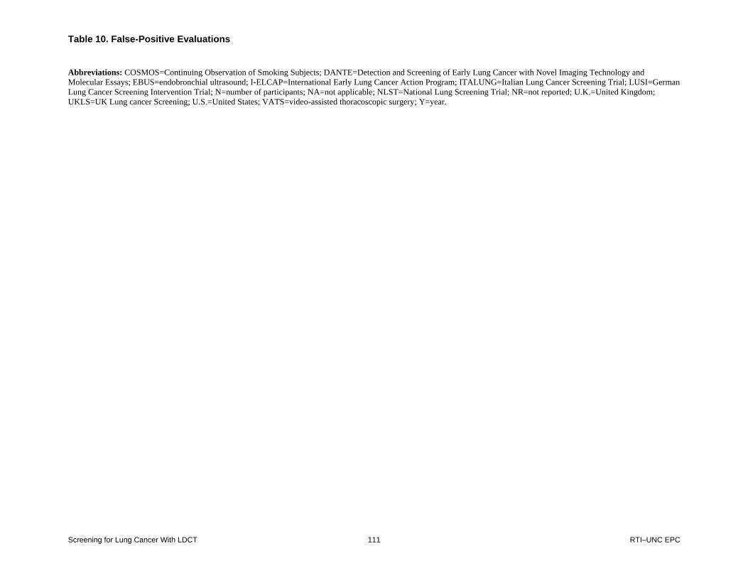
Table 10. False-Positive Evaluations
Screening for Lung Cancer With LDCT 111 RTI–UNC EPC
Abbreviations: COSMOS=Continuing Observation of Smoking Subjects; DANTE=Detection and Screening of Early Lung Cancer with Novel Imaging Technology and
Molecular Essays; EBUS=endobronchial ultrasound; I-ELCAP=International Early Lung Cancer Action Program; ITALUNG=Italian Lung Cancer Screening Trial; LUSI=German
Lung Cancer Screening Intervention Trial; N=number of participants; NA=not applicable; NLST=National Lung Screening Trial; NR=not reported; U.K.=United Kingdom;
UKLS=UK Lung cancer Screening; U.S.=United States; VATS=video-assisted thoracoscopic surgery; Y=year.

Table 11. Summary of Evidence on Screening for Lung Cancer With LDCT
Screening for Lung Cancer With LDCT 112 RTI–UNC EPC
Key Question and Topic
No. of Studies (k), No. of
Observations (n) Summary of Findings
Consistency and
Precision Study
Quality
Limitations (Including Reporting
Bias)
Overall Strength of Evidence Applicability
KQ 1. Benefits of screening
k=7 RCTs (26 publications), 86,486 participants
The good-quality NLST (n=53,542) reported a reduction in lung cancer mortality (IRR 0.85 [95% CI, 0.75 to 0.96]) and all-cause mortality (IRR 0.93 [95% CI, 0.88 to 0.99]) with three rounds of annual LDCT compared with CXR (NNS=323 to prevent 1 lung cancer death over 6.5 years). NELSON (n=15,792) found a reduction in lung cancer mortality (IRR, 0.75 [95% CI, 0.61 to 0.90]) but not all-cause mortality (IRR, 1.01 [95% CI, 0.92 to 1.11]) with four rounds of LDCT screening using volumetric measurements with increasing intervals (baseline, 1 year, 3 years, and 5.5 years) compared with no screening (NNS=130 to prevent 1 lung cancer death over 10 years).
Consistent among trials adequately powered; precise
Good: 1 Fair: 6
All but two of the seven trials were underpowered to assess for a lung cancer mortality benefit.
High for benefit* High-risk current and former smokers (with ≥30 pack-years [NLST] or >15 cigarettes/day for >25 years or >10 cigarettes/day for >30 years [NELSON]); ages 50-74; NLST and NELSON participants were younger, more highly educated, and less likely to be current smokers than the U.S. screening-eligible population; limited racial and ethnic diversity; US population eligible for screening faces higher risk of death from competing causes than trial participants; mainly conducted at large academic centers; NLST did not use current U.S. screening protocols such as Lung-RADS; NELSON used volumetric measurements for screening.

Table 11. Summary of Evidence on Screening for Lung Cancer With LDCT
Screening for Lung Cancer With LDCT 113 RTI–UNC EPC
Key Question and Topic
No. of Studies (k), No. of
Observations (n) Summary of Findings
Consistency and
Precision Study
Quality
Limitations (Including Reporting
Bias)
Overall Strength of Evidence Applicability
KQ 2. Risk prediction models
k=9; 13 risk prediction models evaluated in 9 cohorts comprising 21,922,733 participants
Benefits: Studies of three models (PCLOm2012, LCDRAT, and Kovalchik model) reported increased screen-preventable deaths compared
with risk factor−based criteria (k=4;
21,682,066 participants from 4 cohorts). Most findings from these studies also showed improved NNS. Harms: Most studies did not report on actual harms of screening. Studies of all models reported similar numbers of false-positive selections for screening (i.e., the model selected people to be screened who did not have or develop lung cancer or death from lung cancer) and mixed findings for rates of false-positive selections or false-positive selections per prevented death when comparing risk prediction models to risk factor–based criteria.†
Consistent; imprecise (results highly dependent on risk threshold selected)
Good: 6 Fair: 3
No trials have compared use of a risk prediction model with risk factor-based criteria; evidence base is limited by lack of an established risk threshold; most models were evaluated by a single study in one to two cohorts.
Low for greater benefits and similar or reduced harms
High-risk current and former smokers; mainly applicable to NLST or USPSTF screen-eligible persons (ages 55-74 years or 55-80 years)
KQ 3. Accuracy of screening with LDCT
k=23 n=86,064
Sensitivity ranged from 59% to 100% (k=13, n=76,856) and was over 80% in vast majority of studies. Specificity ranged from 26.4% to 99.7% (k=13, n=75,819) and was over 75% in vast majority. PPV ranged from 3.3% to 43.5%. NPV ranged from 97.7% to 100%. Reliability among radiologists was fair to moderate (k=3).
Reasonably consistent; imprecise (except precise for NPV)
Good: 3 Fair: 20
Incomplete or unreported followup length may have led to differential measurement. Heterogeneity in screening protocols and definitions (e.g., positive tests, indeterminate tests).
Moderate U.S. and highly developed countries; most conducted in past 10 years. Similar LDCT technologies used across studies; varying nodule classification protocols that could likely be replicated in the U.S.; few studies used nodule classification approach recommended by ACR (Lung-RADS).

Table 11. Summary of Evidence on Screening for Lung Cancer With LDCT
Screening for Lung Cancer With LDCT 114 RTI–UNC EPC
Key Question and Topic
No. of Studies (k), No. of
Observations (n) Summary of Findings
Consistency and
Precision Study
Quality
Limitations (Including Reporting
Bias)
Overall Strength of Evidence Applicability
KQs 4 and 5. Harms of screening, workup, or surveillance
Radiation: k=9 n=74,963 participants
Radiation from 1 LDCT: range 0.65 mSv to 2.3 6mSv Cumulative radiation exposure: 20.8 mSv to 32.5 mSv for annual screening for 25 years Radiation-induced cancer: 0.26 to 0.81 major cancers for every 1,000 people screened with 10 annual LDCTs‡
Consistent; imprecise
Good: 3 Fair: 6
Estimates of radiation-induced cancers are based on modeling.
Moderate for radiation-induced harms
Estimates were not provided for lifetime risk of radiation-induced cancers or fatal cancers from annual screening from 55 to 80 (i.e., USPSTF 2013 recommendation).
False positives: k=27 n=115,6544 participants False-positive followup evaluations: k=14 n=56,223 participants
False-positive rates: range 7.9%-49.3% for baseline screening and 0.6%-28.6% for incidence screening rounds; rates generally declined with each round. NLST reported 26.3%, 27.2%, and 15.9% for baseline, year 1, and year 2, respectively; rates were lower in NELSON; the VA implementation study reported 58% of those screened (28.9% of screen-eligibles) at baseline and over 30% variation across eight sites. Invasive procedures for false-positive results, range of rates for every 1,000 people screened (NLST rate): 0.9 to 5.6 needle biopsies (2.5) resulting in 0.3 to 0.7 complications; 5 to 13 surgical procedures; (17 total invasive procedures, resulting in <1 major complication§)
Consistent; imprecise
Good: 8 Fair: 14 Good: 4 Fair: 10
Heterogeneity in screening protocols, definitions of positive and false-positive results, and reporting of procedures and complication rates.
Moderate for harms due to false-positive results
Most studies did not use current nodule evaluation protocols such as Lung-RADS; an evaluation using NLST data estimated that 23.4% of all invasive procedures for false-positive results from the NLST would have been prevented by using Lung-RADS.
Overdiagnosis: k=12 n=95,290 participants
Overdiagnosis: Estimates ranged from 0% to 67.2% that a screen-detected lung cancer is overdiagnosed; NLST data indicate approximately 4 cases of overdiagnosis over 6.5 years (and 3 lung cancer deaths prevented) per 1,000 people screened.ǁ
Inconsistent; imprecise
Good: 2 Fair: 10
Inadequate duration of followup and heterogeneity limit the evaluation.
Low for harms NLST estimate is based on 3 annual screens and 6.5 years of followup; uncertain whether it would increase or decrease with ongoing screening and longer followup.

Table 11. Summary of Evidence on Screening for Lung Cancer With LDCT
Screening for Lung Cancer With LDCT 115 RTI–UNC EPC
Key Question and Topic
No. of Studies (k), No. of
Observations (n) Summary of Findings
Consistency and
Precision Study
Quality
Limitations (Including Reporting
Bias)
Overall Strength of Evidence Applicability
Smoking behavior: k=7 n=29,038 participants
LDCT vs. no screening (k=2): Evidence on cessation an intensity does not indicate harm of false reassurance. Positive or indeterminate results vs. normal results: Abnormal or indeterminate results may increase cessation and continued abstinence, but normal screening test results had no influence.
Inconsistent; Imprecise
Good: 0 Fair: 7
Most RCTs of LDCT did not report on outcomes to assess for false reassurance.
Low for no harms
The two RCTs providing data for LDCT vs. no screening were conducted in Denmark (DLCST) and the Netherlands and Belgium (NELSON).
Psychosocial harms: k=9 n=14,715 participants
General HRQoL: No significant differences over 6 months to 2 y of followup between LDCT and controls (k=2 RCTs, n=3,937); worse HRQoL for persons receiving true-positive results vs. other results. Anxiety and depression: No significant increase over 2 weeks to 2 y of followup for LDCT vs. controls (k=6 RCTs, n=12,096); increased anxiety for individuals receiving true-positive results vs. other results. Distress: no significant increase over approximately 2 y of followup for LDCT vs. controls (k=2 RCTs, n=5,180); temporary increase for those receiving indeterminate results vs. other results. Other potential psychosocial consequences of screening: each generally assessed by a single study, often without a non-LDCT comparison group, and not indicative of harm
Reasonably consistent and precise for HRQoL, anxiety and depression, and distress Consistency unknown and imprecise for other outcomes
Good: 1 Fair: 8
Relatively short followup (2 y or less); RCTs did not assess these outcomes over the duration of the trials.
Moderate for no harm over 2 y (HRQoL, anxiety, and distress) for LDCT vs. controls. Moderate for worse short-term HRQoL, anxiety, and distress for those who received true-positive or indeterminate results vs. other results
High-risk current and former smokers; studies lacked racial and ethnic diversity; most studies conducted in Europe; trials did not use current protocols such as Lung-RADS.

Table 11. Summary of Evidence on Screening for Lung Cancer With LDCT
Screening for Lung Cancer With LDCT 116 RTI–UNC EPC
Key Question and Topic
No. of Studies (k), No. of
Observations (n) Summary of Findings
Consistency and
Precision Study
Quality
Limitations (Including Reporting
Bias)
Overall Strength of Evidence Applicability
Incidental findings (IFs): k=7 n=80,485
Rates of reported significant IFs ranged from 4.4 to 40.7%. Common IFs were coronary artery calcification; aortic aneurysms; emphysema; infectious and inflammatory processes; and masses, nodules, or cysts of the kidney, breast, adrenal, liver, thyroid, pancreas, spine, and lymph nodes. IFs led to consultations, additional imaging, and invasive procedures. Nonlung cancers were diagnosed in 0.39% of NLST LDCT arm participants over 4 years. Incidental detection of thyroid cancer: In NLST,¶ thyroid cancer incidence was roughly double during 3 years of active LDCT screening compared with CXR screening (HR, 2.19 [95% CI, 1.07–4.47]), but not during subsequent years (HR,1.08 [95% CI, 0.49–2.37]).
Consistent; imprecise
Fair: 7 No standard definition for which IFs were significant or actionable. Few studies on followup evaluations and distal outcomes.
Moderate for harms
Screen-eligible adults undergoing LDCT in academic or tertiary lung cancer screening centers.

Table 11. Summary of Evidence on Screening for Lung Cancer With LDCT
Screening for Lung Cancer With LDCT 117 RTI–UNC EPC
Key Question and Topic
No. of Studies (k), No. of
Observations (n) Summary of Findings
Consistency and
Precision Study
Quality
Limitations (Including Reporting
Bias)
Overall Strength of Evidence Applicability
KQ 6. Efficacy of surgical resection for Stage I NSCLC
k=36 uncontrolled cohort studies n=212,274
5-year OS for surgical resection (including lobectomy and SLR approaches), range: 33 to 86% for Stage I, 58 to 83% for Stage IA, and 42 to 79% for Stage IB. In pathologic Stage I patients in the NCDB from 2003 to 2006 the 5-year OS was 61% for surgical resection (n=54,350). Survival rates in the NCDB, SEER, and VA VINCI databases for Stage I, covering the years 2003-2015, ranged from 53% to 75% for lobectomy (n=23,707). Survival rates were generally higher for lobectomy than SLR, for smaller than larger tumors, and for patients who are female, younger, nonsmokers, or had fewer comorbidities than patients who are male, older, smokers, or sicker.
Reasonably consistent; imprecise
Good: 5 Fair: 31
Information related to deviations from intervention, missing data, and sources of survival outcomes often lacking; heterogeneity related to staging of NSCLC (clinical or pathologic) and surgical approaches (among studies and over time).
Moderate for benefit
Persons with Stage I NSCLC; some studies were more than 10 years old and may be less applicable to current approaches and outcomes (studies were from 1983 to 2018)
KQ 6. Efficacy of SBRT for Stage I NSCLC
k=27 uncontrolled cohort studies n=38,915
5-year OS (and other measures of long-term survival) varied substantially across studies (range: 20 to 80%) and by subgroups defined by clinical characteristics (e.g., operability of tumor) and patient age; survival may be higher among younger than older patients.
Inconsistent; imprecise
Good: 2 Fair: 25
Information related to deviations from intervention, missing data, and sources of survival outcomes often lacking; substantial heterogeneity related to staging and operability of tumors.
Low for benefit Persons with operable or inoperable Stage I NSCLC

Table 11. Summary of Evidence on Screening for Lung Cancer With LDCT
Screening for Lung Cancer With LDCT 118 RTI–UNC EPC
Key Question and Topic
No. of Studies (k), No. of
Observations (n) Summary of Findings
Consistency and
Precision Study
Quality
Limitations (Including Reporting
Bias)
Overall Strength of Evidence Applicability
KQ 7. Harms of surgical resection
k=29 uncontrolled cohort studies n=755,427
30-day mortality rates ranged from 0 to 4% in most studies; rates of 90-day mortality were slightly higher (range: 2 to 5% in most studies). Less than one-third of patients in most studies experienced treatment-related adverse events. Adverse events reported in ≥10% of patients included pulmonary events (e.g., air leak, pleural effusion) and cardiac arrhythmias.
Reasonably consistent; reasonably precise
Good: 3 Fair: 26
Information related to deviations from intervention, missing data, and sources of survival outcomes often lacking; potential selective reporting of adverse events.
Moderate for harms
Persons having lobectomy or SLR for the treatment of Stage I NSCLC
KQ 7. Harms of stereotactic body radiation therapy
k=1 RCT (comparing dosing regimens), 1 uncontrolled clinical trial, and 58 uncontrolled cohort studies n=49,654
30- and 90-day mortality rates ranged from 0 to 3%. Adverse events were experienced by a majority of patients, but most were of mild or moderate severity. Adverse events reported in ≥10% of patients included were pulmonary events (e.g., cough, dyspnea, pneumonitis, fibrosis) or respiratory disorders (including dyspnea), chest wall pain, fatigue, and dermatologic reactions. Incidence of rib fracture ranged from 0% (n=80 patients) to 42% (n=169 patients).
Reasonably consistent; imprecise
Good: 1 Fair: 59
Information related to deviations from intervention, missing data, and sources of survival outcomes often lacking; potential selective reporting of adverse events.
Low for harms Persons having SBRT/SABR for the treatment of operable or inoperable Stage I NSCLC
KQ 8. Change in mortality from a specified change in lung cancer incidence (and stage shift)
k=2 RCTs (NLST and NELSON) n=69,334
An absolute increase in lung cancer incidence of 0.5-0.6%, increase in Stage I lung cancers of 19-27%, decrease in Stage IV lung cancers of 14-19% were associated with 52-83 fewer lung cancer deaths and 0 (NELSON) to 84 (NLST) fewer all-cause deaths per 100,000 person-years.
Consistent; precise for lung cancer mortality but imprecise for all-cause mortality
Good: 1 Fair: 1
Reporting bias not detected.
High 3 annual rounds of screening with LDCT (compared with CXR) in NLST or 4 rounds of screening with increasing intervals as conducted in NELSON (volumetric approach); applicable to workup of lung cancers and subsequent treatments used in the NLST and NELSON; same applicability issues as listed for KQ 1.

Table 11. Summary of Evidence on Screening for Lung Cancer With LDCT
Screening for Lung Cancer With LDCT 119 RTI–UNC EPC
* Strength of evidence was graded as moderate prior to final publication of NELSON because of unknown consistency (with a single good quality study that was adequately
powered) but was changed to high after including NELSON in the evidence report.
† The language “false positive” here refers to model performance metrics with respect to lung cancer events (diagnosis or deaths), not with respect to LDCT results. ‡ One study estimated a lifetime risk of fatal cancer of 0.11 per 1,000 subjects after the four screening rounds.134 § NLST reported 11 major complications and 6 deaths within 60 days of invasive procedures among those with false-positive results (2 deaths after surgical resections and 4 after
bronchoscopy). ǁ Based on converting data to per 1,000 screened from study that reported 1.38 cases of overdiagnosis in every 320 patients needed to screen to prevent one death from lung
cancer.140 ¶ This study specifically addressed the potential for overdiagnosis of thyroid cancer through incidental detection.
Abbreviations: ACR=American College of Radiology; CI=confidence interval; CPS=Cancer Prevention Study; CXR=chest X-ray; DLCST=Danish Lung Cancer Screening
Trial; HR=hazard ratio; HRQoL=hazard ratio quality of life; IFs=incidental findings; IRR=incidence rate ratio; KQ=key question; LCDRAT=Lung Cancer Death Risk Assessment
Tool; LCRAT=Lung Cancer Risk Assessment Tool; LDCT=low-dose computed tomography; LLP=Liverpool Lung Project; n=number; NELSON=Nederlands-Leuvens
Longkanker Screenings Onderzoek; NHS=Nurses’ Health Study; NLST=National Lung Screening Trial; NNS=number needed to screen; NPV=negative predictive value;
NSCLC=non-small cell lung cancer; OS=overall survival; PPV=positive predictive value; RCT=randomized, controlled trial; SABR=stereotactic ablative radiation;
SBRT=stereotactic body radiotherapy; SLR=sublobar resection; TSCE=Two-Stage Clonal Expansion; USPSTF=U.S. Preventive Services Task Force; VA=Veteran’s
Administration.

Appendix A. Contextual Questions
Screening for Lung Cancer With LDCT 120 RTI–UNC EPC
CQ 1. What Are the Barriers to Implementing Lung Cancer Screening and Surveillance in Clinical Practice in the United
States (e.g., Barriers to Shared Decision Making, Systematically Eliciting and Documenting a Detailed Smoking History, Systems for Tracking Nodules and
Followup, and Availability of Appropriate LDCT Protocols)? Introduction Since the 2013 USPSTF statement on lung cancer screening, multiple expert groups and
specialty societies have published consensus-based guidance documents on screening
implementation.313, 335-339 Common among these guidance statements is an acknowledgement
that implementation of lung cancer screening is a highly complex process requiring multiple,
inter-connected steps. There is also a growing body of literature aimed at understanding barriers
to implementation of lung cancer screening. A recent perspective article offered a high-level
summary of implementation barriers using a multi-level (patient, provider, and system-level)
framework (Table 6).340 This contextual question section is not on a comprehensive account of
the many clinical and technical aspects of screening implementation. Rather, it highlights some
of the salient barriers to the appropriate and effective implementation of lung cancer screening
that have arisen since the 2013 USPSTF statement. These include barriers to SDM, systematic
identification of screen-eligible patients, systems for tracking nodules and followup, and
availability of LDCT protocols.
Barriers to Shared Decision Making Screening Guidelines and SDM
Lung cancer screening guidelines are virtually unanimous in asserting that informed and SDM
should occur before a patient proceeds with screening.313, 334, 335, 341 The American Cancer
Society recommends that “a process of informed and SDM with a clinician related to the
potential benefits, limitations, and harms associated with screening for lung cancer with low‐
dose computed tomography should occur before any decision is made to initiate lung cancer
screening.”326 The 2013 USPSTF recommendation stated that “the decision to begin screening
should be the result of a thorough discussion of the possible benefits, limitations, and known and
uncertain harms.”1 In 2014, the CMS coverage decision for lung cancer screening was issued that
contained several stipulations including a requirement for “a lung cancer screening counseling
and shared decision-making visit.” Required elements of this visit included “the use of one or
more decision aids, to include benefits, harms, followup diagnostic testing, over-diagnosis, false
positive rate, and total radiation exposure.”39
SDM in Practice
Unfortunately, emerging evidence raises concerns that SDM in practice may be far from what is

Appendix A. Contextual Questions
Screening for Lung Cancer With LDCT 121 RTI–UNC EPC
intended by guidelines. A recent study found that CMS-required SDM visits were evident in just
10 percent of Medicare beneficiaries who had a screening LDCT in 2016.342 Another study
analyzed transcribed recordings of discussions between community physicians (primary care
physicians and pulmonologists) and patients about initiating lung cancer screening.343 In these
discussions (n=14), which were identified by searching a large database, SDM quality was
universally poor and discussion of screening harms was essentially nonexistent. Average time for
screening discussion was 57 seconds and was typically focused on insurance coverage. There
was no evidence that decision aids were used.
Competing Demands
Systematic reviews have found that SDM is difficult to implement well in practice, regardless of
clinical context.344 Factors associated with higher quality SDM include time (duration of
encounter) and decision support interventions (including decision aids). Since the USPSTF 2013
recommendation, multiple studies have identified competing demands and limited time available
to conduct SDM during clinical encounters as a central barrier to implementing SDM.36, 345-349
Patient Perceptions of Screening
Adding to the challenge of SDM implementation is evidence that patient’s “baseline”
perceptions of the benefits and harms of lung cancer screening diverge from what the evidence
shows. In one study of (n=50) screen-eligible patients who had not yet seen a decision aid
overestimated the likelihood of benefitting from screening by orders of magnitude. Patients also
had poor awareness of potential harms of screening.41 In another study involving focus groups of
screening eligible patients (n=45), participants expressed surprise that the magnitude of their
lung cancer risk and benefits of screening were lower than expected.345 These studies suggest
that patient’s baseline perceptions are systematically inaccurate (i.e., biased) in favor of
screening. These biases are not unique to lung cancer screening, and there is robust evidence that
patients, members of the public, and clinicians typically overestimate the benefits and
underestimate the harms of cancer screening tests.350-352
Such biases pose challenges for implementing SDM about screening in practice. Primary care
physicians, who routinely discuss cancer screening with patients, recognize that initiating a
conversation about the availability of a lung cancer screening test will provoke immediate
assumptions on the part of the patient. For meaningful SDM, a provider who initiates such a
discussion about lung cancer screening must be prepared to provide balanced information that
calibrates patient perceptions and expectations to reflect what is known.
Although providing screen-eligible patients individualized, quantitative risk information is
(theoretically) ideal, a first order concern is implementing processes that offer patients a) a
reasonable sense of the benefits and harms of screening, including approximate likelihoods for
these outcomes; b) an understanding that the screening decision involves tradeoffs between these
benefits and harms; and c) a recognition that the “weights” placed on avoiding harms relative to
benefits should reflect the values of the patient him/herself.

Appendix A. Contextual Questions
Screening for Lung Cancer With LDCT 122 RTI–UNC EPC
Timing and Context of SDM
Experts have expressed a range of views about whether SDM about lung cancer screening should
occur in primary care, subspecialty care, and/or within centralized screening programs.353, 354 The
potential benefits of SDM in a subspecialized LDCT screening context are the increased chances
that providers will be knowledgeable about lung cancer screening, have time to dedicate to the
issue of screening, and use established workflows that employ decision support tools. On the
other hand, potential downsides of conducting SDM in centralized screening centers is that
patients are likely to assume that a referral to a lung cancer screening program is a referral for the
screening procedure itself rather than to participate in process by which they will decide whether
screening is right for them. Thus, by the time the patient reaches the screening center context,
absent a good prior discussion, it is likely they will arrive with inaccurate perceptions of the
benefits and harms and will have already decided to be screened.353-356
Emerging evidence lends support to the idea that the timing and context of the SDM process
(i.e., primary care vs. referral setting) can influence patient decisions. One study in a primary
care population of screening-eligible patients (n=50) found viewing a video decision aid
improved understanding of the nature and magnitude of the harms and benefits of screening.41
When asked to indicate their screening preferences, 50 percent preferred screening while the
other 50 percent did not. In another study of screen-eligible primary care patients (n=81) who
were reached through an electronic health record portal and viewed a lung cancer screening
decision aid, screening preferences were again heterogeneous: 30 percent desired screening, 44
percent were “unsure,” and 26 percent declined screening.357 In contrast to these studies in
primary care context, a study of patients attending a tertiary referral lung cancer screening
program (n=423) who received robust patient education and decision support that included
decision aid viewing, 95 percent proceeded to have a screening LDCT.358 In sum, there is
evidence that, in primary care populations, informed preferences about lung cancer screening are
heterogeneous (i.e., that the decision is, in fact, “preference sensitive”). SDM conducted only
after a patient reaches a lung cancer screening center may miss the “decision window” during
which patients actually make the decision.
System and Personnel Barriers to Identifying Eligible Populations (Detailed Smoking Histories) One challenge to population or system level implementation in identifying populations eligible
for screening is that detailed, patient-level smoking histories, including average number of
cigarettes smoked per day, start dates, and quit dates, are not readily available within the
electronic health record.
Moreover, smoking behavior is dynamic, meaning that these data need to be periodically verified
for accuracy. Clinical demonstration studies suggest that, even with additional resources
dedicated to eliciting and documenting smoking histories, these data fields often still have
incomplete or inaccurate information. In the VHA implementation project, substantial resources
were dedicated to having nurses elicit and enter these data. Despite this, among 93,033 patients
who met basic age and comorbidity criteria, a total of 36,555 patients (39.3%) were missing
necessary smoking history data (or the tobacco pack-years were improperly calculated) needed to

Appendix A. Contextual Questions
Screening for Lung Cancer With LDCT 123 RTI–UNC EPC
systematically identify screening-eligible patients.37 In 1-year, single-site primary care
implementation project, nurses and support staff were able to elicit and document smoking
histories in 53 percent of ever smokers between ages 55 and 80 years.359
Availability of Appropriate LDCT Protocols Another potential implementation issue is whether radiology facilities that are capable of
providing LDCT scanning are available. Early studies published soon after the 2013 USPSTF
statement raised concerns about limited capacity.360 However, the number of lung cancer
screening centers is growing rapidly. In 2014, there were 203 screening centers certified by the
American College of Radiology (ACR) in the United States. By 2017, that number had increased
to 1,748.361 According to the ACR website, there are 2,013 ACR-certified centers as of April
2019.362
Systems for Tracking Nodules and Ensuring Followup (Patient Coordination) The availability of centers that are certified to perform LDCT screening and to report results to a
national registry should be distinguished from the availability of infrastructure needed to track
and manage individuals with screen-detected lung nodules. There is broad expert consensus that
screen-detected lung nodules should be managed based on established algorithms,313, 335-339
which call for regular and timely surveillance of screen-detected nodules. Operationalizing
surveillance of lung nodules for a large, high-risk population will require robust longitudinal
tracking and patient coordination systems for the large numbers of individuals with lung nodules.
The VHA Lung Cancer Screening Demonstration Project (LCSDP) found this aspect of
implementation to be challenging and complex, as most patients screened had findings that
required followup. Specifically, 56 percent of screened patients had one or more nodules that
required tracking and 41 percent had incidental findings. Implementation required substantial
resources for manual abstraction of patient information from records and the creation of
dedicated tracking and patient coordination systems.37
While there is no comprehensive accounting of patient coordination and tracking systems in the
United States, surveys and interviews have found that both primary care physicians and
subspecialists have concerns about whether there is sufficient personnel and tracking
infrastructure needed for screening implementation.36, 345-349, 363
Out-of-Pocket Costs for Followup of Screen-Detected Findings ACA insurance plans are required to cover LDCTs done for screening. However, patients with
screen-detected nodules enter diagnostic and surveillance pathways involving evaluations,
imaging, and procedures that are not considered screening and are subject to copays and
deductibles. Since the 2013 USPSTF statement, multiple studies in both patients and providers
have identified the issue that the costs of followup testing after positive screening results will
lead to financial harm for patients.36, 346-349, 364, 365 Even if less aggressive nodule categorization
approaches (e.g., Lung-RADS) are used, the number of patients who will enter surveillance

Appendix A. Contextual Questions
Screening for Lung Cancer With LDCT 124 RTI–UNC EPC
pathways for screen-detected nodules is large. Given that high-deductible insurance plans among
low and middle-income Americans, the issue of patient cost as a barrier to lung cancer screening
implementation requires further study.366
CQ 2.a. Are the Participants of Randomized, Controlled Trials
of Lung Cancer Screening (e.g., NLST) That Reported a Reduction in All-Cause or Lung Cancer Mortality
Representative of Screening-Eligible U.S. Adults (Based on NLST Criteria or USPSTF Recommendations)?
b. How Do the 5-Year Survival Rate and Life Expectancy of Persons Eligible for Lung Cancer Screening in the United
States (Based on NLST Criteria or USPSTF Recommendations) Compare With Those of NLST
Participants? c. Are the Settings and Providers in Randomized, Controlled Trials of Lung Cancer Screening (e.g., NLST) That Reported a
Reduction in All-Cause or Lung Cancer Mortality Representative of U.S. Health Care Settings and Providers?
The Discussion of this report describes the applicability of NLST and other included studies.
Briefly, NLST participants were younger, more highly educated, and less likely to be current
smokers than the U.S. screening-eligible population.367 Furthermore, the NLST was mainly
conducted at large academic centers, potentially limiting its applicability to community-based
practice (e.g., because of challenges with implementation, level of expertise). Many of the trial
centers are well-recognized for expertise in radiology as well as cancer diagnosis and
treatment.31 Community centers may be less equipped for screening programs and for treatment
of lung cancers identified by screening. For example, the NLST publication noted that mortality
associated with surgical resection of lung cancer was much lower in the trial than that reported
for the U.S. population (1% vs. 4%).31, 294
A study using data from the 2012 Health and Retirement Study (HRS) (a national survey of
adults 50 and older) evaluated comorbidities, life expectancy, smoking history, and other
characteristics in the screening-eligible population and in NLST participants; it reported a lower
5-year survival rate and life expectancy in the screening-eligible persons compared with NLST
participants (87% vs. 93%, p<0.001, and 18.7 years vs. 21.2 years, respectively).24 Screening-
eligible HRS respondents were older, more likely to be current smokers, and more likely to have
been diagnosed with comorbidities than NLST participants. The authors concluded that the
general U.S. population eligible for lung cancer screening is probably less likely to benefit from
early detection compared with the NLST participants because they face a high risk of death from
competing causes, such as heart disease, diabetes, or stroke.

Appendix A. Contextual Questions
Screening for Lung Cancer With LDCT 125 RTI–UNC EPC
CQ 3. Does Screening for Lung Cancer With LDCT Have Unintended Benefits From Detecting Incidental Findings
(e.g., Coronary Artery Calcium, Chronic Obstructive Pulmonary Disease, or Extrapulmonary Nodules) Leading to
Interventions That Improve Health Outcomes?
Common incidental findings identified in this systematic review included coronary artery
calcification, aortic aneurysms, emphysema, infections, and masses or nodules (e.g., of the
thyroid or pancreas), among others. There is no trial evidence to indicate that screening with
LDCT for such findings has greater unintended benefit than unintended harm. The USPSTF
portfolio includes evidence reviews and recommendations covering the evidence on potential
benefits and harms of screening for many of these conditions/findings in asymptomatic persons.
The evidence reviews have resulted in I statements (i.e., insufficient evidence) and D
recommendations (i.e., harm of screening greater than benefit). For example, the USPSTF
recommendation statement on nontraditional cardiovascular risk factors concluded that the
current evidence is insufficient to assess the balance of benefits and harms of screening for
coronary artery calcium (CAC) in asymptomatic adults to prevent CVD events.368 Further,
USPSTF had D recommendations for screening for chronic obstructive pulmonary disease
(COPD),369 thyroid cancer,370 and pancreatic cancer in asymptomatic adults. Regarding
screening for aneurysms, USPSTF recommends one-time screening for abdominal aortic
aneurysm (AAA) with ultrasonography in men ages 65 to 75 years who have ever smoked371; an
update is in progress.372 There is no trial evidence to indicate greater benefit than harm for using
LDCT to screen for aneurysms (thoracic or abdominal).
CQ 4. What Is the Effectiveness of Smoking Cessation
Intervention Among Patients Receiving LDCT Screening?
The provision of smoking cessation interventions with LDCT screening is an opportunity to
improve health outcomes. In the NLST, screening with LDCT combined with smoking
abstinence of 15 years provided the greatest reduction in lung cancer mortality (comparing, for
example, with screening with LDCT and current smoking).373 The Centers for Medicare &
Medicaid Services also requires that smokers who undergo screening receive counseling on
smoking cessation so as not to mistake screening as either a substitute for cessation or a
confirmation that it is acceptable to continue smoking if the screening result is normal.374 The
USPSTF recommends that clinicians ask all adults about tobacco use, advise them to stop using
tobacco, and provide behavioral interventions and U.S. Food and Drug Administration (FDA)–
approved pharmacotherapy for cessation to adults who use tobacco.375
Two systematic reviews focused on this contextual question.376, 377 Assessment of these reviews
using AMSTAR-2 criteria indicates at least moderate confidence in the results.378 Neither
systematic review conducted meta-analyses because of heterogeneity of interventions and other
factors. The first systematic review376 included six studies published through July 1, 2015: three
randomized, controlled trials (RCTs) (with a total of 1473 participants) and three uncontrolled

Appendix A. Contextual Questions
Screening for Lung Cancer With LDCT 126 RTI–UNC EPC
studies (total 7,333 participants).376 Two of the RCTs compared the use of written self-help
materials with internet or computer-based tailored self-help materials for smoking cessation
intervention among patients receiving lung cancer screening and found no statistically significant
difference in abstinence rates between groups.379, 380 The third RCT evaluated the effect of
smoking cessation interventions before and after LDCT and found that smoking intervention
before LDCT led to numerically higher abstinence rates at 4 months (33.3 vs. 22.2%) and 6
months (22.2% vs. 11.1%) after treatment,381 although statistical testing was not provided for the
comparison between groups (a subsequently published systematic review conducted its own
statistical tests for those comparisons and reported no significant difference between groups,
p=1.0377). Continuous abstinence rates in uncontrolled studies ranged from 19.8 percent at 1-year
followup382 to 57.1 percent at 6-months followup383 across included studies. No rating of the risk
of bias of included studies was reported. The authors reported that their findings suggest that
there are benefits to implementing smoking cessation interventions in lung cancer screening
programs, which may represent a teachable moment to quit smoking.376
The second systematic review377included nine studies published through May 1, 2018. It
restricted eligibility to RCTs and observational studies with a comparison group, which excluded
the three single-arm studies that were in the other376 systematic review. The included five RCTs
(with a total of 1,620 participants) and four observational studies (total 5,114 participants) were
rated as poor to fair quality with significant potential for bias and limited generalizability.377
Though the studies provided insufficient evidence to support a particular approach to smoking
cessation interventions in the LDCT screening setting, the authors suggested that more intensive
interventions (e.g., multiple counseling sessions) appear to be more effective approaches to
smoking cessation. Table 7 summarizes the five RCTs included in the review. Sample sizes
ranged from 18 to 1,284, with three of the studies having fewer than 100 participants. The largest
study (with 1,284 participants) was part of the NELSON lung cancer screening trial.380 Only one
of the studies found a statistically significant difference in smoking cessation outcomes between
groups, with an intervention of six weekly telephone counseling calls compared to a list of
resources.384 Two of the studies used two or fewer counseling sessions as the intervention, while
the other two distributed tailored smoking cessation resources. Four of the comparison groups for
the RCTs distributed nontailored resources; the other altered the timing of smoking cessation
counseling sessions. The authors of the systematic review conducted a search of ongoing trials,
finding 11 ongoing RCTs assessing smoking cessation interventions in the context of LDCT
screening.377
In sum, limited evidence exists to establish the effectiveness of smoking cessation interventions
in lung cancer screening programs. However, this is an active area of research, with numerous
ongoing trials comparing intervention methods. Further research to determine components of
smoking cessation interventions that can optimize outcome by testing different modalities in lung
cancer screening programs and to identify strategies to effectively integrate smoking cessation
interventions in lung cancer screening sites have also been suggested.

Appendix A Table 1. Overview of Lung-RADS Classification System (Version 1.0)
Screening for Lung Cancer With LDCT 127 RTI–UNC EPC
Category Classification Category Descriptor Category Findings Management
Incomplete 0 Part or all of lungs cannot be evaluated Prior chest CT examination(s) being located for comparison
Additional lung cancer screening CT images and/or comparison to prior chest CT examinations is needed
Negative No nodules and definitely benign nodules
1 No lung nodules Nodule(s) with specific calcifications: complete, central, popcorn, concentric rings, and fat- containing nodules
Continue annual screening with LDCT in 12 months
Benign appearance or behavior
Nodules with a very low 90% likelihood of becoming a clinically active cancer due to size or lack of growth
2 Solid nodule(s): <6 mm, new <4 mm Part solid nodule(s): <6 mm total diameter on baseline screening Nonsolid nodule(s) (GGN): <20 mm OR ≥20 mm and unchanged or slowly growing Category 3 or 4 nodules unchanged for ≥3 months
Continue annual screening with LDCT in 12 months
Probably benign Probably benign
finding(s): short-term followup suggested; includes nodules with a low likelihood of becoming a clinically active cancer
3 Solid nodule(s): ≥6 to <8 mm at baseline OR new 4 mm to <6 mm Part solid nodule(s) ≥6 mm total diameter with solid component <6 mm OR new <6 mm total diameter nonsolid nodule(s) (GGN) ≥20 mm Nonsolid nodule(s) (GGN) ≥20 mm on baseline CT or new
6-month LDCT
Suspicious Findings for which additional diagnostic testing and/or tissue sampling is recommended
4A Solid nodule(s): ≥8 to <15 mm at baseline OR growing <8 mm OR new 6 to <8 mm Part solid nodule(s): ≥6 mm with solid component ≥6 mm to <8 mm OR with a new or growing <4 mm solid component Endobronchial nodule
3-month LDCT; PET/CT may be used when there is a ≥8 mm solid component
4B Solid nodule(s) ≥15 mm OR new or growing and ≥8 mm Part solid nodule(s) with a solid component ≥8 mm OR a new or growing ≥4 mm solid component
Chest CT with or without contrast, PET/CT and/or tissue sampling depending on the *probability of malignancy and comorbidities. PET/CT may be used when there is a ≥8 mm solid component.
4X Category 3 or 4 nodules with additional features or imaging findings that increases the suspicion of malignancy
Adapted from Lung‐RADS™ Version 1.0 Assessment Categories Release date: April 28, 2014
Abbreviations: CT=computed tomography; GGN=ground glass nodule; LDCT=low-dose computed tomography; PET=positron
emission tomography.

Appendix A Table 2. Overview of Lung-RADS Classification System (Version 1.1)
Screening for Lung Cancer With LDCT 128 RTI–UNC EPC
Category Classification Category Descriptor Category Findings Management
Incomplete 0 Part or all of lungs cannot be evaluated; prior chest CT examination(s) being located for comparison
Additional lung cancer screening CT images and/or comparison to prior chest CT examinations is needed
Negative No nodules and definitely benign nodules
1 No lung nodules; nodule(s) with specific calcifications: complete, central, popcorn, concentric rings, and fat- containing nodules
Continue annual screening with LDCT in 12 months
Benign appearance or behavior
Nodules with a very low likelihood of becoming a clinically active cancer due to size or lack of growth
2 Perifissural nodule(s)* <10 mm (524 mm3) Solid nodule(s): <6 mm (<113 mm3), new <4 mm (<34 mm3) Part solid nodule(s): <6 mm total diameter (<113 mm3) on baseline screening Nonsolid nodule(s) (GGN): <30 mm (<14,137 mm3) OR ≥30 mm (≥14,137 mm3) and unchanged or slowly growing Category 3 or 4 nodules unchanged for ≥3 months
Continue annual screening with LDCT in 12 months
Probably benign
Probably benign finding(s): short-term followup suggested; includes nodules with a low likelihood of becoming a clinically active cancer
3 Solid nodule(s): ≥6 to <8 mm (≥113 to <268 mm3) at baseline OR new 4 mm to <6 mm (34 to <113 mm3) Part solid nodule(s): ≥6 mm total diameter (≥113 mm3) with solid component <6 mm (<113 mm3) OR new <6 mm total diameter (<113 mm3) Nonsolid nodule(s): (GGN) ≥30 mm (≥14137 mm3) on baseline CT or new
6-month LDCT
Suspicious Findings for which additional diagnostic testing is recommended
4A Solid nodule(s): ≥8 to <15 mm at baseline (≥268 to <1,767 mm3) OR growing <8 mm (<268 mm3) OR new 6 to <8 mm (113 to <268 mm3) Part solid nodule(s): ≥6 mm (≥113 mm3) with solid component ≥6 mm to <8 mm (≥113 to <268 mm3) OR with a new or growing <4 mm (< 34 mm3) solid component Endobronchial nodule
3-month LDCT; PET/CT may be used when there is a ≥8 mm (≥268 mm3) solid component
Very suspicious
Findings for which additional diagnostic testing and/or tissue sampling is recommended
4B Solid nodule(s) ≥15 mm (≥1,767 mm3) OR new and growing and ≥8 mm (≥ 268 mm3) Part solid nodule(s) with a solid component ≥8 mm (≥268 mm3) OR a new or growing ≥4 mm (≥34 mm3) solid component
Chest CT with or without contrast, PET/CT and/or tissue sampling depending on the *probability of malignancy and comorbidities. PET/CT may be used when there is a ≥8 mm (≥268 mm3) solid component. For new large nodules that develop on an annual repeat screening CT, a 1- month LDCT may be recommended to address potentially infectious or inflammatory conditions
4X Category 3 or 4 nodules with additional features or imaging findings that increase the suspicion of malignancy†
Other Clinically significant or potentially clinically significant findings (nonlung cancer)
S Modifier: may add on to category 0‐4 coding As appropriate to the specific finding
Adapted from Lung‐RADS Version 1.1 Assessment Categories Release date: 2019

Appendix A Table 2. Overview of Lung-RADS Classification System (Version 1.1)
Screening for Lung Cancer With LDCT 129 RTI–UNC EPC
* Solid nodules with smooth margins, an oval, lentiform or triangular shape, and maximum diameter less than 10 mm or 524 mm3 (perifissural nodules) should
be classified as category 2 † These include, for example, spiculation, GGN that doubles in size in 1 year, and enlarged lymph nodes.
Abbreviations: CT=computed tomography; GGN=ground glass nodule; LDCT=low-dose computed tomography; NA=not applicable; PET=positron emission tomography.
Some notes on use of Lung‐RADS Version 1.1:
1) Nodule mean diameter is calculated by measuring both the long and short axes to one decimal point and reporting mean nodule diameter to one decimal point.
2) Size thresholds: Apply to nodules at first detection, and that grow and reach a higher size category.
3) Growth is defined as an increase in size of >1.5 mm (>2 mm3).
4) Exam category: Each exam should be coded 0‐4 based on the nodule(s) with the highest degree of suspicion.

Appendix A Table 3. Typical Treatment Approaches of SCLC, by Stage324
Screening for Lung Cancer With LDCT 130 RTI–UNC EPC
Stage Treatment Approach
Limited disease
Clinical stage T1-2, N0: Lobectomy + chemotherapy +/− concurrent radiation therapy Clinical stage >T1-2, N0: chemotherapy +/− concurrent or sequential radiation therapy tailored to patient performance status
Extensive disease
Chemotherapy +/− radiation therapy tailored to location and symptoms of metastatic site
Abbreviations: SCLC=small-cell lung cancer; T=tumor.

Appendix A Table 4. Stage of Detected Lung Cancers in LDCT Screening Groups for NLST and NELSON Trials
Screening for Lung Cancer With LDCT 131 RTI–UNC EPC
Stage NLST (%) NELSON (%)
Ia 40 58
Ib 10 10
IIa 3.4 5
IIb 3.7 1
IIIa 9.5 14
IIIb 11.7 3
IV 21.7 14
*For reference, the stage distribution based on data from the SEER 18 registry in 2010 is as follows: Ia (11.7%), Ib (8.5%), IIa
(1.0%), IIb (3.1%), IIIa (8.5%), IIIb (14.8%), IV (45.1%), occult or unknown (7.3%).325 Participants in the NLST were younger,
better educated, and healthier than individuals of similar age and smoking eligibility in the United States. (SCLC accounted for 7
percent of CT screen-detected cancers in the NELSON trial and 13 percent in NLST).
Abbreviations: LDCT=low-dose computed tomography; NELSON=The Dutch-Belgian Randomized Lung Cancer Screening
Trial; NLST=National Lung Screening Trial; SCLC=small cell lung cancer

Appendix A Table 5. Recommendations for Lung Cancer Screening With LDCT
Screening for Lung Cancer With LDCT 132 RTI–UNC EPC
Organization Year Target Population Recommendation and Related
Comments Endorse shared
decision making?
American Academy of Family Physicians43
2013 Persons with a high risk of lung cancer on the basis of age and smoking history
Insufficient evidence to recommend screening. Screening cannot be recommended on the basis of a single study conducted in major medical centers.
Yes
American Cancer Society326, 327
2013 NLST criteria. Excludes life-limiting comorbid conditions, metallic implants or devices in the chest or back, or oxygen requirement
Discussion about screening should be initiated, including benefits, limitations, harms. Recommends only if there is access to a high-volume, high-quality lung cancer screening and treatment center.
Yes
American Lung Association330
2015 NLST criteria Screening with LDCT recommended. Screening should occur in institutions that are able to provide a comprehensive screening program; smoking cessation is the best method of reducing lung cancer risk among those who smoke.
Yes
American Association for Thoracic Surgery331
2012 Persons ages 55-79 years with ≥30 pack-year smoking history and persons ages 50-79 years with ≥20 pack-year smoking history and another risk factor for lung cancer* or lung cancer survivor
Annual screening with LDCT. Should be conducted in environments in which there are multidisciplinary teams for managing indeterminate and positive screening scans; desirable to create a program that supports smoking cessation.
Not specified
CHEST Guideline and Expert Panel Report313
2018 Asymptomatic persons ages 55-77 years with same smoking history criteria as NLST
Annual screening with LDCT should be offered (weak recommendation, moderate-quality evidence)
Yes
European Union332 2017 Lung cancer LDCT programs should use a validated risk stratification approach so that only persons deemed to be at high enough risk are screened†
Screening with LDCT. Management of screen-detected solid nodules should use semi-automatically derived volume measurements and volume-doubling time and should be quality assured. Management of lung nodules by lung cancer multidisciplinary teams. National quality assurance boards should be set up by professional bodies to ensure adherence to all minimum technical standards.
All future screenees should be provided with carefully constructed participant information on the potential benefits and harms to enable them to make an informed decision.
National Comprehensive Cancer Network42,
333
2012 NLST criteria and persons age 50 years or older with ≥20 pack-years smoking history and 1 additional risk factor for lung cancer‡
Screening with LDCT. Multidisciplinary screening programs will be helpful; smokers should always be encouraged to quit smoking. It is also reasonable to consider using the PLCOm2012 lung cancer risk calculator to assist in quantifying risk, considering a 1.3% threshold of lung cancer risk (over 6 years).
Patients should have a full understanding of risks and benefits.

Appendix A Table 5. Recommendations for Lung Cancer Screening With LDCT
Screening for Lung Cancer With LDCT 133 RTI–UNC EPC
Organization Year Target Population Recommendation and Related
Comments Endorse shared
decision making?
Canadian Task Force on Preventive Health Care334
2016 NLST criteria Screening with LDCT every year up to 3 consecutive years (weak, low-quality evidence). Screening should only be performed in health care settings with access to expertise in early diagnosis and treatment of lung cancer.
Yes, discussion about benefits and harms, including false-positive screens, adverse effects of invasive followup testing, and overdiagnosis
* Examples of additional risk factors, as specified by the American Association for Thoracic Surgery, include COPD,
environmental or occupational exposure, prior cancer or radiation therapy, and genetic predisposition or family history. † No specific model is recommended. ‡ Examples of additional risk factors, as specified by the National Comprehensive Cancer Network, include radon exposure,
occupational exposure, history of cancer, family history of lung cancer, COPD, and pulmonary fibrosis.
Abbreviations: COPD=chronic obstructive pulmonary disease; CT=computed tomography; LDCT=low-dose computed
tomography; NLST=National Lung Screening Trial.

Appendix A Table 6. Summary of Multilevel Barriers to Effective Lung Cancer Screening
Screening for Lung Cancer With LDCT 134 RTI–UNC EPC
Patient-level barriers
Competing needs and demands for health care
Cost
Fear (e.g., procedures, diagnosis, treatment)
Lack of awareness
Lack of interest due to stigma associated with smoking
Limited access to care due to financial or social factors
Limited information and misinformation
Logistical issues (e.g., inconvenience, time)
Mistrust of the health care system and/or health care
Nihilism
Provider-level barriers
Competing demands for time
Evolving attitudes about the effectiveness of screening
Lack of awareness
Limited information and misinformation
Limited training in SDM
Nihilism related to treatment of lung cancer
Requirement for behavior change (adaptive challenge)
System-level barriers
Lack of support from health system leaders
Limited resources to support screening, including equipment, personnel, and information technology resources
Competing demands for limited resources (e.g., other screening programs or preventive health interventions)
Uncertain return on investment
Complexity of implementation (requires multidisciplinary collaboration) o Conflicting upper age range recommendations for screening o Identification of screening-eligible patients (gaps in smoking status data)
Source: Carter-Harris L, Gould MK. Multilevel barriers to the successful implementation of lung cancer screening: why does it
have to be so hard? Ann Am Thorac Soc. 2017 Aug;14(8):1261-5. doi: 10.1513/AnnalsATS.201703-204PS. PMID: 28541749.340

Appendix A Table 7. Summary of Randomized, Controlled Trials in the Systematic Review From 2019
Screening for Lung Cancer With LDCT 135 RTI–UNC EPC
Author, Year Trial Sample
Size Intervention Comparison Findings
Clark, 2004379 NA 171 Internet-based resources (10 links).
Written self-help materials from the NCI.
No significant difference in 12 month quit rates or change in readiness to quit. Increased number of quit attempts in intervention group (p=0.011).
Aalst, 2012380 NELSON 1284 Computer-generated, tailored self-help material based on input of individual smoking behaviors and history.
Standard brochure with smoking cessation information for different stages of readiness to quit.
No significant difference in point prevalence, quit attempts, or prolonged smoking abstinence at 24 months followup.
Ferketich, 2012381
NA 18 Smoking cessation counseling with a medical oncologist occurring before LDCT performed followed by 12-week tobacco dependence protocol.
Smoking cessation counseling with a medical oncologist occurring after LDCT performed followed by 12-week tobacco dependence protocol.
No significant difference in abstinence among those who received counseling before LDCT and those who received counseling after LDCT at 4 months and 6 months.
Marshall, 2016385 NA 55 Single face-to-face tailored counseling session with take-home audio education materials, printed materials, and telephone helpline referral.
Nontailored printed smoking cessation materials and telephone helpline referral.
No significant difference in quit rates at 12 months for patients receiving counseling intervention compared to the control group.
Taylor, 2017384 NA 92 Resources list plus 6 weekly, proactive counseling calls.
Resource list: Booklet, website, contact information for local resources, text messaging link.
Higher 7-day point prevalence cessation at 3-months in patients who received telephone counseling.
Abbreviations: LDCT=low-dose computed tomography; NA=not applicable; NCI=National Cancer Institute; NELSON=Dutch-
Belgian Randomized Lung Cancer Screening Trial.

Appendix B1. Original Search Strategies and Update Searches
Screening for Lung Cancer With LDCT 136 RTI–UNC EPC
Screening Searches
PubMed, 4-30-18
Search Query Items Found
#1 Search (“Lung Neoplasms”[MeSH] OR NSCLC[tiab] OR “lung cancer”[tiab] OR “lung cancers” [tiab] OR “lung-cancer”[tiab] OR “lung malignancy”[tiab] OR “lung malignancies”[tiab] OR “lung nodule”[tiab] OR “lung nodules” [tiab] OR “pulmonary nodule”[tiab] OR “pulmonary nodules”[tiab] OR “lung mass”[tiab] OR “lung masses”[tiab] OR (“Squamous Cell Carcinoma”[MeSH] OR Adenocarcinoma[MeSH]) and (Lung[MeSH] OR Lung Diseases[MeSH])))
209038
#2 Search (“Mass Screening”[MeSH] OR screen*[tw] OR “Early Diagnosis”[MeSH] OR “Tomography, X-Ray Computed”[Mesh] OR “CT scan”[tiab] OR “CT scans”[tiab] OR “CAT scan”[tiab] OR “CAT scans”[tiab] OR “spiral CT”[tiab] OR “spiral computed tomography”[tiab] OR “low-dose computed tomography”[tiab] OR LDCT[tiab] OR ((early[tiab] or earlier[tiab] or earliest[tiab]) AND (detect*[tiab] or diagnos*[tiab] or discover*[tiab] or find[tiab] or finding[tiab])))
1561323
#3 Search (#1 and #2) 33537
#4 Search (DANTE[tiab] OR “Detection and Screening of Early Lung Cancer by Novel Imaging Technology and Molecular Essays”[All Fields] OR DLCST[tiab] OR “Danish Lung Cancer Screening Trial”[tiab] OR ITALUNG[tiab] OR “Italian Lung Cancer Screening Trial”[All Fields] OR LUSI[tiab] OR “Lung Cancer Screening Intervention”[All Fields] OR MILD[tiab] OR “Multicentric Italian Lung Detection”[All Fields] OR NELSON[tiab] OR “Dutch-Belgian Lung Cancer Screening trial”[All Fields] OR NLST[tiab] OR “National Lung Screening Trial”[All Fields])
306902
#5 Search (#1 and #4) 2661
#6 Search (#3 or #5) 35398
#7 Search (#3 or #5) Filters: Publication date from 2012/01/01 to 2018/12/31 12321
#8 Search (“Risk prediction model”[tw] OR “Risk prediction models”[tw] OR “Risk Assessment”[MeSH] OR “risk assessment”[tw] OR “risk model”[tw] OR “risk models”[tw] OR “Decision Support Techniques”[MeSH] OR “Decision Support Systems, Clinical”[Mesh] OR “clinical prediction”[tw] OR “Logistic Models”[MeSH] OR microsimulation*[tw] OR “simulation model”[tw] OR “simulation models”[tw] OR “Assessment tool”[tw] OR “Assessment tools”[tw] OR “prediction score”[tw] OR “Risk Factors”[MeSH] OR “Predictive Value of Tests”[MeSH] OR “Sensitivity and Specificity”[MeSH] OR (Predict*[tw] AND (model*[tw] OR outcome*[tw] OR risk*[tw] OR rule[tw] OR rules[tw])) OR “risk-targeted”[tw] OR “mortality risk”[tw])
2073965
#9 Search (#1 and #8) 26417
#10 Search (#1 and #8) Filters: Publication date from 2014/04/01 to 2018/12/31 6640
#11 Search (#7 or #10) 16840
#12 Search (#7 or #10) Filters: Humans 16584
#13 Search (#7 or #10) Filters: Humans; English 15409
#14 Search (#7 or #10) Filters: Humans; English; Child: birth-18 years 605
#15 Search (letter[pt] OR newspaper article[pt] OR editorial[pt] OR comment[pt]) 1642710
#16 Search (#13 NOT #14) 14804
#17 Search (#16 NOT #15) 14186
#18 Search (“systematic review”[ti] OR “meta-analysis”[pt] OR “meta-analysis”[ti] OR “systematic literature review”[ti] OR “this systematic review”[tw] OR (“systematic review”[tiab] AND review[pt]) OR meta synthesis[ti] OR “meta synthesis”[ti] OR “cochrane database syst rev”[ta])
178836
#19 Search (#17 and #18) 485
#20 Search (“Randomized Controlled Trial”[Publication Type] OR “Single-Blind Method”[MeSH] OR “Double-Blind Method”[MeSH] OR “Random Allocation”[MeSH] OR ((randomized[title/abstract] OR randomised[title/abstract]) AND controlled[title/abstract] AND trial[title/abstract]))
618965
#21 Search (#17 and #20) 462
#22 Search (“Case-Control Studies”[MeSH] OR “Cohort Studies”[MeSH] OR “Epidemiologic Studies”[MeSH] OR “Follow-Up Studies”[MeSH] OR “Evaluation Studies”[Publication Type] OR “Program Evaluation”[MeSH] OR “observational study”[tw] OR “observational studies”[tw] OR “Cohort Studies”[MeSH] OR “Comparative Study”[pt] OR “Validation Studies”[pt] OR “Prospective Studies”[MeSH] OR “cohort”[tw] OR “case control”[tw])
4054856
#23 Search (#17 and #22) 5869

Appendix B1. Original Search Strategies and Update Searches
Screening for Lung Cancer With LDCT 137 RTI–UNC EPC
Cochrane Library, 5-2-2018 ID Cochrane Library Search Hits
#1 [mh “Lung Neoplasms”] or NSCLC:ti,ab or “lung cancer”:ti,ab or “lung cancers”:ti,ab or “lung-cancer”:ti,ab or “lung malignancy”:ti,ab or “lung malignancies”:ti,ab or “lung nodule”:ti,ab or “lung nodules”:ti,ab or “pulmonary nodule”:ti,ab or “pulmonary nodules”:ti,ab or “lung mass”:ti,ab or “lung masses”:ti,ab or (([mh “Squamous Cell Carcinoma”] or [mh Adenocarcinoma]) and ([mh Lung] or [mh “Lung Diseases”]))
13223
#2 [mh “Mass Screening”] or screen*:kw or [mh “Early Diagnosis”] or [mh “Tomography, X-Ray Computed”] or “CT scan”:ti,ab or “CT scans”:ti,ab or “CAT scan”:ti,ab or “CAT scans”:ti,ab or “spiral CT”:ti,ab or “spiral computed tomography”:ti,ab or “low-dose computed tomography”:ti,ab or LDCT:ti,ab or ((early:ti,ab or earlier:ti,ab or earliest:ti,ab) and (detect*:ti,ab or diagnos*:ti,ab or discover*:ti,ab or find:ti,ab or finding:ti,ab))
42901
#3 #1 and #2 1293
#4 DANTE:ti,ab or “Detection and Screening of Early Lung Cancer by Novel Imaging Technology and Molecular Essays” or DLCST:ti,ab or “Danish Lung Cancer Screening Trial”:ti,ab or ITALUNG:ti,ab or “Italian Lung Cancer Screening Trial” or LUSI:ti,ab or “Lung Cancer Screening Intervention” or MILD:ti,ab or “Multicentric Italian Lung Detection” or NELSON:ti,ab or “Dutch-Belgian Lung Cancer Screening trial” or NLST:ti,ab or “National Lung Screening Trial”:kw
43092
#5 #1 and #4 541
#6 #3 or #5 1615
#7 #6 Publication Year from 2012 to 2018 916
#8 “Risk prediction model”:ti,ab,kw or “Risk prediction models”:ti,ab,kw or [mh “Risk Assessment”] or “risk assessment”:ti,ab,kw or “risk model”:ti,ab,kw or “risk models”:ti,ab,kw or [mh “Decision Support Techniques”] or [mh “Decision Support Systems, Clinical”] or “clinical prediction”:ti,ab,kw or [mh “Logistic Models”] or microsimulation*:ti,ab,kw or “simulation model”:ti,ab,kw or “simulation models”:ti,ab,kw or “Assessment tool”:ti,ab,kw or “Assessment tools”:ti,ab,kw or “prediction score”:ti,ab,kw or [mh “Risk Factors”] or [mh “Predictive Value of Tests”] or [mh “Sensitivity and Specificity”] or (Predict*:ti,ab,kw and (model*:ti,ab,kw or outcome*:ti,ab,kw or risk*:ti,ab,kw or rule:ti,ab,kw or rules:ti,ab,kw)) or “risk-targeted”:ti,ab,kw or “mortality risk”:ti,ab,kw
102435
#9 #1 and #8 1595
#10 #9 Publication Year from 2014 to 2018 637
#11 #7 or #10 1385
#12 child:ti or child:ab or child:kw or children:ti or children:ab or children:kw or adolescen*:ti or adolescen*:ab or adolescen*:kw or teen:ti or teen:ab or teen:kw or teens:ti or teens:ab or teens:kw or teenage*:ti or teenage*:ab or teenage*:kw or youth:ti or youth:ab or youth:kw or youths:ti or youths:ab or youths:kw or pediatric*:ti or pediatric*:ab or pediatric*:kw or paediatric*:ti or paediatric*:ab or paediatric*:kw or boys:ti or boys:ab or boys:kw or girls:ti or girls:ti or girls:kw
191065
#13 #11 not #12 1353
#14 #13 in Cochrane Reviews (Reviews and Protocols) and Other Reviews 46
#15 “randomized controlled trial”:pt or “randomized controlled trial”:ti or “randomized controlled trial as topic”:pt or “single-blind method”:pt or “double-blind method”:pt or “random allocation”:pt
466945
#16 #13 and #15 387
#17 [mh “Case-Control Studies”] or [mh “Cohort Studies”] or [mh “Epidemiologic Studies”] or [mh “ Follow-Up Studies “] or [mh “Seroepidemiologic Studies”] or “Evaluation Studies”:pt or [mh “Program Evaluation”] or “observational study” or “observational studies” or [mh “case-control studies”] or “comparative study”:pt or “validation studies”:pt or [mh “Prospective Studies”] or “cohort” or “case control”
305186
#18 (#13 and #17) not (#14 or #16) in Methods Studies, Technology Assessments, Economic Evaluations and Cochrane Groups
4

Appendix B1. Original Search Strategies and Update Searches
Screening for Lung Cancer With LDCT 138 RTI–UNC EPC
Intervention searches
PubMed, 5-1-2018
Search Query Items Found
#4 Search (“Carcinoma, Non-Small-Cell Lung”[MeSH] OR “non-small-cell lung cancer”[All Fields] OR NSCLC[tiab] OR (“non small cell”[tiab] AND lung*[tiab] AND cancer*[tiab]) OR “Carcinoma, Squamous Cell”[Mesh] OR Adenocarcinoma[MeSH] OR “Carcinoma, Large Cell”[MeSH])
493740
#5 Search ((stag* AND (one or “1” or I or two or “2” or II or 1a or Ia or 1b or Ib or 1c or Ic or 2a or IIa or 2b or IIb)))
836529
#6 Search (((early or earlier or earliest) AND (discover* or found or find or finding or uncover* or diagnos* or detect* or stage* or staging)))
880795
#7 Search (#5 or #6) 1496819
#8 Search (#4 and #7) 106951
#9 Search (“Margins of Excision”[Mesh] OR Pneumonectomy OR Lobectomy OR (resection* and lung*[tw]))
38216
#10 Search (#8 and #9) 3942
#11 Search (letter[pt] OR newspaper article[pt] OR editorial[pt] OR comment[pt]) 1643073
#12 Search (#10 not #11) 3885
#13 Search (#10 not #11) Filters: Humans 3647
#14 Search (#10 not #11) Filters: Humans; English 2921
#15 Search (#10 not #11) Filters: Publication date from 2012/01/01 to 2018/12/31; Humans; English 1253
#16 Search (#10 not #11) Filters: Publication date from 2012/01/01 to 2018/12/31; Humans; English; Child: birth-18 years
45
#17 Search (#15 NOT #16) 1208
#18 Search (“systematic review”[ti] OR “meta-analysis”[pt] OR “meta-analysis”[ti] OR “systematic literature review”[ti] OR “this systematic review”[tw] OR (“systematic review”[tiab] AND review[pt]) OR meta synthesis[ti] OR “meta synthesis”[ti] OR “cochrane database syst rev”[ta])
178955
#19 Search (#17 and #18) 29
#20 Search (“Randomized Controlled Trial”[Publication Type] OR “Single-Blind Method”[MeSH] OR “Double-Blind Method”[MeSH] OR “Random Allocation”[MeSH] OR ((randomized[title/abstract] OR randomised[title/abstract]) AND controlled[title/abstract] AND trial[title/abstract]))
619155
#21 Search (#17 and #20) 41
#22 Search (“Case-Control Studies”[MeSH] OR “Cohort Studies”[MeSH] OR “Epidemiologic Studies”[MeSH] OR “Follow-Up Studies”[MeSH] OR “Evaluation Studies”[Publication Type] OR “Program Evaluation”[MeSH] OR “observational study”[tw] OR “observational studies”[tw] OR “Cohort Studies”[MeSH] OR “Comparative Study”[pt] OR “Validation Studies”[pt] OR “Prospective Studies”[MeSH] OR “cohort”[tw] OR “case control”[tw])
4056042
#23 Search (#17 and #22) 842

Appendix B1. Original Search Strategies and Update Searches
Screening for Lung Cancer With LDCT 139 RTI–UNC EPC
Cochrane Library, 5-2-2018 ID Search Hits
#1 [mh “Carcinoma, Non-Small-Cell Lung”] or “non-small-cell lung cancer” or NSCLC:ti,ab or (“non small cell”:ti,ab and lung*:ti,ab and cancer*:ti,ab) or [mh “Carcinoma, Squamous Cell”] or [mh Adenocarcinoma] or [mh “Carcinoma, Large Cell”]
15389
#2 stag* and (one or “1” or I or two or “2” or II or 1a or Ia or 1b or Ib or 1c or Ic or 2a or IIa or 2b or IIb) 62403
#3 (early or earlier or earliest) and (discover* or found or find or finding or uncover* or diagnos* or detect* or stage* or staging)
51466
#4 #2 or #3 96758
#5 #1 and #4 6205
#6 ([mh “Margins of Excision”] or Pneumonectomy or Lobectomy or (resection* and lung*:ti,ab,kw)) 2545
#7 #5 and #6 540
#8 letter:pt or newspaper article:pt or editorial:pt or comment:pt 9229
#9 #7 not #8 539
#10 child:ti or child:ab or child:kw or children:ti or children:ab or children:kw or adolescen*:ti or adolescen*:ab or adolescen*:kw or teen:ti or teen:ab or teen:kw or teens:ti or teens:ab or teens:kw or teenage*:ti or teenage*:ab or teenage*:kw or youth:ti or youth:ab or youth:kw or youths:ti or youths:ab or youths:kw or pediatric*:ti or pediatric*:ab or pediatric*:kw or paediatric*:ti or paediatric*:ab or paediatric*:kw or boys:ti or boys:ab or boys:kw or girls:ti or girls:ti or girls:kw
190954
#11 #9 not #10 528
#12 #11 Publication Year from 2012 to 2018, in Cochrane Reviews, Other Reviews 30
#13 “randomized controlled trial”:pt or “randomized controlled trial”:ti or “randomized controlled trial as topic”:pt or “single-blind method”:pt or “double-blind method”:pt or “random allocation”:pt
466999
#14 #11 and #13 Publication Year from 2012 to 2018 59
#15 [mh “Case-Control Studies”] or [mh “Cohort Studies”] or [mh “Epidemiologic Studies”] or [mh “ Follow-Up Studies “] or [mh “Seroepidemiologic Studies”] or “Evaluation Studies”:pt or [mh “Program Evaluation”] or “observational study” or “observational studies” or [mh “case-control studies”] or “comparative study”:pt or “validation studies”:pt or [mh “Prospective Studies”] or “cohort” or “case control”
305126
#16 #11 and #15 Publication Year from 2012 to 2018, in Methods Studies, Technology Assessments, Economic Evaluations and Cochrane Groups
2

Appendix B1. Original Search Strategies and Update Searches
Screening for Lung Cancer With LDCT 140 RTI–UNC EPC
SBRT-SABR Patch Searches
PubMed, 8-10-2018
Search Query Items Found
#1 Search (“Carcinoma, Non-Small-Cell Lung”[MeSH] OR “non-small-cell lung cancer”[All Fields] OR NSCLC[tiab] OR (“non small cell”[tiab] AND lung*[tiab] AND cancer*[tiab]) OR “Carcinoma, Squamous Cell”[Mesh] OR Adenocarcinoma[MeSH] OR “Carcinoma, Large Cell”[MeSH])
500717
#2 Search stag* AND (one or “1” or I or two or “2” or II or 1a or Ia or 1b or Ib or 1c or Ic or 2a or IIa or 2b or IIb)
851242
#3 Search (early or earlier or earliest) AND (discover* or found or find or finding or uncover* or diagnos* or detect* or stage* or staging)
896718
#4 Search (#2 or #3) 1523639
#5 Search (#1 and #4) 108779
#6 Search “Radiosurgery”[Mesh] OR “stereotactic body radiotherapy” OR SBRT[tw] OR “stereotactic body RT” OR “Stereotactic RT” OR “stereotactic ablative radiotherapy” OR SABR[tw]
14808
#7 Search (#5 and #6) 1086
#8 Search letter[pt] OR newspaper article[pt] OR editorial[pt] OR comment[pt] 1666509
#9 Search (#7 not #8) 1050
#10 Search ((#9 AND Humans[Mesh:NOEXP]) OR (#9 NOT Animals[Mesh:NOEXP])) 1048
#11 Search ((#9 AND Humans[Mesh:NOEXP]) OR (#9 NOT Animals[Mesh:NOEXP])) Sort by: Author Filters: English
994
#12 Search ((#9 AND Humans[Mesh:NOEXP]) OR (#9 NOT Animals[Mesh:NOEXP])) Sort by: Author Filters: Publication date from 2014/01/01 to 2018/12/31; English
580
#13 Search (((“systematic review”[ti] OR “meta-analysis”[pt] OR “meta-analysis”[ti] OR “systematic literature review”[ti] OR “this systematic review”[tw] OR (“systematic review”[tiab] AND review[pt]) OR meta synthesis[ti] OR “meta synthesis”[ti] OR “cochrane database syst rev”[ta])))
186300
#14 Search (#12 and #13) 22
#15 Search “Randomized Controlled Trial”[Publication Type] OR “Single-Blind Method”[MeSH] OR “Double-Blind Method”[MeSH] OR “Random Allocation”[MeSH] OR ((randomized[title/abstract] OR randomised[title/abstract]) AND controlled[title/abstract] AND trial[title/abstract])
629547
#16 Search (#12 and #15) 15
#17 Search “Case-Control Studies”[MeSH] OR “Cohort Studies”[MeSH] OR “Epidemiologic Studies”[MeSH] OR “Follow-Up Studies”[MeSH] OR “Evaluation Studies”[Publication Type] OR “Program Evaluation”[MeSH] OR “observational study”[tw] OR “observational studies”[tw] OR “Cohort Studies”[MeSH] OR “Comparative Study”[pt] OR “Validation Studies”[pt] OR “Prospective Studies”[MeSH] OR “cohort”[tw] OR “case control”[tw]
4116689
#18 Search (#12 and #17) 282

Appendix B1. Original Search Strategies and Update Searches
Screening for Lung Cancer With LDCT 141 RTI–UNC EPC
Cochrane Library, 8-13-2018 ID Search Hits
#1 [mh “Carcinoma, Non-Small-Cell Lung”] or “non-small-cell lung cancer” or NSCLC:ti,ab or (“non small cell”:ti,ab and lung*:ti,ab and cancer*:ti,ab) or [mh “Carcinoma, Squamous Cell”] or [mh Adenocarcinoma] or [mh “Carcinoma, Large Cell”]
16679
#2 stag* and (one or “1” or I or two or “2” or II or 1a or Ia or 1b or Ib or 1c or Ic or 2a or IIa or 2b or IIb) 68855
#3 (early or earlier or earliest) and (discover* or found or find or finding or uncover* or diagnos* or detect* or stage* or staging)
52720
#4 #2 or #3 101973
#5 #1 and #4 6519
#6 [mh “Radiosurgery”] OR “stereotactic body radiotherapy” OR SBRT:ti,ab,kw OR “stereotactic body RT” OR “Stereotactic RT” OR “stereotactic ablative radiotherapy” OR SABR:ti,ab,kw
605
#7 #5 and #6 115
#8 letter:pt or newspaper article:pt or editorial:pt or comment:pt 9517
#9 #7 not #8 114
#10 child:ti or child:ab or child:kw or children:ti or children:ab or children:kw or adolescen*:ti or adolescen*:ab or adolescen*:kw or teen:ti or teen:ab or teen:kw or teens:ti or teens:ab or teens:kw or teenage*:ti or teenage*:ab or teenage*:kw or youth:ti or youth:ab or youth:kw or youths:ti or youths:ab or youths:kw or pediatric*:ti or pediatric*:ab or pediatric*:kw or paediatric*:ti or paediatric*:ab or paediatric*:kw or boys:ti or boys:ab or boys:kw or girls:ti or girls:ti or girls:kw
199855
#11 #9 and #10 with Cochrane Library publication date between Jan 2014 and Dec 2018 3
Gray Literature
ClinicalTrials.gov, unlimited by status (Completed/Terminated/Has Results, etc.) 5-8-18
Screening
“Other terms” search box:
(screen* OR “Early Diagnosis” OR “X-Ray Computed Tomography” OR “CT scan” OR “CT
scans” OR “CAT scan” OR “CAT scans” OR “spiral CT” OR “spiral computed tomography”
OR “low-dose computed tomography” OR LDCT OR ((early or earlier or earliest) AND (detect*
or diagnos* or discover* or find or finding)) OR DANTE OR “Detection and Screening of Early
Lung Cancer by Novel Imaging Technology and Molecular Essays” OR DLCST OR “Danish
Lung Cancer Screening Trial” OR ITALUNG OR “Italian Lung Cancer Screening Trial” OR
LUSI OR “Lung Cancer Screening Intervention” OR “Multicentric Italian Lung Detection” OR
NELSON and Trial* OR “Dutch-Belgian Lung Cancer Screening trial” OR NLST OR “National
Lung Screening Trial”)
Disease search box
(“Lung Neoplasms” OR NSCLC OR “lung cancer” OR “lung cancers” OR “lung-cancer” OR
“lung malignancy” OR “lung malignancies” OR “lung nodule” OR “lung nodules” OR
“pulmonary nodule” OR “pulmonary nodules” OR “lung mass” OR “lung masses” OR ((
“Squamous Cell Carcinoma” OR Adenocarcinoma) AND lung*))
Limit to Age Groups Checkboxes for Adult and Senior
Last update posted 01/01/2012–05/08/2018
For a search of:
(screen* OR “Early Diagnosis” OR “X-Ray Computed Tomography” OR “CT scan” OR “CT
scans” OR “CAT scan” OR “CAT scans” OR “spiral CT” OR “spiral computed tomography”
OR “low-dose computed tomography” OR LDCT OR ((early or earlier or earliest) AND (detect*
or diagnos* or discover* or find or finding)) OR DANTE OR “Detection and Screening of Early
Lung Cancer by Novel Imaging Technology and Molecular Essays” OR DLCST OR “Danish
Lung Cancer Screening Trial” OR ITALUNG OR “Italian Lung Cancer Screening Trial” OR
LUSI OR “Lung Cancer Screening Intervention” OR “Multicentric Italian Lung Detection” OR
NELSON and Trial* OR “Dutch-Belgian Lung Cancer Screening trial” OR NLST OR “National

Appendix B1. Original Search Strategies and Update Searches
Screening for Lung Cancer With LDCT 142 RTI–UNC EPC
Lung Screening Trial”) AND (“Lung Neoplasms” OR NSCLC OR “lung cancer” OR “lung
cancers” OR “lung-cancer” OR “lung malignancy” OR “lung malignancies” OR “lung nodule”
OR “lung nodules” OR “pulmonary nodule” OR “pulmonary nodules” OR “lung mass” OR
“lung masses” OR (( “Squamous Cell Carcinoma” OR Adenocarcinoma) AND lung*))
[DISEASE] AND INFLECT EXACT ( “Adult” OR “Senior” ) [AGE-GROUP] AND INFLECT
( “01/01/2012” : “05/08/2018” ) [LAST-UPDATE-POSTED]
Intervention search
For a search of:
Condition box:
“Non-Small-Cell Lung Carcinoma” OR “non-small-cell lung cancer” OR NSCLC OR “non
small cell” AND lung* AND cancer* OR “Squamous Cell Carcinoma” OR Adenocarcinoma OR
“Large Cell Carcinoma”
Other terms box:
(early OR earlier OR earliest) OR (stag* AND (one or 1 or I or two or 2 or II or 1a or Ia or 1b or
Ib or 1c or Ic or 2a or IIa or 2b or IIb))
Intervention box:
“Margins of Excision” OR Pneumonectomy OR Lobectomy OR (resection* and lung*)
AND INFLECT EXACT ( “Adult” OR “Senior” ) [AGE-GROUP] AND INFLECT (
“01/01/2012” : “05/08/2018” ) [LAST-UPDATE-POSTED]
All together:
early OR (stag* AND (one or 1 or I or two or 2 or II or 1a or Ia or 1b or Ib or 1c or Ic or 2a or IIa
or 2b or IIb)) | “Non-Small-Cell Lung Carcinoma” OR “non-small-cell lung cancer” OR NSCLC
OR “non small cell” AND lung* AND cancer* OR “Squamous Cell Carcinoma” OR
Adenocarcinoma OR “Large Cell Carcinoma” | “Margins of Excision” OR Pneumonectomy OR
Lobectomy OR resection* and lung* | Adult, Senior | Last update posted from 01/01/2012 to
05/08/2018
WHO ICTRP 5-4-18
Screening search
Title box:
screen* OR “Early Diagnosis” OR “X-Ray Computed Tomography” OR “CT scan” OR “CT
scans” OR “CAT scan” OR “CAT scans” OR “spiral CT” OR “spiral computed tomography”
OR “low-dose computed tomography” OR LDCT OR ((early or earlier or earliest) AND (detect*
or diagnos* or discover* or find or finding)) OR DANTE OR “Detection and Screening of Early
Lung Cancer by Novel Imaging Technology and Molecular Essays” OR DLCST OR “Danish
Lung Cancer Screening Trial” OR ITALUNG OR “Italian Lung Cancer Screening Trial” OR
LUSI OR “Lung Cancer Screening Intervention” OR “Multicentric Italian Lung Detection” OR
(NELSON and Trial*) OR “Dutch-Belgian Lung Cancer Screening trial” OR NLST OR
“National Lung Screening Trial”
Condition box:
“Lung Neoplasms” OR NSCLC OR “lung cancer” OR “lung cancers” OR “lung malignancy”
OR “lung nodule” OR “lung nodules” OR “pulmonary nodule” OR “pulmonary nodules” OR
“lung mass” OR “lung masses” OR “Squamous Cell Carcinoma” OR Adenocarcinoma
Recruitment Status: ALL
Limited to trials registered between Jan 1, 2012 – May 4, 2018
Condition box:

Appendix B1. Original Search Strategies and Update Searches
Screening for Lung Cancer With LDCT 143 RTI–UNC EPC
“Lung Neoplasms” OR NSCLC OR “lung cancer” OR “lung cancers” OR “lung malignancy”
OR “lung nodule” OR “lung nodules” OR “pulmonary nodule” OR “pulmonary nodules” OR
“lung mass” OR “lung masses” OR “Squamous Cell Carcinoma” OR Adenocarcinoma
Intervention search
Condition box:
“Lung Neoplasms” OR NSCLC OR “lung cancer” OR “lung cancers” OR “lung malignancy”
OR “lung nodule” OR “lung nodules” OR “pulmonary nodule” OR “pulmonary nodules” OR
“lung mass” OR “lung masses” OR “Squamous Cell Carcinoma” OR Adenocarcinoma
Intervention box:
“Margins of Excision” OR Pneumonectomy OR Lobectomy OR (resection* and lung*)
Recruitment Status: ALL
Limited to trials registered between Jan 1, 2012 – May 4, 2018
Gray Literature SBRT Searches
ClinicalTrials.gov, 8-13-2018
For a search of:
Condition box:
“Non-Small-Cell Lung Carcinoma” OR “non-small-cell lung cancer” OR NSCLC OR “non
small cell” AND lung* AND cancer* OR “Squamous Cell Carcinoma” OR Adenocarcinoma OR
“Large Cell Carcinoma”
Other terms box:
(early OR earlier OR earliest) OR (stag* AND (one or 1 or I or two or 2 or II or 1a or Ia or 1b or
Ib or 1c or Ic or 2a or IIa or 2b or IIb))
Intervention box:
Radiosurgery OR “stereotactic body radiotherapy” OR SBRT OR “stereotactic body RT”
OR “Stereotactic RT” OR “stereotactic ablative radiotherapy” OR SABR
AND INFLECT EXACT ( “Adult” OR “Senior” ) [AGE-GROUP] AND INFLECT (
“01/01/2014” : “08/13/2018” ) [LAST-UPDATE-POSTED]
WHO ICTRP, 8-14-2018
Condition box:
“Non-Small-Cell Lung Carcinoma” OR “non-small-cell lung cancer” OR NSCLC OR “non
small cell” AND lung* AND cancer* OR “Squamous Cell Carcinoma” OR Adenocarcinoma OR
“Large Cell Carcinoma”
Intervention box:
Radiosurgery OR “stereotactic body radiotherapy” OR SBRT OR “stereotactic body RT” OR
“Stereotactic RT” OR “stereotactic ablative radiotherapy” OR SABR
Recruitment status: ALL
Date of registration between: 01/01/2014 and 08/14/2018

Appendix B1. Original Search Strategies and Update Searches
Screening for Lung Cancer With LDCT 144 RTI–UNC EPC
Update searches
SCREENING
PubMed, May 28, 2019
Search Query Items found
#1 Search (“Lung Neoplasms”[MeSH] OR NSCLC[tiab] OR “lung cancer”[tiab] OR “lung cancers” [tiab] OR “lung-cancer”[tiab] OR “lung malignancy”[tiab] OR “lung malignancies”[tiab] OR “lung nodule”[tiab] OR “lung nodules” [tiab] OR “pulmonary nodule”[tiab] OR “pulmonary nodules”[tiab] OR “lung mass”[tiab] OR “lung masses”[tiab] OR (“Squamous Cell Carcinoma”[MeSH] OR Adenocarcinoma[MeSH]) and (Lung[MeSH] OR Lung Diseases[MeSH])))
219968
#2 Search (“Mass Screening”[MeSH] OR screen*[tw] OR “Early Diagnosis”[MeSH] OR “Tomography, X-Ray Computed”[Mesh] OR “CT scan”[tiab] OR “CT scans”[tiab] OR “CAT scan”[tiab] OR “CAT scans”[tiab] OR “spiral CT”[tiab] OR “spiral computed tomography”[tiab] OR “low-dose computed tomography”[tiab] OR LDCT[tiab] OR ((early[tiab] or earlier[tiab] or earliest[tiab]) AND (detect*[tiab] or diagnos*[tiab] or discover*[tiab] or find[tiab] or finding[tiab])))
1677161
#3 Search (#1 and #2) 36110
#4 Search (DANTE[tiab] OR “Detection and Screening of Early Lung Cancer by Novel Imaging Technology and Molecular Essays”[All Fields] OR DLCST[tiab] OR “Danish Lung Cancer Screening Trial”[tiab] OR ITALUNG[tiab] OR “Italian Lung Cancer Screening Trial”[All Fields] OR LUSI[tiab] OR “Lung Cancer Screening Intervention”[All Fields] OR MILD[tiab] OR “Multicentric Italian Lung Detection”[All Fields] OR NELSON[tiab] OR “Dutch-Belgian Lung Cancer Screening trial”[All Fields] OR NLST[tiab] OR “National Lung Screening Trial”[All Fields])
327764
#5 Search (#1 and #4) 2806
#6 Search (#3 or #5) 38027
#7 Search (#3 or #5) Filters: Publication date from 2017/04/30 to 2019/12/31 3454
#8 Search (“Risk prediction model”[tw] OR “Risk prediction models”[tw] OR “Risk Assessment”[MeSH] OR “risk assessment”[tw] OR “risk model”[tw] OR “risk models”[tw] OR “Decision Support Techniques”[MeSH] OR “Decision Support Systems, Clinical”[Mesh] OR “clinical prediction”[tw] OR “Logistic Models”[MeSH] OR microsimulation*[tw] OR “simulation model”[tw] OR “simulation models”[tw] OR “Assessment tool”[tw] OR “Assessment tools”[tw] OR “prediction score”[tw] OR “Risk Factors”[MeSH] OR “Predictive Value of Tests”[MeSH] OR “Sensitivity and Specificity”[MeSH] OR (Predict*[tw] AND (model*[tw] OR outcome*[tw] OR risk*[tw] OR rule[tw] OR rules[tw])) OR “risk-targeted”[tw] OR “mortality risk”[tw])
2231663
#9 Search (#1 and #8) 28279
#10 Search (#1 and #8) Filters: Publication date from 2017/04/30 to 2019/12/31 2628
#11 Search (#7 or #10) 5194
#12 Search (#7 or #10) NOT (animals[mh] NOT humans[mh]) 5137
#13 Search (#7 or #10) NOT (animals[mh] NOT humans[mh]) Filters: English 4815
#14 Search (#7 or #10) NOT (animals[mh] NOT humans[mh]) Filters: English; Child: birth-18 years 181
#15 Search ((letter[pt] OR newspaper article[pt] OR editorial[pt] OR comment[pt])) 1744651
#16 Search (#13 not #14) 4634
#17 Search (#16 not #15) 4476
#18 Search (“systematic review”[ti] OR “meta-analysis”[pt] OR “meta-analysis”[ti] OR “systematic literature review”[ti] OR “this systematic review”[tw] OR (“systematic review”[tiab] AND review[pt]) OR meta synthesis[ti] OR “meta synthesis”[ti] OR “cochrane database syst rev”[ta])
208402
#19 Search (#17 and #18) 159
#20 Search “Randomized Controlled Trial”[Publication Type] OR “Single-Blind Method”[MeSH] OR “Double-Blind Method”[MeSH] OR “Random Allocation”[MeSH] OR ((randomized[title/abstract] OR randomised[title/abstract]) AND controlled[title/abstract] AND trial[title/abstract])
656996
#21 Search (#17 and #20) 118
#22 Search (“Case-Control Studies”[MeSH] OR “Cohort Studies”[MeSH] OR “Epidemiologic Studies”[MeSH] OR “Follow-Up Studies”[MeSH] OR “Evaluation Studies”[Publication Type] OR “Program Evaluation”[MeSH] OR “observational study”[tw] OR “observational studies”[tw] OR “Cohort Studies”[MeSH] OR “Comparative Study”[pt] OR “Validation Studies”[pt] OR “Prospective Studies”[MeSH] OR “cohort”[tw] OR “case control”[tw])
4286922
#23 Search (#17 and #22) 1908

Appendix B1. Original Search Strategies and Update Searches
Screening for Lung Cancer With LDCT 145 RTI–UNC EPC
Cochrane Library, May 28, 2019 ID Cochrane Library Search Hits
#1 [mh “Lung Neoplasms”] or NSCLC:ti,ab or “lung cancer”:ti,ab or “lung cancers”:ti,ab or “lung-cancer”:ti,ab or “lung malignancy”:ti,ab or “lung malignancies”:ti,ab or “lung nodule”:ti,ab or “lung nodules”:ti,ab or “pulmonary nodule”:ti,ab or “pulmonary nodules”:ti,ab or “lung mass”:ti,ab or “lung masses”:ti,ab or (([mh “Squamous Cell Carcinoma”] or [mh Adenocarcinoma]) and ([mh Lung] or [mh “Lung Diseases”]))
18607
#2 [mh “Mass Screening”] or screen*:kw or [mh “Early Diagnosis”] or [mh “Tomography, X-Ray Computed”] or “CT scan”:ti,ab or “CT scans”:ti,ab or “CAT scan”:ti,ab or “CAT scans”:ti,ab or “spiral CT”:ti,ab or “spiral computed tomography”:ti,ab or “low-dose computed tomography”:ti,ab or LDCT:ti,ab or ((early:ti,ab or earlier:ti,ab or earliest:ti,ab) and (detect*:ti,ab or diagnos*:ti,ab or discover*:ti,ab or find:ti,ab or finding:ti,ab))
51662
#3 #1 and #2 1757
#4 DANTE:ti,ab or “Detection and Screening of Early Lung Cancer by Novel Imaging Technology and Molecular Essays” or DLCST:ti,ab or “Danish Lung Cancer Screening Trial”:ti,ab or ITALUNG:ti,ab or “Italian Lung Cancer Screening Trial” or LUSI:ti,ab or “Lung Cancer Screening Intervention” or MILD:ti,ab or “Multicentric Italian Lung Detection” or NELSON:ti,ab or “Dutch-Belgian Lung Cancer Screening trial” or NLST:ti,ab or “National Lung Screening Trial”:kw
55900
#5 #1 and #4 643
#6 #3 or #5 2126
#7 #6 Publication Year from April 2017 to 2019 948
#8 “Risk prediction model”:ti,ab,kw or “Risk prediction models”:ti,ab,kw or [mh “Risk Assessment”] or “risk assessment”:ti,ab,kw or “risk model”:ti,ab,kw or “risk models”:ti,ab,kw or [mh “Decision Support Techniques”] or [mh “Decision Support Systems, Clinical”] or “clinical prediction”:ti,ab,kw or [mh “Logistic Models”] or microsimulation*:ti,ab,kw or “simulation model”:ti,ab,kw or “simulation models”:ti,ab,kw or “Assessment tool”:ti,ab,kw or “Assessment tools”:ti,ab,kw or “prediction score”:ti,ab,kw or [mh “Risk Factors”] or [mh “Predictive Value of Tests”] or [mh “Sensitivity and Specificity”] or (Predict*:ti,ab,kw and (model*:ti,ab,kw or outcome*:ti,ab,kw or risk*:ti,ab,kw or rule:ti,ab,kw or rules:ti,ab,kw)) or “risk-targeted”:ti,ab,kw or “mortality risk”:ti,ab,kw
114475
#9 #1 and #8 1868
#10 #9 Publication Year from April 2017 to 2019 748
#11 #7 or #10 1514
#12 child:ti or child:ab or child:kw or children:ti or children:ab or children:kw or adolescen*:ti or adolescen*:ab or adolescen*:kw or teen:ti or teen:ab or teen:kw or teens:ti or teens:ab or teens:kw or teenage*:ti or teenage*:ab or teenage*:kw or youth:ti or youth:ab or youth:kw or youths:ti or youths:ab or youths:kw or pediatric*:ti or pediatric*:ab or pediatric*:kw or paediatric*:ti or paediatric*:ab or paediatric*:kw or boys:ti or boys:ab or boys:kw or girls:ti or girls:ti or girls:kw
224306
#13 #11 not #12 1487
#14 #13 in Cochrane Reviews (Reviews and Protocols) 8
#15 “randomized controlled trial”:pt or “randomized controlled trial”:ti or “randomized controlled trial as topic”:pt or “single-blind method”:pt or “double-blind method”:pt or “random allocation”:pt
512254
#16 #13 and #15 160
#17 [mh “Case-Control Studies”] or [mh “Cohort Studies”] or [mh “Epidemiologic Studies”] or [mh “ Follow-Up Studies “] or [mh “Seroepidemiologic Studies”] or “Evaluation Studies”:pt or [mh “Program Evaluation”] or “observational study” or “observational studies” or [mh “case-control studies”] or “comparative study”:pt or “validation studies”:pt or [mh “Prospective Studies”] or “cohort” or “case control”
314610
#18 (#13 and #17) not (#14 or #16) in other study types 0
INTERVENTIONS
PubMed, May 28, 2019 Search Query Items found
#1 Search (“Carcinoma, Non-Small-Cell Lung”[MeSH] OR “non-small-cell lung cancer”[All Fields] OR NSCLC[tiab] OR (“non small cell”[tiab] AND lung*[tiab] AND cancer*[tiab]) OR “Carcinoma, Squamous Cell”[Mesh] OR Adenocarcinoma[MeSH] OR “Carcinoma, Large Cell”[MeSH])
523587
#2 Search ((stag* AND (one or “1” or I or two or “2” or II or 1a or Ia or 1b or Ib or 1c or Ic or 2a or IIa or 2b or IIb)))
895746
#3 Search ((early or earlier or earliest) AND (discover* or found or find or finding or uncover* or diagnos* or detect* or stage* or staging))
941894

Appendix B1. Original Search Strategies and Update Searches
Screening for Lung Cancer With LDCT 146 RTI–UNC EPC
Search Query Items found
#4 Search (#2 or #3) 1601919
#5 Search (#1 and #4) 114508
#6 Search (“Margins of Excision”[Mesh] OR Pneumonectomy OR Lobectomy OR (resection* and lung*[tw]))
40469
#7 Search (#5 and #6) 4291
#8 Search (letter[pt] OR newspaper article[pt] OR editorial[pt] OR comment[pt]) 1744651
#9 Search (#7 not #8) 4226
#10 Search (#9 NOT (animals[mh] NOT humans[mh])) 4225
#11 Search (#9 NOT (animals[mh] NOT humans[mh])) Filters: English 3452
#12 Search (#9 NOT (animals[mh] NOT humans[mh])) Filters: Publication date from 2017/05/01 to 2019/12/31; English
536
#13 Search (#9 NOT (animals[mh] NOT humans[mh])) Filters: Publication date from 2017/05/01 to 2019/12/31; English; Child: birth-18 years
18
#14 Search (#12 not #13) 518
#15 Search ((“systematic review”[ti] OR “meta-analysis”[pt] OR “meta-analysis”[ti] OR “systematic literature review”[ti] OR “this systematic review”[tw] OR (“systematic review”[tiab] AND review[pt]) OR meta synthesis[ti] OR “meta synthesis”[ti] OR “cochrane database syst rev”[ta]))
208402
#16 Search (#14 and #15) 15
#17 Search (“Randomized Controlled Trial”[Publication Type] OR “Single-Blind Method”[MeSH] OR “Double-Blind Method”[MeSH] OR “Random Allocation”[MeSH] OR ((randomized[title/abstract] OR randomised[title/abstract]) AND controlled[title/abstract] AND trial[title/abstract]))
656996
#18 Search (#14 and #17) 13
#19 Search (“Case-Control Studies”[MeSH] OR “Cohort Studies”[MeSH] OR “Epidemiologic Studies”[MeSH] OR “Follow-Up Studies”[MeSH] OR “Evaluation Studies”[Publication Type] OR “Program Evaluation”[MeSH] OR “observational study”[tw] OR “observational studies”[tw] OR “Cohort Studies”[MeSH] OR “Comparative Study”[pt] OR “Validation Studies”[pt] OR “Prospective Studies”[MeSH] OR “cohort”[tw] OR “case control”[tw])
4286922
#20 Search (#14 and #19) 286

Appendix B1. Original Search Strategies and Update Searches
Screening for Lung Cancer With LDCT 147 RTI–UNC EPC
Cochrane Library, May 28, 2019 ID Search Hits
#1 [mh “Carcinoma, Non-Small-Cell Lung”] or “non-small-cell lung cancer” or NSCLC:ti,ab or (“non small cell”:ti,ab and lung*:ti,ab and cancer*:ti,ab) or [mh “Carcinoma, Squamous Cell”] or [mh Adenocarcinoma] or [mh “Carcinoma, Large Cell”]
20425
#2 stag* and (one or “1” or I or two or “2” or II or 1a or Ia or 1b or Ib or 1c or Ic or 2a or IIa or 2b or IIb) 85688
#3 (early or earlier or earliest) and (discover* or found or find or finding or uncover* or diagnos* or detect* or stage* or staging)
62072
#4 #2 or #3 125155
#5 #1 and #4 8538
#6 ([mh “Margins of Excision”] or Pneumonectomy or Lobectomy or (resection* and lung*:ti,ab,kw)) 3692
#7 #5 and #6 900
#8 letter:pt or newspaper article:pt or editorial:pt or comment:pt 14366
#9 #7 not #8 897
#10 child:ti or child:ab or child:kw or children:ti or children:ab or children:kw or adolescen*:ti or adolescen*:ab or adolescen*:kw or teen:ti or teen:ab or teen:kw or teens:ti or teens:ab or teens:kw or teenage*:ti or teenage*:ab or teenage*:kw or youth:ti or youth:ab or youth:kw or youths:ti or youths:ab or youths:kw or pediatric*:ti or pediatric*:ab or pediatric*:kw or paediatric*:ti or paediatric*:ab or paediatric*:kw or boys:ti or boys:ab or boys:kw or girls:ti or girls:ti or girls:kw
224306
#11 #9 not #10 880
#12 #11 Publication Year from 2017 to 2019, in Cochrane Reviews, Cochrane Protocols 9
#13 “randomized controlled trial”:pt or “randomized controlled trial”:ti or “randomized controlled trial as topic”:pt or “single-blind method”:pt or “double-blind method”:pt or “random allocation”:pt
512254
#14 #11 and #13 Publication Year from 2017 to 2019, in Trials 30
#15 [mh “Case-Control Studies”] or [mh “Cohort Studies”] or [mh “Epidemiologic Studies”] or [mh “ Follow-Up Studies “] or [mh “Seroepidemiologic Studies”] or “Evaluation Studies”:pt or [mh “Program Evaluation”] or “observational study” or “observational studies” or [mh “case-control studies”] or “comparative study”:pt or “validation studies”:pt or [mh “Prospective Studies”] or “cohort” or “case control”
314610
#16 #11 and #15 publication date from May 2017 to Dec 2019, in Clinical Answers and Special collections 0

Appendix B1. Original Search Strategies and Update Searches
Screening for Lung Cancer With LDCT 148 RTI–UNC EPC
SBRT-SABR SEARCHES
PubMed, 5-28-19
Search Query Items found
#1 Search ((“Carcinoma, Non-Small-Cell Lung”[MeSH] OR “non-small-cell lung cancer”[All Fields] OR NSCLC[tiab] OR (“non small cell”[tiab] AND lung*[tiab] AND cancer*[tiab]) OR “Carcinoma, Squamous Cell”[Mesh] OR Adenocarcinoma[MeSH] OR “Carcinoma, Large Cell”[MeSH]))
523587
#2 Search (stag* AND (one or “1” or I or two or “2” or II or 1a or Ia or 1b or Ib or 1c or Ic or 2a or IIa or 2b or IIb))
895746
#3 Search ((early or earlier or earliest) AND (discover* or found or find or finding or uncover* or diagnos* or detect* or stage* or staging))
941894
#4 Search (#2 or #3) 1601919
#5 Search (#1 and #4) 114508
#6 Search (“Radiosurgery”[Mesh] OR “stereotactic body radiotherapy” OR SBRT[tw] OR “stereotactic body RT” OR “Stereotactic RT” OR “stereotactic ablative radiotherapy” OR SABR[tw])
15921
#7 Search ((#5 and #6)) 1217
#8 Search (letter[pt] OR newspaper article[pt] OR editorial[pt] OR comment[pt]) 1744651
#9 Search (#7 not #8) 1176
#10 Search (#9 NOT (animals[mh] NOT humans[mh])) 1174
#11 Search (#9 NOT (animals[mh] NOT humans[mh])) Filters: English 1117
#12 Search (#9 NOT (animals[mh] NOT humans[mh])) Filters: Publication date from 2017/08/10 to 2019/12/31; English
253
#13 Search (((“systematic review”[ti] OR “meta-analysis”[pt] OR “meta-analysis”[ti] OR “systematic literature review”[ti] OR “this systematic review”[tw] OR (“systematic review”[tiab] AND review[pt]) OR meta synthesis[ti] OR “meta synthesis”[ti] OR “cochrane database syst rev”[ta])))
208402
#14 Search (#12 and #13) 7
#15 Search (“Randomized Controlled Trial”[Publication Type] OR “Single-Blind Method”[MeSH] OR “Double-Blind Method”[MeSH] OR “Random Allocation”[MeSH] OR ((randomized[title/abstract] OR randomised[title/abstract]) AND controlled[title/abstract] AND trial[title/abstract]))
656996
#16 Search (#12 and #15) 5
#17 Search (“Case-Control Studies”[MeSH] OR “Cohort Studies”[MeSH] OR “Epidemiologic Studies”[MeSH] OR “Follow-Up Studies”[MeSH] OR “Evaluation Studies”[Publication Type] OR “Program Evaluation”[MeSH] OR “observational study”[tw] OR “observational studies”[tw] OR “Cohort Studies”[MeSH] OR “Comparative Study”[pt] OR “Validation Studies”[pt] OR “Prospective Studies”[MeSH] OR “cohort”[tw] OR “case control”[tw])
4286922
#18 Search (#12 and #17) 108

Appendix B1. Original Search Strategies and Update Searches
Screening for Lung Cancer With LDCT 149 RTI–UNC EPC
Cochrane Library, 5-28-19, SABR search ID Search Hits
#1 [mh “Carcinoma, Non-Small-Cell Lung”] or “non-small-cell lung cancer” or NSCLC:ti,ab or (“non small cell”:ti,ab and lung*:ti,ab and cancer*:ti,ab) or [mh “Carcinoma, Squamous Cell”] or [mh Adenocarcinoma] or [mh “Carcinoma, Large Cell”]
20425
#2 stag* and (one or “1” or I or two or “2” or II or 1a or Ia or 1b or Ib or 1c or Ic or 2a or IIa or 2b or IIb) 85688
#3 (early or earlier or earliest) and (discover* or found or find or finding or uncover* or diagnos* or detect* or stage* or staging)
62072
#4 #2 or #3 125155
#5 #1 and #4 8538
#6 [mh “Radiosurgery”] OR “stereotactic body radiotherapy” OR SBRT:ti,ab,kw OR “stereotactic body RT” OR “Stereotactic RT” OR “stereotactic ablative radiotherapy” OR SABR:ti,ab,kw
779
#7 #5 and #6 170
#8 letter:pt or newspaper article:pt or editorial:pt or comment:pt 14366
#9 #7 not #8 169
#10 child:ti or child:ab or child:kw or children:ti or children:ab or children:kw or adolescen*:ti or adolescen*:ab or adolescen*:kw or teen:ti or teen:ab or teen:kw or teens:ti or teens:ab or teens:kw or teenage*:ti or teenage*:ab or teenage*:kw or youth:ti or youth:ab or youth:kw or youths:ti or youths:ab or youths:kw or pediatric*:ti or pediatric*:ab or pediatric*:kw or paediatric*:ti or paediatric*:ab or paediatric*:kw or boys:ti or boys:ab or boys:kw or girls:ti or girls:ti or girls:kw
224306
#11 #9 not #10 with Cochrane Library publication date from Jan 2014 to Dec 2019 163
#12 #11 in in Cochrane Reviews and Cochrane Protocols 4
#13 “randomized controlled trial”:pt or “randomized controlled trial”:ti or “randomized controlled trial as topic”:pt or “single-blind method”:pt or “double-blind method”:pt or “random allocation”:pt
512254
#14 #11 and #13 17
#15 [mh “Case-Control Studies”] or [mh “Cohort Studies”] or [mh “Epidemiologic Studies”] or [mh “ Follow-Up Studies “] or [mh “Seroepidemiologic Studies”] or “Evaluation Studies”:pt or [mh “Program Evaluation”] or “observational study” or “observational studies” or [mh “case-control studies”] or “comparative study”:pt or “validation studies”:pt or [mh “Prospective Studies”] or “cohort” or “case control”
314610
#16 (#11 and #15) NOT (#12 or #14) in Cochrane Reviews, Cochrane Protocols, Trials, Clinical Answers and Special collections
19
Lung Cancer Gray Literature Updates, May 28, 2019
SCREENING
ClinicalTrials.gov, May 28, 2019
436 results
“Other terms” search box:
(screen* OR “Early Diagnosis” OR “X-Ray Computed Tomography” OR “CT scan” OR “CT
scans” OR “CAT scan” OR “CAT scans” OR “spiral CT” OR “spiral computed tomography”
OR “low-dose computed tomography” OR LDCT OR ((early or earlier or earliest) AND (detect*
or diagnos* or discover* or find or finding)) OR DANTE OR “Detection and Screening of Early
Lung Cancer by Novel Imaging Technology and Molecular Essays” OR DLCST OR “Danish
Lung Cancer Screening Trial” OR ITALUNG OR “Italian Lung Cancer Screening Trial” OR
LUSI OR “Lung Cancer Screening Intervention” OR “Multicentric Italian Lung Detection” OR
NELSON and Trial* OR “Dutch-Belgian Lung Cancer Screening trial” OR NLST OR “National
Lung Screening Trial”)
Disease search box:
(“Lung Neoplasms” OR NSCLC OR “lung cancer” OR “lung cancers” OR “lung-cancer” OR
“lung malignancy” OR “lung malignancies” OR “lung nodule” OR “lung nodules” OR
“pulmonary nodule” OR “pulmonary nodules” OR “lung mass” OR “lung masses” OR ((
“Squamous Cell Carcinoma” OR Adenocarcinoma) AND lung*))
Limit to age groups checkboxes for Adult and Older Adults
Last update posted 05/01/2018 – 05/28/2018

Appendix B1. Original Search Strategies and Update Searches
Screening for Lung Cancer With LDCT 150 RTI–UNC EPC
WHO ICTRP, May 28, 2019
51 results
Title box:
screen* OR “Early Diagnosis” OR “X-Ray Computed Tomography” OR “CT scan” OR “CT
scans” OR “CAT scan” OR “CAT scans” OR “spiral CT” OR “spiral computed tomography”
OR “low-dose computed tomography” OR LDCT OR ((early or earlier or earliest) AND (detect*
or diagnos* or discover* or find or finding)) OR DANTE OR “Detection and Screening of Early
Lung Cancer by Novel Imaging Technology and Molecular Essays” OR DLCST OR “Danish
Lung Cancer Screening Trial” OR ITALUNG OR “Italian Lung Cancer Screening Trial” OR
LUSI OR “Lung Cancer Screening Intervention” OR “Multicentric Italian Lung Detection” OR
(NELSON and Trial*) OR “Dutch-Belgian Lung Cancer Screening trial” OR NLST OR
“National Lung Screening Trial”
Condition box:
“Lung Neoplasms” OR NSCLC OR “lung cancer” OR “lung cancers” OR “lung malignancy”
OR “lung nodule” OR “lung nodules” OR “pulmonary nodule” OR “pulmonary nodules” OR
“lung mass” OR “lung masses” OR “Squamous Cell Carcinoma” OR Adenocarcinoma
Recruitment Status: ALL
Limited to trials registered between May 1, 2018 – May 28, 2019
INTERVENTIONS
ClinicalTrials.gov, May 28, 2019
40 results
Condition box:
“Non-Small-Cell Lung Carcinoma” OR “non-small-cell lung cancer” OR NSCLC OR “non
small cell” AND lung* AND cancer* OR “Squamous Cell Carcinoma” OR Adenocarcinoma OR
“Large Cell Carcinoma”
Other terms box:
(early OR earlier OR earliest) OR (stag* AND (one or 1 or I or two or 2 or II or 1a or Ia or 1b or
Ib or 1c or Ic or 2a or IIa or 2b or IIb))
Intervention box:
“Margins of Excision” OR Pneumonectomy OR Lobectomy OR (resection* and lung*)
Limit to age groups checkboxes for Adult and Older Adult
Last update posted 05/01/2018 – 05/28/2019
WHO ICTRP, May 28, 2019
26 results
Condition box:
“Lung Neoplasms” OR NSCLC OR “lung cancer” OR “lung cancers” OR “lung malignancy”
OR “lung nodule” OR “lung nodules” OR “pulmonary nodule” OR “pulmonary nodules” OR
“lung mass” OR “lung masses” OR “Squamous Cell Carcinoma” OR Adenocarcinoma
Intervention box:
“Margins of Excision” OR Pneumonectomy OR Lobectomy OR (resection* and lung*)
Recruitment Status: ALL
Limited to trials registered between May 1, 2018 – May 28, 2019
SBRT-SABR
ClinicalTrials.gov, May 28, 2019
55 results

Appendix B1. Original Search Strategies and Update Searches
Screening for Lung Cancer With LDCT 151 RTI–UNC EPC
Condition box
“Non-Small-Cell Lung Carcinoma” OR “non-small-cell lung cancer” OR NSCLC OR “non
small cell” AND lung* AND cancer* OR “Squamous Cell Carcinoma” OR Adenocarcinoma OR
“Large Cell Carcinoma”
Other terms box:
(early OR earlier OR earliest) OR (stag* AND (one or 1 or I or two or 2 or II or 1a or Ia or 1b or
Ib or 1c or Ic or 2a or IIa or 2b or IIb))
Intervention box:
Radiosurgery OR “stereotactic body radiotherapy” OR SBRT OR “stereotactic body RT” OR
“Stereotactic RT” OR “stereotactic ablative radiotherapy” OR SABR
Limit to age groups checkboxes for Adult and Older Adults
Last update posted 05/01/2018 – 05/28/2018
WHO ICTRP, May 28, 2019
87 results
Condition box:
“Non-Small-Cell Lung Carcinoma” OR “non-small-cell lung cancer” OR NSCLC OR “non
small cell” AND lung* AND cancer* OR “Squamous Cell Carcinoma” OR Adenocarcinoma OR
“Large Cell Carcinoma”
Intervention box:
Radiosurgery OR “stereotactic body radiotherapy” OR SBRT OR “stereotactic body RT” OR
“Stereotactic RT” OR “stereotactic ablative radiotherapy” OR SABR
Recruitment status: ALL
Date of registration between: 05/01/2018 – 05/28/2018

Appendix B2. Eligibility Criteria
Screening for Lung Cancer With LDCT 152 RTI–UNC EPC
Include Exclude Populations KQs 1–5, 8: Asymptomatic adults (age ≥18 years)
KQs 6, 7: Adults (age ≥18 years) with early (Stage I) non-small cell lung cancer
KQs 1–5, 8: Children, persons with symptoms or prior diagnosis of lung cancer KQs 6, 7: Children, persons with nonprimary lung cancer or other than Stage I lung cancer
Risk prediction
KQ 2: Externally validated models including demographic variables, clinical variables, or biomarkers intended for identifying persons at increased risk who are more likely to benefit from screening
KQ 2: Models including a single variable or biomarker, models not considering smoking and age (known risk factors for lung cancer)
Screening KQs 1, 3, 4, 8: LDCT* KQs 1, 3, 4, 8: No screening, chest X-ray, sputum cytology, and other screening modalities
Workup or surveillance
KQ 5: Computed tomography, biopsy, positron emission tomography, or other tests used after screening
Not applicable
Interventions KQs 6, 7: Surgical resection or SBRT KQs 6, 7: Chemotherapy, natural therapies, immunotherapy, or targeted molecular therapy
Comparisons KQs 1, 8: Chest X-ray, no screening, or usual care KQ 2: 2013 USPSTF recommendations or criteria used by trials showing benefit (e.g., NLST) KQ 3:
There is no single gold standard for assessing accuracy
Comparison (reference standard) could be subsequent diagnosis of lung cancer within 1 year (likely resulting from repeat imaging and subsequent biopsy), biopsy, or subsequent imaging
Sensitivity and false-negative screens (false reassurance): Typically determined by considering new lung cancer presenting within 1 year of a normal screening study as false-negative screens
Specificity and false-positive screens: Initial positive LDCT result that is found to be benign with tissue diagnosis or subsequent imaging
KQs 4, 5: Chest X-ray, no screening, usual care, or no comparison group KQs 6, 7: No comparison group is required; although the review will not assess comparative effectiveness of treatments, comparative effectiveness studies are eligible if they provide data for eligible populations, interventions, and outcomes and meet the other eligibility criteria
KQs 1–3, 8: Studies without a comparison group

Appendix B2. Eligibility Criteria
Screening for Lung Cancer With LDCT 153 RTI–UNC EPC
Include Exclude Outcomes KQ 1a: Incidence of lung cancer (all stages), distribution of
lung cancer types and stages KQ 1b: All-cause mortality, lung cancer mortality, quality of life, or functional status KQ 2: Estimated number of deaths from lung cancer or all-cause mortality that can be prevented by screening, estimated screening effectiveness (e.g., number needed to screen), or estimated screening harms KQ 3: Sensitivity, specificity, and predictive value KQ 4: Radiation exposure, false-positive results, overdiagnosis,† smoking cessation rates, psychosocial harms, incidental findings leading to additional tests and subsequent harms, and unnecessary treatment (e.g., surgical resection for a benign nodule) KQ 5: Radiation exposure, false-positive results, overdiagnosis,† smoking cessation rates, psychosocial harms, incidental findings leading to additional tests and subsequent harms, unnecessary treatment (e.g., surgical resection for a benign nodule), and harms of workup (e.g., biopsy leading to an adverse event) KQ 6: 5- and 10-year incidence of advanced disease and mortality (survival rates) KQ 7: Harms of treatment, including mortality, infection, bleeding, bronchopleural fistula, and respiratory failure KQ 8: All-cause and lung cancer mortality
Costs
Study designs
All KQs: Controlled trials KQ 2: Modeling studies are also eligible, clinical prediction tools must include multiple factors KQ 3: Studies evaluating accuracy are also eligible KQs 4, 5, 7: Prospective cohort and case-control studies are also eligible KQ 6: Prospective cohort studies are also eligible
All other study designs‡ KQ 2: Models including a single variable or biomarker, models not considering smoking and age (known risk factors for lung cancer) KQs 1–5, 8: Studies with a sample size less than 1,000 KQs 6, 7: For surgery (established standard treatment), studies with a sample size less than 500; for SBRT, no limit on sample size
Study duration
KQs 1–5, 7, 8: Any length of time KQ 6: At least 5 years of followup
KQ 6: Less than 5 years of followup
Settings Published in or after 2001
Countries Studies conducted in countries categorized as “Very High” on the 2016 Human Development Index (as defined by the United Nations Development Programme)
Studies conducted in countries that are not categorized as “Very High” on the 2016 Human Development Index
Language English Languages other than English
Study quality Good or fair quality Poor quality (according to design-specific USPSTF criteria)
* The review will focus on computed tomography but will also search for and include new trials (published since the search
cutoff dates of the last review) of other screening modalities. Older studies (before 2013) of other screening modalities will not
be carried forward to this update.
† Defined as detection of disease that would never progress to produce symptoms or death.
‡ Systematic reviews are excluded from the evidence review. However, separate searches will be conducted to identify relevant
systematic reviews and the citations of all studies included in those systematic reviews will be reviewed to ensure that database
searches have captured all relevant primary studies.
Abbreviations: KQ=key question; LDCT=low-dose computed tomography; NLST=National Lung Screening Trial;
SBRT=stereotactic body radiotherapy; USPSTF=U.S. Preventive Services Task Force.

Appendix B3. U.S. Preventive Services Task Force Quality Rating Criteria
Screening for Lung Cancer With LDCT 154 RTI–UNC EPC
Randomized, Controlled Trials and Cohort Studies Criteria
Initial assembly of comparable groups
Randomized, controlled trials (RCTs)—adequate randomization, including concealment
and whether potential confounders were distributed equally among groups; cohort
studies—consideration of potential confounders with either restriction or measurement
for adjustment in the analysis; consideration of inception cohorts
Maintenance of comparable groups (includes attrition, crossovers, adherence, and
contamination)
Important differential loss to followup or overall high loss to followup
Measurements that are equal, reliable, and valid (includes masking of outcome
assessment)
Clear definition of interventions
Important outcomes considered
Analysis: Adjustment for potential confounders for cohort studies or intention-to-treat
analysis for RCTs; for cluster RCTs, correction for correlation coefficient
Definition of Ratings Based on Above Criteria
Good: Meets all criteria: Comparable groups are assembled initially and maintained throughout
the study (followup ≥80%); reliable and valid measurement instruments are used and applied
equally to the groups; interventions are spelled out clearly; important outcomes are considered;
and appropriate attention is given to confounders in analysis. In addition, intention-to-treat
analysis is used for RCTs.
Fair: Studies will be graded “fair” if any or all of the following problems occur without the
important limitations noted in the “poor” category below: Generally comparable groups are
assembled initially, but some question remains on whether some (although not major)
differences occurred in followup; measurement instruments are acceptable (although not the
best) and generally applied equally; some but not all important outcomes are considered; and
some but not all potential confounders are accounted for. Intention-to-treat analysis is lacking for
RCTs.
Poor: Studies will be graded “poor” if any of the following major limitations exist: Groups
assembled initially are not close to being comparable or maintained throughout the study;
unreliable or invalid measurement instruments are used or not applied equally among groups
(including not masking outcome assessment); and key confounders are given little or no
attention. Intention-to-treat analysis is lacking for RCTs. Sources: U.S. Preventive Services Task Force. U.S. Preventive Services Task Force, Procedure Manual, Appendix VI. Rockville,
MD: U.S. Preventive Services Task Force; 201549

Appendix B3. U.S. Preventive Services Task Force Quality Rating Criteria
Screening for Lung Cancer With LDCT 155 RTI–UNC EPC
Diagnostic Accuracy Studies Criteria:
Screening test relevant, available for primary care, and adequately described
Credible reference standard, performed regardless of test results
Reference standard interpreted independently of screening test
Indeterminate results handled in a reasonable manner
Spectrum of patients included in study
Sample size
Reliable screening test
Definition of Ratings Based on Above Criteria:
Good: Evaluates relevant available screening test; uses a credible reference standard; interprets
reference standard independently of screening test; assesses reliability of test; has few or handles
indeterminate results in a reasonable manner; includes large number (greater than 100) of broad-
spectrum patients with and without disease.
Fair: Evaluates relevant available screening test; uses reasonable although not best standard;
interprets reference standard independent of screening test; has moderate sample size (50 to 100
subjects) and a “medium” spectrum of patients.
Poor: Has a fatal flaw, such as uses inappropriate reference standard; improperly administers
screening test; biased ascertainment of reference standard; has very small sample size or very
narrow selected spectrum of patients. Source: U.S. Preventive Services Task Force. U.S. Preventive Services Task Force, Procedure Manual, Appendix VI. Rockville,
MD: U.S. Preventive Services Task Force; 201549

Appendix C. Excluded Studies
Screening for Lung Cancer With LDCT 156 RTI–UNC EPC
X1. Non-English
X2. Abstract only
X3. Ineligible population
X4. Ineligible risk prediction model
X5. Ineligible screening modality
X6. Ineligible intervention
X7. Ineligible comparator
X8. Ineligible outcome(s)
X9. Ineligible study design
X10. Ineligible study design
X11. Ineligible sample size
X12. Ineligible duration for KQ 6 (surgery studies)
X13. Ineligible duration for KQ 6 (SBRT/SABR studies)
X14. Eligible, except for country setting
X15. Eligible, except published prior to 2001
X16. Irretrievable
X17. Poor quality
1. How big is too big for lung nodules on
screening scans? BMJ. 2013 Feb
20;346:f1070. doi: 10.1136/bmj.f1070.
PMID: 23427128. E71xclusion Code: X2.
2. Who to screen for lung cancer. BMJ. 2013
Jul 23;347:f4686. doi: 10.1136/bmj.f4686.
PMID: 23882010. Exclusion Code: X10.
3. Quality of life following stereotactic
ablative radiation therapy versus surgery for
early-stage lung cancer: results from the
rosel randomized controlled trial and a
systematic review. International journal of
radiation oncology. 2016;Conference: 58th
Annual Meeting of the American Society for
Radiation Oncology, ASTRO 2016. United
States. 96(2 Supplement 1):S10‐S1. doi:
10.1016/j.ijrobp.2016.06.039. PMID: CN-
01448175. Exclusion Code: X2.
4. Abdelsattar ZM, Allen MS, Shen KR, et al.
Variation in hospital adoption rates of video-
assisted thoracoscopic lobectomy for lung
cancer and the effect on outcomes. Ann
Thorac Surg. 2017 Feb;103(2):454-60. doi:
10.1016/j.athoracsur.2016.08.091. PMID:
27825690. Exclusion Code: X3.
5. Abdelsattar ZM, Shen KR, Yendamuri S, et
al. Outcomes after sleeve lung resections
versus pneumonectomy in the United States.
Ann Thorac Surg. 2017 Nov;104(5):1656-
64. doi: 10.1016/j.athoracsur.2017.05.086.
PMID: 28935348. Exclusion Code: X3.
6. Abdul Rahim M, North J, Costello S.
Stereotactic ablative radiotherapy for
inoperable early stage non-small cell lung
cancer-Dunedin experience. J Med Imaging
Radiat Oncol. 2013;57:120‐. doi:
10.1111/1754-9485.12121. PMID: CN-
01011012. Exclusion Code: X2.
7. Abdulla S, Salavati A, Saboury B, et al.
Quantitative assessment of global lung
inflammation following radiation therapy
using FDG PET/CT: a pilot study. Eur J
Nucl Med Mol Imaging. 2014
Feb;41(2):350-6. doi: 10.1007/s00259-013-
2579-4. PMID: 24085504. Exclusion Code:
X3.
8. Abe J, Okazaki T, Kikuchi N, et al.
Preoperative bronchoscopic cancer
confirmation does not increase risk of
recurrence in stage1A non-small cell lung
cancer. Gen Thorac Cardiovasc Surg. 2018
May;66(5):284-90. doi: 10.1007/s11748-
018-0909-y. PMID: 29564776. Exclusion
Code: X8.
9. Abe T, Shirai K, Saitoh J, et al. Incidence,
risk factors, and dose-volume relationship of
radiation-induced rib fracture after carbon
ion radiotherapy for lung cancer. Acta
Oncol. 2016;55(2):163-6. doi:
10.3109/0284186x.2015.1088169. PMID:
26399488. Exclusion Code: X6.

Appendix C. Excluded Studies
Screening for Lung Cancer With LDCT 157 RTI–UNC EPC
10. Aberle DR, Adams AM, Berg CD, et al.
Baseline characteristics of participants in the
randomized national lung screening trial. J
Natl Cancer Inst. 2010 Dec 1;102(23):1771-
9. doi: 10.1093/jnci/djq434. PMID:
21119104. Exclusion Code: X8.
11. Abramyuk A, Appold S, Zophel K, et al.
Quantitative modifications of TNM staging,
clinical staging and therapeutic intent by
FDG-PET/CT in patients with non small cell
lung cancer scheduled for radiotherapy--a
retrospective study. Lung Cancer. 2012
Nov;78(2):148-52. doi:
10.1016/j.lungcan.2012.08.001. PMID:
22922126. Exclusion Code: X3.
12. Accordino MK, Wright JD, Buono D, et al.
Trends in use and safety of image-guided
transthoracic needle biopsies in patients with
cancer. J Oncol Pract. 2015
May;11(3):e351-9. doi:
10.1200/jop.2014.001891. PMID:
25604594. Exclusion Code: X3.
13. Adebahr S, Collette S, Shash E, et al.
LungTech, an EORTC Phase II trial of
stereotactic body radiotherapy for centrally
located lung tumours: a clinical perspective.
Br J Radiol. 2015 Jul;88(1051):20150036.
doi: 10.1259/bjr.20150036. PMID:
25873481. Exclusion Code: X10.
14. Advani M, Purohit G, Vyas S, et al.
Comparison of Diagnostic Potential of
Narrow Band Imaging Bronchoscopy Over
White Light Bronchoscopy in Lung Cancer.
J Bronchology Interv Pulmonol. 2018
Apr;25(2):132-6. doi:
10.1097/lbr.0000000000000469. PMID:
29346246. Exclusion Code: X3.
15. Agostini P, Lugg ST, Adams K, et al.
Postoperative pulmonary complications and
rehabilitation requirements following
lobectomy: a propensity score matched
study of patients undergoing video-assisted
thoracoscopic surgery versus
thoracotomydagger. Interact Cardiovasc
Thorac Surg. 2017 Jun 1;24(6):931-7. doi:
10.1093/icvts/ivx002. PMID: 28329213.
Exclusion Code: X3.
16. Agostini PJ, Lugg ST, Adams K, et al. Risk
factors and short-term outcomes of
postoperative pulmonary complications after
VATS lobectomy. J Cardiothorac Surg.
2018 Apr 12;13(1):28. doi: 10.1186/s13019-
018-0717-6. PMID: 29673386. Exclusion
Code: X11.
17. Agzarian J, Hanna WC, Schneider L, et al.
Postdischarge venous thromboembolic
complications following pulmonary
oncologic resection: An underdetected
problem. J Thorac Cardiovasc Surg. 2016
Apr;151(4):992-9. doi:
10.1016/j.jtcvs.2015.11.038. PMID:
26707765. Exclusion Code: X3.
18. Ahmed N, Hasan S, Schumacher L, et al.
Stereotactic body radiotherapy for central
lung tumors: Finding the balance between
safety and efficacy in the "no fly" zone.
Thorac Cancer. 2018 Oct;9(10):1211-4. doi:
10.1111/1759-7714.12764. PMID:
30095228. Exclusion Code: X3.
19. Ahn H, Lee KW, Lee KH, et al. Effect of
computed tomography window settings and
reconstruction plane on 8th edition T-stage
classification in patients with lung
adenocarcinoma manifesting as a subsolid
nodule. Eur J Radiol. 2018 Jan;98:130-5.
doi: 10.1016/j.ejrad.2017.11.015. PMID:
29279151. Exclusion Code: X3.
20. Ahn SY, Yoon SH, Yang BR, et al. Risk of
pleural recurrence after percutaneous
transthoracic needle biopsy in stage I non-
small-cell lung cancer. Eur Radiol. 2019
Jan;29(1):270-8. doi: 10.1007/s00330-018-
5561-5. PMID: 29948086. Exclusion Code:
X11.
21. Ai D, Xu G, Feng L, et al.
Dexmedetomidine does not reduce atrial
fibrillation after lung cancer surgery. J
Cardiothorac Vasc Anesth. 2015
Apr;29(2):396-401. doi:
10.1053/j.jvca.2014.05.013. PMID:
25440618. Exclusion Code: X3.
22. Akashita S, Tachibana Y, Sakamaki K, et al.
Detection of pure ground-glass nodules in
the lung by low-dose multi-detector
computed tomography, with use of an
iterative reconstruction method: a
comparison with conventional image
reconstruction by the filtered back-
projection method. Jpn J Radiol. 2015
Mar;33(3):113-21. doi: 10.1007/s11604-
014-0384-z. PMID: 25552203. Exclusion
Code: X3.
23. Akin H, Olcmen A, Isgorucu O, et al.
Approach to patients with chylothorax
complicating pulmonary resection. Thorac
Cardiovasc Surg. 2012 Mar;60(2):135-9.
doi: 10.1055/s-0030-1270990. PMID:
21557161. Exclusion Code: X3.

Appendix C. Excluded Studies
Screening for Lung Cancer With LDCT 158 RTI–UNC EPC
24. Akthar AS, Ferguson MK, Koshy M, et al.
Limitations of PET/CT in the detection of
occult N1 metastasis in clinical stage I(T1-
2aN0) non-small cell lung cancer for staging
prior to stereotactic body radiotherapy.
Technol Cancer Res Treat. 2017
Feb;16(1):15-21. doi:
10.1177/1533034615624045. PMID:
26792491. Exclusion Code: X6.
25. Akthar AS, Koshy M, Ferguson MK, et al.
Effect of endoscopic bronchial ultrasound
on outcomes for stage i non-small-cell lung
cancer patients receiving hypofractionated
radiotherapy. Clin Lung Cancer. 2018
Mar;19(2):e227-e33. doi:
10.1016/j.cllc.2017.08.003. PMID:
28939097. Exclusion Code: X10.
26. Al-Alao BS, O'Callaghan DS, Gately K, et
al. Surgical resection for non-small cell lung
cancer: clinical features and outcomes for a
consecutive series at an Irish tertiary referral
centre. Ir J Med Sci. 2013 Jun;182(2):217-
25. doi: 10.1007/s11845-012-0863-0. PMID:
23139062. Exclusion Code: X11.
27. Al-Ameri A, Malhotra P, Thygesen H, et al.
Risk of malignancy in pulmonary nodules: a
validation study of four prediction models.
Lung Cancer. 2015 Jul;89(1):27-30. doi:
10.1016/j.lungcan.2015.03.018. PMID:
25864782. Exclusion Code: X4.
28. Al-Ameri M, Bergman P, Franco-Cereceda
A, et al. Video-assisted thoracoscopic versus
open thoracotomy lobectomy: a Swedish
nationwide cohort study. J Thorac Dis. 2018
Jun;10(6):3499-506. doi:
10.21037/jtd.2018.05.177. PMID:
30069346. Exclusion Code: X3.
29. Albano D, Borghesi A, Bosio G, et al.
Pulmonary mucosa-associated lymphoid
tissue lymphoma: (18)F-FDG PET/CT and
CT findings in 28 patients. Br J Radiol.
2017 Nov;90(1079):20170311. doi:
10.1259/bjr.20170311. PMID: 28830222.
Exclusion Code: X3.
30. Albers J, Parker W, Kildea J, et al. Chest
wall pain following lung stereotactic body
radiation therapy using 48Gy in three
fractions: A search for predictors. Cancer
Radiother. 2019 Apr;23(2):98-103. doi:
10.1016/j.canrad.2018.07.140. PMID:
30952561. Exclusion Code: X3.
31. Alberti N, Ferretti G, Buy X, et al.
Diaphragmatic hernia after lung
percutaneous radiofrequency ablation:
incidence and risk factors. Cardiovasc
Intervent Radiol. 2014 Dec;37(6):1516-22.
doi: 10.1007/s00270-014-0854-9. PMID:
24519640. Exclusion Code: X6.
32. Alberts L, El Sharouni SY, Hofman FN, et
al. Changes in pulmonary function after
stereotactic body radiotherapy and after
surgery for stage I and II non-small cell lung
cancer, a description of two cohorts.
Anticancer Res. 2015 Dec;35(12):6773-9.
PMID: 26637895. Exclusion Code: X3.
33. Aldrich MC, Mercaldo SF, Sandler KL, et
al. Evaluation of USPSTF Lung Cancer
Screening Guidelines among African
American adult smokers. JAMA Oncol. 2019
Jun 27doi: 10.1001/jamaoncol.2019.1402.
PMID: 31246249. Exclusion Code: X4.
34. Alexander ES, Machan JT, Ng T, et al. Cost
and effectiveness of radiofrequency ablation
versus limited surgical resection for stage I
non-small-cell lung cancer in elderly
patients: is less more? J Vasc Interv Radiol.
2013 Apr;24(4):476-82. doi:
10.1016/j.jvir.2012.12.016. PMID:
23462066. Exclusion Code: X11.
35. Alexander M, Evans SM, Stirling RG, et al.
The influence of comorbidity and the
simplified comorbidity score on overall
survival in non-small cell lung cancer-a
prospective cohort study. J Thorac Oncol.
2016 May;11(5):748-57. doi:
10.1016/j.jtho.2016.01.016. PMID:
26851495. Exclusion Code: X3.
36. Algan O, Confer M, Algan S, et al.
Quantitative evaluation of correlation of
dose and FDG-PET uptake value with
clinical chest wall complications in patients
with lung cancer treated with stereotactic
body radiation therapy. J Xray Sci Technol.
2015;23(6):727-36. doi: 10.3233/xst-
150523. PMID: 26756408. Exclusion Code:
X3.
37. Ali N, Lifford KJ, Carter B, et al. Barriers to
uptake among high-risk individuals
declining participation in lung cancer
screening: a mixed methods analysis of the
UK Lung Cancer Screening (UKLS) trial.
BMJ Open. 2015 Jul 14;5(7):e008254. doi:
10.1136/bmjopen-2015-008254. PMID:
26173719. Exclusion Code: X8.

Appendix C. Excluded Studies
Screening for Lung Cancer With LDCT 159 RTI–UNC EPC
38. Alite F, Balasubramanian N, Adams W, et
al. Decreased Risk of Radiation Pneumonitis
With Coincident Concurrent Use of
Angiotensin-converting Enzyme Inhibitors
in Patients Receiving Lung Stereotactic
Body Radiation Therapy. Am J Clin Oncol.
2018 Jun;41(6):576-80. doi:
10.1097/coc.0000000000000324. PMID:
27560156. Exclusion Code: X3.
39. Al-Jaghbeer M, Marcus M, Durkin M, et al.
Diagnostic yield of electromagnetic
navigational bronchoscopy. Ther Adv Respir
Dis. 2016 Aug;10(4):295-9. doi:
10.1177/1753465816637053. PMID:
26944363. Exclusion Code: X3.
40. Alshora S, McKee BJ, Regis SM, et al.
Adherence to Radiology Recommendations
in a Clinical CT Lung Screening Program. J
Am Coll Radiol. 2018 Feb;15(2):282-6. doi:
10.1016/j.jacr.2017.10.014. PMID:
29289507. Exclusion Code: X8.
41. Altorki NK, Yip R, Hanaoka T, et al.
Sublobar resection is equivalent to
lobectomy for clinical stage 1A lung cancer
in solid nodules. J Thorac Cardiovasc Surg.
2014 Feb;147(2):754-62; Discussion 62-4.
doi: 10.1016/j.jtcvs.2013.09.065. PMID:
24280722. Exclusion Code: X11.
42. Ambrogi MC, Fanucchi O, Dini P, et al.
Wedge resection and radiofrequency
ablation for stage I nonsmall cell lung
cancer. Eur Respir J. 2015 Apr;45(4):1089-
97. doi: 10.1183/09031936.00188014.
PMID: 25700387. Exclusion Code: X11.
43. Amini A, Yang J, Williamson R, et al. Dose
constraints to prevent radiation-induced
brachial plexopathy in patients treated for
lung cancer. Int J Radiat Oncol Biol Phys.
2012 Mar 1;82(3):e391-8. doi:
10.1016/j.ijrobp.2011.06.1961. PMID:
22284035. Exclusion Code: X3.
44. Amir GJ, Lehmann HP. After detection: the
improved accuracy of lung cancer
assessment using radiologic computer-aided
diagnosis. Acad Radiol. 2016
Feb;23(2):186-91. doi:
10.1016/j.acra.2015.10.014. PMID:
26616209. Exclusion Code: X9.
45. Amit G, Purdie TG, Levinshtein A, et al.
Automatic learning-based beam angle
selection for thoracic IMRT. Med Phys.
2015 Apr;42(4):1992-2005. doi:
10.1118/1.4908000. PMID: 25832090.
Exclusion Code: X3.
46. Anami K, Yamashita S, Yamamoto S, et al.
Contralateral mediastinal lymph node
micrometastases assessed by video-assisted
thoracoscopic surgery in stage I non-small
cell left lung cancer. Eur J Cardiothorac
Surg. 2013 Apr;43(4):778-82. doi:
10.1093/ejcts/ezs415. PMID: 22822105.
Exclusion Code: X11.
47. Anand AK, Punnakal AU, Chaudhoory AR,
et al. Stereotactic body radiation therapy
(SBRT) for lung and liver tumours. J Indian
Med Assoc. 2012 Jul;110(7):462-4. PMID:
23520671. Exclusion Code: X9.
48. Andersen MB, Harders SW, Ganeshan B, et
al. CT texture analysis can help differentiate
between malignant and benign lymph nodes
in the mediastinum in patients suspected for
lung cancer. Acta Radiol. 2016
Jun;57(6):669-76. doi:
10.1177/0284185115598808. PMID:
26271125. Exclusion Code: X3.
49. Andratschke N, Zimmermann F, Boehm E,
et al. Stereotactic radiotherapy of
histologically proven inoperable stage I non-
small cell lung cancer: patterns of failure.
Radiother Oncol. 2011 Nov;101(2):245-9.
doi: 10.1016/j.radonc.2011.06.009. PMID:
21724287. Exclusion Code: X11.
50. Andriole GL. Update of the Prostate, Lung,
Colorectal, and Ovarian Cancer Screening
Trial. Recent Results Cancer Res.
2014;202:53-7. doi: 10.1007/978-3-642-
45195-9_6. PMID: 24531777. Exclusion
Code: X3.
51. Antonoff MB, Hofstetter WL, Correa AM,
et al. Clinical prediction of pathologic
complete response in superior sulcus non-
small cell lung cancer. Ann Thorac Surg.
2016 Jan;101(1):211-7. doi:
10.1016/j.athoracsur.2015.06.019. PMID:
26279367. Exclusion Code: X3.
52. Aokage K, Miyoshi T, Ishii G, et al. Clinical
and Pathological Staging Validation in the
Eighth Edition of the TNM Classification
for Lung Cancer: Correlation between Solid
Size on Thin-Section Computed
Tomography and Invasive Size in
Pathological Findings in the New T
Classification. J Thorac Oncol. 2017
Sep;12(9):1403-12. doi:
10.1016/j.jtho.2017.06.003. PMID:
28627462. Exclusion Code: X3.

Appendix C. Excluded Studies
Screening for Lung Cancer With LDCT 160 RTI–UNC EPC
53. Aokage K, Okada M, Suzuki K, et al. Is
cancer history really an exclusion criterion
for clinical trial of lung cancer? Influence of
gastrointestinal tract cancer history on the
outcomes of lung cancer surgery. Jpn J Clin
Oncol. 2017 Feb 15;47(2):145-56. doi:
10.1093/jjco/hyw157. PMID: 28173108.
Exclusion Code: X11.
54. Aokage K, Yoshida J, Ishii G, et al.
Identification of early t1b lung
adenocarcinoma based on thin-section
computed tomography findings. J Thorac
Oncol. 2013 Oct;8(10):1289-94. doi:
10.1097/JTO.0b013e31829f6d3b. PMID:
24457240. Exclusion Code: X6.
55. Aoki M, Sato M, Hirose K, et al. Radiation-
induced rib fracture after stereotactic body
radiotherapy with a total dose of 54-56 Gy
given in 9-7 fractions for patients with
peripheral lung tumor: impact of maximum
dose and fraction size. Radiat Oncol. 2015
Apr 22;10:99. doi: 10.1186/s13014-015-
0406-8. PMID: 25897487. Exclusion Code:
X3.
56. Apostolova I, Rogasch J, Buchert R, et al.
Quantitative assessment of the asphericity of
pretherapeutic FDG uptake as an
independent predictor of outcome in
NSCLC. BMC Cancer. 2014 Dec 1;14:896.
doi: 10.1186/1471-2407-14-896. PMID:
25444154. Exclusion Code: X3.
57. Appel S, Lawrence YR, Goldstein J, et al.
Stereotactic ablative body radiation for stage
I lung cancer in Israel: a retrospective
single-center report. Isr Med Assoc J. 2017
Jan;19(1):39-43. PMID: 28457113.
Exclusion Code: X10.
58. Aprile V, Bertoglio P, Dini P, et al. Is left
upper lobectomy always worthwhile for
early stage lung cancer? A comparison
between left upper lobectomy,
trisegmentectomy, and lingulectomy. J Surg
Oncol. 2018 Mar;117(4):618-24. doi:
10.1002/jso.24884. PMID: 29049856.
Exclusion Code: X11.
59. Arcangeli S, Falcinelli L, Bracci S, et al.
Treatment outcomes and patterns of
radiologic appearance after hypofractionated
image-guided radiotherapy delivered with
helical tomotherapy (HHT) for lung
tumours. Br J Radiol. 2017
Mar;90(1071):20160853. doi:
10.1259/bjr.20160853. PMID: 28256158.
Exclusion Code: X6.
60. Argon A, Nart D, Veral A. The value of
cytokeratin 5/6, p63 and thyroid
transcription factor-1 in adenocarcinoma,
squamous cell carcinoma and non-small-cell
lung cancer of the lung. Turk Patoloji Derg.
2015;31(2):81-8. doi:
10.5146/tjpath.2015.01302. PMID:
25944390. Exclusion Code: X5.
61. Armato SG, 3rd, McNitt-Gray MF, Reeves
AP, et al. The Lung Image Database
Consortium (LIDC): an evaluation of
radiologist variability in the identification of
lung nodules on CT scans. Acad Radiol.
2007 Nov;14(11):1409-21. doi:
10.1016/j.acra.2007.07.008. PMID:
17964464. Exclusion Code: X3.
62. Armato SG, 3rd, Roberts RY, Kocherginsky
M, et al. Assessment of radiologist
performance in the detection of lung
nodules: dependence on the definition of
"truth". Acad Radiol. 2009 Jan;16(1):28-38.
doi: 10.1016/j.acra.2008.05.022. PMID:
19064209. Exclusion Code: X3.
63. Armato SG, 3rd, Roberts RY, McNitt-Gray
MF, et al. The Lung Image Database
Consortium (LIDC): ensuring the integrity
of expert-defined "truth". Acad Radiol. 2007
Dec;14(12):1455-63. doi:
10.1016/j.acra.2007.08.006. PMID:
18035275. Exclusion Code: X3.
64. Arrieta O, Vega-Gonzalez MT, Lopez-
Macias D, et al. Randomized, open-label
trial evaluating the preventive effect of
tetracycline on afatinib induced-skin
toxicities in non-small cell lung cancer
patients. Lung Cancer. 2015 Jun;88(3):282-
8. doi: 10.1016/j.lungcan.2015.03.019.
PMID: 25882778. Exclusion Code: X3.
65. Arslan S, Yilmaz A, Bayramgurler B, et al.
CT- guided transthoracic fine needle
aspiration of pulmonary lesions: accuracy
and complications in 294 patients. Med Sci
Monit. 2002 Jul;8(7):Cr493-7. PMID:
12118196. Exclusion Code: X3.
66. Asakura K, Izumi Y, Yamauchi Y, et al.
Incidence of pleural recurrence after
computed tomography-guided needle biopsy
in stage I lung cancer. PLoS One.
2012;7(8):e42043. doi:
10.1371/journal.pone.0042043. PMID:
22876299. Exclusion Code: X11.

Appendix C. Excluded Studies
Screening for Lung Cancer With LDCT 161 RTI–UNC EPC
67. Asakura K, Mitsuboshi S, Tsuji M, et al.
Pulmonary arterial enlargement predicts
cardiopulmonary complications after
pulmonary resection for lung cancer: a
retrospective cohort study. J Cardiothorac
Surg. 2015 Sep 9;10:113. doi:
10.1186/s13019-015-0315-9. PMID:
26353804. Exclusion Code: X11.
68. Ashraf H, de Hoop B, Shaker SB, et al.
Lung nodule volumetry: segmentation
algorithms within the same software
package cannot be used interchangeably.
Eur Radiol. 2010 Aug;20(8):1878-85. doi:
10.1007/s00330-010-1749-z. PMID:
20306082. Exclusion Code: X3.
69. Asplund SA, Johnsson AA, Vikgren J, et al.
Effect of radiation dose level on the
detectability of pulmonary nodules in chest
tomosynthesis. Eur Radiol. 2014
Jul;24(7):1529-36. doi: 10.1007/s00330-
014-3182-1. PMID: 24792590. Exclusion
Code: X3.
70. Atallah S, Cho BC, Allibhai Z, et al. Impact
of pretreatment tumor growth rate on
outcome of early-stage lung cancer treated
with stereotactic body radiation therapy. Int
J Radiat Oncol Biol Phys. 2014 Jul
1;89(3):532-8. doi:
10.1016/j.ijrobp.2014.03.003. PMID:
24929163. Exclusion Code: X13.
71. Attili F, Petrone G, Abdulkader I, et al.
Accuracy and inter-observer agreement of
the Procore 25 gauge needle for endoscopic
ultrasound-guided tissue core biopsy. Dig
Liver Dis. 2015 Nov;47(11):943-9. doi:
10.1016/j.dld.2015.07.003. PMID:
26216067. Exclusion Code: X3.
72. Avanzo M, Barbiero S, Trovo M, et al.
Voxel-by-voxel correlation between
radiologically radiation induced lung injury
and dose after image-guided, intensity
modulated radiotherapy for lung tumors.
Phys Med. 2017 Oct;42:150-6. doi:
10.1016/j.ejmp.2017.09.127. PMID:
29173909. Exclusion Code: X3.
73. Awais O, Reidy MR, Mehta K, et al.
Electromagnetic Navigation Bronchoscopy-
Guided Dye Marking for Thoracoscopic
Resection of Pulmonary Nodules. Ann
Thorac Surg. 2016 Jul;102(1):223-9. doi:
10.1016/j.athoracsur.2016.02.040. PMID:
27157054. Exclusion Code: X3.
74. Awano N, Ikushima S, Izumo T, et al.
Efficacy and safety of stereotactic body
radiotherapy using CyberKnife in Stage I
primary lung tumor. Jpn J Clin Oncol. 2017
Oct 1;47(10):969-75. doi:
10.1093/jjco/hyx100. PMID: 28981731.
Exclusion Code: X10.
75. Axelsson R, Herlin G, Baath M, et al. Role
of scintigraphy with technetium-99m
depreotide in the diagnosis and management
of patients with suspected lung cancer. Acta
Radiol. 2008 Apr;49(3):295-302. doi:
10.1080/02841850701793777. PMID:
18365818. Exclusion Code: X3.
76. Bach PB, Kattan MW, Thornquist MD, et al.
Variations in lung cancer risk among
smokers. J Natl Cancer Inst. 2003 Mar
19;95(6):470-8. PMID: 12644540.
Exclusion Code: X7.
77. Backhus LM, Farjah F, Liang CK, et al.
Imaging surveillance and survival for
surgically resected non-small-cell lung
cancer. J Surg Res. 2016 Jan;200(1):171-6.
doi: 10.1016/j.jss.2015.06.048. PMID:
26231974. Exclusion Code: X3.
78. Backhus LM, Farjah F, Zeliadt SB, et al.
Predictors of imaging surveillance for
surgically treated early-stage lung cancer.
Ann Thorac Surg. 2014 Dec;98(6):1944-51;
discussion 51-2. doi:
10.1016/j.athoracsur.2014.06.067. PMID:
25282167. Exclusion Code: X8.
79. Bade M, Bahr V, Brandt U, et al. Effect of
smoking cessation counseling within a
randomised study on early detection of lung
cancer in Germany. J Cancer Res Clin
Oncol. 2016 May;142(5):959-68. doi:
10.1007/s00432-015-2105-0. PMID:
26814889. Exclusion Code: X8.
80. Bagan P, De Dominicis F, Hernigou J, et al.
Complete thoracoscopic lobectomy for
cancer: comparative study of three-
dimensional high-definition with two-
dimensional high-definition video systems
dagger. Interact Cardiovasc Thorac Surg.
2015 Jun;20(6):820-3. doi:
10.1093/icvts/ivv031. PMID: 25736286.
Exclusion Code: X11.
81. Baglietto L, Ponzi E, Haycock P, et al. DNA
methylation changes measured in pre-
diagnostic peripheral blood samples are
associated with smoking and lung cancer
risk. Int J Cancer. 2017 Jan 1;140(1):50-61.
doi: 10.1002/ijc.30431. PMID: 27632354.
Exclusion Code: X10.

Appendix C. Excluded Studies
Screening for Lung Cancer With LDCT 162 RTI–UNC EPC
82. Bahig H, Filion E, Vu T, et al. Severe
radiation pneumonitis after lung stereotactic
ablative radiation therapy in patients with
interstitial lung disease. Pract Radiat Oncol.
2016 Sep-Oct;6(5):367-74. doi:
10.1016/j.prro.2016.01.009. PMID:
27068780. Exclusion Code: X10.
83. Bahig H, Filion E, Vu T, et al. Excellent
Cancer Outcomes Following Patient-adapted
Robotic Lung SBRT But a Case for Caution
in Idiopathic Pulmonary Fibrosis. Technol
Cancer Res Treat. 2015 Dec;14(6):667-76.
doi: 10.7785/tcrt.2012.500445. PMID:
24988054. Exclusion Code: X10.
84. Baimatova I, Smith C, Beckert L, et al.
Treatment of octogenarians with lung
cancer: A single centre audit of treatments
and outcomes. N Z Med J. 2015 Aug
7;128(1419):29-34. PMID: 26365843.
Exclusion Code: X3.
85. Baine MJ, Verma V, Schonewolf CA, et al.
Histology significantly affects recurrence
and survival following SBRT for early stage
non-small cell lung cancer. Lung Cancer.
2018 Apr;118:20-6. doi:
10.1016/j.lungcan.2018.01.021. PMID:
29571997. Exclusion Code: X3.
86. Bak SH, Lee HY, Kim JH, et al.
Quantitative CT Scanning Analysis of Pure
Ground-Glass Opacity Nodules Predicts
Further CT Scanning Change. Chest. 2016
Jan;149(1):180-91. doi: 10.1378/chest.15-
0034. PMID: 26313232. Exclusion Code:
X3.
87. Baker RR, Tockman MS, Marsh BR, et al.
Screening for bronchogenic carcinoma: the
surgical experience. J Thorac Cardiovasc
Surg. 1979 Dec;78(6):876-82. PMID:
502570. Exclusion Code: X5.
88. Baldvinsson K, Oskarsdottir GN, Orrason
AW, et al. Resection rate and operability of
elderly patients with non-small cell lung
cancer: Nationwide study from 1991 to
2014. Interact Cardiovasc Thorac Surg.
2017 May 1;24(5):733-9. doi:
10.1093/icvts/ivw415. PMID: 28329255.
Exclusion Code: X3.
89. Baldwin DR, Duffy SW, Wald NJ, et al. UK
Lung Screen (UKLS) nodule management
protocol: modelling of a single screen
randomised controlled trial of low-dose CT
screening for lung cancer. Thorax. 2011
Apr;66(4):308-13. doi:
10.1136/thx.2010.152066. PMID:
21317179. Exclusion Code: X8.
90. Balekian AA, Tanner NT, Fisher JM, et al.
Factors Associated with a Positive Baseline
Screening Exam Result in the National Lung
Screening Trial. Ann Am Thorac Soc. 2016
Sep;13(9):1568-74. doi:
10.1513/AnnalsATS.201602-091OC.
PMID: 27387658. Exclusion Code: X8.
91. Baliga S, Bennion N, Richardson KM, et al.
Stereotactic Body Radiation Therapy in
Unbiopsied PET+ Pulmonary Tumors.
Oncology. 2014;28(1S)PMID: CN-
01010573. Exclusion Code: X2.
92. Bandoh S, Fujita J, Tojo Y, et al. Diagnostic
accuracy and safety of flexible
bronchoscopy with multiplanar
reconstruction images and ultrafast
Papanicolaou stain: evaluating solitary
pulmonary nodules. Chest. 2003
Nov;124(5):1985-92. PMID: 14605077.
Exclusion Code: X3.
93. Bang DH, Lim D, Hwang WS, et al. Lateral
topography for reducing effective dose in
low-dose chest CT. AJR Am J Roentgenol.
2013 Jun;200(6):1294-7. doi:
10.2214/ajr.12.9001. PMID: 23701067.
Exclusion Code: X3.
94. Banik A, Schwarzer R, Pawlowska I, et al.
Women with family cancer history are at
risk for poorer physical quality of life and
lower self-efficacy: a longitudinal study
among men and women with non-small cell
lung cancer. Health Qual Life Outcomes.
2017 Apr 4;15(1):62. doi: 10.1186/s12955-
017-0645-5. PMID: 28376887. Exclusion
Code: X8.
95. Barta JA, Henschke CI, Flores RM, et al.
Lung cancer diagnosis by fine needle
aspiration is associated with reduction in
resection of nonmalignant lung nodules. Ann
Thorac Surg. 2017 Jun;103(6):1795-801.
doi: 10.1016/j.athoracsur.2016.11.055.
PMID: 28274517. Exclusion Code: X3.
96. Baumann JL, Li M, Poulsen A, et al.
Analysis of microsatellite mutations in
buccal cells from a case-control study for
lung cancer. Cancer Epidemiol. 2012
Feb;36(1):e33-9. doi:
10.1016/j.canep.2011.06.003. PMID:
22056752. Exclusion Code: X5.

Appendix C. Excluded Studies
Screening for Lung Cancer With LDCT 163 RTI–UNC EPC
97. Baumann P, Nyman J, Hoyer M, et al.
Outcome in a prospective phase II trial of
medically inoperable stage I non-small-cell
lung cancer patients treated with stereotactic
body radiotherapy. J Clin Oncol. 2009 Jul
10;27(20):3290-6. doi:
10.1200/jco.2008.21.5681. PMID:
19414667. Exclusion Code: X11.
98. Baxa J, Matouskova T, Ludvik J, et al.
Single-source Dual-energy CT as a Part of
(18)F-FDG PET/CT: Direct Comparison of
Iodine-related and Metabolic Parameters in
Non-small Cell Lung Cancer. Anticancer
Res. 2018 Jul;38(7):4131-7. doi:
10.21873/anticanres.12704. PMID:
29970540. Exclusion Code: X3.
99. Bayanati H, R ET, Souza CA, et al.
Quantitative CT texture and shape analysis:
can it differentiate benign and malignant
mediastinal lymph nodes in patients with
primary lung cancer? Eur Radiol. 2015
Feb;25(2):480-7. doi: 10.1007/s00330-014-
3420-6. PMID: 25216770. Exclusion Code:
X3.
100. Bedetti B, Bertolaccini L, Rocco R, et al.
Segmentectomy versus lobectomy for stage
I non-small cell lung cancer: a systematic
review and meta-analysis. J Thorac Dis.
2017 Jun;9(6):1615-23. doi:
10.21037/jtd.2017.05.79. PMID: 28740676.
Exclusion Code: X9.
101. Belfiore G, Ronza F, Belfiore MP, et al.
Patients' survival in lung malignancies
treated by microwave ablation: our
experience on 56 patients. Eur J Radiol.
2013 Jan;82(1):177-81. doi:
10.1016/j.ejrad.2012.08.024. PMID:
23099201. Exclusion Code: X6.
102. Bellezza G, Vannucci J, Bianconi F, et al.
Prognostic implication of aquaporin 1
overexpression in resected lung
adenocarcinoma. Interact Cardiovasc
Thorac Surg. 2017 Dec 1;25(6):856-61. doi:
10.1093/icvts/ivx202. PMID: 29106595.
Exclusion Code: X3.
103. Bellinger CR, Khan I, Chatterjee AB, et al.
Bronchoscopy Safety in Patients With
Chronic Obstructive Lung Disease. J
Bronchology Interv Pulmonol. 2017
Apr;24(2):98-103. doi:
10.1097/lbr.0000000000000333. PMID:
28005831. Exclusion Code: X3.
104. Bendixen M, Jorgensen OD, Kronborg C, et
al. Postoperative pain and quality of life
after lobectomy via video-assisted
thoracoscopic surgery or anterolateral
thoracotomy for early stage lung cancer: a
randomised controlled trial. Lancet Oncol.
2016 Jun;17(6):836-44. doi: 10.1016/s1470-
2045(16)00173-x. PMID: 27160473.
Exclusion Code: X11.
105. Bergamo C, Sigel K, Mhango G, et al.
Inequalities in lung cancer care of elderly
patients with schizophrenia: an
observational cohort study. Psychosom Med.
2014 Apr;76(3):215-20. doi:
10.1097/psy.0000000000000050. PMID:
24677164. Exclusion Code: X3.
106. Bernatowicz K, Keall P, Mishra P, et al.
Quantifying the impact of respiratory-gated
4D CT acquisition on thoracic image
quality: a digital phantom study. Med Phys.
2015 Jan;42(1):324-34. doi:
10.1118/1.4903936. PMID: 25563272.
Exclusion Code: X3.
107. Berrington de Gonzalez A, Kim KP, Berg
CD. Low-dose lung computed tomography
screening before age 55: estimates of the
mortality reduction required to outweigh the
radiation-induced cancer risk. J Med Screen.
2008;15(3):153-8. doi:
10.1258/jms.2008.008052. PMID:
18927099. Exclusion Code: X4.
108. Berry MF, D'Amico TA, Onaitis MW, et al.
Thoracoscopic approach to lobectomy for
lung cancer does not compromise oncologic
efficacy. Ann Thorac Surg. 2014
Jul;98(1):197-202. doi:
10.1016/j.athoracsur.2014.03.018. PMID:
24820392. Exclusion Code: X10.
109. Berry MF, Onaitis MW, Tong BC, et al.
Feasibility of hybrid thoracoscopic
lobectomy and en-bloc chest wall resection.
Eur J Cardiothorac Surg. 2012
Apr;41(4):888-92. doi:
10.1093/ejcts/ezr150. PMID: 22219441.
Exclusion Code: X3.
110. Berry MF, Yang CJ, Hartwig MG, et al.
Impact of Pulmonary Function
Measurements on Long-Term Survival After
Lobectomy for Stage I Non-Small Cell Lung
Cancer. Ann Thorac Surg. 2015
Jul;100(1):271-6. doi:
10.1016/j.athoracsur.2015.02.076. PMID:
25986099. Exclusion Code: X3.

Appendix C. Excluded Studies
Screening for Lung Cancer With LDCT 164 RTI–UNC EPC
111. Berthet JP, Paradela M, Jimenez MJ, et al.
Extended sleeve lobectomy: one more step
toward avoiding pneumonectomy in
centrally located lung cancer. Ann Thorac
Surg. 2013 Dec;96(6):1988-97. doi:
10.1016/j.athoracsur.2013.07.011. PMID:
24035301. Exclusion Code: X3.
112. Bertolaccini L, Davoli F, Pardolesi A, et al.
Conversion due to vascular injury during
video-assisted thoracic surgery lobectomy:
A multicentre retrospective analysis from
the Italian video-assisted thoracic surgery
group registry. Eur J Surg Oncol. 2019
May;45(5):857-62. doi:
10.1016/j.ejso.2018.12.023. PMID:
30661924. Exclusion Code: X10.
113. Bhandari RP, Stanford JD, Packianathan S,
et al. Stereotactic Body Radiation Therapy
for Stage I Non-Small Cell Lung Cancer: A
Retrospective, Single-Center Study of 55
Patients. Oncology. 2016;91(4):194-204.
doi: 10.1159/000447540. PMID: 27427761.
Exclusion Code: X10.
114. Bhatt AD, El-Ghamry MN, Dunlap NE, et
al. Tumor volume change with stereotactic
body radiotherapy (SBRT) for early-stage
lung cancer: evaluating the potential for
adaptive SBRT. Am J Clin Oncol. 2015
Feb;38(1):41-6. doi:
10.1097/COC.0b013e318287bd7f. PMID:
24513663. Exclusion Code: X8.
115. Bhatt KM, Tandon YK, Graham R, et al.
Electromagnetic Navigational Bronchoscopy
versus CT-guided Percutaneous Sampling of
Peripheral Indeterminate Pulmonary
Nodules: A Cohort Study. Radiology. 2018
Mar;286(3):1052-61. doi:
10.1148/radiol.2017170893. PMID:
29156147. Exclusion Code: X3.
116. Biederer J, Ohno Y, Hatabu H, et al.
Screening for lung cancer: Does MRI have a
role? Eur J Radiol. 2017 Jan;86:353-60. doi:
10.1016/j.ejrad.2016.09.016. PMID:
27665148. Exclusion Code: X9.
117. Bilgin Buyukkarabacak Y, Taslak Sengul A,
Meydan BC, et al. The risk of tumor cell
dissemination in mediastinoscopy: a
cytological study. Turk J Med Sci.
2015;45(4):872-6. PMID: 26422860.
Exclusion Code: X3.
118. Bille A, Ahmad U, Woo KM, et al.
Detection of Recurrence Patterns After
Wedge Resection for Early Stage Lung
Cancer: Rationale for Radiologic Follow-
Up. Ann Thorac Surg. 2016
Oct;102(4):1067-73. doi:
10.1016/j.athoracsur.2016.04.056. PMID:
27345095. Exclusion Code: X11.
119. Bille A, Okiror L, Skanjeti A, et al.
Evaluation of integrated positron emission
tomography and computed tomography
accuracy in detecting lymph node metastasis
in patients with adenocarcinoma vs
squamous cell carcinoma. Eur J
Cardiothorac Surg. 2013 Mar;43(3):574-9.
doi: 10.1093/ejcts/ezs366. PMID: 22689182.
Exclusion Code: X10.
120. Bille A, Pelosi E, Skanjeti A, et al.
Preoperative intrathoracic lymph node
staging in patients with non-small-cell lung
cancer: accuracy of integrated positron
emission tomography and computed
tomography. Eur J Cardiothorac Surg. 2009
Sep;36(3):440-5. doi:
10.1016/j.ejcts.2009.04.003. PMID:
19464906. Exclusion Code: X3.
121. Bille A, Woo KM, Ahmad U, et al.
Incidence of occult pN2 disease following
resection and mediastinal lymph node
dissection in clinical stage I lung cancer
patients. Eur J Cardiothorac Surg. 2017 Apr
1;51(4):674-9. doi: 10.1093/ejcts/ezw400.
PMID: 28200091. Exclusion Code: X3.
122. Billmeier SE, Ayanian JZ, He Y, et al.
Predictors of nursing home admission,
severe functional impairment, or death one
year after surgery for non-small cell lung
cancer. Ann Surg. 2013 Mar;257(3):555-63.
doi: 10.1097/SLA.0b013e31828353af.
PMID: 23386241. Exclusion Code: X3.
123. Binkley MS, King MT, Shrager JB, et al.
Pulmonary function after lung tumor
stereotactic ablative radiotherapy depends
on regional ventilation within irradiated
lung. Radiother Oncol. 2017
May;123(2):270-5. doi:
10.1016/j.radonc.2017.03.021. PMID:
28460826. Exclusion Code: X10.
124. Binkley MS, Shrager JB, Chaudhuri A, et al.
Time course and predictive factors for lung
volume reduction following stereotactic
ablative radiotherapy (SABR) of lung
tumors. Radiat Oncol. 2016 Mar 15;11:40.
doi: 10.1186/s13014-016-0616-8. PMID:
26975700. Exclusion Code: X10.

Appendix C. Excluded Studies
Screening for Lung Cancer With LDCT 165 RTI–UNC EPC
125. Bishawi M, Kim B, Moore WH, et al.
Pulmonary function testing after stereotactic
body radiotherapy to the lung. Int J Radiat
Oncol Biol Phys. 2012 Jan 1;82(1):e107-10.
doi: 10.1016/j.ijrobp.2011.01.037. PMID:
21470798. Exclusion Code: X8.
126. Biswas T, Walker P, Podder T, et al. Effect
of race and insurance on the outcome of
stage I non-small cell lung cancer.
Anticancer Res. 2015 Jul;35(7):4243-9.
PMID: 26124385. Exclusion Code: X12.
127. Black WC. Overdiagnosis: An
underrecognized cause of confusion and
harm in cancer screening. J Natl Cancer
Inst. 2000 Aug 16;92(16):1280-2. PMID:
10944539. Exclusion Code: X10.
128. Black WC, Gareen IF, Soneji SS, et al. Cost-
effectiveness of CT screening in the
National Lung Screening Trial. N Engl J
Med. 2014 Nov 06;371(19):1793-802. doi:
10.1056/NEJMoa1312547. PMID:
25372087. Exclusion Code: X8.
129. Blanchon T, Brechot JM, Grenier PA, et al.
Baseline results of the Depiscan study: a
French randomized pilot trial of lung cancer
screening comparing low dose CT scan
(LDCT) and chest X-ray (CXR). Lung
Cancer. 2007 Oct;58(1):50-8. doi:
10.1016/j.lungcan.2007.05.009. PMID:
17624475. Exclusion Code: X11.
130. Bogoni L, Ko JP, Alpert J, et al. Impact of a
computer-aided detection (CAD) system
integrated into a picture archiving and
communication system (PACS) on reader
sensitivity and efficiency for the detection of
lung nodules in thoracic CT exams. J Digit
Imaging. 2012 Dec;25(6):771-81. doi:
10.1007/s10278-012-9496-0. PMID:
22710985. Exclusion Code: X11.
131. Bogot NR, Kazerooni EA, Kelly AM, et al.
Interobserver and intraobserver variability in
the assessment of pulmonary nodule size on
CT using film and computer display
methods. Acad Radiol. 2005 Aug;12(8):948-
56. doi: 10.1016/j.acra.2005.04.009. PMID:
16087090. Exclusion Code: X3.
132. Boiselle PM, White CS, Ravenel JG.
Computed tomographic screening for lung
cancer: current practice patterns at leading
academic medical centers. JAMA Intern
Med. 2014 Feb 1;174(2):286-7. doi:
10.1001/jamainternmed.2013.12693. PMID:
24322527. Exclusion Code: X10.
133. Bollen EC, Goei R, van 't Hof-Grootenboer
BE, et al. Interobserver variability and
accuracy of computed tomographic
assessment of nodal status in lung cancer.
Ann Thorac Surg. 1994 Jul;58(1):158-62.
PMID: 8037515. Exclusion Code: X3.
134. Booth K, Hanna GG, McGonigle N, et al.
The mediastinal staging accuracy of 18F-
Fluorodeoxyglycose positron emission
tomography/computed tomography in non-
small cell lung cancer with variable time
intervals to surgery. Ulster Med J. 2013
May;82(2):75-81. PMID: 24082283.
Exclusion Code: X3.
135. Borghesi A, Farina D, Michelini S, et al.
Pulmonary adenocarcinomas presenting as
ground-glass opacities on multidetector CT:
three-dimensional computer-assisted
analysis of growth pattern and doubling
time. Diagn Interv Radiol. 2016 Nov-
Dec;22(6):525-33. doi:
10.5152/dir.2016.16110. PMID: 27682741.
Exclusion Code: X8.
136. Borst GR, Ishikawa M, Nijkamp J, et al.
Radiation pneumonitis in patients treated for
malignant pulmonary lesions with
hypofractionated radiation therapy.
Radiother Oncol. 2009 Jun;91(3):307-13.
doi: 10.1016/j.radonc.2009.02.003. PMID:
19321217. Exclusion Code: X3.
137. Bosinceanu M, Sandu C, Ionescu LR, et al.
Clinical-epidemiological study on the
incidence of postoperative complications
after pulmonary resection for lung cancer.
Rev Med Chir Soc Med Nat Iasi. 2014 Oct-
Dec;118(4):1040-6. PMID: 25581967.
Exclusion Code: X3.
138. Botling J, Edlund K, Lohr M, et al.
Biomarker discovery in non-small cell lung
cancer: integrating gene expression
profiling, meta-analysis, and tissue
microarray validation. Clin Cancer Res.
2013 Jan 1;19(1):194-204. doi:
10.1158/1078-0432.ccr-12-1139. PMID:
23032747. Exclusion Code: X3.
139. Bott MJ, Patel AP, Crabtree TD, et al.
Pathologic Upstaging in Patients
Undergoing Resection for Stage I Non-
Small Cell Lung Cancer: Are There
Modifiable Predictors? Ann Thorac Surg.
2015 Dec;100(6):2048-53. doi:
10.1016/j.athoracsur.2015.05.100. PMID:
26277562. Exclusion Code: X8.
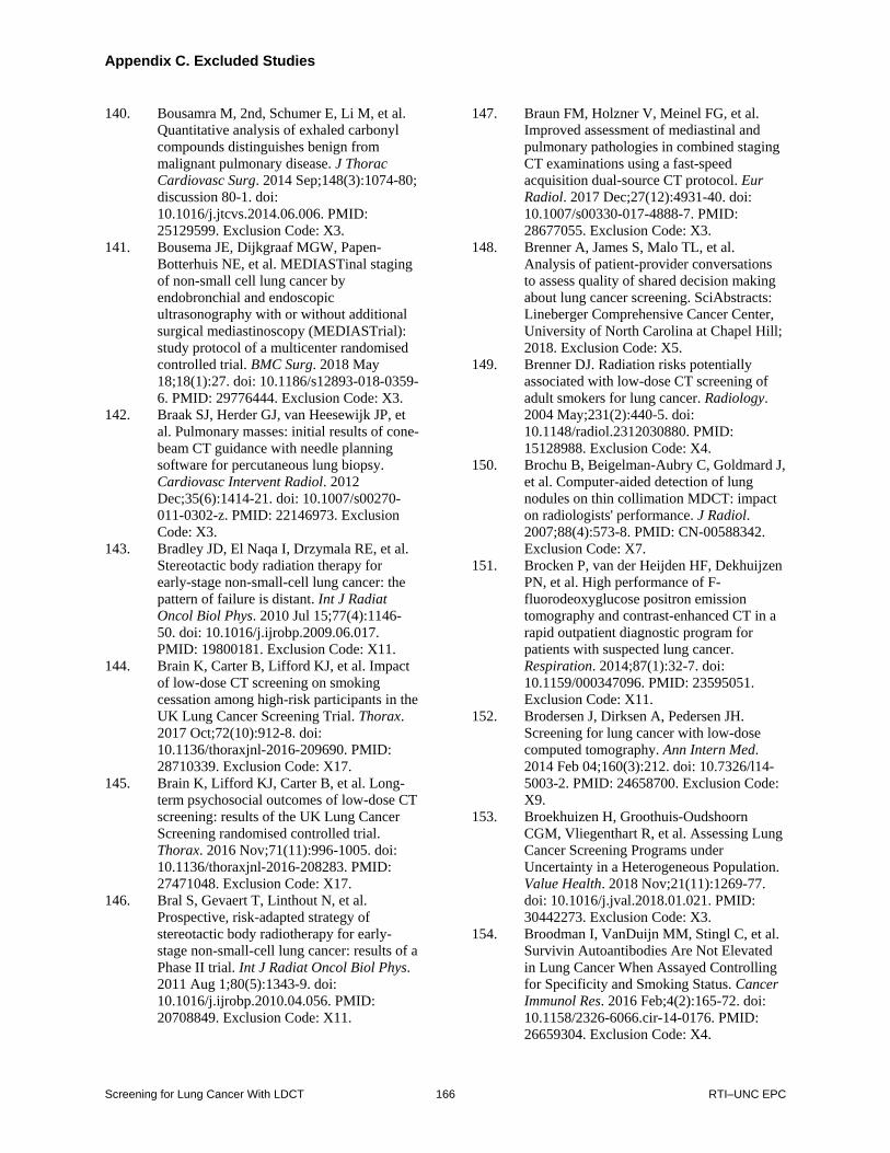
Appendix C. Excluded Studies
Screening for Lung Cancer With LDCT 166 RTI–UNC EPC
140. Bousamra M, 2nd, Schumer E, Li M, et al.
Quantitative analysis of exhaled carbonyl
compounds distinguishes benign from
malignant pulmonary disease. J Thorac
Cardiovasc Surg. 2014 Sep;148(3):1074-80;
discussion 80-1. doi:
10.1016/j.jtcvs.2014.06.006. PMID:
25129599. Exclusion Code: X3.
141. Bousema JE, Dijkgraaf MGW, Papen-
Botterhuis NE, et al. MEDIASTinal staging
of non-small cell lung cancer by
endobronchial and endoscopic
ultrasonography with or without additional
surgical mediastinoscopy (MEDIASTrial):
study protocol of a multicenter randomised
controlled trial. BMC Surg. 2018 May
18;18(1):27. doi: 10.1186/s12893-018-0359-
6. PMID: 29776444. Exclusion Code: X3.
142. Braak SJ, Herder GJ, van Heesewijk JP, et
al. Pulmonary masses: initial results of cone-
beam CT guidance with needle planning
software for percutaneous lung biopsy.
Cardiovasc Intervent Radiol. 2012
Dec;35(6):1414-21. doi: 10.1007/s00270-
011-0302-z. PMID: 22146973. Exclusion
Code: X3.
143. Bradley JD, El Naqa I, Drzymala RE, et al.
Stereotactic body radiation therapy for
early-stage non-small-cell lung cancer: the
pattern of failure is distant. Int J Radiat
Oncol Biol Phys. 2010 Jul 15;77(4):1146-
50. doi: 10.1016/j.ijrobp.2009.06.017.
PMID: 19800181. Exclusion Code: X11.
144. Brain K, Carter B, Lifford KJ, et al. Impact
of low-dose CT screening on smoking
cessation among high-risk participants in the
UK Lung Cancer Screening Trial. Thorax.
2017 Oct;72(10):912-8. doi:
10.1136/thoraxjnl-2016-209690. PMID:
28710339. Exclusion Code: X17.
145. Brain K, Lifford KJ, Carter B, et al. Long-
term psychosocial outcomes of low-dose CT
screening: results of the UK Lung Cancer
Screening randomised controlled trial.
Thorax. 2016 Nov;71(11):996-1005. doi:
10.1136/thoraxjnl-2016-208283. PMID:
27471048. Exclusion Code: X17.
146. Bral S, Gevaert T, Linthout N, et al.
Prospective, risk-adapted strategy of
stereotactic body radiotherapy for early-
stage non-small-cell lung cancer: results of a
Phase II trial. Int J Radiat Oncol Biol Phys.
2011 Aug 1;80(5):1343-9. doi:
10.1016/j.ijrobp.2010.04.056. PMID:
20708849. Exclusion Code: X11.
147. Braun FM, Holzner V, Meinel FG, et al.
Improved assessment of mediastinal and
pulmonary pathologies in combined staging
CT examinations using a fast-speed
acquisition dual-source CT protocol. Eur
Radiol. 2017 Dec;27(12):4931-40. doi:
10.1007/s00330-017-4888-7. PMID:
28677055. Exclusion Code: X3.
148. Brenner A, James S, Malo TL, et al.
Analysis of patient-provider conversations
to assess quality of shared decision making
about lung cancer screening. SciAbstracts:
Lineberger Comprehensive Cancer Center,
University of North Carolina at Chapel Hill;
2018. Exclusion Code: X5.
149. Brenner DJ. Radiation risks potentially
associated with low-dose CT screening of
adult smokers for lung cancer. Radiology.
2004 May;231(2):440-5. doi:
10.1148/radiol.2312030880. PMID:
15128988. Exclusion Code: X4.
150. Brochu B, Beigelman-Aubry C, Goldmard J,
et al. Computer-aided detection of lung
nodules on thin collimation MDCT: impact
on radiologists' performance. J Radiol.
2007;88(4):573-8. PMID: CN-00588342.
Exclusion Code: X7.
151. Brocken P, van der Heijden HF, Dekhuijzen
PN, et al. High performance of F-
fluorodeoxyglucose positron emission
tomography and contrast-enhanced CT in a
rapid outpatient diagnostic program for
patients with suspected lung cancer.
Respiration. 2014;87(1):32-7. doi:
10.1159/000347096. PMID: 23595051.
Exclusion Code: X11.
152. Brodersen J, Dirksen A, Pedersen JH.
Screening for lung cancer with low-dose
computed tomography. Ann Intern Med.
2014 Feb 04;160(3):212. doi: 10.7326/l14-
5003-2. PMID: 24658700. Exclusion Code:
X9.
153. Broekhuizen H, Groothuis-Oudshoorn
CGM, Vliegenthart R, et al. Assessing Lung
Cancer Screening Programs under
Uncertainty in a Heterogeneous Population.
Value Health. 2018 Nov;21(11):1269-77.
doi: 10.1016/j.jval.2018.01.021. PMID:
30442273. Exclusion Code: X3.
154. Broodman I, VanDuijn MM, Stingl C, et al.
Survivin Autoantibodies Are Not Elevated
in Lung Cancer When Assayed Controlling
for Specificity and Smoking Status. Cancer
Immunol Res. 2016 Feb;4(2):165-72. doi:
10.1158/2326-6066.cir-14-0176. PMID:
26659304. Exclusion Code: X4.

Appendix C. Excluded Studies
Screening for Lung Cancer With LDCT 167 RTI–UNC EPC
155. Brooks ED, Sun B, Feng L. Association of
Long-term Outcomes and Survival With
Multidisciplinary Salvage Treatment for
Local and Regional Recurrence After
Stereotactic Ablative Radiotherapy for
Early-Stage Lung Cancer. JAMA Network
Open. 2018;1(4):e181390. doi:
doi:10.1001/jamanetworkopen.2018.1390.
Exclusion Code: X3.
156. Brown LM, Cooke DT, Jett JR, et al. Extent
of Resection and Lymph Node Assessment
for Clinical Stage T1aN0M0 Typical
Carcinoid Tumors. Ann Thorac Surg. 2018
Jan;105(1):207-13. doi:
10.1016/j.athoracsur.2017.07.049. PMID:
29132704. Exclusion Code: X3.
157. Brunelli A, Dinesh P, Woodcock-Shaw J, et
al. Ninety-Day Mortality After Video-
Assisted Thoracoscopic Lobectomy:
Incidence and Risk Factors. Ann Thorac
Surg. 2017 Sep;104(3):1020-6. doi:
10.1016/j.athoracsur.2017.02.083. PMID:
28577845. Exclusion Code: X3.
158. Brunelli A, Pompili C, Salati M, et al.
Preoperative maximum oxygen consumption
is associated with prognosis after pulmonary
resection in stage I non-small cell lung
cancer. Ann Thorac Surg. 2014
Jul;98(1):238-42. doi:
10.1016/j.athoracsur.2014.04.029. PMID:
24874414. Exclusion Code: X11.
159. Brunelli A, Salati M, Rocco G, et al.
European risk models for morbidity
(EuroLung1) and mortality (EuroLung2) to
predict outcome following anatomic lung
resections: an analysis from the European
Society of Thoracic Surgeons database. Eur
J Cardiothorac Surg. 2017 Mar 1;51(3):490-
7. doi: 10.1093/ejcts/ezw319. PMID:
27744321. Exclusion Code: X3.
160. Bryant AK, Mundt R, Sandhu APS, et al.
Stereotactic body radiation therapy versus
surgery for early non-small-cell lung cancer
in the veterans affairs system. International
journal of radiation oncology biology
physics. 2017;99(2):S7‐. doi:
10.1016/j.ijrobp.2017.06.1661. PMID: CN-
01422912. Exclusion Code: X2.
161. Bugge A, Lund MB, Brunborg C, et al.
Survival After Surgical Resection for Lung
Cancer in Patients With Chronic Obstructive
Pulmonary Disease. Ann Thorac Surg. 2016
Jun;101(6):2125-31. doi:
10.1016/j.athoracsur.2015.12.057. PMID:
27016839. Exclusion Code: X3.
162. Bugge AS, Lund MB, Valberg M, et al.
Cause-specific death after surgical resection
for early-stage non-small-cell lung cancer.
Eur J Cardiothorac Surg. 2018 Jan
1;53(1):221-7. doi: 10.1093/ejcts/ezx274.
PMID: 28950311. Exclusion Code: X10.
163. Bui NC, Lee YY, Suh M, et al. Beliefs and
Intentions to Undergo Lung Cancer
Screening among Korean Males. Cancer Res
Treat. 2018 Oct;50(4):1096-105. doi:
10.4143/crt.2017.393. PMID: 29141395.
Exclusion Code: X6.
164. Burt BM, Kosinski AS, Shrager JB, et al.
Thoracoscopic lobectomy is associated with
acceptable morbidity and mortality in
patients with predicted postoperative forced
expiratory volume in 1 second or diffusing
capacity for carbon monoxide less than 40%
of normal. J Thorac Cardiovasc Surg. 2014
Jul;148(1):19-28, dicussion -9.e1. doi:
10.1016/j.jtcvs.2014.03.007. PMID:
24766848. Exclusion Code: X3.
165. Butof R, Simon M, Lock S, et al. PORTAF -
postoperative radiotherapy of non-small cell
lung cancer: accelerated versus conventional
fractionation - study protocol for a
randomized controlled trial. Trials.
2017;18(1)doi: 10.1186/s13063-017-2346-0.
PMID: CN-01441318. Exclusion Code: X6.
166. Calcagni ML, Taralli S, Cardillo G, et al.
Diagnostic performance of (18)F-
Fluorodeoxyglucose in 162 small pulmonary
nodules incidentally detected in subjects
without a history of malignancy. Ann
Thorac Surg. 2016 Apr;101(4):1303-9. doi:
10.1016/j.athoracsur.2015.10.072. PMID:
26822348. Exclusion Code: X3.
167. Calderon-Santiago M, Priego-Capote F,
Turck N, et al. Human sweat metabolomics
for lung cancer screening. Anal Bioanal
Chem. 2015 Jul;407(18):5381-92. doi:
10.1007/s00216-015-8700-8. PMID:
25935675. Exclusion Code: X5.
168. Call S, Obiols C, Rami-Porta R, et al.
Video-Assisted Mediastinoscopic
Lymphadenectomy for Staging Non-Small
Cell Lung Cancer. Ann Thorac Surg. 2016
Apr;101(4):1326-33. doi:
10.1016/j.athoracsur.2015.10.073. PMID:
26806169. Exclusion Code: X3.

Appendix C. Excluded Studies
Screening for Lung Cancer With LDCT 168 RTI–UNC EPC
169. Callahan J, Kron T, Schneider ME, et al. A
prospective investigation into the clinical
impact of 4D-PET/CT in the
characterisation of solitary pulmonary
nodules. Cancer Imaging. 2014 Jun 5;14:24.
doi: 10.1186/1470-7330-14-24. PMID:
25608679. Exclusion Code: X8.
170. Callahan J, Kron T, Siva S, et al.
Geographic miss of lung tumours due to
respiratory motion: a comparison of 3D vs
4D PET/CT defined target volumes. Radiat
Oncol. 2014 Dec 16;9:291. doi:
10.1186/s13014-014-0291-6. PMID:
25511904. Exclusion Code: X3.
171. Cannon DM, Mehta MP, Adkison JB, et al.
Dose-limiting toxicity after hypofractionated
dose-escalated radiotherapy in non-small-
cell lung cancer. J Clin Oncol. 2013 Dec
1;31(34):4343-8. doi:
10.1200/jco.2013.51.5353. PMID:
24145340. Exclusion Code: X6.
172. Cannon NA, Meyer J, Iyengar P, et al.
Neutrophil-lymphocyte and platelet-
lymphocyte ratios as prognostic factors after
stereotactic radiation therapy for early-stage
non-small-cell lung cancer. J Thorac Oncol.
2015 Feb;10(2):280-5. doi:
10.1097/jto.0000000000000399. PMID:
25299234. Exclusion Code: X10.
173. Cao C, Wang D, Chung C, et al. A
systematic review and meta-analysis of
stereotactic body radiation therapy versus
surgery for patients with non-small cell lung
cancer. J Thorac Cardiovasc Surg. 2019
Jan;157(1):362-73.e8. doi:
10.1016/j.jtcvs.2018.08.075. PMID:
30482524. Exclusion Code: X3.
174. Capalbo E, Peli M, Lovisatti M, et al. Trans-
thoracic biopsy of lung lesions: FNAB or
CNB? Our experience and review of the
literature. Radiol Med. 2014
Aug;119(8):572-94. doi: 10.1007/s11547-
013-0360-1. PMID: 24297594. Exclusion
Code: X11.
175. Capasso R, Nizzoli R, Tiseo M, et al. Extra-
pleuric coaxial system for CT-guided
percutaneous fine-needle aspiration biopsy
(FNAB) of small (</=20 mm) lung nodules:
a novel technique using multiplanar
reconstruction (MPR) images. Med Oncol.
2017 Feb;34(2):17. doi: 10.1007/s12032-
016-0871-4. PMID: 28035579. Exclusion
Code: X3.
176. Carlsson A, Nair VS, Luttgen MS, et al.
Circulating tumor microemboli diagnostics
for patients with non-small-cell lung cancer.
J Thorac Oncol. 2014 Aug;9(8):1111-9. doi:
10.1097/jto.0000000000000235. PMID:
25157764. Exclusion Code: X3.
177. Caronia FP, Fiorelli A, Ruffini E, et al. A
comparative analysis of Pancoast tumour
resection performed via video-assisted
thoracic surgery versus standard open
approaches. Interact Cardiovasc Thorac
Surg. 2014 Sep;19(3):426-35. doi:
10.1093/icvts/ivu115. PMID: 25052071.
Exclusion Code: X3.
178. Carozzi FM, Bisanzi S, Carrozzi L, et al.
Multimodal lung cancer screening using the
ITALUNG biomarker panel and low dose
computed tomography. Results of the
ITALUNG biomarker study. Int J Cancer.
2017 Jul 01;141(1):94-101. doi:
10.1002/ijc.30727. PMID: 28387927.
Exclusion Code: X7.
179. Carrafiello G, Mangini M, Fontana F, et al.
Microwave ablation of lung tumours: single-
centre preliminary experience. Radiol Med.
2014 Jan;119(1):75-82. doi:
10.1007/s11547-013-0301-z. PMID:
24234180. Exclusion Code: X6.
180. Carrillo SA, Daniel VC, Hall N, et al.
Fusion positron emission/computed
tomography underestimates the presence of
hilar nodal metastases in patients with
resected non-small cell lung cancer. Ann
Thorac Surg. 2012 May;93(5):1621-4. doi:
10.1016/j.athoracsur.2012.01.006. PMID:
22429676. Exclusion Code: X3.
181. Carter G, Barrett A, Kaye J, et al. A
comprehensive review of nongenetic
prognostic and predictive factors influencing
the heterogeneity of outcomes in advanced
non-small-cell lung cancer (Provisional
abstract). Database of Abstracts of Reviews
of Effects. 2014(2):437-49. PMID: DARE-
12014067536. Exclusion Code: X3.
182. Cassidy A, Myles JP, van Tongeren M, et al.
The LLP risk model: an individual risk
prediction model for lung cancer. Br J
Cancer. 2008 Jan 29;98(2):270-6. doi:
10.1038/sj.bjc.6604158. PMID: 18087271.
Exclusion Code: X7.

Appendix C. Excluded Studies
Screening for Lung Cancer With LDCT 169 RTI–UNC EPC
183. Cassidy RJ, Patel PR, Zhang X, et al.
Stereotactic Body Radiotherapy for Early-
stage Non-small-cell Lung Cancer in
Patients 80 Years and Older: A Multi-center
Analysis. Clin Lung Cancer. 2017
Sep;18(5):551-8.e6. doi:
10.1016/j.cllc.2017.03.006. PMID:
28373068. Exclusion Code: X3.
184. Casutt A, Bouchaab H, Beigelman-Aubry C,
et al. Stereotactic body radiotherapy with
helical TomoTherapy for medically
inoperable early stage primary and second-
primary non-small-cell lung neoplasm: 1-
year outcome and toxicity analysis. Br J
Radiol. 2015 May;88(1049):20140687. doi:
10.1259/bjr.20140687. PMID: 25735428.
Exclusion Code: X3.
185. Casutt A, Prella M, Beigelman-Aubry C, et
al. Fluoroscopic-Guided Radial
Endobronchial Ultrasound Without Guide
Sheath For Peripheral Pulmonary Lesions: A
Safe And Efficient Combination. Arch
Bronconeumol. 2015 Jul;51(7):338-43. doi:
10.1016/j.arbres.2014.09.017. PMID:
25769444. Exclusion Code: X3.
186. Cataldo JK. High-risk older smokers'
perceptions, attitudes, and beliefs about lung
cancer screening. Cancer Med. 2016
Apr;5(4):753-9. doi: 10.1002/cam4.617.
PMID: 26822940. Exclusion Code: X8.
187. Cattoni M, Vallieres E, Brown LM, et al.
External Validation of a Prognostic Model
of Survival for Resected Typical Bronchial
Carcinoids. Ann Thorac Surg. 2017
Oct;104(4):1215-20. doi:
10.1016/j.athoracsur.2017.05.019. PMID:
28821334. Exclusion Code: X3.
188. Cavic M, Spasic J, Krivokuca A, et al. TP53
and DNA-repair gene polymorphisms
genotyping as a low-cost lung
adenocarcinoma screening tool. J Clin
Pathol. 2019 Jan;72(1):75-80. doi:
10.1136/jclinpath-2018-205553. PMID:
30467244. Exclusion Code: X3.
189. Cazzoli R, Buttitta F, Di Nicola M, et al.
microRNAs derived from circulating
exosomes as noninvasive biomarkers for
screening and diagnosing lung cancer. J
Thorac Oncol. 2013 Sep;8(9):1156-62. doi:
10.1097/JTO.0b013e318299ac32. PMID:
23945385. Exclusion Code: X5.
190. Cengiz A, Aydin F, Sipahi M, et al. The role
of F-18 FDG PET/CT in differentiating
benign from malignant pulmonary masses
and accompanying lymph nodes. Tuberk
Toraks. 2018 Jun;66(2):130-5. doi:
10.5578/tt.10809. PMID: 30246656.
Exclusion Code: X3.
191. Centers for Medicare & Medicaid Services.
Proposed decision memo for screening for
lung cancer with low dose computed
tomography (LDCT) (CAG-00439N).
Baltimore, MD: U.S. Centers for Medicare
& Medicaid Services; 2014.
https://www.cms.gov/medicare-coverage-
database/details/nca-proposed-decision-
memo.aspx?NCAId=274 Accessed Nov 20,
2017. Exclusion Code: X10.
192. Ceppi M, Munnia A, Cellai F, et al. Linking
the generation of DNA adducts to lung
cancer. Toxicology. 2017 Sep 1;390:160-6.
doi: 10.1016/j.tox.2017.09.011. PMID:
28928033. Exclusion Code: X3.
193. Cerfolio RJ, Ojha B, Bryant AS, et al. The
accuracy of integrated PET-CT compared
with dedicated PET alone for the staging of
patients with nonsmall cell lung cancer. Ann
Thorac Surg. 2004 Sep;78(3):1017-23;
discussion -23. doi:
10.1016/j.athoracsur.2004.02.067. PMID:
15337041. Exclusion Code: X3.
194. Cerfolio RJ, Watson C, Minnich DJ, et al.
One Hundred Planned Robotic
Segmentectomies: Early Results, Technical
Details, and Preferred Port Placement. Ann
Thorac Surg. 2016 Mar;101(3):1089-95;
Discussion 95-6. doi:
10.1016/j.athoracsur.2015.08.092. PMID:
26846343. Exclusion Code: X3.
195. Cerra-Franco A, Liu S, Azar M, et al.
Predictors of Nodal and Metastatic Failure
in Early Stage Non-small-cell Lung Cancer
After Stereotactic Body Radiation Therapy.
Clin Lung Cancer. 2019 May;20(3):186-
93.e3. doi: 10.1016/j.cllc.2018.12.016.
PMID: 30711394. Exclusion Code: X13.
196. Cha N, Lv M, Zhao YJ, et al. Diagnostic
utility of VEGF mRNA and SP1 mRNA
expression in bronchial cells of patients with
lung cancer. Respirology. 2014
May;19(4):544-8. doi: 10.1111/resp.12272.
PMID: 24661424. Exclusion Code: X3.

Appendix C. Excluded Studies
Screening for Lung Cancer With LDCT 170 RTI–UNC EPC
197. Chabowski M, Szymanska-Chabowska A,
Skotarczak J, et al. The role of
mediastinoscopy in the diagnosis of thoracic
disease: one-year single center experience.
Adv Exp Med Biol. 2015;852:1-4. doi:
10.1007/5584_2014_101. PMID: 25416070.
Exclusion Code: X11.
198. Chambers SK, Baade P, Youl P, et al.
Psychological distress and quality of life in
lung cancer: the role of health-related
stigma, illness appraisals and social
constraints. Psychooncology. 2015
Nov;24(11):1569-77. doi:
10.1002/pon.3829. PMID: 25920906.
Exclusion Code: X6.
199. Chan A, Devanand A, Low SY, et al. Radial
endobronchial ultrasound in diagnosing
peripheral lung lesions in a high tuberculosis
setting. BMC Pulm Med. 2015 Aug
19;15:90. doi: 10.1186/s12890-015-0089-9.
PMID: 26285821. Exclusion Code: X3.
200. Chan EG, Landreneau JR, Schuchert MJ, et
al. Preoperative (3-dimensional) computed
tomography lung reconstruction before
anatomic segmentectomy or lobectomy for
stage I non-small cell lung cancer. J Thorac
Cardiovasc Surg. 2015 Sep;150(3):523-8.
doi: 10.1016/j.jtcvs.2015.06.051. PMID:
26319461. Exclusion Code: X6.
201. Chandarana H, Heacock L, Rakheja R, et al.
Pulmonary nodules in patients with primary
malignancy: comparison of hybrid PET/MR
and PET/CT imaging. Radiology. 2013
Sep;268(3):874-81. doi:
10.1148/radiol.13130620. PMID: 23737537.
Exclusion Code: X3.
202. Chandler PD, Song Y, Lin J, et al. Lipid
biomarkers and long-term risk of cancer in
the Women's Health Study. Am J Clin Nutr.
2016 Jun;103(6):1397-407. doi:
10.3945/ajcn.115.124321. PMID: 27099252.
Exclusion Code: X3.
203. Chang B, Hwang JH, Choi YH, et al.
Natural history of pure ground-glass opacity
lung nodules detected by low-dose CT scan.
Chest. 2013 Jan;143(1):172-8. doi:
10.1378/chest.11-2501. PMID: 22797081.
Exclusion Code: X3.
204. Chang DH, Hiss S, Mueller D, et al.
Radiation Dose Reduction in Computed
Tomography-Guided Lung Interventions
using an Iterative Reconstruction Technique.
Rofo. 2015 Oct;187(10):906-14. doi:
10.1055/s-0035-1553125. PMID: 26085175.
Exclusion Code: X5.
205. Chang JM, Kam KH, Yen YT, et al. From
biportal to uniportal video-assisted
thoracoscopic anatomical lung resection: A
single-institute experience. Medicine
(Baltimore). 2016 Oct;95(40):e5097. doi:
10.1097/md.0000000000005097. PMID:
27749589. Exclusion Code: X3.
206. Chang JY, Komaki R, Wen HY, et al.
Toxicity and patterns of failure of
adaptive/ablative proton therapy for early-
stage, medically inoperable non-small cell
lung cancer. Int J Radiat Oncol Biol Phys.
2011 Aug 1;80(5):1350-7. doi:
10.1016/j.ijrobp.2010.04.049. PMID:
21251767. Exclusion Code: X6.
207. Chang JY, Li QQ, Xu QY, et al. Stereotactic
ablative radiation therapy for centrally
located early stage or isolated parenchymal
recurrences of non-small cell lung cancer:
how to fly in a "no fly zone". Int J Radiat
Oncol Biol Phys. 2014 Apr 1;88(5):1120-8.
doi: 10.1016/j.ijrobp.2014.01.022. PMID:
24661665. Exclusion Code: X3.
208. Chang JY, Senan S, Paul MA, et al.
Stereotactic ablative radiotherapy versus
lobectomy for operable stage I non-small-
cell lung cancer: a pooled analysis of two
randomised trials. Lancet Oncol. 2015
Jun;16(6):630-7. doi: 10.1016/s1470-
2045(15)70168-3. PMID: 25981812.
Exclusion Code: X9.
209. Chang JY, Zhang W, Komaki R, et al. Long-
term outcome of phase I/II prospective study
of dose-escalated proton therapy for early-
stage non-small cell lung cancer. Radiother
Oncol. 2017 Feb;122(2):274-80. doi:
10.1016/j.radonc.2016.10.022. PMID:
28139305. Exclusion Code: X6.
210. Chang WP, Lin CC. Changes in the sleep-
wake rhythm, sleep quality, mood, and
quality of life of patients receiving treatment
for lung cancer: A longitudinal study.
Chronobiol Int. 2017;34(4):451-61. doi:
10.1080/07420528.2017.1293678. PMID:
28282238. Exclusion Code: X3.
211. Chang YY, Chen CK, Yeh YC, et al.
Diagnostic feasibility and safety of CT-
guided core biopsy for lung nodules less
than or equal to 8 mm: A single-institution
experience. Eur Radiol. 2018
Feb;28(2):796-806. doi: 10.1007/s00330-
017-5027-1. PMID: 28884222. Exclusion
Code: X3.

Appendix C. Excluded Studies
Screening for Lung Cancer With LDCT 171 RTI–UNC EPC
212. Charvat H, Sasazuki S, Shimazu T, et al.
Development of a risk prediction model for
lung cancer: The Japan Public Health
Center-based Prospective Study. Cancer Sci.
2018 Mar;109(3):854-62. doi:
10.1111/cas.13509. PMID: 29345859.
Exclusion Code: X7.
213. Chellapandian D, Lehrnbecher T, Phillips B,
et al. Bronchoalveolar lavage and lung
biopsy in patients with cancer and
hematopoietic stem-cell transplantation
recipients: a systematic review and meta-
analysis. J Clin Oncol. 2015 Feb
10;33(5):501-9. doi:
10.1200/jco.2014.58.0480. PMID:
25559816. Exclusion Code: X3.
214. Chen C, Mu CY, Su MQ, et al.
Endobronchial Ultrasound-guided
Transbronchial Needle Aspiration Increases
the Yield of Transbronchial Lung Biopsy for
the Evaluation of Peribronchial Lesions.
Chin Med J (Engl). 2017 5th Jan
2017;130(1):11-4. doi: 10.4103/0366-
6999.196567. PMID: 28051017. Exclusion
Code: X3.
215. Chen C, Zhu WD, Zhang XH, et al. Value of
Ki-67 and computed tomography in the
assessment of peripheral lung
adenocarcinoma. Br J Biomed Sci.
2016;73(1):32-7. doi:
10.1080/09674845.2016.1146434. PMID:
27182675. Exclusion Code: X8.
216. Chen CY, Chen CH, Shen TC, et al. Lung
cancer screening with low-dose computed
tomography: Experiences from a tertiary
hospital in Taiwan. J Formos Med Assoc.
2016 Mar;115(3):163-70. doi:
10.1016/j.jfma.2015.11.007. PMID:
26714425. Exclusion Code: X7.
217. Chen FF, Zhang D, Wang YL, et al. Video-
assisted thoracoscopic surgery lobectomy
versus open lobectomy in patients with
clinical stage non-small cell lung cancer: a
meta-analysis. Eur J Surg Oncol. 2013
Sep;39(9):957-63. doi:
10.1016/j.ejso.2013.06.016. PMID:
23845704. Exclusion Code: X9.
218. Chen HH, Chiu NT, Su WC, et al.
Prognostic value of whole-body total lesion
glycolysis at pretreatment FDG PET/CT in
non-small cell lung cancer. Radiology. 2012
Aug;264(2):559-66. doi:
10.1148/radiol.12111148. PMID: 22692034.
Exclusion Code: X3.
219. Chen K, Chen H, Yang F, et al. Validation
of the Eighth Edition of the TNM Staging
System for Lung Cancer in 2043 Surgically
Treated Patients With Non-small-cell Lung
Cancer. Clin Lung Cancer. 2017
Nov;18(6):e457-e66. doi:
10.1016/j.cllc.2017.04.001. PMID:
28539214. Exclusion Code: X3.
220. Chen S, Li X, Chen M, et al. Limited
diagnostic value of Dual-Time-Point (18)F-
FDG PET/CT imaging for classifying
solitary pulmonary nodules in granuloma-
endemic regions both at visual and
quantitative analyses. Eur J Radiol. 2016
Oct;85(10):1744-9. doi:
10.1016/j.ejrad.2016.07.016. PMID:
27666611. Exclusion Code: X3.
221. Chen T, Luo J, Gu H, et al. Impact of Solid
Minor Histologic Subtype in Postsurgical
Prognosis of Stage I Lung Adenocarcinoma.
Ann Thorac Surg. 2018 Jan;105(1):302-8.
doi: 10.1016/j.athoracsur.2017.08.018.
PMID: 29162222. Exclusion Code: X10.
222. Chen X, Gorlov IP, Ying J, et al. Initial
medical attention on patients with early-
stage non-small cell lung cancer. PLoS One.
2012;7(3):e32644. doi:
10.1371/journal.pone.0032644. PMID:
22412901. Exclusion Code: X3.
223. Chen X, Wu J, Zhang F, et al. Prognostic
Significance of Pre-Operative Monocyte-to-
Lymphocyte Ratio in Lung Cancer Patients
Undergoing Radical Surgery. Lab Med.
2018 Mar 21;49(2):e29-e39. doi:
10.1093/labmed/lmx069. PMID: 29361036.
Exclusion Code: X3.
224. Chen XR, Dong JN, Zhang F, et al. Efficacy
and safety of image-guidance radiotherapy
by helical tomotherapy in patients with lung
cancer. Medicine (Baltimore). 2018
Jan;97(1):e9243. doi:
10.1097/md.0000000000009243. PMID:
29505510. Exclusion Code: X14.
225. Chen Y, Li J, Zhang Y, et al. Early versus
late prophylactic cranial irradiation in
patients with extensive small cell lung
cancer. Strahlenther Onkol. 2018
Oct;194(10):876-85. doi: 10.1007/s00066-
018-1307-1. PMID: 29774395. Exclusion
Code: X3.
226. Chen Z, Xu Z, Sun S, et al. TGF-beta1, IL-
6, and TNF-alpha in bronchoalveolar lavage
fluid: useful markers for lung cancer? Sci
Rep. 2014 Jul 7;4:5595. doi:
10.1038/srep05595. PMID: 24999009.
Exclusion Code: X11.

Appendix C. Excluded Studies
Screening for Lung Cancer With LDCT 172 RTI–UNC EPC
227. Cheng M, Liu L, Yang HS, et al. Circulating
tumor cells are associated with bone
metastasis of lung cancer. Asian Pac J
Cancer Prev. 2014;15(15):6369-74. PMID:
25124627. Exclusion Code: X3.
228. Cheng YD, Duan CJ, Dong S, et al. Clinical
controlled comparison between lobectomy
and segmental resection for patients over 70
years of age with clinical stage I non-small
cell lung cancer. Eur J Surg Oncol. 2012
Dec;38(12):1149-55. doi:
10.1016/j.ejso.2012.08.001. PMID:
22901959. Exclusion Code: X11.
229. Chesterfield-Thomas G, Goldsmith I. Impact
of preoperative pulmonary rehabilitation on
the Thoracoscore of patients undergoing
lung resection. Interact Cardiovasc Thorac
Surg. 2016 Nov;23(5):729-32. doi:
10.1093/icvts/ivw238. PMID: 27430554.
Exclusion Code: X3.
230. Cheung P, Faria S, Ahmed S, et al. Phase II
study of accelerated hypofractionated three-
dimensional conformal radiotherapy for
stage T1-3 N0 M0 non-small cell lung
cancer: NCIC CTG BR.25. J Natl Cancer
Inst. 2014 Aug;106(8)doi:
10.1093/jnci/dju164. PMID: 25074417.
Exclusion Code: X6.
231. Chi A, Wen S, Liao Z, et al. What would be
the most appropriate alpha/beta ratio in the
setting of stereotactic body radiation therapy
for early stage non-small cell lung cancer.
Biomed Res Int. 2013;2013:391021. doi:
10.1155/2013/391021. PMID: 24350266.
Exclusion Code: X10.
232. Chi CT. A multicenter randomized
controlled trial on video-assisted
thoracoscopic surgery and axillary
thoracotomy for resection of early-stage
non-small cell lung cancer.
Http://www.who.int/trialsearch/trial2.aspx?
Trialid=chictr-trc-10000750. 2010PMID:
CN-01833643. Exclusion Code: X16.
233. Chiang A, Thibault I, Warner A, et al. A
comparison between accelerated
hypofractionation and stereotactic ablative
radiotherapy (SABR) for early-stage non-
small cell lung cancer (NSCLC): Results of
a propensity score-matched analysis.
Radiother Oncol. 2016 Mar;118(3):478-84.
doi: 10.1016/j.radonc.2015.12.026. PMID:
26795773. Exclusion Code: X3.
234. Chiappetta M, Rosella F, Dall'armi V, et al.
CT-guided fine-needle ago-biopsy of
pulmonary nodules: predictive factors for
diagnosis and pneumothorax occurrence.
Radiol Med. 2016 Aug;121(8):635-43. doi:
10.1007/s11547-016-0639-0. PMID:
27132130. Exclusion Code: X3.
235. Chiles C, Duan F, Gladish GW, et al.
Association of Coronary Artery
Calcification and Mortality in the National
Lung Screening Trial: A Comparison of
Three Scoring Methods. Radiology. 2015
Jul;276(1):82-90. doi:
10.1148/radiol.15142062. PMID: 25759972.
Exclusion Code: X5.
236. Chlebowski RT, Wakelee H, Pettinger M, et
al. Estrogen Plus Progestin and Lung
Cancer: Follow-up of the Women's Health
Initiative Randomized Trial. Clin Lung
Cancer. 2016 Jan;17(1):10-7.e1. doi:
10.1016/j.cllc.2015.09.004. PMID:
26577506. Exclusion Code: X6.
237. Cho H, Lee HY, Kim J, et al. Pure ground
glass nodular adenocarcinomas: Are
preoperative positron emission
tomography/computed tomography and
brain magnetic resonance imaging useful or
necessary? J Thorac Cardiovasc Surg. 2015
Sep;150(3):514-20. doi:
10.1016/j.jtcvs.2015.06.024. PMID:
26189165. Exclusion Code: X3.
238. Cho HJ, Yun HJ, Yang HC, et al. Prognostic
significance of nuclear factor of activated T-
cells 5 expression in non-small cell lung
cancer patients who underwent surgical
resection. J Surg Res. 2018 Jun;226:40-7.
doi: 10.1016/j.jss.2017.12.036. PMID:
29661287. Exclusion Code: X3.
239. Cho J, Kim ES, Kim SJ, et al. Long-Term
Follow-up of Small Pulmonary Ground-
Glass Nodules Stable for 3 Years:
Implications of the Proper Follow-up Period
and Risk Factors for Subsequent Growth. J
Thorac Oncol. 2016 Sep;11(9):1453-9. doi:
10.1016/j.jtho.2016.05.026. PMID:
27287413. Exclusion Code: X3.
240. Cho J, Ko SJ, Kim SJ, et al. Surgical
resection of nodular ground-glass opacities
without percutaneous needle aspiration or
biopsy. BMC Cancer. 2014 Nov 18;14:838.
doi: 10.1186/1471-2407-14-838. PMID:
25406492. Exclusion Code: X3.

Appendix C. Excluded Studies
Screening for Lung Cancer With LDCT 173 RTI–UNC EPC
241. Cho JH, Choi YS, Kim J, et al. Long-term
outcomes of wedge resection for pulmonary
ground-glass opacity nodules. Ann Thorac
Surg. 2015 Jan;99(1):218-22. doi:
10.1016/j.athoracsur.2014.07.068. PMID:
25440277. Exclusion Code: X11.
242. Choe J, Lee SM, Do KH, et al. Prognostic
value of radiomic analysis of iodine overlay
maps from dual-energy computed
tomography in patients with resectable lung
cancer. Eur Radiol. 2019 Feb;29(2):915-23.
doi: 10.1007/s00330-018-5639-0. PMID:
30054795. Exclusion Code: X11.
243. Choi JW, Park CM, Goo JM, et al. C-arm
cone-beam CT-guided percutaneous
transthoracic needle biopsy of small (</= 20
mm) lung nodules: diagnostic accuracy and
complications in 161 patients. AJR Am J
Roentgenol. 2012 Sep;199(3):W322-30. doi:
10.2214/ajr.11.7576. PMID: 22915422.
Exclusion Code: X3.
244. Choi MJ, Kim Y, Hong YS, et al.
Transthoracic needle biopsy using a C-arm
cone-beam CT system: diagnostic accuracy
and safety. Br J Radiol. 2012
Jun;85(1014):e182-7. doi:
10.1259/bjr/95413532. PMID: 21791505.
Exclusion Code: X10.
245. Choi SH, Chae EJ, Shin SY, et al.
Comparisons of clinical outcomes in
patients with and without a preoperative
tissue diagnosis in the persistent malignant-
looking, ground-glass-opacity nodules.
Medicine (Baltimore). 2016
Aug;95(34):e4359. doi:
10.1097/md.0000000000004359. PMID:
27559945. Exclusion Code: X3.
246. Choi SM, Heo EY, Lee J, et al.
Characteristics of benign solitary pulmonary
nodules confirmed by diagnostic video-
assisted thoracoscopic surgery. Clin Respir
J. 2016 Mar;10(2):181-8. doi:
10.1111/crj.12200. PMID: 25103572.
Exclusion Code: X3.
247. Choi YR, An JY, Kim MK, et al. The
diagnostic efficacy and safety of
endobronchial ultrasound-guided
transbronchial needle aspiration as an initial
diagnostic tool. Korean J Intern Med. 2013
Nov;28(6):660-7. doi:
10.3904/kjim.2013.28.6.660. PMID:
24307841. Exclusion Code: X3.
248. Choi YY, Lee SY, Lee WK, et al. RACK1 is
a candidate gene associated with the
prognosis of patients with early stage non-
small cell lung cancer. Oncotarget. 2015
Feb 28;6(6):4451-66. doi:
10.18632/oncotarget.2865. PMID:
25686824. Exclusion Code: X6.
249. Chopra A, Ford A, De Noronha R, et al.
Incidental findings on positron emission
tomography/CT scans performed in the
investigation of lung cancer. Br J Radiol.
2012 Jul;85(1015):e229-37. doi:
10.1259/bjr/60606623. PMID: 22745208.
Exclusion Code: X3.
250. Chou HP, Chen CK, Shen SH, et al.
Percutaneous cryoablation for inoperable
malignant lung tumors: midterm results.
Cryobiology. 2015 Feb;70(1):60-5. doi:
10.1016/j.cryobiol.2014.12.004. PMID:
25541142. Exclusion Code: X6.
251. Christe A, Ebner L, Steiger P, et al. Impact
of image quality, radiologists, lung
segments, and Gunnar eyewear on
detectability of lung nodules in chest CT.
Acta Radiol. 2013 Jul;54(6):646-51. doi:
10.1177/0284185113483677. PMID:
23612429. Exclusion Code: X3.
252. Christe A, Leidolt L, Huber A, et al. Lung
cancer screening with CT: evaluation of
radiologists and different computer assisted
detection software (CAD) as first and
second readers for lung nodule detection at
different dose levels. Eur J Radiol. 2013
Dec;82(12):e873-8. doi:
10.1016/j.ejrad.2013.08.026. PMID:
24074648. Exclusion Code: X3.
253. Christensen TD, Vad H, Pedersen S, et al.
Coagulation profile in open and video-
assisted thoracoscopic lobectomies: a cohort
study. Interact Cardiovasc Thorac Surg.
2018 Mar 1;26(3):382-8. doi:
10.1093/icvts/ivx328. PMID: 29049684.
Exclusion Code: X3.
254. Christian C, Erica S, Morandi U. The
prognostic impact of tumor size in resected
stage I non-small cell lung cancer: evidence
for a two thresholds tumor diameters
classification. Lung Cancer. 2006
Nov;54(2):185-91. doi:
10.1016/j.lungcan.2006.08.003. PMID:
16996167. Exclusion Code: X10.

Appendix C. Excluded Studies
Screening for Lung Cancer With LDCT 174 RTI–UNC EPC
255. Chua KLM, Sin I, Fong KW, et al.
Stereotactic body radiotherapy for early
stage lung cancer-historical developments
and future strategies. Chin Clin Oncol. 2017
Sep;6(Suppl 2):S20. doi:
10.21037/cco.2017.08.02. PMID: 28917258.
Exclusion Code: X10.
256. Chubachi S, Takahashi S, Tsutsumi A, et al.
Radiologic features of precancerous areas of
the lungs in chronic obstructive pulmonary
disease. Int J Chron Obstruct Pulmon Dis.
2017;12:1613-24. doi:
10.2147/copd.s132709. PMID: 28615934.
Exclusion Code: X3.
257. Chung K, Mets OM, Gerke PK, et al. Brock
malignancy risk calculator for pulmonary
nodules: validation outside a lung cancer
screening population. Thorax. 2018
Sep;73(9):857-63. doi: 10.1136/thoraxjnl-
2017-211372. PMID: 29777062. Exclusion
Code: X3.
258. Cieszanowski A, Lisowska A, Dabrowska
M, et al. MR Imaging of Pulmonary
Nodules: Detection Rate and Accuracy of
Size Estimation in Comparison to Computed
Tomography. PLoS One.
2016;11(6):e0156272. doi:
10.1371/journal.pone.0156272. PMID:
27258047. Exclusion Code: X3.
259. Citak N, Buyukkale S, Kok A, et al. Does
video-assisted mediastinoscopy offer lower
false-negative rates for subcarinal lymph
nodes compared with standard cervical
mediastinoscopy? Thorac Cardiovasc Surg.
2014 Oct;62(7):624-30. doi: 10.1055/s-
0033-1358656. PMID: 24297632. Exclusion
Code: X3.
260. Clausen E, Wittman C, Gingo M, et al.
Chest computed tomography findings in
HIV-infected individuals in the era of
antiretroviral therapy. PLoS One.
2014;9(11):e112237. doi:
10.1371/journal.pone.0112237. PMID:
25409510. Exclusion Code: X8.
261. Cole AM, Pflugeisen B, Schwartz MR, et al.
Cross sectional study to assess the accuracy
of electronic health record data to identify
patients in need of lung cancer screening.
BMC Res Notes. 2018 Jan 10;11(1):14. doi:
10.1186/s13104-018-3124-0. PMID:
29321038. Exclusion Code: X5.
262. Coley SM, Crapanzano JP, Saqi A. FNA,
core biopsy, or both for the diagnosis of
lung carcinoma: Obtaining sufficient tissue
for a specific diagnosis and molecular
testing. Cancer Cytopathol. 2015
May;123(5):318-26. doi:
10.1002/cncy.21527. PMID: 25711901.
Exclusion Code: X3.
263. Cornwell LD, Bakaeen FG, Lan CK, et al.
Endobronchial ultrasonography-guided
transbronchial needle aspiration biopsy for
preoperative nodal staging of lung cancer in
a veteran population. JAMA Surg. 2013
Nov;148(11):1024-9. doi:
10.1001/jamasurg.2013.3776. PMID:
24048268. Exclusion Code: X3.
264. Cornwell LD, Echeverria AE, Samuelian J,
et al. Video-assisted thoracoscopic
lobectomy is associated with greater
recurrence-free survival than stereotactic
body radiotherapy for clinical stage I lung
cancer. J Thorac Cardiovasc Surg. 2018
Jan;155(1):395-402. doi:
10.1016/j.jtcvs.2017.07.065. PMID:
28888379. Exclusion Code: X10.
265. Coroller TP, Mak RH, Lewis JH, et al. Low
incidence of chest wall pain with a risk-
adapted lung stereotactic body radiation
therapy approach using three or five
fractions based on chest wall dosimetry.
PLoS One. 2014;9(4):e94859. doi:
10.1371/journal.pone.0094859. PMID:
24728448. Exclusion Code: X3.
266. Corradetti MN, Haas AR, Rengan R.
Central-airway necrosis after stereotactic
body-radiation therapy. N Engl J Med. 2012
Jun 14;366(24):2327-9. doi:
10.1056/NEJMc1203770. PMID: 22694017.
Exclusion Code: X10.
267. Corradi M, Poli D, Banda I, et al. Exhaled
breath analysis in suspected cases of non-
small-cell lung cancer: a cross-sectional
study. J Breath Res. 2015 Jan
29;9(2):027101. doi: 10.1088/1752-
7155/9/2/027101. PMID: 25634546.
Exclusion Code: X7.
268. Corso CD, Park HS, Kim AW, et al. Racial
disparities in the use of SBRT for treating
early-stage lung cancer. Lung Cancer. 2015
Aug;89(2):133-8. doi:
10.1016/j.lungcan.2015.05.002. PMID:
26051446. Exclusion Code: X8.

Appendix C. Excluded Studies
Screening for Lung Cancer With LDCT 175 RTI–UNC EPC
269. Corso CD, Park HS, Moreno AC, et al.
Stage I Lung SBRT Clinical Practice
Patterns. Am J Clin Oncol. 2017
Aug;40(4):358-61. doi:
10.1097/coc.0000000000000162. PMID:
25503436. Exclusion Code: X8.
270. Coverley D, Higgins G, West D, et al. A
quantitative immunoassay for lung cancer
biomarker CIZ1b in patient plasma. Clin
Biochem. 2017 Apr;50(6):336-43. doi:
10.1016/j.clinbiochem.2016.11.015. PMID:
27867087. Exclusion Code: X3.
271. Crabtree TD, Denlinger CE, Meyers BF, et
al. Stereotactic body radiation therapy
versus surgical resection for stage I non-
small cell lung cancer. J Thorac Cardiovasc
Surg. 2010 Aug;140(2):377-86. doi:
10.1016/j.jtcvs.2009.12.054. PMID:
20400121. Exclusion Code: X11.
272. Crabtree TD, Puri V, Chen SB, et al. Does
the method of radiologic surveillance affect
survival after resection of stage I non-small
cell lung cancer? J Thorac Cardiovasc Surg.
2015 Jan;149(1):45-52, 3.e1-3. doi:
10.1016/j.jtcvs.2014.07.095. PMID:
25218540. Exclusion Code: X12.
273. Crabtree TD, Puri V, Robinson C, et al.
Analysis of first recurrence and survival in
patients with stage I non-small cell lung
cancer treated with surgical resection or
stereotactic radiation therapy. J Thorac
Cardiovasc Surg. 2014 Apr;147(4):1183-91;
discussion 91-2. doi:
10.1016/j.jtcvs.2013.11.057. PMID:
24507980. Exclusion Code: X10.
274. Criss SD, Sheehan DF, Palazzo L, et al.
Population impact of lung cancer screening
in the United States: Projections from a
microsimulation model. PLoS Med. 2018
Feb;15(2):e1002506. doi:
10.1371/journal.pmed.1002506. PMID:
29415013. Exclusion Code: X3.
275. Cronin KA, Gail MH, Zou Z, et al.
Validation of a model of lung cancer risk
prediction among smokers. J Natl Cancer
Inst. 2006 May 03;98(9):637-40. doi:
10.1093/jnci/djj163. PMID: 16670389.
Exclusion Code: X7.
276. Cruz C, Afonso M, Oliveiros B, et al.
Recurrence and Risk Factors for Relapse in
Patients with Non-Small Cell Lung Cancer
Treated by Surgery with Curative Intent.
Oncology. 2017;92(6):347-52. doi:
10.1159/000458533. PMID: 28278499.
Exclusion Code: X11.
277. Currow DC, You H, Aranda S, et al. What
factors are predictive of surgical resection
and survival from localised non-small cell
lung cancer? Med J Aust. 2014 Oct
20;201(8):475-80. PMID: 25332036.
Exclusion Code: X11.
278. Cusumano G, Fournel L, Strano S, et al.
Surgical Resection for Pulmonary
Carcinoid: Long-Term Results of
Multicentric Study-The Importance of
Pathological N Status, More Than We
Thought. Lung. 2017 Dec;195(6):789-98.
doi: 10.1007/s00408-017-0056-8. PMID:
29022070. Exclusion Code: X3.
279. Cykert S, Walker PR, Edwards LJ, et al.
Weighing projections of physical decline in
lung cancer surgery decisions. Am J Med
Sci. 2015 Jan;349(1):61-6. doi:
10.1097/maj.0000000000000354. PMID:
25285513. Exclusion Code: X3.
280. Dabrowska M, Krenke R, Korczynski P, et
al. Diagnostic accuracy of contrast-enhanced
computed tomography and positron
emission tomography with 18-FDG in
identifying malignant solitary pulmonary
nodules. Medicine (Baltimore). 2015
Apr;94(15):e666. doi:
10.1097/md.0000000000000666. PMID:
25881842. Exclusion Code: X3.
281. Dabrowska M, Przybylo Z, Zukowska M, et
al. Should we be concerned about the doses
of ionizing radiation related to diagnostic
and follow-up imaging in patients with
solitary pulmonary nodules? Radiat Prot
Dosimetry. 2018 Jan 1;178(2):201-7. doi:
10.1093/rpd/ncx099. PMID: 28981871.
Exclusion Code: X3.
282. Dai C, Shen J, Ren Y, et al. Choice of
Surgical Procedure for Patients With Non-
Small-Cell Lung Cancer </= 1 cm or > 1 to
2 cm Among Lobectomy, Segmentectomy,
and Wedge Resection: A Population-Based
Study. J Clin Oncol. 2016 Sep
10;34(26):3175-82. doi:
10.1200/jco.2015.64.6729. PMID:
27382092. Exclusion Code: X8.
283. Dai H, Liu J, Liang L, et al. Increased lung
cancer risk in patients with interstitial lung
disease and elevated CEA and CA125 serum
tumour markers. Respirology. 2014
Jul;19(5):707-13. doi: 10.1111/resp.12317.
PMID: 24903079. Exclusion Code: X3.

Appendix C. Excluded Studies
Screening for Lung Cancer With LDCT 176 RTI–UNC EPC
284. Dai J, Liu M, Swensen SJ, et al. Regional
Emphysema Score Predicting Overall
Survival, Quality of Life, and Pulmonary
Function Recovery in Early-Stage Lung
Cancer Patients. J Thorac Oncol. 2017
May;12(5):824-32. doi:
10.1016/j.jtho.2017.01.016. PMID:
28126539. Exclusion Code: X10.
285. Dai L, Tsay JC, Li J, et al. Autoantibodies
against tumor-associated antigens in the
early detection of lung cancer. Lung Cancer.
2016 Sep;99:172-9. doi:
10.1016/j.lungcan.2016.07.018. PMID:
27565936. Exclusion Code: X3.
286. Dale CR, Madtes DK, Fan VS, et al.
Navigational bronchoscopy with biopsy
versus computed tomography-guided biopsy
for the diagnosis of a solitary pulmonary
nodule: a cost-consequences analysis. J
Bronchology Interv Pulmonol. 2012
Oct;19(4):294-303. doi:
10.1097/LBR.0b013e318272157d. PMID:
23207529. Exclusion Code: X10.
287. Dalwadi SM, Szeja SS, Bernicker EH, et al.
Practice Patterns and Outcomes in Elderly
Stage I Non-Small-cell Lung Cancer: A
2004 to 2012 SEER Analysis. Clin Lung
Cancer. 2018 Mar;19(2):e269-e76. doi:
10.1016/j.cllc.2017.11.004. PMID:
29208356. Exclusion Code: X13.
288. Dama E, Melocchi V, Dezi F, et al. An
Aggressive Subtype of Stage I Lung
Adenocarcinoma with Molecular and
Prognostic Characteristics Typical of
Advanced Lung Cancers. Clin Cancer Res.
2017 Jan 1;23(1):62-72. doi: 10.1158/1078-
0432.ccr-15-3005. PMID: 27358486.
Exclusion Code: X6.
289. D'Amelio AM, Jr., Cassidy A, Asomaning
K, et al. Comparison of discriminatory
power and accuracy of three lung cancer risk
models. Br J Cancer. 2010 Jul
27;103(3):423-9. doi:
10.1038/sj.bjc.6605759. PMID: 20588271.
Exclusion Code: X7.
290. Dandachi N, Tiran V, Lindenmann J, et al.
Frequency and clinical impact of
preoperative circulating tumor cells in
resectable non-metastatic lung
adenocarcinomas. Lung Cancer. 2017
Nov;113:152-7. doi:
10.1016/j.lungcan.2017.10.003. PMID:
29110843. Exclusion Code: X8.
291. D'Andrilli A, Maurizi G, Andreetti C, et al.
Sleeve Lobectomy Versus Standard
Lobectomy for Lung Cancer: Functional and
Oncologic Evaluation. Ann Thorac Surg.
2016 May;101(5):1936-42. doi:
10.1016/j.athoracsur.2015.11.057. PMID:
26912305. Exclusion Code: X3.
292. D'Andrilli A, Natoli G, Scarpino S, et al.
Stage I non-small cell lung cancer: the
presence of the lymphocyte-specific protein
tyrosin kinase in the tumour infiltrate is
associated with a better long-term prognosis.
Interact Cardiovasc Thorac Surg. 2012
Jul;15(1):148-51. doi: 10.1093/icvts/ivr128.
PMID: 22457183. Exclusion Code: X11.
293. Dang H, Siewerdsen JH, Stayman JW.
Prospective regularization design in prior-
image-based reconstruction. Phys Med Biol.
2015 Dec 21;60(24):9515-36. doi:
10.1088/0031-9155/60/24/9515. PMID:
26606653. Exclusion Code: X5.
294. David EA, Cooke DT, Chen Y, et al.
Surgery in high-volume hospitals not
commission on cancer accreditation leads to
increased cancer-specific survival for early-
stage lung cancer. Am J Surg. 2015
Oct;210(4):643-7. doi:
10.1016/j.amjsurg.2015.05.002. PMID:
26193801. Exclusion Code: X8.
295. Davis JN, Medbery C, Sharma S, et al.
Stereotactic body radiotherapy for centrally
located early-stage non-small cell lung
cancer or lung metastases from the
RSSearch((R)) patient registry. Radiat
Oncol. 2015 May 15;10:113. doi:
10.1186/s13014-015-0417-5. PMID:
25975848. Exclusion Code: X3.
296. Davis JN, Medbery C, 3rd, Sharma S, et al.
Stereotactic body radiotherapy for early-
stage non-small cell lung cancer: clinical
outcomes from a National Patient Registry.
J Radiat Oncol. 2015;4(1):55-63. doi:
10.1007/s13566-014-0177-0. PMID:
25774243. Exclusion Code: X13.
297. Dawe DE, Pond GR, Ellis PM. Assessment
of Referral and Chemotherapy Treatment
Patterns for Elderly Patients With Non-
small-Cell Lung Cancer. Clin Lung Cancer.
2016 Nov;17(6):563-72.e2. doi:
10.1016/j.cllc.2016.05.012. PMID:
27374398. Exclusion Code: X8.

Appendix C. Excluded Studies
Screening for Lung Cancer With LDCT 177 RTI–UNC EPC
298. de Baere T, Tselikas L, Catena V, et al.
Percutaneous thermal ablation of primary
lung cancer. Diagn Interv Imaging. 2016
Oct;97(10):1019-24. doi:
10.1016/j.diii.2016.08.016. PMID:
27692673. Exclusion Code: X6.
299. de Dios NR, Sanz X, Foro P, et al.
Accelerated hypofractionated radiation
therapy (AHRT) for non-small-cell lung
cancer: can we leave standard fractionation?
Clin Transl Oncol. 2017 Apr;19(4):440-7.
doi: 10.1007/s12094-016-1544-7. PMID:
27553602. Exclusion Code: X6.
300. de Hoop B, van Ginneken B, Gietema H, et
al. Pulmonary perifissural nodules on CT
scans: rapid growth is not a predictor of
malignancy. Radiology. 2012
Nov;265(2):611-6. doi:
10.1148/radiol.12112351. PMID: 22929331.
Exclusion Code: X8.
301. de Koning HJ, Meza R, Plevritis SK, et al.
Benefits and harms of computed
tomography lung cancer screening
strategies: a comparative modeling study for
the U.S. Preventive Services Task Force.
Ann Intern Med. 2014 Mar 04;160(5):311-
20. doi: 10.7326/m13-2316. PMID:
24379002. Exclusion Code: X4.
302. De Roza MA, Quah KH, Tay CK, et al.
Diagnosis of Peripheral Lung Lesions via
Conventional Flexible Bronchoscopy with
Multiplanar CT Planning. Pulm Med.
2016;2016:5048961. doi:
10.1155/2016/5048961. PMID: 27957340.
Exclusion Code: X3.
303. De Ruysscher D, Sharifi H, Defraene G, et
al. Quantification of radiation-induced lung
damage with CT scans: the possible benefit
for radiogenomics. Acta Oncol. 2013
Oct;52(7):1405-10. doi:
10.3109/0284186x.2013.813074. PMID:
23957564. Exclusion Code: X3.
304. De Wever W, Bruyeer E, Demaerel P, et al.
Staging of lung cancer. Do we need a
diagnostic CT of the brain after an
integrated PET/CT for the detection of brain
metastases? Jbr-btr. 2010 Mar-
Apr;93(2):71-6. PMID: 20524514.
Exclusion Code: X3.
305. Debieuvre D, Fraboulet G, Duvert B, et al.
Impact of distance from surgery department
on the outcome of patients followed for non-
small-cell lung cancer in the respiratory
department of nonacademic hospitals:
Results of the KBP-2010-study. Bull
Cancer. 2017 Oct;104(10):840-9. doi:
10.1016/j.bulcan.2017.07.008. PMID:
28965729. Exclusion Code: X3.
306. Decaluwe H, Moons J, Fieuws S, et al. Is
central lung tumour location really
predictive for occult mediastinal nodal
disease in (suspected) non-small-cell lung
cancer staged cN0 on 18F-
fluorodeoxyglucose positron emission
tomography-computed tomography? Eur J
Cardiothorac Surg. 2018 Jul 1;54(1):134-
40. doi: 10.1093/ejcts/ezy018. PMID:
29447330. Exclusion Code: X6.
307. Decaluwe H, Petersen RH, Hansen H, et al.
Major intraoperative complications during
video-assisted thoracoscopic anatomical
lung resections: an intention-to-treat
analysis. Eur J Cardiothorac Surg. 2015
Oct;48(4):588-98; discussion 99. doi:
10.1093/ejcts/ezv287. PMID: 26385060.
Exclusion Code: X3.
308. Decaluwe H, Stanzi A, Dooms C, et al.
Central tumour location should be
considered when comparing N1 upstaging
between thoracoscopic and open surgery for
clinical stage I non-small-cell lung cancer.
Eur J Cardiothorac Surg. 2016
Jul;50(1):110-7. doi: 10.1093/ejcts/ezv489.
PMID: 26819286. Exclusion Code: X8.
309. Deepak JA, Ng X, Feliciano J, et al.
Pulmonologist involvement, stage-specific
treatment, and survival in adults with non-
small cell lung cancer and chronic
obstructive pulmonary disease. Ann Am
Thorac Soc. 2015 May;12(5):742-51. doi:
10.1513/AnnalsATS.201406-230OC.
PMID: 25760983. Exclusion Code: X6.
310. Dell'Amore A, Monteverde M, Martucci N,
et al. Early and long-term results of
pulmonary resection for non-small-cell lung
cancer in patients over 75 years of age: a
multi-institutional study. Interact
Cardiovasc Thorac Surg. 2013
Mar;16(3):250-6. doi: 10.1093/icvts/ivs473.
PMID: 23178392. Exclusion Code: X3.

Appendix C. Excluded Studies
Screening for Lung Cancer With LDCT 178 RTI–UNC EPC
311. Dembitzer FR, Flores RM, Parides MK, et
al. Impact of histologic subtyping on
outcome in lobar vs sublobar resections for
lung cancer: a pilot study. Chest. 2014
Jul;146(1):175-81. doi: 10.1378/chest.13-
2506. PMID: 24556743. Exclusion Code:
X11.
312. Demir Y, Polack BD, Karaman C, et al. The
diagnostic role of dual-phase (18)F-FDG
PET/CT in the characterization of solitary
pulmonary nodules. Nucl Med Commun.
2014 Mar;35(3):260-7. doi:
10.1097/mnm.0000000000000049. PMID:
24468852. Exclusion Code: X3.
313. Deng B, Cassivi SD, de Andrade M, et al.
Clinical outcomes and changes in lung
function after segmentectomy versus
lobectomy for lung cancer cases. J Thorac
Cardiovasc Surg. 2014 Oct;148(4):1186-
92.e3. doi: 10.1016/j.jtcvs.2014.03.019.
PMID: 24746994. Exclusion Code: X11.
314. Deng C, Cao X, Wu D, et al. Small lung
lesions invisible under fluoroscopy are
located accurately by three-dimensional
localization technique on chest wall surface
and performed bronchoscopy procedures to
increase diagnostic yields. BMC Pulm Med.
2016 Nov 29;16(1):166. doi:
10.1186/s12890-016-0328-8. PMID:
27894283. Exclusion Code: X11.
315. Deng CJ, Dai FQ, Qian K, et al. Clinical
updates of approaches for biopsy of
pulmonary lesions based on systematic
review. BMC Pulm Med. 2018 Sep
3;18(1):146. doi: 10.1186/s12890-018-0713-
6. PMID: 30176840. Exclusion Code: X3.
316. Denholm R, Schuz J, Straif K, et al. Is
previous respiratory disease a risk factor for
lung cancer? Am J Respir Crit Care Med.
2014 Sep 1;190(5):549-59. doi:
10.1164/rccm.201402-0338OC. PMID:
25054566. Exclusion Code: X3.
317. Deppen SA, Blume JD, Aldrich MC, et al.
Predicting lung cancer prior to surgical
resection in patients with lung nodules. J
Thorac Oncol. 2014 Oct;9(10):1477-84. doi:
10.1097/jto.0000000000000287. PMID:
25170644. Exclusion Code: X3.
318. Desseroit MC, Visvikis D, Tixier F, et al.
Development of a nomogram combining
clinical staging with (18)F-FDG PET/CT
image features in non-small-cell lung cancer
stage I-III. Eur J Nucl Med Mol Imaging.
2016 Jul;43(8):1477-85. doi:
10.1007/s00259-016-3325-5. PMID:
26896298. Exclusion Code: X3.
319. Detillon D, Veen EJ. Postoperative Outcome
After Pulmonary Surgery for Non-Small
Cell Lung Cancer in Elderly Patients. Ann
Thorac Surg. 2018 Jan;105(1):287-93. doi:
10.1016/j.athoracsur.2017.07.032. PMID:
29132701. Exclusion Code: X3.
320. de-Torres JP, Marin JM, Casanova C, et al.
Identification of COPD Patients at High
Risk for Lung Cancer Mortality Using the
COPD-LUCSS-DLCO. Chest. 2016
Apr;149(4):936-42. doi: 10.1378/chest.15-
1868. PMID: 26513409. Exclusion Code:
X4.
321. DeWees TA, Nikitas J, Rehman S, et al.
Defining Optimal Comorbidity Measures for
Patients With Early-Stage Non-small cell
lung cancer Treated With Stereotactic Body
Radiation Therapy. Pract Radiat Oncol.
2019 Jan;9(1):e83-e9. doi:
10.1016/j.prro.2018.09.001. PMID:
30244094. Exclusion Code: X13.
322. Dhanasopon AP, Salazar MC, Hoag JR, et
al. Fate of pneumonectomy patients variably
captured by non-small cell lung cancer
staging system. Ann Thorac Surg. 2017
Dec;104(6):1829-36. doi:
10.1016/j.athoracsur.2017.06.073. PMID:
29074151. Exclusion Code: X17.
323. Diao K, Chen YH, Catalano PJ, et al.
Radiation toxicity in patients with collagen
vascular disease and intrathoracic
malignancy treated with modern radiation
techniques. Radiother Oncol. 2017
Nov;125(2):301-9. doi:
10.1016/j.radonc.2017.10.002. PMID:
29102264. Exclusion Code: X3.
324. Diaz-Lagares A, Mendez-Gonzalez J,
Hervas D, et al. A Novel Epigenetic
Signature for Early Diagnosis in Lung
Cancer. Clin Cancer Res. 2016 Jul
1;22(13):3361-71. doi: 10.1158/1078-
0432.ccr-15-2346. PMID: 26842235.
Exclusion Code: X5.
325. Dickey M, Roa W, Drodge S, et al. A
planning comparison of 3-dimensional
conformal multiple static field, conformal
arc, and volumetric modulated arc therapy
for the delivery of stereotactic body
radiotherapy for early stage lung cancer.
Med Dosim. 2015 Winter;40(4):347-51. doi:
10.1016/j.meddos.2015.04.006. PMID:
26027510. Exclusion Code: X8.

Appendix C. Excluded Studies
Screening for Lung Cancer With LDCT 179 RTI–UNC EPC
326. Digumarthy SR, Kovacina B, Otrakji A, et
al. Percutaneous CT guided lung biopsy in
patients with pulmonary hypertension:
Assessment of complications. Eur J Radiol.
2016 Feb;85(2):466-71. doi:
10.1016/j.ejrad.2015.12.007. PMID:
26781153. Exclusion Code: X3.
327. Ding M, Zollinger W, Ebeling R, et al.
Using max standardized uptake value from
positron emission tomography to assess
tumor responses after lung stereotactic body
radiotherapy for different prescriptions. J
Appl Clin Med Phys. 2018 Nov;19(6):226-
33. doi: 10.1002/acm2.12453. PMID:
30216639. Exclusion Code: X3.
328. Diot Q, Kavanagh B, Schefter T, et al.
Regional normal lung tissue density changes
in patients treated with stereotactic body
radiation therapy for lung tumors. Int J
Radiat Oncol Biol Phys. 2012 Nov
15;84(4):1024-30. doi:
10.1016/j.ijrobp.2011.11.080. PMID:
22583607. Exclusion Code: X3.
329. Doll C, Duncker-Rohr V, Rucker G, et al.
Influence of experience and qualification on
PET-based target volume delineation. When
there is no expert--ask your colleague.
Strahlenther Onkol. 2014 Jun;190(6):555-
62. doi: 10.1007/s00066-014-0644-y. PMID:
24615189. Exclusion Code: X3.
330. Dominioni L, Poli A, Mantovani W, et al.
Assessment of lung cancer mortality
reduction after chest X-ray screening in
smokers: a population-based cohort study in
Varese, Italy. Lung Cancer. 2013
Apr;80(1):50-4. doi:
10.1016/j.lungcan.2012.12.014. PMID:
23294502. Exclusion Code: X5.
331. Dominioni L, Rotolo N, Mantovani W, et al.
A population-based cohort study of chest x-
ray screening in smokers: lung cancer
detection findings and follow-up. BMC
Cancer. 2012 Jan 17;12:18. doi:
10.1186/1471-2407-12-18. PMID:
22251777. Exclusion Code: X5.
332. Dominioni L, Rotolo N, Poli A, et al. Cost
of a population-based programme of chest x-
ray screening for lung cancer. Monaldi Arch
Chest Dis. 2013 Jun;79(2):67-72. doi:
10.4081/monaldi.2013.94. PMID:
24354094. Exclusion Code: X5.
333. Donahue JM, Morse CR, Wigle DA, et al.
Oncologic efficacy of anatomic
segmentectomy in stage IA lung cancer
patients with T1a tumors. Ann Thorac Surg.
2012 Feb;93(2):381-7; discussion 7-8. doi:
10.1016/j.athoracsur.2011.10.079. PMID:
22209375. Exclusion Code: X11.
334. Dong S, Roberts SA, Chen S, et al. Survival
after lobectomy versus sub-lobar resection
in elderly with stage I NSCLC: a meta-
analysis. BMC Surg. 2019 Apr 15;19(1):38.
doi: 10.1186/s12893-019-0500-1. PMID:
30987622. Exclusion Code: X10.
335. Doseeva V, Colpitts T, Gao G, et al.
Performance of a multiplexed dual analyte
immunoassay for the early detection of non-
small cell lung cancer. J Transl Med. 2015
Feb 12;13:55. doi: 10.1186/s12967-015-
0419-y. PMID: 25880432. Exclusion Code:
X5.
336. Dowling P, Clarke C, Hennessy K, et al.
Analysis of acute-phase proteins, AHSG,
C3, CLI, HP and SAA, reveals distinctive
expression patterns associated with breast,
colorectal and lung cancer. Int J Cancer.
2012 Aug 15;131(4):911-23. doi:
10.1002/ijc.26462. PMID: 21953030.
Exclusion Code: X5.
337. Doyen J, Poudenx M, Gal J, et al.
Stereotactic ablative radiotherapy after
concomitant chemoradiotherapy in non-
small cell lung cancer: A TITE-CRM phase
1 trial. Radiother Oncol. 2018
May;127(2):239-45. doi:
10.1016/j.radonc.2018.03.024. PMID:
29650404. Exclusion Code: X3.
338. Dreyer J, Bremer M, Henkenberens C.
Comorbidity indexing for prediction of the
clinical outcome after stereotactic body
radiation therapy in non-small cell lung
cancer. Radiat Oncol. 2018 Nov
3;13(1):213. doi: 10.1186/s13014-018-1156-
1. PMID: 30390700. Exclusion Code: X3.
339. Du Q, Li E, Liu Y, et al. CTAPIII/CXCL7: a
novel biomarker for early diagnosis of lung
cancer. Cancer Med. 2018 Feb;7(2):325-35.
doi: 10.1002/cam4.1292. PMID: 29356357.
Exclusion Code: X3.
340. Duann CW, Hung JJ, Hsu PK, et al. Surgical
outcomes in lung cancer presenting as
ground-glass opacities of 3 cm or less: a
review of 5 years' experience. J Chin Med
Assoc. 2013 Dec;76(12):693-7. doi:
10.1016/j.jcma.2013.08.005. PMID:
24099986. Exclusion Code: X3.

Appendix C. Excluded Studies
Screening for Lung Cancer With LDCT 180 RTI–UNC EPC
341. Duffy SW, Field JK, Allgood PC, et al.
Translation of research results to simple
estimates of the likely effect of a lung cancer
screening programme in the United
Kingdom. Br J Cancer. 2014 Apr
2;110(7):1834-40. doi: 10.1038/bjc.2014.63.
PMID: 24525696. Exclusion Code: X7.
342. Duijm M, Tekatli H, Oomen-de Hoop E, et
al. Esophagus toxicity after stereotactic and
hypofractionated radiotherapy for central
lung tumors: Normal tissue complication
probability modeling. Radiother Oncol.
2018 May;127(2):233-8. doi:
10.1016/j.radonc.2018.02.004. PMID:
29478763. Exclusion Code: X3.
343. Duncker-Rohr V, Nestle U, Momm F, et al.
Stereotactic ablative radiotherapy for small
lung tumors with a moderate dose.
Favorable results and low toxicity.
Strahlenther Onkol. 2013 Jan;189(1):33-40.
doi: 10.1007/s00066-012-0224-y. PMID:
23179248. Exclusion Code: X10.
344. Dunlap NE, Yang W, McIntosh A, et al.
Computed tomography-based anatomic
assessment overestimates local tumor
recurrence in patients with mass-like
consolidation after stereotactic body
radiotherapy for early-stage non-small cell
lung cancer. Int J Radiat Oncol Biol Phys.
2012 Dec 1;84(5):1071-7. doi:
10.1016/j.ijrobp.2012.01.088. PMID:
22898383. Exclusion Code: X3.
345. Dupont G, Flory L, Morel J, et al.
Postoperative lymphopenia: An independent
risk factor for postoperative pneumonia after
lung cancer surgery, results of a case-control
study. PLoS One. 2018;13(10):e0205237.
doi: 10.1371/journal.pone.0205237. PMID:
30321194. Exclusion Code: X3.
346. Ebara K, Takashima S, Jiang B, et al.
Pleural invasion by peripheral lung cancer:
prediction with three-dimensional CT. Acad
Radiol. 2015 Mar;22(3):310-9. doi:
10.1016/j.acra.2014.10.002. PMID:
25542401. Exclusion Code: X3.
347. Echavarria MF, Cheng AM, Velez-Cubian
FO, et al. Comparison of pulmonary
function tests and perioperative outcomes
after robotic-assisted pulmonary lobectomy
vs segmentectomy. Am J Surg. 2016
Dec;212(6):1175-82. doi:
10.1016/j.amjsurg.2016.09.017. PMID:
27823756. Exclusion Code: X11.
348. Eguchi T, Bains S, Lee MC, et al. Impact of
Increasing Age on Cause-Specific Mortality
and Morbidity in Patients With Stage I Non-
Small-Cell Lung Cancer: A Competing
Risks Analysis. J Clin Oncol. 2017 Jan
20;35(3):281-90. doi:
10.1200/jco.2016.69.0834. PMID:
28095268. Exclusion Code: X3.
349. Elsayad K, Kriz J, Reinartz G, et al. Cone-
beam CT-guided radiotherapy in the
management of lung cancer: Diagnostic and
therapeutic value. Strahlenther Onkol. 2016
Feb;192(2):83-91. doi: 10.1007/s00066-015-
0927-y. PMID: 26630946. Exclusion Code:
X3.
350. Elsayad K, Samhouri L, Scobioala S, et al.
Is tumor volume reduction during
radiotherapy prognostic relevant in patients
with stage III non-small cell lung cancer? J
Cancer Res Clin Oncol. 2018
Jun;144(6):1165-71. doi: 10.1007/s00432-
018-2640-6. PMID: 29623466. Exclusion
Code: X3.
351. Enache I, Noel G, Jeung MY, et al. Impact
of 3D conformal radiotherapy on lung
function of patients with lung cancer: a
prospective study. Respiration.
2013;86(2):100-8. doi: 10.1159/000342371.
PMID: 23154264. Exclusion Code: X3.
352. Endo C, Sakurada A, Notsuda H, et al.
Results of long-term follow-up of patients
with completely resected non-small cell lung
cancer. Ann Thorac Surg. 2012
Apr;93(4):1061-8. doi:
10.1016/j.athoracsur.2012.01.004. PMID:
22386090. Exclusion Code: X11.
353. Eo JS, Kim HK, Kim S, et al. Gallium-68
neomannosylated human serum albumin-
based PET/CT lymphoscintigraphy for
sentinel lymph node mapping in non-small
cell lung cancer. Ann Surg Oncol. 2015
Feb;22(2):636-41. doi: 10.1245/s10434-014-
3986-x. PMID: 25155392. Exclusion Code:
X8.
354. Eom JS, Mok JH, Kim I, et al. Radial probe
endobronchial ultrasound using a guide
sheath for peripheral lung lesions in
beginners. BMC Pulm Med. 2018 Aug
13;18(1):137. doi: 10.1186/s12890-018-
0704-7. PMID: 30103727. Exclusion Code:
X3.

Appendix C. Excluded Studies
Screening for Lung Cancer With LDCT 181 RTI–UNC EPC
355. Erasmus JJ, Gladish GW, Broemeling L, et
al. Interobserver and intraobserver
variability in measurement of non-small-cell
carcinoma lung lesions: implications for
assessment of tumor response. J Clin Oncol.
2003 Jul 1;21(13):2574-82. doi:
10.1200/jco.2003.01.144. PMID: 12829678.
Exclusion Code: X3.
356. Eriguchi T, Takeda A, Sanuki N, et al.
Stereotactic body radiotherapy for operable
early-stage non-small cell lung cancer. Lung
Cancer. 2017 Jul;109:62-7. doi:
10.1016/j.lungcan.2017.04.022. PMID:
28577952. Exclusion Code: X10.
357. Erkmen CP, Mitchell M, Randhawa S, et al.
An Enhanced Shared Decision Making
Model to Address Willingness and Ability to
Undergo Lung Cancer Screening and
Follow-Up Treatment in Minority
Underserved Populations. J Community
Health. 2018 Feb;43(1):27-32. doi:
10.1007/s10900-017-0383-y. PMID:
28612174. Exclusion Code: X8.
358. Erol Y, Cakan A, Ergonul AG, et al.
Psychiatric assessments in patients operated
on due to lung cancer. Asian Cardiovasc
Thorac Ann. 2017 Sep-Oct;25(7-8):518-21.
doi: 10.1177/0218492317724044. PMID:
28776419. Exclusion Code: X3.
359. Etzel CJ, Kachroo S, Liu M, et al.
Development and validation of a lung cancer
risk prediction model for African-
Americans. Cancer Prev Res (Phila). 2008
Sep;1(4):255-65. doi: 10.1158/1940-
6207.CAPR-08-0082. PMID: 19138969.
Exclusion Code: X7.
360. Evangelista L, Cuocolo A, Pace L, et al.
Performance of FDG-PET/CT in solitary
pulmonary nodule based on pre-test
likelihood of malignancy: results from the
ITALIAN retrospective multicenter trial.
Eur J Nucl Med Mol Imaging. 2018
Oct;45(11):1898-907. doi: 10.1007/s00259-
018-4016-1. PMID: 29736699. Exclusion
Code: X3.
361. Everitt S, Ball D, Hicks RJ, et al.
Prospective Study of Serial Imaging
Comparing Fluorodeoxyglucose Positron
Emission Tomography (PET) and
Fluorothymidine PET During Radical
Chemoradiation for Non-Small Cell Lung
Cancer: Reduction of Detectable
Proliferation Associated With Worse
Survival. Int J Radiat Oncol Biol Phys. 2017
Nov 15;99(4):947-55. doi:
10.1016/j.ijrobp.2017.07.035. PMID:
29063854. Exclusion Code: X3.
362. Everitt S, Schneider-Kolsky M, Yuen K, et
al. Dose escalation of radical radiation
therapy in non-small-cell lung cancer using
positron emission tomography/computed
tomography-defined target volumes: are
class solutions obsolete? J Med Imaging
Radiat Oncol. 2008 Apr;52(2):168-77. doi:
10.1111/j.1440-1673.2008.01937.x. PMID:
18373810. Exclusion Code: X3.
363. Evison M, Crosbie PA, Martin J, et al.
EBUS-TBNA in elderly patients with lung
cancer: safety and performance outcomes. J
Thorac Oncol. 2014 Mar;9(3):370-6. doi:
10.1097/jto.0000000000000085. PMID:
24518088. Exclusion Code: X3.
364. Evison M, Edwards T, Balata H, et al.
Prevalence of nodal metastases in lymph
node stations 8 & 9 in a large UK lung
cancer surgical centre without routine pre-
operative EUS nodal staging. Lung Cancer.
2018 Jan;115:127-30. doi:
10.1016/j.lungcan.2017.11.023. PMID:
29290254. Exclusion Code: X3.
365. Evison M, Morris J, Martin J, et al. Nodal
staging in lung cancer: a risk stratification
model for lymph nodes classified as
negative by EBUS-TBNA. J Thorac Oncol.
2015 Jan;10(1):126-33. doi:
10.1097/jto.0000000000000348. PMID:
25371076. Exclusion Code: X3.
366. Ezer N, Veluswamy RR, Mhango G, et al.
Outcomes after Stereotactic Body
Radiotherapy versus Limited Resection in
Older Patients with Early-Stage Lung
Cancer. J Thorac Oncol. 2015
Aug;10(8):1201-6. doi:
10.1097/jto.0000000000000600. PMID:
26200275. Exclusion Code: X3.

Appendix C. Excluded Studies
Screening for Lung Cancer With LDCT 182 RTI–UNC EPC
367. Fagundes CP, Shi Q, Vaporciyan AA, et al.
Symptom recovery after thoracic surgery:
Measuring patient-reported outcomes with
the MD Anderson Symptom Inventory. J
Thorac Cardiovasc Surg. 2015
Sep;150(3):613-9.e2. doi:
10.1016/j.jtcvs.2015.05.057. PMID:
26088408. Exclusion Code: X11.
368. Fakiris AJ, McGarry RC, Yiannoutsos CT,
et al. Stereotactic body radiation therapy for
early-stage non-small-cell lung carcinoma:
four-year results of a prospective phase II
study. Int J Radiat Oncol Biol Phys. 2009
Nov 1;75(3):677-82. doi:
10.1016/j.ijrobp.2008.11.042. PMID:
19251380. Exclusion Code: X11.
369. Fan L, Liu SY, Li QC, et al. Multidetector
CT features of pulmonary focal ground-
glass opacity: differences between benign
and malignant. Br J Radiol. 2012
Jul;85(1015):897-904. doi:
10.1259/bjr/33150223. PMID: 22128130.
Exclusion Code: X3.
370. Fan XL, Liu YX, Tian H. Video-assisted
thoracoscopic surgery for treatment of early-
stage non-small cell lung cancer. Asian Pac
J Cancer Prev. 2013;14(5):2871-7. PMID:
23803046. Exclusion Code: X3.
371. Fang HY, Chen CY, Wang YC, et al.
Consistently lower narcotics consumption
after video-assisted thoracoscopic surgery
for early stage non-small cell lung cancer
when compared to open surgery: a one-year
follow-up study. Eur J Cardiothorac Surg.
2013 Apr;43(4):783-6. doi:
10.1093/ejcts/ezs370. PMID: 22864790.
Exclusion Code: X11.
372. Fang Z, He J, Fang W, et al. Long-term
Outcomes of Thoracoscopic Anatomic
Resections and Systematic
Lymphadenectomy for Elderly High-risk
Patients with Stage IB Non-small-cell Lung
Cancer. Heart Lung Circ. 2016
Apr;25(4):392-7. doi:
10.1016/j.hlc.2015.08.022. PMID:
26546096. Exclusion Code: X11.
373. Fanidi A, Muller DC, Yuan JM, et al.
Circulating Folate, Vitamin B6, and
Methionine in Relation to Lung Cancer Risk
in the Lung Cancer Cohort Consortium
(LC3). J Natl Cancer Inst. 2018 Jan
1;110(1)doi: 10.1093/jnci/djx119. PMID:
28922778. Exclusion Code: X4.
374. Faris NR, Smeltzer MP, Lu F, et al.
Evolution in the Surgical Care of Patients
With Non-Small Cell Lung Cancer in the
Mid-South Quality of Surgical Resection
Cohort. Semin Thorac Cardiovasc Surg.
2017 Spring;29(1):91-101. doi:
10.1053/j.semtcvs.2016.10.001. PMID:
28684006. Exclusion Code: X8.
375. Farjah F, Backhus L, Cheng A, et al. Failure
to rescue and pulmonary resection for lung
cancer. J Thorac Cardiovasc Surg. 2015
May;149(5):1365-71; discussion 71-3.e3.
doi: 10.1016/j.jtcvs.2015.01.063. PMID:
25791948. Exclusion Code: X3.
376. Farjah F, Wood DE, Zadworny ME, et al.
Resected Lung Cancer Patients Who Would
and Would Not Have Met Screening
Criteria. Ann Thorac Surg. 2016
Jan;101(1):274-9. doi:
10.1016/j.athoracsur.2015.06.010. PMID:
26298169. Exclusion Code: X3.
377. Farr KP, Kallehauge JF, Moller DS, et al.
Inclusion of functional information from
perfusion SPECT improves predictive value
of dose-volume parameters in lung toxicity
outcome after radiotherapy for non-small
cell lung cancer: A prospective study.
Radiother Oncol. 2015 Oct;117(1):9-16. doi:
10.1016/j.radonc.2015.08.005. PMID:
26303012. Exclusion Code: X3.
378. Fassina A, Corradin M, Zardo D, et al. Role
and accuracy of rapid on-site evaluation of
CT-guided fine needle aspiration cytology
of lung nodules. Cytopathology. 2011
Oct;22(5):306-12. doi: 10.1111/j.1365-
2303.2010.00802.x. PMID: 20738359.
Exclusion Code: X3.
379. Feczko A, McKeown E, Wilson JL, et al.
Assessing Survival and Grading the Severity
of Complications in Octogenarians
Undergoing Pulmonary Lobectomy. Can
Respir J. 2017;2017:6294895. doi:
10.1155/2017/6294895. PMID: 28270738.
Exclusion Code: X11.
380. Federman DG, Kravetz JD, Lerz KA, et al.
Implementation of an electronic clinical
reminder to improve rates of lung cancer
screening. Am J Med. 2014 Sep;127(9):813-
6. doi: 10.1016/j.amjmed.2014.04.010.
PMID: 24769298. Exclusion Code: X8.

Appendix C. Excluded Studies
Screening for Lung Cancer With LDCT 183 RTI–UNC EPC
381. Feng M, Yang X, Ma Q, et al. Retrospective
analysis for the false positive diagnosis of
PET-CT scan in lung cancer patients.
Medicine (Baltimore). 2017
Oct;96(42):e7415. doi:
10.1097/md.0000000000007415. PMID:
29049175. Exclusion Code: X3.
382. Feng M, Zhao J, Wang L, et al. Upregulated
Expression of Serum Exosomal microRNAs
as Diagnostic Biomarkers of Lung
Adenocarcinoma. Ann Clin Lab Sci. 2018
Nov;48(6):712-8. PMID: 30610040.
Exclusion Code: X3.
383. Ferguson JS, Van Wert R, Choi Y, et al.
Impact of a bronchial genomic classifier on
clinical decision making in patients
undergoing diagnostic evaluation for lung
cancer. BMC Pulm Med. 2016 May
17;16(1):66. doi: 10.1186/s12890-016-0217-
1. PMID: 27184093. Exclusion Code: X3.
384. Ferguson MK, Watson S, Johnson E, et al.
Predicted postoperative lung function is
associated with all-cause long-term
mortality after major lung resection for
cancer. Eur J Cardiothorac Surg. 2014
Apr;45(4):660-4. doi: 10.1093/ejcts/ezt462.
PMID: 24052607. Exclusion Code: X3.
385. Fernandes P, Lareiro S, Vouga L, et al.
Uniportal Video-Assisted Thorascoscopic
Surgery - The New Paradigm in the Surgical
Treatment of Lung Cancer. Rev Port Cir
Cardiotorac Vasc. 2017 Jul-Dec;24(3-
4):127. PMID: 29701369. Exclusion Code:
X2.
386. Fernandez FG, Kosinski AS, Burfeind W, et
al. The Society of Thoracic Surgeons Lung
Cancer Resection Risk Model: Higher
Quality Data and Superior Outcomes. Ann
Thorac Surg. 2016 Aug;102(2):370-7. doi:
10.1016/j.athoracsur.2016.02.098. PMID:
27209606. Exclusion Code: X3.
387. Fernandez-Bussy S, Labarca G, Canals S, et
al. Diagnostic yield of endobronchial
ultrasound-guided transbronchial needle
aspiration for mediastinal staging in lung
cancer. J Bras Pneumol. 2015 May-
Jun;41(3):219-24. doi: 10.1590/s1806-
37132015000004466. PMID: 26176519.
Exclusion Code: X3.
388. Fernando H, Timmerman R. American
College of Surgeons Oncology Group
Z4099/Radiation Therapy Oncology Group
1021: a randomized study of sublobar
resection compared with stereotactic body
radiotherapy for high-risk stage I non-small
cell lung cancer. J Thorac Cardiovasc Surg.
2012;144(3):S35-8. doi:
10.1016/j.jtcvs.2012.06.003. PMID: CN-
00968594. Exclusion Code: X10.
389. Fernando HC, Landreneau RJ, Mandrekar
SJ, et al. Analysis of longitudinal quality-of-
life data in high-risk operable patients with
lung cancer: results from the ACOSOG
Z4032 (Alliance) multicenter randomized
trial. J Thorac Cardiovasc Surg. 2015
Mar;149(3):718-25; discussion 25-6. doi:
10.1016/j.jtcvs.2014.11.003. PMID:
25500100. Exclusion Code: X11.
390. Fielding DI, Chia C, Nguyen P, et al.
Prospective randomised trial of
endobronchial ultrasound-guide sheath
versus computed tomography-guided
percutaneous core biopsies for peripheral
lung lesions. Intern Med J. 2012
Aug;42(8):894-900. doi: 10.1111/j.1445-
5994.2011.02707.x. PMID: 22212110.
Exclusion Code: X11.
391. Filaire M, Tardy MM, Richard R, et al.
Prophylactic tracheotomy and lung cancer
resection in patient with low predictive
pulmonary function: a randomized clinical
trials. Chin Clin Oncol. 2015 Dec;4(4):40.
doi: 10.3978/j.issn.2304-3865.2015.11.05.
PMID: 26730752. Exclusion Code: X3.
392. Filippo L, Principe R, Cesario A, et al.
Smoking cessation intervention within the
framework of a lung cancer screening
program: preliminary results and clinical
perspectives from the "Cosmos-II" Trial.
Lung. 2015 Feb;193(1):147-9. doi:
10.1007/s00408-014-9661-y. PMID:
25323328. Exclusion Code: X6.
393. Finley RJ, Mayo JR, Grant K, et al.
Preoperative computed tomography-guided
microcoil localization of small peripheral
pulmonary nodules: a prospective
randomized controlled trial. J Thorac
Cardiovasc Surg. 2015 Jan;149(1):26-31.
doi: 10.1016/j.jtcvs.2014.08.055. PMID:
25293355. Exclusion Code: X6.

Appendix C. Excluded Studies
Screening for Lung Cancer With LDCT 184 RTI–UNC EPC
394. Fintelmann FJ, Troschel FM, Mario J, et al.
Thoracic Skeletal Muscle Is Associated
With Adverse Outcomes After Lobectomy
for Lung Cancer. Ann Thorac Surg. 2018
May;105(5):1507-15. doi:
10.1016/j.athoracsur.2018.01.013. PMID:
29408306. Exclusion Code: X3.
395. Fiorelli A, Caronia FP, Daddi N, et al.
Sublobar resection versus lobectomy for
stage I non-small cell lung cancer: an
appropriate choice in elderly patients? Surg
Today. 2016 Dec;46(12):1370-82. doi:
10.1007/s00595-016-1314-8. PMID:
27085869. Exclusion Code: X11.
396. Fiorelli A, Vicidomini G, Mazzella A, et al.
The influence of body mass index and
weight loss on outcome of elderly patients
undergoing lung cancer resection. Thorac
Cardiovasc Surg. 2014 Oct;62(7):578-87.
doi: 10.1055/s-0034-1373733. PMID:
24959934. Exclusion Code: X3.
397. Fiorelli A, Vicidomini G, Milione R, et al.
The effects of lung resection on
physiological motor activity of the
oesophagus. Eur J Cardiothorac Surg. 2013
Aug;44(2):250-6;discussion 7. doi:
10.1093/ejcts/ezs711. PMID: 23355690.
Exclusion Code: X3.
398. Fischer-Valuck BW, Boggs H, Katz S, et al.
Comparison of stereotactic body radiation
therapy for biopsy-proven versus
radiographically diagnosed early-stage non-
small lung cancer: a single-institution
experience. Tumori. 2015 May-
Jun;101(3):287-93. doi: 10.5301/tj.5000279.
PMID: 25908031. Exclusion Code: X3.
399. Fitzgerald NR, Flanagan WM, Evans WK,
et al. Eligibility for low-dose computerized
tomography screening among asbestos-
exposed individuals. Scand J Work Environ
Health. 2015 Jul;41(4):407-12. doi:
10.5271/sjweh.3496. PMID: 25837734.
Exclusion Code: X8.
400. Flanagan WM, Evans WK, Fitzgerald NR,
et al. Performance of the cancer risk
management model lung cancer screening
module. Health Rep. 2015 May;26(5):11-8.
PMID: 25993046. Exclusion Code: X7.
401. Fleming C, Rimner A, Cohen GN, et al.
High- and low-dose-rate intraoperative
radiotherapy for thoracic malignancies
resected with close or positive margins.
Brachytherapy. 2016 Mar-Apr;15(2):208-
15. doi: 10.1016/j.brachy.2015.11.007.
PMID: 26825857. Exclusion Code: X3.
402. Fletcher JW, Kymes SM, Gould M, et al. A
comparison of the diagnostic accuracy of
18F-FDG PET and CT in the
characterization of solitary pulmonary
nodules. J Nucl Med. 2008 Feb;49(2):179-
85. doi: 10.2967/jnumed.107.044990.
PMID: 18199626. Exclusion Code: X3.
403. Flores J, Moreno-Koehler A, Finkelman M,
et al. Chest X-ray (CXR) screening
improves outcomes in Lung Cancer (LC) in
the Prostate, Lung, Colon, and Ovary
(PLCO) Randomized Controlled Trial
(RCT). Chest. 2015;148(4 MEETING
ABSTRACT) (no pagination)doi:
10.1378/chest.2247102. PMID: CN-
01129112. Exclusion Code: X5.
404. Flores R, Bauer T, Aye R, et al. Balancing
curability and unnecessary surgery in the
context of computed tomography screening
for lung cancer. J Thorac Cardiovasc Surg.
2014 May;147(5):1619-26. doi:
10.1016/j.jtcvs.2013.11.001. PMID:
24332102. Exclusion Code: X11.
405. Flores R, Taioli E, Yankelevitz DF, et al.
Initiative for Early Lung Cancer Research
on Treatment: Development of Study
Design and Pilot Implementation. J Thorac
Oncol. 2018 Jul;13(7):946-57. doi:
10.1016/j.jtho.2018.03.009. PMID:
29578108. Exclusion Code: X11.
406. Flores RM, Nicastri D, Bauer T, et al.
Computed tomography screening for lung
cancer: mediastinal lymph node resection in
stage IA nonsmall cell lung cancer
manifesting as subsolid and solid Nodules.
Ann Surg. 2017 May;265(5):1025-33. doi:
10.1097/sla.0000000000001802. PMID:
27232256. Exclusion Code: X8.
407. Fontaine-Delaruelle C, Souquet PJ,
Gamondes D, et al. Negative predictive
value of transthoracic core-needle biopsy: a
multicenter study. Chest. 2015
Aug;148(2):472-80. doi: 10.1378/chest.14-
1907. PMID: 25569025. Exclusion Code:
X3.
408. Foster CC, Rusthoven CG, Sher DJ, et al.
Adjuvant chemotherapy following
stereotactic body radiotherapy for early
stage non-small-cell lung cancer is
associated with lower overall: A National
Cancer Database Analysis. Lung Cancer.
2019 Apr;130:162-8. doi:
10.1016/j.lungcan.2019.02.023. PMID:
30885339. Exclusion Code: X3.

Appendix C. Excluded Studies
Screening for Lung Cancer With LDCT 185 RTI–UNC EPC
409. Fox M, Van Berkel V, Bousamra M, 2nd, et
al. Surgical management of pulmonary
carcinoid tumors: sublobar resection versus
lobectomy. Am J Surg. 2013
Feb;205(2):200-8. doi:
10.1016/j.amjsurg.2012.05.008. PMID:
23063095. Exclusion Code: X3.
410. Foy M, Spitz MR, Kimmel M, et al. A
smoking-based carcinogenesis model for
lung cancer risk prediction. Int J Cancer.
2011 Oct 15;129(8):1907-13. doi:
10.1002/ijc.25834. PMID: 21140453.
Exclusion Code: X7.
411. Fraioli F, Catalano C, Almberger M, et al.
Evaluation of effectiveness of a computer
system (CAD) in the identification of lung
nodules with low-dose MSCT: scanning
technique and preliminary results. Radiol
Med. 2005 Jan-Feb;109(1-2):40-8. PMID:
15729185. Exclusion Code: X3.
412. Fraioli F, Screaton NJ, Janes SM, et al. Non-
small-cell lung cancer resectability:
diagnostic value of PET/MR. Eur J Nucl
Med Mol Imaging. 2015 Jan;42(1):49-55.
doi: 10.1007/s00259-014-2873-9. PMID:
25120040. Exclusion Code: X6.
413. Franco I, Chen YH, Chipidza F, et al. Use of
frailty to predict survival in elderly patients
with early stage non-small-cell lung cancer
treated with stereotactic body radiation
therapy. J Geriatr Oncol. 2018
Mar;9(2):130-7. doi:
10.1016/j.jgo.2017.09.002. PMID:
28941581. Exclusion Code: X3.
414. Franke KJ, Hein M, Domanski U, et al.
Transbronchial catheter aspiration and
transbronchial needle aspiration in the
diagnostic workup of peripheral lung
lesions. Lung. 2015 Oct;193(5):767-72. doi:
10.1007/s00408-015-9763-1. PMID:
26156311. Exclusion Code: X11.
415. Freeman RK, Dilts JR, Ascioti AJ, et al. A
comparison of quality and cost indicators by
surgical specialty for lobectomy of the lung.
J Thorac Cardiovasc Surg. 2013
Jan;145(1):68-73; discussion -4. doi:
10.1016/j.jtcvs.2012.09.012. PMID:
23058669. Exclusion Code: X8.
416. Frood R, Prestwich R, Tsoumpas C, et al.
Effectiveness of Respiratory-gated Positron
Emission Tomography/Computed
Tomography for Radiotherapy Planning in
Patients with Lung Carcinoma - A
Systematic Review. Clin Oncol (R Coll
Radiol). 2018 Apr;30(4):225-32. doi:
10.1016/j.clon.2018.01.005. PMID:
29397271. Exclusion Code: X3.
417. Fu M, Travier N, Martin-Sanchez JC, et al.
Identifying high-risk individuals for lung
cancer screening: Going beyond NLST
criteria. PLoS One. 2018;13(4):e0195441.
doi: 10.1371/journal.pone.0195441. PMID:
29621354. Exclusion Code: X8.
418. Fu SS, Rothman AJ, Vock DM, et al.
Program for lung cancer screening and
tobacco cessation: Study protocol of a
sequential, multiple assignment, randomized
trial. Contemp Clin Trials. 2017 Sep;60:86-
95. doi: 10.1016/j.cct.2017.07.002. PMID:
28687349. Exclusion Code: X8.
419. Fu YF, Gao YG, Zhang M, et al. Computed
tomography-guided simultaneous coil
localization as a bridge to one-stage surgery
for multiple lung nodules: a retrospective
study. J Cardiothorac Surg. 2019 Feb
26;14(1):43. doi: 10.1186/s13019-019-0870-
6. PMID: 30808426. Exclusion Code: X3.
420. Fuchs FS, Zirlik S, Hildner K, et al.
Confocal laser endomicroscopy for
diagnosing lung cancer in vivo. Eur Respir
J. 2013 Jun;41(6):1401-8. doi:
10.1183/09031936.00062512. PMID:
22997220. Exclusion Code: X3.
421. Fujii O, Demizu Y, Hashimoto N, et al.
Particle therapy for clinically diagnosed
stage I lung cancer: comparison with
pathologically proven non-small cell lung
cancer. Acta Oncol. 2015 Mar;54(3):315-21.
doi: 10.3109/0284186x.2014.974828.
PMID: 25383445. Exclusion Code: X6.
422. Fukui M, Suzuki K, Matsunaga T, et al.
Outcomes of lung cancer resection for
patients with combined pulmonary fibrosis
and emphysema. Surg Today. 2016
Mar;46(3):341-7. doi: 10.1007/s00595-015-
1234-z. PMID: 26276308. Exclusion Code:
X11.
423. Fukui M, Takamochi K, Matsunaga T, et al.
Risk of the preoperative underestimation of
tumour size of lung cancer in patients with
idiopathic interstitial pneumonias. Eur J
Cardiothorac Surg. 2016 Sep;50(3):428-32.
doi: 10.1093/ejcts/ezw065. PMID:
26987880. Exclusion Code: X8.

Appendix C. Excluded Studies
Screening for Lung Cancer With LDCT 186 RTI–UNC EPC
424. Fukui T, Kato K, Okasaka T, et al.
Predictors for hilar/intrapulmonary lymph
node metastasis in discrete type of clinical
N1 non-small cell lung cancer. Gen Thorac
Cardiovasc Surg. 2017 Nov;65(11):640-5.
doi: 10.1007/s11748-017-0827-4. PMID:
28866794. Exclusion Code: X3.
425. Fukushima T, Tateishi K, Hanaoka M, et al.
Clinical outcomes in patients with small cell
lung cancer in a single institute:
Comparative analysis of radiographic
screening with symptom-prompted patients.
Lung Cancer. 2015 Apr;88(1):48-51. doi:
10.1016/j.lungcan.2015.01.025. PMID:
25703893. Exclusion Code: X3.
426. Fukushima T, Tateishi K, Yamamoto H, et
al. Clinical characteristics and outcomes of
patients with small cell lung cancer detected
by CT screening. Med Oncol.
2013;30(3):623. doi: 10.1007/s12032-013-
0623-7. PMID: 23740004. Exclusion Code:
X3.
427. Fuso L, Varone F, Magnini D, et al. Role of
ultrasound-guided transbronchial biopsy in
the diagnosis of peripheral pulmonary
lesions. Lung Cancer. 2013 Jul;81(1):60-4.
doi: 10.1016/j.lungcan.2013.04.004. PMID:
23639784. Exclusion Code: X8.
428. Fuso L, Varone F, Magnini D, et al.
Influence of the learning effect on the
diagnostic yield of endobronchial
ultrasound-guided transbronchial needle
aspiration of mediastinal and hilar lymph
nodes. J Bronchology Interv Pulmonol. 2017
Jul;24(3):193-9. doi:
10.1097/lbr.0000000000000372. PMID:
28696965. Exclusion Code: X3.
429. Gadaleta CD, Solbiati L, Mattioli V, et al.
Unresectable lung malignancy: combination
therapy with segmental pulmonary arterial
chemoembolization with drug-eluting
microspheres and radiofrequency ablation in
17 patients. Radiology. 2013
May;267(2):627-37. doi:
10.1148/radiol.12120198. PMID: 23249572.
Exclusion Code: X3.
430. Galetta D, Solli P, Borri A, et al.
Bilobectomy for lung cancer: analysis of
indications, postoperative results, and long-
term outcomes. Ann Thorac Surg. 2012
Jan;93(1):251-7; discussion 7-8. doi:
10.1016/j.athoracsur.2011.08.086. PMID:
22112798. Exclusion Code: X3.
431. Gallagher SP, Abolhoda A, Kirkpatrick VE,
et al. Learning Curve of Robotic Lobectomy
for Early-Stage Non-Small Cell Lung
Cancer by a Thoracic Surgeon Adept in
Open Lobectomy. Innovations (Phila). 2018
Sep/Oct;13(5):321-7. doi:
10.1097/imi.0000000000000552. PMID:
30407925. Exclusion Code: X3.
432. Gallardo-Estrella L, Pompe E, de Jong PA,
et al. Normalized emphysema scores on low
dose CT: Validation as an imaging
biomarker for mortality. PLoS One.
2017;12(12):e0188902. doi:
10.1371/journal.pone.0188902. PMID:
29227997. Exclusion Code: X3.
433. Ganti AK, Shostrom V, Alorabi M, et al.
Early stage non-small-cell lung cancer in
octogenarian and older patients: A SEER
Database analysis. Clin Lung Cancer. 2016
Jul;17(4):285-91. doi:
10.1016/j.cllc.2015.11.014. PMID:
26725852. Exclusion Code: X12.
434. Ganti AK, Williams CD, Gajra A, et al.
Effect of age on the efficacy of adjuvant
chemotherapy for resected non-small cell
lung cancer. Cancer. 2015 Aug
1;121(15):2578-85. doi:
10.1002/cncr.29360. PMID: 25873330.
Exclusion Code: X6.
435. Gao S, Wu H, Li W, et al. A pilot study
imaging integrin alphavbeta3 with RGD
PET/CT in suspected lung cancer patients.
Eur J Nucl Med Mol Imaging. 2015
Dec;42(13):2029-37. doi: 10.1007/s00259-
015-3119-1. PMID: 26153145. Exclusion
Code: X3.
436. Gao SJ, Kim AW, Puchalski JT, et al.
Indications for invasive mediastinal staging
in patients with early non-small cell lung
cancer staged with PET-CT. Lung Cancer.
2017 Jul;109:36-41. doi:
10.1016/j.lungcan.2017.04.018. PMID:
28577947. Exclusion Code: X11.
437. Garcia-Garrigos E, Arenas-Jimenez JJ,
Sanchez-Paya J. Best Protocol for Combined
Contrast-Enhanced Thoracic and Abdominal
CT for Lung Cancer: A Single-Institution
Randomized Crossover Clinical Trial. AJR
Am J Roentgenol. 2018 Jun;210(6):1226-34.
doi: 10.2214/ajr.17.19185. PMID:
29570376. Exclusion Code: X3.

Appendix C. Excluded Studies
Screening for Lung Cancer With LDCT 187 RTI–UNC EPC
438. Garcia-Ortega A, Briones-Gomez A,
Fabregat S, et al. Benefit of Chest
Ultrasonography in the Diagnosis of
Peripheral Thoracic Lesions in an
Interventional Pulmonology Unit. Arch
Bronconeumol. 2016 May;52(5):244-9. doi:
10.1016/j.arbres.2015.07.012. PMID:
26411258. Exclusion Code: X3.
439. Garcia-Velloso MJ, Bastarrika G, de-Torres
JP, et al. Assessment of indeterminate
pulmonary nodules detected in lung cancer
screening: Diagnostic accuracy of FDG
PET/CT. Lung Cancer. 2016 Jul;97:81-6.
doi: 10.1016/j.lungcan.2016.04.025. PMID:
27237032. Exclusion Code: X5.
440. Gareen IF, Sicks JD, Jain AA, et al.
Identifying and collecting pertinent medical
records for centralized abstraction in a
multi-center randomized clinical trial: the
model used by the American College of
Radiology arm of the National Lung
Screening Trial. Contemp Clin Trials. 2013
Jan;34(1):36-44. doi:
10.1016/j.cct.2012.09.002. PMID:
22982342. Exclusion Code: X8.
441. Garg K, Keith RL, Byers T, et al.
Randomized controlled trial with low-dose
spiral CT for lung cancer screening:
feasibility study and preliminary results.
Radiology. 2002 Nov;225(2):506-10. doi:
10.1148/radiol.2252011851. PMID:
12409588. Exclusion Code: X8.
442. Gariani J, Martin SP, Hachulla AL, et al.
Noninvasive pulmonary nodule
characterization using transcutaneous
bioconductance: Preliminary results of an
observational study. Medicine (Baltimore).
2018 Aug;97(34):e11924. doi:
10.1097/md.0000000000011924. PMID:
30142805. Exclusion Code: X3.
443. Garrido P, Vinolas N, Isla D, et al. Lung
cancer in Spanish women: The WORLD07
project. Eur J Cancer Care (Engl). 2019
Jan;28(1):e12941. doi: 10.1111/ecc.12941.
PMID: 30277293. Exclusion Code: X3.
444. Ge H, Cai J, Kelsey CR, et al. Quantification
and minimization of uncertainties of internal
target volume for stereotactic body radiation
therapy of lung cancer. Int J Radiat Oncol
Biol Phys. 2013 Feb 1;85(2):438-43. doi:
10.1016/j.ijrobp.2012.04.032. PMID:
22687196. Exclusion Code: X8.
445. Ge X, Gao F, Li M, et al. Diagnostic value
of solid component for lung adenocarcinoma
shown as ground-glass nodule on computed
tomography. Zhonghua yi xue za zhi.
2014;94(13):1010-3. PMID: CN-01116479.
Exclusion Code: X1.
446. Gelbman BD, Cham MD, Kim W, et al.
Radiographic and clinical characterization of
false negative results from CT-guided
needle biopsies of lung nodules. J Thorac
Oncol. 2012 May;7(5):815-20. doi:
10.1097/JTO.0b013e31824abd9c. PMID:
22729035. Exclusion Code: X3.
447. Genestreti G, Bongiovanni A, Burgio MA,
et al. 111In-Pentetreotide (OctreoScan)
scintigraphy in the staging of small-cell lung
cancer: its accuracy and prognostic
significance. Nucl Med Commun. 2015
Feb;36(2):135-42. doi:
10.1097/mnm.0000000000000221. PMID:
25321157. Exclusion Code: X3.
448. Georgiou HD, Taverner J, Irving LB, et al.
Safety and efficacy of radial EBUS for the
investigation of peripheral pulmonary
lesions in patients with advanced COPD. J
Bronchology Interv Pulmonol. 2016
Jul;23(3):192-8. doi:
10.1097/lbr.0000000000000288. PMID:
27454473. Exclusion Code: X11.
449. Geraci T, Baratta V, Young J, et al.
Lobectomy for lung cancer at Veterans
Administration Medical Center versus
academic medical center. Ann Thorac Surg.
2017 Jun;103(6):1715-22. doi:
10.1016/j.athoracsur.2016.12.040. PMID:
28347532. Exclusion Code: X11.
450. Geraldson CT, Stephenson JE, Lagrew JP, et
al. Use of positron emission tomography in
initial staging of nonsmall cell lung
carcinoma: a regional teaching hospital
experience. Am Surg. 2012 Mar;78(3):305-
8. PMID: 22524768. Exclusion Code: X8.
451. Gerber DE, Hamann HA, Santini NO, et al.
Patient navigation for lung cancer screening
in an urban safety-net system: Protocol for a
pragmatic randomized clinical trial.
Contemp Clin Trials. 2017 Sep;60:78-85.
doi: 10.1016/j.cct.2017.07.003. PMID:
28689056. Exclusion Code: X8.

Appendix C. Excluded Studies
Screening for Lung Cancer With LDCT 188 RTI–UNC EPC
452. Ghaly G, Kamel M, Nasar A, et al. Video-
assisted thoracoscopic surgery is a safe and
effective alternative to thoracotomy for
anatomical segmentectomy in patients with
clinical stage I non-small cell lung cancer.
Ann Thorac Surg. 2016 Feb;101(2):465-72;
discussion 72. doi:
10.1016/j.athoracsur.2015.06.112. PMID:
26391692. Exclusion Code: X11.
453. Ghaly G, Rahouma M, Kamel MK, et al.
Clinical predictors of nodal metastases in
peripherally clinical T1a N0 non-small cell
lung cancer. Ann Thorac Surg. 2017
Oct;104(4):1153-8. doi:
10.1016/j.athoracsur.2017.02.074. PMID:
28551047. Exclusion Code: X11.
454. Gianella P, Soccal PM, Plojoux J, et al.
Utility of Rapid On-Site Cytologic
Evaluation during Endobronchial
Ultrasound-Guided Transbronchial Needle
Aspiration in Malignant and Nonmalignant
Disease. Acta Cytol. 2018;62(5-6):380-5.
doi: 10.1159/000493334. PMID: 30244239.
Exclusion Code: X3.
455. Gibson G, Kumar AR, Steinke K, et al. Risk
stratification in the investigation of
pulmonary nodules in a high-risk cohort:
positron emission tomography/computed
tomography outperforms clinical risk
prediction algorithms. Intern Med J. 2017
Dec;47(12):1385-92. doi:
10.1111/imj.13576. PMID: 28782248.
Exclusion Code: X3.
456. Gierada DS, Pilgram TK, Ford M, et al.
Lung cancer: interobserver agreement on
interpretation of pulmonary findings at low-
dose CT screening. Radiology. 2008
Jan;246(1):265-72. doi:
10.1148/radiol.2461062097. PMID:
18024436. Exclusion Code: X11.
457. Gierada DS, Politte DG, Zheng J, et al.
Quantitative computed tomography
classification of lung nodules: initial
comparison of 2- and 3-dimensional
analysis. J Comput Assist Tomogr. 2016 Jul-
Aug;40(4):589-95. doi:
10.1097/rct.0000000000000394. PMID:
27096403. Exclusion Code: X3.
458. Gietema HA, Wang Y, Xu D, et al.
Pulmonary nodules detected at lung cancer
screening: interobserver variability of
semiautomated volume measurements.
Radiology. 2006 Oct;241(1):251-7. doi:
10.1148/radiol.2411050860. PMID:
16908677. Exclusion Code: X3.
459. Gilbert S, Maghera S, Seely AJ, et al.
Identifying patients at higher risk of
prolonged air leak after lung resection. Ann
Thorac Surg. 2016 Nov;102(5):1674-9. doi:
10.1016/j.athoracsur.2016.05.035. PMID:
27457828. Exclusion Code: X3.
460. Gill RR, Zheng Y, Barlow JS, et al. Image-
guided video assisted thoracoscopic surgery
(iVATS) - phase I-II clinical trial. J Surg
Oncol. 2015 Jul;112(1):18-25. doi:
10.1002/jso.23941. PMID: 26031893.
Exclusion Code: X11.
461. Girard P, Caliandro R, Seguin-Givelet A, et
al. Sensitivity of cytology specimens from
bronchial aspirate or washing during
bronchoscopy in the diagnosis of lung
malignancies: an update. Clin Lung Cancer.
2017 Sep;18(5):512-8. doi:
10.1016/j.cllc.2016.11.009. PMID:
28007409. Exclusion Code: X8.
462. Giti R, Hosseinzadeh M. Efficacy of
Bronchial Washing and Brushing Cytology
in the Diagnosis of Non-Neoplastic Lung
Diseases. Acta Med Iran. 2017
Oct;55(10):636-41. PMID: 29228529.
Exclusion Code: X3.
463. Glick D, Lyen S, Kandel S, et al. Impact of
Pretreatment Interstitial Lung Disease on
Radiation Pneumonitis and Survival in
Patients Treated With Lung Stereotactic
Body Radiation Therapy (SBRT). Clin Lung
Cancer. 2018 Mar;19(2):e219-e26. doi:
10.1016/j.cllc.2017.06.021. PMID:
29066051. Exclusion Code: X3.
464. Go T, Misaki N, Matsuura N, et al. Role of
surgery in multi-modality treatment for
carcinomatous pleuritis in patients with non-
small cell lung cancer. Surg Today. 2015
Feb;45(2):197-202. doi: 10.1007/s00595-
014-0987-0. PMID: 25034395. Exclusion
Code: X3.
465. Gobara H, Arai Y, Kobayashi T, et al.
Percutaneous radiofrequency ablation for
patients with malignant lung tumors: a phase
II prospective multicenter study (JIVROSG-
0702). Jpn J Radiol. 2016 Aug;34(8):556-
63. doi: 10.1007/s11604-016-0557-z. PMID:
27260478. Exclusion Code: X6.
466. Gohagan JK, Broski K, Gren LH, et al.
Managing multi-center recruitment in the
PLCO Cancer Screening Trial. Rev Recent
Clin Trials. 2015;10(3):187-93. PMID:
26435288. Exclusion Code: X11.

Appendix C. Excluded Studies
Screening for Lung Cancer With LDCT 189 RTI–UNC EPC
467. Goldwasser DL. Estimation of the tumor
size at cure threshold among aggressive non-
small cell lung cancers (NSCLCs): evidence
from the surveillance, epidemiology, and
end results (SEER) program and the national
lung screening trial (NLST). Int J Cancer.
2017 Mar 15;140(6):1280-92. doi:
10.1002/ijc.30548. PMID: 27925181.
Exclusion Code: X10.
468. Goldwasser DL, Kimmel M. Small median
tumor diameter at cure threshold (<20 mm)
among aggressive non-small cell lung
cancers in male smokers predicts both chest
X-ray and CT screening outcomes in a novel
simulation framework. Int J Cancer. 2013
Jan 1;132(1):189-97. doi: 10.1002/ijc.27599.
PMID: 22510979. Exclusion Code: X7.
469. Gomez DR, Gillin M, Liao Z, et al. Phase 1
study of dose escalation in hypofractionated
proton beam therapy for non-small cell lung
cancer. Int J Radiat Oncol Biol Phys. 2013
Jul 15;86(4):665-70. doi:
10.1016/j.ijrobp.2013.03.035. PMID:
23688815. Exclusion Code: X6.
470. Gomez-Caro A, Boada M, Cabanas M, et al.
False-negative rate after positron emission
tomography/computer tomography scan for
mediastinal staging in cI stage non-small-
cell lung cancer. Eur J Cardiothorac Surg.
2012 Jul;42(1):93-100; discussion doi:
10.1093/ejcts/ezr272. PMID: 22290911.
Exclusion Code: X11.
471. Gong J, Liu JY, Jiang YJ, et al. Fusion of
quantitative imaging features and serum
biomarkers to improve performance of
computer-aided diagnosis scheme for lung
cancer: A preliminary study. Med Phys.
2018 Dec;45(12):5472-81. doi:
10.1002/mp.13237. PMID: 30317652.
Exclusion Code: X3.
472. Gong J, Liu JY, Sun XW, et al. Computer-
aided diagnosis of lung cancer: the effect of
training data sets on classification accuracy
of lung nodules. Phys Med Biol. 2018 Feb
5;63(3):035036. doi: 10.1088/1361-
6560/aaa610. PMID: 29311420. Exclusion
Code: X3.
473. Gonzalez J, de-Torres JP. Lung cancer and
emphysema. Arch Bronconeumol. 2017
Feb;53(2):47-8. doi:
10.1016/j.arbres.2016.07.010. PMID:
27566892. Exclusion Code: X2.
474. Gonzalez-Perez V, Arana E, Barrios M, et
al. Differentiation of benign and malignant
lung lesions: dual-energy computed
tomography findings. Eur J Radiol. 2016
Oct;85(10):1765-72. doi:
10.1016/j.ejrad.2016.07.019. PMID:
27666614. Exclusion Code: X3.
475. Gonzalez-Rivas D, Kuo YC, Wu CY, et al.
Predictive factors of postoperative
complications in single-port video-assisted
thoracoscopic anatomical resection: Two
center experience. Medicine (Baltimore).
2018 Oct;97(40):e12664. doi:
10.1097/md.0000000000012664. PMID:
30290649. Exclusion Code: X3.
476. Gossot D, Lutz JA, Grigoroiu M, et al.
Unplanned procedures during thoracoscopic
segmentectomies. Ann Thorac Surg. 2017
Nov;104(5):1710-7. doi:
10.1016/j.athoracsur.2017.05.081. PMID:
28969898. Exclusion Code: X11.
477. Grassberger C, Daartz J, Dowdell S, et al.
Quantification of proton dose calculation
accuracy in the lung. Int J Radiat Oncol Biol
Phys. 2014 Jun 1;89(2):424-30. doi:
10.1016/j.ijrobp.2014.02.023. PMID:
24726289. Exclusion Code: X3.
478. Grasso RF, Cazzato RL, Luppi G, et al.
Percutaneous lung biopsies: performance of
an optical CT-based navigation system with
a low-dose protocol. Eur Radiol. 2013
Nov;23(11):3071-6. doi: 10.1007/s00330-
013-2932-9. PMID: 23783784. Exclusion
Code: X3.
479. Gray SW, Landrum MB, Lamont EB, et al.
Improved outcomes associated with higher
surgery rates for older patients with early
stage nonsmall cell lung cancer. Cancer.
2012 Mar 1;118(5):1404-11. doi:
10.1002/cncr.26363. PMID: 21800285.
Exclusion Code: X12.
480. Green A, Hauge J, Iachina M, et al. The
mortality after surgery in primary lung
cancer: results from the Danish Lung Cancer
Registry. Eur J Cardiothorac Surg. 2016
Feb;49(2):589-94. doi:
10.1093/ejcts/ezv107. PMID: 25825264.
Exclusion Code: X3.
481. Greenberg AK, Lu F, Goldberg JD, et al. CT
scan screening for lung cancer: risk factors
for nodules and malignancy in a high-risk
urban cohort. PLoS One. 2012;7(7):e39403.
doi: 10.1371/journal.pone.0039403. PMID:
22768300. Exclusion Code: X7.

Appendix C. Excluded Studies
Screening for Lung Cancer With LDCT 190 RTI–UNC EPC
482. Greillier L, Cortot AB, Viguier J, et al.
Perception of Lung Cancer Risk: Impact of
Smoking Status and Nicotine Dependence.
Curr Oncol Rep. 2018 Mar 5;20(Suppl
1):18. doi: 10.1007/s11912-017-0650-1.
PMID: 29508085. Exclusion Code: X3.
483. Grills IS, Mangona VS, Welsh R, et al.
Outcomes after stereotactic lung
radiotherapy or wedge resection for stage I
non-small-cell lung cancer. J Clin Oncol.
2010 Feb 20;28(6):928-35. doi:
10.1200/jco.2009.25.0928. PMID:
20065181. Exclusion Code: X11.
484. Grinberg M, Djureinovic D, Brunnstrom
HR, et al. Reaching the limits of
prognostication in non-small cell lung
cancer: an optimized biomarker panel fails
to outperform clinical parameters. Mod
Pathol. 2017 Jul;30(7):964-77. doi:
10.1038/modpathol.2017.14. PMID:
28281552. Exclusion Code: X3.
485. Grivaux M, Debieuvre D, Herman D, et al.
Early mortality in lung cancer: French
prospective multicentre observational study.
BMC Pulm Med. 2016 Apr 2;16:45. doi:
10.1186/s12890-016-0205-5. PMID:
27039176. Exclusion Code: X3.
486. Grose D, Morrison DS, Devereux G, et al.
The impact of comorbidity upon
determinants of outcome in patients with
lung cancer. Lung Cancer. 2015
Feb;87(2):186-92. doi:
10.1016/j.lungcan.2014.11.012. PMID:
25498829. Exclusion Code: X11.
487. Gross DJ, Chang EH, Rosen PL, et al.
Outcomes in video-assisted thoracoscopic
surgery lobectomies: challenging
preconceived notions. J Surg Res. 2018
Nov;231:161-6. doi:
10.1016/j.jss.2018.05.040. PMID:
30278924. Exclusion Code: X3.
488. Grossi F, Belvedere O, Fasola G, et al.
Tumor measurements on computed
tomographic images of non-small cell lung
cancer were similar among cancer
professionals from different specialties. J
Clin Epidemiol. 2004 Aug;57(8):804-8. doi:
10.1016/j.jclinepi.2003.12.020. PMID:
15485732. Exclusion Code: X3.
489. Grosso M, Priotto R, Ghirardo D, et al.
Comparison of digital tomosynthesis and
computed tomography for lung nodule
detection in SOS screening program. Radiol
Med. 2017 Aug;122(8):568-74. doi:
10.1007/s11547-017-0765-3. PMID:
28429205. Exclusion Code: X3.
490. Grove O, Berglund AE, Schabath MB, et al.
Quantitative computed tomographic
descriptors associate tumor shape
complexity and intratumor heterogeneity
with prognosis in lung adenocarcinoma.
PLoS One. 2015;10(3):e0118261. doi:
10.1371/journal.pone.0118261. PMID:
25739030. Exclusion Code: X11.
491. Gu B, Burt BM, Merritt RE, et al. A
dominant adenocarcinoma with multifocal
ground glass lesions does not behave as
advanced disease. Ann Thorac Surg. 2013
Aug;96(2):411-8. doi:
10.1016/j.athoracsur.2013.04.048. PMID:
23806231. Exclusion Code: X11.
492. Gu C, Pan X, Chen Y, et al. Short-term and
mid-term survival in bronchial sleeve
resection by robotic system versus
thoracotomy for centrally located lung
cancer. Eur J Cardiothorac Surg. 2018 Mar
1;53(3):648-55. doi: 10.1093/ejcts/ezx355.
PMID: 29029111. Exclusion Code: X10.
493. Gu C, Wang R, Pan X, et al. Sublobar
resection versus lobectomy in patients aged
</=35 years with stage IA non-small cell
lung cancer: a SEER database analysis. J
Cancer Res Clin Oncol. 2017
Nov;143(11):2375-82. doi: 10.1007/s00432-
017-2499-y. PMID: 28819775. Exclusion
Code: X11.
494. Gu F, Cheung LC, Freedman ND, et al.
Potential Impact of Including Time to First
Cigarette in Risk Models for Selecting Ever-
Smokers for Lung Cancer Screening. J
Thorac Oncol. 2017 Nov;12(11):1646-53.
doi: 10.1016/j.jtho.2017.08.001. PMID:
28818607. Exclusion Code: X4.
495. Gu F, Wacholder S, Kovalchik S, et al. Time
to smoke first morning cigarette and lung
cancer in a case-control study. J Natl Cancer
Inst. 2014 Jun;106(6):dju118. doi:
10.1093/jnci/dju118. PMID: 24948709.
Exclusion Code: X4.
496. Guckenberger M, Klement RJ, Kestin LL, et
al. Lack of a dose-effect relationship for
pulmonary function changes after
stereotactic body radiation therapy for early-
stage non-small cell lung cancer. Int J
Radiat Oncol Biol Phys. 2013 Mar
15;85(4):1074-81. doi:
10.1016/j.ijrobp.2012.09.016. PMID:
23154077. Exclusion Code: X8.

Appendix C. Excluded Studies
Screening for Lung Cancer With LDCT 191 RTI–UNC EPC
497. Guerra L, De Ponti E, Elisei F, et al.
Respiratory gated PET/CT in a European
multicentre retrospective study: added
diagnostic value in detection and
characterization of lung lesions. Eur J Nucl
Med Mol Imaging. 2012 Sep;39(9):1381-90.
doi: 10.1007/s00259-012-2148-2. PMID:
22588628. Exclusion Code: X3.
498. Guichet PL, Liu BY, Desai B, et al.
Preliminary Results of Lung Cancer
Screening in a Socioeconomically
Disadvantaged Population. AJR Am J
Roentgenol. 2018 Mar;210(3):489-96. doi:
10.2214/ajr.17.18853. PMID: 29166147.
Exclusion Code: X10.
499. Gulati CM, Schreiner AM, Libby DM, et al.
Outcomes of unresected ground-glass
nodules with cytology suspicious for
adenocarcinoma. J Thorac Oncol. 2014
May;9(5):685-91. doi:
10.1097/jto.0000000000000143. PMID:
24722156. Exclusion Code: X3.
500. Guldbrandt LM. The effect of direct referral
for fast CT scan in early lung cancer
detection in general practice. A clinical,
cluster-randomised trial. Dan Med J. 2015
Mar;62(3)PMID: 25748876. Exclusion
Code: X6.
501. Guldbrandt LM, Fenger-Gron M,
Rasmussen TR, et al. The effect of direct
access to CT scan in early lung cancer
detection: an unblinded, cluster-randomised
trial. BMC Cancer. 2015 Nov 25;15:934.
doi: 10.1186/s12885-015-1941-2. PMID:
26608727. Exclusion Code: X6.
502. Guldbrandt LM, Rasmussen TR, Rasmussen
F, et al. Implementing direct access to low-
dose computed tomography in general
practice--method, adaption and outcome.
PLoS One. 2014;9(11):e112162. doi:
10.1371/journal.pone.0112162. PMID:
25383780. Exclusion Code: X8.
503. Gumireddy K, Li A, Chang DH, et al.
AKAP4 is a circulating biomarker for non-
small cell lung cancer. Oncotarget. 2015 Jul
10;6(19):17637-47. doi:
10.18632/oncotarget.3946. PMID:
26160834. Exclusion Code: X5.
504. Gungor G, Demir M, Aydin G, et al.
Improvement of conformal arc plans by
using deformable margin delineation method
for stereotactic lung radiotherapy. J Appl
Clin Med Phys. 2018 Jan;19(1):184-93. doi:
10.1002/acm2.12237. PMID: 29218841.
Exclusion Code: X3.
505. Gunluoglu MZ, Kocaturk CI, Demir A, et al.
Bronchial Stump Recurrence after Complete
Resection of Lung Cancer: Effect of the
Distance between the Tumor and Bronchial
Resection Margin. Thorac Cardiovasc Surg.
2018 Mar;66(2):156-63. doi: 10.1055/s-
0036-1592192. PMID: 27628445. Exclusion
Code: X3.
506. Guo H, Liu S, Guo J, et al. Rapid on-site
evaluation during endobronchial ultrasound-
guided transbronchial needle aspiration for
the diagnosis of hilar and mediastinal
lymphadenopathy in patients with lung
cancer. Cancer Lett. 2016 Feb
28;371(2):182-6. doi:
10.1016/j.canlet.2015.11.038. PMID:
26656954. Exclusion Code: X3.
507. Guo NL, Wan YW. Pathway-based
identification of a smoking associated 6-
gene signature predictive of lung cancer risk
and survival. Artif Intell Med. 2012
Jun;55(2):97-105. doi:
10.1016/j.artmed.2012.01.001. PMID:
22326768. Exclusion Code: X3.
508. Guo X, Li D, Wu Y, et al. Genetic variants
in genes of tricarboxylic acid cycle key
enzymes are associated with prognosis of
patients with non-small cell lung cancer.
Lung Cancer. 2015 Feb;87(2):162-8. doi:
10.1016/j.lungcan.2014.12.005. PMID:
25576295. Exclusion Code: X3.
509. Gupta A, Saar T, Martens O, et al.
Automatic detection of multisize pulmonary
nodules in CT images: Large-scale
validation of the false-positive reduction
step. Med Phys. 2018 Mar;45(3):1135-49.
doi: 10.1002/mp.12746. PMID: 29359462.
Exclusion Code: X3.
510. Guralnik L, Rozenberg R, Frenkel A, et al.
Metabolic PET/CT-guided lung lesion
biopsies: impact on diagnostic accuracy and
rate of sampling error. J Nucl Med. 2015
Apr;56(4):518-22. doi:
10.2967/jnumed.113.131466. PMID:
25698780. Exclusion Code: X3.
511. Gursoy S, Yazgan S, Ucvet A, et al.
Postpneumonectomy bronchopleural fistula
in non-small cell lung cancer patients:
incidence, survival, mortality, and treatment
analysis. Surg Today. 2018 Jul;48(7):695-
702. doi: 10.1007/s00595-018-1648-5.
PMID: 29516277. Exclusion Code: X3.

Appendix C. Excluded Studies
Screening for Lung Cancer With LDCT 192 RTI–UNC EPC
512. Gust L, Toullec A, Benoit C, et al.
Pulmonary Endogenous Fluorescence
Allows the Distinction of Primary Lung
Cancer from the Perilesional Lung
Parenchyma. PLoS One.
2015;10(8):e0134559. doi:
10.1371/journal.pone.0134559. PMID:
26244637. Exclusion Code: X3.
513. Guyatt GH, Lefcoe M, Walter S, et al.
Interobserver variation in the computed
tomographic evaluation of mediastinal
lymph node size in patients with potentially
resectable lung cancer. Canadian Lung
Oncology Group. Chest. 1995
Jan;107(1):116-9. PMID: 7813261.
Exclusion Code: X3.
514. Gwozdz P, Pasieka-Lis M, Kolodziej K, et
al. Prognosis of Patients With Stages I and II
Non-Small Cell Lung Cancer With Nodal
Micrometastases. Ann Thorac Surg. 2018
May;105(5):1551-7. doi:
10.1016/j.athoracsur.2017.12.006. PMID:
29305849. Exclusion Code: X11.
515. Ha D, Choi H, Zell K, et al. Association of
impaired heart rate recovery with
cardiopulmonary complications after lung
cancer resection surgery. J Thorac
Cardiovasc Surg. 2015 Apr;149(4):1168-
73.e3. doi: 10.1016/j.jtcvs.2014.11.037.
PMID: 25583106. Exclusion Code: X3.
516. Haas K, Brillante C, Sharp L, et al. Lung
cancer screening: assessment of health
literacy and readability of online educational
resources. BMC Public Health. 2018 Dec
7;18(1):1356. doi: 10.1186/s12889-018-
6278-8. PMID: 30526544. Exclusion Code:
X3.
517. Haasbeek CJ, Lagerwaard FJ, Slotman BJ,
et al. Outcomes of stereotactic ablative
radiotherapy for centrally located early-stage
lung cancer. J Thorac Oncol. 2011
Dec;6(12):2036-43. doi:
10.1097/JTO.0b013e31822e71d8. PMID:
21892102. Exclusion Code: X11.
518. Halac M, Ozhan M, Yilmaz Aksoy S, et al.
The role of FDG-PET/CT in detecting
unsuspected and unknown distant metastasis
in the initial staging of NSCLC. Turk J Med
Sci. 2014;44(6):1029-40. PMID: 25552157.
Exclusion Code: X8.
519. Halpenny DF, Cunningham JD, Long NM,
et al. Patients with a previous history of
malignancy undergoing lung cancer
screening: clinical characteristics and
radiologic findings. J Thorac Oncol. 2016
Sep;11(9):1447-52. doi:
10.1016/j.jtho.2016.05.006. PMID:
27223458. Exclusion Code: X7.
520. Hamada K, Irie M, Fujino Y, et al.
Prognostic value of preoperative exercise
capacity in patients undergoing
thoracoscopic lobectomy for non-small cell
lung cancer. Lung Cancer. 2019
Feb;128:47-52. doi:
10.1016/j.lungcan.2018.12.013. PMID:
30642452. Exclusion Code: X3.
521. Hamaji M, Chen F, Matsuo Y, et al. Video-
assisted thoracoscopic lobectomy versus
stereotactic radiotherapy for stage I lung
cancer. Ann Thorac Surg. 2015
Apr;99(4):1122-9. doi:
10.1016/j.athoracsur.2014.11.009. PMID:
25661580. Exclusion Code: X10.
522. Hamamoto Y, Kataoka M, Yamashita M, et
al. Lung-cancer related chest events detected
by periodical follow-up CT after stereotactic
body radiotherapy for stage I primary lung
cancer: retrospective analysis of incidence
of lung-cancer related chest events and
outcomes of salvage treatment. Jpn J Radiol.
2012 Oct;30(8):671-5. doi: 10.1007/s11604-
012-0107-2. PMID: 22836906. Exclusion
Code: X13.
523. Hamanaka R, Yokose T, Sakuma Y, et al.
Prognostic impact of vascular invasion and
standardization of its evaluation in stage I
non-small cell lung cancer. Diagn Pathol.
2015 Apr 2;10:17. doi: 10.1186/s13000-015-
0249-5. PMID: 25884820. Exclusion Code:
X11.
524. Hamatake D, Yoshida Y, Miyahara S, et al.
Surgical outcomes of lung cancer measuring
less than 1 cm in diameter. Interact
Cardiovasc Thorac Surg. 2012
Nov;15(5):854-8. doi: 10.1093/icvts/ivs337.
PMID: 22904166. Exclusion Code: X12.
525. Han C, Schultheiss TE, Wong JYC.
Estimation of radiation-induced secondary
cancer risks for early-stage non-small cell
lung cancer patients after stereotactic body
radiation therapy. Pract Radiat Oncol. 2017
May - Jun;7(3):e185-e94. doi:
10.1016/j.prro.2016.10.009. PMID:
28089479. Exclusion Code: X16.

Appendix C. Excluded Studies
Screening for Lung Cancer With LDCT 193 RTI–UNC EPC
526. Han DH, Goo JM, Chong S, et al. Are lung
imaging reporting and data system
categories clear to radiologists? A survey of
the Korean Society of Thoracic Radiology
members on ten difficult-to-classify
scenarios. Korean J Radiol. 2017 Mar-
Apr;18(2):402-7. doi:
10.3348/kjr.2017.18.2.402. PMID:
28246521. Exclusion Code: X3.
527. Han KN, Kim HK, Lee HJ, et al. Single-port
video-assisted thoracoscopic pulmonary
segmentectomy: a report on 30 casesdagger.
Eur J Cardiothorac Surg. 2016 Jan;49 Suppl
1:i42-7. doi: 10.1093/ejcts/ezv406. PMID:
26612717. Exclusion Code: X3.
528. Han SS, Erdogan SA, Toumazis I, et al.
Evaluating the impact of varied compliance
to lung cancer screening recommendations
using a microsimulation model. Cancer
Causes Control. 2017 Sep;28(9):947-58.
doi: 10.1007/s10552-017-0907-x. PMID:
28702814. Exclusion Code: X4.
529. Han SS, ten Haaf K, Hazelton WD, et al.
The impact of overdiagnosis on the selection
of efficient lung cancer screening strategies.
Int J Cancer. 2017 Jun 1;140(11):2436-43.
doi: 10.1002/ijc.30602. PMID: 28073150.
Exclusion Code: X4.
530. Han X, Yang X, Ye X, et al. Computed
tomography-guided percutaneous
microwave ablation of patients 75 years of
age and older with early-stage nonsmall cell
lung cancer. Indian J Cancer. 2015 Dec;52
Suppl 2:e56-60. doi: 10.4103/0019-
509x.172514. PMID: 26728675. Exclusion
Code: X6.
531. Hanas JS, Peyton MD, Lerner MR, et al.
Distinguishing patients with stage I lung
cancer versus control individuals using
serum mass profiling. Cancer Invest. 2014
May;32(4):136-43. doi:
10.3109/07357907.2014.883528. PMID:
24579933. Exclusion Code: X7.
532. Hanauer M, Perentes JY, Krueger T, et al.
Pre-operative localization of solitary
pulmonary nodules with computed
tomography-guided hook wire: report of 181
patients. J Cardiothorac Surg. 2016 Jan
16;11:5. doi: 10.1186/s13019-016-0404-4.
PMID: 26772183. Exclusion Code: X3.
533. Hancock JG, Rosen JE, Antonicelli A, et al.
Impact of adjuvant treatment for
microscopic residual disease after non-small
cell lung cancer surgery. Ann Thorac Surg.
2015 Feb;99(2):406-13. doi:
10.1016/j.athoracsur.2014.09.033. PMID:
25528723. Exclusion Code: X6.
534. Hanna WC, de Valence M, Atenafu EG, et
al. Is video-assisted lobectomy for non-
small-cell lung cancer oncologically
equivalent to open lobectomy? Eur J
Cardiothorac Surg. 2013 Jun;43(6):1121-5.
doi: 10.1093/ejcts/ezs623. PMID: 23299237.
Exclusion Code: X3.
535. Harder EM, Chen ZJ, Park HS, et al. Dose-
volume predictors of esophagitis after
thoracic stereotactic body radiation therapy.
Am J Clin Oncol. 2017 Oct;40(5):477-82.
doi: 10.1097/coc.0000000000000195.
PMID: 26017483. Exclusion Code: X3.
536. Harder EM, Park HS, Chen ZJ, et al.
Pulmonary dose-volume predictors of
radiation pneumonitis following stereotactic
body radiation therapy. Pract Radiat Oncol.
2016 Nov - Dec;6(6):e353-e9. doi:
10.1016/j.prro.2016.01.015. PMID:
27156424. Exclusion Code: X3.
537. Harders SW, Madsen HH, Hjorthaug K, et
al. Characterization of pulmonary lesions in
patients with suspected lung cancer:
computed tomography versus [(1)(8)F]
fluorodeoxyglucose-positron emission
tomography/computed tomography. Cancer
Imaging. 2012 Oct 16;12:437-46. doi:
10.1102/1470-7330.2012.0035. PMID:
23092816. Exclusion Code: X3.
538. Harders SW, Madsen HH, Hjorthaug K, et
al. Mediastinal staging in non-small-cell
lung carcinoma: computed tomography
versus F-18-fluorodeoxyglucose positron-
emission tomography and computed
tomography. Cancer Imaging. 2014 Jun
3;14:23. doi: 10.1186/1470-7330-14-23.
PMID: 25608616. Exclusion Code: X3.
539. Harders SW, Madsen HH, Hjorthaug K, et
al. Limited value of (9)(9)mTc depreotide
single photon emission CT compared with
CT for the evaluation of pulmonary lesions.
Br J Radiol. 2012 Jul;85(1015):e307-13.
doi: 10.1259/bjr/10438644. PMID:
22745210. Exclusion Code: X11.

Appendix C. Excluded Studies
Screening for Lung Cancer With LDCT 194 RTI–UNC EPC
540. Harkenrider MM, Bertke MH, Dunlap NE.
Stereotactic body radiation therapy for
unbiopsied early-stage lung cancer: a multi-
institutional analysis. Am J Clin Oncol. 2014
Aug;37(4):337-42. doi:
10.1097/COC.0b013e318277d822. PMID:
23660597. Exclusion Code: X10.
541. Hart K, Tofthagen C, Wang HL.
Development and evaluation of a lung
cancer screening decision aid. Clin J Oncol
Nurs. 2016 Oct 1;20(5):557-9. doi:
10.1188/16.cjon.557-559. PMID: 27668377.
Exclusion Code: X10.
542. Haruki T, Miwa K, Araki K, et al.
Distribution and prevalence of locoregional
recurrence after video-assisted
thoracoscopic surgery for primary lung
cancer. Thorac Cardiovasc Surg. 2016
Sep;64(6):526-32. doi: 10.1055/s-0035-
1550231. PMID: 26258471. Exclusion
Code: X3.
543. Harzheim D, Sterman D, Shah PL, et al.
Bronchoscopic transparenchymal nodule
access: feasibility and safety in an
endoscopic unit. Respiration.
2016;91(4):302-6. doi: 10.1159/000445032.
PMID: 27007334. Exclusion Code: X3.
544. Hasan S, Colonias A, Mickus T, et al.
Image-based management of empiric lung
stereotactic body radiotherapy (SBRT)
without biopsy: Predictors from a 10-year
single institution experience. Thorac
Cancer. 2018 Jun;9(6):699-706. doi:
10.1111/1759-7714.12635. PMID:
29697204. Exclusion Code: X13.
545. Hasegawa T, Kuroda H, Sato Y, et al. The
Utility of Indigo Carmine and Lipiodol
Mixture for Preoperative Pulmonary Nodule
Localization before Video-Assisted
Thoracic Surgery. J Vasc Interv Radiol.
2019 Mar;30(3):446-52. doi:
10.1016/j.jvir.2018.08.024. PMID:
30819492. Exclusion Code: X11.
546. Hashimoto K, Daddi N, Giuliani M, et al.
The role of endobronchial ultrasound-guided
transbronchial needle aspiration in
stereotactic body radiation therapy for non-
small cell lung cancer. Lung Cancer. 2018
Sep;123:1-6. doi:
10.1016/j.lungcan.2018.06.011. PMID:
30089578. Exclusion Code: X3.
547. Hashimoto N, Iwano S, Kawaguchi K, et al.
Impact of thin-section computed
tomography-determined combined
pulmonary fibrosis and emphysema on
outcomes among patients with resected lung
cancer. Ann Thorac Surg. 2016
Aug;102(2):440-7. doi:
10.1016/j.athoracsur.2016.03.014. PMID:
27157056. Exclusion Code: X11.
548. Hata A, Sekine Y, Kota O, et al. Impact of
combined pulmonary fibrosis and
emphysema on surgical complications and
long-term survival in patients undergoing
surgery for non-small-cell lung cancer. Int J
Chron Obstruct Pulmon Dis. 2016;11:1261-
8. doi: 10.2147/copd.s94119. PMID:
27354784. Exclusion Code: X3.
549. Hata M, Tokuuye K, Kagei K, et al.
Hypofractionated high-dose proton beam
therapy for stage I non-small-cell lung
cancer: preliminary results of a phase I/II
clinical study. Int J Radiat Oncol Biol Phys.
2007 Jul 1;68(3):786-93. doi:
10.1016/j.ijrobp.2006.12.063. PMID:
17379439. Exclusion Code: X6.
550. Hattori A, Matsunaga T, Hayashi T, et al.
Prognostic impact of the findings on thin-
section computed tomography in patients
with subcentimeter non-small cell lung
cancer. J Thorac Oncol. 2017
Jun;12(6):954-62. doi:
10.1016/j.jtho.2017.02.015. PMID:
28257958. Exclusion Code: X11.
551. Hattori A, Matsunaga T, Takamochi K, et al.
Neither maximum tumor size nor solid
component size is prognostic in part-solid
lung cancer: impact of tumor size should be
applied exclusively to solid lung cancer. Ann
Thorac Surg. 2016 Aug;102(2):407-15. doi:
10.1016/j.athoracsur.2016.02.074. PMID:
27177840. Exclusion Code: X3.
552. Hattori A, Matsunaga T, Takamochi K, et al.
Oncological characteristics of radiological
invasive adenocarcinoma with additional
ground-glass nodules on initial thin-section
computed tomography: comparison with
solitary invasive adenocarcinoma. J Thorac
Oncol. 2016 May;11(5):729-36. doi:
10.1016/j.jtho.2016.01.008. PMID:
26802286. Exclusion Code: X10.

Appendix C. Excluded Studies
Screening for Lung Cancer With LDCT 195 RTI–UNC EPC
553. Hattori A, Matsunaga T, Takamochi K, et al.
The oncological outcomes of
segmentectomy in clinical-T1b lung
adenocarcinoma with a solid-dominant
appearance on thin-section computed
tomography. Surg Today. 2016
Aug;46(8):914-21. doi: 10.1007/s00595-
015-1256-6. PMID: 26471507. Exclusion
Code: X11.
554. Hattori A, Matsunaga T, Takamochi K, et al.
Importance of ground glass opacity
component in clinical stage IA radiologic
invasive lung cancer. Ann Thorac Surg.
2017 Jul;104(1):313-20. doi:
10.1016/j.athoracsur.2017.01.076. PMID:
28433219. Exclusion Code: X3.
555. Hattori A, Matsunaga T, Takamochi K, et al.
Locoregional recurrence after
segmentectomy for clinical-T1aN0M0
radiologically solid non-small-cell lung
carcinoma. Eur J Cardiothorac Surg. 2017
Mar 1;51(3):518-25. doi:
10.1093/ejcts/ezw336. PMID: 28082475.
Exclusion Code: X11.
556. Hattori A, Matsunaga T, Takamochi K, et al.
Surgical resection for clinical-Stage I
radiological pure-solid lung cancer that met
the current high risk criteria. Jpn J Clin
Oncol. 2017 Jul 1;47(7):630-8. doi:
10.1093/jjco/hyx054. PMID: 28419333.
Exclusion Code: X10.
557. Hattori A, Suzuki K, Matsunaga T, et al.
What is the appropriate operative strategy
for radiologically solid tumours in
subcentimetre lung cancer patients?dagger.
Eur J Cardiothorac Surg. 2015
Feb;47(2):244-9. doi: 10.1093/ejcts/ezu250.
PMID: 24972722. Exclusion Code: X11.
558. Hattori A, Suzuki K, Takamochi K, et al. Is
bilateral pulmonary lobectomy feasible in
patients with bilateral lung cancers? Thorac
Cardiovasc Surg. 2015 Oct;63(7):589-96.
doi: 10.1055/s-0034-1383816. PMID:
25025891. Exclusion Code: X3.
559. Hattori A, Takamochi K, Kitamura Y, et al.
Risk factor analysis of cerebral infarction
and clinicopathological characteristics of left
upper pulmonary vein stump thrombus after
lobectomy. Gen Thorac Cardiovasc Surg.
2019 Feb;67(2):247-53. doi:
10.1007/s11748-018-1017-8. PMID:
30251002. Exclusion Code: X3.
560. Hattori A, Takamochi K, Matsunaga T, et al.
Oncological outcomes of sublobar resection
for clinical-stage IA high-risk non-small cell
lung cancer patients with a radiologically
solid appearance on computed tomography.
Gen Thorac Cardiovasc Surg. 2016
Jan;64(1):18-24. doi: 10.1007/s11748-015-
0598-8. PMID: 26486132. Exclusion Code:
X11.
561. Hattori K, Matsuda T, Takagi Y, et al.
Preoperative six-minute walk distance is
associated with pneumonia after lung
resection. Interact Cardiovasc Thorac Surg.
2018 Feb 1;26(2):277-83. doi:
10.1093/icvts/ivx310. PMID: 29049742.
Exclusion Code: X3.
562. Hawkins PG, Boonstra PS, Hobson ST, et
al. Radiation-induced lung toxicity in non-
small-cell lung cancer: Understanding the
interactions of clinical factors and cytokines
with the dose-toxicity relationship.
Radiother Oncol. 2017 Oct;125(1):66-72.
doi: 10.1016/j.radonc.2017.09.005. PMID:
28947099. Exclusion Code: X3.
563. Hayashi S, Tanaka H, Kajiura Y, et al.
Stereotactic body radiotherapy for very
elderly patients (age, greater than or equal to
85 years) with stage I non-small cell lung
cancer. Radiat Oncol. 2014 Jun 16;9:138.
doi: 10.1186/1748-717x-9-138. PMID:
24935216. Exclusion Code: X10.
564. Hayes JT, David EA, Qi L, et al. Risk of
pneumonitis after stereotactic body radiation
therapy in patients with previous anatomic
lung resection. Clin Lung Cancer. 2015
Sep;16(5):379-84. doi:
10.1016/j.cllc.2015.01.006. PMID:
25737143. Exclusion Code: X3.
565. Hazelton WD, Goodman G, Rom WN, et al.
Longitudinal multistage model for lung
cancer incidence, mortality, and CT detected
indolent and aggressive cancers. Math
Biosci. 2012 Nov;240(1):20-34. doi:
10.1016/j.mbs.2012.05.008. PMID:
22705252. Exclusion Code: X7.
566. Hazelton WD, Jeon J, Meza R, et al. Chapter
8: the FHCRC lung cancer model. Risk
Anal. 2012 Jul;32 Suppl 1:S99-S116. doi:
10.1111/j.1539-6924.2011.01681.x. PMID:
22882896. Exclusion Code: X4.

Appendix C. Excluded Studies
Screening for Lung Cancer With LDCT 196 RTI–UNC EPC
567. He F, Chen LM, Xiong WM, et al. A case-
control study of the association between
self-reported occupational and recreational
physical activity and lung cancer. Medicine
(Baltimore). 2017 Sep;96(36):e7923. doi:
10.1097/md.0000000000007923. PMID:
28885346. Exclusion Code: X3.
568. Healey TT, March BT, Baird G, et al.
Microwave ablation for lung neoplasms: a
retrospective analysis of long-term results. J
Vasc Interv Radiol. 2017 Feb;28(2):206-11.
doi: 10.1016/j.jvir.2016.10.030. PMID:
27993505. Exclusion Code: X6.
569. Heidinger BH, Anderson KR, Moriarty EM,
et al. Size measurement and T-staging of
lung adenocarcinomas manifesting as solid
nodules </=30 mm on CT: radiology-
pathology correlation. Acad Radiol. 2017
Jul;24(7):851-9. doi:
10.1016/j.acra.2017.01.009. PMID:
28256438. Exclusion Code: X3.
570. Heidinger BH, Nemec U, Anderson KR, et
al. "Rounding" the Size of Pulmonary
Nodules: Impact of Rounding Methods on
Nodule Management, as Defined by the
2017 Fleischner Society Guidelines. Acad
Radiol. 2017 Nov;24(11):1422-7. doi:
10.1016/j.acra.2017.05.013. PMID:
28666724. Exclusion Code: X3.
571. Heineman DJ, Ten Berge MG, Daniels JM,
et al. Clinical staging of stage i non-small
cell lung cancer in the Netherlands-need for
improvement in an era with expanding
nonsurgical treatment options: data from the
Dutch Lung Surgery Audit. Ann Thorac
Surg. 2016 Nov;102(5):1615-21. doi:
10.1016/j.athoracsur.2016.07.054. PMID:
27665481. Exclusion Code: X8.
572. Hennon MW, Dexter EU, Huang M, et al.
Does thoracoscopic surgery decrease the
morbidity of combined lung and chest wall
resection? Ann Thorac Surg. 2015
Jun;99(6):1929-34; discussion 34-5. doi:
10.1016/j.athoracsur.2015.02.038. PMID:
25916876. Exclusion Code: X3.
573. Hennon MW, Kumar A, Devisetty H, et al.
Minimally Invasive Approaches Do Not
Compromise Outcomes for
Pneumonectomy: A Comparison Using the
National Cancer Database. J Thorac Oncol.
2019 Jan;14(1):107-14. doi:
10.1016/j.jtho.2018.09.024. PMID:
30292851. Exclusion Code: X3.
574. Henschke CI, Boffetta P, Yankelevitz DF, et
al. Computed tomography screening: the
international early lung cancer action
program experience. Thorac Surg Clin. 2015
May;25(2):129-43. doi:
10.1016/j.thorsurg.2014.12.001. PMID:
25901557. Exclusion Code: X10.
575. Henschke CI, Salvatore M, Cham M, et al.
Baseline and annual repeat rounds of
screening: implications for optimal regimens
of screening. Eur Radiol. 2018
Mar;28(3):1085-94. doi: 10.1007/s00330-
017-5029-z. PMID: 28983713. Exclusion
Code: X10.
576. Herbrik M, Treffert J, Geiger B, et al.
Diagnostic accuracy of virtual 18F-FDG
PET/CT bronchoscopy for the detection of
lymph node metastases in non-small cell
lung cancer patients. J Nucl Med. 2011
Oct;52(10):1520-5. doi:
10.2967/jnumed.111.092593. PMID:
21908390. Exclusion Code: X3.
577. Herde RF, Kokeny KE, Reddy CB, et al.
Primary pulmonary carcinoid tumor: a long-
term single institution experience. Am J Clin
Oncol. 2018 Jan;41(1):24-9. doi:
10.1097/coc.0000000000000221. PMID:
26270444. Exclusion Code: X3.
578. Hernandez-Arenas LA, Lin L, Yang Y, et al.
Initial experience in uniportal subxiphoid
video-assisted thoracoscopic surgery for
major lung resections. Eur J Cardiothorac
Surg. 2016 Dec;50(6):1060-6. doi:
10.1093/ejcts/ezw189. PMID: 27401700.
Exclusion Code: X11.
579. Herth FJ, Eberhardt R, Sterman D, et al.
Bronchoscopic transparenchymal nodule
access (BTPNA): first in human trial of a
novel procedure for sampling solitary
pulmonary nodules. Thorax. 2015
Apr;70(4):326-32. doi: 10.1136/thoraxjnl-
2014-206211. PMID: 25746631. Exclusion
Code: X5.
580. Heuvelmans MA, Walter JE, Peters RB, et
al. Relationship between nodule count and
lung cancer probability in baseline CT lung
cancer screening: The NELSON study. Lung
Cancer. 2017 Nov;113:45-50. doi:
10.1016/j.lungcan.2017.08.023. PMID:
29110848. Exclusion Code: X7.

Appendix C. Excluded Studies
Screening for Lung Cancer With LDCT 197 RTI–UNC EPC
581. Heuvelmans MA, Walter JE, Vliegenthart R,
et al. Disagreement of diameter and volume
measurements for pulmonary nodule size
estimation in CT lung cancer screening.
Thorax. 2018;73(8):779‐81. doi:
10.1136/thoraxjnl-2017-210770. PMID:
CN-01704274. Exclusion Code: X7.
582. Heye T, Ley S, Heussel CP, et al. Detection
and size of pulmonary lesions: how accurate
is MRI? A prospective comparison of CT
and MRI. Acta Radiol. 2012 Mar
1;53(2):153-60. doi:
10.1258/ar.2011.110445. PMID: 22287146.
Exclusion Code: X3.
583. Heye T, Sommer G, Miedinger D, et al.
Ultrafast 3D balanced steady-state free
precession MRI of the lung: assessment of
anatomic details in comparison to low-dose
CT. J Magn Reson Imaging. 2015
Sep;42(3):602-9. doi: 10.1002/jmri.24836.
PMID: 25545835. Exclusion Code: X11.
584. Higgins KA, O'Connell K, Liu Y, et al.
National Cancer Database analysis of proton
versus photon radiation therapy in non-small
cell lung cancer. Int J Radiat Oncol Biol
Phys. 2017 Jan 1;97(1):128-37. doi:
10.1016/j.ijrobp.2016.10.001. PMID:
27979443. Exclusion Code: X6.
585. Higuchi M, Yaginuma H, Yonechi A, et al.
Long-term outcomes after video-assisted
thoracic surgery (VATS) lobectomy versus
lobectomy via open thoracotomy for clinical
stage IA non-small cell lung cancer. J
Cardiothorac Surg. 2014 May 17;9:88. doi:
10.1186/1749-8090-9-88. PMID: 24886655.
Exclusion Code: X11.
586. Hino H, Karasaki T, Yoshida Y, et al. Risk
factors for postoperative complications and
long-term survival in lung cancer patients
older than 80 years. Eur J Cardiothorac
Surg. 2018 May 1;53(5):980-6. doi:
10.1093/ejcts/ezx437. PMID: 29272371.
Exclusion Code: X3.
587. Hino H, Murakawa T, Ichinose J, et al.
Results of lung cancer surgery for
octogenarians. Ann Thorac Cardiovasc
Surg. 2015;21(3):209-16. doi:
10.5761/atcs.oa.14-00160. PMID:
25740447. Exclusion Code: X11.
588. Hirai K, Takeuchi S, Usuda J. Single-
incision thoracoscopic surgery and
conventional video-assisted thoracoscopic
surgery: a retrospective comparative study
of perioperative clinical outcomesdagger.
Eur J Cardiothorac Surg. 2016 Jan;49 Suppl
1:i37-41. doi: 10.1093/ejcts/ezv320. PMID:
26417062. Exclusion Code: X11.
589. Hiraishi Y, Izumo T, Sasada S, et al. Factors
affecting bacterial culture positivity in
specimens from bronchoscopy in patients
with suspected lung cancer. Respir Investig.
2018 Nov;56(6):457-63. doi:
10.1016/j.resinv.2018.07.006. PMID:
30392535. Exclusion Code: X3.
590. Hishida T, Miyaoka E, Yokoi K, et al. Lobe-
specific nodal dissection for clinical stage I
and II NSCLC: Japanese multi-institutional
retrospective study using a propensity score
analysis. J Thorac Oncol. 2016
Sep;11(9):1529-37. doi:
10.1016/j.jtho.2016.05.014. PMID:
27249959. Exclusion Code: X3.
591. Hjorthaug K, Hojbjerg JA, Knap MM, et al.
Accuracy of 18F-FDG PET-CT in triaging
lung cancer patients with suspected brain
metastases for MRI. Nucl Med Commun.
2015 Nov;36(11):1084-90. doi:
10.1097/mnm.0000000000000371. PMID:
26302460. Exclusion Code: X3.
592. Hofman MS, Smeeton NC, Rankin SC, et al.
Observer variation in FDG PET-CT for
staging of non-small-cell lung carcinoma.
Eur J Nucl Med Mol Imaging. 2009
Feb;36(2):194-9. doi: 10.1007/s00259-008-
0946-3. PMID: 18828012. Exclusion Code:
X3.
593. Hoggart C, Brennan P, Tjonneland A, et al.
A risk model for lung cancer incidence.
Cancer Prev Res (Phila). 2012
Jun;5(6):834-46. doi: 10.1158/1940-
6207.capr-11-0237. PMID: 22496387.
Exclusion Code: X7.
594. Holbek BL, Hansen HJ, Kehlet H, et al.
Thoracoscopic pulmonary wedge resection
without post-operative chest drain: an
observational study. Gen Thorac Cardiovasc
Surg. 2016 Oct;64(10):612-7. doi:
10.1007/s11748-016-0692-6. PMID:
27510705. Exclusion Code: X11.

Appendix C. Excluded Studies
Screening for Lung Cancer With LDCT 198 RTI–UNC EPC
595. Honda T, Kondo T, Murakami S, et al.
Radiographic and pathological analysis of
small lung adenocarcinoma using the new
IASLC classification. Clin Radiol. 2013
Jan;68(1):e21-6. doi:
10.1016/j.crad.2012.09.002. PMID:
23146553. Exclusion Code: X3.
596. Hong S, Park EC, Kim TH, et al. Effect of
pre existing respiratory conditions on
survival of lung cancer patients: A
nationwide population-based cohort study.
Asia Pac J Clin Oncol. 2018 Apr;14(2):e71-
e80. doi: 10.1111/ajco.12697. PMID:
28762660. Exclusion Code: X3.
597. Hong TH, Cho JH, Shin S, et al. Extended
sleeve lobectomy for centrally located non-
small-cell lung cancer: a 20-year single-
centre experience. Eur J Cardiothorac Surg.
2018 Jul 1;54(1):142-8. doi:
10.1093/ejcts/ezy011. PMID: 29408983.
Exclusion Code: X3.
598. Hoppe BS, Huh S, Flampouri S, et al.
Double-scattered proton-based stereotactic
body radiotherapy for stage I lung cancer: a
dosimetric comparison with photon-based
stereotactic body radiotherapy. Radiother
Oncol. 2010 Dec;97(3):425-30. doi:
10.1016/j.radonc.2010.09.006. PMID:
20934768. Exclusion Code: X8.
599. Hoppe BS, Laser B, Kowalski AV, et al.
Acute skin toxicity following stereotactic
body radiation therapy for stage I non-small-
cell lung cancer: who's at risk? Int J Radiat
Oncol Biol Phys. 2008 Dec 1;72(5):1283-6.
doi: 10.1016/j.ijrobp.2008.08.036. PMID:
19028267. Exclusion Code: X11.
600. Horeweg N, van der Aalst CM, Thunnissen
E, et al. Characteristics of lung cancers
detected by computer tomography screening
in the randomized NELSON trial. Am J
Respir Crit Care Med. 2013 Apr
15;187(8):848-54. doi:
10.1164/rccm.201209-1651OC. PMID:
23348977. Exclusion Code: X7.
601. Horeweg N, van Rosmalen J, Heuvelmans
MA, et al. Lung cancer probability in
patients with CT-detected pulmonary
nodules: a prespecified analysis of data from
the NELSON trial of low-dose CT
screening. Lancet Oncol. 2014
Nov;15(12):1332-41. doi: 10.1016/s1470-
2045(14)70389-4. PMID: 25282285.
Exclusion Code: X7.
602. Horne ZD, Clump DA, Vargo JA, et al.
Pretreatment SUVmax predicts progression-
free survival in early-stage non-small cell
lung cancer treated with stereotactic body
radiation therapy. Radiat Oncol. 2014 Jan
30;9:41. doi: 10.1186/1748-717x-9-41.
PMID: 24479954. Exclusion Code: X10.
603. Horne ZD, Richman AH, Dohopolski MJ, et
al. Stereotactic body radiation therapy for
isolated hilar and mediastinal non-small cell
lung cancers. Lung Cancer. 2018 Jan;115:1-
4. doi: 10.1016/j.lungcan.2017.10.014.
PMID: 29290248. Exclusion Code: X3.
604. Horner-Rieber J, Bernhardt D, Dern J, et al.
Histology of non-small cell lung cancer
predicts the response to stereotactic body
radiotherapy. Radiother Oncol. 2017
Nov;125(2):317-24. doi:
10.1016/j.radonc.2017.08.029. PMID:
28919006. Exclusion Code: X13.
605. Hosny A, Parmar C, Coroller TP, et al. Deep
learning for lung cancer prognostication: A
retrospective multi-cohort radiomics study.
PLoS Med. 2018 Nov;15(11):e1002711. doi:
10.1371/journal.pmed.1002711. PMID:
30500819. Exclusion Code: X3.
606. Hostetter JM, Morrison JJ, Morris M, et al.
Personalizing lung cancer risk prediction
and imaging follow-up recommendations
using the National Lung Screening Trial
dataset. J Am Med Inform Assoc. 2017 Nov
1;24(6):1046-51. doi:
10.1093/jamia/ocx012. PMID: 28340104.
Exclusion Code: X8.
607. Hou S, Lin X, Wang S, et al. Combination
of positron emission tomography/computed
tomography and chest thin-layer high-
resolution computed tomography for
evaluation of pulmonary nodules:
Correlation with imaging features,
maximum standardized uptake value, and
pathology. Medicine (Baltimore). 2018
Aug;97(31):e11640. doi:
10.1097/md.0000000000011640. PMID:
30075545. Exclusion Code: X3.
608. Hou YL, Chen H, Ge MJ, et al.
Quantification of serum HBXAP DNA in
lung cancer patients by quantitative
fluorescent polymerase chain reaction. Mol
Biol Rep. 2013 Jun;40(6):4091-6. doi:
10.1007/s11033-013-2488-4. PMID:
23456648. Exclusion Code: X3.

Appendix C. Excluded Studies
Screening for Lung Cancer With LDCT 199 RTI–UNC EPC
609. Howard DH, Richards TB, Bach PB, et al.
Comorbidities, smoking status, and life
expectancy among individuals eligible for
lung cancer screening. Cancer. 2015 Dec
15;121(24):4341-7. doi:
10.1002/cncr.29677. PMID: 26372542.
Exclusion Code: X5.
610. Hoyer M, Roed H, Hansen AT, et al.
Prospective study on stereotactic
radiotherapy of limited-stage non-small-cell
lung cancer. Int J Radiat Oncol Biol Phys.
2006;44(4):S128-35. doi:
https://doi.org/10.1016/j.ijrobp.2006.01.012.
Exclusion Code: X11.
611. Hoyer N, Wille MMW, Thomsen LH, et al.
Interstitial lung abnormalities are associated
with increased mortality in smokers. Respir
Med. 2018 Mar;136:77-82. doi:
10.1016/j.rmed.2018.02.001. PMID:
29501250. Exclusion Code: X7.
612. Hrbacek J, Lang S, Graydon SN, et al.
Dosimetric comparison of flattened and
unflattened beams for stereotactic ablative
radiotherapy of stage I non-small cell lung
cancer. Med Phys. 2014 Mar;41(3):031709.
doi: 10.1118/1.4866231. PMID: 24593713.
Exclusion Code: X8.
613. Hristov B, Eguchi T, Bains S, et al.
Minimally Invasive Lobectomy Is
Associated With Lower Noncancer-specific
Mortality in Elderly Patients: A Propensity
Score Matched Competing Risks Analysis.
Ann Surg. 2018 Apr 18doi:
10.1097/sla.0000000000002772. PMID:
29672399. Exclusion Code: X3.
614. Hristova R, Pompili C, Begum S, et al. An
aggregate score to predict the risk of large
pleural effusion after pulmonary lobectomy.
Eur J Cardiothorac Surg. 2015 Jul;48(1):72-
6. doi: 10.1093/ejcts/ezu413. PMID:
25352659. Exclusion Code: X4.
615. Hsieh TC, Huang WW, Lai CL, et al.
Diagnostic value of tumor markers in lung
adenocarcinoma-associated cytologically
negative pleural effusions. Cancer
Cytopathol. 2013 Sep;121(9):483-8. doi:
10.1002/cncy.21283. PMID: 23408492.
Exclusion Code: X3.
616. Hsu HH, Ko KH, Chou YC, et al. SUVmax
and tumor size predict surgical outcome of
synchronous multiple primary lung cancers.
Medicine (Baltimore). 2016
Feb;95(6):e2351. doi:
10.1097/md.0000000000002351. PMID:
26871768. Exclusion Code: X11.
617. Hsu HH, Shen CH, Tsai WC, et al.
Localization of nonpalpable pulmonary
nodules using CT-guided needle puncture.
World J Surg Oncol. 2015 Aug 15;13:248.
doi: 10.1186/s12957-015-0664-9. PMID:
26271476. Exclusion Code: X11.
618. Hsu LS, Chen CW, Chang CH, et al.
Influences of cardiac motion on computed
tomography-guided biopsy of lung nodules
located near the heart. Medicine (Baltimore).
2017 Nov;96(46):e8558. doi:
10.1097/md.0000000000008558. PMID:
29145264. Exclusion Code: X3.
619. Hsu PK, Lin WC, Chang YC, et al.
Multiinstitutional analysis of single-port
video-assisted thoracoscopic anatomical
resection for primary lung cancer. Ann
Thorac Surg. 2015 May;99(5):1739-44. doi:
10.1016/j.athoracsur.2015.01.041. PMID:
25827674. Exclusion Code: X11.
620. Hu Z, Whitney D, Anderson JR, et al.
Analytical performance of a bronchial
genomic classifier. BMC Cancer. 2016 Feb
26;16:161. doi: 10.1186/s12885-016-2153-
0. PMID: 26920854. Exclusion Code: X3.
621. Huang BT, Lin Z, Lin PX, et al. Monitor
unit optimization in stereotactic body
radiotherapy for small peripheral non-small
cell lung cancer patients. Sci Rep. 2015 Dec
18;5:18453. doi: 10.1038/srep18453. PMID:
26679747. Exclusion Code: X8.
622. Huang BT, Lu JY, Lin PX, et al.
Radiobiological modeling analysis of the
optimal fraction scheme in patients with
peripheral non-small cell lung cancer
undergoing stereotactic body radiotherapy.
Sci Rep. 2015 Dec 11;5:18010. doi:
10.1038/srep18010. PMID: 26657569.
Exclusion Code: X3.
623. Huang BY, Zhou JJ, Yu ZQ, et al. Long-
term observation of CT-guided
radiofrequency ablation of lung neoplasm in
476 consecutive patients by a thoracic
surgical service: a single-institutional
experience. Acad Radiol. 2017
Dec;24(12):1517-25. doi:
10.1016/j.acra.2017.06.018. PMID:
28888381. Exclusion Code: X6.
624. Huang K, Senthi S, Palma DA, et al. High-
risk CT features for detection of local
recurrence after stereotactic ablative
radiotherapy for lung cancer. Radiother
Oncol. 2013 Oct;109(1):51-7. doi:
10.1016/j.radonc.2013.06.047. PMID:
23953413. Exclusion Code: X8.

Appendix C. Excluded Studies
Screening for Lung Cancer With LDCT 200 RTI–UNC EPC
625. Huang L, Wang L, Zhu W, et al. Area--a
consistent method to evaluate pulmonary
tumor size on multidetector CT imaging: an
intraobserver and interobserver agreement
study. Clin Imaging. 2013 Nov-
Dec;37(6):1006-10. doi:
10.1016/j.clinimag.2013.06.011. PMID:
23993800. Exclusion Code: X3.
626. Huang P, Park S, Yan R, et al. Added value
of computer-aided CT image features for
early lung cancer diagnosis with small
pulmonary nodules: a matched case-control
study. Radiology. 2018 Jan;286(1):286-95.
doi: 10.1148/radiol.2017162725. PMID:
28872442. Exclusion Code: X10.
627. Huang TW, Lin KH, Huang HK, et al. The
role of the ground-glass opacity ratio in
resected lung adenocarcinoma. Eur J
Cardiothorac Surg. 2018 Aug 1;54(2):229-
34. doi: 10.1093/ejcts/ezy040. PMID:
29471517. Exclusion Code: X3.
628. Huang Y, Hu Q, Deng Z, et al. MicroRNAs
in body fluids as biomarkers for non-small
cell lung cancer: a systematic review.
Technol Cancer Res Treat. 2014
Jun;13(3):277-87. doi:
10.7785/tcrt.2012.500377. PMID:
24066954. Exclusion Code: X5.
629. Huang Y, Zhao Z, Wang T, et al. A
Comparison between Prethoracoscopy
Localization of Small Pulmonary Nodules
by Means of Medical Adhesive versus
Hookwire. J Vasc Interv Radiol. 2018
Nov;29(11):1547-52. doi:
10.1016/j.jvir.2018.05.013. PMID:
30293726. Exclusion Code: X3.
630. Huang YS, Chen JL, Hsu FM, et al.
Response assessment of stereotactic body
radiation therapy using dynamic contrast-
enhanced integrated MR-PET in non-small
cell lung cancer patients. J Magn Reson
Imaging. 2018 Jan;47(1):191-9. doi:
10.1002/jmri.25758. PMID: 28480541.
Exclusion Code: X8.
631. Hubers AJ, van der Drift MA, Prinsen CF, et
al. Methylation analysis in spontaneous
sputum for lung cancer diagnosis. Lung
Cancer. 2014 May;84(2):127-33. doi:
10.1016/j.lungcan.2014.01.019. PMID:
24598366. Exclusion Code: X3.
632. Huellner MW, de Galiza Barbosa F,
Husmann L, et al. TNM staging of non-
small cell lung cancer: comparison of
PET/MR and PET/CT. J Nucl Med. 2016
Jan;57(1):21-6. doi:
10.2967/jnumed.115.162040. PMID:
26471696. Exclusion Code: X3.
633. Hueto Perez De Heredia J, Cebollero Rivas
P, Cascante Rodrigo JA, et al. Evaluation of
the use of a rapid diagnostic consultation of
lung cancer. Delay time of diagnosis and
therapy. Arch Bronconeumol. 2012
Aug;48(8):267-73. doi:
10.1016/j.arbres.2012.03.009. PMID:
22575811. Exclusion Code: X3.
634. Hulbert A, Hooker CM, Keruly JC, et al.
Prospective CT screening for lung cancer in
a high-risk population: HIV-positive
smokers. J Thorac Oncol. 2014
Jun;9(6):752-9. doi:
10.1097/jto.0000000000000161. PMID:
24828660. Exclusion Code: X7.
635. Hulbert A, Jusue-Torres I, Stark A, et al.
Early detection of lung cancer using DNA
promoter hypermethylation in plasma and
sputum. Clin Cancer Res. 2017 Apr
15;23(8):1998-2005. doi: 10.1158/1078-
0432.ccr-16-1371. PMID: 27729459.
Exclusion Code: X7.
636. Hung JJ, Jeng WJ, Hsu WH, et al. Predictors
of death, local recurrence, and distant
metastasis in completely resected
pathological stage-I non-small-cell lung
cancer. J Thorac Oncol. 2012 Jul;7(7):1115-
23. doi: 10.1097/JTO.0b013e31824cbad8.
PMID: 22592210. Exclusion Code: X10.
637. Hung JJ, Jeng WJ, Hsu WH, et al. Time
trends of overall survival and survival after
recurrence in completely resected stage I
non-small cell lung cancer. J Thorac Oncol.
2012 Feb;7(2):397-405. doi:
10.1097/JTO.0b013e31823b564a. PMID:
22173701. Exclusion Code: X14.
638. Hung MH, Cheng YJ, Chan KC, et al.
Nonintubated uniportal thoracoscopic
surgery for peripheral lung nodules. Ann
Thorac Surg. 2014 Dec;98(6):1998-2003.
doi: 10.1016/j.athoracsur.2014.07.036.
PMID: 25443006. Exclusion Code: X3.
639. Hung MS, Chen IC, Lee CP, et al. Incidence
and risk factors of depression after diagnosis
of lung cancer: A nationwide population-
based study. Medicine (Baltimore). 2017
May;96(19):e6864. doi:
10.1097/md.0000000000006864. PMID:
28489782. Exclusion Code: X8.

Appendix C. Excluded Studies
Screening for Lung Cancer With LDCT 201 RTI–UNC EPC
640. Hung SK, Chen YC, Chiou WY, et al.
Irradiation enhanced risks of hospitalised
pneumonopathy in lung cancer patients: a
population-based surgical cohort study. BMJ
Open. 2017 Sep 27;7(9):e015022. doi:
10.1136/bmjopen-2016-015022. PMID:
28963281. Exclusion Code: X3.
641. Hung SK, Lee MS, Chiou WY, et al. High
incidence of ischemic stroke occurrence in
irradiated lung cancer patients: a population-
based surgical cohort study. PLoS One.
2014;9(4):e94377. doi:
10.1371/journal.pone.0094377. PMID:
24709837. Exclusion Code: X3.
642. Huo J, Hong YR, Bian J, et al. Low rates of
patient-reported physician-patient discussion
about lung cancer screening among current
smokers: data from Health Information
National Trends Survey. Cancer Epidemiol
Biomarkers Prev. 2019 May;28(5):963-73.
doi: 10.1158/1055-9965.EPI-18-0629.
PMID: 31023697. Exclusion Code: X8.
643. Huo J, Xu Y, Sheu T, et al. Complication
rates and downstream medical costs
associated with invasive diagnostic
procedures for lung abnormalities in the
community setting. JAMA Intern Med. 2019
Jan 14doi:
10.1001/jamainternmed.2018.6277. PMID:
30640382. Exclusion Code: X3.
644. Hwang EJ, Kim H, Park CM, et al. Cone
beam computed tomography virtual
navigation-guided transthoracic biopsy of
small (</= 1 cm) pulmonary nodules: impact
of nodule visibility during real-time
fluoroscopy. Br J Radiol. 2018
Jul;91(1087):20170805. doi:
10.1259/bjr.20170805. PMID: 29595322.
Exclusion Code: X3.
645. Hwang EJ, Park CM, Ryu Y, et al.
Pulmonary adenocarcinomas appearing as
part-solid ground-glass nodules: is
measuring solid component size a better
prognostic indicator? Eur Radiol. 2015
Feb;25(2):558-67. doi: 10.1007/s00330-014-
3441-1. PMID: 25274618. Exclusion Code:
X8.
646. Hwang J, Chung MJ, Bae Y, et al.
Computer-aided detection of lung nodules:
influence of the image reconstruction kernel
for computer-aided detection performance. J
Comput Assist Tomogr. 2010 Jan;34(1):31-
4. doi: 10.1097/RCT.0b013e3181b5c630.
PMID: 20118719. Exclusion Code: X5.
647. Hwang Y, Kang CH, Kim HS, et al.
Comparison of thoracoscopic
segmentectomy and thoracoscopic
lobectomy on the patients with non-small
cell lung cancer: a propensity score
matching study. Eur J Cardiothorac Surg.
2015 Aug;48(2):273-8. doi:
10.1093/ejcts/ezu422. PMID: 25406426.
Exclusion Code: X11.
648. Hyun K, Park IK, Song JW, et al.
Electromagnetic navigation bronchoscopic
dye marking for localization of small
subsolid nodules: Retrospective
observational study. Medicine (Baltimore).
2019 Mar;98(11):e14831. doi:
10.1097/md.0000000000014831. PMID:
30882669. Exclusion Code: X3.
649. Hyun SH, Choi JY, Kim K, et al. Volume-
based parameters of (18)F-
fluorodeoxyglucose positron emission
tomography/computed tomography improve
outcome prediction in early-stage non-small
cell lung cancer after surgical resection. Ann
Surg. 2013 Feb;257(2):364-70. doi:
10.1097/SLA.0b013e318262a6ec. PMID:
22968069. Exclusion Code: X3.
650. Icard P, Heyndrickx M, Guetti L, et al.
Morbidity, mortality and survival after 110
consecutive bilobectomies over 12 years.
Interact Cardiovasc Thorac Surg. 2013
Feb;16(2):179-85. doi: 10.1093/icvts/ivs419.
PMID: 23117235. Exclusion Code: X3.
651. Ichiki Y, Nagashima A, Chikaishi Y, et al.
Pneumonectomy for non-small cell lung
cancer. Surg Today. 2012 Sep;42(9):830-4.
doi: 10.1007/s00595-012-0174-0. PMID:
22484985. Exclusion Code: X3.
652. Ichinose J, Kohno T, Fujimori S, et al.
Invasiveness and malignant potential of
pulmonary lesions presenting as pure
ground-glass opacities. Ann Thorac
Cardiovasc Surg. 2014;20(5):347-52.
PMID: 24088912. Exclusion Code: X3.
653. Iguchi T, Hiraki T, Gobara H, et al.
Percutaneous radiofrequency ablation of
lung cancer presenting as ground-glass
opacity. Cardiovasc Intervent Radiol. 2015
Apr;38(2):409-15. doi: 10.1007/s00270-014-
0926-x. PMID: 24938905. Exclusion Code:
X6.

Appendix C. Excluded Studies
Screening for Lung Cancer With LDCT 202 RTI–UNC EPC
654. Iguchi T, Hiraki T, Ishii H, et al.
Transosseous route for CT fluoroscopy-
guided radiofrequency ablation of lung
tumors. J Vasc Interv Radiol. 2015
Nov;26(11):1694-8. doi:
10.1016/j.jvir.2015.08.012. PMID:
26432461. Exclusion Code: X6.
655. Ihara H, Gobara H, Hiraki T, et al.
Radiofrequency ablation of lung tumors
using a multitined expandable electrode:
impact of the electrode array diameter on
local tumor progression. J Vasc Interv
Radiol. 2016 Jan;27(1):87-95. doi:
10.1016/j.jvir.2015.07.025. PMID:
26321016. Exclusion Code: X3.
656. Iizuka Y, Matsuo Y, Umeoka S, et al.
Prediction of clinical outcome after
stereotactic body radiotherapy for non-small
cell lung cancer using diffusion-weighted
MRI and (18)F-FDG PET. Eur J Radiol.
2014 Nov;83(11):2087-92. doi:
10.1016/j.ejrad.2014.07.018. PMID:
25174774. Exclusion Code: X12.
657. Imai K, Minamiya Y, Saito H, et al.
Accuracy of helical computed tomography
for the identification of lymph node
metastasis in resectable non-small cell lung
cancer. Surg Today. 2008;38(12):1083-90.
doi: 10.1007/s00595-008-3801-z. PMID:
19039633. Exclusion Code: X3.
658. Infante M, Sestini S, Galeone C, et al. Lung
cancer screening with low-dose spiral
computed tomography: evidence from a
pooled analysis of two Italian randomized
trials. Eur J Cancer Prev. 2017
Jul;26(4):324-9. doi:
10.1097/cej.0000000000000264. PMID:
27222939. Exclusion Code: X10.
659. Inoue M, Okumura M, Sawabata N, et al.
Clinicopathological characteristics and
surgical results of lung cancer patients aged
up to 50 years: the Japanese Lung Cancer
Registry Study 2004. Lung Cancer. 2014
Feb;83(2):246-51. doi:
10.1016/j.lungcan.2013.11.007. PMID:
24296124. Exclusion Code: X3.
660. Inoue T, Shiomi H, Oh RJ. Stereotactic body
radiotherapy for Stage I lung cancer with
chronic obstructive pulmonary disease:
special reference to survival and radiation-
induced pneumonitis. J Radiat Res. 2015
Jul;56(4):727-34. doi: 10.1093/jrr/rrv019.
PMID: 25887042. Exclusion Code: X10.
661. Iqbal SI, Molgaard C, Williamson C, et al.
Purposeful creation of a pneumothorax and
chest tube placement to facilitate CT-guided
coil localization of lung nodules before
video-assisted thoracoscopic surgical wedge
resection. J Vasc Interv Radiol. 2014
Jul;25(7):1133-8. doi:
10.1016/j.jvir.2014.03.010. PMID:
24788210. Exclusion Code: X3.
662. Irie M, Nakanishi R, Yasuda M, et al. Risk
factors for short-term outcomes after
thoracoscopic lobectomy for lung cancer.
Eur Respir J. 2016 Aug;48(2):495-503. doi:
10.1183/13993003.01939-2015. PMID:
27174883. Exclusion Code: X11.
663. Isaka M, Kojima H, Takahashi S, et al. Risk
factors for local recurrence after lobectomy
and lymph node dissection in patients with
non-small cell lung cancer: Implications for
adjuvant therapy. Lung Cancer. 2018
Jan;115:28-33. doi:
10.1016/j.lungcan.2017.11.014. PMID:
29290258. Exclusion Code: X3.
664. Isaka T, Takahashi K, Maehara T, et al.
Intraoperative core needle biopsy under
complete video-assisted thoracic surgery for
indeterminate tumor of lung. Surg Endosc.
2015 Dec;29(12):3579-87. doi:
10.1007/s00464-015-4112-2. PMID:
25740637. Exclusion Code: X3.
665. Ishida M, Shimazaki T, Suzuki M, et al.
Case series of lung abscesses following
flexible bronchoscopy. Respir Investig. 2015
May;53(3):129-32. doi:
10.1016/j.resinv.2015.01.004. PMID:
25951100. Exclusion Code: X3.
666. Ishijima M, Nakayama H, Itonaga T, et al.
Patients with severe emphysema have a low
risk of radiation pneumonitis following
stereotactic body radiotherapy. Br J Radiol.
2015 Feb;88(1046):20140596. doi:
10.1259/bjr.20140596. PMID: 25490255.
Exclusion Code: X10.
667. Ishikawa Y, Nakayama H, Ito H, et al.
Surgical treatment for synchronous primary
lung adenocarcinomas. Ann Thorac Surg.
2014 Dec;98(6):1983-8. doi:
10.1016/j.athoracsur.2014.07.006. PMID:
25443005. Exclusion Code: X11.
668. Iskender I, Kadioglu SZ, Cosgun T, et al.
False-positivity of mediastinal lymph nodes
has negative effect on survival in potentially
resectable non-small cell lung cancer. Eur J
Cardiothorac Surg. 2012 Apr;41(4):874-9.
doi: 10.1093/ejcts/ezr054. PMID: 22423060.
Exclusion Code: X11.

Appendix C. Excluded Studies
Screening for Lung Cancer With LDCT 203 RTI–UNC EPC
669. Iskender I, Kadioglu SZ, Kosar A, et al. Is
there any maximum standardized uptake
value variation among positron emission
tomography scanners for mediastinal staging
in non-small cell lung cancer? Interact
Cardiovasc Thorac Surg. 2011
Jun;12(6):965-9. doi:
10.1510/icvts.2010.258103. PMID:
21441257. Exclusion Code: X3.
670. Iskender I, Kapicibasi HO, Kadioglu SZ, et
al. Comparison of integrated positron
emission tomography/computed tomography
and mediastinoscopy in mediastinal staging
of non-small cell lung cancer: analysis of
212 patients. Acta Chir Belg. 2012 May-
Jun;112(3):219-25. PMID: 22808763.
Exclusion Code: X3.
671. Ito H, Nakayama H, Murakami S, et al.
Does the histologic predominance of
pathological stage IA lung adenocarcinoma
influence the extent of resection? Gen
Thorac Cardiovasc Surg. 2017
Sep;65(9):512-8. doi: 10.1007/s11748-017-
0790-0. PMID: 28593420. Exclusion Code:
X10.
672. Ito H, Nakayama H, Yamada K, et al.
Outcomes of lobectomy in 'active'
octogenarians with clinical stage I non-
small-cell lung cancer. Ann Thorac
Cardiovasc Surg. 2015;21(1):24-30. doi:
10.5761/atcs.oa.13-00353. PMID:
24747546. Exclusion Code: X11.
673. Ito H, Nakayama H, Yokose T, et al.
Prophylaxis for acute exacerbation of
interstitial pneumonia after lung resection.
Asian Cardiovasc Thorac Ann. 2014
Oct;22(8):948-54. doi:
10.1177/0218492314526187. PMID:
24585320. Exclusion Code: X3.
674. Ito M, Miyata Y, Kushitani K, et al.
Prediction for prognosis of resected pT1a-
1bN0M0 adenocarcinoma based on tumor
size and histological status: relationship of
TNM and IASLC/ATS/ERS classifications.
Lung Cancer. 2014 Aug;85(2):270-5. doi:
10.1016/j.lungcan.2014.05.014. PMID:
24976332. Exclusion Code: X3.
675. Ito R, Iwano S, Shimamoto H, et al. A
comparative analysis of dual-phase dual-
energy CT and FDG-PET/CT for the
prediction of histopathological invasiveness
of non-small cell lung cancer. Eur J Radiol.
2017 Oct;95:186-91. doi:
10.1016/j.ejrad.2017.08.010. PMID:
28987666. Exclusion Code: X8.
676. Iwano S, Ito S, Tsuchiya K, et al. What
causes false-negative PET findings for solid-
type lung cancer? Lung Cancer. 2013
Feb;79(2):132-6. doi:
10.1016/j.lungcan.2012.10.018. PMID:
23177277. Exclusion Code: X3.
677. Iwano S, Kitano M, Matsuo K, et al.
Pulmonary lobar volumetry using novel
volumetric computer-aided diagnosis and
computed tomography. Interact Cardiovasc
Thorac Surg. 2013 Jul;17(1):59-65. doi:
10.1093/icvts/ivt122. PMID: 23526418.
Exclusion Code: X3.
678. Iwata H, Shirahashi K, Mizuno Y, et al.
Feasibility of segmental resection in non-
small-cell lung cancer with ground-glass
opacity. Eur J Cardiothorac Surg. 2014
Sep;46(3):375-9; discussion 9. doi:
10.1093/ejcts/ezu021. PMID: 24562008.
Exclusion Code: X3.
679. Iwata H, Shirahashi K, Yamamoto H, et al.
Propensity score-matching analysis of
hybrid video-assisted thoracoscopic surgery
and thoracoscopic lobectomy for clinical
stage I lung cancerdagger. Eur J
Cardiothorac Surg. 2016 Apr;49(4):1063-7.
doi: 10.1093/ejcts/ezv296. PMID:
26324682. Exclusion Code: X11.
680. Iyen-Omofoman B, Tata LJ, Baldwin DR, et
al. Using socio-demographic and early
clinical features in general practice to
identify people with lung cancer earlier.
Thorax. 2013 May;68(5):451-9. doi:
10.1136/thoraxjnl-2012-202348. PMID:
23321602. Exclusion Code: X3.
681. Izar B, Zhou H, Heist RS, et al. The
prognostic impact of KRAS, its codon and
amino acid specific mutations, on survival in
resected stage I lung adenocarcinoma. J
Thorac Oncol. 2014 Sep;9(9):1363-9. doi:
10.1097/jto.0000000000000266. PMID:
25122432. Exclusion Code: X11.
682. Izumo T, Sasada S, Chavez C, et al. Radial
endobronchial ultrasound images for
ground-glass opacity pulmonary lesions. Eur
Respir J. 2015 Jun;45(6):1661-8. doi:
10.1183/09031936.00167914. PMID:
25573408. Exclusion Code: X3.
683. Jacobs CD, Jafari ME. Early results of lung
cancer screening and radiation dose
assessment by low-dose CT at a community
hospital. Clin Lung Cancer. 2017
Sep;18(5):e327-e31. doi:
10.1016/j.cllc.2017.01.011. PMID:
28237242. Exclusion Code: X11.

Appendix C. Excluded Studies
Screening for Lung Cancer With LDCT 204 RTI–UNC EPC
684. Jacomelli M, Demarzo SE, Cardoso PF, et
al. Radial-probe EBUS for the diagnosis of
peripheral pulmonary lesions. J Bras
Pneumol. 2016 Jul-Aug;42(4):248-53. doi:
10.1590/s1806-37562015000000079.
PMID: 27832231. Exclusion Code: X3.
685. Jaconi M, Pagni F, Vacirca F, et al. C-arm
cone-beam CT-guided transthoracic lung
core needle biopsy as a standard diagnostic
tool: an observational study. Medicine
(Baltimore). 2015 Mar;94(12):e698. doi:
10.1097/md.0000000000000698. PMID:
25816042. Exclusion Code: X3.
686. Jain S, Poon I, Soliman H, et al. Lung
stereotactic body radiation therapy (SBRT)
delivered over 4 or 11 days: a comparison of
acute toxicity and quality of life. Radiother
Oncol. 2013;108(2):320‐5. doi:
10.1016/j.radonc.2013.06.045. PMID: CN-
00913978. Exclusion Code: X3.
687. Jairam PM, de Jong PA, Mali WP, et al.
Impact of cardiovascular calcifications on
the detrimental effect of continued smoking
on cardiovascular risk in male lung cancer
screening participants. PLoS One.
2013;8(6):e66484. doi:
10.1371/journal.pone.0066484. PMID:
23840486. Exclusion Code: X8.
688. Janet-Vendroux A, Loi M, Bobbio A, et al.
Which is the role of pneumonectomy in the
era of parenchymal-sparing procedures?
Early/long-term survival and functional
results of a single-center experience. Lung.
2015 Dec;193(6):965-73. doi:
10.1007/s00408-015-9810-y. PMID:
26411589. Exclusion Code: X3.
689. Jang BS, Kim HJ, Kim BH, et al. Clinical
outcomes of stereotactic ablative
radiotherapy in patients with pulmonary
metastasis. Jpn J Clin Oncol. 2017
Jan;47(1):61-6. doi: 10.1093/jjco/hyw147.
PMID: 28122893. Exclusion Code: X3.
690. Jang H, Kim S, Wu D. Bayesian lead time
estimation for the Johns Hopkins Lung
Project data. J Epidemiol Glob Health. 2013
Sep;3(3):157-63. doi:
10.1016/j.jegh.2013.05.001. PMID:
23932058. Exclusion Code: X4.
691. Jansen RJ, Alexander BH, Hayes RB, et al.
A mathematical model of case-
ascertainment bias: Applied to case-control
studies nested within a randomized
screening trial. PLoS One.
2018;13(3):e0194608. doi:
10.1371/journal.pone.0194608. PMID:
29554151. Exclusion Code: X7.
692. Janssen S, Kaesmann L, Rudat V, et al.
Stereotactic body radiotherapy (SBRT) with
lower doses for selected patients with stage I
non-small-cell lung cancer (NSCLC). Lung.
2016 Apr;194(2):291-4. doi:
10.1007/s00408-016-9849-4. PMID:
26842723. Exclusion Code: X10.
693. Jawitz OK, Wang Z, Boffa DJ, et al. The
differential impact of preoperative
comorbidity on perioperative outcomes
following thoracoscopic and open
lobectomies. Eur J Cardiothorac Surg. 2017
Jan;51(1):169-74. doi:
10.1093/ejcts/ezw239. PMID: 27458143.
Exclusion Code: X3.
694. Jean RA, DeLuzio MR, Kraev AI, et al.
Analyzing risk factors for morbidity and
mortality after lung resection for lung cancer
using the NSQIP Database. J Am Coll Surg.
2016 Jun;222(6):992-1000.e1. doi:
10.1016/j.jamcollsurg.2016.02.020. PMID:
27118714. Exclusion Code: X3.
695. Jenkins N, Irusen EM, Koegelenberg CF.
Pulmonary scar carcinoma in South Africa.
S Afr Med J. 2017 Mar 29;107(4):320-2.
doi: 10.7196/SAMJ.2017.v107i4.12158.
PMID: 28395683. Exclusion Code: X3.
696. Jensen GL, Tang C, Hess KR, et al. Patterns
of metastatic progression after definitive
radiation therapy for early-stage and locally
advanced non-small cell lung cancer. Clin
Exp Metastasis. 2017 Jun;34(5):315-22. doi:
10.1007/s10585-017-9850-1. PMID:
28744603. Exclusion Code: X3.
697. Jensen KW, Hsia DW, Seijo LM, et al.
Multicenter experience with electromagnetic
navigation bronchoscopy for the diagnosis
of pulmonary nodules. J Bronchology Interv
Pulmonol. 2012 Jul;19(3):195-9. doi:
10.1097/LBR.0b013e3182616ece. PMID:
23207460. Exclusion Code: X4.
698. Jeon HW, Kim YD, Kim KS, et al. Sublobar
resection versus lobectomy in solid-type,
clinical stage IA, non-small cell lung cancer.
World J Surg Oncol. 2014 Jul 16;12:215.
doi: 10.1186/1477-7819-12-215. PMID:
25027055. Exclusion Code: X11.
699. Jeon HW, Moon MH, Kim KS, et al. Extent
of removal for mediastinal nodal stations for
patients with clinical stage I non-small cell
lung cancer: effect on outcome. Thorac
Cardiovasc Surg. 2014 Oct;62(7):599-604.
doi: 10.1055/s-0033-1360478. PMID:
24420680. Exclusion Code: X8.

Appendix C. Excluded Studies
Screening for Lung Cancer With LDCT 205 RTI–UNC EPC
700. Jeong SY, Lee KS, Shin KM, et al. Efficacy
of PET/CT in the characterization of solid or
partly solid solitary pulmonary nodules.
Lung Cancer. 2008 Aug;61(2):186-94. doi:
10.1016/j.lungcan.2007.12.021. PMID:
18280613. Exclusion Code: X3.
701. Jett JR, Peek LJ, Fredericks L, et al. Audit
of the autoantibody test, EarlyCDT(R)-lung,
in 1600 patients: an evaluation of its
performance in routine clinical practice.
Lung Cancer. 2014 Jan;83(1):51-5. doi:
10.1016/j.lungcan.2013.10.008. PMID:
24268382. Exclusion Code: X5.
702. Jeyabalan A, Medford AR. Endobronchial
ultrasound-guided transbronchial needle
aspiration: patient satisfaction under light
conscious sedation. Respiration.
2014;88(3):244-50. doi:
10.1159/000363063. PMID: 25170748.
Exclusion Code: X3.
703. Jeyabalan A, Shelley-Fraser G, Medford
AR. Impact of needle gauge on
characterization of endobronchial
ultrasound-guided transbronchial needle
aspiration (EBUS-TBNA) histology
samples. Respirology. 2014 Jul;19(5):735-9.
doi: 10.1111/resp.12293. PMID: 24697875.
Exclusion Code: X3.
704. Jhun BW, Park HY, Jeon K, et al. Nodal
stations and diagnostic performances of
endobronchial ultrasound-guided
transbronchial needle aspiration in patients
with non-small cell lung cancer. J Korean
Med Sci. 2012 Jan;27(1):46-51. doi:
10.3346/jkms.2012.27.1.46. PMID:
22219613. Exclusion Code: X3.
705. Jian ZH, Huang JY, Ko PC, et al. Impact of
coexisting pulmonary diseases on survival
of patients with lung adenocarcinoma: a
STROBE-compliant article. Medicine
(Baltimore). 2015 Jan;94(4):e443. doi:
10.1097/md.0000000000000443. PMID:
25634179. Exclusion Code: X3.
706. Jiang W, Pang X, Xi J, et al. Clinical
outcome of subcentimeter non-small cell
lung cancer after surgical resection: single
institution experience of 105 patients. J Surg
Oncol. 2014 Sep;110(3):233-8. doi:
10.1002/jso.23647. PMID: 24888753.
Exclusion Code: X3.
707. Jiang ZF, Wang M, Xu JL. Thymidine
kinase 1 combined with CEA, CYFRA21-1
and NSE improved its diagnostic value for
lung cancer. Life Sci. 2018 Feb 1;194:1-6.
doi: 10.1016/j.lfs.2017.12.020. PMID:
29247745. Exclusion Code: X4.
708. Jiao D, Yuan H, Zhang Q, et al. Flat detector
C-arm CT-guided transthoracic needle
biopsy of small (</=2.0 cm) pulmonary
nodules: diagnostic accuracy and
complication in 100 patients. Radiol Med.
2016 Apr;121(4):268-78. doi:
10.1007/s11547-015-0604-3. PMID:
26646263. Exclusion Code: X3.
709. Jiao W, Zhao Y, Wang M, et al. A
retrospective study of diaphragmatic motion,
pulmonary function, and quality-of-life
following video-assisted thoracoscopic
lobectomy in patients with nonsmall cell
lung cancer. Indian J Cancer. 2015 Feb;51
Suppl 2:e45-8. doi: 10.4103/0019-
509x.151991. PMID: 25712843. Exclusion
Code: X3.
710. Jimenez AR, Weiss AS, Campagna AC. Use
of pulmonary function test demographic
data to identify high-risk patients for lung
cancer screening. Monaldi Arch Chest Dis.
2018 Feb 19;88(1):891. doi:
10.4081/monaldi.2018.891. PMID:
29557572. Exclusion Code: X5.
711. Jobst BJ, Weinheimer O, Trauth M, et al.
Effect of smoking cessation on quantitative
computed tomography in smokers at risk in
a lung cancer screening population. Eur
Radiol. 2018 Feb;28(2):807-15. doi:
10.1007/s00330-017-5030-6. PMID:
28884215. Exclusion Code: X8.
712. Jonnalagadda S, Bergamo C, Lin JJ, et al.
Beliefs and attitudes about lung cancer
screening among smokers. Lung Cancer.
2012 Sep;77(3):526-31. doi:
10.1016/j.lungcan.2012.05.095. PMID:
22681870. Exclusion Code: X8.
713. Joseph AM, Rothman AJ, Almirall D, et al.
Lung Cancer Screening and Smoking
Cessation Clinical Trials. SCALE (Smoking
Cessation within the Context of Lung
Cancer Screening) Collaboration. Am J
Respir Crit Care Med. 2018 Jan
15;197(2):172-82. doi:
10.1164/rccm.201705-0909CI. PMID:
28977754. Exclusion Code: X10.
714. Joseph S, Harrington R, Walter D, et al.
Plasma osteopontin velocity differentiates
lung cancers from controls in a CT
screening population. Cancer Biomark.
2012;12(4):177-84. doi: 10.3233/cbm-
130306. PMID: 23568008. Exclusion Code:
X4.

Appendix C. Excluded Studies
Screening for Lung Cancer With LDCT 206 RTI–UNC EPC
715. Jouglar E, Isnardi V, Goulon D, et al.
Patterns of locoregional failure in locally
advanced non-small cell lung cancer treated
with definitive conformal radiotherapy:
Results from the Gating 2006 trial.
Radiother Oncol. 2018 Feb;126(2):291-9.
doi: 10.1016/j.radonc.2017.11.002. PMID:
29203290. Exclusion Code: X3.
716. Jung JJ, Cho JH, Hong TH, et al. Intensive
care unit (ICU) readmission after major lung
resection: Prevalence, patterns, and
mortality. Thorac Cancer. 2017 Jan;8(1):33-
9. doi: 10.1111/1759-7714.12406. PMID:
27925393. Exclusion Code: X3.
717. Jung JY, Lee JM, Kim MS, et al.
Comparison of fatigue, depression, and
anxiety as factors affecting posttreatment
health-related quality of life in lung cancer
survivors. Psychooncology. 2018
Feb;27(2):465-70. doi: 10.1002/pon.4513.
PMID: 28755492. Exclusion Code: X3.
718. Jung MY, Chong A, Seon HJ, et al.
Indeterminate pleural metastasis on contrast-
enhanced chest CT in non-small cell lung
cancer: improved differential diagnosis with
(18)F-FDG PET/CT. Ann Nucl Med. 2012
May;26(4):327-36. doi: 10.1007/s12149-
012-0575-6. PMID: 22350956. Exclusion
Code: X3.
719. Jurgens V, Ess S, Cerny T, et al. A Bayesian
generalized age-period-cohort power model
for cancer projections. Stat Med. 2014 Nov
20;33(26):4627-36. doi: 10.1002/sim.6248.
PMID: 24996118. Exclusion Code: X4.
720. Kabolizadeh P, Kalash R, Huq MS, et al.
Dosimetric definitions of total lung volumes
in calculating parameters predictive for
radiation-induced pneumonitis. Am J Clin
Oncol. 2015 Aug;38(4):401-4. doi:
10.1097/COC.0b013e3182a2588f. PMID:
24064747. Exclusion Code: X3.
721. Kadaria D, Archie DS, SultanAli I, et al.
Dual time point positron emission
tomography/computed tomography scan in
evaluation of intrathoracic lesions in an area
endemic for histoplasmosis and with high
prevalence of sarcoidosis. Am J Med Sci.
2013 Nov;346(5):358-62. doi:
10.1097/MAJ.0b013e31827b9b6d. PMID:
23276900. Exclusion Code: X3.
722. Kadeer X, Wang L, Zhang L, et al. Modified
hook-wire placement technique for
localizing multiple pulmonary nodules. J
Surg Oncol. 2018 Dec;118(7):1188-93. doi:
10.1002/jso.25261. PMID: 30332509.
Exclusion Code: X11.
723. Kadota K, Sima CS, Arcila ME, et al. KRAS
mutation is a significant prognostic factor in
early-stage lung adenocarcinoma. Am J Surg
Pathol. 2016 Dec;40(12):1579-90. doi:
10.1097/pas.0000000000000744. PMID:
27740967. Exclusion Code: X3.
724. Kadoya N, Obata Y, Kato T, et al. Dose-
volume comparison of proton radiotherapy
and stereotactic body radiotherapy for non-
small-cell lung cancer. Int J Radiat Oncol
Biol Phys. 2011 Mar 15;79(4):1225-31. doi:
10.1016/j.ijrobp.2010.05.016. PMID:
20732759. Exclusion Code: X6.
725. Kakar M, Mencattini A, Salmeri M.
Extracting fuzzy classification rules from
texture segmented HRCT lung images. J
Digit Imaging. 2013 Apr;26(2):227-38. doi:
10.1007/s10278-012-9514-2. PMID:
22890442. Exclusion Code: X3.
726. Kakinuma R, Ashizawa K, Kobayashi T, et
al. Comparison of sensitivity of lung nodule
detection between radiologists and
technologists on low-dose CT lung cancer
screening images. Br J Radiol. 2012
Sep;85(1017):e603-8. doi:
10.1259/bjr/75768386. PMID: 22919013.
Exclusion Code: X11.
727. Kakinuma R, Noguchi M, Ashizawa K, et
al. Natural history of pulmonary subsolid
nodules: a prospective multicenter study. J
Thorac Oncol. 2016 Jul;11(7):1012-28. doi:
10.1016/j.jtho.2016.04.006. PMID:
27089851. Exclusion Code: X3.
728. Kalathiya RJ, Davenport D, Saha SP. Long-
term survival after pneumonectomy for non-
small-cell lung cancer. Asian Cardiovasc
Thorac Ann. 2013 Oct;21(5):574-81. doi:
10.1177/0218492312467025. PMID:
24570560. Exclusion Code: X3.
729. Kamiya S, Iwano S, Umakoshi H, et al.
Computer-aided Volumetry of Part-Solid
Lung Cancers by Using CT: Solid
Component Size Predicts Prognosis.
Radiology. 2018 Jun;287(3):1030-40. doi:
10.1148/radiol.2018172319. PMID:
29533722. Exclusion Code: X3.
730. Kanakis MA, Misthos PA, Tsimpinos MD,
et al. Benign emptying of the post-
pneumonectomy space: recognizing this rare
complication retrospectively. Interact
Cardiovasc Thorac Surg. 2015
Nov;21(5):685-7. doi: 10.1093/icvts/ivv207.
PMID: 26242316. Exclusion Code: X3.

Appendix C. Excluded Studies
Screening for Lung Cancer With LDCT 207 RTI–UNC EPC
731. Kanarek NF, Hooker CM, Mathieu L, et al.
Survival after community diagnosis of early-
stage non-small cell lung cancer. Am J Med.
2014 May;127(5):443-9. doi:
10.1016/j.amjmed.2013.12.023. PMID:
24486286. Exclusion Code: X3.
732. Kanemoto A, Matsumoto Y, Sugita T.
Timing and characteristics of radiation
pneumonitis after stereotactic body
radiotherapy for peripherally located stage I
lung cancer. Int J Clin Oncol. 2015
Aug;20(4):680-5. doi: 10.1007/s10147-014-
0766-3. PMID: 25373854. Exclusion Code:
X10.
733. Kang H, Hwangbo B, Lee G, et al. EBUS-
centred versus EUS-centred mediastinal
staging in lung cancer: a randomised
controlled trial. Thorax. 2014;69(3):261-8.
doi: 10.1136/thoraxjnl-2013-203881. PMID:
CN-00982562. Exclusion Code: X7.
734. Kann BH, Miccio JA, Stahl JM, et al.
Stereotactic body radiotherapy with adjuvant
systemic therapy for early-stage non-small
cell lung carcinoma: A multi-institutional
analysis. Radiother Oncol. 2019
Mar;132:188-96. doi:
10.1016/j.radonc.2018.10.017. PMID:
30391106. Exclusion Code: X8.
735. Kann BH, Verma V, Stahl JM, et al. Multi-
institutional analysis of stereotactic body
radiation therapy for operable early-stage
non-small cell lung carcinoma. Radiother
Oncol. 2019 May;134:44-9. doi:
10.1016/j.radonc.2019.01.027. PMID:
31005223. Exclusion Code: X13.
736. Kanzaki M, Kikkawa T, Shimizu T, et al.
Presurgical planning using a three-
dimensional pulmonary model of the actual
anatomy of patient with primary lung
cancer. Thorac Cardiovasc Surg. 2013
Mar;61(2):144-50. doi: 10.1055/s-0032-
1328923. PMID: 23344773. Exclusion
Code: X11.
737. Kanzaki M, Maeda H, Wachi N, et al.
Complete video-assisted thoracoscopic
multi-subsegmentectomy based on patients'
specific virtual 3-D pulmonary models.
Asian J Endosc Surg. 2013 May;6(2):110-5.
doi: 10.1111/ases.12015. PMID: 23323687.
Exclusion Code: X11.
738. Kapadia NS, Valle LF, George JA, et al.
Patterns of treatment and outcomes for
definitive therapy of early stage non-small
cell lung cancer. Ann Thorac Surg. 2017
Dec;104(6):1881-8. doi:
10.1016/j.athoracsur.2017.06.065. PMID:
29106887. Exclusion Code: X8.
739. Karki K, Saraiya S, Hugo GD, et al.
Variabilities of magnetic resonance
imaging-, computed tomography-, and
positron emission tomography-computed
tomography-based tumor and lymph node
delineations for lung cancer radiation
therapy planning. Int J Radiat Oncol Biol
Phys. 2017 Sep 1;99(1):80-9. doi:
10.1016/j.ijrobp.2017.05.002. PMID:
28816167. Exclusion Code: X3.
740. Karp I, Sylvestre MP, Abrahamowicz M, et
al. Bridging the etiologic and prognostic
outlooks in individualized assessment of
absolute risk of an illness: application in
lung cancer. Eur J Epidemiol. 2016
Nov;31(11):1091-9. doi: 10.1007/s10654-
016-0180-4. PMID: 27401439. Exclusion
Code: X4.
741. Kashiwabara K, Semba H, Fujii S, et al.
Preoperative percutaneous transthoracic
needle biopsy increased the risk of pleural
recurrence in pathological stage I lung
cancer patients with sub-pleural pure solid
nodules. Cancer Invest. 2016 Sep
13;34(8):373-7. doi:
10.1080/07357907.2016.1212061. PMID:
27532604. Exclusion Code: X11.
742. Kastelijn EA, El Sharouni SY, Hofman FN,
et al. Clinical outcomes in early-stage
NSCLC treated with stereotactic body
radiotherapy versus surgical resection.
Anticancer Res. 2015 Oct;35(10):5607-14.
PMID: 26408733. Exclusion Code: X3.
743. Kates M, Swanson S, Wisnivesky JP.
Survival following lobectomy and limited
resection for the treatment of stage I non-
small cell lung cancer<=1 cm in size: a
review of SEER data. Chest. 2011
Mar;139(3):491-6. doi: 10.1378/chest.09-
2547. PMID: 20576736. Exclusion Code:
X10.
744. Katki HA, Kovalchik SA, Petito LC, et al.
Implications of Nine Risk Prediction Models
for Selecting Ever-Smokers for Computed
Tomography Lung Cancer Screening. Ann
Intern Med. 2018doi: doi: 10.7326/M17-
2701. Exclusion Code: X8.

Appendix C. Excluded Studies
Screening for Lung Cancer With LDCT 208 RTI–UNC EPC
745. Kato T, Ishikawa K, Aragaki M, et al.
Optimal predictive value of preoperative
serum carcinoembryonic antigen for surgical
outcomes in stage I non-small cell lung
cancer: differences according to histology
and smoking status. J Surg Oncol. 2013
May;107(6):619-24. doi: 10.1002/jso.23293.
PMID: 23192365. Exclusion Code: X11.
746. Kaur MN, Xie F, Shiwcharan A, et al.
Robotic Versus Video-Assisted
Thoracoscopic Lung Resection During Early
Program Development. Ann Thorac Surg.
2018 Apr;105(4):1050-7. doi:
10.1016/j.athoracsur.2017.11.013. PMID:
29502935. Exclusion Code: X3.
747. Kavanagh J, Liu G, Menezes R, et al.
Importance of Long-term Low-Dose CT
Follow-up after Negative Findings at
Previous Lung Cancer Screening.
Radiology. 2018 Oct;289(1):218-24. doi:
10.1148/radiol.2018180053. PMID:
29989522. Exclusion Code: X11.
748. Kavurmaci O, Akcam TI, Ergonul AG, et al.
Is the Risk of Postoperative Atrial
Fibrillation Predictable in Patients
Undergoing Surgery Due to Primary Lung
Cancer? Heart Lung Circ. 2018
Jul;27(7):835-41. doi:
10.1016/j.hlc.2017.06.729. PMID:
28800934. Exclusion Code: X3.
749. Kawaguchi T, Sawabata N, Miura S, et al.
Prognostic impact of underlying lung
disease in pulmonary wedge resection for
lung cancer. Int J Clin Oncol. 2019
Apr;24(4):366-74. doi: 10.1007/s10147-018-
1367-3. PMID: 30443810. Exclusion Code:
X11.
750. Kaya A, Can AB. A weighted rule based
method for predicting malignancy of
pulmonary nodules by nodule
characteristics. J Biomed Inform. 2015
Aug;56:69-79. doi:
10.1016/j.jbi.2015.05.011. PMID:
26008877. Exclusion Code: X4.
751. Kayawake H, Chen-Yoshikawa TF, Oda H,
et al. Complications of endobronchial
ultrasound-guided transbronchial needle
aspiration. Ann Thorac Surg. 2017
Nov;104(5):e363-e5. doi:
10.1016/j.athoracsur.2017.06.015. PMID:
29054229. Exclusion Code: X3.
752. Kearney P, Hunsucker SW, Li XJ, et al. An
integrated risk predictor for pulmonary
nodules. PLoS One. 2017;12(5):e0177635.
doi: 10.1371/journal.pone.0177635. PMID:
28545097. Exclusion Code: X3.
753. Kelada OJ, Decker RH, Nath SK, et al. High
Single Doses of Radiation May Induce
Elevated Levels of Hypoxia in Early-Stage
Non-Small Cell Lung Cancer Tumors. Int J
Radiat Oncol Biol Phys. 2018 Sep
1;102(1):174-83. doi:
10.1016/j.ijrobp.2018.05.032. PMID:
30102194. Exclusion Code: X3.
754. Kelley KD, Benninghoff DL, Stein JS, et al.
Medically inoperable peripheral lung cancer
treated with stereotactic body radiation
therapy. Radiat Oncol. 2015 May 28;10:120.
doi: 10.1186/s13014-015-0423-7. PMID:
26018408. Exclusion Code: X3.
755. Kelly RF, Tran T, Holmstrom A, et al.
Accuracy and cost-effectiveness of [18F]-2-
fluoro-deoxy-D-glucose-positron emission
tomography scan in potentially resectable
non-small cell lung cancer. Chest. 2004
Apr;125(4):1413-23. PMID: 15078754.
Exclusion Code: X3.
756. Kelsey CR, Higgins KA, Peterson BL, et al.
Local recurrence after surgery for non-small
cell lung cancer: a recursive partitioning
analysis of multi-institutional data. J Thorac
Cardiovasc Surg. 2013 Oct;146(4):768-
73.e1. doi: 10.1016/j.jtcvs.2013.05.041.
PMID: 23856204. Exclusion Code: X3.
757. Kennedy S, Baerlocher MO. Screening for
lung cancer. CMAJ. 2014 May
13;186(8):E296. doi: 10.1503/cmaj.131385.
PMID: 24638027. Exclusion Code: X9.
758. Kennedy SA, Milovanovic L, Dao D, et al.
Risk factors for pneumothorax complicating
radiofrequency ablation for lung
malignancy: a systematic review and meta-
analysis. J Vasc Interv Radiol. 2014
Nov;25(11):1671-81.e1. doi:
10.1016/j.jvir.2014.07.025. PMID:
25442131. Exclusion Code: X6.
759. Kent M, Landreneau R, Mandrekar S, et al.
Segmentectomy versus wedge resection for
non-small cell lung cancer in high-risk
operable patients. Ann Thorac Surg. 2013
Nov;96(5):1747-54; discussion 54-5. doi:
10.1016/j.athoracsur.2013.05.104. PMID:
23998400. Exclusion Code: X11.
760. Kent MS, Mandrekar SJ, Landreneau R, et
al. Impact of sublobar resection on
pulmonary function: long-term results from
American College of Surgeons Oncology
Group Z4032 (Alliance). Ann Thorac Surg.
2016 Jul;102(1):230-8. doi:
10.1016/j.athoracsur.2016.01.069. PMID:
27101728. Exclusion Code: X11.
Appendix C. Excluded Studies
Screening for Lung Cancer With LDCT 209 RTI–UNC EPC
761. Kent MS, Mandrekar SJ, Landreneau R, et
al. A nomogram to predict recurrence and
survival of high-risk patients undergoing
sublobar resection for lung cancer: an
analysis of a multicenter prospective study
(ACOSOG Z4032). Ann Thorac Surg. 2016
Jul;102(1):239-46. doi:
10.1016/j.athoracsur.2016.01.063. PMID:
27101729. Exclusion Code: X10.
762. Kestin L, Grills I, Guckenberger M, et al.
Dose-response relationship with clinical
outcome for lung stereotactic body
radiotherapy (SBRT) delivered via online
image guidance. Radiother Oncol. 2014
Mar;110(3):499-504. doi:
10.1016/j.radonc.2014.02.002. PMID:
24630539. Exclusion Code: X13.
763. Khairy M, Duong DK, Shariff-Marco S, et
al. An Analysis of Lung Cancer Screening
Beliefs and Practice Patterns for Community
Providers Compared to Academic Providers.
Cancer Control. 2018 Jan-
Dec;25(1):1073274818806900. doi:
10.1177/1073274818806900. PMID:
30375235. Exclusion Code: X3.
764. Khakwani A, Rich AL, Powell HA, et al.
Lung cancer survival in England: trends in
non-small-cell lung cancer survival over the
duration of the National Lung Cancer Audit.
Br J Cancer. 2013 Oct 15;109(8):2058-65.
doi: 10.1038/bjc.2013.572. PMID:
24052044. Exclusion Code: X5.
765. Khan I, Morris S. A non-linear beta-
binomial regression model for mapping
EORTC QLQ- C30 to the EQ-5D-3L in lung
cancer patients: a comparison with existing
approaches. Health Qual Life Outcomes.
2014 Nov 12;12:163. doi: 10.1186/s12955-
014-0163-7. PMID: 25388439. Exclusion
Code: X10.
766. Khan KA, Kennedy MP, Moore E, et al.
Radiological characteristics, histological
features and clinical outcomes of lung
cancer patients with coexistent idiopathic
pulmonary fibrosis. Lung. 2015
Feb;193(1):71-7. doi: 10.1007/s00408-014-
9664-8. PMID: 25381634. Exclusion Code:
X3.
767. Khandhar SJ, Bowling MR, Flandes J, et al.
Electromagnetic navigation bronchoscopy to
access lung lesions in 1,000 subjects: first
results of the prospective, multicenter
NAVIGATE study. BMC Pulm Med. 2017
Apr 11;17(1):59. doi: 10.1186/s12890-017-
0403-9. PMID: 28399830. Exclusion Code:
X8.
768. Khawaja RD, Singh S, Lira D, et al. Role of
compressive sensing technique in dose
reduction for chest computed tomography: a
prospective blinded clinical study. J Comput
Assist Tomogr. 2014 Sep-Oct;38(5):760-7.
doi: 10.1097/rct.0000000000000098. PMID:
24834892. Exclusion Code: X11.
769. Khawaja RD, Singh S, Madan R, et al. Ultra
low-dose chest CT using filtered back
projection: comparison of 80-, 100- and 120
kVp protocols in a prospective randomized
study. Eur J Radiol. 2014 Oct;83(10):1934-
44. doi: 10.1016/j.ejrad.2014.06.024. PMID:
25063211. Exclusion Code: X11.
770. Khorochkov E, Garvin GJ, Potoczny S, et al.
Injection of Saline Into the Biopsy Tract and
Rapid Patient Rollover Decreases
Pneumothorax Size Following Computed
Tomography-Guided Transthoracic Needle
Biopsy. Can Assoc Radiol J. 2018
Nov;69(4):489-92. doi:
10.1016/j.carj.2018.08.002. PMID:
30309700. Exclusion Code: X11.
771. Kiankhooy A, Taylor MD, LaPar DJ, et al.
Predictors of early recurrence for node-
negative t1 to t2b non-small cell lung
cancer. Ann Thorac Surg. 2014
Oct;98(4):1175-83. doi:
10.1016/j.athoracsur.2014.05.061. PMID:
25134863. Exclusion Code: X12.
772. Kim AW, Detterbeck FC, Boffa DJ, et al.
Characteristics associated with the use of
nonanatomic resections among Medicare
patients undergoing resections of early-stage
lung cancer. Ann Thorac Surg. 2012
Sep;94(3):895-901. doi:
10.1016/j.athoracsur.2012.04.091. PMID:
22835558. Exclusion Code: X11.
773. Kim C, Lee SM, Choe J, et al. Volume
doubling time of lung cancer detected in
idiopathic interstitial pneumonia:
comparison with that in chronic obstructive
pulmonary disease. Eur Radiol. 2018
Apr;28(4):1402-9. doi: 10.1007/s00330-017-
5091-6. PMID: 29038933. Exclusion Code:
X8.
774. Kim CH, Lee YC, Hung RJ, et al.
Secondhand tobacco smoke exposure and
lung adenocarcinoma in situ/minimally
invasive adenocarcinoma (AIS/MIA).
Cancer Epidemiol Biomarkers Prev. 2015
Dec;24(12):1902-6. doi: 10.1158/1055-
9965.epi-15-0436. PMID: 26503035.
Exclusion Code: X3.

Appendix C. Excluded Studies
Screening for Lung Cancer With LDCT 210 RTI–UNC EPC
775. Kim D, Ferraris VA, Davenport D, et al.
Outcomes of lobar and sublobar resections
for non-small-cell lung cancer: a single-
center experience. South Med J. 2015
Apr;108(4):230-4. doi:
10.14423/smj.0000000000000272. PMID:
25871994. Exclusion Code: X11.
776. Kim D, Kim HK, Choi YS, et al. Central
lung cancer management: impact of
bronchial resection margin length. Thorac
Cardiovasc Surg. 2015 Oct;63(7):583-8.
doi: 10.1055/s-0034-1383813. PMID:
25032722. Exclusion Code: X3.
777. Kim DH, Jung JH, Son SH, et al.
Quantification of intratumoral metabolic
macroheterogeneity on 18F-FDG PET/CT
and its prognostic significance in pathologic
N0 squamous cell lung carcinoma. Clin Nucl
Med. 2016 Feb;41(2):e70-5. doi:
10.1097/rlu.0000000000000930. PMID:
26284762. Exclusion Code: X6.
778. Kim DH, Son SH, Kim CY, et al. Prediction
for recurrence using F-18 FDG PET/CT in
pathologic N0 lung adenocarcinoma after
curative surgery. Ann Surg Oncol. 2014
Feb;21(2):589-96. doi: 10.1245/s10434-013-
3270-5. PMID: 24046125. Exclusion Code:
X3.
779. Kim ES, Kim YT, Kang CH, et al.
Prevalence of and risk factors for
postoperative pulmonary complications after
lung cancer surgery in patients with early-
stage COPD. Int J Chron Obstruct Pulmon
Dis. 2016;11:1317-26. doi:
10.2147/copd.s105206. PMID: 27366059.
Exclusion Code: X11.
780. Kim H, Lussier YA, Noh OK, et al.
Prognostic implication of pulmonary
function at the beginning of postoperative
radiotherapy in non-small cell lung cancer.
Radiother Oncol. 2014 Dec;113(3):374-8.
doi: 10.1016/j.radonc.2014.11.007. PMID:
25441612. Exclusion Code: X6.
781. Kim H, Park CM, Jeon S, et al. Validation
of prediction models for risk stratification of
incidentally detected pulmonary subsolid
nodules: a retrospective cohort study in a
Korean tertiary medical centre. BMJ Open.
2018 May 24;8(5):e019996. doi:
10.1136/bmjopen-2017-019996. PMID:
29794091. Exclusion Code: X4.
782. Kim H, Park CM, Lee M, et al. Impact of
reconstruction algorithms on CT radiomic
features of pulmonary tumors: analysis of
intra- and inter-reader variability and inter-
reconstruction algorithm variability. PLoS
One. 2016;11(10):e0164924. doi:
10.1371/journal.pone.0164924. PMID:
27741289. Exclusion Code: X8.
783. Kim H, Park CM, Woo S, et al. Pure and
part-solid pulmonary ground-glass nodules:
measurement variability of volume and mass
in nodules with a solid portion less than or
equal to 5 mm. Radiology. 2013
Nov;269(2):585-93. doi:
10.1148/radiol.13121849. PMID: 23864104.
Exclusion Code: X3.
784. Kim H, Park CM, Yoon SH, et al. Open
Bronchus Sign on CT: A Risk Factor for
Hemoptysis after Percutaneous
Transthoracic Biopsy. Korean J Radiol.
2018 Sep-Oct;19(5):880-7. doi:
10.3348/kjr.2018.19.5.880. PMID:
30174477. Exclusion Code: X3.
785. Kim HJ, Cha SI, Kim CH, et al. Risk factors
of postoperative acute lung injury following
lobectomy for nonsmall cell lung cancer.
Medicine (Baltimore). 2019
Mar;98(13):e15078. doi:
10.1097/md.0000000000015078. PMID:
30921242. Exclusion Code: X3.
786. Kim HY, Jung KW, Lim KY, et al. Lung
Cancer Screening with Low-Dose CT in
Female Never Smokers: Retrospective
Cohort Study with Long-term National Data
Follow-up. Cancer Res Treat. 2018
Jul;50(3):748-56. doi: 10.4143/crt.2017.312.
PMID: 28724283. Exclusion Code: X10.
787. Kim HY, Naidich DP, Lim KY, et al.
Transient pulmonary eosinophilia
incidentally found on low-dose computed
tomography: findings in 40 individuals. J
Comput Assist Tomogr. 2008 Jan-
Feb;32(1):101-7. doi:
10.1097/RCT.0b013e31806535e5. PMID:
18303297. Exclusion Code: X3.
788. Kim IH, Lee IH, Lee JE, et al. Prognostic
impact of multiple clinicopathologic risk
factors and c-MET overexpression in
patients who have undergone resection of
stage IB non-small-cell lung cancer. Clin
Lung Cancer. 2016 Sep;17(5):e31-e43. doi:
10.1016/j.cllc.2016.01.005. PMID:
26992846. Exclusion Code: X11.

Appendix C. Excluded Studies
Screening for Lung Cancer With LDCT 211 RTI–UNC EPC
789. Kim JI, Park CM, Kim H, et al. Non-specific
benign pathological results on transthoracic
core-needle biopsy: how to differentiate
false-negatives? Eur Radiol. 2017
Sep;27(9):3888-95. doi: 10.1007/s00330-
017-4766-3. PMID: 28188426. Exclusion
Code: X3.
790. Kim K, Kim SJ, Kim IJ, et al. Prognostic
value of volumetric parameters measured by
F-18 FDG PET/CT in surgically resected
non-small-cell lung cancer. Nucl Med
Commun. 2012 Jun;33(6):613-20. doi:
10.1097/MNM.0b013e328351d4f5. PMID:
22407127. Exclusion Code: X3.
791. Kim KW, Kim HK, Kim J, et al. Outcomes
of curative-intent surgery and adjuvant
treatment for pulmonary large cell
neuroendocrine carcinoma. World J Surg.
2017 Jul;41(7):1820-7. doi:
10.1007/s00268-017-3908-8. PMID:
28204910. Exclusion Code: X3.
792. Kim MP, Correa AM, Hofstetter W, et al.
Limitations of 18F-2-deoxy-D-glucose
positron emission tomography in N1
detection in patients with pathologic stage
II-N1 and implications for management. Ann
Thorac Surg. 2015 Feb;99(2):414-20. doi:
10.1016/j.athoracsur.2014.09.023. PMID:
25497068. Exclusion Code: X3.
793. Kim SH, Kim HK, Choi YS, et al. Pleural
recurrence and long-term survival after
thoracotomy and thoracoscopic lobectomy.
Ann Thorac Surg. 2013 Nov;96(5):1769-75.
doi: 10.1016/j.athoracsur.2013.05.037.
PMID: 23968762. Exclusion Code: X11.
794. Kim SK, Allen-Auerbach M, Goldin J, et al.
Accuracy of PET/CT in characterization of
solitary pulmonary lesions. J Nucl Med.
2007 Feb;48(2):214-20. PMID: 17268017.
Exclusion Code: X3.
795. Kim SK, Manzerova J, Christos P, et al.
Impact of radiation therapy in surgically
resected limited-stage small cell lung
carcinoma. Lung. 2017 Jun;195(3):341-6.
doi: 10.1007/s00408-017-9992-6. PMID:
28353115. Exclusion Code: X11.
796. Kim SR, Han HJ, Park SJ, et al. Comparison
between surgery and radiofrequency
ablation for stage I non-small cell lung
cancer. Eur J Radiol. 2012 Feb;81(2):395-9.
doi: 10.1016/j.ejrad.2010.12.091. PMID:
21310562. Exclusion Code: X6.
797. Kim SS, Song SY, Kwak J, et al. Clinical
prognostic factors and grading system for rib
fracture following stereotactic body
radiation therapy (SBRT) in patients with
peripheral lung tumors. Lung Cancer. 2013
Feb;79(2):161-6. doi:
10.1016/j.lungcan.2012.10.011. PMID:
23182662. Exclusion Code: X3.
798. Kim TJ, Lee JH, Lee CT, et al. Diagnostic
accuracy of CT-guided core biopsy of
ground-glass opacity pulmonary lesions.
AJR Am J Roentgenol. 2008 Jan;190(1):234-
9. doi: 10.2214/ajr.07.2441. PMID:
18094317. Exclusion Code: X3.
799. Kim Y, Kim YK, Lee BE, et al. Ultra-low-
dose CT of the thorax using iterative
reconstruction: evaluation of image quality
and radiation dose reduction. AJR Am J
Roentgenol. 2015 Jun;204(6):1197-202. doi:
10.2214/ajr.14.13629. PMID: 26001228.
Exclusion Code: X3.
800. Kim YJ, Kim SS, Song SY, et al. Feasibility
of stereotactic radiotherapy for lung lesions
and conventional radiotherapy for nodal
areas in primary lung malignancies. Radiat
Oncol. 2018 Jul 11;13(1):127. doi:
10.1186/s13014-018-1071-5. PMID:
29996863. Exclusion Code: X3.
801. Kimura D, Fukuda I, Tsushima T, et al.
Management of acute ischemic stroke after
pulmonary resection: incidence and efficacy
of endovascular thrombus aspiration. Gen
Thorac Cardiovasc Surg. 2019
Mar;67(3):306-11. doi: 10.1007/s11748-
018-1024-9. PMID: 30367330. Exclusion
Code: X11.
802. Kimura T, Matsuura K, Murakami Y, et al.
CT appearance of radiation injury of the
lung and clinical symptoms after stereotactic
body radiation therapy (SBRT) for lung
cancers: are patients with pulmonary
emphysema also candidates for SBRT for
lung cancers? Int J Radiat Oncol Biol Phys.
2006 Oct 1;66(2):483-91. doi:
10.1016/j.ijrobp.2006.05.008. PMID:
16904838. Exclusion Code: X3.
803. Kimura T, Togami T, Takashima H, et al.
Radiation pneumonitis in patients with lung
and mediastinal tumours: a retrospective
study of risk factors focused on pulmonary
emphysema. Br J Radiol. 2012
Feb;85(1010):135-41. doi:
10.1259/bjr/32629867. PMID: 21385918.
Exclusion Code: X3.

Appendix C. Excluded Studies
Screening for Lung Cancer With LDCT 212 RTI–UNC EPC
804. Kinoshita F, Toyokawa G, Matsubara T, et
al. Prognosis of Early-stage Part-solid and
Pure-solid Lung Adenocarcinomas.
Anticancer Res. 2019 May;39(5):2665-70.
doi: 10.21873/anticanres.13391. PMID:
31092466. Exclusion Code: X11.
805. Kiranantawat N, Srisala N, Sungsiri J, et al.
Transthoracic imaging-guided biopsy of
lung lesions: evaluation of benign non-
specific pathologic diagnoses. J Med Assoc
Thai. 2015 May;98(5):501-7. PMID:
26058280. Exclusion Code: X11.
806. Kirchner J, Sawicki LM, Nensa F, et al.
Prospective comparison of (18)F-FDG
PET/MRI and (18)F-FDG PET/CT for
thoracic staging of non-small cell lung
cancer. Eur J Nucl Med Mol Imaging. 2019
Feb;46(2):437-45. doi: 10.1007/s00259-018-
4109-x. PMID: 30074073. Exclusion Code:
X8.
807. Kirienko M, Cozzi L, Rossi A, et al. Ability
of FDG PET and CT radiomics features to
differentiate between primary and metastatic
lung lesions. Eur J Nucl Med Mol Imaging.
2018 Sep;45(10):1649-60. doi:
10.1007/s00259-018-3987-2. PMID:
29623375. Exclusion Code: X3.
808. Kirmani BH, Rintoul RC, Win T, et al.
Stage migration: results of lymph node
dissection in the era of modern imaging and
invasive staging for lung cancer. Eur J
Cardiothorac Surg. 2013 Jan;43(1):104-9;
discussion 9-10. doi: 10.1093/ejcts/ezs184.
PMID: 22529185. Exclusion Code: X8.
809. Kishi T, Matsuo Y, Ueki N, et al.
Pretreatment Modified Glasgow Prognostic
Score predicts clinical outcomes after
stereotactic body radiation therapy for early-
stage non-small cell lung cancer. Int J
Radiat Oncol Biol Phys. 2015 Jul
1;92(3):619-26. doi:
10.1016/j.ijrobp.2015.02.018. PMID:
26068494. Exclusion Code: X13.
810. Kiss N, Isenring E, Gough K, et al. The
prevalence of weight loss during
(chemo)radiotherapy treatment for lung
cancer and associated patient- and
treatment-related factors. Clin Nutr. 2014
Dec;33(6):1074-80. doi:
10.1016/j.clnu.2013.11.013. PMID:
24325888. Exclusion Code: X3.
811. Kiss N, Krishnasamy M, Everitt S, et al.
Dosimetric factors associated with weight
loss during (chemo)radiotherapy treatment
for lung cancer. Eur J Clin Nutr. 2014
Dec;68(12):1309-14. doi:
10.1038/ejcn.2014.166. PMID: 25117989.
Exclusion Code: X6.
812. Kitamura Y, Suzuki K, Teramukai S, et al.
Feasibility of pulmonary resection for lung
cancer in patients with coronary artery
disease or atrial fibrillation. Ann Thorac
Surg. 2017 Feb;103(2):432-40. doi:
10.1016/j.athoracsur.2016.08.077. PMID:
27793400. Exclusion Code: X10.
813. Klabatsa A, Chicklore S, Barrington SF, et
al. The association of 18F-FDG PET/CT
parameters with survival in malignant
pleural mesothelioma. Eur J Nucl Med Mol
Imaging. 2014 Feb;41(2):276-82. doi:
10.1007/s00259-013-2561-1. PMID:
24057459. Exclusion Code: X3.
814. Kleedehn M, Kim DH, Lee FT, et al.
Preoperative pulmonary nodule localization:
a comparison of methylene blue and
hookwire techniques. AJR Am J Roentgenol.
2016 Dec;207(6):1334-9. doi:
10.2214/ajr.16.16272. PMID: 27657546.
Exclusion Code: X3.
815. Klement RJ, Allgauer M, Appold S, et al.
Support vector machine-based prediction of
local tumor control after stereotactic body
radiation therapy for early-stage non-small
cell lung cancer. Int J Radiat Oncol Biol
Phys. 2014 Mar 1;88(3):732-8. doi:
10.1016/j.ijrobp.2013.11.216. PMID:
24411630. Exclusion Code: X8.
816. Klinkenberg TJ, Dinjens L, Wolf RFE, et al.
CT-guided percutaneous hookwire
localization increases the efficacy and safety
of VATS for pulmonary nodules. J Surg
Oncol. 2017 Jun;115(7):898-904. doi:
10.1002/jso.24589. PMID: 28230245.
Exclusion Code: X3.
817. Kluthke RA, Kickuth R, Bansmann PM, et
al. The additional value of the lateral chest
radiograph for the detection of small
pulmonary nodules-a ROC analysis. Br J
Radiol. 2016 Nov;89(1067):20160394. doi:
10.1259/bjr.20160394. PMID: 27605206.
Exclusion Code: X5.
818. Kneuertz PJ, Singer E, D'Souza DM, et al.
Postoperative complications decrease the
cost-effectiveness of robotic-assisted
lobectomy. Surgery. 2019 Feb;165(2):455-
60. doi: 10.1016/j.surg.2018.08.024. PMID:
30268372. Exclusion Code: X11.

Appendix C. Excluded Studies
Screening for Lung Cancer With LDCT 213 RTI–UNC EPC
819. Ko JP, Suh J, Ibidapo O, et al. Lung
adenocarcinoma: correlation of quantitative
CT findings with pathologic findings.
Radiology. 2016 Sep;280(3):931-9. doi:
10.1148/radiol.2016142975. PMID:
27097236. Exclusion Code: X3.
820. Kobayashi H, Ohkubo M, Narita A, et al. A
method for evaluating the performance of
computer-aided detection of pulmonary
nodules in lung cancer CT screening:
detection limit for nodule size and density.
Br J Radiol. 2017 Feb;90(1070):20160313.
doi: 10.1259/bjr.20160313. PMID:
27897029. Exclusion Code: X5.
821. Kobayashi M, Matsumoto T, Ryuge S, et al.
CAXII Is a sero-diagnostic marker for lung
cancer. PLoS One. 2012;7(3):e33952. doi:
10.1371/journal.pone.0033952. PMID:
22439015. Exclusion Code: X4.
822. Kobayashi Y, Fukui T, Ito S, et al. How
long should small lung lesions of ground-
glass opacity be followed? J Thorac Oncol.
2013 Mar;8(3):309-14. doi:
10.1097/JTO.0b013e31827e2435. PMID:
23399958. Exclusion Code: X3.
823. Kocher F, Fiegl M, Mian M, et al.
Cardiovascular comorbidities and events in
NSCLC: often underestimated but worth
considering. Clin Lung Cancer. 2015
Jul;16(4):305-12. doi:
10.1016/j.cllc.2014.12.007. PMID:
25659438. Exclusion Code: X3.
824. Kocher F, Lunger F, Seeber A, et al.
Incidental diagnosis of asymptomatic non-
small-cell lung cancer: a registry-based
analysis. Clin Lung Cancer. 2016
Jan;17(1):62-7.e1. doi:
10.1016/j.cllc.2015.08.006. PMID:
26420370. Exclusion Code: X5.
825. Kodama H, Yamakado K, Hasegawa T, et
al. Radiofrequency ablation using a
multiple-electrode switching system for lung
tumors with 2.0-5.0-cm maximum diameter:
phase II clinical study. Radiology. 2015
Dec;277(3):895-902. doi:
10.1148/radiol.2015141153. PMID:
26053308. Exclusion Code: X3.
826. Kodama H, Yamakado K, Hasegawa T, et
al. Radiofrequency ablation for ground-glass
opacity-dominant lung adenocarcinoma. J
Vasc Interv Radiol. 2014 Mar;25(3):333-9.
doi: 10.1016/j.jvir.2013.11.035. PMID:
24581457. Exclusion Code: X3.
827. Kodama K, Higashiyama M, Okami J, et al.
Oncologic outcomes of segmentectomy
versus lobectomy for clinical T1a N0 M0
non-small cell lung cancer. Ann Thorac
Surg. 2016 Feb;101(2):504-11. doi:
10.1016/j.athoracsur.2015.08.063. PMID:
26542438. Exclusion Code: X11.
828. Koenigkam-Santos M, Optazaite E, Sommer
G, et al. Contrast-enhanced magnetic
resonance imaging of pulmonary lesions:
description of a technique aiming clinical
practice. Eur J Radiol. 2015 Jan;84(1):185-
92. doi: 10.1016/j.ejrad.2014.10.007. PMID:
25455411. Exclusion Code: X3.
829. Kohman LJ, Gu L, Altorki N, et al. Biopsy
first: Lessons learned from Cancer and
Leukemia Group B (CALGB) 140503. J
Thorac Cardiovasc Surg. 2017
Jun;153(6):1592-7. doi:
10.1016/j.jtcvs.2016.12.045. PMID:
28274562. Exclusion Code: X11.
830. Kohutek ZA, Wu AJ, Zhang Z, et al. FDG-
PET maximum standardized uptake value is
prognostic for recurrence and survival after
stereotactic body radiotherapy for non-small
cell lung cancer. Lung Cancer. 2015
Aug;89(2):115-20. doi:
10.1016/j.lungcan.2015.05.019. PMID:
26078260. Exclusion Code: X10.
831. Koike T, Kitahara A, Sato S, et al.
Lobectomy versus segmentectomy in
radiologically pure solid small-sized non-
small cell lung cancer. Ann Thorac Surg.
2016 Apr;101(4):1354-60. doi:
10.1016/j.athoracsur.2015.10.048. PMID:
26794890. Exclusion Code: X11.
832. Koike T, Koike T, Yamato Y, et al.
Prognostic predictors in non-small cell lung
cancer patients undergoing intentional
segmentectomy. Ann Thorac Surg. 2012
Jun;93(6):1788-94. doi:
10.1016/j.athoracsur.2012.02.093. PMID:
22560265. Exclusion Code: X11.
833. Koike T, Koike T, Yoshiya K, et al. Risk
factor analysis of locoregional recurrence
after sublobar resection in patients with
clinical stage IA non-small cell lung cancer.
J Thorac Cardiovasc Surg. 2013
Aug;146(2):372-8. doi:
10.1016/j.jtcvs.2013.02.057. PMID:
23870323. Exclusion Code: X11.

Appendix C. Excluded Studies
Screening for Lung Cancer With LDCT 214 RTI–UNC EPC
834. Koizumi T, Shetty V, Yamaguchi M.
Salivary cytokine panel indicative of non-
small cell lung cancer. J Int Med Res. 2018
Sep;46(9):3570-82. doi:
10.1177/0300060518775563. PMID:
29916282. Exclusion Code: X5.
835. Kolodziejczyk M, Bujko K, Michalski W, et
al. Incidence of isolated nodal failure in non-
small cell lung cancer patients included in a
prospective study of the value of PET-CT.
Radiother Oncol. 2012 Jul;104(1):58-61.
doi: 10.1016/j.radonc.2012.04.012. PMID:
22652096. Exclusion Code: X3.
836. Komatsu H, Izumi N, Tsukioka T, et al.
Prognosis associated with synchronous or
metachronous multiple primary
malignancies in patients with completely
resected non-small cell lung cancer. Surg
Today. 2019 Apr;49(4):343-9. doi:
10.1007/s00595-018-1738-4. PMID:
30417262. Exclusion Code: X3.
837. Komisopoulos G, Mavroidis P, Rodriguez S,
et al. Radiobiologic comparison of helical
tomotherapy, intensity modulated
radiotherapy, and conformal radiotherapy in
treating lung cancer accounting for
secondary malignancy risks. Med Dosim.
2014 Winter;39(4):337-47. doi:
10.1016/j.meddos.2014.06.001. PMID:
25168596. Exclusion Code: X3.
838. Konings R, van Gool MH, Bard MP, et al.
Prognostic value of pre-operative glucose-
corrected maximum standardized uptake
value in patients with non-small cell lung
cancer after complete surgical resection and
5-year follow-up. Ann Nucl Med. 2016
Jun;30(5):362-8. doi: 10.1007/s12149-016-
1070-2. PMID: 26961089. Exclusion Code:
X3.
839. Kopek N, Paludan M, Petersen J, et al. Co-
morbidity index predicts for mortality after
stereotactic body radiotherapy for medically
inoperable early-stage non-small cell lung
cancer. Radiother Oncol. 2009
Dec;93(3):402-7. doi:
10.1016/j.radonc.2009.06.002. PMID:
19559492. Exclusion Code: X11.
840. Korbakis D, Dimitromanolakis A, Prassas I,
et al. Serum LAMC2 enhances the
prognostic value of a multi-parametric panel
in non-small cell lung cancer. Br J Cancer.
2015 Jul 28;113(3):484-91. doi:
10.1038/bjc.2015.171. PMID: 26180921.
Exclusion Code: X3.
841. Korzets Ceder Y, Fenig E, Popvtzer A, et al.
Stereotactic body radiotherapy for central
lung tumors, yes we can! Radiat Oncol.
2018 Apr 25;13(1):77. doi: 10.1186/s13014-
018-1017-y. PMID: 29695273. Exclusion
Code: X3.
842. Koshy M, Malik R, Mahmood U, et al.
Stereotactic body radiotherapy and treatment
at a high volume facility is associated with
improved survival in patients with
inoperable stage I non-small cell lung
cancer. Radiother Oncol. 2015
Feb;114(2):148-54. doi:
10.1016/j.radonc.2014.12.004. PMID:
25586951. Exclusion Code: X13.
843. Kovalchik SA, De Matteis S, Landi MT, et
al. A regression model for risk difference
estimation in population-based case-control
studies clarifies gender differences in lung
cancer risk of smokers and never smokers.
BMC Med Res Methodol. 2013 Nov
19;13:143. doi: 10.1186/1471-2288-13-143.
PMID: 24252624. Exclusion Code: X4.
844. Koyi H, Hillerdal G, Kolbeck KG, et al.
Non-small cell lung cancer (NSCLC) in
octogenarians in clinical practice.
Anticancer Res. 2016 Oct;36(10):5397-402.
doi: 10.21873/anticanres.11115. PMID:
27798905. Exclusion Code: X3.
845. Kozak A, Alchimowicz J, Safranow K, et al.
The impact of the sequence of pulmonary
vessel ligation during anatomic resection for
lung cancer on long-term survival--a
prospective randomized trial. Adv Med Sci.
2013;58(1):156-63. doi: 10.2478/v10039-
012-0061-3. PMID: 23612700. Exclusion
Code: X3.
846. Krafft SP, Rao A, Stingo F, et al. The utility
of quantitative CT radiomics features for
improved prediction of radiation
pneumonitis. Med Phys. 2018
Nov;45(11):5317-24. doi:
10.1002/mp.13150. PMID: 30133809.
Exclusion Code: X6.
847. Kreinbrink P, Blumenfeld P, Tolekidis G, et
al. Lung stereotactic body radiation therapy
(SBRT) for early-stage non-small cell lung
cancer in the very elderly (>/=80years old):
Extremely safe and effective. J Geriatr
Oncol. 2017 Sep;8(5):351-5. doi:
10.1016/j.jgo.2017.07.002. PMID:
28739159. Exclusion Code: X10.

Appendix C. Excluded Studies
Screening for Lung Cancer With LDCT 215 RTI–UNC EPC
848. Kreuter M, Ehlers-Tenenbaum S, Schaaf M,
et al. Treatment and outcome of lung cancer
in idiopathic interstitial pneumonias.
Sarcoidosis Vasc Diffuse Lung Dis. 2015
Jan 5;31(4):266-74. PMID: 25591137.
Exclusion Code: X3.
849. Kron T, Chesson B, Hardcastle N, et al.
Credentialing of radiotherapy centres in
Australasia for TROG 09.02 (Chisel), a
Phase III clinical trial on stereotactic
ablative body radiotherapy of early stage
lung cancer. Br J Radiol. 2018
May;91(1085):20170737. doi:
10.1259/bjr.20170737. PMID: 29446317.
Exclusion Code: X6.
850. Kubik MJ, Bovbel A, Goli H, et al.
Diagnostic value and accuracy of imprint
cytology evaluation during image-guided
core needle biopsies: Review of our
experience at a large academic center. Diagn
Cytopathol. 2015 Oct;43(10):773-9. doi:
10.1002/dc.23300. PMID: 26119885.
Exclusion Code: X3.
851. Kudo S, Imai K, Ishiyama K, et al. New CT
criteria for nodal staging in non-small cell
lung cancer. Clin Imaging. 2014 Jul-
Aug;38(4):448-53. doi:
10.1016/j.clinimag.2014.02.008. PMID:
24651060. Exclusion Code: X3.
852. Kuijvenhoven JC, Crombag L, Breen DP, et
al. Esophageal ultrasound (EUS) assessment
of T4 status in NSCLC patients. Lung
Cancer. 2017 Dec;114:50-5. doi:
10.1016/j.lungcan.2017.10.017. PMID:
29173765. Exclusion Code: X3.
853. Kumagai S, Marumo S, Arita M, et al.
Development and validation of a
preoperative prognostic index independent
of TNM stage in resected non-small cell
lung cancer. BMC Pulm Med. 2017 Dec
4;17(1):166. doi: 10.1186/s12890-017-0529-
9. PMID: 29202834. Exclusion Code: X3.
854. Kumar P, Gareen IF, Lathan C, et al. Racial
differences in tobacco cessation and
treatment usage after lung screening: an
examination of the National Lung Screening
Trial. Oncologist. 2016 Jan;21(1):40-9. doi:
10.1634/theoncologist.2015-0325. PMID:
26712960. Exclusion Code: X7.
855. Kumar S, Feddock J, Li X, et al. Update of a
Prospective Study of Stereotactic Body
Radiation Therapy for Post-Chemoradiation
Residual Disease in Stage II/III Non-Small
Cell Lung Cancer. Int J Radiat Oncol Biol
Phys. 2017 Nov 1;99(3):652-9. doi:
10.1016/j.ijrobp.2017.07.036. PMID:
29280459. Exclusion Code: X3.
856. Kumar SG, Garg MK, Khandelwal N, et al.
Role of digital tomosynthesis and dual
energy subtraction digital radiography in
detecting pulmonary nodules. Eur J Radiol.
2015 Jul;84(7):1383-91. doi:
10.1016/j.ejrad.2015.03.020. PMID:
25892052. Exclusion Code: X5.
857. Kumar V, Cohen JT, van Klaveren D, et al.
Risk-targeted lung cancer screening: a cost-
effectiveness analysis. Ann Intern Med.
2018 Jan 2doi: 10.7326/M17-1401. PMID:
29297005. Exclusion Code: X4.
858. Kundel HL. Visual search and lung nodule
detection on CT scans. Radiology. 2015
Jan;274(1):14-6. doi:
10.1148/radiol.14142247. PMID: 25531475.
Exclusion Code: X10.
859. Kuo CH, Wu CY, Lee KY, et al. Chronic
obstructive pulmonary disease in stage I
non-small cell lung cancer that underwent
anatomic resection: the role of a recurrence
promoter. Copd. 2014 Aug;11(4):407-13.
doi: 10.3109/15412555.2013.838946.
PMID: 24475998. Exclusion Code: X11.
860. Kuo E, Bharat A, Bontumasi N, et al. Impact
of video-assisted thoracoscopic surgery on
benign resections for solitary pulmonary
nodules. Ann Thorac Surg. 2012
Jan;93(1):266-72; discussion 72-3. doi:
10.1016/j.athoracsur.2011.08.035. PMID:
22075217. Exclusion Code: X10.
861. Kuo SW, Chen JS, Huang PM, et al.
Prognostic significance of histologic
differentiation, carcinoembryonic antigen
value, and lymphovascular invasion in stage
I non-small cell lung cancer. J Thorac
Cardiovasc Surg. 2014 Oct;148(4):1200-
7.e3. doi: 10.1016/j.jtcvs.2014.04.038.
PMID: 24993621. Exclusion Code: X3.
862. Kuritzky AM, Aswad BI, Jones RN, et al.
Lobectomy by video-assisted thoracic
surgery vs muscle-sparing thoracotomy for
stage I lung cancer: a critical evaluation of
short- and long-term outcomes. J Am Coll
Surg. 2015 Jun;220(6):1044-53. doi:
10.1016/j.jamcollsurg.2014.12.049. PMID:
25868407. Exclusion Code: X11.

Appendix C. Excluded Studies
Screening for Lung Cancer With LDCT 216 RTI–UNC EPC
863. Kuritzky AM, Ryder BA, Ng T. Long-term
survival outcomes of Video-assisted
Thoracic Surgery (VATS) lobectomy after
transitioning from open lobectomy. Ann
Surg Oncol. 2013 Aug;20(8):2734-40. doi:
10.1245/s10434-013-2929-2. PMID:
23463094. Exclusion Code: X11.
864. Kurt A, Tan S, Tatar IG, et al. The use of
bronchial arteries in the characterization of
primary lung cancer: an MDCT study. J
Comput Assist Tomogr. 2014 Mar-
Apr;38(2):169-73. doi:
10.1097/RCT.0b013e3182aa6753. PMID:
24448502. Exclusion Code: X3.
865. Kusk MW, Karstoft J, Mussmann BR. CT
triage for lung malignancy: coronal
multiplanar reformation versus images in
three orthogonal planes. Acta Radiol. 2015
Nov;56(11):1336-41. doi:
10.1177/0284185114556928. PMID:
25406433. Exclusion Code: X3.
866. Kwan SW, Mortell KE, Hippe DS, et al. An
economic analysis of sublobar resection
versus thermal ablation for early-stage non-
small-cell lung cancer. J Vasc Interv Radiol.
2014 Oct;25(10):1558-64; quiz 65. doi:
10.1016/j.jvir.2014.07.002. PMID:
25130308. Exclusion Code: X8.
867. Kwan SW, Mortell KE, Talenfeld AD, et al.
Thermal ablation matches sublobar resection
outcomes in older patients with early-stage
non-small cell lung cancer. J Vasc Interv
Radiol. 2014 Jan;25(1):1-9.e1. doi:
10.1016/j.jvir.2013.10.018. PMID:
24365502. Exclusion Code: X12.
868. Kwon W, Howard BA, Herndon JE, et al.
FDG uptake on positron emission
tomography correlates with survival and
time to recurrence in patients with stage I
non-small-cell lung cancer. J Thorac Oncol.
2015 Jun;10(6):897-902. doi:
10.1097/jto.0000000000000534. PMID:
25811445. Exclusion Code: X11.
869. Lacasse Y, Martel S, Hebert A, et al.
Accuracy of virtual bronchoscopy to detect
endobronchial lesions. Ann Thorac Surg.
2004 May;77(5):1774-80. doi:
10.1016/j.athoracsur.2003.10.068. PMID:
15111185. Exclusion Code: X3.
870. Lachelt S, Alber M, Sohn M, et al. Intensity-
modulated stereotactic radiotherapy for the
treatment of medically inoperable patients
with NSCLC stage I. Oncol Rep. 2012
Oct;28(4):1309-14. doi:
10.3892/or.2012.1953. PMID: 22895737.
Exclusion Code: X10.
871. Lacroix V, Mosala Nezhad Z, Kahn D, et al.
Pain, quality of life, and clinical outcomes
after robotic lobectomy. Thorac Cardiovasc
Surg. 2017 Aug;65(5):344-50. doi:
10.1055/s-0036-1587590. PMID: 27575276.
Exclusion Code: X11.
872. Lai CY, Chang WS, Hsieh YH, et al.
Association of tissue inhibitor of
metalloproteinase-1 genotypes with lung
cancer risk in Taiwan. Anticancer Res. 2016
Jan;36(1):155-60. PMID: 26722039.
Exclusion Code: X3.
873. Lai Y, Du H, Wang X, et al. Status and
perspectives of clinical modes in surgical
patients with lung cancer: a retrospective
study. Medicine (Baltimore). 2016
Jan;95(2):e2429. doi:
10.1097/md.0000000000002429. PMID:
26765424. Exclusion Code: X11.
874. Lam VK, Miller M, Dowling L, et al.
Community low-dose CT lung cancer
screening: a prospective cohort study. Lung.
2015 Feb;193(1):135-9. doi:
10.1007/s00408-014-9671-9. PMID:
25503535. Exclusion Code: X7.
875. Lammers A, Mitin T, Moghanaki D, et al.
Lung cancer specialists' opinions on
treatment for stage I non-small cell lung
cancer: a multidisciplinary survey. Adv
Radiat Oncol. 2018 Apr-Jun;3(2):125-9. doi:
10.1016/j.adro.2018.01.001. PMID:
29904736. Exclusion Code: X8.
876. Lamprecht B, Porsch P, Wegleitner B, et al.
Electromagnetic navigation bronchoscopy
(ENB): Increasing diagnostic yield. Respir
Med. 2012 May;106(5):710-5. doi:
10.1016/j.rmed.2012.02.002. PMID:
22391437. Exclusion Code: X3.
877. Landreneau JP, Schuchert MJ, Weyant R, et
al. Anatomic segmentectomy and
brachytherapy mesh implantation for clinical
stage I non-small cell lung cancer (NSCLC).
Surgery. 2014 Feb;155(2):340-6. doi:
10.1016/j.surg.2013.06.055. PMID:
24314884. Exclusion Code: X6.
878. Langer SG, Graner BD, Schueler BA, et al.
Sensitivity of thoracic digital tomosynthesis
(DTS) for the identification of lung nodules.
J Digit Imaging. 2016 Feb;29(1):141-7. doi:
10.1007/s10278-015-9818-0. PMID:
26349914. Exclusion Code: X3.

Appendix C. Excluded Studies
Screening for Lung Cancer With LDCT 217 RTI–UNC EPC
879. Langsenlehner U, Hofmann G, Renner W, et
al. Association of vascular endothelial
growth factor--a gene polymorphisms and
haplotypes with breast cancer metastases.
Acta Oncol. 2015 Mar;54(3):368-76. doi:
10.3109/0284186x.2014.948056. PMID:
25152223. Exclusion Code: X3.
880. Lanni TB, Jr., Grills IS, Kestin LL, et al.
Stereotactic radiotherapy reduces treatment
cost while improving overall survival and
local control over standard fractionated
radiation therapy for medically inoperable
non-small-cell lung cancer. Am J Clin
Oncol. 2011 Oct;34(5):494-8. doi:
10.1097/COC.0b013e3181ec63ae. PMID:
20805737. Exclusion Code: X8.
881. Lao L, Hope AJ, Maganti M, et al.
Incidental prophylactic nodal irradiation and
patterns of nodal relapse in inoperable early
stage NSCLC patients treated with SBRT: a
case-matched analysis. Int J Radiat Oncol
Biol Phys. 2014 Sep 1;90(1):209-15. doi:
10.1016/j.ijrobp.2014.05.006. PMID:
24997639. Exclusion Code: X10.
882. Larici AR, Amato M, Ordonez P, et al.
Detection of noncalcified pulmonary
nodules on low-dose MDCT: comparison of
the sensitivity of two CAD systems by using
a double reference standard. Radiol Med.
2012 Sep;117(6):953-67. doi:
10.1007/s11547-012-0795-9. PMID:
22327922. Exclusion Code: X10.
883. Larke FJ, Kruger RL, Cagnon CH, et al.
Estimated radiation dose associated with
low-dose chest CT of average-size
participants in the National Lung Screening
Trial. AJR Am J Roentgenol. 2011
Nov;197(5):1165-9. doi:
10.2214/ajr.11.6533. PMID: 22021510.
Exclusion Code: X3.
884. Lasnon C, Hicks RJ, Beauregard JM, et al.
Impact of point spread function
reconstruction on thoracic lymph node
staging with 18F-FDG PET/CT in non-small
cell lung cancer. Clin Nucl Med. 2012
Oct;37(10):971-6. doi:
10.1097/RLU.0b013e318251e3d1. PMID:
22899197. Exclusion Code: X6.
885. Lau YK, Caverly TJ, Cherng ST, et al.
Development and validation of a
personalized, web-based decision aid for
lung cancer screening using mixed methods:
a study protocol. JMIR Res Protoc. 2014
Dec 19;3(4):e78. doi: 10.2196/resprot.4039.
PMID: 25532218. Exclusion Code: X5.
886. Leader JK, Warfel TE, Fuhrman CR, et al.
Pulmonary nodule detection with low-dose
CT of the lung: agreement among
radiologists. AJR Am J Roentgenol. 2005
Oct;185(4):973-8. doi: 10.2214/ajr.04.1225.
PMID: 16177418. Exclusion Code: X11.
887. Lee C. Screening for Lung Cancer: Effective
Recruitment Methods. AJR Am J
Roentgenol. 2018 Mar;210(3):514-7. doi:
10.2214/ajr.17.18755. PMID: 29261350.
Exclusion Code: X8.
888. Lee CT, Strauss DM, Stone LE, et al.
Preoperative CHA2DS2-VASc Score
Predicts Postoperative Atrial Fibrillation
after Lobectomy. Thorac Cardiovasc Surg.
2019 Mar;67(2):125-30. doi: 10.1055/s-
0038-1675638. PMID: 30485896. Exclusion
Code: X3.
889. Lee DS, Kim YS, Yoo Ie R, et al. Long-term
clinical experience of high-dose ablative
lung radiotherapy: high pre-treatment
[18F]fluorodeoxyglucose-positron emission
tomography maximal standardized uptake
value of the primary tumor adversely affects
treatment outcome. Lung Cancer. 2013
May;80(2):172-8. doi:
10.1016/j.lungcan.2012.12.023. PMID:
23489556. Exclusion Code: X3.
890. Lee GD, Kim HC, Kim YE, et al. Value of
cytologic analysis of bronchial washings in
lung cancer on the basis of bronchoscopic
appearance. Clin Respir J. 2013
Apr;7(2):128-34. doi: 10.1111/j.1752-
699X.2012.00293.x. PMID: 22568634.
Exclusion Code: X8.
891. Lee H, Jeong SH, Jeong BH, et al. Incidence
of brain metastasis at the initial diagnosis of
lung squamous cell carcinoma on the basis
of stage, excluding brain metastasis. J
Thorac Oncol. 2016 Mar;11(3):426-31. doi:
10.1016/j.jtho.2015.11.007. PMID:
26746367. Exclusion Code: X3.
892. Lee H, Jin GY, Han YM, et al. Comparison
of survival rate in primary non-small-cell
lung cancer among elderly patients treated
with radiofrequency ablation, surgery, or
chemotherapy. Cardiovasc Intervent Radiol.
2012 Apr;35(2):343-50. doi:
10.1007/s00270-011-0194-y. PMID:
21626257. Exclusion Code: X11.

Appendix C. Excluded Studies
Screening for Lung Cancer With LDCT 218 RTI–UNC EPC
893. Lee HJ, Kim YT, Park PJ, et al. A novel
detection method of non-small cell lung
cancer using multiplexed bead-based serum
biomarker profiling. J Thorac Cardiovasc
Surg. 2012 Feb;143(2):421-7. doi:
10.1016/j.jtcvs.2011.10.046. PMID:
22104668. Exclusion Code: X5.
894. Lee HN, Lee SM, Choe J, et al. Diagnostic
performance of CT-guided percutaneous
transthoracic core needle biopsy using low
tube voltage (100 kVp): comparison with
conventional tube voltage (120 kVp). Acta
Radiol. 2018 Apr;59(4):425-33. doi:
10.1177/0284185117719589. PMID:
28691525. Exclusion Code: X3.
895. Lee HY, Lee HY, Lee KS, et al. Reliability
of small biopsy or cytology for the diagnosis
of pulmonary mucinous adenocarcinoma. J
Clin Pathol. 2014 Jul;67(7):587-91. doi:
10.1136/jclinpath-2013-202107. PMID:
24739466. Exclusion Code: X3.
896. Lee J, Kim JI, Ye SJ, et al. Dosimetric
effects of roll rotational setup errors on lung
stereotactic ablative radiotherapy using
volumetric modulated arc therapy. Br J
Radiol. 2015;88(1055):20140862. doi:
10.1259/bjr.20140862. PMID: 26369834.
Exclusion Code: X3.
897. Lee JH, Park CM, Kim H, et al. Persistent
part-solid nodules with solid part of 5 mm or
smaller: Can the 'follow-up and surgical
resection after interval growth' policy have a
negative effect on patient prognosis? Eur
Radiol. 2017 Jan;27(1):195-202. doi:
10.1007/s00330-016-4364-9. PMID:
27126519. Exclusion Code: X3.
898. Lee JW, Kim EY, Kim DJ, et al. The
diagnostic ability of (18)F-FDG PET/CT for
mediastinal lymph node staging using (18)F-
FDG uptake and volumetric CT histogram
analysis in non-small cell lung cancer. Eur
Radiol. 2016 Dec;26(12):4515-23. doi:
10.1007/s00330-016-4292-8. PMID:
26943133. Exclusion Code: X3.
899. Lee K, Kim HO, Choi HK, et al. Real-world
treatment patterns for patients 80 years and
older with early lung cancer: a nationwide
claims study. BMC Pulm Med. 2018 Aug
3;18(1):127. doi: 10.1186/s12890-018-0699-
0. PMID: 30075770. Exclusion Code: X3.
900. Lee K, Kim HR, Kim DK, et al. Post-
recurrence survival analysis of stage I non-
small-cell lung cancer. Asian Cardiovasc
Thorac Ann. 2017 Nov;25(9):623-9. doi:
10.1177/0218492317737641. PMID:
29058973. Exclusion Code: X11.
901. Lee KC, Yong HS, Lee J, et al. Is the
epicardial adipose tissue area on non-ECG
gated low-dose chest CT useful for
predicting coronary atherosclerosis in an
asymptomatic population considered for
lung cancer screening? Eur Radiol. 2019
Feb;29(2):932-40. doi: 10.1007/s00330-018-
5562-4. PMID: 29955949. Exclusion Code:
X8.
902. Lee KH, Lee KW, Park JH, et al. Nodule
Classification on Low-Dose Unenhanced CT
and Standard-Dose Enhanced CT: Inter-
Protocol Agreement and Analysis of
Interchangeability. Korean J Radiol. 2018
May-Jun;19(3):516-25. doi:
10.3348/kjr.2018.19.3.516. PMID:
29713230. Exclusion Code: X8.
903. Lee PC, Kamel M, Nasar A, et al.
Lobectomy for non-small cell lung cancer
by video-assisted thoracic surgery: effects of
cumulative institutional experience on
adequacy of lymphadenectomy. Ann Thorac
Surg. 2016 Mar;101(3):1116-22. doi:
10.1016/j.athoracsur.2015.09.073. PMID:
26654728. Exclusion Code: X8.
904. Lee PC, Nasar A, Port JL, et al. Long-term
survival after lobectomy for non-small cell
lung cancer by video-assisted thoracic
surgery versus thoracotomy. Ann Thorac
Surg. 2013 Sep;96(3):951-60; discussion 60-
1. doi: 10.1016/j.athoracsur.2013.04.104.
PMID: 23866808. Exclusion Code: X11.
905. Lee S, Lee JG, Lee CY, et al. Pulmonary
fissure development is a prognostic factor
for patients with resected stage I lung
adenocarcinoma. J Surg Oncol. 2016
Dec;114(7):848-52. doi: 10.1002/jso.24438.
PMID: 27633283. Exclusion Code: X11.
906. Lee S, Stroian G, Kopek N, et al. Analytical
modelling of regional radiotherapy dose
response of lung. Phys Med Biol. 2012 Jun
7;57(11):3309-21. doi: 10.1088/0031-
9155/57/11/3309. PMID: 22572393.
Exclusion Code: X3.
907. Lee SH, Park MJ, Choi SI, et al. Reactive
oxygen species modulator 1 (Romo1) as a
novel diagnostic marker for lung cancer-
related malignant effusion. Medicine
(Baltimore). 2017 Jan;96(4):e5975. doi:
10.1097/md.0000000000005975. PMID:
28121949. Exclusion Code: X3.

Appendix C. Excluded Studies
Screening for Lung Cancer With LDCT 219 RTI–UNC EPC
908. Lee SH, Sung C, Lee HS, et al. Is (18)F-
FDG PET/CT useful for the differential
diagnosis of solitary pulmonary nodules in
patients with idiopathic pulmonary fibrosis?
Ann Nucl Med. 2018 Aug;32(7):492-8. doi:
10.1007/s12149-018-1273-9. PMID:
29974372. Exclusion Code: X8.
909. Lee SM, Goo JM, Park CM, et al.
Preoperative staging of non-small cell lung
cancer: prospective comparison of PET/MR
and PET/CT. Eur Radiol. 2016
Nov;26(11):3850-7. doi: 10.1007/s00330-
016-4255-0. PMID: 26883332. Exclusion
Code: X3.
910. Lee SM, Park CM, Paeng JC, et al.
Accuracy and predictive features of FDG-
PET/CT and CT for diagnosis of lymph
node metastasis of T1 non-small-cell lung
cancer manifesting as a subsolid nodule. Eur
Radiol. 2012 Jul;22(7):1556-63. doi:
10.1007/s00330-012-2395-4. PMID:
22358427. Exclusion Code: X8.
911. Lee SM, Park CM, Song YS, et al. CT
assessment-based direct surgical resection of
part-solid nodules with solid component
larger than 5 mm without preoperative
biopsy: experience at a single tertiary
hospital. Eur Radiol. 2017 Dec;27(12):5119-
26. doi: 10.1007/s00330-017-4917-6. PMID:
28656460. Exclusion Code: X11.
912. Lee SU, Moon SH, Cho KH, et al. Ablative
dose proton beam therapy for stage I and
recurrent non-small cell lung carcinomas :
Ablative dose PBT for NSCLC. Strahlenther
Onkol. 2016 Sep;192(9):649-57. doi:
10.1007/s00066-016-0985-9. PMID:
27282279. Exclusion Code: X6.
913. Lee SY, Hong MJ, Jeon HS, et al.
Functional intronic ERCC1 polymorphism
from regulomeDB can predict survival in
lung cancer after surgery. Oncotarget. 2015
Sep 15;6(27):24522-32. doi:
10.18632/oncotarget.4083. PMID:
26056042. Exclusion Code: X17.
914. Lee T, Park JY, Lee HY, et al. Lung cancer
in patients with idiopathic pulmonary
fibrosis: clinical characteristics and impact
on survival. Respir Med. 2014
Oct;108(10):1549-55. doi:
10.1016/j.rmed.2014.07.020. PMID:
25175479. Exclusion Code: X6.
915. Lee YJ, Choi SM, Lee J, et al. Utility of the
National Lung Screening Trial Criteria for
Estimation of Lung Cancer in the Korean
Population. Cancer Res Treat. 2018
Jul;50(3):950-5. doi: 10.4143/crt.2017.357.
PMID: 29025257. Exclusion Code: X10.
916. Leeman JE, Rimner A, Montecalvo J, et al.
Histologic subtype in core lung biopsies of
early-stage lung adenocarcinoma is a
prognostic factor for treatment response and
failure patterns after stereotactic body
radiation therapy. Int J Radiat Oncol Biol
Phys. 2017 Jan 1;97(1):138-45. doi:
10.1016/j.ijrobp.2016.09.037. PMID:
27839909. Exclusion Code: X10.
917. Lemonnier I, Guillemin F, Arveux P, et al.
Quality of life after the initial treatments of
non-small cell lung cancer: a persistent
predictor for patients' survival. Health Qual
Life Outcomes. 2014 May 15;12:73. doi:
10.1186/1477-7525-12-73. PMID:
24884836. Exclusion Code: X3.
918. Leng S, Liu Y, Weissfeld JL, et al. 15q12
variants, sputum gene promoter
hypermethylation, and lung cancer risk: a
GWAS in smokers. J Natl Cancer Inst. 2015
Feb 23;107(5)doi: 10.1093/jnci/djv035.
PMID: 25713168. Exclusion Code: X5.
919. Lenglet A, Campeau MP, Mathieu D, et al.
Risk-adapted stereotactic ablative
radiotherapy for central and ultra-central
lung tumours. Radiother Oncol. 2019
May;134:178-84. doi:
10.1016/j.radonc.2019.01.035. PMID:
31005213. Exclusion Code: X3.
920. Leong P, Deshpande S, Irving LB, et al.
Endoscopic ultrasound fine-needle
aspiration by experienced pulmonologists: a
cusum analysis. Eur Respir J. 2017
Nov;50(5)doi: 10.1183/13993003.01102-
2017. PMID: 29097432. Exclusion Code:
X3.
921. Leroy S, Benzaquen J, Mazzetta A, et al.
Circulating tumour cells as a potential
screening tool for lung cancer (the AIR
study): protocol of a prospective multicentre
cohort study in France. BMJ Open. 2017
Dec 26;7(12):e018884. doi:
10.1136/bmjopen-2017-018884. PMID:
29282271. Exclusion Code: X10.

Appendix C. Excluded Studies
Screening for Lung Cancer With LDCT 220 RTI–UNC EPC
922. Levine ME, Hosgood HD, Chen B, et al.
DNA methylation age of blood predicts
future onset of lung cancer in the women's
health initiative. Aging (Albany NY). 2015
Sep;7(9):690-700. doi:
10.18632/aging.100809. PMID: 26411804.
Exclusion Code: X7.
923. Lewin G, Morissette K, Dickinson J, et al.
Recommendations on screening for lung
cancer. CMAJ. 2016 Apr 05;188(6):425-32.
doi: 10.1503/cmaj.151421. PMID:
26952527. Exclusion Code: X10.
924. Li B, Chen K, Tian L, et al. Detection of
pulmonary nodules in CT images based on
fuzzy integrated active contour model and
hybrid parametric mixture model. Comput
Math Methods Med. 2013;2013:515386. doi:
10.1155/2013/515386. PMID: 23690876.
Exclusion Code: X3.
925. Li C, Liu B, Meng H, et al. Efficacy and
Radiation Exposure of Ultra-Low-Dose
Chest CT at 100 kVp with Tin Filtration in
CT-Guided Percutaneous Core Needle
Biopsy for Small Pulmonary Lesions Using
a Third-Generation Dual-Source CT
Scanner. J Vasc Interv Radiol. 2019
Jan;30(1):95-102. doi:
10.1016/j.jvir.2018.06.013. PMID:
30149997. Exclusion Code: X11.
926. Li CC, Matthews AK, Rywant MM, et al.
Racial disparities in eligibility for low-dose
computed tomography lung cancer screening
among older adults with a history of
smoking. Cancer Causes Control. 2019
Mar;30(3):235-40. doi: 10.1007/s10552-
018-1092-2. PMID: 30377905. Exclusion
Code: X8.
927. Li D, Li F, Wu Y, et al. Quantification of
serum MET in non-small-cell lung cancer
and its clinical significance. Clin Biochem.
2015 Feb;48(3):110-4. doi:
10.1016/j.clinbiochem.2014.11.021. PMID:
25489726. Exclusion Code: X3.
928. Li D, Zhu G, Di H, et al. Associations
between genetic variants located in mature
microRNAs and risk of lung cancer.
Oncotarget. 2016 Jul 5;7(27):41715-24. doi:
10.18632/oncotarget.9566. PMID:
27232940. Exclusion Code: X3.
929. Li F, Arimura H, Suzuki K, et al. Computer-
aided detection of peripheral lung cancers
missed at CT: ROC analyses without and
with localization. Radiology. 2005
Nov;237(2):684-90. doi:
10.1148/radiol.2372041555. PMID:
16244277. Exclusion Code: X3.
930. Li F, Li Q, Engelmann R, et al. Improving
radiologists' recommendations with
computer-aided diagnosis for management
of small nodules detected by CT. Acad
Radiol. 2006 Aug;13(8):943-50. doi:
10.1016/j.acra.2006.04.010. PMID:
16843846. Exclusion Code: X3.
931. Li GC, Fu YF, Cao W, et al. Computed
tomography-guided percutaneous cutting
needle biopsy for small (</= 20 mm) lung
nodules. Medicine (Baltimore). 2017
Nov;96(46):e8703. doi:
10.1097/md.0000000000008703. PMID:
29145307. Exclusion Code: X3.
932. Li H, Wang R, Zhang D, et al. Lymph node
metastasis outside of a tumor-bearing lobe in
primary lung cancer and the status of
interlobar fissures: The necessity for
removing lymph nodes from an adjacent
lobe. Medicine (Baltimore). 2019
Mar;98(12):e14800. doi:
10.1097/md.0000000000014800. PMID:
30896623. Exclusion Code: X3.
933. Li K, Husing A, Sookthai D, et al. Selecting
high-risk individuals for lung cancer
screening: a prospective evaluation of
existing risk models and eligibility criteria in
the German EPIC Cohort. Cancer Prev Res
(Phila). 2015 Sep;8(9):777-85. doi:
10.1158/1940-6207.capr-14-0424. PMID:
26076698. Exclusion Code: X17.
934. Li M, Yang D, Brock G, et al. Breath
carbonyl compounds as biomarkers of lung
cancer. Lung Cancer. 2015 Oct;90(1):92-7.
doi: 10.1016/j.lungcan.2015.07.005. PMID:
26233567. Exclusion Code: X4.
935. Li N, Xu M, Cai MY, et al. Elevated serum
bilirubin levels are associated with improved
survival in patients with curatively resected
non-small-cell lung cancer. Cancer
Epidemiol. 2015 Oct;39(5):763-8. doi:
10.1016/j.canep.2015.06.007. PMID:
26159684. Exclusion Code: X3.
936. Li Q, Balagurunathan Y, Liu Y, et al.
Comparison Between Radiological Semantic
Features and Lung-RADS in Predicting
Malignancy of Screen-Detected Lung
Nodules in the National Lung Screening
Trial. Clin Lung Cancer. 2018
Mar;19(2):148-56.e3. doi:
10.1016/j.cllc.2017.10.002. PMID:
29137847. Exclusion Code: X11.

Appendix C. Excluded Studies
Screening for Lung Cancer With LDCT 221 RTI–UNC EPC
937. Li Q, Fan L, Cao ET, et al. Quantitative CT
analysis of pulmonary pure ground-glass
nodule predicts histological invasiveness.
Eur J Radiol. 2017 Apr;89:67-71. doi:
10.1016/j.ejrad.2017.01.024. PMID:
28267551. Exclusion Code: X3.
938. Li Q, Kim J, Balagurunathan Y, et al. CT
imaging features associated with recurrence
in non-small cell lung cancer patients after
stereotactic body radiotherapy. Radiat
Oncol. 2017 Sep 25;12(1):158. doi:
10.1186/s13014-017-0892-y. PMID:
28946909. Exclusion Code: X10.
939. Li R, Zhang Y, Yuan Y, et al. Dosimetric
comparison of CT-guided iodine-125 seed
stereotactic brachytherapy and stereotactic
body radiation therapy in the treatment of
NSCLC. PLoS One. 2017;12(11):e0187390.
doi: 10.1371/journal.pone.0187390. PMID:
29121047. Exclusion Code: X3.
940. Li S, Chai H, Huang J, et al. Hybrid video-
assisted thoracic surgery with segmental-
main bronchial sleeve resection for non-
small cell lung cancer. Surg Innov. 2014
Apr;21(2):180-6. doi:
10.1177/1553350613492026. PMID:
23793576. Exclusion Code: X3.
941. Li S, Wang Z, Huang J, et al. Systematic
review of prognostic roles of body mass
index for patients undergoing lung cancer
surgery: does the 'obesity paradox' really
exist? Eur J Cardiothorac Surg. 2017 May
1;51(5):817-28. doi: 10.1093/ejcts/ezw386.
PMID: 28040677. Exclusion Code: X9.
942. Li S, Yang N, Li B, et al. A pilot study using
kernelled support tensor machine for distant
failure prediction in lung SBRT. Med Image
Anal. 2018 Dec;50:106-16. doi:
10.1016/j.media.2018.09.004. PMID:
30266009. Exclusion Code: X6.
943. Li S, Zhao B, Wang X, et al. Overestimated
value of (18)F-FDG PET/CT to diagnose
pulmonary nodules: analysis of 298 patients.
Clin Radiol. 2014 Aug;69(8):e352-7. doi:
10.1016/j.crad.2014.04.007. PMID:
24877581. Exclusion Code: X3.
944. Li S, Zheng Q, Ma Y, et al. Implications of
false negative and false positive diagnosis in
lymph node staging of NSCLC by means of
(1)(8)F-FDG PET/CT. PLoS One.
2013;8(10):e78552. doi:
10.1371/journal.pone.0078552. PMID:
24205256. Exclusion Code: X3.
945. Li S, Zhou K, Du H, et al. Body surface area
is a novel predictor for surgical
complications following video-assisted
thoracoscopic surgery for lung
adenocarcinoma: a retrospective cohort
study. BMC Surg. 2017 Jun 12;17(1):69.
doi: 10.1186/s12893-017-0264-4. PMID:
28606134. Exclusion Code: X3.
946. Li S, Zhou K, Wang M, et al. Degree of
pulmonary fissure completeness can predict
postoperative cardiopulmonary
complications and length of hospital stay in
patients undergoing video-assisted
thoracoscopic lobectomy for early-stage
lung cancer. Interact Cardiovasc Thorac
Surg. 2018 Jan 1;26(1):25-33. doi:
10.1093/icvts/ivx261. PMID: 29049746.
Exclusion Code: X3.
947. Li X, Asmitananda T, Gao L, et al.
Biomarkers in the lung cancer diagnosis: a
clinical perspective. Neoplasma.
2012;59(5):500-7. doi:
10.4149/neo_2012_064. PMID: 22668014.
Exclusion Code: X5.
948. Li X, Qin S, Sun X, et al. Prognostic
Significance of Albumin-Globulin Score in
Patients with Operable Non-Small-Cell
Lung Cancer. Ann Surg Oncol. 2018
Nov;25(12):3647-59. doi: 10.1245/s10434-
018-6715-z. PMID: 30229416. Exclusion
Code: X3.
949. Li X, Zhou C, Hu W. Association between
serum angiotensin-converting enzyme 2
level with postoperative morbidity and
mortality after major pulmonary resection in
non-small cell lung cancer patients. Heart
Lung Circ. 2014 Jul;23(7):661-6. doi:
10.1016/j.hlc.2013.12.013. PMID:
24636159. Exclusion Code: X3.
950. Li XC, Wang W, Liu JQ, et al. Different
limited resection of pulmonary lobe methods
under the thoracoscopy in the treatment of
early nonsmall cell lung cancer occurred in
the old age. Indian J Cancer. 2015 Feb;51
Suppl 2:e29-32. doi: 10.4103/0019-
509x.151995. PMID: 25712838. Exclusion
Code: X3.
951. Li XJ, Wu QF, He DL, et al. Proteomic
profiling of serum from stage I lung
squamous cell carcinoma patients. Asian
Pac J Cancer Prev. 2013;14(4):2273-6.
PMID: 23725126. Exclusion Code: X3.

Appendix C. Excluded Studies
Screening for Lung Cancer With LDCT 222 RTI–UNC EPC
952. Li XQ, Zhang Y, Huang DB, et al. Value of
C-arm computed tomography in
radiofrequency ablation of small lung
lesions. Genet Mol Res. 2014 Aug
7;13(3):6027-36. doi:
10.4238/2014.August.7.17. PMID:
25117360. Exclusion Code: X6.
953. Li Y, Dykstra M, Best TD, et al. Differential
inflammatory response dynamics in normal
lung following stereotactic body radiation
therapy with protons versus photons.
Radiother Oncol. 2019 Apr 20;136:169-75.
doi: 10.1016/j.radonc.2019.04.004. PMID:
31015121. Exclusion Code: X8.
954. Li Z, Ye B, Bao M, et al. Radiologic
predictors for clinical stage IA lung
adenocarcinoma with ground glass
components: a multi-center study of long-
term outcomes. PLoS One.
2015;10(9):e0136616. doi:
10.1371/journal.pone.0136616. PMID:
26339917. Exclusion Code: X11.
955. Liang M, Tang W, Xu DM, et al. Low-dose
CT screening for lung cancer: computer-
aided detection of missed lung cancers.
Radiology. 2016 Oct;281(1):279-88. doi:
10.1148/radiol.2016150063. PMID:
27019363. Exclusion Code: X10.
956. Liao C, Chen J, Liang J, et al. Meta-analysis
study of lymph node staging by 18 F-FDG
PET/CT scan in non-small cell lung cancer:
comparison of TB and non-TB endemic
regions (Provisional abstract). Eur J Radiol.
2012;81(11):3518-23. PMID: DARE-
12012047008. Exclusion Code: X3.
957. Lievens Y, Obyn C, Mertens AS, et al.
Stereotactic body radiotherapy for lung
cancer: how much does it really cost? J
Thorac Oncol. 2015 Mar;10(3):454-61. doi:
10.1097/jto.0000000000000421. PMID:
25376514. Exclusion Code: X8.
958. Ligor T, Pater L, Buszewski B. Application
of an artificial neural network model for
selection of potential lung cancer
biomarkers. J Breath Res. 2015 May
6;9(2):027106. doi: 10.1088/1752-
7155/9/2/027106. PMID: 25944812.
Exclusion Code: X5.
959. Lin CC, Smeltzer MP, Jemal A, et al. Risk-
adjusted margin positivity rate as a surgical
quality metric for non-small cell lung
cancer. Ann Thorac Surg. 2017
Oct;104(4):1161-70. doi:
10.1016/j.athoracsur.2017.04.033. PMID:
28709665. Exclusion Code: X8.
960. Lin KF, Wu HF, Huang WC, et al.
Propensity score analysis of lung cancer risk
in a population with high prevalence of non-
smoking related lung cancer. BMC Pulm
Med. 2017 Sep 6;17(1):120. doi:
10.1186/s12890-017-0465-8. PMID:
28874145. Exclusion Code: X10.
961. Lin KH, Lee RC, Liu RS, et al. The
prognostic value of tumor shadow
disappearance rate on integrated PET/CT
evaluation of solitary pulmonary nodules
with low glucose metabolism. Nucl Med
Commun. 2016 Apr;37(4):356-62. doi:
10.1097/mnm.0000000000000446. PMID:
26796032. Exclusion Code: X8.
962. Lin L, Hu D, Zhong C, et al. Safety and
efficacy of thoracoscopic wedge resection
for elderly high-risk patients with stage I
peripheral non-small-cell lung cancer. J
Cardiothorac Surg. 2013 Dec 21;8:231. doi:
10.1186/1749-8090-8-231. PMID:
24359930. Exclusion Code: X3.
963. Lin Y, Lin WY, Kao CH, et al. Prognostic
value of preoperative metabolic tumor
volumes on PET-CT in predicting disease-
free survival of patients with stage I non-
small cell lung cancer. Anticancer Res. 2012
Nov;32(11):5087-91. PMID: 23155285.
Exclusion Code: X11.
964. Linden PA, D'Amico TA, Perry Y, et al.
Quantifying the safety benefits of wedge
resection: a society of thoracic surgery
database propensity-matched analysis. Ann
Thorac Surg. 2014 Nov;98(5):1705-11;
discussion 11-2. doi:
10.1016/j.athoracsur.2014.06.017. PMID:
25201723. Exclusion Code: X3.
965. Little MW, Chung D, Boardman P, et al.
Microwave ablation of pulmonary
malignancies using a novel high-energy
antenna system. Cardiovasc Intervent
Radiol. 2013 Apr;36(2):460-5. doi:
10.1007/s00270-012-0465-2. PMID:
22968596. Exclusion Code: X6.
966. Liu B, Tang Y, Yi M, et al. Genetic variants
in the plasminogen activator inhibitor-1
gene are associated with an increased risk of
radiation pneumonitis in lung cancer
patients. Cancer Med. 2017 Mar;6(3):681-8.
doi: 10.1002/cam4.1011. PMID: 28211612.
Exclusion Code: X6.
967. Liu BJ, Dong JC, Xu CQ, et al. Accuracy of
18F-FDG PET/CT for lymph node staging
in non-small-cell lung cancers. Chin Med J
(Engl). 2009 Aug 5;122(15):1749-54.
PMID: 19781319. Exclusion Code: X3.

Appendix C. Excluded Studies
Screening for Lung Cancer With LDCT 223 RTI–UNC EPC
968. Liu H, Sintay B, Pearman K, et al. A hybrid
planning strategy for stereotactic body
radiation therapy of early stage non-small-
cell lung cancer. J Appl Clin Med Phys.
2018 Nov;19(6):117-23. doi:
10.1002/acm2.12450. PMID: 30281198.
Exclusion Code: X8.
969. Liu H, Ye J, Kim JJ, et al. Dosimetric
comparison of two arc-based stereotactic
body radiotherapy techniques for early-stage
lung cancer. Med Dosim. 2015
Spring;40(1):76-81. doi:
10.1016/j.meddos.2014.10.004. PMID:
25499078. Exclusion Code: X10.
970. Liu HC, Huang WC, Wu CL, et al. Surgery
for elderly lung cancer. Ann Thorac
Cardiovasc Surg. 2013;19(6):416-22.
PMID: 23445805. Exclusion Code: X11.
971. Liu J, Mazzone PJ, Cata JP, et al. Serum
free fatty acid biomarkers of lung cancer.
Chest. 2014 Sep;146(3):670-9. doi:
10.1378/chest.13-2568. PMID: 24743744.
Exclusion Code: X3.
972. Liu L, Zhang LJ, Chen B, et al. Novel CT-
guided coil localization of peripheral
pulmonary nodules prior to video-assisted
thoracoscopic surgery: a pilot study. Acta
Radiol. 2014 Jul;55(6):699-706. doi:
10.1177/0284185113506136. PMID:
24078457. Exclusion Code: X3.
973. Liu M, Huang J, Xu Y, et al. MR-guided
percutaneous biopsy of solitary pulmonary
lesions using a 1.0-T open high-field MRI
scanner with respiratory gating. Eur Radiol.
2017 Apr;27(4):1459-66. doi:
10.1007/s00330-016-4518-9. PMID:
27516355. Exclusion Code: X3.
974. Liu M, Wampfler JA, Dai J, et al. Chest wall
resection for non-small cell lung cancer: a
case-matched study of postoperative
pulmonary function and quality of life. Lung
Cancer. 2017 Apr;106:37-41. doi:
10.1016/j.lungcan.2017.01.014. PMID:
28285692. Exclusion Code: X3.
975. Liu S, Li C, Yu X, et al. Diagnostic
accuracy of MRI-guided percutaneous
transthoracic needle biopsy of solitary
pulmonary nodules. Cardiovasc Intervent
Radiol. 2015 Apr;38(2):416-21. doi:
10.1007/s00270-014-0915-0. PMID:
24873920. Exclusion Code: X3.
976. Liu S, Ren R, Liu M, et al. MR imaging-
guided percutaneous cryotherapy for lung
tumors: initial experience. J Vasc Interv
Radiol. 2014 Sep;25(9):1456-62. doi:
10.1016/j.jvir.2014.04.025. PMID:
24985720. Exclusion Code: X6.
977. Liu Y, Tang Y, Xue Z, et al. Ratio of lymph
node to primary tumor SUVmax multiplied
by maximal tumor diameter on positron
emission tomography/integrated computed
tomography may be a predictor of
mediastinal lymph node malignancy in lung
cancer. Medicine (Baltimore). 2016
Nov;95(46):e5457. doi:
10.1097/md.0000000000005457. PMID:
27861398. Exclusion Code: X3.
978. Liu Y, Wang H, Li Q, et al. Radiologic
features of small pulmonary nodules and
lung cancer risk in the national lung
screening trial: a nested case-control study.
Radiology. 2018 Jan;286(1):298-306. doi:
10.1148/radiol.2017161458. PMID:
28837413. Exclusion Code: X11.
979. Lococo F, Cesario A, Attili F, et al.
Transoesophageal endoscopic ultrasound-
guided fine-needle aspiration of pleural
effusion for the staging of non-small cell
lung cancer. Interact Cardiovasc Thorac
Surg. 2013 Aug;17(2):237-41. doi:
10.1093/icvts/ivt166. PMID: 23615434.
Exclusion Code: X11.
980. Lococo F, Galeone C, Formisano D, et al.
18F-fluorodeoxyglucose positron emission
tomographic scan in solid-type p-stage-I
pulmonary adenocarcinomas: what can
produce false-negative results? Eur J
Cardiothorac Surg. 2017 Apr 1;51(4):667-
73. doi: 10.1093/ejcts/ezw394. PMID:
28043987. Exclusion Code: X3.
981. Loh SE, Wu DD, Venkatesh SK, et al. CT-
guided thoracic biopsy: evaluating
diagnostic yield and complications. Ann
Acad Med Singapore. 2013 Jun;42(6):285-
90. PMID: 23842769. Exclusion Code: X3.
982. Loo FL, Halligan AM, Port JL, et al. The
emerging technique of electromagnetic
navigation bronchoscopy-guided fine-needle
aspiration of peripheral lung lesions:
promising results in 50 lesions. Cancer
Cytopathol. 2014 Mar;122(3):191-9. doi:
10.1002/cncy.21373. PMID: 24323803.
Exclusion Code: X3.

Appendix C. Excluded Studies
Screening for Lung Cancer With LDCT 224 RTI–UNC EPC
983. Lopatin S, Tsay JC, Addrizzo-Harris D, et
al. Reduced lung function in smokers in a
lung cancer screening cohort with asbestos
exposure and pleural plaques. Am J Ind
Med. 2016 Mar;59(3):178-85. doi:
10.1002/ajim.22571. PMID: 26815630.
Exclusion Code: X8.
984. Lopez Hanninen E, Vogl TJ, Ricke J, et al.
CT-guided percutaneous core biopsies of
pulmonary lesions. Diagnostic accuracy,
complications and therapeutic impact. Acta
Radiol. 2001 Mar;42(2):151-5. PMID:
11259941. Exclusion Code: X3.
985. Lopez-Padilla D, Peghini Gavilanes E,
Revilla Ostolaza TY, et al. Arterial stump
thrombosis after lung resection surgery:
clinical presentation, treatment and progress.
Arch Bronconeumol. 2016 Oct;52(10):512-
8. doi: 10.1016/j.arbres.2016.02.009. PMID:
27156986. Exclusion Code: X3.
986. Lorenz J. Not available. Zentralbl Chir.
2016;141(1):11-2. PMID: CN-01263663.
Exclusion Code: X1.
987. Lou F, Huang J, Sima CS, et al. Patterns of
recurrence and second primary lung cancer
in early-stage lung cancer survivors
followed with routine computed tomography
surveillance. J Thorac Cardiovasc Surg.
2013 Jan;145(1):75-81; discussion -2. doi:
10.1016/j.jtcvs.2012.09.030. PMID:
23127371. Exclusion Code: X3.
988. Louie A, Chen H, Werkhoven EV, et al.
Caro elekta quality of life following
stereotactic ablative radiotherapy for early
stage lung cancer: results from the rosel
randomized controlled trial and a systematic
review. Radiother Oncol. 2016;120:S57‐S8.
PMID: CN-01252401. Exclusion Code: X2.
989. Louie A, Werkhoven E, Chen H, et al.
Patient reported outcomes following
stereotactic ablative radiotherapy or surgery
for stage IA non-small-cell lung cancer:
results from the ROSEL multicenter
randomized trial. Radiother Oncol.
2015;117(1):44-8. doi:
10.1016/j.radonc.2015.08.011. PMID: CN-
01105560. Exclusion Code: X8.
990. Louie AV, Senan S, Dahele M, et al.
Stereotactic ablative radiation therapy for
subcentimeter lung tumors: clinical,
dosimetric, and image guidance
considerations. Int J Radiat Oncol Biol
Phys. 2014 Nov 15;90(4):843-9. doi:
10.1016/j.ijrobp.2014.06.064. PMID:
25585783. Exclusion Code: X3.
991. Louie BE, Farivar AS, Aye RW, et al. Early
experience with robotic lung resection
results in similar operative outcomes and
morbidity when compared with matched
video-assisted thoracoscopic surgery cases.
Ann Thorac Surg. 2012 May;93(5):1598-
604; discussion 604-5. doi:
10.1016/j.athoracsur.2012.01.067. PMID:
22440364. Exclusion Code: X3.
992. Louis E, Adriaensens P, Guedens W, et al.
Detection of lung cancer through metabolic
changes measured in blood plasma. J
Thorac Oncol. 2016 Apr;11(4):516-23. doi:
10.1016/j.jtho.2016.01.011. PMID:
26949046. Exclusion Code: X5.
993. Lovinfosse P, Janvary ZL, Coucke P, et al.
FDG PET/CT texture analysis for predicting
the outcome of lung cancer treated by
stereotactic body radiation therapy. Eur J
Nucl Med Mol Imaging. 2016
Jul;43(8):1453-60. doi: 10.1007/s00259-
016-3314-8. PMID: 26830299. Exclusion
Code: X13.
994. Lu MS, Chen MF, Lin CC, et al. Is chronic
kidney disease an adverse factor in lung
cancer clinical outcome? A propensity score
matching study. Thorac Cancer. 2017
Mar;8(2):106-13. doi: 10.1111/1759-
7714.12414. PMID: 28207203. Exclusion
Code: X11.
995. Lu Q, Cao W, Huang L, et al. CT-guided
percutaneous microwave ablation of
pulmonary malignancies: Results in 69
cases. World J Surg Oncol. 2012 May
7;10:80. doi: 10.1186/1477-7819-10-80.
PMID: 22564777. Exclusion Code: X6.
996. Lucas JT, Jr., Kuremsky JG, Soike M, et al.
Comparison of accelerated
hypofractionation and stereotactic body
radiotherapy for Stage 1 and node negative
Stage 2 non-small cell lung cancer
(NSCLC). Lung Cancer. 2014 Jul;85(1):59-
65. doi: 10.1016/j.lungcan.2014.04.003.
PMID: 24813936. Exclusion Code: X10.
997. Lucchiari C, Masiero M, Veronesi G, et al.
Benefits of E-Cigarettes Among Heavy
Smokers Undergoing a Lung Cancer
Screening Program: randomized Controlled
Trial Protocol. JMIR res protoc.
2016;5(1):e21. doi: 10.2196/resprot.4805.
PMID: CN-01616880. Exclusion Code: X6.

Appendix C. Excluded Studies
Screening for Lung Cancer With LDCT 225 RTI–UNC EPC
998. Luchtenborg M, Jakobsen E, Krasnik M, et
al. The effect of comorbidity on stage-
specific survival in resected non-small cell
lung cancer patients. Eur J Cancer. 2012
Dec;48(18):3386-95. doi:
10.1016/j.ejca.2012.06.012. PMID:
22795582. Exclusion Code: X3.
999. Luciano G, Stella F, Dell'Amore A, et al.
Tracheo-bronchoplastic procedures for
NSCLC: single-centre experience. Heart
Lung Circ. 2015 Oct;24(10):1027-32. doi:
10.1016/j.hlc.2015.04.065. PMID:
25991393. Exclusion Code: X3.
1000. Lugg ST, Agostini PJ, Tikka T, et al. Long-
term impact of developing a postoperative
pulmonary complication after lung surgery.
Thorax. 2016 Feb;71(2):171-6. doi:
10.1136/thoraxjnl-2015-207697. PMID:
26769017. Exclusion Code: X3.
1001. Lugg ST, Tikka T, Agostini PJ, et al.
Smoking and timing of cessation on
postoperative pulmonary complications after
curative-intent lung cancer surgery. J
Cardiothorac Surg. 2017 Jun 19;12(1):52.
doi: 10.1186/s13019-017-0614-4. PMID:
28629433. Exclusion Code: X3.
1002. Luo J, Huang Q, Wang R, et al. Prognostic
and predictive value of the novel
classification of lung adenocarcinoma in
patients with stage IB. J Cancer Res Clin
Oncol. 2016 Sep;142(9):2031-40. doi:
10.1007/s00432-016-2192-6. PMID:
27379889. Exclusion Code: X10.
1003. Luo QQ, Lin H, Tan Q, et al. Analysis of
clinical application of thoracoscopic
lobectomy for lung cancer. World J Surg
Oncol. 2014 May 21;12:157. doi:
10.1186/1477-7819-12-157. PMID:
24886331. Exclusion Code: X3.
1004. Luzzi L, Marulli G, A DEP, et al. Prognostic
factors and clinical outcome in patients with
surgically resected peripheral N0
adenocarcinoma <3 cm. Minerva Chir. 2016
Feb;71(1):1-7. PMID: 26149520. Exclusion
Code: X11.
1005. Lyne C, Zaw S, King B, et al. Low rates of
eligibility for lung cancer screening in
patients undergoing computed tomography
coronary angiography. Intern Med J. 2018
Oct;48(10):1265-8. doi: 10.1111/imj.14061.
PMID: 30288901. Exclusion Code: X5.
1006. MA IJ, Shoni M, Siegert C, et al. Survival
After Stereotactic Body Radiation Therapy
for Clinically Diagnosed or Biopsy-Proven
Early-Stage NSCLC: A Systematic Review
and Meta-Analysis. J Thorac Oncol. 2019
Apr;14(4):583-95. doi:
10.1016/j.jtho.2018.12.035. PMID:
30721798. Exclusion Code: X9.
1007. Ma KF, Chu XY, Liu Y. Clinical
significance of lymphatic vessel invasion in
stage I non-small cell lung cancer patients.
Genet Mol Res. 2015 Mar 13;14(1):1819-27.
doi: 10.4238/2015.March.13.10. PMID:
25867327. Exclusion Code: X11.
1008. Ma S, Wang W, Xia B, et al. Multiplexed
serum biomarkers for the detection of lung
cancer. EBioMedicine. 2016 Sep;11:210-8.
doi: 10.1016/j.ebiom.2016.08.018. PMID:
27575387. Exclusion Code: X5.
1009. Ma SJ, Cummings M, Serra LM, et al.
Three- versus five-fraction regimens of
stereotactic body radiotherapy for peripheral
early-stage non-small-cell lung cancer: a
two-institution propensity score-matched
analysis. Clin Lung Cancer. 2018
May;19(3):e297-e302. doi:
10.1016/j.cllc.2017.11.007. PMID:
29254649. Exclusion Code: X10.
1010. Macdonald IK, Murray A, Healey GF, et al.
Application of a high throughput method of
biomarker discovery to improvement of the
EarlyCDT((R))-Lung Test. PLoS One.
2012;7(12):e51002. doi:
10.1371/journal.pone.0051002. PMID:
23272083. Exclusion Code: X5.
1011. Macke RA, Schuchert MJ, Odell DD, et al.
Parenchymal preserving anatomic resections
result in less pulmonary function loss in
patients with Stage I non-small cell lung
cancer. J Cardiothorac Surg. 2015 Apr
1;10:49. doi: 10.1186/s13019-015-0253-6.
PMID: 25888465. Exclusion Code: X11.
1012. Maclay JD, Farley JM, McCowan C, et al.
Obtaining tissue diagnosis in lung cancer
patients with poor performance status and its
influence on treatment and survival. Respir
Med. 2017 Mar;124:30-5. doi:
10.1016/j.rmed.2017.01.002. PMID:
28284318. Exclusion Code: X3.
1013. Macpherson R, Benamore R, Panakis N, et
al. A proposed new imaging pathway for
patients with suspected lung cancer. Clin
Radiol. 2012 Jun;67(6):564-73. doi:
10.1016/j.crad.2011.10.032. PMID:
22300820. Exclusion Code: X5.

Appendix C. Excluded Studies
Screening for Lung Cancer With LDCT 226 RTI–UNC EPC
1014. Macpherson RE, Higgins GS, Murchison JT,
et al. Non-small-cell lung cancer
dimensions: CT-pathological correlation and
interobserver variation. Br J Radiol. 2009
May;82(977):421-5. doi:
10.1259/bjr/28687035. PMID: 19153186.
Exclusion Code: X3.
1015. MacRedmond R, McVey G, Lee M, et al.
Screening for lung cancer using low dose
CT scanning: results of 2 year follow up.
Thorax. 2006 Jan;61(1):54-6. doi:
10.1136/thx.2004.037580. PMID:
16396954. Exclusion Code: X11.
1016. Maebayashi T, Ishibashi N, Aizawa T, et al.
Significance of stereotactic body
radiotherapy in older patients with early
stage non-small cell lung cancer. J Geriatr
Oncol. 2018 Mar 21doi:
10.1016/j.jgo.2018.03.005. PMID:
29573969. Exclusion Code: X10.
1017. Maeda H, Kanzaki M, Sakamoto K, et al.
Effect of collagen vascular disease-
associated interstitial lung disease on the
outcomes of lung cancer surgery. Surg
Today. 2017 Sep;47(9):1072-9. doi:
10.1007/s00595-017-1476-z. PMID:
28247107. Exclusion Code: X3.
1018. Mahieu J, Rinieri P, Bubenheim M, et al.
Robot-assisted thoracoscopic surgery versus
video-assisted thoracoscopic surgery for
lung lobectomy: can a robotic approach
improve short-term outcomes and operative
safety? Thorac Cardiovasc Surg. 2016
Jun;64(4):354-62. doi: 10.1055/s-0035-
1548733. PMID: 25866978. Exclusion
Code: X3.
1019. Mahmood S, Bilal H, Faivre-Finn C, et al. Is
stereotactic ablative radiotherapy equivalent
to sublobar resection in high-risk surgical
patients with stage I non-small-cell lung
cancer? Interact Cardiovasc Thorac Surg.
2013 Nov;17(5):845-53. doi:
10.1093/icvts/ivt262. PMID: 23900381.
Exclusion Code: X9.
1020. Maisonneuve P, Bagnardi V, Bellomi M, et
al. Lung cancer risk prediction to select
smokers for screening CT--a model based on
the Italian COSMOS trial. Cancer Prev Res
(Phila). 2011 Nov;4(11):1778-89. doi:
10.1158/1940-6207.CAPR-11-0026. PMID:
21813406. Exclusion Code: X7.
1021. Mak RH, Hermann G, Lewis JH, et al.
Outcomes by tumor histology and KRAS
mutation status after lung stereotactic body
radiation therapy for early-stage non-small-
cell lung cancer. Clin Lung Cancer. 2015
Jan;16(1):24-32. doi:
10.1016/j.cllc.2014.09.005. PMID:
25450872. Exclusion Code: X13.
1022. Maki Y, Toyooka S, Soh J, et al. Early
postoperative complications after middle
lobe-preserving surgery for secondary lung
cancer. Surg Today. 2017 May;47(5):601-5.
doi: 10.1007/s00595-016-1413-6. PMID:
27629155. Exclusion Code: X10.
1023. Makinson A, Eymard-Duvernay S, Raffi F,
et al. Feasibility and efficacy of early lung
cancer diagnosis with chest computed
tomography in HIV-infected smokers. AIDS.
2016 Feb 20;30(4):573-82. doi:
10.1097/qad.0000000000000943. PMID:
26829006. Exclusion Code: X3.
1024. Maldonado F, Boland JM, Raghunath S, et
al. Noninvasive characterization of the
histopathologic features of pulmonary
nodules of the lung adenocarcinoma
spectrum using computer-aided nodule
assessment and risk yield (CANARY)--a
pilot study. J Thorac Oncol. 2013
Apr;8(4):452-60. doi:
10.1097/JTO.0b013e3182843721. PMID:
23486265. Exclusion Code: X10.
1025. Maldonado F, Duan F, Raghunath SM, et al.
Noninvasive computed tomography-based
risk stratification of lung adenocarcinomas
in the National Lung Screening Trial. Am J
Respir Crit Care Med. 2015 Sep
15;192(6):737-44. doi:
10.1164/rccm.201503-0443OC. PMID:
26052977. Exclusion Code: X3.
1026. Malhotra J, Mhango G, Gomez JE, et al.
Adjuvant chemotherapy for elderly patients
with stage I non-small-cell lung cancer >/=4
cm in size: an SEER-Medicare analysis. Ann
Oncol. 2015 Apr;26(4):768-73. doi:
10.1093/annonc/mdv008. PMID: 25600562.
Exclusion Code: X6.
1027. Mallorie A, Goldring J, Patel A, et al.
Assessment of nodal involvement in non-
small-cell lung cancer with 18F-FDG-
PET/CT: mediastinal blood pool cut-off has
the highest sensitivity and tumour
SUVmax/2 has the highest specificity. Nucl
Med Commun. 2017 Aug;38(8):715-9. doi:
10.1097/mnm.0000000000000703. PMID:
28658052. Exclusion Code: X5.

Appendix C. Excluded Studies
Screening for Lung Cancer With LDCT 227 RTI–UNC EPC
1028. Mammarappallil JG, Hiatt KD, Ge Q, et al.
Computed tomography fluoroscopy versus
conventional computed tomography
guidance for biopsy of intrathoracic lesions:
a retrospective review of 1143 consecutive
procedures. J Thorac Imaging. 2014
Nov;29(6):340-3. doi:
10.1097/rti.0000000000000109. PMID:
25286292. Exclusion Code: X3.
1029. Mampuya WA, Matsuo Y, Ueki N, et al.
The impact of abdominal compression on
outcome in patients treated with stereotactic
body radiotherapy for primary lung cancer. J
Radiat Res. 2014 Sep;55(5):934-9. doi:
10.1093/jrr/rru028. PMID: 24801474.
Exclusion Code: X10.
1030. Mancini BR, Park HS, Harder EM, et al.
Elderly patients undergoing SBRT for
inoperable early-stage NSCLC achieve
similar outcomes to younger patients. Lung
Cancer. 2016 Jul;97:22-7. doi:
10.1016/j.lungcan.2016.04.011. PMID:
27237023. Exclusion Code: X3.
1031. Mangona VS, Aneese AM, Marina O, et al.
Toxicity after central versus peripheral lung
stereotactic body radiation therapy: a
propensity score matched-pair analysis. Int J
Radiat Oncol Biol Phys. 2015 Jan
1;91(1):124-32. doi:
10.1016/j.ijrobp.2014.08.345. PMID:
25442337. Exclusion Code: X3.
1032. Manners D, Hui J, Hunter M, et al.
Estimating eligibility for lung cancer
screening in an Australian cohort, including
the effect of spirometry. Med J Aust. 2016
Jun 20;204(11):406. PMID: 27318396.
Exclusion Code: X8.
1033. Manyam B, Videtic GM, Reddy CA, et al.
Single fraction SBRT for stage i non-small
cell lung cancer: treatment outcomes and
effect of tumor location on chest wall
toxicity. International journal of radiation
oncology biology physics. 2017;99(2):E478‐
. doi: 10.1016/j.ijrobp.2017.06.1747. PMID:
CN-01422977. Exclusion Code: X2.
1034. Marchiano AV, Cosentino M, Di Tolla G, et
al. FNA and CNB in the diagnosis of
pulmonary lesions: a single-center
experience on 665 patients, comparison
between two periods. Tumori. 2017 Jul
31;103(4):360-6. doi: 10.5301/tj.5000633.
PMID: 28478642. Exclusion Code: X3.
1035. Marcus MW, Chen Y, Raji OY, et al. LLPi:
Liverpool Lung Project Risk Prediction
Model for Lung Cancer Incidence. Cancer
Prev Res (Phila). 2015 Jun;8(6):570-5. doi:
10.1158/1940-6207.capr-14-0438. PMID:
25873368. Exclusion Code: X7.
1036. Marcus MW, Raji OY, Chen Y, et al.
Factors associated with dropout in a lung
cancer highrisk cohort--the Liverpool lung
project. Int J Oncol. 2014 Jun;44(6):2146-
52. doi: 10.3892/ijo.2014.2371. PMID:
24714788. Exclusion Code: X8.
1037. Marcus MW, Raji OY, Duffy SW, et al.
Incorporating epistasis interaction of genetic
susceptibility single nucleotide
polymorphisms in a lung cancer risk
prediction model. Int J Oncol. 2016
Jul;49(1):361-70. doi:
10.3892/ijo.2016.3499. PMID: 27121382.
Exclusion Code: X7.
1038. Marino KA, Sullivan JL, Weksler B.
Electromagnetic navigation bronchoscopy
for identifying lung nodules for
thoracoscopic resection. Ann Thorac Surg.
2016 Aug;102(2):454-7. doi:
10.1016/j.athoracsur.2016.03.010. PMID:
27173068. Exclusion Code: X3.
1039. Markowitz SB, Manowitz A, Miller JA, et
al. Yield of low-dose computerized
tomography screening for lung cancer in
high-risk workers: The Case of 7189 US
Nuclear Weapons Workers. Am J Public
Health. 2018 Oct;108(10):1296-302. doi:
10.2105/AJPH.2018.304518. PMID:
30138066. Exclusion Code: X7.
1040. Marshall HM, Courtney DA, Passmore LH,
et al. Brief tailored smoking cessation
counseling in a lung cancer screening
population is feasible: a pilot randomized
controlled trial. Nicotine Tob Res. 2016
Jul;18(7):1665-9. doi: 10.1093/ntr/ntw010.
PMID: 26834052. Exclusion Code: X5.
1041. Marty-Ane CH, Canaud L, Solovei L, et al.
Video-assisted thoracoscopic lobectomy: an
unavoidable trend? A retrospective single-
institution series of 410 cases. Interact
Cardiovasc Thorac Surg. 2013 Jul;17(1):36-
43. doi: 10.1093/icvts/ivt146. PMID:
23592725. Exclusion Code: X3.
1042. Marwaha G, Stephans KL, Woody NM, et
al. Lung stereotactic body radiation therapy:
regional nodal failure is not predicted by
tumor size. J Thorac Oncol. 2014
Nov;9(11):1693-7. doi:
10.1097/jto.0000000000000313. PMID:
25185531. Exclusion Code: X13.

Appendix C. Excluded Studies
Screening for Lung Cancer With LDCT 228 RTI–UNC EPC
1043. Massion PP, Healey GF, Peek LJ, et al.
Autoantibody signature enhances the
positive predictive power of computed
tomography and nodule-based risk models
for detection of lung cancer. J Thorac
Oncol. 2017 Mar;12(3):578-84. doi:
10.1016/j.jtho.2016.08.143. PMID:
27615397. Exclusion Code: X5.
1044. Mastrangelo G, Marangi G, Ballarin MN, et
al. Lung cancer risk in past asbestos workers
a few decades after exposure cessation and
prospects for screening. Arch Environ
Occup Health. 2016 Jul 3;71(4):237-44. doi:
10.1080/19338244.2015.1134423. PMID:
26730642. Exclusion Code: X7.
1045. Matsui Y, Hiraki T, Gobara H, et al. Phrenic
nerve injury after radiofrequency ablation of
lung tumors: retrospective evaluation of the
incidence and risk factors. J Vasc Interv
Radiol. 2012 Jun;23(6):780-5. doi:
10.1016/j.jvir.2012.02.014. PMID:
22513393. Exclusion Code: X6.
1046. Matsumura Y, Suzuki H, Ohira T, et al.
Matched-pair analysis of a multi-
institutional cohort reveals that epidermal
growth factor receptor mutation is not a risk
factor for postoperative recurrence of lung
adenocarcinoma. Lung Cancer. 2017
Dec;114:23-30. doi:
10.1016/j.lungcan.2017.09.003. PMID:
29173761. Exclusion Code: X11.
1047. Matsumura Y, Yano M, Yoshida J, et al.
Early and late recurrence after intentional
limited resection for cT1aN0M0, non-small
cell lung cancer: from a multi-institutional,
retrospective analysis in Japan. Interact
Cardiovasc Thorac Surg. 2016
Sep;23(3):444-9. doi: 10.1093/icvts/ivw125.
PMID: 27226401. Exclusion Code: X10.
1048. Matsuo Y, Ueki N, Takayama K, et al.
Evaluation of dynamic tumour tracking
radiotherapy with real-time monitoring for
lung tumours using a gimbal mounted linac.
Radiother Oncol. 2014 Sep;112(3):360-4.
doi: 10.1016/j.radonc.2014.08.003. PMID:
25154320. Exclusion Code: X3.
1049. Matsuoka K, Ueda M, Miyamoto Y. Risk
factor for respiratory death after lung cancer
surgery in octogenarians. Asian Cardiovasc
Thorac Ann. 2015 Nov;23(9):1044-9. doi:
10.1177/0218492315605725. PMID:
26385873. Exclusion Code: X11.
1050. Matsuura Y, Mun M, Nakagawa K, et al.
Efficacy and feasibility of a novel and
noninvasive computed tomography-guided
marking technique for peripheral pulmonary
nodules. J Thorac Cardiovasc Surg. 2015
Apr;149(4):1208-9. doi:
10.1016/j.jtcvs.2014.11.040. PMID:
25534486. Exclusion Code: X3.
1051. Mazza F, Ferrari E, Maineri P, et al. Pleural
lavage cytology predicts recurrence and
survival, even in early non-small cell lung
cancer. Surg Today. 2015 Mar;45(3):322-8.
doi: 10.1007/s00595-014-0915-3. PMID:
24817060. Exclusion Code: X11.
1052. Mazza F, Ferrari E, Maineri P, et al.
Pulmonary middle lobectomy for non-small-
cell lung cancer: effectiveness and
prognostic implications. Eur J Cardiothorac
Surg. 2015 Dec;48(6):e117-23. doi:
10.1093/ejcts/ezv314. PMID: 26374866.
Exclusion Code: X3.
1053. Mazzella A, Pardolesi A, Maisonneuve P, et
al. Bronchopleural Fistula After
Pneumonectomy: Risk Factors and
Management, Focusing on Open-Window
Thoracostomy. Semin Thorac Cardiovasc
Surg. 2018 Spring;30(1):104-13. doi:
10.1053/j.semtcvs.2017.10.003. PMID:
29109057. Exclusion Code: X3.
1054. Mazzone PJ, Obuchowski N, Fu AZ, et al.
Quality of life and healthcare use in a
randomized controlled lung cancer screening
study. Ann Am Thorac Soc. 2013
Aug;10(4):324-9. doi:
10.1513/AnnalsATS.201301-007OC.
PMID: 23952850. Exclusion Code: X5.
1055. Mazzone PJ, Wang XF, Lim S, et al.
Accuracy of volatile urine biomarkers for
the detection and characterization of lung
cancer. BMC Cancer. 2015 Dec 23;15:1001.
doi: 10.1186/s12885-015-1996-0. PMID:
26698840. Exclusion Code: X3.
1056. McAleese J, Baluch S, Drinkwater K. The
quality of curative-intent radiotherapy for
non-small cell lung cancer in the UK. Clin
Oncol (R Coll Radiol). 2015 Sep;27(9):498-
504. doi: 10.1016/j.clon.2015.05.006.
PMID: 26073694. Exclusion Code: X8.
1057. McCunney RJ, Li J. Radiation risks in lung
cancer screening programs: a comparison
with nuclear industry workers and atomic
bomb survivors. Chest. 2014 Mar
1;145(3):618-24. doi: 10.1378/chest.13-
1420. PMID: 24590022. Exclusion Code:
X10.

Appendix C. Excluded Studies
Screening for Lung Cancer With LDCT 229 RTI–UNC EPC
1058. McDevitt JL, Mouli SK, Nemcek AA, et al.
Percutaneous cryoablation for the treatment
of primary and metastatic lung tumors:
identification of risk factors for recurrence
and major complications. J Vasc Interv
Radiol. 2016 Sep;27(9):1371-9. doi:
10.1016/j.jvir.2016.04.005. PMID:
27321886. Exclusion Code: X6.
1059. McElnay P, Casali G, Batchelor T, et al.
Adopting a standardized anterior approach
significantly increases video-assisted
thoracoscopic surgery lobectomy rates. Eur
J Cardiothorac Surg. 2014 Jul;46(1):100-5.
doi: 10.1093/ejcts/ezt561. PMID: 24335265.
Exclusion Code: X3.
1060. McElnay PJ, Molyneux M, Krishnadas R, et
al. Pain and recovery are comparable after
either uniportal or multiport video-assisted
thoracoscopic lobectomy: an observation
study. Eur J Cardiothorac Surg. 2015
May;47(5):912-5. doi: 10.1093/ejcts/ezu324.
PMID: 25147352. Exclusion Code: X11.
1061. McGarry RC, Papiez L, Williams M, et al.
Stereotactic body radiation therapy of early-
stage non-small-cell lung carcinoma: phase I
study. Int J Radiat Oncol Biol Phys. 2005
Nov 15;63(4):1010-5. doi:
10.1016/j.ijrobp.2005.03.073. PMID:
16115740. Exclusion Code: X11.
1062. McGuire AL, Hopman WM, Petsikas D, et
al. Outcomes: wedge resection versus
lobectomy for non-small cell lung cancer at
the Cancer Centre of Southeastern Ontario
1998-2009. Can J Surg. 2013
Dec;56(6):E165-70. PMID: 24284157.
Exclusion Code: X11.
1063. McKee BJ, Regis SM, McKee AB, et al.
Performance of ACR Lung-RADS in a
clinical CT lung screening program. J Am
Coll Radiol. 2015 Mar;12(3):273-6. doi:
10.1016/j.jacr.2014.08.004. PMID:
25176499. Exclusion Code: X17.
1064. McKenna RJ, Jr., Houck W, Fuller CB.
Video-assisted thoracic surgery lobectomy:
experience with 1,100 cases. Ann Thorac
Surg. 2006 Feb;81(2):421-5; discussion 5-6.
doi: 10.1016/j.athoracsur.2005.07.078.
PMID: 16427825. Exclusion Code: X3.
1065. McLaughlin J, Han G, Schalper KA, et al.
Quantitative assessment of the heterogeneity
of PD-L1 expression in non-small-cell lung
cancer. JAMA Oncol. 2016 Jan;2(1):46-54.
doi: 10.1001/jamaoncol.2015.3638. PMID:
26562159. Exclusion Code: X8.
1066. McMahon PM, Kong CY, Johnson BE, et al.
Chapter 9: The MGH-HMS lung cancer
policy model: tobacco control versus
screening. Risk Anal. 2012 Jul;32 Suppl
1:S117-24. doi: 10.1111/j.1539-
6924.2011.01652.x. PMID: 22882882.
Exclusion Code: X4.
1067. McMahon PM, Meza R, Plevritis SK, et al.
Comparing benefits from many possible
computed tomography lung cancer screening
programs: extrapolating from the National
Lung Screening Trial using comparative
modeling. PLoS One. 2014;9(6):e99978.
doi: 10.1371/journal.pone.0099978. PMID:
24979231. Exclusion Code: X4.
1068. McWilliams JM, Dalton JB, Landrum MB,
et al. Geographic variation in cancer-related
imaging: Veterans Affairs health care
system versus Medicare. Ann Intern Med.
2014 Dec 2;161(11):794-802. doi:
10.7326/m14-0650. PMID: 25437407.
Exclusion Code: X3.
1069. Medbery RL, Perez SD, Force SD, et al.
Video-assisted thoracic surgery lobectomy
cost variability: implications for a bundled
payment era. Ann Thorac Surg. 2014
May;97(5):1686-92; discussion 92-3. doi:
10.1016/j.athoracsur.2014.01.021. PMID:
24792254. Exclusion Code: X8.
1070. Mediratta N, Poullis M. Smoking status and
30-day mortality in patients undergoing
pulmonary resections. Asian Cardiovasc
Thorac Ann. 2016 Sep;24(7):663-9. doi:
10.1177/0218492316657252. PMID:
27365496. Exclusion Code: X7.
1071. Meena N, Bartter T. Ultrasound-guided
percutaneous needle aspiration by
pulmonologists: a study of factors with
impact on procedural yield and
complications. J Bronchology Interv
Pulmonol. 2015 Jul;22(3):204-8. doi:
10.1097/lbr.0000000000000175. PMID:
26165890. Exclusion Code: X3.
1072. Mehta A, Cordero J, Dobersch S, et al. Non-
invasive lung cancer diagnosis by detection
of GATA6 and NKX2-1 isoforms in exhaled
breath condensate. EMBO Mol Med. 2016
Dec;8(12):1380-9. doi:
10.15252/emmm.201606382. PMID:
27821429. Exclusion Code: X5.

Appendix C. Excluded Studies
Screening for Lung Cancer With LDCT 230 RTI–UNC EPC
1073. Mehta HJ, Ravenel JG, Shaftman SR, et al.
The utility of nodule volume in the context
of malignancy prediction for small
pulmonary nodules. Chest. 2014 Mar
1;145(3):464-72. doi: 10.1378/chest.13-
0708. PMID: 23949741. Exclusion Code:
X3.
1074. Mehta RS, Lenzner D, Argiris A. Race and
health disparities in patient refusal of
surgery for early-stage non-small cell lung
cancer: a SEER cohort study. Ann Surg
Oncol. 2012 Mar;19(3):722-7. doi:
10.1245/s10434-011-2087-3. PMID:
21947698. Exclusion Code: X8.
1075. Meier-Schroers M, Homsi R, Skowasch D,
et al. Lung cancer screening with MRI:
results of the first screening round. J Cancer
Res Clin Oncol. 2018 Jan;144(1):117-25.
doi: 10.1007/s00432-017-2521-4. PMID:
28932985. Exclusion Code: X7.
1076. Meier-Schroers M, Kukuk G, Homsi R, et
al. MRI of the lung using the PROPELLER
technique: artifact reduction, better image
quality and improved nodule detection. Eur
J Radiol. 2016 Apr;85(4):707-13. doi:
10.1016/j.ejrad.2015.12.016. PMID:
26971412. Exclusion Code: X5.
1077. Melfi FM, Fanucchi O, Davini F, et al.
Robotic lobectomy for lung cancer:
evolution in technique and technology. Eur
J Cardiothorac Surg. 2014 Oct;46(4):626-
30; discussion 30-1. doi:
10.1093/ejcts/ezu079. PMID: 24616391.
Exclusion Code: X3.
1078. Meng XX, Kuai XP, Dong WH, et al.
Comparison of lung lesion biopsies between
low-dose CT-guided and conventional CT-
guided techniques. Acta Radiol. 2013
Oct;54(8):909-15. doi:
10.1177/0284185113485937. PMID:
23817682. Exclusion Code: X3.
1079. Merritt RE, Hoang CD, Shrager JB. Lymph
node evaluation achieved by open
lobectomy compared with thoracoscopic
lobectomy for N0 lung cancer. Ann Thorac
Surg. 2013 Oct;96(4):1171-7. doi:
10.1016/j.athoracsur.2013.05.044. PMID:
23915591. Exclusion Code: X11.
1080. Mets OM, Vliegenthart R, Gondrie MJ, et
al. Lung cancer screening CT-based
prediction of cardiovascular events. JACC
Cardiovasc Imaging. 2013 Aug;6(8):899-
907. doi: 10.1016/j.jcmg.2013.02.008.
PMID: 23769488. Exclusion Code: X8.
1081. Meyers TJ, Chang SC, Chang PY, et al.
Case-control study of cumulative cigarette
tar exposure and lung and upper
aerodigestive tract cancers. Int J Cancer.
2017 May 1;140(9):2040-50. doi:
10.1002/ijc.30632. PMID: 28164274.
Exclusion Code: X4.
1082. Meza R, ten Haaf K, Kong CY, et al.
Comparative analysis of 5 lung cancer
natural history and screening models that
reproduce outcomes of the NLST and PLCO
trials. Cancer. 2014 Jun 1;120(11):1713-24.
doi: 10.1002/cncr.28623. PMID: 24577803.
Exclusion Code: X7.
1083. Midthun DE. Screening for lung cancer.
Clin Chest Med. 2011 Dec;32(4):659-68.
doi: 10.1016/j.ccm.2011.08.014. PMID:
22054878. Exclusion Code: X10.
1084. Migliore M, Criscione A, Calvo D, et al.
Preliminary experience with video-assisted
thoracic surgery lobectomy for lung
malignancies: general considerations
moving toward standard practice. Future
Oncol. 2015;11(24 Suppl):43-6. doi:
10.2217/fon.15.305. PMID: 26638923.
Exclusion Code: X3.
1085. Miller AT, Kruger P, Conner K, et al. Initial
outcomes of a lung cancer screening
program in an integrated community health
system. J Am Coll Radiol. 2016
Jun;13(6):733-7. doi:
10.1016/j.jacr.2016.02.013. PMID:
27131618. Exclusion Code: X7.
1086. Miller CJ, Martin B, Stang K, et al.
Predictors of Distant Failure After
Stereotactic Body Radiation Therapy for
Stages I to IIA Non-Small-Cell Lung
Cancer. Clin Lung Cancer. 2019
Jan;20(1):37-42. doi:
10.1016/j.cllc.2018.09.002. PMID:
30279109. Exclusion Code: X13.
1087. Mimae T, Miyata Y, Mimura T, et al.
Radiologic findings to predict low-grade
malignant tumour among clinical T1bN0
lung adenocarcinomas: lessons from
histological subtypes. Jpn J Clin Oncol.
2015 Aug;45(8):767-73. doi:
10.1093/jjco/hyv078. PMID: 26056324.
Exclusion Code: X3.
1088. Mimae T, Suzuki K, Tsuboi M, et al.
Surgical outcomes of lung cancer in patients
with combined pulmonary fibrosis and
emphysema. Ann Surg Oncol. 2015 Dec;22
Suppl 3:S1371-9. doi: 10.1245/s10434-015-
4577-1. PMID: 25956575. Exclusion Code:
X3.

Appendix C. Excluded Studies
Screening for Lung Cancer With LDCT 231 RTI–UNC EPC
1089. Mimae T, Tsutani Y, Miyata Y, et al. Role
of lymphatic invasion in the prognosis of
patients with clinical node-negative and
pathologic node-positive lung
adenocarcinoma. J Thorac Cardiovasc Surg.
2014 Jun;147(6):1820-6. doi:
10.1016/j.jtcvs.2013.11.050. PMID:
24507979. Exclusion Code: X8.
1090. Minami D, Takigawa N, Morichika D, et al.
Endobronchial ultrasound-guided
transbronchial biopsy with or without a
guide sheath for diagnosis of lung cancer.
Respir Investig. 2015 May;53(3):93-7. doi:
10.1016/j.resinv.2014.10.003. PMID:
25951094. Exclusion Code: X3.
1091. Minami D, Takigawa N, Oda N, et al.
Endobronchial ultrasound-guided
transbronchial needle aspiration of hilar and
mediastinal lymph nodes detected on 18F-
fluorodeoxyglucose positron emission
tomography/computed tomography. Jpn J
Clin Oncol. 2016 Jun;46(6):529-33. doi:
10.1093/jjco/hyw023. PMID: 27004902.
Exclusion Code: X3.
1092. Minamimoto R, Senda M, Jinnouchi S, et al.
Detection of lung cancer by FDG-PET
cancer screening program: a nationwide
Japanese survey. Anticancer Res. 2014
Jan;34(1):183-9. PMID: 24403460.
Exclusion Code: X5.
1093. Ming X, Feng Y, Yang C, et al. Radiation-
induced heart disease in lung cancer
radiotherapy: a dosimetric update. Medicine
(Baltimore). 2016 Oct;95(41):e5051. doi:
10.1097/md.0000000000005051. PMID:
27741117. Exclusion Code: X9.
1094. Mitera G, Swaminath A, Rudoler D, et al.
Cost-effectiveness analysis comparing
conventional versus stereotactic body
radiotherapy for surgically ineligible stage I
non-small-cell lung cancer. J Oncol Pract.
2014 May;10(3):e130-6. doi:
10.1200/jop.2013.001206. PMID:
24643574. Exclusion Code: X10.
1095. Mitsuyoshi T, Nakamura M, Matsuo Y, et
al. Dosimetric comparison of lung
stereotactic body radiotherapy treatment
plans using averaged computed tomography
and end-exhalation computed tomography
images: Evaluation of the effect of different
dose-calculation algorithms and prescription
methods. Med Dosim. 2016
Winter;41(4):305-9. doi:
10.1016/j.meddos.2016.07.003. PMID:
27623737. Exclusion Code: X8.
1096. Miura H, Inoue T, Shiomi H, et al.
Differences in rates of radiation-induced
true and false rib fractures after stereotactic
body radiation therapy for Stage I primary
lung cancer. J Radiat Res. 2015
Mar;56(2):332-7. doi: 10.1093/jrr/rru107.
PMID: 25504640. Exclusion Code: X17.
1097. Miura N, Kohno M, Ito K, et al. Lung
cancer surgery in patients aged 80 years or
older: an analysis of risk factors, morbidity,
and mortality. Gen Thorac Cardiovasc Surg.
2015 Jul;63(7):401-5. doi: 10.1007/s11748-
015-0546-7. PMID: 25868520. Exclusion
Code: X3.
1098. Miyasaka Y, Suzuki K, Oh S, et al.
Cardiomegaly is a significant predictor of
postoperative atelectasis following left upper
superior segmentectomy. Thorac
Cardiovasc Surg. 2015 Oct;63(7):609-13.
doi: 10.1055/s-0034-1377061. PMID:
25136944. Exclusion Code: X3.
1099. Miyazaki T, Yamasaki N, Tsuchiya T, et al.
Inflammation-based scoring is a useful
prognostic predictor of pulmonary resection
for elderly patients with clinical stage I non-
small-cell lung cancer. Eur J Cardiothorac
Surg. 2015 Apr;47(4):e140-5. doi:
10.1093/ejcts/ezu514. PMID: 25548132.
Exclusion Code: X3.
1100. Miyazaki T, Yamazaki T, Nakamura D, et
al. Surgery or stereotactic body radiotherapy
for elderly stage I lung cancer? A propensity
score matching analysis. Surg Today. 2017
Dec;47(12):1476-83. doi: 10.1007/s00595-
017-1536-4. PMID: 28447170. Exclusion
Code: X10.
1101. Modh A, Rimner A, Williams E, et al. Local
control and toxicity in a large cohort of
central lung tumors treated with stereotactic
body radiation therapy. Int J Radiat Oncol
Biol Phys. 2014 Dec 1;90(5):1168-76. doi:
10.1016/j.ijrobp.2014.08.008. PMID:
25303891. Exclusion Code: X3.
1102. Mohiuddin K, Haneuse S, Sofer T, et al.
Relationship between margin distance and
local recurrence among patients undergoing
wedge resection for small (</=2 cm) non-
small cell lung cancer. J Thorac Cardiovasc
Surg. 2014 Apr;147(4):1169-75; discussion
75-7. doi: 10.1016/j.jtcvs.2013.11.056.
PMID: 24507406. Exclusion Code: X11.

Appendix C. Excluded Studies
Screening for Lung Cancer With LDCT 232 RTI–UNC EPC
1103. Mohner M. An approach to adjust
standardized mortality ratios for competing
cause of death in cohort studies. Int Arch
Occup Environ Health. 2016
May;89(4):593-8. doi: 10.1007/s00420-015-
1097-z. PMID: 26483290. Exclusion Code:
X3.
1104. Mokhles S, Nuyttens JJ, Maat AP, et al.
Survival and treatment of non-small cell
lung cancer stage I-II treated surgically or
with stereotactic body radiotherapy: patient
and tumor-specific factors affect the
prognosis. Ann Surg Oncol. 2015
Jan;22(1):316-23. doi: 10.1245/s10434-014-
3860-x. PMID: 24962941. Exclusion Code:
X3.
1105. Mokhles S, Verstegen N, Maat AP, et al.
Comparison of clinical outcome of stage I
non-small cell lung cancer treated surgically
or with stereotactic radiotherapy: results
from propensity score analysis. Lung
Cancer. 2015 Mar;87(3):283-9. doi:
10.1016/j.lungcan.2015.01.005. PMID:
25622781. Exclusion Code: X10.
1106. Moloney F, Ryan D, McCarthy L, et al.
Increasing the accuracy of 18F-FDG
PET/CT interpretation of "mildly positive"
mediastinal nodes in the staging of non-
small cell lung cancer. Eur J Radiol. 2014
May;83(5):843-7. doi:
10.1016/j.ejrad.2014.01.016. PMID:
24581594. Exclusion Code: X3.
1107. Montaudon M, Latrabe V, Pariente A, et al.
Factors influencing accuracy of CT-guided
percutaneous biopsies of pulmonary lesions.
Eur Radiol. 2004 Jul;14(7):1234-40. doi:
10.1007/s00330-004-2250-3. PMID:
14963689. Exclusion Code: X3.
1108. Moon MH, Beck KS, Moon YK, et al.
Incidence and clinical features of the
incidentally found vascular stump thrombus
during routine follow up after oncologic
lung surgery. PLoS One.
2017;12(9):e0185140. doi:
10.1371/journal.pone.0185140. PMID:
28953945. Exclusion Code: X11.
1109. Moon SW, Cho DG, Cho KD, et al.
Fluoroscopy-assisted thoracoscopic
resection for small intrapulmonary lesions
after preoperative computed tomography-
guided localization using fragmented
platinum microcoils. Thorac Cardiovasc
Surg. 2012 Sep;60(6):413-8. doi: 10.1055/s-
0031-1299582. PMID: 22383151. Exclusion
Code: X3.
1110. Moon Y, Lee KY, Moon SW, et al. Sublobar
resection margin width does not affect
recurrence of clinical N0 non-small cell lung
cancer presenting as GGO-predominant
nodule of 3 cm or less. World J Surg. 2017
Feb;41(2):472-9. doi: 10.1007/s00268-016-
3743-3. PMID: 27718002. Exclusion Code:
X11.
1111. Moon Y, Lee KY, Park JK. Prognosis After
Sublobar Resection of Small-sized Non-
small Cell Lung Cancer with Visceral
Pleural or Lymphovascular Invasion. World
J Surg. 2017 Nov;41(11):2769-77. doi:
10.1007/s00268-017-4075-7. PMID:
28597091. Exclusion Code: X11.
1112. Moon Y, Park JK, Lee KY, et al. Lymphatic
invasion is a more significant prognostic
factor than visceral pleural invasion in non-
small cell lung cancer with tumours of 3 cm
or less. Respirology. 2017 Aug;22(6):1179-
84. doi: 10.1111/resp.13029. PMID:
28382791. Exclusion Code: X8.
1113. Moon Y, Sung SW, Lee KY, et al. The
importance of the lepidic component as a
prognostic factor in stage I pulmonary
adenocarcinoma. World J Surg Oncol. 2016
Feb 16;14(1):37. doi: 10.1186/s12957-016-
0791-y. PMID: 26879575. Exclusion Code:
X11.
1114. Moore W, Chaya Y, Chaudhry A, et al.
Computed tomography assessment of
ablation zone enhancement in patients with
early-stage lung cancer after stereotactic
ablative radiotherapy. J Comput Assist
Tomogr. 2015 Sep-Oct;39(5):804-9. doi:
10.1097/rct.0000000000000262. PMID:
25974720. Exclusion Code: X3.
1115. Moore W, Talati R, Bhattacharji P, et al.
Five-year survival after cryoablation of
stage I non-small cell lung cancer in
medically inoperable patients. J Vasc Interv
Radiol. 2015 Mar;26(3):312-9. doi:
10.1016/j.jvir.2014.12.006. PMID:
25735518. Exclusion Code: X6.
1116. Moran A, Daly ME, Yip SSF, et al.
Radiomics-based Assessment of Radiation-
induced Lung Injury After Stereotactic Body
Radiotherapy. Clin Lung Cancer. 2017
Nov;18(6):e425-e31. doi:
10.1016/j.cllc.2017.05.014. PMID:
28623121. Exclusion Code: X8.
Appendix C. Excluded Studies
Screening for Lung Cancer With LDCT 233 RTI–UNC EPC
1117. Moran LM, Rodriguez R, Calzado A, et al.
Image quality and dose evaluation in spiral
chest CT examinations of patients with lung
carcinoma. Br J Radiol. 2004
Oct;77(922):839-46. doi:
10.1259/bjr/71539623. PMID: 15482995.
Exclusion Code: X3.
1118. Mordant P, Grand B, Cazes A, et al.
Adenosquamous carcinoma of the lung:
surgical management, pathologic
characteristics, and prognostic implications.
Ann Thorac Surg. 2013 Apr;95(4):1189-95.
doi: 10.1016/j.athoracsur.2012.12.037.
PMID: 23473060. Exclusion Code: X3.
1119. More JM, Eclov NC, Chung MP, et al.
Feasibility and potential utility of
multicomponent exhaled breath analysis for
predicting development of radiation
pneumonitis after stereotactic ablative
radiotherapy. J Thorac Oncol. 2014
Jul;9(7):957-64. doi:
10.1097/jto.0000000000000182. PMID:
24926543. Exclusion Code: X3.
1120. Morgant MC, Pages PB, Orsini B, et al.
Time trends in surgery for lung cancer in
France from 2005 to 2012: a nationwide
study. Eur Respir J. 2015 Oct;46(4):1131-9.
doi: 10.1183/13993003.00354-2015. PMID:
26250496. Exclusion Code: X8.
1121. Morimoto M, Okishio K, Akira M, et al.
Duration of twice-daily thoracic
radiotherapy and time from the start of any
treatment to the end of chest irradiation as
significant predictors of outcomes in
limited-disease small-cell lung cancer. Clin
Lung Cancer. 2017 Mar;18(2):e117-e27.
doi: 10.1016/j.cllc.2016.09.004. PMID:
28340925. Exclusion Code: X3.
1122. Morla O, Liberge R, Arrigoni PP, et al.
Pulmonary nodules and masses in lung
transplant recipients: clinical and CT
findings. Eur Radiol. 2014 Sep;24(9):2183-
91. doi: 10.1007/s00330-014-3264-0. PMID:
24895041. Exclusion Code: X3.
1123. Moroga T, Yamashita S, Tokuishi K, et al.
Thoracoscopic segmentectomy with
intraoperative evaluation of sentinel nodes
for stage I non-small cell lung cancer. Ann
Thorac Cardiovasc Surg. 2012;18(2):89-94.
PMID: 22082811. Exclusion Code: X11.
1124. Moseson EM, Wiener RS, Golden SE, et al.
Patient and clinician characteristics
associated with adherence. A cohort study of
veterans with incidental pulmonary nodules.
Ann Am Thorac Soc. 2016 May;13(5):651-9.
doi: 10.1513/AnnalsATS.201511-745OC.
PMID: 27144794. Exclusion Code: X3.
1125. Mota P, Silva HC, Soares MJ, et al. Genetic
polymorphisms of phase I and phase II
metabolic enzymes as modulators of lung
cancer susceptibility. J Cancer Res Clin
Oncol. 2015 May;141(5):851-60. doi:
10.1007/s00432-014-1868-z. PMID:
25388590. Exclusion Code: X7.
1126. Motoishi M, Sawai S, Hori T, et al. The
preoperative HbA1c level is an independent
prognostic factor for the postoperative
survival after resection of non-small cell
lung cancer in elderly patients. Surg Today.
2018 May;48(5):517-24. doi:
10.1007/s00595-017-1612-9. PMID:
29222669. Exclusion Code: X3.
1127. Moulla Y, Gradistanac T, Wittekind C, et al.
Predictive risk factors for lymph node
metastasis in patients with resected non-
small cell lung cancer: a case control study.
J Cardiothorac Surg. 2019 Jan 16;14(1):11.
doi: 10.1186/s13019-019-0831-0. PMID:
30651112. Exclusion Code: X11.
1128. Mu JW, Gao SG, Xue Q, et al. A matched
comparison study of uniportal versus
triportal thoracoscopic lobectomy and
sublobectomy for early-stage nonsmall cell
lung cancer. Chin Med J (Engl). 2015 Oct
20;128(20):2731-5. doi: 10.4103/0366-
6999.167298. PMID: 26481738. Exclusion
Code: X3.
1129. Muallaoglu S, Karadeniz C, Mertsoylu H, et
al. The clinicopathological and survival
differences between never and ever smokers
with non-small cell lung cancer. J BUON.
2014 Apr-Jun;19(2):453-8. PMID:
24965406. Exclusion Code: X3.
1130. Muka T, Kraja B, Ruiter R, et al. Dietary
mineral intake and lung cancer risk: the
Rotterdam Study. Eur J Nutr. 2017
Jun;56(4):1637-46. doi: 10.1007/s00394-
016-1210-4. PMID: 27073037. Exclusion
Code: X7.

Appendix C. Excluded Studies
Screening for Lung Cancer With LDCT 234 RTI–UNC EPC
1131. Muller DC, Johansson M, Brennan P. Lung
cancer risk prediction model incorporating
lung function: development and validation
in the UK Biobank Prospective Cohort
Study. J Clin Oncol. 2017 Mar
10;35(8):861-9. doi:
10.1200/jco.2016.69.2467. PMID:
28095156. Exclusion Code: X4.
1132. Munir S, Koppikar S, Hopman WM, et al.
Diagnostic yield for cancer and diagnostic
accuracy of computed tomography-guided
core needle biopsy of subsolid pulmonary
lesions. J Thorac Imaging. 2017
Jan;32(1):50-6. doi:
10.1097/rti.0000000000000245. PMID:
27870823. Exclusion Code: X3.
1133. Murakami J, Ueda K, Sano F, et al.
Pulmonary emphysema and tumor
microenvironment in primary lung cancer. J
Surg Res. 2016 Feb;200(2):690-7. doi:
10.1016/j.jss.2015.09.004. PMID:
26428090. Exclusion Code: X3.
1134. Murakawa T, Ichinose J, Hino H, et al.
Long-term outcomes of open and video-
assisted thoracoscopic lung lobectomy for
the treatment of early stage non-small cell
lung cancer are similar: a propensity-
matched study. World J Surg. 2015
May;39(5):1084-91. doi: 10.1007/s00268-
014-2918-z. PMID: 25561187. Exclusion
Code: X11.
1135. Muranishi Y, Sonobe M, Hamaji M, et al.
Surgery for metachronous second primary
lung cancer versus surgery for primary lung
cancer: a propensity score-matched
comparison of postoperative complications
and survival outcomes. Interact Cardiovasc
Thorac Surg. 2018 Apr 1;26(4):631-7. doi:
10.1093/icvts/ivx389. PMID: 29253161.
Exclusion Code: X3.
1136. Muranishi Y, Sonobe M, Menju T, et al.
Atrial fibrillation after lung cancer surgery:
incidence, severity, and risk factors. Surg
Today. 2017 Feb;47(2):252-8. doi:
10.1007/s00595-016-1380-y. PMID:
27382978. Exclusion Code: X3.
1137. Murray L, Karakaya E, Hinsley S, et al.
Lung stereotactic ablative radiotherapy
(SABR): dosimetric considerations for chest
wall toxicity. Br J Radiol.
2016;89(1058):20150628. doi:
10.1259/bjr.20150628. PMID: 26760508.
Exclusion Code: X3.
1138. Murray L, Ramasamy S, Lilley J, et al.
Stereotactic ablative radiotherapy (SABR) in
patients with medically inoperable
peripheral early stage lung cancer: outcomes
for the first UK SABR cohort. Clin Oncol (R
Coll Radiol). 2016 Jan;28(1):4-12. doi:
10.1016/j.clon.2015.09.007. PMID:
26474546. Exclusion Code: X10.
1139. Mytelka DS, Li L, Benoit K. Post-diagnosis
weight loss as a prognostic factor in non-
small cell lung cancer. J Cachexia
Sarcopenia Muscle. 2018 Feb;9(1):86-92.
doi: 10.1002/jcsm.12253. PMID: 29205930.
Exclusion Code: X3.
1140. Nabavizadeh N, Zhang J, Elliott DA, et al.
Electromagnetic navigational bronchoscopy-
guided fiducial markers for lung stereotactic
body radiation therapy: analysis of safety,
feasibility, and interfraction stability. J
Bronchology Interv Pulmonol. 2014
Apr;21(2):123-30. doi:
10.1097/lbr.0000000000000065. PMID:
24739685. Exclusion Code: X3.
1141. Nagata M, Ito H, Matsuzaki T, et al. Body
mass index, C-reactive protein and survival
in smokers undergoing lobectomy for lung
cancerdagger. Eur J Cardiothorac Surg.
2017 Jun 1;51(6):1164-70. doi:
10.1093/ejcts/ezx004. PMID: 28199511.
Exclusion Code: X3.
1142. Nagata Y, Takayama K, Matsuo Y, et al.
Clinical outcomes of a phase I/II study of 48
Gy of stereotactic body radiotherapy in 4
fractions for primary lung cancer using a
stereotactic body frame. Int J Radiat Oncol
Biol Phys. 2005 Dec 1;63(5):1427-31. doi:
10.1016/j.ijrobp.2005.05.034. PMID:
16169670. Exclusion Code: X11.
1143. Nagatani Y, Takahashi M, Ikeda M, et al.
Sub-solid nodule detectability in seven
observers of seventy-nine clinical cases:
comparison between ultra-low-dose chest
digital tomosynthesis with iterative
reconstruction and chest radiography by
receiver-operating characteristics analysis.
Eur J Radiol. 2018 Oct;107:166-74. doi:
10.1016/j.ejrad.2018.08.006. PMID:
30292262. Exclusion Code: X11.

Appendix C. Excluded Studies
Screening for Lung Cancer With LDCT 235 RTI–UNC EPC
1144. Nagatani Y, Takahashi M, Ikeda M, et al.
Sub-solid nodule detection performance on
reduced-dose computed tomography with
iterative reduction: comparison between 20
mA (7 mAs) and 120 mA (42 mAs)
regarding nodular size and characteristics
and association with size-specific dose
estimate. Acad Radiol. 2017 Aug;24(8):995-
1007. doi: 10.1016/j.acra.2017.01.004.
PMID: 28606593. Exclusion Code: X8.
1145. Nagatani Y, Takahashi M, Murata K, et al.
Lung nodule detection performance in five
observers on computed tomography (CT)
with adaptive iterative dose reduction using
three-dimensional processing (AIDR 3D) in
a Japanese multicenter study: Comparison
between ultra-low-dose CT and low-dose
CT by receiver-operating characteristic
analysis. Eur J Radiol. 2015 Jul;84(7):1401-
12. doi: 10.1016/j.ejrad.2015.03.012. PMID:
25892051. Exclusion Code: X3.
1146. Nagayasu T, Yamasaki N, Tsuchiya T, et al.
The evolution of bronchoplasty and
broncho-angioplasty as treatments for lung
cancer: evaluation of 30 years of data from a
single institution. Eur J Cardiothorac Surg.
2016 Jan;49(1):300-6. doi:
10.1093/ejcts/ezv065. PMID: 25721822.
Exclusion Code: X6.
1147. Nair A, Gartland N, Barton B, et al.
Comparing the performance of trained
radiographers against experienced
radiologists in the UK lung cancer screening
(UKLS) trial. Br J Radiol. 2016
Oct;89(1066):20160301. doi:
10.1259/bjr.20160301. PMID: 27461068.
Exclusion Code: X7.
1148. Nair VJ, MacRae R, Sirisegaram A, et al.
Pretreatment [18F]-fluoro-2-deoxy-glucose
positron emission tomography maximum
standardized uptake value as predictor of
distant metastasis in early-stage non-small
cell lung cancer treated with definitive
radiation therapy: rethinking the role of
positron emission tomography in
personalizing treatment based on risk status.
Int J Radiat Oncol Biol Phys. 2014 Feb
1;88(2):312-8. doi:
10.1016/j.ijrobp.2013.10.029. PMID:
24411602. Exclusion Code: X6.
1149. Nakagawa M, Hara M, Sakurai K, et al.
Diagnostic accuracy of 18F-2-deoxy-fluoro-
D-glucose positron emission tomography for
pN1 lymph nodes in patients with lung
cancer. Acta Radiol. 2009 Jul;50(6):638-44.
doi: 10.1080/02841850902971255. PMID:
19492198. Exclusion Code: X3.
1150. Nakagawa T, Negoro Y, Matsuoka T, et al.
Comparison of the outcomes of stereotactic
body radiotherapy and surgery in elderly
patients with cT1-2N0M0 non-small cell
lung cancer. Respir Investig. 2014
Jul;52(4):221-6. doi:
10.1016/j.resinv.2014.01.002. PMID:
24998368. Exclusion Code: X10.
1151. Nakagawa T, Tomioka Y, Toyazaki T, et al.
Association between values of preoperative
6-min walk test and surgical outcomes in
lung cancer patients with decreased
predicted postoperative pulmonary function.
Gen Thorac Cardiovasc Surg. 2018
Apr;66(4):220-4. doi: 10.1007/s11748-018-
0888-z. PMID: 29344798. Exclusion Code:
X11.
1152. Nakajima EC, Frankland MP, Johnson TF,
et al. Assessing the inter-observer variability
of Computer-Aided Nodule Assessment and
Risk Yield (CANARY) to characterize lung
adenocarcinomas. PLoS One.
2018;13(6):e0198118. doi:
10.1371/journal.pone.0198118. PMID:
29856852. Exclusion Code: X3.
1153. Nakajo C, Heinzer S, Montandon S, et al.
Chest CT at a dose below 0.3 mSv: impact
of iterative reconstruction on image quality
and lung analysis. Acta Radiol. 2016
Mar;57(3):311-7. doi:
10.1177/0284185115578469. PMID:
25838452. Exclusion Code: X3.
1154. Nakamura H, Saji H, Marushima H, et al.
Glucose uptake values in positron emission
tomography are useful to predict survival
after sublobar resection for lung cancer.
Thorac Cardiovasc Surg. 2017
Mar;65(2):150-7. doi: 10.1055/s-0036-
1572510. PMID: 26909560. Exclusion
Code: X11.
1155. Nakamura H, Saji H, Shinmyo T, et al.
Close association of IASLC/ATS/ERS lung
adenocarcinoma subtypes with glucose-
uptake in positron emission tomography.
Lung Cancer. 2015 Jan;87(1):28-33. doi:
10.1016/j.lungcan.2014.11.010. PMID:
25481488. Exclusion Code: X3.

Appendix C. Excluded Studies
Screening for Lung Cancer With LDCT 236 RTI–UNC EPC
1156. Nakamura H, Sakai H, Kimura H, et al.
Difference in Postsurgical Prognostic
Factors between Lung Adenocarcinoma and
Squamous Cell Carcinoma. Ann Thorac
Cardiovasc Surg. 2017 Dec 20;23(6):291-7.
doi: 10.5761/atcs.oa.17-00020. PMID:
28966230. Exclusion Code: X3.
1157. Nakamura M, Nishimura H, Nakayama M,
et al. Dosimetric factors predicting radiation
pneumonitis after CyberKnife stereotactic
body radiotherapy for peripheral lung
cancer. Br J Radiol. 2016
Dec;89(1068):20160560. doi:
10.1259/bjr.20160560. PMID: 27805837.
Exclusion Code: X3.
1158. Nakamura S, Fukui T, Taniguchi T, et al.
Prognostic impact of tumor size eliminating
the ground glass opacity component:
modified clinical T descriptors of the tumor,
node, metastasis classification of lung
cancer. J Thorac Oncol. 2013
Dec;8(12):1551-7. doi:
10.1097/jto.0000000000000020. PMID:
24389437. Exclusion Code: X11.
1159. Nakamura T, Otsuki Y, Nakamura H, et al.
Pleural lavage cytology after lung resection
in patients with non-small cell lung cancer
and the feasibility of 20 mL saline solution.
Asian J Surg. 2019 Jan;42(1):283-9. doi:
10.1016/j.asjsur.2018.03.001. PMID:
29628439. Exclusion Code: X3.
1160. Nakanishi R, Nakagawa M, Tokufuchi H, et
al. Video-assisted thoracoscopic lobectomy
for clinical stage I non-small cell lung
cancer: experience with 111 consecutive
patients demonstrating comorbidity.
Minerva Chir. 2012 Feb;67(1):67-75.
PMID: 22361678. Exclusion Code: X11.
1161. Nakao M, Yoshida J, Goto K, et al. Long-
term outcomes of 50 cases of limited-
resection trial for pulmonary ground-glass
opacity nodules. J Thorac Oncol. 2012
Oct;7(10):1563-6. doi:
10.1097/JTO.0b013e3182641b5c. PMID:
22878750. Exclusion Code: X11.
1162. Nakatani M, Tanigawa N, Kariya S, et al.
Analysis of factors influencing accuracy and
complications in CT-guided lung biopsy.
Minim Invasive Ther Allied Technol. 2012
Nov;21(6):415-22. doi:
10.3109/13645706.2012.662155. PMID:
22375617. Exclusion Code: X3.
1163. Nakayama H, Sugahara S, Tokita M, et al.
Proton beam therapy for patients with
medically inoperable stage I non-small-cell
lung cancer at the university of tsukuba. Int
J Radiat Oncol Biol Phys. 2010 Oct
1;78(2):467-71. doi:
10.1016/j.ijrobp.2009.07.1707. PMID:
20056349. Exclusion Code: X6.
1164. Nanda RH, Liu Y, Gillespie TW, et al.
Stereotactic body radiation therapy versus
no treatment for early stage non-small cell
lung cancer in medically inoperable elderly
patients: A National Cancer Data Base
analysis. Cancer. 2015 Dec 1;121(23):4222-
30. doi: 10.1002/cncr.29640. PMID:
26348268. Exclusion Code: X8.
1165. Narsule CK, Sales Dos Santos R, Gupta A,
et al. The efficacy of electromagnetic
navigation to assist with computed
tomography-guided percutaneous thermal
ablation of lung tumors. Innovations (Phila).
2012 May-Jun;7(3):187-90. doi:
10.1097/IMI.0b013e318265b127. PMID:
22885459. Exclusion Code: X6.
1166. Nasir BS, Edwards M, Tiffault V, et al.
Transesophageal pulmonary nodule biopsy
using endoscopic ultrasonography. J Thorac
Cardiovasc Surg. 2014 Sep;148(3):850-5;
discussion 5. doi:
10.1016/j.jtcvs.2014.06.007. PMID:
25048631. Exclusion Code: X11.
1167. Naur TMH, Konge L, Clementsen PF.
Endobronchial Ultrasound-Guided
Transbronchial Needle Aspiration for
Staging of Patients with Non-Small Cell
Lung Cancer without Mediastinal
Involvement at Positron Emission
Tomography-Computed Tomography.
Respiration. 2017;94(3):279-84. doi:
10.1159/000477625. PMID: 28683462.
Exclusion Code: X3.
1168. Navani N, Nankivell M, Lawrence DR, et al.
Lung cancer diagnosis and staging with
endobronchial ultrasound-guided
transbronchial needle aspiration compared
with conventional approaches: an open-
label, pragmatic, randomised controlled
trial. Lancet Respir Med. 2015
Apr;3(4):282-9. doi: 10.1016/s2213-
2600(15)00029-6. PMID: 25660225.
Exclusion Code: X3.
Appendix C. Excluded Studies
Screening for Lung Cancer With LDCT 237 RTI–UNC EPC
1169. Navarria P, Ascolese AM, Mancosu P, et al.
Volumetric modulated arc therapy with
flattening filter free (FFF) beams for
stereotactic body radiation therapy (SBRT)
in patients with medically inoperable early
stage non small cell lung cancer (NSCLC).
Radiother Oncol. 2013 Jun;107(3):414-8.
doi: 10.1016/j.radonc.2013.04.016. PMID:
23725859. Exclusion Code: X10.
1170. Nawa T, Fukui K, Nakayama T, et al. A
population-based cohort study to evaluate
the effectiveness of lung cancer screening
using low-dose CT in Hitachi city, Japan.
Jpn J Clin Oncol. 2019 Feb 1;49(2):130-6.
doi: 10.1093/jjco/hyy185. PMID: 30541133.
Exclusion Code: X10.
1171. Nct. JoLT-Ca Sublobar Resection (SR)
Versus Stereotactic Ablative Radiotherapy
(SAbR) for Lung Cancer.
Https://clinicaltrials.gov/show/nct02468024.
2015PMID: CN-01552762. Exclusion Code:
X16.
1172. Nct. Comparison of Diagnostic and
Therapeutic Efficacy of EBUS-TBNA/EUS-
FNA and TEMLA in Operable NSCLC. A
Randomised Controlled Trial.
Https://clinicaltrials.gov/show/nct03188562.
2017PMID: CN-01588660. Exclusion Code:
X16.
1173. Nelson G, Wu M, Hinkel C, et al. Improved
targeting accuracy of lung tumor biopsies
with scanning-beam digital x-ray
tomosynthesis image guidance. Med Phys.
2016 Dec;43(12):6282. doi:
10.1118/1.4966025. PMID: 27908166.
Exclusion Code: X3.
1174. Nemec U, Heidinger BH, Anderson KR, et
al. Software-based risk stratification of
pulmonary adenocarcinomas manifesting as
pure ground glass nodules on computed
tomography. Eur Radiol. 2018
Jan;28(1):235-42. doi: 10.1007/s00330-017-
4937-2. PMID: 28710575. Exclusion Code:
X3.
1175. Nentwich MF, Bohn BA, Uzunoglu FG, et
al. Lymphatic invasion predicts survival in
patients with early node-negative non-small
cell lung cancer. J Thorac Cardiovasc Surg.
2013 Oct;146(4):781-7. doi:
10.1016/j.jtcvs.2013.04.037. PMID:
23778087. Exclusion Code: X11.
1176. Neroladaki A, Botsikas D, Boudabbous S, et
al. Computed tomography of the chest with
model-based iterative reconstruction using a
radiation exposure similar to chest X-ray
examination: preliminary observations. Eur
Radiol. 2013 Feb;23(2):360-6. doi:
10.1007/s00330-012-2627-7. PMID:
22892722. Exclusion Code: X3.
1177. Netto SM, Silva AC, Nunes RA, et al.
Voxel-based comparative analysis of lung
lesions in CT for therapeutic purposes. Med
Biol Eng Comput. 2017 Feb;55(2):295-314.
doi: 10.1007/s11517-016-1510-0. PMID:
27180182. Exclusion Code: X5.
1178. Neves PC, Guerra M, Martins D, et al.
Surgical treatment of non-small cell lung
cancer in octogenarians - safety and
prognosis. Rev Port Cir Cardiotorac Vasc.
2012 Apr-Jun;19(2):63-7. PMID: 23814772.
Exclusion Code: X3.
1179. Ng CSH, MacDonald JK, Gilbert S, et al.
Optimal Approach to Lobectomy for Non-
Small Cell Lung Cancer: Systemic Review
and Meta-Analysis. Innovations (Phila).
2019 Apr;14(2):90-116. doi:
10.1177/1556984519837027. PMID:
31039680. Exclusion Code: X3.
1180. Nguyen KNB, Hause DJ, Novak J, et al.
Tumor Control and Toxicity after SBRT for
Ultracentral, Central, and Paramediastinal
Lung Tumors. Pract Radiat Oncol. 2019
Mar;9(2):e196-e202. doi:
10.1016/j.prro.2018.11.005. PMID:
30496842. Exclusion Code: X3.
1181. Nguyen NP, Godinez J, Shen W, et al. Is
surgery indicated for elderly patients with
early stage nonsmall cell lung cancer, in the
era of stereotactic body radiotherapy?
Medicine (Baltimore). 2016
Oct;95(43):e5212. doi:
10.1097/md.0000000000005212. PMID:
27787380. Exclusion Code: X9.
1182. Ni J, Xu L, Li W, et al. Simultaneous
determination of thirteen kinds of amino
acid and eight kinds of acylcarnitine in
human serum by LC-MS/MS and its
application to measure the serum
concentration of lung cancer patients.
Biomed Chromatogr. 2016
Nov;30(11):1796-806. doi:
10.1002/bmc.3755. PMID: 27129889.
Exclusion Code: X4.

Appendix C. Excluded Studies
Screening for Lung Cancer With LDCT 238 RTI–UNC EPC
1183. Nie Y, Li Q, Li F, et al. Integrating PET and
CT information to improve diagnostic
accuracy for lung nodules: A semiautomatic
computer-aided method. J Nucl Med. 2006
Jul;47(7):1075-80. PMID: 16818939.
Exclusion Code: X3.
1184. Niezink AG, Dollekamp NJ, Elzinga HJ, et
al. An instrument dedicated for modelling of
pulmonary radiotherapy. Radiother Oncol.
2015 Apr;115(1):3-8. doi:
10.1016/j.radonc.2015.03.020. PMID:
25866028. Exclusion Code: X3.
1185. Nikolaidis G, Raji OY, Markopoulou S, et
al. DNA methylation biomarkers offer
improved diagnostic efficiency in lung
cancer. Cancer Res. 2012 Nov
15;72(22):5692-701. doi: 10.1158/0008-
5472.can-12-2309. PMID: 22962272.
Exclusion Code: X7.
1186. Nishida A, Abiru H, Hayashi H, et al.
Clinicoradiological outcomes of 33 cases of
surgically resected pulmonary pleomorphic
carcinoma: correlation with prognostic
indicators. Eur Radiol. 2016 Jan;26(1):25-
31. doi: 10.1007/s00330-015-3811-3. PMID:
25994197. Exclusion Code: X3.
1187. Nishii T, Yokose T, Miyagi Y, et al.
Prognostic value of EGFR mutations in
surgically resected pathological stage I lung
adenocarcinoma. Asia Pac J Clin Oncol.
2017 Oct;13(5):e204-e11. doi:
10.1111/ajco.12512. PMID: 27349355.
Exclusion Code: X11.
1188. Nishimura S, Takeda A, Sanuki N, et al.
Toxicities of organs at risk in the
mediastinal and hilar regions following
stereotactic body radiotherapy for centrally
located lung tumors. J Thorac Oncol. 2014
Sep;9(9):1370-6. doi:
10.1097/jto.0000000000000260. PMID:
25122433. Exclusion Code: X3.
1189. Nishio W, Yoshimura M, Maniwa Y, et al.
Re-assessment of intentional extended
segmentectomy for clinical T1aN0 non-
small cell lung cancer. Ann Thorac Surg.
2016 Nov;102(5):1702-10. doi:
10.1016/j.athoracsur.2016.05.071. PMID:
27526651. Exclusion Code: X11.
1190. Nitadori J, Bograd AJ, Morales EA, et al.
Preoperative consolidation-to-tumor ratio
and SUVmax stratify the risk of recurrence
in patients undergoing limited resection for
lung adenocarcinoma </=2 cm. Ann Surg
Oncol. 2013 Dec;20(13):4282-8. doi:
10.1245/s10434-013-3212-2. PMID:
23955584. Exclusion Code: X11.
1191. Nomori H, Cong Y, Sugimura H. Limited
thoracotomy for segmentectomy: a
comparison of postoperative pain with
thoracoscopic lobectomy. Surg Today. 2016
Nov;46(11):1243-8. doi: 10.1007/s00595-
015-1302-4. PMID: 26782290. Exclusion
Code: X11.
1192. Nomori H, Mori T, Ikeda K, et al.
Segmentectomy for selected cT1N0M0 non-
small cell lung cancer: a prospective study at
a single institute. J Thorac Cardiovasc Surg.
2012 Jul;144(1):87-93. doi:
10.1016/j.jtcvs.2012.03.034. PMID:
22502969. Exclusion Code: X11.
1193. Nomori H, Shiraishi A, Cong Y, et al.
Differences in postoperative changes in
pulmonary functions following
segmentectomy compared with lobectomy.
Eur J Cardiothorac Surg. 2018 Mar
1;53(3):640-7. doi: 10.1093/ejcts/ezx357.
PMID: 29048464. Exclusion Code: X11.
1194. Nugent SM, Golden SE, Thomas CR, Jr., et
al. Patient-clinician communication among
patients with stage I lung cancer. Support
Care Cancer. 2018 May;26(5):1625-33. doi:
10.1007/s00520-017-3992-1. PMID:
29209835. Exclusion Code: X8.
1195. Nwaejike N, Elbur E, Malagon I, et al. Is
there a role for the high-risk
multidisciplinary team meeting in thoracic
surgery? Interact Cardiovasc Thorac Surg.
2016 Apr;22(4):397-400. doi:
10.1093/icvts/ivv389. PMID: 26810916.
Exclusion Code: X3.
1196. Nwogu CE, D'Cunha J, Pang H, et al. VATS
lobectomy has better perioperative outcomes
than open lobectomy: CALGB 31001, an
ancillary analysis of CALGB 140202
(Alliance). Ann Thorac Surg. 2015
Feb;99(2):399-405. doi:
10.1016/j.athoracsur.2014.09.018. PMID:
25499481. Exclusion Code: X3.
1197. Obuchowski NA. Predicting readers'
diagnostic accuracy with a new CAD
algorithm. Acad Radiol. 2011
Nov;18(11):1412-9. doi:
10.1016/j.acra.2011.07.007. PMID:
21917487. Exclusion Code: X5.
1198. O'Connor GT, Hatabu H. Lung cancer
screening, radiation, risks, benefits, and
uncertainty. JAMA. 2012 Jun
13;307(22):2434-5. doi:
10.1001/jama.2012.6096. PMID: 22692175.
Exclusion Code: X10.

Appendix C. Excluded Studies
Screening for Lung Cancer With LDCT 239 RTI–UNC EPC
1199. Oechsner M, Chizzali B, Devecka M, et al.
Interobserver variability of patient
positioning using four different CT datasets
for image registration in lung stereotactic
body radiotherapy. Strahlenther Onkol. 2017
Oct;193(10):831-9. doi: 10.1007/s00066-
017-1184-z. PMID: 28726056. Exclusion
Code: X8.
1200. Ogawa F, Satoh Y, Iyoda A, et al. Clinical
impact of lung age on postoperative
readmission in non-small cell lung cancer. J
Surg Res. 2015 Jan;193(1):442-8. doi:
10.1016/j.jss.2014.08.028. PMID:
25255723. Exclusion Code: X3.
1201. Ogawa Y, Shibamoto Y, Hashizume C, et al.
Repeat stereotactic body radiotherapy
(SBRT) for local recurrence of non-small
cell lung cancer and lung metastasis after
first SBRT. Radiat Oncol. 2018 Jul
28;13(1):136. doi: 10.1186/s13014-018-
1080-4. PMID: 30055636. Exclusion Code:
X3.
1202. Oh D, Ahn YC, Seo JM, et al. Potentially
curative stereotactic body radiation therapy
(SBRT) for single or oligometastasis to the
lung. Acta Oncol. 2012 May;51(5):596-602.
doi: 10.3109/0284186x.2012.681698.
PMID: 22548366. Exclusion Code: X3.
1203. Oh DS, Reddy RM, Gorrepati ML, et al.
Robotic-Assisted, Video-Assisted
Thoracoscopic and Open Lobectomy:
Propensity-Matched Analysis of Recent
Premier Data. Ann Thorac Surg. 2017
Nov;104(5):1733-40. doi:
10.1016/j.athoracsur.2017.06.020. PMID:
29054214. Exclusion Code: X3.
1204. Oh TK, Jeon JH, Lee JM, et al. Investigation
of opioid use and long-term oncologic
outcomes for non-small cell lung cancer
patients treated with surgery. PLoS One.
2017;12(7):e0181672. doi:
10.1371/journal.pone.0181672. PMID:
28732026. Exclusion Code: X11.
1205. Ohana M, Jeung MY, Bazille G, et al.
Cerebral staging of lung cancer: is one
single contrast-enhanced T1-weighted three-
dimensional gradient-echo sequence
sufficient? Neuroradiology. 2014
Aug;56(8):621-7. doi: 10.1007/s00234-014-
1366-7. PMID: 24829164. Exclusion Code:
X3.
1206. Ohno Y, Koyama H, Nogami M, et al.
Whole-body MR imaging vs. FDG-PET:
comparison of accuracy of M-stage
diagnosis for lung cancer patients. J Magn
Reson Imaging. 2007 Sep;26(3):498-509.
doi: 10.1002/jmri.21031. PMID: 17729341.
Exclusion Code: X3.
1207. Ohno Y, Koyama H, Takenaka D, et al.
Dynamic MRI, dynamic multidetector-row
computed tomography (MDCT), and
coregistered 2-[fluorine-18]-fluoro-2-deoxy-
D-glucose-positron emission tomography
(FDG-PET)/CT: comparative study of
capability for management of pulmonary
nodules. J Magn Reson Imaging. 2008
Jun;27(6):1284-95. doi: 10.1002/jmri.21348.
PMID: 18504748. Exclusion Code: X3.
1208. Ohno Y, Koyama H, Yoshikawa T, et al.
Standard-, Reduced-, and No-Dose Thin-
Section Radiologic Examinations:
Comparison of Capability for Nodule
Detection and Nodule Type Assessment in
Patients Suspected of Having Pulmonary
Nodules. Radiology. 2017 Aug;284(2):562-
73. doi: 10.1148/radiol.2017161037. PMID:
28263700. Exclusion Code: X5.
1209. Ohno Y, Nishio M, Koyama H, et al. Journal
Club: Comparison of assessment of
preoperative pulmonary vasculature in
patients with non-small cell lung cancer by
non-contrast- and 4D contrast-enhanced 3-T
MR angiography and contrast-enhanced 64-
MDCT. AJR Am J Roentgenol. 2014
Mar;202(3):493-506. doi:
10.2214/ajr.13.10833. PMID: 24555585.
Exclusion Code: X8.
1210. Ohri N, Tome W, Kalnicki S, et al.
Stereotactic body radiation therapy for stage
I non-small cell lung cancer: The importance
of treatment planning algorithm and
evaluation of a tumor control probability
model. Pract Radiat Oncol. 2018 Mar -
Apr;8(2):e33-e9. doi:
10.1016/j.prro.2017.10.002. PMID:
29233523. Exclusion Code: X8.
1211. Ohtaka K, Hida Y, Kaga K, et al. Limited
resection and two-staged lobectomy for non-
small cell lung cancer with ground-glass
opacity. J Cardiothorac Surg. 2013 Apr
24;8:111. doi: 10.1186/1749-8090-8-111.
PMID: 23618098. Exclusion Code: X11.

Appendix C. Excluded Studies
Screening for Lung Cancer With LDCT 240 RTI–UNC EPC
1212. Okada A, Hirono T, Watanabe T. Safety and
prognosis of limited surgery for
octogenarians with non-small-cell lung
cancer. Gen Thorac Cardiovasc Surg. 2012
Feb;60(2):97-103. doi: 10.1007/s11748-011-
0880-3. PMID: 22327854. Exclusion Code:
X11.
1213. Okada M, Tsutani Y, Ikeda T, et al. Radical
hybrid video-assisted thoracic
segmentectomy: long-term results of
minimally invasive anatomical sublobar
resection for treating lung cancer. Interact
Cardiovasc Thorac Surg. 2012 Jan;14(1):5-
11. doi: 10.1093/icvts/ivr065. PMID:
22108951. Exclusion Code: X11.
1214. Okada S, Shimada J, Kato D, et al.
Prolonged air leak following lobectomy can
be predicted in lung cancer patients. Surg
Today. 2017 Aug;47(8):973-9. doi:
10.1007/s00595-016-1467-5. PMID:
28091813. Exclusion Code: X11.
1215. Okada S, Shimada J, Kato D, et al. Clinical
significance of prognostic nutritional index
after surgical treatment in lung cancer. Ann
Thorac Surg. 2017 Jul;104(1):296-302. doi:
10.1016/j.athoracsur.2017.01.085. PMID:
28433217. Exclusion Code: X3.
1216. Okada S, Shimada J, Teramukai S, et al.
Risk Stratification According to the
Prognostic Nutritional Index for Predicting
Postoperative Complications After Lung
Cancer Surgery. Ann Surg Oncol. 2018
May;25(5):1254-61. doi: 10.1245/s10434-
018-6368-y. PMID: 29450756. Exclusion
Code: X3.
1217. Okamoto J, Kubokura H, Usuda J. Factors
determining the choice of surgical procedure
in elderly patients with non-small cell lung
cancer. Ann Thorac Cardiovasc Surg. 2016
Jun 20;22(3):131-8. doi: 10.5761/atcs.oa.15-
00365. PMID: 26822739. Exclusion Code:
X11.
1218. Okamoto J, Kubokura H, Usuda J.
Prognostic effect of incompletely lobulated
fissures in p-Stage I non-small-cell lung
cancer. Interact Cardiovasc Thorac Surg.
2018 Feb 1;26(2):264-70. doi:
10.1093/icvts/ivx305. PMID: 29049783.
Exclusion Code: X11.
1219. Okamura K, Takayama K, Izumi M, et al.
Diagnostic value of CEA and CYFRA 21-1
tumor markers in primary lung cancer. Lung
Cancer. 2013 Apr;80(1):45-9. doi:
10.1016/j.lungcan.2013.01.002. PMID:
23352032. Exclusion Code: X5.
1220. Oken MM, Hocking WG, Kvale PA, et al.
Screening by chest radiograph and lung
cancer mortality: the Prostate, Lung,
Colorectal, and Ovarian (PLCO)
randomized trial. JAMA. 2011 Nov
02;306(17):1865-73. doi:
10.1001/jama.2011.1591. PMID: 22031728.
Exclusion Code: X5.
1221. Okereke IC, Bates MF, Jankowich MD, et
al. Effects of implementation of lung cancer
screening at one Veterans Affairs medical
center. Chest. 2016 Nov;150(5):1023-9. doi:
10.1016/j.chest.2016.08.1431. PMID:
27568228. Exclusion Code: X8.
1222. Oki M, Saka H, Ando M, et al. Endoscopic
ultrasound-guided fine needle aspiration and
endobronchial ultrasound-guided
transbronchial needle aspiration: Are two
better than one in mediastinal staging of
non-small cell lung cancer? J Thorac
Cardiovasc Surg. 2014 Oct;148(4):1169-77.
doi: 10.1016/j.jtcvs.2014.05.023. PMID:
24930616. Exclusion Code: X3.
1223. Olaiya B, Gilliland CA, Force SD, et al.
Preoperative Computed Tomography-
Guided Pulmonary Lesion Marking in
Preparation for Fluoroscopic Wedge
Resection-Rates of Success, Complications,
and Pathology Outcomes. Curr Probl Diagn
Radiol. 2019 Jan;48(1):27-31. doi:
10.1067/j.cpradiol.2017.10.012. PMID:
29203261. Exclusion Code: X11.
1224. Omori T, Tajiri M, Baba T, et al. Pulmonary
resection for lung cancer in patients with
idiopathic interstitial pneumonia. Ann
Thorac Surg. 2015 Sep;100(3):954-60. doi:
10.1016/j.athoracsur.2015.03.094. PMID:
26116477. Exclusion Code: X3.
1225. Onaitis MW, Furnary AP, Kosinski AS, et
al. Prediction of long-term survival after
lung cancer surgery for elderly patients in
The Society of Thoracic Surgeons General
Thoracic Surgery Database. Ann Thorac
Surg. 2018 Jan;105(1):309-16. doi:
10.1016/j.athoracsur.2017.06.071. PMID:
29174391. Exclusion Code: X3.

Appendix C. Excluded Studies
Screening for Lung Cancer With LDCT 241 RTI–UNC EPC
1226. Ong P, Grosu H, Eapen GA, et al.
Endobronchial ultrasound-guided
transbronchial needle aspiration for
systematic nodal staging of lung cancer in
patients with N0 disease by computed
tomography and integrated positron
emission tomography-computed
tomography. Ann Am Thorac Soc. 2015
Mar;12(3):415-9. doi:
10.1513/AnnalsATS.201409-429OC.
PMID: 25584815. Exclusion Code: X3.
1227. Onishi H, Marino K, Yamashita H, et al.
Case Series of 23 Patients Who Developed
Fatal Radiation Pneumonitis After
Stereotactic Body Radiotherapy for Lung
Cancer. Technol Cancer Res Treat. 2018 Jan
1;17:1533033818801323. doi:
10.1177/1533033818801323. PMID:
30286697. Exclusion Code: X3.
1228. Onishi H, Shioyama Y, Matsumoto Y, et al.
Excellent survival! Multi-institutional study
of stereotactic body radiation therapy for
medically operable and young (70 years old
or younger) patients with stage I non-small
cell lung cancer. International journal of
radiation oncology. 2016;96(2):E434‐. doi:
10.1016/j.ijrobp.2016.06.1720. PMID: CN-
01420424. Exclusion Code: X2.
1229. Onishi H, Shirato H, Nagata Y, et al.
Stereotactic body radiotherapy (SBRT) for
operable stage I non-small-cell lung cancer:
can SBRT be comparable to surgery? Int J
Radiat Oncol Biol Phys. 2011 Dec
1;81(5):1352-8. doi:
10.1016/j.ijrobp.2009.07.1751. PMID:
20638194. Exclusion Code: X11.
1230. Ooi H, Chen CY, Hsiao YC, et al. Influence
of thyroid transcription factor-1 on
fluorodeoxyglucose uptake and prognosis of
non-small cell lung cancer. Anticancer Res.
2014 May;34(5):2467-75. PMID: 24778062.
Exclusion Code: X3.
1231. Oor JE, Daniels JM, Debets-Ossenkopp YJ,
et al. Bronchial colonization and
complications after lung cancer surgery.
Langenbecks Arch Surg. 2016
Sep;401(6):885-92. doi: 10.1007/s00423-
016-1487-z. PMID: 27485548. Exclusion
Code: X3.
1232. Oosterhuis JW, Verstegen N, Van Der Elst
A, et al. Propensity score-matched analysis
of stage I-II non-small cell lung cancer
treated by video-assisted thoracoscopic
lobectomy or stereotactic ablative
radiotherapy. Interact Cardiovasc Thorac
Surg. 2013;17doi: 10.1093/icvts/ivt288.1.
PMID: CN-01007011. Exclusion Code: X2.
1233. Opoka L, Kunikowska J, Podgajny Z, et al.
Accuracy of FDG PET/CT in the evaluation
of solitary pulmonary lesions - own
experience. Pneumonol Alergol Pol.
2014;82(3):198-205. doi:
10.5603/PiAP.2014.0027. PMID: 24793147.
Exclusion Code: X3.
1234. Ose N, Sawabata N, Minami M, et al.
Lymph node metastasis diagnosis using
positron emission tomography with 2-[18F]
fluoro-2-deoxy-D-glucose as a tracer and
computed tomography in surgical cases of
non-small cell lung cancer. Eur J
Cardiothorac Surg. 2012 Jul;42(1):89-92.
doi: 10.1093/ejcts/ezr287. PMID: 22290887.
Exclusion Code: X6.
1235. O'Shea K, Cameron SJ, Lewis KE, et al.
Metabolomic-based biomarker discovery for
non-invasive lung cancer screening: a case
study. Biochim Biophys Acta. 2016
Nov;1860(11 Pt B):2682-7. doi:
10.1016/j.bbagen.2016.07.007. PMID:
27423423. Exclusion Code: X5.
1236. Oskarsdottir GN, Halldorsson H, Sigurdsson
MI, et al. Lobectomy for non-small cell lung
carcinoma: a nationwide study of short- and
long-term survival. Acta Oncol. 2017
Jul;56(7):936-42. doi:
10.1080/0284186x.2017.1304652. PMID:
28325129. Exclusion Code: X3.
1237. Oswald N, Halle-Smith J, Kerr A, et al.
Perioperative immune function and pain
control may underlie early hospital
readmission and 90 day mortality following
lung cancer resection: A prospective cohort
study of 932 patients. Eur J Surg Oncol.
2019 May;45(5):863-9. doi:
10.1016/j.ejso.2019.02.001. PMID:
30795954. Exclusion Code: X3.
1238. Otake S, Ohtsuka T, Asakura K, et al.
Impact of comorbidity index on morbidity
and survival in non-small cell lung cancer.
Asian Cardiovasc Thorac Ann. 2016
Jan;24(1):30-3. doi:
10.1177/0218492315617834. PMID:
26567555. Exclusion Code: X3.

Appendix C. Excluded Studies
Screening for Lung Cancer With LDCT 242 RTI–UNC EPC
1239. Oudkerk M, Heuvelmans MA. Screening for
lung cancer by imaging: the Nelson study.
Jbr-btr. 2013 May-Jun;96(3):163-6. PMID:
23971173. Exclusion Code: X8.
1240. Ozawa Y, Abe T, Omae M, et al. Impact of
preexisting interstitial lung disease on acute,
extensive radiation pneumonitis:
retrospective analysis of patients with lung
cancer. PLoS One. 2015;10(10):e0140437.
doi: 10.1371/journal.pone.0140437. PMID:
26460792. Exclusion Code: X3.
1241. Ozawa Y, Hara M, Sakurai K, et al.
Diagnostic accuracy of (18)F-2-deoxy-
fluoro-D-glucose positron emission
tomography for pN2 lymph nodes in patients
with lung cancer. Acta Radiol. 2010
Mar;51(2):150-5. doi:
10.3109/02841850903485763. PMID:
20092375. Exclusion Code: X3.
1242. Ozeki N, Iwano S, Taniguchi T, et al.
Therapeutic surgery without a definitive
diagnosis can be an option in selected
patients with suspected lung cancer. Interact
Cardiovasc Thorac Surg. 2014
Nov;19(5):830-7. doi: 10.1093/icvts/ivu233.
PMID: 25038121. Exclusion Code: X11.
1243. Pages PB, Delpy JP, Orsini B, et al.
Propensity score analysis comparing
videothoracoscopic lobectomy with
thoracotomy: a French nationwide study.
Ann Thorac Surg. 2016 Apr;101(4):1370-8.
doi: 10.1016/j.athoracsur.2015.10.105.
PMID: 26872732. Exclusion Code: X3.
1244. Pages PB, Mordant P, Cazes A, et al.
Prognosis of lung cancer resection in
patients with previous extra-respiratory solid
malignancies. Eur J Cardiothorac Surg.
2013 Sep;44(3):534-8. doi:
10.1093/ejcts/ezt031. PMID: 23392106.
Exclusion Code: X3.
1245. Pages PB, Mordant P, Grand B, et al.
History of multiple previous malignancies
should not be a contraindication to the
surgical resection of lung cancer. Ann
Thorac Surg. 2013 Mar;95(3):1000-5. doi:
10.1016/j.athoracsur.2012.11.072. PMID:
23375734. Exclusion Code: X3.
1246. Pairon JC, Andujar P, Rinaldo M, et al.
Asbestos exposure, pleural plaques, and the
risk of death from lung cancer. Am J Respir
Crit Care Med. 2014 Dec 15;190(12):1413-
20. doi: 10.1164/rccm.201406-1074OC.
PMID: 25383951. Exclusion Code: X7.
1247. Pais FM, Shah RA, Vempilly JJ, et al.
Transesophageal approach to lung, adrenal
biopsy and fiducial placement using
endoscopic ultrasonography (EUS): An
interventional pulmonology experience.
Initial experience of the UCSF-FRETOC
(fresno tracheobronchial & oesophageal
center) study group. Respir Med. 2018
Aug;141:52-5. doi:
10.1016/j.rmed.2018.06.019. PMID:
30053972. Exclusion Code: X11.
1248. Paks M, Leong P, Einsiedel P, et al.
Ultralow dose CT for follow-up of solid
pulmonary nodules: A pilot single-center
study using Bland-Altman analysis.
Medicine (Baltimore). 2018
Aug;97(34):e12019. doi:
10.1097/md.0000000000012019. PMID:
30142849. Exclusion Code: X11.
1249. Palade E, Passlick B, Osei-Agyemang T, et
al. Video-assisted vs open mediastinal
lymphadenectomy for Stage I non-small-cell
lung cancer: results of a prospective
randomized trial. Eur J Cardiothorac Surg.
2013 Aug;44(2):244-9; discussion 9. doi:
10.1093/ejcts/ezs668. PMID: 23295451.
Exclusion Code: X11.
1250. Palleschi A, Privitera E, Lazzeri M, et al.
Prophylactic continuous positive airway
pressure after pulmonary lobectomy: a
randomized controlled trial. J Thorac Dis.
2018 May;10(5):2829-36. doi:
10.21037/jtd.2018.05.46. PMID: 29997946.
Exclusion Code: X6.
1251. Palussiere J, Canella M, Cornelis F, et al.
Retrospective review of thoracic neural
damage during lung ablation - what the
interventional radiologist needs to know
about neural thoracic anatomy. Cardiovasc
Intervent Radiol. 2013 Dec;36(6):1602-13.
doi: 10.1007/s00270-013-0597-z. PMID:
23576208. Exclusion Code: X6.
1252. Palussiere J, Lagarde P, Auperin A, et al.
Percutaneous lung thermal ablation of non-
surgical clinical N0 non-small cell lung
cancer: results of eight years' experience in
87 patients from two centers. Cardiovasc
Intervent Radiol. 2015 Feb;38(1):160-6. doi:
10.1007/s00270-014-0999-6. PMID:
25394595. Exclusion Code: X6.
1253. Pan L, Qiang Y, Yuan J, et al. Rapid
retrieval of lung nodule CT images based on
hashing and pruning methods. Biomed Res
Int. 2016;2016:3162649. doi:
10.1155/2016/3162649. PMID: 27995140.
Exclusion Code: X3.

Appendix C. Excluded Studies
Screening for Lung Cancer With LDCT 243 RTI–UNC EPC
1254. Pan TW, Wu B, Xu ZF, et al. Video-assisted
thoracic surgery versus thoracotomy for
non-small-cell lung cancer. Asian Pac J
Cancer Prev. 2012;13(2):447-50. PMID:
22524804. Exclusion Code: X3.
1255. Panchabhai TS, Arrossi AV, Patil PD, et al.
Unexpected Neoplasms in Lungs Explanted
From Lung Transplant Recipients: A Single-
Center Experience and Review of Literature.
Transplant Proc. 2018 Jan - Feb;50(1):234-
40. doi: 10.1016/j.transproceed.2017.12.024.
PMID: 29407315. Exclusion Code: X3.
1256. Papiashvilli M, Sasson L, Azzam S, et al.
Video-assisted thoracic surgery lobectomy
versus lobectomy by thoracotomy for lung
cancer: pilot study. Innovations (Phila).
2013 Jan-Feb;8(1):6-11. doi:
10.1097/IMI.0b013e31828d5f15. PMID:
23571787. Exclusion Code: X3.
1257. Papiashvilli M, Stav D, Cyjon A, et al.
Lobectomy for non-small cell lung cancer:
differences in morbidity and mortality
between thoracotomy and thoracoscopy.
Innovations (Phila). 2012 Jan-Feb;7(1):15-
22. doi: 10.1097/IMI.0b013e3182566221.
PMID: 22576031. Exclusion Code: X3.
1258. Parashar B, Port J, Arora S, et al. Analysis
of stereotactic radiation vs. wedge resection
vs. wedge resection plus Cesium-131
brachytherapy in early stage lung cancer.
Brachytherapy. 2015 Sep-Oct;14(5):648-54.
doi: 10.1016/j.brachy.2015.04.001. PMID:
25998071. Exclusion Code: X10.
1259. Pardolesi A, Park B, Petrella F, et al.
Robotic anatomic segmentectomy of the
lung: technical aspects and initial results.
Ann Thorac Surg. 2012 Sep;94(3):929-34.
doi: 10.1016/j.athoracsur.2012.04.086.
PMID: 22748642. Exclusion Code: X3.
1260. Park B, Lee G, Kim HK, et al. A
retrospective comparative analysis of elderly
and younger patients undergoing pulmonary
resection for stage I non-small cell lung
cancer. World J Surg Oncol. 2016 Jan
19;14(1):13. doi: 10.1186/s12957-015-0762-
8. PMID: 26787343. Exclusion Code: X10.
1261. Park BJ. Robotic lobectomy for non-small
cell lung cancer: long-term oncologic
results. Thorac Surg Clin. 2014
May;24(2):157-62, vi. doi:
10.1016/j.thorsurg.2014.02.011. PMID:
24780419. Exclusion Code: X11.
1262. Park BJ, Melfi F, Mussi A, et al. Robotic
lobectomy for non-small cell lung cancer
(NSCLC): long-term oncologic results. J
Thorac Cardiovasc Surg. 2012
Feb;143(2):383-9. doi:
10.1016/j.jtcvs.2011.10.055. PMID:
22104677. Exclusion Code: X3.
1263. Park ER, Gareen IF, Jain A, et al.
Examining whether lung screening changes
risk perceptions: National Lung Screening
Trial participants at 1-year follow-up.
Cancer. 2013 Apr 1;119(7):1306-13. doi:
10.1002/cncr.27925. PMID: 23280348.
Exclusion Code: X11.
1264. Park HS, Harder EM, Mancini BR, et al.
Central versus peripheral tumor location:
influence on survival, local control, and
toxicity following stereotactic body
radiotherapy for primary non-small-cell lung
cancer. J Thorac Oncol. 2015
May;10(5):832-7. doi:
10.1097/jto.0000000000000484. PMID:
25634007. Exclusion Code: X3.
1265. Park S, Nam BH, Yang HR, et al.
Individualized risk prediction model for
lung cancer in Korean men. PLoS One.
2013;8(2):e54823. doi:
10.1371/journal.pone.0054823. PMID:
23408946. Exclusion Code: X7.
1266. Park SB, Choi JY, Moon SH, et al.
Prognostic value of volumetric metabolic
parameters measured by
[18F]fluorodeoxyglucose-positron emission
tomography/computed tomography in
patients with small cell lung cancer. Cancer
Imaging. 2014 Apr 22;14:2. doi:
10.1186/1470-7330-14-2. PMID: 25609313.
Exclusion Code: X3.
1267. Park SL, Fesinmeyer MD, Timofeeva M, et
al. Pleiotropic associations of risk variants
identified for other cancers with lung cancer
risk: the PAGE and TRICL consortia. J Natl
Cancer Inst. 2014 Apr;106(4):dju061. doi:
10.1093/jnci/dju061. PMID: 24681604.
Exclusion Code: X7.
1268. Park SY, Cho A, Yu WS, et al. Prognostic
value of total lesion glycolysis by 18F-FDG
PET/CT in surgically resected stage IA non-
small cell lung cancer. J Nucl Med. 2015
Jan;56(1):45-9. doi:
10.2967/jnumed.114.147561. PMID:
25525185. Exclusion Code: X11.

Appendix C. Excluded Studies
Screening for Lung Cancer With LDCT 244 RTI–UNC EPC
1269. Park SY, Lee HS, Jang HJ, et al. Wedge
bronchoplastic lobectomy for non-small cell
lung cancer as an alternative to sleeve
lobectomy. J Thorac Cardiovasc Surg. 2012
Apr;143(4):825-31.e3. doi:
10.1016/j.jtcvs.2011.10.057. PMID:
22104687. Exclusion Code: X3.
1270. Pastorino U, Boffi R, Marchiano A, et al.
Stopping smoking reduces mortality in low-
dose computed tomography screening
participants. J Thorac Oncol. 2016
May;11(5):693-9. doi:
10.1016/j.jtho.2016.02.011. PMID:
26921675. Exclusion Code: X8.
1271. Pastorino U, Morelli D, Marchiano A, et al.
Inflammatory status and lung function
predict mortality in lung cancer screening
participants. Eur J Cancer Prev. 2018
Jul;27(4):289-95. doi:
10.1097/cej.0000000000000342. PMID:
28333763. Exclusion Code: X4.
1272. Pastorino U, Rossi M, Rosato V, et al.
Annual or biennial CT screening versus
observation in heavy smokers: 5-year results
of the MILD trial. Eur J Cancer Prev. 2012
May;21(3):308-15. doi:
10.1097/CEJ.0b013e328351e1b6. PMID:
22465911. Exclusion Code: X17.
1273. Pastorino U, Silva M, Sestini S, et al.
Prolonged lung cancer screening reduced
10-year mortality in the MILD trial: new
confirmation of lung cancer screening
efficacy. Ann Oncol. 2019 Jun 5doi:
10.1093/annonc/mdz169. PMID: 31168572.
Exclusion Code: X17.
1274. Patel MV, Ahmed O, Jilani D, et al.
Computed tomography-guided percutaneous
lung biopsy: impact of lesion proximity to
diaphragm on biopsy yield and
pneumothorax rate. J Thorac Imaging. 2014
Nov;29(6):344-9. doi:
10.1097/rti.0000000000000112. PMID:
25314026. Exclusion Code: X3.
1275. Patel S, Cho A, Lamont A, et al.
Implementing a community hospital lung
cancer screening program: a
multidisciplinary program and a
standardized reporting system. J Am Coll
Radiol. 2014 May;11(5):527-30. doi:
10.1016/j.jacr.2013.09.004. PMID:
24529982. Exclusion Code: X10.
1276. Pathak RS, Tibdewal AR, S GL, et al.
Volume changes with stereotactic body
radiation therapy in early lung cancer: Time
trends and outcomes. J Med Imaging Radiat
Oncol. 2016 Oct;60(5):661-7. doi:
10.1111/1754-9485.12476. PMID:
27241661. Exclusion Code: X10.
1277. Patz EF, Jr., Greco E, Gatsonis C, et al.
Lung cancer incidence and mortality in
National Lung Screening Trial participants
who underwent low-dose CT prevalence
screening: a retrospective cohort analysis of
a randomised, multicentre, diagnostic
screening trial. Lancet Oncol. 2016
May;17(5):590-9. doi: 10.1016/s1470-
2045(15)00621-x. PMID: 27009070.
Exclusion Code: X7.
1278. Paul S, Isaacs AJ, Treasure T, et al. Long
term survival with thoracoscopic versus
open lobectomy: propensity matched
comparative analysis using SEER-Medicare
database. BMJ. 2014 Oct 2;349:g5575. doi:
10.1136/bmj.g5575. PMID: 25277994.
Exclusion Code: X3.
1279. Paul S, Lee PC, Mao J, et al. Long term
survival with stereotactic ablative
radiotherapy (SABR) versus thoracoscopic
sublobar lung resection in elderly people:
national population based study with
propensity matched comparative analysis.
BMJ. 2016 Jul 8;354:i3570. doi:
10.1136/bmj.i3570. PMID: 27400862.
Exclusion Code: X13.
1280. Pawaroo D, Cummings NM, Musonda P, et
al. Non-small cell lung carcinoma: accuracy
of PET/CT in determining the size of T1 and
T2 primary tumors. AJR Am J Roentgenol.
2011 May;196(5):1176-81. doi:
10.2214/ajr.10.4980. PMID: 21512089.
Exclusion Code: X3.
1281. Paximadis P, Beebe-Dimmer JL, George J,
et al. Comparing Treatment Strategies for
Stage I Small-cell lung Cancer. Clin Lung
Cancer. 2018 Sep;19(5):e559-e65. doi:
10.1016/j.cllc.2018.03.017. PMID:
29656869. Exclusion Code: X3.
1282. Pedersen JH, Petersen RH, Hansen HJ. Lung
cancer screening trials: Denmark and
beyond. J Thorac Cardiovasc Surg. 2012
Sep;144(3):S7-8. doi:
10.1016/j.jtcvs.2012.03.066. PMID:
22513316. Exclusion Code: X2.

Appendix C. Excluded Studies
Screening for Lung Cancer With LDCT 245 RTI–UNC EPC
1283. Peikert T, Duan F, Rajagopalan S, et al.
Novel high-resolution computed
tomography-based radiomic classifier for
screen-identified pulmonary nodules in the
National Lung Screening Trial. PLoS One.
2018;13(5):e0196910. doi:
10.1371/journal.pone.0196910. PMID:
29758038. Exclusion Code: X8.
1284. Peled N, Hakim M, Bunn PA, Jr., et al. Non-
invasive breath analysis of pulmonary
nodules. J Thorac Oncol. 2012
Oct;7(10):1528-33. doi:
10.1097/JTO.0b013e3182637d5f. PMID:
22929969. Exclusion Code: X3.
1285. Peng H, Wang J, Li J, et al. A circulating
non-coding RNA panel as an early detection
predictor of non-small cell lung cancer. Life
Sci. 2016 Apr 15;151:235-42. doi:
10.1016/j.lfs.2016.03.002. PMID:
26946307. Exclusion Code: X3.
1286. Peng M, Li Z, Hu H, et al. Pulmonary
ground-glass nodules diagnosis: mean
change rate of peak CT number as a
discriminative factor of pathology during a
follow-up. Br J Radiol.
2016;89(1058):20150556. doi:
10.1259/bjr.20150556. PMID: 26562098.
Exclusion Code: X3.
1287. Peng Z, Liu Q, Li M, et al. Comparison of
(11)C-choline PET/CT and enhanced CT in
the evaluation of patients with pulmonary
abnormalities and locoregional lymph node
involvement in lung cancer. Clin Lung
Cancer. 2012 Jul;13(4):312-20. doi:
10.1016/j.cllc.2011.09.005. PMID:
22182444. Exclusion Code: X3.
1288. Penn A, Ma M, Chou BB, et al. Inter-reader
variability when applying the 2013
Fleischner guidelines for potential solitary
subsolid lung nodules. Acta Radiol. 2015
Oct;56(10):1180-6. doi:
10.1177/0284185114551975. PMID:
25293951. Exclusion Code: X3.
1289. Pepek JM, Marks LB, Berry MF, et al.
Accuracy of positron emission tomography
in identifying hilar (N1) lymph node
involvement in non-small cell lung cancer:
Implications for stereotactic body radiation
therapy. Pract Radiat Oncol. 2015 Mar-
Apr;5(2):79-84. doi:
10.1016/j.prro.2014.05.002. PMID:
25413417. Exclusion Code: X3.
1290. Perandini S, Soardi GA, Larici AR, et al.
Multicenter external validation of two
malignancy risk prediction models in
patients undergoing 18F-FDG-PET for
solitary pulmonary nodule evaluation. Eur
Radiol. 2017 May;27(5):2042-6. doi:
10.1007/s00330-016-4580-3. PMID:
27631108. Exclusion Code: X3.
1291. Perandini S, Soardi GA, Motton M, et al.
Limited value of logistic regression analysis
in solid solitary pulmonary nodules
characterization: a single-center experience
on 288 consecutive cases. J Surg Oncol.
2014 Dec;110(7):883-7. doi:
10.1002/jso.23730. PMID: 25088475.
Exclusion Code: X11.
1292. Percac-Lima S, Ashburner JM, Rigotti N, et
al. Lung cancer screening patient navigation
for current smokers in community health
centers: a randomized controlled trial. J Clin
Oncol. 2017;35(15)PMID: CN-01781835.
Exclusion Code: X2.
1293. Percac-Lima S, Ashburner JM, Rigotti NA,
et al. Patient navigation for lung cancer
screening among current smokers in
community health centers a randomized
controlled trial. Cancer medicine.
2018;7(3):894‐902. doi:
10.1002/cam4.1297. PMID: CN-01764144.
Exclusion Code: X6.
1294. Percac-Lima S, Ashburner JM, Shepard JA,
et al. Timeliness of recommended follow-up
after an abnormal finding on diagnostic
chest CT in smokers at high risk of
developing lung cancer. J Am Coll Radiol.
2016 May;13(5):497-504. doi:
10.1016/j.jacr.2016.01.007. PMID:
26924163. Exclusion Code: X11.
1295. Perisinakis K, Seimenis I, Tzedakis A, et al.
Radiation burden and associated cancer risk
for a typical population to be screened for
lung cancer with low-dose CT: A phantom
study. Eur Radiol. 2018 Oct;28(10):4370-8.
doi: 10.1007/s00330-018-5373-7. PMID:
29651767. Exclusion Code: X10.
1296. Persson GF, Nygaard DE, Hollensen C, et
al. Interobserver delineation variation in
lung tumour stereotactic body radiotherapy.
Br J Radiol. 2012 Sep;85(1017):e654-60.
doi: 10.1259/bjr/76424694. PMID:
22919015. Exclusion Code: X8.

Appendix C. Excluded Studies
Screening for Lung Cancer With LDCT 246 RTI–UNC EPC
1297. Peschke A, Wiedemann B, Hoffken G, et al.
Forceps biopsy and suction catheter for
sampling in pulmonary nodules and
infiltrates. Eur Respir J. 2012
Jun;39(6):1432-6. doi:
10.1183/09031936.00024111. PMID:
22005922. Exclusion Code: X3.
1298. Peters S, Michielin O, Zimmermann S.
Dramatic response induced by vemurafenib
in a BRAF V600E-mutated lung
adenocarcinoma. J Clin Oncol. 2013 Jul
10;31(20):e341-4. doi:
10.1200/jco.2012.47.6143. PMID:
23733758. Exclusion Code: X10.
1299. Petersen RH, Hansen HJ, Dirksen A, et al.
Lung cancer screening and video-assisted
thoracic surgery. J Thorac Oncol. 2012
Jun;7(6):1026-31. doi:
10.1097/JTO.0b013e31824fe942. PMID:
22588154. Exclusion Code: X3.
1300. Petit SF, van Elmpt W. Accurate prediction
of target dose-escalation and organ-at-risk
dose levels for non-small cell lung cancer
patients. Radiother Oncol. 2015
Dec;117(3):453-8. doi:
10.1016/j.radonc.2015.07.040. PMID:
26306678. Exclusion Code: X3.
1301. Phillips M, Bauer TL, Cataneo RN, et al.
Blinded validation of breath biomarkers of
lung cancer, a potential ancillary to chest CT
screening. PLoS One.
2015;10(12):e0142484. doi:
10.1371/journal.pone.0142484. PMID:
26698306. Exclusion Code: X5.
1302. Pierson C, Grinchak T, Sokolovic C, et al.
Response criteria in solid tumors
(PERCIST/RECIST) and SUVmax in early-
stage non-small cell lung cancer patients
treated with stereotactic body radiotherapy.
Radiat Oncol. 2018 Feb 27;13(1):34. doi:
10.1186/s13014-018-0980-7. PMID:
29486779. Exclusion Code: X8.
1303. Pilyugin M, Descloux P, Andre PA, et al.
BARD1 serum autoantibodies for the
detection of lung cancer. PLoS One.
2017;12(8):e0182356. doi:
10.1371/journal.pone.0182356. PMID:
28786985. Exclusion Code: X5.
1304. Pinato DJ, Shiner RJ, Seckl MJ, et al.
Prognostic performance of inflammation-
based prognostic indices in primary operable
non-small cell lung cancer. Br J Cancer.
2014 Apr 15;110(8):1930-5. doi:
10.1038/bjc.2014.145. PMID: 24667648.
Exclusion Code: X3.
1305. Pine SR, Mechanic LE, Enewold L, et al.
Differential serum cytokine levels and risk
of lung cancer between African and
European Americans. Cancer Epidemiol
Biomarkers Prev. 2016 Mar;25(3):488-97.
doi: 10.1158/1055-9965.epi-15-0378.
PMID: 26711330. Exclusion Code: X7.
1306. Pinheiro LC, Reeve BB. Investigating the
prognostic ability of health-related quality of
life on survival: a prospective cohort study
of adults with lung cancer. Support Care
Cancer. 2018 Nov;26(11):3925-32. doi:
10.1007/s00520-018-4265-3. PMID:
29777380. Exclusion Code: X3.
1307. Pinheiro LC, Zagar TM, Reeve BB. The
prognostic value of pre-diagnosis health-
related quality of life on survival: a
prospective cohort study of older Americans
with lung cancer. Qual Life Res. 2017
Jul;26(7):1703-12. doi: 10.1007/s11136-
017-1515-7. PMID: 28194618. Exclusion
Code: X3.
1308. Pinsky PF, Dunn B, Gierada D, et al.
Incidental renal tumours on low-dose CT
lung cancer screening exams. J Med Screen.
2017 Jun;24(2):104-9. doi:
10.1177/0969141316657115. PMID:
28482765. Exclusion Code: X8.
1309. Pinsky PF, Gierada DS, Nath PH, et al.
Lung cancer risk associated with new solid
nodules in the National Lung Screening
Trial. AJR Am J Roentgenol. 2017
Nov;209(5):1009-14. doi:
10.2214/ajr.17.18252. PMID: 28898131.
Exclusion Code: X7.
1310. Pinsky PF, Marcus PM, Kramer BS, et al.
Diagnostic procedures after a positive spiral
computed tomography lung carcinoma
screen. Cancer. 2005 Jan 1;103(1):157-63.
doi: 10.1002/cncr.20746. PMID: 15529306.
Exclusion Code: X8.
1311. Pinsky PF, Nath PH, Gierada DS, et al.
Short- and long-term lung cancer risk
associated with noncalcified nodules
observed on low-dose CT. Cancer Prev Res
(Phila). 2014 Dec;7(12):1179-85. doi:
10.1158/1940-6207.capr-13-0438. PMID:
24755313. Exclusion Code: X8.
1312. Pinsky PF, Zhu CS, Kramer BS. Lung
cancer risk by years since quitting in 30+
pack year smokers. J Med Screen. 2015
Sep;22(3):151-7. doi:
10.1177/0969141315579119. PMID:
25926339. Exclusion Code: X7.

Appendix C. Excluded Studies
Screening for Lung Cancer With LDCT 247 RTI–UNC EPC
1313. Planquette B, Le Pimpec-Barthes F,
Trinquart L, et al. Early respiratory acidosis
is a new risk factor for pneumonia after lung
resection. Interact Cardiovasc Thorac Surg.
2012 Mar;14(3):244-8. doi:
10.1093/icvts/ivr115. PMID: 22184462.
Exclusion Code: X3.
1314. Pletnikoff PP, Laukkanen JA, Tuomainen
TP, et al. Cardiorespiratory fitness, C-
reactive protein and lung cancer risk: a
prospective population-based cohort study.
Eur J Cancer. 2015 Jul;51(11):1365-70. doi:
10.1016/j.ejca.2015.04.020. PMID:
26008754. Exclusion Code: X4.
1315. Pletnikoff PP, Tuomainen TP, Laukkanen
JA, et al. Cardiorespiratory fitness and lung
cancer risk: a prospective population-based
cohort study. J Sci Med Sport. 2016
Feb;19(2):98-102. doi:
10.1016/j.jsams.2015.02.008. PMID:
25800392. Exclusion Code: X4.
1316. Pollom EL, Qian Y, Durkee BY, et al.
Hypofractionated intensity-modulated
radiotherapy for patients with non-small-cell
lung cancer. Clin Lung Cancer. 2016
Nov;17(6):588-94. doi:
10.1016/j.cllc.2016.05.024. PMID:
27378172. Exclusion Code: X6.
1317. Pompe E, de Jong PA, de Jong WU, et al.
Inter-observer and inter-examination
variability of manual vertebral bone
attenuation measurements on computed
tomography. Eur Radiol. 2016
Sep;26(9):3046-53. doi: 10.1007/s00330-
015-4145-x. PMID: 26801161. Exclusion
Code: X8.
1318. Pompe E, de Jong PA, Lynch DA, et al.
Computed tomographic findings in subjects
who died from respiratory disease in the
National Lung Screening Trial. Eur Respir
J. 2017 Apr;49(4)doi:
10.1183/13993003.01814-2016. PMID:
28424361. Exclusion Code: X3.
1319. Pompili C, Franks KN, Brunelli A, et al.
Patient reported outcomes following video
assisted thoracoscopic (VATS) resection or
stereotactic ablative body radiotherapy
(SABR) for treatment of non-small cell lung
cancer: protocol for an observational pilot
study (LiLAC). J Thorac Dis. 2017
Aug;9(8):2703-13. doi:
10.21037/jtd.2017.07.35. PMID: 28932579.
Exclusion Code: X10.
1320. Pompili C, Salati M, Refai M, et al.
Preoperative quality of life predicts survival
following pulmonary resection in stage I
non-small-cell lung cancer. Eur J
Cardiothorac Surg. 2013 May;43(5):905-10.
doi: 10.1093/ejcts/ezs532. PMID: 23091229.
Exclusion Code: X11.
1321. Pompili C, Salati M, Refai M, et al.
Recurrent air leak soon after pulmonary
lobectomy: an analysis based on an
electronic airflow evaluationdagger. Eur J
Cardiothorac Surg. 2016 Apr;49(4):1091-4;
discussion 4. doi: 10.1093/ejcts/ezv335.
PMID: 26410629. Exclusion Code: X3.
1322. Potaris K, Athanasiou A, Konstantinou M,
et al. Pulmonary hypertension after
pneumonectomy for lung cancer. Asian
Cardiovasc Thorac Ann. 2014
Nov;22(9):1072-9. doi:
10.1177/0218492314527992. PMID:
24637028. Exclusion Code: X3.
1323. Pou Ucha JL, Nogueiras Alonso JM,
Alvarez Paez AM, et al. Diagnostic yield of
baseline and follow-up PET/CT studies in
ablative therapy for non-small cell lung
cancer. Rev Esp Med Nucl Imagen Mol.
2012 Nov-Dec;31(6):301-7. doi:
10.1016/j.remn.2012.01.001. PMID:
23084012. Exclusion Code: X6.
1324. Poullis M, Shackcloth M, Page R, et al.
Metastatic index of non-small-cell lung
cancer and long-term survival. Asian
Cardiovasc Thorac Ann. 2015
Feb;23(2):185-90. doi:
10.1177/0218492314545833. PMID:
25107893. Exclusion Code: X5.
1325. Poulou LS, Tsagouli P, Ziakas PD, et al.
Computed tomography-guided needle
aspiration and biopsy of pulmonary lesions:
a single-center experience in 1000 patients.
Acta Radiol. 2013 Jul;54(6):640-5. doi:
10.1177/0284185113481595. PMID:
23528565. Exclusion Code: X3.
1326. Pozo-Rodriguez F, Martin de Nicolas JL,
Sanchez-Nistal MA, et al. Accuracy of
helical computed tomography and [18F]
fluorodeoxyglucose positron emission
tomography for identifying lymph node
mediastinal metastases in potentially
resectable non-small-cell lung cancer. J Clin
Oncol. 2005 Nov 20;23(33):8348-56. doi:
10.1200/jco.2004.00.6361. PMID:
16219937. Exclusion Code: X3.

Appendix C. Excluded Studies
Screening for Lung Cancer With LDCT 248 RTI–UNC EPC
1327. Pozzi P, Munarini E, Bravi F, et al. A
combined smoking cessation intervention
within a lung cancer screening trial: a pilot
observational study. Tumori. 2015 May-
Jun;101(3):306-11. doi: 10.5301/tj.5000282.
PMID: 25838249. Exclusion Code: X7.
1328. Presley CJ, Soulos PR, Tinetti M, et al.
Treatment burden of Medicare beneficiaries
with stage I non-small-cell lung cancer. J
Oncol Pract. 2017 Feb;13(2):e98-e107. doi:
10.1200/jop.2016.014100. PMID:
27997301. Exclusion Code: X8.
1329. Price A, Yellowlees A, Keerie C, et al.
Radical radiotherapy with or without
gemcitabine in patients with early stage
medically inoperable non-small cell lung
cancer. Lung Cancer. 2012 Sep;77(3):532-6.
doi: 10.1016/j.lungcan.2012.05.089. PMID:
22672970. Exclusion Code: X6.
1330. Printz C. Off the mark: current lung cancer
screening guidelines miss those most at risk.
Cancer. 2015 Aug 01;121(15):2477-8. doi:
10.1002/cncr.28999. PMID: 26198788.
Exclusion Code: X10.
1331. Priola AM, Priola SM, Cataldi A, et al.
Diagnostic accuracy and complication rate
of CT-guided fine needle aspiration biopsy
of lung lesions: a study based on the
experience of the cytopathologist. Acta
Radiol. 2010 Jun;51(5):527-33. doi:
10.3109/02841851003691979. PMID:
20429755. Exclusion Code: X3.
1332. Priola AM, Priola SM, Giaj-Levra M, et al.
Clinical implications and added costs of
incidental findings in an early detection
study of lung cancer by using low-dose
spiral computed tomography. Clin Lung
Cancer. 2013 Mar;14(2):139-48. doi:
10.1016/j.cllc.2012.05.005. PMID:
22868220. Exclusion Code: X7.
1333. Prionas ND, von Eyben R, Yi E, et al.
Increases in serial pretreatment (18)F-FDG
PET-CT metrics predict survival in early
stage non-small cell lung cancer treated with
stereotactic ablative radiation therapy. Adv
Radiat Oncol. 2019 Apr-Jun;4(2):429-37.
doi: 10.1016/j.adro.2018.11.006. PMID:
31011689. Exclusion Code: X13.
1334. Pritchett MA, Schampaert S, de Groot JAH,
et al. Cone-Beam CT With Augmented
Fluoroscopy Combined With
Electromagnetic Navigation Bronchoscopy
for Biopsy of Pulmonary Nodules. J
Bronchology Interv Pulmonol. 2018
Oct;25(4):274-82. doi:
10.1097/lbr.0000000000000536. PMID:
30179922. Exclusion Code: X11.
1335. Prosch H, Stadler A, Schilling M, et al. CT
fluoroscopy-guided vs. multislice CT biopsy
mode-guided lung biopsies: accuracy,
complications and radiation dose. Eur J
Radiol. 2012 May;81(5):1029-33. doi:
10.1016/j.ejrad.2011.01.064. PMID:
21752567. Exclusion Code: X3.
1336. Pruitt SL, Laccetti AL, Xuan L, et al.
Revisiting a longstanding clinical trial
exclusion criterion: impact of prior cancer in
early-stage lung cancer. Br J Cancer. 2017
Mar 14;116(6):717-25. doi:
10.1038/bjc.2017.27. PMID: 28196065.
Exclusion Code: X8.
1337. Puri V, Crabtree T, Kymes S, et al. A
comparison of surgical intervention and
stereotactic body radiation therapy for stage
I lung cancer in high-risk patients: a
decision analysis (Structured abstract). J
Thorac Cardiovasc Surg. 2012;143(2):428-
36. PMID: NHSEED-22012003670.
Exclusion Code: X10.
1338. Puri V, Patel A, Majumder K, et al.
Intraoperative conversion from video-
assisted thoracoscopic surgery lobectomy to
open thoracotomy: a study of causes and
implications. J Thorac Cardiovasc Surg.
2015 Jan;149(1):55-61, 2.e1. doi:
10.1016/j.jtcvs.2014.08.074. PMID:
25439768. Exclusion Code: X8.
1339. Pyenson BS, Sander MS, Jiang Y, et al. An
actuarial analysis shows that offering lung
cancer screening as an insurance benefit
would save lives at relatively low cost.
Health Aff (Millwood). 2012 Apr;31(4):770-
9. doi: 10.1377/hlthaff.2011.0814. PMID:
22492894. Exclusion Code: X8.
1340. Pyo JS, Kang G, Sohn JH. ALK
immunohistochemistry for ALK gene
rearrangement screening in non-small cell
lung cancer: a systematic review and meta-
analysis. Int J Biol Markers. 2016 Dec
23;31(4):e413-e21. doi:
10.5301/jbm.5000218. PMID: 27338643.
Exclusion Code: X5.

Appendix C. Excluded Studies
Screening for Lung Cancer With LDCT 249 RTI–UNC EPC
1341. Qadri SS, Chaudhry MA, Cale A, et al.
Short- and long-term outcomes of
pneumonectomy in a tertiary center. Asian
Cardiovasc Thorac Ann. 2016
Mar;24(3):250-6. doi:
10.1177/0218492316629851. PMID:
26843470. Exclusion Code: X11.
1342. Qadri SS, Jarvis M, Ariyaratnam P, et al.
Could Thoracoscore predict postoperative
mortality in patients undergoing
pneumonectomy? Eur J Cardiothorac Surg.
2014 May;45(5):864-9. doi:
10.1093/ejcts/ezt517. PMID: 24212768.
Exclusion Code: X3.
1343. Qi H, Wan C, Zhang L, et al. Early effective
treatment of small pulmonary nodules with
video-assisted thoracoscopic surgery
combined with CT-guided dual-barbed
hookwire localization. Oncotarget. 2017 Jun
13;8(24):38793-801. doi:
10.18632/oncotarget.17044. PMID:
28455967. Exclusion Code: X11.
1344. Qian DC, Han Y, Byun J, et al. A novel
pathway-based approach improves lung
cancer risk prediction using germline
genetic variations. Cancer Epidemiol
Biomarkers Prev. 2016 Aug;25(8):1208-15.
doi: 10.1158/1055-9965.epi-15-1318.
PMID: 27222311. Exclusion Code: X4.
1345. Qian H, Gao F, Wang H, et al. The efficacy
and safety of crizotinib in the treatment of
anaplastic lymphoma kinase-positive non-
small cell lung cancer: a meta-analysis of
clinical trials. BMC Cancer. 2014 Sep
19;14:683. doi: 10.1186/1471-2407-14-683.
PMID: 25239305. Exclusion Code: X10.
1346. Qian LQ, Zhao XJ, Luo QQ, et al.
Experience of segmentectomy from 36
Chinese patients with non-small cell lung
cancer at stage I. Chin Med J (Engl). 2013
Jul;126(14):2687-93. PMID: 23876897.
Exclusion Code: X11.
1347. Qian Y, Zhu H, Pollom EL, et al. Sinoatrial
node toxicity after stereotactic ablative
radiation therapy to lung tumors. Pract
Radiat Oncol. 2017 Nov - Dec;7(6):e525-e9.
doi: 10.1016/j.prro.2017.04.005. PMID:
28669706. Exclusion Code: X16.
1348. Qiang G, Liang C, Guo Y, et al. Video-
assisted thoracoscopic lobectomy for elderly
nonsmall cell lung cancer: Short-term and
long-term outcomes. J Cancer Res Ther.
2015 Oct-Dec;11(4):793-7. doi:
10.4103/0973-1482.140930. PMID:
26881520. Exclusion Code: X3.
1349. Qiang G, Liang C, Xiao F, et al. Impact of
chronic obstructive pulmonary disease on
postoperative recurrence in patients with
resected non-small-cell lung cancer. Int J
Chron Obstruct Pulmon Dis. 2016;11:43-9.
doi: 10.2147/copd.s97411. PMID:
26766906. Exclusion Code: X11.
1350. Qiu C, Qu X, Shen H, et al. Evaluation of
prognostic nutritional index in patients
undergoing radical surgery with nonsmall
cell lung cancer. Nutr Cancer.
2015;67(5):741-7. doi:
10.1080/01635581.2015.1032430. PMID:
25941961. Exclusion Code: X3.
1351. Qiu C, Wang G, Xu J, et al. Sublobectomy
versus lobectomy for stage I non-small cell
lung cancer in the elderly. Int J Surg. 2017
Jan;37:1-7. doi: 10.1016/j.ijsu.2016.11.090.
PMID: 27890654. Exclusion Code: X11.
1352. Quadrelli S, Lyons G, Colt H, et al. Clinical
characteristics and prognosis of incidentally
detected lung cancers. Int J Surg Oncol.
2015;2015:287604. doi:
10.1155/2015/287604. PMID: 25685550.
Exclusion Code: X3.
1353. Quero-Valenzuela F, Piedra-Fernandez I,
Martinez-Ceres M, et al. Predictors for 30-
day readmission after pulmonary resection
for lung cancer. J Surg Oncol. 2018
May;117(6):1239-45. doi:
10.1002/jso.24973. PMID: 29355966.
Exclusion Code: X11.
1354. Raghavan G, Shaverdian N, Chan S, et al.
Comparing Outcomes of Patients With
Early-Stage Non-Small-Cell Lung Cancer
Treated With Stereotactic Body
Radiotherapy Based on Frailty Status. Clin
Lung Cancer. 2018 Sep;19(5):e759-e66. doi:
10.1016/j.cllc.2018.05.008. PMID:
29954680. Exclusion Code: X3.
1355. Raghunath S, Maldonado F, Rajagopalan S,
et al. Noninvasive risk stratification of lung
adenocarcinoma using quantitative
computed tomography. J Thorac Oncol.
2014 Nov;9(11):1698-703. doi:
10.1097/jto.0000000000000319. PMID:
25170645. Exclusion Code: X3.
1356. Rai A, Doria-Rose VP, Silvestri GA, et al.
Evaluating lung cancer screening uptake,
outcomes, and costs in the United States:
challenges with existing data and
recommendations for improvement. J Natl
Cancer Inst. 2019 Apr 1;111(4):342-9. doi:
10.1093/jnci/djy228. PMID: 30698792.
Exclusion Code: X10.

Appendix C. Excluded Studies
Screening for Lung Cancer With LDCT 250 RTI–UNC EPC
1357. Raji O, Agbaje OF, Duffy SW, et al. The
predictive ability of LLP lung cancer risk
model with and without seizure 6-Like
(SEZ6L) gene. J Thorac Oncol.
2009(4):S408-9. Exclusion Code: X2.
1358. Raji OY, Agbaje OF, Duffy SW, et al.
Incorporation of a genetic factor into an
epidemiologic model for prediction of
individual risk of lung cancer: the Liverpool
Lung Project. Cancer Prev Res (Phila).
2010 May;3(5):664-9. doi: 10.1158/1940-
6207.CAPR-09-0141. PMID: 20424129.
Exclusion Code: X4.
1359. Raji OY, Duffy SW, Agbaje OF, et al.
Predictive accuracy of the Liverpool Lung
Project risk model for stratifying patients for
computed tomography screening for lung
cancer: a case-control and cohort validation
study. Ann Intern Med. 2012 Aug
21;157(4):242-50. doi: 10.7326/0003-4819-
157-4-201208210-00004. PMID: 22910935.
Exclusion Code: X7.
1360. Ramella S, Spoto S, Fiore M, et al. A
clinical score, including biohumoral
parameters, is a useful pretest index to
discriminate pulmonary infections from
radiation damage in chemoradiation-treated
lung cancer patients. Cancer Invest. 2014
May;32(4):110-4. doi:
10.3109/07357907.2014.883525. PMID:
24548301. Exclusion Code: X3.
1361. Ramos R, Girard P, Masuet C, et al.
Mediastinal lymph node dissection in early-
stage non-small cell lung cancer: totally
thoracoscopic vs thoracotomy. Eur J
Cardiothorac Surg. 2012 Jun;41(6):1342-8;
discussion 8. doi: 10.1093/ejcts/ezr220.
PMID: 22228841. Exclusion Code: X3.
1362. Ramos R, Masuet C, Gossot D. Lobectomy
for early-stage lung carcinoma: a cost
analysis of full thoracoscopy versus
posterolateral thoracotomy. Surg Endosc.
2012 Feb;26(2):431-7. doi: 10.1007/s00464-
011-1891-y. PMID: 21898011. Exclusion
Code: X11.
1363. Ramos R, Nadal E, Peiro I, et al.
Preoperative nutritional status assessment
predicts postoperative outcomes in patients
with surgically resected non-small cell lung
cancer. Eur J Surg Oncol. 2018
Sep;44(9):1419-24. doi:
10.1016/j.ejso.2018.03.026. PMID:
29685760. Exclusion Code: X11.
1364. Rangel MP, Antonangelo L, Acencio MMP,
et al. Detection of sputum cofilin-1 as
indicator of malignancy. Braz J Med Biol
Res. 2018;51(8):e7138. doi: 10.1590/1414-
431x20187138. PMID: 29846436.
Exclusion Code: X10.
1365. Rangel MP, de Sa VK, Martins V, et al.
Tissue hyaluronan expression, as reflected in
the sputum of lung cancer patients, is an
indicator of malignancy. Braz J Med Biol
Res. 2015 Jun;48(6):557-67. doi:
10.1590/1414-431x20144300. PMID:
25992645. Exclusion Code: X4.
1366. Rauschenbach BM, Mackowiak L, Malhotra
HK. A dosimetric comparison of three-
dimensional conformal radiotherapy,
volumetric-modulated arc therapy, and
dynamic conformal arc therapy in the
treatment of non-small cell lung cancer
using stereotactic body radiotherapy. J Appl
Clin Med Phys. 2014 Sep 8;15(5):4898. doi:
10.1120/jacmp.v15i5.4898. PMID:
25207575. Exclusion Code: X8.
1367. Rebonato A, Maiettini D, Andolfi M, et al.
CT-Guided Percutaneous Trans-scapular
Lung Biopsy in the Diagnosis of Peripheral
Pulmonary Lesion Nodules of the Superior
Lobes Using Large Needles. Cardiovasc
Intervent Radiol. 2018 Feb;41(2):284-90.
doi: 10.1007/s00270-017-1768-0. PMID:
28808757. Exclusion Code: X3.
1368. Redberg RF, O'Malley PG. Important
questions about lung cancer screening
programs when incidental findings exceed
lung cancer nodules by 40 to 1. JAMA Intern
Med. 2017 Mar 1;177(3):311-2. doi:
10.1001/jamainternmed.2016.9446. PMID:
28135358. Exclusion Code: X10.
1369. Reddy RM, Murlidhar V, Zhao L, et al.
Pulmonary venous blood sampling
significantly increases the yield of
circulating tumor cells in early-stage lung
cancer. J Thorac Cardiovasc Surg. 2016
Mar;151(3):852-8. doi:
10.1016/j.jtcvs.2015.09.126. PMID:
26614417. Exclusion Code: X8.
1370. Reichert M, Steiner D, Kerber S, et al. A
standardized technique of systematic
mediastinal lymph node dissection by video-
assisted thoracoscopic surgery (VATS) leads
to a high rate of nodal upstaging in early-
stage non-small cell lung cancer. Surg
Endosc. 2016 Mar;30(3):1119-25. doi:
10.1007/s00464-015-4312-9. PMID:
26169635. Exclusion Code: X11.

Appendix C. Excluded Studies
Screening for Lung Cancer With LDCT 251 RTI–UNC EPC
1371. Remick JS, Schonewolf C, Gabriel P, et al.
First clinical report of proton beam therapy
for postoperative radiotherapy for non-
small-cell lung cancer. Clin Lung Cancer.
2017 Jul;18(4):364-71. doi:
10.1016/j.cllc.2016.12.009. PMID:
28162946. Exclusion Code: X3.
1372. Rena O, Massera F, Boldorini R, et al. Non-
small cell lung cancer in surgically treated
women. Tumori. 2013 Nov-Dec;99(6):661-
6. doi: 10.1700/1390.15453. PMID:
24503788. Exclusion Code: X3.
1373. Rennert G, Kremer R, Rennert HS, et al.
Lower lung cancer rates in Jewish smokers
in Israel and the USA. Int J Cancer. 2015
Nov 1;137(9):2155-62. doi:
10.1002/ijc.29587. PMID: 25924736.
Exclusion Code: X3.
1374. Reshko LB, Kalman NS, Hugo GD, et al.
Cardiac radiation dose distribution, cardiac
events and mortality in early-stage lung
cancer treated with stereotactic body
radiation therapy (SBRT). J Thorac Dis.
2018 Apr;10(4):2346-56. doi:
10.21037/jtd.2018.04.42. PMID: 29850140.
Exclusion Code: X3.
1375. Revel MP, Mannes I, Benzakoun J, et al.
Subsolid lung nodule classification: a CT
criterion for improving interobserver
agreement. Radiology. 2018 Jan;286(1):316-
25. doi: 10.1148/radiol.2017170044. PMID:
28796590. Exclusion Code: X3.
1376. Ricardi U, Filippi AR, Guarneri A, et al.
Stereotactic body radiation therapy for early
stage non-small cell lung cancer: results of a
prospective trial. Lung Cancer. 2010
Apr;68(1):72-7. doi:
10.1016/j.lungcan.2009.05.007. PMID:
19556022. Exclusion Code: X11.
1377. Ricardi U, Frezza G, Filippi AR, et al.
Stereotactic Ablative Radiotherapy for stage
I histologically proven non-small cell lung
cancer: an Italian multicenter observational
study. Lung Cancer. 2014 Jun;84(3):248-53.
doi: 10.1016/j.lungcan.2014.02.015. PMID:
24681279. Exclusion Code: X10.
1378. Ricke J, Jurgens JH, Deschamps F, et al.
Irreversible electroporation (IRE) fails to
demonstrate efficacy in a prospective
multicenter phase II trial on lung
malignancies: the ALICE trial. Cardiovasc
Intervent Radiol. 2015 Apr;38(2):401-8. doi:
10.1007/s00270-014-1049-0. PMID:
25609208. Exclusion Code: X3.
1379. Rico M, Martinez E, Pellejero S, et al.
Influence of different treatment techniques
and clinical factors over the intrafraction
variation on lung stereotactic body
radiotherapy. Clin Transl Oncol. 2016
Oct;18(10):1011-8. doi: 10.1007/s12094-
015-1475-8. PMID: 26758718. Exclusion
Code: X8.
1380. Ridge CA, Silk M, Petre EN, et al.
Radiofrequency ablation of T1 lung
carcinoma: comparison of outcomes for first
primary, metachronous, and synchronous
lung tumors. J Vasc Interv Radiol. 2014
Jul;25(7):989-96. doi:
10.1016/j.jvir.2014.02.021. PMID:
24703321. Exclusion Code: X6.
1381. Ridge CA, Yildirim A, Boiselle PM, et al.
Differentiating between subsolid and solid
pulmonary nodules at CT: inter- and
intraobserver agreement between
experienced thoracic radiologists.
Radiology. 2016 Mar;278(3):888-96. doi:
10.1148/radiol.2015150714. PMID:
26458208. Exclusion Code: X3.
1382. Rieber J, Tonndorf-Martini E, Schramm O,
et al. Establishing stereotactic body
radiotherapy with flattening filter free
techniques in the treatment of pulmonary
lesions - initial experiences from a single
institution. Radiat Oncol. 2016 Jun 8;11:80.
doi: 10.1186/s13014-016-0648-0. PMID:
27411832. Exclusion Code: X3.
1383. Rintoul RC, Glover MJ, Jackson C, et al.
Cost effectiveness of endosonography
versus surgical staging in potentially
resectable lung cancer: a health economics
analysis of the ASTER trial from a
European perspective. Thorax. 2014
Jul;69(7):679-81. doi: 10.1136/thoraxjnl-
2013-204374. PMID: 24064440. Exclusion
Code: X3.
1384. Riquet M, Mordant P, Pricopi C, et al. A
review of 250 ten-year survivors after
pneumonectomy for non-small-cell lung
cancer. Eur J Cardiothorac Surg. 2014
May;45(5):876-81. doi:
10.1093/ejcts/ezt494. PMID: 24132299.
Exclusion Code: X3.
1385. Riquet M, Rivera C, Pricopi C, et al. Is the
lymphatic drainage of lung cancer lobe-
specific? A surgical appraisal. Eur J
Cardiothorac Surg. 2015 Mar;47(3):543-9.
doi: 10.1093/ejcts/ezu226. PMID:
24875885. Exclusion Code: X3.

Appendix C. Excluded Studies
Screening for Lung Cancer With LDCT 252 RTI–UNC EPC
1386. Ritchie AJ, Sanghera C, Jacobs C, et al.
Computer vision tool and technician as first
reader of lung cancer screening CT scans. J
Thorac Oncol. 2016 May;11(5):709-17. doi:
10.1016/j.jtho.2016.01.021. PMID:
26994641. Exclusion Code: X5.
1387. Roach MC, Rehman S, DeWees TA, et al.
It's never too late: smoking cessation after
stereotactic body radiation therapy for non-
small cell lung carcinoma improves overall
survival. Pract Radiat Oncol. 2016 Jan-
Feb;6(1):12-8. doi:
10.1016/j.prro.2015.09.005. PMID:
26598909. Exclusion Code: X13.
1388. Robbins HA, Berg CD, Cheung LC, et al.
Identification of candidates for longer lung
cancer screening intervals following a
negative low-dose computed tomography
result. J Natl Cancer Inst. 2019 Apr 12doi:
10.1093/jnci/djz041. PMID: 30976808.
Exclusion Code: X4.
1389. Robinson JW, Brennan PC, Mello-Thoms C,
et al. Reporting instructions significantly
impact false positive rates when reading
chest radiographs. Eur Radiol. 2016
Oct;26(10):3654-9. doi: 10.1007/s00330-
015-4194-1. PMID: 26780639. Exclusion
Code: X3.
1390. Robinson JW, Ryan JT, McEntee MF, et al.
Grey-scale inversion improves detection of
lung nodules. Br J Radiol. 2013
Jan;86(1021):20110812. doi:
10.1259/bjr/2796154510.1259/bjr.20110812
. PMID: 23239692. Exclusion Code: X3.
1391. Robson JM, Vaidyanathan S, Cheyne L, et
al. Occult nodal disease in patients with non-
small-cell lung cancer who are suitable for
stereotactic ablative body radiation. Clin
Lung Cancer. 2014 Nov;15(6):466-9. doi:
10.1016/j.cllc.2014.07.006. PMID:
25220208. Exclusion Code: X8.
1392. Rocha RD, Azevedo AA, Falsarella PM, et
al. Cerebral air embolism during CT-guided
lung biopsy. Thorax. 2015
Nov;70(11):1099-100. doi:
10.1136/thoraxjnl-2015-207205. PMID:
26024689. Exclusion Code: X3.
1393. Rodgers-Fischl PM, Martin JT, Saha SP.
Video-assisted thoracoscopic versus open
lobectomy: costs and outcomes. South Med
J. 2017 Mar;110(3):229-33. doi:
10.14423/smj.0000000000000620. PMID:
28257551. Exclusion Code: X3.
1394. Rodriguez F, Seijo LM, Sanchez PA, et al.
Modified technique for obtaining
mediastinal samples with endobronchial
ultrasound-guided transbronchial needle
aspiration: results from a prospective
observational study. Arch Bronconeumol.
2013 Apr;49(4):135-9. doi:
10.1016/j.arbres.2012.10.006. PMID:
23265621. Exclusion Code: X3.
1395. Rodriguez M, Gomez Hernandez MT,
Novoa NM, et al. Poorer survival in stage IB
lung cancer patients after pneumonectomy.
Arch Bronconeumol. 2015 May;51(5):223-6.
doi: 10.1016/j.arbres.2014.09.007. PMID:
25454899. Exclusion Code: X11.
1396. Roesel C, Terjung S, Weinreich G, et al.
Sarcomatoid carcinoma of the lung: a rare
histological subtype of non-small cell lung
cancer with a poor prognosis even at earlier
tumour stages. Interact Cardiovasc Thorac
Surg. 2017 Mar 1;24(3):407-13. doi:
10.1093/icvts/ivw392. PMID: 28025310.
Exclusion Code: X3.
1397. Rojas-Solano JR, Ugalde-Gamboa L,
Machuzak M. Robotic Bronchoscopy for
Diagnosis of Suspected Lung Cancer: A
Feasibility Study. J Bronchology Interv
Pulmonol. 2018 Jul;25(3):168-75. doi:
10.1097/lbr.0000000000000499. PMID:
29762461. Exclusion Code: X11.
1398. Roman M, Labbouz S, Valtzoglou V, et al.
Lobectomy vs. segmentectomy. A
propensity score matched comparison of
outcomes. Eur J Surg Oncol. 2019
May;45(5):845-50. doi:
10.1016/j.ejso.2018.10.534. PMID:
30409440. Exclusion Code: X3.
1399. Ronit A, Kristensen T, Klitbo DM, et al.
Incidental lung cancers and positive
computed tomography images in people
living with HIV. AIDS. 2017 Sep
10;31(14):1973-7. doi:
10.1097/qad.0000000000001600. PMID:
28857778. Exclusion Code: X10.
1400. Ros-Mazurczyk M, Jelonek K, Marczyk M,
et al. Serum lipid profile discriminates
patients with early lung cancer from healthy
controls. Lung Cancer. 2017 Oct;112:69-74.
doi: 10.1016/j.lungcan.2017.07.036. PMID:
29191603. Exclusion Code: X11.

Appendix C. Excluded Studies
Screening for Lung Cancer With LDCT 253 RTI–UNC EPC
1401. Rossi MM, Peulen HM, Belderbos JS, et al.
Intrafraction motion in stereotactic body
radiation therapy for non-small cell lung
cancer: intensity modulated radiation
therapy versus volumetric modulated arc
therapy. Int J Radiat Oncol Biol Phys. 2016
Jun 1;95(2):835-43. doi:
10.1016/j.ijrobp.2016.01.060. PMID:
27131084. Exclusion Code: X8.
1402. Rostambeigi N, Scanlon P, Flanagan S, et al.
CT Fluoroscopic-Guided Coil Localization
of Lung Nodules prior to Video-Assisted
Thoracoscopic Surgical Resection Reduces
Complications Compared to Hook Wire
Localization. J Vasc Interv Radiol. 2019
Mar;30(3):453-9. doi:
10.1016/j.jvir.2018.10.013. PMID:
30819493. Exclusion Code: X11.
1403. Rotolo N, Floridi C, Imperatori A, et al.
Comparison of cone-beam CT-guided and
CT fluoroscopy-guided transthoracic needle
biopsy of lung nodules. Eur Radiol. 2016
Feb;26(2):381-9. doi: 10.1007/s00330-015-
3861-6. PMID: 26045345. Exclusion Code:
X3.
1404. Ryoo JJ, Malin JL, Ordin DL, et al. Facility
characteristics and quality of lung cancer
care in an integrated health care system. J
Thorac Oncol. 2014 Apr;9(4):447-55. doi:
10.1097/jto.0000000000000108. PMID:
24736065. Exclusion Code: X8.
1405. Sa YJ, Kim JJ, Du Kim Y, et al. A new
protocol for concomitant needle aspiration
biopsy and localization of solitary
pulmonary nodules. J Cardiothorac Surg.
2015 Jul 29;10:104. doi: 10.1186/s13019-
015-0312-z. PMID: 26219285. Exclusion
Code: X11.
1406. Safi S, Rauch G, op den Winkel J, et al.
Sublobar resection, radiofrequency ablation
or radiotherapy in stage I non-small cell
lung cancer. Respiration. 2015;89(6):550-7.
doi: 10.1159/000381555. PMID: 25968471.
Exclusion Code: X11.
1407. Saghir Z, Ashraf H, Dirksen A, et al.
Contamination during 4 years of annual CT
screening in the Danish Lung Cancer
Screening Trial (DLCST). Lung Cancer.
2011 Mar;71(3):323-7. doi:
10.1016/j.lungcan.2010.06.006. PMID:
20619924. Exclusion Code: X8.
1408. Saito H, Konno H, Atari M, et al.
Management of intersegmental plane on
pulmonary segmentectomy concerning
postoperative complications. Ann Thorac
Surg. 2017 Jun;103(6):1773-80. doi:
10.1016/j.athoracsur.2017.01.014. PMID:
28366465. Exclusion Code: X11.
1409. Saito H, Nakagawa T, Ito M, et al.
Pulmonary function after lobectomy versus
segmentectomy in patients with stage I non-
small cell lung cancer. World J Surg. 2014
Aug;38(8):2025-31. doi: 10.1007/s00268-
014-2521-3. PMID: 24682312. Exclusion
Code: X11.
1410. Saji H, Ueno T, Nakamura H, et al. A
proposal for a comprehensive risk scoring
system for predicting postoperative
complications in octogenarian patients with
medically operable lung cancer: JACS1303.
Eur J Cardiothorac Surg. 2018 Apr
1;53(4):835-41. doi: 10.1093/ejcts/ezx415.
PMID: 29240884. Exclusion Code: X3.
1411. Sakaguchi M, Maebayashi T, Aizawa T, et
al. Patient outcomes of monotherapy with
hypofractionated three-dimensional
conformal radiation therapy for stage T2 or
T3 non-small cell lung cancer: a
retrospective study. Radiat Oncol. 2016 Jan
19;11:3. doi: 10.1186/s13014-016-0582-1.
PMID: 26781290. Exclusion Code: X6.
1412. Salati M, Brunelli A, Decaluwe H, et al.
Report from the European Society of
Thoracic Surgeons Database 2017: patterns
of care and perioperative outcomes of
surgery for malignant lung neoplasm. Eur J
Cardiothorac Surg. 2017 Dec 1;52(6):1041-
8. doi: 10.1093/ejcts/ezx272. PMID:
28950348. Exclusion Code: X3.
1413. Salhi B, Huysse W, Van Maele G, et al. The
effect of radical treatment and rehabilitation
on muscle mass and strength: a randomized
trial in stages I-III lung cancer patients.
Lung Cancer. 2014 Apr;84(1):56-61. doi:
10.1016/j.lungcan.2014.01.011. PMID:
24560331. Exclusion Code: X6.
1414. Samson P, Keogan K, Crabtree T, et al.
Interpreting survival data from clinical trials
of surgery versus stereotactic body radiation
therapy in operable Stage I non-small cell
lung cancer patients. Lung cancer
(Amsterdam, Netherlands). 2017;103:6-10.
doi: 10.1016/j.lungcan.2016.11.005. PMID:
CN-01243643. Exclusion Code: X10.

Appendix C. Excluded Studies
Screening for Lung Cancer With LDCT 254 RTI–UNC EPC
1415. Samson P, Rehman S, Juloori A, et al. Local
control for clinical stage I non-small cell
lung cancer treated with 5-fraction
stereotactic body radiation therapy is not
associated with treatment schedule. Pract
Radiat Oncol. 2018 Nov - Dec;8(6):404-13.
doi: 10.1016/j.prro.2018.04.004. PMID:
29907514. Exclusion Code: X8.
1416. Samson P, Robinson CG, Bradley J, et al.
The National Surgical Quality Improvement
Program risk calculator does not adequately
stratify risk for patients with clinical stage I
non-small cell lung cancer. J Thorac
Cardiovasc Surg. 2016 Mar;151(3):697-
705.e1. doi: 10.1016/j.jtcvs.2015.08.058.
PMID: 26410001. Exclusion Code: X11.
1417. Sancheti MS, Chihara RK, Perez SD, et al.
Hospitalization costs after surgery in high-
risk patients with early stage lung cancer.
Ann Thorac Surg. 2018 Jan;105(1):263-70.
doi: 10.1016/j.athoracsur.2017.08.038.
PMID: 29174780. Exclusion Code: X11.
1418. Sancheti MS, Lee R, Ahmed SU, et al.
Percutaneous fiducial localization for
thoracoscopic wedge resection of small
pulmonary nodules. Ann Thorac Surg. 2014
Jun;97(6):1914-8; discussion 9. doi:
10.1016/j.athoracsur.2014.02.028. PMID:
24725836. Exclusion Code: X3.
1419. Sancheti MS, Melvan JN, Medbery RL, et
al. Outcomes after surgery in high-risk
patients with early stage lung cancer. Ann
Thorac Surg. 2016 Mar;101(3):1043-50;
Discussion 51. doi:
10.1016/j.athoracsur.2015.08.088. PMID:
26572255. Exclusion Code: X11.
1420. Sanchez de Cos Escuin J, Serra Mitjans M,
Hernandez Hernandez J, et al. The Spanish
Society of Pulmonology and Thoracic
Surgery Lung Cancer Cooperative Group-II
registry. A descriptive study. Arch
Bronconeumol. 2013 Nov;49(11):462-7. doi:
10.1016/j.arbres.2013.05.002. PMID:
23838409. Exclusion Code: X8.
1421. Sanchez-Salcedo P, Berto J, de-Torres JP, et
al. Lung cancer screening: fourteen year
experience of the Pamplona early detection
program (P-IELCAP). Arch Bronconeumol.
2015 Apr;51(4):169-76. doi:
10.1016/j.arbres.2014.09.019. PMID:
25641356. Exclusion Code: X7.
1422. Sanderson B, McWilliam A, Faivre-Finn C,
et al. Using the Malthus programme to
predict the recruitment of patients to MR-
linac research trials in prostate and lung
cancer. Radiother Oncol. 2017
Jan;122(1):159-62. doi:
10.1016/j.radonc.2016.11.014. PMID:
27939554. Exclusion Code: X5.
1423. Sandhu AP, Lau SK, Rahn D, et al.
Stereotactic body radiation therapy in
octogenarians with stage I lung cancer. Clin
Lung Cancer. 2014 Mar;15(2):131-5. doi:
10.1016/j.cllc.2013.08.007. PMID:
24157245. Exclusion Code: X10.
1424. Sandri A, Papagiannopoulos K, Milton R, et
al. High-risk patients and postoperative
complications following video-assisted
thoracic surgery lobectomy: a case-matched
comparison with lower-risk
counterpartsdagger. Interact Cardiovasc
Thorac Surg. 2015 Dec;21(6):761-5. doi:
10.1093/icvts/ivv254. PMID: 26362624.
Exclusion Code: X11.
1425. Sanford L, Molloy J, Kumar S, et al.
Evaluation of plan quality and treatment
efficiency for single-isocenter/two-lesion
lung stereotactic body radiation therapy. J
Appl Clin Med Phys. 2019 Jan;20(1):118-27.
doi: 10.1002/acm2.12500. PMID:
30548205. Exclusion Code: X8.
1426. Sangha BS, Hague CJ, Jessup J, et al.
Transthoracic computed tomography-guided
lung nodule biopsy: comparison of core
needle and fine needle aspiration techniques.
Can Assoc Radiol J. 2016 Aug;67(3):284-9.
doi: 10.1016/j.carj.2015.10.005. PMID:
27005931. Exclusion Code: X11.
1427. Sas-Korczynska B, Luczynska E, Kamzol
W, et al. Analysis of risk factors for
pulmonary complications in patients with
limited-stage small cell lung cancer : A
single-centre retrospective study.
Strahlenther Onkol. 2017 Feb;193(2):141-9.
doi: 10.1007/s00066-016-1069-6. PMID:
27785518. Exclusion Code: X3.
1428. Sato A, Hamada S, Urashima Y, et al. The
effect of false-positive results on subsequent
participation in chest x-ray screening for
lung cancer. J Epidemiol. 2016 Dec
5;26(12):646-53. doi:
10.2188/jea.JE20150106. PMID: 27374136.
Exclusion Code: X10.

Appendix C. Excluded Studies
Screening for Lung Cancer With LDCT 255 RTI–UNC EPC
1429. Sato H, Toyooka S, Soh J, et al. Advantage
of induction chemoradiotherapy for lung
cancer in securing cancer-free bronchial
margin. Ann Thorac Surg. 2017
Sep;104(3):971-8. doi:
10.1016/j.athoracsur.2017.03.045. PMID:
28610882. Exclusion Code: X3.
1430. Sato S, Koike T, Hashimoto T, et al.
Surgical outcomes of lung cancer patients
with combined pulmonary fibrosis and
emphysema and those with idiopathic
pulmonary fibrosis without emphysema.
Ann Thorac Cardiovasc Surg. 2016 Aug
23;22(4):216-23. doi: 10.5761/atcs.oa.15-
00315. PMID: 27052999. Exclusion Code:
X11.
1431. Sato T, Kondo H, Watanabe A, et al. A
simple risk scoring system for predicting
acute exacerbation of interstitial pneumonia
after pulmonary resection in lung cancer
patients. Gen Thorac Cardiovasc Surg. 2015
Mar;63(3):164-72. doi: 10.1007/s11748-
014-0487-6. PMID: 25352311. Exclusion
Code: X3.
1432. Sato T, Teramukai S, Kondo H, et al. Impact
and predictors of acute exacerbation of
interstitial lung diseases after pulmonary
resection for lung cancer. J Thorac
Cardiovasc Surg. 2014 May;147(5):1604-
11.e3. doi: 10.1016/j.jtcvs.2013.09.050.
PMID: 24267779. Exclusion Code: X10.
1433. Sato T, Watanabe A, Kondo H, et al. Long-
term results and predictors of survival after
surgical resection of patients with lung
cancer and interstitial lung diseases. J
Thorac Cardiovasc Surg. 2015
Jan;149(1):64-9, 70.e1-2. doi:
10.1016/j.jtcvs.2014.08.086. PMID:
25439777. Exclusion Code: X10.
1434. Satoh Y, Motosugi U, Nambu A, et al.
Prognostic value of semiautomatic CT
volumetry in patients with stage I Non-small
cell lung cancer treated with stereotactic
body radiation therapy. J Comput Assist
Tomogr. 2016 May-Jun;40(3):343-50. doi:
10.1097/rct.0000000000000368. PMID:
26854414. Exclusion Code: X10.
1435. Satoh Y, Motosugi U, Saito A, et al.
Pretreatment (18)F-fluorodeoxyglucose
Uptake in the Lung Parenchyma Predicts
Poor Survival After Stereotactic Body
Radiation Therapy in Patients With Stage I
Non-Small Cell Lung Cancer. Technol
Cancer Res Treat. 2018 Jan
1;17:1533033818794934. doi:
10.1177/1533033818794934. PMID:
30222060. Exclusion Code: X13.
1436. Satoh Y, Onishi H, Nambu A, et al.
Volume-based parameters measured by
using FDG PET/CT in patients with stage I
NSCLC treated with stereotactic body
radiation therapy: prognostic value.
Radiology. 2014 Jan;270(1):275-81. doi:
10.1148/radiol.13130652. PMID: 24029640.
Exclusion Code: X10.
1437. Sawabata N, Kanzaki R, Sakamoto T, et al.
Clinical predictor of pre- or minimally
invasive pulmonary adenocarcinoma:
possibility of sub-classification of clinical
T1a. Eur J Cardiothorac Surg. 2014
Feb;45(2):256-61. doi: 10.1093/ejcts/ezt329.
PMID: 23868951. Exclusion Code: X11.
1438. Sawicki LM, Grueneisen J, Buchbender C,
et al. Evaluation of the outcome of lung
nodules missed on 18F-FDG PET/MRI
compared with 18F-FDG PET/CT in
patients with known malignancies. J Nucl
Med. 2016 Jan;57(1):15-20. doi:
10.2967/jnumed.115.162966. PMID:
26514173. Exclusion Code: X3.
1439. Sayan M, Satir Turk M, Celik A, et al.
Surgical outcomes of early-stage small-cell
lung cancer: single-center experience. Asian
Cardiovasc Thorac Ann. 2019
Mar;27(3):187-91. doi:
10.1177/0218492319826724. PMID:
30661378. Exclusion Code: X3.
1440. Saydam O, Gokce M, Kilicgun A, et al.
Accuracy of positron emission tomography
in mediastinal node assessment in coal
workers with lung cancer. Med Oncol. 2012
Jun;29(2):589-94. doi: 10.1007/s12032-011-
9879-y. PMID: 21380783. Exclusion Code:
X3.
1441. Schaal M, Severac F, Labani A, et al.
Diagnostic performance of ultra-low-dose
computed tomography for detecting
asbestos-related pleuropulmonary diseases:
prospective study in a screening setting.
PLoS One. 2016;11(12):e0168979. doi:
10.1371/journal.pone.0168979. PMID:
28033378. Exclusion Code: X11.
Appendix C. Excluded Studies
Screening for Lung Cancer With LDCT 256 RTI–UNC EPC
1442. Schaefer JF, Vollmar J, Schick F, et al.
Solitary pulmonary nodules: dynamic
contrast-enhanced MR imaging--perfusion
differences in malignant and benign lesions.
Radiology. 2004 Aug;232(2):544-53. doi:
10.1148/radiol.2322030515. PMID:
15215548. Exclusion Code: X3.
1443. Schalekamp S, van Ginneken B, Heggelman
B, et al. New methods for using computer-
aided detection information for the detection
of lung nodules on chest radiographs. Br J
Radiol. 2014 Apr;87(1036):20140015. doi:
10.1259/bjr.20140015. PMID: 24625084.
Exclusion Code: X5.
1444. Schanne DH, Nestle U, Allgauer M, et al.
Stereotactic body radiotherapy for centrally
located stage I NSCLC: a multicenter
analysis. Strahlenther Onkol. 2015
Feb;191(2):125-32. doi: 10.1007/s00066-
014-0739-5. PMID: 25159135. Exclusion
Code: X3.
1445. Schillaci O, Travascio L, Bolacchi F, et al.
Accuracy of early and delayed FDG PET-
CT and of contrast-enhanced CT in the
evaluation of lung nodules: a preliminary
study on 30 patients. Radiol Med. 2009
Sep;114(6):890-906. doi: 10.1007/s11547-
009-0400-z. PMID: 19579015. Exclusion
Code: X3.
1446. Schmocker RK, Vanness DJ, Macke RA, et
al. Outpatient air leak management after
lobectomy: a CMS cost analysis. J Surg Res.
2016 Jun 15;203(2):390-7. doi:
10.1016/j.jss.2016.03.043. PMID:
27363648. Exclusion Code: X3.
1447. Schneider L, Farrokhyar F, Schieman C, et
al. The burden of death following discharge
after lobectomydagger. Eur J Cardiothorac
Surg. 2015 Jul;48(1):65-70. doi:
10.1093/ejcts/ezu427. PMID: 25422293.
Exclusion Code: X3.
1448. Schneider T, Puderbach M, Kunz J, et al.
Simultaneous computed tomography-guided
biopsy and radiofrequency ablation of
solitary pulmonary malignancy in high-risk
patients. Respiration. 2012;84(6):501-8. doi:
10.1159/000342874. PMID: 23037897.
Exclusion Code: X6.
1449. Scholten ET, de Jong PA, de Hoop B, et al.
Towards a close computed tomography
monitoring approach for screen detected
subsolid pulmonary nodules? Eur Respir J.
2015 Mar;45(3):765-73. doi:
10.1183/09031936.00005914. PMID:
25431271. Exclusion Code: X7.
1450. Scholten ET, Mali WP, Prokop M, et al.
Non-solid lung nodules on low-dose
computed tomography: comparison of
detection rate between 3 visualization
techniques. Cancer Imaging. 2013 Apr
15;13:150-4. doi: 10.1102/1470-
7330.2013.0016. PMID: 23598304.
Exclusion Code: X8.
1451. Schonewolf CA, Verma V, Post CM, et al.
Outcomes of invasive mediastinal nodal
staging versus positron emission
tomography staging alone for early-stage
non-small cell lung cancer treated with
stereotactic body radiation therapy. Lung
Cancer. 2018 Mar;117:53-9. doi:
10.1016/j.lungcan.2017.07.005. PMID:
28800837. Exclusion Code: X13.
1452. Schreuder A, van Ginneken B, Scholten ET,
et al. Classification of CT Pulmonary
Opacities as Perifissural Nodules: Reader
Variability. Radiology. 2018
Sep;288(3):867-75. doi:
10.1148/radiol.2018172771. PMID:
29969076. Exclusion Code: X8.
1453. Schroeder MC, Tien YY, Wright K, et al.
Geographic variation in the use of adjuvant
therapy among elderly patients with resected
non-small cell lung cancer. Lung Cancer.
2016 May;95:28-34. doi:
10.1016/j.lungcan.2016.02.010. PMID:
27040848. Exclusion Code: X8.
1454. Schuhmann M, Bostanci K, Bugalho A, et
al. Endobronchial ultrasound-guided
cryobiopsies in peripheral pulmonary
lesions: a feasibility study. Eur Respir J.
2014 Jan;43(1):233-9. doi:
10.1183/09031936.00011313. PMID:
23900983. Exclusion Code: X3.
1455. Schwartz RM, Yip R, Flores RM, et al. The
impact of resection method and patient
factors on quality of life among stage IA
non-small cell lung cancer surgical patients.
J Surg Oncol. 2017 Feb;115(2):173-80. doi:
10.1002/jso.24478. PMID: 27790715.
Exclusion Code: X11.
1456. Sconfienza LM, Mauri G, Grossi F, et al.
Pleural and peripheral lung lesions:
comparison of US- and CT-guided biopsy.
Radiology. 2013 Mar;266(3):930-5. doi:
10.1148/radiol.12112077. PMID: 23204543.
Exclusion Code: X3.

Appendix C. Excluded Studies
Screening for Lung Cancer With LDCT 257 RTI–UNC EPC
1457. Scotti V, Bruni A, Francolini G, et al.
Stereotactic Ablative Radiotherapy as an
Alternative to Lobectomy in Patients With
Medically Operable Stage I NSCLC: A
Retrospective, Multicenter Analysis. Clin
Lung Cancer. 2019 Jan;20(1):e53-e61. doi:
10.1016/j.cllc.2018.09.003. PMID:
30348595. Exclusion Code: X13.
1458. Segmen F, Aktas Z, Ozturk A, et al. How
many samples would be optimal for
endobronchial cryobiopsy? Surg Endosc.
2017 Mar;31(3):1219-24. doi:
10.1007/s00464-016-5095-3. PMID:
27412127. Exclusion Code: X3.
1459. Sekine Y, Sakairi Y, Yoshino M, et al. The
impact of combined pulmonary fibrosis and
chronic obstructive pulmonary disease on
long-term survival after lung cancer surgery.
Thorac Cardiovasc Surg. 2014
Jun;62(4):332-7. doi: 10.1055/s-0033-
1363478. PMID: 24879070. Exclusion
Code: X11.
1460. Semaan RW, Lee HJ, Feller-Kopman D, et
al. Same-day computed tomographic chest
imaging for pulmonary nodule targeting
with electromagnetic navigation
bronchoscopy may decrease unnecessary
procedures. Ann Am Thorac Soc. 2016
Dec;13(12):2223-8. doi:
10.1513/AnnalsATS.201607-522BC. PMID:
27925781. Exclusion Code: X8.
1461. Senan S, Verstegen NE, Palma D, et al.
Stages I-II non-small cell lung cancer treated
using either lobectomy by video-assisted
thoracoscopic surgery (VATS) or
stereotactic ablative radiotherapy (SABR):
outcomes of a propensity score-matched
analysis. J Clin Oncol. 2012;30(15)PMID:
CN-01008197. Exclusion Code: X2.
1462. Senbaklavaci O. Lobar lung resection in
elderly patients with non-small cell lung
carcinoma: impact of chronic obstructive
pulmonary disease on surgical outcome. Int
Surg. 2014 Jul-Aug;99(4):319-24. doi:
10.9738/intsurg-d-14-00015.1. PMID:
25058759. Exclusion Code: X3.
1463. Senthi S, Dahele M, van de Ven PM, et al.
Late radiologic changes after stereotactic
ablative radiotherapy for early stage lung
cancer: a comparison of fixed-beam versus
arc delivery techniques. Radiother Oncol.
2013 Oct;109(1):77-81. doi:
10.1016/j.radonc.2013.08.034. PMID:
24060174. Exclusion Code: X10.
1464. Senthi S, Haasbeek CJ, Slotman BJ, et al.
Outcomes of stereotactic ablative
radiotherapy for central lung tumours: a
systematic review. Radiother Oncol. 2013
Mar;106(3):276-82. doi:
10.1016/j.radonc.2013.01.004. PMID:
23462705. Exclusion Code: X9.
1465. Senthi S, Lagerwaard FJ, Haasbeek CJ, et al.
Patterns of disease recurrence after
stereotactic ablative radiotherapy for early
stage non-small-cell lung cancer: a
retrospective analysis. Lancet Oncol. 2012
Aug;13(8):802-9. doi: 10.1016/s1470-
2045(12)70242-5. PMID: 22727222.
Exclusion Code: X3.
1466. Seok Y, Cho S, Kim K, et al. Partly solid
pulmonary nodules: waiting for change or
surgery outright? Interact Cardiovasc
Thorac Surg. 2014 Oct;19(4):556-60. doi:
10.1093/icvts/ivu205. PMID: 24981106.
Exclusion Code: X3.
1467. Seok Y, Cho S, Lee JY, et al. The effect of
postoperative change in bronchial angle on
postoperative pulmonary function after
upper lobectomy in lung cancer patients.
Interact Cardiovasc Thorac Surg. 2014
Feb;18(2):183-8. doi: 10.1093/icvts/ivt463.
PMID: 24243734. Exclusion Code: X3.
1468. Serra Fortuny M, Gallego M, Berna L, et al.
FDG-PET parameters predicting mediastinal
malignancy in lung cancer. BMC Pulm Med.
2016 Dec 8;16(1):177. doi: 10.1186/s12890-
016-0338-6. PMID: 27931198. Exclusion
Code: X3.
1469. Sestini S, Boeri M, Marchiano A, et al.
Circulating microRNA signature as liquid-
biopsy to monitor lung cancer in low-dose
computed tomography screening.
Oncotarget. 2015 Oct 20;6(32):32868-77.
doi: 10.18632/oncotarget.5210. PMID:
26451608. Exclusion Code: X5.
1470. Shafiek H, Valera JL, Togores B, et al. Risk
of postoperative complications in chronic
obstructive lung diseases patients considered
fit for lung cancer surgery: beyond oxygen
consumption. Eur J Cardiothorac Surg.
2016 Oct;50(4):772-9. doi:
10.1093/ejcts/ezw104. PMID: 27059429.
Exclusion Code: X3.

Appendix C. Excluded Studies
Screening for Lung Cancer With LDCT 258 RTI–UNC EPC
1471. Shah C, Grills IS, Kestin LL, et al.
Intrafraction variation of mean tumor
position during image-guided
hypofractionated stereotactic body
radiotherapy for lung cancer. Int J Radiat
Oncol Biol Phys. 2012 Apr 1;82(5):1636-41.
doi: 10.1016/j.ijrobp.2011.02.011. PMID:
21489715. Exclusion Code: X8.
1472. Shao F, Liu Z, Pan Y, et al. Bronchoplasty
using continuous suture in complete monitor
view: a suitable method of thoracoscopic
sleeve lobectomy for non-small cell lung
cancer. World J Surg Oncol. 2016 Apr
30;14:134. doi: 10.1186/s12957-016-0895-
4. PMID: 27130332. Exclusion Code: X3.
1473. Shapiro M, Kadakia S, Lim J, et al. Lobe-
specific mediastinal nodal dissection is
sufficient during lobectomy by video-
assisted thoracic surgery or thoracotomy for
early-stage lung cancer. Chest. 2013
Nov;144(5):1615-21. doi: 10.1378/chest.12-
3069. PMID: 23828253. Exclusion Code:
X11.
1474. Sharifi H, van Elmpt W, Oberije C, et al.
Quantification of CT-assessed radiation-
induced lung damage in lung cancer patients
treated with or without chemotherapy and
cetuximab. Acta Oncol. 2016;55(2):156-62.
doi: 10.3109/0284186x.2015.1080856.
PMID: 26399389. Exclusion Code: X3.
1475. Sharma A, McDermott S, Mathisen DJ, et
al. Preoperative localization of lung nodules
with fiducial markers: feasibility and
technical considerations. Ann Thorac Surg.
2017 Apr;103(4):1114-20. doi:
10.1016/j.athoracsur.2016.09.112. PMID:
28131426. Exclusion Code: X3.
1476. Sharma R, Wang Y, Chen L, et al. Utility of
a novel triple marker (combination of
thyroid transcription factor 1, Napsin A, and
P40) in the subclassification of non-small
cell lung carcinomas using fine-needle
aspiration cases. Hum Pathol. 2016
Aug;54:8-16. doi:
10.1016/j.humpath.2016.02.027. PMID:
27045515. Exclusion Code: X5.
1477. Shaverdian N, Tenn S, Veruttipong D, et al.
The significance of PTV dose coverage on
cancer control outcomes in early stage non-
small cell lung cancer patients treated with
highly ablative stereotactic body radiation
therapy. Br J Radiol.
2016;89(1059):20150963. doi:
10.1259/bjr.20150963. PMID: 26764282.
Exclusion Code: X10.
1478. Shaverdian N, Veruttipong D, Wang J, et al.
Location matters: stage I non-small-cell
carcinomas of the lower lobes treated with
stereotactic body radiation therapy are
associated with poor outcomes. Clin Lung
Cancer. 2017 Mar;18(2):e137-e42. doi:
10.1016/j.cllc.2016.09.001. PMID:
27908620. Exclusion Code: X10.
1479. Shaverdian N, Veruttipong D, Wang J, et al.
Pretreatment immune parameters predict for
overall survival and toxicity in early-stage
non-small-cell lung cancer patients treated
with stereotactic body radiation therapy.
Clin Lung Cancer. 2016 Jan;17(1):39-46.
doi: 10.1016/j.cllc.2015.07.007. PMID:
26372098. Exclusion Code: X3.
1480. Shaverdian N, Wang PC, Steinberg M, et al.
The patient's perspective on stereotactic
body radiation therapy (SBRT) vs. surgery
for treatment of early stage non-small cell
lung cancer (NSCLC). Lung Cancer. 2015
Nov;90(2):230-3. doi:
10.1016/j.lungcan.2015.07.009. PMID:
26358313. Exclusion Code: X10.
1481. She Y, Zhang L, Zhu H, et al. The predictive
value of CT-based radiomics in
differentiating indolent from invasive lung
adenocarcinoma in patients with pulmonary
nodules. Eur Radiol. 2018 Dec;28(12):5121-
8. doi: 10.1007/s00330-018-5509-9. PMID:
29869172. Exclusion Code: X3.
1482. Sheel AR, McShane J, Poullis MP. Survival
of patients with or without symptoms
undergoing potentially curative resections
for primary lung cancer. Ann Thorac Surg.
2013 Jan;95(1):276-84. doi:
10.1016/j.athoracsur.2012.09.051. PMID:
23200231. Exclusion Code: X3.
1483. Sheikhbahaei S, Mena E, Yanamadala A, et
al. The value of FDG PET/CT in treatment
response assessment, follow-up, and
surveillance of lung cancer. AJR Am J
Roentgenol. 2017 Feb;208(2):420-33. doi:
10.2214/ajr.16.16532. PMID: 27726427.
Exclusion Code: X3.
1484. Shelver J, Wendt CH, McClure M, et al.
Effect of an automated tracking registry on
the rate of tracking failure in incidental
pulmonary nodules. J Am Coll Radiol. 2017
Jun;14(6):773-7. doi:
10.1016/j.jacr.2017.02.001. PMID:
28434846. Exclusion Code: X8.

Appendix C. Excluded Studies
Screening for Lung Cancer With LDCT 259 RTI–UNC EPC
1485. Shen J, Liao J, Guarnera MA, et al. Analysis
of MicroRNAs in sputum to improve
computed tomography for lung cancer
diagnosis. J Thorac Oncol. 2014
Jan;9(1):33-40. doi:
10.1097/jto.0000000000000025. PMID:
24305007. Exclusion Code: X5.
1486. Sherman SM, Lane EL. Awareness of risk
factors for breast, lung and cervical cancer
in a UK student population. J Cancer Educ.
2015 Dec;30(4):660-3. doi: 10.1007/s13187-
014-0770-3. PMID: 25510367. Exclusion
Code: X5.
1487. Shi GL, Chen Y, Sun Y, et al. Significance
of serum MicroRNAs in the auxiliary
diagnosis of non-small cell lung cancer. Clin
Lab. 2017 Jan 1;63(1):133-40. doi:
10.7754/Clin.Lab.2016.160710. PMID:
28164492. Exclusion Code: X4.
1488. Shi Q, Wang XS, Vaporciyan AA, et al.
Patient-reported symptom interference as a
measure of postsurgery functional recovery
in lung cancer. J Pain Symptom Manage.
2016 Dec;52(6):822-31. doi:
10.1016/j.jpainsymman.2016.07.005. PMID:
27521528. Exclusion Code: X3.
1489. Shiels MS, Katki HA, Hildesheim A, et al.
Circulating inflammation markers, risk of
lung cancer, and utility for risk stratification.
J Natl Cancer Inst. 2015 Oct;107(10)doi:
10.1093/jnci/djv199. PMID: 26220734.
Exclusion Code: X3.
1490. Shiels MS, Pfeiffer RM, Hildesheim A, et
al. Circulating inflammation markers and
prospective risk for lung cancer. J Natl
Cancer Inst. 2013 Dec 18;105(24):1871-80.
doi: 10.1093/jnci/djt309. PMID: 24249745.
Exclusion Code: X4.
1491. Shien K, Toyooka S, Soh J, et al.
Clinicopathological characteristics and
lymph node metastasis pathway of non-
small-cell lung cancer located in the left
lingular division. Interact Cardiovasc
Thorac Surg. 2015 Jun;20(6):791-6. doi:
10.1093/icvts/ivv062. PMID: 25805399.
Exclusion Code: X3.
1492. Shim SS, Oh YW, Kong KA, et al.
Pulmonary nodule size evaluation with chest
tomosynthesis and CT: a phantom study. Br
J Radiol. 2015 Mar;88(1047):20140040.
doi: 10.1259/bjr.20140040. PMID:
25605344. Exclusion Code: X4.
1493. Shimada Y, Saji H, Kato Y, et al. The
frequency and prognostic impact of
pathological microscopic vascular invasion
according to tumor size in non-small cell
lung cancer. Chest. 2016 Mar;149(3):775-
85. doi: 10.1378/chest.15-0559. PMID:
26379115. Exclusion Code: X3.
1494. Shimamura Y, Sasaki S, Shimohira M, et al.
New technique of percutaneous CT
fluoroscopy-guided marking before video-
assisted thoracoscopic surgery for small
lung lesions: feasibility of using a 25-gauge
needle without local anaesthesia. Br J
Radiol. 2018 Feb;91(1083):20170692. doi:
10.1259/bjr.20170692. PMID: 29172683.
Exclusion Code: X3.
1495. Shimizu K, Mogi A, Yajima T, et al.
Thoracoscopic subsuperior segment
segmentectomy. Ann Thorac Surg. 2017
Nov;104(5):e407-e10. doi:
10.1016/j.athoracsur.2017.07.007. PMID:
29054241. Exclusion Code: X3.
1496. Shimizu K, Nagashima T, Yajima T, et al.
Thoracoscopic medial-basal segment
segmentectomy. Ann Thorac Surg. 2017
Nov;104(5):e403-e6. doi:
10.1016/j.athoracsur.2017.07.006. PMID:
29054240. Exclusion Code: X6.
1497. Shin JY, Yoon JK, Marwaha G. Progress in
the treatment and outcomes for early-stage
non-small cell lung cancer. Lung. 2018
Jun;196(3):351-8. doi: 10.1007/s00408-018-
0110-1. PMID: 29550987. Exclusion Code:
X3.
1498. Shin KE, Lee KS, Yi CA, et al.
Subcentimeter lung nodules stable for 2
years at LDCT: long-term follow-up using
volumetry. Respirology. 2014
Aug;19(6):921-8. doi: 10.1111/resp.12337.
PMID: 24934105. Exclusion Code: X8.
1499. Shin S, Kim HK, Choi YS, et al. Prognosis
of unexpected and expected pathologic N1
non-small cell lung cancer. Ann Thorac
Surg. 2013 Sep;96(3):969-75; discussion 75-
6. doi: 10.1016/j.athoracsur.2013.04.070.
PMID: 23916803. Exclusion Code: X3.
1500. Shin S, Park HY, Kim H, et al. Joint effect
of airflow limitation and emphysema on
postoperative outcomes in early-stage
nonsmall cell lung cancer. Eur Respir J.
2016 Dec;48(6):1743-50. doi:
10.1183/13993003.01148-2016. PMID:
27811074. Exclusion Code: X11.

Appendix C. Excluded Studies
Screening for Lung Cancer With LDCT 260 RTI–UNC EPC
1501. Shin S, Park JS, Kim HK, et al. Approach to
metastasis-suspected nodule accompanying
operable non-small cell lung cancer. Thorac
Cardiovasc Surg. 2014 Oct;62(7):616-23.
doi: 10.1055/s-0033-1363767. PMID:
24752828. Exclusion Code: X3.
1502. Shingyoji M, Nakajima T, Yoshino M, et al.
Endobronchial ultrasonography for positron
emission tomography and computed
tomography-negative lymph node staging in
non-small cell lung cancer. Ann Thorac
Surg. 2014 Nov;98(5):1762-7. doi:
10.1016/j.athoracsur.2014.05.078. PMID:
25149044. Exclusion Code: X8.
1503. Shintani Y, Inoue M, Funaki S, et al.
Clinical usefulness of free subcutaneous fat
pad for reduction of intraoperative air
leakage during thoracoscopic pulmonary
resection in lung cancer cases. Surg Endosc.
2015 Oct;29(10):2910-3. doi:
10.1007/s00464-014-4019-3. PMID:
25537378. Exclusion Code: X3.
1504. Shiono S, Abiko M, Sato T. Limited
resection for clinical stage IA non-small-cell
lung cancers based on a standardized-uptake
value index. Eur J Cardiothorac Surg. 2013
Jan;43(1):e7-e12. doi: 10.1093/ejcts/ezs573.
PMID: 23129358. Exclusion Code: X11.
1505. Shiono S, Katahira M, Abiko M, et al.
Smoking is a perioperative risk factor and
prognostic factor for lung cancer surgery.
Gen Thorac Cardiovasc Surg. 2015
Feb;63(2):93-8. doi: 10.1007/s11748-014-
0461-3. PMID: 25085320. Exclusion Code:
X3.
1506. Shiono S, Yanagawa N, Abiko M, et al.
Detection of non-aggressive stage IA lung
cancer using chest computed tomography
and positron emission
tomography/computed tomography. Interact
Cardiovasc Thorac Surg. 2014
Oct;19(4):637-43. doi:
10.1093/icvts/ivu188. PMID: 24994703.
Exclusion Code: X11.
1507. Shiono S, Yanagawa N, Abiko M, et al.
Noninvasive differential diagnosis of
pulmonary nodules using the Standardized
Uptake Value Index. Ann Thorac
Cardiovasc Surg. 2015;21(3):236-41. doi:
10.5761/atcs.oa.14-00241. PMID:
25740450. Exclusion Code: X3.
1508. Shioyama Y, Onishi H, Takayama K, et al.
Clinical outcomes of stereotactic body
radiotherapy for patients with stage I small-
cell lung cancer: analysis of a subset of the
japanese radiological society multi-
institutional SBRT study group database.
Technol Cancer Res Treat. 2018 Jan
1;17:1533033818783904. doi:
10.1177/1533033818783904. PMID:
29983096. Exclusion Code: X3.
1509. Shirata Y, Jingu K, Koto M, et al.
Prognostic factors for local control of stage I
non-small cell lung cancer in stereotactic
radiotherapy: a retrospective analysis.
Radiat Oncol. 2012 Oct 31;7:182. doi:
10.1186/1748-717x-7-182. PMID:
23110967. Exclusion Code: X13.
1510. Shlomi D, Peled N, Schwarz YA, et al. Non-
invasive early detection of malignant
pulmonary nodules by FISH-based sputum
test. Cancer Genet. 2018 Oct;226-227:1-10.
doi: 10.1016/j.cancergen.2018.04.118.
PMID: 30005848. Exclusion Code: X3.
1511. Shoji F, Haratake N, Akamine T, et al. The
preoperative controlling nutritional status
score predicts survival after curative surgery
in patients with pathological stage I non-
small cell lung cancer. Anticancer Res. 2017
Feb;37(2):741-7. doi:
10.21873/anticanres.11372. PMID:
28179325. Exclusion Code: X8.
1512. Shoji F, Matsubara T, Kozuma Y, et al.
Preoperative Geriatric Nutritional Risk
Index: A predictive and prognostic factor in
patients with pathological stage I non-small
cell lung cancer. Surg Oncol. 2017
Dec;26(4):483-8. doi:
10.1016/j.suronc.2017.09.006. PMID:
29113668. Exclusion Code: X10.
1513. Shoji F, Miura N, Matsubara T, et al.
Prognostic significance of immune-
nutritional parameters for surgically resected
elderly lung cancer patients: a multicentre
retrospective study. Interact Cardiovasc
Thorac Surg. 2018 Mar 1;26(3):389-94. doi:
10.1093/icvts/ivx337. PMID: 29049803.
Exclusion Code: X10.
1514. Shoji F, Yamazaki K, Kouso H, et al. The
impact of pleural lavage cytology both
before and after lung resection on recurrence
of non-small cell lung cancer. Ann Thorac
Surg. 2016 Jun;101(6):2141-6. doi:
10.1016/j.athoracsur.2015.12.037. PMID:
27012585. Exclusion Code: X6.

Appendix C. Excluded Studies
Screening for Lung Cancer With LDCT 261 RTI–UNC EPC
1515. Shrager J. Invited commentary. Ann Thorac
Surg. 2016 Jun;101(6):2146-7. doi:
10.1016/j.athoracsur.2016.01.034. PMID:
27211933. Exclusion Code: X10.
1516. Shroyer AL, Quin JA, Grau-Sepulveda MV,
et al. Geographic variations in lung cancer
lobectomy outcomes: The General Thoracic
Surgery Database. Ann Thorac Surg. 2017
Nov;104(5):1650-5. doi:
10.1016/j.athoracsur.2017.05.066. PMID:
28935347. Exclusion Code: X8.
1517. Shultz DB, Jang SS, Hanlon AL, et al. The
effect of arm position on the dosimetry of
thoracic stereotactic ablative radiation
therapy using volumetric modulated arc
therapy. Pract Radiat Oncol. 2014 May-
Jun;4(3):192-7. doi:
10.1016/j.prro.2013.07.010. PMID:
24766687. Exclusion Code: X8.
1518. Shultz DB, Trakul N, Abelson JA, et al.
Imaging features associated with disease
progression after stereotactic ablative
radiotherapy for stage I non-small-cell lung
cancer. Clin Lung Cancer. 2014
Jul;15(4):294-301.e3. doi:
10.1016/j.cllc.2013.12.011. PMID:
24594400. Exclusion Code: X13.
1519. Shultz DB, Trakul N, Maxim PG, et al.
Vagal and recurrent laryngeal neuropathy
following stereotactic ablative radiation
therapy in the chest. Pract Radiat Oncol.
2014 Jul-Aug;4(4):272-8. doi:
10.1016/j.prro.2013.08.005. PMID:
25012837. Exclusion Code: X3.
1520. Shusharina N, Liao Z, Mohan R, et al.
Differences in lung injury after IMRT or
proton therapy assessed by (18)FDG PET
imaging. Radiother Oncol. 2018
Jul;128(1):147-53. doi:
10.1016/j.radonc.2017.12.027. PMID:
29352608. Exclusion Code: X3.
1521. Siegelman JW, Supanich MP, Gavrielides
MA. Pulmonary nodules with ground-glass
opacity can be reliably measured with low-
dose techniques regardless of iterative
reconstruction: results of a phantom study.
AJR Am J Roentgenol. 2015
Jun;204(6):1242-7. doi:
10.2214/ajr.14.13820. PMID: 26001234.
Exclusion Code: X3.
1522. Sigel K, Wisnivesky J, Crothers K, et al.
Immunological and infectious risk factors
for lung cancer in US veterans with HIV: a
longitudinal cohort study. Lancet HIV. 2017
Feb;4(2):e67-e73. doi: 10.1016/s2352-
3018(16)30215-6. PMID: 27916584.
Exclusion Code: X3.
1523. Sihoe AD, Hiranandani R, Wong H, et al.
Operating on a suspicious lung mass without
a preoperative tissue diagnosis: pros and
cons. Eur J Cardiothorac Surg. 2013
Aug;44(2):231-7; discussion 7. doi:
10.1093/ejcts/ezs671. PMID: 23299236.
Exclusion Code: X11.
1524. Silva M, Galeone C, Sverzellati N, et al.
Screening with low-dose computed
tomography does not improve survival of
small cell lung cancer. J Thorac Oncol.
2016 Feb;11(2):187-93. doi:
10.1016/j.jtho.2015.10.014. PMID:
26845115. Exclusion Code: X3.
1525. Silva M, Sverzellati N, Manna C, et al.
Long-term surveillance of ground-glass
nodules: evidence from the MILD trial. J
Thorac Oncol. 2012 Oct;7(10):1541-6. doi:
10.1097/JTO.0b013e3182641bba. PMID:
22968185. Exclusion Code: X11.
1526. Silvestri GA, Vachani A, Whitney D, et al.
A bronchial genomic classifier for the
diagnostic evaluation of lung cancer. N Engl
J Med. 2015 Jul 16;373(3):243-51. doi:
10.1056/NEJMoa1504601. PMID:
25981554. Exclusion Code: X5.
1527. Sim YT, Goh YG, Dempsey MF, et al. PET-
CT evaluation of solitary pulmonary
nodules: correlation with maximum
standardized uptake value and pathology.
Lung. 2013 Dec;191(6):625-32. doi:
10.1007/s00408-013-9500-6. PMID:
24013495. Exclusion Code: X3.
1528. Simone CB, 2nd, Wildt B, Haas AR, et al.
Stereotactic body radiation therapy for lung
cancer. Chest. 2013 Jun;143(6):1784-90.
doi: 10.1378/chest.12-2580. PMID:
23732589. Exclusion Code: X10.
1529. Simonsen DF, Sogaard M, Bozi I, et al. Risk
factors for postoperative pneumonia after
lung cancer surgery and impact of
pneumonia on survival. Respir Med. 2015
Oct;109(10):1340-6. doi:
10.1016/j.rmed.2015.07.008. PMID:
26209227. Exclusion Code: X3.

Appendix C. Excluded Studies
Screening for Lung Cancer With LDCT 262 RTI–UNC EPC
1530. Sin DD, Tammemagi CM, Lam S, et al. Pro-
surfactant protein B as a biomarker for lung
cancer prediction. J Clin Oncol. 2013 Dec
20;31(36):4536-43. doi:
10.1200/jco.2013.50.6105. PMID:
24248694. Exclusion Code: X7.
1531. Singh S, Pinsky P, Fineberg NS, et al.
Evaluation of reader variability in the
interpretation of follow-up CT scans at lung
cancer screening. Radiology. 2011
Apr;259(1):263-70. doi:
10.1148/radiol.10101254. PMID: 21248232.
Exclusion Code: X11.
1532. Singh SP, Gierada DS, Pinsky P, et al.
Reader variability in identifying pulmonary
nodules on chest radiographs from the
national lung screening trial. J Thorac
Imaging. 2012 Jul;27(4):249-54. doi:
10.1097/RTI.0b013e318256951e. PMID:
22627615. Exclusion Code: X5.
1533. Sinicrope PS, Rabe KG, Brockman TA, et
al. Perceptions of lung cancer risk and
beliefs in screening accuracy of spiral
computed tomography among high-risk lung
cancer family members. Acad Radiol. 2010
Aug;17(8):1012-25. doi:
10.1016/j.acra.2010.04.003. PMID:
20599157. Exclusion Code: X11.
1534. Sio TT, Jensen AR, Miller RC, et al.
Influence of patient's physiologic factors and
immobilization choice with stereotactic
body radiotherapy for upper lung tumors. J
Appl Clin Med Phys. 2014 Sep
8;15(5):4931. doi:
10.1120/jacmp.v15i5.4931. PMID:
25207580. Exclusion Code: X8.
1535. Siva S, Callahan J, Kron T, et al. A
prospective observational study of Gallium-
68 ventilation and perfusion PET/CT during
and after radiotherapy in patients with non-
small cell lung cancer. BMC Cancer. 2014
Oct 2;14:740. doi: 10.1186/1471-2407-14-
740. PMID: 25277150. Exclusion Code:
X10.
1536. Siva S, Kirby K, Caine H, et al. Comparison
of single-fraction and multi-fraction
stereotactic radiotherapy for patients with
18F-fluorodeoxyglucose positron emission
tomography-staged pulmonary
oligometastases. Clin Oncol (R Coll Radiol).
2015 Jun;27(6):353-61. doi:
10.1016/j.clon.2015.01.004. PMID:
25698068. Exclusion Code: X3.
1537. Sivapalan P, Naur TMH, Colella S, et al.
Impact of EBUS-TBNA on PET-CT
imaging of mediastinal nodes. J
Bronchology Interv Pulmonol. 2017
Jul;24(3):188-92. doi:
10.1097/lbr.0000000000000373. PMID:
28696964. Exclusion Code: X3.
1538. Sivrikoz CM, Ak I, Simsek FS, et al. Is
mediastinoscopy still the gold standard to
evaluate mediastinal lymph nodes in patients
with non-small cell lung carcinoma? Thorac
Cardiovasc Surg. 2012 Mar;60(2):116-21.
doi: 10.1055/s-0030-1271148. PMID:
21692019. Exclusion Code: X3.
1539. Slatore C, Sullivan D, Pappas M, et al.
Patient-centered outcomes among lung
cancer screening recipients with computed
tomography: a systematic review
(Provisional abstract). Database of Abstracts
of Reviews of Effects. 2014(2):927-34.
PMID: DARE-12014038387. Exclusion
Code: X9.
1540. Slatore CG, Golden SE, Ganzini L, et al.
Distress and patient-centered
communication among veterans with
incidental (not screen-detected) pulmonary
nodules. A cohort study. Ann Am Thorac
Soc. 2015 Feb;12(2):184-92. doi:
10.1513/AnnalsATS.201406-283OC.
PMID: 25521482. Exclusion Code: X3.
1541. Slatore CG, Wiener RS, Golden SE, et al.
Longitudinal assessment of distress among
veterans with incidental pulmonary nodules.
Ann Am Thorac Soc. 2016
Nov;13(11):1983-91. doi:
10.1513/AnnalsATS.201607-555OC.
PMID: 27599153. Exclusion Code: X3.
1542. Slattery MM, Foley C, Kenny D, et al.
Long-term follow-up of non-calcified
pulmonary nodules (<10 mm) identified
during low-dose CT screening for lung
cancer. Eur Radiol. 2012 Sep;22(9):1923-8.
doi: 10.1007/s00330-012-2443-0. PMID:
22538626. Exclusion Code: X10.
1543. Slavova-Azmanova NS, Lizama C, Johnson
CE, et al. Impact of the introduction of
EBUS on time to management decision,
complications, and invasive modalities used
to diagnose and stage lung cancer: a
pragmatic pre-post study. BMC Cancer.
2016 Jan 28;16:44. doi: 10.1186/s12885-
016-2081-z. PMID: 26822160. Exclusion
Code: X3.

Appendix C. Excluded Studies
Screening for Lung Cancer With LDCT 263 RTI–UNC EPC
1544. Smeltzer MP, Faris NR, Ray MA, et al.
Survival Before and After Direct Surgical
Quality Feedback in a Population-Based
Lung Cancer Cohort. Ann Thorac Surg.
2019 May;107(5):1487-93. doi:
10.1016/j.athoracsur.2018.11.058. PMID:
30594579. Exclusion Code: X3.
1545. Smith CB, Kale M, Mhango G, et al.
Comparative outcomes of elderly stage I
lung cancer patients treated with
segmentectomy via video-assisted
thoracoscopic surgery versus open resection.
J Thorac Oncol. 2014 Mar;9(3):383-9. doi:
10.1097/jto.0000000000000083. PMID:
24495998. Exclusion Code: X11.
1546. Smith CB, Swanson SJ, Mhango G, et al.
Survival after segmentectomy and wedge
resection in stage I non-small-cell lung
cancer. J Thorac Oncol. 2013 Jan;8(1):73-8.
doi: 10.1097/JTO.0b013e31827451c4.
PMID: 23164939. Exclusion Code: X8.
1547. Smith SL, Bowers D, Jennings P, et al.
Pulmonary radiofrequency ablation in a
district general hospital: is it a safe and
effective treatment? Clin Radiol. 2016
Sep;71(9):939.e1-8. doi:
10.1016/j.crad.2016.03.021. PMID:
27157314. Exclusion Code: X3.
1548. Snee MP, McParland L, Collinson F, et al.
The SABRTooth feasibility trial protocol: a
study to determine the feasibility and
acceptability of conducting a phase III
randomised controlled trial comparing
stereotactic ablative radiotherapy (SABR)
with surgery in patients with peripheral
stage I non-small cell lung cancer (NSCLC)
considered to be at higher risk of
complications from surgical resection. Pilot
Feasibility Stud. 2016;2:5. doi:
10.1186/s40814-016-0046-2. PMID:
27965826. Exclusion Code: X10.
1549. Snee MP, McParland L, Collinson F, et al.
Erratum to: The SABRTooth feasibility trial
protocol: a study to determine the feasibility
and acceptability of conducting a phase III
randomised controlled trial comparing
stereotactic ablative radiotherapy (SABR)
with surgery in patients with peripheral
stage I non-small cell lung cancer (NSCLC)
considered to be at higher risk of
complications from surgical resection. Pilot
Feasibility Stud. 2016;2:55. doi:
10.1186/s40814-016-0095-6. PMID:
27976752. Exclusion Code: X10.
1550. Soardi GA, Perandini S, Larici AR, et al.
Multicentre external validation of the BIMC
model for solid solitary pulmonary nodule
malignancy prediction. Eur Radiol. 2017
May;27(5):1929-33. doi: 10.1007/s00330-
016-4538-5. PMID: 27553937. Exclusion
Code: X3.
1551. Soda M, Isobe K, Inoue A, et al. A
prospective PCR-based screening for the
EML4-ALK oncogene in non-small cell
lung cancer. Clin Cancer Res. 2012 Oct
15;18(20):5682-9. doi: 10.1158/1078-
0432.ccr-11-2947. PMID: 22908099.
Exclusion Code: X3.
1552. Soderman C, Johnsson AA, Vikgren J, et al.
Evaluation of accuracy and precision of
manual size measurements in chest
tomosynthesis using simulated pulmonary
nodules. Acad Radiol. 2015 Apr;22(4):496-
504. doi: 10.1016/j.acra.2014.11.012.
PMID: 25601303. Exclusion Code: X3.
1553. Sofocleous CT, Garg SK, Cohen P, et al. Ki
67 is an independent predictive biomarker of
cancer specific and local recurrence-free
survival after lung tumor ablation. Ann Surg
Oncol. 2013 Dec;20 Suppl 3:S676-83. doi:
10.1245/s10434-013-3140-1. PMID:
23897007. Exclusion Code: X3.
1554. Solli P, Casiraghi M, Brambilla D, et al.
Surgical treatment of superior sulcus
tumors: a 15-year single-center experience.
Semin Thorac Cardiovasc Surg. 2017
Spring;29(1):79-88. doi:
10.1053/j.semtcvs.2017.01.010. PMID:
28684003. Exclusion Code: X3.
1555. Solomon J, Mileto A, Nelson RC, et al.
Quantitative features of liver lesions, lung
nodules, and renal stones at multi-detector
row CT examinations: dependency on
radiation dose and reconstruction algorithm.
Radiology. 2016 Apr;279(1):185-94. doi:
10.1148/radiol.2015150892. PMID:
26624973. Exclusion Code: X3.
1556. Sommer G, Tremper J, Koenigkam-Santos
M, et al. Lung nodule detection in a high-
risk population: comparison of magnetic
resonance imaging and low-dose computed
tomography. Eur J Radiol. 2014
Mar;83(3):600-5. doi:
10.1016/j.ejrad.2013.11.012. PMID:
24364923. Exclusion Code: X7.

Appendix C. Excluded Studies
Screening for Lung Cancer With LDCT 264 RTI–UNC EPC
1557. Sommer G, Wiese M, Winter L, et al.
Preoperative staging of non-small-cell lung
cancer: comparison of whole-body
diffusion-weighted magnetic resonance
imaging and 18F-fluorodeoxyglucose-
positron emission tomography/computed
tomography. Eur Radiol. 2012
Dec;22(12):2859-67. doi: 10.1007/s00330-
012-2542-y. PMID: 22772365. Exclusion
Code: X8.
1558. Sommer MS, Trier K, Vibe-Petersen J, et al.
Perioperative rehabilitation in operation for
lung cancer (PROLUCA) - rationale and
design. BMC Cancer. 2014 Jun 4;14:404.
doi: 10.1186/1471-2407-14-404. PMID:
24898680. Exclusion Code: X10.
1559. Son JY, Lee HY, Kim JH, et al. Quantitative
CT analysis of pulmonary ground-glass
opacity nodules for distinguishing invasive
adenocarcinoma from non-invasive or
minimally invasive adenocarcinoma: the
added value of using iodine mapping. Eur
Radiol. 2016 Jan;26(1):43-54. doi:
10.1007/s00330-015-3816-y. PMID:
25981222. Exclusion Code: X3.
1560. Son JY, Lee HY, Lee KS, et al. Quantitative
CT analysis of pulmonary ground-glass
opacity nodules for the distinction of
invasive adenocarcinoma from pre-invasive
or minimally invasive adenocarcinoma.
PLoS One. 2014;9(8):e104066. doi:
10.1371/journal.pone.0104066. PMID:
25102064. Exclusion Code: X3.
1561. Song S, Chang JH, Kim HJ, et al. Survey of
the patterns of using stereotactic ablative
radiotherapy for early-stage non-small cell
lung cancer in Korea. Cancer Res Treat.
2017 Jul;49(3):688-94. doi:
10.4143/crt.2016.219. PMID: 27809459.
Exclusion Code: X8.
1562. Song SY, Choi W, Shin SS, et al.
Fractionated stereotactic body radiation
therapy for medically inoperable stage I lung
cancer adjacent to central large bronchus.
Lung Cancer. 2009 Oct;66(1):89-93. doi:
10.1016/j.lungcan.2008.12.016. PMID:
19168260. Exclusion Code: X11.
1563. Sozzi G, Boeri M, Rossi M, et al. Clinical
utility of a plasma-based miRNA signature
classifier within computed tomography lung
cancer screening: a correlative MILD trial
study. J Clin Oncol. 2014 Mar 10;32(8):768-
73. doi: 10.1200/jco.2013.50.4357. PMID:
24419137. Exclusion Code: X5.
1564. Speicher PJ, Gu L, Wang X, et al. Adjuvant
chemotherapy after lobectomy for T1-2N0
non-small cell lung cancer: are the
guidelines supported? J Natl Compr Canc
Netw. 2015 Jun;13(6):755-61. PMID:
26085391. Exclusion Code: X6.
1565. Spencer K, Kennedy M, Lummis K, et al.
Equivalent cancer-specific survival
following surgical resection or SABR for
stage I lung cancer. Radiother Oncol.
2018;127:S244‐S5. PMID: CN-01630579.
Exclusion Code: X2.
1566. Spiro S, Shah P, Rintoul R, et al.
Lungsearch: a randomised controlled trial of
surveillance for the early detection of lung
cancer in a high risk group. Thorax.
2016;71:A76‐. doi: 10.1136/thoraxjnl-2016-
209333.134. PMID: CN-01360140.
Exclusion Code: X2.
1567. Spiro SG, Hackshaw A. Research in
progress--LungSEARCH: a randomised
controlled trial of surveillance for the early
detection of lung cancer in a high-risk
group. Thorax. 2016 Jan;71(1):91-3. doi:
10.1136/thoraxjnl-2015-207433. PMID:
26138736. Exclusion Code: X5.
1568. Spitz MR, Amos CI, Land S, et al. Role of
selected genetic variants in lung cancer risk
in African Americans. J Thorac Oncol. 2013
Apr;8(4):391-7. doi:
10.1097/JTO.0b013e318283da29. PMID:
23454887. Exclusion Code: X4.
1569. Spitz MR, Etzel CJ, Dong Q, et al. An
expanded risk prediction model for lung
cancer. Cancer Prev Res (Phila). 2008
Sep;1(4):250-4. doi: 10.1158/1940-
6207.CAPR-08-0060. PMID: 19138968.
Exclusion Code: X4.
1570. Spitz MR, Hong WK, Amos CI, et al. A risk
model for prediction of lung cancer. J Natl
Cancer Inst. 2007 May 02;99(9):715-26.
doi: 10.1093/jnci/djk153. PMID: 17470739.
Exclusion Code: X7.
1571. Splatt AM, Steinke K. Major complications
of high-energy microwave ablation for
percutaneous CT-guided treatment of lung
malignancies: Single-centre experience after
4 years. J Med Imaging Radiat Oncol. 2015
Oct;59(5):609-16. doi: 10.1111/1754-
9485.12345. PMID: 26238653. Exclusion
Code: X6.

Appendix C. Excluded Studies
Screening for Lung Cancer With LDCT 265 RTI–UNC EPC
1572. Spratt DE, Wu AJ, Adeseye V, et al.
Recurrence patterns and second primary
lung cancers after stereotactic body radiation
therapy for early-stage non-small-cell lung
cancer: implications for surveillance. Clin
Lung Cancer. 2016 May;17(3):177-83.e2.
doi: 10.1016/j.cllc.2015.09.006. PMID:
26602271. Exclusion Code: X10.
1573. Srisomboon C, Koizumi K, Haraguchi S, et
al. Complete video-assisted thoracoscopic
surgery for lung cancer in 400 patients.
Asian Cardiovasc Thorac Ann. 2013
Dec;21(6):700-7. doi:
10.1177/0218492313479038. PMID:
24569329. Exclusion Code: X3.
1574. St Julien JB, Pinkerman R, Aldrich MC, et
al. Poor survival for veterans with
pathologic stage I non-small-cell lung
cancer. Am J Surg. 2012 Nov;204(5):637-
42. doi: 10.1016/j.amjsurg.2012.07.008.
PMID: 22906246. Exclusion Code: X11.
1575. Stahl JM, Ross R, Harder EM, et al. The
effect of biologically effective dose and
radiation treatment schedule on overall
survival in stage I non-small cell lung cancer
patients treated with stereotactic body
radiation therapy. Int J Radiat Oncol Biol
Phys. 2016 Dec 1;96(5):1011-20. doi:
10.1016/j.ijrobp.2016.08.033. PMID:
27869080. Exclusion Code: X10.
1576. Stam B, Tijhuis A, Rossi M, et al. Cardiac
sparing in stereotactic body radiotherapy of
early stage NSCLC patients. J Thorac
Oncol. 2018;13(10):S1009‐. doi:
10.1016/j.jtho.2018.08.1924. PMID: CN-
01654361. Exclusion Code: X2.
1577. Stamenovic D, Messerschmidt A, Schneider
T. Surgery for lung tumors in the elderly: A
retrospective cohort study on the influence
of advanced age (over 80 years) on the
development of complications by using a
multivariate risk model. Int J Surg. 2018
Apr;52:141-8. doi:
10.1016/j.ijsu.2018.02.008. PMID:
29486244. Exclusion Code: X3.
1578. Stefani A, Nesci J, Casali C, et al. Wedge
resection versus lobectomy for T1N0 non-
small cell lung cancer. Minerva Chir. 2012
Dec;67(6):489-98. PMID: 23334112.
Exclusion Code: X11.
1579. Steger V, Veit S, Walker T, et al. Does dose
matter? Effect of two different neoadjuvant
protocols in advanced NSCLC. Thorac
Cardiovasc Surg. 2013 Mar;61(2):109-15.
doi: 10.1055/s-0032-1311529. PMID:
22821672. Exclusion Code: X3.
1580. Steinfort DP, Farmer MW, Irving LB, et al.
Pulmonologist-performed per-esophageal
needle aspiration of parenchymal lung
lesions using an EBUS bronchoscope:
diagnostic utility and safety. J Bronchology
Interv Pulmonol. 2017 Apr;24(2):117-24.
doi: 10.1097/lbr.0000000000000350. PMID:
28005836. Exclusion Code: X3.
1581. Steinfort DP, Leong TL, Laska IF, et al.
Diagnostic utility and accuracy of rapid on-
site evaluation of bronchoscopic brushings.
Eur Respir J. 2015 Jun;45(6):1653-60. doi:
10.1183/09031936.00111314. PMID:
25537567. Exclusion Code: X3.
1582. Steinhauser Motta JP, Kempa AT, Pinto
Cardoso A, et al. Endobronchial ultrasound
in real life: primary diagnosis and
mediastinal staging of lung cancer in
patients submitted to thoracic surgery. BMC
Pulm Med. 2016 Jul 19;16(1):101. doi:
10.1186/s12890-016-0264-7. PMID:
27435209. Exclusion Code: X3.
1583. Stephans KL, Djemil T, Diaconu C, et al.
Esophageal dose tolerance to
hypofractionated stereotactic body radiation
therapy: risk factors for late toxicity. Int J
Radiat Oncol Biol Phys. 2014 Sep
1;90(1):197-202. doi:
10.1016/j.ijrobp.2014.05.011. PMID:
25015204. Exclusion Code: X3.
1584. Stephans KL, Djemil T, Reddy CA, et al. A
comparison of two stereotactic body
radiation fractionation schedules for
medically inoperable stage I non-small cell
lung cancer: the Cleveland Clinic
experience. J Thorac Oncol. 2009
Aug;4(8):976-82. doi:
10.1097/JTO.0b013e3181adf509. PMID:
19633473. Exclusion Code: X11.
1585. Steunenberg B, Aerts B, De Groot H, et al.
Quality assessment of video
mediastinoscopy performed for staging in
non-small cell lung cancer. Thorac
Cardiovasc Surg. 2016 Sep;64(6):520-5.
doi: 10.1055/s-0035-1556082. PMID:
26220697. Exclusion Code: X3.
1586. Stevens H, Bakker PF, Schlosser NJ, et al.
Use of a dual-head coincidence camera and
18F-FDG for detection and nodal staging of
non-small cell lung cancer: accuracy as
determined by 2 independent observers. J
Nucl Med. 2003 Mar;44(3):336-40. PMID:
12620997. Exclusion Code: X3.

Appendix C. Excluded Studies
Screening for Lung Cancer With LDCT 266 RTI–UNC EPC
1587. Stigt JA, Uil SM, Oostdijk AH, et al. A
diagnostic program for patients suspected of
having lung cancer. Clin Lung Cancer. 2012
Nov;13(6):475-81. doi:
10.1016/j.cllc.2012.03.001. PMID:
22498114. Exclusion Code: X3.
1588. Stolz AJ, Harustiak T, Simonek J, et al.
Pneumonectomy for non-small cell lung
cancer: predictors of early mortality and
morbidity. Acta Chir Belg. 2014 Jan-
Feb;114(1):25-30. PMID: 24720134.
Exclusion Code: X11.
1589. Stolzmann P, Veit-Haibach P, Chuck N, et
al. Detection rate, location, and size of
pulmonary nodules in trimodality PET/CT-
MR: comparison of low-dose CT and
Dixon-based MR imaging. Invest Radiol.
2013 May;48(5):241-6. doi:
10.1097/RLI.0b013e31826f2de9. PMID:
23070096. Exclusion Code: X3.
1590. Stornaiuolo M, La Regina G, Passacantilli S,
et al. Structure-based lead optimization and
biological evaluation of BAX direct
activators as novel potential anticancer
agents. J Med Chem. 2015 Mar
12;58(5):2135-48. doi: 10.1021/jm501123r.
PMID: 25668341. Exclusion Code: X6.
1591. Strano S, Lupo A, Lococo F, et al.
Prognostic significance of vascular and
lymphatic emboli in resected pulmonary
adenocarcinoma. Ann Thorac Surg. 2013
Apr;95(4):1204-10. doi:
10.1016/j.athoracsur.2012.12.024. PMID:
23415237. Exclusion Code: X3.
1592. Su CT, Bhargava A, Shah CD, et al.
Screening Patterns and Mortality
Differences in Patients With Lung Cancer at
an Urban Underserved Community. Clin
Lung Cancer. 2018 Sep;19(5):e767-e73. doi:
10.1016/j.cllc.2018.05.019. PMID:
29937386. Exclusion Code: X10.
1593. Subotic D, Molins L, Soldatovic I, et al.
Completion pneumonectomy: a valuable
option for lung cancer recurrence or new
primaries. World J Surg Oncol. 2018 May
28;16(1):98. doi: 10.1186/s12957-018-1398-
2. PMID: 29807542. Exclusion Code: X3.
1594. Subotic D, Savic M, Atanasijadis N, et al.
Standard versus extended pneumonectomy
for lung cancer: what really matters? World
J Surg Oncol. 2014 Aug 3;12:248. doi:
10.1186/1477-7819-12-248. PMID:
25086948. Exclusion Code: X3.
1595. Sugiura H, Takeda A, Hoshi T, et al. Acute
exacerbation of usual interstitial pneumonia
after resection of lung cancer. Ann Thorac
Surg. 2012 Mar;93(3):937-43. doi:
10.1016/j.athoracsur.2011.12.010. PMID:
22305054. Exclusion Code: X3.
1596. Sui X, Meinel FG, Song W, et al. Detection
and size measurements of pulmonary
nodules in ultra-low-dose CT with iterative
reconstruction compared to low dose CT.
Eur J Radiol. 2016 Mar;85(3):564-70. doi:
10.1016/j.ejrad.2015.12.013. PMID:
26860668. Exclusion Code: X11.
1597. Sui X, Zhao H, Yang F, et al. Analysis of
factors affecting successful microcoil
localization for pulmonary nodules. J Surg
Res. 2018 Apr;224:193-9. doi:
10.1016/j.jss.2017.12.020. PMID:
29506840. Exclusion Code: X10.
1598. Sullivan DR, Forsberg CW, Ganzini L, et al.
Longitudinal changes in depression
symptoms and survival among patients with
lung cancer: a national cohort assessment. J
Clin Oncol. 2016 Nov 20;34(33):3984-91.
doi: 10.1200/jco.2016.66.8459. PMID:
27996350. Exclusion Code: X3.
1599. Sullivan FM, Farmer E, Mair FS, et al.
Detection in blood of autoantibodies to
tumour antigens as a case-finding method in
lung cancer using the EarlyCDT®-Lung
Test (ECLS): study protocol for a
randomized controlled trial. BMC Cancer.
2017;17(1):187. doi: 10.1186/s12885-017-
3175-y. PMID: CN-01443602. Exclusion
Code: X8.
1600. Sumitomo R, Fukui T, Marumo S, et al.
Effects of vessel interruption sequence
during thoracoscopic lobectomy for non-
small cell lung cancer. Gen Thorac
Cardiovasc Surg. 2018 Aug;66(8):464-70.
doi: 10.1007/s11748-018-0943-9. PMID:
29802566. Exclusion Code: X11.
1601. Sun B, Wang Y, Kota K, et al. Telomere
length variation: A potential new telomere
biomarker for lung cancer risk. Lung
Cancer. 2015 Jun;88(3):297-303. doi:
10.1016/j.lungcan.2015.03.011. PMID:
25840848. Exclusion Code: X4.
1602. Sun V, Kim JY, Williams AC, et al. Quality
of life and symptoms following stereotactic
body radiotherapy in early-stage lung cancer
patients. J Community Support Oncol. 2014
Nov;12(11):407-14. PMID: 25789332.
Exclusion Code: X13.

Appendix C. Excluded Studies
Screening for Lung Cancer With LDCT 267 RTI–UNC EPC
1603. Sun Y, Duan Q, Chen X, et al. Comparative
efficacy and toxicity of induction
chemotherapy with concurrent stereotactic
body radiotherapy and stereotactic body
radiotherapy with subsequent chemotherapy
in patients with clinical stage T1-3N0M0
non-small cell lung carcinoma. Clin Transl
Oncol. 2017 Dec;19(12):1498-506. doi:
10.1007/s12094-017-1694-2. PMID:
28589432. Exclusion Code: X6.
1604. Sun Y, Yang M, Mao D, et al. Low-dose
volume perfusion computed tomography
(VPCT) for diagnosis of solitary pulmonary
nodules. Eur J Radiol. 2016 Jun;85(6):1208-
18. doi: 10.1016/j.ejrad.2016.03.026. PMID:
27161072. Exclusion Code: X3.
1605. Sung SY, Kwak YK, Lee SW, et al.
Lymphovascular Invasion Increases the Risk
of Nodal and Distant Recurrence in Node-
Negative Stage I-IIA Non-Small-Cell Lung
Cancer. Oncology. 2018;95(3):156-62. doi:
10.1159/000488859. PMID: 29847825.
Exclusion Code: X10.
1606. Suo S, Cheng J, Cao M, et al. Assessment of
heterogeneity difference between edge and
core by using texture analysis:
differentiation of malignant from
inflammatory pulmonary nodules and
masses. Acad Radiol. 2016 Sep;23(9):1115-
22. doi: 10.1016/j.acra.2016.04.009. PMID:
27298058. Exclusion Code: X3.
1607. Suresh S, Salama GR, Ramjit A, et al. CT-
Guided Fine-Needle Aspiration Biopsy of
Pulmonary Nodules 8 mm or Less Has a
Higher Diagnostic Accuracy than Positron
Emission Tomography-CT. J Vasc Interv
Radiol. 2018 Apr;29(4):520-3. doi:
10.1016/j.jvir.2017.10.004. PMID:
29287961. Exclusion Code: X3.
1608. Suzuki H, Morimoto J, Mizobuchi T, et al.
Does segmentectomy really preserve the
pulmonary function better than lobectomy
for patients with early-stage lung cancer?
Surg Today. 2017 Apr;47(4):463-9. doi:
10.1007/s00595-016-1387-4. PMID:
27484067. Exclusion Code: X8.
1609. Suzuki O, Mitsuyoshi T, Miyazaki M, et al.
Dose-volume-response analysis in
stereotactic radiotherapy for early lung
cancer. Radiother Oncol. 2014
Aug;112(2):262-6. doi:
10.1016/j.radonc.2014.07.004. PMID:
25107554. Exclusion Code: X3.
1610. Suzuki Y, Okamoto T, Fujishita T, et al.
Clinical implications of sarcopenia in
patients undergoing complete resection for
early non-small cell lung cancer. Lung
Cancer. 2016 Nov;101:92-7. doi:
10.1016/j.lungcan.2016.08.007. PMID:
27794415. Exclusion Code: X11.
1611. Swaminath A, Wierzbicki M, Parpia S, et al.
Canadian phase III randomized trial of
stereotactic body radiotherapy versus
conventionally hypofractionated
radiotherapy for stage I, medically
inoperable non-small-cell lung cancer -
rationale and protocol design for the Ontario
Clinical Oncology Group (OCOG)-
LUSTRE Trial. Clin Lung Cancer. 2017
Mar;18(2):250-4. doi:
10.1016/j.cllc.2016.08.002. PMID:
27876603. Exclusion Code: X10.
1612. Swanick CW, Allen PK, Tao R, et al.
Incidence and predictors of chest wall
toxicity after high-dose radiation therapy in
15 fractions. Pract Radiat Oncol. 2017 Jan -
Feb;7(1):63-71. doi:
10.1016/j.prro.2016.05.009. PMID:
27637136. Exclusion Code: X3.
1613. Tachibana K, Nakazato Y, Tsuchida S, et al.
Immediate cytology improves accuracy and
decreases complication rate in real-time
computed tomography-guided needle lung
biopsy. Diagn Cytopathol. 2013
Dec;41(12):1063-8. doi: 10.1002/dc.22940.
PMID: 23239643. Exclusion Code: X3.
1614. Tachihara M, Tamura D, Kiriu T, et al.
Bronchoscopy Using Virtual Navigation and
Endobronchial Ultrasonography with a
Guide Sheath (EBUS-GS) with or without
Fluoroscopy for Peripheral Pulmonary
Lesions. Kobe J Med Sci. 2018 Mar
27;63(4):E99-e104. PMID: 29955020.
Exclusion Code: X3.
1615. Tada A, Hiraki T, Iguchi T, et al. Influence
of radiofrequency ablation of lung cancer on
pulmonary function. Cardiovasc Intervent
Radiol. 2012 Aug;35(4):860-7. doi:
10.1007/s00270-011-0221-z. PMID:
21725672. Exclusion Code: X6.
1616. Tagawa T, Iwata T, Nakajima T, et al.
Evolution of a lung-sparing strategy with
sleeve lobectomy and induction therapy for
non-small cell lung cancer: 20-year
experience at a single institution. World J
Surg. 2016 Apr;40(4):906-12. doi:
10.1007/s00268-015-3330-z. PMID:
26711642. Exclusion Code: X3.

Appendix C. Excluded Studies
Screening for Lung Cancer With LDCT 268 RTI–UNC EPC
1617. Tajima Y, Nakayama H, Itonaga T, et al.
Dosimetric evaluation of compensator
intensity modulation-based stereotactic body
radiotherapy for Stage I non-small-cell lung
cancer. Br J Radiol. 2015
Aug;88(1052):20150122. doi:
10.1259/bjr.20150122. PMID: 25996577.
Exclusion Code: X8.
1618. Takahashi EA, Koo CW, White DB, et al.
Prospective Pilot Evaluation of Radiologists
and Computer-aided Pulmonary Nodule
Detection on Ultra-low-Dose CT With Tin
Filtration. J Thorac Imaging. 2018
Nov;33(6):396-401. doi:
10.1097/rti.0000000000000348. PMID:
30048344. Exclusion Code: X3.
1619. Takahashi N, Sawabata N, Kawamura M, et
al. Multicenter prospective study of sublobar
resection for c-stage I non-small cell lung
cancer patients unable to undergo lobectomy
(KLSG-0801): complete republication. Gen
Thorac Cardiovasc Surg. 2016
Aug;64(8):470-5. doi: 10.1007/s11748-016-
0662-z. PMID: 27234224. Exclusion Code:
X11.
1620. Takahashi Y, Horio H, Hato T, et al.
Prognostic significance of preoperative
neutrophil-lymphocyte ratios in patients
with stage I non-small cell lung cancer after
complete resection. Ann Surg Oncol. 2015
Dec;22 Suppl 3:S1324-31. doi:
10.1245/s10434-015-4735-5. PMID:
26198075. Exclusion Code: X3.
1621. Takashima S, Sone S, Li F, et al. Small
solitary pulmonary nodules (< or =1 cm)
detected at population-based CT screening
for lung cancer: Reliable high-resolution CT
features of benign lesions. AJR Am J
Roentgenol. 2003 Apr;180(4):955-64.
PMID: 12646435. Exclusion Code: X8.
1622. Takeda A, Enomoto T, Sanuki N, et al.
Reassessment of declines in pulmonary
function >/=1 year after stereotactic body
radiotherapy. Chest. 2013 Jan;143(1):130-7.
doi: 10.1378/chest.12-0207. PMID:
22722234. Exclusion Code: X3.
1623. Takeda A, Kunieda E, Fujii H, et al.
Evaluation for local failure by 18F-FDG
PET/CT in comparison with CT findings
after stereotactic body radiotherapy (SBRT)
for localized non-small-cell lung cancer.
Lung Cancer. 2013 Mar;79(3):248-53. doi:
10.1016/j.lungcan.2012.11.008. PMID:
23246123. Exclusion Code: X8.
1624. Takeda A, Kunieda E, Ohashi T, et al.
Severe COPD is correlated with mild
radiation pneumonitis following stereotactic
body radiotherapy. Chest. 2012
Apr;141(4):858-66. doi: 10.1378/chest.11-
1193. PMID: 21885726. Exclusion Code:
X3.
1625. Takeda A, Sanuki N, Fujii H, et al.
Maximum standardized uptake value on
FDG-PET is a strong predictor of overall
and disease-free survival for non-small-cell
lung cancer patients after stereotactic body
radiotherapy. J Thorac Oncol. 2014
Jan;9(1):65-73. doi:
10.1097/jto.0000000000000031. PMID:
24346094. Exclusion Code: X10.
1626. Takeda Y, Miura K, Sato T, et al. Clinically
simplified screening methods to evaluate
maximum standard uptake value from F-18-
FDG-PET/CT in patients with non-small-
cell lung cancer. Medicine (Baltimore). 2018
Jun;97(26):e11226. doi:
10.1097/md.0000000000011226. PMID:
29952981. Exclusion Code: X3.
1627. Takenaka R, Shibamoto Y, Miyakawa A, et
al. The fate of residual tumor masses that
persist after stereotactic body radiotherapy
for solitary lung nodules: will they recur?
Clin Lung Cancer. 2016 Sep;17(5):406-11.
doi: 10.1016/j.cllc.2015.12.003. PMID:
26781347. Exclusion Code: X8.
1628. Takenaka T, Inamasu E, Yoshida T, et al.
Influence of the distance between home and
the hospital on patients with surgically
resected non-small-cell lung cancer. Eur J
Cardiothorac Surg. 2016 Mar;49(3):842-6.
doi: 10.1093/ejcts/ezv253. PMID:
26201956. Exclusion Code: X11.
1629. Takenaka T, Takenoyama M, Toyozawa R,
et al. Concurrent chemoradiotherapy for
patients with postoperative recurrence of
surgically resected non-small-cell lung
cancer. Clin Lung Cancer. 2015
Jan;16(1):51-6. doi:
10.1016/j.cllc.2014.06.001. PMID:
25038000. Exclusion Code: X3.
1630. Takeuchi K, Togashi Y, Kamihara Y, et al.
Prospective and clinical validation of ALK
immunohistochemistry: results from the
phase I/II study of alectinib for ALK-
positive lung cancer (AF-001JP study). Ann
Oncol. 2016 Jan;27(1):185-92. doi:
10.1093/annonc/mdv501. PMID: 26487585.
Exclusion Code: X5.

Appendix C. Excluded Studies
Screening for Lung Cancer With LDCT 269 RTI–UNC EPC
1631. Takx RA, Vliegenthart R, Mohamed
Hoesein FA, et al. Pulmonary function and
CT biomarkers as risk factors for
cardiovascular events in male lung cancer
screening participants: the NELSON study.
Eur Radiol. 2015 Jan;25(1):65-71. doi:
10.1007/s00330-014-3384-6. PMID:
25182625. Exclusion Code: X5.
1632. Talapatra K, Majumder D, Chadha P, et al.
Stereotactic body radiotherapy for lung
tumors: Dosimetric analysis and clinical
outcome. Indian J Cancer. 2018 Apr-
Jun;55(2):170-5. doi:
10.4103/ijc.IJC_555_17. PMID: 30604731.
Exclusion Code: X3.
1633. Talwar A, Rahman NM, Kadir T, et al. A
retrospective validation study of three
models to estimate the probability of
malignancy in patients with small
pulmonary nodules from a tertiary oncology
follow-up centre. Clin Radiol. 2017
Feb;72(2):177.e1-.e8. doi:
10.1016/j.crad.2016.09.014. PMID:
27908443. Exclusion Code: X3.
1634. Talwar A, Willaime JMY, Pickup LC, et al.
Pulmonary nodules: Assessing the imaging
biomarkers of malignancy in a "coffee-
break". Eur J Radiol. 2018 Apr;101:82-6.
doi: 10.1016/j.ejrad.2018.02.004. PMID:
29571806. Exclusion Code: X3.
1635. Tamburini N, Maniscalco P, Mazzara S, et
al. Multidisciplinary management improves
survival at 1 year after surgical treatment for
non-small-cell lung cancer: a propensity
score-matched study. Eur J Cardiothorac
Surg. 2018 Jun 1;53(6):1199-204. doi:
10.1093/ejcts/ezx464. PMID: 29293943.
Exclusion Code: X3.
1636. Tammemagi CM, Pinsky PF, Caporaso NE,
et al. Lung cancer risk prediction: Prostate,
Lung, Colorectal And Ovarian Cancer
Screening Trial models and validation. J
Natl Cancer Inst. 2011 Jul 6;103(13):1058-
68. doi: 10.1093/jnci/djr173. PMID:
21606442. Exclusion Code: X7.
1637. Tammemagi MC, Berg CD, Riley TL, et al.
Impact of lung cancer screening results on
smoking cessation. J Natl Cancer Inst. 2014
Jun;106(6):dju084. doi:
10.1093/jnci/dju084. PMID: 24872540.
Exclusion Code: X5.
1638. Tammemagi MC, Lam SC, McWilliams
AM, et al. Incremental value of pulmonary
function and sputum DNA image cytometry
in lung cancer risk prediction. Cancer Prev
Res (Phila). 2011 Apr;4(4):552-61. doi:
10.1158/1940-6207.CAPR-10-0183. PMID:
21411501. Exclusion Code: X7.
1639. Tammemagi MC, ten Haaf K, Toumazis I, et
al. Development and validation of a
multivariable lung cancer risk prediction
model that includes low-dose computed
tomography screening results: a secondary
analysis of data from the National Lung
Screening Trial. JAMA Netw Open. 2019
Mar 1;2(3):e190204. doi:
10.1001/jamanetworkopen.2019.0204.
PMID: 30821827. Exclusion Code: X7.
1640. Tamura A, Suzuki J, Fukami T, et al.
Chronic pulmonary aspergillosis as a sequel
to lobectomy for lung cancer. Interact
Cardiovasc Thorac Surg. 2015
Nov;21(5):650-6. doi: 10.1093/icvts/ivv239.
PMID: 26323287. Exclusion Code: X3.
1641. Tamura M, Matsumoto I, Saito D, et al.
Mean computed tomography value to
predict the tumor invasiveness in clinical
stage IA lung cancer. Ann Thorac Surg.
2017 Jul;104(1):261-6. doi:
10.1016/j.athoracsur.2017.01.060. PMID:
28410633. Exclusion Code: X8.
1642. Tane S, Nishio W, Okuma H, et al.
Operative outcomes of thoracoscopic
lobectomy for non-small-cell lung cancer.
Asian Cardiovasc Thorac Ann. 2015
Oct;23(8):950-7. doi:
10.1177/0218492315596657. PMID:
26207005. Exclusion Code: X11.
1643. Tang C, Welsh JW, de Groot P, et al.
Ipilimumab with stereotactic ablative
radiation therapy: Phase I results and
immunologic correlates from peripheral T
cells. Clin Cancer Res. 2017 Mar
15;23(6):1388-96. doi: 10.1158/1078-
0432.ccr-16-1432. PMID: 27649551.
Exclusion Code: X3.
1644. Taniguchi D, Yamasaki N, Miyazaki T, et
al. The surgical outcomes of lung cancer
combined with interstitial pneumonia: a
single-institution report. Surg Today. 2017
Nov;47(11):1397-404. doi: 10.1007/s00595-
017-1551-5. PMID: 28589262. Exclusion
Code: X3.

Appendix C. Excluded Studies
Screening for Lung Cancer With LDCT 270 RTI–UNC EPC
1645. Tanner NT, Aggarwal J, Gould MK, et al.
Management of pulmonary nodules by
community pulmonologists: a multicenter
observational study. Chest. 2015
Dec;148(6):1405-14. doi: 10.1378/chest.15-
0630. PMID: 26087071. Exclusion Code:
X3.
1646. Tanner NT, Egede LE, Shamblin C, et al.
Attitudes and beliefs toward lung cancer
screening among US Veterans. Chest. 2013
Dec;144(6):1783-7. doi: 10.1378/chest.13-
0056. PMID: 23764896. Exclusion Code:
X8.
1647. Tao G, Jingying Y, Tan G, et al. A novel
CT-guided technique using medical
adhesive for localization of small pulmonary
ground-glass nodules and mixed ground-
glass nodules (</=20 mm) before video-
assisted thoracoscopic surgery. Diagn Interv
Radiol. 2018 Jul;24(4):209-12. doi:
10.5152/dir.2018.17315. PMID: 30091711.
Exclusion Code: X3.
1648. Tao H, Hayashi T, Sano F, et al. Prognostic
impact of lymphovascular invasion
compared with that of visceral pleural
invasion in patients with pN0 non-small-cell
lung cancer and a tumor diameter of 2 cm or
smaller. J Surg Res. 2013 Nov;185(1):250-4.
doi: 10.1016/j.jss.2013.05.104. PMID:
23830361. Exclusion Code: X11.
1649. Tao H, Onoda H, Okabe K, et al. The impact
of coexisting lung diseases on outcomes in
patients with pathological Stage I non-small-
cell lung cancer. Interact Cardiovasc Thorac
Surg. 2018 Jun 1;26(6):1009-15. doi:
10.1093/icvts/ivx441. PMID: 29394349.
Exclusion Code: X10.
1650. Tartar A, Kilic N, Akan A. Classification of
pulmonary nodules by using hybrid features.
Comput Math Methods Med.
2013;2013:148363. doi:
10.1155/2013/148363. PMID: 23970942.
Exclusion Code: X3.
1651. Taton O, Bondue B, Gevenois PA, et al.
Diagnostic Yield of Combined Pulmonary
Cryobiopsies and Electromagnetic
Navigation in Small Pulmonary Nodules.
Pulm Med. 2018;2018:6032974. doi:
10.1155/2018/6032974. PMID: 30581621.
Exclusion Code: X3.
1652. Taus A, Aguilo R, Curull V, et al. Impact of
18F-FDG PET/CT in the treatment of
patients with non-small cell lung cancer.
Arch Bronconeumol. 2014 Mar;50(3):99-
104. doi: 10.1016/j.arbres.2013.09.017.
PMID: 24360985. Exclusion Code: X3.
1653. Taverner J, Cheang MY, Antippa P, et al.
Negative EBUS-TBNA predicts very low
prevalence of mediastinal disease in staging
of non-small cell lung cancer. J
Bronchology Interv Pulmonol. 2016
Apr;23(2):177-80. doi:
10.1097/lbr.0000000000000234. PMID:
26905440. Exclusion Code: X8.
1654. Tay JH, Irving L, Antippa P, et al. Radial
probe endobronchial ultrasound: factors
influencing visualization yield of peripheral
pulmonary lesions. Respirology. 2013
Jan;18(1):185-90. doi: 10.1111/j.1440-
1843.2012.02276.x. PMID: 23035636.
Exclusion Code: X3.
1655. Taylor MD, LaPar DJ, Isbell JM, et al.
Marginal pulmonary function should not
preclude lobectomy in selected patients with
non-small cell lung cancer. J Thorac
Cardiovasc Surg. 2014 Feb;147(2):738-44;
Discussion 44-6. doi:
10.1016/j.jtcvs.2013.09.064. PMID:
24252944. Exclusion Code: X3.
1656. Tchouta LN, Park HS, Boffa DJ, et al.
Hospital volume and outcomes of robot-
assisted lobectomies. Chest. 2017
Feb;151(2):329-39. doi:
10.1016/j.chest.2016.09.008. PMID:
27687847. Exclusion Code: X3.
1657. Tekatli H, Duijm M, Oomen-de Hoop E, et
al. Normal Tissue Complication Probability
Modeling of Pulmonary Toxicity After
Stereotactic and Hypofractionated Radiation
Therapy for Central Lung Tumors. Int J
Radiat Oncol Biol Phys. 2018 Mar
1;100(3):738-47. doi:
10.1016/j.ijrobp.2017.11.022. PMID:
29413285. Exclusion Code: X3.
1658. Tekatli H, van 't Hof S, Nossent EJ, et al.
Use of stereotactic ablative radiotherapy
(SABR) in non-small cell lung cancer
measuring more than 5 cm. J Thorac Oncol.
2017 Jun;12(6):974-82. doi:
10.1016/j.jtho.2017.02.021. PMID:
28286243. Exclusion Code: X3.
1659. Tembhekar AR, Wright CL, Daly ME.
Cardiac dose and survival after stereotactic
body radiotherapy for early-stage non-small-
cell lung cancer. Clin Lung Cancer. 2017
May;18(3):293-8. doi:
10.1016/j.cllc.2016.12.007. PMID:
28089158. Exclusion Code: X10.

Appendix C. Excluded Studies
Screening for Lung Cancer With LDCT 271 RTI–UNC EPC
1660. Temming S, Kocher M, Stoelben E, et al.
Risk-adapted robotic stereotactic body
radiation therapy for inoperable early-stage
non-small-cell lung cancer. Strahlenther
Onkol. 2018 Feb;194(2):91-7. doi:
10.1007/s00066-017-1194-x. PMID:
28812120. Exclusion Code: X3.
1661. ten Haaf K, de Koning HJ. Should never-
smokers at increased risk for lung cancer be
screened? J Thorac Oncol. 2015
Sep;10(9):1285-91. doi:
10.1097/jto.0000000000000593. PMID:
26287320. Exclusion Code: X4.
1662. ten Haaf K, van Rosmalen J, de Koning HJ.
Lung cancer detectability by test, histology,
stage, and gender: estimates from the NLST
and the PLCO trials. Cancer Epidemiol
Biomarkers Prev. 2015 Jan;24(1):154-61.
doi: 10.1158/1055-9965.epi-14-0745.
PMID: 25312998. Exclusion Code: X8.
1663. Teramoto A, Fujita H, Yamamuro O, et al.
Automated detection of pulmonary nodules
in PET/CT images: Ensemble false-positive
reduction using a convolutional neural
network technique. Med Phys. 2016
Jun;43(6):2821-7. doi: 10.1118/1.4948498.
PMID: 27277030. Exclusion Code: X5.
1664. Terzi A, Bertolaccini L, Viti A, et al. Lung
cancer detection with digital chest
tomosynthesis: baseline results from the
observational study SOS. J Thorac Oncol.
2013 Jun;8(6):685-92. doi:
10.1097/JTO.0b013e318292bdef. PMID:
23612466. Exclusion Code: X5.
1665. Thibault I, Poon I, Yeung L, et al. Predictive
factors for local control in primary and
metastatic lung tumours after four to five
fraction stereotactic ablative body
radiotherapy: a single institution's
comprehensive experience. Clin Oncol (R
Coll Radiol). 2014 Nov;26(11):713-9. doi:
10.1016/j.clon.2014.06.018. PMID:
25085765. Exclusion Code: X3.
1666. Thomas DC, Arnold BN, Hoag JR, et al.
Timing and Risk Factors Associated With
Venous Thromboembolism After Lung
Cancer Resection. Ann Thorac Surg. 2018
May;105(5):1469-75. doi:
10.1016/j.athoracsur.2018.01.072. PMID:
29501644. Exclusion Code: X3.
1667. Thomas PA, Berbis J, Baste JM, et al.
Pneumonectomy for lung cancer:
contemporary national early morbidity and
mortality outcomes. J Thorac Cardiovasc
Surg. 2015 Jan;149(1):73-82. doi:
10.1016/j.jtcvs.2014.09.063. PMID:
25439468. Exclusion Code: X3.
1668. Thomas PA, Falcoz PE, Bernard A, et al.
Bilobectomy for lung cancer: contemporary
national early morbidity and mortality
outcomes. Eur J Cardiothorac Surg. 2016
Feb;49(2):e38-43; discussion e. doi:
10.1093/ejcts/ezv407. PMID: 27070154.
Exclusion Code: X3.
1669. Thornton AA, Owen JE, Kernstine K, et al.
Predictors of finding benefit after lung
cancer diagnosis. Psychooncology. 2012
Apr;21(4):365-73. doi: 10.1002/pon.1904.
PMID: 21254308. Exclusion Code: X3.
1670. Thorsteinsson H, Alexandersson A,
Oskarsdottir GN, et al. Resection rate and
outcome of pulmonary resections for non-
small-cell lung cancer: a nationwide study
from Iceland. J Thorac Oncol. 2012
Jul;7(7):1164-9. doi:
10.1097/JTO.0b013e318252d022. PMID:
22592213. Exclusion Code: X11.
1671. Timmerman R, McGarry R, Yiannoutsos C,
et al. Excessive toxicity when treating
central tumors in a phase II study of
stereotactic body radiation therapy for
medically inoperable early-stage lung
cancer. J Clin Oncol. 2006 Oct
20;24(30):4833-9. doi:
10.1200/jco.2006.07.5937. PMID:
17050868. Exclusion Code: X11.
1672. Timmerman R, Papiez L, McGarry R, et al.
Extracranial stereotactic radioablation:
results of a phase I study in medically
inoperable stage I non-small cell lung
cancer. Chest. 2003 Nov;124(5):1946-55.
PMID: 14605072. Exclusion Code: X11.
1673. Timmerman RD, Kavanagh BD. Stereotactic
body radiation therapy. Curr Probl Cancer.
2005 May-Jun;29(3):120-57. doi:
10.1016/j.currproblcancer.2005.05.001.
PMID: 16059851. Exclusion Code: X10.
1674. Tirzite M, Bukovskis M, Strazda G, et al.
Detection of lung cancer in exhaled breath
with an electronic nose using support vector
machine analysis. J Breath Res. 2017 Aug
21;11(3):036009. doi: 10.1088/1752-
7163/aa7799. PMID: 28585921. Exclusion
Code: X5.

Appendix C. Excluded Studies
Screening for Lung Cancer With LDCT 272 RTI–UNC EPC
1675. Tjulandin S, Imyanitov E, Moiseyenko V, et
al. Prospective cohort study of clinical
characteristics and management patterns for
patients with non-small-cell lung cancer in
the Russian Federation: EPICLIN-Lung.
Curr Med Res Opin. 2015 Jun;31(6):1117-
27. doi: 10.1185/03007995.2015.1036015.
PMID: 25867537. Exclusion Code: X3.
1676. Toba H, Kondo K, Miyoshi T, et al.
Fluoroscopy-assisted thoracoscopic
resection after computed tomography-
guided bronchoscopic metallic coil marking
for small peripheral pulmonary lesions. Eur
J Cardiothorac Surg. 2013 Aug;44(2):e126-
32. doi: 10.1093/ejcts/ezt220. PMID:
23598353. Exclusion Code: X3.
1677. Tockman M. Survival and mortality from
lung cancer in a screened population: The
Johns Hopkins study. Chest.
1986;89(Suppl):325s-6s. Exclusion Code:
X2.
1678. Tokoro Y, Yasuo M, Kobayashi T, et al.
Computed tomography-guided
bronchoscopy in the diagnosis of small
peripheral pulmonary lesions: A
retrospective study of 240 examinations in a
single academic center. Respir Investig.
2016 Sep;54(5):347-54. doi:
10.1016/j.resinv.2016.04.004. PMID:
27566383. Exclusion Code: X3.
1679. Tomita M, Ayabe T, Chosa E, et al. Impact
of smoking on outcome of resected lung
adenocarcinoma. Gen Thorac Cardiovasc
Surg. 2015 Nov;63(11):608-12. doi:
10.1007/s11748-015-0579-y. PMID:
26243355. Exclusion Code: X3.
1680. Tomita M, Ayabe T, Nakamura K. The
advanced lung cancer inflammation index is
an independent prognostic factor after
surgical resection in patients with non-
small-cell lung cancer. Interact Cardiovasc
Thorac Surg. 2018 Feb 1;26(2):288-92. doi:
10.1093/icvts/ivx329. PMID: 29049691.
Exclusion Code: X3.
1681. Tomizawa K, Shimizu S, Ohara S, et al.
Clinical significance of tumor cavitation in
surgically resected early-stage primary lung
cancer. Lung Cancer. 2017 Oct;112:57-61.
doi: 10.1016/j.lungcan.2017.08.004. PMID:
29191601. Exclusion Code: X3.
1682. Tong AN, Yan P, Yuan GH, et al.
Advantages of CyberKnife for inoperable
stage I peripheral non-small-cell lung cancer
compared to three-dimensional conformal
radiotherapy. Mol Clin Oncol. 2015
Mar;3(2):442-8. doi:
10.3892/mco.2014.475. PMID: 25798283.
Exclusion Code: X14.
1683. Tong BC, Kosinski AS, Burfeind WR, Jr., et
al. Sex differences in early outcomes after
lung cancer resection: analysis of the
Society of Thoracic Surgeons General
Thoracic Database. J Thorac Cardiovasc
Surg. 2014 Jul;148(1):13-8. doi:
10.1016/j.jtcvs.2014.03.012. PMID:
24726742. Exclusion Code: X3.
1684. Tong LC, Rudomina D, Rekhtman N, et al.
Impact of touch preparations on core needle
biopsies. Cancer Cytopathol. 2014
Nov;122(11):851-4. doi:
10.1002/cncy.21447. PMID: 24946755.
Exclusion Code: X3.
1685. Toosi K, Velez-Cubian FO, Glover J, et al.
Upstaging and survival after robotic-assisted
thoracoscopic lobectomy for non-small cell
lung cancer. Surgery. 2016
Nov;160(5):1211-8. doi:
10.1016/j.surg.2016.08.003. PMID:
27665362. Exclusion Code: X3.
1686. Torchio R, Mazzucco A, Guglielmo M, et
al. Minute ventilation to carbon dioxide
output (V'E/V'CO2 slope) is the strongest
death predictor before larger lung resections.
Monaldi Arch Chest Dis. 2017 Sep
22;87(3):817. doi:
10.4081/monaldi.2017.817. PMID:
29424191. Exclusion Code: X3.
1687. Torres EL, Fiorina E, Pennazio F, et al.
Large scale validation of the M5L lung
CAD on heterogeneous CT datasets. Med
Phys. 2015 Apr;42(4):1477-89. doi:
10.1118/1.4907970. PMID: 25832038.
Exclusion Code: X3.
1688. Toyokawa G, Yamada Y, Tagawa T, et al.
Significance of spread through air spaces in
early-stage lung adenocarcinomas
undergoing limited resection. Thorac
Cancer. 2018 Oct;9(10):1255-61. doi:
10.1111/1759-7714.12828. PMID:
30079987. Exclusion Code: X11.

Appendix C. Excluded Studies
Screening for Lung Cancer With LDCT 273 RTI–UNC EPC
1689. Toyooka S, Okumura N, Nakamura H, et al.
A Multicenter Randomized Controlled
Study of Paclitaxel plus Carboplatin versus
Oral Uracil-Tegafur as the Adjuvant
Chemotherapy in Resected Non-Small Cell
Lung Cancer. J Thorac Oncol.
2018;13(5):699‐706. doi:
10.1016/j.jtho.2018.02.015. PMID: CN-
01913780. Exclusion Code: X3.
1690. Tramontano AC, Cipriano LE, Kong CY, et
al. Microsimulation model predicts survival
benefit of radiofrequency ablation and
stereotactic body radiotherapy versus
radiotherapy for treating inoperable stage I
non-small cell lung cancer. AJR Am J
Roentgenol. 2013 May;200(5):1020-7. doi:
10.2214/ajr.12.8968. PMID: 23617484.
Exclusion Code: X3.
1691. Tramontano AC, Sheehan DF, McMahon
PM, et al. Evaluating the impacts of
screening and smoking cessation
programmes on lung cancer in a high-burden
region of the USA: a simulation modelling
study. BMJ Open. 2016 Feb
29;6(2):e010227. doi: 10.1136/bmjopen-
2015-010227. PMID: 26928026. Exclusion
Code: X7.
1692. Tran AA, Brown SB, Rosenberg J, et al.
Tract embolization with gelatin sponge
slurry for prevention of pneumothorax after
percutaneous computed tomography-guided
lung biopsy. Cardiovasc Intervent Radiol.
2014 Dec;37(6):1546-53. doi:
10.1007/s00270-013-0823-8. PMID:
24366313. Exclusion Code: X3.
1693. Tremblay A, Taghizadeh N, McWilliams
AM, et al. Low prevalence of high-grade
lesions detected with autofluorescence
bronchoscopy in the setting of lung cancer
screening in the Pan-Canadian Lung Cancer
Screening Study. Chest. 2016
Nov;150(5):1015-22. doi:
10.1016/j.chest.2016.04.019. PMID:
27142184. Exclusion Code: X5.
1694. Tresoldi S, Flor N, Luciani A, et al. Contrast
enhanced chest-MDCT in oncologic
patients. Prospective evaluation of the
prevalence of incidental pulmonary
embolism and added value of thin
reconstructions. Eur Radiol. 2015
Nov;25(11):3200-6. doi: 10.1007/s00330-
015-3739-7. PMID: 25899418. Exclusion
Code: X3.
1695. Trivino A, Congregado M, Loscertales J, et
al. Experience and development of the
video-assisted thoracic surgery lobectomy
technique: comparative study with
conventional surgery in stage I non-small
cell lung cancer. Arch Bronconeumol. 2014
Feb;50(2):57-61. doi:
10.1016/j.arbres.2013.05.007. PMID:
23890810. Exclusion Code: X11.
1696. Trudgen K, Khattar NH, Bensadoun E, et al.
Autoantibody profiling for lung cancer
screening longitudinal retrospective analysis
of CT screening cohorts. PLoS One.
2014;9(2):e87947. doi:
10.1371/journal.pone.0087947. PMID:
24498409. Exclusion Code: X5.
1697. Tseng TS, Park JY, Zabaleta J, et al. Role of
nicotine dependence on the relationship
between variants in the nicotinic receptor
genes and risk of lung adenocarcinoma.
PLoS One. 2014;9(9):e107268. doi:
10.1371/journal.pone.0107268. PMID:
25233467. Exclusion Code: X10.
1698. Tsubamoto M, Nishida T, Higaki N, et al.
Separation between the chest wall and
subpleural lung lesions: A two-step method
to preoperatively exclude invasion or focal
pleural adhesion by multidetector computed
tomography. Eur J Radiol. 2019
Mar;112:180-5. doi:
10.1016/j.ejrad.2019.01.027. PMID:
30777208. Exclusion Code: X3.
1699. Tsukioka T, Izumi N, Mizuguchi S, et al.
Impact of multiple malignancies on surgical
outcomes in patients with 1 cm or smaller
non-small cell lung cancer. Int J Clin Oncol.
2018 Feb;23(1):66-72. doi: 10.1007/s10147-
017-1185-z. PMID: 28864833. Exclusion
Code: X3.
1700. Tsunezuka H, Kato D, Okada S, et al.
Surgical outcome of wide wedge resection
in poor-risk patients with clinical-N0 non-
small cell lung cancer. Gen Thorac
Cardiovasc Surg. 2017 Oct;65(10):581-6.
doi: 10.1007/s11748-017-0803-z. PMID:
28748318. Exclusion Code: X11.
1701. Tsutani Y, Miyata Y, Kushitani K, et al.
Propensity score-matched analysis of
adjuvant chemotherapy for stage I non-small
cell lung cancer. J Thorac Cardiovasc Surg.
2014 Oct;148(4):1179-85. doi:
10.1016/j.jtcvs.2014.05.084. PMID:
25112928. Exclusion Code: X10.

Appendix C. Excluded Studies
Screening for Lung Cancer With LDCT 274 RTI–UNC EPC
1702. Tsutani Y, Miyata Y, Nakayama H, et al.
Oncologic outcomes of segmentectomy
compared with lobectomy for clinical stage
IA lung adenocarcinoma: propensity score-
matched analysis in a multicenter study. J
Thorac Cardiovasc Surg. 2013
Aug;146(2):358-64. doi:
10.1016/j.jtcvs.2013.02.008. PMID:
23477694. Exclusion Code: X11.
1703. Tsutani Y, Miyata Y, Nakayama H, et al.
Appropriate sublobar resection choice for
ground glass opacity-dominant clinical stage
IA lung adenocarcinoma: wedge resection or
segmentectomy. Chest. 2014 Jan;145(1):66-
71. doi: 10.1378/chest.13-1094. PMID:
24551879. Exclusion Code: X8.
1704. Tu Q, Yang Z, Gan J, et al. Transcutaneous
Electrical Acupoint Stimulation Improves
Immunological Function During the
Perioperative Period in Patients With Non-
Small Cell Lung Cancer Undergoing Video-
Assisted Thoracic Surgical Lobectomy.
Technol Cancer Res Treat. 2018 Jan
1;17:1533033818806477. doi:
10.1177/1533033818806477. PMID:
30381011. Exclusion Code: X6.
1705. Tucker SL, Liao Z, Dinh J, et al. Is there an
impact of heart exposure on the incidence of
radiation pneumonitis? Analysis of data
from a large clinical cohort. Acta Oncol.
2014 May;53(5):590-6. doi:
10.3109/0284186x.2013.831185. PMID:
23992110. Exclusion Code: X3.
1706. Tvilum M, Khalil AA, Moller DS, et al.
Clinical outcome of image-guided adaptive
radiotherapy in the treatment of lung cancer
patients. Acta Oncol. 2015;54(9):1430-7.
doi: 10.3109/0284186x.2015.1062544.
PMID: 26206515. Exclusion Code: X3.
1707. Uchida Y, Tsugawa T, Tanaka-Mizuno S, et
al. Exclusion of emphysematous lung from
dose-volume estimates of risk improves
prediction of radiation pneumonitis. Radiat
Oncol. 2017 Oct 2;12(1):160. doi:
10.1186/s13014-017-0891-z. PMID:
28969651. Exclusion Code: X3.
1708. Ueda K, Hayashi M, Tanaka N, et al.
Surgery for undiagnosed ground glass
pulmonary nodules: decision making using
serial computed tomography. World J Surg.
2015 Jun;39(6):1452-9. doi:
10.1007/s00268-015-2979-7. PMID:
25651958. Exclusion Code: X3.
1709. Ueda K, Murakami J, Sano F, et al. Similar
radiopathological features, but different
postoperative recurrence rates, between
Stage I lung cancers arising in
emphysematous lungs and those arising in
nonemphysematous lungs. Eur J
Cardiothorac Surg. 2015 May;47(5):905-11.
doi: 10.1093/ejcts/ezu311. PMID:
25123673. Exclusion Code: X11.
1710. Ueda K, Sano F, Murakami J, et al. Does
pulmonary resection promote the
progression of unresected ground-glass
nodules? Anticancer Res. 2014
Dec;34(12):7401-6. PMID: 25503180.
Exclusion Code: X3.
1711. Uehara H, Matsuura Y, Nakao M, et al.
Prognostic significance of
clinical/pathological stage IA non-small-cell
lung cancer showing partially solid or solid
tumours on radiological exam. Ann Thorac
Cardiovasc Surg. 2015;21(4):338-44. doi:
10.5761/atcs.oa.14-00242. PMID:
25740451. Exclusion Code: X11.
1712. Uehara H, Tsutani Y, Okumura S, et al.
Prognostic role of positron emission
tomography and high-resolution computed
tomography in clinical stage IA lung
adenocarcinoma. Ann Thorac Surg. 2013
Dec;96(6):1958-65. doi:
10.1016/j.athoracsur.2013.06.086. PMID:
24021765. Exclusion Code: X8.
1713. Ueki N, Matsuo Y, Togashi Y, et al. Impact
of pretreatment interstitial lung disease on
radiation pneumonitis and survival after
stereotactic body radiation therapy for lung
cancer. J Thorac Oncol. 2015
Jan;10(1):116-25. doi:
10.1097/jto.0000000000000359. PMID:
25376512. Exclusion Code: X10.
1714. Uribe-Etxebarria Lugariza-Aresti N, Barcelo
Galindez R, Pac Ferrer J, et al. Biopsy of the
sentinel node in lung cancer. Med Clin
(Barc). 2017 Mar 22;148(6):257-9. doi:
10.1016/j.medcli.2016.10.034. PMID:
27993403. Exclusion Code: X3.
1715. Usuda K, Funasaki A, Sekimura A, et al.
FDG-PET/CT and diffusion-weighted
imaging for resected lung cancer: correlation
of maximum standardized uptake value and
apparent diffusion coefficient value with
prognostic factors. Med Oncol. 2018 Apr
9;35(5):66. doi: 10.1007/s12032-018-1128-
1. PMID: 29633024. Exclusion Code: X3.

Appendix C. Excluded Studies
Screening for Lung Cancer With LDCT 275 RTI–UNC EPC
1716. Uzun C, Akkaya Z, Dusunceli Atman E, et
al. Diagnostic accuracy and safety of CT-
guided fine needle aspiration biopsy of
pulmonary lesions with non-coaxial
technique: a single center experience with
442 biopsies. Diagn Interv Radiol. 2017
Mar-Apr;23(2):137-43. doi:
10.5152/dir.2016.16173. PMID: 28029638.
Exclusion Code: X3.
1717. Vagn-Hansen C, Pedersen MR, Rafaelsen
SR. Diagnostic yield and complications of
transthoracic computed tomography-guided
biopsies. Dan Med J. 2016 Jun;63(6)PMID:
27264940. Exclusion Code: X3.
1718. Valan CD, Slagsvold JE, Halvorsen TO, et
al. Survival in limited disease small cell lung
cancer according to N3 lymph node
involvement. Anticancer Res. 2018
Feb;38(2):871-6. doi:
10.21873/anticanres.12296. PMID:
29374714. Exclusion Code: X3.
1719. Valdes G, Robinson C, Lee P, et al. Tumor
control probability and the utility of 4D vs
3D dose calculations for stereotactic body
radiotherapy for lung cancer. Med Dosim.
2015 Spring;40(1):64-9. doi:
10.1016/j.meddos.2014.10.002. PMID:
25542785. Exclusion Code: X11.
1720. Valdes G, Solberg TD, Heskel M, et al.
Using machine learning to predict radiation
pneumonitis in patients with stage I non-
small cell lung cancer treated with
stereotactic body radiation therapy. Phys
Med Biol. 2016 Aug 21;61(16):6105-20.
doi: 10.1088/0031-9155/61/16/6105. PMID:
27461154. Exclusion Code: X10.
1721. van Boerdonk RA, Daniels JM, Snijders PJ,
et al. DNA copy number aberrations in
endobronchial lesions: a validated predictor
for cancer. Thorax. 2014 May;69(5):451-7.
doi: 10.1136/thoraxjnl-2013-203821. PMID:
24227199. Exclusion Code: X7.
1722. van Boerdonk RA, Smesseim I, Heideman
DA, et al. Close surveillance with long-term
follow-up of subjects with preinvasive
endobronchial lesions. Am J Respir Crit
Care Med. 2015 Dec 15;192(12):1483-9.
doi: 10.1164/rccm.201504-0822OC. PMID:
26275031. Exclusion Code: X3.
1723. van den Berg LL, Klinkenberg TJ, Groen
HJ, et al. Patterns of recurrence and survival
after surgery or stereotactic radiotherapy for
early stage NSCLC. J Thorac Oncol. 2015
May;10(5):826-31. doi:
10.1097/jto.0000000000000483. PMID:
25629639. Exclusion Code: X10.
1724. van der Aalst CM, van Klaveren RJ, de
Koning HJ. Does participation to screening
unintentionally influence lifestyle behaviour
and thus lifestyle-related morbidity? Best
Pract Res Clin Gastroenterol. 2010
Aug;24(4):465-78. doi:
10.1016/j.bpg.2010.06.001. PMID:
20833350. Exclusion Code: X9.
1725. van der Heijden EH, Hoefsloot W, van Hees
HW, et al. High definition bronchoscopy: a
randomized exploratory study of diagnostic
value compared to standard white light
bronchoscopy and autofluorescence
bronchoscopy. Respir Res. 2015 Mar
7;16:33. doi: 10.1186/s12931-015-0193-7.
PMID: 25848883. Exclusion Code: X3.
1726. van der Linden N, Bongers ML, Coupe
VMH, et al. Treatment patterns and
differences in survival of non-small cell lung
cancer patients between academic and non-
academic hospitals in the Netherlands. Clin
Lung Cancer. 2017 Sep;18(5):e341-e7. doi:
10.1016/j.cllc.2015.11.011. PMID:
28347579. Exclusion Code: X3.
1727. van Iersel CA, de Koning HJ, Draisma G, et
al. Risk-based selection from the general
population in a screening trial: selection
criteria, recruitment and power for the
Dutch-Belgian randomised lung cancer
multi-slice CT screening trial (NELSON).
Int J Cancer. 2007 Feb 15;120(4):868-74.
doi: 10.1002/ijc.22134. PMID: 17131307.
Exclusion Code: X8.
1728. van Riel SJ, Ciompi F, Jacobs C, et al.
Malignancy risk estimation of screen-
detected nodules at baseline CT: comparison
of the PanCan model, Lung-RADS and
NCCN guidelines. Eur Radiol. 2017
Oct;27(10):4019-29. doi: 10.1007/s00330-
017-4767-2. PMID: 28293773. Exclusion
Code: X3.
1729. van Riel SJ, Ciompi F, Winkler Wille MM,
et al. Malignancy risk estimation of
pulmonary nodules in screening CTs:
comparison between a computer model and
human observers. PLoS One.
2017;12(11):e0185032. doi:
10.1371/journal.pone.0185032. PMID:
29121063. Exclusion Code: X5.
Appendix C. Excluded Studies
Screening for Lung Cancer With LDCT 276 RTI–UNC EPC
1730. VanderWalde NA, Deal AM, Comitz E, et
al. Geriatric assessment as a predictor of
tolerance, quality of life, and outcomes in
older patients with head and neck cancers
and lung cancers receiving radiation therapy.
Int J Radiat Oncol Biol Phys. 2017 Jul
15;98(4):850-7. doi:
10.1016/j.ijrobp.2016.11.048. PMID:
28258889. Exclusion Code: X3.
1731. Vansteenkiste JF, Cho BC, Vanakesa T, et
al. Efficacy of the MAGE-A3 cancer
immunotherapeutic as adjuvant therapy in
patients with resected MAGE-A3-positive
non-small-cell lung cancer (MAGRIT): a
randomised, double-blind, placebo-
controlled, phase 3 trial. Lancet Oncol. 2016
Jun;17(6):822-35. doi: 10.1016/s1470-
2045(16)00099-1. PMID: 27132212.
Exclusion Code: X6.
1732. Van't Westeinde SC, Horeweg N, De Leyn
P, et al. Complications following lung
surgery in the Dutch-Belgian randomized
lung cancer screening trial. Eur J
Cardiothorac Surg. 2012 Sep;42(3):420-9.
doi: 10.1093/ejcts/ezs081. PMID: 22491665.
Exclusion Code: X11.
1733. Varadarajulu S, Schmulewitz N, Wildi SM,
et al. Accuracy of EUS in staging of T4 lung
cancer. Gastrointest Endosc. 2004
Mar;59(3):345-8. PMID: 14997129.
Exclusion Code: X3.
1734. Vardhanabhuti V, Pang CL, Tenant S, et al.
Prospective intra-individual comparison of
standard dose versus reduced-dose thoracic
CT using hybrid and pure iterative
reconstruction in a follow-up cohort of
pulmonary nodules-Effect of detectability of
pulmonary nodules with lowering dose
based on nodule size, type and body mass
index. Eur J Radiol. 2017 Jun;91:130-41.
doi: 10.1016/j.ejrad.2017.04.006. PMID:
28629559. Exclusion Code: X5.
1735. Varela G, Gomez-Hernandez MT.
Stereotactic ablative radiotherapy for early
stage non-small cell lung cancer: a word of
caution. Transl Lung Cancer Res. 2016
Feb;5(1):102-5. doi: 10.3978/j.issn.2218-
6751.2015.10.10. PMID: 26958502.
Exclusion Code: X10.
1736. Varlotto J, Fakiris A, Flickinger J, et al.
Matched-pair and propensity score
comparisons of outcomes of patients with
clinical stage I non-small cell lung cancer
treated with resection or stereotactic
radiosurgery. Cancer. 2013 Aug
1;119(15):2683-91. doi:
10.1002/cncr.28100. PMID: 23605504.
Exclusion Code: X10.
1737. Vassallo L, Traverso A, Agnello M, et al. A
cloud-based computer-aided detection
system improves identification of lung
nodules on computed tomography scans of
patients with extra-thoracic malignancies.
Eur Radiol. 2019 Jan;29(1):144-52. doi:
10.1007/s00330-018-5528-6. PMID:
29948089. Exclusion Code: X3.
1738. Vaz AP, Fernandes G, Souto Moura C, et al.
Integrated PET/CT in non small cell lung
cancer staging--clinical and pathological
agreement. Rev Port Pneumol. 2012 May-
Jun;18(3):109-14. doi:
10.1016/j.rppneu.2012.01.004. PMID:
22405953. Exclusion Code: X11.
1739. Vehmas T. Visually scored calcifications in
thoracic arteries predict death: follow-up
study after lung cancer CT screening. Acta
Radiol. 2012 Jul;53(6):643-7. doi:
10.1258/ar.2012.120247. PMID: 22761343.
Exclusion Code: X10.
1740. Velcheti V, Schalper KA, Carvajal DE, et al.
Programmed death ligand-1 expression in
non-small cell lung cancer. Lab Invest. 2014
Jan;94(1):107-16. doi:
10.1038/labinvest.2013.130. PMID:
24217091. Exclusion Code: X7.
1741. Veluswamy RR, Ezer N, Mhango G, et al.
Limited resection versus lobectomy for
older patients with early-stage lung cancer:
impact of histology. J Clin Oncol. 2015 Oct
20;33(30):3447-53. doi:
10.1200/jco.2014.60.6624. PMID:
26240229. Exclusion Code: X7.
1742. Venkitaraman B, Karunanithi S, Kumar A,
et al. Role of 68Ga-DOTATOC PET/CT in
initial evaluation of patients with suspected
bronchopulmonary carcinoid. Eur J Nucl
Med Mol Imaging. 2014 May;41(5):856-64.
doi: 10.1007/s00259-013-2659-5. PMID:
24435773. Exclusion Code: X5.
1743. Verma A, Abisheganaden J, Light RW.
Identifying malignant pleural effusion by a
cancer ratio (serum LDH: pleural fluid ADA
ratio). Lung. 2016 Feb;194(1):147-53. doi:
10.1007/s00408-015-9831-6. PMID:
26678281. Exclusion Code: X3.

Appendix C. Excluded Studies
Screening for Lung Cancer With LDCT 277 RTI–UNC EPC
1744. Verma A, Lim AY, Tai DY, et al.
Timeliness of diagnosing lung cancer:
number of procedures and time needed to
establish diagnosis: being right the first
time. Medicine (Baltimore). 2015
Jul;94(29):e1216. doi:
10.1097/md.0000000000001216. PMID:
26200646. Exclusion Code: X3.
1745. Verma S, Chan J, Chew C, et al. PET-SUV
Max and Upstaging of Lung Cancer. Heart
Lung Circ. 2019 Mar;28(3):436-42. doi:
10.1016/j.hlc.2017.12.011. PMID:
29428202. Exclusion Code: X3.
1746. Verma V, Shostrom VK, Zhen W, et al.
Influence of fractionation scheme and tumor
location on toxicities after stereotactic body
radiation therapy for large (>/=5 cm) non-
small cell lung cancer: a multi-institutional
analysis. Int J Radiat Oncol Biol Phys. 2017
Mar 15;97(4):778-85. doi:
10.1016/j.ijrobp.2016.11.049. PMID:
28244414. Exclusion Code: X3.
1747. Verma V, Simone CB, 2nd, Allen PK, et al.
Multi-institutional experience of stereotactic
ablative radiation therapy for stage I small
cell lung cancer. Int J Radiat Oncol Biol
Phys. 2017 Feb 1;97(2):362-71. doi:
10.1016/j.ijrobp.2016.10.041. PMID:
28011047. Exclusion Code: X3.
1748. Veronesi G, Maisonneuve P, Rampinelli C,
et al. Computed tomography screening for
lung cancer: results of ten years of annual
screening and validation of cosmos
prediction model. Lung Cancer. 2013
Dec;82(3):426-30. doi:
10.1016/j.lungcan.2013.08.026. PMID:
24099665. Exclusion Code: X4.
1749. Verstegen NE, Lagerwaard FJ, Hashemi
SM, et al. Patterns of disease recurrence
after SABR for early stage non-small-cell
lung cancer: optimizing follow-up schedules
for salvage therapy. J Thorac Oncol. 2015
Aug;10(8):1195-200. doi:
10.1097/jto.0000000000000576. PMID:
26200274. Exclusion Code: X8.
1750. Verstegen NE, Oosterhuis JW, Palma DA, et
al. Stage I-II non-small-cell lung cancer
treated using either stereotactic ablative
radiotherapy (SABR) or lobectomy by
video-assisted thoracoscopic surgery
(VATS): outcomes of a propensity score-
matched analysis. Ann Oncol. 2013
Jun;24(6):1543-8. doi:
10.1093/annonc/mdt026. PMID: 23425947.
Exclusion Code: X3.
1751. Vest MT, Herrin J, Soulos PR, et al. Use of
new treatment modalities for non-small cell
lung cancer care in the Medicare population.
Chest. 2013 Feb 1;143(2):429-35. doi:
10.1378/chest.12-1149. PMID: 23187634.
Exclusion Code: X8.
1752. Vial MR, Khan KA, O'Connell O, et al.
Endobronchial ultrasound-guided
transbronchial needle aspiration in the nodal
staging of stereotactic ablative body
radiotherapy patients. Ann Thorac Surg.
2017 May;103(5):1600-5. doi:
10.1016/j.athoracsur.2016.09.106. PMID:
28027732. Exclusion Code: X3.
1753. Videtic GMM, Donington J, Giuliani M, et
al. Stereotactic body radiation therapy for
early-stage non-small cell lung cancer:
Executive Summary of an ASTRO
Evidence-Based Guideline. Pract Radiat
Oncol. 2017 Sep - Oct;7(5):295-301. doi:
10.1016/j.prro.2017.04.014. PMID:
28596092. Exclusion Code: X16.
1754. Videtic GMM, Woody NM, Reddy CA, et
al. Never too old: a single-institution
experience of stereotactic body radiation
therapy for patients 90 years and older with
early stage lung cancer. Pract Radiat Oncol.
2017 Nov - Dec;7(6):e543-e9. doi:
10.1016/j.prro.2017.06.011. PMID:
28867545. Exclusion Code: X3.
1755. Vinogradskiy Y, Diot Q, Kavanagh B, et al.
Spatial and dose-response analysis of
fibrotic lung changes after stereotactic body
radiation therapy. Med Phys. 2013
Aug;40(8):081712. doi: 10.1118/1.4813916.
PMID: 23927309. Exclusion Code: X8.
1756. Vis DC, Kelly MM, Lee AG, et al.
Contralateral recurrence of inflammatory
myofibroblastic tumour of the lung 10 years
after pneumonectomy. Thorax. 2018
Nov;73(11):1089-90. doi:
10.1136/thoraxjnl-2018-211645. PMID:
29884625. Exclusion Code: X10.
1757. Viti A, Socci L, Congregado M, et al. The
everlasting issue of prolonged air leaks after
lobectomy for non-small cell lung cancer: A
data-driven prevention planning model in
the era of minimally invasive approaches. J
Surg Oncol. 2018 Dec;118(8):1285-91. doi:
10.1002/jso.25289. PMID: 30399200.
Exclusion Code: X3.

Appendix C. Excluded Studies
Screening for Lung Cancer With LDCT 278 RTI–UNC EPC
1758. Volk RJ, Linder SK, Leal VB, et al.
Feasibility of a patient decision aid about
lung cancer screening with low-dose
computed tomography. Prev Med. 2014
May;62:60-3. doi:
10.1016/j.ypmed.2014.02.006. PMID:
24518006. Exclusion Code: X5.
1759. Voltolini L, Bongiolatti S, Luzzi L, et al.
Impact of interstitial lung disease on short-
term and long-term survival of patients
undergoing surgery for non-small-cell lung
cancer: analysis of risk factors. Eur J
Cardiothorac Surg. 2013 Jan;43(1):e17-23.
doi: 10.1093/ejcts/ezs560. PMID: 23129356.
Exclusion Code: X11.
1760. von Reibnitz D, Shaikh F, Wu AJ, et al.
Stereotactic body radiation therapy (SBRT)
improves local control and overall survival
compared to conventionally fractionated
radiation for stage I non-small cell lung
cancer (NSCLC). Acta Oncol. 2018
Nov;57(11):1567-73. doi:
10.1080/0284186x.2018.1481292. PMID:
29873277. Exclusion Code: X13.
1761. Wakeam E, Acuna SA, Leighl NB, et al.
Surgery versus chemotherapy and
radiotherapy for early and locally advanced
small cell lung cancer: a propensity-matched
analysis of survival. Lung Cancer. 2017
Jul;109:78-88. doi:
10.1016/j.lungcan.2017.04.021. PMID:
28577955. Exclusion Code: X3.
1762. Walker PR, Cykert S, Edwards L, et al. An
intervention study to reduce black-white
treatment disparities in early stage non-small
cell lung cancer. J Clin Oncol.
2017;35(15)PMID: CN-01781395.
Exclusion Code: X2.
1763. Walter JE, Heuvelmans MA, Bock GH, et
al. Characteristics of new solid nodules
detected in incidence screening rounds of
low-dose CT lung cancer screening: the
NELSON study. Thorax. 2018
Aug;73(8):741-7. doi: 10.1136/thoraxjnl-
2017-211376. PMID: 29661918. Exclusion
Code: X8.
1764. Walter JE, Heuvelmans MA, ten Haaf K, et
al. Persisting new nodules in incidence
rounds of the NELSON CT lung cancer
screening study. Thorax. 2019
Mar;74(3):247-53. doi: 10.1136/thoraxjnl-
2018-212152. PMID: 30591535. Exclusion
Code: X8.
1765. Walts AE, Marchevsky AM. Potential errors
in staging primary pulmonary
adenocarcinomas by sublobar resection. Ann
Diagn Pathol. 2013 Dec;17(6):471-5. doi:
10.1016/j.anndiagpath.2013.05.006. PMID:
23810206. Exclusion Code: X8.
1766. Wang BY, Liu CY, Hsu PK, et al. Single-
incision versus multiple-incision
thoracoscopic lobectomy and
segmentectomy: a propensity-matched
analysis. Ann Surg. 2015 Apr;261(4):793-9.
doi: 10.1097/sla.0000000000000712. PMID:
24836148. Exclusion Code: X3.
1767. Wang BY, Tu CC, Liu CY, et al. Single-
incision thoracoscopic lobectomy and
segmentectomy with radical lymph node
dissection. Ann Thorac Surg. 2013
Sep;96(3):977-82. doi:
10.1016/j.athoracsur.2013.05.002. PMID:
23870694. Exclusion Code: X3.
1768. Wang C, Duan Q, Kelly M, et al. Accuracy
of bronchial brush and wash specimens
prepared by the ThinPrep method in the
diagnosis of pulmonary small cell
carcinoma. Cancer Cytopathol. 2014
Aug;122(8):577-85. doi:
10.1002/cncy.21435. PMID: 24824569.
Exclusion Code: X5.
1769. Wang CC, Tseng CC, Hsiao CC, et al.
Circulating endothelial-derived activated
microparticle: a useful biomarker for
predicting one-year mortality in patients
with advanced non-small cell lung cancer.
Biomed Res Int. 2014;2014:173401. doi:
10.1155/2014/173401. PMID: 25061601.
Exclusion Code: X3.
1770. Wang D, Koh ES, Descallar J, et al.
Application of novel quantitative techniques
for fluorodeoxyglucose positron emission
tomography/computed tomography in
patients with non-small-cell lung cancer.
Asia Pac J Clin Oncol. 2016 Dec;12(4):349-
58. doi: 10.1111/ajco.12587. PMID:
27550522. Exclusion Code: X3.
1771. Wang H, Shi J, Liu H, et al. Clinical and
diagnostic features of angiosarcoma with
pulmonary metastases: A retrospective
observational study. Medicine (Baltimore).
2017 Sep;96(36):e8033. doi:
10.1097/md.0000000000008033. PMID:
28885371. Exclusion Code: X3.

Appendix C. Excluded Studies
Screening for Lung Cancer With LDCT 279 RTI–UNC EPC
1772. Wang L, Anraku M, Sato M, et al. Impact of
the 8th Edition of the UICC-TNM
Classification on Clinical Stage 0-IA Lung
Adenocarcinoma: Does the New
Classification Predict Postoperative
Prognosis More Precisely than the Previous
One? Ann Thorac Cardiovasc Surg. 2018
Oct 19;24(5):223-9. doi: 10.5761/atcs.oa.18-
00051. PMID: 29848840. Exclusion Code:
X11.
1773. Wang L, Chen Y, Tang K, et al. The Value
of (18)F-FDG PET/CT Mathematical
Prediction Model in Diagnosis of Solitary
Pulmonary Nodules. Biomed Res Int.
2018;2018:9453967. doi:
10.1155/2018/9453967. PMID: 29789808.
Exclusion Code: X10.
1774. Wang L, Pei Y, Li S, et al. Left sleeve
lobectomy versus left pneumonectomy for
the management of patients with non-small
cell lung cancer. Thorac Cancer. 2018
Mar;9(3):348-52. doi: 10.1111/1759-
7714.12583. PMID: 29341464. Exclusion
Code: X3.
1775. Wang L, Yin X, Wang F, et al. The
usefulness of combined diagnostic CT and
(99m)Tc-octreotide somatostatin receptor
SPECT/CT imaging on pulmonary nodule
characterization in patients. Cancer Biother
Radiopharm. 2013 Dec;28(10):731-6. doi:
10.1089/cbr.2013.1482. PMID: 24094076.
Exclusion Code: X8.
1776. Wang P, Zhang D, Guo XG, et al. A
propensity-matched analysis of surgery and
stereotactic body radiotherapy for early
stage non-small cell lung cancer in the
elderly. Medicine (Baltimore). 2016
Dec;95(52):e5723. doi:
10.1097/md.0000000000005723. PMID:
28033277. Exclusion Code: X14.
1777. Wang S, Ye Q, Lu X. Plasma apolipoprotein
E level is associated with the risk of
endobronchial biopsy-induced bleeding in
patients with lung cancer. Lipids Health Dis.
2018 Jul 21;17(1):166. doi: 10.1186/s12944-
018-0821-6. PMID: 30031394. Exclusion
Code: X3.
1778. Wang S, Zhang J, Lu X. Non-linear
association of plasma level of high-density
lipoprotein cholesterol with endobronchial
biopsy bleeding in patients with lung cancer.
Lipids Health Dis. 2019 Jan 18;18(1):17.
doi: 10.1186/s12944-019-0966-y. PMID:
30658636. Exclusion Code: X3.
1779. Wang W, Dou S, Dong W, et al. Impact of
COPD on prognosis of lung cancer: from a
perspective on disease heterogeneity. Int J
Chron Obstruct Pulmon Dis. 2018;13:3767-
76. doi: 10.2147/copd.s168048. PMID:
30538439. Exclusion Code: X3.
1780. Wang W, Feng X, Duan X, et al.
Establishment of two data mining models of
lung cancer screening based on three gene
promoter methylations combined with
telomere damage. Int J Biol Markers. 2017
Mar 2;32(1):e141-e6. doi:
10.5301/jbm.5000232. PMID: 27716889.
Exclusion Code: X7.
1781. Wang W, Liu M, Wang J, et al. Analysis of
the discriminative methods for diagnosis of
benign and malignant solitary pulmonary
nodules based on serum markers. Oncol Res
Treat. 2014;37(12):740-6. doi:
10.1159/000369488. PMID: 25531720.
Exclusion Code: X5.
1782. Wang X, Li H, Zhu XR, et al. Multiple-CT
optimization of intensity-modulated proton
therapy - Is it possible to eliminate adaptive
planning? Radiother Oncol. 2018
Jul;128(1):167-73. doi:
10.1016/j.radonc.2017.09.032. PMID:
29054378. Exclusion Code: X6.
1783. Wang X, Liu H, Shen Y, et al. Low-dose
computed tomography (LDCT) versus other
cancer screenings in early diagnosis of lung
cancer: A meta-analysis. Medicine
(Baltimore). 2018 Jul;97(27):e11233. doi:
10.1097/md.0000000000011233. PMID:
29979385. Exclusion Code: X9.
1784. Wang X, Ma K, Cui J, et al. An individual
risk prediction model for lung cancer based
on a study in a Chinese population. Tumori.
2015 Jan-Feb;101(1):16-23. doi:
10.5301/tj.5000205. PMID: 25702657.
Exclusion Code: X4.
1785. Wang X, Zamdborg L, Ye H, et al. A
matched-pair analysis of stereotactic body
radiotherapy (SBRT) for oligometastatic
lung tumors from colorectal cancer versus
early stage non-small cell lung cancer. BMC
Cancer. 2018 Oct 10;18(1):962. doi:
10.1186/s12885-018-4865-9. PMID:
30305131. Exclusion Code: X10.
1786. Wang XY, Zhao YF, Liu Y, et al. Prognostic
value of metabolic variables of [18F]FDG
PET/CT in surgically resected stage I lung
adenocarcinoma. Medicine (Baltimore).
2017 Sep;96(35):e7941. doi:
10.1097/md.0000000000007941. PMID:
28858121. Exclusion Code: X3.

Appendix C. Excluded Studies
Screening for Lung Cancer With LDCT 280 RTI–UNC EPC
1787. Wang XY, Zhao YF, Liu Y, et al.
Comparison of different automated lesion
delineation methods for metabolic tumor
volume of 18F-FDG PET/CT in patients
with stage I lung adenocarcinoma. Medicine
(Baltimore). 2017 Dec;96(51):e9365. doi:
10.1097/md.0000000000009365. PMID:
29390527. Exclusion Code: X5.
1788. Wang Y, Hu Y, Wang D, et al. The analysis
of volatile organic compounds biomarkers
for lung cancer in exhaled breath, tissues
and cell lines. Cancer Biomark.
2012;11(4):129-37. doi: 10.3233/cbm-2012-
00270. PMID: 23144150. Exclusion Code:
X5.
1789. Wang Y, Liu B, Cao P, et al. Comparison
between computed tomography-guided
percutaneous microwave ablation and
thoracoscopic lobectomy for stage I non-
small cell lung cancer. Thorac Cancer. 2018
Nov;9(11):1376-82. doi: 10.1111/1759-
7714.12842. PMID: 30152596. Exclusion
Code: X10.
1790. Wang Y, McKay JD, Rafnar T, et al. Rare
variants of large effect in BRCA2 and
CHEK2 affect risk of lung cancer. Nat
Genet. 2014 Jul;46(7):736-41. doi:
10.1038/ng.3002. PMID: 24880342.
Exclusion Code: X3.
1791. Wang Y, Wang R, Zheng D, et al. Predicting
the recurrence risk factors and clinical
outcomes of peripheral pulmonary
adenocarcinoma </=3 cm with wedge
resection. J Cancer Res Clin Oncol. 2017
Jun;143(6):1043-51. doi: 10.1007/s00432-
016-2337-7. PMID: 28204972. Exclusion
Code: X14.
1792. Wang Y, Wang R, Zheng D, et al. The
indication of completion lobectomy for lung
adenocarcinoma </=3 cm after wedge
resection during surgical operation. J
Cancer Res Clin Oncol. 2017
Oct;143(10):2095-104. doi:
10.1007/s00432-017-2452-0. PMID:
28616702. Exclusion Code: X14.
1793. Wang Y, Zheng D, Zheng J, et al. Predictors
of recurrence and survival of pathological
T1N0M0 invasive adenocarcinoma
following lobectomy. J Cancer Res Clin
Oncol. 2018 Jun;144(6):1015-23. doi:
10.1007/s00432-018-2622-8. PMID:
29532227. Exclusion Code: X10.
1794. Wang Z, Zhang J, Cheng Z, et al. Factors
affecting major morbidity after video-
assisted thoracic surgery for lung cancer. J
Surg Res. 2014 Dec;192(2):628-34. doi:
10.1016/j.jss.2014.07.051. PMID:
25167779. Exclusion Code: X14.
1795. Wargo JJ, Kim AH, Hart A, et al. It took a
village: good's syndrome. Am J Med. 2015
Jul;128(7):699-701. doi:
10.1016/j.amjmed.2015.03.002. PMID:
25796418. Exclusion Code: X3.
1796. Warren WA, Hagaman JT. Endobronchial
ultrasound-guided transbronchial needle
aspiration for mediastinal staging in a
community medical center. Ann Am Thorac
Soc. 2016 Oct;13(10):1802-7. doi:
10.1513/AnnalsATS.201603-216BC. PMID:
27409724. Exclusion Code: X3.
1797. Warth A, Cortis J, Soltermann A, et al.
Tumour cell proliferation (Ki-67) in non-
small cell lung cancer: a critical reappraisal
of its prognostic role. Br J Cancer. 2014 Sep
9;111(6):1222-9. doi: 10.1038/bjc.2014.402.
PMID: 25051406. Exclusion Code: X3.
1798. Warwick R, Mediratta N, Shackcloth M, et
al. Wedge resection verses lobectomy for
stage 1 non-small-cell lung cancer. Asian
Cardiovasc Thorac Ann. 2013
Oct;21(5):566-73. doi:
10.1177/0218492312466861. PMID:
24570559. Exclusion Code: X8.
1799. Warwick R, Shackcloth M, Mediratta N, et
al. Female sex and long-term survival post
curative resection for non-small-cell lung
cancer. Eur J Cardiothorac Surg. 2013
Oct;44(4):624-30. doi: 10.1093/ejcts/ezt139.
PMID: 23509234. Exclusion Code: X8.
1800. Watanabe I, Kanauchi N, Watanabe H.
Preoperative prognostic nutritional index as
a predictor of outcomes in elderly patients
after surgery for lung cancer. Jpn J Clin
Oncol. 2018 Apr 1;48(4):382-7. doi:
10.1093/jjco/hyy014. PMID: 29528442.
Exclusion Code: X10.
1801. Way T, Chan HP, Hadjiiski L, et al.
Computer-aided diagnosis of lung nodules
on CT scans: ROC study of its effect on
radiologists' performance. Acad Radiol.
2010 Mar;17(3):323-32. doi:
10.1016/j.acra.2009.10.016. PMID:
20152726. Exclusion Code: X3.

Appendix C. Excluded Studies
Screening for Lung Cancer With LDCT 281 RTI–UNC EPC
1802. Wei B, Broussard B, Bryant A, et al. Left
upper lobectomy after coronary artery
bypass grafting. J Thorac Cardiovasc Surg.
2015 Sep;150(3):531-5. doi:
10.1016/j.jtcvs.2015.05.066. PMID:
26149098. Exclusion Code: X3.
1803. Wei B, D'Amico T, Samad Z, et al. The
impact of pulmonary hypertension on
morbidity and mortality following major
lung resection. Eur J Cardiothorac Surg.
2014 Jun;45(6):1028-33. doi:
10.1093/ejcts/ezt495. PMID: 24132298.
Exclusion Code: X11.
1804. Weksler B, Kosinski AS, Burfeind WR, et
al. Racial and ethnic differences in lung
cancer surgical stage: an STS database
study. Thorac Cardiovasc Surg. 2015
Oct;63(7):538-43. doi: 10.1055/s-0035-
1546295. PMID: 25984780. Exclusion
Code: X8.
1805. Welch BT, Brinjikji W, Schmit GD, et al. A
national analysis of the complications, cost,
and mortality of percutaneous lung ablation.
J Vasc Interv Radiol. 2015 Jun;26(6):787-
91. doi: 10.1016/j.jvir.2015.02.019. PMID:
25866239. Exclusion Code: X3.
1806. Wen SW, Han L, Lv HL, et al. A
propensity-matched analysis of outcomes of
patients with clinical stage I non-small cell
lung cancer treated surgically or with
stereotactic radiotherapy: a meta-analysis. J
Invest Surg. 2019 Jan;32(1):27-34. doi:
10.1080/08941939.2017.1370519. PMID:
28985095. Exclusion Code: X9.
1807. Wesolowski SP, Boros PW, Orlowski TM,
et al. Use the lower limit of normal, not 80%
predicted, in judging eligibility for lung
resection. Respiration. 2016;92(2):65-71.
doi: 10.1159/000447974. PMID: 27428328.
Exclusion Code: X3.
1808. White CS, Dharaiya E, Campbell E, et al.
The Vancouver Lung Cancer Risk
Prediction Model: assessment by using a
subset of the National Lung Screening Trial
Cohort. Radiology. 2017 Apr;283(1):264-72.
doi: 10.1148/radiol.2016152627. PMID:
27740906. Exclusion Code: X3.
1809. Widder J, Postmus D, Ubbels JF, et al.
Survival and quality of life after stereotactic
or 3D-conformal radiotherapy for inoperable
early-stage lung cancer. Int J Radiat Oncol
Biol Phys. 2011 Nov 15;81(4):e291-7. doi:
10.1016/j.ijrobp.2011.03.052. PMID:
21640503. Exclusion Code: X13.
1810. Widlak P, Pietrowska M, Polanska J, et al.
Serum mass profile signature as a biomarker
of early lung cancer. Lung Cancer. 2016
Sep;99:46-52. doi:
10.1016/j.lungcan.2016.06.011. PMID:
27565913. Exclusion Code: X4.
1811. Wiener RS, Gould MK, Slatore CG, et al.
Resource use and guideline concordance in
evaluation of pulmonary nodules for cancer:
too much and too little care. JAMA Intern
Med. 2014 Jun;174(6):871-80. doi:
10.1001/jamainternmed.2014.561. PMID:
24710850. Exclusion Code: X3.
1812. Wiener RS, Gould MK, Woloshin S, et al.
'The thing is not knowing': patients'
perspectives on surveillance of an
indeterminate pulmonary nodule. Health
Expect. 2015 Jun;18(3):355-65. doi:
10.1111/hex.12036. PMID: 23252477.
Exclusion Code: X3.
1813. Wikoff WR, Hanash S, DeFelice B, et al.
Diacetylspermine is a novel prediagnostic
serum biomarker for non-small-cell lung
cancer and has additive performance with
pro-surfactant protein B. J Clin Oncol. 2015
Nov 20;33(33):3880-6. doi:
10.1200/jco.2015.61.7779. PMID:
26282655. Exclusion Code: X10.
1814. Wilbur DC, Meyer MG, Presley C, et al.
Automated 3-dimensional morphologic
analysis of sputum specimens for lung
cancer detection: Performance
characteristics support use in lung cancer
screening. Cancer Cytopathol. 2015
Sep;123(9):548-56. doi:
10.1002/cncy.21565. PMID: 26153135.
Exclusion Code: X5.
1815. Williams CD, Gajra A, Ganti AK, et al. Use
and impact of adjuvant chemotherapy in
patients with resected non-small cell lung
cancer. Cancer. 2014 Jul 1;120(13):1939-
47. doi: 10.1002/cncr.28679. PMID:
24668613. Exclusion Code: X6.
1816. Williams CD, Salama JK, Moghanaki D, et
al. Impact of race on treatment and survival
among U.S. veterans with early-stage lung
cancer. J Thorac Oncol. 2016
Oct;11(10):1672-81. doi:
10.1016/j.jtho.2016.05.030. PMID:
27296104. Exclusion Code: X3.

Appendix C. Excluded Studies
Screening for Lung Cancer With LDCT 282 RTI–UNC EPC
1817. Williams CD, Stechuchak KM, Zullig LL, et
al. Influence of comorbidity on racial
differences in receipt of surgery among US
veterans with early-stage non-small-cell
lung cancer. J Clin Oncol. 2013 Feb
1;31(4):475-81. doi:
10.1200/jco.2012.44.1170. PMID:
23269988. Exclusion Code: X8.
1818. Williams SP, Kinshuck AJ, Williams C, et
al. Incidental head and neck findings on
18F-fluoro-deoxy-glucose positron emission
tomography computed tomography. J
Laryngol Otol. 2015 Sep;129(9):898-902.
doi: 10.1017/s0022215115001826. PMID:
26206309. Exclusion Code: X3.
1819. Wilshire CL, Louie BE, Manning KA, et al.
Radiologic evaluation of small lepidic
adenocarcinomas to guide decision making
in surgical resection. Ann Thorac Surg. 2015
Sep;100(3):979-88. doi:
10.1016/j.athoracsur.2015.04.030. PMID:
26231858. Exclusion Code: X11.
1820. Wilson DO, Ryan A, Fuhrman C, et al.
Doubling times and CT screen-detected lung
cancers in the Pittsburgh Lung Screening
Study. Am J Respir Crit Care Med. 2012 Jan
1;185(1):85-9. doi: 10.1164/rccm.201107-
1223OC. PMID: 21997335. Exclusion
Code: X11.
1821. Wilson DO, Weissfeld J. A simple model
for predicting lung cancer occurrence in a
lung cancer screening program: The
Pittsburgh Predictor. Lung Cancer. 2015
Jul;89(1):31-7. doi:
10.1016/j.lungcan.2015.03.021. PMID:
25863905. Exclusion Code: X7.
1822. Wimaleswaran H, Farmer MW, Irving LB,
et al. Pulmonologist-performed
transoesophageal sampling for lung cancer
staging using an endobronchial ultrasound
video-bronchoscope: an Australian
experience. Intern Med J. 2017
Feb;47(2):205-10. doi: 10.1111/imj.13330.
PMID: 27860078. Exclusion Code: X3.
1823. Winkler Wille MM, van Riel SJ, Saghir Z,
et al. Predictive accuracy of the PanCan
Lung Cancer Risk Prediction Model-
external validation based on CT from the
Danish Lung Cancer Screening Trial. Eur
Radiol. 2015 Oct;25(10):3093-9. doi:
10.1007/s00330-015-3689-0. PMID:
25764091. Exclusion Code: X4.
1824. Wisnivesky JP, Bonomi M, Henschke C, et
al. Radiation therapy for the treatment of
unresected stage I-II non-small cell lung
cancer. Chest. 2005 Sep;128(3):1461-7. doi:
10.1378/chest.128.3.1461. PMID:
16162744. Exclusion Code: X6.
1825. Witte B, Stenz C, Vahl CF, et al.
Comparative intention-to-treat analysis of
the video-assisted thoracoscopic surgery
approach to pulmonary segmentectomy for
lung carcinomadouble dagger. Interact
Cardiovasc Thorac Surg. 2015
Sep;21(3):276-83. doi:
10.1093/icvts/ivv143. PMID: 26063694.
Exclusion Code: X3.
1826. Wolf A, Liu B, Leoncini E, et al. Outcomes
for Thoracoscopy Versus Thoracotomy Not
Just Technique Dependent: A Study of
9,787 Patients. Ann Thorac Surg. 2018
Mar;105(3):886-91. doi:
10.1016/j.athoracsur.2017.09.059. PMID:
29397101. Exclusion Code: X3.
1827. Wolf AS, Swanson SJ, Yip R, et al. The
impact of margins on outcomes after wedge
resection for stage I non-small cell lung
cancer. Ann Thorac Surg. 2017
Oct;104(4):1171-8. doi:
10.1016/j.athoracsur.2017.04.024. PMID:
28669499. Exclusion Code: X3.
1828. Wolf FJ, Aswad B, Ng T, et al.
Intraoperative microwave ablation of
pulmonary malignancies with tumor
permittivity feedback control: ablation and
resection study in 10 consecutive patients.
Radiology. 2012 Jan;262(1):353-60. doi:
10.1148/radiol.11110015. PMID: 22106348.
Exclusion Code: X6.
1829. Wolff HB, Alberts L, Kastelijn EA, et al.
Differences in longitudinal health utility
between stereotactic body radiation therapy
and surgery in stage I non-small cell lung
cancer. J Thorac Oncol. 2018
May;13(5):689-98. doi:
10.1016/j.jtho.2018.01.021. PMID:
29391288. Exclusion Code: X3.
1830. Wong OY, Yau V, Kang J, et al. Survival
Impact of Cardiac Dose Following Lung
Stereotactic Body Radiotherapy. Clin Lung
Cancer. 2018 Mar;19(2):e241-e6. doi:
10.1016/j.cllc.2017.08.002. PMID:
28941961. Exclusion Code: X3.
1831. Wood DE, Eapen GA, Ettinger DS, et al.
Lung cancer screening. J Natl Compr Canc
Netw. 2012 Feb;10(2):240-65. PMID:
22308518. Exclusion Code: X10.

Appendix C. Excluded Studies
Screening for Lung Cancer With LDCT 283 RTI–UNC EPC
1832. Woody NM, Stephans KL, Andrews M, et
al. A histologic basis for the efficacy of
SBRT to the lung. J Thorac Oncol. 2017
Mar;12(3):510-9. doi:
10.1016/j.jtho.2016.11.002. PMID:
28017592. Exclusion Code: X13.
1833. Woody NM, Stephans KL, Andrews M, et
al. A biological basis for local failure after
lung stereotactic body radiation therapy.
International journal of radiation oncology
biology physics. Conference: 58th annual
meeting of the american society for
radiation oncology, ASTRO 2016. United
states. 2016;96(2 Supplement 1):E421‐E2.
PMID: CN-01463389. Exclusion Code: X2.
1834. Woody NM, Stephans KL, Marwaha G, et
al. Stereotactic body radiation therapy for
non-small cell lung cancer tumors greater
than 5 cm: safety and efficacy. Int J Radiat
Oncol Biol Phys. 2015 Jun 1;92(2):325-31.
doi: 10.1016/j.ijrobp.2015.01.045. PMID:
25841625. Exclusion Code: X3.
1835. Woody NM, Videtic GM, Stephans KL, et
al. Predicting chest wall pain from lung
stereotactic body radiotherapy for different
fractionation schemes. Int J Radiat Oncol
Biol Phys. 2012 May 1;83(1):427-34. doi:
10.1016/j.ijrobp.2011.06.1971. PMID:
22197087. Exclusion Code: X3.
1836. Wormanns D, Ludwig K, Beyer F, et al.
Detection of pulmonary nodules at
multirow-detector CT: effectiveness of
double reading to improve sensitivity at
standard-dose and low-dose chest CT. Eur
Radiol. 2005 Jan;15(1):14-22. doi:
10.1007/s00330-004-2527-6. PMID:
15526207. Exclusion Code: X3.
1837. Wozniak MB, Scelo G, Muller DC, et al.
Circulating MicroRNAs as non-invasive
biomarkers for early detection of non-small-
cell lung cancer. PLoS One.
2015;10(5):e0125026. doi:
10.1371/journal.pone.0125026. PMID:
25965386. Exclusion Code: X3.
1838. Wu AJ, Williams E, Modh A, et al.
Dosimetric predictors of esophageal toxicity
after stereotactic body radiotherapy for
central lung tumors. Radiother Oncol. 2014
Aug;112(2):267-71. doi:
10.1016/j.radonc.2014.07.001. PMID:
25064471. Exclusion Code: X3.
1839. Wu CF, Fernandez R, de la Torre M, et al.
Mid-term survival outcome of single-port
video-assisted thoracoscopic anatomical
lung resection: a two-centre experience. Eur
J Cardiothorac Surg. 2018 Aug
1;54(2):252-9. doi: 10.1093/ejcts/ezy067.
PMID: 29514185. Exclusion Code: X3.
1840. Wu CY, Chen JS, Lin YS, et al. Feasibility
and safety of nonintubated thoracoscopic
lobectomy for geriatric lung cancer patients.
Ann Thorac Surg. 2013 Feb;95(2):405-11.
doi: 10.1016/j.athoracsur.2012.10.082.
PMID: 23245449. Exclusion Code: X3.
1841. Wu CY, Fu JY, Wu CF, et al. Survival
prediction model using clinico-pathologic
characteristics for nonsmall cell lung cancer
patients after curative resection. Medicine
(Baltimore). 2015 Nov;94(45):e2013. doi:
10.1097/md.0000000000002013. PMID:
26559298. Exclusion Code: X3.
1842. Wu F, Tian SP, Jin X, et al. CT and
histopathologic characteristics of lung
adenocarcinoma with pure ground-glass
nodules 10 mm or less in diameter. Eur
Radiol. 2017 Oct;27(10):4037-43. doi:
10.1007/s00330-017-4829-5. PMID:
28386719. Exclusion Code: X10.
1843. Wu HY, Goan YG, Chang YH, et al.
Qualification and verification of serological
biomarker candidates for lung
adenocarcinoma by targeted mass
spectrometry. J Proteome Res. 2015 Aug
7;14(8):3039-50. doi: 10.1021/pr501195t.
PMID: 26120931. Exclusion Code: X3.
1844. Wu YL, Herbst RS, Mann H, et al.
ADAURA: phase III, Double-blind,
Randomized Study of Osimertinib Versus
Placebo in EGFR Mutation-positive Early-
stage NSCLC After Complete Surgical
Resection. Clin Lung Cancer.
2018;19(4):e533‐e6. doi:
10.1016/j.cllc.2018.04.004. PMID: CN-
01708182. Exclusion Code: X3.
1845. Wurstbauer K, Deutschmann H, Dagn K, et
al. DART-bid (Dose-differentiated
accelerated radiation therapy, 1.8 Gy twice
daily)--a novel approach for non-resected
NSCLC: final results of a prospective study,
correlating radiation dose to tumor volume.
Radiat Oncol. 2013 Mar 5;8:49. doi:
10.1186/1748-717x-8-49. PMID: 23497555.
Exclusion Code: X6.

Appendix C. Excluded Studies
Screening for Lung Cancer With LDCT 284 RTI–UNC EPC
1846. Xia B, Chen GY, Cai XW, et al. Is involved-
field radiotherapy based on CT safe for
patients with limited-stage small-cell lung
cancer? Radiother Oncol. 2012
Feb;102(2):258-62. doi:
10.1016/j.radonc.2011.10.003. PMID:
22056536. Exclusion Code: X6.
1847. Xia H, Sun Z, Deng L, et al. Prognostic
significance of the preoperative lymphocyte
to monocyte ratio in patients with stage I
non-small cell lung cancer undergoing
complete resection. Cancer Invest. 2016 Sep
13;34(8):378-84. doi:
10.1080/07357907.2016.1213276. PMID:
27558529. Exclusion Code: X3.
1848. Xiao F, Yu Q, Zhang Z, et al. Novel
perspective to evaluate the safety of
segmentectomy: clinical significance of
lobar and segmental lymph node metastasis
in cT1N0M0 lung adenocarcinoma. Eur J
Cardiothorac Surg. 2018 Jan 1;53(1):228-
34. doi: 10.1093/ejcts/ezx263. PMID:
28950357. Exclusion Code: X11.
1849. Xiao H, Zhang L, Zhou H, et al. Proteomic
analysis of human saliva from lung cancer
patients using two-dimensional difference
gel electrophoresis and mass spectrometry.
Mol Cell Proteomics. 2012
Feb;11(2):M111.012112. doi:
10.1074/mcp.M111.012112. PMID:
22096114. Exclusion Code: X3.
1850. Xie X, Dijkstra AE, Vonk JM, et al. Chronic
respiratory symptoms associated with
airway wall thickening measured by thin-
slice low-dose CT. AJR Am J Roentgenol.
2014 Oct;203(4):W383-90. doi:
10.2214/ajr.13.11536. PMID: 25247967.
Exclusion Code: X8.
1851. Xie X, Willemink MJ, de Jong PA, et al.
Small irregular pulmonary nodules in low-
dose CT: observer detection sensitivity and
volumetry accuracy. AJR Am J Roentgenol.
2014 Mar;202(3):W202-9. doi:
10.2214/ajr.13.10830. PMID: 24555615.
Exclusion Code: X3.
1852. Xing L, Su J, Guarnera MA, et al. Sputum
microRNA biomarkers for identifying lung
cancer in indeterminate solitary pulmonary
nodules. Clin Cancer Res. 2015 Jan
15;21(2):484-9. doi: 10.1158/1078-
0432.ccr-14-1873. PMID: 25593345.
Exclusion Code: X5.
1853. Xing R, Chen KB, Xuan Y, et al. RBX1
expression is an unfavorable prognostic
factor in patients with non-small cell lung
cancer. Surg Oncol. 2016 Sep;25(3):147-51.
doi: 10.1016/j.suronc.2016.05.006. PMID:
27566015. Exclusion Code: X3.
1854. Xu CH, Cao L, Wei Y, et al. Serum cripto-1
as a clinical marker for lung cancer. Int J
Biol Markers. 2015 Nov 11;30(4):e369-73.
doi: 10.5301/jbm.5000155. PMID:
26109366. Exclusion Code: X3.
1855. Xu CH, Wang Q, Zhan P, et al. GSTP1
Ile105Val polymorphism is associated with
lung cancer risk among Asian population
and smokers: an updated meta-analysis. Mol
Biol Rep. 2014 Jul;41(7):4199-212. doi:
10.1007/s11033-014-3290-7. PMID:
24566690. Exclusion Code: X10.
1856. Xu CH, Wang W, Wei Y, et al. Prognostic
value of the new International Association
for the Study of Lung Cancer/American
Thoracic Society/European Respiratory
Society classification in stage IB lung
adenocarcinoma. Eur J Surg Oncol. 2015
Oct;41(10):1430-6. doi:
10.1016/j.ejso.2015.06.004. PMID:
26123994. Exclusion Code: X11.
1857. Xu DM, Lee IJ, Zhao S, et al. CT screening
for lung cancer: value of expert review of
initial baseline screenings. AJR Am J
Roentgenol. 2015 Feb;204(2):281-6. doi:
10.2214/ajr.14.12526. PMID: 25349980.
Exclusion Code: X11.
1858. Xu DM, van der Zaag-Loonen HJ, Oudkerk
M, et al. Smooth or attached solid
indeterminate nodules detected at baseline
CT screening in the NELSON study: cancer
risk during 1 year of follow-up. Radiology.
2009 Jan;250(1):264-72. doi:
10.1148/radiol.2493070847. PMID:
18984780. Exclusion Code: X11.
1859. Xu DM, Yip R, Smith JP, et al.
Retrospective review of lung cancers
diagnosed in annual rounds of CT screening.
AJR Am J Roentgenol. 2014
Nov;203(5):965-72. doi:
10.2214/ajr.13.12115. PMID: 25247825.
Exclusion Code: X3.
1860. Xu H, Zhang L. The Feasibility of
Thoracoscopic Left Pneumonectomy.
Thorac Cardiovasc Surg. 2019
Mar;67(2):137-41. doi: 10.1055/s-0038-
1642618. PMID: 29715708. Exclusion
Code: X3.

Appendix C. Excluded Studies
Screening for Lung Cancer With LDCT 285 RTI–UNC EPC
1861. Xu H, Zhang L. The Bronchus First and
Vessels Simultaneously Stapled Technique:
A Safe and Simple Method for Video-
Assisted Right Upper Lobe Lobectomy.
Thorac Cardiovasc Surg. 2019
Mar;67(2):131-6. doi: 10.1055/s-0037-
1620276. PMID: 29381881. Exclusion
Code: X3.
1862. Xu N, Wang M, Zhu Z, et al. Integrated
positron emission tomography and
computed tomography in preoperative
lymph node staging of non-small cell lung
cancer. Chin Med J (Engl).
2014;127(4):607-13. PMID: 24534208.
Exclusion Code: X3.
1863. Xu Y, Sun H, Zhang Z, et al. Assessment of
relationship between CT features and serum
tumor marker index in early-stage lung
adenocarcinoma. Acad Radiol. 2016
Nov;23(11):1342-8. doi:
10.1016/j.acra.2016.06.005. PMID:
27426977. Exclusion Code: X3.
1864. Yaffe D, Koslow M, Haskiya H, et al. A
novel technique for CT-guided transthoracic
biopsy of lung lesions: improved biopsy
accuracy and safety. Eur Radiol. 2015
Nov;25(11):3354-60. doi: 10.1007/s00330-
015-3750-z. PMID: 25903714. Exclusion
Code: X3.
1865. Yaffe D, Shitrit D, Gottfried M, et al.
Ipsilateral opposite-side aspiration in
resistant pneumothorax after CT image
guided lung biopsy: complementary role
after simple needle aspiration. Chest. 2013
Sep;144(3):947-51. doi: 10.1378/chest.12-
2790. PMID: 23681216. Exclusion Code:
X3.
1866. Yamagami T, Yoshimatsu R, Miura H, et al.
Diagnostic performance of percutaneous
lung biopsy using automated biopsy needles
under CT-fluoroscopic guidance for ground-
glass opacity lesions. Br J Radiol. 2013
Feb;86(1022):20120447. doi:
10.1259/bjr.20120447. PMID: 23385998.
Exclusion Code: X3.
1867. Yamaguchi M, Furuya A, Edagawa M, et al.
How should we manage small focal pure
ground-glass opacity nodules on high-
resolution computed tomography? A single
institute experience. Surg Oncol. 2015
Sep;24(3):258-63. doi:
10.1016/j.suronc.2015.08.004. PMID:
26298200. Exclusion Code: X3.
1868. Yamamoto T, Jingu K, Shirata Y, et al.
Outcomes after stereotactic body
radiotherapy for lung tumors, with emphasis
on comparison of primary lung cancer and
metastatic lung tumors. BMC Cancer. 2014
Jun 23;14:464. doi: 10.1186/1471-2407-14-
464. PMID: 24957478. Exclusion Code: X3.
1869. Yamamoto T, Kadoya N, Morishita Y, et al.
Assessment and agreement of the CT
appearance pattern and its severity grading
of radiation-induced lung injury after
stereotactic body radiotherapy for lung
cancer. PLoS One. 2018;13(10):e0204734.
doi: 10.1371/journal.pone.0204734. PMID:
30286105. Exclusion Code: X3.
1870. Yamamoto T, Kadoya N, Sato Y, et al.
Prognostic Value of Radiation Pneumonitis
After Stereotactic Body Radiotherapy:
Effect of Pulmonary Emphysema
Quantitated Using CT Images. Clin Lung
Cancer. 2018 Jan;19(1):e85-e90. doi:
10.1016/j.cllc.2017.05.022. PMID:
28655592. Exclusion Code: X3.
1871. Yamamoto T, Kadoya N, Shirata Y, et al.
Formula corrected maximal standardized
uptake value in FDG-PET for partial volume
effect and motion artifact is not a prognostic
factor in stage I non-small cell lung cancer
treated with stereotactic body radiotherapy.
Ann Nucl Med. 2015 Oct;29(8):666-73. doi:
10.1007/s12149-015-0991-5. PMID:
26040413. Exclusion Code: X10.
1872. Yamamoto T, Kadoya N, Shirata Y, et al.
Impact of tumor attachment to the pleura
measured by a pretreatment CT image on
outcome of stage I NSCLC treated with
stereotactic body radiotherapy. Radiat
Oncol. 2015 Feb 7;10:35. doi:
10.1186/s13014-015-0343-6. PMID:
25886315. Exclusion Code: X10.
1873. Yamamoto T, Suzuki H, Nagato K, et al. Is
left upper lobectomy for lung cancer a risk
factor for cerebral infarction? Surg Today.
2016 Jul;46(7):780-4. doi: 10.1007/s00595-
015-1233-0. PMID: 26272485. Exclusion
Code: X3.
1874. Yamanashi K, Okumura N, Yamamoto Y, et
al. Adjuvant chemotherapy for elderly
patients with non-small-cell lung cancer.
Asian Cardiovasc Thorac Ann. 2017
Jun;25(5):371-7. doi:
10.1177/0218492317714669. PMID:
28592139. Exclusion Code: X6.

Appendix C. Excluded Studies
Screening for Lung Cancer With LDCT 286 RTI–UNC EPC
1875. Yamao Y, Yamakado K, Takaki H, et al.
CT-fluoroscopy in chest interventional
radiology: sliding scale of imaging
parameters based on radiation exposure dose
and factors increasing radiation exposure
dose. Clin Radiol. 2013 Feb;68(2):162-6.
doi: 10.1016/j.crad.2012.05.020. PMID:
22906574. Exclusion Code: X3.
1876. Yamashita S, Tokuishi K, Anami K, et al.
Thoracoscopic segmentectomy for T1
classification of non-small cell lung cancer:
a single center experience. Eur J
Cardiothorac Surg. 2012 Jul;42(1):83-8.
doi: 10.1093/ejcts/ezr254. PMID: 22228839.
Exclusion Code: X11.
1877. Yamashita Y, Mukaida H, Harada H, et al.
Post-thoracotomy pain and long-term
survival associated with video-assisted
thoracic surgery lobectomy methods for
clinical T1N0 lung cancer: a patient-
oriented, prospective cohort study. Eur J
Cardiothorac Surg. 2013 Jul;44(1):e71-6.
doi: 10.1093/ejcts/ezt107. PMID: 23509236.
Exclusion Code: X11.
1878. Yan B, Zhang W, Lu F, et al. Safety of
polyethylene glycol recombinant human
granulocyte colony-stimulating factor in
treating non-small cell lung cancer patients
at I b stage. Asian Pac J Trop Med. 2013
Nov;6(11):912-5. doi: 10.1016/s1995-
7645(13)60163-7. PMID: 24083590.
Exclusion Code: X6.
1879. Yan S, Qian W, Guan Y, et al. Improving
lung cancer prognosis assessment by
incorporating synthetic minority
oversampling technique and score fusion
method. Med Phys. 2016 Jun;43(6):2694-
703. doi: 10.1118/1.4948499. PMID:
27277016. Exclusion Code: X4.
1880. Yanagawa M, Honda O, Kikuyama A, et al.
Pulmonary nodules: effect of adaptive
statistical iterative reconstruction (ASIR)
technique on performance of a computer-
aided detection (CAD) system-comparison
of performance between different-dose CT
scans. Eur J Radiol. 2012 Oct;81(10):2877-
86. doi: 10.1016/j.ejrad.2011.09.011. PMID:
21982461. Exclusion Code: X3.
1881. Yanagawa N, Shiono S, Abiko M, et al.
Positive intraoperative pleural lavage
cytology is a predictive marker of disease
recurrence in stage I lung adenocarcinoma.
Interact Cardiovasc Thorac Surg. 2014
May;18(5):621-5. doi: 10.1093/icvts/ivu015.
PMID: 24532312. Exclusion Code: X11.
1882. Yang B, Jhun BW, Shin SH, et al.
Comparison of four models predicting the
malignancy of pulmonary nodules: A single-
center study of Korean adults. PLoS One.
2018;13(7):e0201242. doi:
10.1371/journal.pone.0201242. PMID:
30063725. Exclusion Code: X3.
1883. Yang CC, Fong Y, Lin LC, et al. The age-
adjusted Charlson comorbidity index is a
better predictor of survival in operated lung
cancer patients than the Charlson and
Elixhauser comorbidity indices. Eur J
Cardiothorac Surg. 2018 Jan 1;53(1):235-
40. doi: 10.1093/ejcts/ezx215. PMID:
29106506. Exclusion Code: X3.
1884. Yang CJ, Wang H, Kumar A, et al. Impact
of timing of lobectomy on survival for
clinical stage IA lung squamous cell
carcinoma. Chest. 2017 Dec;152(6):1239-
50. doi: 10.1016/j.chest.2017.07.032. PMID:
28800867. Exclusion Code: X3.
1885. Yang DW, Zhang Y, Hong QY, et al. Role
of a serum-based biomarker panel in the
early diagnosis of lung cancer for a cohort of
high-risk patients. Cancer. 2015 Sep 1;121
Suppl 17:3113-21. doi: 10.1002/cncr.29551.
PMID: 26331818. Exclusion Code: X4.
1886. Yang H, Sun Y, Yao F, et al. Surgical
therapy for bilateral multiple primary lung
cancer. Ann Thorac Surg. 2016
Mar;101(3):1145-52. doi:
10.1016/j.athoracsur.2015.09.028. PMID:
26602007. Exclusion Code: X11.
1887. Yang HX, Woo KM, Sima CS, et al. Long-
term survival based on the surgical approach
to lobectomy for clinical stage I nonsmall
cell lung cancer: comparison of robotic,
video-assisted thoracic surgery, and
thoracotomy lobectomy. Ann Surg. 2017
Feb;265(2):431-7. doi:
10.1097/sla.0000000000001708. PMID:
28059973. Exclusion Code: X11.
1888. Yang J, Lin J, Liu T, et al. Analysis of
lncRNA expression profiles in non-small
cell lung cancers (NSCLC) and their clinical
subtypes. Lung Cancer. 2014
Aug;85(2):110-5. doi:
10.1016/j.lungcan.2014.05.011. PMID:
24906504. Exclusion Code: X5.

Appendix C. Excluded Studies
Screening for Lung Cancer With LDCT 287 RTI–UNC EPC
1889. Yang J, Xu T, Gomez DR, et al.
Polymorphisms in BMP2/BMP4, with
estimates of mean lung dose, predict
radiation pneumonitis among patients
receiving definitive radiotherapy for non-
small cell lung cancer. Oncotarget. 2017 Jun
27;8(26):43080-90. doi:
10.18632/oncotarget.17904. PMID:
28574846. Exclusion Code: X3.
1890. Yang P, Wang Y, Wampfler JA, et al.
Trends in Subpopulations at High Risk for
Lung Cancer. J Thorac Oncol. 2016
Feb;11(2):194-202. doi:
10.1016/j.jtho.2015.10.016. PMID:
26811226. Exclusion Code: X3.
1891. Yang R, Jiao W, Zhao Y, et al. Lymph node
evaluation in totally thoracoscopic
lobectomy with two-port for clinical early-
stage nonsmall-cell lung cancer: Single-
center experience of 1086 cases. Indian J
Cancer. 2015 Dec;52 Suppl 2:e134-9. doi:
10.4103/0019-509x.172511. PMID:
26728672. Exclusion Code: X10.
1892. Yang SC, Lai WW, Lin CC, et al. Cost-
effectiveness of implementing computed
tomography screening for lung cancer in
Taiwan. Lung Cancer. 2017 Jun;108:183-
91. doi: 10.1016/j.lungcan.2017.04.001.
PMID: 28625633. Exclusion Code: X8.
1893. Yang W, Qian F, Teng J, et al. Community-
based lung cancer screening with low-dose
CT in China: results of the baseline
screening. Lung Cancer. 2018 Mar;117:20-
6. doi: 10.1016/j.lungcan.2018.01.003.
PMID: 29496251. Exclusion Code: X14.
1894. Yang W, Sun W, Li Q, et al. Diagnostic
accuracy of CT-guided transthoracic needle
biopsy for solitary pulmonary nodules. PLoS
One. 2015;10(6):e0131373. doi:
10.1371/journal.pone.0131373. PMID:
26110775. Exclusion Code: X3.
1895. Yang X, Ye X, Zheng A, et al. Percutaneous
microwave ablation of stage I medically
inoperable non-small cell lung cancer:
clinical evaluation of 47 cases. J Surg
Oncol. 2014 Nov;110(6):758-63. doi:
10.1002/jso.23701. PMID: 24965604.
Exclusion Code: X6.
1896. Yang Y, Zhou Z, Niu XM, et al. Clinical
analysis of postoperative venous
thromboembolism risk factors in lung cancer
patients. J Surg Oncol. 2012
Nov;106(6):736-41. doi: 10.1002/jso.23190.
PMID: 22711667. Exclusion Code: X11.
1897. Yano M, Yoshida J, Koike T, et al. Survival
of 1737 lobectomy-tolerable patients who
underwent limited resection for cStage IA
non-small-cell lung cancer. Eur J
Cardiothorac Surg. 2015 Jan;47(1):135-42.
doi: 10.1093/ejcts/ezu138. PMID:
24699203. Exclusion Code: X10.
1898. Yano T, Kawashima O, Takeo S, et al. A
Prospective Observational Study of
Pulmonary Resection for Non-small Cell
Lung Cancer in Patients Older Than 75
Years. Semin Thorac Cardiovasc Surg. 2017
Winter;29(4):540-7. doi:
10.1053/j.semtcvs.2017.05.004. PMID:
29698655. Exclusion Code: X3.
1899. Yano T, Shimokawa M, Kawashima O, et
al. The influence of comorbidity on the
postoperative survival in elderly (>/= 75
years old) with lung cancer. Gen Thorac
Cardiovasc Surg. 2018 Jun;66(6):344-50.
doi: 10.1007/s11748-018-0919-9. PMID:
29616463. Exclusion Code: X3.
1900. Yao F, Wang J, Yao J, et al. Three-
dimensional image reconstruction with free
open-source OsiriX software in video-
assisted thoracoscopic lobectomy and
segmentectomy. Int J Surg. 2017
Mar;39:16-22. doi:
10.1016/j.ijsu.2017.01.079. PMID:
28115296. Exclusion Code: X11.
1901. Yao F, Wang J, Yao J, et al. Early
Experience with Video-Assisted
Thoracoscopic Anatomic Segmentectomy. J
Laparoendosc Adv Surg Tech A. 2018
Jul;28(7):819-26. doi:
10.1089/lap.2017.0680. PMID: 29424636.
Exclusion Code: X11.
1902. Yao F, Wang J, Yao J, et al. Reevaluation of
the efficacy of preoperative computed
tomography-guided hook wire localization:
A retrospective analysis. Int J Surg. 2018
Mar;51:24-30. doi:
10.1016/j.ijsu.2018.01.014. PMID:
29367030. Exclusion Code: X3.
1903. Yao W, Lu M, Fan W, et al. Comparison
between microwave ablation and lobectomy
for stage I non-small cell lung cancer: a
propensity score analysis. Int J
Hyperthermia. 2018 Dec;34(8):1329-36.
doi: 10.1080/02656736.2018.1434901.
PMID: 29378462. Exclusion Code: X11.

Appendix C. Excluded Studies
Screening for Lung Cancer With LDCT 288 RTI–UNC EPC
1904. Yasaka K, Katsura M, Hanaoka S, et al.
High-resolution CT with new model-based
iterative reconstruction with resolution
preference algorithm in evaluations of lung
nodules: Comparison with conventional
model-based iterative reconstruction and
adaptive statistical iterative reconstruction.
Eur J Radiol. 2016 Mar;85(3):599-606. doi:
10.1016/j.ejrad.2016.01.001. PMID:
26860673. Exclusion Code: X3.
1905. Ye B, Cheng M, Ge XX, et al. Factors that
predict lymph node status in clinical stage
T1aN0M0 lung adenocarcinomas. World J
Surg Oncol. 2014 Feb 21;12:42. doi:
10.1186/1477-7819-12-42. PMID:
24559138. Exclusion Code: X8.
1906. Ye B, Cheng M, Li W, et al. Predictive
factors for lymph node metastasis in clinical
stage IA lung adenocarcinoma. Ann Thorac
Surg. 2014 Jul;98(1):217-23. doi:
10.1016/j.athoracsur.2014.03.005. PMID:
24841547. Exclusion Code: X8.
1907. Yeginsu A, Vayvada M, Karademir BC, et
al. Combined Off-Pump Coronary Artery
Bypass Grafting and Lung Resection in
Patients with Lung Cancer Accompanied by
Coronary Artery Disease. Braz J Cardiovasc
Surg. 2018 Sep-Oct;33(5):483-9. doi:
10.21470/1678-9741-2018-0126. PMID:
30517257. Exclusion Code: X3.
1908. Yegya-Raman N, Kim S, Deek MP, et al.
Daily Image Guidance With Cone Beam
Computed Tomography May Reduce
Radiation Pneumonitis in Unresectable Non-
Small Cell Lung Cancer. Int J Radiat Oncol
Biol Phys. 2018 Aug 1;101(5):1104-12. doi:
10.1016/j.ijrobp.2018.03.025. PMID:
29730063. Exclusion Code: X3.
1909. Yegya-Raman N, Reyhan M, Kim S, et al.
Association of Target Volume Margins With
Locoregional Control and Acute Toxicities
for Non-small cell lung cancer Treated With
Concurrent Chemoradiation Therapy. Pract
Radiat Oncol. 2019 Jan;9(1):e74-e82. doi:
10.1016/j.prro.2018.08.007. PMID:
30144583. Exclusion Code: X3.
1910. Yendamuri S, Battoo A, Attwood K, et al.
Concomitant Mediastinoscopy Increases the
Risk of Postoperative Pneumonia After
Pulmonary Lobectomy. Ann Surg Oncol.
2018 May;25(5):1269-76. doi:
10.1245/s10434-018-6397-6. PMID:
29488189. Exclusion Code: X3.
1911. Yendamuri S, Battoo A, Dy G, et al.
Transcervical extended mediastinal
lymphadenectomy: experience from a North
American cancer center. Ann Thorac Surg.
2017 Nov;104(5):1644-9. doi:
10.1016/j.athoracsur.2017.05.022. PMID:
28942077. Exclusion Code: X3.
1912. Yendamuri S, Sharma R, Demmy M, et al.
Temporal trends in outcomes following
sublobar and lobar resections for small (</=
2 cm) non-small cell lung cancers--a
Surveillance Epidemiology End Results
database analysis. J Surg Res. 2013
Jul;183(1):27-32. doi:
10.1016/j.jss.2012.11.052. PMID:
23260233. Exclusion Code: X8.
1913. Yeo J, Crawford EL, Zhang X, et al. A lung
cancer risk classifier comprising genome
maintenance genes measured in normal
bronchial epithelial cells. BMC Cancer.
2017 May 2;17(1):301. doi:
10.1186/s12885-017-3287-4. PMID:
28464886. Exclusion Code: X5.
1914. Yi CA, Lee KS, Lee HY, et al. Coregistered
whole body magnetic resonance imaging-
positron emission tomography (MRI-PET)
versus PET-computed tomography plus
brain MRI in staging resectable lung cancer:
comparisons of clinical effectiveness in a
randomized trial. Cancer. 2013 May
15;119(10):1784-91. doi:
10.1002/cncr.28000. PMID: 23423920.
Exclusion Code: X3.
1915. Yi E, Bae MK, Cho S, et al. Pathological
prognostic factors of recurrence in early
stage lung adenocarcinoma. ANZ J Surg.
2018 Apr;88(4):327-31. doi:
10.1111/ans.14033. PMID: 28702948.
Exclusion Code: X11.
1916. Yildirim D, Tamam M, Sanli Y, et al.
Virtual bronchoscopy using FDG-PET/CT
images for the evaluation of lung cancer.
Eur Rev Med Pharmacol Sci. 2012
Dec;16(14):1951-60. PMID: 23242722.
Exclusion Code: X8.
1917. Yilmaz A, Uskul TB, Bayramgurler B, et al.
Cell type accuracy of transthoracic fine
needle aspiration material in primary lung
cancer. Respirology. 2001 Jun;6(2):91-4.
PMID: 11422887. Exclusion Code: X3.

Appendix C. Excluded Studies
Screening for Lung Cancer With LDCT 289 RTI–UNC EPC
1918. Yip PY, Cooper WA, Kohonen-Corish MR,
et al. Phosphorylated Akt expression is a
prognostic marker in early-stage non-small
cell lung cancer. J Clin Pathol. 2014
Apr;67(4):333-40. doi: 10.1136/jclinpath-
2013-201870. PMID: 24265323. Exclusion
Code: X11.
1919. Yip R, Henschke CI, Yankelevitz DF, et al.
The impact of the regimen of screening on
lung cancer cure: a comparison of I-ELCAP
and NLST. Eur J Cancer Prev. 2015
May;24(3):201-8. doi:
10.1097/cej.0000000000000065. PMID:
25089376. Exclusion Code: X7.
1920. Yip R, Yankelevitz DF, Hu M, et al. Lung
cancer deaths in the National Lung
Screening Trial attributed to nonsolid
nodules. Radiology. 2016 Nov;281(2):589-
96. doi: 10.1148/radiol.2016152333. PMID:
27378239. Exclusion Code: X3.
1921. Yong JR, Qi S, van Triest HJ, et al.
Automatic segmentation of juxta-pleural
tumors from CT images based on
morphological feature analysis. Biomed
Mater Eng. 2014;24(6):3137-44. doi:
10.3233/bme-141136. PMID: 25227023.
Exclusion Code: X3.
1922. Yoo SS, Jin C, Jung DK, et al. Putative
functional variants of XRCC1 identified by
RegulomeDB were not associated with lung
cancer risk in a Korean population. Cancer
Genet. 2015 Jan-Feb;208(1-2):19-24. doi:
10.1016/j.cancergen.2014.11.004. PMID:
25592768. Exclusion Code: X3.
1923. Yoon HJ, Chung MJ, Hwang HS, et al.
Adaptive statistical iterative reconstruction-
applied ultra-low-dose CT with radiography-
comparable radiation dose: usefulness for
lung nodule detection. Korean J Radiol.
2015 Sep-Oct;16(5):1132-41. doi:
10.3348/kjr.2015.16.5.1132. PMID:
26357505. Exclusion Code: X6.
1924. Yoshida J, Ishii G, Hishida T, et al. Limited
resection trial for pulmonary ground-glass
opacity nodules: case selection based on
high-resolution computed tomography-
interim results. Jpn J Clin Oncol. 2015
Jul;45(7):677-81. doi: 10.1093/jjco/hyv057.
PMID: 25900903. Exclusion Code: X11.
1925. Yoshida Y, Kage H, Murakawa T, et al.
Worse prognosis for stage IA lung cancer
patients with smoking history and more
severe chronic obstructive pulmonary
disease. Ann Thorac Cardiovasc Surg.
2015;21(3):194-200. doi:
10.5761/atcs.oa.14-00200. PMID:
25641032. Exclusion Code: X11.
1926. Yoshida Y, Murayama T, Sato Y, et al.
Gender differences in long-term survival
after surgery for non-small cell lung cancer.
Thorac Cardiovasc Surg. 2016
Sep;64(6):507-14. doi: 10.1055/s-0035-
1558995. PMID: 26366891. Exclusion
Code: X3.
1927. Yoshimatsu R, Yamagami T, Tanaka O, et
al. Comparison of fully automated and semi-
automated biopsy needles for lung biopsy
under CT fluoroscopic guidance. Br J
Radiol. 2012 Mar;85(1011):208-13. doi:
10.1259/bjr/15132704. PMID: 21828150.
Exclusion Code: X3.
1928. Yoshitake T, Nakamura K, Shioyama Y, et
al. Stereotactic body radiation therapy for
stage I non-small cell lung cancer patients
with chronic respiratory insufficiency
requiring domiciliary oxygen therapy.
Anticancer Res. 2012 Sep;32(9):4041-4.
PMID: 22993357. Exclusion Code: X10.
1929. Young RP, Hopkins RJ. A review of the
Hispanic paradox: time to spill the beans?
Eur Respir Rev. 2014 Dec;23(134):439-49.
doi: 10.1183/09059180.00000814. PMID:
25445942. Exclusion Code: X6.
1930. Young RP, Hopkins RJ, Hay BA, et al. Lung
cancer susceptibility model based on age,
family history and genetic variants. PLoS
One. 2009;4(4):e5302. doi:
10.1371/journal.pone.0005302. PMID:
19390575. Exclusion Code: X7.
1931. Yousaf-Khan U, van der Aalst C, de Jong
PA, et al. Final screening round of the
NELSON lung cancer screening trial: the
effect of a 2.5-year screening interval.
Thorax. 2017 Jan;72(1):48-56. doi:
10.1136/thoraxjnl-2016-208655. PMID:
27364640. Exclusion Code: X17.
1932. Yu V, Kishan AU, Cao M, et al. Dose
impact in radiographic lung injury following
lung SBRT: statistical analysis and
geometric interpretation. Med Phys. 2014
Mar;41(3):031701. doi: 10.1118/1.4863483.
PMID: 24593705. Exclusion Code: X3.

Appendix C. Excluded Studies
Screening for Lung Cancer With LDCT 290 RTI–UNC EPC
1933. Yu X, Li Y, Shi C, et al. Risk factors of
lymph node metastasis in patients with non-
small cell lung cancer </= 2 cm in size: A
monocentric population-based analysis.
Thorac Cancer. 2018 Jan;9(1):3-9. doi:
10.1111/1759-7714.12490. PMID:
29034994. Exclusion Code: X10.
1934. Yu YC, Hsu PK, Yeh YC, et al. Surgical
results of synchronous multiple primary
lung cancers: similar to the stage-matched
solitary primary lung cancers? Ann Thorac
Surg. 2013 Dec;96(6):1966-74. doi:
10.1016/j.athoracsur.2013.04.142. PMID:
24021769. Exclusion Code: X3.
1935. Yuan ST, Brown RK, Zhao L, et al. Timing
and intensity of changes in FDG uptake with
symptomatic esophagitis during
radiotherapy or chemo-radiotherapy. Radiat
Oncol. 2014 Jan 27;9(1):37. doi:
10.1186/1748-717x-9-37. PMID: 24467939.
Exclusion Code: X3.
1936. Yuan X, Zhang J, Quan C, et al.
Differentiation of malignant and benign
pulmonary nodules with first-pass dual-input
perfusion CT. Eur Radiol. 2013
Sep;23(9):2469-74. doi: 10.1007/s00330-
013-2842-x. PMID: 23793548. Exclusion
Code: X3.
1937. Yuan Y, Matsumoto T, Hiyama A, et al.
Role of high-resolution CT in the diagnosis
of small pulmonary nodules coexisting with
potentially operable lung cancer. Radiat
Med. 2002 Sep-Oct;20(5):237-45. PMID:
12450103. Exclusion Code: X3.
1938. Yun JK, Kim MA, Choi CM, et al. Surgical
Outcomes after Pulmonary Resection for
Non-Small Cell Lung Cancer with Localized
Pleural Seeding First Detected during
Surgery. Thorac Cardiovasc Surg. 2018
Mar;66(2):142-9. doi: 10.1055/s-0035-
1564928. PMID: 26669768. Exclusion
Code: X3.
1939. Yung RC, Zeng MY, Stoddard GJ, et al.
Transcutaneous computed bioconductance
measurement in lung cancer: a treatment
enabling technology useful for adjunctive
risk stratification in the evaluation of
suspicious pulmonary lesions. J Thorac
Oncol. 2012 Apr;7(4):681-9. doi:
10.1097/JTO.0b013e31824a8dcd. PMID:
22425917. Exclusion Code: X3.
1940. Zanon M, Pacini GS, de Souza VVS, et al.
Early detection of lung cancer using ultra-
low-dose computed tomography in coronary
CT angiography scans among patients with
suspected coronary heart disease. Lung
Cancer. 2017 Dec;114:1-5. doi:
10.1016/j.lungcan.2017.10.004. PMID:
29173759. Exclusion Code: X3.
1941. Zehentmayr F, Wurstbauer K, Deutschmann
H, et al. DART-bid: dose-differentiated
accelerated radiation therapy, 1.8 Gy twice
daily: high local control in early stage (I/II)
non-small-cell lung cancer. Strahlenther
Onkol. 2015 Mar;191(3):256-63. doi:
10.1007/s00066-014-0754-6. PMID:
25245469. Exclusion Code: X3.
1942. Zeliadt SB, Hoffman RM, Birkby G, et al.
Challenges implementing lung cancer
screening in federally qualified health
centers. Am J Prev Med. 2018
Apr;54(4):568-75. doi:
10.1016/j.amepre.2018.01.001. PMID:
29429606. Exclusion Code: X8.
1943. Zeng Q, Liu M, Zhou N, et al. Serum human
epididymis protein 4 (HE4) may be a better
tumor marker in early lung cancer. Clin
Chim Acta. 2016 Apr 1;455:102-6. doi:
10.1016/j.cca.2016.02.002. PMID:
26851650. Exclusion Code: X3.
1944. Zer A, Domachevsky L, Rapson Y, et al.
The Role of 18F-FDG PET/CT on staging
and prognosis in patients with small cell
lung cancer. Eur Radiol. 2016
Sep;26(9):3155-61. doi: 10.1007/s00330-
015-4132-2. PMID: 26685851. Exclusion
Code: X3.
1945. Zhang B, Lee SW, Chen S, et al. Action
Levels on Dose and Anatomic Variation for
Adaptive Radiation Therapy Using Daily
Offline Plan Evaluation: Preliminary
Results. Pract Radiat Oncol. 2019
Jan;9(1):49-54. doi:
10.1016/j.prro.2018.08.006. PMID:
30142442. Exclusion Code: X6.
1946. Zhang C, Yu H, Li X, et al. Diagnostic value
of computed tomography scanning in
differentiating malignant from benign
solitary pulmonary nodules: a meta-analysis
(Provisional abstract). Database of Abstracts
of Reviews of Effects. 2014(2):8551-8.
PMID: DARE-12014034768. Exclusion
Code: X9.

Appendix C. Excluded Studies
Screening for Lung Cancer With LDCT 291 RTI–UNC EPC
1947. Zhang F, Ying L, Jin J, et al. The C-reactive
protein/albumin ratio predicts long-term
outcomes of patients with operable non-
small cell lung cancer. Oncotarget. 2017 Jan
31;8(5):8835-42. doi:
10.18632/oncotarget.13053. PMID:
27823974. Exclusion Code: X3.
1948. Zhang G, Zou L, Xu H. Anodic alumina
coating for extraction of volatile organic
compounds in human exhaled breath vapor.
Talanta. 2015 Jan;132:528-34. doi:
10.1016/j.talanta.2014.09.035. PMID:
25476340. Exclusion Code: X5.
1949. Zhang H, Gao S, Chen B, et al. Comparison
of the accuracy of <sup>99m</sup>Tc-
3P<inf>4</inf>-RGD<inf>2</inf> SPECT
and CT in diagnosing solitary pulmonary
nodules. Oncol Lett. 2016;12(4):2517-23.
doi: 10.3892/ol.2016.5030. PMID: CN-
01211975. Exclusion Code: X3.
1950. Zhang J, Li B, Ding X, et al. Genetic
variants in inducible nitric oxide synthase
gene are associated with the risk of
radiation-induced lung injury in lung cancer
patients receiving definitive thoracic
radiation. Radiother Oncol. 2014
May;111(2):194-8. doi:
10.1016/j.radonc.2014.03.001. PMID:
24746566. Exclusion Code: X6.
1951. Zhang J, Xu J, Lu H, et al. Altered
phosphatidylcholines expression in sputum
for diagnosis of non-small cell lung cancer.
Oncotarget. 2016 Sep 27;7(39):63158-65.
doi: 10.18632/oncotarget.11283. PMID:
27542233. Exclusion Code: X5.
1952. Zhang J, Xue ZQ, Chu XY, et al. Surgical
treatment and prognosis of octogenarians
with non-small cell lung cancer. Asian Pac J
Trop Med. 2012 Jun;5(6):465-8. doi:
10.1016/s1995-7645(12)60079-0. PMID:
22575979. Exclusion Code: X3.
1953. Zhang L, Ren Y, Liu Y. Comparison of the
effects of lobectomy on immunologic
function between video-assisted
thoracoscopic surgery and traditional open
surgery for non-small-cell lung cancer. Am J
Ther. 2016 Nov/Dec;23(6):e1406-e13. doi:
10.1097/mjt.0000000000000254. PMID:
25909924. Exclusion Code: X3.
1954. Zhang M, Yoshizawa A, Kawakami S, et al.
The histological characteristics and clinical
outcomes of lung cancer in patients with
combined pulmonary fibrosis and
emphysema. Cancer Med. 2016
Oct;5(10):2721-30. doi: 10.1002/cam4.858.
PMID: 27546810. Exclusion Code: X3.
1955. Zhang R, Kyriss T, Dippon J, et al.
American Society of Anesthesiologists
physical status facilitates risk stratification
of elderly patients undergoing thoracoscopic
lobectomy. Eur J Cardiothorac Surg. 2018
May 1;53(5):973-9. doi:
10.1093/ejcts/ezx436. PMID: 29228138.
Exclusion Code: X11.
1956. Zhang S, Liu Y. Computer-based three-
dimensional reconstruction of lung cancer
using 64-slice CT scanning data and virtual
surgery. Nan fang yi ke da xue xue bao
[Journal of Southern Medical University].
2016;36(4):562-6. PMID: CN-01167967.
Exclusion Code: X1.
1957. Zhang X, Zhang F, Qiao W, et al. Low
Hematocrit Is a Strong Predictor of Poor
Prognosis in Lung Cancer Patients. Biomed
Res Int. 2018;2018:6804938. doi:
10.1155/2018/6804938. PMID: 30417013.
Exclusion Code: X3.
1958. Zhang Y, Elgizouli M, Schottker B, et al.
Smoking-associated DNA methylation
markers predict lung cancer incidence. Clin
Epigenetics. 2016;8:127. doi:
10.1186/s13148-016-0292-4. PMID:
27924164. Exclusion Code: X7.
1959. Zhang Y, Ma Y, Li Y, et al. Are exon 19
deletions and L858R different in early stage
lung adenocarcinoma? J Cancer Res Clin
Oncol. 2018 Jan;144(1):165-71. doi:
10.1007/s00432-017-2526-z. PMID:
29026990. Exclusion Code: X3.
1960. Zhang Y, Schottker B, Ordonez-Mena J, et
al. F2RL3 methylation, lung cancer
incidence and mortality. Int J Cancer. 2015
Oct 1;137(7):1739-48. doi:
10.1002/ijc.29537. PMID: 25821117.
Exclusion Code: X7.
1961. Zhang Y, Sun Y, Chen H. A propensity
score matching analysis of survival
following segmentectomy or wedge
resection in early-stage lung invasive
adenocarcinoma or squamous cell
carcinoma. Oncotarget. 2016 Mar
22;7(12):13880-5. doi:
10.18632/oncotarget.7284. PMID:
26871600. Exclusion Code: X8.
1962. Zhang Y, Zhang L, Li R, et al. Genetic
variations in cancer-related significantly
mutated genes and lung cancer
susceptibility. Ann Oncol. 2017 Jul
1;28(7):1625-30. doi:
10.1093/annonc/mdx161. PMID: 28383694.
Exclusion Code: X7.

Appendix C. Excluded Studies
Screening for Lung Cancer With LDCT 292 RTI–UNC EPC
1963. Zhang Z, Feng H, Xiao F, et al. Limited
resection in clinical stage I non-small cell
lung cancer patients aged 75 years old or
more: a meta-analysis. Ann Transl Med.
2018 Sep;6(18):359. doi:
10.21037/atm.2018.08.35. PMID:
30370286. Exclusion Code: X9.
1964. Zhang Z, Gao S, Mao Y, et al. Surgical
outcomes of synchronous multiple primary
non-small cell lung cancers. Sci Rep. 2016
Jun 2;6:23252. doi: 10.1038/srep23252.
PMID: 27254665. Exclusion Code: X11.
1965. Zhao B, James LP, Moskowitz CS, et al.
Evaluating variability in tumor
measurements from same-day repeat CT
scans of patients with non-small cell lung
cancer. Radiology. 2009 Jul;252(1):263-72.
doi: 10.1148/radiol.2522081593. PMID:
19561260. Exclusion Code: X3.
1966. Zhao DP, Yang CL, Zhou X, et al.
Association between CLPTM1L
polymorphisms (rs402710 and rs401681)
and lung cancer susceptibility: evidence
from 27 case-control studies. Mol Genet
Genomics. 2014 Oct;289(5):1001-12. doi:
10.1007/s00438-014-0868-7. PMID:
24907075. Exclusion Code: X3.
1967. Zhao H, Marshall HM, Yang IA, et al.
Screen-detected subsolid pulmonary
nodules: long-term follow-up and
application of the PanCan lung cancer risk
prediction model. Br J Radiol.
2016;89(1060):20160016. doi:
10.1259/bjr.20160016. PMID: 26882046.
Exclusion Code: X8.
1968. Zhao L, Tong L, Lin J, et al.
Characterization of solitary pulmonary
nodules with 18F-FDG PET/CT relative
activity distribution analysis. Eur Radiol.
2015 Jul;25(7):1837-44. doi:
10.1007/s00330-015-3592-8. PMID:
25636419. Exclusion Code: X3.
1969. Zhao S, Jiang T, Zhang L, et al.
Clinicopathological and prognostic
significance of regulatory T cells in patients
with non-small cell lung cancer: A
systematic review with meta-analysis.
Oncotarget. 2016 Jun 14;7(24):36065-73.
doi: 10.18632/oncotarget.9130. PMID:
27153545. Exclusion Code: X3.
1970. Zhao W, Wang H, Xie J, et al. A
Clinicopathological Study of Small Lung
Adenocarcinoma 1 cm or Less in Size:
Emphasis on Histological Subtypes
Associated With Lymph Node Metastasis
and Recurrence. Int J Surg Pathol. 2018
Feb;26(1):4-11. doi:
10.1177/1066896917721649. PMID:
28805108. Exclusion Code: X3.
1971. Zhao X, Qian L, Luo Q, et al.
Segmentectomy as a safe and equally
effective surgical option under complete
video-assisted thoracic surgery for patients
of stage I non-small cell lung cancer. J
Cardiothorac Surg. 2013 Apr 29;8:116. doi:
10.1186/1749-8090-8-116. PMID:
23628209. Exclusion Code: X11.
1972. Zhao Y, de Bock GH, Vliegenthart R, et al.
Performance of computer-aided detection of
pulmonary nodules in low-dose CT:
comparison with double reading by nodule
volume. Eur Radiol. 2012 Oct;22(10):2076-
84. doi: 10.1007/s00330-012-2437-y. PMID:
22814824. Exclusion Code: X11.
1973. Zhao ZR, Xi SY, Li W, et al. Prognostic
impact of pattern-based grading system by
the new IASLC/ATS/ERS classification in
Asian patients with stage I lung
adenocarcinoma. Lung Cancer. 2015
Dec;90(3):604-9. doi:
10.1016/j.lungcan.2015.10.026. PMID:
26547801. Exclusion Code: X11.
1974. Zheng A, Wang X, Yang X, et al. Major
complications after lung microwave
ablation: a single-center experience on 204
sessions. Ann Thorac Surg. 2014
Jul;98(1):243-8. doi:
10.1016/j.athoracsur.2014.03.008. PMID:
24793688. Exclusion Code: X6.
1975. Zheng A, Ye X, Yang X, et al. Local
efficacy and survival after microwave
ablation of lung tumors: a retrospective
study in 183 patients. J Vasc Interv Radiol.
2016 Dec;27(12):1806-14. doi:
10.1016/j.jvir.2016.08.013. PMID:
27789077. Exclusion Code: X6.
1976. Zheng H, Jiang S, Chen C. Stepwise tactile
localization and wedge resections for deep
pulmonary nodules during video-assisted
thoracoscopic surgery. Thorac Cardiovasc
Surg. 2016 Mar;64(2):182-6. doi: 10.1055/s-
0035-1545262. PMID: 25785763. Exclusion
Code: X11.

Appendix C. Excluded Studies
Screening for Lung Cancer With LDCT 293 RTI–UNC EPC
1977. Zheng X, Xiong H, Li Y, et al. Application
of quantitative autofluorescence
bronchoscopy image analysis method in
identifying bronchopulmonary cancer.
Technol Cancer Res Treat. 2017
Aug;16(4):482-7. doi:
10.1177/1533034616656466. PMID:
27436838. Exclusion Code: X3.
1978. Zheng Y, Shi A, Wang W, et al.
Posttreatment Immune Parameters Predict
Cancer Control and Pneumonitis in Stage I
Non-Small-Cell Lung Cancer Patients
Treated With Stereotactic Ablative
Radiotherapy. Clin Lung Cancer. 2018
Jul;19(4):e399-e404. doi:
10.1016/j.cllc.2017.12.012. PMID:
29519614. Exclusion Code: X10.
1979. Zhong C, Fang W, Mao T, et al. Comparison
of thoracoscopic segmentectomy and
thoracoscopic lobectomy for small-sized
stage IA lung cancer. Ann Thorac Surg.
2012 Aug;94(2):362-7. doi:
10.1016/j.athoracsur.2012.04.047. PMID:
22727321. Exclusion Code: X11.
1980. Zhong C, Yao F, Zhao H. Clinical outcomes
of thoracoscopic lobectomy for patients with
clinical N0 and pathologic N2 non-small cell
lung cancer. Ann Thorac Surg. 2013
Mar;95(3):987-92. doi:
10.1016/j.athoracsur.2012.10.083. PMID:
23261117. Exclusion Code: X3.
1981. Zhong Y, Xu XQ, Pan XL, et al.
Retrospective evaluation of safety, efficacy
and risk factors for pneumothorax in
simultaneous localizations of multiple
pulmonary nodules using hook wire system.
Cardiovasc Intervent Radiol. 2017
Sep;40(9):1408-14. doi: 10.1007/s00270-
017-1631-3. PMID: 28357573. Exclusion
Code: X11.
1982. Zhou H, Tapias LF, Gaissert HA, et al.
Lymph node assessment and impact on
survival in video-assisted thoracoscopic
lobectomy or segmentectomy. Ann Thorac
Surg. 2015 Sep;100(3):910-6. doi:
10.1016/j.athoracsur.2015.04.034. PMID:
26165483. Exclusion Code: X17.
1983. Zhou S, Pei G, Han Y, et al. Sleeve
lobectomy by video-assisted thoracic
surgery versus thoracotomy for non-small
cell lung cancer. J Cardiothorac Surg. 2015
Sep 10;10:116. doi: 10.1186/s13019-015-
0318-6. PMID: 26357875. Exclusion Code:
X3.
1984. Zhou T, Lu H, Zhang J, et al. Pulmonary
nodule detection model based on SVM and
CT image feature-level fusion with rough
sets. Biomed Res Int. 2016;2016:8052436.
doi: 10.1155/2016/8052436. PMID:
27722173. Exclusion Code: X3.
1985. Zhu H, Zhang L, Wang Y, et al. Improved
image quality and diagnostic potential using
ultra-high-resolution computed tomography
of the lung with small scan FOV: A
prospective study. PLoS One.
2017;12(2):e0172688. doi:
10.1371/journal.pone.0172688. PMID:
28231320. Exclusion Code: X3.
1986. Zhu J, Zhang HP, Ni J, et al. Endobronchial
ultrasound-guided transbronchial needle
aspiration for diagnosing mediastinal
lymphadenectasis: a cohort study from a
single center. Clin Respir J. 2017
Mar;11(2):159-67. doi: 10.1111/crj.12317.
PMID: 25918974. Exclusion Code: X3.
1987. Zhuang YP, Wang HY, Zhang J, et al.
Diagnostic accuracy and safety of CT-
guided fine needle aspiration biopsy in
cavitary pulmonary lesions. Eur J Radiol.
2013 Jan;82(1):182-6. doi:
10.1016/j.ejrad.2012.09.011. PMID:
23079048. Exclusion Code: X3.
1988. Zimmermann M, Nickl S, Lambers C, et al.
Discrimination of clinical stages in non-
small cell lung cancer patients by serum
HSP27 and HSP70: a multi-institutional
case-control study. Clin Chim Acta. 2012 Jul
11;413(13-14):1115-20. doi:
10.1016/j.cca.2012.03.008. PMID:
22465083. Exclusion Code: X7.
1989. Zinovev D, Duo Y, Raicu DS, et al.
Consensus versus disagreement in imaging
research: a case study using the LIDC
database. J Digit Imaging. 2012
Jun;25(3):423-36. doi: 10.1007/s10278-011-
9445-3. PMID: 22193755. Exclusion Code:
X3.

Appendix D Table 1. Risk of Bias and Overall Quality Assessment Ratings for Nonrandomized Studies
Screening for Lung Cancer With LDCT 294 RTI–UNC EPC
Appendix D Table 1. Risk of Bias and Overall Quality Assessment Ratings for Non-Randomized Studies
First Author, Year Study Name Quality Rating Confounding*
Participant Selection
Intervention Classification
Intervention Deviation
Missing Data
Outcome Measurement Reporting
Comments for Fair- or Poor-Quality Studies
Ackerson, 2018293 N/A Fair
NA Medium Low Low NI Medium Low Nearly a quarter (24%) of SBRT patients lacked pathologic confirmation of NSCLC; ROB stemming from this small study with some data collected retrospectively (70 SBRT patients).
Allibhai, 2013257 NA Fair
NA Low Low NI NI Low High There was no information on deviations from the intended SBRT therapy or on missing data reported in the article. Additionally, the only adverse event reported was radiation pneumonitis.
Anderson, 2009148 ELCAP Fair
NA Medium Low Low Low Medium Low Risk of selection bias and self-reported outcome
Arnold, 2017197 NCDB: 2003-2012 Fair
NA Medium Low NI Low Low Low Risk of selection bias, as reported by the authors, and lack of information on deviations from treatment before entry into the study.
Badellino, 2017268 NA Fair
NA Low Low Low NI Medium Medium Risk for information and reporting bias related to the outcomes and no information on missing data.
Baine, 2019230 NCDB: 2004-2014 Fair
NA Low Low NI Low Medium Low Risk of outcome measurement bias; lack of detail about systems for outcome ascertainment.
Ball, 2019291
CHISEL Fair
NA Medium Low Low Low Low Medium Risk of selection bias because nearly half (43%) of the SBRT sample had a history of previous cancer, and also risk of reporting bias because the trial protocol did not require the recording of toxicities occurring after local treatment failure

Appendix D Table 1. Risk of Bias and Overall Quality Assessment Ratings for Nonrandomized Studies
Screening for Lung Cancer With LDCT 295 RTI–UNC EPC
First Author, Year Study Name Quality Rating Confounding*
Participant Selection
Intervention Classification
Intervention Deviation
Missing Data
Outcome Measurement Reporting
Comments for Fair- or Poor-Quality Studies
Barriger, 2012288 N/A Fair
NA Medium Medium NI Medium Medium Low Risk of selection bias; unclear how many patients had histologically confirmed NSCLC; ROB due to intervention classification because different treatment planning systems were used depending on the year of treatment; potential missing data bias because of the exclusion of patients with incomplete dosimetry data.
Baumann, 2006225 N/A Fair
NA Low Low NI Low Medium Low Risk of outcome measurement bias because of ascertainment methods used.
Berry, 2018214 California Cancer Registry: 2013-2014 Fair
NA Medium Low NI Low Low Low Risk of selection bias; no detail was given about how patients’ cancers were staged; no information about potential deviation from intended surgeries.
Bibault, 2015252 NA Fair
NA Medium Low NI NI Low Low No information for multiple domains and risk of selection bias due to inclusion of patients with previous treatment of lung cancer (surgery and SBRT).
Bongers, 2011284 N/A Fair
NA Medium Low Low Low Medium Low Risk of outcome measurement bias; a “high proportion” of patients returned to their pulmonology outpatient clinics (number not reported) for longer-term followup; unclear how many patients or how their referring institutions collected data about chest wall toxicity that could be used in this analysis.
Brooks, 2017202 NA Fair
NA Low Low NI NI Low Low Sparse information was reported for multiple domains; it is unclear what the data source was for survival outcomes.

Appendix D Table 1. Risk of Bias and Overall Quality Assessment Ratings for Nonrandomized Studies
Screening for Lung Cancer With LDCT 296 RTI–UNC EPC
First Author, Year Study Name Quality Rating Confounding*
Participant Selection
Intervention Classification
Intervention Deviation
Missing Data
Outcome Measurement Reporting
Comments for Fair- or Poor-Quality Studies
Brunelli, 2015179 NA Good
NA Low Low NI Low Low Low NA
Bryant, 2018194 VINCI: 2006-2015 Fair
NA Low Medium NI Medium Low Low Potential misclassification of surgical approach subtype or bias related to missing data.
Byrne, 2008159 PLuSS Fair
NA Medium Low Low Medium Medium Low Risk of selection bias, missing data, and outcome measurement
Chang, 2007190
SEER: 1988-1997 Fair
NA Low Low NI NI Low Low No information from multiple domains.
Chang, 2012280
N/A Fair
NA Low Low Medium NI Low Low Deviation from the SBRT protocol for patients with centrally located tumors close to critical structures; no information about whether any patients were excluded during sample selection.
Chung, 2017130 NLST Good
Low Medium Low Low Low Low Low NA
Cox, 2003152 Mayo Lung Project Fair
NA Medium Low Low Low Medium Low Risk of selection and outcome measurement bias
Cox, 2017184 NCDB: 2003-2006 Fair
NA Low Low NI NI NI Low No information reported for several ROB domains.
Crabtree/ Timmeran, 2013,263 2010270 RTOG 0236 Good
NA Low Low Low NI Low Low NA
Crucitti, 2015109 “Un repiro per la vita” Good
NA Low Low Low NI Low Low NA

Appendix D Table 1. Risk of Bias and Overall Quality Assessment Ratings for Nonrandomized Studies
Screening for Lung Cancer With LDCT 297 RTI–UNC EPC
First Author, Year Study Name Quality Rating Confounding*
Participant Selection
Intervention Classification
Intervention Deviation
Missing Data
Outcome Measurement Reporting
Comments for Fair- or Poor-Quality Studies
Cummings, 2018231 N/A Fair
NA Low Low NI NI Medium Medium Risk of outcome measurement bias due to ascertainment methods and a short followup period in the single-fraction SBRT arm; risk of selective outcome reporting bias in that Grade 1-2 toxicities were not recorded or reported.
Detillon, 2019229 Netherlands Cancer Registry Fair
NA Medium Medium NI NI Low Low Risk of selection bias in that nearly half of the sample (48%) lacked histological confirmation of NSCLC; no information reported about SBRT dosing used to treat the sample.
Dhanasopon, 2017386 NCDB, 2004-2013 Poor
Medium High Low Low High Low Low Risk of selection bias due to confounding by indication, and bias due to missing data, in that some patients were excluded from the analysis for missing data, but the authors did not report how many.
Dunn, 2017161 UKLS Fair
Medium Medium Low Low Low Low Low Risk of confounding and selection bias.
Dziedzic, 2017215 Polish National Lung Cancer Registry Fair
NA Medium Low NI Medium Low Low Risk of selection bias in that a “significant number of patients” did not receive PET staging of their tumors; ROB due to exclusion of patients with missing or “inconsistent” data without details about the criteria for that process.
Eba, 2016199 NA Fair
NA Low Low NI Low NI Medium Lack of information on multiple domains; risk of reporting bias due to the identification of 3-yr OS as the primary endpoint but the reporting of results for 5-yr OS.
Ezer, 2018271 SEER: 2000-2009 Fair
NA Low Low NI NI Low Low No information from multiple domains, including deviation from intended intervention and missing data.

Appendix D Table 1. Risk of Bias and Overall Quality Assessment Ratings for Nonrandomized Studies
Screening for Lung Cancer With LDCT 298 RTI–UNC EPC
First Author, Year Study Name Quality Rating Confounding*
Participant Selection
Intervention Classification
Intervention Deviation
Missing Data
Outcome Measurement Reporting
Comments for Fair- or Poor-Quality Studies
Fair NA Low Low NI NI Medium Low Risk of outcome measurement bias that may have led to an underestimation of toxicity events due to the study’s short median followup time, reliance on retrospective review of hospital medical records for some ascertainment, and small sample (N=74 patients with 78 tumors).
Fernandez, 2012186 SEER-Medicare: 1998-2005 Fair
NA Low Low NI NI Low Low No information on multiple domains.
Ferrero, 2015260 NA Fair
NA NI Low Low Medium Low Low Medium ROB due to missing data over the course of followup and no information on potential sources of selection bias.
Fischer-Valuck, 2013279 N/A Fair
NA Low Low Low NI NI Low No or minimal information on multiple domains.
Gareen, 2014154
NLST Fair
NA Medium Medium Low Low Low Medium Risk of selection bias because selection was related to outcome, but with adjustment. Potential for selective reporting bias in that results are inconsistent with a priori plan.
Goya, 2005193 Japanese Joint Committee of Lung Cancer Registry Fair
NA Medium Low NI Low NI Low Potential selection bias as the type of hospital may be related to survival outcomes; little information reported about the source(s) of the data from individual hospitals.
Grills, 2012254 NA Fair
NA Medium Medium Low NI Low Low Risk of selection and intervention classification bias across multiple institutions; lack of information on missing data; unclear how harms were included or excluded in the reporting

Appendix D Table 1. Risk of Bias and Overall Quality Assessment Ratings for Nonrandomized Studies
Screening for Lung Cancer With LDCT 299 RTI–UNC EPC
First Author, Year Study Name Quality Rating Confounding*
Participant Selection
Intervention Classification
Intervention Deviation
Missing Data
Outcome Measurement Reporting
Comments for Fair- or Poor-Quality Studies
Guckenberger, 2013278 N/A Fair
NA Medium Low NI Medium Medium Low Risk of selection bias because the study may have included patients with secondary tumors; exclusion of one of 13 centers from the toxicity analysis may have introduced bias related to missing data; risk of measurement bias because not all participating centers had in-house databases for managing the data of SBRT patients; no information about adherence to SBRT protocols.
Guerrera, 2015169 NA Fair
NA Low Low Low NI NI Low Lack of information for several domains.
Haasbeek, 2010289 N/A Fair
NA Medium Low Low NI Medium Low Risk of selection bias in that majority of sample (61%) lacked histological confirmation of NSCLC; risk of outcome measurement bias due to a short median followup period and some reliance on referring lung physicians or GPs for the toxicity data of patients opting out of followup at the study center.
Haidar, 2014255 NA Fair
NA Low Low Low Medium Low Low Some missing data related to pathologic confirmation of disease.
Handa, 2018216
N/A Fair
NA Low Low NI NI Medium Low Risk of outcome measurement bias because of reliance on a single hospital’s existing data; no information from multiple domains.
Henschke, 2006126 I-ELCAP Fair
NA Medium Low Low Medium Low Low Patient selection, decreased patients in annual screening
Henschke, 2006127 I-ELCAP Fair
NA Medium Low Low Low Low Low Risk of selection bias
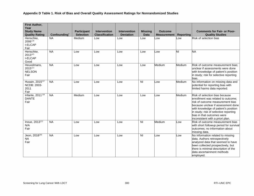
Appendix D Table 1. Risk of Bias and Overall Quality Assessment Ratings for Nonrandomized Studies
Screening for Lung Cancer With LDCT 300 RTI–UNC EPC
First Author, Year Study Name Quality Rating Confounding*
Participant Selection
Intervention Classification
Intervention Deviation
Missing Data
Outcome Measurement Reporting
Comments for Fair- or Poor-Quality Studies
Henschke, 2006129 I-ELCAP Fair
NA Medium Low Low Low Low Low Risk of selection bias
Henschke, 2013105 I-ELCAP Good
NA Low Low Low Low Low NI NA
Heuvelmans, 2015323 NELSON Fair
NA Low Low Low Low Medium Medium Risk of outcome measurement bias; unclear if assessments were done with knowledge of patient’s position in study; risk for selective reporting bias.
Husain, 2015237 NCDB: 2003-2011 Fair
NA Low Low Low NI Low Medium No information on missing data and potential for reporting bias with limited harms data reported.
Infante, 2011138 DANTE Fair
NA Medium Low Low Low Low Medium Risk of selection bias because enrollment was related to outcome; risk of outcome measurement bias because unclear if assessment done with knowledge of patient’s position in study; risk of selective reporting bias in that outcomes were inconsistent with a priori plan.
Inoue, 2013222
N/A Fair
NA Low Low Low NI Medium Low Risk of outcome measurement bias with short followup period for survival outcomes; no information about missing data.
Jeon, 2018266 NA Fair
NA Low Low Low NI Low Low No information related to missing data. Authors retrospectively analyzed data that seemed to have been collected prospectively, but there is minimal description of the data ascertainment methods employed.

Appendix D Table 1. Risk of Bias and Overall Quality Assessment Ratings for Nonrandomized Studies
Screening for Lung Cancer With LDCT 301 RTI–UNC EPC
First Author, Year Study Name Quality Rating Confounding*
Participant Selection
Intervention Classification
Intervention Deviation
Missing Data
Outcome Measurement Reporting
Comments for Fair- or Poor-Quality Studies
Jeppesen, 2013221 N/A Fair
NA Medium Low NI NI Low Low Missing information about multiple domains. Risk for selection bias in that some patients may have had Stage II cancer, depending on which classification system the investigators applied to the sample.
Jeppesen, 2018201 NA Fair
NA Low Low Low NI Medium Medium No information on missing data and potential for information bias related to outcomes, which were ascertained from medical records and only selectively reported.
Kaerlev, 2012155 DLCST Good
NA Low Low Low Low Low Low NA
Karasawa, 2018227 N/A Fair
NA Low Low Low Medium Medium Low Risk of bias from exclusion of 33% of eligible SBRT patients because of a dose difference due to a difference in calculation method; risk of outcome measurement bias.
Katoh, 2017253 NA Fair
NA Low Low Low NI Low Medium No information on missing data and very little information was provided on harms, except radiation pneumonitis, other than reporting that 1 grade 2 dermatitis and 10 grade 2 thoracic wall pain cases, no other toxicities were reported; data were not presented for Stage 1 patients only.
Khullar, 2015176 NCDB: 2003-2006 Fair
NA Low Low NI NI Low Low Lack of information for multiple domains.
Khullar, 2015187
NCDB: 2003-2011 Fair
NA Low Low NI Medium NI Low No information for several ROB domains. Patient characteristics are presented for the entire cohort of patients but not the subcohort that contributed to the long-term survival analyses.

Appendix D Table 1. Risk of Bias and Overall Quality Assessment Ratings for Nonrandomized Studies
Screening for Lung Cancer With LDCT 302 RTI–UNC EPC
First Author, Year Study Name Quality Rating Confounding*
Participant Selection
Intervention Classification
Intervention Deviation
Missing Data
Outcome Measurement Reporting
Comments for Fair- or Poor-Quality Studies
Kinsinger, 201737 VA Population (LCSDP) Fair
NA Medium NI NI Low NI Low Risk of selection bias because not all eligible patients (veterans) participated
Koshy, 2015198 NCDB: 2003-2006 Fair
NA Medium Low Low NI Low Low Risk of selection bias (only 64% of patients receiving SBRT were included) and lack of information related to missing data.
Lagerwaard, 2008290
N/A Fair
NA Medium Low Low NI Medium Low Risk of selection bias because most patients lacked histopathological confirmation of NSCLC; unclear how many of the 18% of patients with a prior lung cancer were actually experiencing a secondary tumor or recurrence (vs. a primary tumor); risk of outcome measurement bias due to short median followup period and at least some reliance on referring lung physicians or GPs for toxicity data of patients opting out of followup at the study center.
Lagerwaard, 2012281 N/A Fair
NA Low Low Low Medium Medium Low Risk of bias due to missing data that may have affected toxicity results; HRQOL data were missing for a large percentage of patients at 18- and 24-month time points; unclear how many patients were missing toxicity data, but the reasons for attrition, such as patients returning to their local hospitals, likely also reduced the availability of toxicity data.

Appendix D Table 1. Risk of Bias and Overall Quality Assessment Ratings for Nonrandomized Studies
Screening for Lung Cancer With LDCT 303 RTI–UNC EPC
First Author, Year Study Name Quality Rating Confounding*
Participant Selection
Intervention Classification
Intervention Deviation
Missing Data
Outcome Measurement Reporting
Comments for Fair- or Poor-Quality Studies
Lagerwaard, 2012223 N/A Fair
NA Medium Low Low NI Medium Low Risk of selection bias in that probable operability of patients for selection into study was determined post hoc; risk of outcome measurement bias because investigators relied on referring physicians for toxicity data for an unknown number of patients opting out of followup at the study center.
Lakha, 2014173 SEER: 2004-2010 Fair
NA Low Low NI NI Low Low No information on multiple domains.
Lam, 2018200 NCDB: 2004-2014 Fair
NA Low Low NI NI NI Low No information provided related to several domains.
Landreneau, 2014174 NA Fair
NA Low Low NI NI Low Low Lack of information related to intervention deviation and missing data. The analysis was among propensity-score matched groups defined by surgical approach; this isn’t a selection bias but might impact generalizability (e.g., unmatched had surgery earlier than matched, were less likely to have COPD, and had larger tumors).
Lee, 2015387
RegulomeDB Poor
NA (KQs 6 & 7) NI NI NI NI NI NI Uncontrolled study with very little information reported that was relevant to ROB assessment.
Lee, 2018232 N/A Fair
NA Low Low NI Medium Medium Low ROB due to exclusion of patients not followed up at the study’s hospital, and also potential outcome measurement bias due to the post hoc nature of the analysis.

Appendix D Table 1. Risk of Bias and Overall Quality Assessment Ratings for Nonrandomized Studies
Screening for Lung Cancer With LDCT 304 RTI–UNC EPC
First Author, Year Study Name Quality Rating Confounding*
Participant Selection
Intervention Classification
Intervention Deviation
Missing Data
Outcome Measurement Reporting
Comments for Fair- or Poor-Quality Studies
Licht, 2013247 DLCR: 2007-2011 Fair
NA Medium Low Low Low Low Low Risk of selection bias because authors only included standard lobectomies in the study in an “attempt to make VATS and thoracotomy groups more comparable.” Authors did not provide their definition of a nonstandard lobectomy.
Lindberg, 2015205 NA Fair
NA Low Low Low NI Low Low Lack of information on missing data.
Liu, 2018217 SEER: 2000-2013 Fair
NA Low Low NI Medium Low Low ROB due to missing data because patients were excluded for missing examinable lymph node counts and clinical features.
Louie, 2016236 STS-GTS: 2009-2013 Fair
NA Medium Low Low NI NI Low No information for multiple domains and potential selection bias resulting from exclusion of cases from low-volume centers.
Lutz, 2019218 N/A Fair
NA Low Low NI Low Medium Low No information on intervention deviation; risk of outcome measurement bias because no statistical methods or adjustments were used to explore the impact of including 140 patients (22.5% of the 621 in the study sample) who were upstaged in the analysis.
Lv, 2018219 SEER: 2004-2014 Fair
NA Low Low NI NI Low Medium No information from multiple domains; risk of reporting bias because the study’s only eligible survival data, 5-year OS, were reported for the total sample, not the lobectomy or SLR arms, for which overall survival curves were compared.
Ma, 2017267 NA Fair
NA Low Low NI NI Low Medium Medium risk of reporting bias due to certain data points not being collected and no information on multiple other domains.

Appendix D Table 1. Risk of Bias and Overall Quality Assessment Ratings for Nonrandomized Studies
Screening for Lung Cancer With LDCT 305 RTI–UNC EPC
First Author, Year Study Name Quality Rating Confounding*
Participant Selection
Intervention Classification
Intervention Deviation
Missing Data
Outcome Measurement Reporting
Comments for Fair- or Poor-Quality Studies
Maeda, 2010189 NA Fair
NA Low Low NI NI Low Low No information on multiple domains.
Maeda, 2012182 NA Fair
NA Low Low NI NI Low Low No information on multiple domains.
Manyam, 2019228 N/A Fair
NA Low Low Low NI Medium Low Risk of outcome measurement bias for 5-year OS due to a short followup period (median 23.8 months).
Mascalchi, 2006134 ITALUNG Fair
NA Medium Low Low Low Low Low Risk of selection bias
Mathieu, 2014258 NA Fair
NA Low Low Low Medium Low Medium ROB related to exclusion of patients with disease recurrence from analysis and reporting of only some toxicities.
Matsuo, 2012283 N/A Fair
NA Low Low Low NI Medium Low No information on missing data; risk of outcome measurement bias in that the analysis was based on post hoc ascertainment.
Matsuo, 2014196 NA Fair
NA Medium Low NI NI Low Low No information for multiple domains and some concern over selection of patients given that the original trial was stopped early due to slow patient enrollment.
Mediratta, 2014175 NA Good
NA Low Low NI Low Low Low NA
Melvan, 2015241 NCDB: 2003-2011 Fair
NA Low Low NI Medium Low Low Medium ROB related missing data and no information on intervention deviation.
Menezes, 2010119 NA Fair
NA Medium Low Low Medium Low Low Unclear selection, poor followup after 1st annual screening

Appendix D Table 1. Risk of Bias and Overall Quality Assessment Ratings for Nonrandomized Studies
Screening for Lung Cancer With LDCT 306 RTI–UNC EPC
First Author, Year Study Name Quality Rating Confounding*
Participant Selection
Intervention Classification
Intervention Deviation
Missing Data
Outcome Measurement Reporting
Comments for Fair- or Poor-Quality Studies
Miura, 2015388 NA Poor
NA High Low Low NI Low Low Restricting the study sample to patients with at least 12 months of followup CT scans after SBRT (i.e., a minimum of 4 followup scans) introduces a high risk of selection bias; unclear why this criterion was applied since all radiation-induced rib fractures after the first day of SBRT were included; unclear how many cases of rib fractures were missed as a result of the exclusion. Additionally, more than half of the patients had multiple cancers (some lung, some other sites).
Moon, 2018212 SEER: 2000-2014 Fair
NA Low Medium NI NI Low Low ROB due to intervention classification in that it was unclear how many segmentectomy patients received “intentional” vs. “compromised” procedures based on their comorbidities, and potential bias due to lack of information about missing data.
Morgan, 2017165 NA Fair
NA Medium NI Low Low Low Low Risk of selection bias.
Mutter, 2012285
N/A Fair
NA Low Low Low NI Medium Low Risk of outcome measurement bias in that median followup time (16 months) was shorter than the median time to rib fracture diagnosis (27 months).
Nagata, 2015203 JCOG0403 Fair
NA Low Low NI NI Medium Medium Long-term followup methods unclear, lack of information on missingness of data, and potential reporting bias related to grade 1-2 harms.
Nakamura, 2015171 NA Fair
NA Low Low NI NI NI Medium Post hoc analysis focused on intraoperative blood loss and lack of information on multiple domains.

Appendix D Table 1. Risk of Bias and Overall Quality Assessment Ratings for Nonrandomized Studies
Screening for Lung Cancer With LDCT 307 RTI–UNC EPC
First Author, Year Study Name Quality Rating Confounding*
Participant Selection
Intervention Classification
Intervention Deviation
Missing Data
Outcome Measurement Reporting
Comments for Fair- or Poor-Quality Studies
Nguyen, 2016164 NLST Fair
NA Low Low Low Low NI Low Medium risk of selection bias due to possible healthy-volunteer bias, and potential outcome measurement bias because definition of “potentially significant” extrapulmonary findings was left up to radiologists interpreting CT scans to decide.
Nyman, 2016265 SPACE† Fair
NA Low Low Medium Medium Low Low A small percentage of patients did not receive SBRT as intended, data sources are unclear, and there is some missing data, though it is not thoroughly described.
Okada, 2006191 NA Fair
NA Low Low Medium NI Low Low ROB related to deviations from intended surgical approach and unknown attrition/missing data.
Olsen, 2011286 N/A Fair
NA Low Low Low NI Medium Low Risk of outcome measurement bias in that median followup time (range of 11 to 16 months) was short and may not have been enough time for some toxicity events to occur.
Onishi, 2007224 N/A Fair
NA Low Low NI NI Medium Low Potential risk of outcome measurement bias due to post hoc use data from 14 different hospitals. Missing information on multiple domains.
Palma, 2010287 Amsterdam Cancer Registry Fair
NA Medium Medium NI NI Medium Low Risk of selection bias due to lack of histologic confirmation of NSCLC among 33% of RT patients (and unknown proportion of those receiving SBRT). At least some risk of intervention misclassification because no dosing information was available for RT treatments given. Post hoc analysis of data from population-based registry. No information for several domains.
Pegna, 201397 ITALUNG Good
NA Low Low Low Low Low Low NA

Appendix D Table 1. Risk of Bias and Overall Quality Assessment Ratings for Nonrandomized Studies
Screening for Lung Cancer With LDCT 308 RTI–UNC EPC
First Author, Year Study Name Quality Rating Confounding*
Participant Selection
Intervention Classification
Intervention Deviation
Missing Data
Outcome Measurement Reporting
Comments for Fair- or Poor-Quality Studies
Pinsky, 201462 NLST Fair
NA Low Low Low Low Low Medium Post hoc analysis
Pinsky, 201598 NLST Fair
NA Medium Low NI NI Medium Low Medium risk of outcome measurement bias because of difference in how radiologists were instructed to assess nodule growth using Lung-RADS criteria vs. NLST criteria. They applied Lung-RADS criteria in a post-hoc fashion to patients previously screened using NLST criteria, which created the potential for discrepancy in terms of how nodes discovered at baseline were later classified as having growth during post-baseline scans. A sensitivity analysis was done assuming that all nodules reported with growth in NLST met Lung-RADS criteria for growth, but that assumption may have been incorrect in at least some cases.
Puri, 2014240 NA Fair
NA Low Low NI NI Low Low Lack of information for multiple domains. This is a post hoc analysis of two trial datasets that prospectively collected data.
Puri, 2015250 NCDB: 1998-2010 Fair
NA Medium Low Medium NI Low Medium Risk of selection bias in terms of the patients that completed followup; authors were unable to explain the large difference in median survival between the surgical and SBRT groups. No information provided on missingness of data. Author utilized propensity-score matching to compare surgery to SBRT, but results were only presented for surgery overall and SLR.
Pinsky, 2018137 NLST Fair
Medium Low Low Low Low Medium Low Post hoc analysis of NLST RCT, looking at intervention arm of study.

Appendix D Table 1. Risk of Bias and Overall Quality Assessment Ratings for Nonrandomized Studies
Screening for Lung Cancer With LDCT 309 RTI–UNC EPC
First Author, Year Study Name Quality Rating Confounding*
Participant Selection
Intervention Classification
Intervention Deviation
Missing Data
Outcome Measurement Reporting
Comments for Fair- or Poor-Quality Studies
Rampinelli, 2017132 COSMOS Fair
Yes Low Low Low NI NI NI Bias due to the source of harms data; data on the harm of radiation-induced cancer from LDCT program were estimated (not measured) based on an assumed relationship (which in turn is based on data from other studies of radiation therapy) that appears to be controversial.
Razi, 2016178 SEER: 1998-2007 Fair
NA Low Low NI NI Low Low Lack of information for multiple domains.
Robinson, 2013264 NA Fair
NA Low Low NI NI NI Low There was no information for several domains.
Rosen, 2014242 NCDB: 2004-2009 Fair
NA Low Low NI Medium Low Low Medium ROB related to missing data (both in terms of how missingness of some variables resulted in exclusion from study and in unknown stage data) and lack of information on intervention deviation.
Rosen, 2014256 NA Fair
NA Low Low Low NI Low Medium Risk of reporting bias and lack of information on missing data.
Rosen, 2016183
NCDB: 2008-2012 Good
NA Low Low Low NI Low Low NA
Samson, 2015238 NCDB: 1998-2010; Washington SOM 2000-2012 Fair
NA Low Low NI NI NI Low Lack of information for several domains.

Appendix D Table 1. Risk of Bias and Overall Quality Assessment Ratings for Nonrandomized Studies
Screening for Lung Cancer With LDCT 310 RTI–UNC EPC
First Author, Year Study Name Quality Rating Confounding*
Participant Selection
Intervention Classification
Intervention Deviation
Missing Data
Outcome Measurement Reporting
Comments for Fair- or Poor-Quality Studies
Samson, 2017243 NCDB: 2004-2013 Fair
NA Medium Low NI NI Low Low Medium risk of selection bias and lack of information on several additional domains.
Sawabata, 2011188 Japanese Joint Committee of Lung Cancer Registry Fair
NA Low Low NI Low NI Low No information for multiple domains and few details provided by the authors, except as they related to tumor characteristics.
Scheel, 2015168
NA Fair
NA Low Low Low NI Medium Low No information on extent of missing data and potential for outcome misclassification.
Schuchert, 2012246 NA Fair
NA Medium Low NI NI Low Low Patients were identified from multiple databases, including billing records. Authors report that patients who received incomplete resections were not included; if incomplete resection is associated with poorer outcomes, there could be selection bias present. Lack of information related to several other domains.
Sekihara, 2018220 N/A Fair
NA Low Low NI NI Medium Low Risk of outcome measurement bias in that the study relied on a post hoc analysis of a single hospital’s data, and no information from multiple domains.
Shapiro, 2012245 SEER: 1992-2002 Fair
NA Low Low NI Medium Low Low Medium ROB related to missing data.
Shibamoto, 2012234 N/A Good
NA Low Low Low Low Low Low NA

Appendix D Table 1. Risk of Bias and Overall Quality Assessment Ratings for Nonrandomized Studies
Screening for Lung Cancer With LDCT 311 RTI–UNC EPC
First Author, Year Study Name Quality Rating Confounding*
Participant Selection
Intervention Classification
Intervention Deviation
Missing Data
Outcome Measurement Reporting
Comments for Fair- or Poor-Quality Studies
Shirvani, 2012235 SEER: 2001-2007 Fair
NA Low Low NI NI Low Low No information on multiple domains.
Shirvani, 2014239 SEER: 2003-2009 Fair
NA Medium Low Low NI Low Low Risk of selection bias resulting from exclusion of patients with incomplete Medicare records. Lack of information on missing outcomes data.
Speicher, 2016185 NCDB: 2003-2006 Fair
NA Low Low NI Medium NI Low No information for several ROB domains. Patient characteristics are presented for the entire cohort of patients but not the subcohort that contributed to the long-term survival analyses.
Stanic, 2014277 RTOG 0236 Fair
NA Medium Low NI Medium Medium Low Risk of self-selection bias indicated by fact that most of the sample was female when majority of NSCLC patients in the population are male. Potential bias from missing data for PFT outcomes due to test noncompliance at each timepoint of interest. Potential bias from outcome measurement for PFT outcomes in that large variation occurred in the timing of assessments.
Stephens, 2014180 NA Fair
NA Low Low Medium NI Low Low ROB related to patients who crossed over from one surgical approach to another and lack of information related to missing data.
Stokes, 2018249 NCDB: 2004-2013 Fair
NA Medium NI NI Low NI Low No information provided related to several domains. Missing data informed selection of patients into the analysis (i.e., complete case analysis).
Strand, 2006192 Cancer Registry of Norway Good
NA Low Low Low Low Low Low NA

Appendix D Table 1. Risk of Bias and Overall Quality Assessment Ratings for Nonrandomized Studies
Screening for Lung Cancer With LDCT 312 RTI–UNC EPC
First Author, Year Study Name Quality Rating Confounding*
Participant Selection
Intervention Classification
Intervention Deviation
Missing Data
Outcome Measurement Reporting
Comments for Fair- or Poor-Quality Studies
Styn, 2009149 PLuSS Fair
NA Medium Low Low Low Medium Low Potential bias from outcomes and patient selection.
Su, 2014172 ACOSOG Z0030 (Alliance) Fair
NA Low Low NI NI Low Low Lack of information on several domains.
Sun, 2017195
NA Fair
NA Medium Low Low Low NI Low Risk of selection bias with respect to inclusion of patients with a prior history of lung or other cancers. Additionally, 5% of the enrolled patients had no followup imaging or records and were not included in the analysis.
Swensen, 2002166 NA Fair
NA Medium Low Low Low Low Low Potential bias from patient selection.
Swensen, 2005128 NA Fair
NA Medium Low Low Medium Low Low Potential bias from patient selection and missing data.
Taremi, 2012259 NA Fair
NA Medium Low NI NI Low Low Medium risk of selection bias related to short followup periods and lack of information related to missing data.
Taremi, 2012262 NA Fair
NA Low Low NI NI Low Low Lack of information for multiple domains.
Thalanayar, 2015141
PLuSS Fair
NA Medium Low Low Low Medium Low Potential bias due to patient selection and outcome.
Townsend, 2005150
Mayo Lung Project Fair
NA Medium Low Low Medium Medium Low Possible selection, outcome and missing data bias as listed.

Appendix D Table 1. Risk of Bias and Overall Quality Assessment Ratings for Nonrandomized Studies
Screening for Lung Cancer With LDCT 313 RTI–UNC EPC
First Author, Year Study Name Quality Rating Confounding*
Participant Selection
Intervention Classification
Intervention Deviation
Missing Data
Outcome Measurement Reporting
Comments for Fair- or Poor-Quality Studies
Toyoda, 2008124 NA Fair
NA Medium Low Low Low Low Low Potential bias due to patient selection.
Tsushima, 2008121 NA Fair
NA Medium Low Low Low Low Low Potential bias due to patient selection.
Tsutani, 2014177 NA Good
NA Low Low NI Low Low Low NA
Ubels, 2015204
NA Fair
NA Medium Low NI NI Low Low Lack of information for multiple domains. The study included a small number of patients; almost 10% of them were excluded after enrollment for reasons that may be associated with poorer outcomes.
Ueda, 2018272 N/A Fair
NA Low Low NI NI Medium Low No information in multiple domains; risk of outcome measurement bias because ECG was inconsistently used to identify POAF, the study’s primary outcome of interest, and more broadly, this was a small post hoc analysis of a single hospital’s data.
Uhlig, 2018226 NCDB: 2004-2013 Fair
NA Low Low NI Low Medium Low Risk of outcome measurement bias due to the post hoc nature of the analysis and lack of detail about systems for outcome ascertainment.
Valle, 2016244 NCCN: 2007-2011 Fair
NA Medium Low NI Medium NI Low Risk of selection bias and bias due to missing data; no information provided for additional domains.
Veronesi, 2008122 COSMOS Fair
NA Medium Low Low Low Low Low Risk of patient selection bias.
Veronesi, 2008125 COSMOS Fair
NA Medium Low Low Low Low Low Risk of selection bias.

Appendix D Table 1. Risk of Bias and Overall Quality Assessment Ratings for Nonrandomized Studies
Screening for Lung Cancer With LDCT 314 RTI–UNC EPC
First Author, Year Study Name Quality Rating Confounding*
Participant Selection
Intervention Classification
Intervention Deviation
Missing Data
Outcome Measurement Reporting
Comments for Fair- or Poor-Quality Studies
Veronesi, 2012133 COSMOS Fair
Medium Low Low Low Medium Medium Medium ROB likely affects the size of effects this study demonstrates: namely, that (for scenario A) greater VDT is associated with lower LC mortality (and likely overdiagnosis), as well as for scenario B) that LDCT screening finds indolent lesions, i.e., those with long VDT. Sources of bias include confounding, missing data, outcomes measurement, and selective reporting.
Videtic, 2014269 NA Fair
NA Medium Low Low NI Low Low Risk of selection bias and lack of information on missingness of data.
Wagnetz, 2012116 NA Fair
NA Medium Low Low NI Low Low Risk of selection bias and no information on missing data.
Walker, 2015131 NA Fair
NA Medium Low Low Low Low Low ROB due to patient selection.
Walter, 201676 NELSON Good
NA Low Low Low Low Low Low NA
Westover, 2012282 N/A Fair
NA Low Low Low Low Medium Low Medium risk of outcome measurement bias; study relied on a database and drew data from a very small sample of 15 patients.
Wilson, 2008135
PLuSS Fair
NA Medium Low Low Low Low Low ROB due to patient selection.

Appendix D Table 1. Risk of Bias and Overall Quality Assessment Ratings for Nonrandomized Studies
Screening for Lung Cancer With LDCT 315 RTI–UNC EPC
First Author, Year Study Name Quality Rating Confounding*
Participant Selection
Intervention Classification
Intervention Deviation
Missing Data
Outcome Measurement Reporting
Comments for Fair- or Poor-Quality Studies
Wink, 2019233 N/A Fair
NA Medium Medium NI NI Medium Low Missing information for multiple domains; post hoc analysis with short followup period for 5-year OS (median 48.1 months for surviving patients); risk of outcome measurement bias; OS data were retrieved from national databases, and unclear how comprehensively they captured mortality; risk of selection bias because majority (68%) of sample did not have histologic confirmation of NSCLC; variation in how SBRT was planned across the study’s treating institutions.
Yang, 2016248 NCDB: 2010-2012 Fair
NA Low Low NI NI NI Low No information on multiple domains. Authors report that outcomes of surgical approach were evaluated with intent-to-treat analysis but provide no additional information.
Ye, 2018292 N/A Fair
NA Medium Low Low NI Low Low Risk of selection bias in that nearly a fifth (19%) of SBRT patients lacked pathologic confirmation of NSCLC.
Yousaf-Khan, 2017389 NELSON Poor
Low Medium Low Low Medium Low Low High risk of self-selection bias for the fourth round of screening, the focus of this substudy. Enrollment of 78% of eligible patients from 3rd round of screening (about 70% of initial sample) and confirmation in the article that the fourth round’s patients differed significantly from the initial sample in several ways (e.g., more current smokers in the fourth round).
Zhai, 2014170 NA Fair
NA Low Low NI NI Low Low Lack of information for several domains.
Zhao, 2014107 NELSON Fair
NA Low Low Medium Low Low NI This longitudinal study did not apply the same imaging followup protocol to all patients in terms of the intervals between scans.

Appendix D Table 1. Risk of Bias and Overall Quality Assessment Ratings for Nonrandomized Studies
Screening for Lung Cancer With LDCT 316 RTI–UNC EPC
First Author, Year Study Name Quality Rating Confounding*
Participant Selection
Intervention Classification
Intervention Deviation
Missing Data
Outcome Measurement Reporting
Comments for Fair- or Poor-Quality Studies
Zhao, 2017181 SEER: 2004-2012 Fair
NA Low Low NI NI Low Low Lack of information related to several domains.
Zhou, 2015390 Mass General Hospital Database Poor
NA High Low NI Medium NI Medium High risk of selection bias, medium ROB related to missing data/patient attrition and reporting of harms (i.e., ‘complications’), and lack of information on additional domains.
Zhou, 2018213 SEER: 2004-2013 Fair
NA Medium Low NI Low Low Low Risk of selection bias because no detail was given about how patients’ cancers were staged, and no information about potential deviation from intended interventions.
* Bias due to confounding did not apply to KQ6/7 studies. † Only the SBRT arm of the study was eligible for this review.
Abbreviations: ACOSOG=American College of Surgeons Oncology Group; CHISEL=A Randomised Phase III Trial of Highly Conformal Hypofractionated Image Guided
(“Stereotactic”) Radiotherapy (HypoRT) Versus Conventionally Fractionated Radiotherapy (ConRT) for Inoperable Early Stage I Non-small Cell Lung Cancer; COPD=chronic
obstructive pulmonary disease; COSMOS=Continuous Observation of Smoking Subjects study; CT=computed tomography; DANTE=Detection and Screening of Early Lung
Cancer with Novel Imaging Technology trial; DLCR=Danish Lung Cancer Registry; ECG=electrocardiogram; GP=general practitioner; HRQOL=health-related quality of life; I-
ELCAP=International Early Lung Cancer Action Project; ITALUNG=The Italian Lung Study; JCOG=Japan Clinical Oncology Group; KQ=Key Question; LC=lung cancer;
LDCT=low-dose computed tomography; LCSDP=Lung Cancer Screening Demonstration Project; Lung-RADS=ACR Lung Imaging Reporting and Data System; NA=not
applicable; NCCN=National Comprehensive Cancer Network; NCDB=National Cancer Database; NELSON=Dutch-Belgian Randomized Lung Cancer Screening Trial; NI=no
information; NLST=National Lung Screening Trial; OS=overall surgery; PFT=pulmonary function test; POAF=postoperative atrial fibrillation; RCT=randomized, controlled trial;
ROB=risk of bias; RT=radiotherapy; RTOG=Radiation Therapy Oncology Group; SBRT=stereotactic body radiation therapy; SEER=Surveillance, Epidemiology, and End
Results; SLR=sublobar resection; SOM=School of Medicine; SPACE=Stereotactic Precision And Conventional Radiotherapy Evaluation; STS-GTS=Society of Thoracic Surgeons
General Thoracic Surgery Database; VATS=video-assisted thoracoscopic surgery; VDT=volume doubling time; VINCI=Veteran’s Affairs Informatics and Computing
Infrastructure; vs.=versus.

Appendix D Table 2. Risk of Bias and Overall Quality Assessment Ratings for Randomized Studies: Part 1
Screening for Lung Cancer With LDCT 317 RTI–UNC EPC
Appendix D Table 2. Risk of Bias and Overall Quality Assessment Ratings for Randomized Studies: Part 1
First Author, Year Study Name Quality Rating
Was randomization
adequate?
Was allocation
concealment adequate?
Were groups
similar at baseline?
Eligibility criteria
specified?
Were outcome measurements
equal, reliable and valid?
Were outcome accessors masked?
Were care providers masked?
Were Patients masked?
Aberle, 201131 NLST Good
Yes Yes Yes Yes Yes LC mortality: Yes LC incidence: unclear (but likely)
Unclear (but unlikely)
No
Aberle, 201356 NLST Good
Yes Yes Yes Yes Yes LC mortality: Yes LC incidence: Unclear (but likely)
Unclear (but unlikely)
No
Aggestrup, 2012158 DLCST Fair
Yes Yes Yes Yes Yes Unclear No No
Ashraf, 2009143 DLCST Fair
Yes Yes Yes Yes Yes Unclear No No
Ashraf, 2014145 DLCST Good
Yes Unclear Yes Yes Yes Unclear (but unlikely to bias smoking cessation)
Unclear (but unlikely to bias smoking cessation)
No
Becker, 201258
LUSI Good
Yes Unclear Yes Yes Yes NR No (but unlikely to bias baseline screening results)
No
Becker, 201557, 201971 LUSI Fair
Yes Unclear Yes Yes Yes NR No No
Brain, 2016391 UKLS Poor
Yes Yes Yes Yes Yes NR No No
Brain, 2017392 UKLS Poor
Yes Yes Yes Yes Unclear Not reported No No
Church, 201355
NLST Good
Yes Yes Yes Yes Yes LC mortality: Yes LC incidence: Unclear (but likely)
Unclear (but unlikely)
No
Clark, 2016147 NLST Fair
Yes Yes Yes Yes Unclear Yes Unclear (but unlikely)
No

Appendix D Table 2. Risk of Bias and Overall Quality Assessment Ratings for Randomized Studies: Part 1
Screening for Lung Cancer With LDCT 318 RTI–UNC EPC
First Author, Year Study Name Quality Rating
Was randomization
adequate?
Was allocation
concealment adequate?
Were groups
similar at baseline?
Eligibility criteria
specified?
Were outcome measurements
equal, reliable and valid?
Were outcome accessors masked?
Were care providers masked?
Were Patients masked?
Croswell, 2010117 NA Fair
Yes Yes Yes Yes Yes No No No
De Koning, 202074
Yes* Yes* Yes Yes Unclear Yes Unclear No
Field, 201695 UKLS Fair
Yes Unclear Yes Yes No Unclear Unclear No
Field, 2016115 UKLS Fair
Yes Yes Yes Yes Yes NR No No
Gohagan, 200468 LSS Fair
Yes Yes Yes Yes Yes No No No
Gohagan, 200567 LSS Fair
Yes Yes Yes Yes Yes No No No
Heleno, 2018139 DLCST Fair
Yes NR Mostly (slightly higher risk)
Yes Yes NR No No
Horeweg, 201396 NELSON Poor
Yes Unclear Yes Yes Yes Unclear Unclear No
Horeweg, 201432 NELSON Poor
Yes Unclear Yes Yes Yes Unclear Unclear No
Infante, 200870
DANTE Fair
Yes Yes Yes Yes Yes Unclear No No
Infante, 200969 DANTE Fair
Yes Yes Yes Yes Yes Unclear No No

Appendix D Table 2. Risk of Bias and Overall Quality Assessment Ratings for Randomized Studies: Part 1
Screening for Lung Cancer With LDCT 319 RTI–UNC EPC
First Author, Year Study Name Quality Rating
Was randomization
adequate?
Was allocation
concealment adequate?
Were groups
similar at baseline?
Eligibility criteria
specified?
Were outcome measurements
equal, reliable and valid?
Were outcome accessors masked?
Were care providers masked?
Were Patients masked?
Infante, 201559 DANTE Fair
Yes No Yes Yes Yes Unclear Unclear No
Lopes Penga, 2009123 ITALUNG Fair
Yes Yes Yes Yes Yes No No No
National Lung Screening Trial Research Team, 201972
NLST Fair
Yes Yes Yes Yes Unclear; ascertainment methods differed for trial years (when outcome verification committee was involved) and post-trial years
Unclear Unclear (but unlikely)
No
O’Grady, 2014167 PLCO, NLST Good
Yes Yes Yes Yes Yes Yes No Yes
Paci, 201760 ITALUNG Fair
Yes Yes Yes Yes Yes Yes No No
Pastorino, 201278 and 201973, 79 MILD Poor
No Unclear No Yes Unclear Unclear No No
Patz, 2014140 NLST Fair
Yes Yes Yes Yes Yes Yes No No
Pedersen, 2009120 DLCST Good
Yes Yes Yes Yes Yes Unclear No No
Pinsky, 2005393, Doroudi, 201873 LSS Fair
Yes Yes Yes Yes Yes No No No

Appendix D Table 2. Risk of Bias and Overall Quality Assessment Ratings for Randomized Studies: Part 1
Screening for Lung Cancer With LDCT 320 RTI–UNC EPC
First Author, Year Study Name Quality Rating
Was randomization
adequate?
Was allocation
concealment adequate?
Were groups
similar at baseline?
Eligibility criteria
specified?
Were outcome measurements
equal, reliable and valid?
Were outcome accessors masked?
Were care providers masked?
Were Patients masked?
Pinsky, 201361 NLST Fair
Yes Yes Yes Yes Yes Yes No No
Rasmussen, 2014156 DLCST Fair
Yes Yes Yes Yes Yes Yes No No
Saghir, 201263 DLCST Fair
Yes Yes Yes Yes Yes Unclear No No
Sverzellati, 201680 MILD Fair
Yes (for annual vs. Biennial)
Unclear Yes Yes Yes Unclear No No
Tanner, 201564
NLST Fair
Yes Yes Yes Yes Yes Yes No No
Taylor, 2007153 NLST/LSS Fair
Yes Yes Yes Yes Yes No No No
van den Bergh, 2010160 NELSON Fair
Yes Yes Yes Yes Yes Unclear No No
van den Bergh, 2011157 NELSON Fair
Yes
Yes Yes Yes Unclear Yes No No
van der Aalst, 2010146 NELSON Fair
Yes Yes Yes Yes Yes No No No
van der Aalst, 2011151 NELSON Fair
Yes Yes Yes Yes Yes No No No
van Klaveren, 2009118
NELSON Good
Yes Yes Yes Yes Yes No No No

Appendix D Table 2. Risk of Bias and Overall Quality Assessment Ratings for Randomized Studies: Part 1
Screening for Lung Cancer With LDCT 321 RTI–UNC EPC
First Author, Year Study Name Quality Rating
Was randomization
adequate?
Was allocation
concealment adequate?
Were groups
similar at baseline?
Eligibility criteria
specified?
Were outcome measurements
equal, reliable and valid?
Were outcome accessors masked?
Were care providers masked?
Were Patients masked?
van’t Westeinde, 2012136 NELSON Fair
Yes Yes Yes Yes Yes No No No
Videtic, 2015261 RTOG 0915 Fair
Unclear Unclear Unclear Yes Yes Unclear No No
Wille, 201665
DLCST Good
Yes Yes Yes Yes Yes Unclear No No
Young, 2015142 NLST Fair
Yes Yes Yes Yes Yes Yes No No
* Details of randomization and allocation concealment were not published but were obtained by written personal communication from the first author. In short, all invited persons
were given an 8-character random number by an external consultant. For persons responding with informed consent, these personal identification numbers were shifted by 1-8
positions (blindly, randomly computer generated), and then sorted again (ascending numbers) to be randomized. No investigator had control nor insight into this random process,
and it also ensured that the consultant could not influence this process.
Abbreviations: CT=computed tomography; DANTE=Detection and Screening of Early Lung Cancer with Novel Imaging Technology trial; DLCST=Danish Lung Cancer
Screening Trial; ITALUNG=The Italian Lung Study; LC=lung cancer; LUSI=German Lung Cancer Screening Intervention Trial; MILD=Multicentric Italian Lung Detection trial;
NELSON=Dutch-Belgian Randomized Lung Cancer Screening Trial; NLST=National Lung Screening Trial; NR=Not reported; PLCO=Prostate, Lung, Colorectal, and Ovarian
Cancer Screening Trial; RTOG=Radiation Therapy Oncology Group; UKLS=UK Lung Cancer Screening trial; vs.=versus

Appendix D Table 3. Risk of Bias and Overall Quality Assessment Ratings for Randomized Studies: Part 2
Screening for Lung Cancer With LDCT 322 RTI–UNC EPC
Appendix D Table 3. Risk of Bias and Overall Quality Assessment Ratings for Randomized Studies: Part 2
First Author, Year Study Name Quality Rating
What was the reported adherence to the
intervention?
Did the study have crossovers or
contamination? What was the overall
attrition?
What was the differential attrition?
Did the study have differential attrition >10% or overall high
attrition, raising concerns for bias?
Aberle, 201131 NLST Good
CT: 95% CXR: 93%
Minimal (average annual rate of CT in CXR group: 4.3%)
Average over three screening rounds: 6.1%
Average over three screening rounds: LDCT: 5% CXR: 7% T0: LDCT: 1.5% CXR: 2.6% T1: LDCT: 6.0% CXR: 8.8% T2: LDCT: 7.1% CXR: 10.6%
No
Aberle, 201356
NLST Good
T1: LDCT: 94% CXR: 91.2% T2: LDCT: 92.9% CXR: 89.4%
NR for T1 and T2 screens; over three rounds, 4.3% (per Aberle 75)
Overall: T1: 7.4% T2: 8.8%
T1: LDCT: 6.0% CXR: 8.8% T2: LDCT: 7.1% CXR: 10.6%
No
Aggestrup, 2012158 DLCST Fair
94.3% NR 5.6% CT: 2.9% Control: 8.2%
No
Ashraf, 2009143 DLCST Fair
NR NR 8.3% CT: 5.3% Control: 11.6%
11.6% of the control group had missing data
Ashraf, 2014145 DLCST Good
Year 5: 89% CT: 90% Control: 89% (Higher for years 1-4)
Yes Year 5: 14.6% CT: 6% Control: 12%
No Sensitivity analysis complete case vs. imputation LOCF same results
Becker, 201258 LUSI Good
CT: 99.9% Control: 99.9%
No (baseline screen) 0% (baseline screen) None No

Appendix D Table 3. Risk of Bias and Overall Quality Assessment Ratings for Randomized Studies: Part 2
Screening for Lung Cancer With LDCT 323 RTI–UNC EPC
First Author, Year Study Name Quality Rating
What was the reported adherence to the
intervention?
Did the study have crossovers or
contamination? What was the overall
attrition?
What was the differential attrition?
Did the study have differential attrition >10% or overall high
attrition, raising concerns for bias?
Becker, 201557, 201971 LUSI Fair
Year 3: CT: 93.4% Control: 94.5% Year 2: CT: 94.6% Control: 91.5% Year 1: CT: 99.9% Control: 99.9%
NR Over five years: 0.1% - 6.9%
Year 3: CT: 6.6% Control: 5.5% Year 2: CT: 5.4% Control: 8.5% Year 1: CT: 0.1% Control: 0.1%
No
Brain, 2016391 UKLS Poor
For psychosocial outcomes, adherence to followup surveys: T0: 99.9% T1: CT: 84% Control: 78% T2: CT: 82% Control: 65%
NR For psychosocial outcomes: T0: 0.01% T1: CT: 16% Control: 22% T2: CT: 18% Control: 35%
Psychosocial outcomes: T0: 0.01% T1: CT: 16% Control: 22% T2: CT: 18% Control: 35%
Yes
Brain, 2017392
UKLS Poor
For smoking cessation outcomes, adherence to followup surveys: T1: 65% T2: 57%
NR T1: 35% T2: 43%
T1: CT: 31% Control: 39% T2: CT: 35% Control: 51%
Yes
Church, 201355 NLST Good
98% NR for baseline scree, over three rounds, 4.3% (per Aberle 75)
2% LDCT: 1.5% CXR: 2.6%
No
Clark, 2016147 NLST Fair
NR for this subset of ACRIN centers; likely >90% given overall study adherence
NR for ACRIN centers subset; but for overall study 4.3%
NR for ACRIN center subset; likely similar to overall study
NR No
Croswell, 2010117 NA Fair
84.2% NR 15.8% CT: 15.8% Yes

Appendix D Table 3. Risk of Bias and Overall Quality Assessment Ratings for Randomized Studies: Part 2
Screening for Lung Cancer With LDCT 324 RTI–UNC EPC
First Author, Year Study Name Quality Rating
What was the reported adherence to the
intervention?
Did the study have crossovers or
contamination? What was the overall
attrition?
What was the differential attrition?
Did the study have differential attrition >10% or overall high
attrition, raising concerns for bias?
De Koning, 202074
90% among men for all rounds (95.8% for round 1 of screening); NR for women
Unclear Missing data <2% for the primary outcome from linkages with national registries
<2% No
Field, 201695 UKLS Fair
98.30% NR 1.70% Unclear Unclear
Field, 2016115 UKLS Fair
98% NR For psychosocial outcomes: T0: 0.01% T1: CT: 16% Control: 22% T2: CT: 18% Control: 35%
For psychosocial outcomes: Baseline: 0% T1 (2 weeks after LDCT or control notification): 6% T2 (10–27 months): 17%
T1: No T2: Yes
Gohagan, 200468 LSS Fair
95.5% 0.90% 4.5% CT: 4.5% No
Gohagan, 200567 LSS Fair
85.8% NR 14.2% CT: 14.2% Yes
Heleno, 2018139
DLCST Fair
NR Yes NR NR No
Horeweg, 201396 NELSON Poor
For all three waves: ~90-95%
Unclear 3.80% Unclear Unclear
Horeweg, 201432 NELSON Poor
90% NR 3.80% Unclear Unclear
Infante, 200870 DANTE Fair
Baseline only: 100% No 0% 0% No

Appendix D Table 3. Risk of Bias and Overall Quality Assessment Ratings for Randomized Studies: Part 2
Screening for Lung Cancer With LDCT 325 RTI–UNC EPC
First Author, Year Study Name Quality Rating
What was the reported adherence to the
intervention?
Did the study have crossovers or
contamination? What was the overall
attrition?
What was the differential attrition?
Did the study have differential attrition >10% or overall high
attrition, raising concerns for bias?
Infante, 200969 DANTE Fair
90.9% <10% (presented as combined arms needing CT)
9.1% CT: 9.1% No
Infante, 201559 DANTE Fair
94% Yes 6% Unclear NA
Lopes Penga, 2009123 ITALUNG Fair
87.2% NR 12.7% CT: 12.7% Yes
National Lung Screening Trial Research Team, 201972 NLST Fair
NR in this article, and post-trial screening was unclear, but original NLST reported CT: 95% CXR: 93%
Unclear; post-trial screening was not ascertained
11/33 centers (representing 12.4% of trial participants) did not have a home state cancer registry for linkage for lung cancer incidence; for mortality, linkage to national death index was available for all but 2.2%
NR Unclear for lung cancer incidence; no for lung cancer mortality and all-cause mortality
O’Grady, 2014167 PLCO, NLST Good
High Some Unclear Unclear Unclear
Paci, 201760 ITALUNG Fair
Across 4 rounds of LDCT screening: 81%
Yes (but minimal) Low (conducted ITT analysis)
Low No

Appendix D Table 3. Risk of Bias and Overall Quality Assessment Ratings for Randomized Studies: Part 2
Screening for Lung Cancer With LDCT 326 RTI–UNC EPC
First Author, Year Study Name Quality Rating
What was the reported adherence to the
intervention?
Did the study have crossovers or
contamination? What was the overall
attrition?
What was the differential attrition?
Did the study have differential attrition >10% or overall high
attrition, raising concerns for bias?
Pastorino, 201278 and 201979 MILD Poor
>95% Unclear for 5-year followup; by 10-year followup, 1.2% of control group had LDCT
Low (conducted ITT analysis) for loss to followup, but many people in control group had shorter followup duration.
Unclear for loss to followup, but less followup among control group (e.g., 44.9 vs. 56 months in 5-year followup study); percentage of subjects with available data at 10 years: 46.2% (805/1723) controls vs. 81.4% (1934/2376) LDCT
Yes, when considering differential followup, 35.2% fewer people from the control group had 10-year followup than in the LDCT group
Patz, 2014140 NLST Fair
NR in this paper NR Low No No
Pedersen, 2009120 DLCST Good
Unclear but appears to be 100% for baseline CT
NR Number of CTs obtained NR
NR It was NR whether any of the 2,052 patients did not have a CT
Pinsky, 2005393 Doroudi, 201873 LSS Fair
85.8% NR 14.2% CT: 14.2% Yes
Pinsky, 201361 NLST Fair
NR in this paper NR Low No No
Rasmussen, 2014156 DLCST Fair
Unclear Not much Moderate for COS-LC None at baseline, somewhat lower for COS-LC survey completion in control arm in subsequent rounds
Depends on which round of screening you are referring to
Saghir, 201263 DLCST Fair
High, mean annual participation 95.5%
Minimal Low Low No
Sverzellati, 201680 MILD Fair
High, until years T5 and T6 NR Low in early years, then moderate
Unclear In later years, yes

Appendix D Table 3. Risk of Bias and Overall Quality Assessment Ratings for Randomized Studies: Part 2
Screening for Lung Cancer With LDCT 327 RTI–UNC EPC
First Author, Year Study Name Quality Rating
What was the reported adherence to the
intervention?
Did the study have crossovers or
contamination? What was the overall
attrition?
What was the differential attrition?
Did the study have differential attrition >10% or overall high
attrition, raising concerns for bias?
Tanner, 201564 NLST Fair
High in NLST NR Low Low No
Taylor, 2007153 NLST/LSS Fair
79.7% NR 20.3% For entire survey group: 20.3%
Yes
van den Bergh, 2010160 NELSON Fair
For all, 86.7% CT at T0: 91.0% (630/692) T1: 93.6% (641/685) T2: 93.0% (620/667) T3: 87.7% (600/684)
NR 13.3% 13.3% Yes, at third annual screen
van den Bergh, 2011157 NELSON Fair
NR in this paper No T0, All: 87.9% LDCT: 89.8% Control: 85.9% T1 Screen: 87.7% T2, All: 78.9% LDCT: 89.3%, Control 64.7%
LDCT: 89% vs. Control: 65%
Yes
van der Aalst, 2010146
NELSON Fair
92.1% No NA NA NA
van der Aalst, 2011151
NELSON Fair
92.1% No NA NA NA
van Klaveren, 2009118 NELSON Good
96.4% NR 3.5% CT: 3.5% No
van’t Westeinde, 2012136 NELSON Fair
NR in this publication (but >90% in other NELSON publications)
NR NR NR NR

Appendix D Table 3. Risk of Bias and Overall Quality Assessment Ratings for Randomized Studies: Part 2
Screening for Lung Cancer With LDCT 328 RTI–UNC EPC
First Author, Year Study Name Quality Rating
What was the reported adherence to the
intervention?
Did the study have crossovers or
contamination? What was the overall
attrition?
What was the differential attrition?
Did the study have differential attrition >10% or overall high
attrition, raising concerns for bias?
Videtic, 2015261 RTOG 0915 Fair
100% of analyzable patients
No 10/94 (11%) patients were excluded post-randomization due to withdrawal of consent, protocol violations, or RT dosing not met
34Gy group: 8/47 (17%) 48Gy group: 2/47 (4%)
Yes
Wille, 201665 DLCST Good
NR in this paper No Low Low (LDCT: 20; Control: 14)
No
Young, 2015142 NLST Fair
High in NLST NR Low Low No
Abbreviations: ACRIN=American College of Radiology Imaging Network; COS-LC=consequences of screening-lung cancer; CT=computed tomography; CXR=chest X-ray;
DANTE=Detection and Screening of Early Lung Cancer with Novel Imaging Technology trial; DLCST=Danish Lung Cancer Screening Trial; ITALUNG=The Italian Lung
Study; ITT=intention-to-treat; LDCT=low-dose computed tomography; LOCF: last observation carried forward; LUSI=German Lung Cancer Screening Intervention Trial;
MILD=Multicentric Italian Lung Detection trial; NA=not applicable; NELSON=Dutch-Belgian Randomized Lung Cancer Screening Trial; NLST=National Lung Screening Trial;
NR=not reported; PLCO=Prostate, Lung, Colorectal, and Ovarian Cancer Screening Trial; RTOG=Radiation Therapy Oncology Group; T=timepoint; UKLS=UK Lung Cancer
Screening trial; vs.=versus.

Appendix D Table 4. Risk of Bias and Overall Quality Assessment Ratings for Randomized Studies: Part 3
Screening for Lung Cancer With LDCT 329 RTI–UNC EPC
Appendix D Table 4. Risk of Bias and Overall Quality Assessment Ratings for Randomized Studies: Part 3
First Author, Year Study Name Quality Rating
What was the method used to handle missing data?
Did the study use intention to screen
analysis or ITT (e.g., rather than per
protocol)?
Were ascertainment techniques for
outcomes adequately described?
Were ascertainment
techniques equal, valid, and
reliable? Overall Rating Comments for Poor-Quality Studies
Aberle, 201131 NLST Good
NR, (small amount of missing data from baseline questionnaires) Missing data from Aberle 2010: 0.4%, similar each arm
Yes Yes Yes Good NA
Aberle, 201356
NLST Good
Missing data excluded from screening accuracy calculations
Yes Yes Yes Good NA
Aggestrup, 2012158
DLCST Fair
NR ITT NR NA Fair NA
Ashraf, 2009143 DLCST Fair
Functioned under the assumption that they were still smokers
ITT No NA Fair NA
Ashraf, 2014145 DLCST Good
Complete case and LOCF imputation
Yes Yes Yes Good NA
Becker, 201258 LUSI Good
Missing data NR, but baseline data appears complete
Yes Yes Yes Good NA
Becker, 201557, 201971 LUSI Fair
NR, but less than 10 participants each round were lost to followup, so complete case analysis would likely not bias results
Yes Yes Yes Fair NA

Appendix D Table 4. Risk of Bias and Overall Quality Assessment Ratings for Randomized Studies: Part 3
Screening for Lung Cancer With LDCT 330 RTI–UNC EPC
First Author, Year Study Name Quality Rating
What was the method used to handle missing data?
Did the study use intention to screen
analysis or ITT (e.g., rather than per
protocol)?
Were ascertainment techniques for
outcomes adequately described?
Were ascertainment
techniques equal, valid, and
reliable? Overall Rating Comments for Poor-Quality Studies
Brain, 2016391 UKLS Poor
For individual instruments: mean replacement imputation within domain if <35% missing data If > 35% missing data within domain, dropped from analysis. For participants lost to followup - complete case analysis. Sensitivity analysis performed where inverse probability weighting used to adjust for missing data
Yes Yes Yes Poor Poor quality rating is due to high attrition for psychosocial outcomes and greater loss to followup, as well as lack of reporting contamination and cross-overs in the control group.
Brain, 2017392 UKLS Poor
Imputed all missing followup as smokers; sensitivity analysis of complete case with unclear use of inverse probability weighting to account for nonresponse
Yes Yes Yes Poor Poor quality rating due to high attrition for smoking cessation outcomes, single imputation conducted under the assumption that smoking status was positive, and unclear use of IPW
Church, 201355
NLST Good
Missing data excluded from screening accuracy calculations
Yes Yes Yes Good NA
Clark, 2016147 NLST Fair
Complete case for smoking status data (5.8% missing followup forms)
Yes Yes Unclear Fair NA
Croswell, 2010117 NA Fair
NR No NR NA Fair NA
De Koning, 202074
None (but missing data very low)
Yes Mostly, but some details NR
Unclear Fair NA

Appendix D Table 4. Risk of Bias and Overall Quality Assessment Ratings for Randomized Studies: Part 3
Screening for Lung Cancer With LDCT 331 RTI–UNC EPC
First Author, Year Study Name Quality Rating
What was the method used to handle missing data?
Did the study use intention to screen
analysis or ITT (e.g., rather than per
protocol)?
Were ascertainment techniques for
outcomes adequately described?
Were ascertainment
techniques equal, valid, and
reliable? Overall Rating Comments for Poor-Quality Studies
Field, 201695 UKLS Poor
Unclear No No NA Poor Poor quality rating due to numerous unclear domains, including allocation concealment and accessor and provider masking, differential attrition, and methods used to handle missing data. No reporting on crossovers or contamination. This study did not report on the control arm
Field, 2016115 UKLS Fair
Mean replacement strategy was used when participants were missing data for psychosocial variables
Yes Yes Yes Fair NA
Gohagan, 200468 LSS Fair
NR No NR NA Fair NA
Gohagan, 200567
LSS Fair
NR No NR NA Fair NA
Heleno, 2018139 DLCST Fair
NR Yes Elsewhere Presumably Fair NA
Horeweg, 201396 NELSON Poor
Did not include patients without a CT
No No NA Poor Poor quality rating due to numerous unclear domains, including allocation concealment, masking, crossover and contamination. No data from the control arm is reported in this study.
Horeweg, 201432 NELSON Poor
Excluded from analysis No No NA Poor Poor quality rating due to lack of control arm inclusion, and because several groups were not reported due to unavailable data.
Infante, 200870 DANTE Fair
NR Yes NR NA Fair NA

Appendix D Table 4. Risk of Bias and Overall Quality Assessment Ratings for Randomized Studies: Part 3
Screening for Lung Cancer With LDCT 332 RTI–UNC EPC
First Author, Year Study Name Quality Rating
What was the method used to handle missing data?
Did the study use intention to screen
analysis or ITT (e.g., rather than per
protocol)?
Were ascertainment techniques for
outcomes adequately described?
Were ascertainment
techniques equal, valid, and
reliable? Overall Rating Comments for Poor-Quality Studies
Infante, 200969 DANTE Fair
NI No NR NA Fair NA
Infante, 201559 DANTE Fair
Unclear Yes No NA Fair NA
Lopes Penga, 2009123 ITALUNG Fair
NR No NR NA Fair NA
National Lung Screening Trial Research Team, 201972 NLST Fair
NR Yes Yes Unclear for lung cancer incidence; yes for lung cancer mortality and all-cause mortality
Fair NA
O’Grady, 2014167 PLCO, NLST Good
Use of missing indicator variable
Yes Yes Yes Good NA
Paci, 201760 ITALUNG Fair
NA Yes Yes Unclear (31 deaths of 335 in the trial underwent cause-of-death review; the other deaths did not undergo the same rigorous evaluation based on an algorithm with uncertain validity)
Fair NA

Appendix D Table 4. Risk of Bias and Overall Quality Assessment Ratings for Randomized Studies: Part 3
Screening for Lung Cancer With LDCT 333 RTI–UNC EPC
First Author, Year Study Name Quality Rating
What was the method used to handle missing data?
Did the study use intention to screen
analysis or ITT (e.g., rather than per
protocol)?
Were ascertainment techniques for
outcomes adequately described?
Were ascertainment
techniques equal, valid, and
reliable? Overall Rating Comments for Poor-Quality Studies
Pastorino, 201278 and 201979 MILD Poor
Unclear Yes No Unclear Poor Poor-quality rating due to high risk of selection bias, unclear methods of randomization and allocation concealment, changing protocol and addition of a control arm later in the trial, lack of similar groups at baseline for important variables (e.g., proportion of current smokers), differential followup between groups, and high risk of measurement bias.
Patz, 2014140 NLST Fair
NR in this paper Yes Yes Yes Fair NA
Pedersen, 2009120 DLCST Good
NR Appears all had a CT in intervention arm
NR NA Good NA
Pinsky, 2005393 Doroudi, 201873 LSS Fair
NR NR NR Unclear Fair NA
Pinsky, 201361 NLST Fair
NR in this paper Yes Yes Yes Fair NA
Rasmussen, 2014156 DLCST Fair
Imputation Yes Yes Yes Fair NA
Saghir, 201263 DLCST Fair
NR Yes Yes Yes Fair NA
Sverzellati, 201680 MILD Fair
NR in this paper Unclear Yes Unclear Fair NA
Tanner, 201564 NLST Fair
NR Yes Yes (in other publications)
Yes Fair NA

Appendix D Table 4. Risk of Bias and Overall Quality Assessment Ratings for Randomized Studies: Part 3
Screening for Lung Cancer With LDCT 334 RTI–UNC EPC
First Author, Year Study Name Quality Rating
What was the method used to handle missing data?
Did the study use intention to screen
analysis or ITT (e.g., rather than per
protocol)?
Were ascertainment techniques for
outcomes adequately described?
Were ascertainment
techniques equal, valid, and
reliable? Overall Rating Comments for Poor-Quality Studies
Taylor, 2007153 NLST/LSS Fair
NR No NR NA Fair NA
van den Bergh, 2010160 NELSON Fair
Not used for each round
No NR NA Fair NA
van den Bergh, 2011157
NELSON Fair
NR Yes Yes (in other publications)
Yes Fair NA
van der Aalst, 2010146
NELSON Fair
NI No NR NA Fair NA
van der Aalst, 2011151 NELSON Fair
NI No NR NA Fair NA
van Klaveren, 2009118 NELSON Good
NI No NR NA Good NA
van’t Westeinde, 2012136 NELSON Fair
Took only positive results from NELSON, thus missing data NR
No NR NA Fair NA
Videtic, 2015261 RTOG 0915 Fair
Unclear Yes Yes Yes Fair NA
Wille, 201665 DLCST Good
NA Yes Yes Yes Good NA
Young, 2015142 NLST Fair
NR in this paper Yes Yes (in other publications)
Yes Fair NA

Appendix D Table 4. Risk of Bias and Overall Quality Assessment Ratings for Randomized Studies: Part 3
Screening for Lung Cancer With LDCT 335 RTI–UNC EPC
Abbreviations: ACRIN=American College of Radiology Imaging Network; COPD=chronic obstructive pulmonary disease; CT=computed tomography; DANTE=Detection and
Screening of Early Lung Cancer with Novel Imaging Technology trial; DLCST=Danish Lung Cancer Screening Trial; ITALUNG=The Italian Lung Study; IPW=inverse
probability weighting; ITT=Intention-to-treat; KQ=Key Question; LDCT=low-dose computed tomography; LOCF: Last observation carried forward; LUSI=German Lung Cancer
Screening Intervention Trial; MILD=Multicentric Italian Lung Detection trial; NA=Not applicable; NELSON=Dutch-Belgian Randomized Lung Cancer Screening Trial; NI=Not
included; NLST=National Lung Screening Trial; NR=Not reported; PLCO=Prostate, Lung, Colorectal, and Ovarian Cancer Screening Trial; RTOG=Radiation Therapy Oncology
Group; UKLS=UK Lung Cancer Screening trial.

Appendix D Table 5. Risk of Bias and Overall Quality Assessment Ratings for Risk Prediction Model (KQ 2) Studies: Part 1
Screening for Lung Cancer With LDCT 336 RTI–UNC EPC
Appendix D Table 5. Risk of Bias and Overall Quality Assessment Ratings for Risk Prediction Model (KQ 2) Studies: Part 1
First Author, Year Model Name Quality Rating
Does study sample
adequately capture the population of interest?
Was there selective inclusion of
participants in the model based on data availability?
Enrolled consecutive patients or a
random sample?
Selection criteria clearly
described?
Was followup duration for the
cohort the same as the time horizon of
the prediction reported?
Is a valid and reliable definition and method for measurement of
the outcomes reported?
Was the same outcome definition
(and method for measurement) used in
all patients?
De-Torres, 201586 COPD-LUCSS Fair
Maybe Some Yes Yes Yes Yes Somewhat
Katki, 201682 PLCO, NLST, NHIS Good
Yes Some Yes Yes Somewhat Yes Yes
Kovalckik, 201354 NLST Good
Yes No Yes Yes Yes Yes Yes
Landy, 201987 NHIS Fair
Yes (NHIS) NR No, complex sampling survey design
Yes, based on model development papers82, 83
NA – modeling study NR/NA - modeling study
NA – modeling study
Li, 2015394 EPIC Cohort Poor
No Yes No Yes No (only for 1 of the models)
Yes Yes
Markaki, 201893 HUNT2 Cohort Fair
Yes No No. Entire county was sampled. 70% response rate
Yes Yes, max followup 16 yrs. Two models estimated – 6 year and 16 year.
Yes, probably (although some uncertainty about validity and reliability of registry and ICD codes)
Yes
Tammemagi, 201383 PLCOm2012 Good
Yes No Yes Yes Yes Yes Yes
Tammemagi, 201485 NLST, PLCO Good
Yes No Yes Yes Yes Yes Yes
ten Haaf, 201781 NA Good
Yes No Yes Yes Yes Yes Yes

Appendix D Table 5. Risk of Bias and Overall Quality Assessment Ratings for Risk Prediction Model (KQ 2) Studies: Part 1
Screening for Lung Cancer With LDCT 337 RTI–UNC EPC
First Author, Year Model Name Quality Rating
Does study sample
adequately capture the population of interest?
Was there selective inclusion of
participants in the model based on data availability?
Enrolled consecutive patients or a
random sample?
Selection criteria clearly
described?
Was followup duration for the
cohort the same as the time horizon of
the prediction reported?
Is a valid and reliable definition and method for measurement of
the outcomes reported?
Was the same outcome definition
(and method for measurement) used in
all patients?
Weber, 201784 PLCOm2012 Good
Yes Some Yes Yes Yes Yes Yes
Abbreviations: COPD=Chronic Obstructive Pulmonary Disease; COPD-LUCSS=COPD-Lung Cancer Screening Score; EPIC=European Prospective Investigation of Cancer and
Nutrition; KQ=key question; NA=not applicable; NHIS=National Health Interview Survey; NLST=National Lung Screening Trial; PLCO=Prostate, Lung, Colorectal, and Ovarian
Cancer Screening Trial

Appendix D Table 6. Risk of Bias and Overall Quality Assessment Ratings for Risk Prediction Model (KQ 2) Studies: Part 2
Screening for Lung Cancer With LDCT 338 RTI–UNC EPC
Appendix D Table 6. Risk of Bias and Overall Quality Assessment Ratings for Risk Prediction Model (KQ 2) Studies: Part 2
First Author, Year Model Name Quality Rating
Were the outcomes assessed without
knowledge of the candidate predictors?
Are valid and reliable definitions and
methods for measurement and classification of
candidate predictor(s) reported?
Was the same predictor
definition and method of
measurement used in all patients?
Were all relevant
predictors included?
Were predictors assessed blinded for the outcome,
and for each other (if relevant)?
How were the predictors handled in the modelling?
De-Torres, 201586 COPD-LUCSS Fair
Yes Yes Yes Yes Yes Categorized/dichotomized
Katki, 201682 PLCO, NLST, NHIS Good
Yes Yes Yes Yes Yes Depends on predictor
Kovalckik, 201354 NLST Good
Yes Yes for most (all historical/questionnaire) Unclear how NLST defined COPD/emphysema
Yes Yes Yes Continuous for age, categorical for race/ethnicity, count for years since smoking cessation, first degree relatives with lung cancer, binary for sex, emphysema, and nonlinear for BMI
Landy, 201987 NHIS Fair
NA – modeling study
NR NR Yes, based on development papers82, 83
NA – modeling study Refer to development papers82, 83
Li, 2015394 EPIC Cohort Poor
NR Yes Yes No (in this external validation, only for the Bach model were all predictors included)
Yes Varies by model and predictor
Markaki, 201893
HUNT2 Cohort Fair
Probably yes (linkage to registry data on outcomes)
Probably yes (survey questions, although uncertain validity of that approach for hours of daily indoor exposure to smoke, which was in their final model)
Yes Yes Yes – prospective Multiple ways (continuous cigarettes/day; log transformation – pack-years, years since quit, BMI, smoke exposure; p-spline transformation – age)

Appendix D Table 6. Risk of Bias and Overall Quality Assessment Ratings for Risk Prediction Model (KQ 2) Studies: Part 2
Screening for Lung Cancer With LDCT 339 RTI–UNC EPC
First Author, Year Model Name Quality Rating
Were the outcomes assessed without
knowledge of the candidate predictors?
Are valid and reliable definitions and
methods for measurement and classification of
candidate predictor(s) reported?
Was the same predictor
definition and method of
measurement used in all patients?
Were all relevant
predictors included?
Were predictors assessed blinded for the outcome,
and for each other (if relevant)?
How were the predictors handled in the modelling?
Tammemagi, 201383 PLCOm2012 Good
Yes Yes/No Self-reported demo, smoking
Yes/Unclear - most predictors are self-reported. - Unclear how COPD defined for NLST and PLCO (primary papers reviewed and no information - Oken and Aberle)
Yes Yes Continuous for age, BMI, education, duration smoking, smoking quit time, categorized for race/ethnicity, COPD, cancer history, family history cancer, smoking status, nonlinear transformation for smoking intensity
Tammemagi, 201485 NLST, PLCO Good
Yes Yes Yes Yes Yes Continuous for age, BMI, education, duration smoking, smoking quit time, categorized for race/ethnicity, COPD, cancer history, family history cancer, smoking status, nonlinear transformation for smoking intensity
ten Haaf, 201781 NA Good
Yes Yes Probably- “data on predictor variables in each trial were collected through epidemiologic questionnaires administered at study entry and harmonized across both trials”
Yes Yes Nine models were considered. Refer to primary papers. (pull refs)
Weber, 201784 PLCOm2012 Good
Yes Yes Yes Yes Yes Varies
Abbreviations: BMI=Body Mass Index; COPD=chronic obstructive pulmonary disease; COPD-LUCSS=COPD-Lung Cancer Screening Score; EPIC=European Prospective
Investigation of Cancer and Nutrition; KQ=key question; NA=not applicable; NHIS=National Health Interview Survey; NLST=National Lung Screening Trial; NR=Not reported;
PLCO=Prostate, Lung, Colorectal, and Ovarian Cancer Screening Trial

Appendix D Table 7. Risk of Bias and Overall Quality Assessment Ratings for Risk Prediction Model (KQ 2) Studies: Part 3
Screening for Lung Cancer With LDCT 340 RTI–UNC EPC
Appendix D Table 7. Risk of Bias and Overall Quality Assessment Ratings for Risk Prediction Model (KQ 2) Studies: Part 3
First Author, Year Model Name Quality Rating
Number (%) participants with missing
data
Did the study have high attrition
(>10%), raising concerns for bias?
How was missing data handled?
Modeling assumptions
satisfied?
Describe the method for selection of
predictors for inclusion in
multivariable modelling
Describe the method for selection of predictors during multivariable
modelling and criteria used
De-Torres, 201586 COPD-LUCSS Fair
Low No Imputation or assumption
NR All Significance in multivariable model, backwards selection
Katki, 201682 PLCO, NLST, NHIS Good
Less than 2% No Imputation or assumption
Yes Previous models Akaike and other
Kovalckik, 201354 NLST Good
From NLST parent paper < 1%
No NR Not reported Based on prior studies
Lasso regression
Landy, 201987 NHIS Fair
1.8% for race/ethnicity, 0.4% for education, 2.9% for body mass index, 0.4% for number of years since quitting, 7.3% for number of cigarettes smoked per day, 0.3% for number of years of smoking, 0.2% for presence of emphysema, and 12.1% for family history of lung cancer
NA – modeling study Multiple imputation NR Refer to development papers82, 83
Refer to development papers82, 83

Appendix D Table 7. Risk of Bias and Overall Quality Assessment Ratings for Risk Prediction Model (KQ 2) Studies: Part 3
Screening for Lung Cancer With LDCT 341 RTI–UNC EPC
First Author, Year Model Name Quality Rating
Number (%) participants with missing
data
Did the study have high attrition
(>10%), raising concerns for bias?
How was missing data handled?
Modeling assumptions
satisfied?
Describe the method for selection of
predictors for inclusion in
multivariable modelling
Describe the method for selection of predictors during multivariable
modelling and criteria used
Li, 2015394 EPIC Cohort Poor
NR, although less than half of the EPIC cohort were included (20,700/53,088)
NR Not reported for the outcomes; for predictors, they only included people with data for predictors and for the predictors that they had no data for in their cohort (e.g., COPD, pneumonia, emphysema, dust exposure, family history of lung cancer) they assumed that risk factors were absent
Not applicable - external validation only
Not applicable - external validation only (although see entry for missing data regarding how they didn’t include all predictors from the original models)
Not applicable - external validation only
Markaki, 201893 HUNT2 Cohort Fair
9.35% total for all predictors
NR Multiple imputation NR Authors selected risk factors for lung cancer and other smoking-related behaviors
Backwards selection; criteria not reported
Tammemagi, 201383 PLCOm2012 Good
PLCO < 5% for all predictors NLST < 1% for all predictors
No NR; presumably complete case
Yes Predictor selection guided by predictive performance, not based on univariate association p value
Predictor selection guided by predictive performance, not based on univariate association p value
Tammemagi, 201485 NLST, PLCO Good
< 5% missing per Oken 2011 and Kovalchik supplement (32)
Not reported, but per Oken 2011, overall adherence was high 91.2% of participants had undergone at least 1 CXR screen,
NR Yes Predictor selection guided by predictive performance, not based on univariate association p value
Predictor selection guided by predictive performance, not based on univariate association p value

Appendix D Table 7. Risk of Bias and Overall Quality Assessment Ratings for Risk Prediction Model (KQ 2) Studies: Part 3
Screening for Lung Cancer With LDCT 342 RTI–UNC EPC
First Author, Year Model Name Quality Rating
Number (%) participants with missing
data
Did the study have high attrition
(>10%), raising concerns for bias?
How was missing data handled?
Modeling assumptions
satisfied?
Describe the method for selection of
predictors for inclusion in
multivariable modelling
Describe the method for selection of predictors during multivariable
modelling and criteria used
ten Haaf, 201781 NA Good
<7% across all predictors
Probably No Reviewing primary studies: Aberle and Oken NLST <10% both arm PLCO intervention arm 16.5% PLCO control arm 8.8%
Multiple imputation NR NA - Model validation only
NA - Model validation only
Weber, 201784 PLCOm2012 Good
Low No Imputation or assumption
Yes NA – Model validation only
NA – Model validation only
Abbreviations: COPD=chronic obstructive pulmonary disease; COPD-LUCSS=COPD-Lung Cancer Screening Score; CXR=chest X-ray; EPIC=European Prospective
Investigation of Cancer and Nutrition; KQ=key question; NA=not applicable; NHIS=National Health Interview Survey; NLST=National Lung Screening Trial; NR=not reported;
PLCO=Prostate, Lung, Colorectal, and Ovarian Cancer Screening Trial.

Appendix D Table 8. Risk of Bias and Overall Quality Assessment Ratings for Risk Prediction Model (KQ 2) Studies: Part 4
Screening for Lung Cancer With LDCT 343 RTI–UNC EPC
Appendix D Table 8. Risk of Bias and Overall Quality Assessment Ratings for Risk Prediction Model (KQ 2) Studies: Part 4
First Author, Year Study Name Quality Rating
Were a priori cut points used for
classification measures?
Method used for testing model performance: development
dataset only or separate external
validation?
Were coefficients
of final model
published?
In case of poor
validation, was the model
adjusted or updated?
Was there comparison of the
distribution of predictors for
development and validation datasets?
Overall Rating
Comments for Poor Quality Studies
De-Torres, 201586 NLST Fair
Some Separate, external Yes NA Unclear Fair NA
Katki, 201682 PLCO, NLST, NHIS Good
Yes Separate, external Yes NA Yes Good NA
Kovalckik, 201354 NLST Good
NA Development cohort: NLST CXR arm Validation cohort: PLCO CXR arm
No NA No Good NA
Landy, 201987 NHIS Fair
Yes NA Refer to development papers82, 83
NA NA Fair NA
Li, 2015394 EPIC Cohort Poor
No Separate external validation
No NA NA Poor Concerns with missing data, missing predictor variables, selection of the sample, and changing the original models for analyses. Limited to ever smokers from among the larger cohort, introducing risk for bias.
Markaki, 201893 HUNT2 Cohort Fair
Yes External validation Yes NA NR Fair NA
Tammemagi, 201383 PLCOm2012 Good
No External Validation
Yes NA Yes Good NA
Tammemagi, 201485 NLST, PLCO Good
No External Validation
Yes NA Yes Good NA

Appendix D Table 8. Risk of Bias and Overall Quality Assessment Ratings for Risk Prediction Model (KQ 2) Studies: Part 4
Screening for Lung Cancer With LDCT 344 RTI–UNC EPC
First Author, Year Study Name Quality Rating
Were a priori cut points used for
classification measures?
Method used for testing model performance: development
dataset only or separate external
validation?
Were coefficients
of final model
published?
In case of poor
validation, was the model
adjusted or updated?
Was there comparison of the
distribution of predictors for
development and validation datasets?
Overall Rating
Comments for Poor Quality Studies
ten Haaf, 201781 NA Good
No External Validation
Review primary studies. PLCOm2012 has published coefficients
NA Yes Good NA
Weber, 201784 PLCO, 2012 Good
Yes Separate, external Yes NA Unclear Good NA
Abbreviations: COPD=chronic obstructive pulmonary disease; COPD-LUCSS=COPD-Lung Cancer Screening Score; CXR=chest X-ray; EPIC=European Prospective
Investigation of Cancer and Nutrition; KQ=key question; NA=not applicable; LC=lung cancer; LLP=Liverpool Lung Project; NHIS=National Health Interview Survey;
NLST=National Lung Screening Trial; PLCO=Prostate, Lung, Colorectal, and Ovarian Cancer Screening Trial; USPSTF=United States Preventive Services Task Force.

Appendix D Table 9. Risk of Bias and Overall Quality Assessment Ratings for Accuracy (KQ 3) Studies: Part 1
Screening for Lung Cancer With LDCT 345 RTI–UNC EPC
Appendix D Table 9. Risk of Bias and Overall Quality Assessment Ratings for Accuracy (KQ 3) Studies: Part 1
First Author, Year Study Name Quality Rating
Was a consecutive or random sample of patients used?
Was a case
control design
avoided?
Did the study avoid
inappropriate exclusions?
Bias due to patient
selection?
Were the test and reference
standard results interpreted
independently (blinded)?
If a threshold was used, was it pre-specified?
Were the tests
adequately described (or referenced)?
Were methods for calculating
accuracy clearly reported and
valid?
Bias due to index test (LDCT)?
Aberle, 201131 NLST Good
Yes Yes Yes Low Yes Yes Yes NA Low
Aberle, 201156 NLST Good
Yes Yes Yes Low Yes Yes Yes Yes Low
Becker, 201258 LUSI Good
Yes Yes Yes Low Yes Yes Yes NA Low
Becker, 201557 LUSI Good
Yes Yes Yes Low Yes Yes Yes NA Low
Chung, 2017130 NLST Good
Yes Yes Yes Low Yes Yes Yes Yes Low
Church, 201355
NLST Good
Yes Yes Yes Low Yes Yes Yes Yes Low
Croswell, 2010117
NA Fair
Yes Yes Yes Low Unclear Yes Yes Yes Low
Crucitti, 2015109 “Un repiro per la vita” Fair/Poor
Yes Yes Yes Low Yes Yes Yes Yes for false-positive screens
Low
De Koning, 202074
Yes Yes Yes Low Yes Yes Yes No High
Field, 201695 UKLS Fair
Random Yes Yes High Unclear Yes Yes Yes Low
Field, 2016115 UKLS Fair
Yes Yes Yes Low Yes Yes Yes Yes Low
Gierada, 2017108 NLST Fair
Yes (in parent trial)
No Yes Low Not completely Yes Yes Yes Low

Appendix D Table 9. Risk of Bias and Overall Quality Assessment Ratings for Accuracy (KQ 3) Studies: Part 1
Screening for Lung Cancer With LDCT 346 RTI–UNC EPC
First Author, Year Study Name Quality Rating
Was a consecutive or random sample of patients used?
Was a case
control design
avoided?
Did the study avoid
inappropriate exclusions?
Bias due to patient
selection?
Were the test and reference
standard results interpreted
independently (blinded)?
If a threshold was used, was it pre-specified?
Were the tests
adequately described (or referenced)?
Were methods for calculating
accuracy clearly reported and
valid?
Bias due to index test (LDCT)?
Gohagan, 200468 LSS Fair
Yes Yes Yes Unclear Unclear Yes Yes Yes Low
Henschke, 2004129 I-ELCAP Fair
Yes Yes Yes Unclear Unclear Yes Yes Yes Low
Henschke, 2006126 I-ELCAP Fair
Yes Yes Yes Unclear Unclear Yes Yes Yes Low
Henschke, 2006127 I-ELCAP Fair
Yes Yes Yes Unclear Unclear Yes Yes Yes Low
Henschke, 2013105 I-ELCAP Fair
Yes Yes Unclear Low Unclear Multiple thresholds used
Yes Yes Low
Henschke, 2016104 I-ELCAP Fair
Probably Yes Yes Low Yes Yes Yes NA Low
Heuvelmans, 2015323 NELSON Poor
Random Yes Yes Low Unclear Yes Yes Yes Unclear
Heuvelmans, 2013112 NELSON Fair
Yes Yes Yes Low Yes No Yes Yes Low
Horeweg, 201396 NELSON Fair
Random Yes Yes Low Unclear Yes Yes Yes Low

Appendix D Table 9. Risk of Bias and Overall Quality Assessment Ratings for Accuracy (KQ 3) Studies: Part 1
Screening for Lung Cancer With LDCT 347 RTI–UNC EPC
First Author, Year Study Name Quality Rating
Was a consecutive or random sample of patients used?
Was a case
control design
avoided?
Did the study avoid
inappropriate exclusions?
Bias due to patient
selection?
Were the test and reference
standard results interpreted
independently (blinded)?
If a threshold was used, was it pre-specified?
Were the tests
adequately described (or referenced)?
Were methods for calculating
accuracy clearly reported and
valid?
Bias due to index test (LDCT)?
Horeweg, 201432 NELSON Fair
Random Yes No Low Unclear Yes No Yes High
Infante, 200969 DANTE Fair
Yes Yes Yes Unclear Unclear Yes Yes Yes Low
Infante, 201559 DANTE Fair
Random Yes Yes Low Unclear Yes Yes Yes Low
Kinsinger, 201737 LCSDP Fair
No Yes NA Unclear NR NR Yes Yes Low
Lopes Penga, 2009123 ITALUNG Fair
Yes Yes Yes Unclear Unclear Yes Yes Yes Low
McKee, 2015395 NA Poor
Yes Yes Yes Low Yes Yes Yes Yes Low
McWilliams, 2013114 PanCan, BCCA Fair
Yes Yes Yes Low Yes Yes Yes Yes Low
Menezes, 2010119 NA Fair
Yes Yes Yes Unclear Unclear Yes Yes Yes Low
Paci, 201760 ITALUNG Good
Yes Yes Yes Low Yes Yes Yes Yes Low
Pedersen, 2009120 DLCST Good
Yes Yes Yes Low Unclear Yes Yes Yes Low

Appendix D Table 9. Risk of Bias and Overall Quality Assessment Ratings for Accuracy (KQ 3) Studies: Part 1
Screening for Lung Cancer With LDCT 348 RTI–UNC EPC
First Author, Year Study Name Quality Rating
Was a consecutive or random sample of patients used?
Was a case
control design
avoided?
Did the study avoid
inappropriate exclusions?
Bias due to patient
selection?
Were the test and reference
standard results interpreted
independently (blinded)?
If a threshold was used, was it pre-specified?
Were the tests
adequately described (or referenced)?
Were methods for calculating
accuracy clearly reported and
valid?
Bias due to index test (LDCT)?
Pinsky, 201598 NLST Fair
Yes Yes Yes Low Yes Yes Yes Yes Low
Pinsky, 201562 NLST Good
Yes Yes Yes Low Yes Yes Yes Yes Low
Pinsky, 201599
NLST Fair
Yes Yes Yes Low Yes Yes Yes Yes Low
Pinsky, 2015100 NLST Fair
Yes Yes Yes Low Yes Yes Yes Yes Low
Scholten, 2013101 NA Good
Yes Yes Yes Low Unclear Yes Yes Yes Low
Sverzellati, 201680 MILD Fair
Yes Yes Yes Low NR Yes Yes No Unclear
Swensen, 2005128 NA Fair
Yes Yes Yes Low Unclear Yes Yes Yes Low
Tammemagi, 2017113 PanCan Fair
Yes Yes Yes Low Yes Yes Yes Yes Low
Toyoda, 2008124 NA Fair
Yes Yes Yes Unclear Unclear Yes NR Yes Unclear
Tsushima, 2008121 NA Fair
Yes Yes Yes Unclear Unclear Yes Yes Yes Low

Appendix D Table 9. Risk of Bias and Overall Quality Assessment Ratings for Accuracy (KQ 3) Studies: Part 1
Screening for Lung Cancer With LDCT 349 RTI–UNC EPC
First Author, Year Study Name Quality Rating
Was a consecutive or random sample of patients used?
Was a case
control design
avoided?
Did the study avoid
inappropriate exclusions?
Bias due to patient
selection?
Were the test and reference
standard results interpreted
independently (blinded)?
If a threshold was used, was it pre-specified?
Were the tests
adequately described (or referenced)?
Were methods for calculating
accuracy clearly reported and
valid?
Bias due to index test (LDCT)?
van Klaveren, 2009118 NELSON Good
Yes Yes Yes Low Unclear Yes Yes Yes Low
Van Riel, 2015111 NELSON Good
Yes NA NA Unclear Yes NA Yes Yes Low
Veronesi, 2008122 COSMOS Fair
Yes Yes Yes Unclear Unclear Yes Yes Yes Low
Veronesi, 2008125 COSMOS Fair
Yes Yes Yes Unclear Unclear Yes Yes Yes Low
Wagnetz, 2012116 NA Fair
Yes Yes Yes Unclear Unclear Yes Yes Yes Low
Walter, 201676 NELSON Good
Yes Yes Yes Low Yes Yes Yes Yes Low
Wang, 2012110 NELSON Fair
Yes Yes No High Yes Yes Yes Yes Low
Wille, 2014106 DLCST Fair
Yes Yes Yes Low Yes Yes Yes NA Low
Xu, 200677 NELSON Fair
Yes Yes Yes Low Unclear Yes Yes Yes Low
Yankelevitz, 2015103 I-ELCAP Fair
Unknown Yes Yes Unclear Yes Yes Yes NA Low

Appendix D Table 9. Risk of Bias and Overall Quality Assessment Ratings for Accuracy (KQ 3) Studies: Part 1
Screening for Lung Cancer With LDCT 350 RTI–UNC EPC
First Author, Year Study Name Quality Rating
Was a consecutive or random sample of patients used?
Was a case
control design
avoided?
Did the study avoid
inappropriate exclusions?
Bias due to patient
selection?
Were the test and reference
standard results interpreted
independently (blinded)?
If a threshold was used, was it pre-specified?
Were the tests
adequately described (or referenced)?
Were methods for calculating
accuracy clearly reported and
valid?
Bias due to index test (LDCT)?
Yip, 2014102 NLST, I-ELCAP Fair
Yes Yes No Unclear Yes Yes Yes NA Low
Yousaf-Khan, 2017389 NELSON Poor
Yes Yes Yes Unclear Yes Yes Yes Yes Unclear
Zhao, 201175 NELSON Fair
Yes Yes Yes Low Yes Yes Yes Yes Low
Zhao, 2014107
NELSON Fair
Yes Yes Yes Low Yes Yes Yes Yes Low
Abbreviations: BCCA=British Columbia Cancer Agency; DANTE=Detection and Screening of Early Lung Cancer with Novel Imaging Technology Trial; DLCST=Danish Lung
Cancer Screening Trial; I-ELCAP=International Early Lung Cancer Action Project; ITALUNG=The Italian Lung Study; KQ=Key Question; LCSDP=Lung Cancer Screening
Demonstration Project; LDCT=Low-Dose Computed Tomography; LUSI=German Lung Cancer Screening Intervention Trial; MILD=Multicentric Italian Lung Detection Trial;
NA=not applicable; NELSON=Dutch-Belgian Randomized Lung Cancer Screening Trial; NLST=National Lung Screening Trial; NR=Not reported; PanCan=Pan-Canadian Early
Detection of Lung Cancer Study; UKLS=UK Lung Cancer Screening Trial.
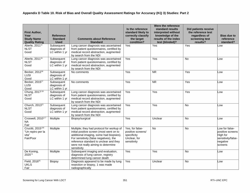
Appendix D Table 10. Risk of Bias and Overall Quality Assessment Ratings for Accuracy (KQ 3) Studies: Part 2
Screening for Lung Cancer With LDCT 351 RTI–UNC EPC
Appendix D Table 10. Risk of Bias and Overall Quality Assessment Ratings for Accuracy (KQ 3) Studies: Part 2
First Author, Year Study Name Quality Rating
Reference Standard
Used Comments about Reference
Standard
Is the reference standard likely to correctly classify
the target condition?
Were the reference standard results
interpreted without knowledge of the
results of the index test (blinded)?
Did patients receive the reference test
regardless of screening test
results?
Bias due to reference standard?
Aberle, 201131 NLST Good
Subsequent diagnosis of LC within 1 yr
Lung cancer diagnosis was ascertained from patient questionnaires, certified by medical record abstraction, augmented by search from the NDI
Yes Yes Yes Low
Aberle, 201156 NLST Good
Subsequent diagnosis of LC within 1 yr
Lung cancer diagnosis was ascertained from patient questionnaires, certified by medical record abstraction, augmented by search from the NDI
Yes Yes No Low
Becker, 201258 LUSI Good
Subsequent diagnosis of LC within 1 yr
No comments Yes NR Yes Low
Becker, 201557 LUSI Good
Subsequent diagnosis of LC within 1 yr
No comments Yes NR Yes Low
Chung, 2017130 NLST Good
Subsequent diagnosis of LC within 1 yr
Lung cancer diagnosis was ascertained from patient questionnaires, certified by medical record abstraction, augmented by search from the NDI
Yes Yes Yes Low
Church, 201355 NLST Good
Subsequent diagnosis of LC within 1 yr
Lung cancer diagnosis was ascertained from patient questionnaires, certified by medical record abstraction, augmented by search from the NDI
Yes Yes No Low
Croswell, 2010117 NA Fair
Multiple Biopsy/surgical Yes Unclear No Low
Crucitti, 2015109 “Un repiro per la vita” Fair/Poor
Multiple Multiple; they had protocol for workup of initial positive screen (most went on to additional imaging, some had biopsies). For sensitivity (false negatives), the reference standard is unclear and they were not really aiming to determine sensitivity
Yes, for false-positive screens/ specificity Unclear, for sensitivity
No No Low for false-positive screens High for sensitivity/false- negative screens
De Koning, 202074
Multiple Subsequent imaging and evaluation, diagnosis of lung cancer, registry determined lung cancer death
Yes Yes No Low
Field, 201695 UKLS Fair
Biopsy Diagnosis appeared to be made by lung resection or biopsy, 1 was made radiographically
Yes Unclear No Low

Appendix D Table 10. Risk of Bias and Overall Quality Assessment Ratings for Accuracy (KQ 3) Studies: Part 2
Screening for Lung Cancer With LDCT 352 RTI–UNC EPC
First Author, Year Study Name Quality Rating
Reference Standard
Used Comments about Reference
Standard
Is the reference standard likely to correctly classify
the target condition?
Were the reference standard results
interpreted without knowledge of the
results of the index test (blinded)?
Did patients receive the reference test
regardless of screening test
results?
Bias due to reference standard?
Field, 2016115 UKLS Fair
Other Lung cancer diagnosis ascertained from biopsy and search of the following databases: the Office for National Statistics the Hospital Episode Statistics database and the National Cancer Registration Service. However, timing of followup to determine lung cancer is unclear and authors are clear that lung cancer incidence will only be studied after data is pooled with other European studies.
Yes Yes Yes Low
Gierada, 2017108 NLST Fair
Subsequent diagnosis of LC within 1 yr
Prior reading of same LDCT scan along with clinical knowledge of progression to lung cancer
Yes Yes Yes Unclear
Gohagan, 200468 LSS Fair
Multiple No algorithm for this protocol; abstracted histology
Yes Unclear No Low
Henschke, 2004129 I-ELCAP Fair
Biopsy No comments Yes Unclear No Low
Henschke, 2006126 I-ELCAP Fair
Biopsy No comments Yes Unclear No Low
Henschke, 2006127 I-ELCAP Fair
Multiple Biopsy/surgical resection Yes Unclear No Low
Henschke, 2013105
I-ELCAP Fair
Subsequent diagnosis of LC within 1 yr
No comments Yes Unclear Unclear Unclear
Henschke, 2016104
I-ELCAP Fair
Biopsy Diagnosis defined on cytology from nonsurgical biopsy and path from resection
Unclear No Yes Unclear

Appendix D Table 10. Risk of Bias and Overall Quality Assessment Ratings for Accuracy (KQ 3) Studies: Part 2
Screening for Lung Cancer With LDCT 353 RTI–UNC EPC
First Author, Year Study Name Quality Rating
Reference Standard
Used Comments about Reference
Standard
Is the reference standard likely to correctly classify
the target condition?
Were the reference standard results
interpreted without knowledge of the
results of the index test (blinded)?
Did patients receive the reference test
regardless of screening test
results?
Bias due to reference standard?
Heuvelmans, 2015323 NELSON Poor
Other Histology was the reference Yes Unclear No High
Heuvelmans, 2013112 NELSON Fair
Biopsy Median followup was 4.4 yrs Yes Yes No Low
Horeweg, 201396
NELSON Fair
Other Histology Yes Unclear No Unclear
Horeweg, 201432 NELSON Fair
Biopsy No comments Yes Unclear No High
Infante, 200969 DANTE Fair
Biopsy No comments Yes Unclear No Low
Infante, 201559 DANTE Fair
Other Reference standard was histology Yes Unclear No Unclear
Kinsinger, 201737 LCSDP Fair
Multiple Reference standard is LC diagnosis made by clinical teams, date are extracted from VA system central data system and probably other sources since these screened patients were tracked in a registry
Yes No Yes Low
Lopes Penga, 2009123 ITALUNG Fair
Biopsy No comments Yes Unclear No Low
McKee, 2015395
NA Poor
Subsequent diagnosis of LC within 1 yr
1) Diagnosed lung cancer=biopsy proven + positive PET for patients that could not undergo biopsy 2) Biopsy-prove lung cancer
Yes No No Unclear
McWilliams, 2013114 PanCan, BCCA Fair
Biopsy 1) Histopathological exam of resected surgical specimens 2) Cytopathology from needle-aspiration biopsy samples
Yes No No Unclear

Appendix D Table 10. Risk of Bias and Overall Quality Assessment Ratings for Accuracy (KQ 3) Studies: Part 2
Screening for Lung Cancer With LDCT 354 RTI–UNC EPC
First Author, Year Study Name Quality Rating
Reference Standard
Used Comments about Reference
Standard
Is the reference standard likely to correctly classify
the target condition?
Were the reference standard results
interpreted without knowledge of the
results of the index test (blinded)?
Did patients receive the reference test
regardless of screening test
results?
Bias due to reference standard?
Menezes, 2010119 NA Fair
Biopsy No comments Yes Unclear No Low
Paci, 201760 ITALUNG Good
Multiple Biopsy reports, telephone followup, death registry
Yes Yes Yes Low
Pedersen, 2009120
DLCST Good
Biopsy No comments Yes Unclear No Low
Pinsky, 201598 NLST Fair
Subsequent diagnosis of LC within 1 yr
No comments Yes Yes Yes Low
Pinsky, 201562 NLST Good
Subsequent diagnosis of LC within 1 yr
No comments Yes Yes Yes Low
Pinsky, 201599 NLST Fair
Subsequent diagnosis of LC within 1 yr
No comments Yes Unclear Yes Low
Pinsky, 2015100 NLST Fair
Subsequent diagnosis of LC within 1 yr
No comments Yes Unclear Yes Low
Scholten, 2013101 NA Good
Biopsy No comments Yes Unclear No Low
Sverzellati, 201680 MILD Fair
Subsequent diagnosis of LC within 1 yr
No comments Yes Unclear Unclear Unclear
Swensen, 2005128 NA Fair
Multiple Determined by an individual’s provider Unclear Unclear No High

Appendix D Table 10. Risk of Bias and Overall Quality Assessment Ratings for Accuracy (KQ 3) Studies: Part 2
Screening for Lung Cancer With LDCT 355 RTI–UNC EPC
First Author, Year Study Name Quality Rating
Reference Standard
Used Comments about Reference
Standard
Is the reference standard likely to correctly classify
the target condition?
Were the reference standard results
interpreted without knowledge of the
results of the index test (blinded)?
Did patients receive the reference test
regardless of screening test
results?
Bias due to reference standard?
Tammemagi, 2017113 PanCan Fair
Subsequent diagnosis of LC within 2 yrs
Any of the following 1) histopathological exam of resected surgical specimens 2) cytopathology from FNA 3) nodules stable, nodules not visible, benign calcification developed
Yes No No Low
Toyoda, 2008124 NA Fair
Other Histology used, but procedures unclear Yes Unclear No Low
Tsushima, 2008121 NA Fair
Multiple Biopsy/surgical Yes Unclear No Low
van Klaveren, 2009118 NELSON Good
Biopsy No comments Yes Unclear No Low
Van Riel, 2015111 NELSON Good
Other This is study only looked at inter observer agreement, and its potential effect on subsequent testing. As such there was no clear reference standard in sensitivity and specificity were not calculated. In other words, this was the study of reliability.
NA NA NA Unclear
Veronesi, 2008122 COSMOS Fair
Biopsy No comments Yes Unclear No Low
Veronesi, 2008125 COSMOS Fair
Biopsy No comments Yes Unclear No Low
Wagnetz, 2012116 NA Fair
Multiple Biopsy/VATS Yes Unclear No Low
Walter, 201676 NELSON Good
Multiple LC diagnosis based on histology; benignity based on histology or stable size for at least 2 yrs
Yes Unclear Yes Low

Appendix D Table 10. Risk of Bias and Overall Quality Assessment Ratings for Accuracy (KQ 3) Studies: Part 2
Screening for Lung Cancer With LDCT 356 RTI–UNC EPC
First Author, Year Study Name Quality Rating
Reference Standard
Used Comments about Reference
Standard
Is the reference standard likely to correctly classify
the target condition?
Were the reference standard results
interpreted without knowledge of the
results of the index test (blinded)?
Did patients receive the reference test
regardless of screening test
results?
Bias due to reference standard?
Wang, 2012110 NELSON Fair
Multiple Pathology or no cancers diagnosed in 2 yr followup
Yes No No Unclear
Wille, 2014106 DLCST Fair
Subsequent imaging
Last annual CT scan Unclear Yes Yes Unclear
Xu, 200677
NELSON Fair
Multiple Brushings, biopsy, VATS, resection Yes Unclear Not planned Low
Yankelevitz, 2015103
I-ELCAP Fair
Biopsy Diagnosis defined on cytology from nonsurgical biopsy and path from resection -- for nonsolid nodules, FNAs can be operator dependent and not useful (according to authors in introduction)
Yes No No High
Yip, 2014102 NLST, I-ELCAP Fair
Biopsy Outcome is lung cancer incidence, relies on biopsy.
Yes No No High
Yousaf-Khan, 2017389 NELSON Poor
Multiple LC diagnosis based on histology; benignity based on histology or stable size for at least 2 yrs
Yes Yes Yes Low
Zhao, 201175 NELSON Fair
Subsequent diagnosis of LC within 1 yr
Subsequent diagnosis of LC within various time intervals, according to NELSON management protocol. Intervals could be 3 mos, 1 yr, or 2 yrs.
Yes Yes Unclear Low
Zhao, 2014107 NELSON Fair
Multiple Nodules were classified as benign or malignant based on histologic examination or as benign based on stable volume for more than 2 yrs after baseline.
Yes Unclear No, but they were subject to the same multicomponent reference standard
Unclear
Abbreviations: BCCA=British Columbia Cancer Agency; DANTE=Detection and Screening of Early Lung Cancer with Novel Imaging Technology Trial; DLCST=Danish Lung
Cancer Screening Trial; I-ELCAP=International Early Lung Cancer Action Project; ITALUNG=The Italian Lung Study; KQ=Key Question; LC=lung cancer; LCSDP=Lung
Cancer Screening Demonstration Project; LUSI=German Lung Cancer Screening Intervention Trial; MILD=Multicentric Italian Lung Detection Trial; NA=not applicable;
NDI=National Death Index; NELSON=Dutch-Belgian Randomized Lung Cancer Screening Trial; NLST=National Lung Screening Trial; NR=Not reported; PanCan=Pan-
Canadian Early Detection of Lung Cancer Study; UKLS=UK Lung Cancer Screening Trial.

Appendix D Table 11. Risk of Bias and Overall Quality Assessment Ratings for Accuracy (KQ 3) Studies: Part 3
Screening for Lung Cancer With LDCT 357 RTI–UNC EPC
Appendix D Table 11. Risk of Bias and Overall Quality Assessment Ratings for Accuracy (KQ 3) Studies: Part 3
First Author, Year Study Name Quality Rating
Is the time period
between the test and
reference test short enough ?
Did the whole or a random
selection of the patients receive
the reference test?
Did patients receive the
same reference standard for the
outcome specified?
Did the study have
high attrition raising
concern for bias?
Was an appropriate
method used to handle
missing data?
Bias due to flow and timing?
Quality Rating
Comments any Study With Poor Quality
Aberle, 201131 NLST Good
Yes Only patients with positive screen underwent diagnostic evaluation, but all received questionnaire and NDI search
Yes No Yes Low Good NA
Aberle, 201156 NLST Good
Yes Only patients with positive screen underwent diagnostic evaluation, but all received questionnaire and NDI Search
Yes No Yes Low Good NA
Becker, 201258 LUSI Good
Yes Whole sample - LC diagnosis by biopsy or followup
Yes No NR Low Good NA
Becker, 201557 LUSI Good
Yes Whole sample - LC diagnosis by biopsy or followup
Yes No Yes Low Good NA
Chung, 2017130 NLST Good
Yes Only patients with positive screen underwent diagnostic evaluation, but all received questionnaire and NDI Search
Yes No Yes Low Good NA
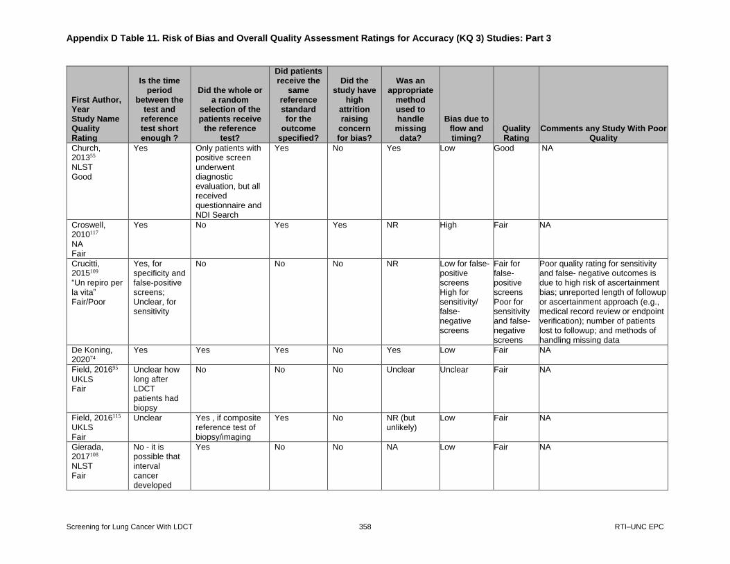
Appendix D Table 11. Risk of Bias and Overall Quality Assessment Ratings for Accuracy (KQ 3) Studies: Part 3
Screening for Lung Cancer With LDCT 358 RTI–UNC EPC
First Author, Year Study Name Quality Rating
Is the time period
between the test and
reference test short enough ?
Did the whole or a random
selection of the patients receive
the reference test?
Did patients receive the
same reference standard for the
outcome specified?
Did the study have
high attrition raising
concern for bias?
Was an appropriate
method used to handle
missing data?
Bias due to flow and timing?
Quality Rating
Comments any Study With Poor Quality
Church, 201355 NLST Good
Yes Only patients with positive screen underwent diagnostic evaluation, but all received questionnaire and NDI Search
Yes No Yes Low Good NA
Croswell, 2010117 NA Fair
Yes No Yes Yes NR High Fair NA
Crucitti, 2015109 “Un repiro per la vita” Fair/Poor
Yes, for specificity and false-positive screens; Unclear, for sensitivity
No No No NR Low for false-positive screens High for sensitivity/ false- negative screens
Fair for false-positive screens Poor for sensitivity and false- negative screens
Poor quality rating for sensitivity and false- negative outcomes is due to high risk of ascertainment bias; unreported length of followup or ascertainment approach (e.g., medical record review or endpoint verification); number of patients lost to followup; and methods of handling missing data
De Koning, 202074
Yes Yes Yes No Yes Low Fair NA
Field, 201695
UKLS Fair
Unclear how long after LDCT patients had biopsy
No No No Unclear Unclear Fair NA
Field, 2016115 UKLS Fair
Unclear Yes , if composite reference test of biopsy/imaging
Yes No
NR (but unlikely)
Low Fair NA
Gierada, 2017108 NLST Fair
No - it is possible that interval cancer developed
Yes No No NA Low Fair NA

Appendix D Table 11. Risk of Bias and Overall Quality Assessment Ratings for Accuracy (KQ 3) Studies: Part 3
Screening for Lung Cancer With LDCT 359 RTI–UNC EPC
First Author, Year Study Name Quality Rating
Is the time period
between the test and
reference test short enough ?
Did the whole or a random
selection of the patients receive
the reference test?
Did patients receive the
same reference standard for the
outcome specified?
Did the study have
high attrition raising
concern for bias?
Was an appropriate
method used to handle
missing data?
Bias due to flow and timing?
Quality Rating
Comments any Study With Poor Quality
Gohagan, 200468 LSS Fair
Unclear No Unclear No NR Low Fair NA
Henschke, 2004129 I-ELCAP Fair
Yes No Yes No NR Low Fair NA
Henschke, 2006126 I-ELCAP Fair
Yes No Yes Yes (>10%) NR Low Fair NA
Henschke, 2006127 I-ELCAP Fair
Yes No Yes No NR Low Fair NA
Henschke, 2013105 I-ELCAP Fair
Yes Unclear No Unclear Unclear Unclear Fair NA
Henschke, 2016104 I-ELCAP Fair
Yes Selection Yes NR NR Unclear Fair NA
Heuvelmans, 2015323 NELSON Poor
Unclear No Yes No Unclear High Poor Poor quality rating is due to several unclear and high categories, including unclear independence of test and reference standard interpretation, unclear bias due to index test
Heuvelmans, 2013112 NELSON Fair
NA No Yes No NA Low Fair NA

Appendix D Table 11. Risk of Bias and Overall Quality Assessment Ratings for Accuracy (KQ 3) Studies: Part 3
Screening for Lung Cancer With LDCT 360 RTI–UNC EPC
First Author, Year Study Name Quality Rating
Is the time period
between the test and
reference test short enough ?
Did the whole or a random
selection of the patients receive
the reference test?
Did patients receive the
same reference standard for the
outcome specified?
Did the study have
high attrition raising
concern for bias?
Was an appropriate
method used to handle
missing data?
Bias due to flow and timing?
Quality Rating
Comments any Study With Poor Quality
Horeweg, 201396 NELSON Fair
Unclear No Yes No Yes Low Fair NA
Horeweg, 201432 NELSON Fair
Unclear No Yes No Yes Low Fair NA
Infante, 200969 DANTE Fair
Yes No Yes No NR Low Fair NA
Infante, 201559 DANTE Fair
Unclear No Yes No Unclear Unclear Fair NA
Kinsinger, 201737 LCSDP Fair
NR Yes Yes NR NR Unclear Fair NA
Lopes Penga, 2009123 ITALUNG Fair
Yes No Yes Yes NR Low Fair NA
McKee, 2015395 NA Poor
Yes Yes Yes Yes Unclear High Poor Poor rating is due to the lack of clinical followup and inclusion of 26% of the sample in analysis. Given the small number of cases reclassified, results could change if data from missing participants had been available.
McWilliams, 2013114
PanCan, BCCA Fair
Unclear Biopsy Yes No Yes Unclear Fair NA

Appendix D Table 11. Risk of Bias and Overall Quality Assessment Ratings for Accuracy (KQ 3) Studies: Part 3
Screening for Lung Cancer With LDCT 361 RTI–UNC EPC
First Author, Year Study Name Quality Rating
Is the time period
between the test and
reference test short enough ?
Did the whole or a random
selection of the patients receive
the reference test?
Did patients receive the
same reference standard for the
outcome specified?
Did the study have
high attrition raising
concern for bias?
Was an appropriate
method used to handle
missing data?
Bias due to flow and timing?
Quality Rating
Comments any Study With Poor Quality
Menezes, 2010119 NA Fair
Yes No Yes Yes NR Unclear Fair NA
Paci, 201760
ITALUNG Good
Yes All received the same outcome assessment process
Not all received biopsy
No Yes Low Good NA
Pedersen, 2009120 DLCST Good
Yes No Yes No NR Low Good NA
Pinsky, 201598 NLST Fair
Yes All received the same outcome assessment process
Not all received biopsy
No Yes Low Fair NA
Pinsky, 201562 NLST Good
Yes All received the same outcome assessment process
Not all received biopsy
No Yes Low Good NA
Pinsky, 201599 NLST Fair
Yes All received the same outcome assessment process
Not all received biopsy
No Yes Low Fair NA
Pinsky, 2015100 NLST Fair
Yes All received the same outcome assessment process
Not all received biopsy
No Yes Low Fair NA
Scholten, 2013101 NA Good
Yes No Yes No NR Low Good NA
Sverzellati, 201680 MILD Fair
Yes Unclear Not all received biopsy
Unclear Unclear Unclear Fair NA

Appendix D Table 11. Risk of Bias and Overall Quality Assessment Ratings for Accuracy (KQ 3) Studies: Part 3
Screening for Lung Cancer With LDCT 362 RTI–UNC EPC
First Author, Year Study Name Quality Rating
Is the time period
between the test and
reference test short enough ?
Did the whole or a random
selection of the patients receive
the reference test?
Did patients receive the
same reference standard for the
outcome specified?
Did the study have
high attrition raising
concern for bias?
Was an appropriate
method used to handle
missing data?
Bias due to flow and timing?
Quality Rating
Comments any Study With Poor Quality
Swensen, 2005128 NA Fair
Yes No Yes Yes, for fourth annual (20%)
NR High Fair NA
Tammemagi, 2017113 PanCan Fair
Yes Yes - if composite reference test of biopsy/imaging.
Yes Yes Unclear High Fair NA
Toyoda, 2008124 NA Fair
Yes No Yes No NR Low Fair NA
Tsushima, 2008121 NA Fair
Yes No Yes No NR Low Fair NA
van Klaveren, 2009118 NELSON Good
Yes No Yes No NR Low Good NA
Van Riel, 2015111 NELSON Good
NA NA NA NA NA Low Good NA
Veronesi, 2008122 COSMOS Fair
Yes No Yes No NR Low Fair NA
Veronesi, 2008125 COSMOS Fair
Yes No Yes No NR Low Fair NA
Wagnetz, 2012116 NA Fair
Yes No Yes No NR Low Fair NA

Appendix D Table 11. Risk of Bias and Overall Quality Assessment Ratings for Accuracy (KQ 3) Studies: Part 3
Screening for Lung Cancer With LDCT 363 RTI–UNC EPC
First Author, Year Study Name Quality Rating
Is the time period
between the test and
reference test short enough ?
Did the whole or a random
selection of the patients receive
the reference test?
Did patients receive the
same reference standard for the
outcome specified?
Did the study have
high attrition raising
concern for bias?
Was an appropriate
method used to handle
missing data?
Bias due to flow and timing?
Quality Rating
Comments any Study With Poor Quality
Walter, 201676 NELSON Good
Yes Yes Same protocol for all
No NA Low Good NA
Wang, 2012110 NELSON Fair
Yes All Yes NR NR Unclear Fair NA
Wille, 2014106
DLCST Fair
Yes Intervention arm Yes No
NR Low Fair NA
Xu, 200677 NELSON Fair
NR Not planned Likely planned, but unclear
No data No data Unclear Fair NA
Yankelevitz, 2015103 I-ELCAP Fair
Yes Selection Yes For the sample of nonsolid lesions, none.
NR Unclear Fair NA
Yip, 2014102 NLST, I-ELCAP Fair
Yes Selection Yes No Yes Low Fair NA
Yousaf-Khan, 2017389
NELSON Poor
Yes Yes Same protocol for all
Yes None High Poor Poor quality assessment is due to a number of unclear bias ratings, including the length of time (2 yrs) between index and reference test, and the participation rate (which was optional in the fourth round of screening).
Zhao, 201175 NELSON Fair
Yes All received the same outcome assessment process
Not all received biopsy
Unclear based on this paper
Unclear Unclear Fair NA

Appendix D Table 11. Risk of Bias and Overall Quality Assessment Ratings for Accuracy (KQ 3) Studies: Part 3
Screening for Lung Cancer With LDCT 364 RTI–UNC EPC
First Author, Year Study Name Quality Rating
Is the time period
between the test and
reference test short enough ?
Did the whole or a random
selection of the patients receive
the reference test?
Did patients receive the
same reference standard for the
outcome specified?
Did the study have
high attrition raising
concern for bias?
Was an appropriate
method used to handle
missing data?
Bias due to flow and timing?
Quality Rating
Comments any Study With Poor Quality
Zhao, 2014107 NELSON Fair
Unclear No No Unclear Unclear Unclear Fair NA
Abbreviations: BCCA=British Columbia Cancer Agency; DANTE=Detection and Screening of Early Lung Cancer with Novel Imaging Technology Trial; DLCST=Danish Lung
Cancer Screening Trial; I-ELCAP=International Early Lung Cancer Action Project; ITALUNG=The Italian Lung Study; KQ=key question; LC=lung cancer; LCSDP=Lung
Cancer Screening Demonstration Project; LUSI=German Lung Cancer Screening Intervention Trial; MILD=Multicentric Italian Lung Detection Trial; NA=not applicable;
NDI=National Death Index; NELSON=Dutch-Belgian Randomized Lung Cancer Screening Trial; NLST=National Lung Screening Trial; NR=not reported; PanCan=Pan-
Canadian Early Detection of Lung Cancer Study; UKLS=UK Lung Cancer Screening Trial.

Appendix E Table 1. Screening Details of Included Trials, KQ 1
Screening for Lung Cancer With LDCT 365 RTI–UNC EPC
Appendix E Table 1. Screening Details of Included Trials, KQ1

Appendix E Table 1. Screening Details of Included Trials, KQ 1
Screening for Lung Cancer With LDCT 366 RTI–UNC EPC
Trial Inclusion Criteria Exclusion Criteria Description of Screening
Method Positive Screen and Evaluation Strategy

Appendix E Table 1. Screening Details of Included Trials, KQ 1
Screening for Lung Cancer With LDCT 367 RTI–UNC EPC
DANTE59 Male ages 60-74 who are current or former smokers, of at least 20 pack-years who quit less than 10 years ago
Severe comorbid conditions, life expectancy of less than 5 years, inability to comply with followup protocol, history of a previous malignancy within 10 years prior to enrollment in study
LDCT, contiguous 5 mm increments and 1.25 pitch with high-resolution bone algorithm (width 1700, level -600). Single-slice scanner with low-dose setting (140 kvp, 40 mA)
Noncalcified pulmonary nodules, hilar masses, FGG lesions, major atelectasis, endobronchial lesions, mediastinal adenopathy, pleural effusion, or pleural masses. Solid lesions, smooth and 10 mm or less: LDCT at 3, 6, and 12 months. If no change, followup 1 year. Solid lesions, nonsmooth and 5 mm or less: LDCT at 3-6 and 12 months. If no change, followup 1 year. Solid lesions, 6 mm to <10 mm: Commonly oral antibiotics. New HRCT after 6-8 weeks. Solid lesions, 10 mm to 20 mm: Oral antibiotics and new HRCT after 6-8 weeks. If no regression, PET-scan. If PET positive, tissue dx. PET-negative, close followup. Solid lesions, 20 mm or larger: Discretional antibiotics and repeat LDCT or contrast-enhanced CT and PET Subsolid (FGG) <10 mm: Oral antibiotics and new HRCT 6-8 weeks. If no regression, followup 6 months Subsolid (FGG) >10 mm: antibiotics and new HRCT 6-8 weeks. If no regression, evaluate case by case

Appendix E Table 1. Screening Details of Included Trials, KQ 1
Screening for Lung Cancer With LDCT 368 RTI–UNC EPC
Trial Inclusion Criteria Exclusion Criteria Description of Screening
Method Positive Screen and Evaluation Strategy
DLCST65 Adult ages 50-70 who are current or former smokers with at least 20 pack-years of smoking. Former smokers should have quit after age 50 and abstinent for <10 years. Ability to climb 2 flights of stairs without pause and lung function by spirometry FEV1 of at least 30% of predicted volume
Weight of > 130 kg, history of cancer diagnosis and treatment, lung tuberculosis, illness that could shorten life expectancy to less than 10 years, chest CT in the last year for any reason
LDCT (120 kV and 40 mA)
Nodules were classified into 5 categories: 1=nodules up to 15 mm with benign characteristics; 2=no nodules or nodules <5 mm; 3=5-15 mm without benign characteristics; 4 ≥15 mm; 5=rapid-growing nodules (>25% increase in volume). Categories 1 and 2 were considered negative screening results. Category 3 were rescanned in 3 months. Categories 4 and 5 were referred to chest physicians for diagnostic workup.
ITALUNG60 55-69 years old with a smoking history at least 20 pack-years in the last 10 years or quit within the last 10 years
History of previous cancer other than nonmelanoma skin cancer and general conditions precluding thoracic surgery
LDCT, 120 to 140 kV, 20 to 43 mAs, pitch 1-2
At least one calcified solid or part solid with solid part at least 5 mm. Noncalcified, nonsolid at least 10 mm (Management of positive screens fundamentally derived from I-ELCAP)
LUSI57, 58, 71 Adults ages 50-69 years with a history of at least 25 years of smoking of at least 15 cigarettes per day or at least 30 years of smoking at least 10 cigarettes per day; including ex-smokers who stopped at most 10 years ago
History of cancer in the last 5 years, medical condition preventing surgical treatment and having a serious illness that shortens life expectancy to less than 10 years
LDCT (1.6-2 mSv, 1 mm slice thickness, reconstruction interval 0.8 mm and 0.7 mm, respectively)
Early recall for repeat CT depending on largest nodule size: 6 months for 5-7 diameter nodules, 3 months for 8-10 diameter and immediate pulmonology referral for >10 mm nodule. VDT calculated for known nodules and VDT above 600 days was considered negative while VDT < 600 days was positive and subject to early recall with timing to screening depending on size of nodule

Appendix E Table 1. Screening Details of Included Trials, KQ 1
Screening for Lung Cancer With LDCT 369 RTI–UNC EPC
Trial Inclusion Criteria Exclusion Criteria Description of Screening
Method Positive Screen and Evaluation Strategy
LSS67, 73 Men and women ages 55 to 74 years with ≥30 pack-year smoking history and quit within 10 years
History of a spiral CT exam of the lungs or thorax in the previous 24 months, history of lung cancer, current treatment for any cancer other than nonmelanoma skin cancer, removal of a portion of or an entire lung, and participation in another cancer screening trial or a cancer primary prevention trial other than a smoking cessation study.
120-140 kVp, 60 mA, scan time of one second, 5 mm collimation, pitch of 2 or equivalent (depending on the model and type of scanner), and contiguous reconstructions
Noncalcified nodules ≥4 mm and several other specific findings (even with nodules <4 mm). At the 1-year examination, any noncalcified nodule ≥4 mm was considered a positive screen, and other abnormalities could be considered suspicious for lung cancer at the discretion of the radiologist
NELSON32, 74-77, 79 Men and women ages 50 to 74 years who smoked >15 cigarettes/day for >25 years or >10 cigarettes/day for >30 years; current smokers or those who quit ≤10 years ago
Moderate or severe health problems and an inability to climb 2 flights of stairs; weight >140 kg; current or past renal cancer, melanoma, or breast cancer; a diagnosis of lung cancer or treatment related to lung cancer within the past 5 years; or a chest CT scan within the past year
LDCT, using volumetric approach and volume doubling time (VDT); 80-140 kV, 40-80 mAs
Volume of solid nodule >500 mm3; pleural-based solid nodule with a minimal diameter of >10 mm; solid component in a partial solid nodule with a volume of >500 mm3; or an indeterminate baseline screen (e.g., solid nodule 50-500 mm3) with VDT<400 days on 3-month repeat
NLST31, 61, 72 Ages 55-74 with 30 or more pack-year history of smoking and currently smoking or quit within the past 15 years
Previous history of lung cancer, history of chest CT within 18 months before enrollment, history of hemoptysis or unexplained weight loss of more than 15 lb in preceding year
LDCT with average effective dose of 1.5 mSv
≥1 noncalcified nodule measuring at least 4 mm in long-axis diameter, mediastinal masses, pleural disease or atelectasis of more than one segment. Interpreting radiologist judgment regarding whether results were positive on the basis of findings such as noncalcified hilar or mediastinal adenopathy, atelectasis, and pleural disease. Results and recommendations were sent to the participant and his/her health provider.
Abbreviations: CT= computed tomography; DANTE=Detection and Screening of Early Lung Cancer with Novel Imaging Technology trial; DLCST=Danish Lung Cancer
Screening Trial; dx=diagnosis; FEV=forced expiratory volume; FGG=focal ground glass; HRCT=high-resolution computed tomography; I-ELCAP=International Early Lung
Cancer Action Project; ITALUNG=The Italian Lung Study; KQ=key question; LDCT=low-dose computed tomography; LSS=The Lung Screening Study; LUSI=German Lung
Cancer Screening Intervention Trial; NELSON=Nederlands-Leuvens Longkanker Screenings Onderzoek; NLST=National Lung Screening Trial; PET=positron emission
tomography; VDT=volume doubling time.

Appendix E Table 2. Types and Stages of Incident Lung Cancer, KQ 1
Screening for Lung Cancer With LDCT 370 RTI–UNC EPC
Appendix E Table 2. Types and Stages of Incident Lung Cancer, KQ 1
Study Cumulative Incidence Type of Incident Lung Cancer Stages of Incident Lung Cancer
DANTE59 LDCT: 106 (8.2%) Control: 73 (5.2%)
LDCT Adenocarcinoma=44 (42%) Squamous 25=(24%) Non-small cell NOS=7 (6.7%) Other=7 (6.7%) Small cell=9 (8.7%) Missing=12 (11.5%) Control Adenocarcinoma=19 (26%) Squamous=17 (23.6%) Non-small cell NOS=8 (11.1%) Other=6 (8.3%) Small cell=6 (8.3%) Missing=16 (22.2%)
LDCT Stage IA=31 (29.8%) Stage IB=16 (15.4%) Stage II=7 (6.7%) Stage IIIA=9 (8.7%) Stage IIIB=8 (7.7%) Stage IV=26 (25% ) Missing=7 (6.7%) Control Stage IA=6 (8.3%) Stage IB=10 (13.9%) Stage II=5 (6.9%) Stage IIIA=6 (8.3%) Stage IIIB=6 (8.3%) Stage IV=33 (45.8 %) Missing=6 (8.3%)

Appendix E Table 2. Types and Stages of Incident Lung Cancer, KQ 1
Screening for Lung Cancer With LDCT 371 RTI–UNC EPC
Study Cumulative Incidence Type of Incident Lung Cancer Stages of Incident Lung Cancer
DLSCT65 LDCT: 100 (4.9%) Control : 53 (2.6%)
LDCT Adenocarcinoma=40 (40%) Adenocarcinoma + broncho-alveolar carcinoma=17 (17%) Adenocarcinoma + squamous cell carcinoma=1 (1%) Squamous cell carcinoma =14 (14%) Broncho-alveolar carcinoma=1 (1%) Non-small cell lung cancer=14 (14%), Non-small cell lung cancer + broncho-alveolar carcinoma=0 Small cell lung cancer + non-small cell lung cancer=0 Small cell lung cancer=11 (11%) Large cell neuroendocrine carcinoma=1(1%) Carcinoid=0 Unknown histology 1 (1%) Control: Adenocarcinoma=18 (34%) Adenocarcinoma + broncho-alveolar carcinoma=0 Adenocarcinoma + squamous cell carcinoma=0 Broncho-alveolar carcinoma=0 Squamous cell carcinoma=9 (17%) Non-small cell lung cancer=9 (17%) Non-small cell lung cancer + broncho-alveolar carcinoma=1 (1.9%) Small cell lung cancer + non-small cell lung cancer=3 (5.7%) Small cell lung cancer=11 (21%) Large cell neuroendocrine carcinoma=0 Unknown histology=1 (1.9%)
LDCT Stage I=50 (50%) Stage II=4 (4%) Stage IIIA=15 (15%) Stage IIIB=8 (8%) Stage IV=23 (23%) Unknown stage=0 Control: Stage I=8 (15.1%) Stage II=2 (3.8%) Stage IIIA=3 (5.7%) Stage IIIB=6 (11.3%) Stage IV=32 (60.4%) Unknown stage=2 (3.8%)

Appendix E Table 2. Types and Stages of Incident Lung Cancer, KQ 1
Screening for Lung Cancer With LDCT 372 RTI–UNC EPC
Study Cumulative Incidence Type of Incident Lung Cancer Stages of Incident Lung Cancer
ITALUNG60 LDCT: 67 (4.1%) Control: 71 (4.5%)
LDCT Adenocarcinoma=29 (43%) Squamous cell carcinoma=14 (21%) Small cell lung cancers=10 (15%) Carcinoid=2 (3%) Non-small cell carcinoma=3 (4%) Unclassified=9 (13%) Control Adenocarcinoma=21 (30%) Squamous cell carcinoma=17 (24%) Small cell lung cancers=11 (15%) Carcinoid=0 (0%) Non-small cell carcinoma=5 (7%) Unclassified=17 (24%)
LDCT Stage 1=24 (36%) Stage II=5 (7%) Stage III=9 (13%) Stage IV =24 (36%) Unknown =5 (7%) Control Stage 1=8 (11%) Stage II=5 (7%) Stage III=8 (11%) Stage IV=35 (49%) Unknown=15 (21%) p=0.005
LUSI57, 58, 71 Reported for years 0-5 LDCT: 63 (3.1%) Control: 36 (1.8%) Reported at any time, with followup to a median of 8.8 years LDCT: 85 (4.2%) Control: 67 (3.3%)
Data limited to the first 5 years (99 cancers) LDCT: Small cell carcinoma=5 (8%) Squamous cell=10 (16%) Adenocarcinoma=43 (68%) Large cell carcinoma=1 (2%) Carcinoid=2 (3%) Carcinoma unspecified=2 (3%) Control Small cell carcinoma=9 (25%) Squamous cell carcinoma=6 (17%) Adenocarcinoma=18 (50%) Large cell carcinoma=1 (3%) Carcinoid=0 (0%) Carcinoma unspecified=2 (6%)
Data provided for followup to a median of 8.8 years (152 cancers) LDCT Stage IA=37 (44%) Stage IB=11 (13%) Stage IIA=3 (4%) Stage IIB=4 (5%) Stage IIIA=10 (12%) Stage IIIB=2 (2%) Stage IV=17 (20%) Unknown=1 (1%) Control Stage IA=2 (3%) Stage IB=4 (6%) Stage IIA=5 (7%) Stage IIB=4 (6%) Stage IIIA=16 (24%) Stage IIIB=5 (7%) Stage IV=30 (45%) Unknown=1 (1%)

Appendix E Table 2. Types and Stages of Incident Lung Cancer, KQ 1
Screening for Lung Cancer With LDCT 373 RTI–UNC EPC
Study Cumulative Incidence Type of Incident Lung Cancer Stages of Incident Lung Cancer
LSS67 LDCT: 40 (2.4%) Control: 20 (1.5%)
LDCT Adenocarcinoma=24 (60%) Squamous cell carcinoma=5 (13%) Small cell carcinoma=4 (10%) Large cell carcinoma=4 (10%) Non-small cell carcinoma NOS=3 (8%) Carcinoid tumor=NR Unknown=NR Control Adenocarcinoma=9 (45%) Squamous cell carcinoma=6 (30%) Small cell Carcinoma=2 (10%) Large cell carcinoma=1 (5%) Non-small cell carcinoma NOS=0 Carcinoid tumor=1 (5%) Unknown=1 (5%)
LDCT Stage I =19 (48%) Stage II=3 (8%) Stage III=11 (28%) Stage IV=5 (13%) Unknown=2 (5%) Control Stage I=8 (40%) Stage II=1 (5%) Stage III=5 (25%) Stage IV=4 (20%) Unknown=2 (10%)
NELSON32, 74-77 Data reported for male participants only LDCT: 344 (5.2%) Control: 304 (4.6%)
LDCT Adenocarcinoma=179 (52%) Squamous cell carcinoma=77 (22%) Small cell carcinoma=40 (12%) Non-small cell carcinoma NOS=16 (5%) Other=32 (9%) Control Adenocarcinoma=133 (44%) Squamous cell carcinoma= 94 (31%) Small cell carcinoma=46 (15%) Non-small cell carcinoma NOS=13 (4%) Other=18 (6%)
LDCT Stage IA=105 (31%) Stage IB=34 (10%) Stage IIA=12 (4%) Stage IIB=17 (5%) Stage IIIA=34 (10%) Stage IIIB=27 (8%) Stage IV=92 (27%) Unknown=23 (7%) Control Stage IA=21 (7%) Stage IB=20 (7%) Stage IIA=13 (4%) Stage IIB=17 (6%) Stage IIIA=43 (14%) Stage IIIB=34 (11%) Stage IV=139 (46%) Unknown=17 (6%)

Appendix E Table 2. Types and Stages of Incident Lung Cancer, KQ 1
Screening for Lung Cancer With LDCT 374 RTI–UNC EPC
Study Cumulative Incidence Type of Incident Lung Cancer Stages of Incident Lung Cancer
NLST31, 61 Median followup of 6.5 years LDCT: 1,089 (4.1%) Control: 969 (3.6%) Post-trial followup to a median of 11.3 years LDCT: 1701 (6.4%) Control: 1681 (6.3%)
Median followup of 6.5 years LDCT Bronchioloalveolar carcinoma=111 (10%) Adenocarcinoma=389 (35%) Squamous cell=249 (22%) Large cell carcinoma=40 (4%) Non-small cell or other=137 (12%) Small cell carcinoma=143 (13%) Carcinoid=6 (0.5%) Unknown=34 (3%) Control Bronchioloalveolar carcinoma=36 (4%) Adenocarcinoma=337 (34%) Squamous cell=214 (22%) Large cell carcinoma=44 (4%) Non-small cell or other=162 (16%) Small cell carcinoma=163 (16%) Carcinoid=3 (0.3%) Unknown=34 (3%) Median followup of 11.3 years LDCT Bronchioloalveolar carcinoma=121 (7%) Adenocarcinoma=608 (36%) Squamous cell=416 (25%) Large cell carcinoma=56 (3%) Other non-small cell=196 (12%) Small cell carcinoma=245 (14%) Carcinoid=12 (0.7%) Unknown=47 (3%) Control Bronchioloalveolar carcinoma=46 (3%) Adenocarcinoma=598 (36%) Squamous cell=395 (24%) Large cell carcinoma=53 (3%) Other non-small cell=251 (15%) Small cell carcinoma=291 (17%) Carcinoid=7 (0.4%) Unknown=40 (2%)
Median followup of 6.5 years LDCT Stage 1A 416 (40%) Stage 1B =104 (10%) Stage IIA= 35 (3.4%) Stage IIB=38 (3.7%) Stage IIIA=99 (9.5%) Stage IIIB=122 (11.7%) Stage IV=226 (21.7%) Control Stage 1A=196 (21.1%) Stage 1B=93 (10%) Stage IIA=32 (3.4%) Stage IIB=42 (4.5%) Stage IIIA=109 (11.7%) Stage IIIB=122 (13.1%) Stage IV=335 (36.1%) Median followup of 11.3 years LDCT Stage 1A=523 (31%) Stage 1B=148 (9%) Stage IIA=91 (5%) Stage IIB=43 (3%) Stage IIIA=204 (12%) Stage IIIB=84 (5%) Stage IV=468 (28%) Occult=5 Unknown=112 (7%) Control Stage 1A=326 (19%) Stage 1B=134 (8%) Stage IIA=80 (5%) Stage IIB=66 (4%) Stage IIIA=216 (13%) Stage IIIB=94 (6%) Stage IV=597 (36%) Occult=4 Unknown=143 (9%)
Abbreviations: DANTE=Detection and Screening of Early Lung Cancer with Novel Imaging Technology trial; DLCST=Danish Lung Cancer Screening Trial; ITALUNG=The
Italian Lung Study; KQ=key question; LDCT=low-dose computed tomography; LSS=The Lung Screening Study; LUSI=German Lung Cancer Screening Intervention Trial;
NELSON=Nederlands-Leuvens Longkanker Screenings Onderzoek; NLST=National Lung Screening Trial; NOS=not otherwise specified; NR=not reported.

Appendix E Table 3. Incidental Findings From KQ 4 Studies
Screening for Lung Cancer With LDCT 375 RTI–UNC EPC
Appendix E Table 3. Incidental Findings from KQ4 Studies

Appendix E Table 3. Incidental Findings From KQ 4 Studies
Screening for Lung Cancer With LDCT 376 RTI–UNC EPC
Author, Year Study Name Sample Size Incidental Findings Reported
Field, 2016115 UKLS
4,055 Thoracic incidental findings for which supplementary radiology report was submitted: 115 Thoracic incidental findings included: Aortic dilatation: 4 Severe aortic valve calcification: 5 Mediastinal mass: 4 Mediastinal or hilar lymphadenopathy: 6 Pneumonia: 41 Bronchiectasis: 5 Pleural thickening: 8 Smoking related interstitial lung disease: 7 Severe emphysema: 9 Interstitial fibrosing lung disease (unspecified): 6 Non-specific interstitial pneumonia: 2 Usual interstitial pneumonia: 12 Sarcoidosis: 1 Oesophageal thickening or dilatation: 2 Breast mass: 1 Lobar collapse: 2 Extrathoracic incidental findings for which supplementary radiology report was submitted: 13 Extrathoracic incidental findings included: Biliary dilatation: 1 Adrenal mass: 3 Cirrhosis: 1 Hydronephrosis: 1 Liver mass: 1 Pancreatic cysts: 1 Renal mass: 3 Splenomegaly: 1 Thyroid mass: 1

Appendix E Table 3. Incidental Findings From KQ 4 Studies
Screening for Lung Cancer With LDCT 377 RTI–UNC EPC
Author, Year Study Name Sample Size Incidental Findings Reported
Kinsinger, 201737 LCSDP 2,106 Patients with ≥1 incidental finding reported: 857 (40.7%) Total N of incidental findings: 1,044 Findings included: Abdominal abnormalities and masses: 146 (14%) Aortic dilation or aneurysm: 87 (8.3%) Infectious, inflammatory, or interstitial processes: 265 (25.4%) Thyroid nodules: 25 (2.4%) Other (including CAC and emphysema): 521 (49.9%) Patients with incidental (nonnodule) findings, by demonstration site: Site 1: 211 (47.7%) Site 2: 106 (46.5%) Site 3: 135 (63.4%) Site 4: 89 (20.0%) Site 5: 149 (60.3%) Site 6: 54 (40.0%) Site 7: 81 (31.4%) Site 8: 32 (23.0%)

Appendix E Table 3. Incidental Findings From KQ 4 Studies
Screening for Lung Cancer With LDCT 378 RTI–UNC EPC
Author, Year Study Name Sample Size Incidental Findings Reported
Morgan, 2017165 NA 320 Incidental findings resulting in further evaluation: 15% Findings included: Respiratory Total respiratory: 223 (69.6%) Emphysema: 162 (50.6%) Bronchial wall thickening: 126 (39.4%) Atelectasis: 52 (16.3) Ground-glass opacity: 26 (8.1%) Bronchiectasis: 14 (4.4%) Cardiovascular: Total cardiovascular: 216 (67.5%) CAC: 182 (56.0%) Aortic calcification: 66 (20.6%) Aortic dilation: 26 (8.1%) Endocrinological Total endocrinological: 23 (7.2%) Adrenal nodule 12 (3.8%) Thyroid nodule 11 (3.4%) Gastrointestinal Total gastrointestinal: 79 (24.7%) Hiatal hernia: 30 (9.4%) Liver cyst: 22 (6.8%) Dilated esophagus: 7 (2.2%) Gallstone: 6 (1.9%) Diaphragmatic hernia: 5 (1.6%) Other: 9 (2.8%) Genitourinary Total genitourinary: 14 (4.4%) Renal cyst: 8 (2.5%) Renal stone: 4 (1.3%) Renal mass: 2 (0.6%) Other Systems Total other: 78 (24.4%) Degenerative joint disease: 74 (23.1%) Compression fracture: 3 (0.9%) Breast nodule: 1 (0.3%) Splenic lesion: 1 (0.3%)

Appendix E Table 3. Incidental Findings From KQ 4 Studies
Screening for Lung Cancer With LDCT 379 RTI–UNC EPC
Author, Year Study Name Sample Size Incidental Findings Reported
Nguyen, 2017164 NLST 17,309 Extrapulmonary findings (≥1 finding): 10,166 (58.7%) Potentially significant findings: 3,398 (19.6%) Minor findings 9,152 (52.9%) Significant cardiovascular findings: 1,378 (8.0%) Significant above diaphragm: 1,255 (7.3%) Significant below diaphragm: 1,311 (7.6%) Extrapulmonary malignancies: Thyroid: 14 (0.08%, 1 malignancy for every 14 found incidentally) Adrenal: 0 (0%) Kidney: 45 (0.26%, 1:37 renal abnormalities to find a malignancy) Liver: 8 (0.05%, none had significant findings on screening)
O’Grady, 2015167 NLST, PLCO 195,642 (53,248 from NLST 142,394 from PLCO)
Incidental thyroid cancer findings: NLST Control: 23 Intervention: 37 PLCO Control: 130 Intervention: 104
Pinsky, 201462 NLST 26,722 NLST patients from LDCT arm 19,612 in <65 age cohort 7,110 in 65+ age cohort
This study reports emphysema, significant cardiovascular abnormality, abnormalities above the diaphragm, and abnormalities below the diaphragm. However, it is NR which of those are incidental. Aggregate frequencies of reported abnormal findings on screening: <65 age cohort: 6.9% 65+ age cohort: 9.2% p <0.001

Appendix E Table 3. Incidental Findings From KQ 4 Studies
Screening for Lung Cancer With LDCT 380 RTI–UNC EPC
Author, Year Study Name Sample Size Incidental Findings Reported
Swensen, 2002166 NA 1,520 Patients with nonpulmonary incidental findings of significance: 210 (14%) Findings included: Renal cell cancer: 4 Indeterminate renal mass: 33 Renal calculi: 24 Bronchial carcinoid: 2 Tracheal nodule: 7 Lobar collapse: 2 Bronchiectasis: 11 Breast cancer: 3 Breast nodule: 17 Atrial myxoma: 1 Abdominal aortic aneurysm: 51 Pericardial effusion: 9 Pleural effusion: 4 Pulmonary artery calcification: 1 Lymphoma: 2 Spine metastasis: 1 Adrenal mass: 35 Pheochromocytoma: 1 Gastric tumor: 2
Wilson, 2008135 PLuSS 3,642 Number of screenings due to significant incidental finding: Initial screening: 82/3,642 (2.3%) Imaging studies: 19/3,642 (0.5%) Repeat screening: 50/3,423 (1.5%)
Abbreviations: CAC=coronary artery calcification; KQ=key question; LCSDP=Lung Cancer Screening Demonstration Project; NA=not applicable; NLST=National Lung
Screening Trial; NR=not reported; PLCO=Prostate, Lung, Colorectal, and Ovarian Cancer Screening Trial; PLuSS=Pittsburgh Lung Screening Study; UKLS=UK Lung Cancer
Screening Trial.

Appendix E Table 4. KQs 6 and 7: Study and Patient Characteristics Table
Screening for Lung Cancer With LDCT 381 RTI–UNC EPC
Appendix E Table 4. KQ6&7 a Study and Patient Characteristics Table
Study Identifiers Author, Year Treatment Type(s) N Enrolled KQs Addressed Quality
Study Characteristics Study or Database Name
Country Study Years
Eligibility Criteria Followup
Funding Source
Baseline Patient Characteristics Age
Gender Race/Ethnicity Comorbidities
NSCLC Characteristics Stage
TNM Edition(s) Histology
Tumor Size
Treatment Characteristics Surgical Approach
SBRT/SABR Dosing and Frequency
Allibhai, 2013257 SBRT/SABR N=185 KQ 7 Fair
NA Canada 2004-2010 Patients with T1-2N0M0 NSCLC deemed medically inoperable by an experienced thoracic surgeon Followup, median (range) 15.2 mos Funding source University, private, other unspecified
Age, median 74.8 yrs Male, N (%) 93 (50.3) Race/ethnicity NR Comorbidities NR
Stage, N (%) T1: 133 (72) T2: 52 (28) TNM edition(s) NR Histology, N (%) NR Tumor diameter, mean (range) 2.2 cm (0.6 to 5.7 cm)
SBRT patients, N (%) 185 (100) SBRT dosing NR, but dosage options included 48 Gy x 4 fx, 54 to 60 Gy x 3 fx, 60 Gy x 8 fx, and 50 Gy x 10 fx, depending on tumor location and size
Arnold, 2017197 SBRT/SABR N=75 KQ 6 Fair
NCDB U.S. 2004-2012 Patients diagnosed with c-stage I NSCLC with tumors ≤4 cm who were ≥90 years old at the time of diagnosis, had no other cancer, and had no history of radiation therapy or nonstandard therapy Followup, median (range) NR Funding source NR
Age NR Male, N (%) NR Race/ethnicity NR Charlson Comorbidity Index, N (%) NR
Stage, N (%) c-stage 1: 75 (100) TNM edition(s) NR Histology, N (%) NR Tumor size, N (%) NR
SBRT patients, N (%) 75 (100) SBRT dosing 100-200 Gy x 3-5 fx: 75 (100)

Appendix E Table 4. KQs 6 and 7: Study and Patient Characteristics Table
Screening for Lung Cancer With LDCT 382 RTI–UNC EPC
Study Identifiers Author, Year Treatment Type(s) N Enrolled KQs Addressed Quality
Study Characteristics Study or Database Name
Country Study Years
Eligibility Criteria Followup
Funding Source
Baseline Patient Characteristics Age
Gender Race/Ethnicity Comorbidities
NSCLC Characteristics Stage
TNM Edition(s) Histology
Tumor Size
Treatment Characteristics Surgical Approach
SBRT/SABR Dosing and Frequency
Badellino, 2017268 SBRT/SABR N=148 KQ 7 Fair
NA Italy 2004-2014 Patients with Stage I NSCLC and contraindication to surgery after multidisciplinary evaluation; ECOG performance status ≤2; accurate staging with PET and brain CT scan; prior thoracic radiation therapy Followup, median 20.5 mos Funding source NR
Age, mean (range) SBRT 3D-CRT: 75 yrs (53 to 89 yrs) SBRT VMAT: 76 yrs (52 to 88 yrs) Male, N (%) SBRT 3D-CRT: 77 (81) SBRT VMAT: 40 (75.5) Race/ethnicity NR Comorbidities, N (%) NR
Stage, N (%) Clinical T1a SBRT 3D-CRT: 78 (82.1) SBRT VMAT: 30 (56.6) Clinical T1b SBRT 3D-CRT: 17 (17.9) SBRT VMAT: 23 (43.4) TNM edition(s) NR Histology, N (%) Adenocarcinoma SBRT 3D-CRT: 19 (20) SBRT VMAT: 16 (30.2) Squamous cell carcinoma SBRT 3D-CRT: 16 (16.8) SBRT VMAT: 9 (17) Other SBRT 3D-CRT: 12 (12.7) SBRT VMAT: 4 (7.5) Unknown SBRT 3D-CRT: 48 (50.5) SBRT VMAT: 24 (45.3) Tumor diameter, mean (range) SBRT 3D-CRT: 2.5 cm (1 to 5 cm) SBRT VMAT: 3 cm (1.3 to 5 cm)
SBRT patients, N (%) 148 (100) SBRT dosing, N (%) Total dose in Gy, mean (range) SBRT 3D-CRT: 110.8 (100-120) SBRT VMAT: 110.5 (100-120) 15 Gy x 3 fx SBRT 3D-CRT: 82 (86) SBRT VMAT: 9 (16) 14 Gy x 3 fx SBRT 3D-CRT: 12 (13) SBRT VMAT: 2 (3) 11 Gy x 5 fx SBRT 3D-CRT: 1 (1) SBRT VMAT: 21 (39) 7.5 Gy x 8 fx SBRT 3D-CRT: 0 (0) SBRT VMAT: 21 (39)

Appendix E Table 4. KQs 6 and 7: Study and Patient Characteristics Table
Screening for Lung Cancer With LDCT 383 RTI–UNC EPC
Study Identifiers Author, Year Treatment Type(s) N Enrolled KQs Addressed Quality
Study Characteristics Study or Database Name
Country Study Years
Eligibility Criteria Followup
Funding Source
Baseline Patient Characteristics Age
Gender Race/Ethnicity Comorbidities
NSCLC Characteristics Stage
TNM Edition(s) Histology
Tumor Size
Treatment Characteristics Surgical Approach
SBRT/SABR Dosing and Frequency
Bibault, 2015252 SBRT/SABR Total N=205 Monte Carlo N=88 Type A N=117 KQ 7 Fair
NA France 2007-2013 Inoperable patients with stage NSCLC, T1 or T2 tumors >15 mm without lymph node or distant metastasis, and performance status ≤2 treated with SBRT Followup, median (range) Monte Carlo protocol: 15 mos (3 to 40 mos) Type A algorithm: 24 mos (3 to 55 mos) Private funding (Accuray, Oscar Lambret Comprehensive Cancer Center)
Age, median (range) Monte Carlo: 70.5 yrs (46 to 87 yrs) Type A: 69 yrs (49 to 92 yrs) Male, N (%) Total: 173 (84.4) Monte Carlo: 72 (81.8) Type A: 101 (86.3) Race/ethnicity NR Comorbidities, N (%) Respiratory failure Monte Carlo: 67 (76.1) Type A: 96 (82.1)
Stage, N (%) Monte Carlo T1a: 36 (38.7) T1b: 31 (33.3) T2a: 24 (25.8) T2b: 2 (2.2) Type A T1a: 50 (42.0) T1b: 36 (30.3) T2a: 30 (25.2) T2b: 3 (2.5) TNM edition(s) NR Histology, N (%) NR Tumor diameter, median (range) 22 mm (15 to 60 mm)
SBRT patients, N (%) 205 (100) SBRT dosing 60 Gy x 3 fx: NR 60 Gy x 5 fx: NR

Appendix E Table 4. KQs 6 and 7: Study and Patient Characteristics Table
Screening for Lung Cancer With LDCT 384 RTI–UNC EPC
Study Identifiers Author, Year Treatment Type(s) N Enrolled KQs Addressed Quality
Study Characteristics Study or Database Name
Country Study Years
Eligibility Criteria Followup
Funding Source
Baseline Patient Characteristics Age
Gender Race/Ethnicity Comorbidities
NSCLC Characteristics Stage
TNM Edition(s) Histology
Tumor Size
Treatment Characteristics Surgical Approach
SBRT/SABR Dosing and Frequency
Brooks, 2017202 SBRT/SABR N=772 KQs 6, 7 Fair
NA U.S. 2004-2014 Patients with c-stage 1-2 (T1-3N0M0) NSCLC not involving the bronchial tree or blocking the airway, with or without a lung cancer history (but no evidence of previous disease), and treated with SBRT; current cancer must have been outside the previously irradiated field, if patients had received previous radiation therapy for NSCLC Followup, median Age <75 yrs: 54.6 mos Age ≥75 yrs: 55.2 mos Funding source University, other unspecified source(s)
Age, median (range) Mean (range): 73.9 yrs (46 to 91.8 yrs) Median: NR Male, N (%) 385 (49.9) Race/ethnicity NR Comorbidities, N (%) NR
Stage, N (%) Stage T1: 642 (83.2) Stage T2: 120 (15.5) Stage T3 (and Stage 2 NSCLC): 10 (1.3) TNM edition(s) 7th edition Histology, N (%) Adenocarcinoma: 405 (52.5) Squamous cell: 268 (34.7) Other: 19 (2.5) NSCLC NOS: 72 (9.3) No pathologic features: 8 (1.0) Tumor size, mean (SD) NR
SBRT patients, N (%) 772 (100) SBRT dosing, N (%) 50 Gy x 4 fx (BED 112.5 Gy): 636 (82.4) 70 Gy x 10 fx (BED 119 Gy): 99 (12.8) Other (75-149.6 Gy): 37 (4.8)

Appendix E Table 4. KQs 6 and 7: Study and Patient Characteristics Table
Screening for Lung Cancer With LDCT 385 RTI–UNC EPC
Study Identifiers Author, Year Treatment Type(s) N Enrolled KQs Addressed Quality
Study Characteristics Study or Database Name
Country Study Years
Eligibility Criteria Followup
Funding Source
Baseline Patient Characteristics Age
Gender Race/Ethnicity Comorbidities
NSCLC Characteristics Stage
TNM Edition(s) Histology
Tumor Size
Treatment Characteristics Surgical Approach
SBRT/SABR Dosing and Frequency
Brunelli, 2015179 Surgery N=1,370 KQ 6 Good
NA U.S., Italy, Spain 2000-2011 Patients with p-stage 1 NSCLC with predicted postoperative FEV1 and DLCO <30% in association with peak oxygen consumption <10 mL/(kg-x-min) and deemed in stable cardiac condition and, if unstable hemodynamic conditions were present, receiving optimized cardiologic treatment prior to complete resection (but no induction chemotherapy) Followup, median 77 mos Funding source NR
Age, mean (SD) 66.1 yrs (10.2 yrs) Male, N (%) 1,014 (74) Race/ethnicity NR Comorbidities, N (%) NR
Stage, N (%) p-stage 1: 1,370 (100) Stage pT1: 533 (39) TNM edition(s) 7th edition Histology, N (%) NR Tumor size, mean (SD) NR
Surgical approach, N (%) L/Bi-L: 1,304 (95.2) P: 66 (4.8)
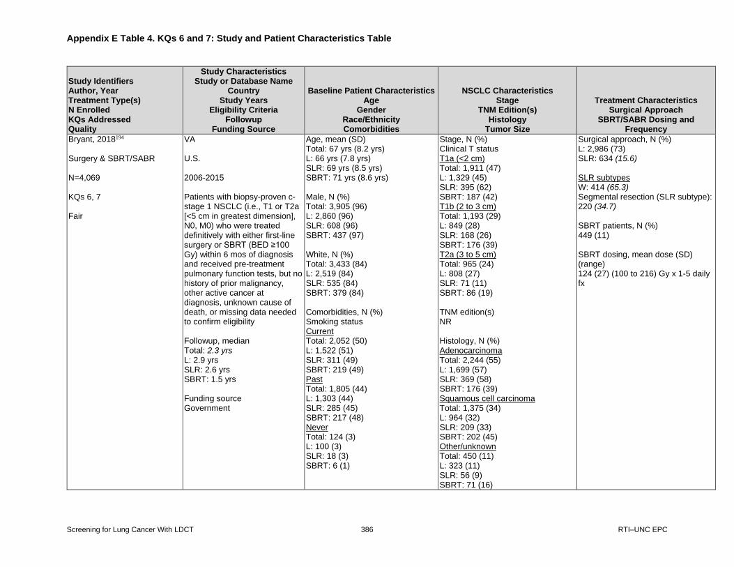
Appendix E Table 4. KQs 6 and 7: Study and Patient Characteristics Table
Screening for Lung Cancer With LDCT 386 RTI–UNC EPC
Study Identifiers Author, Year Treatment Type(s) N Enrolled KQs Addressed Quality
Study Characteristics Study or Database Name
Country Study Years
Eligibility Criteria Followup
Funding Source
Baseline Patient Characteristics Age
Gender Race/Ethnicity Comorbidities
NSCLC Characteristics Stage
TNM Edition(s) Histology
Tumor Size
Treatment Characteristics Surgical Approach
SBRT/SABR Dosing and Frequency
Bryant, 2018194 Surgery & SBRT/SABR N=4,069 KQs 6, 7 Fair
VA U.S. 2006-2015 Patients with biopsy-proven c-stage 1 NSCLC (i.e., T1 or T2a [<5 cm in greatest dimension], N0, M0) who were treated definitively with either first-line surgery or SBRT (BED ≥100 Gy) within 6 mos of diagnosis and received pre-treatment pulmonary function tests, but no history of prior malignancy, other active cancer at diagnosis, unknown cause of death, or missing data needed to confirm eligibility Followup, median Total: 2.3 yrs L: 2.9 yrs SLR: 2.6 yrs SBRT: 1.5 yrs Funding source Government
Age, mean (SD) Total: 67 yrs (8.2 yrs) L: 66 yrs (7.8 yrs) SLR: 69 yrs (8.5 yrs) SBRT: 71 yrs (8.6 yrs) Male, N (%) Total: 3,905 (96) L: 2,860 (96) SLR: 608 (96) SBRT: 437 (97) White, N (%) Total: 3,433 (84) L: 2,519 (84) SLR: 535 (84) SBRT: 379 (84) Comorbidities, N (%) Smoking status Current Total: 2,052 (50) L: 1,522 (51) SLR: 311 (49) SBRT: 219 (49) Past Total: 1,805 (44) L: 1,303 (44) SLR: 285 (45) SBRT: 217 (48) Never Total: 124 (3) L: 100 (3) SLR: 18 (3) SBRT: 6 (1)
Stage, N (%) Clinical T status T1a (<2 cm) Total: 1,911 (47) L: 1,329 (45) SLR: 395 (62) SBRT: 187 (42) T1b (2 to 3 cm) Total: 1,193 (29) L: 849 (28) SLR: 168 (26) SBRT: 176 (39) T2a (3 to 5 cm) Total: 965 (24) L: 808 (27) SLR: 71 (11) SBRT: 86 (19) TNM edition(s) NR Histology, N (%) Adenocarcinoma Total: 2,244 (55) L: 1,699 (57) SLR: 369 (58) SBRT: 176 (39) Squamous cell carcinoma Total: 1,375 (34) L: 964 (32) SLR: 209 (33) SBRT: 202 (45) Other/unknown Total: 450 (11) L: 323 (11) SLR: 56 (9) SBRT: 71 (16)
Surgical approach, N (%) L: 2,986 (73) SLR: 634 (15.6) SLR subtypes W: 414 (65.3) Segmental resection (SLR subtype): 220 (34.7) SBRT patients, N (%) 449 (11) SBRT dosing, mean dose (SD) (range) 124 (27) (100 to 216) Gy x 1-5 daily fx

Appendix E Table 4. KQs 6 and 7: Study and Patient Characteristics Table
Screening for Lung Cancer With LDCT 387 RTI–UNC EPC
Study Identifiers Author, Year Treatment Type(s) N Enrolled KQs Addressed Quality
Study Characteristics Study or Database Name
Country Study Years
Eligibility Criteria Followup
Funding Source
Baseline Patient Characteristics Age
Gender Race/Ethnicity Comorbidities
NSCLC Characteristics Stage
TNM Edition(s) Histology
Tumor Size
Treatment Characteristics Surgical Approach
SBRT/SABR Dosing and Frequency
Bryant, 2018194 (continued)
Unknown Total: 88 (2) L: 61 (2) SLR: 20 (3) SBRT: 7 (2)
Tumor size, mean (SD) NR
Chang, 2007190 Surgery N=10,761 KQ 6 Fair
SEER U.S. 1988-1997 Patients with diagnostic confirmation of T1N0M0 NSCLC with tumors ≤3.0 cm, but without VPI or within 2 cm of the carina, who underwent surgical resection Followup, median NR Funding source NR
Age, median 67 yrs Age, N (%) <67 yrs: 4,936 (45.7) ≥67 yrs: 5,825 (54.1) Male, N (%) 5,441 (50.6) Race/ethnicity NR Comorbidities, N (%) NR
Stage, N (%) Stage 1A: 10,761 (100) TNM edition(s) NR Histology, N (%) Adenocarcinoma: 4,520 (42) SCC: 2,690 (25) Tumor size, N (%) 0.1 to 2.0 cm: 6,161 (57.3) 2.1 to 2.9 cm: 4,600 (42.7)
Surgical approach, N (%) L: 8,527 (79.2) SLR: 2,234 (20.8)

Appendix E Table 4. KQs 6 and 7: Study and Patient Characteristics Table
Screening for Lung Cancer With LDCT 388 RTI–UNC EPC
Study Identifiers Author, Year Treatment Type(s) N Enrolled KQs Addressed Quality
Study Characteristics Study or Database Name
Country Study Years
Eligibility Criteria Followup
Funding Source
Baseline Patient Characteristics Age
Gender Race/Ethnicity Comorbidities
NSCLC Characteristics Stage
TNM Edition(s) Histology
Tumor Size
Treatment Characteristics Surgical Approach
SBRT/SABR Dosing and Frequency
Cox, 2017184 Surgery N=1,544 1,991 KQ 6 Fair
NCDB U.S. 2003-2006 Patients with cT1-T2N0M0 lepidic adenocarcinoma treated with lobectomy or SLR, but not induction chemotherapy, radiotherapy, local excision, or pneumonectomy Followup, median NR Funding source Government
Age, median (range) Total: NR L: 69 yrs (61.0 to 75.0 yrs) SLR: 70 yrs (62.0 to 77.0 yrs) Male, N (%) Total: 711 (35.7) L: 572 (37.0) SLR: 139 (31.1) White, N (%) Total: 1,803 (90.6) L: 1,391 (90.1) SLR: 412 (92.2) Charlson Comorbidity Index, N (%) Score=0, n (%) Total: 1,217 (61.1) L: 982 (63.6) SLR: 235 (52.6) Score=1, n (%) Total: 571 (28.7) L: 424 (27.5) SLR: 147 (32.9) Score=2, n (%) Total: 203 (10.2) L: 138 (8.9) SLR: 65 (14.5) Score >1, n (%) Total: 774 (38.9) L: 562 (36.4) SLR: 212 (47.4)
Stage, N (%) Clinical T1 Total: 1,403 (70.5) L: 1,043 (67.6) SLR: 360 (80.5) Clinical T2 Total: 588 (29.5) L: 501 (32.4) SLR: 87 (19.5)
Surgical approach, N (%) L: 1,544 (77.5) SLR: 447 (22.5)

Appendix E Table 4. KQs 6 and 7: Study and Patient Characteristics Table
Screening for Lung Cancer With LDCT 389 RTI–UNC EPC
Study Identifiers Author, Year Treatment Type(s) N Enrolled KQs Addressed Quality
Study Characteristics Study or Database Name
Country Study Years
Eligibility Criteria Followup
Funding Source
Baseline Patient Characteristics Age
Gender Race/Ethnicity Comorbidities
NSCLC Characteristics Stage
TNM Edition(s) Histology
Tumor Size
Treatment Characteristics Surgical Approach
SBRT/SABR Dosing and Frequency
Cox, 2017184 (continued)
TNM edition(s) 6th edition Histology, N (%) Adenocarcinoma: 1,991 (100) Tumor diameter, median (IQR) Total: NR L: 2.4 cm (1.7 to 3.4 cm) SLR: 1.6 cm (1.1 to 2.4 cm)

Appendix E Table 4. KQs 6 and 7: Study and Patient Characteristics Table
Screening for Lung Cancer With LDCT 390 RTI–UNC EPC
Study Identifiers Author, Year Treatment Type(s) N Enrolled KQs Addressed Quality
Study Characteristics Study or Database Name
Country Study Years
Eligibility Criteria Followup
Funding Source
Baseline Patient Characteristics Age
Gender Race/Ethnicity Comorbidities
NSCLC Characteristics Stage
TNM Edition(s) Histology
Tumor Size
Treatment Characteristics Surgical Approach
SBRT/SABR Dosing and Frequency
Crabtree, 2013263 Timmerman, 2010270 SBRT/SABR N=55 KQ 7 Fair
RTOG 0236 Canada, U.S. 2004-2006 Medically inoperable, nonpregnant, nonlactating patients aged ≥18 with Zubrod performance status score of 0-2 and cytologically or histologically proven, NSCLC, staged as T1-T2N0M0, with no active systemic, pulmonary, or pericardial infection, no history of nonsynchronous malignancy within 2 years of study entry, radiotherapy to thorax, and no plans to receive conventional radiotherapy, chemotherapy, biological therapy, vaccine therapy, or surgery as treatment (except at disease progression) Followup, median 34.4 mos Funding source Government, academic
Age, median (range) 72 yrs (48 to 89 yrs) Age >75 yrs, N (%) 21 (38.9) Male, N (%) 21 (38) White, N (%) 51 (93) Comorbidities, N (%) NR
Stage, N (%) Stage 1A: 44 (80) Stage 1B: 11 (20) TNM edition(s) NR Histology, N (%) Squamous cell carcinoma: 17 (31) Adenocarcinoma: 19 (35) Large cell undifferentiated: 3 (5) NSCLC NOS: 16 (29) Tumor size, mean (SD) NR
SBRT patients, N (%) 55 (100) SBRT dosing, N (%) 20 Gy x 3 fx: 55 (100)

Appendix E Table 4. KQs 6 and 7: Study and Patient Characteristics Table
Screening for Lung Cancer With LDCT 391 RTI–UNC EPC
Study Identifiers Author, Year Treatment Type(s) N Enrolled KQs Addressed Quality
Study Characteristics Study or Database Name
Country Study Years
Eligibility Criteria Followup
Funding Source
Baseline Patient Characteristics Age
Gender Race/Ethnicity Comorbidities
NSCLC Characteristics Stage
TNM Edition(s) Histology
Tumor Size
Treatment Characteristics Surgical Approach
SBRT/SABR Dosing and Frequency
Eba, 2016199 SBRT/SABR N=40 KQ 6 Fair
NA Japan 2002-2007 Patients with histologically or cytologically proven adenocarcinoma at c-stage IA (i.e., T1N0M0) and being operable (i.e., judged able to undergo lobectomy or larger lung resection prior to registration in the JCOG0403 OR JCOG0201 trials) Followup, median NR Funding source Government
Age, median (IQR) 79 yrs (74.5 to 83.5 yrs) Male, N (%) 20 (50) Race/ethnicity NR Comorbidities, N (%) NR
c-stage, N (%) 1A: 40 (100) TNM edition(s) 6th edition Histology, N (%) Adenocarcinoma: 40 (100) Tumor size, median (IQR) 2.4 cm (1.9 to 2.6 cm)
SBRT patients, N (%) 40 (100) SBRT dosing, N (%) 48 Gy x 4 fx: 40 (100)

Appendix E Table 4. KQs 6 and 7: Study and Patient Characteristics Table
Screening for Lung Cancer With LDCT 392 RTI–UNC EPC
Study Identifiers Author, Year Treatment Type(s) N Enrolled KQs Addressed Quality
Study Characteristics Study or Database Name
Country Study Years
Eligibility Criteria Followup
Funding Source
Baseline Patient Characteristics Age
Gender Race/Ethnicity Comorbidities
NSCLC Characteristics Stage
TNM Edition(s) Histology
Tumor Size
Treatment Characteristics Surgical Approach
SBRT/SABR Dosing and Frequency
Fernandez, 2012186 Surgery N=657 KQ 6 Fair
SEER U.S. 1998-2005 Patients ≥66 with c-stage 1A NSCLC treated with SLR, but no other therapies within 1 yr before diagnosis or lymph node sampling at time of surgery Followup, median 34.1 mos Funding source NR
Age, mean (SD) 75.5 yrs (5.7 yrs) Age, N (%) 66-70 yrs: 133 (20.2) 71-75 yrs: 219 (33.3) 76-80 yrs: 177 (26.9) 81-85 yrs: 92 (14.0) 86+ yrs: 36 (5.5) Male, N (%) 305 (46.4) White, N (%) 601 (91.5) Klabunde-modified Charlson Comorbidity Scores, N (%) 0: 181 (28.1) 1: 243 (37.7) 2: 104 (16.2) 3+: 116 (18.0)
Stage, N (%) c-stage 1A: 657 (100) TNM edition(s) 6th edition Histology, N (%) Adenocarcinoma: 324 (49.3) Large cell: 24 (3.7) Squamous cell: 199 (30.3) NSCLC NOS: 110 (16.7) Tumor size, mean (SD) 17.9 mm (6.3 mm) Tumor size, N (%) ≤10 mm: 98 (14.9) 11-20 mm: 362 (55.1) 21-30 mm: 197 (30.0)
Surgical approach, N (%) W: 547 (83.3) S: 92 (14.0) SLR NOS: 18 (2.7)
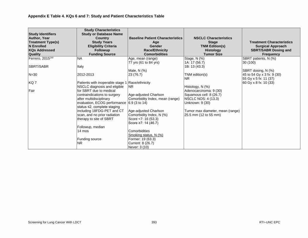
Appendix E Table 4. KQs 6 and 7: Study and Patient Characteristics Table
Screening for Lung Cancer With LDCT 393 RTI–UNC EPC
Study Identifiers Author, Year Treatment Type(s) N Enrolled KQs Addressed Quality
Study Characteristics Study or Database Name
Country Study Years
Eligibility Criteria Followup
Funding Source
Baseline Patient Characteristics Age
Gender Race/Ethnicity Comorbidities
NSCLC Characteristics Stage
TNM Edition(s) Histology
Tumor Size
Treatment Characteristics Surgical Approach
SBRT/SABR Dosing and Frequency
Ferrero, 2015260 SBRT/SABR N=30 KQ 7 Fair
NA Italy 2012-2013 Patients with inoperable stage 1 NSCLC diagnosis and eligible for SBRT due to medical contraindications to surgery after multidisciplinary evaluation, ECOG performance status ≤2, complete staging including 18FDG-PET and CT scan, and no prior radiation therapy to site of SBRT Followup, median 14 mos Funding source NR
Age, mean (range) 77 yrs (61 to 84 yrs) Male, N (%) 23 (76.7) Race/ethnicity NR Age-adjusted Charlson Comorbidity Index, mean (range) 6.9 (3 to 14) Age-adjusted Charlson Comorbidity Index, N (%) Score <7: 16 (53.3) Score ≥7: 14 (46.7) Comorbidities Smoking status, N (%) Former: 19 (63.3) Current: 8 (26.7) Never: 3 (10)
Stage, N (%) 1A: 17 (56.7) 1B: 13 (43.3) TNM edition(s) NR Histology, N (%) Adenocarcinoma: 9 (30) Squamous cell: 8 (26.7) NSCLC NOS: 4 (13.3) Unknown: 9 (30) Tumor max diameter, mean (range) 25.5 mm (12 to 55 mm)
SBRT patients, N (%) 30 (100) SBRT dosing, N (%) 45 to 54 Gy x 3 fx: 9 (30) 55 Gy x 5 fx: 11 (37) 60 Gy x 8 fx: 10 (33)

Appendix E Table 4. KQs 6 and 7: Study and Patient Characteristics Table
Screening for Lung Cancer With LDCT 394 RTI–UNC EPC
Study Identifiers Author, Year Treatment Type(s) N Enrolled KQs Addressed Quality
Study Characteristics Study or Database Name
Country Study Years
Eligibility Criteria Followup
Funding Source
Baseline Patient Characteristics Age
Gender Race/Ethnicity Comorbidities
NSCLC Characteristics Stage
TNM Edition(s) Histology
Tumor Size
Treatment Characteristics Surgical Approach
SBRT/SABR Dosing and Frequency
Goya, 2005193 Surgery N=3,965 eligible (of 6,644 analyzed) KQ 6 Fair
NA Japan 1994-2001 Patients with primary histological NSCLC (adenocarcinoma, SCC, large-cell carcinoma, and adenosquamous carcinoma) resected at time of thoracotomy in 1994 at certified teaching hospitals and with complete data Followup, median NR, but ≥5 yrs Funding source NR
Age, mean (range) NR Male, N (%) NR Race/ethnicity NR Comorbidities, N (%) NR
Stage, N (%) c-stage IA: 2423 (36.5) IB: 1542 (23.2) IIA: 150 (2.3) IIB: 746 (11.2) IIIA: 1270 (19.1) IIIB 366 (5.5) IV: 147 (2.2) TNM edition(s) 5th edition Histology, N (%) NR Tumor size, mean (SD) NR
Surgical approach, N (%) NR

Appendix E Table 4. KQs 6 and 7: Study and Patient Characteristics Table
Screening for Lung Cancer With LDCT 395 RTI–UNC EPC
Study Identifiers Author, Year Treatment Type(s) N Enrolled KQs Addressed Quality
Study Characteristics Study or Database Name
Country Study Years
Eligibility Criteria Followup
Funding Source
Baseline Patient Characteristics Age
Gender Race/Ethnicity Comorbidities
NSCLC Characteristics Stage
TNM Edition(s) Histology
Tumor Size
Treatment Characteristics Surgical Approach
SBRT/SABR Dosing and Frequency
Grills, 2012254 SBRT/SABR N=483 (505 tumors) KQs 6, 7 Fair
NA U.S. 1998-2010 Patients diagnosed with c-stage IA-IIB (T1-T3 N0 M0; peripheral T3 including chest-wall invasion only) or locally recurrent (after prior wedge resection) NSCLC Followup, median; mean 1.3 yrs; 1.6 yrs William Beaumont Hospital, Royal Oak, Michigan; University of Wuerzburg, Wuerzburg, Germany; Netherlands Cancer Institute, Amsterdam, The Netherlands; Princess Margaret Hospital, University of Toronto, Toronto, Ontario, Canada; and Thomas Jefferson University Hospital, Philadelphia, PA; Elekta
Age, median (range) 74 yrs (42 to 94 yrs) Male, N (%) 251 (52) Race/ethnicity NR Comorbidities NR
Clinical Stage, N (%) IA (T1N0): 318 (63) IB (T2N0): 167 (33) IIA (T3N0): 10 (2) Local recurrence: 5 (1) TNM edition(s) 6th edition Histology, N (%) Adenocarcinoma: 237 (47) Squamous cell carcinoma: 162 (32) Large cell/NOS/mixed: 111 (22) Tumor size, N (%) NR
SBRT patients, N (%) 483 (100) SBRT dosing William Beaumont Hospital (N=108) 12 Gy x 4 fx: 69 (63.9) 12 Gy x 5 fx: 34 (31.5) Netherlands Cancer Institute (N=187) 18 Gy x 3 fx: 182 (97.3) Thomas Jefferson University (N=21) 10 Gy x 5 fx: 10 (47.6) Princess Margaret Hospital (N=129) 7.5 Gy x 8 fx: 13 (10.1) 12 Gy x 4 fx: 59 (45.7) 18 Gy x 3 fx: 27 (20.9) 20 Gy x 3 fx: 20 (15.5) University of Wuerzburg (N=60) 12.5 Gy x 3 fx: 35 (58.3)

Appendix E Table 4. KQs 6 and 7: Study and Patient Characteristics Table
Screening for Lung Cancer With LDCT 396 RTI–UNC EPC
Study Identifiers Author, Year Treatment Type(s) N Enrolled KQs Addressed Quality
Study Characteristics Study or Database Name
Country Study Years
Eligibility Criteria Followup
Funding Source
Baseline Patient Characteristics Age
Gender Race/Ethnicity Comorbidities
NSCLC Characteristics Stage
TNM Edition(s) Histology
Tumor Size
Treatment Characteristics Surgical Approach
SBRT/SABR Dosing and Frequency
Guerrera, 2015169 Surgery N=848 KQ 6 Fair
NA Italy 2004-2012 Consecutive patients receiving surgical resection, but no preoperative treatment regimen, for Stage 1A and 1B NSCLC of any histology except a neuroendocrine subtype Followup, mean 48 mos Funding source NR
Age, median (IQR) 67 yrs (62 to 73 yrs) >75 yrs, N (%): 135 (14) Male, N (%) 610 (72) Race/ethnicity NR Comorbidities ≥1 comorbidity, N (%) 563 (77) “Smoking habit,” N (%) 727 (86)
Stage, N (%) pT status pT1a: 249 (29) pT1b: 190 (23) pT2a: 409 (48) TNM edition(s) NR Histology, N (%) Adenocarcinoma: 551 (65) Bronchioalveolar carcinoma: 25 (3) Squamous cell carcinoma: 247 (29) Large-cell carcinoma: 25 (3) Tumor size, median (IQR) 2.5 cm (1.8 to 3.5 cm)
Surgical approach, N (%) W: 85 (10) S: 81 (10) L: 651 (77) Sleeve resection: 3 (0.5) Bi-L: 12 (1) P: 16 (1.5)
Haidar, 2014255 SBRT/SABR N=55 KQ 7 Fair
NA U.S. 2002-2012 Patients treated with SBRT for early stage lung cancer Followup, median (range) NPC: 24.2 mos (1.9 to 64.6 mos) PC: 25.8 mos (4.3 to 53.4 mos) Funding source Government
Age, mean (range) NPC: 78 yrs (63 to 89 yrs) PC: 78.2 yrs (60 to 88 yrs) Male, N (%) Total: 35 (63.64) NPC: 17 (74) PC: 18 (56) Race/ethnicity NR Comorbidities Smokers Total: 53 (96.36) NPC: 22 (96) PC: 31 (97)
Stage, N (%) NPC: T1aN0M0, IA: 11 (46) T1bN0M0, IA: 7 (29) T2aN0M0, IB: 5 (21) T2bN0M0, IIA: 0 (0) T2bN0M0, IIB: 0 (0) T3aN0M0, IIB: 1 (4) TNM edition(s) 7th edition Histology, N (%) NR Tumor size, N (%) Mean (SD) NPC: 2.5 (1.1) PC: 2.7 (1.25)
SBRT patients, N (%) 55 (100) SBRT dosing, median (range) NPC: 50 Gy (48 to 56 Gy)

Appendix E Table 4. KQs 6 and 7: Study and Patient Characteristics Table
Screening for Lung Cancer With LDCT 397 RTI–UNC EPC
Study Identifiers Author, Year Treatment Type(s) N Enrolled KQs Addressed Quality
Study Characteristics Study or Database Name
Country Study Years
Eligibility Criteria Followup
Funding Source
Baseline Patient Characteristics Age
Gender Race/Ethnicity Comorbidities
NSCLC Characteristics Stage
TNM Edition(s) Histology
Tumor Size
Treatment Characteristics Surgical Approach
SBRT/SABR Dosing and Frequency
Husain, 2015237 Surgery N=112,216 KQ 7 Fair
NCDB U.S. 2003-2011 Adult patients with c-stage I NSCLC Followup, median (range) NR Funding source NR
Age, median 68 yrs Age, N (%) <65 yrs: 24,535 (34.5) 65-74.99 yrs: 27,557 (42.3) ≥75 yrs: 19,083 (23.3) Male, N (%) 33,168 (46.6) White, N (%) 63,095 (88.6) Charlson-Deyo Comorbidity Index, N (%) 0: 34,750 (48.8) 1: 26,186 (36.8) 2: 10,239 (14.4)
Clinical Stage, N (%) T1N0: 48,294 (67.9) T2N0: 22,881 (32.1) TNM edition(s) 6th or 7th editions Histology, N (%) Large cell: 2278 (3.2) Squamous cell: 20,243 (28.4) Adenocarcinoma: 43,699 (61.4) Other: 4955 (7.0) Tumor size, median 2.4 cm Tumor size, N (%) ≤2 cm: 29,851 (41.9) 2.1-3 cm: 19,919 (28.0) 3.1-5 cm: 15,749 (22.1) >5 cm: 5,006 (7.0) Unknown: 650 (0.9)
Surgical approach, N (%) L: 57,569 (80.9) SLR: 13,606 (19.1)

Appendix E Table 4. KQs 6 and 7: Study and Patient Characteristics Table
Screening for Lung Cancer With LDCT 398 RTI–UNC EPC
Study Identifiers Author, Year Treatment Type(s) N Enrolled KQs Addressed Quality
Study Characteristics Study or Database Name
Country Study Years
Eligibility Criteria Followup
Funding Source
Baseline Patient Characteristics Age
Gender Race/Ethnicity Comorbidities
NSCLC Characteristics Stage
TNM Edition(s) Histology
Tumor Size
Treatment Characteristics Surgical Approach
SBRT/SABR Dosing and Frequency
Jeon, 2018266 SBRT/SABR N=53 KQ 7 Fair
NA South Korea 2006-2015 Patients with Stage 1 NSCLC tumor size <5 cm, and peripheral tumor location ≥2 cm away from bronchial tree and treated with SBRT, regardless of medical operability Followup, median 37.1 mos Funding source Government
Age, median (range) 74 yrs (54 to 87 yrs) Age, N (%) ≤75 yrs: 32 (60.3) >75 yrs: 21 (39.7) Male, N (%) 40 (75.5) Race/ethnicity NR Comorbidities, N (%) Medically inoperable (i.e., significant comorbidities): 40 (75.5)
Stage, N (%) Clinical T1: 33 (62.3) Clinical T2: 20 (37.7) TNM edition(s) NR Histology, N (%) Adenocarcinoma: 22 (41.5) Squamous cell: 24 (45.3) Others: 5 (9.4) Unproven: 2 (3.8) Maximum tumor diameter range 1.5 to 4.9 cm Maximum tumor diameter, N (%) ≤2 cm: 3 (5.7) >2 to ≤3 cm: 30 (56.6) >3 cm: 20 (37.7)
SBRT patients, N (%) 53 (100) SBRT dosing, N (%) 5 Gy x 8 fx (40 Gy): 1 (1.9) 6 Gy x 7 fx (42 Gy): 2 (3.8): 6 Gy x 8 fx (48 Gy): 1 (1.9): 7 Gy x 7 fx (49 Gy): 4 (7.6) 7 Gy x 8 fx (56 Gy): 1 (1.9) 9 Gy x 5 fx (45 Gy): 2 (3.8) 10 Gy x 5 fx (50 Gy): 10 (18.8) 11.6 Gy x 5 fx (58 Gy): 1 (1.9) 12 Gy x 4 fx (48 Gy): 10 (18.8) 15 Gy x 3 fx (45 Gy): 2 (3.8) 16 Gy x 3 fx (48 Gy): 3 (5.7) 18 Gy x 3 fx (54 Gy): 16 (30.1)
Jeppesen, 2018201 SBRT/SABR N=136 KQs 6, 7 Fair
NA Denmark 2007-2013 Patients with histologically or cytologically proven, localized T1-2N0M0 NSCLC with a maximum tumor diameter of 5 cm Followup, median 70.1 mos Funding source Private
Age, mean (range) 72.5 yrs (49 to 90 yrs) Male, N (%) 61 (45) Race/ethnicity NR Comorbidities, N (%) Charlson Comorbidity Index, N (%) Score 0-1: 71 (53) Score 2-3: 38 (28) Score 4+: 26 (19) Smoking history, mean/median (NR which was used) (range) 41 (0-130)
Stage, N (%) NR TNM edition(s) 6th edition Histology, N (%) Adenocarcinoma: 77 (57) Squamous cell: 38 (28) Other: 21 (15) Tumor diameter, mean (range) 3.1 cm (1.2 to 5.0 cm)
SBRT patients, N (%) 136 (100) SBRT dosing, N (%) Total central dose to gross tumor volume (GTV) 45 Gy x 3 fx (BED 112 Gy): NR (prior to October 2008) 66 Gy x 3 fx (BED 211 Gy): NR (after October 2008) Total central dose to PTV 30 Gy x 3 fx (BED was NR): NR (prior to October 2008) 45 Gy x 3 fx (BED 112 Gy): NR (after October 2008)

Appendix E Table 4. KQs 6 and 7: Study and Patient Characteristics Table
Screening for Lung Cancer With LDCT 399 RTI–UNC EPC
Study Identifiers Author, Year Treatment Type(s) N Enrolled KQs Addressed Quality
Study Characteristics Study or Database Name
Country Study Years
Eligibility Criteria Followup
Funding Source
Baseline Patient Characteristics Age
Gender Race/Ethnicity Comorbidities
NSCLC Characteristics Stage
TNM Edition(s) Histology
Tumor Size
Treatment Characteristics Surgical Approach
SBRT/SABR Dosing and Frequency
Katoh, 2017253 SBRT/SABR N=283 (286 tumors) KQs 6, 7 Fair
NA Japan 2000-2012 Patients diagnosed with histologically proven NSCLC and peripherally located c-stage I and IIA and treated with SBRT, but not thoracic radiation therapy for simultaneous malignant tumors within three months before or after starting SBRT Followup, median (range) 28 mos (0 to 127 mos) Japan Society for the Promotion of Science; Hokkaido University
Age, median (range) 78 yrs (52 to 90 yrs) Male, N (%) 215 (75.6) Race/ethnicity NR Comorbidities NR
Stage, N (%) 1a and 1b: 195 (68.2) 2a and 2b: 91 (31.8) TNM edition(s) 7th edition Histology, N (%) Adenocarcinoma: 185 (65.4) Squamous cell carcinoma: 80 (28.3) Maximum tumor diameter, median (range) 1.9 cm (0.7 to 4.0 cm)
SBRT patients, N (%) 283 (100) SBRT dosing, N (%) 40 Gy x 4 fx: 94 (32.9) 48 Gy x 4 fx: 149 (52.1) 50 Gy x 5 fx: 19 (6.6)

Appendix E Table 4. KQs 6 and 7: Study and Patient Characteristics Table
Screening for Lung Cancer With LDCT 400 RTI–UNC EPC
Study Identifiers Author, Year Treatment Type(s) N Enrolled KQs Addressed Quality
Study Characteristics Study or Database Name
Country Study Years
Eligibility Criteria Followup
Funding Source
Baseline Patient Characteristics Age
Gender Race/Ethnicity Comorbidities
NSCLC Characteristics Stage
TNM Edition(s) Histology
Tumor Size
Treatment Characteristics Surgical Approach
SBRT/SABR Dosing and Frequency
Khullar, 2015176 Surgery N= 54,350 (of 92,929 total) KQ 6 Fair
NCDB U.S. 2003-2006 Patients who underwent pulmonary resection for a first or single invasive cancer, who were treated (not with palliative care) at the reporting facility, and for whom data about laterality and survival were available Followup, median NR Funding source Government, university
Age, mean (SD) 66.0 yrs (10.3 yrs) Male, N (%) NR Race/ethnicity NR Comorbidities, N (%) NR
Stage, N (%) Analytic stage 1: 54,350 (62.9) Analytic stage 2: 14,137 (16.4) Analytic stage 3: 13,830 (16.0) Analytic stage 4: 4,023 (4.7) TNM edition(s) NR Histology, N (%) NR Tumor size, mean (SD) NR
Surgical approach, N (%) W: 54,350 (100)

Appendix E Table 4. KQs 6 and 7: Study and Patient Characteristics Table
Screening for Lung Cancer With LDCT 401 RTI–UNC EPC
Study Identifiers Author, Year Treatment Type(s) N Enrolled KQs Addressed Quality
Study Characteristics Study or Database Name
Country Study Years
Eligibility Criteria Followup
Funding Source
Baseline Patient Characteristics Age
Gender Race/Ethnicity Comorbidities
NSCLC Characteristics Stage
TNM Edition(s) Histology
Tumor Size
Treatment Characteristics Surgical Approach
SBRT/SABR Dosing and Frequency
Khullar, 2015187 Surgery N=28,241 KQs 6, 7 Fair
NCDB U.S. 2003-2011 Patients undergoing surgical resection, but not neoadjuvant radiation or palliative care, at the same facility providing diagnosis for preoperative clinical T1a N0 NSCLC with known laterality as their first or only lifetime cancer from 2003 to 2011 (OS measures limited to patients treated from 2003-2006) Followup, median NR Funding source Government, university
Age, mean (SD) L: 65.8 yrs (9.9 yrs) W: 68.9 yrs (9.8 yrs) S: 68.7 yrs (9.4 yrs) Male, N (%) Total: 11,382 (40.3) L: 8001 (40.6) W: 2937 (40.3) S: 444 (36.2) White, N (%) Total: 25,136 (89.0) L: 17,464 (89.4) W: 6570 (90.9) S: 1102 (91.1) Charlson Comorbidity Index, N (%) Score of 0 Total: 13,366 (47.3) L: 9849 (50.0) W: 3017 (41.4) S: 500 (40.8) Score of 1 Total: 10,783 (38.2) L: 7293 (37.0) W: 2976 (40.8) S: 514 (41.9) Score of 2+ Total: 4,092 (14.5) L: 2576 (13.1) W: 1304 (17.9) S: 212 (17.3)
Stage, N (%) c-stage T1A N0 NSCLC: 28,241 (100) TNM edition(s) NR Histology, N (%) Adenocarcinoma Total: 18,193 (64.4) L: 13,097 (66.4) W: 4336 (59.4) S: 760 (62.0) Squamous cell carcinomas Total: 6,041 (21.4) L: 3957 (20.1) W: 1795 (24.6) S: 289 (23.6) Unknown histology Total: 913 (3.2) L: 596 (3.0) W: 278 (3.8) S: 39 (3.2) Tumor size, mean (SD) L: 1.52 cm (0.39 cm) W: 1.4 cm (0.42 cm) S: 1.46 cm (0.40 cm)
Surgical approach, N (%) L: 19,718 (69.8) W: 7,297 (25.8) S: 1,226 (4.3)

Appendix E Table 4. KQs 6 and 7: Study and Patient Characteristics Table
Screening for Lung Cancer With LDCT 402 RTI–UNC EPC
Study Identifiers Author, Year Treatment Type(s) N Enrolled KQs Addressed Quality
Study Characteristics Study or Database Name
Country Study Years
Eligibility Criteria Followup
Funding Source
Baseline Patient Characteristics Age
Gender Race/Ethnicity Comorbidities
NSCLC Characteristics Stage
TNM Edition(s) Histology
Tumor Size
Treatment Characteristics Surgical Approach
SBRT/SABR Dosing and Frequency
Koshy, 2015198 SBRT/SABR N=498 KQ 6 Fair
NCDB U.S. 2003-2006 Patients with histologically confirmed first primary Stage 1 NSCLC who received all or part of their first course of treatment at Commission on Cancer-accredited facilities and SBRT at a calculated BED of ≥70 Gy in 1 to 10 fx Followup, median 68 mos Funding source NR
Age, N (%) 18-59 years: 40 (8.0) 60-69 years: 91 (18.3) 70-79 years: 231 (46.4) 80+ years: 136 (27.3) Male, N (%) 219 (44.0) White, N (%) 444 (89.2) Charlson Comorbidity Index, N (%) Score=0: 327 (65.7) Score=1: 125 (25.1) Score=2+: 46 (9.2)
Stage, N (%) T1: 334 (67.1) T2: 164 (32.9) TNM edition(s) 6th edition Histology, N (%) Adenocarcinoma: 180 (36.1) NSCLC NOS: 155 (31.1) Squamous cell: 146 (29.3) Large cell: 17 (3.4) Tumor size, median (IQR) T1 tumors: 2 cm (1.6 to 2.5 cm) T2 tumors: 3.7 cm (3.2 to 4.5 cm)
SBRT patients, N (%) 498 (100) SBRT dosing 20 Gy x 3 fx: 169 (34) 12 Gy x 4 fx: 80 (16) 18 Gy x 3 fx: 50 (10) 15 Gy x 3 fx: 50 (10) 16 Gy x 3 fx: 20 (4) Other dose & fx schedules: 129 (26)

Appendix E Table 4. KQs 6 and 7: Study and Patient Characteristics Table
Screening for Lung Cancer With LDCT 403 RTI–UNC EPC
Study Identifiers Author, Year Treatment Type(s) N Enrolled KQs Addressed Quality
Study Characteristics Study or Database Name
Country Study Years
Eligibility Criteria Followup
Funding Source
Baseline Patient Characteristics Age
Gender Race/Ethnicity Comorbidities
NSCLC Characteristics Stage
TNM Edition(s) Histology
Tumor Size
Treatment Characteristics Surgical Approach
SBRT/SABR Dosing and Frequency
Lakha, 2014173 Surgery N=16,315 KQ 6 Fair
SEER U.S. 2004-2010 Patients with incident, histologically confirmed NSCLC diagnosed with tumors <7 cm, without lymph node involvement (N0) or distant metastasis (M0), who were experiencing their first and only malignancy, and who underwent lobectomy but not preoperative radiotherapy Followup, median NR Funding source NR
Age, N (%) <50 yrs: 942 (5.8) 50-60 yrs: 2,985 (18.3) >60 yrs: 12,388 (75.9) Male, N (%) 7,707 (47.2) White, N (%) 13,077 (80.2) Comorbidities, N (%) NR
Stage, N (%) NR TNM edition(s) 7th edition Histology, N (%) Adenocarcinoma: 9,784 (60.0) Squamous cell carcinoma: 4,273 (26.2) Large-cell carcinoma: 598 (3.7) Other: 257 (1.6) Tumor size, N (%) <2 cm: 6,048 (37.1) 2-3 cm: 4,674 (28.6) >3-5 cm: 4,345 (26.6) >5-7 cm: 1,248 (7.6)
Surgical approach, N (%) L: 16,315 (100)

Appendix E Table 4. KQs 6 and 7: Study and Patient Characteristics Table
Screening for Lung Cancer With LDCT 404 RTI–UNC EPC
Study Identifiers Author, Year Treatment Type(s) N Enrolled KQs Addressed Quality
Study Characteristics Study or Database Name
Country Study Years
Eligibility Criteria Followup
Funding Source
Baseline Patient Characteristics Age
Gender Race/Ethnicity Comorbidities
NSCLC Characteristics Stage
TNM Edition(s) Histology
Tumor Size
Treatment Characteristics Surgical Approach
SBRT/SABR Dosing and Frequency
Lam, 2018200 SBRT/SABR N=4,454 KQs 6, 7 Fair
NCDB U.S. 2004-2014 Patients with T1a-1bN0M0 NSCLC tumors ≤3 cm and histology classifications of adenocarcinoma, squamous cell carcinoma, large cell carcinoma, or NSCLC NOS (but no nodal or extrapulmonary disease) who were treated at high-volume centers without chemotherapy, immunotherapy, or hormone therapy, had available survival data, and were not lost to followup Followup, median 50.2 mos Funding source NR
Age, median (range) NR Male, N (%) NR Race/ethnicity NR Comorbidities, N (%) NR
Stage, N (%) NR TNM edition(s) 7th edition Histology, N (%) NR Tumor size, mean (SD) NR
SBRT patients, N (%) 4,454 (100) SBRT dosing, N (%) NR

Appendix E Table 4. KQs 6 and 7: Study and Patient Characteristics Table
Screening for Lung Cancer With LDCT 405 RTI–UNC EPC
Study Identifiers Author, Year Treatment Type(s) N Enrolled KQs Addressed Quality
Study Characteristics Study or Database Name
Country Study Years
Eligibility Criteria Followup
Funding Source
Baseline Patient Characteristics Age
Gender Race/Ethnicity Comorbidities
NSCLC Characteristics Stage
TNM Edition(s) Histology
Tumor Size
Treatment Characteristics Surgical Approach
SBRT/SABR Dosing and Frequency
Landreneau, 2014174 Surgery N=614 KQs 6, 7 Fair
NA U.S. NR Patients with c-stage 1 NSCLC >5 cm with data available from the Lung Cancer Database of the University of Pittsburgh Followup, median 5.4 yrs Funding source Government, private (Thoracic Surgery Foundation for Research and Education)
Age, mean (SD) Total: 68.5 yrs (NR) S: 68.5 yrs (9.2 yrs) L: 68.4 yrs (9.2 yrs) Male, N (%) Total: 283 (45.4) S: 139 (44.6) L: 144 (46.2) Race/ethnicity NR Comorbidities, N (%) COPD, N (%) Total: 208 (33.3) S: 103 (33.0) L: 105 (33.7) Smoking status, N (%) Ever Total: 480 (92.9) S: 290 (92.9) L: 290 (92.9) Never Total: 44 (7.1) S: 22 (7.1) L: 22 (7.1)
Stage, N (%) c-stage 1A Total: 487 (78.0) S: 248 (79.5) L: 239 (76.6) c-stage 1B Total: 137 (22.0) S: 64 (20.5) L: 73 (23.4) TNM edition(s) 7th edition Histology, N (%) Adenocarcinoma Total: 360 (57.7) S: 177 (56.7) L: 183 (58.7) Squamous cell Total: 186 (29.8) S: 89 (28.5) L: 97 (31.1) Tumor size, mean (SD) Total: 2.2 cm (NR) S: 2.2 cm (1.0 cm) L: 2.2 cm (1.1 cm)
Surgical approach, N (%) S: 312 (50) L: 312 (50)

Appendix E Table 4. KQs 6 and 7: Study and Patient Characteristics Table
Screening for Lung Cancer With LDCT 406 RTI–UNC EPC
Study Identifiers Author, Year Treatment Type(s) N Enrolled KQs Addressed Quality
Study Characteristics Study or Database Name
Country Study Years
Eligibility Criteria Followup
Funding Source
Baseline Patient Characteristics Age
Gender Race/Ethnicity Comorbidities
NSCLC Characteristics Stage
TNM Edition(s) Histology
Tumor Size
Treatment Characteristics Surgical Approach
SBRT/SABR Dosing and Frequency
Licht, 2013247 Surgery N=1,513 KQ 7 Fair
Danish Lung Cancer Registry The Netherlands 2007-2011 Patients undergoing standard anatomic lobectomy for c-stage 1 NSCLC Followup, median 28 mos Funding source NR
Age, N (%) <50 yrs: 51 (3.4) 50-59 yrs: 257 (17.0) 60-69 yrs: 613 (40.5) 70-79 yrs: 515 (34.0) >80 yrs: 77 (5.1) Male, N (%) Total: 742 (49) VATS L: 319 (44.5) Open L: 423 (53.1) Race/ethnicity NR Charlson Comorbidity Index Median score; mean score (95% CI) VATS L: 1; 1.1 (1.0 to 1.2) Open L: 0; 1.0 (0.9 to 1.1) Specific scores, n (%) Score=0: 762 (50.4) Score=1: 343 (22.7) Score=2: 207 (13.7) Score=3: 106 (7.0) Score=4: 37 (2.4) Score=5: 28 (1.9) Score ≥6: 30 (2.0)
Stage, N (%) c-stage T1: 787 (52.0) c-stage T2: 726 (48.0) TNM edition(s) NR Histology, N (%) Adenocarcinoma Total: 745 (49.2) VATS L: 390 (54.4) Open L: 355 (44.6) Squamous cell Total: 398 (26.3) VATS L: 145 (20.2) Open L: 253 (31.8) Adenosquamous cell Total: 14 (0.9) VATS L: 8 (1.1) Open L: 6 (0.8) Mixed tumor Total: 246 (16.3) VATS L: 124 (17.3) Open L: 122 (15.3)
Surgical approach, N (%) VATS L: 71 (47) Open L: 796 (53)

Appendix E Table 4. KQs 6 and 7: Study and Patient Characteristics Table
Screening for Lung Cancer With LDCT 407 RTI–UNC EPC
Study Identifiers Author, Year Treatment Type(s) N Enrolled KQs Addressed Quality
Study Characteristics Study or Database Name
Country Study Years
Eligibility Criteria Followup
Funding Source
Baseline Patient Characteristics Age
Gender Race/Ethnicity Comorbidities
NSCLC Characteristics Stage
TNM Edition(s) Histology
Tumor Size
Treatment Characteristics Surgical Approach
SBRT/SABR Dosing and Frequency
Licht, 2013247 (continued)
Bronchioloalveolar carcinoma Total: 8 (0.5) VATS L: 5 (0.7) Open L: 3 (0.4) Large-cell carcinoma Total: 60 (4.0) VATS L: 23 (3.2) Open L: 37 (4.6) Sarcomatoid carcinoma Total: 9 (0.6) VATS L: 4 (0.6) Open L: 5 (0.6) Salivary gland-like carcinoma Total: 1 (0.1) VATS L: 0 (0) Open L: 1 (0.1) Non-specified NSCLC Total: 32 (2.1) VATS L: 18 (2.5) Open L: 14 (1.8) Tumor size, mean (SD) NR

Appendix E Table 4. KQs 6 and 7: Study and Patient Characteristics Table
Screening for Lung Cancer With LDCT 408 RTI–UNC EPC
Study Identifiers Author, Year Treatment Type(s) N Enrolled KQs Addressed Quality
Study Characteristics Study or Database Name
Country Study Years
Eligibility Criteria Followup
Funding Source
Baseline Patient Characteristics Age
Gender Race/Ethnicity Comorbidities
NSCLC Characteristics Stage
TNM Edition(s) Histology
Tumor Size
Treatment Characteristics Surgical Approach
SBRT/SABR Dosing and Frequency
Lindberg, 2015205 SBRT/SABR N=57 KQs 6, 7 Fair
NA Sweden, Norway, Denmark 2003-2005 Patients with Stage 1 T1-2N0M0 peripherally located NSCLC with no central tumor growth adjacent to trachea, main bronchus, or esophagus who were medically inoperable or refused surgery and received SBRT at the prescribed dose of 15 Gy x 3 fx; life expectancy of ≥12 mos; Karnofsky performance score ≥70; no prior malignancy within last 5 yrs Followup, median 41.5 mos Funding source Government, private
Age, mean (range) 75.2 yrs (58.9 to 86.8 yrs) Male, N (%) 26 (46) Race/ethnicity NR Comorbidities, N (%) COPD: 37 (65)
Stage, N (%) Stage T1a: 10 (18) Stage T1b: 31 (54) Stage T2a: 16 (28) TNM edition(s) 7th edition Histology, N (%) Adenocarcinoma: 19 (33) Squamous cell: 8 (14) Large cell carcinoma: 1 (2) NSCLC NOS: 10 (18) Not analyzed: 19 (33) Tumor volume, median (range) 16 mL (1 to 51 mL) Tumor diameter, median (range) 25 mm (6 to 50 mm)
SBRT patients, N (%) 57 (100) SBRT dosing, N (%) 15 Gy x 3 fx: 57 (100)

Appendix E Table 4. KQs 6 and 7: Study and Patient Characteristics Table
Screening for Lung Cancer With LDCT 409 RTI–UNC EPC
Study Identifiers Author, Year Treatment Type(s) N Enrolled KQs Addressed Quality
Study Characteristics Study or Database Name
Country Study Years
Eligibility Criteria Followup
Funding Source
Baseline Patient Characteristics Age
Gender Race/Ethnicity Comorbidities
NSCLC Characteristics Stage
TNM Edition(s) Histology
Tumor Size
Treatment Characteristics Surgical Approach
SBRT/SABR Dosing and Frequency
Louie, 2016236 Surgery 13,598 KQ 7 Fair
STS-GTS Database (Society of Thoracic Surgeons Database) U.S. 2009-2013 Patients with c-stage 1-2 NSCLC undergoing primary lobectomy performed using VATS or robotic approach, but not converted to open procedures or combined with chemotherapy or radiation therapy, at high-volume centers Followup, median (range) NR Funding source NR
Age, median (IQR) Total: 68.0 yrs (61.0-75.0 yrs) Robotic L: 69.0 yrs (61.0-75.0 yrs) VATS L: 68.0 yrs (61.0-75.0 yrs) Male, N (%) Total: 5,857 (43.1) Robotic L: 527 (43.2) VATS L: 5,330 (43.1) Race/ethnicity NR Comorbidities COPD Total: 4,497 (33.1) Robotic L: 425 (34.8) VATS L: 4,072 (32.9) Ever smoker Total: 11,339 (83.4) Robotic L: 985 (80.7) VATS L: 10,354 (83.6) ASA risk class 1, N (%) Total: 57 (0.4) Robotic L: 7 (0.6) VATS L: 50 (0.4) ASA risk class 2, N (%) Total: 2,528 (18.6) Robotic L: 197 (16.1) VATS L: 2,331 (18.8)
Stage, N (%) cT1aN0 Total: 6,095 (44.8) Robotic L: 515 (42.2) VATS L: 5,580 (45.1) cT1bN0 Total: 3,481 (25.6) Robotic L: 328 (26.9) VATS L: 3,153 (25.5) cT2aN0 Total: 2,655 (19.5) Robotic L: 269 (22.0) VATS L: 2,386 (19.3) cT2bN0 Total: 636 (4.7) Robotic L: 51 (4.2) VATS L: 585 (4.7) cT1aN1 Total: 196 (1.4) Robotic L: 16 (1.3) VATS L: 180 (1.5) cT1bN1 Total: 196 (1.4) Robotic L: 15 (1.2) VATS L: 181 (1.5) cT2aN1 Total: 240 (1.8) Robotic L: 18 (1.5) VATS L: 222 (1.8) cT2bN1 Total: 99 (0.7) Robotic L: 8 (0.7) VATS L: 91 (0.7)
Surgical approach, N (%) Robotic L: 1,220 (9.0) VATS L: 12,378 (91.0)

Appendix E Table 4. KQs 6 and 7: Study and Patient Characteristics Table
Screening for Lung Cancer With LDCT 410 RTI–UNC EPC
Study Identifiers Author, Year Treatment Type(s) N Enrolled KQs Addressed Quality
Study Characteristics Study or Database Name
Country Study Years
Eligibility Criteria Followup
Funding Source
Baseline Patient Characteristics Age
Gender Race/Ethnicity Comorbidities
NSCLC Characteristics Stage
TNM Edition(s) Histology
Tumor Size
Treatment Characteristics Surgical Approach
SBRT/SABR Dosing and Frequency
Louie, 2016236 (continued)
ASA risk class 3, N (%) Total: 10,037 (73.8) Robotic L: 932 (76.4) VATS L: 9,105 (73.6) ASA risk class 4, N (%) Total: 969 (7.1) Robotic L: 84 (6.9) VATS L: 885 (7.1) ASA risk class 5, N (%) Total: 3 (0.0) Robotic L: 0 (0) VATS L: 3 (0.0)
TNM edition(s) NR Histology, N (%) NR Tumor size, N (%) NR

Appendix E Table 4. KQs 6 and 7: Study and Patient Characteristics Table
Screening for Lung Cancer With LDCT 411 RTI–UNC EPC
Study Identifiers Author, Year Treatment Type(s) N Enrolled KQs Addressed Quality
Study Characteristics Study or Database Name
Country Study Years
Eligibility Criteria Followup
Funding Source
Baseline Patient Characteristics Age
Gender Race/Ethnicity Comorbidities
NSCLC Characteristics Stage
TNM Edition(s) Histology
Tumor Size
Treatment Characteristics Surgical Approach
SBRT/SABR Dosing and Frequency
Ma, 2017267 SBRT/SABR N=155 patients (159 tumors) KQ 7 Fair
RPCI database U.S. 2007-2015 Patients receiving definitive single-fx or triple-fx SBRT for peripheral NSCLC, but not participating in the RTOG 0915 clinical trial Followup, median 22.2 mos Funding source Private
Age, median (IQR) Total: 76.3 yrs (70.5 to 82.3 yrs) Single-fx SBRT: 76.4 yrs (70.6 to 82.5 yrs) Triple-fx SBRT: 76.2 yrs (70.1 to 82.0 yrs) Male, N (%) Total: 77 (48.4) Single-fx SBRT: 30 (46) Triple-fx SBRT: 47 (50) Race/ethnicity NR Comorbidities Medically operable, n (%) Total: 42 (26.4) Single-fx SBRT: 21 (32) Triple-fx SBRT: 21 (22) Medical inoperable, n (%) Total: 117 (73.6) Single-fx SBRT: 44 (68) Triple-fx SBRT: 73 (78) Smoking history Median pack-years (IQR): 50 (40-75) <50 pack-years, n (%) Total: 57 (35.8) Single-fx SBRT: 27 (42) Triple-fx SBRT: 30 (32)
Stage, N (%) Stage 1A Total: 120 (75.5) Single-fx SBRT: 54 (83) Triple-fx SBRT: 66 (70) Stage 1B Total: 35 (22.0) Single-fx SBRT: 10 (15) Triple-fx SBRT: 25 (27) Stage T2a Total: 24 (15.1) Single-fx SBRT: 1 (2) Triple-fx SBRT: 1 (1) Stage T2b Total: 2 (1.3) Single-fx SBRT: 0 (0) Triple-fx SBRT: 2 (2) TNM edition(s) 7th edition Histology, N (%) Adenocarcinoma Total: 69 (43.4) Single-fx SBRT: 34 (52) Triple-fx SBRT: 35 (37) Squamous cell Total: 64 (40.3) Single-fx SBRT: 25 (38) Triple-fx SBRT: 39 (41) Other Total: 6 (3.8) Single-fx SBRT: 1 (2) Triple-fx SBRT: 5 (5)
SBRT patients, N (%) 155 (100) SBRT dosing, N (%) 30 Gy x 1 fx (median dose of 30 Gy): 65 (40.9) 48-60 Gy x 3 fx (median dose of 60 Gy): 94 (59.1)

Appendix E Table 4. KQs 6 and 7: Study and Patient Characteristics Table
Screening for Lung Cancer With LDCT 412 RTI–UNC EPC
Study Identifiers Author, Year Treatment Type(s) N Enrolled KQs Addressed Quality
Study Characteristics Study or Database Name
Country Study Years
Eligibility Criteria Followup
Funding Source
Baseline Patient Characteristics Age
Gender Race/Ethnicity Comorbidities
NSCLC Characteristics Stage
TNM Edition(s) Histology
Tumor Size
Treatment Characteristics Surgical Approach
SBRT/SABR Dosing and Frequency
Ma, 2017267 (continued)
≥50 pack-years, n (%) Total: 96 (60.4) Single-fx SBRT: 34 (52) Triple-fx SBRT: 62 (66) NA Total: 6 (3.8) Single-fx SBRT: 4 (6) Triple-fx SBRT: 2 (2)
NA Total: 20 (12.6) Single-fx SBRT: 5 (8) Triple-fx SBRT: 15 (16) Tumor size, median (IQR) Total: 2.1 cm (1.5 to 3 cm) Single-fx SBRT: 2 cm (1.5 to 3 cm) Triple-fx SBRT: 2.2 cm (1.5 to 3 cm)
Maeda, 2010189 Surgery N=734 KQ 6 Fair
NA Japan 1994-2003 Patients with p-stage I NSCLC with tumors up to 3 cm in maximum dimension and who underwent complete resection, but no pre- or post-operative chemotherapy or radiotherapy Followup, median NR Funding source Government, university
Age, median (range) Age <65 yrs: 371 (52.0) Age ≥65 yrs: 342 (48.0) Male, N (%) 385 (54) Race/ethnicity NR Comorbidities, N (%) Smoking status Nonsmoker: 318 (44.6) Current or former smoker: 395 (55.4)
Stage, N (%) Overall with Stage 1: 605 (84.9) Stage 1A: 357 (50.1) Stage 1B: 248 (34.8) TNM edition(s) 7th edition Histology, N (%) Adenocarcinoma: 569 (79.8) Squamous cell carcinoma: 104 (14.6) Large cell carcinoma: 27 (3.8) Adenosquamous carcinoma: 9 (1.3) Pleomorphic carcinoma: 4 (0.56) Tumor size, N (%) ≤20 mm: 393 (55.1) >20 mm: 320 (44.9)
Surgical approach, N (%) L: 616 (86.4) S or W: 97 (13.6)

Appendix E Table 4. KQs 6 and 7: Study and Patient Characteristics Table
Screening for Lung Cancer With LDCT 413 RTI–UNC EPC
Study Identifiers Author, Year Treatment Type(s) N Enrolled KQs Addressed Quality
Study Characteristics Study or Database Name
Country Study Years
Eligibility Criteria Followup
Funding Source
Baseline Patient Characteristics Age
Gender Race/Ethnicity Comorbidities
NSCLC Characteristics Stage
TNM Edition(s) Histology
Tumor Size
Treatment Characteristics Surgical Approach
SBRT/SABR Dosing and Frequency
Maeda, 2012182 Surgery N=1,074 KQ 6 Fair
NA Japan 1992-2007 Consecutive patients receiving complete surgical resection by lobectomy or systematic lymph node dissection for c-stage 1A NSCLC adenocarcinoma Followup, median 57 mos Funding source Government, other not disclosed
Age, N (%) <65 yrs: 562 (53) >65 yrs: 508 (47) Male, N (%) 499 (47) Race/ethnicity NR Comorbidities, N (%) Smoking status Never: 528 (49.3) Ever: 543 (50.7) Smoking history No pack-years: 528 (49.3) 0 ≤ pack-years ≤ 10: 75 (7.0) 10 < pack-years ≤ 20: 74 (6.9) 0 ≤ pack-years ≤ 20: 677 (63) 20 < pack-years ≤ 40: 182 (17.0) 40 < pack-years ≤ 60: 126 (11.8) Pack-years >60: 85 (7.9) Pack-years >20: 393 (37)
Stage, N (%) c-stage 1A: 1,070 (100) TNM edition(s) 7th edition Histology, N (%) Adenocarcinoma: 1,074 (100) Well-differentiated histology: 456 (43) Moderately/poorly differentiated histology: 614 (57) Tumor size, N (%) <2.0 cm (T1a): 703 (66) 2.1 to 3.0 cm (T1b): 367 (34)
Surgical approach, N (%) L: 1,074 (!00)

Appendix E Table 4. KQs 6 and 7: Study and Patient Characteristics Table
Screening for Lung Cancer With LDCT 414 RTI–UNC EPC
Study Identifiers Author, Year Treatment Type(s) N Enrolled KQs Addressed Quality
Study Characteristics Study or Database Name
Country Study Years
Eligibility Criteria Followup
Funding Source
Baseline Patient Characteristics Age
Gender Race/Ethnicity Comorbidities
NSCLC Characteristics Stage
TNM Edition(s) Histology
Tumor Size
Treatment Characteristics Surgical Approach
SBRT/SABR Dosing and Frequency
Mathieu, 2015258 SBRT/SABR N=45 KQ 7 Fair
NA Canada 2010-2013 Patients with biopsy-confirmed stage 1 NSCLC (T1-T2aN0M0), Karnofsky performance status ≥60, and medical inoperability because of poor pulmonary function, medical comorbidities, or surgery refusal, but no prior thoracic radiation or cancer in last 5 yrs Followup, median 41 mos Funding source NR
Age, median (range) 77 yrs (60 to 94 yrs) Male, N (%) 17 (38) Race/ethnicity NR Comorbidities, N (%) Charlson Comorbidity Index Score=0-2: 4 (9) Score=3-4: 24 (53) Score >4: 17 (38) Smoking pack-years, median (range) 45 (0-100)
Stage, N (%) T1a: 19 (42) T1b: 17 (38) T2a: 9 (20) TNM edition(s) NR Histology, N (%) Adenocarcinoma: 25 (56) Other: 4 (9) Squamous cell: 14 (31) Large cell: 2 (4) Gross tumor volume, median (range) 7.9 mL (0.5 to 35.9 mL)
SBRT patients, N (%) 45 (100) SBRT dosing 60 Gy x 3 fx: 32 (71) 50 Gy x 4 fx: 7 (16) 50 Gy x 5 fx: 6 (13)

Appendix E Table 4. KQs 6 and 7: Study and Patient Characteristics Table
Screening for Lung Cancer With LDCT 415 RTI–UNC EPC
Study Identifiers Author, Year Treatment Type(s) N Enrolled KQs Addressed Quality
Study Characteristics Study or Database Name
Country Study Years
Eligibility Criteria Followup
Funding Source
Baseline Patient Characteristics Age
Gender Race/Ethnicity Comorbidities
NSCLC Characteristics Stage
TNM Edition(s) Histology
Tumor Size
Treatment Characteristics Surgical Approach
SBRT/SABR Dosing and Frequency
Matsuo, 2014196 SBRT/SABR N=115 KQs 6, 7 Fair
Databases maintained by Departments of Radiation Oncology & Thoracic Surgery of Kyoto University Hospital Japan 2003-2009 Consecutive patients with histological confirmation of c-Stage I NSCLC ≤50 mm who underwent SBRT because of medical comorbidities Followup, median 6.7 yrs Japan Society for the Promotion of Science
Age, median (range) 77 yrs (56 to 88 yrs) Male, N (%) 83 (72.17) Charlson Comorbidity Index score, median (range) 2 (0 to 8) Race/ethnicity NR
Stage, N (%) c-stage I: 115 (100) TNM edition(s) NR Histology, N (%) Adenocarcinoma: 58 (50.43) Squamous cell carcinoma: 41 (35.65) Large cell carcinoma: 4 (3.48) Others: 12 (10.43) Tumor diameter, median (range) 25 mm (10 mm to 45 mm)
SBRT patients, N (%) 115 (63.89) SBRT dosing, N (%) 48 Gy x 4 fx: 108 (93.91) 60 Gy x 8 fx: 5 (4.35) 56 Gy x 4 fx: 1 (0.09) 60 Gy x 4 fx: 1 (0.09)

Appendix E Table 4. KQs 6 and 7: Study and Patient Characteristics Table
Screening for Lung Cancer With LDCT 416 RTI–UNC EPC
Study Identifiers Author, Year Treatment Type(s) N Enrolled KQs Addressed Quality
Study Characteristics Study or Database Name
Country Study Years
Eligibility Criteria Followup
Funding Source
Baseline Patient Characteristics Age
Gender Race/Ethnicity Comorbidities
NSCLC Characteristics Stage
TNM Edition(s) Histology
Tumor Size
Treatment Characteristics Surgical Approach
SBRT/SABR Dosing and Frequency
Mediratta, 2014175 Surgery N=540 KQs 6, 7 Good
NA U.K. 2001-2012 Patients with Stage 1 adenocarcinoma or squamous carcinoma who had undergone a potentially curative wedge resection Followup, median 1,012 days Funding source NR
Age, median (IQR) 72 yrs (64 to 77 yrs) Male, N (%) 274 (51) Race/ethnicity NR Comorbidities COPD, N (%) 114 (21) Emphysema, N (%) 21 (4) Smoking status, N (%) Current: 128 (24) Former: 305 (57) Never: 107 (19) Pack-year history, median (IQR) 25 (23.2 to 26.7)
Stage, N (%) T stage 1: 411 (76) T stage 2: 129 (24) N stage 0: 540 (100) TNM edition(s) NR Histology, N (%) Adenocarcinoma: 400 (74) Squamous carcinoma: 140 (26) Tumor diameter, median (IQR) 20.5 mm (15 to 25 mm)
Surgical approach, N (%) W: 540 (100)

Appendix E Table 4. KQs 6 and 7: Study and Patient Characteristics Table
Screening for Lung Cancer With LDCT 417 RTI–UNC EPC
Study Identifiers Author, Year Treatment Type(s) N Enrolled KQs Addressed Quality
Study Characteristics Study or Database Name
Country Study Years
Eligibility Criteria Followup
Funding Source
Baseline Patient Characteristics Age
Gender Race/Ethnicity Comorbidities
NSCLC Characteristics Stage
TNM Edition(s) Histology
Tumor Size
Treatment Characteristics Surgical Approach
SBRT/SABR Dosing and Frequency
Melvan, 2015241 Surgery N=127,366 (of 215,645 total) KQ 7 Fair
NCDB U.S. 2003-2011 Patients with one lifetime NSCLC diagnosis that underwent surgical resection, or such cases where the reported tumor was the first of multiple diagnoses Followup NR Funding source Government, university
Age, mean (SD) NR Age, N (%) NR Male, N (%) NR Race/ethnicity NR Comorbidities NR
Analytic stage, N (%)† 0 to 1: 127,366 (62.7) II: 35,679 (17.6) III: 31,090 (15.3) IV: 8,938 (4.4) Missing: 12,572 (5.83) TNM edition(s) NR Histology, N (%) NR Tumor size NR
Surgical approach, N (%) W, <1 lobe: NR S: NR L: NR P: NR

Appendix E Table 4. KQs 6 and 7: Study and Patient Characteristics Table
Screening for Lung Cancer With LDCT 418 RTI–UNC EPC
Study Identifiers Author, Year Treatment Type(s) N Enrolled KQs Addressed Quality
Study Characteristics Study or Database Name
Country Study Years
Eligibility Criteria Followup
Funding Source
Baseline Patient Characteristics Age
Gender Race/Ethnicity Comorbidities
NSCLC Characteristics Stage
TNM Edition(s) Histology
Tumor Size
Treatment Characteristics Surgical Approach
SBRT/SABR Dosing and Frequency
Nagata, 2015203 SBRT/SABR N=169 KQs 6, 7 Fair
JCOG Japan 2004-2008 Histologically or cytologically proven NSCLC (clinical T1N0M0) staged by at least bronchoscopy and CT; ECOG performance status 0 to 2; age ≥20 years; PaO2 ≥60 torr (under room air); FEV 1.0 ≥700 mL; no history of radiation therapy, chemotherapy, or continuous systemic steroid therapy; classified as either operable or inoperable by surgeons, study coordinator, and group coordinator; dose constraints of all organs at risk expected to be fulfilled; no apparent or active pneumonitis, pulmonary fibrosis, infectious disease, severe psychological disorder, or synchronous or metachronous cancer within last 5 yrs Followup, median NR Funding source Government, private (nonprofit organization)
Age, N (%) <75 yrs: 59 (34.9) 76-80 yrs: 61 (36.1) 81 yrs: 49 (29) Male, N (%) 122 (72.2) White, N (%) 0 (0) Comorbidities, N (%) Smoking history (no further details provided): 130 (76.9)
Stage, N (%) NR TNM edition(s) 6th edition Histology, N (%) Adenocarcinoma: 90 (53.3) Squamous cell carcinoma: 61 (36.1) Other: 18 (10.7) Tumor size, median (range) 21 mm (9 to 30 mm)
SBRT patients, N (%) 169 (100) SBRT dosing, N (%) Prescribed dose of 48 Gy x 4 fx: NR Planned dose constraint for lung of 40 Gy x 4 fx: NR

Appendix E Table 4. KQs 6 and 7: Study and Patient Characteristics Table
Screening for Lung Cancer With LDCT 419 RTI–UNC EPC
Study Identifiers Author, Year Treatment Type(s) N Enrolled KQs Addressed Quality
Study Characteristics Study or Database Name
Country Study Years
Eligibility Criteria Followup
Funding Source
Baseline Patient Characteristics Age
Gender Race/Ethnicity Comorbidities
NSCLC Characteristics Stage
TNM Edition(s) Histology
Tumor Size
Treatment Characteristics Surgical Approach
SBRT/SABR Dosing and Frequency
Nakamura, 2015171 Surgery N=1,336 KQ 6 Fair
NA Japan 1983-2012 Patients undergoing resection of lung cancer in the hospital Followup, mean (SD) (range) 37 mos (34 mos) (1 to 219 mos) Funding source NR
Age, N (%) <69: 553 (54.4) ≥69: 463 (45.6) Male, N (%) 640 (63.0) Race/ethnicity NR Comorbidities NR
Stage, N (%) (for N analyzed=1016) p-stage 1A: 402 (39.6) p-stage 1B: 220 (21.7) p-stage 2A: 92 (9.1) p-stage 2B: 71 (6.99) p-stage 3A: 192 (18.9) p-stage 3B: 10 (0.98) p-stage 4: 29 (2.9) TNM edition(s) 7th edition Histology, N (%) Adenocarcinoma: 660 (67.0) Nonadenocarcinoma: 356 (35.0) Tumor size, N (%) NR
Surgical approach, N (%) L: 1,016 (76.0) SLR: 174 (13.0) P: 106 (8.0) Combined resection: 40 (3.0)

Appendix E Table 4. KQs 6 and 7: Study and Patient Characteristics Table
Screening for Lung Cancer With LDCT 420 RTI–UNC EPC
Study Identifiers Author, Year Treatment Type(s) N Enrolled KQs Addressed Quality
Study Characteristics Study or Database Name
Country Study Years
Eligibility Criteria Followup
Funding Source
Baseline Patient Characteristics Age
Gender Race/Ethnicity Comorbidities
NSCLC Characteristics Stage
TNM Edition(s) Histology
Tumor Size
Treatment Characteristics Surgical Approach
SBRT/SABR Dosing and Frequency
Nyman, 2016265 SBRT/SABR N=49 KQ 7 Fair
SPACE Norway, Sweden 2007-2011 Patients with Stage 1 T1-2N0M0 NSCLC (max tumor diameter ≤6 cm and no central tumor growth adjacent to trachea, main bronchus, or esophagus) who were medically inoperable or refused surgery and had WHO performance status score 0-2, but no prior malignancy in last 5 yrs, history of thoracic radiotherapy, or current neoadjuvant or adjuvant chemotherapy or targeted drugs Followup, median 37 mos Funding source Private
Age, mean (range) 73 yrs (57 to 86 yrs) Male, N (%) 22 (45) Race/ethnicity NR Comorbidities, N (%) COPD: 35 (71)
Stage, N (%) Clinical T1: 26 (53) Clinical T2: 23 (47) TNM edition(s) 6th edition Histology, N (%) Adenocarcinoma: 16 (33) Squamous cell: 9 (18) NSCLC NOS: 5 (10) Not performed: 18 (37) Missing: 1 (2) Tumor size, mean (SD) NR
SBRT patients, N (%) 49 (100) SBRT dosing, N (%) 66 Gy x 3 fx: 49 (100)

Appendix E Table 4. KQs 6 and 7: Study and Patient Characteristics Table
Screening for Lung Cancer With LDCT 421 RTI–UNC EPC
Study Identifiers Author, Year Treatment Type(s) N Enrolled KQs Addressed Quality
Study Characteristics Study or Database Name
Country Study Years
Eligibility Criteria Followup
Funding Source
Baseline Patient Characteristics Age
Gender Race/Ethnicity Comorbidities
NSCLC Characteristics Stage
TNM Edition(s) Histology
Tumor Size
Treatment Characteristics Surgical Approach
SBRT/SABR Dosing and Frequency
Okada, 2006191 Surgery N=567 KQ 6 Fair
NA Japan 1992-2001 Patients with a clinical T1N0M0 peripheral lung tumor of 2 cm or less in every dimension, located in the outer one third of the lung on CT scan, and able to tolerate a lobectomy as evaluated by cardiopulmonary functional tests, no history of previously treated cancer Followup, median (range) Total: 72 mos (22 to 158 mos) L: 71 mos (22 to 158 mos) SLR: 72 mos (29 to 155 mos) Funding source NR
Age, mean (range) SLR: 63.2 yrs (35 to 82 yrs) L: 64.0 yrs (38 to 84 yrs) Male, N (%) 313 (55.2) Race/ethnicity NR Comorbidities, N (%) NR
Stage, N (%) c-stage T1N0M0: 567 (100) TNM edition(s) NR Histology, N (%) Adenocarcinoma: 505 (89) SCC: 57 (10.1) Adenosquamous: 5 (0.9) Tumor size, mean (range) SLR: 15.7 mm (5 to 20 mm) L: 16.2 mm (8 to 20 mm) Tumor size, N (%) 0 to 10 mm: 57 (10.1) 11 to 20 mm: 510 (89.9) Tumor size, mean (range) SLR: 15.7 mm (5 to 20 mm) L: 16.2 mm (8 to 20 mm) Tumor size, N (%) 0 to 10 mm: 57 (10.1) 11 to 20 mm: 510 (89.9)
Surgical approach, N (%) Assigned L: 262 (46.2) SLR: 305 (53.8) Actual S: 230 (40.6) W: 32 (5.6) L: 303 (48.9) T: 2 (0.4)
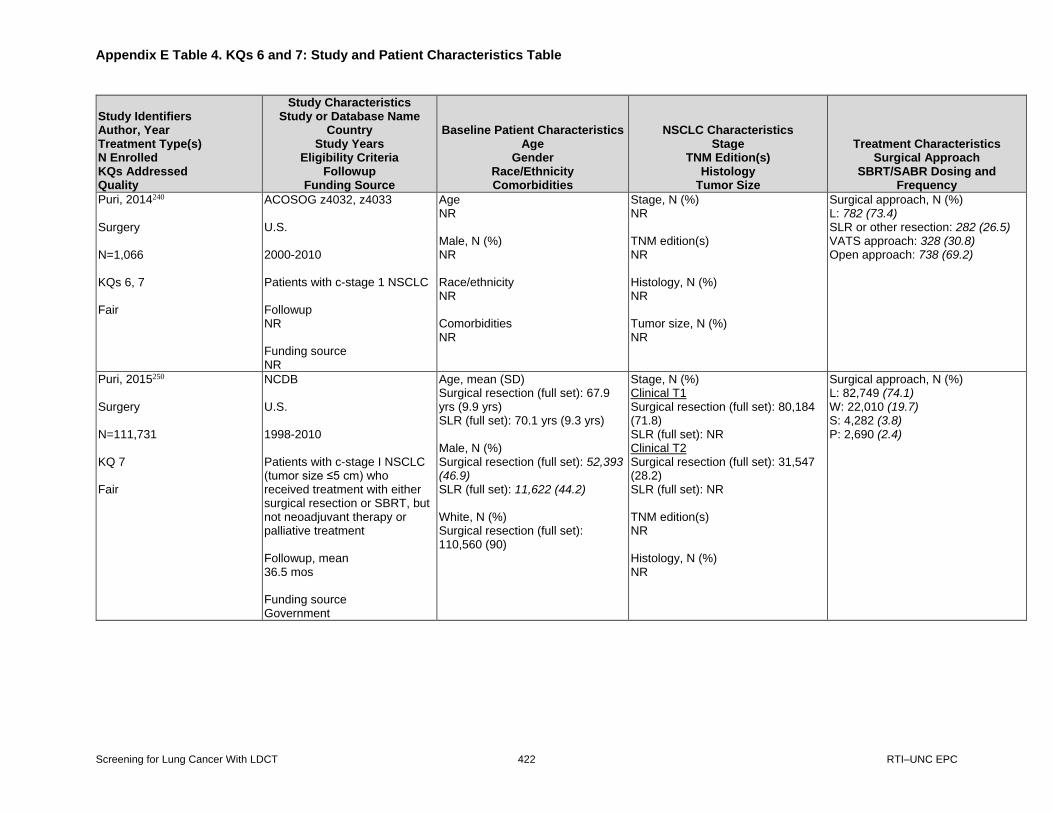
Appendix E Table 4. KQs 6 and 7: Study and Patient Characteristics Table
Screening for Lung Cancer With LDCT 422 RTI–UNC EPC
Study Identifiers Author, Year Treatment Type(s) N Enrolled KQs Addressed Quality
Study Characteristics Study or Database Name
Country Study Years
Eligibility Criteria Followup
Funding Source
Baseline Patient Characteristics Age
Gender Race/Ethnicity Comorbidities
NSCLC Characteristics Stage
TNM Edition(s) Histology
Tumor Size
Treatment Characteristics Surgical Approach
SBRT/SABR Dosing and Frequency
Puri, 2014240 Surgery N=1,066 KQs 6, 7 Fair
ACOSOG z4032, z4033 U.S. 2000-2010 Patients with c-stage 1 NSCLC Followup NR Funding source NR
Age NR Male, N (%) NR Race/ethnicity NR Comorbidities NR
Stage, N (%) NR TNM edition(s) NR Histology, N (%) NR Tumor size, N (%) NR
Surgical approach, N (%) L: 782 (73.4) SLR or other resection: 282 (26.5) VATS approach: 328 (30.8) Open approach: 738 (69.2)
Puri, 2015250 Surgery N=111,731 KQ 7 Fair
NCDB U.S. 1998-2010 Patients with c-stage I NSCLC (tumor size ≤5 cm) who received treatment with either surgical resection or SBRT, but not neoadjuvant therapy or palliative treatment Followup, mean 36.5 mos Funding source Government
Age, mean (SD) Surgical resection (full set): 67.9 yrs (9.9 yrs) SLR (full set): 70.1 yrs (9.3 yrs) Male, N (%) Surgical resection (full set): 52,393 (46.9) SLR (full set): 11,622 (44.2) White, N (%) Surgical resection (full set): 110,560 (90)
Stage, N (%) Clinical T1 Surgical resection (full set): 80,184 (71.8) SLR (full set): NR Clinical T2 Surgical resection (full set): 31,547 (28.2) SLR (full set): NR TNM edition(s) NR Histology, N (%) NR
Surgical approach, N (%) L: 82,749 (74.1) W: 22,010 (19.7) S: 4,282 (3.8) P: 2,690 (2.4)

Appendix E Table 4. KQs 6 and 7: Study and Patient Characteristics Table
Screening for Lung Cancer With LDCT 423 RTI–UNC EPC
Study Identifiers Author, Year Treatment Type(s) N Enrolled KQs Addressed Quality
Study Characteristics Study or Database Name
Country Study Years
Eligibility Criteria Followup
Funding Source
Baseline Patient Characteristics Age
Gender Race/Ethnicity Comorbidities
NSCLC Characteristics Stage
TNM Edition(s) Histology
Tumor Size
Treatment Characteristics Surgical Approach
SBRT/SABR Dosing and Frequency
Puri, 2015250 (continued)
SLR (full set): 24,016 (91.3) Charlson/Deyo Comorbidity Score, N (%) Score=0 Surgical resection (full set): 42,761 (50.6) SLR (full set): 9,087 (45.5) Score=1 Surgical resection (full set): 30,401 (36) SLR (full set): 7,662 (38.4 Score=2 Surgical resection (full set): 11,342 (13.4) SLR (full set): 3,211 (12.2))
Tumor size, mean (SD) Surgical resection (full set): 24.1 mm (10.7 mm) SLR (full set): 19.3 mm (9.0 mm)
Razi, 2016178 Surgery N=1,640 KQ 6 Fair
SEER U.S. 1998-2007 Patients aged ≥75 y who underwent lobectomy or SLR (W or S), but not chemotherapy or radiotherapy, for Stage IA (T1a/b, N0, M0) NSCLC, restricted by histology to either squamous cell or adenocarcinoma Followup, median NR Funding source NR
Age, median 78 yrs Age, mean (SE) L/Bi-L: 78.7 yrs (0.10 yrs) S: 79.4 yrs (0.35 yrs) W: 79.7 yrs (0.17 yrs) Male, N (%) L/Bi-L: 502 (47.8) S: 48 (40.3) W: 222 (47.2) White, N (%) L/Bi-L: 951 (90.5) S: 108 (90.8) W: 427 (90.9) Comorbidities, N (%) NR
Stage, N (%) c-stage 1A: 1,640 (100) TNM edition(s) 7th edition Histology, N (%) Adenocarcinoma L/Bi-L: 657 (62.5) S: 71 (59.7) W: 254 (54) Squamous cell carcinoma L/Bi-L: 394 (37.5) S: 48 (40.3) W: 216 (46)
Surgical approach, N (%) L/Bi-L: 1,051 (64.1) S: 119 (7.3) W: 470 (28.6)

Appendix E Table 4. KQs 6 and 7: Study and Patient Characteristics Table
Screening for Lung Cancer With LDCT 424 RTI–UNC EPC
Study Identifiers Author, Year Treatment Type(s) N Enrolled KQs Addressed Quality
Study Characteristics Study or Database Name
Country Study Years
Eligibility Criteria Followup
Funding Source
Baseline Patient Characteristics Age
Gender Race/Ethnicity Comorbidities
NSCLC Characteristics Stage
TNM Edition(s) Histology
Tumor Size
Treatment Characteristics Surgical Approach
SBRT/SABR Dosing and Frequency
Razi, 2016178 (continued)
Tumor size, mean (SE) L/Bi-L: 2.2 cm (0.02 cm) S: 1.9 cm (0.06 cm) W: 1.9 cm (0.03 cm)
Robinson, 2013264 SBRT/SABR N=78 KQ 7 Fair
NA U.S. 2004-2008 Patients with pathologically confirmed Stage 1 NSCLC and receiving SBRT with BED ≥100 Gy Followup, median 50.3 mos Funding source NR
Age, median (range) 76 yrs (31 to 93 yrs) Male, N (%) 44 (56.4) White, N (%) 68 (87.2) Charlson Comorbidity Index, median (range) Raw score: 4 (2 to 10) Age-adjusted score: 7 (3 to 12) ACE-27 Comorbidity Index, median (range) 2 (0 to 3)
Clinical Stage, N (%) T1a: 36 (46.2) T1b: 20 (25.6) T2a: 19 (24.4) T2b: 3 (3.8) T3: 0 (0) TNM edition(s) 7th edition Histology, N (%) Adenocarcinoma: 36 (46.2) Squamous cell: 25 (32.1) NSCLC NOS: 16 (20.5) Other/unknown: 1 (1.3) Maximal tumor size, median (range) 2 cm (1.1 to 6 cm)
SBRT patients, N (%) 78 (100) SBRT dosing, N (%) 54 Gy x 3 fx: 68 (87.2) 50 Gy x 5 fx: 4 (5.1) 45 Gy x 3 fx: 6 (7.7)

Appendix E Table 4. KQs 6 and 7: Study and Patient Characteristics Table
Screening for Lung Cancer With LDCT 425 RTI–UNC EPC
Study Identifiers Author, Year Treatment Type(s) N Enrolled KQs Addressed Quality
Study Characteristics Study or Database Name
Country Study Years
Eligibility Criteria Followup
Funding Source
Baseline Patient Characteristics Age
Gender Race/Ethnicity Comorbidities
NSCLC Characteristics Stage
TNM Edition(s) Histology
Tumor Size
Treatment Characteristics Surgical Approach
SBRT/SABR Dosing and Frequency
Rosen, 2014256 SBRT/SABR N=79 KQ 7 Fair
NA U.S. 2005-2010 Patients with biopsy-proven or clinically diagnosed T1-2N0M0 NSCLC and treated with TomoTherapy helical SBRT Followup, median 27 mos Funding source NR
Age, median (range) 73 (27 to 92) Male, N (%) 33 (42) Race/ethnicity NR Comorbidities NR
Stage, N (%) T1a: 59 (75) T1b: 20 (25) TNM edition(s) NR Histology, N (%) Adenocarcinoma: 22 (28) Squamous cell: 18 (23) NSCLC NOS: 22 (28) No histology: 17 (21) Tumor diameter, mean (range) 2.3 cm (0.5 to 6.0 cm)
SBRT patients, N (%) 79 (100) SBRT dosing, N (%) 12 Gy x 4 fx: 20 (25.3) 12 Gy x 5 fx: 59 (74.7)
Rosen, 2014242 Surgery N=66,283 (of 119,146 total) KQ 7 Fair
NCDB U.S. 2004-2009 All patients over the age of 19 years diagnosed with NSCLC and undergoing surgical resection (W, S, L/Bi-L, extended L/Bi-L, P) Followup, median (range) NR Funding source University
Age, median (range) NR Male, N (%) NR Race/ethnicity NR Comorbidities NR
Stage, N (%) p-stage 1: 66,283 (55.6) p-stage 2: 17,434 (14.6) p-stage 3: 15,610 (13.1) p-stage 4: 4,196 (3.5) Unknown: 15,623 (13.1) TNM edition(s) NR Histology, N (%) NR Tumor size, N (%) NR
Surgical approach, N (%) W: NR S: NR L/Bi-L: NR Extended L/Bi-L: NR P: NR
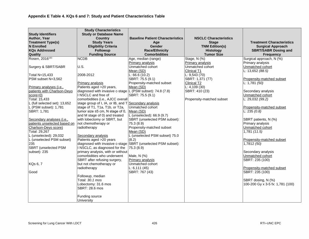
Appendix E Table 4. KQs 6 and 7: Study and Patient Characteristics Table
Screening for Lung Cancer With LDCT 426 RTI–UNC EPC
Study Identifiers Author, Year Treatment Type(s) N Enrolled KQs Addressed Quality
Study Characteristics Study or Database Name
Country Study Years
Eligibility Criteria Followup
Funding Source
Baseline Patient Characteristics Age
Gender Race/Ethnicity Comorbidities
NSCLC Characteristics Stage
TNM Edition(s) Histology
Tumor Size
Treatment Characteristics Surgical Approach
SBRT/SABR Dosing and Frequency
Rosen, 2016183 Surgery & SBRT/SABR Total N=15,433 PSM subset N=3,562 Primary analyses (i.e., patients with Charlson-Deyo score=0) Total: 15,433 L (full selected set): 13,652 L (PSM subset): 1,781 SBRT: 1,781 Secondary analyses (i.e., patients unselected based on Charlson-Deyo score) Total: 29,267 L (unselected): 29,032 L (unselected PSM subset) 235 SBRT (unselected PSM subset): 235 KQs 6, 7 Good
NCDB U.S. 2008-2012 Primary analysis Patients aged >20 years, diagnosed with invasive c-stage I NSCLC and free of comorbidities (i.e., AJCC overall stage group of I, IA, or IB, and T stage of T1, T1a, T1b, or T2a, tumor size ≤5 cm, N stage of 0, and M stage of 0) and treated with lobectomy or SBRT, but not chemotherapy or radiotherapy Secondary analysis Patients aged >20 years diagnosed with invasive c-stage I NSCLC, as diagnosed for the primary analysis, with or without comorbidities who underwent SBRT after refusing surgery, but not chemotherapy or radiotherapy Followup, median Total: 30.1 mos Lobectomy: 31.6 mos SBRT: 28.6 mos Funding source University
Age, median (range) Primary analysis Unmatched cohort Mean (SD) L: 66.6 (10.2) SBRT: 75.5 (9.1) Propensity-matched subset Mean (SD) L (PSM subset): 74.8 (7.8) SBRT: 75.5 (9.1) Secondary analysis Unmatched cohort Mean (SD) L (unselected): 66.9 (9.7) SBRT (unselected PSM subset): 75.3 (8.9) Propensity-matched subset Mean (SD) L (unselected PSM subset) 75.0 (8.2) SBRT (unselected PSM subset): 75.3 (8.9) Male, N (%) Primary analysis Unmatched cohort L: 6,111 (45) SBRT: 767 (43)
Stage, N (%) Primary analysis Unmatched cohort Clinical T1 L: 9,543 (70) SBRT: 1,371 (77) Clinical T2 L: 4,109 (30) SBRT: 410 (23) Propensity-matched subset
Surgical approach, N (%) Primary analysis Unmatched cohort L: 13,652 (88.5) Propensity-matched subset L: 1,781 (50) Secondary analysis Unmatched cohort L: 29,032 (99.2) Propensity-matched subset L: 235 (0.8) SBRT patients, N (%) Primary analysis Unmatched cohort 1,781 (11.5) Propensity-matched subset 1,7812 (50) Secondary analysis Unmatched cohort SBRT: 235 (100) Propensity-matched subset SBRT: 235 (100) SBRT dosing, N (%) 100-200 Gy x 3-5 fx: 1,781 (100)

Appendix E Table 4. KQs 6 and 7: Study and Patient Characteristics Table
Screening for Lung Cancer With LDCT 427 RTI–UNC EPC
Study Identifiers Author, Year Treatment Type(s) N Enrolled KQs Addressed Quality
Study Characteristics Study or Database Name
Country Study Years
Eligibility Criteria Followup
Funding Source
Baseline Patient Characteristics Age
Gender Race/Ethnicity Comorbidities
NSCLC Characteristics Stage
TNM Edition(s) Histology
Tumor Size
Treatment Characteristics Surgical Approach
SBRT/SABR Dosing and Frequency
Rosen, 2016183 (continued)
Propensity-matched cohort L (PSM subset): 777 (44) SBRT: 767 (43) Secondary analysis Unmatched cohort L (unselected): 13,713 (47) SBRT (unselected PSM subset): 95 (40) Propensity-matched cohort L (unselected PSM subset) 84 (36) SBRT (unselected PSM subset): 95 (40) White, N (%) Primary analysis Unmatched cohort L: 11,938 (87) SBRT: 1,616 (91) Propensity-matched cohort L (PSM subset): 1,610 (90) SBRT: 1,616 (91) Secondary analysis Unmatched cohort L (unselected): 25,573 (88) SBRT (unselected PSM subset): 208 (89)
Clinical T1 L (PSM subset): 1,374 (77) SBRT: 1,371 (77) Clinical T2 L (PSM subset): 407 (23) SBRT: 410 (23) Secondary analysis Unmatched cohort

Appendix E Table 4. KQs 6 and 7: Study and Patient Characteristics Table
Screening for Lung Cancer With LDCT 428 RTI–UNC EPC
Study Identifiers Author, Year Treatment Type(s) N Enrolled KQs Addressed Quality
Study Characteristics Study or Database Name
Country Study Years
Eligibility Criteria Followup
Funding Source
Baseline Patient Characteristics Age
Gender Race/Ethnicity Comorbidities
NSCLC Characteristics Stage
TNM Edition(s) Histology
Tumor Size
Treatment Characteristics Surgical Approach
SBRT/SABR Dosing and Frequency
Rosen, 2016183 (continued)
Propensity-matched cohort L (unselected PSM subset) 212 (90) SBRT (unselected PSM subset): 208 (89) Comorbidities, N (%) Primary analysis Charlson-Deyo score: 0 (0) for all patients in the unmatched and propensity-matched cohorts Secondary analysis, N (%) Unmatched cohort Charlson-Deyo score=0 L (unselected): 13,652 (47) SBRT (unselected PSM subset): 157 (67) Charlson-Deyo score=1 L (unselected): 10,877 (37) SBRT (unselected PSM subset): 50 (21) Charlson-Deyo score=2+ L (unselected): 4,503 (16) SBRT (unselected PSM subset): 28 (12) Propensity-matched subset Charlson-Deyo score=0 L (unselected PSM subset) 156 (66) SBRT (unselected PSM subset): 157 (67)
Clinical T1 L (unselected): 20,555 (71) SBRT (unselected PSM subset): 174 (74) Clinical T2 L (unselected): 8,477 (29) SBRT (unselected PSM subset): 61 (26) Propensity-matched cohort Clinical T1 L (unselected PSM subset) 175 (74) SBRT (unselected PSM subset): 174 (74) Clinical T2 L (unselected PSM subset) 60 (26) SBRT (unselected PSM subset): 61 (26)

Appendix E Table 4. KQs 6 and 7: Study and Patient Characteristics Table
Screening for Lung Cancer With LDCT 429 RTI–UNC EPC
Study Identifiers Author, Year Treatment Type(s) N Enrolled KQs Addressed Quality
Study Characteristics Study or Database Name
Country Study Years
Eligibility Criteria Followup
Funding Source
Baseline Patient Characteristics Age
Gender Race/Ethnicity Comorbidities
NSCLC Characteristics Stage
TNM Edition(s) Histology
Tumor Size
Treatment Characteristics Surgical Approach
SBRT/SABR Dosing and Frequency
Rosen, 2016183 (continued)
Charlson-Deyo score=1 L (unselected PSM subset) 52 (22) SBRT (unselected PSM subset): 50 (21) Charlson-Deyo score=2+ L (unselected PSM subset) 27 (11) SBRT (unselected PSM subset): 28 (12)
TNM edition(s) 6th & 7th editions Histology, N (%) Primary analysis, N (%) Unmatched cohort Adenocarcinoma L: 9,379 (69) SBRT: 850 (48) Squamous cell carcinoma L: 3,502 (26) SBRT: 583 (33) Large cell carcinoma L: 317 (2) SBRT: 17 (1)

Appendix E Table 4. KQs 6 and 7: Study and Patient Characteristics Table
Screening for Lung Cancer With LDCT 430 RTI–UNC EPC
Study Identifiers Author, Year Treatment Type(s) N Enrolled KQs Addressed Quality
Study Characteristics Study or Database Name
Country Study Years
Eligibility Criteria Followup
Funding Source
Baseline Patient Characteristics Age
Gender Race/Ethnicity Comorbidities
NSCLC Characteristics Stage
TNM Edition(s) Histology
Tumor Size
Treatment Characteristics Surgical Approach
SBRT/SABR Dosing and Frequency
Rosen, 2016183 (continued)
Other L: 454 (3) SBRT: 331 (19) Propensity-matched cohort Adenocarcinoma L: 886 (50) SBRT: 850 (48) Squamous cell carcinoma L: 646 (36) SBRT: 583 (33) Large cell carcinoma L: 21 (1) SBRT: 17 (1) Other L: 228 (13) SBRT: 331 (19) Secondary analysis, N (%) Unmatched cohort Adenocarcinoma L (unselected): 18,200 (63) SBRT (unselected PSM subset): 125 (53) Squamous cell carcinoma L (unselected): 9,130 (31) SBRT (unselected PSM subset): 69 (29) Large cell carcinoma L (unselected): 797 (3) SBRT (unselected PSM subset): 2 (0.9)

Appendix E Table 4. KQs 6 and 7: Study and Patient Characteristics Table
Screening for Lung Cancer With LDCT 431 RTI–UNC EPC
Study Identifiers Author, Year Treatment Type(s) N Enrolled KQs Addressed Quality
Study Characteristics Study or Database Name
Country Study Years
Eligibility Criteria Followup
Funding Source
Baseline Patient Characteristics Age
Gender Race/Ethnicity Comorbidities
NSCLC Characteristics Stage
TNM Edition(s) Histology
Tumor Size
Treatment Characteristics Surgical Approach
SBRT/SABR Dosing and Frequency
Rosen, 2016183 (continued)
Other L (unselected): 905 (3) SBRT (unselected PSM subset): 39 (17) Propensity-matched cohort Adenocarcinoma L (unselected PSM subset) 125 (53) SBRT (unselected PSM subset): 125 (53) Squamous cell carcinoma L (unselected PSM subset) 61 (26) SBRT (unselected PSM subset): 69 (29) Large cell carcinoma L (unselected PSM subset) 4 (1.7) SBRT (unselected PSM subset): 2 (0.9) Other L (unselected PSM subset) 45 (19) SBRT (unselected PSM subset): 39 (17)

Appendix E Table 4. KQs 6 and 7: Study and Patient Characteristics Table
Screening for Lung Cancer With LDCT 432 RTI–UNC EPC
Study Identifiers Author, Year Treatment Type(s) N Enrolled KQs Addressed Quality
Study Characteristics Study or Database Name
Country Study Years
Eligibility Criteria Followup
Funding Source
Baseline Patient Characteristics Age
Gender Race/Ethnicity Comorbidities
NSCLC Characteristics Stage
TNM Edition(s) Histology
Tumor Size
Treatment Characteristics Surgical Approach
SBRT/SABR Dosing and Frequency
Rosen, 2016183 (continued)
Tumor size, mean (SD) Primary analysis Unmatched cohort L: 25.4 mm (10.5 mm) SBRT: 23.8 mm (9.3 mm) Propensity-matched subset L: 23.7 mm (10.0 mm) SBRT: 23.8 mm (9.3 mm) Secondary analysis Unmatched cohort L (unselected): 25.1 mm (10.5 mm) SBRT (unselected PSM subset): 24.0 mm (9.3 mm) Propensity-matched subset L (unselected PSM subset) 23.6 mm (9.9 mm) SBRT (unselected PSM subset): 24.0 mm (9.3 mm)

Appendix E Table 4. KQs 6 and 7: Study and Patient Characteristics Table
Screening for Lung Cancer With LDCT 433 RTI–UNC EPC
Study Identifiers Author, Year Treatment Type(s) N Enrolled KQs Addressed Quality
Study Characteristics Study or Database Name
Country Study Years
Eligibility Criteria Followup
Funding Source
Baseline Patient Characteristics Age
Gender Race/Ethnicity Comorbidities
NSCLC Characteristics Stage
TNM Edition(s) Histology
Tumor Size
Treatment Characteristics Surgical Approach
SBRT/SABR Dosing and Frequency
Samson, 2015238 Surgery N=55,653 KQs 6, 7 Fair
NCDB U.S. 2000-2012 Overall and PSM samples: Patients diagnosed with c-stage I NSCLC undergoing surgical resection Single-center sample: Patients diagnosed with c-stage I NSCLC undergoing surgical resection Followup, median (range) NR Funding source Government
Age, median (range) PSM analysis Early surgery: 68.7 (9.8) Delayed surgery: 68.8 (9.7) Single-center analysis Early surgery: 66.0 (10.1) Delayed surgery: 66.9 (9.7) Male, N (%) PSM analysis Male Early surgery: 6,505 (48.1) Delayed surgery: 6,564 (48.6) Single-center analysis Female Early surgery: 276 (53) Delayed surgery: 232 (52) White, N (%) PSM analysis Early surgery: 11,765 (87.1) Delayed surgery: 11,787 (87.2) Single-center analysis Early surgery: 466 (89) Delayed surgery: 368 (82)
Stage, N (%) PSM analysis AJCC clinical T1 Early surgery: 8,608 (63.7) Delayed surgery: 8,656 (64.1) AJCC clinical T2 Early surgery: 4,903 (36.3) Delayed surgery: 4,855 (35.9) Single-center analysis AJCC clinical T1 Early surgery: 338 (65) Delayed surgery: 342 (76) TNM edition(s) NR Histology, N (%) Malignant cytologic diagnosis: 568 (1.0)
Surgical approach, N (%) PSM analysis Total: 27,022 Early surgery: 13,511 Delayed surgery: 13,511 Single-center analysis Total: 971 Early surgery: 522 Delayed surgery: 449

Appendix E Table 4. KQs 6 and 7: Study and Patient Characteristics Table
Screening for Lung Cancer With LDCT 434 RTI–UNC EPC
Study Identifiers Author, Year Treatment Type(s) N Enrolled KQs Addressed Quality
Study Characteristics Study or Database Name
Country Study Years
Eligibility Criteria Followup
Funding Source
Baseline Patient Characteristics Age
Gender Race/Ethnicity Comorbidities
NSCLC Characteristics Stage
TNM Edition(s) Histology
Tumor Size
Treatment Characteristics Surgical Approach
SBRT/SABR Dosing and Frequency
Samson, 2015238 (continued)
Charlson Comorbidity Index, N (%) PSM analysis Score=0 Early surgery: 6,459 (47.8) Delayed surgery: 6,490 (48.0) Score=1 Early surgery: 5,011 (37.1) Delayed surgery: 4,895 (36.2) Score=2+ Early surgery: 2,041 (15.1) Delayed surgery: 2,126 (15.7) Adult Comorbidity Evaluation, N (%) Single-center analysis Score=0 Early surgery: 83 (16) Delayed surgery: 37 (8) Score=1 Early surgery: 211 (40) Delayed surgery: 156 (35) Score=2
Tumor size in mm, mean (SD) PSM analysis Size Early surgery: 30.1 (22.6) Delayed surgery: 30.5 (21.3) Single-center analysis NR

Appendix E Table 4. KQs 6 and 7: Study and Patient Characteristics Table
Screening for Lung Cancer With LDCT 435 RTI–UNC EPC
Study Identifiers Author, Year Treatment Type(s) N Enrolled KQs Addressed Quality
Study Characteristics Study or Database Name
Country Study Years
Eligibility Criteria Followup
Funding Source
Baseline Patient Characteristics Age
Gender Race/Ethnicity Comorbidities
NSCLC Characteristics Stage
TNM Edition(s) Histology
Tumor Size
Treatment Characteristics Surgical Approach
SBRT/SABR Dosing and Frequency
Samson, 2015238 (continued)
Early surgery: 125 (24) Delayed surgery: 125 (28) Score=3 Early surgery: 63 (12) Delayed surgery: 79 (18) Pulmonary hypertension Single-center analysis, N (%) Early surgery: 3 (0.6) Delayed surgery: 7 (1.6) Smoking status Single-center analysis, N (%) Current smokers Early surgery: 170 (33) Delayed surgery: 164 (37) Former smokers Early surgery: 302 (58) Delayed surgery: 244 (54) Never smokers Early surgery: 50 (10) Delayed surgery: 41 (9)
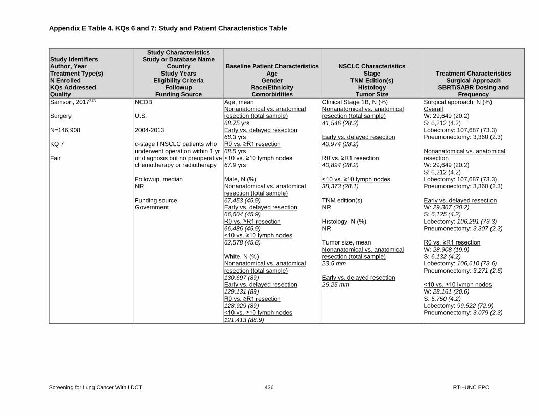
Appendix E Table 4. KQs 6 and 7: Study and Patient Characteristics Table
Screening for Lung Cancer With LDCT 436 RTI–UNC EPC
Study Identifiers Author, Year Treatment Type(s) N Enrolled KQs Addressed Quality
Study Characteristics Study or Database Name
Country Study Years
Eligibility Criteria Followup
Funding Source
Baseline Patient Characteristics Age
Gender Race/Ethnicity Comorbidities
NSCLC Characteristics Stage
TNM Edition(s) Histology
Tumor Size
Treatment Characteristics Surgical Approach
SBRT/SABR Dosing and Frequency
Samson, 2017243 Surgery N=146,908 KQ 7 Fair
NCDB U.S. 2004-2013 c-stage I NSCLC patients who underwent operation within 1 yr of diagnosis but no preoperative chemotherapy or radiotherapy Followup, median NR Funding source Government
Age, mean Nonanatomical vs. anatomical resection (total sample) 68.75 yrs Early vs. delayed resection 68.3 yrs R0 vs. ≥R1 resection 68.5 yrs <10 vs. ≥10 lymph nodes 67.9 yrs Male, N (%) Nonanatomical vs. anatomical resection (total sample) 67,453 (45.9) Early vs. delayed resection 66,604 (45.9) R0 vs. ≥R1 resection 66,486 (45.9) <10 vs. ≥10 lymph nodes 62,578 (45.8) White, N (%) Nonanatomical vs. anatomical resection (total sample) 130,697 (89) Early vs. delayed resection 129,131 (89) R0 vs. ≥R1 resection 128,929 (89) <10 vs. ≥10 lymph nodes 121,413 (88.9)
Clinical Stage 1B, N (%) Nonanatomical vs. anatomical resection (total sample) 41,546 (28.3) Early vs. delayed resection 40,974 (28.2) R0 vs. ≥R1 resection 40,894 (28.2) <10 vs. ≥10 lymph nodes 38,373 (28.1) TNM edition(s) NR Histology, N (%) NR Tumor size, mean Nonanatomical vs. anatomical resection (total sample) 23.5 mm Early vs. delayed resection 26.25 mm
Surgical approach, N (%) Overall W: 29,649 (20.2) S: 6,212 (4.2) Lobectomy: 107,687 (73.3) Pneumonectomy: 3,360 (2.3) Nonanatomical vs. anatomical resection W: 29,649 (20.2) S: 6,212 (4.2) Lobectomy: 107,687 (73.3) Pneumonectomy: 3,360 (2.3) Early vs. delayed resection W: 29,367 (20.2) S: 6,125 (4.2) Lobectomy: 106,291 (73.3) Pneumonectomy: 3,307 (2.3) R0 vs. ≥R1 resection W: 28,908 (19.9) S: 6,132 (4.2) Lobectomy: 106,610 (73.6) Pneumonectomy: 3,271 (2.6) <10 vs. ≥10 lymph nodes W: 28,161 (20.6) S: 5,750 (4.2) Lobectomy: 99,622 (72.9) Pneumonectomy: 3,079 (2.3)
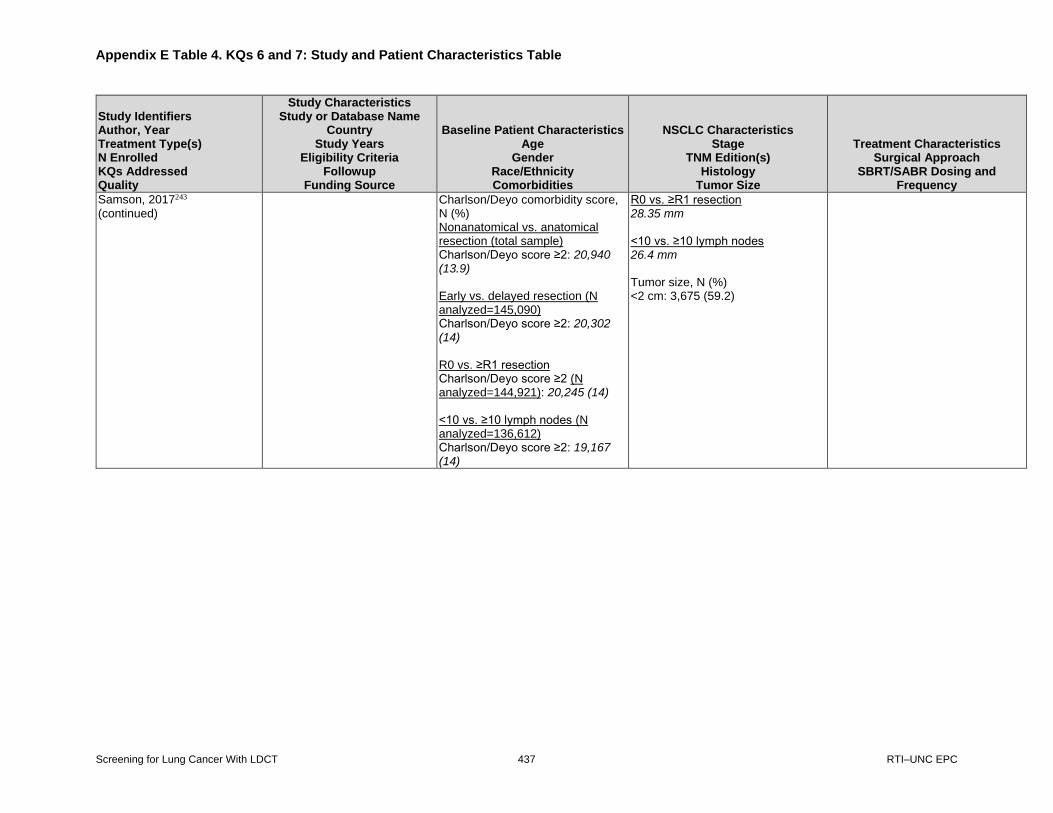
Appendix E Table 4. KQs 6 and 7: Study and Patient Characteristics Table
Screening for Lung Cancer With LDCT 437 RTI–UNC EPC
Study Identifiers Author, Year Treatment Type(s) N Enrolled KQs Addressed Quality
Study Characteristics Study or Database Name
Country Study Years
Eligibility Criteria Followup
Funding Source
Baseline Patient Characteristics Age
Gender Race/Ethnicity Comorbidities
NSCLC Characteristics Stage
TNM Edition(s) Histology
Tumor Size
Treatment Characteristics Surgical Approach
SBRT/SABR Dosing and Frequency
Samson, 2017243 (continued)
Charlson/Deyo comorbidity score, N (%) Nonanatomical vs. anatomical resection (total sample) Charlson/Deyo score ≥2: 20,940 (13.9) Early vs. delayed resection (N analyzed=145,090) Charlson/Deyo score ≥2: 20,302 (14) R0 vs. ≥R1 resection Charlson/Deyo score ≥2 (N analyzed=144,921): 20,245 (14) <10 vs. ≥10 lymph nodes (N analyzed=136,612) Charlson/Deyo score ≥2: 19,167 (14)
R0 vs. ≥R1 resection 28.35 mm <10 vs. ≥10 lymph nodes 26.4 mm Tumor size, N (%) <2 cm: 3,675 (59.2)

Appendix E Table 4. KQs 6 and 7: Study and Patient Characteristics Table
Screening for Lung Cancer With LDCT 438 RTI–UNC EPC
Study Identifiers Author, Year Treatment Type(s) N Enrolled KQs Addressed Quality
Study Characteristics Study or Database Name
Country Study Years
Eligibility Criteria Followup
Funding Source
Baseline Patient Characteristics Age
Gender Race/Ethnicity Comorbidities
NSCLC Characteristics Stage
TNM Edition(s) Histology
Tumor Size
Treatment Characteristics Surgical Approach
SBRT/SABR Dosing and Frequency
Sawabata, 2011188 Surgery N=9,083 eligible (of 11,663 total) KQ 6 Fair
Japanese Joint Committee of Lung Cancer Registry (JJCLCR) Japan 2004-2010 Pathological diagnosis of any type of lung cancer at a participating institution, diagnosis obtained in 2004, and treated by surgery Followup, median 2 to 78 mos Funding source NR
Age, median (range) NR Male, N (%) NR Race/ethnicity NR Comorbidities, N (%) NR
Stage, N (%) c-stage 1A: 6,295 (54) c-stage 1B: 2,788 (23.9) p-stage 1A: 5,611 (48.1) p-stage 1B: 2,398 (20.4) TNM edition(s) 6th & 7th editions Histology, N (%) NR Tumor size, mean (SD) NR
Surgical approach, N (%) NR
Scheel, 2015168 Surgery N=800 KQs 6, 7 Fair
NR U.S. 2000-2012 Patients who underwent initial resection by lobectomy or SLR of p-stage I NSCLC between January 2000 and December 2012 at Washington University School of Medicine Followup, median (range) NR Funding source Government
Age, mean (SD) NR for overall sample, but range from 64.3-65.5 across surg exp groups Male, N (%) 361 (45.1) White, N (%) 690 (86.3) Smoking status, N (%) Never: 96 (12.0) Past: 421 (52.6) Current: 283 (35.4)
Stage, N (%) p-stage I: 800 (100) TNM edition(s) NR Histology, N (%) NR Tumor size, N (%) NR
Surgical approach, N (%) All Resections: 800 L: 638 (79.8) SLR: 162 (20.2)

Appendix E Table 4. KQs 6 and 7: Study and Patient Characteristics Table
Screening for Lung Cancer With LDCT 439 RTI–UNC EPC
Study Identifiers Author, Year Treatment Type(s) N Enrolled KQs Addressed Quality
Study Characteristics Study or Database Name
Country Study Years
Eligibility Criteria Followup
Funding Source
Baseline Patient Characteristics Age
Gender Race/Ethnicity Comorbidities
NSCLC Characteristics Stage
TNM Edition(s) Histology
Tumor Size
Treatment Characteristics Surgical Approach
SBRT/SABR Dosing and Frequency
Schuchert, 2012246 Surgery N=899 KQ 7 Fair
NA U.S. 1999-2010 Patients with p-stage 1 NSCLC without multicentric disease and undergoing complete anatomic segmentectomy or lobectomy, but not preoperative radiotherapy or chemotherapy Followup, median 37 mos Funding source NR
Age, mean (SD) (range) Total: 69.2 yrs (NR) (22-91 yrs) Anatomic S: 70.0 yrs (8.6 yrs) (40 to 91 yrs) L: 68.4 yrs (10.1 yrs) (22 to 90 yrs) Male, N (%) Total: 427 (47.5) Anatomic S: 145 (47.5) L: 282 (47.5) Race/ethnicity NR Comorbidities, N (%) NR
Stage, N (%) p-stage 1A Total: 477 (53.1) Anatomic S: 187 (61.3) L: 290 (57.5) p-stage 1B Total: 422 (46.9) Anatomic S: 118 (38.7) L: 304 (60.3) TNM edition(s) 6th edition Histology, N (%) Adenocarcinoma Overall: 487 (54.2) Anatomic S: 161 (52.8) L: 326 (54.9) Squamous cell Overall: 295 (32.8) Anatomic S: 96 (31.5) L: 199 (33.5) Other NSCLC Overall: 117 (13.0) Anatomic S: 48 (15.7) L: 69 (11.6) Tumor size, median (IQR) (range) Overall: NR Anatomic S: 2.0 cm (1.5 to 2.8 cm) (0.2 to 5.0 cm) L: 2.5 cm (1.8 to 4.0 cm) (0.2 to 12 cm)
Surgical approach, N (%) Anatomic S: 305 (33.9) L: 594 (66.1) Open procedure Total: 489 (54.4) Anatomic S: 120 (39.3) L: 369 (62.1) VATS approach Total: 410 (45.6) Anatomic S: 185 (60.7) L: 225 (37.9)

Appendix E Table 4. KQs 6 and 7: Study and Patient Characteristics Table
Screening for Lung Cancer With LDCT 440 RTI–UNC EPC
Study Identifiers Author, Year Treatment Type(s) N Enrolled KQs Addressed Quality
Study Characteristics Study or Database Name
Country Study Years
Eligibility Criteria Followup
Funding Source
Baseline Patient Characteristics Age
Gender Race/Ethnicity Comorbidities
NSCLC Characteristics Stage
TNM Edition(s) Histology
Tumor Size
Treatment Characteristics Surgical Approach
SBRT/SABR Dosing and Frequency
Shapiro, 2012245 Surgery N=4,975 KQ 7 Fair
SEER U.S. 1992-2002 Patients with Stage I NSCLC aged ≥65 years who underwent lobectomy, but no pre-operative chemotherapy or radiotherapy Followup, median NR Funding source Government
Age, N (%) 65-69 yrs: 1,421 (28.6) 70-74 yrs: 1,662 (33.4) 75-79 yrs: 1,244 (25) ≥80 yrs: 648 (13) Male, N (%) 2,565 (51.6) White, N (%) 4,348 (87.4) Comorbidities, N (%) Charlson/Deyo score 0-1, N (%): 4,238 (85.2) Charlson/Deyo score 2-3, N (%): 479 (9.6) Charlson/Deyo score ≥4, N (%): 258 (5.2)
Stage, N (%) NR TNM edition(s) 5th edition Histology, N (%) Adenocarcinoma: 2,205 (44.3) Bronchioalveolar carcinoma: 219 (4.4) Squamous carcinoma: 1,631 (32.8) Large cell carcinoma: 267 (5.4) Other: 653 (13.1) Tumor size, N (%) ≤20 mm: 1,473 (29.6) 21-30 mm: 1,526 (30.7) 31-50 mm: 1,409 (28.3) 51-70 mm: 403 (8.1) ≥71 mm: 164 (3.3)
Surgical approach, N (%) L: 4,975 (100)

Appendix E Table 4. KQs 6 and 7: Study and Patient Characteristics Table
Screening for Lung Cancer With LDCT 441 RTI–UNC EPC
Study Identifiers Author, Year Treatment Type(s) N Enrolled KQs Addressed Quality
Study Characteristics Study or Database Name
Country Study Years
Eligibility Criteria Followup
Funding Source
Baseline Patient Characteristics Age
Gender Race/Ethnicity Comorbidities
NSCLC Characteristics Stage
TNM Edition(s) Histology
Tumor Size
Treatment Characteristics Surgical Approach
SBRT/SABR Dosing and Frequency
Shirvani, 2012235 Surgery & SBRT/SABR N=10,923 KQ 7 Fair
SEER-Medicare database U.S. 2001-2007 Patients aged >66 years diagnosed with early-stage (IA-IB) NSCLC without prior or future malignancy within 120 days of index cancer and who were treated with SABR, but not nonstandard therapies (e.g., chemotherapy), reported in the SEER-Medicare cohort demographic Followup, median 3.2 yrs Cancer Prevention & Research Institute of Texas; NCI; and Varian Medical Systems
Age, median 75 yrs Age 66-69, N (%) SLR: 234 (18) SABR: 11 (9) L: 1,408 (22) Age 70-74, N (%) SLR: 362 (28) SABR: 20 (16) L: 2,055 (31) Age 75-79, N (%) SLR: 392 (31) SABR: 29 (23) L: 1,907 (29) Age ≥80, N (%) SLR: 289 (23) SABR: 64 (52) L: 1,161 (18) Male, N (%) SLR: 571 (45) SABR: 49 (40) L: 3,011 (46) White, N (%) SLR: 1,184 (93) SABR: >11 (>90) L: 5,927 (91)
Stage, N (%) NR TNM edition(s) NR Histology, N (%) NSCLC, NOS SLR: 84 (7) SABR: 34 (27) L: 373 (6) Adenocarcinoma SLR: 749 (59) SABR: 53 (43) L: 3,931 (60) Squamous cell carcinoma SLR: 389 (30) SABR: 36 (29) L: 1,982 (30)
Surgical approach, N (%) SLR: 1,277 (11.7) L: 6,531 (58.9) SABR patients, N (%) 124 (1.1) SABR dosing NR

Appendix E Table 4. KQs 6 and 7: Study and Patient Characteristics Table
Screening for Lung Cancer With LDCT 442 RTI–UNC EPC
Study Identifiers Author, Year Treatment Type(s) N Enrolled KQs Addressed Quality
Study Characteristics Study or Database Name
Country Study Years
Eligibility Criteria Followup
Funding Source
Baseline Patient Characteristics Age
Gender Race/Ethnicity Comorbidities
NSCLC Characteristics Stage
TNM Edition(s) Histology
Tumor Size
Treatment Characteristics Surgical Approach
SBRT/SABR Dosing and Frequency
Shirvani, 2012235 (continued)
Charlson Comorbidity Index, N (%) Score=0 SLR: 339 (27) SABR: 28 (23) L: 2,814 (43) Score=1 SLR: 447 (35) SABR: 42 (34) L: 2,042 (31) Score ≥2 SLR: 457 (36) SABR: 54 (44) L: 1,495 (23) Missing Total: 366 (3)
Tumor size, N (%) ≤2.0 cm SLR: 820 (64) SABR: 48 (39) L: 2,723 (42) 2.1-3.0 cm SLR: 316 (25) SABR: 48 (39) L: 2,188 (34) 3.1-5.0 cm SLR: 141 (11) SABR: 28 (23) L: 1,620 (25)

Appendix E Table 4. KQs 6 and 7: Study and Patient Characteristics Table
Screening for Lung Cancer With LDCT 443 RTI–UNC EPC
Study Identifiers Author, Year Treatment Type(s) N Enrolled KQs Addressed Quality
Study Characteristics Study or Database Name
Country Study Years
Eligibility Criteria Followup
Funding Source
Baseline Patient Characteristics Age
Gender Race/Ethnicity Comorbidities
NSCLC Characteristics Stage
TNM Edition(s) Histology
Tumor Size
Treatment Characteristics Surgical Approach
SBRT/SABR Dosing and Frequency
Shirvani, 2014239 Surgery & SBRT/SABR N=9,093 KQ 7 Fair
SEER-Medicare U.S. 2003-2009 Patients with early-stage, node-negative, pathologically confirmed NSCLC who underwent lobectomy, SLR, or SABR without undergoing any nonstandard therapies Followup, median (range) NR Funding source Government
Age, N (%) 66 to 69 yrs L: 1515 (21.0) SLR: 235 (15.7) SABR: 39 (10.2) 70 to 74 yrs L: 2182 (30.2) SLR: 415 (27.7) SABR: 71 (18.6) 75 to 79 yrs L: 2069 (28.7) SLR: 435 (29.1) SABR: 94 (24.6) ≥80 yrs L: 1449 (20.1) SLR: 411 (27.5) SABR: 178 (46.6) Male, N (%) L: 3365 (46.6) SLR: 693 (46.3) SABR: 143 (37.4) White, N (%) L: 6456 (89.5) SLR: 1360 (90.9) SABR: 340 (89.0) Charlson Comorbidity Index, N (%) 0 L: 4368 (60.5) SLR: 792 (52.9) SABR: 170 (44.5)
Stage, N (%) NR TNM edition(s) NR Histology, N (%) NSCLC, NOS L: 366 (5.1) SLR: 90 (6.0) SABR: 82 (21.5) Adenocarcinoma L: 4371 (60.6) SLR: 866 (57.9) SABR: 178 (46.6) Squamous cell carcinomas L: 2236 (31.0) SLR: 482 (32.2) SABR: >110 (>25) Large cell cancer L: 242 (3.4) SLR: 58 (3.9) SABR: <11 (<5)
Surgical approach, N (%) L: 7,215 (79.3) SLR: 1,496 (16.5) SABR patients, N (%) 382 (4.2) SABR dosing NR

Appendix E Table 4. KQs 6 and 7: Study and Patient Characteristics Table
Screening for Lung Cancer With LDCT 444 RTI–UNC EPC
Study Identifiers Author, Year Treatment Type(s) N Enrolled KQs Addressed Quality
Study Characteristics Study or Database Name
Country Study Years
Eligibility Criteria Followup
Funding Source
Baseline Patient Characteristics Age
Gender Race/Ethnicity Comorbidities
NSCLC Characteristics Stage
TNM Edition(s) Histology
Tumor Size
Treatment Characteristics Surgical Approach
SBRT/SABR Dosing and Frequency
Shirvani, 2014239 (continued)
1 L: 1700 (23.6) SLR: 379 (25.3) SABR: 108 (28.3) ≥ 2 L: 1147 (15.9) SLR: 325 (21.7) SABR: 104 (27.2) COPD L: 4459 (61.8) SLR: 1136 (75.9) SABR: 296 (77.5)
Tumor size by T stage, N (%) T1a (0.0 to 2.0 cm) L: 3169 (43.9) SLR: 964 (64.4) SABR: 153 (40.1) T1b (2.1 to 3.0 cm) L: 2370 (32.8) SLR: 355 (23.7) SABR: 153 (40.1) T2a (3.1 to 5.0 cm) L: 1676 (23.2) SLR: 177 (11.8) SABR: 76 (19.9)

Appendix E Table 4. KQs 6 and 7: Study and Patient Characteristics Table
Screening for Lung Cancer With LDCT 445 RTI–UNC EPC
Study Identifiers Author, Year Treatment Type(s) N Enrolled KQs Addressed Quality
Study Characteristics Study or Database Name
Country Study Years
Eligibility Criteria Followup
Funding Source
Baseline Patient Characteristics Age
Gender Race/Ethnicity Comorbidities
NSCLC Characteristics Stage
TNM Edition(s) Histology
Tumor Size
Treatment Characteristics Surgical Approach
SBRT/SABR Dosing and Frequency
Speicher, 2016185 Surgery N=39,403 KQ 6 Fair
NCDB U.S. 2003-2006 Patients with c-stage 1A T1N0M0 NSCLC undergoing a lobectomy or SLR without induction therapy Followup, median 6.3 yrs Funding source Government, other unspecified
Age, mean (SD); Median Total: 67.4 yrs (9.8 yrs); 68.0 yrs L: 66.7 yrs (9.7 yrs); 67.0 yrs SLR: 69.6 yrs (9.5 yrs); 70.0 yrs Male, N (%) Total: 17,112 (43.4) L: 12,970 (43.6) SLR: 4,142 (42.8) White, N (%) Total: 35,266 (89.5) L: 26,491 (89.1) SLR: 8,775 (90.8) Charlson Comorbidity Index, N (%) (for initial sample only, but unclear if the same for survival analysis sample) Score=0 Total: 18,438 (46.8) L: 14,615 (49.1) SLR: 3,823 (39.5) Score=1 Total: 15,110 (38.3) L: 11,053 (37.2) SLR: 4,057 (42.0) Score ≥2 Total: 5,855 (14.9) L: 4,068 (13.7) SLR: 1,787 (18.5)
Stage, N (%) c-stage T1N0M0: 39,403 (100) TNM edition(s) 5th, 6th, and 7th editions Histology, N (%) NR Tumor size, mean (SD) (for initial sample only, but unclear if the same for survival analysis sample) Total: 2.0 cm (0.9 cm) L: 2.0 cm (0.9 cm) SLR: 1.7 cm (0.7 cm)
Surgical approach, N (%) (for initial sample only, but unclear if the same for survival analysis sample) L: 29,736 (75.5) SLR: 9,667 (24.5) SLR subtypes W: 8,192 (84.7) S: 1,475 (15.3)

Appendix E Table 4. KQs 6 and 7: Study and Patient Characteristics Table
Screening for Lung Cancer With LDCT 446 RTI–UNC EPC
Study Identifiers Author, Year Treatment Type(s) N Enrolled KQs Addressed Quality
Study Characteristics Study or Database Name
Country Study Years
Eligibility Criteria Followup
Funding Source
Baseline Patient Characteristics Age
Gender Race/Ethnicity Comorbidities
NSCLC Characteristics Stage
TNM Edition(s) Histology
Tumor Size
Treatment Characteristics Surgical Approach
SBRT/SABR Dosing and Frequency
Stephens, 2014180 Surgery N=963 KQs 6, 7 Fair
NA U.S. 2002-2011 Consecutive patients with c-stage 1 NSCLC undergoing VATS or open lobectomy, but not other types of lobectomy, preoperative chemotherapy, or radiotherapy Followup, median NR Funding source NR
Age, mean (SD) Total: NR VATS L: 66 yrs (10 yrs) Open L: 67 yrs (10 yrs) Male, N (%) Total: 454 (47.1) VATS L: 134 (44) Open L: 320 (49) Race/ethnicity NR Comorbidities, N (%) COPD Total: 135 (14.0) VATS L: 42 (14) Open L: 93 (14)
Stage, N (%) Clinical T1 Total: 613 (63.7) VATS L: 229 (75) Open L: 384 (58) Clinical T2 Total: 350 (36.3) VATS L: 78 (25) Open L: 272 (42) TNM edition(s) Clinical: 6th edition Pathologic: 7th edition Histology, N (%) Adenocarcinoma Total: 658 (68.3) VATS L: 231 (75) Open L: 427 (65) Squamous cell Total: 226 (23.5) VATS L: 54 (18) Open L: 172 (26) Other NSCLC Total: 79 (8.2) VATS L: 22 (7) Open L: 57 (9) Tumor diameter, mean (SD) Total: 2.9 cm (NR) VATS L: 2.5 cm (1 cm) Open L: 3.2 cm (2 cm)
Surgical approach, N (%) Planned VATS L: 307 (31.9) Open L: 656 (68.1) Actual (taking conversion into account) VATS lobectomy: 285 (29.6) Open lobectomy: 678 (70.4)

Appendix E Table 4. KQs 6 and 7: Study and Patient Characteristics Table
Screening for Lung Cancer With LDCT 447 RTI–UNC EPC
Study Identifiers Author, Year Treatment Type(s) N Enrolled KQs Addressed Quality
Study Characteristics Study or Database Name
Country Study Years
Eligibility Criteria Followup
Funding Source
Baseline Patient Characteristics Age
Gender Race/Ethnicity Comorbidities
NSCLC Characteristics Stage
TNM Edition(s) Histology
Tumor Size
Treatment Characteristics Surgical Approach
SBRT/SABR Dosing and Frequency
Stokes, 2018249 Surgery & SBRT/SABR N=84,839 KQ 7 Fair
NCDB U.S. 2004-2013 Patients with T1-2N0M0 NSCLC as first primary malignancy and treated with surgery or SBRT at a CoC-accredited facility, without missing treatment or followup timing data Followup, median NR Funding source University, other unspecified source
Age, N (%) ≤55 years All surgery: 9,816 (12.8) SBRT: 241 (2.9) 56-60 years All surgery: 8,472 (11.1) SBRT: 416 (5.1) 61-65 years All surgery: 12,069 (15.8) SBRT: 800 (9.7) 66-70 years All surgery: 15,338 (20.0) SBRT: 1,279 (15.6) 71-75 years All surgery: 14,030 (18.3) SBRT: 1,556 (18.9) 76-80 years All surgery: 10,761 (14.0) SBRT: 1,720 (20.9) ≥81 years All surgery: 6,137 (8.0) SBRT: 2,204 (26.8) Male, N (%) All surgery: 34,427 (44.9) SBRT: 3,601 (43.8) White, N (%) All surgery: 67,744 (88.4) SBRT: 7,314 (89.0)
Stage, N (%) T1a All surgery: 32,802 (42.8) SBRT: 3,293 (40.1) T1b All surgery: 19,468 (25.4) SBRT: 2,688 (32.7) T1 NOS All surgery: 2,472 (3.2) SBRT: 154 (1.9) T2a All surgery: 21,881 (28.6) SBRT: 2,081 (25.3) TNM edition(s) 7th edition Histology, N (%) Adenocarcinoma All surgery: 41,110 (53.7) SBRT: 3,511 (42.7) Squamous cell All surgery: 20,031 (26.1) SBRT: 2,831 (34.5) Other All surgery: 15,482 (20.2) SBRT: 1,874 (22.8)
Surgical approach, N (%) All surgery: 76,623 (90.3) P: 1,532 (2.0) L: 59,536 (77.7) SLR: 15,555 (20.3) SBRT patients, N (%) 8,216 (9.7) SBRT dosing, N (%) 50 Gy x 5 fx: 1,586 (19.3) 60 Gy x 3 fx: 1,454 (17.7) 48 Gy x 4 fx: 1,397 (17.0) 54 Gy x 3 fx: 1,150 (14.0) Other dosage & fx schedules: 2,629 (32.0)

Appendix E Table 4. KQs 6 and 7: Study and Patient Characteristics Table
Screening for Lung Cancer With LDCT 448 RTI–UNC EPC
Study Identifiers Author, Year Treatment Type(s) N Enrolled KQs Addressed Quality
Study Characteristics Study or Database Name
Country Study Years
Eligibility Criteria Followup
Funding Source
Baseline Patient Characteristics Age
Gender Race/Ethnicity Comorbidities
NSCLC Characteristics Stage
TNM Edition(s) Histology
Tumor Size
Treatment Characteristics Surgical Approach
SBRT/SABR Dosing and Frequency
Stokes, 2018249 (continued)
Charlson Comorbidity Index, N (%) Score=0 All surgery: 37,066 (48.4) SBRT: 4,602 (56.0) Score=1 All surgery: 28,150 (36.7) SBRT: 2,210 (26.9) Score ≥2 All surgery: 11,407 (14.9) SBRT: 1,404 (17.1)
Tumor size, mean (SD) NR
Strand, 2006192 Surgery N=1,375 eligible (of 3,211 total) KQ 6 Good
Cancer Registry of Norway Norway 1993-2002 All patients diagnosed with lung cancer in Norway (mandatorily reported to registry) who had a surgical procedure Followup, median 5 yrs Funding source NR
Age, median (range) NR Male, N (%) NR Race/ethnicity NR Comorbidities, N (%) NR
Stage, N (%) p-stage of N enrolled (n=2,144), N (%) I: 1375 (64.1) II: 532 (24.8) III: 196 (9.1) IV: 41 (1.9) p-stage I breakdown, N (% of 1375): IA: 559 (40.7) IB: 816 (59.3) TNM edition(s) 5th edition Histology, N (%) NR Tumor size, mean (SD) NR
Surgical approach, N (%) L: NR Bi-L: NR P: NR SLR: NR

Appendix E Table 4. KQs 6 and 7: Study and Patient Characteristics Table
Screening for Lung Cancer With LDCT 449 RTI–UNC EPC
Study Identifiers Author, Year Treatment Type(s) N Enrolled KQs Addressed Quality
Study Characteristics Study or Database Name
Country Study Years
Eligibility Criteria Followup
Funding Source
Baseline Patient Characteristics Age
Gender Race/Ethnicity Comorbidities
NSCLC Characteristics Stage
TNM Edition(s) Histology
Tumor Size
Treatment Characteristics Surgical Approach
SBRT/SABR Dosing and Frequency
Su, 2014172 Surgery N=1,023 KQ 6 Fair
Alliance trial ACOSOG Z0030 U.S. NR Patients with a tissue diagnosis of c-stage T1-2 NSCLC, N0 or nonhilar N1, M0 before randomization and ECOG performance status <3 who received surgical resection by means of P, L, Bi-L, or S Followup, median (range) 6.7 yrs Funding source NR
Age, median (range) NR Male, N (%) NR Race/ethnicity NR Comorbidities NR
c-stage, N (%) T1: 578 (57) T2: 440 (43) TNM edition(s) NR Histology, N (%) NR Tumor size, N (%) NR
Surgical approach, N (%) P: NR L: NR Bi-L: NR S: NR
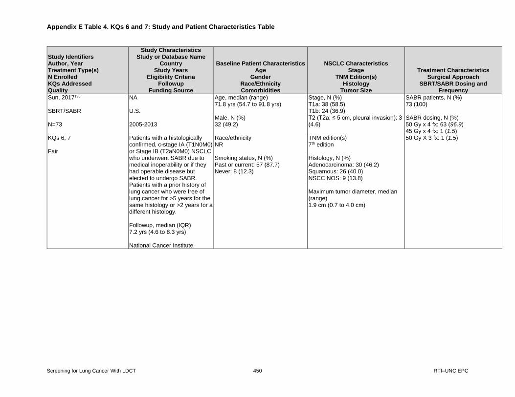
Appendix E Table 4. KQs 6 and 7: Study and Patient Characteristics Table
Screening for Lung Cancer With LDCT 450 RTI–UNC EPC
Study Identifiers Author, Year Treatment Type(s) N Enrolled KQs Addressed Quality
Study Characteristics Study or Database Name
Country Study Years
Eligibility Criteria Followup
Funding Source
Baseline Patient Characteristics Age
Gender Race/Ethnicity Comorbidities
NSCLC Characteristics Stage
TNM Edition(s) Histology
Tumor Size
Treatment Characteristics Surgical Approach
SBRT/SABR Dosing and Frequency
Sun, 2017195 SBRT/SABR N=73 KQs 6, 7 Fair
NA U.S. 2005-2013 Patients with a histologically confirmed, c-stage IA (T1N0M0) or Stage IB (T2aN0M0) NSCLC who underwent SABR due to medical inoperability or if they had operable disease but elected to undergo SABR. Patients with a prior history of lung cancer who were free of lung cancer for >5 years for the same histology or >2 years for a different histology. Followup, median (IQR) 7.2 yrs (4.6 to 8.3 yrs) National Cancer Institute
Age, median (range) 71.8 yrs (54.7 to 91.8 yrs) Male, N (%) 32 (49.2) Race/ethnicity NR Smoking status, N (%) Past or current: 57 (87.7) Never: 8 (12.3)
Stage, N (%) T1a: 38 (58.5) T1b: 24 (36.9) T2 (T2a: ≤ 5 cm, pleural invasion): 3 (4.6) TNM edition(s) 7th edition Histology, N (%) Adenocarcinoma: 30 (46.2) Squamous: 26 (40.0) NSCC NOS: 9 (13.8) Maximum tumor diameter, median (range) 1.9 cm (0.7 to 4.0 cm)
SABR patients, N (%) 73 (100) SABR dosing, N (%) 50 Gy x 4 fx: 63 (96.9) 45 Gy x 4 fx: 1 (1.5) 50 Gy X 3 fx: 1 (1.5)

Appendix E Table 4. KQs 6 and 7: Study and Patient Characteristics Table
Screening for Lung Cancer With LDCT 451 RTI–UNC EPC
Study Identifiers Author, Year Treatment Type(s) N Enrolled KQs Addressed Quality
Study Characteristics Study or Database Name
Country Study Years
Eligibility Criteria Followup
Funding Source
Baseline Patient Characteristics Age
Gender Race/Ethnicity Comorbidities
NSCLC Characteristics Stage
TNM Edition(s) Histology
Tumor Size
Treatment Characteristics Surgical Approach
SBRT/SABR Dosing and Frequency
Taremi, 2012259 SBRT/SABR N=46 KQ 7 Fair
NA Canada 2004-2008 Medically inoperable patients with T1-2N0M0 NSCLC treated with 18-20 Gy x 3 fx and with >6 mos followup Followup, median 24.9 mos Funding source Industry, other unspecified
Age, median (range) 72.8 yrs (48.3 to 89.6 yrs) Male, N (%) 22 (48) Race/ethnicity NR COPD, N (%) 29 (63)
Stage, N (%) All patients had T1-2N0M0 NSCLC TNM edition(s) NR Histology, N (%) NR Tumor size, mean (SD) 2.6 cm (1.2 cm)
SBRT patients, N (%) 46 (100) SBRT dosing, N (%) 18 or 20 Gy x 3 fx: 46 (100)

Appendix E Table 4. KQs 6 and 7: Study and Patient Characteristics Table
Screening for Lung Cancer With LDCT 452 RTI–UNC EPC
Study Identifiers Author, Year Treatment Type(s) N Enrolled KQs Addressed Quality
Study Characteristics Study or Database Name
Country Study Years
Eligibility Criteria Followup
Funding Source
Baseline Patient Characteristics Age
Gender Race/Ethnicity Comorbidities
NSCLC Characteristics Stage
TNM Edition(s) Histology
Tumor Size
Treatment Characteristics Surgical Approach
SBRT/SABR Dosing and Frequency
Taremi, 2012262 SBRT/SABR N=108 KQ 7 Fair
NA Canada 2004-2008 Medically inoperable patients with synchronous Stage T1-T2N0M0 NSCLC, either biopsy-proven or identified by “suspicious” pulmonary lesions according to evidence of interval progression on ≥2 serial CT imaging studies (minimum of 1 mo apart) and/or increased FDG uptake on PET scan, and ECOG performance status of 0-3 Followup, median 19.1 mos Funding source Government, private
Age, mean (SD) 72.6 yrs (48.3 to 90 yrs) Male, N (%) 53 (49.1) Race/ethnicity NR Comorbidities, N (%) NR
Stage, N (%) T stage 1 (≤3 cm): 86 (79.6) T stage 2 (>3 cm): 28 (20.4) TNM edition(s) NR Histology, N (%) Adenocarcinoma: 34 (29.8) Squamous cell carcinoma: 22 (19.3) Large cell carcinoma: 6 (5.3) NSCLC, NOS: 19 (16.7) No biopsy or nondiagnostic sample: 33 (28.9) Tumor size, mean (SD) 2.4 cm (1.1 cm)
SBRT patients, N (%) 108 (100) SBRT dosing, N (%) 60 Gy x 3 fx: 31 (27.2) 54 Gy x 3 fx: 20 (17.5) 48 Gy x 4 fx: 43 (37.7) 60 Gy x 8 fx: 9 (7.9) 50 Gy x 10 fx: 11 (9.6)

Appendix E Table 4. KQs 6 and 7: Study and Patient Characteristics Table
Screening for Lung Cancer With LDCT 453 RTI–UNC EPC
Study Identifiers Author, Year Treatment Type(s) N Enrolled KQs Addressed Quality
Study Characteristics Study or Database Name
Country Study Years
Eligibility Criteria Followup
Funding Source
Baseline Patient Characteristics Age
Gender Race/Ethnicity Comorbidities
NSCLC Characteristics Stage
TNM Edition(s) Histology
Tumor Size
Treatment Characteristics Surgical Approach
SBRT/SABR Dosing and Frequency
Tsutani, 2014177 Surgery N=618 KQs 6, 7 Good
NA Japan 2005-2010 Patients with clinical T1N0 M0 Stage IA NSCLC adenocarcinoma (no synchronous multiple tumors) who underwent preoperative staging using HRT and FDG-PET/CT and had definitive histopathologic diagnosis, followed by complete curative resection without neoadjuvant chemotherapy or radiotherapy Followup, median 42.9 mos Funding source NR
Age, mean (range) 66 yrs (31 to 89 yrs) Male, N (%) 272 (44) Race/ethnicity NR Comorbidities, N (%) NR
Stage, N (%) Clinical T1a: 354 (57.3) Clinical T1b: 264 (42.7) TNM edition(s) 7th edition Histology, N (%) Adenocarcinoma: 618 (100) Adenocarcinoma in situ: 97 (15.7) Tumor size, mean Whole tumor: 2.0 cm Solid tumor: 1.1 cm
Surgical approach, N (%) L: 383 (62.0) SLR: 235 (38.0) S: 98 (15.9) W: 137 (22.2)

Appendix E Table 4. KQs 6 and 7: Study and Patient Characteristics Table
Screening for Lung Cancer With LDCT 454 RTI–UNC EPC
Study Identifiers Author, Year Treatment Type(s) N Enrolled KQs Addressed Quality
Study Characteristics Study or Database Name
Country Study Years
Eligibility Criteria Followup
Funding Source
Baseline Patient Characteristics Age
Gender Race/Ethnicity Comorbidities
NSCLC Characteristics Stage
TNM Edition(s) Histology
Tumor Size
Treatment Characteristics Surgical Approach
SBRT/SABR Dosing and Frequency
Ubels, 2015204 SBRT/SABR N=39 KQs 6, 7 Fair
NA The Netherlands 2006-2008 Patients who refused surgery or had inoperable Stage T1-2N0M0 NSCLC with pathological confirmation of diagnosis and no progressive disease 3 weeks after treatment Followup, median 38 mos Funding source NR
Age, median (range) 77 yrs (55 to 87 yrs) Male, N (%) NR Race/ethnicity NR Comorbidities Charlson Comorbidity Index, N (%) Score=0-2: 20 (51) Score=3: 13 (33) Score ≥3: 6 (15) COPD, N (%) 22 (56)
Stage, N (%) Stage T1: 17 (44) Stage T2: 21 (54) Stage T3: 1 (3) TNM edition(s) NR Histology, N (%) Adenocarcinoma: 8 (21) Squamous cell: 14 (36) Large cell carcinoma: 13 (33) Other: 4 (10) Tumor size, mean (SD) NR
SBRT patients, N (%) 39 (100) SBRT dosing, N (%) 60 Gy x 3 fx: 30 (76.9) 48-50 Gy x 5-6 fx: 7 (17.9) 45 Gy x 3 fx: 2 (5.1)
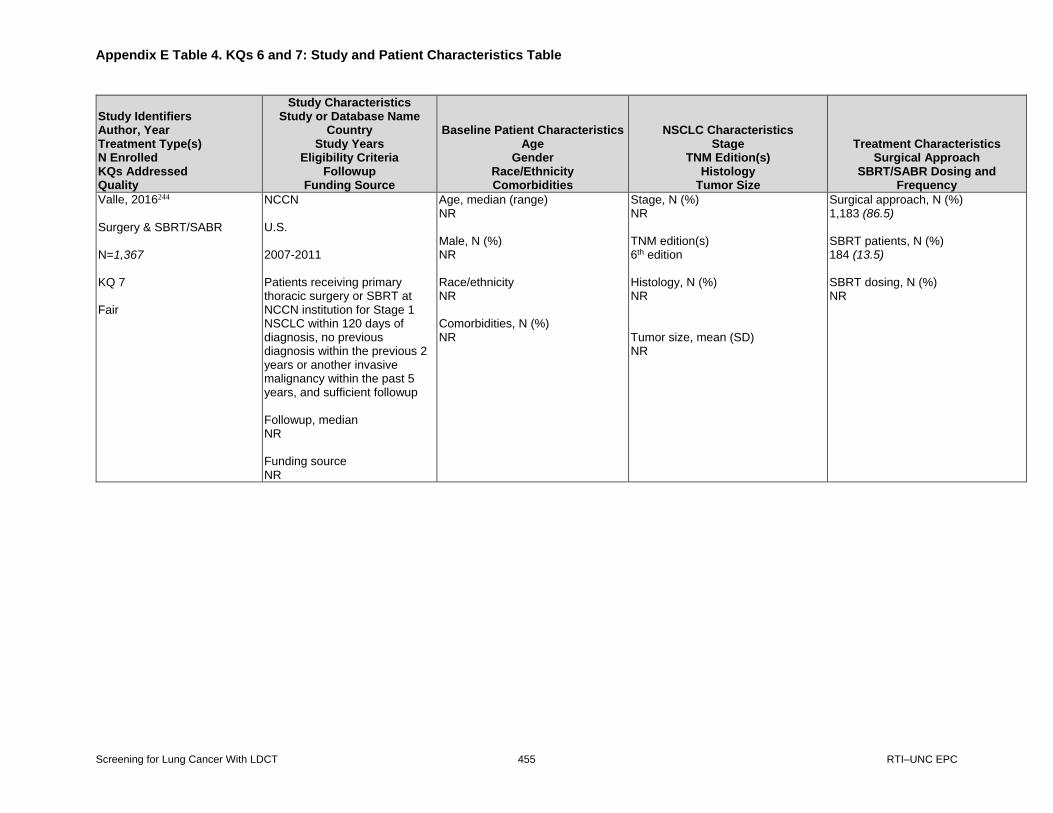
Appendix E Table 4. KQs 6 and 7: Study and Patient Characteristics Table
Screening for Lung Cancer With LDCT 455 RTI–UNC EPC
Study Identifiers Author, Year Treatment Type(s) N Enrolled KQs Addressed Quality
Study Characteristics Study or Database Name
Country Study Years
Eligibility Criteria Followup
Funding Source
Baseline Patient Characteristics Age
Gender Race/Ethnicity Comorbidities
NSCLC Characteristics Stage
TNM Edition(s) Histology
Tumor Size
Treatment Characteristics Surgical Approach
SBRT/SABR Dosing and Frequency
Valle, 2016244 Surgery & SBRT/SABR N=1,367 KQ 7 Fair
NCCN U.S. 2007-2011 Patients receiving primary thoracic surgery or SBRT at NCCN institution for Stage 1 NSCLC within 120 days of diagnosis, no previous diagnosis within the previous 2 years or another invasive malignancy within the past 5 years, and sufficient followup Followup, median NR Funding source NR
Age, median (range) NR Male, N (%) NR Race/ethnicity NR Comorbidities, N (%) NR
Stage, N (%) NR TNM edition(s) 6th edition Histology, N (%) NR Tumor size, mean (SD) NR
Surgical approach, N (%) 1,183 (86.5) SBRT patients, N (%) 184 (13.5) SBRT dosing, N (%) NR

Appendix E Table 4. KQs 6 and 7: Study and Patient Characteristics Table
Screening for Lung Cancer With LDCT 456 RTI–UNC EPC
Study Identifiers Author, Year Treatment Type(s) N Enrolled KQs Addressed Quality
Study Characteristics Study or Database Name
Country Study Years
Eligibility Criteria Followup
Funding Source
Baseline Patient Characteristics Age
Gender Race/Ethnicity Comorbidities
NSCLC Characteristics Stage
TNM Edition(s) Histology
Tumor Size
Treatment Characteristics Surgical Approach
SBRT/SABR Dosing and Frequency
Videtic, 2014269 SBRT/SABR N=80 KQ 7 Fair
NA U.S. 2009-2012 Medically inoperable patients who received treated with single-fx SBRT, including those potentially eligible for the RTOG 0915 or RPCI trials based on tumor size and peripheral location requirements, but who missed eligibility criteria for enrollment (e.g., biopsy result showing proof of malignancy) and received single-fx SBRT up off-protocol Followup, median (range) Single-fx SBRT at 30 Gy: 18.7 mos (1.8 to 43.0 mos) Single-fx SBRT at 34 Gy: 17.8 mos (0.1 to 39.4 mos) Funding source NR
Age, median (range) Single-fx SBRT at 30 Gy: 75 (48 to 91) Single-fx SBRT at 34 Gy: 73 (53 to 84) Male, N (%) Single-fx SBRT at 30 Gy: 36 (65.5) Single-fx SBRT at 34 Gy: 11 (44) Race/ethnicity NR Comorbidities, N (%) Current smoking status Single-fx SBRT at 30 Gy: 10 (18.2) Single-fx SBRT at 34 Gy: 8 (32) Pack-year history, median (range) Single-fx SBRT at 30 Gy: 50 (0 to 100) Single-fx SBRT at 34 Gy: 55 (5 to 125)
Stage, N (%) Clinical T1a Single-fx SBRT at 30 Gy: 49 (89.1) Single-fx SBRT at 34 Gy: 24 (96) Clinical T1b Single-fx SBRT at 30 Gy: 6 (10.9) Single-fx SBRT at 34 Gy: 1 (4) TNM edition(s) NR Histology, N (%) Adenocarcinoma Single-fx SBRT at 30 Gy: 12 (32) Single-fx SBRT at 34 Gy: 8 (43) Squamous cell carcinoma Single-fx SBRT at 30 Gy: 14 (37) Single-fx SBRT at 34 Gy: 9 (47) Other Single-fx SBRT at 30 Gy: 4 (10) Single-fx SBRT at 34 Gy: 0 (0) Nondiagnostic Single-fx SBRT at 30 Gy: 8 (21) Single-fx SBRT at 34 Gy: 2 (10) Tumor size, median (range) Single-fx SBRT at 30 Gy: 1.7 cm (0.9 to 4.8 cm) Single-fx SBRT at 34 Gy: 1.7 cm (1.0 to 4.0 cm)
SBRT patients, N (%) 80 (100) SBRT dosing, N (%) 30 Gy x 1 fx: 55 (69) 34 Gy x 1 fx: 25 (31)

Appendix E Table 4. KQs 6 and 7: Study and Patient Characteristics Table
Screening for Lung Cancer With LDCT 457 RTI–UNC EPC
Study Identifiers Author, Year Treatment Type(s) N Enrolled KQs Addressed Quality
Study Characteristics Study or Database Name
Country Study Years
Eligibility Criteria Followup
Funding Source
Baseline Patient Characteristics Age
Gender Race/Ethnicity Comorbidities
NSCLC Characteristics Stage
TNM Edition(s) Histology
Tumor Size
Treatment Characteristics Surgical Approach
SBRT/SABR Dosing and Frequency
Videtic, 2015261 SBRT/SABR N=84 KQ 7 Fair
RTOG 0915 U.S. 2009-2011 Patients with cytologic or histologic diagnosis of NSCLC >2 cm staged as T1-T2N0M0 and based on CT and PET imaging; Zubrod performance status of 0-2; deemed medically inoperable by thoracic oncology specialist or declining surgery despite being operable; and no prior or planned use of concomitant antineoplastic therapy during SBRT protocol Followup, median 30.2 mos Funding source Government
Age, median (range) SBRT at any dose: 75 yrs (52 to 89 yrs) SBRT 34 Gy x 1 fx: 75 yrs (57 to 89 yrs) SBRT 48 Gy x 4 fx: 75 yrs (52 to 87 yrs) Male, N (%) Male SBRT at any dose: 38 (45.2) SBRT 34 Gy x 1 fx: 16 (41.0) SBRT 48 Gy x 4 fx: 22 (48.9) Race/ethnicity NR Comorbidities, N (%) NR
Stage, N (%) T1 SBRT at any dose: 72 (85.7) SBRT 34 Gy x 1 fx: 32 (82) SBRT 48 Gy x 4 fx: 40 (88.9) T2 SBRT at any dose: 12 (14.3) SBRT 34 Gy x 1 fx: 7 (18.0) SBRT 48 Gy x 4 fx: 5 (11.1) TNM edition(s) 6th edition Histology, N (%) Adenocarcinoma SBRT at any dose: 49 (58.3) SBRT 34 Gy x 1 fx: 23 (59.0) SBRT 48 Gy x 4 fx: 26 (57.8) Squamous cell SBRT at any dose: 25 (29.8) SBRT 34 Gy x 1 fx: 9 (23.1) SBRT 48 Gy x 4 fx: 16 (35.6) NSCLC NOS SBRT at any dose: 10 (11.9) SBRT 34 Gy x 1 fx: 7 (17.9) SBRT 48 Gy x 4 fx: 3 (6.7) Max tumor diameter, median (range) SBRT at any dose: 2.0 cm (0.8 to 4.98 cm) SBRT 34 Gy x 1 fx: 2.0 cm (1.0 to 4.98 cm) SBRT 48 Gy x 4 fx: 2.0 cm (0.8 to 4.3 cm)
SBRT patients, N (%) 84 (100) SBRT dosing, N (%) 34 Gy x 1 fx: 39 (46.4) 48 Gy x 4 fx: 45 (53.6)

Appendix E Table 4. KQs 6 and 7: Study and Patient Characteristics Table
Screening for Lung Cancer With LDCT 458 RTI–UNC EPC
Study Identifiers Author, Year Treatment Type(s) N Enrolled KQs Addressed Quality
Study Characteristics Study or Database Name
Country Study Years
Eligibility Criteria Followup
Funding Source
Baseline Patient Characteristics Age
Gender Race/Ethnicity Comorbidities
NSCLC Characteristics Stage
TNM Edition(s) Histology
Tumor Size
Treatment Characteristics Surgical Approach
SBRT/SABR Dosing and Frequency
Yang, 2016248 Surgery N=20,191 KQ 7 Fair
NCDB U.S. 2010-2012 Patients diagnosed with clinical T Stage 1-2, N0, M0 NSCLC, undergoing lobectomy, with available data on surgical approach, and no history of unrelated malignancy Followup, median NR Funding source Government, professional association
Age, median (IQR) Open surgery vs. MIS – PMS subset (N=18,780) Open L: 68 yrs (60 to 74 yrs) MIS L: 68 yrs (60 to 74 yrs) VATS vs. robotic L – PMS subset (N=3,876) VATS L: 69 yrs (62 to 74 yrs) Robotic L: 68 yrs (61 to 74 yrs) Male, N (%) Open surgery vs. MIS – PMS subset (N=18,780) Open L: 5,385 (57.3) MIS L: 5,375 (57.2) VATS vs. robotic L – PMS subset (N=3,876) VATS L: 1,079 (55.7) Robotic L: 1,099 (56.7) White, N (%) Open surgery vs. MIS – PMS subset (N=18,780) Open L: 8,336 (88.8) MIS L: 8,263 (88) VATS vs. robotic L – PMS subset (N=3,876) VATS L: 1,721 (88.8) Robotic L: 1,687 (87)
Stage, N (%) Open surgery vs. MIS – PMS subset (N=18,780) Clinical T status T1 Open L: 6,685 (71.2) MIS L: 6,598 (70.3) T2 Open L: 2,705 (28.8) MIS L: 2,792 (29.7) Pathologic T status T0 (in situ) Open L: 7 (0.1) MIS L: 12 (0.1) T1 Open L: 5,398 (59.7) MIS L: 5,259 (57.8) T2 Open L: 3,222 (35.6) MIS L: 3,386 (37.2) T3 Open L: 362 (4.0) MIS L: 393 (4.3) T4 Open L: 56 (0.6) MIS L: 44 (0.5) Pathologic N status N0 Open L: 7,861 (87.8) MIS L: 7,969 (88.5)
Surgical approach, N (%) Open surgery vs. MIS – PMS subset (N=18,780) MIS L: 9,390 (50) VATS L: 9,390 (50) VATS vs. robotic L – PMS subset (N=3,876) Robotic L: 1,938 (50) Open L: 1,938 (50)

Appendix E Table 4. KQs 6 and 7: Study and Patient Characteristics Table
Screening for Lung Cancer With LDCT 459 RTI–UNC EPC
Study Identifiers Author, Year Treatment Type(s) N Enrolled KQs Addressed Quality
Study Characteristics Study or Database Name
Country Study Years
Eligibility Criteria Followup
Funding Source
Baseline Patient Characteristics Age
Gender Race/Ethnicity Comorbidities
NSCLC Characteristics Stage
TNM Edition(s) Histology
Tumor Size
Treatment Characteristics Surgical Approach
SBRT/SABR Dosing and Frequency
Yang, 2016248 (continued)
Comorbidities, N (%) Open surgery vs. MIS – PSM subset (N=18,780) Charlson=0 Open L: 4,747 (50.6) MIS L: 4,670 (49.7) Charlson=1 Open L: 3,426 (36.5) MIS L: 3,446 (36.7) Charlson=2+ Open L: 1,217 (13.0) MIS L: 1,274 (13.6) VATS vs. robotic L – PSM subset (N=3,876) Charlson=0 VATS L: 863 (44.5) Robotic L: 889 (45.9) Charlson=1 VATS L: 811 (41.8) Robotic L: 762 (39.3) Charlson=2+ VATS L: 264 (13.6) Robotic L: 287 (14.8)
N1 Open L: 728 (8.1) MIS L: 691 (7.7) N2 Open L: 366 (4.1) MIS L: 338 (3.8) N3 Open L: 1 (0.0) MIS L: 3 (0.0) Pathologic M status M0 Open L: 9,300 (99.8) MIS L: 9,295 (99.7) M1 Open L: 21 (0.2) MIS L: 26 (0.3) VATS vs. robotic L – PMS subset (N=3,876) Clinical T status T1 VATS L: 1,445 (74.6) Robotic L: 1,401 (72.3) T2 VATS L: 493 (25.4) Robotic L: 537 (27.7) Pathologic T status T0 (in situ) VATS L: 5 (0.3) Robotic L: 3 (0.2) T1 VATS L: 1,143 (61.0) Robotic L: 1,112 (59.5)

Appendix E Table 4. KQs 6 and 7: Study and Patient Characteristics Table
Screening for Lung Cancer With LDCT 460 RTI–UNC EPC
Study Identifiers Author, Year Treatment Type(s) N Enrolled KQs Addressed Quality
Study Characteristics Study or Database Name
Country Study Years
Eligibility Criteria Followup
Funding Source
Baseline Patient Characteristics Age
Gender Race/Ethnicity Comorbidities
NSCLC Characteristics Stage
TNM Edition(s) Histology
Tumor Size
Treatment Characteristics Surgical Approach
SBRT/SABR Dosing and Frequency
Yang, 2016248 (continued)
T2 VATS L: 625 (33.4) Robotic L: 665 (35.6) T3 VATS L: 87 (4.6) Robotic L: 82 (4.4) T4 VATS L: 13 (0.7) Robotic L: 7 (0.4) Pathologic N status N0 VATS L: 1,661 (89.4) Robotic L: 1,652 (89.0) N1 VATS L: 138 (7.4) Robotic L: 136 (7.3) N2 VATS L: 65 (3.5) Robotic L: 67 (3.6) N3 VATS L: 0 (0) Robotic L: 2 (0.1) Pathologic M status M0 VATS L: 1,910 (99.7) Robotic L: 1,910 (99.7) M1 VATS L: 6 (0.3) Robotic L: 6 (0.3) TNM edition(s) 7th edition

Appendix E Table 4. KQs 6 and 7: Study and Patient Characteristics Table
Screening for Lung Cancer With LDCT 461 RTI–UNC EPC
Study Identifiers Author, Year Treatment Type(s) N Enrolled KQs Addressed Quality
Study Characteristics Study or Database Name
Country Study Years
Eligibility Criteria Followup
Funding Source
Baseline Patient Characteristics Age
Gender Race/Ethnicity Comorbidities
NSCLC Characteristics Stage
TNM Edition(s) Histology
Tumor Size
Treatment Characteristics Surgical Approach
SBRT/SABR Dosing and Frequency
Yang, 2016248 (continued)
Histology, N (%) Open surgery vs. MIS – PMS subset (N=18,780) Well differentiated Open L: 1,813 (20.5) MIS L: 1,826 (20.5) Moderately differentiated Open L: 4,167 (47.2) MIS L: 4,255 (47.9) Poorly differentiated Open L: 2,758 (31.2) MIS L: 2,722 (30.6) Undifferentiated/anaplastic Open L: 97 (1.1) MIS L: 86 (1) VATS vs. robotic L – PMS subset (N=3,876) Well differentiated VATS L: 359 (19.6) Robotic L: 415 (22.5) Moderately differentiated VATS L: 897 (48.9) Robotic L: 865 (46.9) Poorly differentiated VATS L: 565 (30.8) Robotic L: 551 (29.9) Undifferentiated/anaplastic VATS L: 13 (0.7) Robotic L: 14 (0.8)

Appendix E Table 4. KQs 6 and 7: Study and Patient Characteristics Table
Screening for Lung Cancer With LDCT 462 RTI–UNC EPC
Study Identifiers Author, Year Treatment Type(s) N Enrolled KQs Addressed Quality
Study Characteristics Study or Database Name
Country Study Years
Eligibility Criteria Followup
Funding Source
Baseline Patient Characteristics Age
Gender Race/Ethnicity Comorbidities
NSCLC Characteristics Stage
TNM Edition(s) Histology
Tumor Size
Treatment Characteristics Surgical Approach
SBRT/SABR Dosing and Frequency
Yang, 2016248 (continued)
Tumor size, mean (SD) Open surgery vs. MIS (N=18,780) Pathologic tumor size Open L: 2.7 cm (2.1 cm) MIS L: 2.7 cm (2.1 cm) VATS vs. Robotic L (N=3,876) Pathologic tumor size VATS L: 2.6 cm (1.4 cm) Robotic L: 2.7 cm (2.3 cm)
Zhai, 2014170 Surgery N=724 (subset of sample with Stage 1 NSCLC) KQ 6 Fair
NA U.S. 1992-2010 Patients >18 years old with pathologically confirmed newly diagnosed NSCLC (Stages 1-2) and not receiving adjuvant therapy who were consecutively recruited and followed Followup, median 41 mos Funding source Government
Age, mean (SD) NR Male, N (%) NR Race/ethnicity NR Comorbidities 271 (37.4)
Stage, N (%) c-stage 1A: 538 (59.6) c-stage 1B: 186 (20.6) TNM edition(s) NR Histology, N (%) NR Tumor size, N (%) NR
Surgical approach, N (%) W: NR L: NR Others: NR

Appendix E Table 4. KQs 6 and 7: Study and Patient Characteristics Table
Screening for Lung Cancer With LDCT 463 RTI–UNC EPC
Study Identifiers Author, Year Treatment Type(s) N Enrolled KQs Addressed Quality
Study Characteristics Study or Database Name
Country Study Years
Eligibility Criteria Followup
Funding Source
Baseline Patient Characteristics Age
Gender Race/Ethnicity Comorbidities
NSCLC Characteristics Stage
TNM Edition(s) Histology
Tumor Size
Treatment Characteristics Surgical Approach
SBRT/SABR Dosing and Frequency
Zhao, 2017181 Surgery N=7,989 KQ 6 Fair
SEER U.S. 2004-2012 Patients with histologically diagnosed Stage 1A lung adenocarcinoma measuring ≥10 but ≤20 mm, not confined to hilus or a primary tumor in main bronchus, and treated with segmentectomy or lobectomy, but not post-operative radiotherapy Followup, median 52.2 mos Funding source Government, university
Age, mean (SD) S: 69.8 yrs (9.2 yrs) L: 67.3 yrs (9.6 yrs) Male, N (%) Female Total: 3,184 (39.9) S: 202 (35.8) L: 2,982 (40.2) White, N (%) Total: 6,834 (85.5) S: 498 (88.3) L: 6,336 (85.3) Comorbidities, N (%) NR
Stage, N (%) p-stage T1a: 7,989 (100) c-stage T1b: 7,989 (100) TNM edition(s) 8th edition Histology, N (%) Major adenocarcinoma Total: 7,413 (92.8) S: 506 (89.7) L: 6,907 (93.0) Mucinous adenocarcinoma Total: 348 (4.4) S: 27 (4.8) L: 321 (4.3) BAC, nonmucinous Total: 228 (2.9) S: 31 (5.5) L: 197 (2.7) Tumor size, median (SD) Total: NR S: 15.6 mm (2.9 mm) L: 16.2 mm (2.9 mm)
Surgical approach, N (%) S: 564 (7.1) L: 7,425 (92.9)
* FEV1 was not available in 13/115 SBRT patients.
† Analytical stage included the AJCC p-stage group if available; otherwise, the c-stage group was used.
Abbreviations: 3D-CRT=three-dimensional conformal radiation therapy; ASA=American Society of Anesthesiologists; BAC=broncholoalveolar carcinoma; Bi-L=bilobectomy;
c-stage=clinical stage; cm=centimeter(s); CoC=Commission on Cancer; COPD=chronic obstructive pulmonary disease; DLCO=diffusing capacity of the lungs for carbon
monoxide; ECOG=Eastern Cooperative Oncology Group; FEV1=forced expiratory volume in one second; fx=fraction(s); Gy=Gray; IQR=interquartile range; JCOG=Japan
Clinical Oncology Group; KQ=Key Question; L=lobectomy; N=number of patients enrolled or analyzed; NA=not applicable; NCDB=National Cancer Database; NCI=National
Cancer Institute; NOS=not otherwise specified; NPC=not pathologically confirmed; NR=not reported; NSCLC=non-small cell lung cancer; p-stage=pathologic stage;
PC=pathologically confirmed; S=segmentectomy; SBRT/SABR=stereotactic body radiotherapy/stereotactic ablative radiotherapy; SD=standard deviation; SEER=Surveillance,
Epidemiology, and End Results Program; SLR=sublobar resection; STS-GTS=Society of Thoracic Surgeons-General Thoracic Surgery Database; surg exp=surgical experience;
T=thoracotomy; TNM=tumor-node-metastasis cancer staging system; U.K.=United Kingdom; U.S.=United States; VATS=video-assisted thoracoscopic lobectomy;
VMAT=volumetric modulated arc therapy; VPI=visceral pleural invasion; W=wedge resection.

Appendix E Table 5. KQs 6 and 7: Surgery Results
Screening for Lung Cancer With LDCT 464 RTI–UNC EPC
Appendix E Table 5. KQ6 & 7 Surgery Results
Study Identifiers Author, Year N Enrolled (Analyzed) KQs Addressed Quality
Long-Term Survival and Mortality Outcomes, %
(95% CI)*
Long-Term Progression-Related Outcomes, %
(95% CI)* Short-Term Mortality, N (%)* Adverse Events, N (%)*
Brunelli, 2015179 N=1,370 (1,370) KQs 6, 7 Good
5-yr OS ThRCRI prognostic class A (score 0 to 1) Total: 66 (NR) p-stage T1: 73 (NR) p-stage T2: 61 (NR) ThRCRI prognostic class B (score 1.5 to 2.5) Total: 53 (NR) p-stage T1: 64 (NR) p-stage T2: 48 (NR) ThRCRI prognostic class C (score >2.5) Total: 35 (NR) p-stage T1: 55 (NR) p-stage T2: 32 (NR)
5-yr LCSS ThRCRI prognostic class A (score 0 to 1) Total: 77 (NR) ThRCRI prognostic class B (score 1.5 to 2.5) Total: 75 (NR) ThRCRI prognostic class C (score >2.5) Total: 55 (NR)
30-day mortality 24 (1.8) Postoperative cardiac-related mortality, % ThRCRI prognostic class A (score 0 to 1) Total: 0.03 ThRCRI prognostic class B (score 1.5 to 2.5) Total: 1.4 ThRCRI prognostic class C (score >2.5) Total: 4.1
Major postoperative cardiac events in hospital or within 30 days of surgery (i.e., acute MI, pulmonary edema, ventricular fibrillation or primary cardiac arrest, complete heart block, any cardiac-related death) Total sample 80 (5.8) ThRCRI prognostic class A (score 0 to 1), % Total: 11 ThRCRI prognostic class B (score 1.5 to 2.5), % Total: 19 ThRCRI prognostic class C (score >2.5), % Total: 42 Major postoperative cardiac morbidity, % ThRCRI prognostic class A (score 0 to 1) Total: 4 ThRCRI prognostic class B (score 1.5 to 2.5) Total: 11 ThRCRI prognostic class C (score >2.5) Total: 17 Cardiac event mortality during followup, % ThRCRI prognostic class A (score 0 to 1) Total: 1.5 ThRCRI prognostic class B (score 1.5 to 2.5) Total: 7 ThRCRI prognostic class C (score >2.5) Total: 13

Appendix E Table 5. KQs 6 and 7: Surgery Results
Screening for Lung Cancer With LDCT 465 RTI–UNC EPC
Study Identifiers Author, Year N Enrolled (Analyzed) KQs Addressed Quality
Long-Term Survival and Mortality Outcomes, %
(95% CI)*
Long-Term Progression-Related Outcomes, %
(95% CI)* Short-Term Mortality, N (%)* Adverse Events, N (%)*
Bryant, 2018194 N=3,620 for surgery (total=3,620; L=2,986; SLR=634) KQs 6, 7 Fair
5-yr OS L: 70 (NR) SLR: 56 (NR) 5-yr LC mortality L: 23 (NR) SLR: 32 (NR)
NR 30-day mortality L: 57 (1.9) SLR: 11 (1.7) 90-day mortality L: 107 (3.6) SLR: 16 (2.5)
NR
Chang, 2007190
N=10,761 (total=10,761; L=8,527; SLR=2,234) KQ 6 Fair
5-yr OS All surg: 57.8 (NR) L: 61.4 (NR) SLR: 44.0 (NR) 5-yr OS after all surgeries and stratified by gender Female: 63.0 (NR) Male: 52.8 (NR) p<0.0001 5-yr OS after all surgeries and stratified by age Age <67: 65.2 (NR) Age ≥67: 51.5 (NR) p<0.0001
NR NR NR
Cox, 2017184 N=1,991 (total=1,991; L=1,544; SLR=447) KQ 6 Fair
5-yr OS Total: NR L: 70.5 (NR) SLR: 67.8 (NR)
NR NR NR
Fernandez, 2012186 N=657 (657) KQ 6 Fair
Unadjusted 5-yr OS 41 (NR) Unadjusted 5-yr DSS 59 (NR)
NR NR NR

Appendix E Table 5. KQs 6 and 7: Surgery Results
Screening for Lung Cancer With LDCT 466 RTI–UNC EPC
Study Identifiers Author, Year N Enrolled (Analyzed) KQs Addressed Quality
Long-Term Survival and Mortality Outcomes, %
(95% CI)*
Long-Term Progression-Related Outcomes, %
(95% CI)* Short-Term Mortality, N (%)* Adverse Events, N (%)*
Goya, 2005193 N=3,965 eligible (total=3,965; c-stage 1A=2,423 and 1B=1,542; p-stage 1A=2,009 and 1B=1,418) KQ 6 Fair
5-yr OS c-stage 1 IA: 72.1 (NR) IB: 49.9 (NR) p-stage 1 IA: 79.5 (NR) IB: 60.1 (NR)
NR NR NR
Guerrera, 2015169
N=848 (848) KQ 6 Fair
5-yr OS 74 (0.71 to 0.77)
NR NR NR
Husain, 2015237 N=112,216 (71,175) KQ 7 Fair
NR NR Overall 30-day mortality All surg: 1,566 (2.2) 30-day mortality – subgroup analyses stratified by comorbidities and age, % Charlson-Deyo Comorbidity Score=0 Age <75 yrs All surg: 1.3 L: 1.2
NR

Appendix E Table 5. KQs 6 and 7: Surgery Results
Screening for Lung Cancer With LDCT 467 RTI–UNC EPC
Study Identifiers Author, Year N Enrolled (Analyzed) KQs Addressed Quality
Long-Term Survival and Mortality Outcomes, %
(95% CI)*
Long-Term Progression-Related Outcomes, %
(95% CI)* Short-Term Mortality, N (%)* Adverse Events, N (%)*
Husain, 2015237 (continued)
SLR: 1.5 Age ≥75 yrs All surg: 3.3 L: 3.4 SLR: 3.1 Charlson-Deyo Comorbidity Score=1 Age <75 yrs All surg: 1.6 L: 1.6 SLR: 1.6 Age ≥75 yrs All surg: 4.1 L: 4.5 SLR: 2.7 Charlson-Deyo Comorbidity Score=2 Age <75 yrs All surg: 2.3 L: 2.3 SLR: 2.3 Age ≥75 yrs All surg: 5.8 L: 6.6 SLR: 3.9
Khullar, 2015176 N= 54,350 (54,350) KQ 6 Fair
5-yr OS c-stage 1: 57.2 (NR) p-stage 1: 60.5 (NR)
NR NR NR

Appendix E Table 5. KQs 6 and 7: Surgery Results
Screening for Lung Cancer With LDCT 468 RTI–UNC EPC
Study Identifiers Author, Year N Enrolled (Analyzed) KQs Addressed Quality
Long-Term Survival and Mortality Outcomes, %
(95% CI)*
Long-Term Progression-Related Outcomes, %
(95% CI)* Short-Term Mortality, N (%)* Adverse Events, N (%)*
Khullar, 2015187 Total N=28,241 (total=28,241; for OS: L=4,857; W=1,891; S=286; for 30-day mortality: L=19,718; W=7,297; S=1,226) KQs 6, 7 Fair
5-yr OS Total: NR L: 70.4 (69.0 to 71.7) W: 54.6 (52.3 to 56.9) S: 59.6 (53.5 to 65.2)
NR 30-day mortality Total: 445 (1.6) L: 316 (1.6) W: 110 (1.5) S: 19 (1.6)
NR
Lakha, 2014173 N=16,315 (15,067) KQ 6 Fair
5-yr OS Range: 53 (49 to 57) to 75 (74 to 77) <2 cm without VPI: 75 (74 to 77) <2 cm with VPI: 70 (66 to 75) 2-3 cm without VPI: 68 (65 to 70) 2-3 cm with VPI: 61 (56 to 65) >3-5 cm without VPI: 60 (57 to 62) >3-5 cm with VPI: 53 (49 to 57)
5-yr LCSS Range: 66 (62 to 69) to 88 (86 to 89) <2 cm without VPI: 88 (86 to 89) <2 cm with VPI: 84 (81 to 87) 2-3 cm without VPI: 79 (77 to 81) 2-3 cm with VPI: 71 (67 to 75) >3-5 cm without VPI: 72 (70 to 74) >3-5 cm with VPI: 66 (62 to 69)
NR NR
Landreneau, 2014174 N=624 (624) KQs 6, 7 Fair
5-yr OS Total: NR S: 54 (0.47 to 0.61) L: 60 (0.54 to 0.67)
NR 30-day mortality Total: 12 (1.9) S: 4 (1.2) (95% CI, 0.4-32) L: 8 (2.5) (95% CI, 1.1 to 5.0) 90-day mortality Total: 23 (3.7) S: 8 (2.6) (95% CI, 1.1-5.0) L: 15 (4.8) (95% CI, 2.7 to 7.8)
Overall morbidity Total: 217 (34.8) S: 115 (36.9) L: 102 (32.7)

Appendix E Table 5. KQs 6 and 7: Surgery Results
Screening for Lung Cancer With LDCT 469 RTI–UNC EPC
Study Identifiers Author, Year N Enrolled (Analyzed) KQs Addressed Quality
Long-Term Survival and Mortality Outcomes, %
(95% CI)*
Long-Term Progression-Related Outcomes, %
(95% CI)* Short-Term Mortality, N (%)* Adverse Events, N (%)*
Licht, 2013247 N=1,513 (total=1,513; VATS L=717; Open L=796) KQ 7 Fair
NR NR 30-day mortality Total: 31 (2.0) VATS L: 8 (1.1) Open L: 23 (2.9)
NR

Appendix E Table 5. KQs 6 and 7: Surgery Results
Screening for Lung Cancer With LDCT 470 RTI–UNC EPC
Louie, 2016236 13,598 (13,598) KQ 7 Fair
NR NR 30-day mortality Total: 106 (0.8) Robotic L: 7 (0.6) VATS L: 99 (0.8) In-hospital mortality Total: 78 (0.6) Robotic L: 4 (0.3) VATS L: 74 (0.6)
Air leak >5 days Total: 1,334 (9.8) Robotic L: 122 (10.0) VATS L: 1,212 (9.8) Adult respiratory distress syndrome Total: 61 (0.4) Robotic L: 2 (0.2) VATS L: 59 (0.5) Atelectasis requiring bronchoscopy Total: 391 (2.9) Robotic L: 31 (2.5) VATS L: 360 (2.9) Atrial arrhythmia requiring treatment Total: 1,346 (9.9) Robotic L: 125 (10.2) VATS L: 1,221 (9.9) Bronchopleural fistulas Total: 42 (0.3) Robotic L: 7 (0.6) VATS L: 35 (0.3) Chylothorax requiring medical intervention Total: 64 (0.5) Robotic L: 4 (0.3) VATS L: 60 (0.5) DVT Total: 52 (0.4) Robotic L: 5 (0.4) VATS L: 47 (0.4) Emphysema requiring treatment Total: 50 (0.4) Robotic L: 6 (0.5) VATS L: 44 (0.4) Initial ventilatory support >48 hours Total: 50 (0.4) Robotic L: 6 (0.5) VATS L: 44 (0.4) Myocardial infarction Total: 42 (0.3) Robotic L: 5 (0.4) VATS L: 37 (0.3) Pneumonia Total: 442 (3.3)

Appendix E Table 5. KQs 6 and 7: Surgery Results
Screening for Lung Cancer With LDCT 471 RTI–UNC EPC
Study Identifiers Author, Year N Enrolled (Analyzed) KQs Addressed Quality
Long-Term Survival and Mortality Outcomes, %
(95% CI)*
Long-Term Progression-Related Outcomes, %
(95% CI)* Short-Term Mortality, N (%)* Adverse Events, N (%)*
Louie, 2016236 (continued)
Robotic L: 33 (2.7) VATS L: 409 (3.3) Pneumothorax requiring CT reinsertion Total: 474 (3.5) Robotic L: 51 (4.2) VATS L: 423 (3.4) Pulmonary embolus Total: 62 (0.5) Robotic L: 4 (0.3) VATS L: 58 (0.5) Recurrent laryngeal nerve paresis/paralysis Total: 26 (0.2) Robotic L: 2 (0.2) VATS L: 24 (0.2) Reintubation Total: 308 (2.3) Robotic L: 25 (2.0) VATS L: 283 (2.3) Required reoperation for bleeding Total: 65 (0.9) Robotic L: 3 (0.8) VATS L: 62 (0.9) Respiratory failure Total: 119 (1.9) Robotic L: 16 (1.9) VATS L: 103 (1.9) Tracheostomy Total: 99 (0.7) Robotic L: 9 (0.7) VATS L: 90 (0.7)
Maeda, 2010189
N=734 (713) KQ 6 Fair
5-yr OS 85.8 (NR) 10-yr OS 71.3 (NR)
NR NR NR

Appendix E Table 5. KQs 6 and 7: Surgery Results
Screening for Lung Cancer With LDCT 472 RTI–UNC EPC
Study Identifiers Author, Year N Enrolled (Analyzed) KQs Addressed Quality
Long-Term Survival and Mortality Outcomes, %
(95% CI)*
Long-Term Progression-Related Outcomes, %
(95% CI)* Short-Term Mortality, N (%)* Adverse Events, N (%)*
Maeda, 2012182 N=1,074 (1,070 with c-stage 1A; 691 with p-stage 1A NSCLC) KQ 6 Fair
5-yr OS c-stage 1 Total: NR NR by smoking history p-stage 1 Total: NR Smoking history of pack-years ≤20 96.5 (NR) Smoking history of pack-years >20 85.9 (NR) 5-yr OS subgroup analyses Age <65: 83.3 (NR) Age >65: 76.9 (NR) Women: 85.4 (NR) Men: 74.4 (NR) Smoking history of 0 ≤ pack-years ≤ 20 85.5 (NR) Smoking history of no pack-years 85.2 (NR) Smoking history of 0 ≤ pack-years ≤10 89.2 (NR) Smoking history of 10 < pack-years ≤20 84.5 (NR) Smoking history of 20 < pack-years ≤40 73 (NR)
NR NR NR

Appendix E Table 5. KQs 6 and 7: Surgery Results
Screening for Lung Cancer With LDCT 473 RTI–UNC EPC
Study Identifiers Author, Year N Enrolled (Analyzed) KQs Addressed Quality
Long-Term Survival and Mortality Outcomes, %
(95% CI)*
Long-Term Progression-Related Outcomes, %
(95% CI)* Short-Term Mortality, N (%)* Adverse Events, N (%)*
Maeda, 2012182 (continued)
Smoking history of 40 < pack-years ≤60 69.7 (NR) Smoking history of pack-years >60 69.3 (NR) Smoking history of pack-years >20 71.1 (NR)
Mediratta, 2014175 N=540 (540) KQs 6, 7 Good
5-yr OS 65 (NR)
NR Overall in-hospital mortality (duration of assessment not specified) 5 (0.9)
NR
Melvan, 2015241 N=127,366 (127,366) KQ 7 Fair
NR NR 30-day mortality 2,989 (2.4)
NR
Nakamura, 2015171 N=1,336 (1,016) KQ 6 Fair
5-yr OS p-stage 1A: 81.7 (NR) p-stage 1B: 62.6 (NR)
NR NR Intraoperative blood loss, mean (SD) p-stage 1A: 330 mL (322 mL) p-stage 1B: 415 mL (434 mL)
Okada, 2006191 N=567 (total=567; SLR=305; L=262) KQ 6 Fair
5-yr OS, N (%) SLR: 173 (89.6) L: 158 (89.1) 5-yr DSS, N (%) SLR: 159 (85.9) L: 149 (83.4)
NR NR NR

Appendix E Table 5. KQs 6 and 7: Surgery Results
Screening for Lung Cancer With LDCT 474 RTI–UNC EPC
Study Identifiers Author, Year N Enrolled (Analyzed) KQs Addressed Quality
Long-Term Survival and Mortality Outcomes, %
(95% CI)*
Long-Term Progression-Related Outcomes, %
(95% CI)* Short-Term Mortality, N (%)* Adverse Events, N (%)*
Puri, 2014240 N=1,066 (1,066) KQs 6, 7 Fair
Median OS (used only for descriptive purposes) 8.8 yrs
NR High-risk vs. normal-risk surgery patients 30-day/hospital mortality High-risk: 2 (1) Normal-risk: 14 (2); p=0.75 Respiratory failure High-risk: 7 (4) Normal-risk: 41 (5); p=0.70 Pneumonia High-risk: 9 (5) Normal-risk: 51 (6); p=0.61 Air leak >5 days High-risk: 16 (8) Normal-risk: 54 (6); p=0.36 Emphysema High-risk: 1 (0.5) Normal-risk: 3 (0.3); p=0.55 Atrial fibrillation High-risk: 25 (13) Normal-risk: 116 (13); p=1.00 Hemorrhage requiring reoperation High-risk: 2 (1) Normal-risk: 8 (1); p=1.00 Pulmonary embolism High-risk: 2 (1) Normal-risk: 3 (0.3); p=0.23 Myocardial infarction High-risk: 0 (0) Normal-risk: 5 (1); p=0.59 Stroke High-risk: 0 (0) Normal-risk: 3 (0.3); p=1.00
NR

Appendix E Table 5. KQs 6 and 7: Surgery Results
Screening for Lung Cancer With LDCT 475 RTI–UNC EPC
Study Identifiers Author, Year N Enrolled (Analyzed) KQs Addressed Quality
Long-Term Survival and Mortality Outcomes, %
(95% CI)*
Long-Term Progression-Related Outcomes, %
(95% CI)* Short-Term Mortality, N (%)* Adverse Events, N (%)*
Puri, 2015250 N=111,731 for surg (all surg=109,485; all surg PSM subset=5,355; all SLR=19,339; SLR PSM subset=4,555) KQ 7 Fair
NR NR 30-day mortality All surg: 2,596/109,485 (2.4) All surg PSM subset: 136 (2.5) All SLR: 716/19,339 (3.7) SLR PSM subset: 89 (2)
NR
Razi, 2016178
N=1,640 (1,640) KQ 6 Fair
Overall sample 5-yr OS L/Bi-L: 50.2 (NR) S: 43.8 (NR) W: 38.6 (NR) Subset of patients with T1a tumors 5-yr OS L/Bi-L: 51.8 (NR) S: 45.9 (NR) W: 43.4 (NR)
Overall sample 5-yr LCSS L/Bi-L: 64.5 (NR) S: 59.1 (NR) W: 52.7 (NR) Subset of patients with T1a tumors 5-yr LCSS L/Bi-L: 66.3 (NR) S: 61.7 (NR) W: 59 (NR)
NR NR
Rosen, 2014242 N=66,283 (66,283) KQ 7 Fair
NR NR 30-day mortality 1,790 (2.7)
NR

Appendix E Table 5. KQs 6 and 7: Surgery Results
Screening for Lung Cancer With LDCT 476 RTI–UNC EPC
Study Identifiers Author, Year N Enrolled (Analyzed) KQs Addressed Quality
Long-Term Survival and Mortality Outcomes, %
(95% CI)*
Long-Term Progression-Related Outcomes, %
(95% CI)* Short-Term Mortality, N (%)* Adverse Events, N (%)*
Rosen, 2016183 Primary analyses (i.e., patients selected for Charlson-Deyo score=0) N=13,652 for surg (unmatched: 13,652; PSM subset: 1,781) Secondary analyses (i.e., patients unselected based on Charlson-Deyo score) N=29,032 for surg (unmatched=29,032; PSM subset=235) KQs 6, 7 Good
5-yr OS Primary PSM analysis 59 (NR) Secondary PSM analysis 58 (NR)
NR 30-day mortality Primary unmatched analysis 252 (2) Secondary unmatched analysis 667 (2) 90-day mortality Primary unmatched analysis 449 (3) Secondary unmatched analysis 1,163 (4)
NR

Appendix E Table 5. KQs 6 and 7: Surgery Results
Screening for Lung Cancer With LDCT 477 RTI–UNC EPC
Study Identifiers Author, Year N Enrolled (Analyzed) KQs Addressed Quality
Long-Term Survival and Mortality Outcomes, %
(95% CI)*
Long-Term Progression-Related Outcomes, %
(95% CI)* Short-Term Mortality, N (%)* Adverse Events, N (%)*
Samson, 2015238 N=55,653 (N=55,653 for unmatched analysis; 27,022 for PSM analysis; 971 for single-center analysis) KQs 6, 7 Fair
NR NR 30-day mortality PSM analysis Early surg: 322 (2.4) Delayed surg: 391 (2.9) Single-center analysis Early surg: 6 (1.1) Delayed surg: 14 (3.1)
Single-center analysis Pneumonia Early surg: 33 (6) Delayed surg: 51 (11) Wound infection Early surg: 4 (1) Delayed surg: 5 (1) Blood transfusion Early surg: 18 (3) Delayed surg: 39 (9) Air leak Early surg: 59 (11) Delayed surg: 43 (10) Respiratory failure Early surg: 27 (5) Delayed surg: 44 (10) Arrhythmia Early surg: 94 (18) Delayed surg: 79 (18) DVT Early surg: 8 (2) Delayed surg: 13 (3) Myocardial infarction Early surg: 5 (1) Delayed surg: 1 (0.2) Reintubation Early surg: 23 (4) Delayed surg: 34 (8) Renal failure Early surg: 9 (2) Delayed surg: 11 (2)

Appendix E Table 5. KQs 6 and 7: Surgery Results
Screening for Lung Cancer With LDCT 478 RTI–UNC EPC
Study Identifiers Author, Year N Enrolled (Analyzed) KQs Addressed Quality
Long-Term Survival and Mortality Outcomes, %
(95% CI)*
Long-Term Progression-Related Outcomes, %
(95% CI)* Short-Term Mortality, N (%)* Adverse Events, N (%)*
Samson, 2017243 N=146,908 (total=146,908; nonanatomical vs. anatomical=146,908; early vs. delayed=145,090; R0 vs. ≥R1=144,921; <10 vs. ≥10 LNs=136,612) KQ 7 Fair
NR NR 30-day mortality Nonanatomical vs. anatomical resection Nonanatomical: 491 (1.8) Anatomical: 2,158 (2.2) Between-group p <0.001 Early vs. delayed resection Early (<8 weeks): 2,000 (2.0) Delayed (≥8 weeks): 619 (2.6) Between-group p <0.001 R0 vs. ≥R1 resection R0: 2,433 (2.0) ≥R1: 163 (3.8) Between-group p <0.001 <10 vs. ≥10 lymph nodes <10 LNs obtained: 1,674 (2.1) ≥10 LNs obtained: 790 (2.3) Between-group p=0.06 90-day mortality Nonanatomical vs. anatomical resection Nonanatomical: 975 (3.6) Anatomical: 3,866 (4.0) Between-group p=0.003 Early vs. delayed resection Early (<8 weeks): 3,633 (3.7) Delayed (≥8 weeks): 1,148 (4.8) Between-group p <0.001 R0 vs. ≥R1 resection R0: 4,434 (3.8) ≥R1: 312 (7.4) Between-group p <0.001 <10 vs. ≥10 LNs <10 LNs obtained: 3,090 (3.9) ≥10 LNs obtained: 1,396 (4.0) Between-group p=0.23
NR

Appendix E Table 5. KQs 6 and 7: Surgery Results
Screening for Lung Cancer With LDCT 479 RTI–UNC EPC
Study Identifiers Author, Year N Enrolled (Analyzed) KQs Addressed Quality
Long-Term Survival and Mortality Outcomes, %
(95% CI)*
Long-Term Progression-Related Outcomes, %
(95% CI)* Short-Term Mortality, N (%)* Adverse Events, N (%)*
Sawabata, 2011188 N=9,083 eligible (9,083) KQ 6 Fair
5-yr OS c-stage 1A: 82 (NR) c-stage 1B: 63.4 (NR) p-stage 1A: 85.9 (NR) p-stage 1B: 69.3 (NR)
NR NR NR
Scheel, 2015168
All surg N=800 L patients N=638 SLR patients N=162 KQs 6, 7 Fair
5-yr OS 71.9 (NR)
NR 30-day hospital mortality – all surg patients 8 (1.0) 30-day hospital mortality – L patients 7 (0.9)
Any perioperative morbidity – all surg patients 220 (27.5) Any perioperative morbidity – L patients 188 (23.5) Specific AEs – all surg patients Pneumonia 42 (5.3) Emphysema 3 (0.4) Blood Transfusion 9 (1.1) Hemorrhage Requiring Reoperation 6 (0.8) Bronchopleural Fistula 3 (0.4) Prolonged Air Leak 54 (6.8) Respiratory Failure 43 (5.4) Dysrhythmia 98 (12.3) DVT 12 (1.5) Renal Failure 7 (0.9) Stroke 4 (0.5)

Appendix E Table 5. KQs 6 and 7: Surgery Results
Screening for Lung Cancer With LDCT 480 RTI–UNC EPC
Study Identifiers Author, Year N Enrolled (Analyzed) KQs Addressed Quality
Long-Term Survival and Mortality Outcomes, %
(95% CI)*
Long-Term Progression-Related Outcomes, %
(95% CI)* Short-Term Mortality, N (%)* Adverse Events, N (%)*
Scheel, 2015168 (continued)
Specific AEs – L patients Pneumonia 37 (4.6) Emphysema 3 (0.4) Blood Transfusion 9 (1.1) Hemorrhage Requiring Reoperation 5 (0.6) Bronchopleural Fistula 3 (0.4) Air Leak 43 (5.4) Respiratory Failure 40 (5.0) Dysrhythmia 91 (11.4) DVT 9 (1.1) Renal Failure 6 (0.8) Stroke 2 (0.3)

Appendix E Table 5. KQs 6 and 7: Surgery Results
Screening for Lung Cancer With LDCT 481 RTI–UNC EPC
Study Identifiers Author, Year N Enrolled (Analyzed) KQs Addressed Quality
Long-Term Survival and Mortality Outcomes, %
(95% CI)*
Long-Term Progression-Related Outcomes, %
(95% CI)* Short-Term Mortality, N (%)* Adverse Events, N (%)*
Schuchert, 2012246 Total: N=899 (total=899; S=305; L=594) Aged ≥80 yrs: N=103 (S=39, L=64) Aged 70 to 79 yrs: N=358 (S=132; L=226) Aged <70 yrs: N=439 (S=134; L=305) KQ 7 Fair
NR
NR
30-day mortality Overall: 17 (1.9) S: 4 (1.3) L: 13 (2.2) 90-day mortality Overall: 37 (4.1) S: 11 (3.6) L: 26 (4.4)
Overall morbidity Total sample Overall: 380 (42.3) S: 109 (35.7) L: 271 (45.7) Aged ≥80 yrs Overall: 55 (53.0) S: 17 (43.6) L: 38 (58.7) Aged 70-79 yrs Overall: 142 (39.6) S: 48 (36.4) L: 94 (41.4) Aged <70 yrs Overall: 161 (36.7) S: 45 (33.6) L: 116 (38.1) Major morbidity Overall: 111 (12.3) S: 28 (9.2) L: 83 (14.0) Pulmonary morbidity Overall: 220 (24.5) S: 52 (17.0) L: 168 (28.3) Estimated blood loss, median (range) Overall: NR S: 185 mL (10 to 650 mL) L: 291 mL (50 to 800 mL) Operative time, median (range) Overall: NR S: 147 mins (35 to 296 mins) L: 216 mins (40 to 381 mins)

Appendix E Table 5. KQs 6 and 7: Surgery Results
Screening for Lung Cancer With LDCT 482 RTI–UNC EPC
Study Identifiers Author, Year N Enrolled (Analyzed) KQs Addressed Quality
Long-Term Survival and Mortality Outcomes, %
(95% CI)*
Long-Term Progression-Related Outcomes, %
(95% CI)* Short-Term Mortality, N (%)* Adverse Events, N (%)*
Shapiro, 2012245 N=4,975 (4,975 for most outcomes; 4,645 for hospital stay outcomes) KQ 7 Fair
NR NR 30-day mortality Total: 178 (3.6) (95% CI, 3.1 to 4.1) ≤10 LNs: 135 (3.6) >10 LNs: 43 (3.6)
Extrapulmonary infections (shock, septicemia, bacterial infection, postoperative infection, bacteremia, kidney infection, and infection-related procedure) Total: 295 (5.9) ≤10 LNs: 220 (5.8) >10 LNs: 75 (6.3) Transfusion Total: 180 (3.6) ≤10 LNs: 143 (3.8) >10 LNs: 37 (3.1) 30-day readmission Total: 380 (7.6) ≤10 LNs: 301 (8.0) >10 LNs: 79 (6.6) Prolonged LOS Total: 808 (16.2) ≤10 LNs: 633 (18.0) >10 LNs: 175 (5.5) Postoperative ICU stay Total: 3,390 (68.1) ≤10 LNs: 2,547 (72.4) >10 LNs: 843 (74.9) Cardiovascular complications (acute MI and acute coronary occlusion without MI) Total: 82 (1.6) ≤10 LNs: 64 (1.7) >10 LNs: 18 (1.5)

Appendix E Table 5. KQs 6 and 7: Surgery Results
Screening for Lung Cancer With LDCT 483 RTI–UNC EPC
Study Identifiers Author, Year N Enrolled (Analyzed) KQs Addressed Quality
Long-Term Survival and Mortality Outcomes, %
(95% CI)*
Long-Term Progression-Related Outcomes, %
(95% CI)* Short-Term Mortality, N (%)* Adverse Events, N (%)*
Shapiro, 2012245 (continued)
Thromboembolic complications (DVT and pulmonary embolism) Total: 98 (2) ≤10 LNs: 64 (1.7) >10 LNs: 34 (2.8) Respiratory complications (adult respiratory distress syndrome, respiratory failure, bronchitis, pneumonia, empyema, abscess of lung, abscess of mediastinum and respiratory infection) Total: 1,414 (28.4) ≤10 LNs: 1,095 (29.0) >10 LNs: 319 (26.7) Reoperation (thoracotomy for postoperative complications, reoperation for emphysema, bronchial fistula repair, and hemorrhage control) Total: 73 (1.5) ≤10 LNs: 48 (1.3) >10 LNs: 25 (2.1)
Shirvani, 2012235 N=7,808 for surg (total=7,808; SLR=1,277; L=6,531) KQ 7 Fair
NR NR 30-day mortality SLR: 15.32 (1.2) L: 84.90 (1.3) 90-day mortality SLR: 52.36 (4.1) L: 267.77 (4.1)
NR
Shirvani, 2014239
N=8,711 for surg (total=8,711; L=7,215; SLR=1,496) KQ 7 Fair
NR NR 90-day mortality L: 289 (4.0) SLR: 55 (3.7)
NR

Appendix E Table 5. KQs 6 and 7: Surgery Results
Screening for Lung Cancer With LDCT 484 RTI–UNC EPC
Study Identifiers Author, Year N Enrolled (Analyzed) KQs Addressed Quality
Long-Term Survival and Mortality Outcomes, %
(95% CI)*
Long-Term Progression-Related Outcomes, %
(95% CI)* Short-Term Mortality, N (%)* Adverse Events, N (%)*
Speicher, 2016185 N=39,403 (total=11,990; NR for L or SLR) KQ 6 Fair
5-yr OS Total: NR L: 66.2 (NR) SLR: 51.2 (NR)
NR NR NR
Stephens, 2014180 N=963 (963) KQs 6, 7 Fair
5-yr OS Total: NR VATS L: 78 (NR) Open L: 68 (NR)
NR 30-day mortality Total: 10 (1.0) VATS L: 1 (0.3) Open L: 9 (1) 90-day mortality Total: 19 (2.0) VATS L: 3 (1) Open L: 16 (2)
Pneumonia Total: 72 (7.5) VATS L: 17 (6) Open L: 55 (8) Sepsis Total: 13 (1.3) VATS L: 3 (1) Open L: 10 (2) Bleeding Total: 10 (1.0) VATS L: 3 (1) Open L: 7 (1) Bronchopleural fistulas Total: 2 (0.2) VATS L: 0 (0) Open L: 2 (0.3) Respiratory arrest Total: 11 (1.1) VATS L: 2 (1) Open L: 9 (1)

Appendix E Table 5. KQs 6 and 7: Surgery Results
Screening for Lung Cancer With LDCT 485 RTI–UNC EPC
Study Identifiers Author, Year N Enrolled (Analyzed) KQs Addressed Quality
Long-Term Survival and Mortality Outcomes, %
(95% CI)*
Long-Term Progression-Related Outcomes, %
(95% CI)* Short-Term Mortality, N (%)* Adverse Events, N (%)*
Stephens, 2014180 (continued)
Pulmonary morbidity Total: 139 (14.4) VATS L: 29 (9) Open L: 110 (17) Overall morbidity Total: 279 (29.0) VATS L: 59 (19) Open L: 220 (33.5) Atelectasis Total: 26 (2.7) VATS L: 5 (2) Open L: 21 (3) Air leak >5 days Total: 64 (6.6) VATS L: 13 (4) Open L: 51 (8) Tracheostomy Total: 15 (1.6) VATS L: 3 (1) Open L: 12 (2) Reintubation Total: 25 (2.6) VATS L: 7 (2) Open L: 18 (3) Acute respiratory distress syndrome Total: 12 (1.2) VATS L: 3 (1) Open L: 9 (1) Myocardial infarction Total: 10 (1.0) VATS L: 2 (1) Open L: 8 (1)

Appendix E Table 5. KQs 6 and 7: Surgery Results
Screening for Lung Cancer With LDCT 486 RTI–UNC EPC
Study Identifiers Author, Year N Enrolled (Analyzed) KQs Addressed Quality
Long-Term Survival and Mortality Outcomes, %
(95% CI)*
Long-Term Progression-Related Outcomes, %
(95% CI)* Short-Term Mortality, N (%)* Adverse Events, N (%)*
Stephens, 2014180 (continued)
Atrial arrhythmia Total: 168 (17.4) VATS L: 36 (12) Open L: 132 (20) Ventricular arrhythmia Total: 4 (0.4) VATS L: 0 (0) Open L: 4 (1) Cerebrovascular accident Total: 4 (0.4) VATS L: 1 (0.3) Open L: 3 (1) Pulmonary embolism Total: 2 (0.2) VATS L: 1 (0.3) Open L: 1 (0.2) DVT Total: 0 (0) VATS L: 0 (0) Open L: 0 (0) Emphysema Total: 3 (0.3) VATS L: 0 (0) Open L: 3 (1) Renal failure Total: 13 (1.3) VATS L: 1 (0.3) Open L: 12 (2) Reoperation Total: 15 (1.6) VATS L: 9 (3) Open L: 6 (1)

Appendix E Table 5. KQs 6 and 7: Surgery Results
Screening for Lung Cancer With LDCT 487 RTI–UNC EPC
Study Identifiers Author, Year N Enrolled (Analyzed) KQs Addressed Quality
Long-Term Survival and Mortality Outcomes, %
(95% CI)*
Long-Term Progression-Related Outcomes, %
(95% CI)* Short-Term Mortality, N (%)* Adverse Events, N (%)*
Stephens, 2014180 (continued)
Chest tube duration, median days (IQR) Total: NR VATS L: 2 (4) Open L: 3 (20) Operative time, median mins (IQR) Total: NR VATS L: 173 (57) Open L: 160 (57) LOS, median days (IQR) Total: NR VATS L: 4 (8) Open L: 6 (7)
Stokes, 2018249 N=76,623 for surg (all surg=76,623; L=59,536; p=1,532; SLR=15,555) KQ 7 Fair
NR NR 30-day mortality All surg: 1,586 (2.07) L: 1,191 (2.0) P: 120 (7.82) SLR: 275 (1.77) 30-day mortality stratified by age ≤55 yrs All surg: 95 (0.97) L: 64 (0.8) P: 18 (5.2) SLR: 13 (0.96) 56 to 60 yrs All surg: 83 (0.98) L: 64 (0.9) P: 12 (5.1) SLR: 7 (0.5)
NR

Appendix E Table 5. KQs 6 and 7: Surgery Results
Screening for Lung Cancer With LDCT 488 RTI–UNC EPC
Study Identifiers Author, Year N Enrolled (Analyzed) KQs Addressed Quality
Long-Term Survival and Mortality Outcomes, %
(95% CI)*
Long-Term Progression-Related Outcomes, %
(95% CI)* Short-Term Mortality, N (%)* Adverse Events, N (%)*
Stokes, 2018249 (continued)
61 to 65 yrs All surg: 160 (1.33) L: 119 (1.25) P: 19 (7.12) SLR: 22 (0.96) 66 to 70 yrs All surg: 272 (1.77) L: 207 (1.73) P: 23 (7.69) SLR: 42 (1.38) 71 to 75 yrs All surg: 352 (2.51) L: 271 (2.51) P: 24 (11.88) SLR: 57 (1.88) 76 to 80 yrs All surg: 383 (3.56) L: 283 (3.54) P: 20 (15.27) SLR: 80 (3.02) ≥81 yrs All surg: 242 (3.94) L: 185 (4.44) P: 2 (6.9) SLR: 55 (2.83) 90-day mortality All surg: 2,751 (3.59) L: 2,060 (3.46) P: 182 (11.86) SLR: 509 (3.27)

Appendix E Table 5. KQs 6 and 7: Surgery Results
Screening for Lung Cancer With LDCT 489 RTI–UNC EPC
Study Identifiers Author, Year N Enrolled (Analyzed) KQs Addressed Quality
Long-Term Survival and Mortality Outcomes, %
(95% CI)*
Long-Term Progression-Related Outcomes, %
(95% CI)* Short-Term Mortality, N (%)* Adverse Events, N (%)*
Stokes, 2018249 (continued)
90-day mortality stratified by age ≤55 yrs All surg: 152 (1.55) L: 108 (1.33) P: 28 (8.12) SLR: 16 (1.19) 56 to 60 yrs All surg: 145 (1.71) L: 106 (1.53) P: 20 (8.47) SLR: 19 (1.47) 61 to 65 yrs All surg: 278 (2.3) L: 211 (2.22) P: 29 (10.86) SLR: 38 (1.67) 66 to 70 yrs All surg: 471 (3.07) L: 354 (2.95) P: 33 (11.04) SLR: 84 (2.76) 71 to 75 yrs All surg: 626 (4.46) L: 486 (4.5) P: 35 (17.33) SLR: 105 (3.46) 76 to 80 yrs All surg: 629 (5.85) L: 462 (5.79) P: 32 (24.43) SLR: 135 (5.1) ≥81 yrs All surg: 448 (7.3) L: 333 (8.0) P: 2 (6.9) SLR: 113 (5.81)

Appendix E Table 5. KQs 6 and 7: Surgery Results
Screening for Lung Cancer With LDCT 490 RTI–UNC EPC
Study Identifiers Author, Year N Enrolled (Analyzed) KQs Addressed Quality
Long-Term Survival and Mortality Outcomes, %
(95% CI)*
Long-Term Progression-Related Outcomes, %
(95% CI)* Short-Term Mortality, N (%)* Adverse Events, N (%)*
Strand, 2006192 N=1,375 eligible (total=1,375; Stage 1A=559; stage 1B=816) KQ 6 Good
5-yr OS Total: 50.8 (NR) Stage 1A: 63.5 (NR) Stage 1B: 42.1 (NR)
NR NR NR
Su, 2014172
N=578 (578 for OS, 542 for DFS) – T1 patients only KQ 6 Fair
5-yr OS 72 (68 to 76)
5-yr DFS 77 (73 to 81)
NR NR
Tsutani, 2014177 N=618 (325 with N0 status only) KQs 6, 7 Good
N0 patients only 5-yr OS 95.9 (NR)
NR No 30-day mortality NR

Appendix E Table 5. KQs 6 and 7: Surgery Results
Screening for Lung Cancer With LDCT 491 RTI–UNC EPC
Study Identifiers Author, Year N Enrolled (Analyzed) KQs Addressed Quality
Long-Term Survival and Mortality Outcomes, %
(95% CI)*
Long-Term Progression-Related Outcomes, %
(95% CI)* Short-Term Mortality, N (%)* Adverse Events, N (%)*
Valle, 2016244 Surg N=1,183 (1,067 for perioperative death, 1,033 for acute toxicity) KQ 7 Fair
NR NR 30-day mortality 16 (1.5)
Any acute toxicity (within 60 days of surg) 241 (23) reporting 381 toxicity events Pneumonia (infectious) 41 (4) Hemoptysis 0 (0) Respiratory failure 31 (3) Home oxygen 93 (9) Pleural effusion (nonmalignant) 62 (6) Pneumothorax 41 (4) ICU admission 31 (3) Dyspnea requiring hospitalization 10 (1) Hospitalization (other) 31 (3)
Yang, 2016248 N=20,191 (for open L vs. MIS L analysis: total=18,780; open L=9,390; MIS L=9,390; for VATS L vs. robotic L analysis: total=3,876; VATS L=1,938; robotic L=1,938) KQ 7 Fair
NR NR 30-day mortality Open vs. MIS analysis Open L: 117 (1.8) MIS L: 79 (1.5) VATS vs. robotic analysis VATS L: 17 (1.5) Robotic L: 12 (1.3) 30-day readmission Open vs. MIS analysis Open L: 375 (4) MIS L: 467 (5) VATS vs. robotic analysis VATS L: 103 (5.3) Robotic L: 89 (4.6) Conversion from MIS to open procedure VATS vs. robotic analysis VATS L: 340 (17.5) Robotic L: 200 (10.3)
NR

Appendix E Table 5. KQs 6 and 7: Surgery Results
Screening for Lung Cancer With LDCT 492 RTI–UNC EPC
Study Identifiers Author, Year N Enrolled (Analyzed) KQs Addressed Quality
Long-Term Survival and Mortality Outcomes, %
(95% CI)*
Long-Term Progression-Related Outcomes, %
(95% CI)* Short-Term Mortality, N (%)* Adverse Events, N (%)*
Zhai, 2014170 N=724 (subset of sample with Stage 1 NSCLC) KQ 6 Fair
5-yr OS Total: 73.7 (NR) COPD: 72.8 (NR) No COPD: 74.0 (NR)
NR NR NR
Zhao, 2017181 N=7,989 (total=7,989; S=564; L=7,425) KQ 6 Fair
5-yr OS Total: NR S: 69.0 (NR) L: 70.7 (NR)
5-yr LCSS Total: NR S: 81.3 (NR) L: 83.6 (NR)
NR NR
* Unless otherwise specified.
Abbreviations: CI=confidence interval; COPD=chronic obstructive pulmonary disease; DSS=disease-specific survival; DVT=deep vein thrombosis; ICU=intensive care unit;
KQs=key questions; L/Bi-L=bilobectomy; LC=lung cancer; LNs=lymph node; MI=myocardial infarction; N=number; NR=not reported; NSCLC=non-small cell lung cancer;
OS=overall survival; PSM=propensity score-matched; R0 resection=resection for cure or complete remission; R1= microscopic residual disease; SD=standard deviation;
SLR=sublobar resection; surg=surgery; ThRCRI=Thoracic Revised Cardiac Risk Index; VATS=video-assisted thoracoscopic surgery; VPI= visceral pleural invasion; vs=versus;
yr=year.

Appendix E Table 6. KQs 6 and 7: SBRT Results
Screening for Lung Cancer With LDCT 493 RTI–UNC EPC
Appendix E Table 6. KQ6 & 7 SBRT Results
Study Identifiers Author, Year N Enrolled (Analyzed) KQs Addressed Quality
Long-Term Survival and Mortality
Outcomes
Long-Term Progression-
Related Outcomes Short-Term
Mortality
Radiation Toxicities Rib Fractures
Radiation Pneumonitis, N (%) Patients*
Rib Fracture, N (%) Patients*
Other Adverse Events Specific Outcome, N (%)
Patients*
Allibhai, 2013257 N=185 (185) KQ 7 Fair
NR NR NR Radiation pneumonitis of any grade 18 (8.2) Grade ≥2 radiation pneumonitis 15 (8.2) Grade 3 radiation pneumonitis 3 (1.8) No Grade 4 or 5 radiation toxicities
NR
Arnold, 2017197 N=127 (127) KQ 6 Fair
5-yr OS, % (95% CI) 20.4 (NR)
NR NR NR NR
Badellino, 2017268
N=148 (148) KQ 7 Fair
NR NR NR RTOG grade ≥3 radiation pneumonitis SBRT 3D-CRT: 4 (3.8) SBRT VMAT: 1 (2.1)
NR
Bibault, 2015252 N=205 (total=205; Monte Carlo dose calculation protocol=88; Type A dose calculation algorithm=117) KQ 7 Fair
NR NR NR Radiation pneumonitis 14 (6.8) Death during followup 24 (12)
Lung fibrosis 56 (27.0) Rib fracture 2 (1.0)
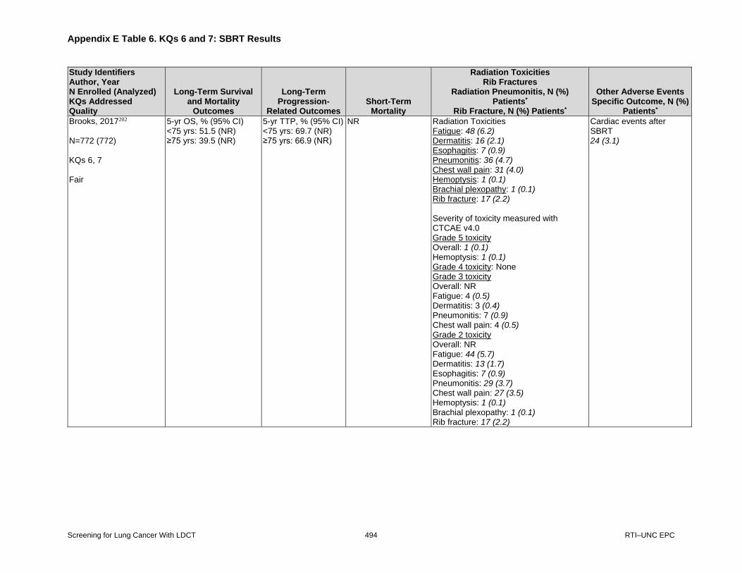
Appendix E Table 6. KQs 6 and 7: SBRT Results
Screening for Lung Cancer With LDCT 494 RTI–UNC EPC
Study Identifiers Author, Year N Enrolled (Analyzed) KQs Addressed Quality
Long-Term Survival and Mortality
Outcomes
Long-Term Progression-
Related Outcomes Short-Term
Mortality
Radiation Toxicities Rib Fractures
Radiation Pneumonitis, N (%) Patients*
Rib Fracture, N (%) Patients*
Other Adverse Events Specific Outcome, N (%)
Patients*
Brooks, 2017202 N=772 (772) KQs 6, 7 Fair
5-yr OS, % (95% CI) <75 yrs: 51.5 (NR) ≥75 yrs: 39.5 (NR)
5-yr TTP, % (95% CI) <75 yrs: 69.7 (NR) ≥75 yrs: 66.9 (NR)
NR Radiation Toxicities Fatigue: 48 (6.2) Dermatitis: 16 (2.1) Esophagitis: 7 (0.9) Pneumonitis: 36 (4.7) Chest wall pain: 31 (4.0) Hemoptysis: 1 (0.1) Brachial plexopathy: 1 (0.1) Rib fracture: 17 (2.2) Severity of toxicity measured with CTCAE v4.0 Grade 5 toxicity Overall: 1 (0.1) Hemoptysis: 1 (0.1) Grade 4 toxicity: None Grade 3 toxicity Overall: NR Fatigue: 4 (0.5) Dermatitis: 3 (0.4) Pneumonitis: 7 (0.9) Chest wall pain: 4 (0.5) Grade 2 toxicity Overall: NR Fatigue: 44 (5.7) Dermatitis: 13 (1.7) Esophagitis: 7 (0.9) Pneumonitis: 29 (3.7) Chest wall pain: 27 (3.5) Hemoptysis: 1 (0.1) Brachial plexopathy: 1 (0.1) Rib fracture: 17 (2.2)
Cardiac events after SBRT 24 (3.1)

Appendix E Table 6. KQs 6 and 7: SBRT Results
Screening for Lung Cancer With LDCT 495 RTI–UNC EPC
Study Identifiers Author, Year N Enrolled (Analyzed) KQs Addressed Quality
Long-Term Survival and Mortality
Outcomes
Long-Term Progression-
Related Outcomes Short-Term
Mortality
Radiation Toxicities Rib Fractures
Radiation Pneumonitis, N (%) Patients*
Rib Fracture, N (%) Patients*
Other Adverse Events Specific Outcome, N (%)
Patients*
Bryant, 2018194 N=3,620 for SBRT/SABR (total=3,620; L=2,986; SLR=634) KQs 6, 7 Fair
5-yr OS, % (95% CI) 44 (NR) 5-yr cancer-specific mortality, N (%) 202 (45)
NR 30-day mortality, N (%) 2 (0.5) 90-day mortality, N (%) 6 (1.4) 90-day mortality stratified by age, N (%) ≤55 yrs 7 (2.9) 56 to 60 yrs, N (%) 8 (1.92) 61 to 65 yrs, N (%) 19 (2.38) 66 to 70 yrs, N (%) 33 (2.58) 71 to 75 yrs, N (%) 38 (2.44) 76 to 80 yrs, N (%) 56 (3.26) ≥81 yrs, N (%) 80 (3.63)
NR NR

Appendix E Table 6. KQs 6 and 7: SBRT Results
Screening for Lung Cancer With LDCT 496 RTI–UNC EPC
Study Identifiers Author, Year N Enrolled (Analyzed) KQs Addressed Quality
Long-Term Survival and Mortality
Outcomes
Long-Term Progression-
Related Outcomes Short-Term
Mortality
Radiation Toxicities Rib Fractures
Radiation Pneumonitis, N (%) Patients*
Rib Fracture, N (%) Patients*
Other Adverse Events Specific Outcome, N (%)
Patients*
Crabtree, 2013263 Timmerman, 2010270 N=55 (55) KQ 7 Fair
NR NR None Protocol-specified AEs related to treatment (grade 3 measures of lung injury, esophageal injury, heart injury, and nerve damage, and any grade 4-5 toxicity related to treatment) FEV1: 2 (3.6) Hypocalcemia: 1 (1.8) Hypoxia: 2 (3.6) Pneumonitis NOS: 2 (3.6) Pulmonary function test decreased NOS: 4 (7.3) Maximum for protocol: 4 (7.3) Grade 3 AEs: 7 (12.7) (95% CI, 9.6 to 15.8) Grade 3+ AEs at 30 days: 5 (9.1) Grade 3+ AEs at 90 days: 12 (21.8) Grade 4 AEs: 2 (3.6) (95% CI, 2.7 to 4.5) No grade 5 treatment-related AEs reported Additional patients with AEs attributable to SBRT but not classified prospectively as protocol-specified: 6 (10.9) (95% CI, 8.2 to 13.6)
AEs related (or not) to treatment Overall: 45 (81.8) Blood or bone marrow: 6 (10.9) Cardiovascular: 2 (3.6) Constitutional symptoms: 20 (36.4) Dermatology or skin: 7 (12.7) GI tract: 6 (10.9) Lymphatics: 2 (3.6) Metabolic or laboratory: 5 (9.1) Musculoskeletal or soft tissue: 11 (20) Neurology: 6 (10.9) Pain: 14 (25.5) Pulmonary or upper respiratory tract: 33 (60) Renal or genitourinary: 1 (1.8) Infection: 3 (5.5) Coagulation: 2 (3.6) Hemorrhage or bleeding: 2 (3.6)
Eba, 2016199 N=40 (40) KQ 6 Fair
5-yr OS, % (95% CI) 67.0 (50.0 To 79.3)
NR NR NR NR

Appendix E Table 6. KQs 6 and 7: SBRT Results
Screening for Lung Cancer With LDCT 497 RTI–UNC EPC
Study Identifiers Author, Year N Enrolled (Analyzed) KQs Addressed Quality
Long-Term Survival and Mortality
Outcomes
Long-Term Progression-
Related Outcomes Short-Term
Mortality
Radiation Toxicities Rib Fractures
Radiation Pneumonitis, N (%) Patients*
Rib Fracture, N (%) Patients*
Other Adverse Events Specific Outcome, N (%)
Patients*
Ferrero, 2015260 N=30 (30) KQ 7 Fair
NR NR NR Any toxicity: 16 (53.3) Asthenia: 15 (50) Cough: 3 (10) Thoracic pain: 1 (3.3) Severity measured using RTOG scoring system Grade 1: 5 (15) Grade 2: 6 (20) Grade 3: 4 (13.3) Grade 4: 1 (3.3)
NR
Grills, 2012254 N=483 (483) KQs 6, 7 Fair
NR NR NR Grade 2 or higher pneumonitis 34 (7) Rib fracture 39 (8)
Respiratory failure 1 (0.2) Chronic myositis 24 (5) Grade 2 or higher dermatitis 10 (2)

Appendix E Table 6. KQs 6 and 7: SBRT Results
Screening for Lung Cancer With LDCT 498 RTI–UNC EPC
Study Identifiers Author, Year N Enrolled (Analyzed) KQs Addressed Quality
Long-Term Survival and Mortality
Outcomes
Long-Term Progression-
Related Outcomes Short-Term
Mortality
Radiation Toxicities Rib Fractures
Radiation Pneumonitis, N (%) Patients*
Rib Fracture, N (%) Patients*
Other Adverse Events Specific Outcome, N (%)
Patients*
Haidar, 2014255 N=55 (total=55; NPC=23; PC=32) KQ 7 Fair
NR NR NR NPC Acute toxicity: 2 (8.7) Chronic toxicity: 3 (13) PC Acute toxicity: 4 (13) Chronic toxicity: 6 (19) NPC Acute toxicity 2 (8.7): Grade 1 hemoptysis and Grade 2 pleural effusion Chronic toxicity 3 (13): Grade 2 dyspnea, Grade 2 cough and Grade 1 pulmonary fibrosis PC Acute toxicity 4 (13): Grade 3 esophagitis, Grade 2 pneumonitis, Grade 1 cough, and Grade 1 pruritis Chronic toxicity 6 (19): Grade 2 pneumonitis, Grade 1 cough, Grade 1 pneumonitis, Grade 2 dyspnea, and Grade 2 atelectasis
NR
Jeon, 2018266 N=53 (53) KQ 7 Fair
NR NR NR Chest pain, including rib fracture: 5 (9.4) Rib fracture: 2 (3.8) Dyspnea: 6 (11.3) Grade 1 Overall: 5 (9.4) Chest pain, including rib fracture: 5 (9.4) Rib fracture: 2 (3.8) Grade 3 Overall: 4 (7.5) Dyspnea: 4 (7.5) Grade 4 Overall: 2 (3.8) Dyspnea: 2 (3.8)
NR

Appendix E Table 6. KQs 6 and 7: SBRT Results
Screening for Lung Cancer With LDCT 499 RTI–UNC EPC
Study Identifiers Author, Year N Enrolled (Analyzed) KQs Addressed Quality
Long-Term Survival and Mortality
Outcomes
Long-Term Progression-
Related Outcomes Short-Term
Mortality
Radiation Toxicities Rib Fractures
Radiation Pneumonitis, N (%) Patients*
Rib Fracture, N (%) Patients*
Other Adverse Events Specific Outcome, N (%)
Patients*
Jeppesen, 2018201 N=136 (136) KQs 6, 7 Fair
5-yr OS, % (95% CI) 35 (NR)
NR None “Most common side effects were skin rash, rib fracture, cough and radiological pneumonitis/fibrosis without clinical symptoms” No acute grade 3+ toxicity
NR
Katoh, 2017253
N=283 (283) KQs 6, 7 Fair
NR NR NR Grade 2 or higher radiation pneumonitis 38 (13.4)
NR
Koshy, 2015198 N=498 (498) KQ 6 Fair
5-yr OS, %, 95% CI 30 (NR)
NR NR NR NR
Lam, 2018200 N=4,454 (4,454) KQs 6, 7 Fair
Unmatched 5-yr OS, % (95% CI) 33.4 (NR)
NR NR NR Unplanned readmissions within 30 days 17 (0.4)

Appendix E Table 6. KQs 6 and 7: SBRT Results
Screening for Lung Cancer With LDCT 500 RTI–UNC EPC
Study Identifiers Author, Year N Enrolled (Analyzed) KQs Addressed Quality
Long-Term Survival and Mortality
Outcomes
Long-Term Progression-
Related Outcomes Short-Term
Mortality
Radiation Toxicities Rib Fractures
Radiation Pneumonitis, N (%) Patients*
Rib Fracture, N (%) Patients*
Other Adverse Events Specific Outcome, N (%)
Patients*
Lindberg, 2015205 N=57 (57 for survival and early toxicity; 34 for late toxicity) KQs 6, 7 Fair
5-yr OS, % (95% CI) 30 (18 To 42)
5-yr LCSS, % (95% CI) 74 (59 to 89) 5-yr PFS, % (95% CI) 52 (33 to 70)
None Early toxicity (≤36 mos) Overall: NR Atelectasis: 2 (3.5) Cough: 6 (10.5) Dyspnea: 12 (21.1) Exudate: 4 (7.0) Fatigue: 9 (15.8) Fibrosis: 12 (21.1) Heart: 2 (3.5) Lung infection: 2 (3.5) Pain: 4 (7.0) Pericardial effusion: 1 (1.8) Pneumonitis: 6 (10.5) Rib fracture: 2 (3.5) Skin and subcutaneous tissue: 5 (8.8) Upper airway infection: 1 (1.8) Late toxicity (>36 mos), N(%) Overall: NR COPD exacerbation: 2 (6) Cough: 1 (3) Dyspnea: 3 (8.8) Exudate: 1 (3) Lung infection: 1 (3) Rib fracture: 6 (17.6) Upper airway infection: 1 (3) Ventricle tachycardia: 1 (3) Severity of toxicity measured with CTCAE v4.0 or, for fibrosis, RTOG late toxicity scale (early=occurring within 36 mos of treatment; late=occurring after 36 mos of treatment) Grade 5 toxicity: None Grade 3-4 early toxicity (≤36 mos) Overall: NR Cough: 1 (1.8) Dyspnea: 3 (5.3) Exudate: 1 (1.8)
NR

Appendix E Table 6. KQs 6 and 7: SBRT Results
Screening for Lung Cancer With LDCT 501 RTI–UNC EPC
Study Identifiers Author, Year N Enrolled (Analyzed) KQs Addressed Quality
Long-Term Survival and Mortality
Outcomes
Long-Term Progression-
Related Outcomes Short-Term
Mortality
Radiation Toxicities Rib Fractures
Radiation Pneumonitis, N (%) Patients*
Rib Fracture, N (%) Patients*
Other Adverse Events Specific Outcome, N (%)
Patients*
Lindberg, 2015205 (continued)
Fatigue: 1 (1.8) Fibrosis: 3 (5.3) Heart: 1 (1.8) Lung infection: 1 (1.8) Pain: 2 (3.5) Pericardial effusion: 1 (1.8) Grade 3-4 late toxicity (>36 mos) (N analyzed=34) Overall: NR Dyspnea: 1 (3) Rib fracture: 1 (3) Ventricle tachycardia: 1 (3) Grade 2 early toxicity (≤36 mos) Overall: NR Atelectasis: 2 (3.5) Cough: 5 (8.8) Dyspnea: 9 (15.8) Exudate: 3 (5.3) Fatigue: 8 (14.0) Fibrosis: 9 (15.8) Heart: 1 (1.8) Lung infection: 1 (1.8) Pain: 2 (3.5) Pneumonitis: 6 (10.5) Rib fracture: 2 (3.5) Skin and subcutaneous tissue: 5 (8.8) Upper airway infection: 1 (1.8) Grade 2 late toxicity (>36 mos) (N analyzed=34) Overall: NR COPD exacerbation: 2 (6) Cough: 1 (3) Dyspnea: 2 (6) Exudate: 1 (3) Lung infection: 1 (3) Rib fracture: 5 (14.7) Upper airway infection: 1 (3)

Appendix E Table 6. KQs 6 and 7: SBRT Results
Screening for Lung Cancer With LDCT 502 RTI–UNC EPC
Study Identifiers Author, Year N Enrolled (Analyzed) KQs Addressed Quality
Long-Term Survival and Mortality
Outcomes
Long-Term Progression-
Related Outcomes Short-Term
Mortality
Radiation Toxicities Rib Fractures
Radiation Pneumonitis, N (%) Patients*
Rib Fracture, N (%) Patients*
Other Adverse Events Specific Outcome, N (%)
Patients*
Ma, 2017267 N=155 (155) patients with 159 (159) tumors KQ 7 Fair
NR NR NR Pulmonary embolism Total: 1 (0.6) Single-fx SBRT: 0 (0) Triple-fx SBRT: 1 (1.1) Severity of toxicity measured with CTCAE v4.0 Grade ≥3 pulmonary toxicity Total: 1 (0.6) Single-fx SBRT: 0 (0) Triple-fx SBRT: 1 (1.1), a case of pulmonary embolism No Grade ≥3 pulmonary toxicity occurred within 6 months of SBRT
NR

Appendix E Table 6. KQs 6 and 7: SBRT Results
Screening for Lung Cancer With LDCT 503 RTI–UNC EPC
Study Identifiers Author, Year N Enrolled (Analyzed) KQs Addressed Quality
Long-Term Survival and Mortality
Outcomes
Long-Term Progression-
Related Outcomes Short-Term
Mortality
Radiation Toxicities Rib Fractures
Radiation Pneumonitis, N (%) Patients*
Rib Fracture, N (%) Patients*
Other Adverse Events Specific Outcome, N (%)
Patients*
Mathieu, 2015258 N=45 (45) KQ 7 Fair
NR NR NR Radiation pneumonitis of any grade 2 (4.4) Mortality due to radiation pneumonitis 1 (2.2) Dyspnea 4 (8.9) Cough 1 (2.2) Radiation-induced rib fractures 3 (7) Pneumothorax requiring chest tube placement 2 (4.4) Severity of toxicity measured with CTCAE v3.0 (acute=occurring within 4 mos of treatment; late=occurring after 4 mos of treatment) Grade 5 toxicity: Radiation pneumonitis: 1 (2.2) Grade 3 toxicity Overall: 4 (8.9) Acute dyspnea: 1 (2.2) Late: 3 (7), of which 2 were cases of dyspnea and 1 was case of dyspnea, cough, and radiation pneumonitis Grade ≤2 toxicity (measured with CTCAE v3.0) Overall: 3 (7) Acute: 0 (0) Late radiation-induced rib fractures: 3 (7)
NR
Matsuo, 2014196
N=115 (115) KQs 6, 7 Fair
5-yr OS, % (95% CI) 40.3 (31.1 To 49.3)
5-yr cause-specific death, % (95% CI) 33.8 (25.1 to 42.6)
NR NR No treatment-related deaths

Appendix E Table 6. KQs 6 and 7: SBRT Results
Screening for Lung Cancer With LDCT 504 RTI–UNC EPC
Study Identifiers Author, Year N Enrolled (Analyzed) KQs Addressed Quality
Long-Term Survival and Mortality
Outcomes
Long-Term Progression-
Related Outcomes Short-Term
Mortality
Radiation Toxicities Rib Fractures
Radiation Pneumonitis, N (%) Patients*
Rib Fracture, N (%) Patients*
Other Adverse Events Specific Outcome, N (%)
Patients*
Nagata, 2015203 N=164 (total=164; operable=64; inoperable=100) KQs 6, 7 Fair
5-yr OS, % (95% CI) Operable 54.0 (41.0 To 65.4) Inoperable 42.8 (33.0 To 52.3)
NR NR Grade 2 toxicities using CTCAE v3.0 Total: NR, but 17 events reported Mild symptomatic fractures: 7 (4.1) Chest wall pain: 3 (1.8) Cough: 3 (1.8) Chest pain: 2 (1.2) Brachial plexopathy: 1 (0.6) Dermatitis: 1 (0.6) Grade 3 toxicities using CTCAE v3.0 Total: 15 (8.9) Dyspnea: 13 (7.7) Hypoxia: 9 (5.3) Pneumonitis: 10 (5.9) Chest pain: 3 (1.8) Cough: 1 (0.6) Grade 4 toxicities using CTCAE v3.0 Total: 2 (1.2) Dyspnea: 2 (1.2) Hypoxia: 1 (0.6) Pneumonitis: 1 (0.6)
NR

Appendix E Table 6. KQs 6 and 7: SBRT Results
Screening for Lung Cancer With LDCT 505 RTI–UNC EPC
Study Identifiers Author, Year N Enrolled (Analyzed) KQs Addressed Quality
Long-Term Survival and Mortality
Outcomes
Long-Term Progression-
Related Outcomes Short-Term
Mortality
Radiation Toxicities Rib Fractures
Radiation Pneumonitis, N (%) Patients*
Rib Fracture, N (%) Patients*
Other Adverse Events Specific Outcome, N (%)
Patients*
Nyman, 2016265 N=49 (49) KQ 7 Fair
NR NR NR Esophagitis: 4 (8) Pneumonitis: 9 (18.4) Dyspnea: 32 (65.3) Fibrosis: 24 (49.0) Cough: 25 (51.0) Skin reactions: 16 (32.7) Rib fractures: 8 (16.3) Severity measured using CTCAE v3.0 grading system Grade 1 Esophagitis: 4 (8) Pneumonitis: 7 (15) Dyspnea: 19 (40) Fibrosis: 20 (42) Cough: 19 (40) Skin reactions: 13 (27) Rib fractures: 6 (13) Grade 2 Esophagitis: 0 (0) Pneumonitis: 2 (4) Dyspnea: 8 (17) Fibrosis: 4 (8) Cough: 5 (10) Skin reactions: 2 (4) Rib fractures: 2 (4) Grade 3 Esophagitis: 0 (0) Pneumonitis: 0 (0) Dyspnea: 5 (10) Fibrosis: 0 (0) Cough: 1 (2) Skin reactions: 1 (2) Rib fractures: 0 (0) No Grade 4 or 5 toxicities
NR

Appendix E Table 6. KQs 6 and 7: SBRT Results
Screening for Lung Cancer With LDCT 506 RTI–UNC EPC
Study Identifiers Author, Year N Enrolled (Analyzed) KQs Addressed Quality
Long-Term Survival and Mortality
Outcomes
Long-Term Progression-
Related Outcomes Short-Term
Mortality
Radiation Toxicities Rib Fractures
Radiation Pneumonitis, N (%) Patients*
Rib Fracture, N (%) Patients*
Other Adverse Events Specific Outcome, N (%)
Patients*
Robinson, 2013264 N=78 (78) KQ 7 Fair
NR NR NR Esophagitis: 2 (2.6) Pneumonitis: 6 (7.7) Brachial plexopathy: 1 (1.3) Pleural effusion: 1 (1.3) Soft-tissue necrosis: 1 (1.3) Chest-wall pain: 15 (19.2) Severity measured using CTCAE v4.0 grading system Grade 1 Total: 12 (15.4) Acute: 18 (1.3) Late: 11 (14.1) Grade 2 Total: 11 (14.1) Acute: 1 (1.3) Late: 10 (12.8) Grade 3 Total: 4 (5.1) Acute: 0 (0) Late: 4 (5.1)
NR
Rosen, 2014256 N=79 (79) KQ 7 Fair
NR NR None Chest wall pain 6 (7.6) (95% CI, 3.5 to 15.9) Rib fracture, N (%) 2 (2.5) (95% CI, 0.7 to 8.7) Significant skin reactions 3 (3.8) (95% CI, 1.3 to 10.5) No cases of life-threatening radiation pneumonitis, clinically significant pulmonary complications, necrosis, or fatal hemoptysis “All cases of rib fracture were late findings and resolved without intervention beyond topical creams and oral pain medications”
NR

Appendix E Table 6. KQs 6 and 7: SBRT Results
Screening for Lung Cancer With LDCT 507 RTI–UNC EPC
Study Identifiers Author, Year N Enrolled (Analyzed) KQs Addressed Quality
Long-Term Survival and Mortality
Outcomes
Long-Term Progression-
Related Outcomes Short-Term
Mortality
Radiation Toxicities Rib Fractures
Radiation Pneumonitis, N (%) Patients*
Rib Fracture, N (%) Patients*
Other Adverse Events Specific Outcome, N (%)
Patients*
Rosen, 2016183 Primary analyses (i.e., patients selected for Charlson-Deyo score=0) N=1,781 for SBRT/SABR (1,781 for both unmatched and PSM sets) Secondary analyses (i.e., patients unselected based on Charlson-Deyo score) N=235 for SBRT/SABR (235 for both unmatched and PSM sets) KQs 6, 7 Good
5-yr OS, % (95% CI) Primary Analysis Propensity-Matched Healthy Subset 29 (NR) Secondary Analysis Propensity-Matched Healthy Subset With SBRT Patients Refusing Surgery 40 (NR)
NR NR NR NR
Shirvani, 2012235 N=124 for SABR/SBRT (124) KQ 7 Fair
NR NR 30-day mortality, N (%) 0 (0) 90-day mortality, N (%) 1 (0.8)
NR NR
Shirvani, 2014239 N=382 (382) KQ 7 Fair
NR NR 90-day mortality, N (%) 5 (1.3)
NR NR
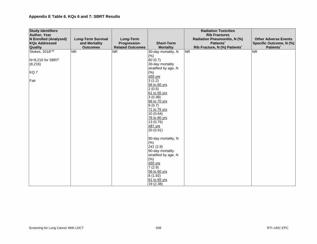
Appendix E Table 6. KQs 6 and 7: SBRT Results
Screening for Lung Cancer With LDCT 508 RTI–UNC EPC
Study Identifiers Author, Year N Enrolled (Analyzed) KQs Addressed Quality
Long-Term Survival and Mortality
Outcomes
Long-Term Progression-
Related Outcomes Short-Term
Mortality
Radiation Toxicities Rib Fractures
Radiation Pneumonitis, N (%) Patients*
Rib Fracture, N (%) Patients*
Other Adverse Events Specific Outcome, N (%)
Patients*
Stokes, 2018249 N=8,216 for SBRT (8,216) KQ 7 Fair
NR NR 30-day mortality, N (%) 60 (0.7) 30-day mortality stratified by age, N (%) ≤55 yrs 3 (1.2) 56 to 60 yrs 2 (0.5) 61 to 65 yrs 3 (0.38) 66 to 70 yrs 9 (0.7) 71 to 75 yrs 10 (0.64) 76 to 80 yrs 13 (0.76) ≥81 yrs 20 (0.91) 90-day mortality, N (%) 241 (2.9) 90-day mortality stratified by age, N (%) ≤55 yrs 7 (2.9) 56 to 60 yrs 8 (1.92) 61 to 65 yrs 19 (2.38)
NR NR

Appendix E Table 6. KQs 6 and 7: SBRT Results
Screening for Lung Cancer With LDCT 509 RTI–UNC EPC
Study Identifiers Author, Year N Enrolled (Analyzed) KQs Addressed Quality
Long-Term Survival and Mortality
Outcomes
Long-Term Progression-
Related Outcomes Short-Term
Mortality
Radiation Toxicities Rib Fractures
Radiation Pneumonitis, N (%) Patients*
Rib Fracture, N (%) Patients*
Other Adverse Events Specific Outcome, N (%)
Patients*
Stokes, 2018249 (continued)
66 to 70 yrs 33 (2.58) 71-75 yrs 38 (2.44) 76-80 yrs 56 (3.26) ≥81 yrs 80 (3.63)
Sun, 2017195 N=73 (65) KQs 6, 7 Fair
5-yr OS, % (95% CI) 55.7 (49.4 To 62.0) 5-yr mortality, % (95% CI) NR 10-yr mortality, % (95% CI) NR
NR NCI CTCAE (v. 3) Grade 1: 49 (75.4) Grade 2: 7 (10.8) Grade 3: 1 (1.5) Radiation pneumonitis, N (%) 57 (87.7)
Dermatitis Total: 21 (32.3) Grade 1: 16 (24.6) Grade 2: 3 (4.6) Grade 3: 2 (3.1) Hemoptysis Total: 1 (1.5) Grade 1: 1 (1.5) Grade 2: 0 (0) Grade 3: 0 (0) Dyspnea/shortness of breath Total: 19 (29.2) Grade 1: 11 (16.9) Grade 2: 8 (12.3) Grade 3: 0 (0) Fatigue Total: 9 (13.9) Grade 1: 7 (10.8) Grade 2: 2 (3.1) Grade 3: 0 (0) Chest wall pain Total: 23 (35.4) Grade 1: 15 (23.1) Grade 2: 7 (10.8) Grade 3: 1 (1.5) Rib fracture Total: 16 (24.6) Grade 1: 13 (20.0) Grade 2: 3 (4.6) Grade 3: 0 (0)

Appendix E Table 6. KQs 6 and 7: SBRT Results
Screening for Lung Cancer With LDCT 510 RTI–UNC EPC
Study Identifiers Author, Year N Enrolled (Analyzed) KQs Addressed Quality
Long-Term Survival and Mortality
Outcomes
Long-Term Progression-
Related Outcomes Short-Term
Mortality
Radiation Toxicities Rib Fractures
Radiation Pneumonitis, N (%) Patients*
Rib Fracture, N (%) Patients*
Other Adverse Events Specific Outcome, N (%)
Patients*
Sun, 2017195 (continued)
Brachial plexopathy Total: 5 (7.7) Grade 1: 3 (4.6) Grade 2: 2 (3.1) Grade 3: 0 (0)
Taremi, 2012259 N=46 (total=46; female=24; male=22; COPD=29; diabetes=8) KQ 7 Fair
NR NR NR RIBI Total: 17 (37) Female: 11 (45.8) with 30 fractures Male: 6 (27.3) with 13 fractures COPD: 11 (37.9) Diabetes: 2 (25.0) Total N of rib fractures: 41 ribs with 43 fracture sites Median time to developing a rib fracture (range) 21 mos (7 to 40 mos) Chest wall pain toxicity Patients without rib fractures 7 (24) Patients with rib fractures 14 (82) Patients with chest wall pain received higher dose of radiation to the ribs compared to patients without chest wall pain (62.76 Gy, range: 28.4 to 88.05 Gy vs. 47.21 Gy, range: 15.9 to 73.19 Gy; p value: 0.008).
NR

Appendix E Table 6. KQs 6 and 7: SBRT Results
Screening for Lung Cancer With LDCT 511 RTI–UNC EPC
Study Identifiers Author, Year N Enrolled (Analyzed) KQs Addressed Quality
Long-Term Survival and Mortality
Outcomes
Long-Term Progression-
Related Outcomes Short-Term
Mortality
Radiation Toxicities Rib Fractures
Radiation Pneumonitis, N (%) Patients*
Rib Fracture, N (%) Patients*
Other Adverse Events Specific Outcome, N (%)
Patients*
Taremi, 2012259 (continued)
In all patients except one (with 6 fractured ribs), pain became more stable after 6–8 months. Multivariate analysis found that D to 0.5 cc of the ribs (D0.5) and volume of rib receiving ≥25 Gy (V25) were significantly associated with RIBI Grading of chest wall pain and rib fractures using CTCAE v3.0 Grade 1 With rib fractures 5 (29.4) Without rib fractures 4 (13.8) Radiologic fractures (denominator is total N of fracture sites) 10 (23.3) Grade 2 With rib fractures 6 (35.3) Without rib fractures 3 (10.3) Radiologic fractures (based on total N of fracture sites) 19 (44.2) Grade 3 With rib fractures 3 (17.6) Without rib fractures 0 (0) Radiologic fractures (based on total N of fracture sites) 14 (32.5)

Appendix E Table 6. KQs 6 and 7: SBRT Results
Screening for Lung Cancer With LDCT 512 RTI–UNC EPC
Study Identifiers Author, Year N Enrolled (Analyzed) KQs Addressed Quality
Long-Term Survival and Mortality
Outcomes
Long-Term Progression-
Related Outcomes Short-Term
Mortality
Radiation Toxicities Rib Fractures
Radiation Pneumonitis, N (%) Patients*
Rib Fracture, N (%) Patients*
Other Adverse Events Specific Outcome, N (%)
Patients*
Taremi, 2012262 N=108 KQ 7 Fair
NR NR None Any acute/early toxicity (i.e., ≤3 mos after SBRT) Overall N of patients with acute/early toxicity: 77 (71.3) Fatigue: 54 (50) Cough/dyspnea: 39 (36.1) Pneumonitis: 4 (3.7) Anorexia: 3 (2.8) Chest wall pain: 12 (11.1) Dyspepsia/dysphagia: 13 (12) Skin toxicity: 12 (11.1) Any late toxicity (i.e., >3 mos after SBRT) Overall N of patients with late toxicity: 74 (68.5) Fatigue: 38 (35.2) Cough/dyspnea: 43 (39.8) Pneumonitis: 24 (22.2) Chest wall pain: 16 (14.8) Rib fracture: 27 (25) Pleural effusion: 2 (1.9) Hemoptysis: 5 (4.6) Skin toxicity: 1 (0.9) Grading of acute/early toxicities using CTCAE v3.0 Grade 1: Patients NR, but 102 (74.4%) total events Grade 2: Patients NR, but 31 (22.6%) total events Grade 3: 4 (3.7) Grading of late toxicities using CTCAE v3.0 Grade 1: Patients NR, but 96 (59.3%) total events Grade 2: Patients NR, but 54 (33.3%) total events Grade 3: 6 (5.6)
NR
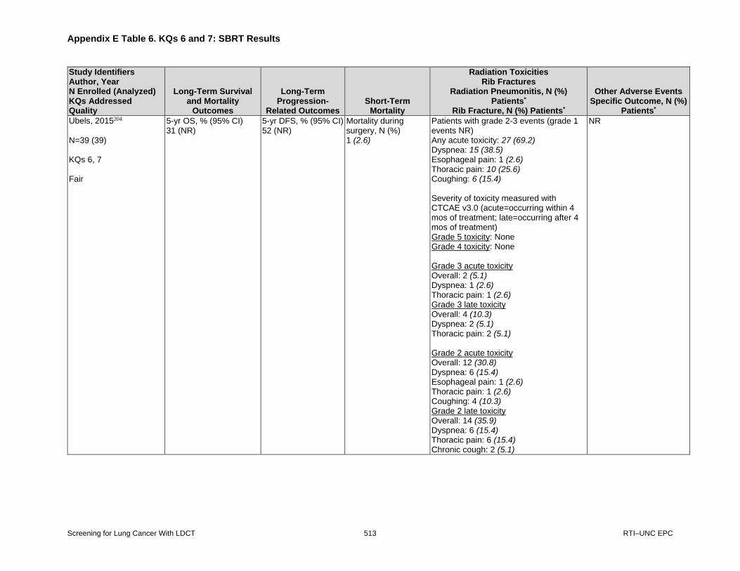
Appendix E Table 6. KQs 6 and 7: SBRT Results
Screening for Lung Cancer With LDCT 513 RTI–UNC EPC
Study Identifiers Author, Year N Enrolled (Analyzed) KQs Addressed Quality
Long-Term Survival and Mortality
Outcomes
Long-Term Progression-
Related Outcomes Short-Term
Mortality
Radiation Toxicities Rib Fractures
Radiation Pneumonitis, N (%) Patients*
Rib Fracture, N (%) Patients*
Other Adverse Events Specific Outcome, N (%)
Patients*
Ubels, 2015204 N=39 (39) KQs 6, 7 Fair
5-yr OS, % (95% CI) 31 (NR)
5-yr DFS, % (95% CI) 52 (NR)
Mortality during surgery, N (%) 1 (2.6)
Patients with grade 2-3 events (grade 1 events NR) Any acute toxicity: 27 (69.2) Dyspnea: 15 (38.5) Esophageal pain: 1 (2.6) Thoracic pain: 10 (25.6) Coughing: 6 (15.4) Severity of toxicity measured with CTCAE v3.0 (acute=occurring within 4 mos of treatment; late=occurring after 4 mos of treatment) Grade 5 toxicity: None Grade 4 toxicity: None Grade 3 acute toxicity Overall: 2 (5.1) Dyspnea: 1 (2.6) Thoracic pain: 1 (2.6) Grade 3 late toxicity Overall: 4 (10.3) Dyspnea: 2 (5.1) Thoracic pain: 2 (5.1) Grade 2 acute toxicity Overall: 12 (30.8) Dyspnea: 6 (15.4) Esophageal pain: 1 (2.6) Thoracic pain: 1 (2.6) Coughing: 4 (10.3) Grade 2 late toxicity Overall: 14 (35.9) Dyspnea: 6 (15.4) Thoracic pain: 6 (15.4) Chronic cough: 2 (5.1)
NR

Appendix E Table 6. KQs 6 and 7: SBRT Results
Screening for Lung Cancer With LDCT 514 RTI–UNC EPC
Study Identifiers Author, Year N Enrolled (Analyzed) KQs Addressed Quality
Long-Term Survival and Mortality
Outcomes
Long-Term Progression-
Related Outcomes Short-Term
Mortality
Radiation Toxicities Rib Fractures
Radiation Pneumonitis, N (%) Patients*
Rib Fracture, N (%) Patients*
Other Adverse Events Specific Outcome, N (%)
Patients*
Valle, 2016244 N=184 (176 for perioperative death, 148 for acute toxicity) KQ 7 Fair
30-day mortality 3 (1.7)
NR NR Any acute toxicity (i.e., within 180 days of treatment completion): 34 (23) reporting 60 toxicity events Radiation pneumonitis: 13 (9) Home oxygen: 4 (3) Pleural effusion (nonmalignant): 4 (3) Pneumonia (infectious): 15 (10) Pneumothorax: 0 (0) ICU admission: 0 (0) Respiratory failure: 2 (1) Dyspnea requiring hospitalization: 4 (3) Hemoptysis: 3 (2) Hospitalization (other): 6 (4)
NR
Videtic, 2014269 N=80 (80) KQ 7 Fair
NR NR NR Patients experiencing any toxicity Single-fx SBRT at 30 Gy: 4 (7.3) Single-fx SBRT at 34 Gy: 4 (16) Chest wall pain/neuropathy Single-fx SBRT at 30 Gy: 2 (3.6) Single-fx SBRT at 34 Gy: 4 (16) Pneumonitis Single-fx SBRT at 30 Gy: 2 (3.6) Single-fx SBRT at 34 Gy: 0 (0) Grade 1 toxicity Single-fx SBRT at 30 Gy: 0 (0) Single-fx SBRT at 34 Gy: 1 (4) Grade 2 toxicity Single-fx SBRT at 30 Gy: 4 (7.3) Single-fx SBRT at 34 Gy: 3 (12)
NR
Videtic, 2015261 N=84 (84) KQ 7 Fair
NR NR 30-day mortality, N (%) SBRT at any dose: 1 (1.2) SBRT 34 Gy x 1 fx: 1 (2.6) SBRT 48 Gy x 4 fx: 0 (0)
Rates of prespecified grade 3 or higher toxicities at 1 year SBRT at any dose: 10 (11.9) SBRT 34 Gy x 1 fx: 4 (10.3) (95% CI, 2.9 to 24.2) SBRT 48 Gy x 4 fx: 6 (13.3) (95% CI, 5.1 to 26.8)
Respiratory failure leading to death, possibly related to SBRT SBRT at any dose: 1 (1.2) SBRT 34 Gy x 1 fx: 0 (0) SBRT 48 Gy x 4 fx: 1 (2.2)

Appendix E Table 6. KQs 6 and 7: SBRT Results
Screening for Lung Cancer With LDCT 515 RTI–UNC EPC
Study Identifiers Author, Year N Enrolled (Analyzed) KQs Addressed Quality
Long-Term Survival and Mortality
Outcomes
Long-Term Progression-
Related Outcomes Short-Term
Mortality
Radiation Toxicities Rib Fractures
Radiation Pneumonitis, N (%) Patients*
Rib Fracture, N (%) Patients*
Other Adverse Events Specific Outcome, N (%)
Patients*
Videtic, 2015261 (continued)
Fatigue/malaise SBRT at any dose: 11 (13.1) SBRT 34 Gy x 1 fx: 6 (15) SBRT 48 Gy x 4 fx: 5 (11) Musculoskeletal disorders (including pain) SBRT at any dose: 11 (13.1) SBRT 34 Gy x 1 fx: 8 (21) SBRT 48 Gy x 4 fx: 3 (7) Injury (including fracture) SBRT at any dose: 8 (9.5) SBRT 34 Gy x 1 fx: 7 (18) SBRT 48 Gy x 4 fx: 1 (2) Respiratory disorders SBRT at any dose: 33 (39.3) SBRT 34 Gy x 1 fx: 18 (46) SBRT 48 Gy x 4 fx: 15 (33) Adverse changes in DLCO SBRT at any dose: 6 (7.1) SBRT 34 Gy x 1 fx: 4 (10.3) SBRT 48 Gy x 4 fx: 2 (4.4) Adverse changes in FVC SBRT at any dose: 1 (1.2) SBRT 34 Gy x 1 fx: 0 (0) SBRT 48 Gy x 4 fx: 1 (2.2) Pneumonitis SBRT at any dose: 2 (2.4) SBRT 34 Gy x 1 fx: 0 (0) SBRT 48 Gy x 4 fx: 2 (4.4) General disorder leading to death, but possibly unrelated to SBRT SBRT at any dose: 1 (1.2) SBRT 34 Gy x 1 fx: 1 (2.6) SBRT 48 Gy x 4 fx: 0 (0)

Appendix E Table 6. KQs 6 and 7: SBRT Results
Screening for Lung Cancer With LDCT 516 RTI–UNC EPC
Study Identifiers Author, Year N Enrolled (Analyzed) KQs Addressed Quality
Long-Term Survival and Mortality
Outcomes
Long-Term Progression-
Related Outcomes Short-Term
Mortality
Radiation Toxicities Rib Fractures
Radiation Pneumonitis, N (%) Patients*
Rib Fracture, N (%) Patients*
Other Adverse Events Specific Outcome, N (%)
Patients*
Videtic, 2015261 (continued)
Respiratory failure leading to death, possibly related to SBRT SBRT at any dose: 1 (1.2) SBRT 34 Gy x 1 fx: 0 (0) SBRT 48 Gy x 4 fx: 1 (2.2) Severity measured using CTCAE v4.0 grading system Grade 1 fatigue/malaise SBRT at any dose: 7 (8.3) SBRT 34 Gy x 1 fx: 2 (5) SBRT 48 Gy x 4 fx: 5 (11) Grade 1 musculoskeletal disorders (including pain) SBRT at any dose: 8 (9.5) SBRT 34 Gy x 1 fx: 5 (13) SBRT 48 Gy x 4 fx: 3 (7) Grade 1 injury (including fracture) SBRT at any dose: 4 (4.8) SBRT 34 Gy x 1 fx: 4 (10) SBRT 48 Gy x 4 fx: 0 (0) Grade 1 respiratory disorders SBRT at any dose: 21 (25) SBRT 34 Gy x 1 fx: 13 (33) SBRT 48 Gy x 4 fx: 8 (18) Grade 2 fatigue/malaise SBRT at any dose: 4 (4.8) SBRT 34 Gy x 1 fx: 4 (10) SBRT 48 Gy x 4 fx: 0 (0) Grade 2 musculoskeletal disorders (including pain) SBRT at any dose: 3 (3.6) SBRT 34 Gy x 1 fx: 3 (8) SBRT 48 Gy x 4 fx: 0 (0)

Appendix E Table 6. KQs 6 and 7: SBRT Results
Screening for Lung Cancer With LDCT 517 RTI–UNC EPC
Study Identifiers Author, Year N Enrolled (Analyzed) KQs Addressed Quality
Long-Term Survival and Mortality
Outcomes
Long-Term Progression-
Related Outcomes Short-Term
Mortality
Radiation Toxicities Rib Fractures
Radiation Pneumonitis, N (%) Patients*
Rib Fracture, N (%) Patients*
Other Adverse Events Specific Outcome, N (%)
Patients*
Videtic, 2015261 (continued)
Grade 2 injury (including fracture) SBRT at any dose: 4 (4.8) SBRT 34 Gy x 1 fx: 3 (8) SBRT 48 Gy x 4 fx: 1 (2) Grade 2 respiratory disorders SBRT at any dose: 7 (8.3) SBRT 34 Gy x 1 fx: 5 (13) SBRT 48 Gy x 4 fx: 2 (4) Grade 3 SBRT at any dose: 9 (10.7) SBRT 34 Gy x 1 fx: 4 (10.3) SBRT 48 Gy x 4 fx: 5 (11.1) Grade 5 SBRT at any dose: 2 (2.4) SBRT 34 Gy x 1 fx: 1 (2.6) SBRT 48 Gy x 4 fx: 1 (2.2)
* Unless otherwise specified.
Abbreviations: 3D-CRT=three-dimensional conformal radiation therapy; 95% CI=95% confidence interval; AE(s)=adverse event(s); cc=cubic centimeters; COPD=chronic
obstructive pulmonary disease; CTCAE=Common Terminology Criteria for Adverse Events; FVC=forced vital capacity; fx=fraction(s); D0.5=dose to 0.5 cc of the ribs;
DVT=deep vein thrombosis; Gy=Gray; ICU=intensive care unit; IQR=interquartile range; LC=lung cancer; LN(s)=lymph node(s); LOS=length of stay; MI=myocardial infarction;
N=number; NCI=National Cancer Institute; NR=not reported; OS=overall survival; PSM=propensity score matching (or matched); RIBI=radiation-induced bone injury;
RTOG=Radiation Therapy Oncology Group; SBRT/SABR=stereotactic body radiotherapy/stereotactic ablative radiotherapy; SD=standard deviation; SLR=sublobar resection;
surg=surgery; ThRCRI=Thoracic Revised Cardiac Risk Index; TTP=time-to-progression; V25=volume of rib receiving ≥25 Gy; VATS=video-assisted thoracoscopic lobectomy;
VMAT=volumetric modulated arc therapy; VPI=visceral pleural invasion; vs.=versus.

Appendix E Table 7. KQs 6 and 7 Update Search Results: Study and Patient Characteristics
Screening for Lung Cancer With LDCT 518 RTI–UNC EPC
Appendix E Table 7. KQs 6 and 7 Update Search Results: Study and Patient Characteristics
Study Identifiers Author, Year Treatment Type(s) N Enrolled KQs Addressed Quality
Study Characteristics Study or Database Name
Country Study Years
Followup, Median (Range)
Baseline Patient Characteristics
Age, Median (Range) Gender, N (%)
Race/Ethnicity, N (%) Smoking Status, N (%) Comorbidities, N (%)
NSCLC and Treatment Characteristics
Stage, N (%) Histology, N (%)
Surgical Approach or SBRT/SABR Dosing and Frequency
Ackerson, 2018293 SBRT N=70 KQ 7 Fair
NA United States 2007-2014 Followup: 65 mos.
Age: 74 (IQR 69 to 79) Female: 35 (50) Race/Ethnicity: NR Smoking Status: Never: 2 (2.9) Current: 14 (20) Past: 54 (77) Comorbidities: 89% of SBRT patients were deemed medically inoperable prior to treatment Charlson Comorbidity Index: Mean (SD): 3.7 (1.4) Median (IQR): 3 (3 to 5)
Clinical T-Stage T1a: 25 (36) T1b: 29 (41) T2a: 15 (21) T2b: 1 (1) Histology: NR SBRT Dosing and Frequency: SBRT was given every 48-72 hours using 3-5 fractions. Overall, 34 (49%) patients received 12–12.5 Gy x 4; 24 (34%) patients received 18–20 Gy × 3; and 8 (11%) patients received 10 Gy × 5. Less common fractionation schemes were used to treat 4 (6%) patients.

Appendix E Table 7. KQs 6 and 7 Update Search Results: Study and Patient Characteristics
Screening for Lung Cancer With LDCT 519 RTI–UNC EPC
Study Identifiers Author, Year Treatment Type(s) N Enrolled KQs Addressed Quality
Study Characteristics Study or Database Name
Country Study Years
Followup, Median (Range)
Baseline Patient Characteristics
Age, Median (Range) Gender, N (%)
Race/Ethnicity, N (%) Smoking Status, N (%) Comorbidities, N (%)
NSCLC and Treatment Characteristics
Stage, N (%) Histology, N (%)
Surgical Approach or SBRT/SABR Dosing and Frequency
Baine, 2019230 SBRT N=26,725 KQs 6 & 7 Fair
NCDB United States 2004-2014 Followup: 26.7 mos.
Age: 75 (IQR 69 to 81) Female: 14,265 (53.4) Race/Ethnicity White: 23,861 (89.3) Black: 2,230 (8.3) Indian: 76 (0.3) Pacific Islander: 258 (1.0) Other: 72 (0.3) Missing: 228 (0.9) Smoking Status: NR Comorbidities Charlson-Deyo Comorbidity Score, N (%) 0-2: 25,485 (95.4) ≥3: 1,240 (4.6)
Stage IA: 19,856 (74.3) IB: 5,659 (21.2) I NOS: 1,210 (4.5) Histology Squamous: 9,160 (34.3) Adenocarcinoma: 11,672 (43.7) NSCLC NOS: 4,457 (16.7) Other: 1,436 (5.4) SBRT Dosing, N (%) 48 Gy: 5,727 (21.4) 50 Gy: 9,677 (36.2) 54 Gy: 4,368 (16.3) 60 Gy: 6,953 (26) SBRT Fractions, N (%) 3: 7,835 (29.3) 4: 7,212 (27) 5: 9,291 (34.8) Other: 2,387 (8.9)

Appendix E Table 7. KQs 6 and 7 Update Search Results: Study and Patient Characteristics
Screening for Lung Cancer With LDCT 520 RTI–UNC EPC
Study Identifiers Author, Year Treatment Type(s) N Enrolled KQs Addressed Quality
Study Characteristics Study or Database Name
Country Study Years
Followup, Median (Range)
Baseline Patient Characteristics
Age, Median (Range) Gender, N (%)
Race/Ethnicity, N (%) Smoking Status, N (%) Comorbidities, N (%)
NSCLC and Treatment Characteristics
Stage, N (%) Histology, N (%)
Surgical Approach or SBRT/SABR Dosing and Frequency
Ball, 2019291 SABR N=66 KQ 7 Fair
CHISEL Australia/New Zealand Dec. 2009-June 2015 Followup: 2.6 yrs (IQR 1.6 to 3.6 yrs)
Age, Mean (SD): 74 (8) Female: 30 (45) Race/Ethnicity: NR Smoking status Current or previous smoker: 63 (97) Current smoker: 20 (31) Smoker pack-years Mean (SD): 51 (30) Median (IQR): 42 (33 to 60) Comorbidities Medically inoperable: 58 (88) Previous cancer: 28 (43) Colinet Simplified Comorbidity Score† Mean (SD): 10 (3)
Median (IQR): 9 (8 to 11)
T Stage 1: 47 (71) 2a: 19 (29) Histology Adenocarcinoma: 32 (48) LCC: 1 (2) Mixed: 2 (3) Non-small-cell carcinoma NOS: 9 (14) SCC: 22 (33) SABR Dosing and Frequency: Overall, 8 (13%) of the 63 patients not withdrawing before treatment received 54 Gy total in 3 fractions x 18 Gy. Because their tumors were <2 cm from the chest wall, the other 55 (87%) of 63 patients received 48 Gy total in 4 fractions x 12 Gy. Treatment was initiated ideally within 4 weeks, but later than 6 weeks after, randomization.
Barriger, 2012288 SBRT N=251 KQ 7 Fair
NA United States Feb. 2000-Oct. 2008 Followup: 17 mos. (0.3-89 mos.)
Age: 74 (45 to 100) Female: 109 (43) Race/Ethnicity: NR Smoking Status Never smoker: 6 (2) Quit >30 years: 15 (6) Quit 3 mos. to 30 years: 145 (58) Current or quit <3 months: 82 (33) Unknown: 3 (1) Comorbidities All patients were medically inoperable COPD: 192 (76) Oxygen dependent: 56 (22)
Stage IA: 138 (55) IB: 108 (43) IIB: 5 (2) Histology SCC: 76 (30) Adenocarcinoma: 70 (28) NSCLC unspecified: 105 (42) SBRT Dosing and Frequency: Median prescribed dose was 60 Gy (range: 24 to 72 Gy) delivered in 3 fractions, with a dose/fraction of 8 to 24 Gy, each separated by 2-3 days, to the 80% isodose line. Treatment time was a median of 8 days (range: 4 to 84 days).

Appendix E Table 7. KQs 6 and 7 Update Search Results: Study and Patient Characteristics
Screening for Lung Cancer With LDCT 521 RTI–UNC EPC
Study Identifiers Author, Year Treatment Type(s) N Enrolled KQs Addressed Quality
Study Characteristics Study or Database Name
Country Study Years
Followup, Median (Range)
Baseline Patient Characteristics
Age, Median (Range) Gender, N (%)
Race/Ethnicity, N (%) Smoking Status, N (%) Comorbidities, N (%)
NSCLC and Treatment Characteristics
Stage, N (%) Histology, N (%)
Surgical Approach or SBRT/SABR Dosing and Frequency
Baumann, 2006225 SBRT N=141 KQs 6 & 7 Fair
NA Sweden/Denmark 1996-2003 Followup: 33 mos. (1 to 107 mos.)
Age, mean (range): 74 (56 to 90) Female: 72 (51) Race/Ethnicity: NR Smoking Status: NR Comorbidities All patients were medically inoperable COPD: 78 (55) CVD: 25 (18) COPD+CVD: 21 (15) Other malignancies: 14 (10) Other compromising diseases: 3 (2)
T Stage 1: 56 (40) 2: 85 (60) Histology SCC: 39 (28) Adenocarcinoma: 44 (31) BAC: 3 (2) NSCLC NOS: 21 (15) No histology: 34 (24) SBRT Dosing and Frequency: A total dose of 30 to 48 Gy was given in 2 to 4 fractions, with a dose/fraction of 10 to 20 Gy, generally 2 to 3 days apart.
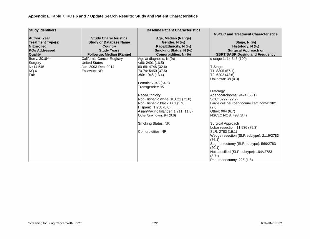
Appendix E Table 7. KQs 6 and 7 Update Search Results: Study and Patient Characteristics
Screening for Lung Cancer With LDCT 522 RTI–UNC EPC
Study Identifiers Author, Year Treatment Type(s) N Enrolled KQs Addressed Quality
Study Characteristics Study or Database Name
Country Study Years
Followup, Median (Range)
Baseline Patient Characteristics
Age, Median (Range) Gender, N (%)
Race/Ethnicity, N (%) Smoking Status, N (%) Comorbidities, N (%)
NSCLC and Treatment Characteristics
Stage, N (%) Histology, N (%)
Surgical Approach or SBRT/SABR Dosing and Frequency
Berry, 2018214 Surgery N=14,545 KQ 6 Fair
California Cancer Registry United States Jan. 2003-Dec. 2014 Followup: NR
Age at diagnosis, N (%) <60: 2401 (16.5) 60-69: 4746 (32.6) 70-79: 5450 (37.5) ≥80: 1948 (13.4) Female: 7948 (54.6) Transgender: <5 Race/Ethnicity Non-Hispanic white: 10,621 (73.0) Non-Hispanic black: 861 (5.9) Hispanic: 1,258 (8.6) Asian/Pacific Islander: 1,711 (11.8) Other/unknown: 94 (0.6) Smoking Status: NR Comorbidities: NR
c-stage 1: 14,545 (100) T Stage T1: 8305 (57.1) T2: 6202 (42.6) Unknown: 38 (0.3) Histology Adenocarcinoma: 9474 (65.1) SCC: 3227 (22.2) Large cell neuroendocrine carcinoma: 382 (2.6) Other: 964 (6.7) NSCLC NOS: 498 (3.4) Surgical Approach Lobar resection: 11,536 (79.3) SLR: 2783 (19.1) Wedge resection (SLR subtype): 2119/2783 (76.1) Segmentectomy (SLR subtype): 560/2783 (20.1) Not specified (SLR subtype): 104*/2783 (3.7*) Pneumonectomy: 226 (1.6)

Appendix E Table 7. KQs 6 and 7 Update Search Results: Study and Patient Characteristics
Screening for Lung Cancer With LDCT 523 RTI–UNC EPC
Study Identifiers Author, Year Treatment Type(s) N Enrolled KQs Addressed Quality
Study Characteristics Study or Database Name
Country Study Years
Followup, Median (Range)
Baseline Patient Characteristics
Age, Median (Range) Gender, N (%)
Race/Ethnicity, N (%) Smoking Status, N (%) Comorbidities, N (%)
NSCLC and Treatment Characteristics
Stage, N (%) Histology, N (%)
Surgical Approach or SBRT/SABR Dosing and Frequency
Bongers, 2011284 SABR N= 500 (530 tumors) KQ 7 Fair
NA Netherlands April 2003-April 2009 Followup: 33 mos. (13 to 86 mos.)
Age: 74 (42 to 92) Female: 209 (41.8) Race/Ethnicity: NR Smoking Status: NR Comorbidities 374 (74.8%) medically inoperable
T Stage (n=530) 1: 307 (57.9) 2: 223 (42.1) Histology (n=500) Adenocarcinoma: 61* (12.2*) SCC: 57* (11.4*) NSCLC NOS: 64* (12.8*) Not obtained: 318 (63.6) SABR Dosing and Frequency (n=530) 3 x 20 Gy: 215 (40.6) 5 x 12 Gy: 226 (42.6) 8 x 7.5 Gy: 89 (16.8)
Chang, 2012280 SABR N=130 KQ 7 Fair
NA United States Feb. 2005-Dec. 2009 Followup: 26 mos. (6-78 mos.)
Age: 74 (48-91) Female: 63 (48.5) Race/Ethnicity: NR Smoking Status: NR Comorbidities: COPD Stage 0-II: 73 (56) COPD Stage III-IV: 57 (44) History of other type of cancer: 37 (28.5)
Stage IA: 112 (86) IB: 18 (14) Histology SCC: 36 (28) Adenocarcinoma: 58 (45) NSCLC NOS: 36 (28) SABR Dosing and Frequency: 50 Gy total, (to PTV between 75% & 90% isodose lines) administered in 4 fractions over 4 consecutive days.

Appendix E Table 7. KQs 6 and 7 Update Search Results: Study and Patient Characteristics
Screening for Lung Cancer With LDCT 524 RTI–UNC EPC
Study Identifiers Author, Year Treatment Type(s) N Enrolled KQs Addressed Quality
Study Characteristics Study or Database Name
Country Study Years
Followup, Median (Range)
Baseline Patient Characteristics
Age, Median (Range) Gender, N (%)
Race/Ethnicity, N (%) Smoking Status, N (%) Comorbidities, N (%)
NSCLC and Treatment Characteristics
Stage, N (%) Histology, N (%)
Surgical Approach or SBRT/SABR Dosing and Frequency
Cummings, 2018231 SBRT N=163 KQs 6 & 7 Fair
NA United States 2007-2015 Followup 1-Fraction (SF): 24 mos. (1.6 to 64 mos) 5-Fraction (FF): 40 mos. (3 to 97 mos.)
Age: 77 (52 to 97) Female: 89* (54.6*) Race/Ethnicity: NR Pack-years smoking, years (range) SF: 50 (0 to 140) FF: 47.5 (0 to 125) Comorbidities: NR
T Stage T1b: 92 (56.4*) T1c: 34 (20.9*) NR: 37* (22.7*) Histology Adenocarcinoma: 77 (47.2*) SCC: 46 (28.2*) LCC: 1 (0.6*) BAC: 9* (5.5*) Other: 7 (4.3*) No pathology: 23* (14.1*) SBRT Dosing and Frequency: Median doses were 30 Gy in the SF arm and 50 Gy in the FF arm. In the latter, most patients received 50 Gy total in 5 fractions; 18 patients received 60 Gy total in 5 fractions.

Appendix E Table 7. KQs 6 and 7 Update Search Results: Study and Patient Characteristics
Screening for Lung Cancer With LDCT 525 RTI–UNC EPC
Study Identifiers Author, Year Treatment Type(s) N Enrolled KQs Addressed Quality
Study Characteristics Study or Database Name
Country Study Years
Followup, Median (Range)
Baseline Patient Characteristics
Age, Median (Range) Gender, N (%)
Race/Ethnicity, N (%) Smoking Status, N (%) Comorbidities, N (%)
NSCLC and Treatment Characteristics
Stage, N (%) Histology, N (%)
Surgical Approach or SBRT/SABR Dosing and Frequency
Detillon, 2019229 SBRT N=378 (159 and 36 for primary and secondary PSM analyses, respectively) KQ 6 Fair
Netherlands Cancer Registry Netherlands 2010-2015 Followup Unmatched analysis: 32 mos. Primary PSM analysis: 32 mos. Secondary PSM analysis: 33 mos.
Age, Mean (SD) Unmatched analysis: 74.9 (5.9) Primary PSM analysis: 74.3 (5.4) Secondary PSM analysis: 73.5 (5.8) Female Unmatched analysis: 154 (40.7) Primary PSM analysis: 61 (38.4) Secondary PSM analysis: 14 (38.9) Race/Ethnicity: NR Smoking Status: NR Comorbidities Unmatched analysis Pulmonary: 221 (61.9) Cardiac: 158 (44.3) Hypertension: 139 (38.9) Previous malignancy: 126 (35.3) Vascular: 116 (32.5) Diabetes: 73 (20.4) Unknown: 21 (5.6) Primary PSM analysis Pulmonary: 82 (51.6) Cardiac: 67 (42.1) Hypertension: 66 (41.5) Previous malignancy: 68 (42.8) Vascular: 53 (33.3) Diabetes: 27 (17.0) Secondary PSM analysis Pulmonary: 20 (55.6) Cardiac: 14 (38.9) Hypertension: 15 (41.7) Previous malignancy: 12 (33.3) Vascular: 13 (36.1) Diabetes: 6 (16.7)
T Stage Unmatched analysis 1a: 139 (36.8) 1b: 117 (31.0) 2a: 91 (24.1) Unknown: 31 (8.2) Primary PSM analysis 1a: 49 (30.8) 1b: 53 (33.3) 2a: 57 (35.8) Secondary PSM analysis 1a: 14 (38.9) 1b: 12 (33.3) 2a: 10 (27.8) Histology Unmatched analysis Adenocarcinoma: 77 (20.4) SCC: 65 (17.2) Other: 50 (13.2) Unknown: 186 (49.2) Primary PSM analysis Adenocarcinoma: 63 (39.6) SCC: 54 (34.0) Other or unknown: 42 (26.4) Secondary PSM analysis Adenocarcinoma: 8 (22.2) SCC: 5 (13.9) Other or unknown: 23 (63.9) SBRT Dosing and Frequency: Total Gy NR, schedules varied between 3 to 8 fractions, delivered 2-3 times per week in the case of multiple fractions.

Appendix E Table 7. KQs 6 and 7 Update Search Results: Study and Patient Characteristics
Screening for Lung Cancer With LDCT 526 RTI–UNC EPC
Study Identifiers Author, Year Treatment Type(s) N Enrolled KQs Addressed Quality
Study Characteristics Study or Database Name
Country Study Years
Followup, Median (Range)
Baseline Patient Characteristics
Age, Median (Range) Gender, N (%)
Race/Ethnicity, N (%) Smoking Status, N (%) Comorbidities, N (%)
NSCLC and Treatment Characteristics
Stage, N (%) Histology, N (%)
Surgical Approach or SBRT/SABR Dosing and Frequency
Detillon, 2019229 SBRT N=378 (159 and 36 for primary and secondary PSM analyses, respectively) KQ 6 Fair (continued)
N (%) of Comorbidities Unmatched analysis None: 9 (2.5) 1: 73 (20.5) 2: 105 (29.4) 3: 75 (21.0) ≥4: 95 (26.6) Primary PSM analysis None: 6 (3.8) 1: 27 (17.0) 2: 54 (34.0) 3: 34 (21.4) ≥4: 38 (23.9) Secondary PSM analysis None: 2 (5.6) 1: 7 (19.4) 2: 12 (33.3) 3: 8 (22.2) ≥4: 7 (19.4) Charlson Comorbidity Score, N (%) Unmatched analysis 0: 9 (2.5) 1: 61 (17.1) 2: 79 (22.1) 3: 83 (23.2) ≥4: 125 (35.0) Primary PSM analysis 0: 6 (3.8) 1: 17 (10.7) 2: 42 (26.4) 3: 40 (25.2) ≥4: 54 (34.0)

Appendix E Table 7. KQs 6 and 7 Update Search Results: Study and Patient Characteristics
Screening for Lung Cancer With LDCT 527 RTI–UNC EPC
Study Identifiers Author, Year Treatment Type(s) N Enrolled KQs Addressed Quality
Study Characteristics Study or Database Name
Country Study Years
Followup, Median (Range)
Baseline Patient Characteristics
Age, Median (Range) Gender, N (%)
Race/Ethnicity, N (%) Smoking Status, N (%) Comorbidities, N (%)
NSCLC and Treatment Characteristics
Stage, N (%) Histology, N (%)
Surgical Approach or SBRT/SABR Dosing and Frequency
Detillon, 2019229 SBRT N=378 (159 and 36 for primary and secondary PSM analyses, respectively) KQ 6 Fair (continued)
Secondary PSM analysis None: 2 (5.6) 1: 6 (16.7) 2: 10 (27.8) 3: 6 (16.7) ≥4: 12 (33.3)
Dziedzic, 2017215 Surgery N=6,905 KQs 6 & 7 Fair
Polish National Lung Cancer Registry Poland Jan. 2007-Dec. 2013 Followup: 36.9 mos. (95% CI, 36.1 to 37.9 mos.)
Age: 63.3 (IQR, 57.6 to 70.1) Female: 2,865 (41.5*) Race/Ethnicity: NR Smoking Status: NR Comorbidities: NR
Stage IA: 5,147* (74.5*) IB: 1,758* (25.5*) Histology Adenocarcinoma: 3,181* (46.1*) SCC: 2,235* (32.4*) Other: 1,489* (21.6*) Surgical Approach Lobectomy: 5,911 (85.6*) Segmentectomy: 233 (3.4*) Wedge resection: 761 (11*)

Appendix E Table 7. KQs 6 and 7 Update Search Results: Study and Patient Characteristics
Screening for Lung Cancer With LDCT 528 RTI–UNC EPC
Study Identifiers Author, Year Treatment Type(s) N Enrolled KQs Addressed Quality
Study Characteristics Study or Database Name
Country Study Years
Followup, Median (Range)
Baseline Patient Characteristics
Age, Median (Range) Gender, N (%)
Race/Ethnicity, N (%) Smoking Status, N (%) Comorbidities, N (%)
NSCLC and Treatment Characteristics
Stage, N (%) Histology, N (%)
Surgical Approach or SBRT/SABR Dosing and Frequency
Ezer, 2018271 Surgery N=9,508 KQ 7 Fair
SEER United States 2000-2009 Followup: NR
Age Total: 74.5* Open Lobectomy: 74 (5) VATS Lobectomy: 75 (6) Female Total: 5,143* (54.1*) Open Lobectomy: 4,414 (53) VATS Lobectomy: 729 (61) Race/Ethnicity White Total: 8,489* (89.3*) Open Lobectomy: 7,429 (88) VATS Lobectomy: 1,060 (89) African American Total: NR Open Lobectomy: 426 (5) VATS Lobectomy: >11 (exact N was NR) Hispanic Total: 105* (1.1*) Open Lobectomy: 94 (1) VATS Lobectomy: ≤11 (exact N was NR) Other Total: 445* (4.7*) Open Lobectomy: 374 (5) VATS Lobectomy: 71 (6) Smoking Status: NR Comorbidity score, N (%) <1 Total: 2,930* (30.8*) Open Lobectomy: 2,504 (30) VATS Lobectomy: 426 (36)
Stage I Total: 8,281* (87.1*) Open Lobectomy: 7,204 (87) VATS Lobectomy: 1,077 (91) Stage II Total: 1,227* (12.9*) Open Lobectomy: 1,119 (13) VATS Lobectomy: 108 (9) Histology Adenocarcinoma Total: 5,681* (59.7*) Open Lobectomy: 4,943 (60) VATS Lobectomy: 738 (62) SCC Total: 2,902* (30.5*) Open Lobectomy: 2,616 (32) VATS Lobectomy: 286 (24) LCC Total: 295* (3.1*) Open Lobectomy: 271 (3) VATS Lobectomy: 24 (2) Other Total: 630* (6.6*) Open Lobectomy: 493 (6) VATS Lobectomy: 137 (12) Surgical Approach Open Lobectomy: 8,323 (87.5*) VATS Lobectomy: 1,185 (12.5*)

Appendix E Table 7. KQs 6 and 7 Update Search Results: Study and Patient Characteristics
Screening for Lung Cancer With LDCT 529 RTI–UNC EPC
Study Identifiers Author, Year Treatment Type(s) N Enrolled KQs Addressed Quality
Study Characteristics Study or Database Name
Country Study Years
Followup, Median (Range)
Baseline Patient Characteristics
Age, Median (Range) Gender, N (%)
Race/Ethnicity, N (%) Smoking Status, N (%) Comorbidities, N (%)
NSCLC and Treatment Characteristics
Stage, N (%) Histology, N (%)
Surgical Approach or SBRT/SABR Dosing and Frequency
Ezer, 2018271 Surgery N=9,508 KQ 7 Fair (continued)
1-1.5 Total: 3,081* (32.4*) Open Lobectomy: 2,679 (32) VATS Lobectomy: 402 (34) 1.5-2.5 Total: 1,125* (11.8*) Open Lobectomy: 1,029 (12) VATS Lobectomy: 96 (8) >2.5 Total: 2,372* (24.9*) Open Lobectomy: 2,111 (25) VATS Lobectomy: 261 (22)
Factor, 2014276 SBRT N=74 (78 tumors) KQ 7 Fair
NA United States Dec. 2006-Jul. 2012 Followup: Local control: 14.4 mos. Overall survival: 18.8 mos.
Age: 78.5 (56 to 93) Female: Patients NR, but 42 (54) tumors Race/Ethnicity: NR Smoking Status: NR Comorbidities: Patients were either medically inoperable or refused surgery
Stage IA: Patients NR, but 52 (67) tumors IB: Patients NR, but 26 (33) tumors Histology Adenocarcinoma: Patients NR, but 41 (53) tumors SCC: Patients NR, but 23 (29) tumors NSCLC NOS: Patients NR, but 10 (13) tumors Unknown: Patients NR, but 4 (5) tumors SBRT Dosing and Frequency: Median dose of 4800 cGy total, administered in 4 fractions over 4 consecutive days

Appendix E Table 7. KQs 6 and 7 Update Search Results: Study and Patient Characteristics
Screening for Lung Cancer With LDCT 530 RTI–UNC EPC
Study Identifiers Author, Year Treatment Type(s) N Enrolled KQs Addressed Quality
Study Characteristics Study or Database Name
Country Study Years
Followup, Median (Range)
Baseline Patient Characteristics
Age, Median (Range) Gender, N (%)
Race/Ethnicity, N (%) Smoking Status, N (%) Comorbidities, N (%)
NSCLC and Treatment Characteristics
Stage, N (%) Histology, N (%)
Surgical Approach or SBRT/SABR Dosing and Frequency
Fischer-Valuck, 2012279 SBRT N=62 KQ 7 Fair
NA United States March 2005-Aug. 2010 Followup: 28 mos. (4 to 78 mos.)
Age, Mean (Range): 72.6 (27 to 92) Female: 35 (56.5) Race/Ethnicity: NR Smoking history: Yes: 52 (83.8) No: 10 (16.2) Comorbidities: NR
Stage IA: 44 (70.9) IB: 18 (29.1) Histology Adenocarcinoma: 22 (35.4) SCC: 22 (35.4) BAC: 3 (4.8) NSCLC NOS: 15 (24.1) SBRT Dosing and Frequency Dosing, N (%) 48 Gy (4 x 12 Gy): 13 (20.9) 60 Gy (5 x 12 Gy): 49 (79.1)
Guckenberger, 2013278 SBRT N=582 KQ 7 Fair
NA Germany/Austria Study Years: 1998-2011 Followup, Mean: 21.4 mos
Age: 72.2 (30.9 to 92.4) Female: 177 (30.4) Race/Ethnicity: NR Smoking Status: NR Comorbidities: NR
Stage IA: 327 (56.2) IB: 236 (40.5) I (unclear): 19 (3.3) Histology Adenocarcinoma: 231 (39.7) SCC: 195 (33.5) Other: 55 (9.5) Unknown or no biopsy: 101 (1.9) SBRT Dosing and Frequency, Median (Range): N of SBRT Fractions: 3 (1 to 20) Single-fraction dose PTV-encompassing (Gy): 12.5 (2.9 to 33) Total dose PTV-encompassing (Gy): 37.5 (12 to 64)

Appendix E Table 7. KQs 6 and 7 Update Search Results: Study and Patient Characteristics
Screening for Lung Cancer With LDCT 531 RTI–UNC EPC
Study Identifiers Author, Year Treatment Type(s) N Enrolled KQs Addressed Quality
Study Characteristics Study or Database Name
Country Study Years
Followup, Median (Range)
Baseline Patient Characteristics
Age, Median (Range) Gender, N (%)
Race/Ethnicity, N (%) Smoking Status, N (%) Comorbidities, N (%)
NSCLC and Treatment Characteristics
Stage, N (%) Histology, N (%)
Surgical Approach or SBRT/SABR Dosing and Frequency
Haasbeek, 2010289 SBRT N=193 (203 tumors) KQ 7 Fair
NA Netherlands 2003-2008 Followup: 12.6 mos. (3 to 52 mos.)
Age: 79 (57 to 91) Female: 62 (32) Race/Ethnicity: NR Smoking Status: NR Comorbidities Medically inoperable: 155 (80) COPD: 140* (72.5*)
T Stage (n=203) T1: 118 (58) T2: 85 (42) Histology (n=75) Adenocarcinoma: 23 (30.7*) SCC: 18 (24*) Undifferentiated NSCLC: 34 (45.3*) SBRT Dosing and Frequency: All patients received 60 Gy total. Half of the patients (101) received 12 Gy in 5 fractions; 69 (34%) received 20 Gy in 3 fractions; 33 (16%) received 7.5 Gy in 8 fractions.
Handa, 2018216 Surgery N=711 KQ 6 Fair
NA Japan April 2000-Dec. 2015 Followup: 52.3 mos.
Age: NR Female: NR Race/Ethnicity: NR Smoking Status: NR Comorbidities: NR
Stage: NR Histology: NR Surgical Approach: Lobectomy or bilobectomy

Appendix E Table 7. KQs 6 and 7 Update Search Results: Study and Patient Characteristics
Screening for Lung Cancer With LDCT 532 RTI–UNC EPC
Study Identifiers Author, Year Treatment Type(s) N Enrolled KQs Addressed Quality
Study Characteristics Study or Database Name
Country Study Years
Followup, Median (Range)
Baseline Patient Characteristics
Age, Median (Range) Gender, N (%)
Race/Ethnicity, N (%) Smoking Status, N (%) Comorbidities, N (%)
NSCLC and Treatment Characteristics
Stage, N (%) Histology, N (%)
Surgical Approach or SBRT/SABR Dosing and Frequency
Inoue, 2013222 SBRT N=109 KQs 6 & 7 Fair
NA Japan June 2005-Nov. 2010 Followup: 25 mos (4 to 72 mos.)
Age: 78 (47 to 90) Female: 35 (32.1*) Race/Ethnicity: NR Smoking Status: NR Comorbidities: NR
Stage (all N0M0) T1a: 47 (43.1*) T1b: 32 (29.4*) T2: 30 (27.5*) Histology Adenocarcinoma: 65 (59.6*) SCC 29 (26.6*) LCC: 1 (0.9*) NSCLC NOS: 8 (7.3*) Unproven: 6 (5.5*) SBRT Dosing and Frequency: Treatment period of 4 to 7 days 2005-2006: 48 Gy total, administered in 4 fractions 2007-2010: 40 Gy total, administered in 4 fractions to 95% volume of PTV (~45 to 50 Gy)
Jeppesen, 2013221 SBRT N=100 KQ 6 Fair
NA Denmark Aug. 2005-June 2012 Followup: 35.4 mos. (8.8 to 90.5 mos.)
Age, Mean (Range): 73.3 (52 to 88) Female: 55 (55) Race/Ethnicity: NR Smoking Status: Smoker or ex-smoker: 81 (81) Never smoker: 19 (19) Comorbidities: All patients were medically inoperable
T-Stage T1: 72 (72) T2: 28 (28) Histology: Adenocarcinoma: 59 (59) SCC: 28 (28) Other: 13 (13) SBRT Dosing and Frequency: 15 to 22 Gy x 3, delivered in 9 days

Appendix E Table 7. KQs 6 and 7 Update Search Results: Study and Patient Characteristics
Screening for Lung Cancer With LDCT 533 RTI–UNC EPC
Study Identifiers Author, Year Treatment Type(s) N Enrolled KQs Addressed Quality
Study Characteristics Study or Database Name
Country Study Years
Followup, Median (Range)
Baseline Patient Characteristics
Age, Median (Range) Gender, N (%)
Race/Ethnicity, N (%) Smoking Status, N (%) Comorbidities, N (%)
NSCLC and Treatment Characteristics
Stage, N (%) Histology, N (%)
Surgical Approach or SBRT/SABR Dosing and Frequency
Karasawa, 2018227 SABR N=56 KQs 6 & 7 Fair
NA Japan Oct. 2003-Dec. 2010 Followup: 10.6 yrs
Age: 79 (49 to 91) Female: 17 (30.4*) Race/Ethnicity: NR Smoking Status: NR Comorbidities High risk operable: 27 (48.2*) Medically inoperable: 21 (37.5*) Pulmonary risk factor: 31 (55.4*) Cardiac risk factor: 8 (14.3*) Central nervous system factor: 4 (7.1*) Hepatic risk factor: 1 (1.8*)
T Stage T1: 41 (73.2*) T2: 15 (26.8*) Histology: Adenocarcinoma: 34 (60.7*) SCC: 18 (32.1*) Large cell neuroendocrine cancer: 1 (1.8*) NSCLC NOS: 2 (3.6*) Unproven: 1 (1.8*) SBRT Dosing and Frequency: Patients primarily received 48 Gy total, delivered in 4 fractions over 1 week.
Lagerwaard, 2012281 SABR N=382 KQ 7 Fair
NA Netherlands April 2003-Nov. 2008 Followup: 23 mos.
Age: 74 (47 to 91) Female: 152 (39.8) Race/Ethnicity: NR Smoking Status: NR Comorbidities: History of prior lung cancer: 65 (17) COPD (mild, moderate, severe, or very severe) (n=361): 304* (84*) Medically inoperable: 323 (84.6)
Stage IA: 230 (60.2) IB: 152 (39.8) Histology: NR SABR Dosing and Frequency: 60 Gy total, administered in 3, 5, or 8 fractions (depending on tumor diameter and location)

Appendix E Table 7. KQs 6 and 7 Update Search Results: Study and Patient Characteristics
Screening for Lung Cancer With LDCT 534 RTI–UNC EPC
Study Identifiers Author, Year Treatment Type(s) N Enrolled KQs Addressed Quality
Study Characteristics Study or Database Name
Country Study Years
Followup, Median (Range)
Baseline Patient Characteristics
Age, Median (Range) Gender, N (%)
Race/Ethnicity, N (%) Smoking Status, N (%) Comorbidities, N (%)
NSCLC and Treatment Characteristics
Stage, N (%) Histology, N (%)
Surgical Approach or SBRT/SABR Dosing and Frequency
Lagerwaard, 2012223 SABR N=177 KQs 6 & 7 Fair
NA Netherlands April 2003-Dec. 2010 Followup: 31.5 mos.
Age: 76 (50 to 91) Female: 76 (43) Race/Ethnicity: NR Smoking Status: Current or former: 168 (95) Never smoked: 9 (5) Comorbidities: COPD: 112* (63*) Charlson Comorbidity Score: Median (range): 2 (0 to 5) 0: 18 (10) 1: 59 (33) 2: 38 (22) 3: 39 (22) 4: 16 (9) 5: 7 (4)
Stage IA: 106* (60) 1B: 71* (40) Histology (n=60) Adenocarcinoma: 20 (33) SCC: 16 (27) Undifferentiated NSCLC: 24 (38) SBRT Dosing and Frequency: 60 Gy total, delivered as 20 Gy in 3 fractions for 34% of patients; 12 Gy in 5 fractions for 46%; and 7.5 Gy in 8 fractions for 19%.
Lagerwaard, 2008290 SBRT N=206 (219 tumors) KQ 7 Fair
NA Netherlands Study Years NR Followup: 12 mos. (3 to 44 mos.)
Age: 73 (NR) Female: 91 (44) Race/Ethnicity: NR Smoking Status: NR Comorbidities Medically inoperable: 167 (81) COPD: 151* (73.3*) Previous malignancy: 80* (39) Previous lung cancer: 37 (18)
T Stage (n=219) T1: 129 (59) T2: 90 (41) Histology (n=64) Adenocarcinoma: 23 (36) SCC: 19 (30) Undifferentiated NSCLC: 22 (34 SBRT Dosing and Frequency: Total Gy NR, delivered as 20 Gy in 3 fractions for 93/219 (43%) tumors; 12 Gy in 5 fractions for 99/219 (45%); and 7.5 Gy in 8 fractions for 27 (12%).

Appendix E Table 7. KQs 6 and 7 Update Search Results: Study and Patient Characteristics
Screening for Lung Cancer With LDCT 535 RTI–UNC EPC
Study Identifiers Author, Year Treatment Type(s) N Enrolled KQs Addressed Quality
Study Characteristics Study or Database Name
Country Study Years
Followup, Median (Range)
Baseline Patient Characteristics
Age, Median (Range) Gender, N (%)
Race/Ethnicity, N (%) Smoking Status, N (%) Comorbidities, N (%)
NSCLC and Treatment Characteristics
Stage, N (%) Histology, N (%)
Surgical Approach or SBRT/SABR Dosing and Frequency
Lee, 2017232 SBRT/SABR N=169 (178 tumors) KQs 6 & 7 Fair
NA South Korea June 2000-May 2015 Followup: 32 mos. (2 to 195 mos.)
Age: 73 (40 to 91) Female: 38 (22) Race/Ethnicity: NR Smoking Status: NR Comorbidities Charlson Comorbidity Index Score, median (range): 5 (0 to 8) Poor lung function: 86 (51) Other comorbidity: 27 (16)
Stage T1a: 39 (22) T1b: 70 (39) T2a: 69 (39) Histology (n=178) Adenocarcinoma: 87 (49) SCC: 78 (44) NSCLC unspecified: 5 (3) Others: 3 (2) No biopsy: 5 (3) SABR Dosing and Frequency 82 (46%) patients received 60 Gy total in 4 fractions, 51 (29%) patients received 48 Gy total in 4 fractions, 18 (10%) patients received 54 Gy total in 3 fractions, 10 (6%) patients received 60 Gy total in 3 fractions, and 17 (10%) patients received other doses/fractionations.
Liu, 2018217 Surgical resection N=3,219 KQ 6 Fair
SEER United States 2004-2013 Followup: 37 mos. (1 to 120 mos.)
Age: 69 (20 to 92) Female: 1,918 (59.6) Race/Ethnicity: White: 2,805 (87.1) Black: 271 (8.4) Other: 143 (4.5) Smoking Status: NR Comorbidities: NR
Stage IA: 3,219 (100) Histology Adenocarcinoma: 1,892 (58.8) SCC: 738 (22.9) LCC: 142 (4.4) Other: 447 (13.9) Surgical Approach Wedge: 2,327 (72.3) Segmental: 892 (27.7)

Appendix E Table 7. KQs 6 and 7 Update Search Results: Study and Patient Characteristics
Screening for Lung Cancer With LDCT 536 RTI–UNC EPC
Study Identifiers Author, Year Treatment Type(s) N Enrolled KQs Addressed Quality
Study Characteristics Study or Database Name
Country Study Years
Followup, Median (Range)
Baseline Patient Characteristics
Age, Median (Range) Gender, N (%)
Race/Ethnicity, N (%) Smoking Status, N (%) Comorbidities, N (%)
NSCLC and Treatment Characteristics
Stage, N (%) Histology, N (%)
Surgical Approach or SBRT/SABR Dosing and Frequency
Lutz, 2018218 Surgical resection N=632 KQs 6 & 7 Fair
NA France Jan. 2007-Aug. 2016 Followup, mean: 34.5
Age: 65 (IQR, 59 to 71) Female: 321 (50.8) Race/Ethnicity: NR Smoking Status: NR Comorbidities: NR
p-Stage IA: 281 (44.5) IB: 201 (31.8) IIA: 54 (8.5) IIB: 39 (6.2) IIIA: 57 (9) Histology Adenocarcinoma: 498 (78.8) SCC: 102 (16.1) LCC: 32 (5.1) Surgical Approach: All patients underwent surgical resection. The most common types of resection were right upper lobe (29.1%), segmentectomy (25.3%), and left upper lobe (15.5%).
Lv, 2018219 Surgical resection N=861 KQ 6 Fair
SEER United States Jan. 2004-Dec. 2014 Followup: 39 mos. (0 to 131 mos.)
Age, mean (SD) Lobectomy (n=662): 65.9 (10.9) Sublobar resection (n=199): 66.6 (11.3) Female: 570* (66.2*) Race/Ethnicity White: 711* (82.6*) Black/other: 150* (17.4*) Smoking Status: NR Comorbidities: NR
Stage IA: 861 (100) Histology Adenocarcinoma: 656* (76.2*) SCC: 113* (13.1*) Other: 92* (10.7*) Surgical Approach: 662 (76.9) patients underwent lobectomy, and 199 (23.1) patients underwent sub-lobar resection.

Appendix E Table 7. KQs 6 and 7 Update Search Results: Study and Patient Characteristics
Screening for Lung Cancer With LDCT 537 RTI–UNC EPC
Study Identifiers Author, Year Treatment Type(s) N Enrolled KQs Addressed Quality
Study Characteristics Study or Database Name
Country Study Years
Followup, Median (Range)
Baseline Patient Characteristics
Age, Median (Range) Gender, N (%)
Race/Ethnicity, N (%) Smoking Status, N (%) Comorbidities, N (%)
NSCLC and Treatment Characteristics
Stage, N (%) Histology, N (%)
Surgical Approach or SBRT/SABR Dosing and Frequency
Manyam, 2019228 SBRT/SABR N=139 (146 tumors) KQs 6 & 7 Fair
NA United States 2009-2016 Followup: 23.8 mos. (3.1 to 87.9 mos.)
Age: 76 (47 to 92) Female: 74 (53.2*) Race/Ethnicity: NR Smoking Status Pack-years, median (range): 50 (0 to 160) Smoking during treatment: 38 (27.5) Comorbidities Pulmonary: 80 (58) Cardiac: 12 (9) Refusal: 5 (4) Other/multifactorial: 42 (29)
Stage IA: 135 (93) IB: 10 (6) IIA: 1 (1) Histology Adenocarcinoma: 39 (27) SCC: 38 (26) Others: 8 (6) Nondiagnostic: 14 (10) No biopsy: 47 (32) SBRT Dosing and Frequency: Of 146 lesions, 80 (55%) were treated with 30 Gy and 66 (45%) were treated with 34 Gy.
Matsuo, 2012283 SBRT N=74 KQ 7 Fair
NA Japan Sept. 2003-March 2008 Followup: 31.4 mos. (4.2 to 65.0 mos.)
Age: 77 (63 to 88) Female: 19 (25.7*) Race/Ethnicity: NR Smoking Status: NR Comorbidities: Inoperable: 50 (67.6*)
Stage T1a: 26 (35.1*) T1b: 27 (36.5*) T2a: 21 (28.4*) Histology Adenocarcinoma: 36 (48.6*) SCC: 30 (40.5*) Other (LCC or NSCLC NOS): 8 (10.8*) SBRT Dosing and Frequency: 48 Gy total, administered in 4 fractions at the isocenter; median (range) overall treatment time was 5 days (4 to 12 days)

Appendix E Table 7. KQs 6 and 7 Update Search Results: Study and Patient Characteristics
Screening for Lung Cancer With LDCT 538 RTI–UNC EPC
Study Identifiers Author, Year Treatment Type(s) N Enrolled KQs Addressed Quality
Study Characteristics Study or Database Name
Country Study Years
Followup, Median (Range)
Baseline Patient Characteristics
Age, Median (Range) Gender, N (%)
Race/Ethnicity, N (%) Smoking Status, N (%) Comorbidities, N (%)
NSCLC and Treatment Characteristics
Stage, N (%) Histology, N (%)
Surgical Approach or SBRT/SABR Dosing and Frequency
Moon, 2018212 Surgical resection N=15,358 KQ 6 Fair
SEER United States 2000-2014 Followup: 56 mos. (IQR, 25 to 95 mos.)
Age, mean (SD) Lobectomy: 65.5 (10.2) Segmentectomy: 67.8 (10) Female: 9,037* (58.8*) Race/Ethnicity American Indian or Alaska Native: 47* (0.3*) Asian or Pacific Islander: 859* (5.6*) Black: 1,084* (7.1*) White: 13,318* (86.7*) Unknown: 50* (0.3*) Smoking Status: NR Comorbidities: NR
Stage IA: 15,358 (100) Histology Adenocarcinoma: 10,645* (69.3*) SCC: 2,881* (18.8*) Others: 1,832* (11.2*) Surgical Approach: Lobectomy: 14,549 (94.7*) Segmentectomy: 809 (5.3*)
Mutter, 2012285 SBRT/SABR N=126 KQ 7 Fair
NA United States May 2006-July 2009 Followup: 16 mos. (3 to 43 mos.)
Age: 77 (55 to 95) Female: NR Race/Ethnicity: NR Smoking Status: NR Comorbidities: NR
Stage T1a: 63 (50*) T1b: 32 (25.4*) T2a: 27 (21.4*) Histology Adenocarcinoma: 93 (74) NSCLC unspecified: 2 (2) Not determined: 1 (1) Squamous: 30 (24) SBRT Dosing and Frequency Dose, Median (range): 54 Gy (40 to 60 Gy) Treatment time, Median (range): 7 days (4 to 19 days) Number of total fractions: 3 fractions: 73 (57.9*) 4 fractions: 38 (30.2*) 5 fractions: 15 (11.9*)

Appendix E Table 7. KQs 6 and 7 Update Search Results: Study and Patient Characteristics
Screening for Lung Cancer With LDCT 539 RTI–UNC EPC
Study Identifiers Author, Year Treatment Type(s) N Enrolled KQs Addressed Quality
Study Characteristics Study or Database Name
Country Study Years
Followup, Median (Range)
Baseline Patient Characteristics
Age, Median (Range) Gender, N (%)
Race/Ethnicity, N (%) Smoking Status, N (%) Comorbidities, N (%)
NSCLC and Treatment Characteristics
Stage, N (%) Histology, N (%)
Surgical Approach or SBRT/SABR Dosing and Frequency
Olsen, 2011286 SBRT/SABR N=130 KQ 7 Fair
NA United States June 2004-June 2009 11 mos. (2 to 33 mos.)
Age: 75 (31 to 92) Female: 65* (50*) Race/Ethnicity: NR Smoking Status: NR Comorbidities: 117 of 130 patients were considered medically inoperable
Stage T1a: 56 (43.1*) T1b: 44 (33.8*) T2a: 24 (18.5*) Histology Biopsy-proven: 110* (84.6*) SBRT Dosing and Frequency 9 Gy in 5 fractions: 8 (6.2*) 10 Gy in 5 fractions: 11 (8.5*) 18 Gy in 3 fractions: 111 (85.4*)
Onishi, 2007224 SBRT/SABR N=257 KQs 6 & 7 Fair
NA Japan April 1995-March 2004 Followup: 38 mos. (2 to 128 mos.)
Age: 74 (39 to 92) Female: NR Race/Ethnicity: NR Smoking Status: NR Comorbidities Pulmonary chronic disease: 168 (65.4*) Medically inoperable: 158 (61.5*)
Stage IA: 164 (63.8*) IB: 93 (36.2*) Histology Adenocarcinoma: 120 (46.7*) SCC: 111 (43.2*) Other: 26 (10.1*) SBRT Dosing and Frequency: 10 to 75 Gy total (at isocenter) was administered in 1 to 22 fractions.
Palma, 2010287
SBRT/SABR N=99* KQ 7 Fair
Amsterdam Cancer Registry Netherlands 1999-2007 Followup: 54 mos.
Age: NR Female: NR Race/Ethnicity: NR Smoking Status: NR Comorbidities: NR
T Stage IA or IB: 99* (100) Histology: NR SBRT Dosing and Frequency: NR

Appendix E Table 7. KQs 6 and 7 Update Search Results: Study and Patient Characteristics
Screening for Lung Cancer With LDCT 540 RTI–UNC EPC
Study Identifiers Author, Year Treatment Type(s) N Enrolled KQs Addressed Quality
Study Characteristics Study or Database Name
Country Study Years
Followup, Median (Range)
Baseline Patient Characteristics
Age, Median (Range) Gender, N (%)
Race/Ethnicity, N (%) Smoking Status, N (%) Comorbidities, N (%)
NSCLC and Treatment Characteristics
Stage, N (%) Histology, N (%)
Surgical Approach or SBRT/SABR Dosing and Frequency
Sekihara, 2017220 Surgical resection N=1,356* KQ 6 Fair
NA Japan Jan. 2002-March 2013 Followup: 40 mos.
Age: NR Female: NR Race/Ethnicity: NR Smoking Status: NR Comorbidities: NR
Pathological Stage I: 1,356* Clinical Stage I: 1,612* (78.5*) Histology: NR Surgical Approach: NR
Shibamoto, 2012234 SBRT N=180 KQs 6 & 7 Good
NA Japan May 2004-Nov. 2008 Followup: 36 mos.
Age: 77 (29 to 92) Female: 57 (31.7*) Race/Ethnicity: NR Smoking Status: NR Comorbidities Medically inoperable: 120 (66.7*)
Stage IA: 128 (71.1*) IB: 52 (28.9*) Histology Adenocarcinoma: 104 (57.8*) SCC: 60 (33.3*) NSCLC NOS: 16 (8.9*) SBRT Dosing and Frequency 44 Gy in 4 fractions: 4 (2.2*) 48 Gy in 4 fractions: 124 (68.9*) 52 Gy in 4 fractions: 52 (28.9*) Prescribed dose was 44 Gy in 4 fractions with ≥3 day interfraction intervals (tumors with a max dimension <1.5 cm); 48 Gy in 4 fractions (tumors with a max dimension of 1.5 cm to 3.0 cm); and 52 Gy in 4 fractions (tumors with a max tumor dimension >3 cm)

Appendix E Table 7. KQs 6 and 7 Update Search Results: Study and Patient Characteristics
Screening for Lung Cancer With LDCT 541 RTI–UNC EPC
Study Identifiers Author, Year Treatment Type(s) N Enrolled KQs Addressed Quality
Study Characteristics Study or Database Name
Country Study Years
Followup, Median (Range)
Baseline Patient Characteristics
Age, Median (Range) Gender, N (%)
Race/Ethnicity, N (%) Smoking Status, N (%) Comorbidities, N (%)
NSCLC and Treatment Characteristics
Stage, N (%) Histology, N (%)
Surgical Approach or SBRT/SABR Dosing and Frequency
Stanic, 2014277 SBRT N=55 KQ 7 Fair
RTOG 0236 United States Study Years: NR Followup: 2 yrs
Age: 72 (48 to 89) Female: 34 (61.8) Race/Ethnicity White: 51 (92.7) Asian: 2 (3.6) African American: 2 (3.6) Smoking Status: NR Comorbidities All patients were medically inoperable Total with >1 reason for being medically inoperable: 38 (69.1) Hypoxemia and/or hypercapnia: 10 (18.5) Severe pulmonary hypertension: 6 (10.9) Diabetes mellitus with severe end-organ damage: 3 (5.5) Severe cerebral, cardiac, or peripheral vascular disease: 24 (43.6) Severe chronic cardiac disease: 22 (40.0)
Stage IA: 44 (80) IB: 11 (20) Histology: All patients were confirmed to have NSCLC (histological details NR) SBRT Dosing and Frequency: 60 Gy total, administered in 3 fractions, separated by ≥40 hours, and the full regimen was completed within 14 days
Ueda, 2018272 Surgical resection N=607 KQ 7 Fair
NA Japan Feb. 2008-March 2013 Followup: NR
Age Lobectomy: 67 (35 to 86) Segmentectomy: 67 (27 to 84) Female: 313* (51.6*) Race/Ethnicity: NR Smoking Status ≥40 pack years: 316* (52.1*) Comorbidities History of ischemic heart disease: 53* (8.7*) History of lung cancer resection: 36* (5.9*)
Stage IA: 607 (100) Histology: NR Surgical Approach Lobectomy: 443 (73*) Segmentectomy: 164 (27*)

Appendix E Table 7. KQs 6 and 7 Update Search Results: Study and Patient Characteristics
Screening for Lung Cancer With LDCT 542 RTI–UNC EPC
Study Identifiers Author, Year Treatment Type(s) N Enrolled KQs Addressed Quality
Study Characteristics Study or Database Name
Country Study Years
Followup, Median (Range)
Baseline Patient Characteristics
Age, Median (Range) Gender, N (%)
Race/Ethnicity, N (%) Smoking Status, N (%) Comorbidities, N (%)
NSCLC and Treatment Characteristics
Stage, N (%) Histology, N (%)
Surgical Approach or SBRT/SABR Dosing and Frequency
Uhlig, 2018226 SBRT N=27,732 (1,070 in PSM analysis) KQs 6 & 7 Fair
NCDB United States 2004-2013 Followup, mean: 52.4 mos. (IQR, 32.1 to 75.2 mos.)
Age: 59 (IQR 53 to 65) Female: 601 (56.2) Race/Ethnicity African American: 55 (5.1) White: 984 (92) Other: 31 (2.9) Smoking Status: NR Comorbidities: Charlson comorbidity index score 0: 510 (47.7) 1: 335 (31.3) ≥2: 225 (21.0)
Stage I: 1,070 (100) Histology: NR SBRT Dosing and Frequency: NR for patients in PSM set, but in larger unmatched sample, the median biologic equivalent dose delivered was 100 Gy (IQR, 88 to 221 Gy). The median number of treatment sessions was 5 (IQR, 4 to 16 sessions) over a median duration of 10 days (IQR, 7 to 27 days).
Westover, 2012282 SBRT N=15 (20 tumors) KQ 7 Fair
NA United States July 2008-Sept. 2010 Followup: 24.1 mos.
Age: 78 (62-89) Female: 12* (80*) Race/Ethnicity: NR Smoking Status: 14* (93.3*) Comorbidities COPD: 8 (53.3*) Interstitial lung disease: 1* (6.7*) History of prior lung cancer: 8* (53.3*) Systematic lupus erythematosus: 1* (6.7*)
Stage T1a: 16/20 (80) tumors T1b: 2/20 (10) tumors T2a: 2/20 (10) tumors Histology Adenocarcinoma: 9/20 (45*) tumors NSCLC NOS: 4/20 (20*) tumors SCC: 3/20 (15*) tumors No biopsy: 4/20 (20*) tumors SBRT Dosing and Frequency Median (range) total dose: 45 Gy (42-50 Gy) Median (range) fraction size: 14 Gy (10-16 Gy) 17/20 tumors (85%*) received 3 fractions

Appendix E Table 7. KQs 6 and 7 Update Search Results: Study and Patient Characteristics
Screening for Lung Cancer With LDCT 543 RTI–UNC EPC
Study Identifiers Author, Year Treatment Type(s) N Enrolled KQs Addressed Quality
Study Characteristics Study or Database Name
Country Study Years
Followup, Median (Range)
Baseline Patient Characteristics
Age, Median (Range) Gender, N (%)
Race/Ethnicity, N (%) Smoking Status, N (%) Comorbidities, N (%)
NSCLC and Treatment Characteristics
Stage, N (%) Histology, N (%)
Surgical Approach or SBRT/SABR Dosing and Frequency
Wink, 2019233 SABR N=554 KQ 6 Fair
NA Netherlands/Germany 2007-2015 Followup: 36.1 mos. (1.1 to 118.3 mos.)
Age: 74 (42 to 91) Female: 234 (42.2) Race/Ethnicity: NR Smoking Status: NR Comorbidities: NR
Stage T1: 428 (77.2) T2: 124 (22.4) Histology Adenocarcinoma: 66 (11.9) SCC: 66 (11.9) LCC: 28 (5.1) AIS: 3 (0.5) NSCLC NOS: 14 (2.5) Suspicion of malignancy, but no histology: 377 (68.1) SABR Dosing and Frequency: The median prescribed dose was 54 Gy (range, 45 to 75 Gy in 3 to 8 fractions).
Ye, 2018292 SBRT N=100 KQ 7 Fair
NA China Jan. 2010-June 2016 Followup: 26.5 mos.
Age: 73 Female: 27 (27*) Race/Ethnicity: NR Smoking Status Never: 38 (38*) Previous: 43 (43*) Current: 19 (19*) Comorbidities COPD: 55 (55*)
Stage T1: 69 (69*) T2a: 31 (31*) Histology Adenocarcinoma: 59 (59*) SCC: 22 (22*) Unknown: 19 (19*) SBRT Dosing and Frequency: Most patients received 60 Gy in 10 fractions, and the rest received 50 Gy in 5 fractions. The study’s reported counts are NR here because they are incorrect.

Appendix E Table 7. KQs 6 and 7 Update Search Results: Study and Patient Characteristics
Screening for Lung Cancer With LDCT 544 RTI–UNC EPC
Study Identifiers Author, Year Treatment Type(s) N Enrolled KQs Addressed Quality
Study Characteristics Study or Database Name
Country Study Years
Followup, Median (Range)
Baseline Patient Characteristics
Age, Median (Range) Gender, N (%)
Race/Ethnicity, N (%) Smoking Status, N (%) Comorbidities, N (%)
NSCLC and Treatment Characteristics
Stage, N (%) Histology, N (%)
Surgical Approach or SBRT/SABR Dosing and Frequency
Zhou, 2017213 Surgical resection N=20,850 KQ 6 Fair
SEER United States 2004-2013 Followup: 38 mos. (IQR, 17 to 66 mos.)
Age <60: 4,079 (19.6) 60-74: 11,112 (53.3) ≥75: 5,659 (27.1) Female: 11,339 (54.4) Race/Ethnicity White: 17,732 (85) Black: 1,665 (8) Other/unknown: 1,453 (7) Smoking Status: NR Comorbidities: NR
Stage IA1: 1,402 (6.7) IA2: 7,037 (33.8) IA3: 5,198 (24.9) IB: 7,213 (34.6) Histology Adenocarcinoma: 11,591 (55.6) SCC: 5,717 (27.4) BAC: 1,792 (8.6) Adenosquamous carcinoma: 578 (2.8) LCC: 411 (2.0) Other: 761 (3.6) Surgical Approach Lobectomy: 16,363 (78.5) Sublobar resection: 4,244 (20.4) Pneumonectomy: 243 (1.2)
*Indicates that data was calculated by abstractors † Per the Colinet Simplified Comorbidity Score, a higher score indicates that a patient has a greater number of comorbidities.396
Abbreviations: AIS=adenocarcinoma in situ; BAC=bronchoalveolar carcinoma; cGy=centigray; COPD=chronic obstructive pulmonary disease; CVD=cardiovascular disease;
ILD=interstitial lung disease; IQR=interquartile range; KQ=key question; LCC=large cell carcinoma; mos=months; NA=not applicable; NOS=not otherwise specified; NR=not
reported; NSCLC=non-small cell lung cancer; PSM=propensity score matching; SABR=stereotactic ablative radiotherapy; SBRT=stereotactic body radiation therapy;
SCC=squamous cell carcinoma.

Appendix E Table 8. KQs 6 and 7 Update Search Results: Surgery Study Outcomes
Screening for Lung Cancer With LDCT 545 RTI–UNC EPC
Appendix E Table 8. KQs 6 and 7 Update Search Results: Surgery Study Outcomes
Study Identifiers Author, Year Treatment Type N Enrolled (Analyzed) KQs Addressed Quality
5-Year Survival Outcomes
Overall Survival, % (95 CI) Lung Cancer Specific Survival, % (95 CI)
Progression- or Recurrence-Free Survival, % (95 CI)
Short-Term Outcomes
30-Day Mortality, N (%) 90-Day Mortality, N (%)
Perioperative Morbidity with ≥10% Incidence
Berry, 2018214 Surgery N=14,545 KQ 6 Fair
Overall survival: 64.9 (95% CI, 64 to 65.8) Lung cancer-specific survival Total sample: 76.9 (95% CI, 76.1 to 77.7) SLR: 70.8 (95% CI, 68.6 to 72.9) Lobar resection: 78.5 (77.6 to 79.4) SLR vs. lobar resection: between-group p <0.0001 (both unadjusted and multivariable adjusted analyses)
NA
Dziedzic, 2017215 Surgery N=6,905 KQs 6 & 7 Fair
Overall survival Unmatched population (n=6,905) Any resection: 76.6 (95% CI, 75.4 to 78) Wedge resection (n=761) Total: 58.1 (95% CI, 53.6 to 62.5) Stage IA: 61.3 (95% CI, 56.2 to 66.5) Stage IB: 44.8 (95% CI, 35.5 to 54.1) Lobectomy (n=5,911) Total: 79.1 (95% CI, 77.7 to 80.5) Stage IA: 80.5 (95% CI, 78.9 to 82.2) Stage IB: 75.3 (95% CI, 72.6 to 77.9) Segmentectomy (n=233) Total: 78.3 (95% CI, 70.6 to 86) Stage IA: 78 (95% CI, 68.8 to 87.1) Stage IB: 78.6 (95% CI, 64.3 to 92.9)95% CI,95% CI,95% CI,95% CI,
30-day mortality Any resection: 1.6 (95% CI, 1.3 to 1.9) Wedge resection: 1.4 (95% CI, 0.6 to 2.3) Lobectomy: 1.6 (95% CI, 1.2 to 1.9) Segmentectomy: 2.6 (95% CI, 0.5 to 4.6) 90-day mortality Any resection: 2.4 (95% CI, 2.1 to 2.8) Wedge resection: 3 (95% CI, 1.8 to 4.2) Lobectomy: 2.3 (95% CI, 1.9 to 2.7) Segmentectomy: 4.3 (95% CI, 1.7 to 6.9)
Ezer, 2018271
Surgery N=9,508 KQ 7 Fair
NA 30-day mortality: 322* (3.4*) Respiratory complications: 3,000* (31.6*) Extended length of stay: 1,397* (14.7*)
Handa, 2018216 Surgery N=711 KQ 6 Fair
Overall Survival: 81.3 (95% CI, NR) NA

Appendix E Table 8. KQs 6 and 7 Update Search Results: Surgery Study Outcomes
Screening for Lung Cancer With LDCT 546 RTI–UNC EPC
Study Identifiers Author, Year Treatment Type N Enrolled (Analyzed) KQs Addressed Quality
5-Year Survival Outcomes
Overall Survival, % (95 CI) Lung Cancer Specific Survival, % (95 CI)
Progression- or Recurrence-Free Survival, % (95 CI)
Short-Term Outcomes
30-Day Mortality, N (%) 90-Day Mortality, N (%)
Perioperative Morbidity with ≥10% Incidence
Liu, 2018217 Surgical resection N=3,219 KQ 6 Fair
Lung cancer specific survival Total sample 1-6 examined LNs (n=2,410): 75 ≥7 examined LNs (n=809): 83 Wedge resection only 1-6 examined LNs (n=1,777): 74 ≥7 examined LNs (n=550): 81 Segmental only 1-6 examined LNs (n=633): 78 ≥7 examined LNs (n=259): 87
NA
Lutz, 2018218 Surgical resection N=632 KQs 6 & 7 Fair
Overall survival Total: 75 (69.9 to 80.1) Stage IA: 79.5 (NR) Stage IB: 76.9 (NR) Lung cancer specific survival Total: 86.4 (82.3 to 90.5) Stage IA: 71.1 (NR) Stage IB: 63.6 (NR)
30-day or in-hospital mortality: 6 (0.95) Any complication: 185 (29.3)
Lv, 2018219 Surgical resection N= 861 KQ 6 Fair
Overall survival: 75 (NR) NA
Moon, 2018212 Surgical resection N=15,358 KQ 6 Fair
Overall survival Lobectomy: 76.0 (75.2 to 76.8) Segmentectomy: 74.4 (95% CI, 67.7 to 75.4) Lung cancer specific survival Lobectomy: 86.0 (85.4 to 86.7) Segmentectomy: 84.7 (81.6 to 88)
NA

Appendix E Table 8. KQs 6 and 7 Update Search Results: Surgery Study Outcomes
Screening for Lung Cancer With LDCT 547 RTI–UNC EPC
Study Identifiers Author, Year Treatment Type N Enrolled (Analyzed) KQs Addressed Quality
5-Year Survival Outcomes
Overall Survival, % (95 CI) Lung Cancer Specific Survival, % (95 CI)
Progression- or Recurrence-Free Survival, % (95 CI)
Short-Term Outcomes
30-Day Mortality, N (%) 90-Day Mortality, N (%)
Perioperative Morbidity with ≥10% Incidence
Sekihara, 2017220 Surgical resection N=1,356* KQ 6 Fair
Overall survival, patients with Stage I: ILD (n=62): 44 (NR) Non-ILD (n=1,294): 84.6 (NR) Lung cancer specific survival: ILD (n=62): 56.6 (NR) Non-ILD (n=1,294): 89.8 (NR) Recurrence-free survival: ILD (n=62): 37.3 (NR) Non-ILD (n=1,294): 63.1 (NR)
NA
Ueda, 2018272 Surgical resection N=607 KQ 7 Fair
NA Post-operative atrial fibrillation Total: 37 (6.1*) Lobectomy (n=443): 34 (7.7*) Segmentectomy (n=164): 3 (1.8*)
Zhou, 2017213 Surgical resection N=20,850 KQ 6 Fair
Overall death from any cause Pneumonectomy: 52.4 (44.5 to 59.2) Lobectomy: 33.0 (32.1 to 33.8) Sublobar: 47.8 (45.8 to 49.6) Cause-specific death Pneumonectomy: 34.9 (28.2 to 41.7) Lobectomy: 20.1 (19.4 to 20.9) Sublobar: 28.1 (26.5 to 29.7)
NA
*Indicates that data were calculated by abstractors.
Abbreviations: CI=confidence interval; ILD=interstitial lung disease; KQ=key question; NA=not applicable; NR=not reported; PSM=propensity score matching; SLR=sub-lobar
resection.

Appendix E Table 9. KQs 6 and 7 Update Search Results: SBRT Study Outcomes
Screening for Lung Cancer With LDCT 548 RTI–UNC EPC
Appendix E Table 9. KQs 6 and 7 Update Search Results: SBRT Study Outcomes
Study Identifiers Author, Year Treatment Type N Enrolled (Analyzed) KQs Addressed Quality
5-Year Survival Outcomes
Overall Survival, % (95 CI) Lung Cancer Specific Survival, % (95 CI)
Progression- or Recurrence-Free Survival, % (95 CI)
Short-Term Outcomes
30-Day Mortality, N (%) 90-Day Mortality, N (%)
Rib Fractures, N (%) Radiation Pneumonitis, N (%)
Adverse Event with ≥10% Incidence, N (%)
Ackerson, 2018293 SBRT N=70 KQ 7 Fair
NA 30-day mortality 1 (1.4*) Adverse events Any: 12 (17) Dyspnea Total: 6 (8.6*) Grade 1: 4 (5.7*) Grade 2: 2 (2.9*) Chest Wall Toxicity: 6 (8.5)
Baine, 2019230 SBRT N=26,725 KQs 6 & 7 Fair
Overall survival: 30.6 [95% CI, 29.9 to 31.4] 30-day mortality: 3 (0.01) 90-day mortality: 134 (0.5)
Ball, 2019291 SABR N=66 KQ 7 Fair
NA No treatment-related deaths occurred RP (Grades 1-2 only): 10 (18) Adverse events Grade 4: 1 (2*) Grade 3: 8* (12*) Grades 1-2 Total: 22 (39) Dyspnea: 22 (39) Cough: 33 (59) Fatigue: 32 (57) Chest wall pain: 21 (38) Pulmonary fibrosis: 22 (39) Dermatitis radiation: 6 (11) Nausea: 9 (16) Atelectasis: 9 (16) Pleural effusion: 7 (12) Fracture (type unspecified): 5 (9)

Appendix E Table 9. KQs 6 and 7 Update Search Results: SBRT Study Outcomes
Screening for Lung Cancer With LDCT 549 RTI–UNC EPC
Study Identifiers Author, Year Treatment Type N Enrolled (Analyzed) KQs Addressed Quality
5-Year Survival Outcomes
Overall Survival, % (95 CI) Lung Cancer Specific Survival, % (95 CI)
Progression- or Recurrence-Free Survival, % (95 CI)
Short-Term Outcomes
30-Day Mortality, N (%) 90-Day Mortality, N (%)
Rib Fractures, N (%) Radiation Pneumonitis, N (%)
Adverse Event with ≥10% Incidence, N (%)
Barriger, 2012288 SBRT N=251 KQ 7 Fair
NA RP Total: 42 (17) lesions Grade 4: 1 (0.4) Grade 3: 5 (2) Grade 2: 17 (7) Grade 1: 19 (8) Note: RP overall developed at a median time of 5.6 mos. (range: 0.5 to 32.2 mos.). Grade 1 RP developed at a median time of 8.4 mos. (range: 1.3 to 32.2 mos.), and symptomatic RP (Grades 2-4) developed at a median of 3.5 mos. (range: 0.5 to 12 months; p=0.002).
Baumann, 2006225 SBRT N=141 (138 for results) KQs 6 & 7 Fair
Overall survival: 26 (95% CI, NR) Lung-cancer specific survival: 40 (95% CI, NR) Total failure-free survival: 36 (95% CI, NR)
Rib fractures: 8 (5.8*) Pneumonitis: 1 (0.7*) Any side effect: 55* (40*) Grade 3-4 toxicity: 14 (10*) Lung fibrosis: 21 (15.2*)
Bongers, 2011284 SABR N= 500 (530 tumors) KQ 7 Fair
NA Rib fractures (all late-onset [>3 mos. post-SABR]) (n=500): 8 (1.6) Note: Rib fractures developed at a median time of 24 mos. (range: 6 to 27 mos.) Chest wall pain (n=500 patients) Total: 57 (11.4), of which 32 (6.4) were early onset (i.e., ≤3 mos. post-SABR) and 25 (5) were late onset (i.e., >3 mos. post-SABR) Grade 3 (severe): 10 (2), of which 5 (1) were early onset and 5 (1) were late onset Grade 1-2: 47* (9.5*), of which 27 (5.4) were early onset and 20 (4.1) were late onset

Appendix E Table 9. KQs 6 and 7 Update Search Results: SBRT Study Outcomes
Screening for Lung Cancer With LDCT 550 RTI–UNC EPC
Study Identifiers Author, Year Treatment Type N Enrolled (Analyzed) KQs Addressed Quality
5-Year Survival Outcomes
Overall Survival, % (95 CI) Lung Cancer Specific Survival, % (95 CI)
Progression- or Recurrence-Free Survival, % (95 CI)
Short-Term Outcomes
30-Day Mortality, N (%) 90-Day Mortality, N (%)
Rib Fractures, N (%) Radiation Pneumonitis, N (%)
Adverse Event with ≥10% Incidence, N (%)
Chang, 2012280 SABR N=130 KQ 7 Fair
NA RP Grade 2-3: 15* (11.5*) Grade 0-1: 115* (88.5*) Adverse events Chest pain: 12 (9.3*)
Cummings, 2018231 SBRT N=163 KQs 6 & 7 Fair
Overall survival Total: 33 (95% CI, NR) SF: 17 (95% CI, NR) FF: 39 (95% CI, NR)
RP, Grade 3: 1 (0.6*) Hospitalization Total: 9* (5.5*) SF: 3 (1.8*) FF: 6 (3.7*)
Detillon, 2019229 SBRT N=378 (159 and 36 for primary and secondary PSM analyses, respectively) KQ 6 Fair
Overall survival Unmatched analysis: 29 (95% CI, NR) Primary PSM analysis: 29 (95% CI, NR) Secondary PSM analysis (adjusted for cT1a, histology and pathological confirmation): 49 (95% CI, NR)
NA
Factor, 2014276 SBRT N=74 (78 tumors) KQ 7 Fair
NA Adverse events Grade 2 RP: 1 (1.4*) No other toxicities experienced
Fischer-Valuck, 2012279 SBRT N=62 KQ 7 Fair
NA Rib fractures: 2 (3.2) [95% CI, 0.3 to 11.6] RP: 1 (1.6) [95% CI, 0.3 to 8.6] Adverse events Chest wall pain: 6 (9.6) [95% CI, 3.5 to 21]
Guckenberger, 2013278 SBRT N=582 KQ 7 Fair
NA 30-day mortality: 3 (0.5) 60-day mortality: 10 (1.7) RP ≥Grade 2: 38/512 (7.4) Grade 5: 2/512 (0.4)

Appendix E Table 9. KQs 6 and 7 Update Search Results: SBRT Study Outcomes
Screening for Lung Cancer With LDCT 551 RTI–UNC EPC
Study Identifiers Author, Year Treatment Type N Enrolled (Analyzed) KQs Addressed Quality
5-Year Survival Outcomes
Overall Survival, % (95 CI) Lung Cancer Specific Survival, % (95 CI)
Progression- or Recurrence-Free Survival, % (95 CI)
Short-Term Outcomes
30-Day Mortality, N (%) 90-Day Mortality, N (%)
Rib Fractures, N (%) Radiation Pneumonitis, N (%)
Adverse Event with ≥10% Incidence, N (%)
Haasbeek, 2010289 SBRT N=193 (203 tumors) KQ 7 Fair
NA Rib fractures: 3 (1.6) RP ≥Grade 3: 4 (2.1) Adverse events in the first 3 mos. Any: 116 (60) Fatigue: 63* (32.6) Respiratory symptoms: 21* (10.9*)
Inoue, 2013222 SBRT N=109 KQs 6 & 7 Fair
5-year overall survival Overall sample: 64 (57-80) T1a patients: 75 (58-97)
RP Grade 3: 3 (2.8) Grade 2: 15 (13.8)
Jeppesen, 2013221 SBRT N=100 KQ 6 Fair
5-year overall survival 34 (NR) 5-year lung cancer-specific survival: 61 (NR)
No acute toxicity

Appendix E Table 9. KQs 6 and 7 Update Search Results: SBRT Study Outcomes
Screening for Lung Cancer With LDCT 552 RTI–UNC EPC
Study Identifiers Author, Year Treatment Type N Enrolled (Analyzed) KQs Addressed Quality
5-Year Survival Outcomes
Overall Survival, % (95 CI) Lung Cancer Specific Survival, % (95 CI)
Progression- or Recurrence-Free Survival, % (95 CI)
Short-Term Outcomes
30-Day Mortality, N (%) 90-Day Mortality, N (%)
Rib Fractures, N (%) Radiation Pneumonitis, N (%)
Adverse Event with ≥10% Incidence, N (%)
Karasawa, 2018227 SABR N=56 KQs 6 & 7 Fair
Overall survival: 44.6 (95% CI, 31.6 to 57.7) Women had better overall survival than men, although the difference was not statistically significant (p=0.078). Women: 64.7 (95% CI, 42 to 87.4) Men: 35.9 (95% CI, 20.8 to 51) No significant difference between patients with T1 vs. T2 tumors (p=0.288). T1: 48.8 (95% CI, 33.5 to 64.1) T2: 33.3 (95% CI, 9.5 to 57.2) No significant difference between operable cases and inoperable or high-risk operable cases (p=0.466). Operable: 62.5 (95% CI, 29 to 96) High-risk operable: 44.4 (95% CI, 25.7 to 63.2) Inoperable: 38.1 (95% CI, 17.3 to 58.9) No significant difference between patients aged ≤79 yrs vs. ≥80 yrs (p=0.337). Aged ≤79 yrs: 48.4 (95% CI, 36.7 to 63.3) Aged ≥80 yrs: 40 (95% CI, 20.8 to 59.2)
Grade 3 Pulmonary toxicity: 1 (2) Cholecystitis: 1 (2) Grade 4 Stomach perforation: 1 (2)
Lagerwaard, 2012281 SABR N=382 KQ 7 Fair
NA Rib fracture: 4 (1) RP: Early, ≥Grade 3: 7 (1.8*) Late, ≥Grade 3: 9 (2) Adverse effects: Clinician-reported early side effects: 145* (38) Fatigue: 103* (27)
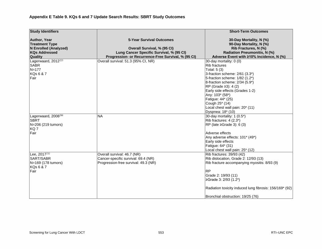
Appendix E Table 9. KQs 6 and 7 Update Search Results: SBRT Study Outcomes
Screening for Lung Cancer With LDCT 553 RTI–UNC EPC
Study Identifiers Author, Year Treatment Type N Enrolled (Analyzed) KQs Addressed Quality
5-Year Survival Outcomes
Overall Survival, % (95 CI) Lung Cancer Specific Survival, % (95 CI)
Progression- or Recurrence-Free Survival, % (95 CI)
Short-Term Outcomes
30-Day Mortality, N (%) 90-Day Mortality, N (%)
Rib Fractures, N (%) Radiation Pneumonitis, N (%)
Adverse Event with ≥10% Incidence, N (%)
Lagerwaard, 2012223 SABR N=177 KQs 6 & 7 Fair
Overall survival: 51.3 (95% CI, NR) 30-day mortality: 0 (0) Rib fractures Total: 5 (3) 3-fraction scheme: 2/61 (3.3*) 5-fraction scheme: 1/82 (1.2*) 8-fraction scheme: 2/34 (5.9*) RP (Grade ≥3): 4 (2) Early side effects (Grades 1-2) Any: 103* (58*) Fatigue: 44* (25) Cough 25* (14) Local chest wall pain: 20* (11) Dyspnea: 18* (10)
Lagerwaard, 2008290 SBRT N=206 (219 tumors) KQ 7 Fair
NA 30-day mortality: 1 (0.5*) Rib fractures: 4 (2.3*) RP (late ≥Grade 3): 6 (3) Adverse effects Any adverse effects: 101* (49*) Early side effects Fatigue: 64* (31) Local chest wall pain: 25* (12)
Lee, 2017232
SART/SABR N=169 (178 tumors) KQs 6 & 7 Fair
Overall survival: 46.7 (NR) Cancer-specific survival: 69.4 (NR) Progression-free survival: 49.3 (NR)
Rib fractures: 39/93 (42) Rib dislocation, Grade 2: 12/93 (13) Rib fracture accompanying myositis: 8/93 (9) RP Grade 2: 19/93 (11) ≥Grade 3: 2/93 (1.2*) Radiation toxicity induced lung fibrosis: 156/169* (92) Bronchial obstruction: 19/25 (76)

Appendix E Table 9. KQs 6 and 7 Update Search Results: SBRT Study Outcomes
Screening for Lung Cancer With LDCT 554 RTI–UNC EPC
Study Identifiers Author, Year Treatment Type N Enrolled (Analyzed) KQs Addressed Quality
5-Year Survival Outcomes
Overall Survival, % (95 CI) Lung Cancer Specific Survival, % (95 CI)
Progression- or Recurrence-Free Survival, % (95 CI)
Short-Term Outcomes
30-Day Mortality, N (%) 90-Day Mortality, N (%)
Rib Fractures, N (%) Radiation Pneumonitis, N (%)
Adverse Event with ≥10% Incidence, N (%)
Manyam, 2019228 SBRT/SABR N=139 (146 tumors) KQs 6 & 7 Fair
Overall survival: 28.7 (19.6 to 37.9) Rib fracture: 7/146 (4.8) Chest wall toxicity, overall: 18/146 (12.3) Grade 1: 3/146 (2.1*) Grade 2: 13/146 (8.9*) Grade 3: 2/146 (1.4)
Matsuo, 2012283 SBRT N=74 KQ 7 Fair
NA Symptomatic RP Total: 15 (20.3) Grade 2: 14 (18.9*) Grade 3: 1 (1.4*)
Mutter, 2012285 SBRT/SABR N=126 KQ 7 Fair
NA Rib fractures: 5 (4) Chest wall pain Grade 1: 19 (15) Grade 2: 16 (13) Grade 3: 19 (15) Grade ≥2 Estimated actuarial incidence over 2 yrs: 39%
Olsen, 2011286 SBRT/SABR N=130 KQ 7 Fair
NA RP, Grade 2: 4 (3.1*) Chest wall toxicity: 21 (16)
Onishi, 2007224
SBRT/SABR N=257 KQs 6 & 7 Fair
Overall survival: 47.2 ( 38.7 to 53.5) For operable patients (n=99): 64.8 (53.6 to 75.9) For inoperable patients (n=158): 35 (25.9 to 44.1) Lung cancer specific survival: 73.2 (66.1 to 80.2)
Rib fracture: 4 (1.6) Symptomatic radiation-induced pulmonary complications Grade >1: 28 (10.9) Grade ≥2: 14 (5.4)
Palma, 2010287 SBRT/SABR N=99* KQ 7 Fair
NA 30-day mortality: 1* (1.0)

Appendix E Table 9. KQs 6 and 7 Update Search Results: SBRT Study Outcomes
Screening for Lung Cancer With LDCT 555 RTI–UNC EPC
Study Identifiers Author, Year Treatment Type N Enrolled (Analyzed) KQs Addressed Quality
5-Year Survival Outcomes
Overall Survival, % (95 CI) Lung Cancer Specific Survival, % (95 CI)
Progression- or Recurrence-Free Survival, % (95 CI)
Short-Term Outcomes
30-Day Mortality, N (%) 90-Day Mortality, N (%)
Rib Fractures, N (%) Radiation Pneumonitis, N (%)
Adverse Event with ≥10% Incidence, N (%)
Shibamoto, 2012234 SBRT/SABR N=180 KQs 6 & 7 Good
Overall survival: 52 For operable patients (n=60): 70 For inoperable patients (n=120): 44
RP: ≥Grade 2: 24 (13.3) Grade 3: 2 (1.1)
Stanic, 2014277
SBRT N=55 KQ 7 Fair
NA RP Total: 9 (16.4*) Grade 1: 4 (7.3*) Grade 2: 3 (5.5*) Grade 3: 2 (3.6*) Adverse effects Pulmonary function toxicity at any time during 2-yr followup: 49 (89*) Grade 1 pulmonary/upper respiratory function toxicity: 11 (20*) Grade 2 pulmonary/upper respiratory function toxicity: 13 (23.6*) Grade 3 pulmonary/upper respiratory function toxicity: 8 (14.5*) Cough: 15* (27.3*) Dyspnea: 17* (30.9*) Pleural effusion: 5* (9.1*) Other pulmonary/upper respiratory toxicity: 7* (12.7*)
Uhlig, 2018226 SBRT/SABR N=27,732 (1,070 in PSM analysis) KQs 6 & 7 Fair
Overall survival in PSM cohort: 26.1 (22.7 to 29.9) 30-day mortality: None 90-day mortality: None 30-day post-treatment unplanned hospital readmission rate in PSM cohort: 2 (0.2)

Appendix E Table 9. KQs 6 and 7 Update Search Results: SBRT Study Outcomes
Screening for Lung Cancer With LDCT 556 RTI–UNC EPC
Study Identifiers Author, Year Treatment Type N Enrolled (Analyzed) KQs Addressed Quality
5-Year Survival Outcomes
Overall Survival, % (95 CI) Lung Cancer Specific Survival, % (95 CI)
Progression- or Recurrence-Free Survival, % (95 CI)
Short-Term Outcomes
30-Day Mortality, N (%) 90-Day Mortality, N (%)
Rib Fractures, N (%) Radiation Pneumonitis, N (%)
Adverse Event with ≥10% Incidence, N (%)
Westover, 2012282 SBRT N=15 (20 tumors) KQ 7 Fair
NA Rib fracture: 3/20 (15*) tumors RP Total: 7/20 (35*) tumors Grade 3: 1/20 (5*) tumor Grade 1: 6/20 (30*) tumors Adverse events Dermatitis: 4/20 (20*) tumors Fatigue: 2/20 (10*) tumors
Wink, 2019233 SBRT/SABR N=554 KQ 6 Fair
Overall survival: 47 NA
Ye, 2018292 SBRT/SABR N=100 KQ 7 Fair
NA 30-day mortality: 0 Rib fracture: 0 Acute RP: 6 (6*) Late Grade 2 RP: 8 (8*)
*Indicates that data were calculated by abstractors.
Abbreviations: CI=confidence interval; KQ=key question; Max=maximum; mos=months; NA=not applicable; PSM=propensity score matched; RP=radiation pneumonitis;
RR=risk ratio; SABR=stereotactic ablative radiotherapy; SBRT=stereotactic body radiation therapy.

Appendix E Figure 1. Sensitivity Analysis for Trial Results for Lung Cancer Incidence (KQ 1), Including Studies Rated as Poor Quality
Screening for Lung Cancer With LDCT 557 RTI–UNC EPC
Appendix E Figure 1. Sensitivity Analysis for Trial Results for Lung Cancer Incidence (KQ 1), Including Studies Rated as Poor Quality
Note: G1=LDCT; G2=Control; The MILD trial randomized participants to annual screening, biennial screening, or a control group. For the 10-year followup, the annual and
biennial screening groups were combined. At the 10-year followup, the median duration of screening for those in the screening groups was 6.2 years.
Abbreviations: DANTE=Detection and Screening of Early Lung Cancer with Novel Imaging Technology and Molecular Essays; DLCST=Danish Lung Cancer Screening Trial;
ITALUNG=Italian Lung Cancer Screening Trial; MILD=Multicentric Italian Lung Detection; NELSON=Nederlands-Leuvens Longkanker Screenings Onderzoek;
NLST=National Lung Screening Trial.
NLST, 2011, 2013
NLST, 2019
DANTE, 2015
DLCST, 2016
ITALUNG, 2017
MILD, 2019
MILD, 2012
MILD, 2012
NELSON, 2020
year
Study,
59
59
100
56
65
66
66
66
100
%
Male,
6.5
11.3
8.4
9.8
9.3
10
4.4
4.4
10
y
Follow-up,
61
61
65
58
61
57
57
57
58
age, y
Mean
56
48
47
36
39
38
39
39
38
pack-years
Mean
0, 1, 2
0, 1, 2
0, 1, 2, 3, 4
0, 1, 2, 3, 4
0, 1, 2, 3
multiple
0, 1, 2, 3, 4
0, 2, 4
0, 1, 3, 5.5
intervals, y
Screening
26722
26722
1276
2052
1613
2736
1190
1186
6583
G1_n
26732
26730
1196
2052
1593
1723
1723
1723
6612
G2_n
1089
1701
106
100
67
98
34
25
341
G1_Events
969
1681
73
53
71
60
20
20
304
G2_Events
1.12 (1.02, 1.22)
1.01 (0.95, 1.08)
1.35 (1.00, 1.81)
1.89 (1.36, 2.64)
0.92 (0.66, 1.28)
1.16 (0.84, 1.59)
1.97 (1.13, 3.41)
1.46 (0.81, 2.62)
1.14 (0.97, 1.33)
RR (95% CI)
1.12 (1.02, 1.22)
1.01 (0.95, 1.08)
1.35 (1.00, 1.81)
1.89 (1.36, 2.64)
0.92 (0.66, 1.28)
1.16 (0.84, 1.59)
1.97 (1.13, 3.41)
1.46 (0.81, 2.62)
1.14 (0.97, 1.33)
RR (95% CI)
Favors Intervention Favors Control
1.25 .5 2 5

Appendix E Figure 2. Sensitivity Analysis for Early-Stage Lung Cancer Incidence (KQ 1), Including Studies Rated as Poor Quality
Screening for Lung Cancer With LDCT 558 RTI–UNC EPC
Appendix E Figure 2. Sensitivity Analysis for Early Stage lung Cancer Incidence (KQ 1), Including Studies Rated as Poor Quality
Abbreviations: DANTE=Detection and Screening of Early Lung Cancer with Novel Imaging Technology and Molecular Essays; DLCST=Danish Lung Cancer Screening Trial;
ITALUNG=Italian Lung Cancer Screening Trial; MILD=Multicentric Italian Lung Detection; NELSON=Nederlands-Leuvens Longkanker Screenings Onderzoek;
NLST=National Lung Screening Trial.
NLST, 2019
DANTE, 2015
DLCST, 2016
ITALUNG, 2017
NELSON, 2020
MILD, 2019
year
Study,
59
100
56
65
100
65.5
%
Male,
11.3
8.4
9.8
9.3
10
10
y
Follow-up,
61
65
58
61
58
57
age, y
Mean
56
47
36
39
38
38
pack-years
Mean
0, 1, 2
0, 1, 2, 3, 4
0, 1, 2, 3, 4
0, 1, 2, 3
0, 1, 3, 5.5
multiple
intervals, y
Screening
26722
1276
2052
1613
6583
2376
G1_n
26730
1196
2052
1593
6612
1723
G2_n
818
54
54
29
168
53
(No.)
stage
G1_Early
615
21
10
13
71
18
(No.)
stage
G2_Early
1.33 (1.20, 1.48)
2.38 (1.44, 3.94)
5.42 (2.76, 10.63)
2.17 (1.13, 4.16)
2.39 (1.81, 3.16)
2.08 (1.22, 3.56)
RR (95% CI)
1.33 (1.20, 1.48)
2.38 (1.44, 3.94)
5.42 (2.76, 10.63)
2.17 (1.13, 4.16)
2.39 (1.81, 3.16)
2.08 (1.22, 3.56)
RR (95% CI)
Favors Intervention Favors Control
1.25 .5 2 5

Appendix E Figure 3. Sensitivity Analysis for Late-Stage Lung Cancer Incidence (KQ 1), Including Studies Rated as Poor Quality
Screening for Lung Cancer With LDCT 559 RTI–UNC EPC
Appendix E Figure 3. Sensitivity Analysis for Late Stage lung Cancer Incidence (KQ 1), Including Studies Rated as Poor Quality
Abbreviations: DANTE=Detection and Screening of Early Lung Cancer with Novel Imaging Technology and Molecular Essays; DLCST=Danish Lung Cancer Screening Trial;
ITALUNG=Italian Lung Cancer Screening Trial; MILD=Multicentric Italian Lung Detection; NELSON=Nederlands-Leuvens Longkanker Screenings Onderzoek;
NLST=National Lung Screening Trial.
NLST,2019
DANTE, 2015
DLCST, 2016
ITALUNG, 2017
NELSON, 2020
MILD, 2019
year
Study,
59
100
56
65
100
65.5
%
Male,
11.3
8.4
9.8
9.3
10
10
y
Follow-up,
61
65
58
61
58
57
age, y
Mean
56
47
36
39
38
38
pack-years
Mean
0, 1, 2
0, 1, 2, 3, 4
0, 1, 2, 3, 4
0, 1, 2, 3
0, 1, 3, 5.5
multiple
intervals, y
Screening
26722
1276
2052
1613
6583
2376
G1_n
26732
1196
2052
1593
6612
1723
G2_n
766
43
46
33
153
45
(No.)
stage
G1_Late
918
45
41
43
216
42
(No.)
stage
G2_Late
0.84 (0.76, 0.92)
0.89 (0.59, 1.35)
1.13 (0.74, 1.72)
0.75 (0.47, 1.17)
0.72 (0.58, 0.88)
0.76 (0.50, 1.16)
RR (95% CI)
0.84 (0.76, 0.92)
0.89 (0.59, 1.35)
1.13 (0.74, 1.72)
0.75 (0.47, 1.17)
0.72 (0.58, 0.88)
0.76 (0.50, 1.16)
RR (95% CI)
Favors Intervention Favors Control
1.25 .5 2 5

Appendix E Figure 4. Sensitivity Analysis for Trial Results for Lung Cancer Mortality (KQ 1), Including Studies Rated as Poor Quality
Screening for Lung Cancer With LDCT 560 RTI–UNC EPC
Appendix E Figure 4. Sensitivity Analysis for Trial Results for Lung Cancer Mortality (KQ 1), Including Studies Rated as Poor Quality
Abbreviations: DANTE=Detection and Screening of Early Lung Cancer with Novel Imaging Technology and Molecular Essays; DLCST=Danish Lung Cancer Screening Trial;
ITALUNG=Italian Lung Cancer Screening Trial; LSS=Lung Screening Study; MILD=Multicentric Italian Lung Detection; NELSON=Nederlands-Leuvens Longkanker
Screenings Onderzoek; NLST=National Lung Screening Trial.
NLST, 2011, 2013
DANTE, 2015
DLCST, 2016
ITALUNG, 2017
MILD, 2019
LSS, 2018
NELSON, 2020
year
Study,
59
100
56
65
66
59
84
%
Male,
6.5
8.4
9.8
9.3
10
5.2
10
y
Follow-up,
61
65
58
61
57
NR
58
age, y
Mean
56
47
36
39
38
54
38
pack-years
Mean
0, 1, 2
0, 1, 2, 3, 4
0, 1, 2, 3, 4
0, 1, 2, 3
multiple
NR
0, 1, 3, 5.5
intervals, y
Screening
26722
1276
2052
1613
2736
1660
7900
G1_n
26732
1196
2052
1593
1723
1658
7892
G2_n
469
59
39
43
40
32
181
G1_Events
552
55
38
60
40
26
242
G2_Events
280
543
201
293
173
383
241
person-years
per 100,000
LDCT, deaths
332
544
194
421
247
310
324
person-years
per 100,000
Control, deaths
0.85 (0.75, 0.96)
1.00 (0.69, 1.44)
1.03 (0.66, 1.61)
0.70 (0.47, 1.03)
0.70 (0.45, 1.09)
1.24 (0.74, 2.07)
0.75 (0.61, 0.90)
RR (95% CI)
0.85 (0.75, 0.96)
1.00 (0.69, 1.44)
1.03 (0.66, 1.61)
0.70 (0.47, 1.03)
0.70 (0.45, 1.09)
1.24 (0.74, 2.07)
0.75 (0.61, 0.90)
RR (95% CI)
Favors Intervention Favors Control
1.25 .5 2 5

Appendix E Figure 5. Sensitivity Analysis for Trial Results for All-Cause Mortality (KQ 1), Including Studies Rated as Poor Quality
Screening for Lung Cancer With LDCT 561 RTI–UNC EPC
Appendix E Figure 5. Sensitivity Analysis for Trial Results for All-Cause Mortality (KQ 1), Including Studies Rated as Poor Quality
Abbreviations: DANTE=Detection and Screening of Early Lung Cancer with Novel Imaging Technology and Molecular Essays; DLCST=Danish Lung Cancer Screening Trial;
ITALUNG=Italian Lung Cancer Screening Trial; LSS=Lung Screening Study; MILD=Multicentric Italian Lung Detection; NELSON=Nederlands-Leuvens Longkanker
Screenings Onderzoek; NLST=National Lung Screening Trial.
NLST, 2011, 2013
DANTE, 2015
DLCST, 2016
ITALUNG, 2017
MILD, 2019
MILD, 2012
MILD, 2012
LSS, 2019
NELSON, 2020
year
Study,
59
100
56
65
66
66
66
59
100
%
Male,
6.5
8.4
9.8
9.3
10
4.4
4.4
5.2
10
y
Follow-up,
61
65
58
61
57
57
57
NR
58
age, y
Mean
56
47
36
39
39
39
39
54
38
pack-years
Mean
0, 1, 2
0, 1, 2, 3, 4
0, 1, 2, 3, 4
0, 1, 2, 3
multiple
0, 2, 4
0, 1, 2, 3, 4
NR
0, 1, 3, 5.5
intervals, y
Screening
26722
1276
2052
1613
2376
1186
1190
1660
6583
G1_n
26732
1196
2052
1593
1723
1723
1723
1658
6612
G2_n
1912
180
165
154
137
20
31
139
868
G1_Events
2039
176
163
181
106
20
20
116
860
G2_Events
1141
1655
849
1051
594
363
558
1667
1393
person-years
per 100,000
LDCT, deaths
1225
1742
834
1270
654
310
310
1384
1376
person-years
per 100,000
Control, deaths
0.93 (0.88, 0.99)
0.95 (0.77, 1.17)
1.02 (0.82, 1.26)
0.83 (0.67, 1.03)
0.91 (0.71, 1.17)
1.17 (0.63, 2.17)
1.79 (1.02, 3.14)
1.20 (0.94, 1.53)
1.01 (0.92, 1.11)
RR (95% CI)
0.93 (0.88, 0.99)
0.95 (0.77, 1.17)
1.02 (0.82, 1.26)
0.83 (0.67, 1.03)
0.91 (0.71, 1.17)
1.17 (0.63, 2.17)
1.79 (1.02, 3.14)
1.20 (0.94, 1.53)
1.01 (0.92, 1.11)
RR (95% CI)
Favors Intervention Favors Control
1.25 .5 2 5