Salmeterol Reduces Dyspnea and Improves Lung Function in Patients With COPD
Upload
nottinghamCategory
view
0download
0
BioMed CentralRespiratory Research
ss
Open AcceResearchSalmeterol and cytokines modulate inositol-phosphate signalling in Human airway smooth muscle cells via regulation at the receptor locusNatalie Smith†, Claudia A Browning†, Nathalie Duroudier, Ceri Stewart, Samantha Peel, Caroline Swan, Ian P Hall and Ian Sayers*Address: Division of Therapeutics & Molecular Medicine, University Hospital of Nottingham, Nottingham, UK
Email: Natalie Smith - [email protected]; Claudia A Browning - [email protected]; Nathalie Duroudier - [email protected]; Ceri Stewart - [email protected]; Samantha Peel - [email protected]; Caroline Swan - [email protected]; Ian P Hall - [email protected]; Ian Sayers* - [email protected]
* Corresponding author †Equal contributors
AbstractBackground: Airway hyper-responsiveness (AHR) is a key feature of asthma and a causal relationship between airwayinflammation and AHR has been identified. The aim of the current study was to clarify the effect of proinflammatorycytokines and asthma medication on primary human airway smooth muscle (ASM) inositol phosphate (IPx) signalling anddefine the regulatory loci involved.
Methods: Primary Human ASM cells were isolated from explants of trachealis muscle from individuals with no historyof respiratory disease. The effect of cytokine or asthma medication on histamine or bradykinin induced IPx signalling wasassessed by [3H] inositol incorporation. Quantitative Real Time PCR was used to measure mRNA levels of receptorsand downstream signalling components. Transcriptional mechanisms were explored using a combination of 5'RapidAmplification of cDNA Ends (5'RACE) and promoter-reporter techniques.
Results: Treatment of Human ASM cells with IL-13, IFNγ or salmeterol for 24 hours lead to a modest augmentation ofhistamine induced IPx responses (144.3 +/- 9.3, 126.4 +/- 7.5 and 117.7 +/- 5.2%, p < 0.05). Similarly, TNFα, IFNγ orsalmeterol treatment augmented bradykinin induced IPx responses (127.4 +/- 8.3, 128.0 +/- 8.4 and 111.7 +/- 5.0%, P <0.05). No treatment significantly influenced sodium fluoride induced IPx responses suggesting regulation occurs at thereceptor locus. Analyses of mRNA expression of components of the IPx pathway i.e. H1 Histamine Receptor (HRH1),B2 Bradykinin Receptor (BDKRB2), Gαq/11 and PLC-β1 identified that a significant induction of receptor mRNA (>2fold) was a feature of these responses explaining the cytokine and spasmogen specificity. The HRH1 and BDKRB2promoter regions were mapped in ASM and promoter-reporter analyses identified that salmeterol can induce HRH1 (>2fold) and BDKRB2 (2–5 fold) transcription. The effect of cytokines on HRH1 and BDKRB2 promoter-reporterexpression suggested a more complex regulation of mRNA expression involving additional loci to the core promoter.
Conclusion: Our results indicate that the spasmogen specific receptor locus may be a key site of regulation determiningthe magnitude of spasmogen mediated ASM IPx responses during airway inflammation or following asthma medication.These data provide further insight into the molecular basis of AHR and extend our understanding of potentiallydetrimental effects associated with existing therapies used in the treatment of asthma.
Published: 28 September 2007
Respiratory Research 2007, 8:68 doi:10.1186/1465-9921-8-68
Received: 8 June 2007Accepted: 28 September 2007
This article is available from: http://respiratory-research.com/content/8/1/68
© 2007 Smith et al; licensee BioMed Central Ltd. This is an Open Access article distributed under the terms of the Creative Commons Attribution License (http://creativecommons.org/licenses/by/2.0), which permits unrestricted use, distribution, and reproduction in any medium, provided the original work is properly cited.
Page 1 of 15(page number not for citation purposes)

Respiratory Research 2007, 8:68 http://respiratory-research.com/content/8/1/68
BackgroundAsthma is characterised by airway inflammation, reversi-ble airway narrowing and airway hyper-responsiveness(AHR) caused by stimuli that normally elicit limited or noresponse. Excessive smooth muscle contractility has longbeen recognized as a feature of asthma, emphasised by thefact that drugs designed to prevent or reverse bronchocon-striction are still the most effective drug classes available.Spasmogens such as acetylcholine, bradykinin and hista-mine induce airway smooth muscle (ASM) contractionvia inositol phosphate/calcium signalling mechanismsand thereby regulate the tone and calibre of the airways[1,2].
There is strong evidence for a causal relationship betweenairway inflammation and altered airway function includ-ing hyperreactivity to contractile agents and hyporeactiv-ity to relaxant agents in asthma [3]. It has been shown thatseveral cytokines are elevated in the lung during inflam-mation in asthma including; interleukin (IL)1β, TumourNecrosis Factor (TNF)α [4] and IL13 [5]. Administrationof the recombinant cytokine to mouse airways e.g. IL-13,has been shown to mimic several of the features of asthmaincluding AHR [6]. Similarly, viral infection has beenassociated with asthma exacerbation and increased AHRand this is thought to be mediated at least in part by ele-vated interferon (IFN)γ expression in the airways [7].Recently, it has also been shown that elevated CD4+IFNγ+and CD8+IFNγ + cells are a feature of asthmatic airwaysand that CD8+IFNγ + cell numbers correlated with AHR[8]. A direct interaction between cytokines and HumanASM has been described involving alterations in thecapacity of airway smooth muscle in culture to respond torelaxation and contractile agents [9,10]. Proinflammatorycytokines including; IL1β, TNFα and IL-13 have beenshown to augment bradykinin induced Ca2+ release inhuman ASM cells in culture [11-13]. Similarly, studieshave been completed in order to determine the effect ofcytokines on other agonist mediated responses (e.g. hista-mine or acetylcholine) including inositol phosphate (IPx)signalling, however, data have been conflicting [14,15].Recently, a mechanism involving a role for the CD38/cyclic adenosine diphosphate pathway in regulating cal-cium homeostasis has been proposed at least in part toexplain the effects of cytokines on agonist induced Ca2+
signalling [16]. IL-13 treatment of Human ASM in culturewas shown to increase expression of CD38 mRNA whichwas accompanied by an increase in ADP-ribosyl cyclaseactivity and net intracellular Ca2+ responses to bradykininand histamine [17].
Asthma maintenance medications including long actingβ2-adrenoceptor agonists and inhaled corticosteroidshave been shown to suppress AHR in the short term in vivo[18,19] and are the mainstay therapy for asthma. While
clinically effective in many patients, concerns have beenraised regarding potential adverse effects associated withchronic use of β2-adrenoceptor agonists [20-22] Anincrease in bronchial hyper-responsiveness in response tohistamine has been observed following chronic use of theshort acting β2-adrenoceptor agonist, salbutamol inasthma and COPD patients [23]. We have recently identi-fied a potentially deleterious effect of prolonged exposureof Human ASM cells in culture to short acting β2 adreno-ceptor agonists [24]. Prolonged treatment (24 hours) ofHuman ASM with salbutamol or isoprenaline augmentedhistamine mediated IPx production potentially providinga cellular mechanism explaining the clinical observations[24]. Recently, the outcomes of the Salmeterol Multicen-tre Asthma Research Trial (SMART) have been publishedwhich involved the assessment of 26,355 subjects [22].This study indicated that prolonged use of the long actingβ2-adrenoceptor agonist, salmeterol was associated withrespiratory related deaths [22] suggesting that the adverseeffects inferred for short acting β2-adrenoceptor agonistsmay also be of clinical importance for long acting β2-adrenoceptor agonist therapy.
Contractile agents including histamine, bradykinin andacetylcholine bind to G-protein coupled receptors(GPCR) on the surface of ASM which leads to the dissoci-ation of Gαq from βγ subunits. Both Gαq and βγ subunitsare capable of binding and activating phospholipase C(PLC)β1 which catalyses the generation of inositol 1,4,5-triphosphate (IP3) and diacylglycerol (DAG) from phos-phatidyl 4,5-biphospate (IP2). These second messengersstimulate Ca2+ release from intracellular stores and pro-tein kinase C (PKC) activity, resulting in the activation ofcontractile and growth machinery in ASM [25].
There is accumulating evidence suggesting that the G-Pro-tein Coupled Receptor (GPCR) may be a key regulatorylocus i.e. the level of receptor expression provides specifi-city to the augmentation of signalling responses in airwaysmooth muscle cells [9]. This has been demonstrated formultiple GPCRs including; Cysteinyl leukotriene receptor1 [26], β2 adrenoceptor [27], H1 histamine receptor [28]and B2 bradykinin receptor [29] where increases in mRNAlevels are accompanied by augmented spasmogenresponses following cytokines or drug treatment.
In the current study we aimed to comprehensively definethe effect of key pro-inflammatory cytokines, IL1β, TNFα,IL-13, IFNγ together with dexamethasone and salmeterolon Human ASM IPx signalling pathways using a combina-tion of cell signalling, gene expression and promoter anal-yses.
Page 2 of 15(page number not for citation purposes)

Respiratory Research 2007, 8:68 http://respiratory-research.com/content/8/1/68
MethodsReagentsAll chemicals were analytical grade or higher. Plasticwarewas from Costar (High Wycombe, UK). All chemicals andreagents were purchased from Sigma-Aldrich (Poole, UK)unless otherwise stated. The [3H]-myo-inositol was pur-chased from Perkin Elmer (Bucks, UK). Inositol freeDMEM was made to order by Invitrogen (Paisley, UK).
Cell CultureHuman airway smooth muscle cells (ASM) were isolatedfrom explants of trachealis muscle obtained from individ-uals with no history of respiratory disease as described[24,30]. Briefly, a section of trachealis muscle was isolatedby dissection just above the carina and washed in Dul-becco's Modified Eagle's Medium (DMEM) supplementedwith penicillin (200 U/ml), streptomycin (200 µg/l) andamphotericin B (0.5 µg/l). Explants (0.2 × 0.2 cm) wereplaced in 6 well plates and allowed to adhere. Cells wereallowed to grow with fresh complete medium (10% foetalcalf serum and 2 mM glutamine) added regularly, follow-ing 7–10 days growth cells approached confluency. Ascells reached confluency, explants were removed and thecells were harvested using 5 mg/ml trypsin/2 mg/mlEDTA. Primary ASM cells exhibited >95% cell staining forα-actin. Airway smooth muscle cells were maintained incomplete medium and passaged using trypsin/EDTA. Eth-ical approval for the use of primary cells was obtainedfrom the local ethics committee. Airway smooth musclecells from four individuals were used.
Quantification of [3H]-inositol phosphates[3H]-inositol phosphate formation in primary humanASM cells was quantified essentially as described [24,30].Briefly, cells were plated in 24 wells at a density of 0.5 ×10⁵ cells/well. Cells were allowed to grow for 2–3 daysuntil ~80% confluent, then medium was replaced with300 µl of inositol free DMEM, supplemented with 2 mMglutamine and containing [3H]-myo-inositol at a concen-tration of 8.1 MBq/ml. Cells were then incubated for 24hours followed by the addition of IL-1β, TNFα, IFNγ (10ng/ml), IL-13 (50 ng/ml) (PeproTech EC Ltd, London,UK), dexamethasone (1 µM) or salmeterol (1 µM) andincubated for a further 24 hours. The medium was thenremoved and cells were washed three times with 1 mlHanks/20 mM HEPES and 300 µl Hanks/20 mM HEPEScontaining 20 mM LiCl was added. Cells were incubatedfor 30 min at 37°C, then agonists were added in a volumeof 10 µl (100 µM Histamine, 0.1 µM Bradykinin or 10mM Sodium Fluoride). Cells were incubated for a further30 min and then the reaction was stopped by removingthe medium and addition of 1 ml ice cold 50% methanol/60 mM HCl. Samples were stored at -20°C for at least 24h. An 800 µl aliquot of each sample was neutralised using~4.6 ml 5.5 mM Tris-HCl/11 mM NaOH. Total [3H]-inosi-
tol phosphates were separated from unincorporated [3H]-inositol using anion-exchange chromatography onDowex-Cl columns and measured using scintillationcounting [24,30].
Quantification of HRH1, BDKRB2, GNA11 and PLCB1 gene expressionHuman ASM cells were transferred to 100 mm Petri dishesat a density of 105 cells/ml. Cells were allowed to grow for2–3 days until ~80% confluent. Complete medium wasreplaced with serum free medium and the cells weregrown for a further 24 hours followed by the addition ofcytokines or drugs (as described above). Isolation of cellsfor RNA extraction (at 4 and 24 hours post cytokine ordrug addition) involved removing medium, a phosphatebuffered saline (PBS) wash and collection in 1 ml PBSusing a cell scraper. Cells were collected by centrifugation(300 × g 5 min), supernatant discarded and cell pelletsresuspended in 50 µl PBS. 450 µl RNAlater was added toeach sample followed by mixing and storage at -80°C.Total RNA was extracted from samples using the RNAeasykit (Qiagen) as directed by the manufacturer. cDNA wasgenerated from 1 µg RNA template using random hexam-ers and the Superscript kit (Invitrogen, Paisley, UK) asdirected by the manufacturer. TaqMan assays specific forthe Histamine H1 Receptor (HRH1), Bradykinin B2Receptor (BDKRB2), Gαq/11 protein (GNA11) andphosholipase C-β1 (PLCB1) transcripts were designedusing Primer Express Software (Applied Biosystems, War-rington, UK). See Table 1. Real Time PCR (Applied Biosys-tems) was carried out as directed by the manufacturer in areaction volume of 20 µl containing ~100 ng of cDNA,300 nM of each Primer, 200 nM Probe (Eurogentec Ltd,Southampton, UK) and 10 µl universal mastermix(Applied Biosystems). Differences in the quantity ofcDNA template were normalised using a VIC labelled 18sribosomal RNA endogenous control (Applied Biosys-tems). PCR was performed using the ABI-7700 and datacollected using Sequence Detection Software (AppliedBiosystems). Data was analysed as directed by the manu-facturer and Ct values were normalised using the 18sribosomal RNA signal. Relative differences in gene expres-sion were calculated using the 2dCt value as continuousvariable (Sequence Detection System Compendium 7700Version 4.0, Applied Biosystems).
5'Rapid Amplification of BDKRB2 cDNA ends5'RACE was performed in order to identify the putativepromoter regions of the BDKRB2 gene in Human ASM.Total RNA was extracted from Human ASM cells derivedfrom two different donors. Total RNA extractions wereperformed using the RNeasy Mini Kit (Qiagen, Crawley,UK) as described above. 5'RACE was performed using theGeneRacer Kit (Invitrogen) according to the manufac-turer's protocol. 2.5 µg of total RNA was used as template.
Page 3 of 15(page number not for citation purposes)

Respiratory Research 2007, 8:68 http://respiratory-research.com/content/8/1/68
The mRNA was reverse-transcribed with SuperScript II RT(Invitrogen) and the GeneRacer OligodT primer (50 µM)to generate RACE ready cDNA. A nested PCR approachwas used to identify the 5' structure of the BDKRB2 gene;PCR1 (GeneRacer 5'Primer/BDKRB2 primer 1 specific forthe open reading frame of the BDKRB2 gene). Reactionconditions included; 1 µl RACE ready cDNA template,0.025 U/µl Platinum Taq DNA Polymerase High Fidelity(Invitrogen), 0.6 µM of each primer, 200 µM dNTPs, 2mM Magnesium Sulphate and a staged PCR cyclingapproach; 94°C for 5 min, 94°C for 1 min 30 sec/72°Cfor 3 min × 5, 94°C for 1 min 30 sec/70°C for 1 min 30sec/72°C for 3 min × 5, 94°C for 1 min 30 sec/68°C for 1min 30 sec/72°C for 3 min × 20 and a final extension of72°C for 10 min. The second stage PCR (PCR 2) utilisednested primers (GeneRacer 5' Nested Primer/BDKRB2primer 2 specific for the open reading frame of theBDKRB2 gene). Reaction conditions were as described forPCR 1 except 1 µl cDNA (from PCR1) was used as tem-
plate and PCR cycling involved; 94°C for 5 min, 94°C for1 min 30 sec/68°C for 1 min 30 sec/72°C for 3 min × 25and a final extension of 72°C for 10 min. 5'RACE prod-ucts generated in PCR 2 were cloned into the pCR-4-TOPO vector using the TOPO TA Cloning Kit for Sequenc-ing as directed by the manufacturer (Invitrogen). LB agarplates and LB broths (100 µg/mL ampicillin) were used toselect and grow single colonies of positive clones. PlasmidDNA was purified using the FastPlasmid Mini Kit (Eppen-dorf, Cambridge, UK) and the presence of inserts was con-firmed by restriction analysis with EcoRI (Promega,Southampton, UK). Positive clones showing an insertafter restriction digest were sequenced using M13 primers(Invitrogen), ABI BigDye Terminator v3.1 Cycle Sequenc-ing Kit and ABI Prism 310 DNA sequencer (Applied Bio-systems). Sequences were analysed and the 5' structure ofthe BDKRB2 gene identified using Chromas v2.0 ([31])and BLAST (NCBI).
Table 1: Primers and probes
REAL TIME PCRGENE 5'PRIMER 3'PRIMER PROBE (5'FAM/
3'TAMRA)LOCATION
HRH1 TCTCGGTGGCGGACTTGA CATGAGCAGGTAGAGGATGTTCAT
CGTGGGTGCCGTCGT ORF
BDKRB2 GATCAGCACCTTCCTGGATACG GATGATGCGCTCGTCCTGG CATCGCCTCGGCATCCTCTCCA
ORF
GNA11 TCAGCGAATACGACCAAGTCC AGGGCTTTGCTCTCCTCCA TGGAGTCGGACAACGAGAACCGG
Exon 5/6 boundry
PLCB1 CTGCCTGCTGTCTTTGTCTACATAG
TCGGATTGGGTTTGATAAAGCT TGAAAGACTATGTGCCAGACACA TATGCAGATG
Exon 22/23 boundary
5'RACE PRIMER 1 (PCR 1) PRIMER 2 (PCR 2)
BDKRB2 CACGAACAGCACCCAGAGGAAGG CCAGCCCAGCCACTCCACTTG
PLASMID CONSTRUCTION 5'PRIMER
3'PRIMER LOCATION IN CONTIG
CONSTRUCT
HRH1-1 CTGACTCGAGCCTCCTCTCCCTGTGAGCTT
CTGAAAGCTTGCCGGCTCCGGGGAAAGTT
51,750 – 52,761 HRH1 1 kb
HRH1-2 CTGACTCGAGGAAAGTGTGGACGCAGCCATT
CTGAAAGCTTGCCGGCTCCGGGGAAAGTT
50,782 – 52,761 HRH1 2 kb
HRH1-3 CTGACTCGAGGTTCAATCATAGCTCACTGCAG
CTGAAAGCTTGCCGGCTCCGGGGAAAGTT
49,768 – 52,761 HRH1 3 kb
HRH1-4 CTGACTCGAGTCCACAGCTTGTTGCACAGC
CTGAAAGCTTGCCGGCTCCGGGGAAAGTT
48,718 – 52,761 HRH1 4 kb
BDKRB2-1 CTGACTCGAGCCCAGGCCACCCATAAACTG
CTGAAAGCTTCAAGCGGCATGGGCACTTCA
101,405 – 102,439 BDKRB2 1 kb
BDKRB2-2 CTGACTCGAGGGGCGGATGTGTGGGTAGAT
CTGAAAGCTTCAAGCGGCATGGGCACTTCA
100,436 – 102,439 BDKRB2 2 kb
BDKRB2-3 CTGACTCGAGCCTCCTATGTGCCGGGTGAT
CTGAAAGCTTCAAGCGGCATGGGCACTTCA
99,416 – 102,439 BDKRB2 3 kb
BDKRB2-4 CTGACTCGAGATGTTGGCCAGGCTGATCTC
CTGAAAGCTTCAAGCGGCATGGGCACTTCA
98,414 – 102,439 BDKRB2 4 kb
*HRH1 reference contig = AC083855, BDKRB2 reference contig = AL355102.5. Bold denotes HindIII and XhoI restriction sites.
Page 4 of 15(page number not for citation purposes)

Respiratory Research 2007, 8:68 http://respiratory-research.com/content/8/1/68
Luciferase Reporter Plasmid ConstructionPCR primers were designed to generate 1, 2, 3 and 4 kbamplicons encompassing the predicted BDKRB2 tran-scription start sites (TSS) in HASM but not crossing theExonI/IntronI interface (see Table 1). The most 3' TSS wasat position 95,741,003 NCBI Build 35 (102,403 bp withinAL355102.5)). A repeat polymorphism [GGTGGGGAC]nexists in this region therefore DNA fragments were gener-ated to include the common n = 2 repeat observed in theCaucasian population [32]. Primers were designed toengineer a 5' HindIII and a 3' XhoI site into the DNA frag-ments to facilitate cloning into the pGL4-Luc2 vector(Promega). The BDKRB2 4 kb PCR included; 1 µl genomicDNA template (Caucasian), 0.025 U/µl Platinum TaqDNA Polymerase High Fidelity, 0.2 µM of each primer,200 µM dNTPs, 2 mM Magnesium Sulphate and cyclingparameters; 94°C for 2 min, 94°C for 30 sec/62°C 30 sec/68°C for 4 min × 30 and a final extension of 68°C for 7min. The PCR product was cloned into the pCR-4-TOPOvector and sequenced (as described above). Thesequenced pCR-4 vector containing the BDKRB2 4 kbfragment was used as PCR template for the generation ofthe BDKRB2 1, 2 and 3 kb fragments as described above.The 1, 2, 3 and 4 kb BDKRB2 fragments were excised fromthe pCR-4 vectors using HindIII/XhoI and cloned intopGL4-Luc2 using standard molecular biology techniques.An identical series of pGL4-Luc2 vectors containing 1, 2,3, and 4 kb HRH1 promoter fragments encompassing allof the identified transcription start sites in Human ASMbut not crossing the ExonI/IntronI interface (the most 3'TSS was at 52,754 bp within AC083855, [33]) were alsogenerated (Table 1).
Quantification of HRH1 and BDKRB2 core promoter transcriptional activity in CHO-K1 or Human ASMCHO-K1 cells were cultured in complete DMEM (10%foetal calf serum and 2 mM glutamine) and harvestedusing 5 mg/ml trypsin/2 mg/ml EDTA. Cells were platedat a density of 25,000 cells/well into 48-well tissue cultureplates (Corning, Sunderland, UK) using completemedium (see above) and allowed to grow for 24 h until~80% confluent. Complete medium was replaced withserum free medium and the cells were transfected with0.12 µg of pGL4-Luc2 using Fugene 6 (Roche) at a 3:1ratio (Fugene:DNA). For derivatives of pGL4-Luc2 con-taining the HRH1, BDKRB2 or SV40 control inserts equi-molar amounts of DNA were used and the amount ofFugene corrected accordingly. Cells were allowed to growfor 40 h prior to a PBS wash and harvested using PassiveLysis Buffer (PLB, 300 µl/well, rocked for 15 min and sub-jected to freeze/thaw at -80°C). Human ASM experimentswere completed in an analogous manner except cells wereallowed to grow for 24–72 hours until ~80% confluent.Following transfection cells were allowed to grow for 16hours prior to the addition of cytokines or drug (at con-
centrations described above). Following 4 or 24 hours,ASM cells were washed with PBS and harvested using Pas-sive Lysis Buffer as described in a volume of 100 µl/well.Firefly luciferase was quantified using 5 µl (CHO-K1) or20 µl (ASM) of cell lysate and the luciferase assay systemas directed by the manufacturer (Promega) and a ModelTD-20e luminometer (Turner Biosystems, Sunnyvale,CA). Data was normalised to fold over the empty pGL4-Luc2 vector. Cytokine or drug treated samples were com-pared to medium alone treated samples by designatedmedium treated samples 100%.
Promoter Database AnalysisSpecific transcription factor binding sites were identifiedusing; BioInformatics & Molecular Analysis Section [34],Transcription Element Search System [35], TFSEARCH[36] and WWW Signal Scan [37].
Statistical AnalysesDifferences between outcome measures for different treat-ment groups were compared by Analysis of Variance(ANOVA) in conjunction with Dunnett's Multiple Com-parison Post Hoc Test or Students' T test as appropriate.Figures represent mean values (+/- S.E.M). Statistical anal-yses were completed using GraphPad Prism (GraphPad,San Diego, CA), a P value < 0.05 was considered signifi-cant.
ResultsExposure to Salmeterol, TNFα, IL-13 or IFNγ can augment the magnitude of histamine and/or bradykinin stimulated inositol phosphate signalling in Human ASMIn order to assess the effect of the micro-environment onsubsequent IPx signalling responses in Human ASM weincubated cells with a series of pro-inflammatorycytokines implicated in the pathogenesis of asthma. Thesecytokines included; IL-1β, TNFα, IFNγ, or IL-13. Thecapacity of the ASM cells to produce IPx in response totwo specific mediators of airway hyper-responsiveness;histamine and bradykinin and the non-specific Gαq/PLCactivator sodium fluoride was determined. Similarly, theeffect of two drug classes used routinely in the treatmentof asthma was evaluated within the same model; i.e. thelong acting β2-adrenoceptor agonist salmeterol and thesteroid dexamethasone (Table 2). Significant differenceswere observed in the capacity of the ASM cells to produceIPx in response to histamine (p < 0.0001) or bradykinin(p = 0.0004) following cytokine or drug pre-treatment.There was not a statistically significant change in thecapacity of the ASM cells to produce IPx following sodiumfluoride stimulation (although differences were observedand there was a large degree of variation in these data).More specifically, IL-13, IFNγ and salmeterol were shownto significantly augment the capacity of the ASM cells toproduce IPx in response to histamine stimulation (p <
Page 5 of 15(page number not for citation purposes)

Respiratory Research 2007, 8:68 http://respiratory-research.com/content/8/1/68
0.05), however, these effects were modest; IL-13 (144.4 +/- 9.3%) compared to medium control (100%)), IFNγ(126.4 +/- 7.5%) and salmeterol (117.7 +/- 5.2%). Simi-larly, TNFα, IFNγ and salmeterol were shown to augmentbradykinin mediated IPx production in ASM cells (p <0.05), TNFα (127.4 +/- 8.3%), IFNγ (128.0 +/- 8.4%) andsalmeterol (111.7 +/- 5.0%).
Augmentation of IPx signalling by Salmeterol, TNFα, IL-13 or IFNγ is accompanied by elevated mRNA levels of the relevant GPCRIn order to test the hypothesis that the site of regulationdetermining the alteration in IPx signalling observed fol-lowing cytokine or drug pre-treatment occurs at the recep-tor locus we determined the mRNA expression levels ofthe H1 Histamine and B2 Bradykinin Receptors in oursamples using Real Time PCR, i.e. the two key receptorsmediating histamine or bradykinin activation of humanASM cells ([29,38], Figure 1). Quantification of the HRH1mRNA levels identified a significant induction of geneexpression following drug or cytokine treatment for 4 (p <0.0001) or 24 hours (p < 0.0001) (Figures 1A and 1B). Asignificantly elevated level of HRH1 mRNA expressionwas observed following 4 hours treatment of Human ASMwith IL-13 (254 +/- 14.5% compared to medium control(100%)) or IFNγ(228.7 +/- 30.9%). At 24 hours the IL-13treated cells maintained an elevated level of HRH1 mRNAexpression (298.5 +/- 17.0%) and salmeterol treated cellsdemonstrated a significantly elevated level of HRH1mRNA expression (217.3 +/- 47.1%). Quantification ofBDKRB2 mRNA expression in Human ASM cells follow-ing cytokine or drug treatment identified a significanteffect on receptor mRNA expression at 4 (p < 0.0001) and24 hours (p < 0.0001) post treatment (Figures 1C and1D). A significant induction of BDKRB2 mRNA expres-sion was observed at 4 hours following TNFα (461.0 +/-20.4%) or IFNγ (322.1 +/- 53.3%) treatment. Following24 hours treatment of ASM cells an elevated level ofBDKRB2 mRNA was observed in the TNFα (156.2 +/- 6.5)
and salmeterol (147.1 +/- 14.7%) treated cells. Treatmentof Human ASM cells with IL-13 did not significantly influ-ence BDKRB2 mRNA expression.
Augmentation of IPx signalling by Salmeterol, TNFα, IL-13 or IFNγ is not accompanied by elevated mRNA expression of the relevant G-protein or phoshopholipase C isoformTo further define the potential mechanisms responsiblefor the alterations in the capacity of Human ASM to gen-erate inositol phosphates we quantified the level of Gαq/11 and PLCβ1 mRNA expression in these samples. The H1histamine and B2 bradykinin receptors are thought to sig-nal via coupling to the Gαq/11 and phospholipase Cβ1isoforms [39-41]. Analysis of GNA11 and PLCB1 mRNAexpression in Human ASM cells following 4 or 24 hourstreatment with Salmeterol, TNFα, IL-13 or IFNγ did notidentify any significant differences in mRNA levels com-pared to medium treated cells (data not shown). Theabsence of a significant alteration in GNA11 or PLCB1expression levels does not exclude the potential that thesemolecules have altered activation states, which was notevaluated in the current analyses.
Mapping and bioinformatics analyses of the HRH1 and BDKRB2 gene promoters in human ASM predicts multiple relevant transcription factor binding sites within the core promoter regionsIn order to facilitate a more comprehensive analysis of thepotential transcriptional mechanisms involved in HRH1and BDKRB2 mRNA induction following Salmeterol,TNFα, IL-13 or IFNγ treatment the BDKRB2 transcriptionstart site(s) and promoter regions were mapped inHuman ASM cells using 5'RACE (Figure 2). Figure 2Ashows a diagrammatic representation of the identified5'region and transcript of the BDKRB2 gene in HumanASM. The BDKRB2 transcript was found to be composedof three exons separated by 32,109 bp and 3,223 bpintrons respectively spanning ~39 kbps on chromosome14. The coding region was found to be contained within
Table 2: Effect of pre-treatment with drugs or cytokines on the magnitude of Human ASM IPx signalling responses following histamine, bradykinin or sodium fluoride stimulation.
Cytokine/drug Histamine response Bradykinin response NaF response
IL-1β 97.0 (+/- 4.1) 97.8 (+/- 4.3) 85.9 (+/- 8.1)TNFα 113.0 (+/- 8.4) 127.4 (+/- 8.3) +** 102.8 (+/- 11.9)IL-13 144.3 (+/- 9.3) +** 113.8 (+/- 7.0) 124.2 (+/- 15.6)IFNγ 126.4 (+/- 7.5) ** 128.0 (+/- 8.4) +** 132.3 (+/- 14.0)Salmeterol 117.7 (+/- 5.2) ** 111.7 (+/- 5.0) * 116.2 (+/- 10.9)Dexamethasone 105.6 (+/- 4.4) 100.3 (+/- 5.0) 113.2 (+/- 10.4)
ASM cells were serum starved for 24 hours and then treated with medium alone, 10 ng/ml IL-1β, 10 ng/ml TNFα, 10 ng/ml IFNγ, 50 ng/ml IL-13, 1 µM dexamethasone or 1 µM salmeterol for 24 hours. Cells were loaded with [3H]-myo-inositol and IPx production following 100 µM Histamine, 0.1 µM bradykinin or 10 mM Sodium Fluoride stimulation was determined as described [30]. The IPx production in the medium alone treated cells was normalised to 100% and the other treatment groups determined relative to this. Data represents the mean response +/- S.E.M, n = 9 (three independent experiments for 3 donors). Data within groups was compared by ANOVA and post hoc Dunnett's Multiple Copmarison Test (+p < 0.05) or T-Test (*p < 0.05, **p < 0.01).
Page 6 of 15(page number not for citation purposes)

Respiratory Research 2007, 8:68 http://respiratory-research.com/content/8/1/68
exon 2 and 3 and two close transcription start sites wereidentified (Figure 2A and 2B). These data are in excellentagreement with previous analyses using a placental cDNAlibrary [42]. The number of sequence clones representingthe two potential transcription starts site are shown, withthe dominant site (TSS1) being 5' to the previouslyreported TSS in placental cells ([42], Figure 2A and 2B).During the course of this study we also characterised the5' region of the HRH1 gene in Human ASM which hasnow been reported elsewhere [33]. The HRH1 transcriptwas found to be predominantly (85%) composed of twoexons; a 5' untranslated and an exon containing the cod-ing region separated by a 104.4 kb intron [33]. The coreHRH1 promoter was identified and three potential tran-scription start sites were identified [33]. 4 kb of each corepromoter encompassing all of the identified transcriptionstart sites was used as the basis for the generation of aseries of promoter-reporter constructs. In order to poten-tially identify transcription factor binding sites with rele-
vance to Salmeterol, TNFα, IL-13 or IFNγ treatment cellactivation (i.e. NF-κB, AP-1, STAT, CREB and CRE-BP[43]) within these 4 kb sequences we completed analysesusing transcription factor binding site databases (Figure2C). Analysis of the HRH1 core promoter sequence usingthe TFSEARCH database identified the presence of AP-1 (6sites, between 1–4 kb), NF-κB (1, 1–2 kb), CRE-BP (3, 1–4 kb), CREB (1, 3–4 kb), CRE/CRE-BP (1, 3–4 kb) andSTATx (1, 3–4 kb) binding sites (data not shown) how-ever, only the AP-1 (4 sites, between 1–4 kb) and NF-κB(1, 1–2 kb) were identified in two or more databases (Fig-ure 2C). Analysis of the BDKRB2 core promoter usingTFSEARCH identified NF-κB (6 sites, between 0–3 kb),AP-1 (5, 1–4 kb), STATx (2, 3–4 kb), CREB (3, 0–4 kb),CREB-BP (4, 0–4 kb) and CREB/CREB-BP (2, 0–4 kb)binding sites (data not shown). Following analyses usingmultiple databases NF-κB (2, 0–1 kb), CREB (2, 0–4 kb)and AP-1 (9, 0–4 kb) transcription binding sites wereobserved in two or more analyses (Figure 2C).
Effect of salmeterol or cytokine treatment on H1 Histamine Receptor and B2 Bradykinin Receptor mRNA expression in Human ASMFigure 1Effect of salmeterol or cytokine treatment on H1 Histamine Receptor and B2 Bradykinin Receptor mRNA expression in Human ASM. ASM cells were serum starved for 24 hours and then treated with medium alone, 10 ng/ml TNFα, 10 ng/ml IFNγ, 50 ng/ml IL-13 or 1 µM salmeterol for 4 or 24 hours. mRNA levels of the H1 Histamine Receptor and B2 Bradykinin Receptor were quantified using Real Time PCR and normalised using the 18s ribosomal RNA endogenous control. HRH1 mRNA quanti-fication following 4 hours (A) and 24 hours (B) stimulation. BDKRB2 mRNA quantification following 4 hours (C) and 24 hours (D) stimulation. Data is normalised to medium control = 100%, n = 3 independent experiments in triplicate, mean +/- S.E.M. Dunnett's Multiple Comparison Test (*p < 0.05).
0
100
200
300
400
Medium IL-13 IFNG SALM
Treatment
Rel
ativ
eE
xpre
ssio
n(+
/-SE
M)
0
100
200
300
400
Medium IL-13 IFNG SALM
Treatment
Rel
ativ
eE
xpre
ssio
n(+
/-SE
M)
0
100
200
300
400
500
600
Medium TNFA IL-13 IFNG SALM
Treatment
Rel
ativ
eE
xpre
ssio
n(+
/-S
EM
)
0
100
200
300
Medium TNFA IL-13 IFNG SALM
Treatment
Rel
ativ
eE
xpre
ssi
on(+
/-SE
M)
* **
*
*
* * *
A
C
B
D
Page 7 of 15(page number not for citation purposes)
Respiratory Research 2007, 8:68 http://respiratory-research.com/content/8/1/68
Page 8 of 15(page number not for citation purposes)
Identification of B2 Bradykinin Receptor gene transcription start site(s) and promoter region in Human ASM (A, B) and tran-scription factor analyses of core HRH1 and BDKRB2 promoter (C)Figure 2Identification of B2 Bradykinin Receptor gene transcription start site(s) and promoter region in Human ASM (A, B) and tran-scription factor analyses of core HRH1 and BDKRB2 promoter (C). Diagrammatic representation of the BDKRB2 genomic structure including identified exons and transcription start sites (TSS) in Human ASM (A) (Exon 1 5'boundary as described [42]). Number of pCR-4 clones clustering to the two potential transcription start sites (B). Data represents combined data from two Human ASM donors. Relevant transcription factor binding sites identified within the 4 kb of the core promoter of HRH1 and BDKRB2 genes by at least two of the four databases utilised (C).
ATGTSS1 TSS2
32,109 3,223
Exon I100bp
Exon II113bp
Exon III
3642bp
A
B
0
5
10
15
20
25
30
35
40
TSS1 102,288 - 102,346 (58bp) TSS2 102,375 - 102,403 (28bp)
Location in Contig AL355102.5
Num
ber
ofcl
ones
(n)
C

Respiratory Research 2007, 8:68 http://respiratory-research.com/content/8/1/68
Promoter-luciferase constructs encompassing 1, 2, 3 and 4 kb fragments of the HRH1 and BDKRB2 core promoters show transcriptional activity in CHO-K1 and Human ASM cellsPreliminary luciferase experiments using CHO-K1 cellstransfected with pGL4 constructs for 40 hours providedclear evidence that all of the HRH1 and BDKRB2 pro-moter-luciferase plasmids were transcriptionally active(all >15 fold over pGL4-Luc2 control, p = 0.0002 and p =0.0004 respectively, data not shown). Interestingly thegreatest activity in CHO-K1 cells was observed for theplasmids containing the longest segment of the two corepromoters i.e. the HRH1-4 kb (106.5 +/- 34.3 fold overpGL4-Luc2, p < 0.01) and BDKRB2-4 kb (219.4 +/- 75.3fold over pGL4-Luc2, p < 0.01) plasmids. Basal activity ofHRH1 promoter containing plasmids transfected intoHuman ASM showed low level activity over pGL4-Luc2 at20 hours post transfection with maximal activity observedfor the pGL4-HRH1-2 kb transfection samples (2.1 +/- 0.2fold, Figure 3A). Analyses of luciferase activity in HumanASM cells transfected with the series of BDKRB2 or controlconstructs for 20 hours demonstrated significant differ-ences in activity between constructs (ANOVA, p =
0.0001). The BDKRB2 1 kb (5.6 +/- 0.9 fold) and 4 kb (3.8+/- 0.8 fold) constructs showed clear activity (Figure 3B).The pGL4-BDKRB2-3 kb transfection samples showedsimilar to pGL4-Luc2 activity (Figure 3B). Transfection ofHuman ASM cells with HRH1 promoter containing con-structs for 40 hours again demonstrated a modest tran-scriptional activity over pGL4-Luc2 as observed for the 20hour transfections (maximum HRH1 2 kb, 2.5 +/- 0.8fold, Figure 3C). Transfection of Human ASM cells withBDKRB2 promoter constructs again identified the 1 kband 4 kb promoter constructs as demonstrating the high-est level of activity as shown at 20 hours post transfection(8.7 +/- 1.7 fold and 5.5 +/- 1.1 fold respectively, Figure3D). pGL4-SV40-Luc2 control luciferase levels were;CHO-K1 (649 +/- 99 fold) and Human ASM (20 hours1229 +/- 173 fold, 40 hours 1255 +/- 157 fold). These dataconfirmed that the primary ASM cells were being effi-ciently transfected using this protocol.
Stimulation of HRH1-luciferase transfected Human ASM for 4 and 24 hours with IL-13, IFNγ or salmeterol provides evidence for multiple mechanisms determining HRH1 mRNA levelsIn order to investigate the effect of cytokines or salmeterolon HRH1 promoter activity in Human ASM, cells weretransfected with the series of pGL4-HRH1 constructs for16 hours and then stimulated with the appropriatecytokine or salmeterol for 4 (Figure 4) or 24 (Figure 5)hours prior to luciferase quantification. These analyseswere not susceptible to transfection efficiency bias as com-parisons were made within transfections. Predominantly,for these pGL4-Luc2 control transfections the backgroundluciferase activity was not affected by stimulation withcytokines or salmeterol, however, a significant reductionin pGL4-Luc2 mediated luciferase activity was observedfollowing 24 hours TNFα treatment (37.4 +/- 5.0 com-pared to medium control (100%), Figure 5A). Stimula-tion of Human ASM cells transfected with pGL4-SV40-Luc2 control plasmid did not identify any effect of TNFα,IFNγ, IL-13 or salmeterol treatment for 4 and 24 hours onSV40 mediated luciferase production (Figures 4B and 5B).Analyses of Human ASM cells transfected with the HRH11, 2, 3 and 4 kb promoter constructs and stimulated for 4hours with IFNγ, IL-13 or salmeterol provided evidencethat the level of transcription was influenced by treatmentfor the 3 and 4 kb constructs (ANOVA, p = 0.05 and p =0.05 respectively Figures 4E and 4F). However, followingpost-hoc analyses only salmeterol significantly aug-mented transcription of the 4 kb core HRH1 promoter(224.5 +/- 37.4 versus medium control (100%), p < 0.05Figure 4F). There was suggestive evidence that IL-13 mayinfluence transcription of the HRH1 3 kb promoter(204.1 +/- 50.1) and IFNγ may influence the 4 kb con-struct (164.4 +/- 36.8, Figures 4E and 4F) although thesedifferences were not statistically significant. Analyses of
Transcriptional activity of HRH1 and BDKRB2 core promoter fragmentsFigure 3Transcriptional activity of HRH1 and BDKRB2 core promoter fragments. Luciferase activity in Human ASM cell lysates 20 hours post transfection with control, HRH1 and BDKRB2 luciferase constructs (A and B) (n = 4), Luciferase activity in Human ASM cell lysates 40 hours post transfection with con-trol, HRH1 and BDKRB2 luciferase constructs (C and D) (n = 5). Dunnett's Multiple Comparison Test (compared to pGL4-Luc2 control **p < 0.01).
pGL4-L
uc2
pGL4-BDKRB2-
1kb
pGL4-BDKRB2-
2kb
pGL4-BDKRB2-
3kb
pGL4-HBDKRB2-
4kb
0
2
4
6
8
10
Transfection
Fo
ldo
ver
pG
L4-
Lu
c2(R
LU
+/-
SE
M)
( , )
pGL4-Luc2
pGL4-HRH1-
1kb
pGL4-HRH1-
2kb
pGL4-HRH1-
3kb
pGL4-HRH1-
4kb
0
1
2
3
4
Transfection
Fo
ldo
ver
pG
L4-
Lu
c2(R
LU
+/-
SE
M)
pGL4-Luc2
pGL4-BDKRB2-
1kb
pGL4-BDKRB2-
2kb
pGL4-BDKRB2-
3kb
pGL4-HBDKRB2-
4kb
0
2
4
6
8
10
12
Transfection
Fo
ldo
ver
pG
L4-
Lu
c2(R
LU
+/-
SE
M) **
**
( , )
pGL4-Luc2
pGL4-HRH1-
1kb
pGL4-HRH1-
2kb
pGL4-HRH1-
3kb
pGL4-HRH1-
4kb
0
1
2
3
Transfection
Fo
ldo
ver
pG
L4-
Lu
c2(R
LU
+/-
SE
M)
**
**
**
A B
DC
Page 9 of 15(page number not for citation purposes)
Respiratory Research 2007, 8:68 http://respiratory-research.com/content/8/1/68
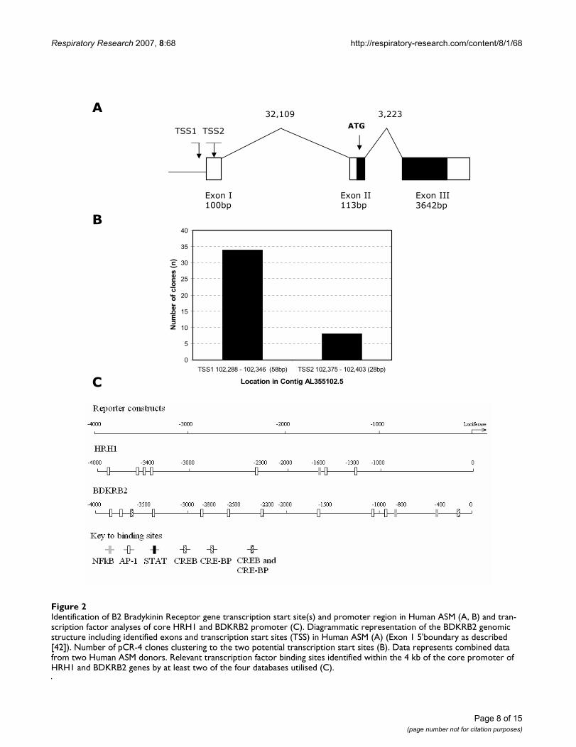
Human ASM cells transfected with the HRH1 1, 2, 3 and4 kb promoter constructs and stimulated for 24 hourswith IFNγ, IL-13 or salmeterol provided evidence that thelevel of transcription was affected for the 1 kb constructs(ANOVA, p = 0.03, Figure 5C) and that salmeterol signif-icantly augments the HRH1 1 kb construct transcriptionalactivity (250.3 +/- 60.3, p < 0.05, Figure 5C). As previ-ously, there was evidence that IL-13 influences transcrip-tion of the HRH1 3 kb promoter (205.3 +/- 90.2, Figure5E).
Stimulation of BDKRB2-luciferase transfected Human ASM for 4 and 24 hours with TNFα and IFNγ does not identify a significant effect on BDKRB2 transcription however, salmeterol stimulates BDKRB2 transcriptionIn an analogous manner to that described for the HRH1promoter analyses the effect of TNFα, IFNγ or salmeterolon BDKRB2 mediated transcription was evaluated usingthe pGL4-BDKRB2-luciferase transfected Human ASMcells. Following 4 hours stimulation with TNFα or IFNγthere were no apparent effects on BDKRB2 mediated tran-scription for the 1, 2, 3 and 4 kb constructs. Salmeteroltreatment resulted in an augmentation of BDKRB2 medi-ated transcription for the 1, 2, 3 and 4 kb constructs
Effect of salmeterol or cytokine treatment for 24 hours on HRH1 promoter activity in Human ASM cells transfected with HRH1-Luciferase or control constructsFigure 5Effect of salmeterol or cytokine treatment for 24 hours on HRH1 promoter activity in Human ASM cells transfected with HRH1-Luciferase or control constructs. Human ASM cells were transfected and stimulated as described in Figure 4. Following 24 hours treatment cells were harvested and firefly luciferase was quantified. pGL4-Luc2 (A), pGL4-SV40-Luc2 (B), pGL4-HRH1-1 kb-Luc2 (C), pGL4-HRH1-2 kb-Luc2 (D), pGL4-HRH1-3 kb-Luc2 (E), pGL4-HRH1-4 kb-Luc2 (F) transfections. Data is normalised to the mean luciferase activity of each construct transfection treated with medium alone +/- S.E.M. (n = 5 independent experiments) Dunnett's Multiple Copmarison Test (**p < 0.01).
Medium TNFA IFNG IL-13 SALM0
50
100
150
200
250
300
Treatment
Nor
mal
ised
RLU
(+/-
SE
M)
Medium TNFA IFNG IL-13 SALM0
50
100
150
200
250
300
Treatment
Nor
mal
ised
RLU
(+/-
SE
M)
Medium IFNG IL-13 SALM0
100
200
300
400
Treatment
Nor
ma
lised
RLU
(+/-
SE
M)
Medium IFNG IL-13 SALM0
50
100
150
200
250
300
Treatment
Nor
mal
ised
RLU
(+/-
SE
M)
Medium IFNG IL-13 SALM0
50
100
150
200
250
300
Treatment
Nor
mal
ised
RLU
(+/-
SE
M)
Medium IFNG IL-13 SALM0
50
100
150
200
250
300
Treatment
Nor
mal
ised
RLU
(+/-
SE
M)
**
**
B
D
E F
C
A
Effect of salmeterol or cytokine treatment for 4 hours on HRH1 promoter activity in Human ASM cells transfected with HRH1-Luciferase or control constructsFigure 4Effect of salmeterol or cytokine treatment for 4 hours on HRH1 promoter activity in Human ASM cells transfected with HRH1-Luciferase or control constructs. Complete medium was replaced with serum free medium and cells were transfected with 0.12 µg of pGL4-Luc2 using Fugene 6 (Roche) at a 3:1 ratio (Fugene:DNA). For derivatives of pGL4-Luc2 containing the HRH1 or SV40 control inserts equimolar amounts of DNA were used. Cells were allowed to grow for 16 hours prior to the addition of medium alone, 10 ng/ml TNFα, 10 ng/ml IFNγ, 50 ng/ml IL-13 or 1 µM salm-eterol. Following 4 hours treatment cells were harvested and firefly luciferase was quantified. pGL4-Luc2 (A), pGL4-SV40-Luc2 (B), pGL4-HRH1-1 kb-Luc2 (C), pGL4-HRH1-2 kb-Luc2 (D), pGL4-HRH1-3 kb-Luc2 (E), pGL4-HRH1-4 kb-Luc2 (F) transfections. Data is normalised to the mean luciferase activity of each construct transfection treated with medium alone +/- S.E.M. (n = 4 independent experiments). Dunnett's Multiple Comparison Test (*p < 0.05).
Medium TNFA IFNG IL-13 SALM0
50
100
150
200
250
300
Treatment
Nor
ma
lised
RLU
(+/-
SE
M)
Medium TNFA IFNG IL-13 SALM0
50
100
150
200
250
300
Treatment
Nor
mal
ised
RLU
(+/-
SE
M)
Medium IFNG IL-13 SALM0
50
100
150
200
250
300
Treatment
Nor
ma
lised
RLU
(+/-
SE
M)
Medium IFNG IL-13 SALM0
50
100
150
200
250
300
Treatment
Nor
mal
ised
RLU
(+/-
SE
M)
Medium IFNG IL-13 SALM0
50
100
150
200
250
300
Treatment
Nor
ma
lised
RLU
(+/-
SE
M)
Medium IFNG IL-13 SALM0
50
100
150
200
250
300
Treatment
Nor
mal
ised
RLU
(+/-
SE
M)
*
A B
D
E F
C
*
Page 10 of 15(page number not for citation purposes)

Respiratory Research 2007, 8:68 http://respiratory-research.com/content/8/1/68
(409.4 +/- 65.7, 446.7 +/- 111.9, 245.3 +/- 30.8 and 542.4+/- 148.5 respectively, p < 0.01, Figure 6). Following 24hours stimulation of BDKRB2-luciferase transfectedHuman ASM cells with TNFα, IFNγ or salmeterol a similarpattern to the 4 hour experiments was observed. TNFαand IFNγ stimulation did not significantly influenceBDKRB2 mediated transcription (Figure 7). Salmeteroltreatment of BDKRB2-luciferase transfected Human ASMcells for 24 hours significantly augmented BDKRB2 medi-ated transcription for the 1, 2 and 4 kb constructs (190.9+/- 30.0, 208.8 +/- 42.7 and 377.7 +/- 110.7 respectively,p < 0.05, Figures 7A, 7B, 7D), however, the effect wasapparent for all constructs.
DiscussionIn this study we aimed to clarify the effect of pro-inflam-matory cytokines (IL-1β, TNFα, IFNγ, or IL-13) and tworepresentatives of drug classes, dexamethasone and salm-eterol on agonist mediated IPx signalling in Human ASM.To this end we have demonstrated that TNFα, IFNγ or sal-meterol can significantly augment bradykinin induced IPxsignalling responses and IL-13, IFNγ or salmeterol can sig-
nificantly augment histamine induced IPx signallingresponses in Human ASM. Further analyses using non-specific activation of the IPx pathway suggested a regula-tory mechanism involving the receptor locus, which wasconfirmed by the quantification of the H1 Histamine andB2 Bradykinin receptor mRNA levels in treated cells.Finally, we sought to identify transcriptional mechanismsunderlying these effects by mapping the core HRH1 andBDKRB2 promoter regions in Human ASM and complet-ing promoter-reporter analyses. These data confirmed thatsalmeterol can induce HRH1 and BDKRB2 transcription,however, the cytokine analyses suggested a role for addi-tional mechanisms regulating cytokine induced GPCRmRNA levels.
The mechanisms underlying the potentially deleteriouseffects of prolonged use of β2-adrenoceptor agonists inasthma are unclear at this time. This study represents thefirst demonstration that salmeterol can modestly augmentbradykinin induced IPx signalling in Human ASM and hasidentified a mechanism involving the induction of B2Bradykinin receptor mRNA via increased transcription at
Effect of salmeterol or cytokine treatment for 24 hours on BDKRB2 promoter activity in Human ASM cells transfected with BDKRB2-Luciferase constructsFigure 7Effect of salmeterol or cytokine treatment for 24 hours on BDKRB2 promoter activity in Human ASM cells transfected with BDKRB2-Luciferase constructs. Human ASM cells were transfected and stimulated as described in Figure 4. Following 24 hours treatment cells were harvested and firefly luciferase was quantified. pGL4-BDKRB2-1kb-Luc2 (A), pGL4-BDKRB2-2kb-Luc2 (B), pGL4-BDKRB2-3kb-Luc2 (C), pGL4-BDKRB2-4kb-Luc2 (D) transfections. Data is normalised to the mean luciferase activity of each construct transfection treated with medium alone +/- S.E.M. (n = 5 independent experiments). Dunnett's Multiple Comparison Test (*p < 0.05, **p < 0.01).
Medium TNFA IFNG SALM0
50
100
150
200
250
300
Treatment
Nor
ma
lised
RLU
(+/-
SE
M)
Medium TNFA IFNG SALM0
50
100
150
200
250
300
Treatment
Nor
mal
ised
RLU
(+/-
SE
M)
Medium TNFA IFNG SALM0
50
100
150
200
250
300
Treatment
Nor
mal
ised
RLU
(+/-
SE
M)
Medium TNFA IFNG SALM0
50100150200250300350400450500
Treatment
Nor
ma
lised
RLU
(+/-
SE
M)
**
**
*
B
DC
A
Effect of salmeterol or cytokine treatment for 4 hours on BDKRB2 promoter activity in Human ASM cells transfected with BDKRB2-Luciferase constructsFigure 6Effect of salmeterol or cytokine treatment for 4 hours on BDKRB2 promoter activity in Human ASM cells transfected with BDKRB2-Luciferase constructs. Human ASM cells were transfected and stimulated as described in Figure 4 except derivatives of pGL4-Luc2 containing the BDKRB2 inserts were used. Following 4 hours treatment cells were harvested and firefly luciferase was quantified. pGL4-BDKRB2-1kb-Luc2 (A), pGL4-BDKRB2-2kb-Luc2 (B), pGL4-BDKRB2-3kb-Luc2 (C), pGL4-BDKRB2-4kb-Luc2 (D) transfections. Data is nor-malised to the mean luciferase activity of each construct transfection treated with medium alone +/- S.E.M. (n = 4 independent experiments). Dunnett's Multiple Copmarison Test (**p < 0.01).
Medium TNFA IFNG SALM0
100
200
300
400
500
Treatment
Nor
ma
lised
RLU
(+/-
SE
M)
Medium TNFA IFNG SALM0
100
200
300
400
500
600
Treatment
Nor
mal
ised
RLU
(+/-
SE
M)
Medium TNFA IFNG SALM0
50
100
150
200
250
300
Treatment
Nor
ma
lised
RLU
(+/-
SE
M)
Medium TNFA IFNG SALM0
100
200
300
400
500
600
700
Treatment
No
rmal
ised
RLU
(+/-
SE
M)
** **
****
B
DC
A
Page 11 of 15(page number not for citation purposes)
Respiratory Research 2007, 8:68 http://respiratory-research.com/content/8/1/68
the core promoter. The promoter-reporter analysesshowed a robust and pronounced effect of salmeterol onBDKRB2 promoter driven transcription which correlatedwith the identification of a consensus CREB site at -121 inthe BDKRB2 promoter suggesting that all constructs (1–4kb) should show induction of transcription. Interestingly,it has recently been shown that a CREB binding site at -44to -51 in the rat BDKRB2 promoter is a major determinantof transcriptional regulation of the rat gene [44]. Salme-terol was also shown to augment histamine induced IPxsignalling in Human ASM via an identical mechanisminvolving the H1 Histamine receptor, however, thereporter driven transcriptional affects were more modestpotentially reflecting the low level of HRH1 promoteractivity observed in the current study. These data extendour recent study that demonstrated that the short acting β2adrenoceptor agonists; salbutamol and isoprenalinecould augment histamine induced IPx signallingresponses [24] and show that multiple contractile agentresponses are affected by prolonged use of this class ofdrugs. In support of the current findings, previous data
examining the effect of β2 adrenoceptor agonists on hista-mine mediated contraction in bovine ASM demonstrateda modest augmentation (1.5 fold) following fenoterolpre-treatment (18 hours) which was shown to be accom-panied by an increase in HRH1 mRNA expression [28]. Inthis bovine system both transcriptional and post-tran-scriptional regulation were identified as determinants ofHRH1 mRNA expression [28]. The clinical implications ofthese findings are that this mechanism may at least in partexplain the accumulating evidence to suggest that pro-longed use of long acting β2 adrenoceptor agonist mono-therapy can lead to adverse effects in asthma asdemonstrated in the SMART study [22]. This study of sal-meterol use for 28 weeks was a multicentre, randomized,double blind, parallel group, placebo controlled designinvolving 26,355 subjects at 6,163 sites in the UnitedStates [22]. Interim analyses demonstrated that there wasa significant increase in respiratory related deaths (24 vs.11, RR 2.16 (95% CI 1.06 to 4.41)) and asthma relateddeaths (13 vs. 3, RR 4.37 (95% CI 1.25 to 15.34)) in thesalmeterol versus placebo group [22].
Several studies have identified a direct interactionbetween cytokines and Human ASM leading to alterationsin the capacity of airway smooth muscle in culture torespond to contractile agents [9,10]. TNFα can augmentbradykinin induced Ca2+ signalling (~2 fold) and IPx sig-nalling (~2 fold) [12]. We have demonstrated similaraffects and identified that induction of BDKRB2 mRNA isa feature of this response. Our data suggested that thisinduction of BDKRB2 mRNA may involve other mecha-nisms than transcription at the core promoter e.g. post-transcriptional mechanisms including RNA stability. Thecritical importance of 3'UTRs of genes determining mRNAexpression has been established including a role for AU-rich elements (ARE) and RNA stabilising regions [45].Several RNA binding proteins including; tristetraprolin(TTP), human antigen R (HuR) and AU-rich factor 1(AUF1) have been identified that mediate RNA stabilityand therefore determine mRNA levels [46]. Interestingly,TNFα has been shown to induce HuR activity leading toincreased eotaxin mRNA stability [47]. In support of thecurrent study, TNFα has been shown to augment BDKRB2mRNA expression in human embryonic lung fibroblastcells [48]. Nuclear run on assays suggested a prominentrole for post-transcriptional mechanisms leading to ele-vated BDKRB2 mRNA following TNFα treatment [48]which would at least in part explain the lack of transcrip-tional regulation identified in the current promoter-reporter analyses. The 3'untranslated region of theHuman B2 bradykinin receptor has been identified a keydeterminant of BDKRB2 mRNA expression [49]. TNFαhas also been shown to augment bradykinin induced con-tractile responses in a mouse tracheal ring model and a
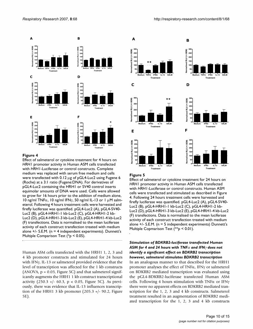
Schematic representation of mechanisms involved in the induction of hyperresponsiveness in Human ASMFigure 8Schematic representation of mechanisms involved in the induction of hyperresponsiveness in Human ASM. Cytokines and salmeterol up-regulate mRNA expression of the relevant GPCR leading to increased IP3 production in response to specific contractile agents (as demonstrated in the current manuscript). In addition, cytokines can up-regulate CD38 leading to subsequent increased cADPR production [9]. Sec-ond messengers IP3 and cADPR activate the Ryanodine and IP3 receptor respectively leading to elevated calcium release from intracellular stores and downstream contractile responses.
IP3R
(+)
Airway environmente.g. cytokines
Contractile Agente.g. Histamine, Bradykinin
GPCR
GDP
GTP
IP3
GTP
GDP
G q
PLC 1
G qCD38
[Ca2+]
Airway Hyper-responsiveness
(+)
RyR
ADP RibosylCyclase
cADPRHydrolase
cADPR
Page 12 of 15(page number not for citation purposes)

Respiratory Research 2007, 8:68 http://respiratory-research.com/content/8/1/68
role for increased B2 bradykinin receptor mRNA was iden-tified [50] suggesting the mechanism exists across species.
IL-13 has been shown to augment histamine and bradyki-nin induced Ca2+ signalling (~30% and 40% respectively)in Human ASM [13]. In the current study we have shownIL-13 specifically augments histamine induced IPx signal-ling and that this specificity involved selective regulationat the receptor locus. The modest induction of HRH1 tran-scription mediated by IL-13 in the promoter-reporteranalyses again suggests multiple mechanisms determinethe HRH1 mRNA level, i.e. a transcriptional mechanismdoes not adequately explain the robust >3 fold inductionin mRNA levels observed. As part of a wider microarraystudy IL-13 has previously been shown to induce HRH1expression in Human ASM (~2.6 fold, 4 hours) [51].Interestingly, IL-4 has been shown to induce activity of theRNA stablilising protein HuR and increase mRNA expres-sion levels of eotaxin in epithelial cells [47]. IL-4 and IL-13 can both activate the STAT6 pathway and thereforepotentially the IL-13 dependent increase in HRH1 mRNAmay involve post-transcriptional mechanisms involvingHuR.
IFNγ has been shown to augment bradykinin inducedCa2+ signalling (~1.5 fold) in Human ASM [52]. We havedemonstrated that IFNγ can augment both histamine andbradykinin induced IPx signalling via induction of GPCRmRNA levels. Analysis of the BDKRB2 and HRH1 corepromoter did not identify a transcriptional mechanism toexplain the induction of mRNA levels in treated cells.Importantly, the β1-adrenergic receptor, a member of theGPCR family has been shown to be regulated via posttran-scriptional mechanism which determine mRNA stabilityand level [53]. By inference, our core promoter analysespotentially suggest that post-transcriptional mechanismsmay be important for GPCR mRNA regulation bycytokines.
In agreement with the proposed mechanism described inthe current study, IFNγ has been shown to induce cystei-nyl leukotriene receptor 1 (CysLTR1) mRNA expression inHuman ASM with subsequent augmentation of LTD4mediated Ca2+ signalling responses [26]. These data sug-gest that regulation of Human ASM responses to multiplecontractile agents involves regulation at the GPCR locus.In the current study we did not identify any effect of dex-amethasone or IL1β on Human ASM IPx signalling incontrast to others [15,38], which may reflect cell donor/experimental differences. Previous data has demonstratedthat e.g. IL-13 can augment KCl induced force generationin mouse tracheal rings suggesting a regulatory mecha-nism downstream of that examined in the current study[13]. A mechanism to explain these observations has beenidentified involving cytokine activation leading to ele-
vated CD38 and subsequent cyclic ADP ribose (cADPr)which activates the ryanodine receptor (RyR) and modu-lates the calcium response in Human ASM [9]. Therefore,it is likely that regulation at the GPCR locus demonstratedin the current study provides some specificity and ampli-fication of Ca2+ responses via IP3 generation and thatinduction of CD38 also contributes to the overall magni-tude of Ca2+ and contractile responses (see Figure 8). Ourfinding that IL-13 did not influence bradykinin inducedIPx production or BDKRB2 mRNA levels may suggest thataugmentation of this specific agonist response is moredependent on the CD38/cADPr pathway although we cannot exclude the influence of different cell donors/experi-mental designs.
These data demonstrate that a range of pro-inflammatorycytokines known to be over-expressed in the asthmaticlung alter Human ASM IPx signalling capacity and byinference may be involved in the molecular basis of AHRin asthma. It is important to note that TNFα identified inthe current analyses as augmenting IPx production hasalso been shown to augment adenylyl cyclase activity inHuman ASM [27]. Therefore, the potential contractileeffects resulting from increased IPx production inresponse to cytokine and spasmogen may at least in partbe countered by the increased relaxation capacity of ASMdue to an augmentation of cyclic AMP production. It isinteresting that IFNγ augmented responses to multiplecontractile agents suggesting that this cytokine could havea key role in AHR development during e.g. viral infectionand in asthma per se as CD8+IFNγ + cell numbers in theairways of asthma subjects correlate with AHR [8]. Thefinding that salmeterol can modestly augment HumanASM responses to contractile agents and potentially leadto AHR also has clinical implications. This study has iden-tified some interesting findings however, the use of singledose and time points may have identified sub-optimaleffects. Similarly, other limitations include the measure-ment of global IPx production as an outcome measureand not specific IPx production, cytosolic calcium andcontractile responses which would have been desirable.
ConclusionWe have presented evidence that the regulation of HumanASM responses to contractile agents can at least in part beexplained by regulation at the GPCR locus. Severalcytokines (TNFα, IFNγ, IL-13) and salmeterol modestlyaugment histamine and bradykinin IPx signallingresponses in cultured Human ASM cells in a cytokine andagonist specific manner. This augmentation was accom-panied by elevated mRNA levels of the specific GPCR pro-viding a mechanism to explain this specificity. Finally, wedemonstrated that cytokine modulation of HRH1 andBDKRB2 mRNA levels may involve additional mecha-nisms to a transcriptional mechanism involving the core
Page 13 of 15(page number not for citation purposes)

Respiratory Research 2007, 8:68 http://respiratory-research.com/content/8/1/68
HRH1 and BDKRB2 promoters. The effects observed forsalmeterol mediated GPCR mRNA induction did involvetranscriptional regulation at the core promoter. While theeffects on IPx signalling were modest there is accumulat-ing evidence that small changes in IPx signalling and sub-sequent cytosolic calcium concentrations can havephysiological implications for contractile responses andairway hyper-responsiveness. These findings provide agreater insight into the molecular basis of AHR in asthma.
AbbreviationsAHR, airway hyperresponsiveness, ASM, airway smoothmuscle, PLC, phospholipase C, IPx, inositol phosphates,IL, interleukin, TNF, tumour necrosis factor, IFN, inter-feron, GPCR, G-protein coupled receptor, COPD, chronicobstructive pulmonary disease, DAG, diacylglycerol, IP2,inositol 4,5-biphosphate, IP3, inositol 1,4,5-triphosphate,PKC, protein kinase C, DMEM, Dulbecco's ModifiedEagles Medium, EDTA, Ethylenediaminetetraacetic acid,PBS, phosphate buffered saline, Ct, threshold cycle, RACE,rapid amplification of cDNA ends, PCR, polymerase chainreaction, PLB, passive lysis buffer, NK-κB, nuclear factorκB, AP-1, activator protein 1, CREB, cyclic AMP response-element-binding protein, STAT, signal tranducer and acti-vator of transcription, CHO, Chinese Hamster Ovarian.
Competing interestsThe author(s) declare that they have no competing inter-ests.
Authors' contributionsIS designed the study, carried out laboratory-based exper-iments (unless otherwise stated), completed data analysesand wrote the manuscript. NS completed the IPx signal-ling work and generated some of the RNA samples. CABgenerated the promoter-luciferase constructs. ND and SPgenerated some of the RNA/cDNA samples. CES com-pleted the promoter database analyses. CS designed theHRH1 TaqMan Assay. IPH contributed to the design ofthe study and writing of the manuscript. All authors readand approved the final manuscript.
AcknowledgementsThis work was supported by a grant from the Nottingham Hospital Special Trustees.
References1. Hall IP: Second messengers, ion channels and pharmacology
of airway smooth muscle. Eur Respir J 2000, 15(6):1120-1127.2. Johnson EN, Druey KM: Heterotrimeric G protein signaling:
role in asthma and allergic inflammation. J Allergy Clin Immunol2002, 109(4):592-602.
3. Townley RG, Horiba M: Airway hyperresponsiveness: a story ofmice and men and cytokines. Clin Rev Allergy Immunol 2003,24(1):85-110.
4. Broide DH, Lotz M, Cuomo AJ, Coburn DA, Federman EC, Wasser-man SI: Cytokines in symptomatic asthma airways. J Allergy ClinImmunol 1992, 89(5):958-967.
5. Huang SK, Xiao HQ, Kleine-Tebbe J, Paciotti G, Marsh DG, Lichten-stein LM, Liu MC: IL-13 expression at the sites of allergen chal-lenge in patients with asthma. J Immunol 1995,155(5):2688-2694.
6. Grunig G, Warnock M, Wakil AE, Venkayya R, Brombacher F, Ren-nick DM, Sheppard D, Mohrs M, Donaldson DD, Locksley RM, et al.:Requirement for IL-13 independently of IL-4 in experimentalasthma. Science 1998, 282(5397):2261-2263.
7. Brooks GD, Buchta KA, Swenson CA, Gern JE, Busse WW: Rhino-virus-induced interferon-gamma and airway responsivenessin asthma. Am J Respir Crit Care Med 2003, 168(9):1091-1094.
8. Cho SH, Stanciu LA, Holgate ST, Johnston SL: Increased inter-leukin-4, interleukin-5, and interferon-gamma in airwayCD4+ and CD8+ T cells in atopic asthma. Am J Respir Crit CareMed 2005, 171(3):224-230.
9. Amrani Y, Tliba O, Deshpande DA, Walseth TF, Kannan MS, Panet-tieri RA Jr: Bronchial hyperresponsiveness: insights into newsignaling molecules. Curr Opin Pharmacol 2004, 4(3):230-234.
10. Shore SA: Direct effects of Th2 cytokines on airway smoothmuscle. Curr Opin Pharmacol 2004, 4(3):235-240.
11. Amrani Y, Martinet N, Bronner C: Potentiation by tumournecrosis factor-alpha of calcium signals induced by bradyki-nin and carbachol in human tracheal smooth muscle cells. BrJ Pharmacol 1995, 114(1):4-5.
12. Amrani Y, Krymskaya V, Maki C, Panettieri RA Jr: Mechanismsunderlying TNF-alpha effects on agonist-mediated calciumhomeostasis in human airway smooth muscle cells. Am J Phys-iol 1997, 273(5 Pt 1):L1020-1028.
13. Tliba O, Deshpande D, Chen H, Van Besien C, Kannan M, PanettieriRA Jr, Amrani Y: IL-13 enhances agonist-evoked calcium sig-nals and contractile responses in airway smooth muscle. Br JPharmacol 2003, 140(7):1159-1162.
14. Hotta K, Emala CW, Hirshman CA: TNF-alpha upregulates Gial-pha and Gqalpha protein expression and function in humanairway smooth muscle cells. Am J Physiol 1999, 276(3 Pt1):L405-411.
15. Pype JL, Xu H, Schuermans M, Dupont LJ, Wuyts W, Mak JC, BarnesPJ, Demedts MG, Verleden GM: Mechanisms of interleukin1beta-induced human airway smooth muscle hyporespon-siveness to histamine. Involvement of p38 MAPK NF-kap-paB. Am J Respir Crit Care Med 2001, 163(4):1010-1017.
16. Deshpande DA, White TA, Dogan S, Walseth TF, Panettieri RA, Kan-nan MS: CD38/cyclic ADP-ribose signaling: role in the regula-tion of calcium homeostasis in airway smooth muscle. Am JPhysiol Lung Cell Mol Physiol 2005, 288(5):L773-788.
17. Deshpande DA, Dogan S, Walseth TF, Miller SM, Amrani Y, PanettieriRA, Kannan MS: Modulation of calcium signaling by inter-leukin-13 in human airway smooth muscle: role of CD38/cyclic adenosine diphosphate ribose pathway. Am J Respir CellMol Biol 2004, 31(1):36-42.
18. Koh YY, Lee MH, Sun YH, Park Y, Kim CK: Improvement in bron-chial hyperresponsiveness with inhaled corticosteroids inchildren with asthma: importance of family history of bron-chial hyperresponsiveness. Am J Respir Crit Care Med 2002,166(3):340-345.
19. Cheung D, Timmers MC, Zwinderman AH, Bel EH, Dijkman JH, SterkPJ: Long-term effects of a long-acting beta 2-adrenoceptoragonist, salmeterol, on airway hyperresponsiveness inpatients with mild asthma. N Engl J Med 1992,327(17):1198-1203.
20. Crane J, Pearce N, Flatt A, Burgess C, Jackson R, Kwong T, Ball M,Beasley R: Prescribed fenoterol and death from asthma inNew Zealand, 1981-83: case-control study. Lancet 1989,1(8644):917-922.
21. Taylor DR, Sears MR, Herbison GP, Flannery EM, Print CG, Lake DC,Yates DM, Lucas MK, Li Q: Regular inhaled beta agonist inasthma: effects on exacerbations and lung function. Thorax1993, 48(2):134-138.
22. Nelson HS, Weiss ST, Bleecker ER, Yancey SW, Dorinsky PM: TheSalmeterol Multicenter Asthma Research Trial: a compari-son of usual pharmacotherapy for asthma or usual pharma-cotherapy plus salmeterol. Chest 2006, 129(1):15-26.
23. van Schayck CP, Graafsma SJ, Visch MB, Dompeling E, van Weel C,van Herwaarden CL: Increased bronchial hyperresponsivenessafter inhaling salbutamol during 1 year is not caused by sub-
Page 14 of 15(page number not for citation purposes)

Respiratory Research 2007, 8:68 http://respiratory-research.com/content/8/1/68
Publish with BioMed Central and every scientist can read your work free of charge
"BioMed Central will be the most significant development for disseminating the results of biomedical research in our lifetime."
Sir Paul Nurse, Cancer Research UK
Your research papers will be:
available free of charge to the entire biomedical community
peer reviewed and published immediately upon acceptance
cited in PubMed and archived on PubMed Central
yours — you keep the copyright
Submit your manuscript here:http://www.biomedcentral.com/info/publishing_adv.asp
BioMedcentral
sensitization to salbutamol. J Allergy Clin Immunol 1990,86(5):793-800.
24. Sayers I, Swan C, Hall IP: The effect of beta2-adrenoceptor ago-nists on phospholipase C (beta1) signalling in human airwaysmooth muscle cells. Eur J Pharmacol 2006, 531(1–3):9-12.
25. Billington CK, Penn RB: Signaling and regulation of G protein-coupled receptors in airway smooth muscle. Respir Res 2003,4:2.
26. Amrani Y, Moore PE, Hoffman R, Shore SA, Panettieri RA Jr: Inter-feron-gamma modulates cysteinyl leukotriene receptor-1expression and function in human airway myocytes. Am JRespir Crit Care Med 2001, 164(11):2098-2101.
27. Pascual RM, Billington CK, Hall IP, Panettieri RA Jr, Fish JE, Peters SP,Penn RB: Mechanisms of cytokine effects on G protein-cou-pled receptor-mediated signaling in airway smooth muscle.Am J Physiol Lung Cell Mol Physiol 2001, 281(6):L1425-1435.
28. Mak JC, Roffel AF, Katsunuma T, Elzinga CR, Zaagsma J, Barnes PJ:Up-regulation of airway smooth muscle histamine H(1)receptor mRNA, protein, and function by beta(2)-adreno-ceptor activation. Molecular pharmacology 2000, 57(5):857-864.
29. Schmidlin F, Scherrer D, Daeffler L, Bertrand C, Landry Y, Gies JP:Interleukin-1beta induces bradykinin B2 receptor geneexpression through a prostanoid cyclic AMP-dependentpathway in human bronchial smooth muscle cells. Molecularpharmacology 1998, 53(6):1009-1015.
30. Daykin K, Widdop S, Hall IP: Control of histamine induced inosi-tol phospholipid hydrolysis in cultured human trachealsmooth muscle cells. Eur J Pharmacol 1993, 246(2):135-140.
31. Chromas(v2) [http://technelysium.com.au]32. Braun A, Kammerer S, Bohme E, Muller B, Roscher AA: Identifica-
tion of polymorphic sites of the human bradykinin B2 recep-tor gene. Biochem Biophys Res Commun 1995, 211(1):234-240.
33. Swan C, Richards SA, Duroudier NP, Sayers I, Hall IP: Alternativepromoter use and splice variation in the human histamineH1 receptor gene. Am J Respir Cell Mol Biol 2006, 35(1):118-126.
34. BIMAS [http://bimas.cit.nih.gov/molbio/signal/]35. TESS [http://www.cbil.upenn.edu/cgi-bin/tess/tess?RQ=WEL
COME]36. TFSEARCH [http://www.cbrc.jp/research/db/TFSEARCH.html]37. WWWSignalScan [http://bimas.cit.nih.gov/molbio/signal/]38. Hardy E, Farahani M, Hall IP: Regulation of histamine H1 recep-
tor coupling by dexamethasone in human cultured airwaysmooth muscle. Br J Pharmacol 1996, 118(4):1079-1084.
39. Arthur JM, Collinsworth GP, Gettys TW, Raymond JR: Agonist-induced translocation of Gq/11alpha immunoreactivitydirectly from plasma membrane in MDCK cells. Am J Physiol1999, 276(4 Pt 2):F528-534.
40. Wang YX, Kotlikoff MI: Signalling pathway for histamine activa-tion of non-selective cation channels in equine tracheal myo-cytes. J Physiol 2000, 523(Pt 1):131-138.
41. Lee CH, Park D, Wu D, Rhee SG, Simon MI: Members of the Gqalpha subunit gene family activate phospholipase C beta iso-zymes. J Biol Chem 1992, 267(23):16044-16047.
42. Kammerer S, Braun A, Arnold N, Roscher AA: The human brady-kinin B2 receptor gene: full length cDNA, genomic organiza-tion and identification of the regulatory region. BiochemBiophys Res Commun 1995, 211(1):226-233.
43. Barnes PJ, Adcock IM: Transcription factors and asthma. EurRespir J 1998, 12(1):221-234.
44. Saifudeen Z, Dipp S, Fan H, El-Dahr SS: Combinatorial control ofthe bradykinin B2 receptor promoter by p53, CREB, KLF-4,and CBP: implications for terminal nephron differentiation.Am J Physiol Renal Physiol 2005, 288(5):F899-909.
45. Conne B, Stutz A, Vassalli JD: The 3' untranslated region of mes-senger RNA: A molecular 'hotspot' for pathology? Nat Med2000, 6(6):637-641.
46. Seko Y, Cole S, Kasprzak W, Shapiro BA, Ragheb JA: The role ofcytokine mRNA stability in the pathogenesis of autoimmunedisease. Autoimmun Rev 2006, 5(5):299-305.
47. Atasoy U, Curry SL, Lopez de Silanes I, Shyu AB, Casolaro V, GorospeM, Stellato C: Regulation of eotaxin gene expression by TNF-alpha and IL-4 through mRNA stabilization: involvement ofthe RNA-binding protein HuR. J Immunol 2003,171(8):4369-4378.
48. Haddad EB, Fox AJ, Rousell J, Burgess G, McIntyre P, Barnes PJ, ChungKF: Post-transcriptional regulation of bradykinin B1 and B2
receptor gene expression in human lung fibroblasts bytumor necrosis factor-alpha: modulation by dexamethasone.Molecular pharmacology 2000, 57(6):1123-1131.
49. Zamorano R, Suchindran S, Gainer JV: 3'-Untranslated region ofthe type 2 bradykinin receptor is a potent regulator of geneexpression. Am J Physiol Renal Physiol 2006, 290(2):F456-464.
50. Zhang Y, Adner M, Cardell LO: Up-regulation of bradykininreceptors in a murine in-vitro model of chronic airwayinflammation. Eur J Pharmacol 2004, 489(1–2):117-126.
51. Jarai G, Sukkar M, Garrett S, Duroudier N, Westwick J, Adcock I,Chung KF: Effects of interleukin-1beta, interleukin-13 andtransforming growth factor-beta on gene expression inhuman airway smooth muscle using gene microarrays. Eur JPharmacol 2004, 497(3):255-265.
52. Deshpande DA, Walseth TF, Panettieri RA, Kannan MS: CD38/cyclicADP-ribose-mediated Ca2+ signaling contributes to airwaysmooth muscle hyper-responsiveness. Faseb J 2003,17(3):452-454.
53. Pende A, Tremmel KD, DeMaria CT, Blaxall BC, Minobe WA, Sher-man JA, Bisognano JD, Bristow MR, Brewer G, Port J: Regulation ofthe mRNA-binding protein AUF1 by activation of the beta-adrenergic receptor signal transduction pathway. J Biol Chem1996, 271(14):8493-8501.
Page 15 of 15(page number not for citation purposes)
Copyright © 2022 FDOKUMEN