Role of a Novel PH-Kinase Domain Interface in PKB/Akt Regulation: Structural Mechanism for...
12
Role of a Novel PH-Kinase Domain Interface in PKB/Akt Regulation: Structural Mechanism for Allosteric Inhibition Ve ´ ronique Calleja 1[* , Michel Laguerre 2[ , Peter J. Parker 3,4 , Banafshe ´ Larijani 1* 1 Cell Biophysics Laboratory, Cancer Research UK, Lincoln’s Inn Fields Laboratories, London Research Institute, London, United Kingdom, 2 UMR 5248-CNRS, Institut Europe ´ en de Chimie et Biologie, Pessac, France, 3 Protein Phosphorylation Laboratory, Cancer Research UK, Lincoln’s Inn Fields Laboratories, London Research Institute, London, United Kingdom, 4 Division of Cancer Studies KCL, Guy’s Hospital, London, United Kingdom Protein kinase B (PKB/Akt) belongs to the AGC superfamily of related serine/threonine protein kinases. It is a key regulator downstream of various growth factors and hormones and is involved in malignant transformation and chemo-resistance. Full-length PKB protein has not been crystallised, thus studying the molecular mechanisms that are involved in its regulation in relation to its structure have not been simple. Recently, the dynamics between the inactive and active conformer at the molecular level have been described. The maintenance of PKB’s inactive state via the interaction of the PH and kinase domains prevents its activation loop to be phosphorylated by its upstream activator, phosphoinositide-dependent protein kinase-1 (PDK1). By using a multidisciplinary approach including molecular modelling, classical biochemical assays, and Fo ¨ rster resonance energy transfer (FRET)/two-photon fluorescence lifetime imaging microscopy (FLIM), a detailed model depicting the interaction between the different domains of PKB in its inactive conformation was demonstrated. These findings in turn clarified the molecular mechanism of PKB inhibition by AKT inhibitor VIII (a specific allosteric inhibitor) and illustrated at the molecular level its selectivity towards different PKB isoforms. Furthermore, these findings allude to the possible function of the C-terminus in sustaining the inactive conformer of PKB. This study presents essential insights into the quaternary structure of PKB in its inactive conformation. An understanding of PKB structure in relation to its function is critical for elucidating its mode of activation and discovering how to modulate its activity. The molecular mechanism of inhibition of PKB activation by the specific drug AKT inhibitor VIII has critical implications for determining the mechanism of inhibition of other allosteric inhibitors and for opening up opportunities for the design of new generations of modulator drugs. Citation: Calleja V, Laguerre M, Parker PJ, Larijani B (2009) Role of a novel PH-kinase domain interface in PKB/Akt regulation: Structural mechanism for allosteric inhibition. PLoS Biol 7(1): e1000017. doi:10.1371/journal.pbio.1000017 Introduction Protein kinase B (PKB/Akt) is a key regulator downstream of various growth factors and hormonal signals. It activates a panel of proteins that regulate proliferation, growth, survival, or metabolism and is involved in human cancer [1,2]. In particular, its overexpression induces malignant transforma- tion and chemoresistance [3]. PKB belongs to the AGC superfamily of related serine/threonine protein kinases. Three isoforms of PKB exist in mammals (PKBa/Akt1, PKBb/Akt2, and PKBc/Akt3) that comprise an N-terminal pleckstrin homology (PH) domain, a flexible hinge between the PH and the kinase domain, a catalytic (kinase) domain, and a C-terminal regulatory part (containing a hydrophobic motif, or HM) (for review [4,5]). The phosphorylation of Thr 308 in the kinase domain of PKBa/Akt 1 by phosphoinositide- dependent protein kinase-1 (PDK1) [6] and Ser 473 in the hydrophobic motif by mTORC2 complex [7] and/or DNAPK [8], is central for PKB activation [9]. These phosphorylations were shown to be dependent on the colocalisation of PKB and PDK1 at the plasma membrane through the interaction of their PH domains with PtdIns (3,4,5) P 3 and PtdIns (3,4) P 2 for the former [10–12] and PtdIns (3,4,5) P 3 for the latter [13]. The dephosphorylation of these residues by okadaic acid- sensitive and -insensitive phosphatases was shown to deacti- vate PKB [14,15]. Recently, the cytoplasmic interaction of inactive PKB with PDK1 was published [14]. The maintenance of PKB in an inactive conformation by the interaction of its PH and kinase domains (PH-in) prior to stimulation prevented the phos- phorylation of Thr 308 by the associated PDK1. Upon stimulation, PKB PH-domain interaction with phosphoinosi- tides and its concomitant change in conformation allowed the separation of the PH and kinase domains and the associated co-recruited PDK1 to phosphorylate Thr 308. Here, the intramolecular interactions of the PKB domains in its inactive conformation were studied in detail using Academic Editor: Alex Toker, Beth Israel Deaconess Medical Center, United States of America Received September 23, 2008; Accepted December 5, 2008; Published January 20, 2009 Copyright: Ó 2009 Calleja et al. This is an open-access article distributed under the terms of the Creative Commons Attribution License, which permits unrestricted use, distribution, and reproduction in any medium, provided the original author and source are credited. Abbreviations: FLIM, fluorescence lifetime imaging microscopy; FRET, Fo ¨ rster resonance energy transfer; GFP, green fluorescent protein; HM, hydrophobic motif; mRFP, monomeric red fluorescent protein; PDGF, platelet-derived growth factor; PDK1, 3-phosphoinositide-dependent protein kinase-1; PH, pleckstrin homology; PH-in, protein kinase B in an inactive conformation; PKB, protein kinase B * To whom correspondence should be addressed. E-mail: banafshe.larijani@cancer. org.uk (BL); [email protected] (VC) [ These authors contributed equally to this work. PLoS Biology | www.plosbiology.org January 2009 | Volume 7 | Issue 1 | e1000017 0189 P L o S BIOLOGY
-
Upload
independent -
Category
Documents
-
view
1 -
download
0
Transcript of Role of a Novel PH-Kinase Domain Interface in PKB/Akt Regulation: Structural Mechanism for...
Role of a Novel PH-Kinase Domain Interfacein PKB/Akt Regulation: Structural Mechanismfor Allosteric InhibitionVeronique Calleja
1[*, Michel Laguerre
2[, Peter J. Parker
3,4, Banafshe Larijani
1*
1 Cell Biophysics Laboratory, Cancer Research UK, Lincoln’s Inn Fields Laboratories, London Research Institute, London, United Kingdom, 2 UMR 5248-CNRS, Institut
Europeen de Chimie et Biologie, Pessac, France, 3 Protein Phosphorylation Laboratory, Cancer Research UK, Lincoln’s Inn Fields Laboratories, London Research Institute,
London, United Kingdom, 4 Division of Cancer Studies KCL, Guy’s Hospital, London, United Kingdom
Protein kinase B (PKB/Akt) belongs to the AGC superfamily of related serine/threonine protein kinases. It is a keyregulator downstream of various growth factors and hormones and is involved in malignant transformation andchemo-resistance. Full-length PKB protein has not been crystallised, thus studying the molecular mechanisms that areinvolved in its regulation in relation to its structure have not been simple. Recently, the dynamics between the inactiveand active conformer at the molecular level have been described. The maintenance of PKB’s inactive state via theinteraction of the PH and kinase domains prevents its activation loop to be phosphorylated by its upstream activator,phosphoinositide-dependent protein kinase-1 (PDK1). By using a multidisciplinary approach including molecularmodelling, classical biochemical assays, and Forster resonance energy transfer (FRET)/two-photon fluorescence lifetimeimaging microscopy (FLIM), a detailed model depicting the interaction between the different domains of PKB in itsinactive conformation was demonstrated. These findings in turn clarified the molecular mechanism of PKB inhibitionby AKT inhibitor VIII (a specific allosteric inhibitor) and illustrated at the molecular level its selectivity towards differentPKB isoforms. Furthermore, these findings allude to the possible function of the C-terminus in sustaining the inactiveconformer of PKB. This study presents essential insights into the quaternary structure of PKB in its inactiveconformation. An understanding of PKB structure in relation to its function is critical for elucidating its mode ofactivation and discovering how to modulate its activity. The molecular mechanism of inhibition of PKB activation bythe specific drug AKT inhibitor VIII has critical implications for determining the mechanism of inhibition of otherallosteric inhibitors and for opening up opportunities for the design of new generations of modulator drugs.
Citation: Calleja V, Laguerre M, Parker PJ, Larijani B (2009) Role of a novel PH-kinase domain interface in PKB/Akt regulation: Structural mechanism for allosteric inhibition.PLoS Biol 7(1): e1000017. doi:10.1371/journal.pbio.1000017
Introduction
Protein kinase B (PKB/Akt) is a key regulator downstreamof various growth factors and hormonal signals. It activates apanel of proteins that regulate proliferation, growth, survival,or metabolism and is involved in human cancer [1,2]. Inparticular, its overexpression induces malignant transforma-tion and chemoresistance [3]. PKB belongs to the AGCsuperfamily of related serine/threonine protein kinases.Three isoforms of PKB exist in mammals (PKBa/Akt1,PKBb/Akt2, and PKBc/Akt3) that comprise an N-terminalpleckstrin homology (PH) domain, a flexible hinge betweenthe PH and the kinase domain, a catalytic (kinase) domain,and a C-terminal regulatory part (containing a hydrophobicmotif, or HM) (for review [4,5]). The phosphorylation of Thr308 in the kinase domain of PKBa/Akt 1 by phosphoinositide-dependent protein kinase-1 (PDK1) [6] and Ser 473 in thehydrophobic motif by mTORC2 complex [7] and/or DNAPK[8], is central for PKB activation [9]. These phosphorylationswere shown to be dependent on the colocalisation of PKBand PDK1 at the plasma membrane through the interactionof their PH domains with PtdIns (3,4,5) P3 and PtdIns (3,4) P2
for the former [10–12] and PtdIns (3,4,5) P3 for the latter [13].The dephosphorylation of these residues by okadaic acid-sensitive and -insensitive phosphatases was shown to deacti-vate PKB [14,15].
Recently, the cytoplasmic interaction of inactive PKB with
PDK1 was published [14]. The maintenance of PKB in aninactive conformation by the interaction of its PH and kinasedomains (PH-in) prior to stimulation prevented the phos-phorylation of Thr 308 by the associated PDK1. Uponstimulation, PKB PH-domain interaction with phosphoinosi-tides and its concomitant change in conformation allowedthe separation of the PH and kinase domains and theassociated co-recruited PDK1 to phosphorylate Thr 308.Here, the intramolecular interactions of the PKB domains
in its inactive conformation were studied in detail using
Academic Editor: Alex Toker, Beth Israel Deaconess Medical Center, United Statesof America
Received September 23, 2008; Accepted December 5, 2008; Published January20, 2009
Copyright: � 2009 Calleja et al. This is an open-access article distributed under theterms of the Creative Commons Attribution License, which permits unrestricteduse, distribution, and reproduction in any medium, provided the original authorand source are credited.
Abbreviations: FLIM, fluorescence lifetime imaging microscopy; FRET, Forsterresonance energy transfer; GFP, green fluorescent protein; HM, hydrophobic motif;mRFP, monomeric red fluorescent protein; PDGF, platelet-derived growth factor;PDK1, 3-phosphoinositide-dependent protein kinase-1; PH, pleckstrin homology;PH-in, protein kinase B in an inactive conformation; PKB, protein kinase B
* To whom correspondence should be addressed. E-mail: [email protected] (BL); [email protected] (VC)
[ These authors contributed equally to this work.
PLoS Biology | www.plosbiology.org January 2009 | Volume 7 | Issue 1 | e10000170189
PLoS BIOLOGY
molecular dynamics in conjunction with classical biochemicalapproaches and in situ Forster resonance energy transfer(FRET)/two-photon fluorescence lifetime imaging microscopy(FLIM). A PH domain-induced cavity was discovered in theinactive PKBa kinase domain. The formation of this structurewas dependent on the PH domain residue Trp 80. Theposition of PKBa C-terminal HM at the apex of the PHdomain-induced cavity in the inactive conformation of PKBwas also described. Unlike in PKBa, a PH domain-inducedcavity did not appear in PKBc suggesting a potential role ofthis structure in a differential regulation of these two PKBisoforms.
These findings led to the elucidation of the molecularmechanism of inhibition of PKB by the highly specificallosteric inhibitor, AKT inhibitor VIII, and its relationshipto the C-terminal HM motif of PKB.
Results and Discussion
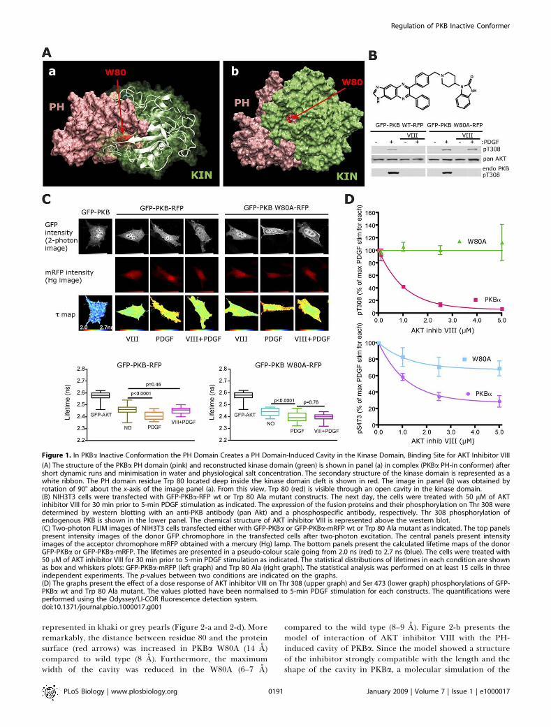
Previously, the molecular model of the inactive PKBconformer (PH-in conformer) showed that the PH and kinasedomains interacted [14]. By using molecular dynamics, anunpredicted formation of a PH-induced cavity in the PKBkinase domain was observed (Figure 1A). The location of thiscavity appeared to be in the same region as the PKBhydrophobic pocket (binding site for PKB HM) described inthe crystal structure of the isolated PKB kinase domain [16].The initial docking model of the PKB PH and kinase domainsinteraction prior to performing molecular dynamics revealedthat the residue Trp 80, critically positioned at the tip of theVL3 loop of the PH domain, was buried deep inside the PKBkinase domain cleft in the PH-in conformer. However, therefined dynamic model showed that through this PH-induced
cavity, the Trp 80 residue, located at its centre, was accessiblefrom outside, whereas PKB remained in its inactive con-former (Figure 1A-b).To illustrate the importance of the positioning of the Trp
80 and the PH-induced cavity, we took advantage of a specificPKB inhibitor, AKT inhibitor VIII [17–19] (Figure 1B,chemical structure). The selectivity has been attributed toan allosteric mode of binding, noncompetitive with ATP orsubstrates, and with a mechanism of action dependent on thepresence of the PH domain of the full-length protein. In vivoAKT inhibitor VIII prevented the phosphorylation of Thr 308and Ser 473 (Figure 1B, western blot, and [20]), and as a result,it was proposed to target a specific PKB conformation.Recently, its efficacy was shown to be dependent on thepresence of the Trp 80, but the molecular mechanism was notdetermined [21]. The targeting of the PKB inactive conformerby AKT inhibitor VIII and consequent prevention of PKBconformation change was a plausible explanation for the lossof Thr 308 phosphorylation.To verify this postulate in vivo, experiments with the PKB
fluorescent probe (green fluorescent protein [GFP]-PKB-monomeric red fluorescent protein [mRFP]) [14] wereperformed. The platelet-derived growth factor (PDGF)-induced change in the conformation of PKB was monitoredby FRET/two-photon FLIM (Figure 1C). The pretreatment ofNIH3T3 cells with AKT inhibitor VIII prevented both thetranslocation of GFP-PKB-mRFP to the plasma membraneand its change in conformation induced by PDGF. Toillustrate the importance of the interaction of the inhibitorwith Trp 80, the GFP-PKB W80A-mRFP mutant was used.Upon PDGF stimulation, the W80A mutant behaved as wild-type PKB. However, in response to PDGF, it was evident thatthe mutant was not inhibitor sensitive. The graphs in Figure1C illustrate the statistical data obtained by FRET/two-photonFLIM. These results established that the inhibitor ‘‘locked’’PKB in its PH-in conformer and confirmed in vivo theimportance of the Trp 80 residue in this mechanism.The inhibition of PDGF-induced Thr 308 and Ser 473
phosphorylation by preincubation with increasing doses ofAKT inhibitor VIII was tested on GFP-PKBa and on the Trp80 mutant (Figure 1D). At 5 lM AKT inhibitor VIII, thephosphorylation of Thr 308 was inhibited by 95%, but Ser473 was inhibited by only 70%. This differential in theinhibition of Thr 308 phosphorylation compared to Ser 473indicated that the mechanism of action of the inhibitoraffected the two sites in a distinct manner. The mutation ofthe Trp 80 to Ala prevented entirely the loss of Thr 308phosphorylation induced by 5 lM inhibitor, confirming thatthe loss of this critical residue prevented the locking of PKBin the PH-in conformation as observed in Figure 1C.However, the possibility that the inhibitor might stillinterfere with the second site phosphorylation could not beexcluded since a slight decrease of Ser 473 phosphorylationwas observed in the W80A mutant.To understand the significance of the Trp 80 residue in
PKB, the interaction of PKBa PH mutant W80A with thekinase domain in the PH-in conformation was determined bymolecular dynamics (Figure 2-c and 2-d). The alteration ofthe shape and length of the PH domain-induced cavity in themutant PKBa W80A as compared to wild-type PKBa isillustrated in Figure 2-c and 2-d (compare to Figures 1A-b,and 2-a). Both cavities are filled with water molecules
PLoS Biology | www.plosbiology.org January 2009 | Volume 7 | Issue 1 | e10000170190
Regulation of PKB Inactive Conformer
Author Summary
A critical protein in cell-signalling pathways, called protein kinase B,regulates many aspects of cell biology from metabolism toproliferation and survival, by modifying other proteins with theaddition of a phosphate group. Hence, deregulation of its activityhas acute consequences on cell function. Increased activity of atumour-promoting form of protein kinase B or of upstreamregulatory proteins has been observed in tumours, while impairedprotein kinase B function has been linked to diabetes. Therefore,understanding the molecular mechanism of protein kinase Bactivation will help reveal how its activity might be regulated tolimit disease progression. Toward this end, we studied how proteinkinase B structure relates to its function, to identify molecularmechanisms regulating its kinase activity, modifying its cellularlocalization, and altering its binding to other proteins. By determin-ing the spatial organization of different regions of the protein ininactive protein kinase B, we discovered a cavity at the interface oftwo distinct functional regions of the inactive form. We alsolocalized the C-terminal end of the protein to the apex of the cavity,suggesting a role of this domain in regulating the inactive form ofthe protein. This represents a novel example of negative regulationby inhibition across these different regions of the protein. Fromthese findings, we elucidated the mechanism of action of a highlyspecific protein kinase B inhibitor, AKT inhibitor VIII. We determinedthat simultaneous binding of the inhibitor to the two differentfunctional regions, through the cavity, ‘‘locks’’ protein kinase B in aninactive conformation and prevents regulatory proteins fromaccessing the C-terminal domain.
represented in khaki or grey pearls (Figure 2-a and 2-d). Moreremarkably, the distance between residue 80 and the proteinsurface (red arrows) was increased in PKBa W80A (14 A)compared to wild type (8 A). Furthermore, the maximumwidth of the cavity was reduced in the W80A (6–7 A)
compared to the wild type (8–9 A). Figure 2-b presents themodel of interaction of AKT inhibitor VIII with the PH-induced cavity of PKBa. Since the model showed a structureof the inhibitor strongly compatible with the length and theshape of the cavity in PKBa, a molecular simulation of the
Figure 1. In PKBa Inactive Conformation the PH Domain Creates a PH Domain-Induced Cavity in the Kinase Domain, Binding Site for AKT Inhibitor VIII
(A) The structure of the PKBa PH domain (pink) and reconstructed kinase domain (green) is shown in panel (a) in complex (PKBa PH-in conformer) aftershort dynamic runs and minimisation in water and physiological salt concentration. The secondary structure of the kinase domain is represented as awhite ribbon. The PH domain residue Trp 80 located deep inside the kinase domain cleft is shown in red. The image in panel (b) was obtained byrotation of 908 about the x-axis of the image panel (a). From this view, Trp 80 (red) is visible through an open cavity in the kinase domain.(B) NIH3T3 cells were transfected with GFP-PKBa-RFP wt or Trp 80 Ala mutant constructs. The next day, the cells were treated with 50 lM of AKTinhibitor VIII for 30 min prior to 5-min PDGF stimulation as indicated. The expression of the fusion proteins and their phosphorylation on Thr 308 weredetermined by western blotting with an anti-PKB antibody (pan Akt) and a phosphospecific antibody, respectively. Thr 308 phosphorylation ofendogenous PKB is shown in the lower panel. The chemical structure of AKT inhibitor VIII is represented above the western blot.(C) Two-photon FLIM images of NIH3T3 cells transfected either with GFP-PKBa or GFP-PKBa-mRFP wt or Trp 80 Ala mutant as indicated. The top panelspresent intensity images of the donor GFP chromophore in the transfected cells after two-photon excitation. The central panels present intensityimages of the acceptor chromophore mRFP obtained with a mercury (Hg) lamp. The bottom panels present the calculated lifetime maps of the donorGFP-PKBa or GFP-PKBa-mRFP. The lifetimes are presented in a pseudo-colour scale going from 2.0 ns (red) to 2.7 ns (blue). The cells were treated with50 lM of AKT inhibitor VIII for 30 min prior to 5-min PDGF stimulation as indicated. The statistical distributions of lifetimes in each condition are shownas box and whiskers plots: GFP-PKBa-mRFP (left graph) and Trp 80 Ala (right graph). The statistical analysis was performed on at least 15 cells in threeindependent experiments. The p-values between two conditions are indicated on the graphs.(D) The graphs present the effect of a dose response of AKT inhibitor VIII on Thr 308 (upper graph) and Ser 473 (lower graph) phosphorylations of GFP-PKBa wt and Trp 80 Ala mutant. The values plotted have been normalised to 5-min PDGF stimulation for each constructs. The quantifications wereperformed using the Odyssey/LI-COR fluorescence detection system.doi:10.1371/journal.pbio.1000017.g001
PLoS Biology | www.plosbiology.org January 2009 | Volume 7 | Issue 1 | e10000170191
Regulation of PKB Inactive Conformer
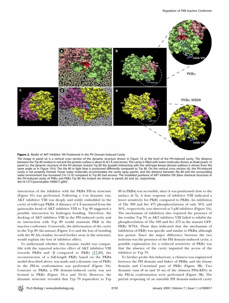
interaction of the inhibitor with the PKBa PH-in structure(Figure S1) was performed. Following a 1-ns dynamic run,AKT inhibitor VIII was deeply and stably embedded in thecavity of wild type PKBa. A distance of 4 A measured from thequinoxalin head of AKT inhibitor VIII to Trp 80 suggested apossible interaction by hydrogen bonding. Therefore, thedocking of AKT inhibitor VIII in the PH-induced cavity andits interaction with Trp 80 would maintain PKB in theinactive conformer. Conversely, the deformation of the cavityin the Trp 80 Ala mutant (Figure 2-e) and the loss of bondingwith the 80 Ala residue, located farther away in the structure,would explain the loss of inhibitor effect.
To understand whether this dynamic model was compat-ible with the reported selective effect of AKT inhibitor VIIItowards PKBa and -b compared to PKBc [17,20], thereconstruction of a full-length PKBc based on the PKBamodel described above, was made and a dynamic run of PKBcin the PH-in conformation was performed (Figure 3A).Contrary to PKBa, a PH domain-induced cavity was notformed in PKBc (Figure 3A-a and 3A-b). However, thedynamic structure revealed that Trp 79 (equivalent to Trp
80 in PKBa) was accessible, since it was positioned close to thesurface (6 A). A dose response of inhibitor VIII indicated alower sensitivity for PKBc compared to PKBa. An inhibitionof Thr 308 and Ser 473 phosphorylations of only 50% and30%, respectively, was observed at 5 lM inhibitor (Figure 3A).The mechanism of inhibition also required the presence ofthe residue Trp 79, as AKT inhibitor VIII failed to inhibit thephosphorylation of Thr 308 and Ser 473 in the mutant GFP-PKBc W79A. These data indicated that the mechanism ofinhibition of PKBc was specific and similar to PKBa, althoughless potent. Since the major difference between the twoisoforms was the presence of the PH domain-induced cavity, apossible explanation for a reduced sensitivity of PKBc wasthat the absence of the cavity impaired the access of theinhibitor to Trp 79.To further probe this behaviour, a chimera was engineered
between the PH domain and linker of PKBa, and the kinasedomain and C-terminal part of PKBc (Figure 3B). Twodynamic runs (8 ns and 10 ns) of the chimera PHa-KINc inthe PH-in conformation were performed (Figure 3B). Thepartial reopening of an unstable PH domain-induced cavity
Figure 2. Model of AKT Inhibitor VIII Positioned in the PH Domain-Induced Cavity
The image in panel (a) is a vertical cross section of the dynamic structure shown in Figure 1A at the level of the PH-induced cavity. The distancebetween the Trp 80 residue in red and the protein surface is about 8–8.5 A (red arrow). The cavity is filled with water molecules shown as khaki pearls. Inpanel (c), the dynamic structure of the PH domain mutant Trp 80 Ala (purple) interacting with the wild-type kinase domain (yellow) is shown from thesame angle as in Figure 1A-b. The Ala 80 in light blue is positioned differently compared to Trp 80. On the vertical cross section (d), the PH-inducedcavity is not properly formed. Fewer water molecules accommodate the cavity (grey pearls), and the distance between Ala 80 and the surroundingwater environment has increased (14–15 A) compared to Trp 80 (red arrows). The modelled positions of AKT inhibitor VIII (blue chemical structure) inthe PH-induced cavity of PKBa and PKBa Trp 80 Ala mutant are shown in panels (b) and (e), respectively.doi:10.1371/journal.pbio.1000017.g002
PLoS Biology | www.plosbiology.org January 2009 | Volume 7 | Issue 1 | e10000170192
Regulation of PKB Inactive Conformer
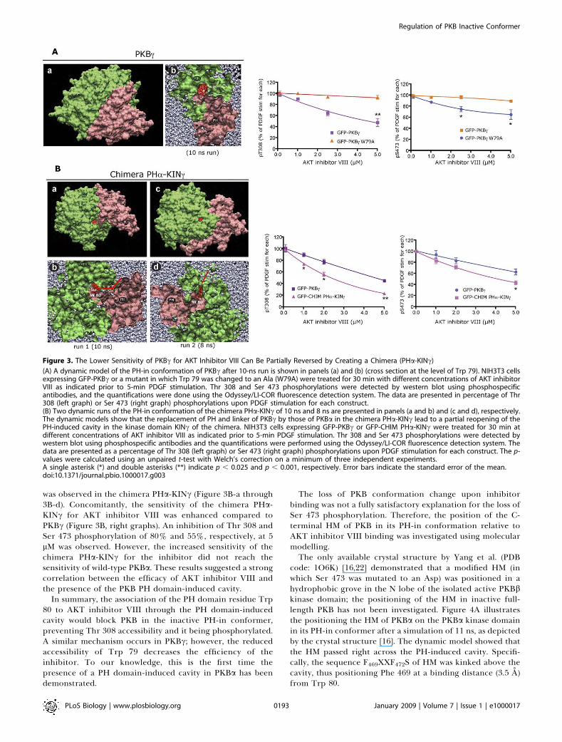
was observed in the chimera PHa-KINc (Figure 3B-a through3B-d). Concomitantly, the sensitivity of the chimera PHa-KINc for AKT inhibitor VIII was enhanced compared toPKBc (Figure 3B, right graphs). An inhibition of Thr 308 andSer 473 phosphorylation of 80% and 55%, respectively, at 5lM was observed. However, the increased sensitivity of thechimera PHa-KINc for the inhibitor did not reach thesensitivity of wild-type PKBa. These results suggested a strongcorrelation between the efficacy of AKT inhibitor VIII andthe presence of the PKB PH domain-induced cavity.
In summary, the association of the PH domain residue Trp80 to AKT inhibitor VIII through the PH domain-inducedcavity would block PKB in the inactive PH-in conformer,preventing Thr 308 accessibility and it being phosphorylated.A similar mechanism occurs in PKBc; however, the reducedaccessibility of Trp 79 decreases the efficiency of theinhibitor. To our knowledge, this is the first time thepresence of a PH domain-induced cavity in PKBa has beendemonstrated.
The loss of PKB conformation change upon inhibitorbinding was not a fully satisfactory explanation for the loss ofSer 473 phosphorylation. Therefore, the position of the C-terminal HM of PKB in its PH-in conformation relative toAKT inhibitor VIII binding was investigated using molecularmodelling.The only available crystal structure by Yang et al. (PDB
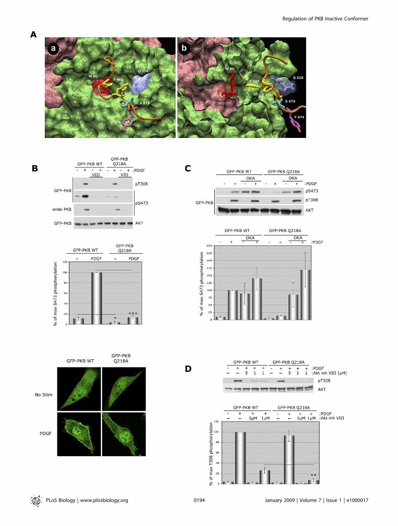
code: 1O6K) [16,22] demonstrated that a modified HM (inwhich Ser 473 was mutated to an Asp) was positioned in ahydrophobic grove in the N lobe of the isolated active PKBbkinase domain; the positioning of the HM in inactive full-length PKB has not been investigated. Figure 4A illustratesthe positioning the HM of PKBa on the PKBa kinase domainin its PH-in conformer after a simulation of 11 ns, as depictedby the crystal structure [16]. The dynamic model showed thatthe HM passed right across the PH-induced cavity. Specifi-cally, the sequence F469XXF472S of HM was kinked above thecavity, thus positioning Phe 469 at a binding distance (3.5 A)from Trp 80.
Figure 3. The Lower Sensitivity of PKBc for AKT Inhibitor VIII Can Be Partially Reversed by Creating a Chimera (PHa-KINc)
(A) A dynamic model of the PH-in conformation of PKBc after 10-ns run is shown in panels (a) and (b) (cross section at the level of Trp 79). NIH3T3 cellsexpressing GFP-PKBc or a mutant in which Trp 79 was changed to an Ala (W79A) were treated for 30 min with different concentrations of AKT inhibitorVIII as indicated prior to 5-min PDGF stimulation. Thr 308 and Ser 473 phosphorylations were detected by western blot using phosphospecificantibodies, and the quantifications were done using the Odyssey/LI-COR fluorescence detection system. The data are presented in percentage of Thr308 (left graph) or Ser 473 (right graph) phosphorylations upon PDGF stimulation for each construct.(B) Two dynamic runs of the PH-in conformation of the chimera PHa-KINc of 10 ns and 8 ns are presented in panels (a and b) and (c and d), respectively.The dynamic models show that the replacement of PH and linker of PKBc by those of PKBa in the chimera PHa-KINc lead to a partial reopening of thePH-induced cavity in the kinase domain KINc of the chimera. NIH3T3 cells expressing GFP-PKBc or GFP-CHIM PHa-KINc were treated for 30 min atdifferent concentrations of AKT inhibitor VIII as indicated prior to 5-min PDGF stimulation. Thr 308 and Ser 473 phosphorylations were detected bywestern blot using phosphospecific antibodies and the quantifications were performed using the Odyssey/LI-COR fluorescence detection system. Thedata are presented as a percentage of Thr 308 (left graph) or Ser 473 (right graph) phosphorylations upon PDGF stimulation for each construct. The p-values were calculated using an unpaired t-test with Welch’s correction on a minimum of three independent experiments.A single asterisk (*) and double asterisks (**) indicate p , 0.025 and p , 0.001, respectively. Error bars indicate the standard error of the mean.doi:10.1371/journal.pbio.1000017.g003
PLoS Biology | www.plosbiology.org January 2009 | Volume 7 | Issue 1 | e10000170193
Regulation of PKB Inactive Conformer
PLoS Biology | www.plosbiology.org January 2009 | Volume 7 | Issue 1 | e10000170194
Regulation of PKB Inactive Conformer

From this model, Gln 218 was identified as being at aninteracting distance for Ser 473. In addition, this residueappeared to be at about 5 A from AKT inhibitor VIII in thedynamic interaction model (Figure S1). Therefore, muta-genesis was performed to assess its potential role in the HMposition and its relation to the inhibitor binding. By mutatingGln 218 to Ala in GFP-PKBa, the basal as well as PDGF-stimulated Ser 473 phosphorylation decreased significantly,suggesting that this newly identified residue was critical forthe stability of the Ser 473 phosphorylation (Figure 4B). Thiseffect was not due to an impaired translocation of GFP-PKBaQ218A compared to GFP-PKBa wt upon PDGF stimulation(Figure 4B, bottom panel). Indeed, the level of Thr 308phosphorylation upon stimulation was not modified signifi-cantly (Figure 4B, top panel). The recovery of Ser 473phosphorylation upon pretreatment with okadaic acid priorto PDGF stimulation indicated that the loss of Ser 473phosphorylation in GFP-PKBa Q218A mutant was due to anenhanced dephosphorylation (Figure 4C). A plausible ex-planation for this result was that the HM might be incorrectlypositioned and thus was becoming more accessible tophosphatases.
In order to determine whether the Gln 218 Ala mutationaffected the binding of AKT inhibitor VIII, the effect of anoptimal (5 lM) and suboptimal (1 lM) dose of inhibitor VIIIon Thr 308 phosphorylation was tested (Figure 4D). Theenhanced inhibition of Thr 308 phosphorylation in the Gln218 Ala mutant at 1 lM inhibitor suggested that the HM andthe inhibitor might compete for the same or a proximatebinding site.
In summary, the position of the HM in the inactive andactivated state of PKB is likely to be very similar. Theinteraction between the Phe 469 and the Trp 80 suggestedthat a role of the HM motif in the regulation of PKB prior toits translocation and Ser 473 phosphorylation could beenvisioned. Furthermore, the C-terminal HM and AKTinhibitor VIII could overlap at a common binding site.
To determine whether the HM was implicated in AKTinhibitor binding as well as competing for the same (or aclose) binding site, a deletion mutant of the last 39 residues ofPKBa C-terminal part was made (Figure 5A). A dose-dependent inhibition of Thr 308 phosphorylation by AKT
inhibitor VIII was performed on wild-type PKBa and mutantPKBa DCt. The deletion of the PKBa C-terminal part did notprevent the inhibition of Thr 308 phosphorylation by AKTinhibitor VIII. Moreover, a low dose of inhibitor (1 lM)slightly, but significantly, lowered Thr 308 phosphorylation inPKBa DCt compared to wild-type PKBa. This result showedthat the binding of the inhibitor was not dependent on the C-terminal part of PKB and indicated further that the C-terminal HM and AKT inhibitor VIII could potentially becompeting for a common or close binding site on the kinasedomain.To understand the mechanism involved in the loss of Ser
473 phosphorylation upon AKT inhibitor binding, experi-ments with okadaic acid and the inhibitor were performed(Figure 5B). Since the data in Figures 4D and 5A suggestedthat the HM and AKT inhibitor VIII could compete for thesame binding site, the inhibitor binding could lead to anincorrect positioning of the HM, leading to an increased S473dephosphorylation. A similar mechanism was shown abovewhen mutating the residue Gln 218. Alternatively, theinhibitor binding could prevent the phosphorylation of Ser473 by steric hindrance. As previously described [14], theinhibition of PP2A family of phosphatases by okadaic acidinduced the phosphorylation of Ser 473 (but not Thr 308) to asimilar extent as PDGF stimulation. However, the pretreat-ment with inhibitor VIII prior to okadaic acid, with andwithout PDGF, prevented the phosphorylation of Ser 473.This result suggested that loss of Ser 473 phosphorylation waslikely to be due to the loss of accessibility of Ser 473 residueto upstream kinase(s) rather than a dephosphorylation.
ConclusionThe novel findings from this study are summarised in
Figure 6 (see figure legend). The molecular mechanism ofinteraction of the allosteric inhibitor AKT inhibitor VIII to acritical PH domain residue, Trp 80, was consistent with themodel of binding through a PH-induced cavity formed in thekinase domain of PKB. This cavity was present in PKBa,whereas it was negligible in PKBc. Chimera constructs of thePH and kinase domains suggested that the dimensions of thecavity were strongly correlated with the sensitivity to theinhibitor.
Figure 4. The Position of the C-Terminal Hydrophobic Motif (HM) of PKBa in the PH-in Conformer Is at Close Proximity to its Position after Stimulation
and Interferes with AKT Inhibitor VIII Binding
(A) PKBa C-terminal hydrophobic motif (HMa) (orange) interacting with PKBa kinase domain (green) in the PH-in conformer is presented in the modelpanels (a) and (b) (cross section). The PH domain is in pink, and the PH domain residue Trp 80 is in red. The model shows that two phenylalanines (inyellow) from the sequence F469XXF472S of HM are found positioned right above the PH-induced cavity in PKBa kinase domain. The Phe 469 is at bindingdistance (;3.5 A) from Trp 80 (panel [b]). The principal amino acid residues are indicated.(B) NIH3T3 cells expressing GFP-PKBa wt or a mutant Q218A, were treated for 30 min with 5 lM AKT inhibitor VIII prior to 5-min PDGF stimulation asindicated. Thr 308 and Ser 473 phosphorylations of exogenous and endogenous PKB were detected by western blot using phosphospecific antibodies.Total protein was detected using a pan AKT antibody. The histogram presents the quantification of Ser 473 phosphorylation as a percentage of maximalphosphorylation upon PDGF stimulation in GFP-PKBa wt. A one-sample t-test was performed (triple asterisks [***]) showing an extremely significantdifference with p , 0.0001. An unpaired t-test with Welch’s correction (single asterisk [*]) shows a very significant difference with a p , 0.05. Thebottom panels show confocal images at mid-section of NIH3T3 cells expressing GFP-PKBa wt or GFP-PKBa Q218A prior to (No Stim) or upon 5-minPDGF stimulation. endo, endogenous; OKA, okadaic acid.(C) NIH3T3 cells expressing GFP-PKBa wt or the mutant (Q218A), were treated for 45 min with 1 lM of okadaic acid prior to 5-min PDGF stimulation asindicated. In the top panels, Ser 473 and Thr 308 phosphorylations were detected by western blot using phosphospecific antibodies. Total protein wasdetected using a pan AKT antibody. The histogram presents the quantitative analysis of Ser 473 phosphorylation as a percentage of PDGF stimulation inGFP-PKBa wt, from three independent experiments.(D) NIH3T3 cells expressing GFP-PKBa wt or mutant Q218A were treated for 30 min with different doses of AKT inhibitor VIII (AKT inh VIII) prior to 5-minPDGF stimulation as indicated. Thr 308 phosphorylation was detected by western blot using a phosphospecific antibody. Total protein was detectedusing a pan AKT antibody. The histogram presents the quantitative analysis of Thr 308 phosphorylation as a percentage of PDGF stimulation in wildtype, from eight independent experiments. An unpaired t-test with Welch’s correction was performed with (double asterisks [**]) representing p ,0.005.doi:10.1371/journal.pbio.1000017.g004
PLoS Biology | www.plosbiology.org January 2009 | Volume 7 | Issue 1 | e10000170195
Regulation of PKB Inactive Conformer

The in vivo FRET data showed that in the presence of AKTinhibitor VIII, PKB was maintained in its PH-in conforma-tion. In this conformation, Thr 308 and Ser 473 phosphor-ylation were impaired. However, a difference in theinhibition of phosphorylation between the two sites wasobserved. The inhibition of phosphorylation on Thr 308 waslinked to PKB being maintained in its PH-in conformationand not being accessible to PDK1.
Recently published in vitro data indicated that the bindingof PKB PH domain to phosphoinositides did not seem to beaffected by the inhibitor interaction [21]. However, thetranslocation of the GFP-PKB-mRFP reporter was prevented
in presence of inhibitor. These results indicated that themaintenance of PKB at the plasma membrane appeared torequire a change in conformation of the protein. The lack ofPKB translocation could explain the loss of Ser 473phosphorylation upon inhibitor binding. However, ourresults indicated that the inhibitor binding created a sterichindrance preventing the upstream kinase(s) from phosphor-ylating Ser 473.We have addressed the possible molecular mechanisms
involved in the regulation of the PKB inactive conformationby examining intramolecular domain interaction dynamics.The discovery of a PH-kinase domain interface in relation to
Figure 5. The Loss of Ser 473 Phosphorylation in PKBa upon AKT Inhibitor VIII Binding Is Due to Steric Hindrance
(A) NIH3T3 cells expressing GFP-PKBa;pha; wt or GFP-PKBa DCt, deleted from the C-terminal HM motif (last 39 amino acids), were treated for 30 minwith different concentrations of AKT inhibitor VIII prior to 5-min PDGF stimulation (stim) as indicated. Thr 308 and Ser 473 (only for wild type)phosphorylations were detected by western blot using phosphospecific antibodies, and the quantifications were performed using the Odyssey/LI-CORfluorescence system. For statistical analysis, a two-tailed paired t-test was done from six independent experiments. Double asterisks (**) indicate p-values , 0.001 at a concentration of 1 lM AKT inhibitor VIII.(B) NIH3T3 cells expressing wild-type GFP-PKBa were treated for 30 min with 5 lM of AKT inhibitor VIII (VIII) followed by 1 lM of okadaic acid (OKA) for afurther 40 min prior to 5-min PDGF stimulation as indicated. Ser 473 (left panels) and Thr 308 (right panels) phosphorylations were detected by westernblot using phosphospecific antibodies, and the quantifications were done using the Odyssey/LI-COR fluorescence system. The data are presented as apercentage of PDGF stimulation.Error bars indicate the standard error of the mean.doi:10.1371/journal.pbio.1000017.g005
PLoS Biology | www.plosbiology.org January 2009 | Volume 7 | Issue 1 | e10000170196
Regulation of PKB Inactive Conformer

the C-terminal HM was essential in the understanding of therole of PKB quaternary structure in the control of this criticalregulator. Yet, the role played by the flexible linker that joinsthe PH and the kinase domains close to this interface, mightalso be important to comprehend fully this regulation.
In addition, we have depicted the possible molecularmechanism involved in the association of a new class ofselective allosteric inhibitors that to date had remainedelusive. We anticipate this will facilitate the optimisation of anew generation of more potent or more selective inhibitors.
Materials and Methods
Materials and antibodies. Okadaic acid sodium salt and AKTinhibitor VIII were from Calbiochem (Merck KGaA). Human PDGFwas from R&D Systems. Phospho-Akt Thr-308 (both rabbit polyclonaland monoclonal) and Ser-473, as well as pan Akt, were from CellSignaling (New England BioLabs UK). Anti-GFP antibodies were in-house monoclonals. Other chemicals were from Sigma-Aldrich. Glass-bottom microwell culture dishes (35 mm; MatTek dishes) were fromMatTek Corporation. The Odyssey blocking buffer was from LI-CORUK. The IRdye 800 anti-rabbit and IRdye 700 anti-mouse were used as
secondary antibodies for the LI-COR/Odyssey system (from RocklandIncorporated).
Fusion constructs design and direct site mutagenesis. pEGFP-HA-Akt construct was described previously (murine AKT1; Genbankaccession number NM_009652) [23]; to simplify, we called thisconstruct GFP-PKBa.
GFP-PKB-mRFPwas created by putting PCR-amplifiedmRFP (EcoRI-XbaI) instead of yellow fluorescent protein (YFP) in the vector pCDNA3-GFP-PKB-YFP (described previously [14,24]), using the oligonucleotidesmRFP-Ct sense: AGAGAATTCGCCTCCTCCGAGGA CGTCATCAAG-GAGTTCATGCGCTTCAAGG and mRFP-Ct antisense: ATCTCTAGATTATGCACCGGTGGAGTGGCGGCCCTCGGCGCGCTCGTACTGTTCC.
Note that an extra XbaI site had to be removed using the QuickChange mutagenesis kit from Stratagene in the pCDNA3-GFP-PKB-YFP construct before the subcloning of mRFP (EcoRI-XbaI). TheW80A mutation on GFP-PKB-mRFP was performed by Maria Deakfrom Dario Alessi’s laboratory (Dundee, UK).
GFP-PKBa DCt was created by PCR amplification of GFP-PKBawithout the C-terminal part, using the oligonucleotides AKT1 DCt-sense: GGCATGGACGAGCT GTACAAGGGT and AKT1 DCt anti-sense: TACGGATCCTCACTCCTCATCGAAATACCTGG.
The PCR amplification was then placed instead of wild-type PKBain pEGFP-HA-Akt (GFP-PKBa) construct using SalI-BamHI restric-tion sites.
Figure 6. Mechanism of Inhibition of PKB Activity by AKT Inhibitor VIII
(A) The upper panel shows PKBa in its inactive conformation (PH-in) prior to stimulation. The interaction of the PH domain (light grey) and the kinasedomain (light blue) creates a PH domain-induced cavity in the kinase domain. The PH domain residue Trp 80 (red oval) interacts with the Phe 469 of C-terminal HM in orange through the PH-induced cavity. Both regulatory residues (Thr 308 and Ser 473) are unphosphorylated. Upon PDGF stimulation(lower panel), PKBa adopts its active (PH-out) conformation. PH and kinase domains separate, allowing the phosphorylation of the regulatory residuesThr 308 of the T-loop of the kinase domain and Ser 473 (black discs) on the HM. The PH domain-induced cavity closes up, and the HM residues Phe 469,Phe 473, and phosphorylated Ser 473 interact with the hydrophobic pocket.(B) The upper panel shows the binding of AKT inhibitor VIII (dark blue) to the Trp 80 through the PH-induced cavity of PKBa in its inactive conformation.The inhibitor and the C-terminal HM compete for binding of the same region. The dotted orange line shows the position of the C-terminal HM withoutinhibitor. The position of the C-terminal HM in presence of the inhibitor is represented as a solid orange line. The orange arrow represents themovement of the C-terminal. The lower panel depicts the locking of PKBa in the PH-in conformation. The binding of AKT inhibitor VIII prevents thechange in conformation upon PDGF stimulation and Thr 308 from being phosphorylated. The phosphorylation of Ser 473 is prevented by a sterichindrance due to the binding of AKT inhibitor VIII in the same region as HM.(C) The mutation of the PH domain residue 80 to alanine (white oval) creates a PKBa insensitive to the inhibitor. Despite the preincubation with AKTinhibitor VIII, the drug fails to inhibit PDGF-induced change in conformation and activation of PKBa.(D) PKBa kinase domain residue Gln 218, positioned at close proximity to the C-terminal HM, was mutated to Ala (black spot). The mutation of thisresidue destabilises the positioning of the C-terminal HM, allowing Ser 473 to become more accessible to phosphatases. Upon PDGF stimulation, PKBaQ218A translocation to the plasma membrane and change in conformation allow the phosphorylation of Thr 308. However, Ser 473 remainsdephosphorylated.doi:10.1371/journal.pbio.1000017.g006
PLoS Biology | www.plosbiology.org January 2009 | Volume 7 | Issue 1 | e10000170197
Regulation of PKB Inactive Conformer

GFP-PKBc was created by PCR amplification of PKBc frompCDNA3-Myr HA AKT3 human (Addgene plasmid 9017). Theoligonucleotides used for the amplification were PCR-PKBc sense:GTCGACATGTACCCATACGATGTTCCAGATTACGCTTCCA-GAATGAGCGATGTTACCATTGTGAAAGAAGGTTGG and PCR-PKBc antisense: GGATCCTTATTCTCGTCCACTTGCAGA GTAG-GAAAATTGAGGG. Prior to the PCR amplification of PKBc, aninternal BamHI site was removed in pCDNA3-Myr HA AKT3 bydirect site mutagenesis using: PKBc-BamHI-less sense:CTTTCAGGGCTCTTGATAAAGGACCCAAATAAACGCCTTGGand PKBc-BamHI-less antisense: CCAAGGCGTTTATTTGGGTCCTTTATCAAGAGCCCTGAAAG. The PCR product was subclonedinto the intermediate cloning vector zero blunt TOPO fromInvitrogen, then excised using SalI-BamHI restriction enzymes. Theexcised PCR amplification was then put in phase in C-terminus ofGFP (instead of PKBa in the vector GFP-PKBa cut with SalI-BamHI.
The chimera of the PH domain and linker of PKBa in fusion withthe kinase domain and C-terminal of PKBc (GFP-CHIM PHa-KINc)was done first by creating a modified GFP-PKBa construct containinga BamHI site using the oligonucleotides AKT1-BamON-sense:GGGGCTGAAGAGATGGAGGGATCCCTGGCCAAGCCCAAGCACand AKT1-BamON-antisense: GTGCTTGGGCTTGGCCAGGGATCCCTCCATCTCTTCAGCCCC.
A PCR amplification of PKBc kinase domain was then performedusing the oligonucleotides ChimAKT3-sense: AGTGGATCCCTGGC-CAAGCCCAAGCACAGAAAGA CAATGAATGATTTTGACTATTTGand ChimAKT3-antisense: ACTGGATCCTTA TTCTCGTCCACTTG-CAGAGTAGG
The PCR product was cut with BamHI and inserted in the modifiedGFP-PKBa vector containing a BamHI site.
The mutants of PKB were obtained by direct site mutagenesis usingthe Quick Change mutagenesis kit from Stratagene using theoligonucleotides PKBa-W80A sense: CATCCGCTGCCTGCAGGCGAC-CACAGTCATTGAGCGC; PKBa-W80A antisense: GCGCTCAAT-GACTGTGGTCGCCTGCAGGCAGCGGATG; PKBa-Q218A sense:CGGCCCTCAAGTACTCATTCGCGACCCACGACCGCCTCTGC;PKBa-Q218A antisense: GCAGAGGCGGTCGTGGGTCGCGAATGAG-TACTTGAGGGCCG; PKBc-W79A sense: CAGATGTCTCCAGGCGAC-TACTGTTATAGAGAGAACATTTCATG; and PKBc-W79A-antisense:CATGAAATGTTCTCTCTATAACAGTAGTCGCCTGGAGACATCTG.
Cell culture and transfections. NIH3T3 cells from ATCC weremaintained in DMEM 10% Donor Calf Serum (DCS) and seeded at150,000 in a well of a six-well plate or in a MatTek dish. Thetransfection was done with 2 lg of DNA of the different constructs(2þ2 lg for cotransfections) using Lipofect AMINE/PLUS reagent(GIBCO-BRL) in OPTIMEM medium (GIBCO-BRL) as recommendedby the manufacturer. The cells were left for 3 h in the transfectionmix, and then the medium was removed and replaced with DMEM10% DCS. The experiments were performed 24 or 48 h aftertransfection.
Lysis conditions. After stimulation or treatment as indicated, thecells were lysed for 15 min on ice in lysis buffer (20 mM Tris-HCl [pH7.4], 150 mM NaCl, 100 mM NaF, 10 mM Na4P2O7, 10 mM EDTAsupplemented with COMPLETE protease inhibitor cocktail tablet[Roche]). To terminate the reaction, 43 SDS sample buffer (125 mMTris-HCl [pH 6.8], 6% SDS, 20% glycerol, 0.02% bromophenol bluesupplemented with 10% b-mercaptoethanol) was added, and thesamples boiled for 5 min. The proteins were separated on a 10% SDS-PAGE gel.
Western blot analysis (classical ECL). After electrophoresis, thepolyacrilamide gels were transferred onto PVDF membrane (Immo-bilon P; Millipore) and incubated in blocking buffer TBS-T (10 mMTris-HCl [pH 7.4], 150 mM NaCl, 0.05% Tween-20) supplementedwith 3% BSA for 1 h. The membranes were then incubated with thedifferent antibodies: phospho-Akt (Thr-308) antibody (rabbit poly-clonal from Cell Signaling), phospho-Akt (Ser-473) (rabbit polyclonalantibody raised against the C-terminus phospho-peptide HFPQFpSY-SASS of Akt1) or with the pan AKT at 1:1,000 for 1 to 3 h in TBS-T/3% BSA. The membranes were then washed in TBS-T with 0.2%Tween-20 and 1% milk for 1 h. The secondary HRP-antibodies wereused at 1:5,000 in TBS-T with 5% milk for 1 h. Western blots wererevealed by incubation with ECL (Amersham Biosciences). Densityanalysis of bands was done with NIH ImageJ 1.33u (U.S. NationalInstitutes of Health, http://rsb.info.nih.gov/ij/). The analysis wasperformed by subtracting the background of the autoradiographyand normalised as indicated on the graphs.
Western blot (LI-COR/Odyssey fluorescence system). After electro-phoresis, the polyacrilamide gels were transferred onto PVDFmembrane (Immobilon FL; Millipore), incubated in the Odysseyblocking buffer for 1 h, and then with the different antibodies
phospho-Akt (Thr-308) antibody (rabbit polyclonal or monoclonalfrom Cell Signaling) or phospho-Akt (Ser-473) (rabbit polyclonalantibody raised against the C-terminus phospho-peptide HFPQFpSY-SASS of Akt1) at 1:1,000 together with an anti-GFP in-housemonoclonal at 1:2,500 for 4 h in Odyssey blocking buffer. Themembranes were then washed in PBS 0.2% Tween-20 with 1%odyssey buffer for 1 h. The secondary antibodies IRdye 800 anti-rabbit and IRdye 700 anti-mouse (Rockland) were used at 1:2,500 and1:5,000, respectively, in Odyssey blocking for 1 h. The blots werescanned with the infrared LI-COR scanner, allowing for simultaneousdetection of two targets (anti-phospho and total protein) in the sameexperiment. For each experiment, the values of the phospho bandswere normalized for the total amount of protein in each lane.
Forster resonance energy transfer (FRET) by two-photon fluores-cence lifetime imaging microscopy (FLIM). NIH3T3 cells were seededat 150,000 on 35-mm glass-bottom tissue culture dishes (MatTek) andtransfected as described above. Twenty-four hours after transfection,the cells were treated in DMEM containing 10% DCS. The cells werewashed twice with PBS, and then fixed in 4%paraformaldehyde in PBSfor 10 min. The dishes were washed twice with PBS, and then 2 ml ofPBS supplemented with 2.5% (w/v) DABCO as an antifade was addedto the dishes. The images were acquired straightaway or stored at 4 8C.
Details about the method to detect FRET by time domain FLIMwas as previously described [14,25]. We used a nonparametric Mann-Whitney test to compare the medians of the two datasets for the two-photon FLIM data using GraphPad InStat software (version 3.0 forMac-2001). To interpret the distribution of data, box and whiskerplots were used. The box and whisker plot is a histogram-like methodfor displaying upper and lower quartiles, and maximum andminimum values in addition to median.
Molecular modelling. In order to check the validity of the models,we performed several molecular dynamics simulations. Vacuumcalculations were performed on an SGI Octane workstation usingInsightII and Discover ver. 2000 (Accelrys). The hydrated modelscalculations were performed using NAMD [26] software v2.6 andCHARMM 27 force-field. All simulations were performed on a 32-processor IBM PC-cluster using 8 to 16 processors. The simulationboxes were built using VMD v1.8.6 facilities. All images were capturedfrom VMD [27] using the internal ray-tracing software Tachyon.
Two models were built starting from the model of the PH/KINPKBa complex previously described [14]. The whole complex wasimmersed in a box of water, fully ionized, with all charges neutralizedby counter ions, and salt was added to reach the biologicalconcentration of 150 mM. The wild type was built first, and theW80 was subsequently mutated into A80.
Model 1 (wild type) totalled 47,114 atoms with 13,394 watermolecules (TIP3P) in a box of 88.94 3 73.99 3 77.87 A3, at T¼ 300 Kand P ¼ 1 bar. Time of simulation: 6.22 ns.
Model 2 (W80A) totalled 47,103 atoms with 13,395 water molecules(TIP3P) in a box of 88.94 3 73.99 3 77.87 A3, at T¼ 300 K and P¼ 1bar. Time of simulation: 6.13 ns.
During the two simulations, a water cavity opened quickly (afterabout 0.2 ns) just in front of residue 80 going from the outer surfaceof the protein directly to the surface of residue 80.
For wild-type (PKBa W80), the cavity was stable and almost as wideas deep. W80 was located about 8–8.5 A from the outer surface of theprotein, and the cavity had a maximum opening of around 8–9 A. Thecavity was full of water (see Figure 2).
For the W80A mutant, the cavity appeared more unstable withfluctuations and was deeper than it was wide. A80 was located 14–15A from the protein surface (compared to 8–8.5 A in the wild type),and the maximum opening was around 6–7 A. The cavity was againfull of water.
Positioning of the C-terminal. The final model of the wild-type PH/KIN PKBa complex after molecular dynamics simulation was super-imposed (kinase domain only) over the published X-ray structure ofAKT-2 (PKBb) [22] (PDB codes: 1O6K and 1O6L). After super-imposition, the kinase domain of PKBb was deleted, leaving the C-terminal (466–479) of PKBb lying on the surface of the PH/KINcomplex of PKBa. The C-terminal was then mutated to correspond tothe sequence of the C-terminal of PKBa, as below:
1O6K:QRTHFPQFDYSASIPKBa:RRPHFPQFSYSASGUnderlined residues were mutated during the building (only three
residues). The bold and underlined S474D mutation, which wasintroduced in PKBb [16], was also present (as S473D) in the firstmodel of PKBa. However, in a second step, the reverse mutationD473S was built in order to get back to the wild-type model.
With the D473 mutation, a first model was built in a box ofdimensions: 78.743 91.283 73.66 A3, at T¼ 300 K and P¼ 1 bar, with
PLoS Biology | www.plosbiology.org January 2009 | Volume 7 | Issue 1 | e10000170198
Regulation of PKB Inactive Conformer

a total amount of 48,537 atoms of which 13,793 were water molecules,simulation time: 3.43 ns. During the simulation, an interactionbetween R465 and D473 was observed due to the fact that the C-terminal was truncated on its N-terminus. Thus the N-terminal of theC-terminal was moved toward the surface of the protein away fromD473, and a second model was built: 79.52 3 91.79 3 73.82 A3, at T¼300 K and P ¼ 1 bar, with a total amount of 49,619 atoms of which14,153 water molecules, simulation time: 7.2 ns.
With the reverse D473S mutation, one model was built: 78.74 391.28 3 73.66 A3, at T¼ 300 K and P¼ 1 bar, with a total amount of48,531 atoms of which 13,872 were water molecules, simulation time:11.2 ns.
Even though the C-terminal is an isolated short peptide strand, itdisplays a sustainable stability for the duration of the variousmolecular dynamics runs. This is due to many close contacts andhydrogen bonds with mainly the kinase domain. There are hydrogenbondings of the C-terminal backbone with several polar groups of thekinase domain: F472 with S216, F469 with Q218, P467 with T219, anda strong salt bridge between R465 and E184. A full p-stacking (face toface) between the F462 and F225 residue is observed. Very closecontacts between the C-terminal and the following residues in thekinase domain: K183, V187, A188, H220, D221, and L223 are alsoobserved. It is of note that when the C-terminal was added to the PH/KIN complex, the number of salt bridges between the PH and kinasedomains increased from seven bridges for the PH/KIN complex to tenbridges for the full PH/KIN/C-term complex. This implied astabilization of the AKT complex upon binding of the C-terminal.
AKT inhibitor VIII binding simulation. The starting point was thepreviously equilibratedmodel of the PH/KIN complex of human PKBawith the cavity filled with water molecules. The inhibitor was carefullydocked within the water cavity. The complex was submitted to asimulated annealing procedure to get the best docking solution usingthe Affinity module within Insight software. Initially, random dockingwas performed with a flexible ligand without electrostatic interactionsand with Van der Waals interactions reduced to 10%. At a secondstage, the best solutions were submitted to a simulated annealing (SA)procedure with all nonbonded interactions on all free protein side-chains and the tethered backbone. Fifty stages of SA of 100 fs eachwere performed from 500 to 300 K before full minimization (1,000steps). The best solution was submitted to several 1-ns moleculardynamics without any constraints. In all cases, the benzimidazolonemoiety located at the extremity of the inhibitor was associated byhydrogen bonding to the residues Q218, T219, or S216. Theimidazolo-quinoxaline moiety remained in close contact with thekey residue W80 (Figure S2). The aromatic part of the inhibitor, whichwas embedded inside the cavity, was surrounded by a hydrophobiccore containing: I186, F225, and H194 residues, as well as the W80.
Supporting Information
Figure S1. Molecular Dynamic Simulation of AKT Inhibitor VIII inPKBa PH-in
The dynamic structure of the PKBa PH domain (pink) and thereconstructed kinase domain (green) is shown in complex (PKBa PH-in conformer). The secondary structure of the kinase domain isrepresented as a white ribbon. The PH domain residue Trp 80 isshown in red. The position of AKT inhibitor VIII (blue) is presentedfollowing a dynamic run of 1 ns. The inhibitor is deeply and stablyembedded in the cavity of wild-type PKBa PH-in conformation. Adistance of 4 A (between the two N) or less than 3 A (between H andN) can be measured from the quinoxalin head of AKT inhibitor VIIIto the Trp 80, suggesting a possible interaction by hydrogen bonding.The kinase domain residue Q218 highlighted in yellow is positionedat less than 5 A from AKT inhibitor VIII.
Found at doi:10.1371/journal.pbio.1000017.sg001 (2.15 MB PDF).
Figure S2. Distance between the Nitrogen Atom of the Imidazolo-Quinoxaline Moiety of Inhibitor VIII and the Nitrogen of the IndoleNucleus of Trp 80 upon Time
Found at doi:10.1371/journal.pbio.1000017.sg002 (411 KB PDF).
Acknowledgments
The authors would like to thank Richard Treisman for scientificdiscussion, Maria Deak and Dario Alessi for their assistance regardingthe mutant W80 of PKB, William Sellers for the construction of theAddgene 9017 plasmid, Paul Barber and Boris Vojnovic (Graylaboratories) for the analysis software (TR12), and Richard Byrnefor the critical reading of the manuscript.
Author contributions. VC and BL were involved in the design ofthe FRET experiments. VC performed the FRET experiments. VC wasresponsible for the design of the molecular biology tools andperformed the biochemical experiments. ML was responsible forthe molecular modelling. VC and BL were responsible for the set upand optimisation of two-photon FLIM. VC, ML, PJP, and BL wereinvolved in the project strategy and in the scientific discussions. BLsupervised the project. VC, ML, PJP, and BL were involved in writingthe manuscript.
Funding. The project was funded by the core funding of CancerResearch UK (CR-UK) to Lincoln’s Inn Fields Laboratories.
Competing interests. The authors have declared that no competinginterests exist.
References1. Hill MM, Hemmings BA (2002) Inhibition of protein kinase B/Akt.
Implications for cancer therapy. Pharmacol Ther 93: 243–251.2. Nicholson KM, Anderson NG (2002) The protein kinase B/Akt signalling
pathway in human malignancy. Cell Signal 14: 381–395.3. Kim D, Dan HC, Park S, Yang L, Liu Q, et al. (2005) AKT/ PKB signaling
mechanisms in cancer and chemoresistance. Front Biosci 10: 975–987.4. Brazil DP, Hemmings BA (2001) Ten years of protein kinase B signalling: a
hard Akt to follow. Trends Biochem Sci 26: 657–664.5. Cameron AJ, De Rycker M, Calleja V, Alcor D, Kjaer S, et al. (2007) Protein
kinases, from B to C. Biochem Soc Trans 35: 1013–1017.6. Alesi DR, James SR, Downes CP, Holmes AB, Gaffney PR, et al. (1997)
Characterization of a 3-phosphoinositide-dependent protein kinase whichphosphorylates and activates protein kinase Balpha. Curr Biol 7: 261–269.
7. Sarbassov DD, Guertin DA, Ali SM, Sabatini DM (2005) Phosphorylation andregulation of Akt/ PKB by the rictor-mTOR complex. Science 307: 1098–1101.
8. Feng J, Park J, Cron P, Hess D, Hemmings BA (2004) Identification of aPKB/Akt hydrophobic motif Ser-473 kinase as DNA-dependent proteinkinase. J Biol Chem 279: 41189–41196.
9. Alessi DR, Andjelkovic M, Caudwell B, Cron P, Morrice N, et al. (1996)Mechanism of activation of protein kinase B by insulin and IGF-1. EMBO J15: 6541–6551.
10. Lasserre R, Guo XJ, Conchonaud F, Hamon Y, Hawchar O, et al. (2008) Raftnanodomains contribute to Akt/ PKB plasma membrane recruitment andactivation. Nat Chem Biol 4: 538–547.
11. Milburn CC, Deak M, Kelly SM, Price NC, Alessi DR, et al. (2003) Binding ofphosphatidylinositol 3,4,5-trisphosphate to the pleckstrin homologydomain of protein kinase B induces a conformational change. Biochem J375: 531–538.
12. Thomas CC, Deak M, Alessi DR, van Aalten DM (2002) High-resolutionstructure of the pleckstrin homology domain of protein kinase b/akt boundto phosphatidylinositol (3,4,5)-trisphosphate. Curr Biol 12: 1256–1262.
13. Komander D, Fairservice A, Deak M, Kular GS, Prescott AR, et al. (2004)Structural insights into the regulation of PDK1 by phosphoinositides andinositol phosphates. EMBO J 23: 3918–3928.
14. Calleja V, Alcor D, Laguerre M, Park J, Vojnovic B, et al. (2007)Intramolecular and intermolecular interactions of protein kinase B defineits activation in vivo. PLoS Biol 5: e95. doi:10.1371/journal.pbio.0050095
15. Gao T, Furnari F, Newton AC (2005) PHLPP: a phosphatase that directlydephosphorylates Akt, promotes apoptosis, and suppresses tumor growth.Mol Cell 18: 13–24.
16. Yang J, Cron P, Thompson V, Good VM, Hess D, et al. (2002) Molecularmechanism for the regulation of protein kinase B/Akt by hydrophobicmotif phosphorylation. Mol Cell 9: 1227–1240.
17. Barnett SF, Bilodeau MT, Lindsley CW (2005) The Akt/ PKB family ofprotein kinases: a review of small molecule inhibitors and progress towardstarget validation. Curr Top Med Chem 5: 109–125.
18. Lindsley CW, Zhao Z, Leister WH, Robinson RG, Barnett SF, et al. (2005)Allosteric Akt ( PKB) inhibitors: discovery and SAR of isozyme selectiveinhibitors. Bioorg Med Chem Lett 15: 761–764.
19. Zhao Z, Leister WH, Robinson RG, Barnett SF, Defeo-Jones D, et al. (2005)Discovery of 2,3,5-trisubstituted pyridine derivatives as potent Akt1 andAkt2 dual inhibitors. Bioorg Med Chem Lett 15: 905–909.
20. Logie L, Ruiz-Alcaraz AJ, Keane M, Woods YL, Bain J, et al. (2007)Characterization of a protein kinase B inhibitor in vitro and in insulin-treated liver cells. Diabetes 56: 2218–2227.
21. Green CJ, Goransson O, Kular GS, Leslie NR, Gray A, et al. (2008) Use ofAkt inhibitor and a drug-resistant mutant validates a critical role for PKB/Akt in the insulin-dependent regulation of glucose and system A aminoacid uptake. J Biol Chem.
22. Yang J, Cron P, Good VM, Thompson V, Hemmings BA, et al. (2002) Crystalstructure of an activated Akt/protein kinase B ternary complex with GSK3-peptide and AMP-PNP. Nat Struct Biol 9: 940–944.
23. Watton SJ, Downward J (1999) Akt/ PKB localisation and 3’ phosphoinosi-
PLoS Biology | www.plosbiology.org January 2009 | Volume 7 | Issue 1 | e10000170199
Regulation of PKB Inactive Conformer

tide generation at sites of epithelial cell-matrix and cell-cell interaction.Curr Biol 9: 433–436.
24. Calleja V, Ameer-Beg SM, Vojnovic B, Woscholski R, Downward J, et al.(2003) Monitoring conformational changes of proteins in cells byfluorescence lifetime imaging microscopy. Biochem J 372: 33–40.
25. Alcor D, Calleja V, Larijani B (2009) Revealing signalling in single cells bysingle and two-photon fluorescence lifetime imaging microscopy. In:
Larijani B, Woscholski R, Rosser CA, editors. Lipid signaling protocols.Walker JM, series editor. Methods in molecular biology. Volume 462.:Totowa (New Jersey): Humana Press. pp. 307–343.
26. Phillips JC, Braun R, Wang W, Gumbart J, Tajkhorshid E, et al. (2005)Scalable molecular dynamics with NAMD. J Comput Chem 26: 1781–1802.
27. Humphrey W, Dalke A, Schulten K (1996) VMD: visual molecular dynamics.J Molc Graph 14: 33–38.
PLoS Biology | www.plosbiology.org January 2009 | Volume 7 | Issue 1 | e10000170200
Regulation of PKB Inactive Conformer