Assinados os tratados argentino-brasilei ros - Coleção Digital ...
Upload
independentCategory
view
2download
0
1
AJP-HCP #00198-2007R.1
Reverse electron flow-induced ROS production is attenuated by
activation of mitochondrial Ca2+-sensitive K+ channels
André Heinen,1,6 Mohammed Aldakkak,1 David F. Stowe,1-5 Samhita S. Rhodes,1
Matthias L. Riess,1 Srinivasan G. Varadarajan,1 Amadou K.S. Camara1
Anesthesiology Research Laboratories, Departments of Anesthesiology1 and Physiology,2
Cardiovascular Research Center,3 Medical College of Wisconsin, Milwaukee, Wisconsin;
Zablocki VA Medical Center Research Service,4 Milwaukee, Wisconsin; and Department of
Biomedical Engineering,5 Marquette University, Milwaukee, Wisconsin. Laboratory of
Experimental Intensive Care and Anesthesiology,6 University of Amsterdam, 1100DD
Amsterdam, the Netherlands
Please address all correspondence to: Amadou KS Camara, Ph.D., M4280, 8701
Watertown Plank Road, Medical College of Wisconsin, Milwaukee, Wisconsin 53226. Tel:
414-456-5624, Fax: 414-456-6507, email: [email protected]
Abbreviated title: Mitochondrial Ca2+ Sensitive K+ Channels and O2 free radicals
Page 1 of 30
Copyright Information
Articles in PresS. Am J Physiol Heart Circ Physiol (May 18, 2007). doi:10.1152/ajpheart.00198.2007
Copyright © 2007 by the American Physiological Society.
2
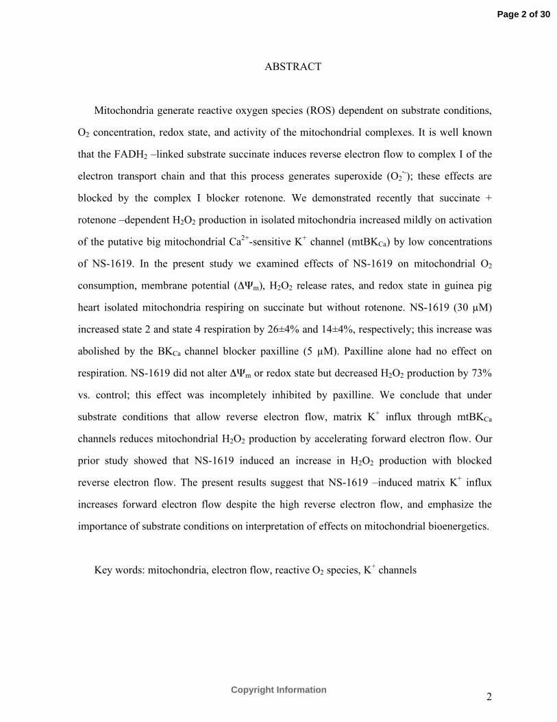
ABSTRACT
Mitochondria generate reactive oxygen species (ROS) dependent on substrate conditions,
O2 concentration, redox state, and activity of the mitochondrial complexes. It is well known
that the FADH2 –linked substrate succinate induces reverse electron flow to complex I of the
electron transport chain and that this process generates superoxide (O2·-); these effects are
blocked by the complex I blocker rotenone. We demonstrated recently that succinate +
rotenone –dependent H2O2 production in isolated mitochondria increased mildly on activation
of the putative big mitochondrial Ca2+-sensitive K+ channel (mtBKCa) by low concentrations
of NS-1619. In the present study we examined effects of NS-1619 on mitochondrial O2
consumption, membrane potential (ΔΨm), H2O2 release rates, and redox state in guinea pig
heart isolated mitochondria respiring on succinate but without rotenone. NS-1619 (30 µM)
increased state 2 and state 4 respiration by 26±4% and 14±4%, respectively; this increase was
abolished by the BKCa channel blocker paxilline (5 µM). Paxilline alone had no effect on
respiration. NS-1619 did not alter ΔΨm or redox state but decreased H2O2 production by 73%
vs. control; this effect was incompletely inhibited by paxilline. We conclude that under
substrate conditions that allow reverse electron flow, matrix K+ influx through mtBKCa
channels reduces mitochondrial H2O2 production by accelerating forward electron flow. Our
prior study showed that NS-1619 induced an increase in H2O2 production with blocked
reverse electron flow. The present results suggest that NS-1619 –induced matrix K+ influx
increases forward electron flow despite the high reverse electron flow, and emphasize the
importance of substrate conditions on interpretation of effects on mitochondrial bioenergetics.
Key words: mitochondria, electron flow, reactive O2 species, K+ channels
Page 2 of 30
Copyright Information
3
INTRODUCTION
MITOCHONDRIA are known to generate reactive oxygen species (ROS), which include
superoxide radical (O2·-), hydrogen peroxide (H2O2), and hydroxyl radical (·OH) as byproducts
of aerobic metabolism (3, 35). Excess release of ROS has been shown to play a role in the
etiology of various pathological disorders including cardiovascular disease (1), degenerative
changes in aging (5), Alzheimer’s disease (29), and diabetes (22), as well as in
ischemia/reperfusion (IR) injury (42). ROS are key elements in a variety of cellular signaling
pathways (24), including cardioprotection against IR induced by ischemic and
pharmacological preconditioning (27, 31).
O2·- can be generated at several sites along the mitochondrial electron transport chain
(ETC) including complex III (12, 36) and complex I (8, 18, 20). Complex III generates O2·-
through the oxidation of ubisemiquinone, a radical intermediate formed through the cycle in
the complex. The Qo site of the cycle is a major source of O2·- production and it is close to the
intermembrane space. In contrast, O2·- generated from complex I is released into the matrix.
One study (20) suggests that the primary site of O2·- generation in the mitochondrial ETC is
flavin mononucleotide (FMN) of complex I via reverse electron flow, not forward electron
flow via ubiquinone of complex III. Regardless of the source of O2·- is, the mechanism and
quantity of O2·- generated is dependent on the experimental substrate and energetic conditions
(18, 21, 37). When FADH2 related substrates are used and electrons enter the ETC at complex
II (succinate dehydrogenase), O2·- can be generated by reverse electron flow to complex I (21).
The resulting large increase in O2·- generation is dependent on a high inner mitochondrial
membrane (IMM) potential (ΔΨm), and is sensitive to complex I blockade by rotenone, which
prevents reverse electron flow as a source of O2·- generation (18, 21). It is believed that in the
presence of a high proton motive force electrons are passed to NAD+ until the pool is fully
reduced to NADH; once this occurs semiquinone can only lose its unpaired electron to O2
because all upstream redox centers are fully reduced (18).
Page 3 of 30
Copyright Information
4
K+ channels located in the IMM appear to play an important role in regulating
mitochondrial function (15, 25), but the mechanism remains unclear. Xu et al. (41) found
evidence for big Ca2+-sensitive K+ (mtBKCa) channels in the IMM of guinea pig ventricular
cells. Sato et al. (32) demonstrated that opening of mtBKCa channels increases flavoprotein
oxidation in ventricular myocytes placed in glucose-free Tyrode’s solution, indicating an
increase in electron transport in oxidized mitochondria. Recently, we investigated the effects
of mtBKCa channel opening and closing on function of mitochondria isolated from guinea pig
hearts (16). We reported that putative mtBKCa channel opening with low concentrations of
NS-1619 accelerated states 2 and 4 respiration (electron flow) and H2O2 generation at a stable
ΔΨm in the presence of succinate and rotenone (16). In the present study we investigated
effects of NS-1619 on respiration, ΔΨm, redox state (NADH and FAD), and H2O2 generation
using succinate alone, which can induce ROS generation via reverse electron flow. We
proposed that under these conditions H2O2 production would decrease because of a relative
increase in forward electron flow, induced by matrix K+ influx, thus countering the larger
reverse electron flow caused by succinate with subsequent O2·- formation at complex I.
MATERIALS AND METHODS
All experiments were performed in accordance with the Guide for the Care and Use of
Laboratory Animals (National Institutes of Health No. 85-23, revised 1996), and were
approved by the Institutional Animal Care and Use Committee (Medical College of
Wisconsin, Milwaukee, Wisconsin).
Mitochondrial isolation. Heart mitochondria were isolated from ketamine-anesthetized
guinea pigs (250-300 g) by differential centrifugation as described previously (30) with
moderate modifications. Briefly, ventricles were excised, placed in an isolation buffer: 200
mM mannitol, 50 mM sucrose, 5 mM KH2PO4, 5 mM 3-(n-morpholino) propranesulfonic acid
(MOPS), 1 mM EGTA, 0.1% bovine serum albumin (BSA), pH 7.15 (adjusted with KOH),
and minced into 1 mm3 pieces. The suspension was initially homogenized for 15 s in 2.5 ml
Page 4 of 30
Copyright Information
5
isolation buffer containing 5 U/ml protease, and for another 15 s after addition of 17 ml
isolation buffer. The suspension was centrifuged at 8000g for 10 min; the pellet was
resuspended in 25 ml isolation buffer and centrifuged again at 750g for 10 min. Next, the
supernatant was centrifuged at 8000g for 10 min, and the final pellet was suspended in 0.5 ml
isolation buffer and kept on ice. The protein content was determined by the Bradford method
(4). All isolation procedures were conducted at 4°C.
Mitochondrial O2 consumption. Oxygen consumption was measured polarographically at
27oC using a respirometry system (System S 200A; Strathkelvin Instruments, Glasgow,
Scotland). Mitochondria (0.25 mg protein/ml) were suspended in respiration buffer containing
130 mM KCl, 5 mM K2HPO4, 20 mM MOPS, 2.5 mM EGTA, 1 µM Na4P2O7, 0.1% BSA, pH
7.15 adjusted with KOH. Buffer [Ca2+] was less than 100 nM as assessed by the fluorescence
dye indo-1. Respiration was initiated by administration of complex II substrate succinate (10
mM). State 3 respiration was determined after addition of 250 µM ADP, and state 4
respiration was measured after complete phosphorylation of ADP to ATP. The respiratory
control index (RCI) was calculated as the ratio of mean slopes during state 3 and state 4
respiration (state 3 slope/state 4 slope).
Mitochondrial H2O2 release rate. Rates of mitochondrial H2O2 release were measured
spectrophotometrically (QM-8, Photon Technology International, PTI) at 27°C following
oxidation of amplex red (25 µM; Molecular Probes) to the highly fluorescent product
resorufin in the presence of 0.1 U/ml horseradish peroxidase. Excitation and emission
wavelengths (λex and λem) were set to 530 nm and 583 nm, respectively. Mitochondria (0.5
mg/ml) were suspended in respiration buffer. Time-controls received 0.3% dimethyl sulfoxide
(DMSO). Maximal H2O2 production was stimulated in some experiments by addition of
complex III blocker antimycin A (5 µM). Antimycin A is believed to inhibit cytochrome b
oxidation by cytochrome c1 to cause accumulation of ubisemiquinone, which is oxidized by
molecular O2 to generate O2•- and H2O2 (7). Catalase (300 U/ml) was added to confirm H2O2
production by attenuating the H2O2 signal, since catalase converts H2O2 to H2O. H2O2 release
rates were expressed as arbitrary fluorescence units (afu, Figures 4 and 5A) or as percent of
Page 5 of 30
Copyright Information

6
time control experiments (Figure 5B). Baseline H2O2 levels were calibrated from a mean of 3
standard curves of photon counts over a range of 10-200 nM H2O2 (added to assay medium in
the presence of reactants amplex red and horseradish peroxidase); each regression was linear
(R >0.99).
Mitochondrial redox state. Mitochondrial redox state was measured by the
autofluorescence that arises from compounds endogenous to the mitochondrion. The NAD+
signal is not fluorescent, but an increase in NADH fluorescence signal reflects an increase in
the ratio of NADH to NAD+, i.e, a net shift in the pyridine nucleotide pool to the reduced
state. In contrast to NADH, a decrease in the FAD signal (flavoprotein fluorescence) occurs
when the carrier binds to electrons. Thus a decrease in FAD reflects an increase in the ratio of
reduced to oxidized flavoprotein (14). NADH and FAD were measured from the same aliquot
of mitochondrial suspension (0.5 mg/ml) in respiratory buffer, with the aid of an electronic
chopper that switched between the excitations for NADH and FAD so that the time resolution
for the three NADH and FAD emission signals was 7 s. NADH was determined by exciting at
350 nm λex and recording at 460 nm λem and 405 nm λem (the latter reference wavelength is
less sensitive to NADH changes). The fluorescence ratio, F460/F405, is interpreted as a
measure of NADH. Mitochondrial FAD autofluorescence was recorded at 540 nm λem from
light filtered at 480 nm λex.
Mitochondrial membrane potential. Mitochondrial membrane potential (ΔΨm) was
monitored at 27°C in a cuvette- based spectrophotometer (QM-8, PTI) operating 503 nm λex
and 527 nm λem, respectively, in the presence of the fluorescence dye rhodamine 123 (50 nM).
Mitochondria (0.5 mg/ml) were suspended in respiration buffer. ΔΨm was expressed as the
percentage of rhodamine 123 fluorescence in the presence of fully coupled mitochondria
relative to the fluorescence after addition of 4 µM of the uncoupler carbonyl-cyanide-m-
chlorophenylhydrazenone (CCCP). To verify the functional integrity of mitochondria, 250
µM ADP was added and repolarization of ΔΨm after complete phosphorylation of ADP was
measured.
Page 6 of 30
Copyright Information
7
Chemicals and reagents. Rhodamine 123, amplex red, and indo-1 were purchased from
Molecular Probes (Eugene, OR) and high purity KCl from EMD Chemicals (Gibbstown, NJ).
All other chemicals were purchased from Sigma Chemical Co. NS-1619, paxilline, and
amplex red were dissolved in DMSO before adding to the experimental buffer.
Statistical analyses. Group data were compared by analysis of variance. If F values (P <
0.05) were significant, post hoc comparisons of means tests (Student-Newman-Keuls) were
considered statistically significant when P < 0.05 (two-tailed); * vs. control; # vs. NS-1619.
Data are presented as means ±SEM.
RESULTS
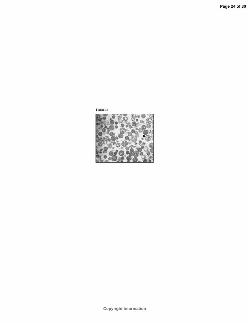
Mitochondrial integrity after isolation. The morphological integrity of mitochondria after
the isolation procedure was verified by electron microscopy. Figure 1 shows guinea pig heart
isolated mitochondria with intact inner and outer membranes suspended in isolation buffer. A
RCI of 2.5±0.1 in the control group with succinate as substrate demonstrated strong coupling
of respiration and oxidative phosphorylation. This indicates functioning mitochondria with
integrity of the respiratory complexes in the IMM after the isolation procedure.
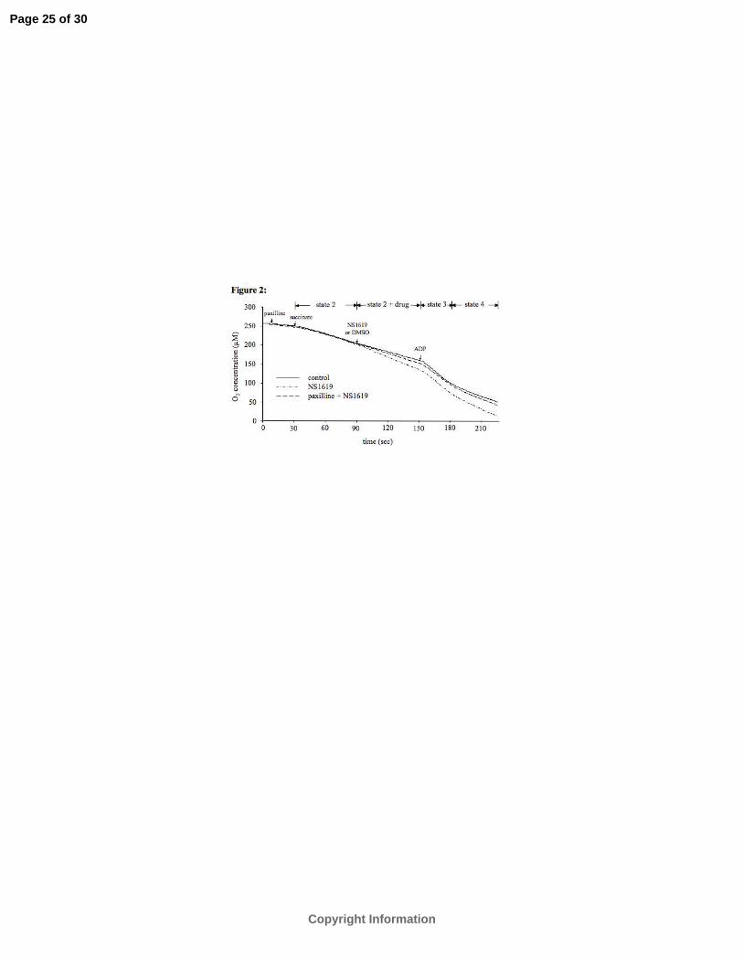
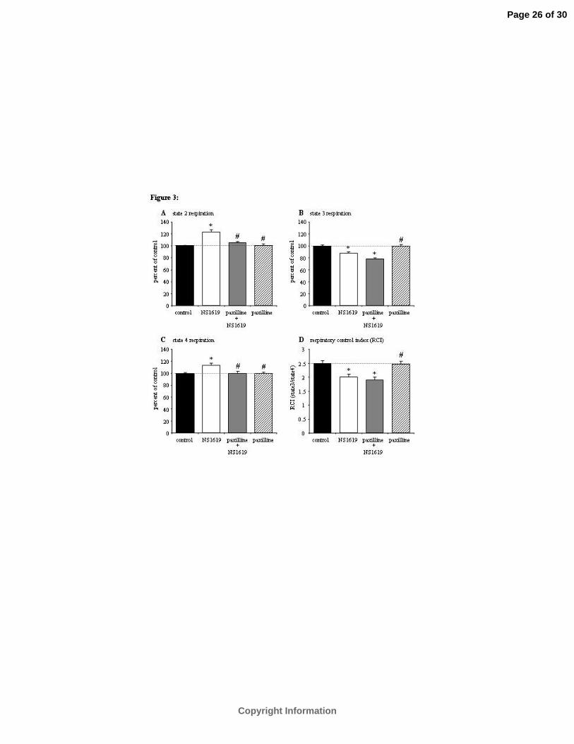
Mitochondrial respiration. The experimental procedure and sample traces for respiration
experiments are shown in figure 2. Opening of putative mtBKCa channels by 30 µM NS-1619
significantly increased state 2 respiration by 26±4% over control and state 4 respiration by
14±4% over control (Figures 3A and 3C). To verify that the increase in state 2 and state 4
respiration was due to mtBKCa channel opening by NS-1619, 5 µM paxilline was added in the
absence or presence of 30 µM NS-1619 (Figure 3). Pre-administration of paxilline
significantly blocked the NS-1619 -induced increase in state 2 (to 4±3% from 26±4%) and
state 4 respiration (to -1±3% from 14±4%). Paxilline alone had no significant effect on
respiration (0±2%), which indicates that mtBKCa channels were closed under these
experimental conditions. State 3 respiration was decreased statistically by NS-1619 (-12±3%
vs. control), but co-administration of paxilline reduced but did not significantly block this
Page 7 of 30
Copyright Information
8
effect (-21±2%) (Figure 3B), which may indicate a mtBKCa channel independent effect. These
data demonstrate that a low concentration of NS-1619 increases succinate -supported
respiration, but only during the resting states when the basal respiratory rate is low.
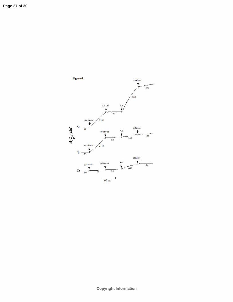
Reverse electron flow induced H2O2 generation. To verify that the major mechanism of
ROS production with the complex II substrate succinate is due to reverse electron flow, we
measured mitochondrial H2O2 release rate. In representative tracings figure 4 shows that
succinate initiated a large increase in the H2O2 release rate that was abolished by either the
mitochondrial uncoupler CCCP (panel A) or the complex I blocker rotenone (panel B). This
verified in our model that reverse electron flow into complex I of the ETC is the main
mechanism by which O2·-
is generated with succinate alone as the substrate. Moreover, the rate
of H2O2 release in succinate -supported respiration was approximately 35 times higher than
the rate attained by the complex I substrate pyruvate (panel C). Antimycin A, a complex III
blocker, increased the H2O2 release rate much more after uncoupling with CCCP than after
blocking complex I with rotenone.
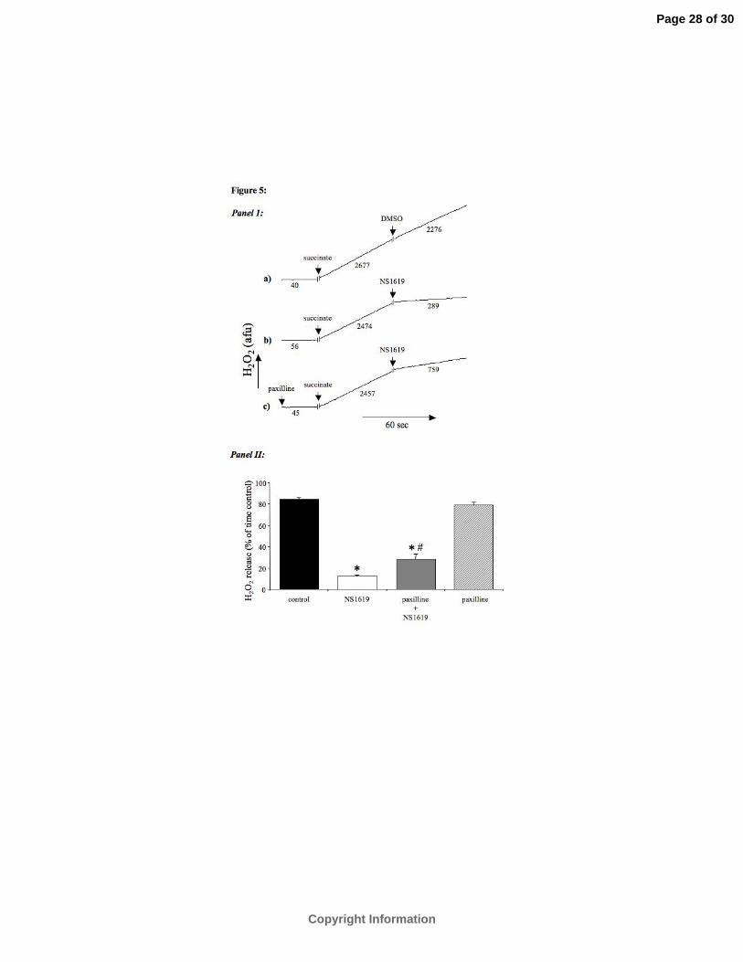
Effect of mtBKCa channel opening on H2O2 generation. To investigate the effect of
mtBKCa channel opening on reverse electron flow -induced H2O2 production we measured the
H2O2 release rate after addition of NS-1619 in succinate -supported mitochondria. Results are
expressed as percent of time controls. Figure 5 shows that addition of NS-1619 significantly
decreased the H2O2 release rate from 85±2% to 12±1%. Pre-administration of paxilline
statistically attenuated this reduction in ROS generation (28±5% vs. 12±1%). Paxilline alone
had no significant effect on mitochondrial H2O2 release rate (79±2% vs 85±2%).
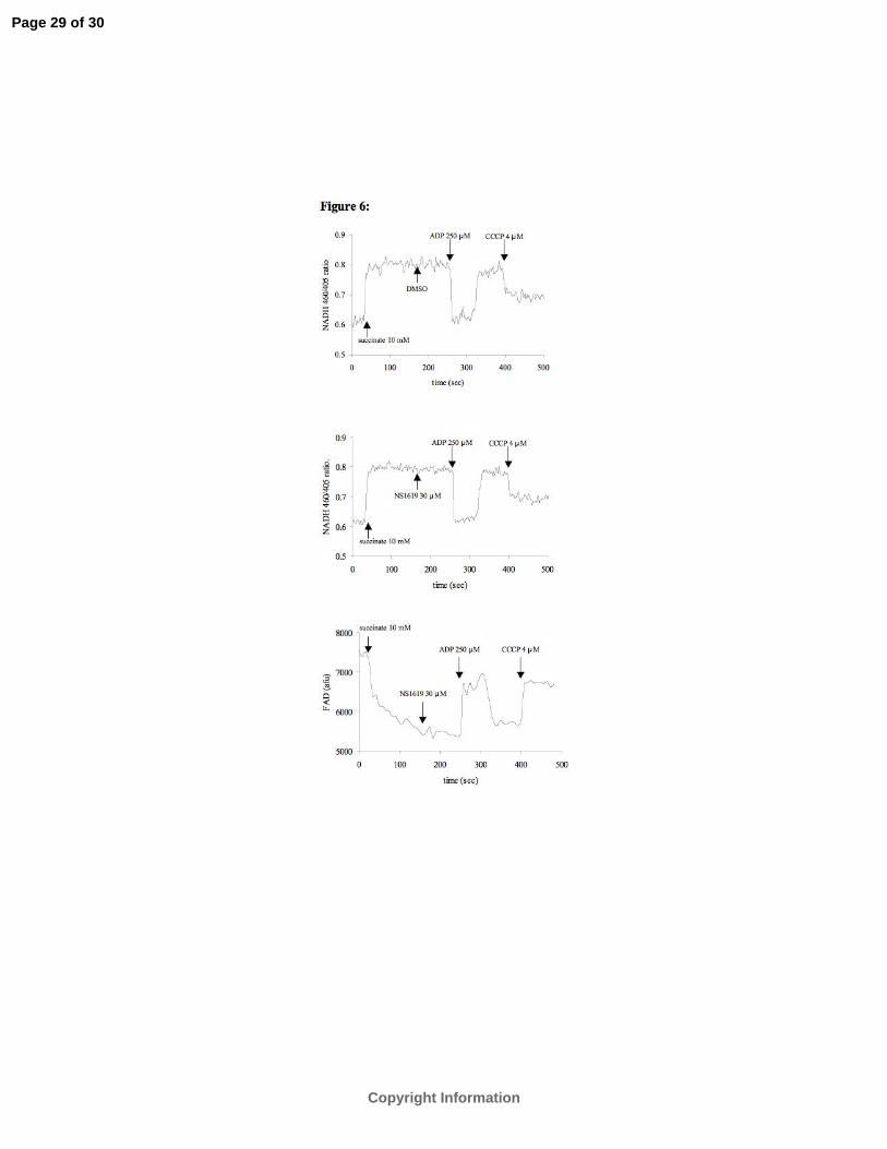
Effect of NS-1619 on mitochondrial redox state. In this in vitro preparation, oxidation of
succinate and reduction of molecular O2 involves in part, reverse electron flow, specifically of
electrons entering at complex II to react with NAD+ to produce NADH at complex I. As
shown in Figure 6, addition of succinate invariably increased NADH and decreased FAD.
NADH also increases while FAD decreases with pyruvate, a complex I substrate, but this
occurs because pyruvate directly reduces NAD+ to NADH with forward electron flow. To
confirm that NADH and FAD signals reflect changes in redox state, we noted that ADP
Page 8 of 30
Copyright Information
9
transiently decreased NADH and increased FAD (state 3) whereas CCCP, a protonophore
uncoupler, maximally oxidized mitochondria as observed by the low NADH and high FAD
states. Importantly, NS-1619, 30 µM, (and lower concentrations, not shown) did not
significantly alter either NADH (1.2 ±1.3%) or FAD (0.8 ±1.4%) compared to the vehicle
(0%) (P>0.05, n=6); however, 50 and 100 µM NS-1619 significantly and dose-dependently
decreased NADH to 18± 2%, and 98 ±3% (P<0.05, n=3) of the response to ADP (100%);
these higher concentrations, however, did not alter FAD (-2 ±3% vs control; P<0.05, n=3),
reflecting its independence of the NADH redox state.
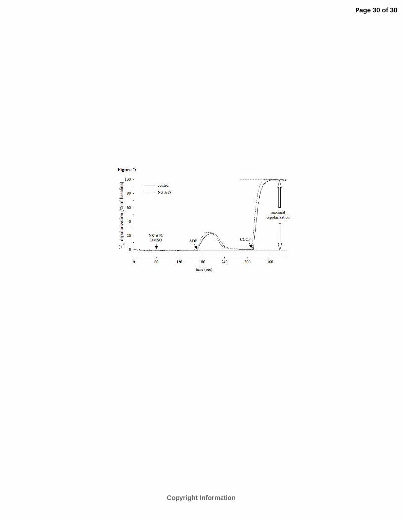
Mitochondrial membrane potential (ΔΨm). To examine if the effects of mtBKCa channel
opening on reducing ROS generation were due to depolarization of the IMM, we measured
fluorescence of the ΔΨm -sensitive dye rhodamine 123 (Figure 7). Administration of 30 µM
NS-1619 had no significant effect on ΔΨm during succinate supported respiration (0.2±0.1%
vs. 0.3±0.1%, P>0.05, n=6; summary data not displayed).
DISCUSSION
The major conclusions of this study are that during succinate -supported respiration a)
mtBKCa channel activation by 30 µM NS-1619 decreases reverse electron flow-induced H2O2
production, which is attenuated by paxilline, and b) this reduction in H2O2 production by NS-
1619 is not due to depolarization of Ψm or to a more oxidized redox state. The relative
decrease in reverse electron flow, and thereby O2·- generation, is likely due to NS-1619 -
enhanced forward electron flow.
Reverse electron flow as a mechanism for O2·- generation. The ETC is the main source of
O2·- generation during normal metabolism (19). Any reduced metal ion component of the ETC
can serve as potential source of O2·- by one-electron transfer to O2 to generate O2
·- (10). Using
specific respiratory complex blockers, O2·- has been shown to be released into the matrix or
cytosol at complex III and into the matrix at complex I (12). In turn, O2·- is dismutated by
superoxide dismutase in the matrix (SOD2) or cytosol (SOD1) to form H2O2, most of which is
Page 9 of 30
Copyright Information
10
detoxified to H2O by the glutathione system. But H2O2 is also a progenitor of the highly
reactive hydroxyl radical (·OH) in the presence of a reduced transition metal such as Fe2+.
Since H2O2 is highly permeable through the IMM, the H2O2 measured in this study likely
reflects its generation in the matrix.
Several studies have reported that O2·- generation is greater during respiration supported
with FADH2 -linked substrates than with complex I substrates (21, 37). O2·- generation with
complex II substrate succinate is caused by reverse electron flow into complex I of the ETC
and is largely dependent on a high ΔΨm (20, 21). This is supported by our observation that
uncoupling of mitochondria with CCCP, which collapses ΔΨm by allowing proton reentry
through the IMM, halted H2O2 generation. Moreover, complex I blocker rotenone and ADP
(state 2 to state 3 transition) largely attenuated reverse electron flow induced H2O2 generation
as also shown by others (20, 21, 37). In hearts utilizing NADH –linked substrates, blocking
electron flow at complex I reduces mitochondrial damage during ischemia reperfusion injury
at least in part due to reduced ROS production (11).
Possible significance of reverse electron flux. Significant O2·- generation occurs via
forward electron flow in the presence of complex I Q site inhibitors like rotenone, but much
more is generated during reverse electron flow through complex I. Lambert and Brand (18)
argue that the site of generation by complex I is likely a ubisemiquinone-binding site rather
than upstream flavin or FeS centers. Reverse electron flux may be a significant factor in
ischemia reperfusion injury. Physiologically, succinate is synthesized at low concentrations
(0.2–0.4 mM) inside mitochondria in vivo and is not a natural substrate. However, it rises
substantially during ischemia or hypoxia (up to 4–7 mM) (20). It is possible that during early
ischemia, when NADH levels are high, and during initial reperfusion, the oxidation of
accumulated succinate generates the high ΔΨm and reducing power necessary for reversal of
electron transfer and O2•- generation at Complex I. In phosphorylating mitochondria
respiration is controlled by both ATP turnover and electron supply. Adenine nucleotide
translocase (ANT) is an important site of control in oxidative phosphorylation (14) as it
catalyzes the one for one exchange of adenine nucleotides. In energized mitochondria, ANT
Page 10 of 30
Copyright Information
11
preferentially ejects more ATP than ADP brought into the matrix. This would lead to a greater
extramitochondrial ATP/ADP ratio, which could lead to activation of succinate
dehydrogenase (complex II) and stimulation of reverse electron flow.
Effects of NS-1619 and paxilline on the BKCa channel. NS-1619 (1,3-Dihydro-1-[2-
hydroxy-5-(trifluoromethyl)phenyl]-5- (trifluoromethyl)-2H- benzimidazol-2-one), a benzi-
midazole derivative, promotes opening of high conductance (300 pS) BKCa channels in
membranes of a wide variety of cell types (34). NS-1619 –induced effects on smooth muscle
can be blocked by charybdotoxin or paxilline, but not by glibenclamide, which indicates the
action of NS-1619 in plasma membranes is predominately on the BKCa channel. The rapid
effect of NS-1619 suggests that its mechanism of action is either directly on the channel
protein itself or on a closely associated modulatory protein (28). The absence of intracellular
Ca2+ prevents BKCa channel activation by NS-1619 and the drug may increase channel
activation by making the channel more sensitive to intracellular [Ca2+] (28). The BKCa
channels are tetramers of a pore-forming α subunit of the slo gene family, and a regulatory β
subunit, which is structurally unique and trans-membrane spanning; the α subunit, encoded by
a single gene, is comprised of 7 trans-membrane segments and 4 intracellular hydrophobic
domains (17, 23, 39). Several groups are attempting to more clearly identify and characterize
these channels in cardiac IMM.
Modulation of mitochondrial function by mtBKCa channels. Xu et al. (41) first reported
evidence for mtBKCa channels in the IMM of guinea pig ventricular cells. Patch-clamp
recordings from mitoplasts of these cells showed Ca2+-dependent, large K+ conductance
channels in the IMM and immunoblots of cardiac mitochondria with antibodies against the C
terminal part of BKCa channel identified a 55 kDa protein as part of this putative channel. The
β1 subunit of the mtBKCa channel, as tentatively identified in the IMM, interacts with the
cytochrome c oxidase subunit I (26). The binding sites for charybdotoxin and NS-1619 are
likely in the cytosolic compartment, whereas sites for Ca2+ are likely on the matrix side of the
IMM (40).
Page 11 of 30
Copyright Information

12
The ultimate bioenergetic modulating effects of K+ influx into the mitochondrial matrix
through K+ channels, including both mtBKCa and mtKATP channels, however, remains unclear.
Sato et al. (32) demonstrated that NS-1619 increases flavoprotein oxidation in ventricular
myocytes placed in glucose-free Tyrode’s solution; this suggested an increase in electron
transport in oxidized mitochondria. Recently, we reported on the concentration-dependent
effects of NS-1619 on respiration, ΔΨm, and H2O2 generation in isolated guinea pig heart
mitochondria respiring on complex II substrate succinate in the presence of complex I blocker
rotenone to prevent reverse electron flow (16). NS-1619 increased states 2 and 4 respiration,
effects that were inhibited by paxilline. These findings are in agreement with the hypothesis of
O’Rourke (25), who suggested that mitochondrial K+ channels function as energy (stored as
the proton gradient, ∆µH) dissipating channels by expending ∆µH, in part, to eject K+ that
enters the matrix through activated K+ channels via an electroneutral K+/H+ exchanger. This
decrease in ∆µH would stimulate respiration to compensate for net proton leak with the
consequence of a maintained ΔΨm (16). In the present report we demonstrated during
succinate -supported respiration that putative mtBKCa channel opening by 30 µM NS-1619,
and by inference lower concentrations, again increased states 2 and 4 respiration, and had no
effect on redox state or ∆Ψm.
Cancherini et al. (9) reported recently that NS-1619 stimulated non-phosphorylating
respiration (state 4) and inhibited ADP –stimulated respiration (state 3) in rat heart isolated
mitochondria. These effect of NS-1619 were also described before by Debska et al. (13) and
by our group (16) and are confirmed again in this study in guinea pig heart mitochondria.
However, in the former study (9) evidence was presented that NS-1619 does not specifically
transport K+ via a channel or cation transporter. In the presence of the complex V inhibitor
oligomycin they reported that NS-1619 depolarized Ψm in K+ containing as well as K+ free
buffer, that the respiratory effects were not blocked by paxilline, that NS-1619 –induced
matrix swelling occurred also in a TEA based buffer, and that the latter effect was not blocked
by paxilline. On the basis of these findings Cancherini et al. (9) suggested that NS-1619
promotes non-selective permeabilization of the IMM to ions rather than acting on a specific
Page 12 of 30
Copyright Information

13
IMM K+ channel. However, in this and our prior study (16), the effect of NS-1619 to enhance
state 4 respiration was inhibited by paxilline, a known BKCa channel inhibitor and uncoupling
did not occur at less than 30 µM NS-1619.
From these pharmacologic results, we conclude that the NS-1619 -induced increase in
state 4 respiration, and the state and substrate dependent effects on H2O2 production, are likely
mtBKCa channel mediated. We cannot reconcile differences between these studies, but we
agree that the specificity of NS-1619 for the putative mtBKCa channel remains speculative.
Moreover, the putative mtBKCa channel will need to be better identified and characterized in
the IMM to substantiate its role in modulating mitochondrial bioenergetics. What is evident to
us, however, is that NS-1619 clearly initiates pharmacologic preconditioning against cardiac
ischemia reperfusion injury and that this protective effect is effectively blocked by a O2·-
dismutase mimetic as well as by paxilline (33).
Effect of putative mtBKCa channel opening on reverse electron flow -induced H2O2
production. Under physiological conditions, reverse electron flow does not occur because
forward electron flow through complex I via NADH prevents it. However, under
pathophysiological conditions in which NADH is depleted, reverse electron flow may lead to
O2·- generation at complex I (6, 35, 37). In succinate-supported isolated mitochondria O2
·-
generation due to reverse electron flow to complex I is dependent on a fully charged ∆Ψm
under state 4 condition; reverse flow is blocked by the complex I blocker rotenone. Our
results suggest that reverse electron flow-induced H2O2 production can be modulated by
matrix K+ flux. The H2O2 release rate during enhanced state 4 respiration by NS-1619 can
either increase (no reverse electron flow) (16), or decrease (reverse electron flow), as shown
in this report. The changes in H2O2 production were not caused by an effect of NS-1619 on
∆Ψm or redox state, which did not change. Thus we propose that matrix K+ influx through
activated mtBKCa channels can reduce H2O2 production due to reverse electron flow by
accelerating forward electron flow. This is in agreement with a previous finding by
Votyakova et al. (38), who demonstrated that very low concentrations of the mitochondrial
Page 13 of 30
Copyright Information
14
uncoupler FCCP, which did not depolarize ∆Ψm, were sufficient to reduce reverse electron
flow- induced O2·- generation.
That NS-1619, at concentrations at or below 30 µM, did not alter the mitochondrial redox
state, is consistent with our previous finding with pyruvate or succinate + rotenone (16), in
which ∆Ψm remained high, a condition that can lead to enhanced O2·- generation and
downstream reactants. Reverse electron flow-induced O2·- generation depends on the delivery
of electrons downstream of complex I, i.e. from complex II, a high proton motive force, and a
low redox potential across complex I (i.e. no pyruvate to increase the NADH/NAD+ ratio).
Thus, a factor that limits reverse electron flow-induced H2O2 production is supply of NADH
by pyruvate at complex I, which in its absence is supported by NADH generation from NAD+
by succinate at complex I. Indeed, Liu et al. (20) showed that inhibition of complex II with
malonate completely abolished the succinate-induced reduction of NAD+. This suggested that
electrons for reducing NAD+ come directly from succinate through reverse electron transport
and not from other components of the tricarboxylic cycle (20). The specific site for O2·-
generation under physiological or pathological conditions may be the FMN group of complex
I (20) or a ubisemiquinone binding site (18).
Batandier et al. (2) proposed that with succinate as substrate, any decrease in NADH level
or ∆Ψm would abolish O2·- generation at this site. Such a scenario is partially in agreement
with our study, in which we show that NADH decreased, while FAD did not change, at
higher, but not lower, NS-1619 concentrations. It is notable that rotenone blocks succinate-
supported NADH formation via reverse electron flow, but also blocks oxidation of NADH by
directly inhibiting complex I. The end result is that with rotenone, NADH increases during
oxidation of succinate because succinate is converted to malate, which generates NADH that
feeds into complex I.
How modulation of mitochondrial function by mtBKCa channel opening might
differentially alter O2·- generation depending on substrate conditions requires further study.
Under the same O2 tension conditions, mitochondria appear to generate O2·- by different
mechanisms: a) by accelerating resting state respiration, e.g. due to mtBKCa channel activation
Page 14 of 30
Copyright Information
15
or valinomycin -induced matrix K+ influx, under forward electron flow conditions while ∆Ψm
is maintained, and b) by reverse electron flow into complex I during succinate supported
respiration with no fall in ∆Ψm. However, we could not demonstrate NS-1619 -induced H2O2
production with the substrate pyruvate (16). We propose that H2O2 production decreased with
the increase in NS-1619 -induced respiration in the presence of succinate because of a
compensatory outward flux of K+ and influx of protons via K+/H+ exchange, which accelerates
forward electron flow, thus reducing the impact of reverse flow on O2·- generation. Clearly,
the consequences of altered matrix K+ flux likely alter the flux of other cations in addition to
H+, for example Na+ and Ca2+ by Na+/H+ and Na+/Ca2+ exchange in the IMM.
In summary, we report that in fully membrane-polarized and reduced mitochondria matrix
K+ influx can either increase or decrease O2·- generation depending on substrate conditions.
This work emphasizes the impact of experimental substrate conditions when analyzing
mitochondrial bioenergetics, and may help to explain some of the conflicting results in the
literature regarding the effect of K+ channel activation and matrix K+ flux on modulating
mitochondrial function and H2O2 production. Moreover, caution must be taken on the effect of
NS-1619 to open mtBKCa channels or of paxilline to block them because although paxilline
completely reversed the increase in respiration, it only incompletely blocked the decrease in
H2O2 production. We must have a precise identification and characterization of the several
putative mitochondrial K+ channels and a better grasp of the pharmacology of the drugs used
to explore these mechanisms. Much research remains to be done to understand the
physiological (cell conditioning) and pathological (cell damage) conditions by which matrix
K+ flux modulates matrix pH, respiration and O2·- generation.
ACKNOWLEDGMENTS
The authors wish to thank the following for their assistance with these studies: Anita
Tredeau, Ming Tao Jiang, Michelle M. Henry, Anna Fekete, Janice Burke, James S. Heisner,
Richard Carlson jr., Dan Beard, and Meilin Huang. This work has been published in part in
abstract form: Heinen et al. Circulation 112:S.II, 272, 2005.
Page 15 of 30
Copyright Information
16
GRANTS
This study was supported in part by Grants KO1 HL73246-02 (AKSC) from the National
Institutes of Health (Bethesda, MD); 0355608Z (DFS) from the American Heart Association
(Dallas, TX); the Foundation for Anesthesia Education and Research (SGV); and
P30EY01931 (Janice Burke) from the National Eye Institute.
REFERENCES
1. Ballinger SW. Mitochondrial dysfunction in cardiovascular disease. Free Radic Biol Med
38: 1278-1295, 2005.
2. Batandier C, Guigas B, Detaille D, El-Mir MY, Fontaine E, Rigoulet M, and Leverve
XM. The ROS production induced by a reverse-electron flux at respiratory-chain complex
1 is hampered by metformin. J Bioenerg Biomembr 38: 33-42, 2006.
3. Boveris A, and Cadenas E. Mitochondrial production of superoxide anions and its
relationship to the antimycin insensitive respiration. FEBS Lett 54: 311-314, 1975.
4. Bradford MM. A rapid and sensitive method for the quantitation of microgram quantities
of protein utilizing the principle of protein-dye binding. Anal Biochem 72: 248-254, 1976.
5. Brand MD, Buckingham JA, Esteves TC, Green K, Lambert AJ, Miwa S, Murphy
MP, Pakay JL, Talbot DA, and Echtay KS. Mitochondrial superoxide and aging:
uncoupling-protein activity and superoxide production. Biochem Soc Symp 203-213, 2004.
6. Brookes PS, Yoon Y, Robotham JL, Anders MW, and Sheu SS. Calcium, ATP, and
ROS: a mitochondrial love-hate triangle. Am J Physiol Cell Physiol 287: C817-833, 2004.
7. Cadenas E, and Boveris A. Enhancement of hydrogen peroxide formation by
protophores and ionophores in antimycin-supplemented mitochondria. Biochem J 188: 31-
37, 1980.
Page 16 of 30
Copyright Information
17
8. Cadenas E, Boveris A, Ragan CI, and Stoppani AO. Production of superoxide radicals
and hydrogen peroxide by NADH-ubiquinone reductase and ubiquinol-cytochrome c
reductase from beef-heart mitochondria. Arch Biochem Biophys 180: 248-257, 1977.
9. Cancherini DV, Queliconi BB, and Kowaltowski AJ. Pharmacological and
physiological stimuli do not promote Ca2+-sensitive K+ channel activity in isolated heart
mitochondria. Cardiovasc Res 73: 720-728, 2007.
10. Chance B, Sies H, and Boveris A. Hydroperoxide metabolism in mammalian organs.
Physiol Rev 59: 527-605, 1979.
11. Chen Q, Camara AK, Stowe DF, Hoppel CL, and Lesnefsky EJ. Modulation of
electron transport protects cardiac mitochondria and decreases myocardial injury during
ischemia and reperfusion. Am J Physiol Cell Physiol 292: C137-147, 2007.
12. Chen Q, Vazquez EJ, Moghaddas S, Hoppel CL, and Lesnefsky EJ. Production of
reactive oxygen species by mitochondria: central role of complex III. J Biol Chem 278:
36027-36031, 2003.
13. Debska G, Kicinska A, Dobrucki J, Dworakowska B, Nurowska E, Skalska J,
Dolowy K, and Szewczyk A. Large-conductance K+ channel openers NS1619 and NS004
as inhibitors of mitochondrial function in glioma cells. Biochem Pharmacol 65: 1827-
1834, 2003.
14. Duchen MR. Mitochondria in health and disease: perspectives on a new mitochondrial
biology. Mol Aspects Med 25: 365-451, 2004.
15. Garlid KD, and Paucek P. Mitochondrial potassium transport: the K+ cycle. Biochim
Biophys Acta 1606: 23-41, 2003.
16. Heinen A, Camara AK, Aldakkak M, Rhodes SS, Riess ML, and Stowe DF.
Mitochondrial Ca2+-induced K+ influx increases respiration and enhances ROS production
while maintaining membrane potential. Am J Physiol Cell Physiol 292: C148-156, 2007.
Page 17 of 30
Copyright Information
18
17. Kaczorowski GJ, Knaus HG, Leonard RJ, McManus OB, and Garcia ML. High-
conductance calcium-activated potassium channels; structure, pharmacology, and
function. J Bioenerg Biomembr 28: 255-267, 1996.
18. Lambert A, Brand MD. Inhibitors of the quinone-binding site allow rapid superoxide
production from mitochondrial NADH:ubiquinone oxidoreductase (complex I). J Biol
Chem 279: 39414-39420, 2004.
19. Lesnefsky EJ, Moghaddas S, Tandler B, Kerner J, and Hoppel CL. Mitochondrial
dysfunction in cardiac disease: ischemia-reperfusion, aging, and heart failure. J Mol Cell
Cardiol 33: 1065-1089, 2001.
20. Liu Y, Fiskum G, and Schubert D. Generation of reactive oxygen species by the
mitochondrial electron transport chain. J Neurochem 80: 780-787, 2002.
21. Loschen G, Flohe L, and Chance B. Respiratory chain linked H2O2 production in pigeon
heart mitochondria. FEBS Lett 18: 261-264, 1971.
22. Lowell BB, and Shulman GI. Mitochondrial dysfunction and type 2 diabetes. Science
307: 384-387, 2005.
23. Munujos P, Knaus HG, Kaczorowski GJ, and Garcia ML. Cross-linking of
charybdotoxin to high-conductance calcium-activated potassium channels: identification
of the covalently modified toxin residue. Biochemistry 34: 10771-10776, 1995.
24. Nishida M, Maruyama Y, Tanaka R, Kontani K, Nagao T, and Kurose H. G alphai
and G alphao are target proteins of reactive oxygen species. Nature 408: 492-495, 2000.
25. O'Rourke B. Evidence for mitochondrial K+ channels and their role in cardioprotection.
Circ Res 94: 420-432, 2004.
26. Ohya S, Kuwata Y, Sakamoto K, Muraki K, and Imaizumi Y. Cardioprotective effects
of estradiol include the activation of large-conductance Ca2+-activated K+ channels in
cardiac mitochondria. Am J Physiol Heart Circ Physiol 289: H1635-1642, 2005.
Page 18 of 30
Copyright Information
19
27. Oldenburg O, Cohen MV, and Downey JM. Mitochondrial KATP channels in
preconditioning. J Mol Cell Cardiol 35: 569-575, 2003.
28. Olesen SP, Munch E, Moldt P, and Drejer J. Selective activation of Ca2+-dependent K+
channels by a novel benzimidazolone. Eur J Pharmacol 251: 53-59, 1994.
29. Rego AC, and Oliveira CR. Mitochondrial dysfunction and reactive oxygen species in
excitotoxicity and apoptosis: implications for the pathogenesis of neurodegenerative
diseases. Neurochem Res 28: 1563-1574, 2003.
30. Riess ML, Eells JT, Kevin LG, Camara AKS, Henry MM, Stowe DF. Attenuation of
mitochondrial respiration by sevoflurane in isolated cardiac mitochondrial is mediated in
part by reactive oxygen species. Anesthesiology 100: 498-505, 2004.
31. Riess ML, Kevin LG, McCormick J, Jiang MT, Rhodes SS, and Stowe DF. Anesthetic
preconditioning: the role of free radicals in sevoflurane-induced attenuation of
mitochondrial electron transport in Guinea pig isolated hearts. Anesth Analg 100: 46-53,
2005.
32. Sato T, Saito T, Saegusa N, and Nakaya H. Mitochondrial Ca2+-activated K+ channels in
cardiac myocytes: a mechanism of the cardioprotective effect and modulation by protein
kinase A. Circulation 111: 198-203, 2005.
33. Stowe DF, Aldakkak M, Camara AK, Riess ML, Heinen A, Varadarajan SG, and
Jiang MT. Cardiac mitochondrial preconditioning by Big Ca2+-sensitive K+ channel
opening requires superoxide radical generation. Am J Physiol Heart Circ Physiol 290:
H434-440, 2006.
34. Szewczyk A, Skalska J, Glab M, Kulawiak B, Malinska D, Koszela-Piotrowska I, and
Kunz WS. Mitochondrial potassium channels: from pharmacology to function. Biochim
Biophys Acta 1757: 715-720, 2006.
Page 19 of 30
Copyright Information
20
35. Turrens JF. Mitochondrial formation of reactive oxygen species. J Physiol 552: 335-344,
2003.
36. Turrens JF, Alexandre A, and Lehninger AL. Ubisemiquinone is the electron donor for
superoxide formation by complex III of heart mitochondria. Arch Biochem Biophys 237:
408-414, 1985.
37. Turrens JF, and Boveris A. Generation of superoxide anion by the NADH
dehydrogenase of bovine heart mitochondria. Biochem J 191: 421-427, 1980.
38. Votyakova TV, and Reynolds IJ. ∆Ψm -dependent and -independent production of
reactive oxygen species by rat brain mitochondria. J Neurochem 79: 266-277, 2001.
39. Wallner M, Meera P, Ottolia M, Kaczorowski GJ, Latorre R, Garcia ML, Stefani E,
and Toro L. Characterization of and modulation by a beta-subunit of a human maxi KCa
channel cloned from myometrium. Receptors Channels 3: 185-199, 1995.
40. Wu SN. Large-conductance Ca2+- activated K+ channels: physiological role and
pharmacology. Curr Med Chem 10: 649-661, 2003.
41. Xu W, Liu Y, Wang S, McDonald T, Van Eyk JE, Sidor A, and O'Rourke B.
Cytoprotective role of Ca2+- activated K+ channels in the cardiac inner mitochondrial
membrane. Science 298: 1029-1033, 2002.
42. Yoshida T, Maulik N, Engelman RM, Ho YS, and Das DK. Targeted disruption of the
mouse Sod I gene makes the hearts vulnerable to ischemic reperfusion injury. Circ Res 86:
264-269, 2000.
Page 20 of 30
Copyright Information
21
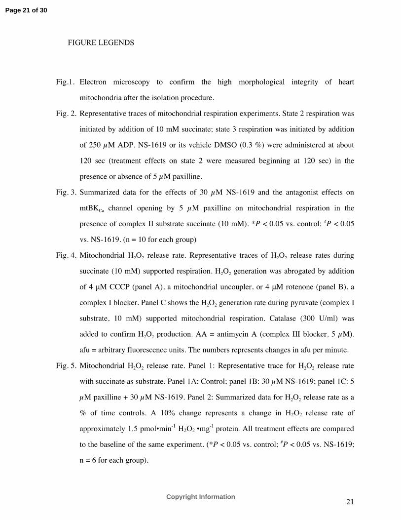
FIGURE LEGENDS
Fig.1. Electron microscopy to confirm the high morphological integrity of heart
mitochondria after the isolation procedure.
Fig. 2. Representative traces of mitochondrial respiration experiments. State 2 respiration was
initiated by addition of 10 mM succinate; state 3 respiration was initiated by addition
of 250 µM ADP. NS-1619 or its vehicle DMSO (0.3 %) were administered at about
120 sec (treatment effects on state 2 were measured beginning at 120 sec) in the
presence or absence of 5 µM paxilline.
Fig. 3. Summarized data for the effects of 30 µM NS-1619 and the antagonist effects on
mtBKCa channel opening by 5 µM paxilline on mitochondrial respiration in the
presence of complex II substrate succinate (10 mM). *P < 0.05 vs. control; #P < 0.05
vs. NS-1619. (n = 10 for each group)
Fig. 4. Mitochondrial H2O2 release rate. Representative traces of H2O2 release rates during
succinate (10 mM) supported respiration. H2O2 generation was abrogated by addition
of 4 μM CCCP (panel A), a mitochondrial uncoupler, or 4 μM rotenone (panel B), a
complex I blocker. Panel C shows the H2O2 generation rate during pyruvate (complex I
substrate, 10 mM) supported mitochondrial respiration. Catalase (300 U/ml) was
added to confirm H2O2 production. AA = antimycin A (complex III blocker, 5 µM).
afu = arbitrary fluorescence units. The numbers represents changes in afu per minute.
Fig. 5. Mitochondrial H2O2 release rate. Panel 1: Representative trace for H2O2 release rate
with succinate as substrate. Panel 1A: Control; panel 1B: 30 µM NS-1619; panel 1C: 5
µM paxilline + 30 µM NS-1619. Panel 2: Summarized data for H2O2 release rate as a
% of time controls. A 10% change represents a change in H2O2 release rate of
approximately 1.5 pmol•min-1 H2O2 •mg-1 protein. All treatment effects are compared
to the baseline of the same experiment. (*P < 0.05 vs. control; #P < 0.05 vs. NS-1619;
n = 6 for each group).
Page 21 of 30
Copyright Information
22
Fig. 6. Effects of NS-1619 on mitochondrial redox state (NADH and FAD). Representative
traces for FAD and NADH (460/405 nm λem). Concentrations up to 30 µM NS-1619
had no effect to decrease the redox state as did ADP and CCCP; higher concentrations
decreased the NADH but not the FAD redox state (see text).
Fig. 7. Effects of NS-1619 on inner mitochondrial membrane potential (ΔΨm). Representative
traces for ΔΨm with succinate as substrate. NS-1619 (30 µM) or its vehicle DMSO
(0.3%) was added. To verify a highly charged ΔΨm and the functional integrity of
mitochondria, 250 µM ADP was added as indicated. Maximal depolarization was
measured after addition of 4 µM CCCP to uncouple oxidative phosphorylation.
Page 22 of 30
Copyright Information

23
Page 23 of 30
Copyright Information
Page 24 of 30
Copyright Information
Page 25 of 30
Copyright Information
Page 26 of 30
Copyright Information
Page 27 of 30
Copyright Information
Page 28 of 30
Copyright Information
Page 29 of 30
Copyright Information
Page 30 of 30
Copyright Information
Copyright © 2022 FDOKUMEN