Genetic diversity of ORF3 and spike genes of porcine epidemic diarrhea virus in Thailand
Reproduction of Mucohaemorrhagic Diarrhea and ColitisIndistinguishable from Swine Dysentery followingExperimental Inoculation with ‘‘Brachyspira hampsonii’’Strain 30446Joseph E. Rubin1,2, Matheus O. Costa1, Janet E. Hill1, Heather E. Kittrell2, Champika Fernando1,
Yanyun Huang2, Brendan O’Connor3, John C. S. Harding2*
1 Department of Veterinary Microbiology, Western College of Veterinary Medicine, University of Saskatchewan, Saskatoon, Saskatchewan, Canada, 2 Department of Large
Animal Clinical Sciences, Western College of Veterinary Medicine, University of Saskatchewan, Saskatoon, Saskatchewan, Canada, 3 Prairie Diagnostic Services Inc.,
Saskatoon, Saskatchewan, Canada
Abstract
Background: Mucohaemorrhagic diarrhea caused by Brachyspira hyodysenteriae, swine dysentery, is a severe productionlimiting disease of swine. Recently, pigs in western Canada with clinical signs indistinguishable from swine dysentery wereobserved. Despite the presence of spirochetes on fecal smears, recognized Brachyspira spp. including B. hyodysenteriaecould not be identified. A phylogenetically distinct Brachyspira, called ‘‘B. hampsonii’’ strain 30446, however was isolated.The purpose of this study was to experimentally reproduce mucohaemorrhagic colitis and characterize strain 30446shedding following inoculation.
Methods and Findings: Eighteen 13-week-old pigs were randomly assigned to inoculation (n = 12) or control (n = 6) groupsin each of two trials. In trial 1, pigs were inoculated with a tissue homogenate collected from clinically affected field cases. Intrial 2, pigs were inoculated with a pure broth culture of strain 30446. In both trials, mucohaemorrhagic diarrhea wassignificantly more common in inoculated pigs than controls, all of which remained healthy. In animals withmucohaemorrhagic diarrhea, significantly more spirochetes were observed on Gram stained fecal smears, and highernumbers of strain 30446 genome equivalents were detected by quantitative PCR (qPCR). Strain 30446 was cultured fromcolon and/or feces of all affected but no control animals at necropsy.
Conclusions: ‘‘Brachyspira hampsonii’’ strain 30446 causes mucohaemorrhagic diarrhea in pigs following a 4–9 dayincubation period. Fecal shedding was detectable by day 4 post inoculation, and rarely preceded the onset of mucoid orhaemorrhagic diarrhea by more than 2 days. Culture and 30446-specific qPCR are reliable methods of detection of thisorganism in feces and tissues of diarrheic pigs. The emergence of a novel Brachyspira spp., such as ‘‘B. hampsonii’’, createsdiagnostic challenges including higher risk of false negative diagnostic tests. We therefore recommend diagnosticlaboratories routinely use Brachyspira culture, nox-based and species-specific PCR, and DNA sequencing to diagnoseBrachyspira-associated colitis in pigs.
Citation: Rubin JE, Costa MO, Hill JE, Kittrell HE, Fernando C, et al. (2013) Reproduction of Mucohaemorrhagic Diarrhea and Colitis Indistinguishable from SwineDysentery following Experimental Inoculation with ‘‘Brachyspira hampsonii’’ Strain 30446. PLoS ONE 8(2): e57146. doi:10.1371/journal.pone.0057146
Editor: Bernhard Kaltenboeck, Auburn University, United States of America
Received September 16, 2012; Accepted January 17, 2013; Published February 27, 2013
Copyright: � 2013 Rubin et al. This is an open-access article distributed under the terms of the Creative Commons Attribution License, which permitsunrestricted use, distribution, and reproduction in any medium, provided the original author and source are credited.
Funding: This research was supported by grants from the Canadian Swine Health Board (www.swinehealth.ca) and by internal funding from the University ofSaskatchewan Industry Liaison Office, Western College of Veterinary Medicine Research Trust (www.usask.ca). Salary support for HK was provided by the Merck-Merial Veterinary Scholars Summer Research Program. The funders had no role in study design, data collection and analysis, decision to publish, or preparation ofthe manuscript.
Competing Interests: The University of Saskatchewan has an intellectual property application pertaining to Brachyspira sp. sask30446, titled: ‘‘DiagnosticMethod for Colitis’’ (PCT Patent 2012/006730), submitted in July 2011 and now published and available on line at: (http://patent.ipexl.com/WO/2012ZZSLASHZZ006730.html). JER, JCH and JEH are named co-inventors. BOC is an employee of Prairie Diagnostic Services Inc. Salary support for HK wasprovided by the Merck-Merial Veterinary Scholars Summer Research Program. Except for one additional unpublished PCT application, there are no further patents,products in development or marketed products to declare. This does not alter the authors’ adherence to all the PLOS ONE policies on sharing data and materials,as detailed online in the guide for authors.
* E-mail: [email protected]
Introduction
Swine dysentery is a mucohaemorrhagic colitis causing severe
production losses in pigs, resulting from infection with the
intestinal spirochaete Brachyspira hyodysenteriae. The first clinical
description of swine dysentery was published in 1921, although it
was not until 1971 that B. hyodysenteriae (then Treponema hyodysenter-
iae) was recognized as the cause [1]. Spirochetal colitis is a less
severe illness caused by B. pilosicoli, which is characterized by
diarrhea (non-haemorrhagic with a wet cement consistency) and
poor feed conversion in chronic cases [2]. Other organisms within
the genus Brachyspira are varyingly associated with disease,
PLOS ONE | www.plosone.org 1 February 2013 | Volume 8 | Issue 2 | e57146

including B. murdochii, B. intermedia, B. innocens and the provisionally
named ‘B. suanatina’, although considerable strain level differences
in pathogenicity (particularly among B. intermedia) are apparent [3–
5]. In October 2009, grow-finish pigs with clinical signs
indistinguishable from swine dysentery were observed in a
commercial barn in Saskatchewan, Canada [6,7]. Tissues,
carcasses and rectal swabs collected from a number of affected
pigs over several months were submitted to Prairie Diagnostic
Services Inc. (PDS) at the University of Saskatchewan in
Saskatoon, Canada. Fibrinous mucohaemorrhagic colitis and
typhlitis with superficial necrosis was observed grossly. Histolog-
ically, sub-acute to chronic muco-purulent to fibrino-suppurative
colitis with superficial necrosis was observed. No recognized
pathogens could be identified. All samples were negative for
Lawsonia intracellularis and Salmonella spp., and despite large
numbers of spirochetes seen on Gram strained fecal smears, B.
hyodysenteriae and B. pilosicoli were not detected. The apparent
spirochetosis prompted further testing of samples by PCR using
genus-specific primers targeting the Brachyspira NADH oxidase
(nox) gene [8]. The sequence of this 939 bp PCR amplicon was
identical to clade 2 of the recently described, provisionally named
‘‘Brachyspira hampsonii’’ [9]. The particular strain identified in
western Canada and used in these trials is named 30446. Although
strain 30446 is also phenotypically indistinguishable from ‘‘B.
hampsonii’’ clade 2 [9], there is distinct variability within ‘‘B.
hampsonii’’ (clades 1 and 2), and the pathogenicity of strain 30446
may not be reflective of all ‘‘B. hampsonii’’ isolates. As the species
has not been formally recognized this study will refer precisely to
‘‘B. hampsonii’’ strain 30446.
The purpose of this study was to investigate the pathogenicity of
‘‘Brachyspira hampsonii’’ strain 30446 in experimentally infected pigs.
The results of two infection trials in grower pigs, involving
inoculation with either tissue homogenate (trial 1) or pure culture
(trial 2) are presented.
Materials and Methods
Ethics StatementBoth trials were designed and conducted in accordance with the
Canadian Council for Animal Care and approved by the
University of Saskatchewan Committee on Animal Care and
Supply (Protocol #20110038).
Trial 1. Tissue Homogenate InoculationSource of strain 30446. ‘‘Brachyspira hampsonii’’ strain 30446
infected material was obtained from clinically affected 13-week-old
pigs from a porcine reproductive and respiratory syndrome
(PRRS) negative farm. Following necropsy, the colonic and caecal
mucosa were removed from the underlying sub-mucosa and
muscularis by scraping with the edge of a glass microscope slide,
and then frozen at 280uC within four hours of collection. To
confirm the absence of pathogens other than strain 30446, sections
of small and large intestine were processed routinely for
histopathology, bacterial culture, and PCR.Brachyspira culture. Brachyspira was cultured by streaking
out approximately 10 mg of feces or intestinal contents onto BJ and
CVS agar plates [10,11]. Plates were incubated anaerobically
using a commercial system (Anaerogen, Oxoid Limited, Basing-
stoke, United Kingdom) at 42uC for 48 hours. Bacterial colonies
were not formed, instead, positive cultures were indicated by zones
Table 1. Primer sequences used to detect and identify Brachyspira spp.
Target Gene Application Primer Name Primer Sequence (59-39) Reference
Nox Brachyspira genus specific NOX F TGG CAT ACT ATC TCA TCA [8]
NOX R GAT GGA AGC TAT ATG TAT CTT A
Adh MLST scheme ADH-F206 GAA GTT TAG TAA AAG ACT TTA AAC C [12]
ADH-R757 CTG CTT CAG CAA AAG TTT CAA C
Alp MLST scheme ALP-F354 TCC AGA TGA GGC TAT ACT TC [12]
ALP-R1262 TAT GCT CTT TTT GCT AAT ATT G
Est MLST scheme EST-F229 GAT GCT TCA GGC GGA GTT ATG [12]
EST-R847 CCA CAC TCA TAG CAT AAA TAC TG
Gdh MLST scheme GDH-F514 GGA GTT GGT GCT AGA GAG AT [12]
GDH-R1157 ATC TCT AAA GCA GAA GTA GCA
Glpk MLST scheme GLPK-F123 AGG CTG GGT AGA ACA TAA TGC [12]
GLPK-R1158 TCT TTA CTT TGA TAA GCA ATA GC
Pgm MLST scheme PGM-F172 GTT GGT ACT AAC AGA ATG AAT A [12]
PGM-R1220 CCG TCT TTA TCG CGT ACA TT
Thi MLST scheme THI-F163 TGT GTT ATA CAA TCA GCA CTT C [12]
THI-R1079 GTA GTA AGT ATT CTA GCT CCA G
Nox Brachyspira sp. 30446 SYBR assay JH224 TCG CTA AAT TAT TCC AAC AAG GA This study
JH225 AAA CGC ATT TCT ATT CCA GCA
cpn60 Brachyspira hyodysenteriae SYBR assay JH0073 AGT GAA ATA GTT GCT CAT ATC AAA T This study
JH0074 GCA TCA CTG ATT AAA GAA CCA AT
cpn60 Brachyspira pilosicoli SYBR assay JH0077 ACA ATG ATA AAG AGA TAG GTG CTT This study
JH0078 CTA ATG AAA GGC TAG TTT CTA ATG AT
doi:10.1371/journal.pone.0057146.t001
Mucohaemorrhagic Colitis ‘‘B. hampsonii’’ 30446
PLOS ONE | www.plosone.org 2 February 2013 | Volume 8 | Issue 2 | e57146

of strong b-haemolysis from which motile spirochetes could be
seen microscopically.
DNA extraction and PCR. DNA was extracted from samples
using either the QIAmp DNA stool mini kit (feces or colon
contents) or DNEasy blood and tissue kit (cultured bacteria or
terminal colon tissue) (Qiagen Inc., Toronto, Ontario) and 2 ml of
extract used as template in PCR reactions. DNA was extracted in
triplicate in terminal colon tissues and colonic contents. To
differentiate strain 30446 from other Brachyspira spp., partial 16S
rRNA, cpn60, nox, adh, alp, est, gdh, glpK, pgm and thi were amplified
and sequenced using previously published primers (Table 1).
To specifically detect ‘‘B. hampsonii’’ strain 30446, B. hyodysenteriae
and B. pilosicoli, SYBR green qPCR assays were developed
targeting either nox (strain 30446), or cpn60 (B. hyodysenteriae and
B. pilosicoli) (Table 1). Product sizes were 215 bp for strain 30446,
120 bp for B. hyodysenteriae and 111 bp for B. pilosicoli. Quantitative
Figure 1. Phylogenetic tree of Brachyspira spp. Phylogenetic tree based on alignment of 810 bp of the nox gene of Brachyspira spp., including‘‘B. hampsonii’’ strain 30446. The alignment was created using CLUSTALw, followed by distance calculation (F84 matrix) and neighbour joining usingPHYLIP. The tree is a consensus of 100 boostrap iterations, and bootstrap values are indicated at the major nodes. GenBank accession numbers fornox sequences are indicated in the tree. Scale bar indicates 0.02 substitutions per site.doi:10.1371/journal.pone.0057146.g001
Mucohaemorrhagic Colitis ‘‘B. hampsonii’’ 30446
PLOS ONE | www.plosone.org 3 February 2013 | Volume 8 | Issue 2 | e57146

Mucohaemorrhagic Colitis ‘‘B. hampsonii’’ 30446
PLOS ONE | www.plosone.org 4 February 2013 | Volume 8 | Issue 2 | e57146

PCR reactions were conducted on a Bio-Rad MyiQ thermocycler
with iQ SYBR green supermix (Bio-Rad Laboratories (Canada)
Ltd., Mississauga, Ontario) according to the manufacturer’s
instructions. Quantification was accomplished by use of a serially
diluted standard curve of plasmids containing target sequences. All
reactions were run in duplicate and each run included both
extraction negatives and no template controls. For samples that
resulted in a Ct value higher than the lowest standard, but with
dissociation curves consistent with the expected product, a result of
detected but not quantifiable (DNQ) was reported. The detection
limits of the assays were defined by the linear portion of the
standard curve for each assay, which were 101–107 copies per
reaction (103–109 copies per gram of feces or tissue) for B.
hyodysenteriae and B. pilosicoli, and 102–107 copies per reaction (104–
109 copies per gram of feces or tissue) for ‘‘B. hampsonii’’.
Pigs. Eighteen five-week old, Landrace male weanling piglets
were purchased from a PRRS negative high health commercial
farm in Saskatchewan, Canada, with no history of swine dysentery
or previous laboratory diagnosis of Brachyspira spp. The pigs were
conveniently selected, of average body weight compared to their
cohorts and all appeared healthy. Farm selection was based on the
screening of four and seven week old pigs from three farms for B.
hyodysenteriae, B. pilosicoli and strain 30446 prior to trial 1. Animals
were randomly assigned to control (CTRL, n = 6) and inoculated
(INOC, n = 12) groups on arrival, and held for a 10-day
acclimation period prior to inoculation. They were fed a
commercially prepared, non-medicated, pelleted starter diet ad
libitum, and housed in separate rooms in 49669 pens each
containing 3 pigs. During the acclimation period all pigs were
tested for strain 30446, B. hyodysenteriae and B. pilosicoli in feces by
qPCR ten, seven and five days and immediately prior to first
inoculation. Fecal DNA extracts for pre-trial screening were tested
in duplicate from DNA extraction through PCR (independent
technical replicates were done from DNA extraction through
PCR).
Preparation of inoculum. The inoculum was prepared by
mixing approximately one part mucosal scraping and three parts
0.1 M pH 7.0 phosphate buffered saline (PBS) in a sterile blender.
Strain 30446 was identified in the inocula and differentiated from
other Brachyspira species by a novel nox sequence (Figure 1) and
using a previously published MLST protocol for Brachyspira spp.
[12]. Of the seven published MLST primer pairs, amplicons were
not generated from three. Unique sequences were generated from
pgm (Genbank accession JX469445, 93% sequence identity to B.
Figure 2. Fecal consistency, shedding and tissue concentrations in pigs following inoculation with tissue homogenate containing‘‘Brachyspira hampsonii’’ strain 30446. Daily fecal consistency scores (line, left ordinate; 0 = formed, normal; 1 = soft, wet cement consistency;2 = runny or watery; 3 = mucoid diarrhea; or 4 = bloody diarrhea). Fecal smear spirochete scores (grey bars, left ordinate: 0 = negative; 1 = less than 1spirochete/high power field (hpf); 2 = between 2 and 10 spirochetes/hpf; 3 = between 11 and 49 spirochetes/hpf; 4 = greater than 50 spirochetes/hpf).Strain 30446 DNA concentration (copies/g, triangles, right ordinate) in colon tissue samples collected at necropsy. Pig IDs are indicated in the upperright corner of each panel. ND = fecal smear spirochete score not done.doi:10.1371/journal.pone.0057146.g002
Table 2. Detection of ‘‘B. hampsonii’’ strain 30446 in pre-inoculation screening of fecal samples during the acclimation period1.
Trial 1 Trial 2
Pig ID Day -10 Day -7 Day -5 Pig ID Day -8 Day -5 Day -2 Day 0
Inoculated Inoculated
51 – – – 683 – – – –
53 – – – 684 – – – 6.806104
54 – – – 686 – – – –
55 – – – 688 DNQ – – –
57 – – – 689 DNQ – – –
59 DNQ – – 690 – – DNQ DNQ
61 – – – 693 – – – –
63 – – – 694 – – DNQ –
64 – – DNQ 695 – – – DNQ
65 – – – 696 DNQ – DNQ DNQ
67 – – DNQ 697 DNQ – – –
68 – – – 700 DNQ – – DNQ
Control Control
52 – – – 685 – – – DNQ
56 – – – 687 DNQ – – –
58 – – – 691 DNQ – – DNQ
60 – – – 692 – – – –
62 – – – 698 – – – –
66 – – – 699 – – – DNQ
1Quantitative PCR results for all pigs at ten, seven and five days prior to inoculation in trial 1, and eight, five and two days prior to inoculation, and at inoculation in trial2. Hyphens indicate that strain 30446 was not detected, DNQ indicates detectable but not quantifiable concentrations, and a number indicates the concentration ofstrain 30446 in genome equivalents/g of feces.doi:10.1371/journal.pone.0057146.t002
Mucohaemorrhagic Colitis ‘‘B. hampsonii’’ 30446
PLOS ONE | www.plosone.org 5 February 2013 | Volume 8 | Issue 2 | e57146
murdochii), thi (JX469446, 92% sequence identity to B. hyodysenteriae),
glpk (JX469444, 95% sequence identity to B. murdochii) and est
(JX469443, 93% sequence identity to B. murdochii). The concen-
tration of ‘‘B. hampsonii’’ strain 30446 in the inoculum was
determined by qPCR and three daily inoculum doses of 3.426108,
1.806108 and 6.376107 genome equivalents were given. These
doses were intermediate to recent trials with B. murdochii where 106
colony forming units were used and a ‘B. suanatina’ sp. nov. trial
where 30 ml of a 108 to109 cells/mL inoculum was used [5,13].
Inoculation of pigs. Pigs were inoculated on three consec-
utive days (D0, D1 and D2) as previously described [13,14]. To
decrease gastric transit time, feed was removed 16 hours prior to,
and returned one hour after inoculation. Prior to inoculation, pigs
were sedated with 8 mg/kg IM azaperone (Stresnil, Vetoquinol
Canada Inc., Lavaltrie, Quebec). Pigs in the INOC group were
intra-gastrically inoculated using an 18 French feeding tube,
followed by 50 mL of sterile PBS (0.1 M, pH 7.0). CTRL pigs
were mock inoculated with 50 mL of PBS.
Observation of pigs and daily sample collection. The
pigs were observed and scored twice daily for responsiveness, skin
colour, appetite and body condition, respiratory effort and fecal
consistency. Fecal consistency was scored daily as: 0 = formed,
normal; 1 = soft, wet cement consistency; 2 = runny or watery;
3 = mucoid diarrhea; or 4 = bloody diarrhea. Gram stained fecal
smears were made from rectal swabs collected daily from each pig.
An investigator blinded to the slide ID, evaluated and scored all
slides as: 0 = negative; 1 = less than 1 spirochete/high power field
(hpf); 2 = between 2 and 10 spirochetes/hpf; 3 = between 11 and
49 spirochetes/hpf; 4 = greater than 50 spirochetes/hpf. Fecal
samples collected from each pig on days 3, 7, 10 and 14 post
inoculation were also tested by qPCR for ‘‘B. hampsonii’’ strain
30446. For statistical analysis, the qPCR results were categorized:
0 = not detected; 1 = DNQ; and 2 = quantifiable.
Necropsy. After the intensity of mucohaemorrhagic diarrhea
peaked (INOC), or at the end of the study (CTRL) on day 16, pigs
were euthanized by cranial captive bolt and exsanguinated. A
complete necropsy was performed with special attention to the
stomach, duodenum, jejunum, ileum, spiral colon, caecum and
rectum. Samples for histological examination, Salmonella culture on
brilliant green agar following enrichment in selenite broth (pooled
colon and caecum), Lawsonia intracellularis PCR (ileum) [15],
porcine circovirus 2 (PCV2) immunohistochemistry (ileum,
mesenteric lymph node) [16] and porcine reproductive and
respiratory syndrome virus PCR (serum; Tetracore Inc., Rockville,
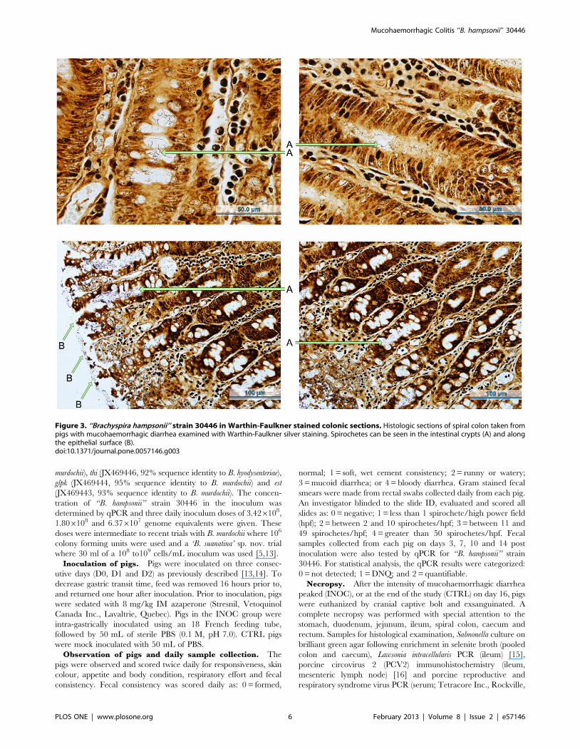
Figure 3. ‘‘Brachyspira hampsonii’’ strain 30446 in Warthin-Faulkner stained colonic sections. Histologic sections of spiral colon taken frompigs with mucohaemorrhagic diarrhea examined with Warthin-Faulkner silver staining. Spirochetes can be seen in the intestinal crypts (A) and alongthe epithelial surface (B).doi:10.1371/journal.pone.0057146.g003
Mucohaemorrhagic Colitis ‘‘B. hampsonii’’ 30446
PLOS ONE | www.plosone.org 6 February 2013 | Volume 8 | Issue 2 | e57146

MD) were submitted to PDS. Colonic tissue and contents were
tested by qPCR for B. hyodysenteriae, B. pilosicoli and ‘‘B. hampsonii’’
strain 30446 in triplicate as done in pre-trial screening. To detect
viable strain 30446, colonic tissue was cultured for Brachyspira and
if isolated, was speciated by sequencing nox PCR amplicons.
Histology. The pathologist (YH) responsible for analysis of
the samples was blinded to the identity of the slides. The presence
or absence of superficial necrosis and inflammation were scored in
Haematoxylin-Eosin stained sections of colon, caecum and
rectum. Scoring for inflammation was based on the severity of
neutrophil infiltration in the mucosa and fibrinous exudate on the
surface. Necrosis of the mucosa was assessed by visualization of
apoptotic cells and degenerated nuclei. Additionally, Warthin-
Faulkner stained colon sections were examined for the presence of
Brachyspira-like organisms associated with the lesions.
Statistics. Statistical analysis was performed using SPSS
version 18.0 (SPSS Inc., Chicago, IL). The presence or absence
of mucohaemorrhagic diarrhea in INOC versus CTRL groups
was compared using the Fisher’s exact test. The presence or
absence of histologic and gross lesions in INOC pigs with or
without mucohaemorrhagic diarrhea, and CTRL pigs was
compared using the Fisher’s exact test. Spirochete slide score
from colonic swabs and strain 30446 DNA concentration
(0 = negative, 1 = DNQ, 2 = quantifiable) in INOC pigs with and
without mucohaemorrhagic diarrhea, and in CTRL pigs were
compared using the Kruskal-Wallis test followed by post-hoc
Mann-Whitney test if significant. Two-tailed P-values #0.05 were
considered significant.
Trial 2. Pure Broth Culture InoculationThe methodology for trial 2 was similar to that of trial 1; only
major differences will be noted below. Pigs were sourced from the
same farm following pre-screening of three and six week old pigs
for B. hyodysenteriae, B. pilosicoli and ‘‘B. hampsonii’’ strain 30446.
Randomly assigned CTRL (n = 6) and INOC (n = 12) pigs were
held for an 8 day acclimation prior to inoculation. Groups were
housed in separate rooms containing 2 pigs (INOC) or 3 pigs
(CTRL) per pen. During the acclimation period, feces were
collected eight, five and two days, and immediately prior to first
inoculation and tested for Brachyspira spp. by PCR and culture as
described above.
‘‘B. hampsonii’’ strain 30446 was cultivated in vitro on JBS broth
from one of the pig colons used in trial 1 (isolate 6953).
Approximately 2 cm2 of solid media with haemolytic zones were
used to inoculate JBS broth (brain heart infusion with 5% (v/v)
fetal calf serum, 5% (v/v) sheep’s blood, and 1% (w/v) glucose).
Broth cultures were incubated in glass vials with magnetic stir bars
anaerobically at 39uC for 24 hours with constant stirring.
‘‘B. hampsonii’’ strain 30446 was administered by intra-gastric
tube for three consecutive days at doses of 2.786106, 5.046108
and 4.506108 genome equivalents, followed by 50 ml PBS. CTRL
pigs were mock inoculated with an equivalent volume of sterile
JBS broth followed by 50 ml PBS. One INOC pig (#683) did not
receive a complete dose of inoculum on D1, and the CTRL group
inadvertently did not have feed removed prior to the second
inoculation. Daily observations and fecal consistency scoring was
performed as described above. The Gram stained smears made
from rectal swabs were scored: 0 = less than 1 spirochete/hpf;
1 = between 2 and 10 spirochetes/hpf; 2 = between 11 and 49
spirochetes/hpf; 3 = greater than 50 spirochetes/hpf. Daily rectal
swabs were cultured for Brachyspira and results were scored:
0 = negative; 1 = less than 10 colonies/1u streak; 2 = less than 10
colonies/2u streak; 3 = less than 10 colonies/3u streak; or 4 = less
than 10 colonies/4u streak. Fecal samples collected daily from
Ta
ble
3.
Co
mp
aris
on
of
his
tolo
gic
alan
dg
ross
lesi
on
san
dsp
iro
che
ten
um
be
rso
nW
arth
in-F
aulk
ne
rst
ain
ed
colo
nic
sect
ion
s1.
Ex
pe
rim
en
tH
isto
log
icL
esi
on
sG
ross
Le
sio
ns
Sp
iro
che
tes
on
Sil
ve
rS
tain
Co
lon
Ca
ecu
mR
ect
um
Co
lon
Ca
ecu
mA
bse
nt-
Ra
reA
bu
nd
an
t
Mu
coid
an
d/o
rh
ae
mo
rrh
ag
icco
liti
sE
de
ma
an
dco
ng
est
ion
Mu
co-f
ibri
no
us
typ
hli
tis
Tri
al
1T
issu
eH
om
og
en
ate
Co
ntr
ol
0/6
{0
/6{
0/6
{0
/6{
0/6
{0
/6{
2/6
0/6
Ino
cula
ted
9/1
25
/12
6/1
27
/12
9/1
27
/12
3/1
29
/12
Dia
rrh
eic
9/9
*{5
/9{
5/9
{7
/9*{
9/9
*{7
/9*{
0/9
9/9
No
dia
rrh
ea
0/3
*0
/31
/30
/3*
0/3
*0
/3*
3/3
0/3
Tri
al
2P
ure
Cu
ltu
reC
on
tro
l0
/6{
0/6
{0
/6{
0/6
{0
/6{
0/6
{6
/60
/6
Ino
cula
ted
7/1
26
/12
8/1
27
/12
5/1
25
/12
4/1
28
/12
Dia
rrh
eic
7/8
#{
6/8
{8
/8#{
7/8
#{
5/8
{5
/8{
0/8
8/8
No
dia
rrh
ea
0/4
#0
/40
/4#
0/4
#0
/40
/44
/40
/4
1St
atis
tica
lco
mp
aris
on
sw
ere
amo
ng
CT
RL
(n=
6),
INO
Cw
ith
mu
coh
aem
orr
hag
icd
iarr
he
a(n
=9
)an
dIN
OC
wit
ho
ut
mu
coh
aem
orr
hag
icd
iarr
he
a(n
=3
)in
tria
l1
.In
tria
l2
,co
mp
aris
on
sw
ere
amo
ng
CT
RL
(n=
6),
INO
Cw
ith
mu
coh
aem
orr
hag
icd
iarr
he
a(n
=8
)an
dIN
OC
wit
ho
ut
mu
coh
aem
orr
hag
icd
iarr
he
a(n
=4
)in
tria
l2
.{{
#*W
ith
inco
lum
n,
anim
als
wit
hsi
mila
rsu
pe
rscr
ipts
are
stat
isti
cally
dif
fere
nt
(P#
0.0
5).
do
i:10
.13
71
/jo
urn
al.p
on
e.0
05
71
46
.t0
03
Mucohaemorrhagic Colitis ‘‘B. hampsonii’’ 30446
PLOS ONE | www.plosone.org 7 February 2013 | Volume 8 | Issue 2 | e57146
Mucohaemorrhagic Colitis ‘‘B. hampsonii’’ 30446
PLOS ONE | www.plosone.org 8 February 2013 | Volume 8 | Issue 2 | e57146

each pig were tested by qPCR for ‘‘B. hampsonii’’ strain 30446. For
statistical analysis, the qPCR results were categorized: 0 = not
detected; 1 = DNQ; and 2 = quantifiable.
Necropsy examinations were performed after the intensity of
mucohaemorrhagic diarrhea peaked (INOC), on day 14 for non-
diarrheic INOC, or day 15 for CTRL. Histopathologic and
microbiologic assessments of tissues were performed as described
above. Brachyspira cultured from feces collected on the day of
euthanasia and from terminal colon tissues was verified by nox
PCR and speciated by sequencing.
Statistical analysis was identical to that described for trial 1
above. In addition, daily fecal culture results in INOC pigs with
and without mucohaemorrhagic diarrhea, and in CTRL pigs were
compared using the Kruskal-Wallis test followed by post-hoc
Mann-Whitney test if significant.
Results
Source Farm ScreeningAll pigs tested at the source farm during the pre-trial screening
were negative for B. hyodysenteriae and B. pilosicoli by qPCR. DNQ
levels of ‘‘B. hampsonii’’ strain 30446 were found in 1/20 pigs prior
to trial 1 and 3/20 pigs prior to trial 2. As DNQ levels of strain
30446 were detected at all three potential source farms tested, the
farm with the lowest prevalence was used to supply the pigs for
both trials. The pre-trial screening was conducted on different pigs
from those used in each experiment.
Trial 1. Tissue Homogenate InoculationDuring the acclimation period, 3/12 INOC pigs had DNQ
levels of strain 30446 DNA present in feces at single time points
(Table 2). The study continued for 16 days following the first
inoculation. All CTRL animals remained healthy throughout the
study, and spirochetes were never seen in the feces nor detected by
qPCR. In some INOC pigs, strain 30446 was detected in feces at
very low levels (DNQ) on day 3 post inoculation (PI) (#53, #54,
#55, #65), day 7 PI (#64) and at quantifiable levels on day 7 PI
(#64; 2.676105 genome equivalents/g) and day 10 PI (#68;
4.456107 genome equivalents). Of the 12 INOC pigs, nine
developed mucohaemorrhagic diarrhea between days 4 and
10 PI (Figure 2). Of the remaining three INOC pigs, two (#61
and #63) developed soft feces on days nine and ten, which
resolved on days 12 and 14 PI respectively. Pig #54 developed
watery diarrhea on day four PI. Neither blood nor mucous were
observed from any of these three animals. Mucohaemorrhagic
diarrhea was significantly more common in INOC (9 of 12) than
CTRL (0 of 6) (P,0.001).
In pigs that developed mucohaemorrhagic diarrhea, the
number of spirochetes seen on Gram stained slides increased
concurrently with elevated fecal consistency scores (Figure 2). In
one INOC pig (#67), transient mild diarrhea was observed four
days before the first spirochetes were seen, and three days prior to
the onset of persistent diarrhea (Figure 2). Of the three INOC pigs
that did not develop mucohaemorrhagic diarrhea, spirochetes
were not observed from two (#61 and #63), transient low level
spirochete shedding was observed on a single day in the other
(#54) (Figure 2). Significantly higher terminal spirochete slide
scores were seen in pigs with mucohaemorrhagic diarrhea than
either INOC pigs without mucohaemorrhagic diarrhea (P = 0.011)
or CTRL (P,0.001). Similarly, in terminal colon, strain 30446
concentrations were significantly higher in pigs with mucohae-
morrhagic diarrhea (1.846107–1.326108) than either INOC
without mucohaemorrhagic diarrhea (negative - DNQ,
P = 0.009) or CTRL (negative, P,0.001). Neither B. hyodysenteriae
nor B. pilosicoli were detected in terminal colon tissue or contents
from any pig.
Lawsonia intracellularis, B. hyodysenteriae, B. pilosicoli, Salmonella spp.,
and PRRS virus were not detected in terminal samples from any
pig. A single INOC pig (#57) was weakly positive for PCV2, and
all other animals were negative. Culture of colonic tissue revealed
‘‘B. hampsonii’’ strain 30446 in nine, and B. intermedia (98% identical
over 805 bp of the nox gene to B. intermedia ATCC 51140T) in one
(#54) INOC pigs. Two INOC pigs (#61 and #63) were culture
negative. The nox sequence of an isolate similar to B. intermedia
(97% identical over 824 bp of the nox gene to B. intermedia ATCC
51140T) was also isolated by culture from one CTRL pig (#56).
Gross pathological findings were consistent with clinical signs.
Lesions were significantly more common in pigs with mucohae-
morrhagic diarrhea than in INOC pigs without mucohaemor-
rhagic diarrhea (Table 3). In affected pigs, enlargement of the
mesenteric lymph nodes, fibrinous typhlocolitis, meso-colonic
edema and/or congestion and abundant mucohaemorrhagic
caecal and rectal contents were seen. Small intestinal lesions were
seen in four INOC pigs including serosal congestion of the ileum
(n = 1) and jejunum (n = 3). Hyperkeratosis of the gastric
epithelium was seen in 14 pigs (INOC n = 8 and CTRL n = 6).
Mild to moderate erosions were seen in the pars esophagea of 5
pigs, one INOC with mucohaemorrhagic diarrhea, one INOC
without mucohaemorrhagic diarrhea, and three CTRL.
Histologically, necrosis of the superficial colonic mucosa, which
was covered by a bacteria-rich, mucoid exudate that extended into
the superficial crypts, was observed. When examined using
Warthin-Faulkner silver stains, numerous long, thin spirochetes
were seen in the crypts and among the mixed bacteria on the
surface (Figure 3). There was mild but variable congestion and
haemorrhage in the lamina propria, and a mild neutrophil
infiltration in the lamina propria and lumen in some cases. Lesions
were most consistent in the colon but were also detected in the
caecum and rectum. Lesions were significantly more common in
pigs with mucohaemorrhagic diarrhea than in either INOC
without mucohaemorrhagic diarrhea or CTRL (Table 3).
Trial 2. Pure Broth Culture Inoculation‘‘B. hampsonii’’ strain 30446 was detected at DNQ levels in
various INOC (8 of 12) and CTRL (4 of 6) pigs during the
acclimation period, but generally as single events (Table 2). One
INOC pig (#684) had a quantifiable level of strain 30446
(6.806104 copies/g) on the day of inoculation.
The study continued for 14 days following the first inoculation.
All CTRL animals remained healthy throughout the study.
Although low numbers of spirochetes were seen by direct
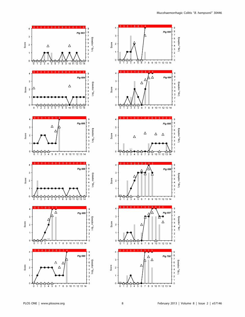
Figure 4. Fecal consistency, shedding and tissue concentrations in pigs following inoculation with pure broth cultivated‘‘Brachyspira hampsonii’’ strain 30446. Fecal consistency scores (line, left ordinate: 0 = formed, normal; 1 = soft, wet cement consistency; 2 = runnyor watery; 3 = mucoid diarrhea; or 4 = bloody diarrhea). Fecal smear spirochete scores (grey bars, left ordinate: 0 = less than 1 spirochete/high powerfield (hpf); 1 = between 2 and 10 spirochetes/hpf; 2 = between 11 and 49 spirochetes/hpf; 3 = greater than 50 spirochetes/hpf). Strain 30446 DNAconcentration in feces (triangles, right ordinate), upside down triangles indicate DNQ. The red bar at the top of each panel indicates the semi-quantitative fecal culture score (0 = negative; 1 = less than 10 colonies/1u streak; 2 = less than 10 colonies/2u streak; 3 = less than 10 colonies/3ustreak;4 = less than 10 colonies/4u streak). Pig IDs are indicated in the upper right corner of each panel. ND = fecal smear spirochete score not done.doi:10.1371/journal.pone.0057146.g004
Mucohaemorrhagic Colitis ‘‘B. hampsonii’’ 30446
PLOS ONE | www.plosone.org 9 February 2013 | Volume 8 | Issue 2 | e57146

microscopic examination in feces of CTRL pigs, no sample was
positive by qPCR or culture during following inoculation. Of the
12 INOC pigs, eight developed mucohaemorrhagic diarrhea
between 5 and 9 days PI (Figures 4, 5, 6). Of the remaining four
INOC pigs (#683, #684, #688 and #695) soft feces were
variably observed between days 1 and 14. Low levels of strain
30446 shedding (1.486104 to 2.676105 genome copies/g) were
detected by qPCR in the feces of three of these pigs (#683, #684
and #695), and positive culture results were obtained for #683,
#688 and #695. Mucohaemorrhagic diarrhea was significantly
more common in INOC (8 of 12) than CTRL (0 of 6) (P = 0.013).
While fecal consistency score, spirochete slide score, culture
results and qPCR counts varied daily, when elevated, they tended
to move together as a group (Figure 4). Gram stain slide scores of
3+ were seen before or concurrently with diarrhea among pigs that
developed mucohaemorrhagic diarrhea, whereas scores above 2+were not observed in pigs without mucohaemorrhagic diarrhea.
Similarly, 2+ or 3+ cultures were only observed in pigs that
developed mucohaemorrhagic diarrhea (Figure 4). Compared to
CTRL or INOC without mucohaemorrhagic diarrhea, INOC
pigs with mucohaemorrhagic diarrhea had significantly higher
terminal spirochete slide scores (P = 0.009 for INOC with
diarrhea, P = 0.026 for INOC without diarrhea) and more
frequent isolation of 30446 by culture (P = 0.001 for INOC with
diarrhea, P = 0.004 for INOC without diarrhea). A significantly
higher number of genome equivalents of strain 30446 DNA was
detected by qPCR in the colonic tissue of pigs with mucohaemor-
rhagic diarrhea than CTRL (P,0.001). Although the same
association was seen between pigs with mucohaemorrhagic
diarrhea (9.656103–1.856107 genome copies/g) and INOC
without mucohaemorrhagic diarrhea (DNQ - 1.036104 genome
copies/g), the non-parametric method of analysis was insensitive to
this difference (P = 0.157). Sequencing of nox PCR amplicons from
terminal fecal and colon tissue cultures revealed isolates with 99–
100% sequence similarity to strain 30446 in all pigs with
mucohaemorrhagic diarrhea and one INOC pig without muco-
haemorrhagic diarrhea (#684). An isolate with sequence similarity
to B. intermedia (97% identity over 825 bp of the nox gene to B.
intermedia ATCC 51140T) was grown from one other INOC pig
without mucohaemorrhagic diarrhea (#695). All CTRL pigs were
culture negative. Importantly, nox sequence of the Brachyspira
species isolated from the terminal fecal samples from the INOC
group matched the nox sequences of the isolates from terminal
colon culture.
Lawsonia intracellularis, B. hyodysenteriae, B. pilosicoli, Salmonella spp.
and PRRS were not detected in terminal samples from any pig.
Pig #695 tested positive for PCV2 in ileum and lymph node by
immunohistochemistry, while all others tested negative.
Necropsy. Gross pathological findings were consistent with
the clinical signs. Lesions were significantly more common in pigs
with mucohaemorrhagic diarrhea than in INOC pigs without
mucohaemorrhagic diarrhea or CTRL (Table 3). In affected pigs,
lesions were typified by mucoid or mucohaemorrhagic colitis and
fibrino-mucoid typhlitis. Lesions ranging in severity from mild
congestion to severe fibrino-necrotic colitis with profuse mucous
were seen (Figure 7). Small intestinal lesions were found in three
pigs with mucohaemorrhagic diarrhea and one INOC without
mucohaemorrhagic diarrhea, and consisted of mild corrugation
and thickening of the ileum grossly. Hyperkeratosis of the pars
esophagea was seen in 14 pigs (INOC n = 8, CTRL n = 6).
Additionally, one pig with mucohaemorrhagic diarrhea had a well
demarcated area of superficial necrosis in the gastric fundus, and
one INOC pig without mucohaemorrhagic diarrhea had small
erosions of the pars esophagea.
Figure 5. Mucohaemorrhagic diarrhea associated with ‘‘Brachy-spira hampsonii’’ strain 30446. Fecal consistency following inocula-tion with pure broth ‘‘Brachyspira hampsonii’’ strain 30446 cultureranged from that similar to wet cement (A, right side), clots of blood (A,left side) or mucus (B), or severe watery mucohaemorrhagic diarrhea (C).Images A, B, C were taken on days 5, 6 and 8 PI respectively.doi:10.1371/journal.pone.0057146.g005
Mucohaemorrhagic Colitis ‘‘B. hampsonii’’ 30446
PLOS ONE | www.plosone.org 10 February 2013 | Volume 8 | Issue 2 | e57146
The distribution of histologic lesions was significantly associated
with clinical signs and was similar to that observed in trial 1
(Table 3). There were no abnormal findings in either CTRL or
INOC without mucohaemorrhagic diarrhea. Inflammation and/
or necrosis of the superficial mucosa of the colon, caecum and
rectum were seen in all affected pigs.
Amplicons were generated from nox PCR from terminal colon
tissue from all INOC pigs. Sequencing revealed 11 amplicons with
99–100% sequence identity to strain 30446 (including all affected
pigs) and one amplicon with 97% sequence identity to B. intermedia
ATCC 51140T (from #695, an INOC pig that did not develop
mucohaemorrhagic diarrhea). When tested by qPCR, strain 30446
specific amplicons were generated for all 12 INOC pigs and no
CTRL pigs. When sequenced, 10 of these amplicons were 99–
100% similar to strain 30446, while sequencing failed for the other
two. The colon contents from one CTRL pig (#685) were positive
for B. pilosicoli by qPCR (1.656104 copies/g of feces) and its
identity was confirmed by sequencing. Neither B. hyodysenteriae nor
B. pilosicoli were detected in any other pigs.
Discussion
Swine dysentery, which by definition is caused by B. hyodysenter-
iae [17]̧ has recently undergone a period of relative quiescence in
North America. The emergence of a clinically indistinguishable
illness associated with a distinct organism, ‘‘B. hampsonii’’ strain
30446 poses diagnostic challenges for producers, veterinarians and
diagnosticians [6,7,18,19]. The lack of pathognomonic clinical or
pathological findings associated with ‘‘B. hampsonii’’ strain 30446
necessitates a reliance on laboratory tests to make a specific
etiological diagnosis. Unfortunately there is no standardized
method of identification. Although there have been no attempts
to standardize testing amongst laboratories to date, the use of
direct examination, culture followed by genus specific PCR and
sequencing, and species-specific PCR are recommended. A
number of Brachyspira species specific PCR assays have been
published [19–22] but due to the apparent widespread use of
techniques developed in house or modifications to published
protocols, the relative sensitivity and specificity of these assays are
unknown. Thus, when using PCR, negative results should be
interpreted with caution. In light of these challenges, evaluation of
fecal smears may be invaluable diagnostically. While fecal smears
cannot differentiate between Brachyspira species, the presence or
absence of spirochetes is useful for interpreting molecular test
results.
This research confirms that ‘‘Brachyspira hampsonii’’ strain 30446
induces a mucohaemorrhagic diarrhea and colitis in pigs that is
indistinguishable from swine dysentery. Trial 1 preceded our
ability to grow ‘‘B. hampsonii’’ strain 30446 in broth culture, but the
development of JBS broth in October 2011 made it possible to use
a pure culture inoculum in trial 2. This pure broth culture was
prepared from the tissue inoculum used in trial 1. The results of
trial 2 demonstrate that strain 30446 causes mucohaemorrhagic
diarrhea indistinguishable from the field cases where it was first
observed, establishing this organism as a pathogen of swine.
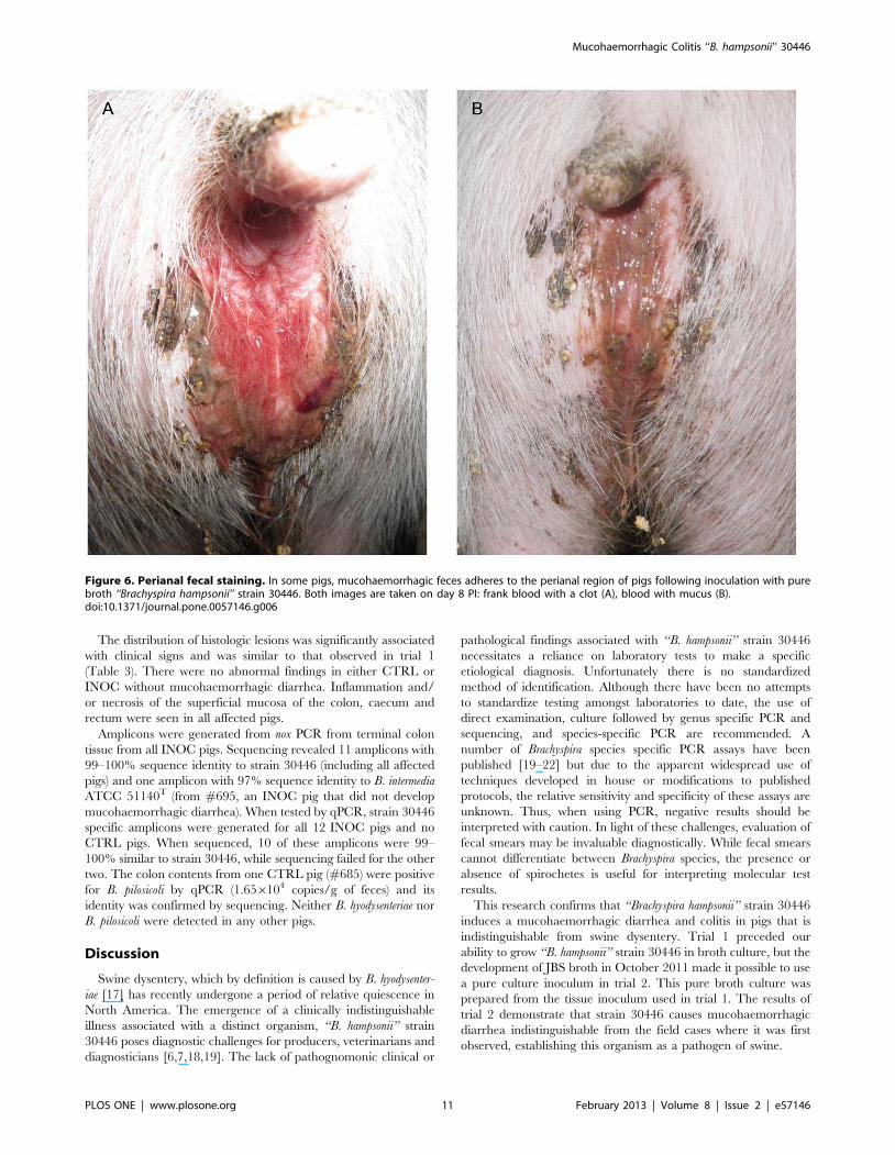
Figure 6. Perianal fecal staining. In some pigs, mucohaemorrhagic feces adheres to the perianal region of pigs following inoculation with purebroth ‘‘Brachyspira hampsonii’’ strain 30446. Both images are taken on day 8 PI: frank blood with a clot (A), blood with mucus (B).doi:10.1371/journal.pone.0057146.g006
Mucohaemorrhagic Colitis ‘‘B. hampsonii’’ 30446
PLOS ONE | www.plosone.org 11 February 2013 | Volume 8 | Issue 2 | e57146

Figure 7. Colonic mucosal lesions associated with ‘‘Brachyspira hampsonii’’ strain 30446. Gross colonic mucosal lesions observed followinginoculation with pure broth culture containing strain 30446. Images are of different INOC pigs euthanized between D6 and D12 post-inoculation.A = mild to moderate hyperemia and/or congestion with scant mucous deposited on mucosal surface; B = moderate to severe mucosal congestionwith normal looking contents adhering to mucosa; C = moderate to severe fibrinomucoid exudate adhering to hyperemic mucosal surface prior towashing; D = patchy fibrinomucoid exudate adhering to hyperemic mucosal surface after washing; E = severe fibrinonecrotic colitis; F = thick adherentmucoid exudate on mucosal surface of colon.doi:10.1371/journal.pone.0057146.g007
Mucohaemorrhagic Colitis ‘‘B. hampsonii’’ 30446
PLOS ONE | www.plosone.org 12 February 2013 | Volume 8 | Issue 2 | e57146
The design of these trials was based on a successful, previously
published trial utilizing B. murdochii [5]. Because the minimum
infectious dose of strain 30446 is unknown, average doses of
1.956108 genome equivalents in trial 1 and 3.196108 genome
equivalents in trial 2 were used, intermediate to those used in
previous studies. Disease developed between four and ten days PI
in trial 1 and five to nine days PI in trial 2 indicating that a dose
sufficient to cause disease was used. Interestingly, the incubation
period observed in these trials was consistent with an infection trial
conducted as part of the initial description of swine dysentery in
1921 [1].
Pre-screening fecal samples of experimental pigs revealed a low
level of ‘‘B. hampsonii’’ strain 30446 colonization (reported as DNQ)
in 3/18 pigs in trial 1, and 12/18 pigs in trial 2 based on qPCR. In
addition, 1 pig in trial 2 (#684) had 6.806104 genome
equivalents/g of ‘‘B. hampsonii’’ strain 30446 in feces on D0
(Table 2). This finding suggests that strain 30446 is not an obligate
pathogen but instead causes disease when present in sufficient
numbers or when host defences are compromised. The use of
strain 30446 negative pigs was preferable, but none were available
at the time this research was undertaken. Low (DNQ) pre-
challenge levels of ‘‘B. hampsonii’’ strain 30446 however, clearly did
not induce sufficient mucosal immunity to protect against disease
as evidenced by the 3/3 trial 1 pigs and 6/8 trial 2 pigs that
developed mucohaemorrhagic diarrhea following challenge.
Noteworthy is pig #684 that did not develop mucohaemorrhagic
diarrhea following challenge, suggesting that shedding ,104
genome equivalents/g feces may have been associated with
sufficient mucosal immunity to protect against disease. The results
of a number of diagnostic cases completed by our team
demonstrates a similar trend whereby clinical cases typically have
greater than 105 ‘‘B. hampsonii’’ strain 30446 genome equivalents/g
of tissue or feces, whereas age-matched non-clinical animals in the
same airspace have fewer than 105 genomic copies/g [23]. Pre-
trial colonization of CTRL animals, all of which remained healthy,
supports the conclusion that DNQ levels of strain 30446 in feces
are incidental. These findings are consistent with a previous report
that concluded that concentrations of B. hyodysenteriae greater than
105 CFU/gram of feces are required for the development of
lesions [24].
Three pigs in trial 1 and four pigs in trial 2 did not develop
mucohaemorrhagic diarrhea in spite of being inoculated. In trial 2,
one pig (#683) only received two of three inoculum doses, and one
(#684) was the pig with 104 copies/g strain 30446 on D0.
Whether failure to produce disease in these seven animals reflects
normal biological variation, pre-existing immunity or the use of a
marginally infectious dose is unknown.
Pathological findings in affected pigs, characterized by mucoid
or mucohaemorrhagic colitis and muco-fibrinous typhlitis, were
consistent with previous reports describing swine dysentery [25].
Histopathological findings in the caecum and colon of affected pigs
were consistent with, but mild in comparison to gross lesions. The
mild thickening and congestion seen in the ileum of four INOC
pigs (one unaffected and three with mucohaemorrhagic diarrhea)
in trial 2, mimicked early or mild proliferative ileitis caused by L.
intracellularis, however the ileum of all pigs were negative by PCR
for L. intracellularis and in no pigs were lesions typical of ileitis seen
histologically. Whether or not these mild ileal lesions are a feature
of strain 30446 associated disease or an incidental finding is
unknown.
A number of atypical or novel, phylogenetically distinct,
strongly b-haemolytic Brachyspira have been reported to cause
disease in pigs [19]. Based on partial nox sequence, strain 30446
clusters separately from the other known species with good
bootstrap support (Figure 1). The partial nox sequence for strain
30446 is identical to the provisionally named ‘‘B. hampsonii’’, and
94.8% similar over 810 bp to Serpulina sp. P280/1, a porcine
clinical isolate from the United Kingdom [9,26,27]. It is
approximately 92% identical to the B. innocens, and B. murdochii
strains examined, which is less similar than these two species are to
each other (pairwise identities between B. innocens and murdochii are
96–97% over this same region). Furthermore, three of seven
primer sets from a previously published MLST scheme for
Brachyspira sp, failed to yield a product, while novel sequences were
generated for the other four.
Two recent reports describe infection experiments using murine
[28] and porcine [19] models of swine dysentery with North
American Brachyspira strains including ‘‘B. hampsonii’’ strain 30446
isolated from pigs with signs of swine dysentery in Iowa between
2008 and 2011. These experiments were the first to demonstrate
the causal relationship between strain 30446 and mucohaemor-
rhagic typhlocolitis in pigs. The authors found that strongly b-
haemolytic strains of Brachyspira spp., including strain 30446,
produced disease and colonic lesions typical of those associated
with B. hyodysenteriae. In the porcine experiment [19], 4 of 10 pigs
inoculated with strain 30446 had diarrhea on days 7 and 14 post
infection, and 2 of 10 were culture or PCR positive at necropsy on
day 16. By contrast, the incidence of mucohaemorrhagic diarrhea
reported here in trial 1 and 2 was 75% and 67% respectively in
INOC pigs. Furthermore, following inoculation with pure broth
culture, strain 30446 was isolated by culture from 10/12 pigs, was
detected by PCR in feces in 11/12 pigs and repeatedly for 3 or
more days in 8 pigs. Testing for other relevant pathogens including
PRRS virus, Lawsonia intracellularis and Salmonella spp., were
negative. Collectively, these data provide substantive evidence of
causality and provide the first report characterizing fecal shedding
using a Canadian ‘‘B. hampsonii’’ 30446 isolate. While strain 30446
used for the present research clearly falls within ‘‘B. hampsonii’’
clade 2, the relationship between this strain and other members of
the species, particularly clade 1, is unclear. Hence, additional
research is necessary to more fully understand the clinical
relevance of this novel and diverse Brachyspira species in swine.
In summary, our results confirm the causal relationship between
‘‘Brachyspira hampsonii’’ strain 30446 and mucohaemorrhagic
diarrhea in swine. The emergence of ‘‘Brachyspira hampsonii’’ strain
30446 therefore poses potential diagnostic challenges since the
specificity of some currently used PCR assays may not detect this
organism, and Brachyspira culture is not widely used in diagnostic
laboratories in many countries. To date, cases have been
diagnosed in pigs from Alberta, Saskatchewan, Iowa, Illinois,
Minnesota, Missouri and North Carolina [7,9]. The prevalence of
carrier animals, risk factors for infection, non-porcine reservoirs,
antimicrobial susceptibility, minimal infectious dose and the
efficacy of cleaning practices for eliminating ‘‘B. hampsonii’’ strain
30446 are entirely unknown. Research is ongoing in our lab to
address these and other important questions.
Author Contributions
Conceived and designed the experiments: JER JEH BO JCH. Performed
the experiments: JER MOC JEH HEK CF YH BO JCH. Analyzed the
data: JER MOC CF. Contributed reagents/materials/analysis tools: JEH
JCH. Wrote the paper: JER MOC JEH CF BO JCH.
Mucohaemorrhagic Colitis ‘‘B. hampsonii’’ 30446
PLOS ONE | www.plosone.org 13 February 2013 | Volume 8 | Issue 2 | e57146

References
1. Whiting RA, Doyle LP, Spray RS (1921) Swine Dysentery. Bulletin: 3–15.
2. Duhamel GE (2001) Comparative pathology and pathogenesis of naturallyacquired and experimentally induced colonic spirochetosis. Anim Health Res
Rev 2: 3–17.3. Trott DJ, Stanton TB, Jensen NS, Duhamel GE, Johnson JL, et al. (1996)
Serpulina pilosicoli sp. nov., the agent of porcine intestinal spirochetosis. Int J Syst
Bacteriol 46: 206–215.4. Komarek V, Maderner A, Spergser J, Weissenbock H (2009) Infections with
weakly haemolytic Brachyspira species in pigs with miscellaneous chronic diseases.Vet Microbiol 134: 311–317.
5. Jensen TK, Christensen AS, Boye M (2010) Brachyspira murdochii colitis in pigs.
Vet Pathol 47: 334–338.6. Harding JCS, Chirino-Trejo M, Fernando C, Jacobson M, Forster Z, et al.
(2010) Detection of a novel Brachyspira species associated with haemorrhagic andnecrotizing colitis. Int Pig Vet Soc Congr; July 18–21, 2010; Vancouver,
Canada. pp. 740.7. Harding J, Chirino-Trejo M, Vermette C, Fernando C, Jacobson M, et al.
(2010) Detection of a novel Brachyspira species associated with haemorrhagic and
necrotizing colitis. West Can Assoc Swine Vet Conf; October 15–16, 2010;Saskatoon, SK. pp. 65–70.
8. Rohde J, Rothkamp A, Gerlach GF (2002) Differentiation of porcine Brachyspira
species by a novel nox PCR-based restriction fragment length polymorphism
analysis. J Clin Microbiol 40: 2598–2600.
9. Chander Y, Primus A, Oliveira S, Gebhart CJ (2012) Phenotypic and molecularcharacterization of a novel strongly hemolytic Brachyspira species, provisionally
designated ‘‘Brachyspira hampsonii’’. J Vet Diagn Invest 24: 903–910.10. Jenkinson SR, Wingar CR (1981) Selective medium for the isolation of Treponema
hyodysenteriae. Vet Rec 109: 384–385.11. Kunkle RA, Kinyon JM (1988) Improved selective medium for the isolation of
Treponema hyodysenteriae. J Clin Microbiol 26: 2357–2360.
12. Rasback T, Johansson KE, Jansson DS, Fellstrom C, Alikhani MY, et al. (2007)Development of a multilocus sequence typing scheme for intestinal spirochaetes
within the genus Brachyspira. Microbiology 153: 4074–4087.13. Rasback T, Jansson DS, Johansson KE, Fellstrom C (2007) A novel
enteropathogenic, strongly haemolytic spirochaete isolated from pig and
mallard, provisionally designated ‘Brachyspira suanatina’ sp. nov. EnvironMicrobiol 9: 983–991.
14. Jacobson M, Fellstrom C, Lindberg R, Wallgren P, Jensen-Waern M (2004)Experimental swine dysentery: comparison between infection models. J Med
Microbiol 53: 273–280.15. Jones GF, Ward GE, Murtaugh MP, Lin G, Gebhart CJ (1993) Enhanced
detection of intracellular organism of swine proliferative enteritis, ileal symbiont
intracellularis, in feces by polymerase chain reaction. J Clin Microbiol 31: 2611–2615.
16. Harding JCS, Auckland CD, Tumber A, McIntosh KA, Parker SE, et al. (2008)
Porcine circovirus-2 DNA concentration distinguishes wasting from non-wasting
pigs and is correlated with lesion distribution, severity and nucleocapsid staining
intensity. J Vet Diagn Invest 20: 274–282.
17. Taylor DJ, Alexander TJL (1971) The production of dysentery in swine by
feeding cultures containing a spirochaete. Br Vet J 127: 58–61.
18. Gebhart CJ, Chander Y, Vannucci F, Kelly M (2012) Bacterial enteric diseases
of growing pigs: dysentery and proliferative enteropathy. Int Pig Vet Soc Congr;
June 10–13, 2012; Jeju, Korea. pp. 48–50.
19. Burrough ER, Strait EL, Kinyon JM, Bower LP, Madson DM, et al. (2012)
Epub. Available: http://vdi.sagepub.com/content/early/2012/09/04/
1040638712457927.abstract. Comparative virulence of clinical Brachyspira spp.
isolates in inoculated pigs. J Vet Diagn Invest.
20. Rasback T, Fellstrom C, Bergsjo B, Cizek A, Collin K, et al. (2005) Assessment
of diagnostics and antimicrobial susceptibility testing of Brachyspira species using a
ring test. Vet Microbiol 109: 229–243.
21. Song Y, Hampson DJ (2009) Development of a multiplex qPCR for detection
and quantitation of pathogenic intestinal spirochaetes in the faeces of pigs and
chickens. Vet Microbiol 137: 129–136.
22. Primus A, Oliveira S, Gebhart CJ (2011) Identification of a new potentially
virulent Brachyspira species affecting swine. Amer Assoc Swine Vet Conf; March
5–8, 2011; Phoenix, AZ. pp. 109–110.
23. Harding J, Hill JE, Chirino-Trejo M, O’Connor B, Rubin J, et al. (2011)
Detection of a novel Brachyspira species associated with haemorrhagic and
necrotizing colitis. Carlos Pijoan Symposium on Swine Dysentery; Allen D
Leman Swine Conference; September 17–20, 2011; St. Paul, MN. pp. 27–32.
24. Wilcock BP, Olander HJ (1979) Studies on the pathogenesis of swine dysentery.
I. Characterization of the lesions in colons and colonic segments inoculated with
pure cultures of colonic content containing Treponema hyodysenteriae. Vet Pathol
16: 450–465.
25. Kinyon JM, Harris DL, Glock RD (1977) Entoeropathogenicity of various
isolates of Treponema hyodysenteriae. Infect Immun 15: 638–646.
26. Neef NA, Lysons RJ, Trott DJ, Hampson DJ, Jones PW, et al. (1994)
Pathogenicity of porcine intestinal spirochetes in gnotobiotic pigs. Infect Immun
62: 2395–2403.
27. Atyeo RF, Stanton TB, Jensen NS, Suriyaarachichi DS, Hampson DJ (1999)
Differentiation of Serpulina species by NADH oxidase gene (nox) sequence
comparisons and nox-based polymerase chain reaction tests. Vet Microbiol 67:
47–60.
28. Burrough E, Strait E, Kinyon J, Bower L, Madson D, et al. (2012) Epub. Available:
http://www.sciencedirect.com/science/article/pii/S0378113512003604. Com-
parison of atypical Brachyspira spp. clinical isolates and classic strains in a mouse
model of swine dysentery. Vet Microbiol.
Mucohaemorrhagic Colitis ‘‘B. hampsonii’’ 30446
PLOS ONE | www.plosone.org 14 February 2013 | Volume 8 | Issue 2 | e57146
Copyright © 2022 FDOKUMEN