Enteric pathogens associated with diarrhea in children in Fayoum, Egypt

Infection, Genetics and Evolution 21 (2014) 205–213
Contents lists available at ScienceDirect
Infection, Genetics and Evolution
journal homepage: www.elsevier .com/locate /meegid
Genetic diversity of ORF3 and spike genes of porcine epidemic diarrheavirus in Thailand
1567-1348/$ - see front matter � 2013 Elsevier B.V. All rights reserved.http://dx.doi.org/10.1016/j.meegid.2013.11.001
⇑ Corresponding author. Address: Department of Veterinary Microbiology, Fac-ulty of Veterinary Science, Chulalongkorn University, Henry Dunant Road, Pathum-wan, Bangkok 10330, Thailand. Tel.: +66 2 218 9583; fax: +66 2 251 1656.
E-mail address: [email protected] (D. Nilubol).
Gun Temeeyasen a, Anchalee Srijangwad a, Thitima Tripipat a, Pavita Tipsombatboon b,Jittima Piriyapongsa b, Waranyoo Phoolcharoen c, Taksina Chuanasa c, Angkana Tantituvanont d,Dachrit Nilubol a,⇑a Department of Veterinary Microbiology, Faculty of Veterinary Science, Bangkok 10330, Thailandb Genome Institute, National Center for Genetic Engineering and Biotechnology, National Science and Technology Development Agency, Pathumthani 12120, Thailandc Department of Pharmacognosy and Pharmaceutical Botany, Faculty of Pharmaceutical Sciences, Chulalongkorn University, Bangkok 10330, Thailandd Department of Pharmaceutics and Industrial Pharmacy, Faculty of Pharmaceutical Sciences, Chulalongkorn University, Bangkok 10330, Thailand
a r t i c l e i n f o a b s t r a c t
Article history:Received 21 June 2013Received in revised form 2 November 2013Accepted 5 November 2013Available online 26 November 2013
Keywords:Porcine epidemic diarrhea virusSpike geneORF3 geneDiversityThailand
Porcine epidemic diarrhea virus (PEDV) has become endemic in the Thai swine industry, causingeconomic losses and repeated outbreaks since its first emergence in 2007. In the present study, 69 ThaiPEDV isolates were obtained from 50 swine herds across Thailand during the period 2008–2012. Bothpartial and complete nucleotide sequences of the spike (S) glycoprotein and the nucleotide sequencesof ORF3 genes were determined to investigate the genetic diversity and molecular epidemiology of ThaiPEDV. Based on the analysis of the partial S glycoprotein genes, the Thai PEDV isolates were clustered into2 groups related to Korean and Chinese field isolates. The results for the complete spike genes, however,demonstrated that both groups were grouped in the same cluster. Interestingly, both groups of Thai PEDVisolates had a 4-aa (GENQ) insertion between positions 55 and 56, a 1-aa insertion between positions 135and 136, and a 2-aa deletion between positions 155 and 156, making them identical to the Korean KNUseries and isolates responsible for outbreaks in China in recent years. In addition to the complete Ssequences, the ORF3 gene analyses suggested that the isolates responsible for outbreaks in Thailandare not vaccine related. The results of this study suggest that the PEDV isolates responsible for outbreaksin Thailand since its emergence represent a variant of PEDV that was previously reported in China andKorea.
� 2013 Elsevier B.V. All rights reserved.
1. Introduction
Porcine epidemic diarrhea virus (PEDV) is the etiologic agent ofporcine epidemic diarrhea (PED), a devastating enteric disease thatis characterized by vomiting, acute severe watery diarrhea anddehydration, which results in significant high mortality in pigletsunder 7 days of age (Pensaert and de Bouck, 1978). In addition tocausing enteric problem in pigs, PEDV infection in sows can leadto a reduction of subsequent reproductive performance(Olanratmanee et al., 2010). PED was first recognized in Belgiumand the United Kingdom in 1976–1978 (Pensaert and de Bouck,1978; Wood, 1977). Post-emergence, disease outbreaks have beenreported in several European countries (Chasey and Cartwright,1978). At present, PED disease has continued to causing devastat-ing enteric disease and severe economic losses in many Asian
countries, including China, Korea, Japan and Thailand (Gao et al.,2013; Li et al., 2012b; Park et al., 2007a, 2011; Sato et al., 2011).
PEDV is an enveloped, single-stranded RNA virus belonging tothe genus Alphacoronavirus, family Coronaviridae and order Nidovi-rales. The PEDV genome is approximately 28 kb in length and iscomposed of 7 open reading frames (ORF) (Kocherhans et al.,2001). ORF1a and ORF1b cover 70% of the entire genome andencode the non-structural replicase gene. The remaining 5 ORFscode for major structural proteins including the spike (S) glycopro-tein, ORF3, envelope protein, membrane protein and nucleocapsidprotein. Among the proteins encoded by these genes, the S glyco-protein is a glycosylated protein located on the envelope of thevirus. It is involved with pathogenesis by binding to cell receptorson the host cell membranes and penetrating cells by membranefusion. The S glycoprotein consists of 2 domains, including S1and S2 domains. Whereas the S1 domain is involved in viral attach-ment, the S2 domain undergoes conformational changes followingS1 attachment to facilitate fusion of the virus envelope with thehost membrane, allowing entry of the viral genome into the host.
206 G. Temeeyasen et al. / Infection, Genetics and Evolution 21 (2014) 205–213
Because of its important role in viral attachment and entry, the S1domain was characterized and demonstrated to contain epitopesinvolved with neutralizing antibodies (Chang et al., 2002; Cruzet al., 2006, 2008; Sun et al., 2008). Because of its role in the induc-tion of neutralizing antibodies, the S protein is the primary targetfor the development of a vaccine against PED (Kang et al., 2006;Oszvald et al., 2007). Several investigators have reported a high de-gree of genetic diversity observed in the S glycoprotein gene (Leeet al., 2010; Li et al., 2012b; Park et al., 2007a, 2011). Thus, thisgene is important for understanding the genetic relatedness ofPEDV field isolates, the epidemiological status of the virus and vac-cine development.
In addition to the S glycoprotein gene, the accessory ORF3 genehas received a large amount of attention in the context of PEDV vir-ulence. The virulence of PEDV can be reduced by altering the ORF3gene through cell culture adaptation (Park et al., 2007b, 2008). TheORF3 genes have been used to differentiate between field and vac-cine-derived isolates. The vaccine-derived isolates have uniquecharacteristics at amino acids 82–99, where a continuous deletionof 17 amino acids has been observed (Park et al., 2008). Therefore,this deletion can be used to differentiate between field and atten-uated vaccine viruses. The differentiation of the ORF3 gene can beused for molecular epidemiology studies of PEDV.
In Thailand, a PED outbreak was first reported in 1995 in a farmlocated in the southern region of the country (Srinuntapunt et al.,1995). The disease did not spread to other swine-producing areasat that time; however, in late 2007, the disease re-emerged in Nak-orn Pathom, a province with the highest pig densities in Thailand(Puranaveja et al., 2009). The source of viral introduction has notbeen identified to date. There is only speculation concerning con-taminated pork meat and bone meal, the illegal introduction ofthe vaccine or both. Since its re-emergence, PED has spreadthroughout the country, causing pandemic PED outbreaks in Thai-land resulting in >90% of Thai swine farms being infected. Cur-rently, the disease develops until reaching an endemic stage inwhich many herds experience recurrent outbreaks. To develop amore successful control and prevention program, further investiga-tion related to the genetic diversity and molecular epidemiology ofPEDV in Thailand is needed. The objectives of the present studywere to investigate the genetic diversity of the S glycoproteinand ORF3 genes of PEDV in Thailand. The results of a study onthe genetic differences of the S genes may provide informationon the heterogeneity of the outbreak. Moreover, such a studymay also provide a basis of knowledge that can be used for thedevelopment of an effective vaccine.
2. Materials and methods
2.1. Source of specimens and PEDV isolation
A total of 120 porcine intestinal samples were collected from 50pig farms that experienced PED outbreaks from 2008 to 2012. Ineach outbreak, intestinal samples were collected from 3- to4-day-old piglets that displayed the clinical features associatedwith PED, including vomiting, watery diarrhea and dehydration.
The intestinal samples were subjected to PEDV isolation usingthe continuous Vero cell line (ATCC, CCL-81) (Hofmann and Wyler,1988). Briefly, the intestinal samples of piglets were minced intosmall pieces and suspended in 10 ml of phosphate buffer saline(PBS; 0.1 M, pH 7.2). The suspended samples were vortexed andclarified by centrifugation at 1350g for 10 min. The supernatantwas filtered through 0.45-lm filters and stored at �80 �C until use.
Five hundred microliters of supernatant samples was dilutedwith maintenance media (MM) consisted of Dulbecco’s modifiedEagle’s medium (DMEM, Gibco, USA) supplemented with 10 lg/
ml trypsin (Gibco, USA) and inoculated into a 25-cm2 flask thathad been previously plated with Vero cells for 2 days or until fullconfluence was reached. The inoculated 25-cm2 flask was incu-bated at 37 �C in 5% CO2 for 1 h. Then, the media was replaced with10 ml of MM, and incubated again for a period of 2–3 days. Cyto-pathic effect (CPE) was observed with a light microscope. Then,the 25-cm2 flasks were frozen and thawed 2 times before beingcentrifuged at 1350g for 10 min to collect the supernatant, whichwas stored at �80 �C. The clarified supernatants were subjectedto RT-PCR. Partial S, complete S and ORF3 genes were amplifiedand sequenced. The intestinal samples were considered negativefor PEDV isolation following 3 blind passages.
2.2. Reverse transcription-polymerase chain reaction
Total RNA was extracted from the supernatant from the previ-ous procedure using the Nucleospin� RNA Virus kit (Macherey-Na-gel Inc., PA, USA) in accordance with the manufacturer’sinstructions. cDNA was synthesized from the extracted RNA usingM-MuLV Reverse Transcriptase (New England BioLabs Inc., MA,USA).
PCR amplification was performed on the cDNA. To amplify thepartial S genes, the following primers were utilized: PEDF (50-TTCTGA GTC ACG AAC AGC CA-30) and PEDR (50-CAT ATG CAG CCTGCT CTG AA-30) (Park et al., 2007a). PCR amplification was per-formed using Platinum� Tag DNA polymerase High Fidelity (Invit-rogen, CA, USA). After the initial incubation at 94 �C for 5 min, thereactions were subjected to 35 cycles of PCR as follows: 94 �C for30 s, 55 �C for 30 s, and 72 �C for 30 s. These cycles were followedby a terminal 7-min extension at 72 �C.
To amplify the complete S genes, the sequence was separatelyconstructed for S1 and S2,with the primers S-F-1 (50-ACG TAAACA AAT GAG GTC TTT-30) and S-R-1 (50-ATA CAC CAA CAC AGGCTC TGT-30) used to amplify S1 and the primers S-F-2 (50-GGTTTC TAC CAT TCT AAT GAC G-30) and S-R-2 (50-GTA TTG AAAAAG TCC AAG AAA CA-30) used to amplify S2 (Lee et al., 2010).PCR amplification was performed using Platinum� Tag DNA poly-merase High Fidelity (Invitrogen, CA, USA). After an initial incuba-tion at 94 �C for 5 min, the reactions were subjected to 35 cycles ofPCR as follows: 94 �C for 30 s, 55 �C (58 �C for S2) for 30 s, and 72 �Cfor 2 min. These cycles were followed by a terminal 10-min exten-sion at 72 �C. The PCR products were visualized by agarose gelelectrophoresis.
To amplify the ORF3 genes, primers ORF3F (50-CCT AGA CTT CAACCT TAC GA-30) and ORF3R (50-CAG GAA AAA GAG TAC GAA AA-30)were used, and PCR amplification was performed using Platinum�
Tag DNA polymerase High Fidelity (Invitrogen, CA, USA). After aninitial incubation at 94 �C for 5 min, the reactions were subjectedto 30 cycles of PCR as follows: 94 �C for 60 s, 50 �C for 60 s, and72 �C for 60 s. These cycles were followed by a terminal 10-minextension at 72 �C. The PCR products were visualized by agarosegel electrophoresis.
2.3. Cloning, plasmid purification and sequence determination
The PCR products were cloned into plasmid vectors for the sub-sequent transformation of Escherichia coli cells by using a commer-cial kit (pGEM-T� Easy Vector System I (Promega, WI, USA), andthe controls were included at all stages of cloning and transforma-tion. Bacterial transformant colonies were randomly selected fromeach sample for plasmid purification using the Nucleospin� Plas-mid kit (Macherey-Nagel Inc., PA, USA), and the 10 elected colonieswere grown in LB broth for 24 h and subjected to plasmid isolationand sequencing. Sequencing reactions were performed by BiobasicInc. (Ontario, Canada) using an ABI Prism 3730XL DNA sequencer.
G. Temeeyasen et al. / Infection, Genetics and Evolution 21 (2014) 205–213 207
2.4. Sequence analyses
The nucleotide and deduced amino acid sequences were alignedusing the CLUSTALW program (Thompson et al., 1994). The per-centage of identity between every pair of isolate sequences at thenucleotide and amino acid levels was calculated using MEGA 5.2(Tamura et al., 2011). The analyses included only sequences inwhich nucleotide sequence data are complete and accession num-bers are available in GenBank (Supplementary Material 1). Phylo-genetic analyses of the partial S, complete S, and ORF3 genes wasseparately performed using a Bayesian Markov chain Monte Carlo(BMCMC) method implemented in the program BEAST v1.7.4(Drummond et al., 2012). The analysis of the partial S geneincluded the nucleotide sequences from the present study, previ-ously reported partial S sequences of the Thai PED (Puranavejaet al., 2009) and isolates from Belgium, United Kingdom (UK),China and Korea (Park et al., 2007a). The analysis of the completeS and ORF3 genes included the nucleotide sequences from thepresent study and isolates from Belgium, UK, China and Korea(Gao et al., 2013; Lee et al., 2010; Li et al., 2012a).
The strict molecular clock model with a coalescent constant-size tree prior and empirical base frequencies were used for allanalyses. The best fitted substitution model and among-site rateheterogeneity model for each gene data set was determined usingModel Generator v0.85 (Keane et al., 2006). For each analysis, threeindependent BEAST runs were performed; each consisting of 50million generations with sampling of every 1000 generations andthe first 10% discarded as burn-in. LogCombinerv1.7.4 (http://beast.bio.ed.ac.uk/LogCombiner) was used to combine logand treefiles from independent runs of BEAST. Tracer v1.5 (http://beast.-bio.ed.ac.uk/Tracer) was used to confirm that post-burn-in treesyielded an effective sample size (ESS) of >200 for all parameters.A 50% majority-rule consensus tree was generated from the treefile using SumTrees v.3.3.1 (Sukumaran and Holder, 2010). Theresulting tree was viewed in FigTree v1.4.0 (http://tree.bio.ed.a-c.uk/software/figtree/).
3. Results
3.1. Phylogenic analysis of the partial spike gene
Of the 120 porcine intestinal samples collected in the years2008–2012, 69 PEDV isolates were collected and the origins ofthose Thai isolates are shown in Fig. 1. All isolates produced CPEcharacterized by cell fusion and syncytia formation in the first pas-sage (Supplementary Material 2). To investigate the heterogeneityof these 69 Thai PEDV isolates, the partial spike genes of the 69field isolates were amplified. The nucleotide sequences revealedthat these partial S glycoprotein genes of the 69 field isolatesconsisted of 657 nucleotides encoding a protein of 219 amino acidslocated on the full length of the S glycoprotein gene at nucleotidepositions 1462–2118 or amino acid positions 488–706.
Identical partial S nucleotide sequences were identified andexcluded, resulting in further genetic analysis of 30 genes. Thephylogenetic tree of 30 partial S glycoprotein genes was con-structed together with another 16 previously reported Thai PEDVisolates (Puranaveja et al., 2009) as well as some partial S glycopro-tein genes that were previously isolated in other countries (Parket al., 2007a). Based on the previously reported phylogenetic anal-ysis (Park et al., 2007a), the PEDV isolates were further divided into3 groups, designated G1, G2 and G3; group G1 was further dividedinto 3 subgroups, which were designated G1-1, G1-2 and G1-3.When using the clustering system based on this phylogenetic anal-ysis (Park et al., 2007a), the phylogenetic tree demonstrated thatthe Thai PEDV isolates were further divided into 2 groups
(Fig. 2). Both groups of Thai PEDV isolates were in G1 but differedin subgroup. The most dominant isolates were clustered in sub-group G1-1, which comprised all 16 previously reported Thai PEDVisolates along with 27 of 30 current Thai isolates. Anothersubgroup designated G1-4 comprised 3 of 30 isolates, includingAGPED0609_1, AGPED0609_2 and SPPED0111_1. This subgroupdid not fit into the previously reported clustering system. It sat be-tween subgroups 1-2 and 1-3. Therefore, we named it subgroupG1-4. After comparing the nucleotide and amino acid sequencesof all Thai field PEDV isolates, it was demonstrated that the nucle-otide and amino acid sequence identity of the all Thai field PEDVisolates ranged from 94.4–99.9% and 85.8–100.0%, respectively.The percentage of identity between nucleotide and amino acidsequences of the Thai G1-1 isolates ranged from 96.4–99.7% and(92.5–100.0%), respectively. The nucleotide and amino acidsequences of isolates in G1-1 were more divergent compared tothose of G1-4, in which all 3 isolates had 100.0% identity at thenucleotide and amino acid level. Subgroups G1-1 and G1-4 had94.4–95.5% and 85.8–88.5% identity, respectively, at the nucleotideand amino acid level.
3.2. Genetic analyses of complete spike gene
To investigate the genetic diversity of 2 groups of Thai fieldPEDV isolates and their genetic relationship with PEDV isolatesfrom other countries, 13 different Thai field PEDV isolates, includ-ing 10 isolates from subgroup G1-1 and 3 isolates from subgroupG1-4, were randomly selected, and the full-length nucleotide se-quences of their S glycoprotein genes were investigated. The phy-logenetic tree of their complete S genes was constructed togetherwith those of other PEDV isolates from other countries (Supple-mentary Material 1). The phylogenetic analysis revealed that thecomplete S glycoprotein genes were further divided into 3 clusters,including clusters 1, 2 and 3 (Fig. 3). Cluster 1 comprised Chineseisolates (CH/FJND-3, CH1, CH8, CHGD-01, HB-2011-1, HB-2011-2,HB-2011-3, HB-2011-4, HB-2012-1, HB-2012-2, HB-2012-3, HB-2012-4, BJ-2011-2, BJ-2011-3, BJ-2012-1, BJ-2012-2, ZJ-2011-1and ZJ-2011-2) and Korean field isolates (KNU-0802,KNU-0902,CNU-091222-01 and CNU-091222-02). Based on the results ofthe partial S gene comparisons, all 13 PEDV Thai isolates in sub-groups G1-1 and G1-4 were included in cluster 1. Cluster 2 com-prised 7 isolates from Korea (KNU-0801, KNU-0901, KNU-0903,KNU-0904, KNU-0905, Spk1 and Chinju99). Cluster 3 comprised16 reference strains, including isolates from Korea (DR13 andSM98), China (CH2, CH3, CH4, CH5, CH6, CH7, CH/FJND-1, CH/FJND-2, JS-2004-2, LZC, LJB03 and DX) and Europe (CV777 andBr1/87).
The sequence homology between the S glycoprotein genes wasmeasured. The nucleotide and amino acid sequence identity of thecluster 1 isolates ranged from 94.4–99.9% and 93.1–99.9%, respec-tively (Supplementary Material 3). The isolates had 93.0–97.8%(90.7–96.5%) and 92.4–98.1% (90.1–97.7%) nucleotide (deducedamino acid) sequence identity with members of groups 2 and 3,respectively. The percentage of identity between nucleotide andamino acid sequences of all 13 Thai field PEDV isolates ranged from94.9–99.8% and 94.0-99.7%, respectively. All 13 Thai PEDV isolatesdisplayed 94.9–99.9% and 94.0–99.9% identity to Chinese isolatesin this cluster at the nucleotide and amino acid levels, respectively.In addition, all 13 Thai PEDV isolates displayed 94.9–99.8% and94.0–99.7% identity to Korean isolates (KNU0902, CNU-091222-01 and CNU-091222-02) at the nucleotide and amino acid levels,respectively.
It was demonstrated that the S glycoprotein genes of the ThaiPEDV isolates are 4158 nucleotides in length and encode 1386 ami-no acid residues. The nucleotide and deduced amino acid se-quences of the complete S genes were aligned separately to
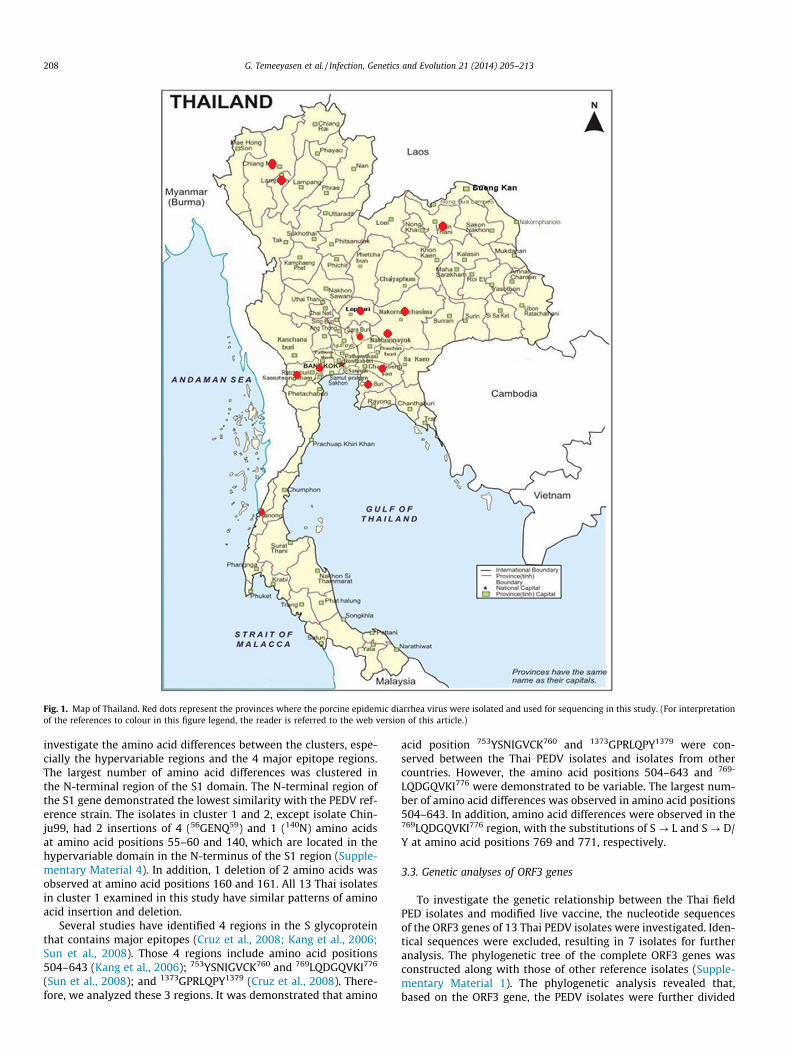
Fig. 1. Map of Thailand. Red dots represent the provinces where the porcine epidemic diarrhea virus were isolated and used for sequencing in this study. (For interpretationof the references to colour in this figure legend, the reader is referred to the web version of this article.)
208 G. Temeeyasen et al. / Infection, Genetics and Evolution 21 (2014) 205–213
investigate the amino acid differences between the clusters, espe-cially the hypervariable regions and the 4 major epitope regions.The largest number of amino acid differences was clustered inthe N-terminal region of the S1 domain. The N-terminal region ofthe S1 gene demonstrated the lowest similarity with the PEDV ref-erence strain. The isolates in cluster 1 and 2, except isolate Chin-ju99, had 2 insertions of 4 (56GENQ59) and 1 (140N) amino acidsat amino acid positions 55–60 and 140, which are located in thehypervariable domain in the N-terminus of the S1 region (Supple-mentary Material 4). In addition, 1 deletion of 2 amino acids wasobserved at amino acid positions 160 and 161. All 13 Thai isolatesin cluster 1 examined in this study have similar patterns of aminoacid insertion and deletion.
Several studies have identified 4 regions in the S glycoproteinthat contains major epitopes (Cruz et al., 2008; Kang et al., 2006;Sun et al., 2008). Those 4 regions include amino acid positions504–643 (Kang et al., 2006); 753YSNIGVCK760 and 769LQDGQVKI776
(Sun et al., 2008); and 1373GPRLQPY1379 (Cruz et al., 2008). There-fore, we analyzed these 3 regions. It was demonstrated that amino
acid position 753YSNIGVCK760 and 1373GPRLQPY1379 were con-served between the Thai PEDV isolates and isolates from othercountries. However, the amino acid positions 504–643 and 769-
LQDGQVKI776 were demonstrated to be variable. The largest num-ber of amino acid differences was observed in amino acid positions504–643. In addition, amino acid differences were observed in the769LQDGQVKI776 region, with the substitutions of S ? L and S ? D/Y at amino acid positions 769 and 771, respectively.
3.3. Genetic analyses of ORF3 genes
To investigate the genetic relationship between the Thai fieldPED isolates and modified live vaccine, the nucleotide sequencesof the ORF3 genes of 13 Thai PEDV isolates were investigated. Iden-tical sequences were excluded, resulting in 7 isolates for furtheranalysis. The phylogenetic tree of the complete ORF3 genes wasconstructed along with those of other reference isolates (Supple-mentary Material 1). The phylogenetic analysis revealed that,based on the ORF3 gene, the PEDV isolates were further divided

Fig. 2. Phylogenetic analysis of the porcine epidemic diarrhea virus isolates based on the nucleotide sequences of the partial spike glycoprotein genes. The tree includesnucleotide sequences of Thai isolates from the present study (light blue font) and a previous study (red font) as well as sequences from China (pink font), Korea (dark bluefont) and European countries (green font). The 50% majority-rule consensus tree was constructed using Bayesian MCMC method. Each internal node with the posteriorprobability of the corresponding clade >0.5 is labeled. (For interpretation of the references to color in this figure legend, the reader is referred to the web version of thisarticle.)
G. Temeeyasen et al. / Infection, Genetics and Evolution 21 (2014) 205–213 209
into 3 clusters: clusters 1, 2 and 3 (Fig. 4). Cluster 1 was further di-vided into 2 sub-clusters: sub-clusters 1-1 and 1-2. All Thai fieldisolates were in cluster 1 and sub-cluster 1-1, as were the isolatesfrom China. Sub-cluster 1-2 consisted of Chinju99 and DR13, iso-lates from Korea. Cluster 2 consisted of CV777 and isolates witha unique truncated portion, a 17-amino acid deletion at position82–98, which represented attenuated virus vaccines, and no ThaiPEDV isolate was included in this group. Cluster 3 consisted ofMC, which was isolated in China.
The sequence identity of the ORF3 genes in cluster 1 rangedfrom 96.0–100.0% and 93.7–100.0%, at the nucleotide and aminoacid levels, respectively (Supplementary Material 5). They have
94.8–99.0.5% (92.4–99.5%) and 94.1–96.9% (93.7–97.3%) nucleo-tide (deduced amino acid) sequence identity with members ofgroups 2 and 3, respectively. The ORF3 genes of the Thai PEDVisolates were relatively conserved. Only point substitution wasobserved (Supplementary Material 6).
4. Discussion
Since its emergence in late 2007, porcine epidemic diarrheavirus has continued to cause economic damage to the Thai swineindustry. The disease has developed to an endemic stage. Several

Fig. 3. Phylogenetic analysis of the porcine epidemic diarrhea virus isolates based on the nucleotide sequences of the complete spike glycoprotein genes. The tree includesnucleotide sequences from Thailand (light blue font), China (pink font), Korea (dark blue font) and European countries (green font). The 50% majority-rule consensus tree wasconstructed using Bayesian MCMC method. Each internal node with the posterior probability of the corresponding clade >0.5 is labeled. (For interpretation of the referencesto color in this figure legend, the reader is referred to the web version of this article.)
210 G. Temeeyasen et al. / Infection, Genetics and Evolution 21 (2014) 205–213
herds continue to experience repeated outbreaks, but the diseaseseverity, as demonstrated by piglet mortality, has lessened.Although several control methods including vaccination and strictbiosecurity have been implemented, these measures have hadvarying degrees of success. The recurrence of PED outbreaks hasled to the necessity for further investigation into the genetic diver-sity of PEDV. A better understanding of the genetic diversity ofPEDV may facilitate the development of a more successful controlprogram and vaccines.
To investigate the heterogeneity of PEDV in Thailand, the partialand complete S genes were characterized. The partial S gene wasinitially characterized because it can be used to characterize a verylarge number of samples, unlike the characterization of the com-
plete S gene, in which the characterization is more complicateddue to the large size of the gene. In addition, previous studies havereported the use of the partial S gene, including the neutralizationepitope regions of PEDV, to investigate the genetic diversity ofPEDV and to divide it into clusters; these studies have also demon-strated that this region displays high similarity when using com-plete S glycoprotein genes (Park et al., 2007a). Therefore, weinitially chose to study the partial S genes to understand the heter-ogeneity of PEDV in Thailand. Based on the characterization of thepartial S glycoprotein gene, the results of the present study demon-strated that Thai PEDV isolates are further divided into 2 groups.However, those 2 groups were grouped in the same cluster, cluster1, based on the characterization of the complete S genes. In cluster

Fig. 4. Phylogenetic analysis of the porcine epidemic diarrhea virus isolates based on the nucleotide sequences of the ORF3 genes. The trees include nucleotide sequencesfrom Thailand (light blue font), China (pink font), Korea (dark blue font) and European countries (green font). The 50% majority-rule consensus tree was constructed usingBayesian MCMC method. Each internal node with the posterior probability of the corresponding clade >0.5 is labeled. (For interpretation of the references to color in thisfigure legend, the reader is referred to the web version of this article.)
G. Temeeyasen et al. / Infection, Genetics and Evolution 21 (2014) 205–213 211
1, which appeared to be the most prevalent cluster of virus in Thai-land, PEDV consisted of isolates genetically related to KNU-serialisolates, PEDV field isolates from Korea, and isolates that havecaused outbreaks in China in recent years.
A previous report suggested that Thai PEDV isolates were Chi-na-like isolates and similar to an isolate named JS2004 that wasisolated in China. The results of the present study are in contrastwith that previous report. The results of the current study, basedon the comparison of the complete S glycoprotein genes of PEDVfield isolates, which are the isolates that continue to cause eco-nomic damage to the Thai swine industry, indicate that the Thaiisolates belong to cluster 1. The Thai PEDV isolates collected from2008–2012 possess a unique and common genome characteristicdefined by the insertion of 4 amino acids (GENQ) between posi-tions 55 and 56, a 1-aa (N) insertion between positions 135 and136, and the deletion of 2 amino acids between positions 155and 156. These unique isolates were genetically related to or iden-tical to previously reported Korean KNU-serial isolates (Lee et al.,2010) and isolates that were responsible for recent severe
outbreaks in China (Gao et al., 2013; Li et al., 2012a). In addition,the isolates with unique characteristics, which were reported tobe a variant of PEDV, emerged in China and caused more severeoutbreaks (Li et al., 2012a). The isolate JS2004, however, does notpossess the 2 discontinuous insertions and the one deletion sharedby the above mentioned isolates. The discrepancy of the results be-tween the present study and previous studies could be explainedby the current study’s investigation of both partial and completeS glycoprotein genes, whereas the previous study reported on onlythe partial S genes. These contradictions may have arisen becausethe phylogenetic analysis of the partial S gene was based on only657 nucleotides that are involved in coding for amino acid posi-tions 488–706 of the complete S genes. This coverage level wasnot sufficient to represent the whole gene of the virus. In addition,the 2 discontinuous deletions and the one insertion mentionedabove are located in the N-terminus of S1 gene region. The deducedamino acid, therefore, highlights the disadvantage of phylogeneticanalysis using only partial S genes to investigate the molecular epi-demiology or heterogeneity of PEDV isolates.
212 G. Temeeyasen et al. / Infection, Genetics and Evolution 21 (2014) 205–213
The method of PEDV introduction to Thailand is not known. Ithas been speculated that imported pork meat and bone materialsfrom countries where PEDV is endemic is a possible route of intro-duction. The outbreak was first reported in Nakorn Pathom prov-ince, which is located in the central region of Thailand, an areavery distant from the border. In addition, no genetic materials suchas gilts or boar were imported to this farm. The only observedchanges were that the raw material for manufacturing feed waschanged from fish meal to pork meat and bone meal due to highfish meal prices.
To investigate whether the isolates are closely related to fieldisolates in Korea or to vaccine isolates, the ORF3 genes of Thai fieldisolates were sequenced. The ORF3 genes have been used to differ-entiate between field and vaccine-derived isolates. The vaccine-derived isolates have a unique characteristic at nucleotide position245–293, a continuous deletion of 49 NT. The results of this studydemonstrated that the Thai field isolates do not possess thatunique 49-nucleotide deletion suggesting that they are notvaccine-related.
PEDV has become endemic and thus continues to cause damageto the Thai swine industry. Although the severity of the disease hasbeen reduced and the mortality following repeated outbreaks hasbeen contained to piglets farrowed from primi-parous sows, thedisease continues to cause economic losses. Repeated outbreaksin herds occur from every 3–6 months. Herds with repeated out-breaks have higher production costs compared with herds withno repeated outbreaks. To investigate whether the heterogeneityof PEDV plays an important role in disease recurrence, the S genesof 10 isolates from 3 herds with repeated outbreaks were charac-terized. The genetic analysis suggested that all 10 isolates weregrouped in cluster 1 and had only few amino acid differences inthe S genes. These results suggest that the heterogeneity of the Sgene might not contribute to disease recurrence in herds. Otherfactors, including the immune status of replacement gilts prior tointroduction, might play a more important role in diseaserecurrence.
Several management strategies including vaccination have beenimplemented to control PEDV in swine herds. Most common vacci-nation protocol includes 2 dosages of modified live and killed vac-cines in gilts prior to introduction and pre-farrow vaccination at 12and 14 weeks of gestation with KV. Although PEDV vaccines arenot commercially available in Thailand, we have noticed the useof these vaccines, illegally smuggled, from China and Korea. Theirefficacy is questionable, as herds with heavy vaccination programs,including whole herd vaccination and pre-farrow vaccination, stillexperience repeated outbreaks. Because the neutralizing epitopesof Thai PEDV isolates are hypervariable compared with PEDV iso-lates in vaccines, these amino acid differences could be the causeof vaccination failure. The use of smuggled vaccines that are man-ufactured from old seed stock needs to be further investigated todetermine the efficacy of such vaccine.
The results of the present study demonstrated that Thai PEDVisolates possess a unique characteristic in the S gene defined bytwo insertions and one deletion that are genetically similar tothose of isolates responsible for recent outbreaks in China and Kor-ean field isolates. In addition, the Thai PEDV isolates displayed ahigh degree of genetic heterogeneity, especially in the neutralizingepitope region. This finding may explain why the current illegaluse of the PEDV vaccine, which is based on old seed stock includingstrains from China and KPED9 from Korea, has not resulted in suc-cessful PED control. Confirmation of this explanation could lead tothe development of a new vaccine that is more suitable for PEDVoutbreaks in Thailand.
Acknowledgements
The authors are grateful to the National Research Council ofThailand, the National Science and Technology DevelopmentAgency and the Thailand Research Fund for funding this research.
Appendix A. Supplementary data
Supplementary data associated with this article can be found, inthe online version, at http://dx.doi.org/10.1016/j.meegid.2013.11.001.
References
Chang, S.H., Bae, J.L., Kang, T.J., Kim, J., Chung, G.H., Lim, C.W., Laude, H., Yang, M.S.,Jang, Y.S., 2002. Identification of the epitope region capable of inducingneutralizing antibodies against the porcine epidemic diarrhea virus. Mol. Cells14, 295–299.
Chasey, D., Cartwright, S.F., 1978. Virus-like particles associated with porcineepidemic diarrhoea. Res. Vet. Sci. 25, 255–256.
Cruz, D.J., Kim, C.J., Shin, H.J., 2006. Phage-displayed peptides having antigenicsimilarities with porcine epidemic diarrhea virus (PEDV) neutralizing epitopes.Virology 354, 28–34.
Cruz, D.J., Kim, C.J., Shin, H.J., 2008. The GPRLQPY motif located at the carboxy-terminal of the spike protein induces antibodies that neutralize Porcineepidemic diarrhea virus. Virus Res. 132, 192–196.
Drummond, A.J., Suchard, M.A., Xie, D., Rambaut, A., 2012. Bayesian phylogeneticswith BEAUti and the BEAST 1.7. Mol. Biol. Evol. 29, 1969–1973.
Gao, Y., Kou, Q., Ge, X., Zhou, L., Guo, X., Yang, H., 2013. Phylogenetic analysis ofporcine epidemic diarrhea virus field strains prevailing recently in China. Arch.Virol. 158, 711–715.
Hofmann, M., Wyler, R., 1988. Propagation of the virus of porcine epidemic diarrheain cell culture. J. Clin. Microbiol. 26, 2235–2239.
Keane, T.M., Creevey, C.J., Pentony, M.M., Naughton, T.J., McLnerney, J.O., 2006.Assessment of methods for amino acid matrix selection and their use onempirical data shows that ad hoc assumptions for choice of matrix are notjustified. BMC Evol Biol 6, 29.
Kang, T.J., Han, S.C., Yang, M.S., Jang, Y.S., 2006. Expression of synthetic neutralizingepitope of porcine epidemic diarrhea virus fused with synthetic B subunit ofEscherichia coli heat-labile enterotoxin in tobacco plants. Protein Expr. Purif. 46,16–22.
Kocherhans, R., Bridgen, A., Ackermann, M., Tobler, K., 2001. Completion of theporcine epidemic diarrhoea coronavirus (PEDV) genome sequence. Virus Genes23, 137–144.
Lee, D.K., Park, C.K., Kim, S.H., Lee, C., 2010. Heterogeneity in spike protein genes ofporcine epidemic diarrhea viruses isolated in Korea. Virus Res. 149, 175–182.
Li, W., Li, H., Liu, Y., Pan, Y., Deng, F., Song, Y., Tang, X., He, Q., 2012a. New variants ofporcine epidemic diarrhea virus, China, 2011. Emerg. Infect. Dis. 18, 1350–1353.
Li, Z.L., Zhu, L., Ma, J.Y., Zhou, Q.F., Song, Y.H., Sun, B.L., Chen, R.A., Xie, Q.M., Bee, Y.Z.,2012b. Molecular characterization and phylogenetic analysis of porcineepidemic diarrhea virus (PEDV) field strains in south China. Virus Genes 45,181–185.
Olanratmanee, E.O., Kunavongkrit, A., Tummaruk, P., 2010. Impact of porcineepidemic diarrhea virus infection at different periods of pregnancy onsubsequent reproductive performance in gilts and sows. Anim. Reprod. Sci.122, 42–51.
Oszvald, M., Kang, T.J., Tomoskozi, S., Tamas, C., Tamas, L., Kim, T.G., Yang, M.S.,2007. Expression of a synthetic neutralizing epitope of porcine epidemicdiarrhea virus fused with synthetic B subunit of Escherichia coli heat labileenterotoxin in rice endosperm. Mol. Biotechnol. 35, 215–223.
Park, S.J., Kim, H.K., Song, D.S., Moon, H.J., Park, B.K., 2011. Molecularcharacterization and phylogenetic analysis of porcine epidemic diarrhea virus(PEDV) field isolates in Korea. Arch. Virol. 156, 577–585.
Park, S.J., Moon, H.J., Luo, Y., Kim, H.K., Kim, E.M., Yang, J.S., Song, D.S., Kang, B.K., Lee,C.S., Park, B.K., 2008. Cloning and further sequence analysis of the ORF3 gene ofwild- and attenuated-type porcine epidemic diarrhea viruses. Virus Genes 36,95–104.
Park, S.J., Moon, H.J., Yang, J.S., Lee, C.S., Song, D.S., Kang, B.K., Park, B.K., 2007a.Sequence analysis of the partial spike glycoprotein gene of porcine epidemicdiarrhea viruses isolated in Korea. Virus Genes 35, 321–332.
Park, S.J., Song, D.S., Ha, G.W., Park, B.K., 2007b. Cloning and further sequenceanalysis of the spike gene of attenuated porcine epidemic diarrhea virus DR13.Virus Genes 35, 55–64.
Pensaert, M.B., de Bouck, P., 1978. A new coronavirus-like particle associated withdiarrhea in swine. Arch. Virol. 58, 243–247.
Puranaveja, S., Poolperm, P., Lertwatcharasarakul, P., Kesdaengsakonwut, S.,Boonsoongnern, A., Urairong, K., Kitikoon, P., Choojai, P., Kedkovid, R.,

G. Temeeyasen et al. / Infection, Genetics and Evolution 21 (2014) 205–213 213
Teankum, Thanawongnuwech, R., . Chinese-like strain of porcine epidemicdiarrhea virus, Thailand. Emerg. Infect. Dis. 15, 1112–1115.
Sato, T., Takeyama, N., Katsumata, A., Tuchiya, K., Kodama, T., Kusanagi, K., 2011.Mutations in the spike gene of porcine epidemic diarrhea virus associated withgrowth adaptation in vitro and attenuation of virulence in vivo. Virus Genes 43,72–78.
Srinuntapunt, S., Trongwongsa, L., Antarasena, C., Sangsuwan, W., Prommuang, P.,1995. Porcine epidemic diarrhea in Trang province. J. Thai. Vet. Med. Assoc. 46,11–19.
Sukumaran, J., Holder, M.T., 2010. DendroPy: a Python library for phylogeneticcomputing. Bioinformatics 26, 1569–1571.
Sun, D., Feng, L., Shi, H., Chen, J., Cui, X., Chen, H., Liu, S., Tong, Y., Wang, Y., Tong, G.,2008. Identification of two novel B cell epitopes on porcine epidemic diarrheavirus spike protein. Vet. Microbiol. 131, 73–81.
Tamura, K., Peterson, D., Peterson, N., Stecher, G., Nei, M., Kumar, S., 2011. MEGA5:molecular evolutionary genetics analysis using maximum likelihood,evolutionary distance, and maximum parsimony methods. Mol. Biol. Evol. 28,2731–2739.
Thompson, J.D., Higgins, D.G., Gibson, T.J., 1994. CLUSTAL W: improving thesensitivity of progressive multiple sequence alignment through sequenceweighting, position-specific gap penalties and weight matrix choice. NucleicAcids Res. 22, 4673–4680.
Wood, E.N., 1977. An apparently new syndrome of porcine epidemic diarrhoea. Vet.Rec. 100, 243–244.
Copyright © 2022 FDOKUMEN