Cytologic and endoscopic findings after intrapulmonary blood inoculation in horses
6
LS Refereed CYTOLOGIC AND ENDOSCOPIC FINDINGS AFTERINTRAPULMONARY BLOODINOCULATION IN HORSES Douglas L. Step, DVM; ~Kathy P. Freeman, DVM, PhD; 2 Robin D. Gleed, BVSc; ~ Richard P. Hackett, DVM 1 SUMMARY The bronchial trees of 8 horses were inoculated with citrated autologous blood, and subsequent observations were compared with those from 3 controls. Free blood was ob- served at the external nares of six of the eight horses after inoculation. Changes in the appearance of the trachea and changes in the cytologic properties of tracheal wash aspirates, stained using a rapid Papanicolaou method, were followed over time. The cell types found after blood inoculation includ- ed free red blood cells, erythrophages, and siderophages. Erythrophages were found only in the few days after inocula- tion while siderophages persisted until at least 4 weeks. Finely stippled "early" siderophages were distinguished from gran- ular "aged" siderophages. Direct visual assessment ceased to detect blood after 7 days. Ten percent of the tracheal wash samples contained insufficient cells to permit interpretation. In the 4 weeks after blood inoculation, cytologic evaluation was diagnostic of intrapulmonary blood in 20 out of 21 tracheal washes with sufficient cells for evaluation; that is a false negative rate of 1 in 21. Cytologic interpretation gave 1 false positive diagnosis in 20 tracheal washes in the period up Authors addresses: ~Department of Clinical Sciences, New York State College of Veterinary Medicine, Cornell University, Ithaca, NY 14853; =Con- suiting Pathologist, Family Medical Laboratory, Enid, OK 73701 Acknowledgments: The authors wish to thank Roberta Kelleher and Carol Collyer for their assistance with the experiments and Wendy Brashear for her help with the manuscript. This work was carried out with funds from the Harry M. Zweig Memorial Fund and in the facilities of both the Cornell Equine Research Park and Cornell Equine Performance Testing Ctinics. to 4 weeks. This protocol could be modified to study the effects of exercise, drug administration or other variables on the clearance of blood from the pulmonary tree. Comparison of such studies with results from horses with exercise-in- duced pulmonary hemorrhage (EIPH) may improve our understanding, diagnosis, treatment and monitoring of natu- rally occurring EIPH in horses. INTRODUCTION Exercise-induced pulmonary hemorrhage (EIPH) oc- curs commonly in race horsesla,3 and is an important cause of financial loss to the horse racing industry. In many horses EIPH is not severe enough to cause free blood at the external nares, and in these animals diagnosis is usually confmned by endoscopic examination of the trachea and bronchi after the horse has been exercised. 1,4 Cytologic examination of the fluid aspirated after tracheal wash also may be used to document prior intrapulmonary hemorrhage. 4-7 Free red blood cells (RBCs), erythrophages, and siderophages have been identified in such aspirates. The cytologic profile of EIPH is based on observations of clinical cases but, because clinical EIPH probably is characterized by sequential bleed- ing episodes of variable intensity at undetermined intervals, our knowledge of the time course for phagocytosis and clearance of RBCs from the pulmonary tree is imprecise. The ptt~ose of this study was to improve our ability to interpret endoscopic and cytologic findings associated with clearance of intrapulmonary blood. A single inoculation of 340 EQUINE VETERINARY SCIENCE
-
Upload
independent -
Category
Documents
-
view
1 -
download
0
Transcript of Cytologic and endoscopic findings after intrapulmonary blood inoculation in horses

LS
Refereed
CYTOLOGIC AND ENDOSCOPIC FINDINGS AFTER INTRAPULMONARY BLOOD INOCULATION IN HORSES
Douglas L. Step, DVM; ~ Kathy P. Freeman, DVM, PhD; 2 Robin D. Gleed, BVSc; ~ Richard P. Hackett, DVM 1
SUMMARY
The bronchial trees of 8 horses were inoculated with citrated autologous blood, and subsequent observations were compared with those from 3 controls. Free blood was ob- served at the external nares of six of the eight horses after inoculation. Changes in the appearance of the trachea and changes in the cytologic properties of tracheal wash aspirates, stained using a rapid Papanicolaou method, were followed over time. The cell types found after blood inoculation includ- ed free red blood cells, erythrophages, and siderophages. Erythrophages were found only in the few days after inocula- tion while siderophages persisted until at least 4 weeks. Finely stippled "early" siderophages were distinguished from gran- ular "aged" siderophages. Direct visual assessment ceased to detect blood after 7 days. Ten percent of the tracheal wash samples contained insufficient cells to permit interpretation. In the 4 weeks after blood inoculation, cytologic evaluation was diagnostic of intrapulmonary blood in 20 out of 21 tracheal washes with sufficient cells for evaluation; that is a false negative rate of 1 in 21. Cytologic interpretation gave 1 false positive diagnosis in 20 tracheal washes in the period up
Authors addresses: ~Department of Clinical Sciences, New York State College of Veterinary Medicine, Cornell University, Ithaca, NY 14853; =Con- suiting Pathologist, Family Medical Laboratory, Enid, OK 73701 Acknowledgments: The authors wish to thank Roberta Kelleher and Carol Collyer for their assistance with the experiments and Wendy Brashear for her help with the manuscript. This work was carried out with funds from the Harry M. Zweig Memorial Fund and in the facilities of both the Cornell Equine Research Park and Cornell Equine Performance Testing Ctinics.
to 4 weeks. This protocol could be modified to study the effects of exercise, drug administration or other variables on the clearance of blood from the pulmonary tree. Comparison of such studies with results from horses with exercise-in- duced pulmonary hemorrhage (EIPH) may improve our understanding, diagnosis, treatment and monitoring of natu- rally occurring EIPH in horses.
INTRODUCTION
Exercise-induced pulmonary hemorrhage (EIPH) oc- curs commonly in race horsesla,3 and is an important cause of financial loss to the horse racing industry. In many horses EIPH is not severe enough to cause free blood at the external nares, and in these animals diagnosis is usually confmned by endoscopic examination of the trachea and bronchi after the horse has been exercised. 1,4 Cytologic examination of the fluid aspirated after tracheal wash also may be used to document prior intrapulmonary hemorrhage. 4-7 Free red blood cells (RBCs), erythrophages, and siderophages have been identified in such aspirates. The cytologic profile of EIPH is based on observations of clinical cases but, because clinical EIPH probably is characterized by sequential bleed- ing episodes of variable intensity at undetermined intervals, our knowledge of the time course for phagocytosis and clearance of RBCs from the pulmonary tree is imprecise.
The ptt~ose of this study was to improve our ability to interpret endoscopic and cytologic findings associated with clearance of intrapulmonary blood. A single inoculation of
340 EQUINE VETERINARY SCIENCE

Table 1. Description of the horses in the study.
Age (yrs) Gender Breed Weight (kg) C1 17 F SB 480 C2 7 F SB 49O C3 8 MC SB 465' T1 12 MC SB 540 T2 4 F SB 570 T3 5 MC TB 440 "1"4 19 F SB 480 T5 16 F SB 510 T6 8 F SB 530 T7 9 F SB 480 T8 19 F SB 480 F, female; MC, castrated male; SB, Standardbred; TB Thoroughbred.
autologous blood into the pulmonary tree was used, followed by serial endoscopic examinations of the trachea and cyto- logic evaluation of tracheal washes. The specific questions addressed were: a) What is the time course of endoscopic and cytologic changes associated with a single inoculation of blood? b) Could endoscopy and tracheal wash cause false diagnosis of intrapulmonary blood?
M A T E R I A L S A N D M E T H O D S
Experimental animals. Eleven horses with no signs of respiratory disease were allocated by block randomization to control (C1-C3,n=3) or treatment (T1-T8,n=8) groups (Table 1). On day 0, and at intervals thereafter, each horse was evaluated by endoscopic visual inspection of the Irachea and tracheal wash for cytologic evaluation. Any animal with signs of pulmonary hemorrhage atday 0 would have been excluded from the study; none were. All horses were kept at pasture except C1, which was kept in a stable throughout the study. Rectal temperature, pulse rate, and respiratory rate were measured prior to each evaluation.
Initially 4 horses (C 1 and T I-T3) were used to determine an appropriate volume of blood for safe inoculation and suitable intervals for making evaluations. Horse C1 received no blood and, using the procedure described below, T1 received 1000 ml, T2 received 250 ml, and T3 received 500 ml of blood. These volumes were chosen arbitrarily. Endo- scopic evaluations were performed on days 0, 1, 3, 7, 14, 28, and 120. We observed no substantial differences regardless of whether 250, 5130 or 1000 ml of blood were used and therefore we chose arbitrarily to use 500 ml in the remaining horses to be treated.
Study of the remaining horses (C2-3, T4-8) was started once it became apparent that blood inoculation was without adverse effects. Horses C2-3 received no blood, and horses T4-8 received 500 ml each. These horses were evaluated on days 0, 14, 28, and 90.
Procedure for instillation ofautologous blood. Blood from horses in the treatment group was diverted aseptically from a jugular vein using a 12 gauge intravenous catheter and collected by gravity in a sterile glass jar containing sodium
citrate (20% w/v) anticoagulant solution. There was 35 ml of sodium citrate solution for each liter of blood coUected. The blood was instilled into the airway within 30 minutes of its collection. Horses were sexiated with (0.67-0.80 mg/kg) xyla- zine a hydrochloride administered intravenously, and then restrained with the aid of a nose twitch. Butorphanol tartrate ~ (0.22-0.44 mg/kg intravenously) was administered after the xylazine to produce additional sedation and to help suppress coughing. A 200 cm long videoendoscope c that had been modified by the addition of an inflatable ~w~essory cuff at the tip (Fig. 1 and 2) was passed via the nose into the trachea. The endoscope then was advanced to the Iracheal carina, and the carina was topically desensitized with 20 ml of 2% (w/v) lidocaine d injected through the sampling port of the endo- scope. The endoscope was advanced into the left main stem bronchus and was directed dorsally and laterally at subse- quent branchings for 5 or 6 generations of bronchi (170-175 cm from the nares). Then the accessory cult was inflated with 30-35 ml air to prevent backflow of instilled blood and the blood was injected through the sampling port of the endo- scope. The horses were watched for several hours after blood inoculation in order to detect any adverse responses.
Visual assessment and tracheal wash. At each sched- uled evaluation, the horses were subjected to visual assess- ment of the airway by tracheoscopy followed by a tracheal wash for cytologic examination. Either a 200 cm videoendo- scope c or 180 cm fiberoptiscope e was used for these evalua- tions. The same investigator assessed tracheal appearance on each occasion. The presence of clear mucus, blood, purulent material, or any other abnormality was noted. Endoscopic tracheal wash was performed by injection of 30 ml of 0.9% sodium chloride solution into the trachea at the level of the thoracic inlet using a sterile plastic tube (1.19 mm internal diameter) passed through the sampling port of the endoscope. Invariably, the saline collected in a pool, and the sample for cytologic evaluation was aspirated from this pool. The vol- ume and appearance of the retrieved fluid was recorded.
Processing of tracheal wash samples. Tracheal wash samples either were immediately processed or were placed in an equal volume of 40% alcohol at4°C and processed within 24 hours. Samples were centrifuged at 2500 revolutions per minute for 10 minutes. The entire sediment from each speci- men was mixed, smeared on gelatin-coatod slides, and stained by a modified rapid Papanicolaou method, s These slides were reviewed in their entirety by an experimentally blinded cy- tologist who subjectively scored the quantity of various cell types as few, moderate, or many. This subjective quantitation was considered to be a valid assessment since the entire sediment obtained following centrifugation was examined. On those slides on which siderophages were identified, the presence of intracellular iron was confirmed by restraining,
=Gemini Rugby Laboratories, Inc., Rockville Center, NY. bTorbugesic, Bristol Laboratories, Syracuse, NY. cVideoEndoscope, Welch Allen, Inc., Skaneateles Falls, NY. %idocaine 2% Injectable, The Butler Company, Columbus, OH. eOlympus Corporation, Medical Instrument Division, Lake Success, NY.
Volume 11, Number 6, 1991 341

Table 2. Free RBCs, erythrophages, and siderophages in tracheal washes.
Volume blood Horse# Inoculum ml Day 0 Day I Day 3 Day 7 Day 14 Day 28 Day 90 C-I None 0 +R 0 0 0 0 C-2 None 0 - 0 0 0 C-3 None 0 9 9 9 0 0 0 T-1 1000 0 +R +R 0 +R, ++Se U T-2 250 0 ++R,+E ++R,++E +R,+Se +++Se +Se T-3 500 0 +R,+E +R +R +R,+++Se ++Se T-4 500 0 9 9 9 ++Sa +Sa O T-5 500 0 ++Sa +++Sa 0 T-6 500 0 +Sa ++Sa U T-7 500 0 U U 0 T-8 500 0 U ++Sa U
Day 120 0
0 0 0
Free red blood cells (R), erythrophages (E), "eady" siderophages (Se) and "aged" siderophages (Sa) in serial tracheal wash aspirates from horses inoculated with blood (T) and controls (C). 0, none of the above cells seen; +, few seen; ++, moderate numbers seen; +++, many seen; U, unsatisfactory sample; -, not evaluated on this day.
Figure 1. A 16 mm internal diameter endotracheal latex cuff was attached to the fiber-optic endoscope by folding the end and securing it with a 3/8 inch Penrose drain, the latex tube of the cuff was connected to the large end of a tom cat catheter (31/2 French gauge, 41/2 inch long). The small end of the tom cat catheter was cut to fit 1.19 mm internal diameter polyethylene tube. A blunted 18 gauge needle was attached to the polyethylene tube to allow the cuff to be inflated or deflated. The latex tube, tom cat catheter, and polyethylene tube were glued with rubber cement.
using Pearl's Prussian blue method (after the slides were soaked in xylene to remove the coverslip and mounting medium).
R E S U L T S
Physical examination. Temperature, pulse, and respira- tory rate for each horse were nonnaP before blood was inoculated and remained so at each subsequent observation. No abnormal airway sounds were noted at any examination. Epistaxis was observed in 6 of 8 horses from the treatment group (including T2 which received only 250 ml blood); in all cases, the epistaxis resolved within 60 minutes of the blood inoculation. No epistaxis was seen in any horse from the control group.
Figure 2. Modified endoscope with cuff inflated.
Visual examination of the trachea and wash aspirate. Blood in the airway was not observed through the endoscope at initial examination of any horse. The control horses never had evidence of free blood on visual assessment; however, hyperemia of the wachea was noted in C2 on day 28. Free blood or blood-tinged mucus was noted in T1 (days 1 and 3), T2 (days 1,3, and 7), and T3 (day 1). Blood was not observed endoscopically at any other time.
A mean of 28 ml of fluid was recovered from the 56 Iracheal washes. In all cases where flee blood was observed endoscopically, the aspirate also was colored red. Red colored aspirate was not seen in horses which did not have free blood observed endoscopically.
Cytologic evaluation. The results of the scoring for RBC s, erythrophages and siderophages in Iracheal washes are given in Table 2. Siderophages usually constitute a small percentage of the macrophages present in a tracheal wash; hence many macrophages need to be present for confident cytologic interpretation. Six (10%) of the tracheal washes had very few or no macrophages; thus, these specimens were considered unsatisfactory and no further attempt was made to interpret them (marked U in Table 2). NoRBCs, erythrophag-
342 EQUINE VETERINARY SCIENCE
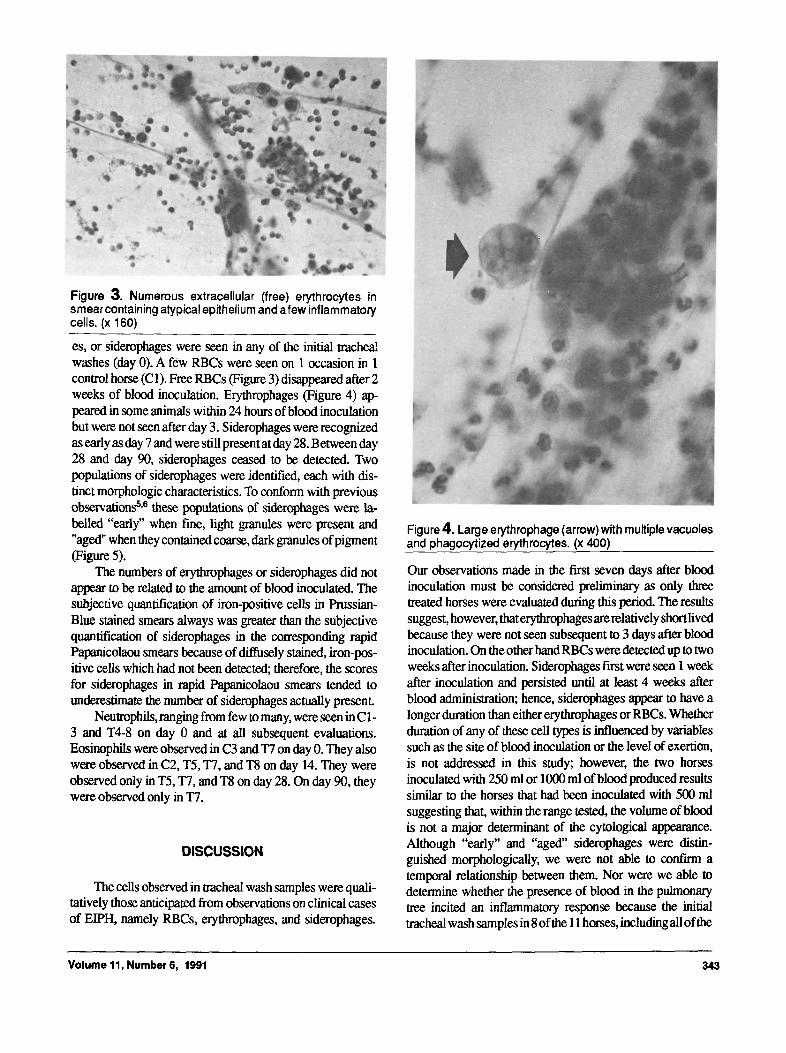
Figure 3. Numerous extracellular (free) erythrocytes in smear containing atypical epithelium and afew inflammatory cells. (x 160)
es, or siderophages were seen in any of the initial tracheal washes (day 0). A few RBCs were seen on 1 occasion in 1 control horse (C1). Free RBCs (Figure 3) disappeared after 2 weeks of blood inoculation. Erythrophages (Figure 4) ap- peared in some animals within 24 hours of blood inoculation but were not seen after day 3. Siderophages were recognized as early as day 7 and were still present at day 28. Between day 28 and day 90, siderophages ceased to be detected. Two populations of siderophages were identified, each with dis- tinct morphologic chaxacteristics. To conform with previous observations 5,6 these populations of siderophages were la, belled "early" when fine, light granules were present and "aged" when they contained coarse, dark granules of pigment (Figure 5).
The numbers of erythrophages or siderophages did not appear to be related to the amount of blood inoculated. The subjective quantification of iron-positive ceils in Prussian- Blue stained smears always was greater than the subjective quantification of siderophages in the corresponding rapid Papanicolaou smears because of diffusely stained, iron-pos- itive cells which had not been detected; therefore, the scores for siderophages in rapid Papanicolaou smears tended to underestimate the number of siderophages aclamlly present.
Neutrophils, ranging from few to many, were seen in C1- 3 and T4-8 on day 0 and at all subsequent evaluations. Eosinophils were observed in C3 and T7 on day 0. They also were observed in C2, T5, T7, and T8 on day 14. They were observed only in T5, T7, and T8 on day 28. On day 90, they were observed only in T7.
DISCUSSION
The cells observed in tracheal wash samples were quali- tatively those anticipated from observations on clinical cases of EIPH, namely RBCs, erythrophages, and siderophages.
Figure 4. Large erythrophage (arrow) with multiple vacuoles and phagocytized erythrocytes. (x 400)
Our observations made in the first seven days after blood inoculation must be considered preliminary as only three treated horses were evaluated during this period. The results suggest, however, thaterythrophages arerelatively shortlived because they were not seen subsequent to 3 days after blood inoculation. On the other hand RBCs were detected up to two weeks after inoculation. Siderophages first were seen 1 week after inoculation and persisted until at least 4 weeks after blood administration; hence, siderophages appear to have a longer duration than either erythrophages or RBCs. Whether duration of any of these cell types is influenced by variables such as the site of blood inoculation or the level of exertion, is not addressed in this study; however, the two horses inoculated with 250 ml or 1000 ml of blood produced results similar to the horses that had been inoculated with 500 ml suggesting that, within the range tested, the volume of blood is not a major determinant of the cytological appearance. Although "early" and "aged" siderophages were distin- guished morphologically, we were not able to confirm a temporal relationship between them. Nor were we able to determine whether the presence of blood in the pulmonary tree incited an inflammatory response because the initial tracheal wash samples in 8 of the 11 horses, including all of the
Volume 11, Number 6, 1991 343

Figure 5. Large "aged* siderophages (arrows) containing large green-brown granules. (x 400)
controls, contained inflammatory cells. The finding of in- flammatory cells in so many horses which were normal on physical examination suggested that subclinical inflamma- tion may be a prevalent problem. The inflammatory cells persisted in subsequent washes and occurred in combination with RBCs, erythrophages, and siderophages in those animals that received intrapulmonary blood. Three horses that had no inflammatory cells (T1, T2, and T3) at day 0, had neutrophils for 2 weeks after instillation of blood. These inflammatory changes may have been the result of blood instillation but could have been caused by repeated tracheal washes or some other factor, for instance viral, irritant, or allergic challenge.
Tracheoscopy and tracheal wash often are used as diag- nostic tools in horses suspected of having EIPH but which have not been observed with blood at the nares. In this study, tracheoscopy alone did not detect blood at more than 7 days after blood inoculation, and gross assessment of tracheal wash aspirates was exactly similar to tracheoscopy in its ability to detect blood. In the 4 weeks after blood inoculation, cytologic findings compatible with clearance of aging pulmonary blood were found in all horses. By 3 months, however, cytologic evaluation did not detect blood. This finding suggests that intrapulmonary blood was fairly rapidly and uniformly
cleared from the airways of all animals. The disappearance of erythrocytes, erythrophages and siderophages represents a balance of mucociliary action, phagocytosis, metabolism and movement into the interstitium. Although this balance may differ between individuals and with the volume of free blood these mechanisms appeared to be effective in eliminating blood. Clearance of the blood occurred in animals with and without preceding pulmonary inflammation. Whether a sin- gle episode of naturally occurring EIPH would result in similar sequential findings is not known; however, if the cytologic findings are similar this would have important implications for the diagnosis and monitoring of animals with EIPH.
Ten percent of the tracheal wash samples contained too few cells for interpretation. Inadequate cellularity occurs commonly in tracheal wash aspirates from patients, however failure to recognize this inadequacy may result in false negative diagnoses of intrapulmonary blood or other process- es. In addition, staining techniques may make a difference in quanfitation and detection of siderophages; the significance and specificity of diffusely stained nongranular iron-positive siderophages which were detectable only with Prussian-Blue stain is not known. It may be that the stippled and granular siderophages, which could be detected with the Papanicolaou stain, are specifically associated with erythrocyte breakdown. Comparison of these results with those from other studies should take into account the method of slide production and staining.
There was one important failure of cytologic evaluat_ion to detect blood within the 4 week period after inoculation. In this instance (T1 on day 7), no evidence of blood was found in a sample of adequate cellularity from a horse in which subsequent tracheal washes were posifive. During the 4 weeks when cytologic evaluation was useful for detecting blood, there were 21 adequate samples collected from inoculated horses; hence, there was arate for false negative diagnosis of 1 in 21. In another instance (C1 on day 1), tracheoscopy and tracheal wash produced a false positive in the form of RBCs seen at cytologic evaluation of a control horse. This probably was the result of trauma to the mucous membranes by the endoscope or sampling catheter. The blood was insufficient to be detected in subsequent evaluations of that animal. Nevertheless, this finding represents a false positive diagnosis of intrapulmonary blood in 1 out of a possible 20 samples (day 0 samples are included in this group of 20). These findings support observations made in foals ~ o and suggest that diagno- sis of intrapulmonary blood should not be based solely on finding a small number of red blood cells in a single tracheal wash. It would be of interest to use controlled conditions to investigate multiple tracheal washes and to compare tracheal wash with bronchoalveolar lavage in experimentally inocu- lated animals and those with naturally occurring EIPH.
Blood inoculated into the pulmonary tree produced en- doscopic and cytologic signs similar to those seen in clinical cases of EIPHbutproducedepistaxis in the majorityofhorses,
344 EQUINE VETERINARY SCIENCE

a sign not consisten tl y fo mad in E IP H. As with an y e x perimen- tal protocol from which we hope to gain an understanding of a clinical condition (such as EIPH), certain assumptions must be borne in mind when exlrapolating conclusions from that experiment to the clinical condition. This protocol assumes that repeated endoscopy, sodium cilrate and lidocaine do not affect the clearance of RBCs. It must also be kept in mind that the horses used in this study, unlike most cases of EIPH, were not in a program of regular, strenuous exercise. Exertion probably increases acute distribution of blood within the pulmonary tree and may affect mucociliary clearance, local immune response, and tracheobronchial secretion. We chose to use unexercised horses not only because it is difficult to find horses in athletic training which are definitely free of EIPH but also to reduce the number of experimental variables. Investigation of the interaction of exercise with RBC clear- ance is a logical extension of this work.
In summary, pulmonary inoculation ofautologous blood resembles EIPH cytologically and endoscopically. Free red blood cells and erythrophages preceded the finding of sid- erophages in cytologic specimens. Evidence of pulmonary blood ceased to be disposed cytologically between 28 and 90 days. Further studies of the clearance of smaller volumes of intrapulmonary blood in exercising and resting horses and comparison with endoscopic and cytologic findings in ani- mals known to have E/PH may lead to better understanding of factors effecting pulmonary clearance of hemorrhage. This may lead to an improvement in the diagnosis and monitoring of animals with EIPH.
REFERENCES
1. Pascoe JR, Wheat JD: Historical background, preva- lence, clinical findings and diagnosis of exercise-induced pulmo- nary hemorrhage in the racing horse. In Proceedings Annu Mtg Amer Amer Assoc Equine Practnr, pp.417-420,1981.
2. Speirs VC, van Veenedaal JC, Harrison IW, Smythe GB, Anderson GA, Wilson DV, Gilbo B: Pulmonary hemorrhage in Standardbred horses after racing. Aust Vet J 59:38-40,1982.
3. Clarke AF: Review of exercise-induced pulmonary hem- orrhage and its possible relationship with mechanical stress. Equine VetJ 17:166-172,1985.
4. Pascoe JR, Raphel C: Pulmonary hemorrhage in exercis- ing horses. Compend Contin Educ Pract Vet 4:$411-$416,1982.
5. Roszel JF, Freeman KP, Slusher SH, Morris WR, Haury KD, Cudd TA: Siderophages in pulmonary cytology specimens from racing and non-racing horses. In Proceedings 31st Annu Mtg Amer Assoc Equine Practnr, pp.182-189,1985.
6. Roszel JF, Freeman KP, Slusher SH, Morris WR, Haury KD., Cudd TA: Siderophages in pulmonary cytology specimens from racing and non-racing horses. In Proceedings 33rd Annu Mtg Amer Assoc Equine Practnr, pp.321-329,1987.
7. Whitwell KE, Greet TRC: Collection and evaluation of tracheobronchial washes in the horse. Equine Vet J 16:499- 508,1984.
8. Freeman KP: A rapid Papanicolaou stain for equine cytologic specimens. Equine Pract 12:37-41,1990.
9. Blood DC, Henderson JA, Radostits OM: Veterinary Medicine, 5th Ed. Philadelphia:Lea &Febiger, 1979.
10. Crane SA, Zeimer EL, Sweeney CR: Cytologic and bac- teriologic evaluation of tracheobronchial aspirates from clinically normal foals. Am J Vet Res 50:2042-2048,1989.
Volume 11, Number 6, 1991 345