Renalase attenuates hypertension, renal injury and cardiac ...
12
Renalase attenuates hypertension, renal injury and cardiac remodelling in rats with subtotal nephrectomy Jianyong Yin a , Zeyuan Lu a , Feng Wang a, *, Zhenzhen Jiang a , Limin Lu b , Naijun Miao b , Niansong Wang a, * a Department of Nephrology and Rheumatology, Shanghai Jiao Tong University Affiliated Sixth People’s Hospital, Shanghai, China b Department of Physiology and Pathophysiology, Shanghai Medical College, Fudan University, Shanghai, China Received: September 22, 2015; Accepted: January 15, 2016 Abstract Chronic kidney disease is associated with higher risk of cardiovascular complication and this interaction can lead to accelerated dysfunction in both organs. Renalase, a kidney-derived cytokine, not only protects against various renal diseases but also exerts cardio-protective effects. Here, we investigated the role of renalase in the progression of cardiorenal syndrome (CRS) after subtotal nephrectomy. Sprague–Dawley rats were randomly subjected to sham operation or subtotal (5/6) nephrectomy (STNx). Two weeks after surgery, sham rats were intravenously injected with Hanks’ balanced salt solution (sham), and STNx rats were randomly intravenously injected with adenovirus-b-gal (STNx+Ad-b- gal) or adenovirus-renalase (STNx+Ad-renalase) respectively. After 4 weeks of therapy, Ad-renalase administration significantly restored plasma, kidney and heart renalase expression levels in STNx rats. We noticed that STNx rats receiving Ad-renalase exhibited reduced protein- uria, glomerular hypertrophy and interstitial fibrosis after renal ablation compared with STNx rats receiving Ad-b-gal; these changes were asso- ciated with significant decreased expression of genes for fibrosis markers, proinflammatory cytokines and nicotinamide adenine dinucleotide phosphate (NADPH) oxidase components. At the same time, systemic delivery of renalase attenuated hypertension, cardiomyocytes hypertro- phy and cardiac interstitial fibrosis; prevented cardiac remodelling through inhibition of pro-fibrotic genes expression and phosphorylation of extracellular signal-regulated kinase (ERK)-1/2. In summary, these results indicate that renalase protects against renal injury and cardiac remodelling after subtotal nephrectomy via inhibiting inflammation, oxidative stress and phosphorylation of ERK-1/2. Renalase shows potential as a therapeutic target for the prevention and treatment of CRS in patients with chronic kidney disease. Keywords: renalase cardiorenal syndromes subtotal nephrectomy renal dysfunction cardiac remodelling Introduction Recently, it has been increasingly recognized that primary disorder of kidney will induce or worsen pathological injuries in heart, which in turn can lead to accelerated organ dysfunction in both systems. This phenomenon has been defined as chronic renocar- diac syndrome or type 4 cardiorenal syndrome (CRS) [1]. Clinical studies demonstrated that patients with primary chronic kidney disease (CKD) show higher incidence of cardiovascular diseases (CVD) and a 10- to 20-fold increased risk of cardiac death com- pared with that of the general population [2, 3]. However, there are still no effective strategies for treatment of CRS for now. Thus, it is urgent to identify underlying mechanisms of CRS pro- gression for developing novel therapy. Although the exact pathophysiology mechanisms underlying this bidirectional cardiorenal crosstalk are not fully elucidated yet, tradi- tional risks in CKD such as humoural and haemodynamic disturbance and activation of renin–angiotensin–aldosterone system and sympa- thetic nerve system (SNS) may contribute to the pathogenesis of CVD [4, 5]. Recently, it has been suggested that abnormalities in bone- mineral axis and deficiency of erythropoietin (EPO) or klotho may directly aggravate cardiac hypertrophy and participate in the patho- genesis of worsening of renal function and cardiovascular complica- tion [6, 7]. Therefore, special emphasis should be placed on the endocrine function of kidney [8]. The reduction or loss of EPO, cal- citriol or klotho and yet-to-be known kidney-derived hormones or cytokines as a result of renal dysfunction may be potential mecha- nisms of CRS [9]. *Correspondence to: Feng WANG, M.D., Ph.D. and Niansong WANG, M.D., Ph.D. E-mail: [email protected] and [email protected] ª 2016 The Authors. Journal of Cellular and Molecular Medicine published by John Wiley & Sons Ltd and Foundation for Cellular and Molecular Medicine. This is an open access article under the terms of the Creative Commons Attribution License, which permits use, distribution and reproduction in any medium, provided the original work is properly cited. doi: 10.1111/jcmm.12813 J. Cell. Mol. Med. Vol 20, No 6, 2016 pp. 1106-1117
-
Upload
khangminh22 -
Category
Documents
-
view
0 -
download
0
Transcript of Renalase attenuates hypertension, renal injury and cardiac ...

Renalase attenuates hypertension, renal injury and
cardiac remodelling in rats with subtotal nephrectomy
Jianyong Yin a, Zeyuan Lu a, Feng Wang a, *, Zhenzhen Jiang a, Limin Lu b,Naijun Miao b, Niansong Wang a, *
a Department of Nephrology and Rheumatology, Shanghai Jiao Tong University Affiliated Sixth People’s Hospital,Shanghai, China
b Department of Physiology and Pathophysiology, Shanghai Medical College, Fudan University, Shanghai, China
Received: September 22, 2015; Accepted: January 15, 2016
Abstract
Chronic kidney disease is associated with higher risk of cardiovascular complication and this interaction can lead to accelerated dysfunction inboth organs. Renalase, a kidney-derived cytokine, not only protects against various renal diseases but also exerts cardio-protective effects.Here, we investigated the role of renalase in the progression of cardiorenal syndrome (CRS) after subtotal nephrectomy. Sprague–Dawley ratswere randomly subjected to sham operation or subtotal (5/6) nephrectomy (STNx). Two weeks after surgery, sham rats were intravenouslyinjected with Hanks’ balanced salt solution (sham), and STNx rats were randomly intravenously injected with adenovirus-b-gal (STNx+Ad-b-gal) or adenovirus-renalase (STNx+Ad-renalase) respectively. After 4 weeks of therapy, Ad-renalase administration significantly restoredplasma, kidney and heart renalase expression levels in STNx rats. We noticed that STNx rats receiving Ad-renalase exhibited reduced protein-uria, glomerular hypertrophy and interstitial fibrosis after renal ablation compared with STNx rats receiving Ad-b-gal; these changes were asso-ciated with significant decreased expression of genes for fibrosis markers, proinflammatory cytokines and nicotinamide adenine dinucleotidephosphate (NADPH) oxidase components. At the same time, systemic delivery of renalase attenuated hypertension, cardiomyocytes hypertro-phy and cardiac interstitial fibrosis; prevented cardiac remodelling through inhibition of pro-fibrotic genes expression and phosphorylation ofextracellular signal-regulated kinase (ERK)-1/2. In summary, these results indicate that renalase protects against renal injury and cardiacremodelling after subtotal nephrectomy via inhibiting inflammation, oxidative stress and phosphorylation of ERK-1/2. Renalase shows potentialas a therapeutic target for the prevention and treatment of CRS in patients with chronic kidney disease.
Keywords: renalase� cardiorenal syndromes� subtotal nephrectomy� renal dysfunction� cardiac remodelling
Introduction
Recently, it has been increasingly recognized that primary disorderof kidney will induce or worsen pathological injuries in heart,which in turn can lead to accelerated organ dysfunction in bothsystems. This phenomenon has been defined as chronic renocar-diac syndrome or type 4 cardiorenal syndrome (CRS) [1]. Clinicalstudies demonstrated that patients with primary chronic kidneydisease (CKD) show higher incidence of cardiovascular diseases(CVD) and a 10- to 20-fold increased risk of cardiac death com-pared with that of the general population [2, 3]. However, thereare still no effective strategies for treatment of CRS for now.
Thus, it is urgent to identify underlying mechanisms of CRS pro-gression for developing novel therapy.
Although the exact pathophysiology mechanisms underlying thisbidirectional cardiorenal crosstalk are not fully elucidated yet, tradi-tional risks in CKD such as humoural and haemodynamic disturbanceand activation of renin–angiotensin–aldosterone system and sympa-thetic nerve system (SNS) may contribute to the pathogenesis of CVD[4, 5]. Recently, it has been suggested that abnormalities in bone-mineral axis and deficiency of erythropoietin (EPO) or klotho maydirectly aggravate cardiac hypertrophy and participate in the patho-genesis of worsening of renal function and cardiovascular complica-tion [6, 7]. Therefore, special emphasis should be placed on theendocrine function of kidney [8]. The reduction or loss of EPO, cal-citriol or klotho and yet-to-be known kidney-derived hormones orcytokines as a result of renal dysfunction may be potential mecha-nisms of CRS [9].
*Correspondence to: Feng WANG, M.D., Ph.D. and Niansong WANG,M.D., Ph.D.
E-mail: [email protected] and [email protected]
ª 2016 The Authors.
Journal of Cellular and Molecular Medicine published by John Wiley & Sons Ltd and Foundation for Cellular and Molecular Medicine.
This is an open access article under the terms of the Creative Commons Attribution License, which permits use,
distribution and reproduction in any medium, provided the original work is properly cited.
doi: 10.1111/jcmm.12813
J. Cell. Mol. Med. Vol 20, No 6, 2016 pp. 1106-1117

Renalase, a recently discovered flavoprotein from kidney, can reg-ulate blood pressure by degrading circulatory catecholamines [10].Our previous reports demonstrated that renalase is up-regulated dur-ing various stresses including hypoxia, ischaemia/reperfusion andoxidative stress [11–13]. Administration of recombinant renalase pro-tects against AKI, contrast-induced nephropathy and cardiac ischae-mia/reperfusion injury [12–15]. Clinical studies showed that renalasedeficiency or single-nucleotide polymorphisms are associated withessential hypertension, cardiac hypertrophy, stroke and diabetes [16–20]. Recently, mounting evidence suggested that renalase exerts itscytoprotective effects by interacting with its plasma membrane recep-tor other than metabolizing catecholamines [21–23]. Thus, renalasemay serve as a cytokine that regulates kidney function in autocrine orparacrine manner [24]. Nevertheless, whether renalase supplementa-tion could prevent the progression of renal injury and occurrence ofremote organ injury in CKD and its mechanisms still remainunknown.
Western blot analyses demonstrated that plasma renalase con-centration in CKD patients is substantially decreased in compar-ison to healthy population [10]. However, several clinical studiesusing ELISA reported opposite results [25, 26]. These oppositeresults may be inaccurate because of a not validated antibodyused in the commercially kit which would non-specifically recog-nize renalase breakdown products or unrelated epitopes [27]. Con-sistently, renalase deficiency is also present in rats subjected tosubtotal nephrectomy [28]. Thus, it is reasonable to speculate thatrenalase may be also a crucial kidney-derived modulator for CRSprogression. In present study, we examined the effects of renalasesupplementation by an adenovirus delivery on the progression oftype 4 CRS in a rat subtotal nephrectomy model. Furthermore, thepotential mechanisms mediating renalase’s protective effects werealso investigated.
Material and methods
Construction of adenovirus vectors
The replication-deficient recombinant adenovirus vectors expressing the
rat renalase mRNA sequence or b-galactosidase (b-gal) were generated
respectively, under the control of the cytomegalovirus enhancer/pro-
moter. These adenoviruses were amplified in HEK-293A cells and puri-fied by CsCl ultracentrifugation and store at �80°C in Hanks’ balanced
salt solution (HBSS) with a concentration of 1.0 9 1010 plaque forma-
tion unit (PFU)/ml.
Animal
All the animal experiments were approved by the Animal Care andEthics Committee of Shanghai Jiao Tong University Affiliated Sixth Peo-
ple’s Hospital. Male Sprague–Dawley rats were provided by Shanghai
Science Academy animal center. All the rats were housed in a 12/
12 hrs light/dark cycle with free access to water and fed with standardrat chow.
Rat model of CKD and experimental protocols
Male Sprague–Dawley rats weighing 200–250 g, after 7-day adaptionperiod, were randomly allocated to either sham-operated group or 5/6
subtotal nephrectomy group. The animals were anaesthetized by
intraperitoneally injection of sodium pentobarbital (50 mg/kg) and two-
stage subtotal nephrectomy was performed as previously described[29]. Briefly, the upper and lower poles of the left kidney were resected
and bleeding was controlled. One week later, the right kidney was
removed after ligation of the renal blood vessels and the ureter. The
sham-operated rats underwent the same procedures, but only the envel-ope capsule was removed.
Two weeks after surgery, the rats were further randomly divided into
following groups: (i) sham-operated treated with tail vein injection of1.2 ml HBSS (sham, n = 10); (ii) subtotal nephrectomy treated with tail
vein injection of 1.2 9 1010 PFU control adenovirus (STNx+Ad-b-gal,n = 10); (iii) subtotal nephrectomy treated with tail vein injection of
1.2 9 1010 PFU adenovirus-renalase (STNx+Ad-renalase, n = 10). Theeffect of renalase gene delivery on CRS was assessed 4 weeks after
adenovirus injection (corresponding to Week 6 of CKD). At the end of
the experiment, all the rats were kept 24 hrs in metabolic cages for
urine collection and echocardiography was also examined during thelast week. Then all the animals were killed and blood as well as kidney
and heart tissues were harvested for analyses.
Renal function assessment
Blood samples were collected from abdominal aorta and centrifuged to
obtain plasma and then separated into aliquots and stored at �20°C.Blood urea nitrogen (BUN) and urinary creatinine and proteinuria were
measured by commercial kit (Nanjingjiancheng, Nanjing, Jiangsu,
China). The ratio of left kidney weight to bodyweight (bw) was also cal-
culated to evaluate renal dysfunction. Plasma norepinephrine concentra-tion was evaluated by ELISA kit (LDN, Nordhorn, Germany).
Systolic blood pressure measurement
Systolic blood pressure (SBP) was measured prior to adenovirus injec-
tion and once a week after gene delivery by a tail-cuff method, using
CODA blood pressure systems (Kent Scientific, Torrington, CT, USA).Prior to the actual experiments, the conscious animals were training for
three consecutive days to become accustomed to the procedure. For
each animal, at least 15 measurements were recorded to calculate a
mean blood pressure and heart rate.
Cardiac functional assessment
Transthoracic echocardiography was performed at the end-point using a
VeVo770 High Resolution Imaging System (Visual Sonics Inc., Toronto, ON,
Canada) with 17.5 MHz transducer as previously described [30]. M-mode
and 2D parasternal short-axis views were analysed in the anaesthetized ratsby trained echocardiographers who were blinded to the treatments.
Before being killed, all the rats underwent LV (left ventricular)
catheterization to assess haemodynamics changes as previously
described [31]. The transducer was attached to RM6240BD multichan-
ª 2016 The Authors.
Journal of Cellular and Molecular Medicine published by John Wiley & Sons Ltd and Foundation for Cellular and Molecular Medicine.
1107
J. Cell. Mol. Med. Vol 20, No 6, 2016

nel biotic signal collection and analysis instrument (Taimeng, Chengdu,China) to determine cardiac function. LV systolic pressure, LV end-dia-
stolic pressure (LVEDP) and the maximal rate of pressure rise (dP/
dtmax) and fall (dP/dtmin) were recorded respectively.
Heart weight (HW) index were analysed among the three groups. Thehearts were rapidly excised, cleaned and weighed. The left ventricles were
separated and weighed. The ratios of the whole HW to bw, the ratios of
the LV weight (LW) to bw were calculate to evaluate cardiac hypertrophy.
Histology analysis
All the tissues were fixed in 4% paraformaldehyde and embedded inparaffin. Tissue sections (4–6 lm) were stained with haematoxylin and
eosin, Masson trichrome solution or Sirus Red to assess histological
injury and fibrosis.
A semi-quantitative score was used to determine the severity ofglomerulosclerosis and tubulointerstitial injury as described previously
[32, 33]. Glomerular area was measured as described previously [34].
All evaluation was performed by an observer who was blind to theexperimental protocol. To evaluate the degree of fibrosis, 10 non-over-
lapping fields of each section were randomly chosen. Cross-section area
of cardiomyocytes was calculated from the average of 50 cardiomy-
ocytes per section as described previously [35].For immunohistochemistry, kidney sections were stained with antirat
monoclonal antibodies against CD68 (Sigma-Aldrich, St. Louis, MO,
USA), CD86, CD163 and renalase (both from Abcam, Cambridge, MA,
USA) as previously described [36]. All the measurements were calcu-lated by ImagePro Plus Systems.
Quantitative real-time PCR
Total RNA were extracted from the renal cortex or LV tissue using Trizol
(Invitrogen, Carlsbad, CA, USA) and was reverse transcribed into cDNA
with M-MLV Reverse Transcriptase (Promega, Madison, WI, USA).Real-time PCR was performed with SYBE Green PCR master Mix (Tar-
kara, Dalian, China) using StepOnePlus PCR Systems (Applied Biosys-
tems, Foster City, CA, USA) according to the manufacturer’s protocol.
Quantitation was normalized to internal control GAPDH and 2�DDCT
method was used to determine relative gene expression levels. All the
primer pairs are seen in Table S1.
Western blot analysis
Total protein was prepared from frozen tissues by homogenization. Pro-
tein concentrations were determined by BCA assay (Beyotime, Suzhou,Jiangsu, China) and protein samples were separated by 10–12% SDS-
PAGE and then transferred to polyvinylidene difluoride membrane. The
membranes were blocked with 5% non-fat dried milk and then incu-
bated with primary antibodies against matrix metalloproteinase 1(MMP-1), tissue inhibitor of metalloproteinase-1 (TIMP-1), renalase
(Abcam), phospho-ERK-1/2, total ERK-1/2, phospho-p38, total p38 (Cell
Signaling Technology, Danvers, MA, USA). Horseradish peroxidase-con-jugated secondary antibodies (Beyotime) were used and visualized by
Image Quant LAS 4000 Mini System (GE Healthcare, Pittsburgh, PA,
USA). The bands were analysed using Image J software and GAPDH or
tubulin (Proteintech, Chicago, IL, USA) was used as internal control.
Statistical analyses
SPSS software 19.0 (IBM, Armonk, NY, USA) was used for all the sta-tistical analyses. All the values are expressed as mean � S.E.M. One-
way ANOVA followed by a with Tukey’s Multiple Comparison Test was
used to compare parametric data while Kruskal–Wallis test followed by
the Mann–Whitney U-test was used for non-parametric data compari-son. A value of P < 0.05 were considered statistically significant.
Results
Adenovirus-mediated gene delivery efficacy
As shown in Figure S1, subtotal nephrectomy resulted in dramaticdecrease in renalase expression both in the plasma, remnant kidneyand heart compared with sham group. Renalase gene deliveryrestored renalase expression nearly to normal levels in circulation,kidney and heart according to Western blot and PCR results in STNxrats (Fig. S1). Consistently, immunohistochemistry analysis alsoshowed adenovirus-renalase effectively increase the percentage ofrenalase-positive cells in kidney compared with Ad-b-gal-treatedSTNx rats (Fig. S2).
Renalase gene delivery ameliorated renaldysfunction in STNx rats
Compared to sham group, STNx rats exhibited significant renal dys-function characterized by increased plasma BUN, urine 24 hrs totalprotein levels, and decreased creatinine clearance as shown inTable 1. After 4 weeks of treatment, systemic delivery of renalasesignificantly decreased plasma BUN and proteinuria in STNx rats.Kidney weight/bw was also lower in Ad-renalase-treated group thanAd-b-gal-treated group (Table 1). In addition, Ad-renalase deliveryattenuated creatinine clearance decline in STNx rats. Furthermore,plasma norepinephrine level in Ad-renalase-treated STNx rats wasalso significantly lower than Ad-b-gal-treated STNx rats (Table 1);suggesting renalase may participate in the metabolism of cate-cholamines.
Renalase supplementation attenuated renalpathological injury
Kidney sections from STNx rats revealed a remarkable glomerularhypertrophy and tubular enlargement (Fig. 1A). Ad-renalase treat-ment significantly decreased glomerular area (Fig. 1G) and amelio-rated glomerularsclerosis and tubulointerstitial injury after subtotalnephrectomy as assessed by pathological score (Fig. 1E and F). Toevaluate the extent of interstitial fibrosis, kidney tissues were stainedwith Masson and Sirus Red. Results showed that renal interstitialfibrosis in Ad-renalase-treated rats was less severe than that in Ad-b-gal-treated STNx rats as shown in Figure 1B–D. Consistent with the
1108 ª 2016 The Authors.
Journal of Cellular and Molecular Medicine published by John Wiley & Sons Ltd and Foundation for Cellular and Molecular Medicine.

histological data, mRNA levels of fibrosis markers, including collagenI, collagen III and transforming growth factor (TGF)-b1 were signifi-cantly higher in the remnant kidneys of Ad-b-gal-treated STNx ratsthan in those of sham rats; and Ad-renalase treatment significantlydecreased mRNA expression of these fibrosis markers (Fig. 3A).
Renalase inhibited inflammation and oxidativestress in the remnant kidney
To explore whether renalase was involved in macrophage infiltration,activation and polarization, we examined renal infiltration of totalmacrophage (CD68), M1-like (CD86) and M2-like macrophage(CD163). Immunohistochemistry analysis showed that CKD rats dis-played increased all subtypes of macrophage infiltration andenhanced M1 polarization. Ad-renalase administration significantlydiminished total macrophage infiltration numbers especially M1 num-bers than Ad-b-gal-treated rats after subtotal nephrectomy (Fig. 2).In addition, Ad-renalase-treated rats exhibited slightly more M2 infil-tration than Ad-b-gal-treated rats.
To determine the effects of renalase supplementation on inflamma-tion and oxidative stress, mRNA levels of pro-inflammatory cytokinesand NADPH oxidase components were also measured by real-time PCR.Nephrectomized rats showed higher expression levels of pro-inflamma-tory cytokines, including tumour necrosis factor (TNF)-a, interleukin(IL)-6, monocyte chemotactic protein (MCP)-1 (Fig. 3B) and NADPHoxidase components, including gp91phox, p47phox and p67phox comparedwith sham (Fig. 3C); and Ad-renalase treatment decreased the expres-sion of pro-inflammatory cytokines and NADPH oxidase components.Taking together, these findings indicated renalase supplementationmight protect renal injury after renal ablation via inhibiting inflammationactivation and oxidative stress in remnant kidney.
Effects of renalase on systolic blood pressure
As shown in Figure 4, SBP in STNx rats gradually increased duringthe observation compared with the sham group and Ad-renalase
delivery significantly decreased SBP in STNx rats compared with Ad-b-gal-treated rats.
Renalase supplementation prevented cardiacremodelling in STNx rats
The HW/bw and LV weight/bw were significantly higher in STNx ratsthan the sham and significantly lower in Ad-renalase-treated rats thanthe Ad-b-gal-treated rats (Table 2). Concomitant alterations werefound in echocardiography (Table 2 and Fig. S3), 4 weeks of treat-ment with Ad-renalase significantly decreased LVPWd, LVAWd andincreased LVIDs and LVIDs (Table 2). However, there was no differ-ence in FS and EF among the three groups.
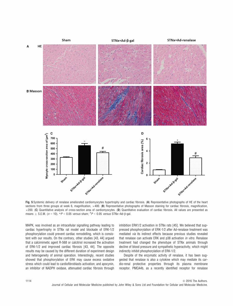
Histological sections of hearts demonstrated that subtotalnephrectomy led to elevated cross-section area of cardiomyocytesand Ad-renalase delivery ameliorated cardiac cardiomyocyteshypertrophy in STNx rats (Fig. 5A and C). Cardiac fibrosisassessment, determined by Masson staining, showed that STNxrats had more matrix deposition than sham; and lesser cardiacinterstitial fibrosis was observed in the Ad-renalase-treated ratscompared with Ad-b-gal-treated STNx rats (Fig. 5B and D). Inaddition, Western blot showed that expression of MMP-1 wassignificantly lower in Ad-b-gal-treated rats than sham and signifi-cantly higher in Ad-renalase-treated rats than Ad-b-gal-treatedrats. Meanwhile, the expression of TIMP-1 and TGF-b was signif-icantly higher in Ad-b-gal-treated rats than sham and significantlylower in Ad-renalase-treated rats compared with Ad-b-gal-treatedrats (Fig. 6A–C).
Deactivation of mitogen-activated proteinkinases after treatment with Ad-renalase
The phosphorylated of ERK-1/2 (p-ERK-1/2) in heart (Fig. 6D) andkidney (Fig. 7A) was both significantly increased in STNx rats. AndAd-renalase treatment significantly decreased the phosphorylation ofERK-1/2 compared with Ad-b-gal-treated rats. However, there is no
Table 1 General characteristics of rats and renal function assessments 6 weeks after surgery
Sham STNx+Ad-b-gal STNx+Ad-renalase
Bodyweight (g) 485 � 17 410 � 19* 425 � 15
Left kidney weight/bw (mg/g) 5.31 � 0.17 4.5 � 0.48** 4.1 � 0.39**,#
Serum urea nitrogen 5.62 � 0.86 17.23 � 1.21** 13.59 � 0.99*,#
Proteinuria (mg/24 hrs) 12.08 � 1.29 134 � 9.76*** 59.90 � 5.79***,###
Creatinine clearance (ml/min.) 1.56 � 0.18 0.87 � 0.20** 1.13 � 0.2*,#
Norepinephrine (pg/ml) 438 � 49 1280 � 81* 667 � 78#
*P < 0.05 versus sham; #P < 0.05 versus STNx+Ad-b-gal; **P < 0.01 versus sham; ##P < 0.01 versus STNx+Ad-b-gal; ***P < 0.001 versussham; ###P < 0.001 versus STNx+Ad-b-gal.All values are presented as means � S.E.M. (n = 10).STNx: subtotal nephrectomy; bw: bodyweight.
ª 2016 The Authors.
Journal of Cellular and Molecular Medicine published by John Wiley & Sons Ltd and Foundation for Cellular and Molecular Medicine.
1109
J. Cell. Mol. Med. Vol 20, No 6, 2016

1110 ª 2016 The Authors.
Journal of Cellular and Molecular Medicine published by John Wiley & Sons Ltd and Foundation for Cellular and Molecular Medicine.
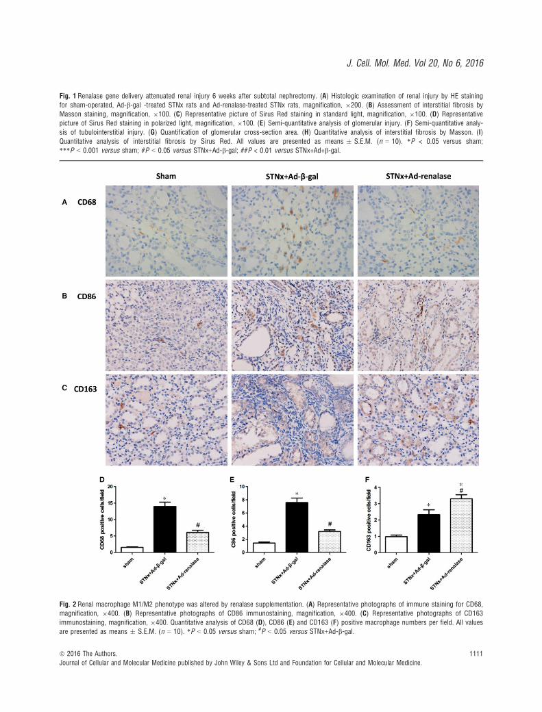
Fig. 2 Renal macrophage M1/M2 phenotype was altered by renalase supplementation. (A) Representative photographs of immune staining for CD68,magnification, 9400. (B) Representative photographs of CD86 immunostaining, magnification, 9400. (C) Representative photographs of CD163
immunostaining, magnification, 9400. Quantitative analysis of CD68 (D), CD86 (E) and CD163 (F) positive macrophage numbers per field. All values
are presented as means � S.E.M. (n = 10). *P < 0.05 versus sham; #P < 0.05 versus STNx+Ad-b-gal.
Fig. 1 Renalase gene delivery attenuated renal injury 6 weeks after subtotal nephrectomy. (A) Histologic examination of renal injury by HE staining
for sham-operated, Ad-b-gal -treated STNx rats and Ad-renalase-treated STNx rats, magnification, 9200. (B) Assessment of interstitial fibrosis by
Masson staining, magnification, 9100. (C) Representative picture of Sirus Red staining in standard light, magnification, 9100. (D) Representativepicture of Sirus Red staining in polarized light, magnification, 9100. (E) Semi-quantitative analysis of glomerular injury. (F) Semi-quantitative analy-
sis of tubulointerstitial injury. (G) Quantification of glomerular cross-section area. (H) Quantitative analysis of interstitial fibrosis by Masson. (I)Quantitative analysis of interstitial fibrosis by Sirus Red. All values are presented as means � S.E.M. (n = 10). *P < 0.05 versus sham;
***P < 0.001 versus sham; #P < 0.05 versus STNx+Ad-b-gal; ##P < 0.01 versus STNx+Ad+b-gal.
ª 2016 The Authors.
Journal of Cellular and Molecular Medicine published by John Wiley & Sons Ltd and Foundation for Cellular and Molecular Medicine.
1111
J. Cell. Mol. Med. Vol 20, No 6, 2016

significant difference in the expression of phosphorylated p38 (p-p38) among the three groups (Fig. 7B).
Discussion
In a pathological circumstance such as CRS, bidirectional heart–kid-ney interactions could lead to a vicious cycle which must be pre-vented. It has been identified that several hormones or cytokinessecreted by kidney or heart have beneficial effect on other organ andmay represent as potential therapeutic targets. For instance, EPO pre-
vented cardiac remodelling in nephrectomized rats beyond haemato-poiesis and independent of kidney function [37, 38]. Conversely,overexpression of hormones such as brain natriuretic peptide [29]and Follistatin-like 1 (Fstl-1) secreted by heart, attenuated renal fibro-sis and cardiac-specific Fstl-1 knockout mice exhibited exacerbationof renal injury after subtotal nephrectomy [39]. In the present study,we confirmed the beneficial effects of renalase supplementation onfunctional and histological alterations of kidney and heart and investi-gated the underlying mechanism in a rat model with progressive renalinjury. We demonstrated that systemic delivery of renalase, a kidney-derived protein, attenuated both renal and cardiac fibrosis, improvedrenal function and prevented cardiac remodelling 6 weeks after renalablation through its anti-inflammatory and anti-oxidant effects andinhibition of ERK pathway, indicating that renalase may play a crucialrole in the interaction between kidney and heart.
Renalase is a novel protein secreted by kidney and firstly reportedto be a catecholamine-metabolizing enzyme. Our previous reportsdemonstrated that only kidney tubular epithelial cells could secreterenalase in vitro, thus tubular epithelial cells may be the primarysource of circulation renalase [40]. Reduction of plasma and kidneyrenalase levels may contribute to elevated circulating catecholaminesand consequent SNS activation in CKD rats. In agreement with Bara-ka’s study [41], we found that renalase supplementation indeeddecreased the circulation norepinephrine level, SBP and attenuatedcardiac hypertrophy. More importantly, rats with renalase administra-tion also showed improvement of albuminuria, glomerulosclerosis,tubular dilation and interstitial fibrosis after renal ablation, which wasnot observed in previous report [41]. This difference may be causedby distinct methods of renalase supplementation. Recombinant pro-tein subcutaneously injection is susceptible to be degraded while ade-novirus can broadly infect various cells and leads to effective geneoverexpression, is thought to be a more appropriate tool for
Fig. 4 Systolic blood pressure of sham-operated, Ad-b-gal-treated STNx
rats and Ad-renalase-treated STNx rats. Systolic blood pressure valuesare expressed as mean � S.E.M. (n = 10). *P < 0.05 versus sham;#P < 0.05 versus STNx+Ad-b-gal; **P < 0.01 versus sham; ##P < 0.01
versus STNx+Ad-b-gal; ***P < 0.001 versus sham; ###P < 0.001 ver-
sus STNx+Ad-b-gal.
Fig. 3 Ad-renalase-treated rats exhibited decreased mRNA
expression of fibrosis markers, pro-inflammatory cytokinesand NADPH oxidative stress components in the kidney.
(A) Relative mRNA expression collagen I, collagen III and
TGF-b1. (B) Relative mRNA expression of TNF-a, IL-6 and
MCP-1. (C) Relative mRNA expression of gp91phox,P47phox and P67phox. All values are expressed as
mean � S.E.M. (n = 10). *P < 0.05 versus sham;#P < 0.05 versus STNx+Ad-b-gal; **P < 0.01 versus
sham.
1112 ª 2016 The Authors.
Journal of Cellular and Molecular Medicine published by John Wiley & Sons Ltd and Foundation for Cellular and Molecular Medicine.

long-term experiments. These findings indicated that Ad-renalase-administered rats had a lesser degree of CRS; regulation of bloodpressure by degrading circulation norepinephrine may be at least inpart mechanism for its protective effects.
Inflammation and oxidative stress plays a pivotal role in theprogression of CKD. Our recent reports had shown that renalaseprotected HK2 cells against the cytotoxicity of H2O2 through sup-pressing oxidative stress and apoptosis and recombinant renalaseprotected against contrast-induced nephropathy through anti-oxida-tion and anti-inflammation mechanism [12, 13]. Other reportsdemonstrated that renalase-knockout mice led to exacerbation ofrenal injury caused by acute cisplatin nephrotoxicity and ischemia/reperfusion damage through up-regulation of pro-inflammatorycytokines [15, 23]. Herein, we found that renalase supplementationalso inhibited inflammatory response in remnant kidney via down-regualtion of pro-inflammatory cytokines and NADPH oxidase com-ponents. Besides, we observed that Ad-renalase treatment couldnot only reduce macrophage infiltration but also regulate change
in macrophage M1/M2 phenotype during CKD progression. Rena-lase might attenuate renal inflammation via inhibiting pro-inflam-matory M1-like macrophage polarization and promoting anti-inflammation or antifibrosis M2-like macrophage activation.
Evidence showed that renalase-knockout mice exhibit mild LVhypertrophy and renalase polymorphisms are associated with cardiachypertrophy in patients. Our results proved that renalase supplemen-tation ameliorated cardiac fibrosis and cardiomyocytes hypertrophy,decreased LVPWd and LVEDP and restored cardiac diastolic functionin CKD rats. SBP decline after renalase supplementation might bemainly accounted for improvement of cardiac function. Beside, thisamelioration was accompanied by remarkably milder cardiomyocyteshypertrophy, reduced expression of pro-fibrotic markers and inhibi-tion of ERK-1/2 activation.
It has been established that mitogen-activated protein kinase(MAPK) activity plays an important role in cardiomyocytes hypertro-phy and pressure-overload induced cardiac remodelling. Takahashiet al. [42] reported that the phosphorylation of ERK-1/2, not p38
Table 2 Cardiac function assessment 6 weeks after surgery
Sham STNx+Ad-b-gal STNx+Ad-renalase
HR 371 � 31 410 + 13 402 � 19
HW/bw (mg/g) 2.35 � 0.17 3.9 � 0.25*** 3.68 � 0.21**,#
LW/HW (mg/g) 4.1 � 0.36 6.1 � 0.53*** 5.6 � 0.49**,#
Echocardiography
LVPWd (mm) 2.01 � 0.08 2.88 � 0.16*** 1.99 � 0.13###
LVPWs (mm) 3.22 � 0.16 4.24 � 0.08*** 3.60 � 0.15##
LVAWd (mm) 1.81 � 0.07 2.43 � 0.01*** 1.91 � 0.08##
LVAWs (mm) 3.10 � 0.15 4.22 � 0.13*** 3.64 � 0.13*,#
LVIDd (mm) 7.05 � 0.27 5.57 � 0.20* 7.05 � 0.31##
LVIDs (mm) 3.32 � 0.40 1.58 � 0.19** 2.66 � 0.29##
Fractional shortening (%) 52.02 � 3.57 71.34 � 3.26 60.59 � 3.64
EF (%) 94.35 � 1.6 80.81 � 3.39 87.72 � 2.7
Cardiac catheterization
LVEDP (mmHg) 2.3 � 0.49 6.5 � 0.56** 4.5 � 0.40*,#
LVESP (mmHg) 70.45 � 5.98 69.43 � 7.89 80.32 � 5.76
+dP/dtmax (mmHg/sec.) 4954 � 389 5130 � 447 4986 � 450
�dP/dtmin (mmHg/sec.) �4823 � 430 �3960 � 536 �4423 � 378
*P < 0.05 versus sham; #P < 0.05 versus STNx+Ad-b-gal; **P < 0.01 versus sham; ##P < 0.01 versus STNx+Ad-b-gal; ***P < 0.001 versussham; ###P < 0.001 versus STNx+Ad-b-gal.All values are presented as means � S.E.M. (n = 8).LVPWd: LV end-diastolic posterior wall thickness; LVPWs: LV end-systole posterior wall thickness; LVAWd: LV end-diastolic anterior wall thick-ness; LVAWs: LV end-systole anterior wall thickness; LVIDd: LV internal diastolic diameter; LVIDs: LV internal systolic diameter; dP/dtmax: rateof LV pressure rise; �dP/tmin: rate of LV pressure fall; LVEDP: LV end-diastolic pressure; LVESP: LV end-systolic pressure.
ª 2016 The Authors.
Journal of Cellular and Molecular Medicine published by John Wiley & Sons Ltd and Foundation for Cellular and Molecular Medicine.
1113
J. Cell. Mol. Med. Vol 20, No 6, 2016
MAPK, was involved as an intracellular signalling pathway leading tocardiac hypertrophy in STNx rat model and blockade of ERK-1/2phosphorylation could prevent cardiac remodelling, which is consis-tent with our results. On the contrary, other studies [43, 44] arguedthat a calcimimetic agent R-568 or calcitriol increased the activationof ERK-1/2 and improved cardiac fibrosis [43, 44]. The oppositeresults may be caused by the different duration of experiment designand heterogeneity of animal operation. Interestingly, recent studiesshowed that phosphorylation of ERK may cause excess oxidativestress which could lead to cardiofibroblasts activation; and apocynin,an inhibitor of NADPH oxidase, attenuated cardiac fibrosis through
inhibition ERK1/2 activation in STNx rats [45]. We believed that sup-pressed phosphorylation of ERK-1/2 after Ad-renalase treatment wasmediated via its indirect effects because previous studies revealedthat renalase can activate ERK and p38 activation in vitro. Renalasetreatment had changed the phenotype of STNx animals throughdecline of blood pressure and sympathetic hyperactivity, which mightindirectly inhibit phosphorylation of ERK-1/2.
Despite of the enzymatic activity of renalase, it has been sug-gested that renalase is also a cytokine which may mediate its car-dio-renal protective properties through its plasma membranereceptor. PMCA4b, as a recently identified receptor for renalase
Fig. 5 Systemic delivery of renalase ameliorated cardiomyocytes hypertrophy and cardiac fibrosis. (A) Representative photographs of HE of the heartsections from three groups at week 6, magnification, 9400. (B) Representative photographs of Masson staining for cardiac fibrosis, magnification,
9200. (C) Quantitative analysis of cross-section area of cardiomyocytes. (D) Quantitative evaluation of cardiac fibrosis. All values are presented as
means � S.E.M. (n = 10). *P < 0.05 versus sham; #P < 0.05 versus STNx+Ad-b-gal.
1114 ª 2016 The Authors.
Journal of Cellular and Molecular Medicine published by John Wiley & Sons Ltd and Foundation for Cellular and Molecular Medicine.

[22], is a calcium pump that participates in Ca2+-dependent sig-nalling. Previous reports demonstrated that PMCA4b transgenicmice exhibited improvement of LV hypertrophy following transverseaortic constriction or phenylephrine/Ang II infusion and PMCA4boverexpression inhibited cardiomyocyte hypertrophy in vitro [46].Supplementation of renalase may directly antagonize cardiac hyper-trophy through activating extracellular PMCA4b on cardiomyocytesand related downstream signalling pathway in an endocrinemanner.
There are some limitations of this study. First, the study durationis only 6 weeks which may be too short for CKD animals. Second,
we could not conclude that renalase exerts its protective effects bylowering blood pressure or directly inhibiting fibrosis and hypertro-phy of hearts because of lack of in vitro studies. Thus, kidney-speci-fic renalase-knockout animal should be used in future study todetermine the mechanism of renalase’s protective effects in type 4CRS.
In summary, renalase supplementation by systemic delivery ofadenovirus-renalase ameliorated both renal dysfunction and cardiacremodelling after subtotal nephrectomy in catecholamine-lowering-dependent/independent manners. The potential mechanism of its pro-tective effects may be mediated by its anti-inflammatory, anti-oxidant
Fig. 6 Ad-renalase treatment normalized
cardiac expression of pro-fibrotic markers
and phosphorylated ERK-1/2 in CKD rats.
Representative Western blot and quantifi-cation of TGF-b (A), MMP-1 (B), TIMP-1
(C) and phosphorylated ERK-1/2 (D)expression in left ventricle from three
groups at week 6. All values are presentedas means � S.E.M. (n = 10). *P < 0.05
versus sham; **P < 0.01 versus sham;#P < 0.05 versus STNx+Ad-b-gal.
Fig. 7 Ad-renalase treatment suppressed
ERK-1/2 phosphorylation in the remnantkidney. Representative Western blots of
phosphorylated ERK-1/2 (A) and phospho-
rylated p38 (B) in kidney. Values are pre-
sented as means � S.E.M. (n = 10).*P < 0.05 versus sham; **P < 0.01 ver-
sus sham; ##P < 0.01 versus STNx+Ad-b-gal; NS, no statistically significant.
ª 2016 The Authors.
Journal of Cellular and Molecular Medicine published by John Wiley & Sons Ltd and Foundation for Cellular and Molecular Medicine.
1115
J. Cell. Mol. Med. Vol 20, No 6, 2016

function and inhibition of ERK1/2 pathway. Therefore, renalase is acrucial modulator of CRS progression and renalase supplementationmay be a promising therapy for prevention and deterioration of CRSin CKD patients.
Acknowledgements
The research is supported by National Nature Science Foundation of China
(81270824 and 81570603), Shanghai Pujiang Program (15PJ1406700), and
Shanghai Talents Development Fund.
Conflicts of interest
None.
Supporting information
Additional Supporting Information may be found in the onlineversion of this article:
Figure S1 Adenovirus-mediated renalase expression efficacy in vivo.
Figure S2 Evaluation of renalase expression efficacy by immunos-taining.
Figure S3 Reprehensive echocardiography at week 6.
Table S1 Primer sequences used in real-time PCR.
References
1. House AA, Anand I, Bellomo R, et al. AcuteDialysis Quality Initiative Consensus G. Defi-nition and classification of Cardio-Renal
Syndromes: workgroup statements from the
7th ADQI Consensus Conference. Nephrol
Dial Transplant. 2010; 25: 1416–20.2. Sarnak MJ, Levey AS, Schoolwerth AC,
et al. Kidney disease as a risk factor for
development of cardiovascular disease - A
statement from the American Heart Asso-ciation councils on kidney in cardiovascu-
lar disease, high blood pressure research,
clinical cardiology, and epidemiology andprevention. Circulation. 2003; 108: 2154–69.
3. Clark LE, Khan I. Outcomes in CKD: what we
know and what we need to know. NephronClin Pract. 2010; 114: C95–102.
4. Tsuruya K, Eriguchi M. Cardiorenal syn-
drome in chronic kidney disease. Curr Opin
Nephrol Hypertens. 2015; 24: 154–62.5. McCullough PA, Kellum JA, Haase M, et al.
Pathophysiology of the cardiorenal syn-
dromes: executive summary from the ele-venth consensus conference of the Acute
Dialysis Quality Initiative (ADQI). Contrib
Nephrol. 2013; 182: 82–98.6. Charytan DM, Fishbane S, Malyszko J,
et al. Cardiorenal syndrome and the role of
the bone-mineral axis and anemia. Am J Kid-
ney Dis. 2015; 66: 196–205.7. Hu MC, Shi MJ, Cho HJ, et al. Klotho and
phosphate are modulators of pathologic ure-
mic cardiac remodeling. J Am Soc Nephrol.
2015; 26: 1290–302.8. Kurt B, Kurtz A. Plasticity of renal endocrine
function. Am J Physiol Regul Integr Comp
Physiol. 2015; 308: R455–66.
9. Ros S, Carrero JJ. Endocrine alterations andcardiovascular risk in CKD: is there a link?Nefrologia. 2013; 33: 181–7.
10. Xu J, Li G, Wang P, et al. Renalase is a nov-el, soluble monoamine oxidase that regu-
lates cardiac function and blood pressure. JClin Invest. 2005; 115: 1275–80.
11. Wang F, Cai H, Zhao Q, et al. Epinephrineevokes renalase secretion via alpha-adreno-
ceptor/NF-kappaB pathways in renal proxi-mal tubular epithelial cells. Kidney Blood
Press Res. 2014; 39: 252–9.12. Wang F, Zhang G, Xing T, et al. Renalase
contributes to the renal protection of delayed
ischaemic preconditioning via the regulation
of hypoxia-inducible factor-1alpha. J Cell
Mol Med. 2015; 19: 1400–9.13. Zhao B, Zhao Q, Li J, et al. Renalase pro-
tects against contrast-induced nephropathy
in Sprague-Dawley rats. PLoS ONE. 2015;
10: e0116583.14. Wu Y, Xu J, Velazquez H, et al. Renalase
deficiency aggravates ischemic myocardial
damage. Kidney Int. 2011; 79: 853–60.15. Lee HT, Kim JY, Kim M, et al. Renalase
protects against ischemic AKI. J Am Soc
Nephrol. 2013; 24: 445–55.16. Farzaneh-Far R, Desir GV, Na B, et al. A
functional polymorphism in renalase
(Glu37Asp) is associated with cardiac hyper-
trophy, dysfunction, and ischemia: data from
the heart and soul study. PLoS ONE. 2010;5: e13496.
17. Buraczynska M, Zukowski P, BuraczynskaK, et al. Renalase gene polymorphisms inpatients with type 2 diabetes, hypertension
and stroke. NeuroMol Med. 2011; 13: 321–7.
18. Stec A, Semczuk A, Furmaga J, et al. Poly-morphism of the renalase gene in end-stagerenal disease patients affected by hyperten-
sion. Nephrol Dial Transplant. 2012; 27:
4162–6.19. Zhang R, Li X, Liu N, et al. An association
study on renalase polymorphisms
and ischemic stroke in a Chinese
population. NeuroMol Med. 2013; 15: 396–404.
20. Wang F, Huang B, Li J, et al. Renalase
might be associated with hypertension and
insulin resistance in Type 2 diabetes. RenFail. 2014; 36: 552–6.
21. Beaupre BA, Hoag MR, Moran GR. Renalasedoes not catalyze the oxidation of cate-
cholamines. Arch Biochem Biophys. 2015;579: 62–6.
22. Wang L, Velazquez H, Chang J, et al. Iden-tification of a receptor for extracellular rena-
lase. PLoS ONE. 2015; 10: e0122932.23. Wang L, Velazquez H, Moeckel G, et al.
Renalase prevents AKI independent of amine
oxidase activity. J Am Soc Nephrol. 2014;25: 1226–35.
24. Guo X, Wang L, Velazquez H, et al. Rena-lase: its role as a cytokine, and an update on
its association with type 1 diabetes andischemic stroke. Curr Opin Nephrol Hyper-
tens. 2014; 23: 513–8.25. Malyszko J, Koc-Zorawska E, Malyszko JS,
et al. Renalase, stroke, and hypertension inhemodialyzed patients. Ren Fail. 2012; 34:
727–31.26. Zbroch E, Malyszko J, Malyszko JS, et al.
Renalase, a novel enzyme involved in blood
pressure regulation, is related to kidney
function but not to blood pressure in
1116 ª 2016 The Authors.
Journal of Cellular and Molecular Medicine published by John Wiley & Sons Ltd and Foundation for Cellular and Molecular Medicine.

hemodialysis patients. Kidney Blood PressRes. 2012; 35: 395–9.
27. Desir GV, Peixoto AJ. Renalase in hyperten-
sion and kidney disease. Nephrol Dial
Transpl. 2014; 29: 22–8.28. Li G, Xu J, Wang P, et al. Catecholamines
regulate the activity, secretion, and synthesis
of renalase. Circulation. 2008; 117: 1277–82.29. Kasahara M, Mukoyama M, Sugawara A,
et al. Ameliorated glomerular injury in mice
overexpressing brain natriuretic peptide with
renal ablation. J Am Soc Nephrol. 2000; 11:1691–701.
30. Fu MQ, Zhou JM, Qian JY, et al. Adiponec-tin through its biphasic serum level is a use-
ful biomarker during transition fromdiastolic dysfunction to systolic dysfunction
- an experimental study. Lipids Health Dis.
2012; 11: 106.31. Yang XL, Zhang HP, Jia YX, et al. Effects of
intermedin1-53 on myocardial fibrosis. Acta
Bioch Bioph Sin. 2013; 45: 141–8.32. Adamczak M, Gross ML, Krtil J, et al. Rev-
ersal of glomerulosclerosis after high-dose
enalapril treatment in subtotally nephrec-
tomized rats. J Am Soc Nephrol. 2003; 14:
2833–42.33. Wang F, Zhang G, Lu Z, et al. Antithrombin
III/SerpinC1 insufficiency exacerbates renal
ischemia/reperfusion injury. Kidney Int.
2015; 88: 796–803.
34. Ohashi K, Iwatani H, Kihara S, et al. Exac-erbation of albuminuria and renal fibrosis in
subtotal renal ablation model of adiponectin-
knockout mice. Arterioscler Thromb Vasc
Biol. 2007; 27: 1910–7.35. Liu S, Kompa AR, Kumfu S, et al. Subtotal
nephrectomy accelerates pathological car-
diac remodeling post-myocardial infarction:implications for cardiorenal syndrome. Int J
Cardiol. 2013; 168: 1866–80.36. Wang F, Xing T, Wang N. Construction and
DNA immunization of human renalaseeukaryotic expression vector. NDT Plus.
2011; 4: 221.
37. Gut N, Piecha G, Aldebssi F, et al. Erythro-poietin combined with ACE inhibitor pre-vents heart remodeling in 5/6
nephrectomized rats independently of blood
pressure and kidney function. Am J Nephrol.2013; 38: 124–35.
38. Toba H, Nakashima K, Oshima Y, et al. Ery-thropoietin prevents vascular inflammation
and oxidative stress in subtotal nephrec-tomized rat aorta beyond haematopoiesis.
Clin Exp Pharmacol Physiol. 2010; 37:
1139–46.39. Hayakawa S, Ohashi K, Shibata R, et al.
Cardiac myocyte-derived follistatin-like 1
prevents renal injury in a subtotal nephrec-
tomy model. J Am Soc Nephrol. 2015; 26:
636–46.
40. Wang F, Xing T, Li J, et al. Renalase’sexpression and distribution in renal tissue
and cells. PLoS ONE. 2012; 7: e46442.
41. Baraka A, El Ghotny S. Cardioprotective
effect of renalase in 5/6 nephrectomizedrats. J Cardiovasc Pharmacol Ther. 2012;
17: 412–6.42. Takahashi H, Takeishi Y, Miyamoto T,
et al. Protein kinase C and extracellular sig-
nal regulated kinase are involved in cardiac
hypertrophy of rats with progressive renal
injury. Eur J Clin Invest. 2004; 34: 85–93.43. Koleganova N, Piecha G, Ritz E, et al. Cal-
citriol ameliorates capillary deficit and fibro-
sis of the heart in subtotally nephrectomized
rats. Nephrol Dial Transplant. 2009; 24:778–87.
44. Koleganova N, Piecha G, Ritz E, et al. Inter-stitial fibrosis and microvascular disease ofthe heart in uremia: amelioration by a cal-
cimimetic. Lab Invest. 2009; 89: 520–30.45. Liu Y, Liu Y, Liu X, et al. Apocynin attenuates
cardiac injury in type 4 cardiorenal syndromevia suppressing cardiac fibroblast growth
factor-2 with oxidative stress inhibition. J Am
Heart Assoc. 2015; 4: e001598.
46. Wu X, Chang B, Blair NS, et al. Plasmamembrane Ca2+-ATPase isoform 4 antago-
nizes cardiac hypertrophy in association with
calcineurin inhibition in rodents. J Clin
Invest. 2009; 119: 976–85.
ª 2016 The Authors.
Journal of Cellular and Molecular Medicine published by John Wiley & Sons Ltd and Foundation for Cellular and Molecular Medicine.
1117
J. Cell. Mol. Med. Vol 20, No 6, 2016