Chapter 12. Secondary hypertension - Nature
13
GUIDELINES (JSH 2009) Chapter 12. Secondary hypertension Hypertension Research (2009) 32, 78–90; doi:10.1038/hr.2008.13 OVERVIEW AND SCREENING Hypertension is classified into essential and secondary hypertension according to cause. Although secondary hypertension is less frequent compared with essential hypertension, it may be cured by appropriate treatment. Therefore, its diagnosis is important, and the cause of hypertension should be considered in evaluating hypertensive patients. Secondary hypertension has many types, as shown in Table 12-1. Renal parenchymal hypertension is caused by various kidney diseases such as chronic glomerulonephritis and polycystic kidney disease (PKD), and usually occurs along with reduced renal function. Reno- vascular hypertension (RVHT) is caused by renal artery stenosis and is usually accompanied by hyperactivity of the renin–angiotensin (RA) system. Primary aldosteronism (PA), Cushing’s syndrome and pheo- chromocytoma are due to an excessive production of aldosterone, cortisol and catecholamines, respectively. Both a deficiency and excess of thyroid hormone may cause hypertension, and hyperparathyroidism is characterized by hypercalcemia. In aortic coarctation, blood pressure is high in the upper but low in the lower limbs. Sleep apnea syndrome is often accompanied by obesity, and blood pressure increases markedly during apneic periods. In brainstem vascular compression, compression of the ventrolateral medulla oblongata by blood vessels is observed. Neurogenic hypertension is also caused by increases in intracranial pressure due to brain tumor or other causes, cerebrovascular disorders, hyperventilation and panic disorder. Hypertension may also be brought on by various drugs that cause Na retention or sympathetic activation. Although the frequency of secondary hypertension varies among populations, it has been reported to be approximately 5% in the general population. However, it is 50% or higher in young patients with severe hypertension. Renal hypertension is considered to be the most frequent form of secondary hypertension, but PA has been reported to be more prevalent than previously considered, and accounts for 3–10% of all hypertensive patients. 643,644 Hypothyroidism, sleep apnea syndrome and brainstem vascular compression are also observed in many hypertensive patients. Therefore, secondary hyper- tension is considered to account for about 10% of all hypertension. Screening for secondary hypertension involves a close evaluation of history, physical examinations, and blood and urine tests. The possibility of secondary hypertension is higher in hypertension with an early onset, severe hypertension and resistant hypertension. Table 12-1 presents findings suggestive of major types of secondary hyper- tension and tests necessary for their differential diagnosis. Cushing’s syndrome and pheochromocytoma usually present characteristic clin- ical features, but symptoms may not be clear, and similar symptoms are often observed in other diseases. The possibility of secondary hypertension should be considered in the diagnosis and treatment of all hypertensive patients. POINT 12A Renal parenchymal hypertension 1. Renal parenchymal hypertension is hypertension occurring with renal parenchymal disorders and is one of the most frequent forms of secondary hypertension. 2. Although hypertension occurs from an early stage in glomerular disorders, it occurs in the terminal stage in renal interstitial disorders. However, hypertension occurs frequently from an early stage in PKD, one of the tubulo- interstitial disorders. 3. As hypertension accelerates the progression of nephropathy, antihypertensive treatment is important for both the preven- tion of cardiovascular events and the protection of the kidney. 4. In glomerular disorders (glomerulonephritis and diabetic nephropathy), the glomerular capillary pressure generally increases and the urinary protein level remains high. An aggressive antihypertensive treatment (target: o125/ 75 mm Hg) primarily using RA system inhibitors is necessary. 5. In renal interstitial disorders (pyelonephritis and PKD) and hypertensive nephrosclerosis, the glomerular capillary pres- sure is generally normal or low, and the urinary protein level is low. The control of blood pressure to o130/80 mm Hg using antihypertensive drugs (of any type) should be attempted. However, if proteinuria increases, a more aggressive blood pressure control using RA system inhibitors would be required, as with glomerular disorders. 1) RENAL PARENCHYMAL HYPERTENSION Renal parenchymal hypertension, caused by renal parenchymal dis- orders, is a common form of secondary hypertension, accounting for 2–5% of all hypertension. 645–647 In the Hisayama Study, which followed up a general population aged X40 years, autopsy was performed on 131 hypertensive patients during the 20 years after 1961, and the frequency of secondary hypertension was 3.8%, with that of renal hypertension being 3.1%. 645 Although the incidence of and mortality rates due to stroke and heart diseases have decreased due to improvements in antihyperten- sive treatment, the incidence of end-stage renal failure continues to increase. In the 35 192 patients in whom hemodialysis was initiated in 2006, the most frequent underlying disease was diabetic nephropathy (42.9%), with chronic glomerulonephritis being the second most frequent (25.6%) and nephrosclerosis being the third (9.4%). Together with PKD, which was the fourth most frequent underlying disease (2.4%), these four diseases accounted for 80%. 138 Most of these Hypertension Research (2009) 32, 78–90 & 2009 The Japanese Society of Hypertension All rights reserved 0916-9636/09 $32.00 www.nature.com/hr
-
Upload
khangminh22 -
Category
Documents
-
view
2 -
download
0
Transcript of Chapter 12. Secondary hypertension - Nature

GUIDELINES (JSH 2009)
Chapter 12. Secondary hypertensionHypertension Research (2009) 32, 78–90; doi:10.1038/hr.2008.13
OVERVIEW AND SCREENING
Hypertension is classified into essential and secondary hypertensionaccording to cause. Although secondary hypertension is less frequentcompared with essential hypertension, it may be cured by appropriatetreatment. Therefore, its diagnosis is important, and the cause ofhypertension should be considered in evaluating hypertensive patients.Secondary hypertension has many types, as shown in Table 12-1.
Renal parenchymal hypertension is caused by various kidney diseasessuch as chronic glomerulonephritis and polycystic kidney disease(PKD), and usually occurs along with reduced renal function. Reno-vascular hypertension (RVHT) is caused by renal artery stenosis and isusually accompanied by hyperactivity of the renin–angiotensin (RA)system. Primary aldosteronism (PA), Cushing’s syndrome and pheo-chromocytoma are due to an excessive production of aldosterone,cortisol and catecholamines, respectively. Both a deficiency and excessof thyroid hormone may cause hypertension, and hyperparathyroidismis characterized by hypercalcemia. In aortic coarctation, blood pressureis high in the upper but low in the lower limbs. Sleep apnea syndrome isoften accompanied by obesity, and blood pressure increases markedlyduring apneic periods. In brainstem vascular compression, compressionof the ventrolateral medulla oblongata by blood vessels is observed.Neurogenic hypertension is also caused by increases in intracranialpressure due to brain tumor or other causes, cerebrovascular disorders,hyperventilation and panic disorder. Hypertension may also be broughton by various drugs that cause Na retention or sympathetic activation.Although the frequency of secondary hypertension varies among
populations, it has been reported to be approximately 5% in thegeneral population. However, it is 50% or higher in young patientswith severe hypertension. Renal hypertension is considered to be themost frequent form of secondary hypertension, but PA has beenreported to be more prevalent than previously considered, andaccounts for 3–10% of all hypertensive patients.643,644 Hypothyroidism,sleep apnea syndrome and brainstem vascular compression are alsoobserved in many hypertensive patients. Therefore, secondary hyper-tension is considered to account for about 10% of all hypertension.Screening for secondary hypertension involves a close evaluation
of history, physical examinations, and blood and urine tests. Thepossibility of secondary hypertension is higher in hypertension withan early onset, severe hypertension and resistant hypertension. Table12-1 presents findings suggestive of major types of secondary hyper-tension and tests necessary for their differential diagnosis. Cushing’ssyndrome and pheochromocytoma usually present characteristic clin-ical features, but symptoms may not be clear, and similar symptomsare often observed in other diseases. The possibility of secondaryhypertension should be considered in the diagnosis and treatment ofall hypertensive patients.
POINT 12A
Renal parenchymal hypertension
1. Renal parenchymal hypertension is hypertension occurring
with renal parenchymal disorders and is one of the most
frequent forms of secondary hypertension.
2. Although hypertension occurs from an early stage in
glomerular disorders, it occurs in the terminal stage in
renal interstitial disorders. However, hypertension occurs
frequently from an early stage in PKD, one of the tubulo-
interstitial disorders.
3. As hypertension accelerates the progression of nephropathy,
antihypertensive treatment is important for both the preven-
tion of cardiovascular events and the protection of the kidney.
4. In glomerular disorders (glomerulonephritis and diabetic
nephropathy), the glomerular capillary pressure generally
increases and the urinary protein level remains high.
An aggressive antihypertensive treatment (target: o125/
75mmHg) primarily using RA system inhibitors is necessary.
5. In renal interstitial disorders (pyelonephritis and PKD) and
hypertensive nephrosclerosis, the glomerular capillary pres-
sure is generally normal or low, and the urinary protein level is
low. The control of blood pressure to o130/80mmHg using
antihypertensive drugs (of any type) should be attempted.
However, if proteinuria increases, a more aggressive blood
pressure control using RA system inhibitors would be
required, as with glomerular disorders.
1) RENAL PARENCHYMAL HYPERTENSION
Renal parenchymal hypertension, caused by renal parenchymal dis-orders, is a common form of secondary hypertension, accountingfor 2–5% of all hypertension.645–647 In the Hisayama Study, whichfollowed up a general population aged X40 years, autopsy wasperformed on 131 hypertensive patients during the 20 years after1961, and the frequency of secondary hypertension was 3.8%, withthat of renal hypertension being 3.1%.645
Although the incidence of and mortality rates due to stroke andheart diseases have decreased due to improvements in antihyperten-sive treatment, the incidence of end-stage renal failure continues toincrease. In the 35 192 patients in whom hemodialysis was initiated in2006, the most frequent underlying disease was diabetic nephropathy(42.9%), with chronic glomerulonephritis being the second mostfrequent (25.6%) and nephrosclerosis being the third (9.4%). Togetherwith PKD, which was the fourth most frequent underlying disease(2.4%), these four diseases accounted for 80%.138 Most of these
Hypertension Research (2009) 32, 78–90& 2009 The Japanese Society of Hypertension All rights reserved 0916-9636/09 $32.00
www.nature.com/hr

chronic kidney diseases (CKDs) induce hypertension, but hyperten-sion promotes the progression of kidney damage and establishes avicious circle leading to end-stage renal failure.648,649 As there is noradical treatment for CKD at present, blood pressure control byantihypertensive drug therapy primarily using RA system inhibitors,angiotensin receptor blockers (ARBs) or agiotensin-convertingenzyme (ACE) inhibitors, is extremely important for the preventionof end-stage renal failure. In Japan, marked regional differences areobserved in the incidence of end-stage renal failure650,651 and, as anegative correlation exists between its incidence and the prescribedamount of RA system inhibitors,652 RA system inhibitors are con-sidered to actually prevent the progression of CKD.On account of the close relationship between CKD and hyperten-
sion, it is often difficult to determine if they are concurrent. Ifabnormal findings have been obtained on urinalysis, or renal dysfunc-tion has appeared before hypertension, or if the presence of hyperten-sion or proteinuria/renal dysfunction (superimposed pre-eclampsia)from an early period of pregnancy can be confirmed, hypertension islikely to be caused by CKD. Also, if hypertension is mild relative toabnormal urinary findings or kidney damage, or if there are fewhypertensive cardiovascular complications concurrent with the kidneydisorders, CKD is considered to underlie the hypertension. Urinalysisand measurement of the serum creatinine concentration should beperformed in all hypertensive patients and, if abnormality persists,kidney morphology must be examined by ultrasonography or CT.As prognosis may be improved by early treatment in CKD, it is
recommended to refer patients suspected of having renal parenchymaldisorders to nephrologists. Hypertensive nephrosclerosis, which causesrenal dysfunction on the basis of essential hypertension, and diabeticnephropathy are discussed in Chapter 6.
a. Chronic glomerulonephritisPatients with chronic glomerulonephritis frequently develophypertension from an early stage. Blood pressure increasesfurther with the progression of renal dysfunction, and hypertension
occurs in nearly all patients with end-stage renal failure.653 Hyperten-sion is observed more often with the exacerbation of kidney biopsyfindings. It may be caused by body fluid expansion due to Naretention (increased salt sensitivity), inappropriate activation ofthe RA system and an involvement of the sympathetic nervoussystem.648,649,654,655
The therapeutic strategy for hypertension associated with chronicglomerulonephritis is basically the same as that for diabetic nephro-pathy (Table 12-2). Generally, the urinary protein excretion is often1 g day�1 or higher, reflecting an increase in the glomerular capillarypressure. The basic treatment consists of reductions in salt and proteinintake combined with guidance on smoking cessation.423 Concerningantihypertensive drug therapy, aggressive treatment to reduce bloodpressure to o125/75mmHg primarily using RA system inhibitors isimportant. Combination drug therapy including diuretics is oftennecessary.387,440,656
As RA system inhibitors reduce proteinuria even in normotensiveIgA657 and diabetic416,487 nephropathy, they are used as kidney-protecting drugs. On the other hand, the kidney-protecting effect ofRA system inhibitors on CKD that is not accompanied by proteinuriahas not been established.
b. Chronic pyelonephritisIn renal interstitial disorders, typically chronic pyelonephritis, hyper-tension is rarely observed at an early stage and occurs only after renaldysfunction has progressed, unlike glomerular diseases such asglomerulonephritis and diabetic nephropathy.653,658 It is the sixth mostfrequent disease leading to end-stage renal failure (0.8%), followingrapidly progressive glomerulonephritis.138 The glomerular capillarypressure is generally normal or low, and the urinary protein level islow (see Table 12-2). The target level of blood pressure control iso130/80mmHg, and the type of antihypertensive drug is notspecified.Chronic pyelonephritis often shows few symptoms in contrast to
acute pyelonephritis. It rarely presents symptoms directly caused by
Table 12-1 Major types of secondary hypertension, their suggestive findings and examinations necessary for differential diagnosis
Underlying disease or condition Suggestive findings Examinations necessary for differential diagnosis
Renal parenchymal
hypertension
Proteinuria, hematuria, kidney dysfunction, a history of kidney
disease
Seroimmunological test, renal ultrasonography/CT,
kidney biopsy
Renovascular hypertension Young age, rapid blood-pressure increase, abdominal vascular
bruit, hypokalemia
PRA, PAC, renal Doppler ultrasonography, renal scintigraphy,
angiography
Primary aldosteronism Weakness of the limbs, nocturnal pollakiuria, hypokalemia PRA, PAC, adrenal CT, saline or furosemide load test, adrenal
venous blood collection
Cushing’s syndrome Central obesity, moon face, striated skin, hyperglycemia Cortisol, ACTH, abdominal CT, brain (pituitary) MRI
Pheochromocytoma Paroxysmal/labile hypertension, palpitation, headache, sweating,
neurofibroma
Blood/urinary catecholamines and their metabolites,
abdominal ultrasonography/CT, MIBG scintigraphy
Hypothyroidism Bradycardia, edema, hypoactivity, increases in the levels of
lipids, CPK and LDH
Thyroid hormone/autoantibody, thyroid ultrasonography
Hyperthyroidism Tachycardia, sweating, weight loss, a decrease in the
cholesterol level
Thyroid hormone/autoantibody, thyroid ultrasonography
Hyperparathyroidism Hypercalcemia Parathyroid hormone
Aortic coarctation Differences in blood pressure between the upper and lower
limbs, vascular murmurs
Thoracic (abdominal) CT, MRI/MRA, angiography
Brainstem vascular compression Resistant hypertension, facial spasm, trigeminal neuralgia Brain (medullary) MRI/MRA
Sleep apnea syndrome Snoring, daytime sleepiness, obesity Overnight sleep monitoring
Drug-induced hypertension Previous drug administration, resistant hypertension,
hypokalemia
Confirmation of previously administered drugs
Abbreviations: LDH, lactate dehydrogenase; MRA, magnetic resonance angiography; PAC, plasma aldosterone concentration; PRA, plasma renin activity.
Chapter 12. Secondary hypertension
79
Hypertension Research

urinary tract infection, and asymptomatic bacteriuria, lower urinarytract symptoms such as pollakiuria, discomfort of the lateral or dorsallumbar region, and intermittent mild fever may be the only findings. Iftubulointerstitial damage progresses, hypertension, Na loss, impair-ment of urine-concentrating ability, hyperkalemia and acidosisdevelop. Clinically, as the ability to concentrate urine is reduced andNa is lost, patients are likely to develop dehydration. Hypertensionoccurs only after renal dysfunction has markedly progressed. If theurinary protein level is 1 g day�1 or higher, focal glomerulosclerosisbased on tubulointerstitial damage is possible.659 This conditionrequires aggressive antihypertensive treatment primarily using RAsystem inhibitors with a target blood pressure level of o125/75mmHg.As chronic pyelonephritis occurs more frequently in women as a
complication of vesicoureteral reflux, urological diagnosis and treat-ment are also important.
c. Polycystic kidney diseasePolycystic kidney disease is a disease in which a large number ofcysts develop in the bilateral kidneys. Confirmation of the presenceof many cysts in the bilateral kidneys by ultrasound tomography orCT is necessary for diagnosis.660 The genes responsible for PKD arePKD1 (short arm of chromosome 16) and PKD2 (long arm ofchromosome 4); the disease is transmitted by autosomal dominantinheritance in most patients and rarely by autosomal recessiveinheritance. PKD1 accounts for 80–90% of the disease, with PKD2accounting for the rest.661 The number of patients treated for PKD atmedical institutions accounts for 1 in 2000–4000 of the population.662
The disease is progressive, and renal function decreases gradually,causing end-stage renal failure in about 40% of patients in their 50s.662
Hypertension is observed in about 60% of patients at an early stage,when renal function remains normal,653,663 and it occurs in all patientswith end-stage renal failure.664 Cysts displace blood vessels, causingischemia in local kidney tissues, and the resultant increasein renin secretion is involved in the occurrence of hypertension.665
RA system inhibitors often show blood pressure-lowering effects,occasionally inducing rapid decreases in blood pressure and renalfunction. PKD is complicated by cerebral aneurysm in about 10% ofpatients, and as aneurysmal rupture causes intracranial hemorrhage,control of the blood pressure to o130/80mmHg is recommended, aswith other CKDs.It is not known whether RA system inhibitors also show kidney-
protecting effects in PKD.660,666-670 There are many reports that othertreatments to reduce the glomerular capillary pressure; for example,strict blood pressure control671 and dietary protein restriction,672 areineffective for the renal protection of PKD. Histopathologically,
kidney ischemia rather than glomerular hypertension is considered toplay a central role in the progression of kidney damage.
POINT 12B
Renovascular hypertension
1. Renovascular hypertension is hypertension caused by steno-
sis or obstruction of the renal artery, and is observed in about
1% of all hypertensive patients. Its primary cause is athero-
sclerosis in middle-aged and elderly patients and fibromus-
cular dysplasia in younger patients. Atherosclerotic RVHT
is often complicated by other vascular diseases such as
ischemic heart disease, carotid and peripheral arterial dis-
eases. Bilateral renal artery stenosis/obstruction causes
progressive renal failure called ischemic nephropathy.
2. Renovascular hypertension often presents as severe or resistant
hypertension. Abdominal vascular bruit, lateral difference in
the kidney size, kidney dysfunction and hypokalemia are clues
to the diagnosis, but they are not observed in all patients. If the
renal function deteriorates after the administration of an RA
system inhibitor, bilateral RVHT should be suspected.
3. Morphological (CT angiography, magnetic resonance angio-
graphy and renal arteriography) and functional (plasma renin
activity and renography) examinations are important for the
definitive diagnosis of RVHT. Renal Doppler ultrasonography
is useful for both morphological and functional screening.
4. Percutaneous transluminal renal angioplasty (PTRA) is per-
formed for the treatment of RVHT, but its indications require
further evaluation. Although PTRA is effective in reducing
blood pressure, evidence for its kidney-protecting effect is
insufficient. Surgical vascular reconstruction may also be
indicated. As for conservative treatment, blood pressure con-
trol should be attempted using antihypertensive drugs. RA
system inhibitors are effective for unilateral RVHT, but must
be avoided in bilateral RVHT.
2) RENOVASCULAR HYPERTENSION
Renovascular hypertension is hypertension caused by stenosis orobstruction of the renal artery and is observed in about 1% ofhypertensive patients. Its etiological mechanism is the activation ofthe RA system by a reduction in the renal perfusion pressure. Themost frequent cause of renal artery stenosis is atherosclerosis, which iscommon in middle-aged and elderly people, followed by fibromus-cular dysplasia, which occurs more frequently in young people.Aortitis syndrome (Takayasu’s arteritis), which frequently affects
Table 12-2 Level of proteinuria and the goal of antihypertensive therapy with respect to underlying disease of CKD
Underlying disease Glomerular capillary pressure Proteinuria a (g day�1) Target blood pressure (mmHg) Recommended antihypertensive drugs
Diabetic nephropathy,
glomerulonephritis
High Usually X1 g day�1 o125/75b RA system inhibitors
Nephrosclerosis,
polycystic kidney,
renal interstitial disorders
Normal–low Usually o1 g day�1 o130/80 Any typesc
Abbreviations: CKD, chronic kidney disease; RA, renin–angiotensin.Some patients with diabetic nephropathy or glomerulonephritis are treated with RA system inhibitors to protect the kidney even in the absence of hypertension.In CKD patients without proteinuria, kidney-protecting actions of RA system inhibitors have not been established.aA reference urinary protein level of 1 g day�1 was roughly established.bIn diabetic nephropathy or glomerulonephritis patients showing a urinary protein level of o1 g day�1, the target blood pressure values are o130/80 mmHg.cAs urinary protein level increases, strict blood pressure control with RA system inhibitors should be recommended to lower glomerular capillary pressure.
Chapter 12. Secondary hypertension
80
Hypertension Research

young women, is also occasionally noted. RVHTmay also be caused bycongenital malformations, aortic dissection, compression of the renalartery by extrarenal masses and thromboembolism. Stenosis is eitherunilateral or bilateral. Atherosclerosis usually occurs at the origin ofthe renal artery, whereas fibromuscular dysplasia occurs more often inthe middle to distal parts.673
Atherosclerosis of the renal artery suggests advanced systemicarteriosclerosis, and is often complicated by other atheroscleroticvascular diseases. According to reports in Japan, renal artery stenosiswas disclosed by autopsy in 12674 and 10%675 of patients withmyocardial infarction and stroke, respectively, 7%676 of those whounderwent cardiac catheterization, and 27% of those with severecarotid artery stenosis.677 Fibromuscular dysplasia has subtypes,such as intimal and medial thickening, and may be accompanied byother lesions of vascular stenosis. Aortitis syndrome is accompaniedby findings of inflammation, stenosis or dilation of other large vessels,and lateral or vertical differences in the blood pressure are often noted.Renovascular hypertension is often grade III and may cause
malignant hypertension. Renal function may be normal, but it isimpaired if stenosis exists bilaterally. Renal failure caused by bilateralrenal artery stenosis is called ischemic nephropathy. Ischemic nephro-pathy accounts for about 10% of underlying diseases of end-stagerenal failure678 and causes the rapid progression of kidney damage inmiddle-aged and elderly people. If pulmonary edema that cannot beexplained by cardiac function is noted, the possibility of ischemicnephropathy should be considered.
a. Diagnostic cluesTable 12-3 shows histories and clinical signs that suggest RVHT andischemic nephropathy. However, these histories or signs are notobserved in all patients.
b. Examinations for a definitive diagnosisFor the diagnosis of RVHT, it is important to confirm the presence ofstenosis in the renal artery (morphological diagnosis) and hyperactiv-
ity of the RA system due to stenosis, causing hypertension (functionaldiagnosis) (Figure 12-1).If RVHT is suspected, the plasma renin activity (PRA) should be
measured first as a functional examination. PRA usually increases inpatients with unilateral renal artery stenosis but is occasionally normalin patients with a long clinical course and those with bilateral stenosis.It should also be noted that PRA is affected by antihypertensivemedication. Renal scintigraphy (renography) is useful for the evalua-tion of a split renal function and lateral difference in the renal bloodflow. With captopril loading, the difference between the stenosed andintact sides becomes clearer. Measurement of PRA before and aftercaptopril administration is also useful, because PRA shows anexcessive increase after loading in RVHT. Split renal vein sampling,although invasive, may also be useful, and hypertension is consideredto be due to renal artery stenosis if PRA on the stenosed side is X1.5times higher than that on the intact side.Non-invasive renal Doppler ultrasonography is highly useful for both
morphological and functional screening.679 Renal artery stenosis isevaluated by detecting the blood flow at the origin of the renal arteryand in the segmental and interlobular arteries in the kidney. The resistanceindex based on the intrarenal blood flow pattern has been suggested to bean index for the prediction of the effectiveness of PTRA.680 CT angio-graphy and magnetic resonance angiography have been reported to beuseful,681 but indications for both examinations must be evaluatedcarefully in patients with a reduced renal function. Confirmativemorphological examination is made by aortography or selective renalarteriography. For the evaluation of indications for treatment, particularlyPTRA, morphological and functional examinations should be performedin combination, and angiography employing a contrast medium shouldbe conducted only in patients expected to respond to revascularization.
c. TreatmentsVascular reconstruction. Percutaneous transluminal renal angioplastyis now performed frequently for the treatment of RVHT and renalartery stenosis. This procedure is relatively non-invasive and can beperformed repeatedly compared with surgical revascularization. Theinitial success rate of PTRA is high for fibromuscular dysplasia,682 andis considered to be the first choice unless it is technically difficult. Thelong-term prognosis of fibromuscular dysplasia after PTRA is alsorelatively good, but restenosis may occur.683 In atherosclerotic renalartery stenosis, the initial response rate to PTRA using a balloon alonewas relatively low, the restenosis rate was high and therapeutic resultswere not always satisfactory.684 The therapeutic results are reported tohave improved after the introduction of stent use.685
However, in a prospective study comparing PTRAwith drug therapyagainst atherosclerotic renal artery stenosis, PTRA was shown to beeffective for blood pressure control, but its effects on renal function
Table 12-3 Diagnostic clues to renovascular hypertension
Hypertension that develops at p30 years of age, or at X50 years of age
Recent onset or rapid exacerbation of hypertension
Grade III or resistant hypertension
Symptoms or findings of vascular disease in other sites
Deterioration of renal function after the start of treatment with ACE inhibitors or ARB
Abdominal vascular bruit
Laterality in the kidney size
Hypokalemia
Progressive renal failure, congestive heart failure and pulmonary edema
Patients in whom renovascular hypertension is suspected
Peripheral blood PRA
Renal arteriography, Split renal vein PRA
Morphological diagnosis
MRA, CTA Renal Dopplerultrasonography
Renal scintigraphy(captopril loading)
Captopril-loadedPRA
Functional diagnosis
Figure 12-1 Examinations to make a definitive diagnosis of renovascular hypertension.
Chapter 12. Secondary hypertension
81
Hypertension Research

were unclear.686 Recent systematic reviews reported that the bloodpressure-reducing effect of PTRA was better than that of drug therapyin bilateral stenosis,687 but was not statistically significant in overallanalysis.688 Also, in the PTRA group, the amount of antihypertensivedrugs required was lower and cardiovascular or renovascular compli-cations were fewer, but no difference was observed in renal function,compared with the drug therapy group.688 Thus, no definite consensushas been reached regarding the usefulness of PTRA at present, and theresults of large-scale clinical studies currently in progress are awaited.According to overseas guidelines for PTRA and stent placement, thesetreatments are recommended when a decrease in blood pressure,recovery of the kidneys and an alleviation of pulmonary edema dueto ischemic nephropathy are expected.689 Therefore, PTRA should beperformed only after a sufficient evaluation of indications.If vascular reconstruction by PTRA is difficult, or if hypertension is
resistant to drug therapy, surgical reconstruction by bypass surgery orautologous kidney transplantation should be considered. Surgicalvascular reconstruction has also been very effective in Japan. Further,in patients with severely impaired renal function but enhanced reninsecretion on the stenosed side, blood pressure is expected to bereduced by nephrectomy.
Antihypertensive drug therapy. Treatment using antihypertensivedrugs should be given until vascular reconstructive surgery and inpatients in whom vascular reconstruction is impossible or should beavoided. Although b-blockers, which suppress the RA system, andARB and ACE inhibitors are effective in lowering blood pressure,bilateral renal artery stenosis is a contraindication for ARBs and ACEinhibitors. Ca channel blockers have no marked effect on the RAsystem and are safe. The use of diuretics, which stimulate the RAsystem, should be limited to a complementary level, but the presenceof renal failure may be an indication for use. When using ARBs andACE inhibitors, they should be started at a low dose, and the doseshould be adjusted by paying attention to excessive decreases in bloodpressure, hyperkalemia and renal function. If the renal functiondeteriorates rapidly, administration should be discontinued, and thedrugs should be substituted for other antihypertensive agents. Ashypertension is often severe, multiple drug regimens may be requiredto control blood pressure in patients with RVHT.
POINT 12C
Endocrine hypertension
1. As endocrine hypertension is often cured by appropriate
treatment, patients should be referred without delay to the
specialists of The Japanese Society of Hypertension and/or
the Japan Endocrine Society.
2. It is noted that primary aldosteronism (PA) is a more common
cause of hypertension than previously considered and that it
often causes target organ damage. In hypertensive patients,
especially in those at risk, it is recommended to measure
both plasma aldosterone concentration (PAC) and PRA. If the
PAC to PRA ratio (ARR) is 4200 (PAC: pgml�1), confirma-
tory tests for aldosterone excess should be performed,
followed by investigations to determine the localization of
the lesion site. If the disease is unilateral, laparoscopic
adrenalectomy is the treatment of choice. If it is bilateral,
antihypertensive agents such as aldosterone antagonists
are indicated.
3. For the diagnosis of Cushing’s syndrome, attention should be
paid to characteristic physical findings and the dexametha-
sone suppression test should be performed. In cases of
adrenal incidentaloma, subclinical Cushing’s syndrome
should be diagnosed according to the diagnostic criteria of
the Ministry of Health, Labour and Welfare, Japan.
4. Pheochromocytoma is diagnosed based on the measurement
of catecholamines and their metabolites and imaging exam-
inations. The tumor is occasionally found as adrenal inciden-
taloma even in elderly people. An a-blocker is the first choice
of treatment. As about 10% of pheochromocytomas are
malignant, a careful follow-up is necessary after initial surgery.
5. Characteristic physical findings are clues to the diagnoses of
acromegaly, Basedow’s disease and hypothyroidism. Hyper-
calcemia suggests primary hyperparathyroidism. In all these
conditions, hypertension is often alleviated by the treatment
of the causative disease.
3) ENDOCRINE HYPERTENSION
Endocrine hypertension is a group of diseases in which hypertension iscaused by excessive hormone secretion due to a tumor or hyperplasiaof the endocrine organs. PA, Cushing’s syndrome and pheochromo-cytoma are the major causes of endocrine hypertension. Althoughthese causes of hypertension are often cured by treatments of theprimary lesion, target organ damage may develop if not appropriatelytreated. In addition, some tumors causing hypertension could behistologically malignant. Appropriate diagnosis is therefore essentialand patients should be referred to specialists of The Japanese Societyof Hypertension and/or the Japan Endocrine Society without delay.
a. Primary aldosteronismPA is typically associated with hypertension, suppressed renin secre-tion, hypokalemia, hypomagnesemia and metabolic alkalosis due toexcess aldosterone secretion. Prevalence appears to be more frequentthan previously considered and is reported to account for about3–10% of all hypertensive patients, although lower prevalence hasbeen suggested.690–692 As the disease often damages target organsincluding the brain, cardiovascular system and kidneys,693 earlydiagnosis and treatment are important. It occurs more often inwomen, with a male–female ratio of 1:1.5.
Diagnostic clues. PA should be suspected in all patients with hyper-tension, especially untreated patients. A screening test is stronglyrecommended in patients at higher risk,694 including patients withhypokalemia (serum K levelp3.5mEq l�1, including hypokalemiainduced by diuretics), grade II–III hypertension (about 10%), resistanthypertension (about 20%), adrenal incidentaloma (about 3%) andthose aged p40 years with target organ damages (Table 12-4). As theserum K level has recently been reported to be normal in about threeout of four patients with PA,695 PA cannot be excluded even inpatients without hypokalemia.
Table 12-4 Hypertensive patients at higher risk of PA in whom a
screening test is strongly recommended
Hypokalemia (including diuretic-induced hypokalemia)
Grade II/III hypertension
Resistant hypertension
Adrenal incidentaloma
Patients aged p40 years with target organ damage
Abbreviation: PA, primary aldosteronism.
Chapter 12. Secondary hypertension
82
Hypertension Research

Screening tests.Measurement of PRA and PAC. PRA and PAC should be mea-
sured simultaneously, particularly in the high-prevalence group men-tioned above. As the values are affected by the time of blood sampling,posture, and antihypertensive medication, blood sampling understandard conditions (untreated, between early morning and 0900hours, fasting, after at least 30min of recumbancy) is desirable. Asantihypertensive medication can cause a false-positive or false-negativeresult (Table 12-5), measurement is preferably performed in anuntreated condition or after a 2-week medication-free period. Ifwithdrawal of medication is difficult due to the necessity of bloodpressure control, measurement is recommended to be performed afterreplacing the drugs with Ca channel blockers, a-blockers, and/orhydralazine, which have less marked effects on the PRA or PAC. Asspironolactone has marked effects, it should be withdrawn for at least2 months. Also, as values can show considerable variation even in thesame patient, repeated measurements are recommended.696 As PAC isexpressed in ng per 100ml or pgml�1, caution is required thatabsolute values expressed in the latter units are 10 times higher thanthose in the former.
Evaluation of the aldosterone to renin ratio (ARR). As the ARRincreases in PA, it is useful for screening.697 Although cutoff valuesranging from 200 to 1000 (PAC: pgml�1) have been reported, 200 isrecommended for screening. ARR is, however, markedly affected by alow renin level; PAC4150 pgml�1 has been proposed as a supple-mentary condition in addition to the ARR alone.698
Confirmatory tests. If the screening tests are positive, confirmatorytests should be performed to establish autonomous secretion ofaldosterone independent of the RA system. The captopril challengetest699 shows an excellent sensitivity despite a relatively low specificityand can be performed at the outpatient clinic because of its simplicity.The furosemide-upright test has been commonly performed in Japan,but it has a slightly lower sensitivity and specificity and may involve
considerable physical stress. The saline infusion test,700 commonlyperformed in the United States and other countries, has been reportedto be excellent in sensitivity and specificity but is not indicated forpatients with impaired cardiac and/or renal function (Table 12-6). If atleast one of these tests is positive, investigations to determine thedisease subtype and site of the lesion should be initiated.
Disease subtype and localization of the lesion site. Aldosterone-produ-cing adenoma and idiopathic hyperaldosteronism due to bilateraladrenal hyperplasia are major types of PA, but there are also rare typesincluding glucocorticoid-remediable aldosteronism, adrenal cancerand unilateral adrenal hyperplasia. Comprehensive diagnosis ismade by adrenal CT, adrenal scintigraphy and adrenal vein sampling.
Treatments. For unilateral aldosterone-producing adenoma, laparo-scopic adrenalectomy is the treatment of choice. As the serum K levelrapidly normalizes after surgery, blood pressure decreases slowly.Hypertension may not be completely normalized in patients with a5-year or longer history of hypertension, essential hypertension, renaldamage and resistance to spironolactone. The control of hypertensionis usually improved. Strict control of hypertension and hypokalemiashould be continued in patients with no surgical indications and thoseawaiting surgery. Although an aldosterone antagonist is the first-choice antihypertensive agent, blood pressure should be controlledthrough its concomitant use with Ca channel blockers, which havebeen reported to suppress aldosterone secretion after initiating admin-istration. Eplerenone more selectively binds to mineralocorticoidreceptors than spironolactone and less frequently causes adverse effectsincluding gynecomastia. The efficacy of eplerenon in PA awaitsinvestigation in a large number of patients. The preoperative admin-istration of an aldosterone antagonist has been reported to reducerapid postoperative changes in hemodynamics through the activationof the RA system or other mechanisms and to prevent electrolyteabnormalities and renal dysfunction.701 On the other hand, ashypoaldosteronism after operation may cause hyperkalemia andhyponatremia, a careful postoperative management of body fluidvolume and electrolyte concentration is mandatory.
Summary of the diagnostic procedure and timing of referral to thespecialists. The Endocrine Society, USA, issued the Clinical Guide-lines of Primary Aldosteronism,694 and the Japan Endocrine Societyalso presents a diagnostic and therapeutic guide on its homepage.702
Figure 12-2 shows the procedure for the diagnosis of PA on routineclinical practice of hypertension. In hypertensive patients, particularlythose at a higher risk of PA, simultaneous measurement of PRA andPAC is strongly recommended. If the ARR exceeds 200 (PAC: pgml�1)and particularly if the PAC exceeds 150 pgml�1, patients should bereferred to specialists in hypertension and/or endocrinology. It isnoted, however, that ARR values can vary in each determination696
Table 12-5 Influence of various antihypertensive agents on PAC, PRA
and ARR
PAC PRA ARR a
ACE inhibitors
ARB
k mm kb
b-blockers k kk mc
Ca channel blockers -Bk m kb,d
Aldosterone antagonists m mm kb
Abbreviations: PAC, plasma aldosterone concentration; PRA, plasma renin activity.aARR: PAC/PRA ratio.bPossibility of false-negative results.cPossibility of false-positive results.dThe influence is less marked than those of ACE inhibitors and ARBs.
Table 12-6 Confirmatory tests of aldosterone excess
Methods Criteria for positive results Adverse effects
Captopril challenge test Oral administration of captopril at 50mg
(crushed)
ARR (60 or 90 min)X200 (orX350)a Hypotension
Furosemide-upright test i.v. injection of furosemide at 40 mg and
upright posture for 2 h
PRAmaxp1.0 ng ml�1 h�1 (orp2.0) Orthostatic hypotension, a decrease in the serum K level
Saline infusion test i.v. drip infusion of saline at 2 l per 4 h PAC (4h)X85pg ml�1 (p50B100) Rise in blood pressure, not indicated in patients with
impaired heart/kidney function, a decrease in the serum
K level
Abbreviations: PAC, plasma aldosterone concentration; PRA, plasma renin activity.aARR is calculated with PAC in the unit of pgml�1.
Chapter 12. Secondary hypertension
83
Hypertension Research

and so repeated measurements are recommended. Alternatively,patients may be referred after a captopril challenge test, which canbe performed in the outpatient clinic. Specialists should perform atleast one of the confirmatory tests and, if positive and surgery isindicated, decide on the execution of adrenal CT and adrenal veinsampling (Figure 12-2).
b. Other conditions associated with hypermineralocorticoid excessOf the subtypes of congenital adrenocortical hyperplasia, deficiency of17a-hydroxylase and 11b-hydroxylase causes hypertension with hypo-kalemia due to an increase in deoxycorticosterone. Glucocorticoidsupplementation is the basic treatment of choice. Deoxycorticosterone(DOC)- and corticosterone-producing tumors are rare causes ofmineralocorticoid excess.
c. Cushing’s syndromeCharacteristic Cushing’s signs, hypertension and diabetes mellitus arecaused by the autonomous and excessive secretion of cortisol. Thedisease is observed more frequently in women, with a male–femaleratio of 1:3–1:4. The disease conditions are classified into adrenocor-ticotropic hormone (ACTH)-dependent and ACTH-independentforms. The former includes Cushing’s syndrome due to adrenaladenoma and ACTH-independent macronodular adrenal hyperplasia,whereas the latter includes Cushing’s disease due to ACTH-producingpituitary tumors and ectopic ACTH-producing tumors.
Diagnostic clues. Attention must be paid to characteristic physicalfindings including central obesity, moon face, buffalo humps, redstriae, skin thinning and bruises and defeminizing symptoms such ashirsutism due to androgen excess. Additional findings, although notspecific to the disease, include hypertension, diabetes mellitus, hyper-lipidemia, osteoporosis, urolithiasis and nail trichophytosis. Cardio-vascular complications such as heart failure are common and affectdisease prognosis.703 On general laboratory tests, eosinopenia andhypokalemia should be noted. As there is a report that 7.5% ofpatients with adrenal incidentaloma had Cushing’s syndrome, acareful differential diagnosis is necessary.
Endocrinological examinations. Increases in plasma cortisol level andurinary free cortisol excretion, the absence of cortisol suppression onthe dexamethasone suppression test (overnight method; 0.5 and 1mg)and the disappearance of diurnal changes in the plasma cortisol levelmust be established. Then, whether the condition is ACTH dependentor independent must be determined by measuring the plasma ACTHand its response to a corticotropin-releasing hormone (CRH) chal-lenge test. The adrenal and pituitary glands are subjected to imagingworkup by adrenal CT and pituitary MRI, respectively.
Treatments. Treatments by laparoscopic adrenalectomy for adrenaladenoma, trans-sphenoidal hypophysectomy for Cushing’s disease andsurgical resection of the causative mass for ectopic ACTH-producingtumor are the treatments of choice. Although strict antihypertensive
Subjects
Screening tests
Consultation
Close investigations
Candidate for, and patient’s wish for surgery
Hypertensive patients∗1
Specialists∗6
Confirmatory tests∗7
ARR∗2,3,4>200(PAC> 150pg/mL)
Captopril challenge test∗5
Positive
Yes No
Diagnosis of localization/disease subtype∗8
Unilateral Bilateral
Treatments Surgery(antihypertensive agents)
Aldosteroneantagonists etc
∗1 Especially at higher risk (all patients if possible).∗2 ARR: PAC/PRA ratio∗3 Antihypertensive agents: Test after switching drugs to Ca channel antagonists and/or α-blockers.∗4 Repeated measurements are recommended.∗5 Cessasion drugs in the morning of the test day, test should be done from early morning to
∗7 At least one of the following tests should be performed: captopril challenge,furosemide-upright, and saline infusion.
∗8 Adrenal CT, adrenal scintigraphy, and/or adrenal vein sampling
∗6 Certified specialists of the Japan Society of Hypertension or Endocrinology.9 AM, fasting, after 30 minutes bed rest.
Figure 12-2 Algorithm for the diagnosis of primary aldosteronism.
Chapter 12. Secondary hypertension
84
Hypertension Research

therapy is necessary before surgery or in inoperable patients, Cushing’ssyndrome is generally refractory to medical treatment. Blockers of theRA system, Ca channel blockers, diuretics and a-blockers are used incombination.
Adrenal subclinical Cushing’s syndrome. Although about 50% ofadrenal incidentalomas are considered to be non-functioning, subcli-nical Cushing’s syndrome is often observed in patients with ‘apparentlynon-functioning’ adrenal incidentaloma. The diagnostic criteria bythe Ministry of Health, Labour and Welfare, Japan, include (1) thepresence of an adrenal incidentaloma, (2) absence of Cushing’s signs,(3) a normal basal blood cortisol level and (4) the absence of cortisolsuppression on dexamethasone suppression test (overnight method) asessential findings. Diagnosis of adrenal subclinical Cushing’s syndromeis made if there are additional subitems including the suppression ofACTH secretion. The syndrome is often complicated by hypertension,obesity and abnormal glucose tolerance, frequently exacerbated withtime, but alleviated after surgery, so that surgery should be consideredif possible. If the tumor is 4 cm or greater in diameter or increases insize, it should be resected, considering the possibility of malignancy.
Timing of referral to the specialists. If Cushing’s syndrome is sus-pected due to characteristic Cushing’s signs or the concurrence ofresistant hypertension and diabetes mellitus, or if adrenal incidenta-loma has been detected, patients should be referred to specialists.
d. PheochromocytomaPheochromocytoma is associated with hypertension and abnormalglucose tolerance due to catecholamine excess. The disease is observedat any age, even in elderly people. As about 10% of the disease is eitherextra-adrenal, bilateral, multiple and malignant, it is called ‘the 10%disease’. The disease may be observed as a lesion of multiple endocrineneoplasia, in which multiple lesions develop in endocrine glands, andattention to familial history is necessary. Pheochromocytoma can bediagnosed by the measurement of catecholamines and imagingexamination, and blood pressure and catecholamine levels are normal-ized by resection of the tumor. The greatest problem is malignantpheochromocytoma. Its diagnosis is difficult at initial surgery if notassociated with distant metastasis. Recently, the involvement ofmutation of the pheochromocytoma-sensitive gene, particularly suc-cinate dehydrogenase subunits B and D (SDHB and SDHD), has beensuggested.704 Pathophysiological significance and clinical implications,however, await further investigation.
Diagnostic clues. Symptoms including headache, palpitation, per-spiration, pallor, weight loss and paroxysmal hypertension are sugges-tive of pheochromocytoma. Hypertensive crisis is induced by variousstimuli including exercise, stress, excretion and alcohol consumption.It may also be induced by intravenous metoclopramide injection. Thedisease may be detected as adrenal incidentaloma.
Endocrinological examinations. Increases in the plasma catechola-mine level, 24-h urinary catecholamine excretion and urinary excre-tions of the metabolites, metanephrine and normetanephrine, must beconfirmed. Provocation tests (glucagon, metoclopramide) and thephentolamine (Regitin) test using a decrease in blood pressure as anindex1–12 are not recommended because of problems with specificityand safety. If the plasma noradrenaline level is increased, the clonidine,a central a2-receptor agonist, test is useful.
Imaging workup. The localization of the tumor is determined by CT.However, as the use of a contrast medium is essentially contra-
indicated because of possible induction of hypertensive crisis, phen-tolamine and propranolol must be prepared if contrast enhancementis indicated. On MRI, a low signal intensity on T1-weighted and ahigh signal intensity on T2-weighted images are characteristic find-ings. If the localization of the tumor is unknown or extraadrenal,the whole body should be scanned by iodine-131 metaiodobenzylgua-nidine (131I-MIBG) scintigraphy, MRI and/or CT. Although MIBGscintigraphy is useful for the detection of metastatic lesions ofmalignant pheochromocytoma, false-negative results are experiencedif the lesion is small or functionally weak. Although 18-fluorinefluorodeoxyglucose positron emission tomography (18F-FDG-PET)is useful in patients with negative results on MIBG scintigraphy, it isnot covered by medical insurance in Japan.
Treatments. Resection of the tumor is the treatment of choice. Forpreoperative blood pressure management, correction of the circulatingplasma volume and prevention of intraoperative crises, a1-blockers,such as doxazosin, should be administered. b-blockers are used for thetreatment of tachycardia and arrhythmias with concomitant admin-istration of a1-blockers. As the differentiation between benign andmalignant diseases is difficult by histopathological examination, aperiodic postoperative follow-up is recommended. Pheochromocy-toma crises should be treated by the intravenous injection or dripinfusion of phentolamine, followed by the administration of selectivea1-blockers.
Timing of referral to the specialists. Palpitation and paroxysmalhypertension are suggestive of pheochromocytoma. Patients should bereferred to specialists if a high blood catecholamine level, high meta-nephrine or normetanephrine level (corrected for creatinine) in a spoturine sample (4300ngmg�1�Cr) or adrenal incidentaloma is noted.
e. Other endocrine hypertensionAcromegaly. Acromegaly is caused by growth hormone-producingpituitary adenoma. Hypertension is noted in about 40% of patients.The diagnosis is suggested by characteristic physical findings, includ-ing enlargement of the peripheral parts of limbs, and is established byhigh blood growth hormone and insulin-like growth factor-1 levels,the absence of growth hormone suppression on the 75 g oral glucosetolerance test (OGTT), paradoxical responses on the thyroid-releasinghormone (TRH) test and the presence of a pituitary tumor. Trans-sphenoidal hypophysectomy is the treatment of choice. Hypertensionis treated with Ca channel blockers, blockers of the RA system, andothers in combination.
Basedow’s disease. Isolated systolic hypertension with an increasedpulse pressure is characteristic. Signs and symptoms including palpi-tation, finger tremor, increased appetite, weight loss, goiter andexophthalmos suggest the disease. The diagnosis is based on themeasurement of fT3, fT4, thyrotropin-releasing hormone (TSH) andthyroid autoantibody (TSAb or TRAb). The disease is treated by theadministration of antithyroid drugs. b-blockers are effective for thecontrol of palpitation, tachycardia, and systolic hypertension, and theyare used from before the administration of antithyroid drugs untilnormalization of the thyroid function. Patients should be referred tospecialists for the differentiation of the disease from other thyrotoxicdisorders such as painless thyroiditis.
Hypothyroidism. Hashimoto’s disease is the major cause of hypothyr-oidism. It is, however, rarely diagnosed due to hypertension. Non-specific symptoms such as malaise, goiter and hypercholesterolemiaare clues to the diagnosis. Sodium levothyroxine replacement therapy
Chapter 12. Secondary hypertension
85
Hypertension Research

comprises the treatment. Hypertension can be treated with Ca channelblockers, blockers of the RA system, and others in combination.
Primary hyperparathyroidism. The disease is caused by parathyroidadenoma or hyperplasia. Hypertension is observed in about 20% ofpatients, but it rarely leads to diagnosis. Hypercalcemia and/orurolithiasis are clues to the diagnosis. Resection of the pathologicalparathyroid gland is the treatment of choice.
POINT 12D
Vascular hypertension
1. Vascular hypertension includes aortitis syndrome (Takayasu’s
arteritis), other forms of angiitic hypertension (polyarteritis
nodosa, generalized scleroderma), aortic coarctation and
vascular hypertension accompanied by an increase in cardiac
output (aortic insufficiency, patent ductus arteriosus, arter-
iovenous fistula and so on). Therapeutic principles matched
for various diseases must be followed.
Hypertension due to brain or central nervous system disorders
1. Hypertension due to brain or central nervous system disor-
ders includes that caused by an increase in intracranial
pressure due to cerebrovascular disorders (cerebral hemor-
rhage, cerebral infarction and chronic subdural hemorrhage),
brain tumor, encephalitis (myelitis), brain injury, and so on
(Cushing’s response) or compression by arteries around the
rostral ventrolateral medulla, which is the center of sympa-
thetic activities. Radical treatment for each cause should be
performed first.
Hereditary hypertension
1. Essential hypertension is a multiple-factor disorder in which
genetic and environmental factors are involved. Genetic
factors are considered to be involved in 30–70% of patients.
2. The contribution of each individual gene polymorphism to
hypertension is small, but salt sensitivity gene polymorphism
is frequently observed in the Japanese population.
3. Congenital blood pressure abnormalities caused by single-
gene abnormalities exist but are rare.
4) VASCULAR HYPERTENSION
a. Arteritis syndrome (Takayasu’s arteritis)Arteritis syndrome (Takayasu’s arteritis) is an as-yet-unexplained non-specific large-vessel arteritis that causes obstructive or dilating lesions inthe aorta and its major branches, the pulmonary and coronary arteries.705
This disease is observed more frequently in women, and its primaryfindings are lateral differences in the pulse and blood pressure, neck orabdominal bruit, and an enhanced carotid sinus reflex.706 Hypertensionis observed in about 40% of patients with aortitis syndrome andmarkedly affects the prognosis of this disease. Its diagnostic methodusing FDG-PET has recently been established.707 Hypertension of thisdisease occurs by more than one pathogenic mechanism, and itsvariations are (1) RVHT, (2) hypertension due to aortic coarctation(variant aortic coarctation), (3) hypertension due to aortic insufficiencyand (4) hypertension due to aortic wall sclerosis.705,706
Renovascular hypertension is observed in about 20% of patientswith aortitis syndrome. In patients with bilateral subclavian artery
stenosis, it must be remembered that the upper limb blood pressure islower than the aortic blood pressure, leading to underestimation.Vascular reconstruction is indicated for aortic coarctation and RVHTwhen (1) antihypertensive drugs have become ineffective for thesufficient control of blood pressure, (2) antihypertensive treatmentcauses renal dysfunction, (3) congestive heart failure has occurred and(4) the renal artery has narrowed bilaterally.705 PTRA for RVHT ismildly invasive and effective, but the restenosis rate is higher in thisdisease than in diseases such as fibromuscular dysplasia, and the long-term efficacy of this procedure is about 50%, which is lower than the90% for successful bypass surgery. Moreover, aortic insufficiency is animportant complication that determines the prognosis of this disease,so that, under an appropriate antihypertensive treatment, indicationsof aortic valve replacement (including Bentall’s operation) should beevaluated following those for aortic insufficiency in general.Surgical treatment for this disease should be performed after the
complete resolution of active inflammation or control of inflammationwith corticosteroids. Although the long-term outcome of surgery inpatients with this disease is generally good, attention must be paid to theoccurrence of anastomotic aneurysm and dilation of the rest of theascending aorta.708 Hypertension due to renal artery stenosis and aorticcoarctation, congestive heart failure due to aortic insufficiency, ischemicheart disease, dissecting aneurysm and aneurysmal rupture are consid-ered to be important lesions that determine the prognosis. Therefore, thelong-term prognosis is expected to be improved by early and appropriatetreatments with medicine (steroids and antihypertensive drugs) andappropriate surgical treatments for severely ill patients.705
Antihypertensive treatments for this disease are basically the sameas those for renovascular or essential hypertension. However, ascerebral blood flow may be reduced in patients with stenotic lesionsin the carotid artery, sufficient evaluation of, and attention to,the cerebral blood flow is necessary in conducting antihypertensivetreatment.
b. Other forms of angiitisHypertension due to angiitic syndrome other than aortitis syndromeincludes polyarteritis nodosa and progressive systemic scleroderma(PSS). Necrotic arteritis of small- and middle-sized muscular arteriesof the whole body, including the renal artery in polyarteritis nodosaand spasms of the renal vessels in PSS,709 is involved in the etiology ofhypertension. Polyarteritis nodosa is complicated by hypertension(4160/95mmHg) in about 30% of patients, and some patientsdevelop rapidly progressing nephritis. Patients with PSS often showrenal crisis (malignant hypertension and renal failure). Other thanPSS, causes of death in the acute period are cerebral hemorrhage,myocardial infarction, heart failure and renal failure, all of which areclosely related to the hypertension they complicate; therefore, theimportance of blood pressure control must be recognized. For con-ditions other than PSS, steroid pulse therapy and immunosuppressanttherapies are performed in combination in the acute period. Themethod for blood pressure control is the same as that for renalparenchymal or essential hypertension. In PSS, a treatment basicallythe same as that for malignant hypertension is indicated, and ACEinhibitors and Ca channel blockers are markedly effective.
c. Coarctation of aortaIn this condition, blood pressure is increased in the upper limbproximal to the site of stenosis and reduced in the lower limb distalto the site of stenosis, with a systolic blood pressure difference betweenthe upper and lower limbs of 20–30mmHg or greater. Proximalhypertension is an indication for the surgical relief of stenosis or
Chapter 12. Secondary hypertension
86
Hypertension Research

angioplasty using a balloon catheter in childhood, and a betteroutcome has been reported on earlier treatment.710 In thisdisease, the RA system and sympathetic nervous system aswell as mechanical factors such as an increase in the arterial reflectionwaves from the site of aortic coarctation,711,712 an increase in theperipheral vascular resistance in the upper body and weakeningof the Windkessel effect of the aorta are known to be involved inhypertension.713 Hypertension may persist for a long period afterrepair depending on its preoperative duration, and antihypertensivetreatments appropriate for the condition should be performed in sucha situation.
d. Vascular hypertension accompanied by an increase in cardiacoutputIn patients with aortic insufficiency, patent ductus arteriosus, arter-iovenous fistula, and so on, systemic hypertension may be causedprimarily by an increase in the stroke volume.Hypertension is cured by treatment of the primary disease in all
these patients.
5) HYPERTENSION DUE TO DISEASES OF THE BRAIN
OR CENTRAL NERVOUS SYSTEM
Hypertension due to cerebrovascular accidents (cerebral hemorrhage,cerebral infarction and chronic subdural hematoma) is described indetail in Chapter 6. In patients with intracranial diseases such asbrain tumors (particularly those in the posterior cranial fossa),encephalitis (myelitis) and brain injury, peripheral sympathetic ner-vous system activities are increased through the mechanical stressincreased intracranial pressure at the brainstem including the nuclei ofthe solitary tract of the medulla oblongata, possibly causing hyperten-sion (Cushing’s response),648 but this rarely happens. Occasionally,patients with cerebrovascular accidents may present with paroxysmalhypertension and be misdiagnosed with pheochromocytoma. Also, themechanism of hypertension caused by an increase in sympatheticactivities due to the compression of the rostral ventrolateral medulla,which is the vasomotor center of sympathetic nervous system, bysurrounding arteries, and patients in whom such hypertension wasrelieved by surgical decompression were reported in the 1980s.714
Similar hypertensive cases have been reported on the involvement ofan increased sympathetic activity due to the compression of the rostralventrolateral medulla by surrounding arteries.715�719
Brain tumors should be detected promptly by head CTor MRI, andradical treatment such as removal or debulking of the tumor must beconsidered first. In patients with head trauma, early administration ofan intravenous analgesic at a relatively high dose is considered usefulin reducing intracranial pressure and controling hypertension. Thetherapeutic efficacy of decompressing rostral ventrolateral medulla hasnot been fully established, and the treatment is only considered forpatients not responding to ordinary antihypertensive medication.720
For medical treatment, b-blockers, a-blockers and centrally actingagents can be used first and may be combined with Ca channelblockers or other drugs if necessary.
6) HEREDITARY HYPERTENSION
Essential hypertension is a multifactorial disease in which manygenetic and environmental factors are involved. The morbidity ofhypertension is reported to be approximately 3.5 times higher in pairsof hypertensive siblings than in the general population,721 and thecontribution of genetic factors is estimated to be approximately30–70%. Common individual variations in the nucleotide sequenceof the genome observed in the general population are called ‘genetic
polymorphisms’. Many correlations between gene polymorphisms andthe predisposition to hypertension or its complications have beenreported primarily concerning the RA system, and multiple single-nucleotide polymorphisms722 and candidate loci723 were clarified bythe genome-wide association study conducted in Japan as part of theMillennium Genome Project. However, the contribution of each genepolymorphism is relatively small, and the diagnosis of essentialhypertension based on information concerning gene polymorphismsalone is considered difficult. On the other hand, the risk allelefrequency of salt-sensitive hypertension was higher in the Japanese724
population than in Caucasians. Therefore, the possible usefulness ofinformation concerning gene polymorphisms for the design of lifestylemodifications, such as salt reduction, and selection of antihypertensivedrugs has also been suggested.In contrast, rare heritable blood pressure abnormalities caused by
single gene mutation and diagnosed by gene analysis have beenreported. Particularly, many such blood pressure abnormalities aredue to gene mutations of channels or cotransporters regulating waterand electrolyte balance at the renal tubular level,725 and differentialdiagnosis should be considered when blood pressure abnormalitiesoccur in children or are accompanied by electrolyte abnormalities.Table 12-7 shows the genes responsible for and clinical characteristicsof hereditary blood pressure abnormalities.In conducting gene analyses, adherence to the Ethics Guidelines for
Human Genome/Gene Analysis Research726 is essential.
POINT 12E
Drug-induced hypertension
1. Non-steroidal anti-inflammatory drugs (NSAIDs) raise the
blood pressure and antagonize the antihypertensive effects
of diuretics, b-blockers, ACE inhibitors and ARBs. Their
effects tend to be more notable in elderly people.
2. The administration of glycyrrhizin, a major active component
of glycyrrhiza, at a high dose may induce hypertension
accompanied by hypokalemia (pseudoaldosteronism). Atten-
tion is necessary particularly when using kampo drugs.
If the discontinuation of administration is difficult, use an
aldosterone antagonist.
3. Glucocorticoids also induce an increase in blood pressure if
used at a large dose. If their administration is unavoidable,
common antihypertensive drugs (Ca channel blockers, ACE
inhibitors, ARBs, b-blockers, diuretics, and so on) should be
used.
4. The use of cyclosporine, tacrolimus, erythropoietin, estrogen
and drugs with sympathomimetic actions may cause an
increase in blood pressure. If blood pressure increases during
the use of these drugs, a reduction in the dose or dis-
continuation of administration should be considered. If
they cannot be discontinued, use Ca channel blockers, ACE
inhibitors, ARBs or a-blockers.
7) DRUG-INDUCED HYPERTENSION
Drugs such as NSAIDs, glycyrrhizin preparations, glucocorticoids,cyclosporine, erythropoietin, oral contraceptives and sympathomi-metic drugs are suggested to have hypertensive effects, induce hyper-tension and attenuate the blood pressure-lowering effects ofantihypertensive drugs if used concomitantly (Table 12-8). Manyhypertensive patients also have other diseases and consult multiplemedical organizations. Therefore, if the blood pressure management
Chapter 12. Secondary hypertension
87
Hypertension Research

used to be adequate but has become difficult, or in poorly controlledhypertension, the possibility of drug-induced hypertension should beconsidered. Also, if these drugs are used, attention must be paid toblood pressure control, and their administration simply as routinemust be avoided.
a. Non-steroidal anti-inflammatory drugsNon-steroidal anti-inflammatory drugs cause water and Na retentionand suppress vasodilation by inhibiting cyclooxygenase (COX) in theprocess of prostaglandin production from arachidonic acid in thekidney.727 In elderly patients and patients with renal dysfunction, renalprostaglandins maintain the renal function as a compensatorymechanism and contribute to the prevention of an increase in bloodpressure. However, NSAIDs inhibit prostaglandin production andincrease blood pressure by suppressing the renal function. COX hastwo isoforms, COX-1 and COX-2, which is induced in inflammation.Although classic NSAIDs non-selectively inhibit both, there are alsoselective inhibitors of COX-2. The harmful effects of non-selective andselective COX-2 inhibitors on the cardiovascular system are related tothe suppression ratio between COX-1 and COX-2, tissue-specific COXdistribution and so on, rather than the selectivity. Therefore, similarcaution is necessary when using NSAIDs that are non-selective as wellas selective COX-2 inhibitors.728–730
In elderly people, NSAIDs often cause acute renal dysfunction,which further aggravates the rise in blood pressure, and they alsoincrease the risk of heart failure if used concomitantly with diureticscompared with diuretics alone. Therefore, if NSAIDs are administeredto elderly hypertensive patients, they should be used at a low dose for alimited period with careful observation and examination of the renalfunction.Diuretics simultaneously inhibit the reabsorption of NaCl and
stimulate prostacyclin production in the renal tubules. Therefore,the antihypertensive effects of diuretics are reduced when they areused with NSAIDs. The antihypertensive effects of ACE inhibitors andb-blockers are also reduced by their concomitant use with NSAIDs.The effects of their concomitant use with ARBs have not beenevaluated sufficiently, but ARBs appear to be affected similarly toACE inhibitors. The effects of NSAIDs on the antihypertensive effectsof Ca channel blockers are considered to be minor.
b. Glycyrrhiza (licorice), glycyrrhizinGlycyrrhiza is contained in drugs for liver and gastrointestinaldiseases, many other kampo drugs, supplements, cosmetics and soon. Glycyrrhizin, a major active component of glycyrrhizia,inhibits 11b-hydroxylated steroid dehydrogenase, which metabolizescortisol into inactive cortisone, enhances the actions of endogenous
Table 12-7 Genes involved in congenital blood pressure abnormalities and their clinical features
Hereditary hypertension Causative genes Clinical features
Early-onset type hypertension with
severe exacerbation during pregnancy
Mineralocorticoid receptor (MR) (NR3C2),
autosomal dominant
Onset at o20 years of age, development of eclampsia,
blood-pressure increase through the actions of progesterone
on mutant MR
Glucocorticoid-remediable
aldosteronism (GRA) (FH-I)
11b-hydroxylase (CYP11B1) and aldosterone
synthase (CYP11B2) chimera, autosomal
dominant
Low PRA, high PAC, low K (rare), glucocorticoid/spirono-
lactone responsiveness
11b-hydroxylase deficiency (11b-OHD) 11b-hydroxylase (CYP11B1), autosomal
recessive
Congenital adrenal hyperplasia, low PRA, high DOC,
high ACTH, low cortisol, virilization
17a-hydroxylase deficiency (17a-OHD) 17a-hydroxylase (CYP17), autosomal recessive Congenital adrenal hyperplasia, low PRA, high DOC,
high ACTH, low cortisol, feminization
Liddle syndrome Epithelial Na channel b/g subunits (SCNN1B,
SCNN1G), autosomal dominant
Low PRA, low PAC, metabolic alkalosis, Na retention,
low K, triamterene responsiveness
Gordon syndrome (PHA IIC, IIB) Serine–threonine kinase, (IIC: WNK1; IIB:
WNK4), autosomal dominant
High K, low PRA, metabolic acidosis, normal PAC,
thiazide responsiveness
Apparent mineralocorticoid excess
(AME) (New syndrome)
11b-hydroxysteroid dehydrogenase (HSD11B2),
autosomal recessive
Low PRA, low PAC, low K, delayed growth, metabolic
alkalosis, spironolactone responsiveness
Metabolic defects cluster
(hypertension, hypercholesterolemia,
hypomagnesemia)
Mitochondrial tRNA, isoleucine (MTTI),
maternal inheritance
Low Mg, low K, permeability: 50%, onset at o50 years
of age
Hereditary hypotension
Type 1/2 Bartter syndrome Type 1: Na-K-2Cl cotransporter (SLC12A1),
autosomal recessive
Type 2: ATP-sensitive K channel (KCNJ1),
autosomal recessive
Severe, low K, low Mg, metabolic alkalosis, hyperprosta-
glandin E2 syndrome, high PRA, high PAC
Type 3/4 Bartter syndrome Type 3: kidney Cl channel (CLCNKB), autosomal
recessive
Type 4: Barttin (BSND), autosomal recessive
Onset during childhood, polyuria, tetanus (rare),
low K,
high PRA, high PAC, hypocalciuria
Gitelman syndrome Thiazide-sensitive Na-Cl cotransporter
(SLC12A3), autosomal recessive
Onset during adolescence, milder than Bartter syndrome,
hypocalciuria, high PRA, high PAC, low K, low Mg
PHA I Mineralocorticoid receptor (NR3C2), autosomal
dominant, epithelial Na channel a/b/g subunit
(SCNN1A/B/G), autosomal recessive
Onset during the neonatal period/infancy, high PRA,
low Na, high K, age-related amelioration of symptoms
Abbreviations: PAC, plasma aldosterone concentration; PRA, plasma renin activity.
Chapter 12. Secondary hypertension
88
Hypertension Research
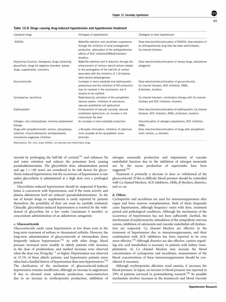
steroids by prolonging the half-life of cortisol,731 and enhances Naand water retention and reduces the potassium level, causingpseudoaldosteronism. The glycyrrhizin dose, administration periodand age (4¼60 years) are considered to be risk factors for glycyr-rhizin-induced hypertension, but the occurrence of hypertension is rareunless glycyrrhizin is administered at a high dose over a prolongedperiod.Glycyrrhizin-induced hypertension should be suspected if hypoka-
lemia is concurrent with hypertension, and if the renin activity andplasma aldosterone level are reduced (pseudoaldosteronism). As theuse of kampo drugs or supplements is rarely reported by patientsthemselves, the possibility of their use must be carefully evaluated.Clinically, glycyrrhizin-induced hypertension is resolved by the with-drawal of glycyrrhiza for a few weeks (maximum 4 months) orconcomitant administration of an aldosterone antagonist.
c. GlucocorticoidsGlucocorticoids rarely cause hypertension at low doses even in thelong-term treatment of asthma or rheumatoid arthritis. However, thelong-term administration of glucocorticoids at intermediate dosesfrequently induces hypertension.732 As with other drugs, bloodpressure increased more notably in elderly patients with increasesin the dose of prednisolone, and marked increases were observedwhen the dose was 20mgday�1 or higher. Hypertension was observedin 37.1% of these elderly patients, and hypertensive patients moreoften had a familial history of hypertension than non-hypertensives.733
The clarification of the mechanism of glucocorticoid-inducedhypertension remains insufficient, although an increase in angiotensinII due to elevated renin substrate production, vasoconstrictiondue to an increase in erythropoietin production, inhibition of
nitrogen monoxide production and impairment of vascularendothelial function due to the inhibition of nitrogen monoxideuse by the excess production of superoxides have beensuggested.734
Treatment is primarily a decrease in dose or withdrawal of theglucocorticoid. If this is difficult, blood pressure should be controlledwith Ca channel blockers, ACE inhibitors, ARBs, b-blockers, diuretics,and so on.
d. OthersCyclosporine and tacrolimus are used for immunosuppression afterorgan and bone marrow transplantation. Both of them frequentlycause hypertension, although frequency varies with dose, treatmentperiod and pathological conditions. Although the mechanism of theoccurrence of hypertension has not been sufficiently clarified, theinvolvement of nephrotoxicity, stimulation of the sympathetic nervoussystem, inhibition of calcineurin and vascular endothelial cell dysfunc-tion are suspected. Ca channel blockers are effective in thetreatment of hypertension due to immunosuppressants, and theircombination with ACE inhibitors has been reported to be evenmore effective.735 Although diuretics are also effective, caution regard-ing uric acid metabolism is necessary in patients with kidney trans-plantation. As Ca channel blockers may increase the bloodconcentration of cyclosporine and tacrolimus, measurement of theblood concentrations of these immunosuppressants should be con-sidered if necessary.Although erythropoietin alleviates renal anemia, it increases the
blood pressure. In Japan, an increase in blood pressure was reported in29% of patients surveyed in postmarketing research.736 Its possiblemechanism involves increases in the hematocrit and blood viscosity
Table 12-8 Drugs causing drug-induced hypertension and hypertension treatment
Causative drugs Etiologies of hypertension Strategies to treat hypertension
NSAIDs Water/Na retention and vasodilator suppression
through the inhibition of renal prostaglandin
production, attenuation of the antihypertensive
effects of ACE inhibitors/ARBs/b-blockers/
diuretics
Dose reduction/discontinuation of NSAIDs, dose elevation of
an antihypertensive drug that has been administered,
Ca channel blockers
Glycyrrhiza (licorice), therapeutic drugs containing
glycyrrhizin, drugs for digestive disorders, kampo
drugs, supplements, cosmetics
Water/Na retention and K reduction through the
enhancement of intrinsic steroid actions related
to the prolongation of the half-life of cortisol
associated with the inhibition of 11b-hydroxy-
lated steroid dehydrogenase
Dose reduction/discontinuation of kampo drugs, aldosterone
antagonist
Glucocorticoids Increases in renin-substrate and erythropoietin
productions and the inhibition of NO production
may be involved in the mechanism, but it
remains to be clarified.
Dose-reduction/discontinuation of glucocorticoids,
Ca channel blockers, ACE inhibitors, ARBs,
b-blockers, diuretics
Cyclosporine, tacrolimus Nephrotoxicity, activation of the sympathetic
nervous system, inhibition of calcineurin,
vascular endothelial cell dysfunction
Ca channel blockers, combination therapy with Ca channel
blockers and ACE inhibitors, diuretics
Erythropoietin Enhancement of vascular viscosity, vascular
endothelial dysfunction, an increase in the
intracellular Na level
Dose reduction/discontinuation of erythropoietin, Ca channel
blockers, ACE inhibitors, ARBs, b-blockers, diuretics
Estrogen, oral contraceptives, hormone replacement
therapy
An increase in renin-substrate production Discontinuation of estrogen preparations, ACE inhibitors,
ARBs
Drugs with sympathomimetic actions, phenylpropa-
nolamine, tricyclic/tetracyclic antidepressants,
monoamine oxygenase inhibitors
a-Receptor stimulation, inhibition of catechola-
mine reuptake at the sympathetic nerve
terminals
Dose reduction/discontinuation of drugs with sympathomi-
metic actions, a1-blockers
Abbreviations: NO, nitric oxide; NSAIDs, non-steroidal anti-inflammatory drugs.
Chapter 12. Secondary hypertension
89
Hypertension Research

associated with recovery from anemia by erythropoietin treatment anda resultant increase in peripheral vascular resistance, but this possibi-lity has been refuted by one report. An increase in the intracellular Naconcentration, vascular endothelial dysfunction and genetic predis-position may also be involved. There is also a report that no increasein blood pressure due to erythropoietin was observed before hemo-dialysis.737 However, although erythropoietin is reduced or discon-tinued if hypertension develops or if blood pressure increases,antihypertensive drugs have also been reported to be effective if theincrease is mild.738 On the other hand, blood pressure control hasbeen reported to be insufficient despite the administration of anti-hypertensive drugs in chronic dialysis patients (patients registered atthe Japanese Society for Dialysis Therapy), of whom 82% were takingerythropoietin.739
Estrogen is used in oral contraceptives and drugs for climactericdisturbance, but has been considered to cause an increase in bloodpressure or thromboembolism at a high dose. Details of the mechan-ism of estrogen-induced hypertension have not been clarified,although an increased renin substrate production in the liver hasbeen proposed. An investigation of the relationship between the use oforal contraceptives and health showed that oral contraceptives weresafe, although blood pressure was slightly higher in users than in non-users.740 However, although the increase in blood pressure was dosedependent, caution is necessary even at a low dose. No sufficientanalysis of the relationship between oral contraceptives and hyperten-sion has been made in Japan. When using oral contraceptives, bloodpressure should be measured periodically, their use should be dis-continued if an increase in the blood pressure is observed and othercontraceptive measures should be selected. If they cannot be discon-tinued, the administration of ACE inhibitors or ARBs should beconsidered. Concerning hormone replacement therapy, see hyperten-sion related to menopause in Chapter 9.
Drugs with sympathomimetic actions may induce increases in theblood pressure. An overdose of phenylpropanolamine, which iscontained in drugs for the common cold, may elevate the bloodpressure. Caution is needed in its concomitant use during treatmentwith a b-blocker alone, because it may induce a state of dominanta-receptor stimulation and cause a marked increase in blood pressure.Tri- or tetracyclic antidepressants may also inhibit the antihypertensiveeffects of peripheral sympatholytic drugs by inhibiting catecholamineuptake by sympathetic nerve terminals and induce hypertensive crisisor hypertensive urgency.741 Monoamine oxidase inhibitors, which areused for the treatment of Parkinson’s disease, also cause an increase inblood pressure or orthostatic dysregulation. A monoamine oxidaseinhibitor and a tricyclic antidepressant must not be used simulta-neously. The concomitant use of a monoamine oxidase inhibitor withephedrine or methylephedrine may also cause an elevation in bloodpressure and tachycardia. If hypertension is induced by these drugs, areduction of the dose or discontinuation of administration is neces-sary, but if discontinuation is impossible, a1-blockers or centralsympatholytic agents should be administered.As metoclopramide, a dopamine (D2) receptor antagonist used for
the treatment of gastrointestinal disorders, b-blockers and tricyclicantidepressants, may cause the clinical activation of pheochromocy-toma and hypertensive crisis,742 caution is needed in their use.
Citation InformationWe recommend that any citations to information in the Guidelines arepresented in the following format:
The Japanese Society of Hypertension Guidelines for the Managementof Hypertension (JSH 2009). Hypertens Res 2009; 32: 3–107.
Please refer to the title page for the full list of authors.
Chapter 12. Secondary hypertension
90
Hypertension Research