Nephroprotective Effect of Ursolic Acid in a Murine Model of Gentamicin-Induced Renal Damage
Upload
independentCategory
view
0download
0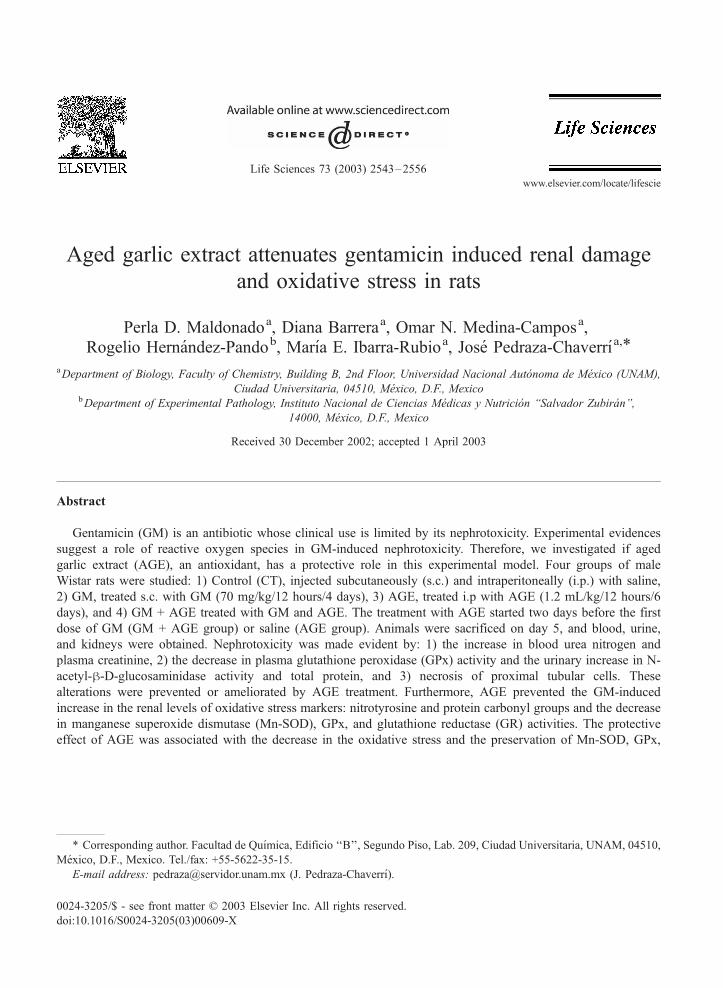
www.elsevier.com/locate/lifescie
Life Sciences 73 (2003) 2543–2556
Aged garlic extract attenuates gentamicin induced renal damage
and oxidative stress in rats
Perla D. Maldonadoa, Diana Barreraa, Omar N. Medina-Camposa,Rogelio Hernandez-Pandob, Marıa E. Ibarra-Rubioa, Jose Pedraza-Chaverrıa,*
aDepartment of Biology, Faculty of Chemistry, Building B, 2nd Floor, Universidad Nacional Autonoma de Mexico (UNAM),
Ciudad Universitaria, 04510, Mexico, D.F., MexicobDepartment of Experimental Pathology, Instituto Nacional de Ciencias Medicas y Nutricion ‘‘Salvador Zubiran’’,
14000, Mexico, D.F., Mexico
Received 30 December 2002; accepted 1 April 2003
Abstract
Gentamicin (GM) is an antibiotic whose clinical use is limited by its nephrotoxicity. Experimental evidences
suggest a role of reactive oxygen species in GM-induced nephrotoxicity. Therefore, we investigated if aged
garlic extract (AGE), an antioxidant, has a protective role in this experimental model. Four groups of male
Wistar rats were studied: 1) Control (CT), injected subcutaneously (s.c.) and intraperitoneally (i.p.) with saline,
2) GM, treated s.c. with GM (70 mg/kg/12 hours/4 days), 3) AGE, treated i.p with AGE (1.2 mL/kg/12 hours/6
days), and 4) GM + AGE treated with GM and AGE. The treatment with AGE started two days before the first
dose of GM (GM + AGE group) or saline (AGE group). Animals were sacrificed on day 5, and blood, urine,
and kidneys were obtained. Nephrotoxicity was made evident by: 1) the increase in blood urea nitrogen and
plasma creatinine, 2) the decrease in plasma glutathione peroxidase (GPx) activity and the urinary increase in N-
acetyl-h-D-glucosaminidase activity and total protein, and 3) necrosis of proximal tubular cells. These
alterations were prevented or ameliorated by AGE treatment. Furthermore, AGE prevented the GM-induced
increase in the renal levels of oxidative stress markers: nitrotyrosine and protein carbonyl groups and the decrease
in manganese superoxide dismutase (Mn-SOD), GPx, and glutathione reductase (GR) activities. The protective
effect of AGE was associated with the decrease in the oxidative stress and the preservation of Mn-SOD, GPx,
0024-3205/$ - see front matter D 2003 Elsevier Inc. All rights reserved.
doi:10.1016/S0024-3205(03)00609-X
* Corresponding author. Facultad de Quımica, Edificio ‘‘B’’, Segundo Piso, Lab. 209, Ciudad Universitaria, UNAM, 04510,
Mexico, D.F., Mexico. Tel./fax: +55-5622-35-15.
E-mail address: [email protected] (J. Pedraza-Chaverrı).

P.D. Maldonado et al. / Life Sciences 73 (2003) 2543–25562544
and GR activities in renal cortex. These data suggest that AGE may be a useful agent for the prevention of
GM-nephrotoxicity.
D 2003 Elsevier Inc. All rights reserved.
Keywords: Aged garlic extract; Garlic; Gentamicin; Aminoglycosides; Acute renal failure; Antioxidant enzymes; Oxidative
stress; Nitrotyrosine; Nephrotoxicity; Reactive oxygen species; Superoxide dismutase; Glutathione peroxidase; Glutathione
reductase
Introduction
Gentamicin (GM) is an aminoglycoside antibiotic widely used as an agent for the treatment of life-
threatening gram-negative infections (Ali, 1995). However, the clinical usefulness of the drug is limited
by the fact that 10–20% of the patients treated with GM for more than seven days show some signs of
nephrotoxicity (Lerner et al., 1986). Despite the introduction of newer and less toxic antibiotics, GM
continues playing a useful role in the treatment of serious enterococcal, mycobacterial, and gram-
negative infections, due to its effectiveness against resistant h-lactamic microorganisms, its low cost,
and the low levels of resistance among enterobacteriaceae family bacterium (Edson and Terrell, 1999).
Therefore, a potential therapeutic approach to protect or reverse GM-induced renal damage would have
very important clinical advantages. The exact mechanism by which GM induces nephrotoxicity is
unknown; however, reactive oxygen species (ROS) appear to be involved (Guidet and Shah, 1989;
Nakajima et al., 1994; Walker and Shah, 1987, 1988; Yang et al., 1995). It has been found that renal
cortical lipoperoxidation (Nakajima et al., 1994), and renal hydrogen peroxide (H2O2) generation
(Guidet and Shah, 1989) are increased in GM-treated rats. It has also been found that GM-induced
renal damage is lowered by the dietary antioxidants vitamin E and selenium (Ademuyiwa et al., 1990),
and our laboratory reported that a 2% garlic powder diet prevented oxidative stress and nephrotoxicity
induced by GM (Pedraza-Chaverrı et al., 2000). The 2% garlic powder was given 14 days in advance
and along all GM-treatment, which is not compatible with clinical practice. In addition, many people
refuse to take garlic powder or fresh garlic due to their pungent odor. Therefore we searched for an
odorless garlic presentation with antioxidant properties. During garlic aging (up to 20 months) unstable
and highly odorous compounds in fresh garlic are converted into more stable and much less odorous
compounds (Amagase et al., 2001). This garlic preparation is known as aged garlic extract (AGE)
whose chemical composition is different from that of garlic powder (Lawson, 1998). The most
abundant compounds (mg/g product) in AGE are: alliin (0.3), g-glutamylcysteines (0.34), g-glutamyl-
S-allylcysteines (0.25), S-allylcysteine (0.6), and S-allylmercaptocysteine (0.15), whereas in garlic
powder the most abundant compounds are: alliin (11.5) and g-glutamylcysteines (26) (Lawson, 1998).
Moreover, it has been shown that AGE exhibits antioxidant properties in vitro (Borek, 2001; Ide and
Lau, 1999; Kim et al., 2001; Yamasaki et al., 1994) and in vivo (Borek, 2001; Dillon et al., 2002). In
addition, AGE is a commercially available garlic preparation that has been widely studied for its high
antioxidant content and its health-protective potential (Borek, 2001; Ide and Lau, 1999, 2001; Kim et
al., 2001; Morihara et al., 2002; Yamasaki et al., 1994). Taking into account all the AGE properties
above described, in this work we studied the effect of AGE, an antioxidant, on the GM-induced renal
damage.
P.D. Maldonado et al. / Life Sciences 73 (2003) 2543–2556 2545
Methods
Reagents
AGE was kindly provided by Wakunaga of America Co., Ltd. (Mission Viejo, CA, USA). GM was
from Schering-Plough (Mexico City, Mexico). Xanthine, xanthine oxidase, nitroblue tetrazolium (NBT),
diethyldithiocarbamic acid (DDC), nicotinamide adenine dinucleotide (NADPH) and 3-3V-diaminoben-
zidine tetrahydrochloride (DAB) were from Sigma Chemical Co. (St. Louis, MO, USA). Rabbit anti-rat
polyclonal antibodies against manganese superoxide dismutase (Mn-SOD) and copper, zinc superoxide
dismutase (Cu, Zn-SOD) were from Stressgen Biotechnologies Co. (Victoria, BC, Canada). Rabbit anti-
human catalase (CAT) polyclonal antibodies were from Calbiochem (San Diego, CA, USA). Enhanced
chemiluminiscence (ECL) kit for Western blot was purchased from Amersham Life Sciences (Buck-
inghamshire, England). Rabbit anti-nitrotyrosine polyclonal antibodies were from Upstate (Lake Placid,
NY, USA). All other chemicals were reagent grade.
Superoxide anion (O2�) scavenging activity of AGE in vitro
Xanthine-xanthine oxidase system was used to determine the O2� scavenging activity of AGE. O2
�
production and xanthine oxidase activity were measured as NBT reduction and uric acid production,
respectively (Bielski et al., 1980) using a DU 64 Beckman spectrophotometer. Tubes without AGE were
taken as 100% of NBT reduction.
Experimental design
Male Wistar rats (Harlan Teklad, Mexico City, Mexico) initially weighing 225–250 g were used.
All animals had free access to water and commercial rodent diet (Harlan Teklad), and were randomly
divided in four groups as follows (n = 6 rats per group): 1) Control (CT), injected subcutaneously
(s.c.) and intraperitoneally (i.p.) with isotonic saline solution, 2) GM, treated with GM; 3) AGE,
treated with AGE; and 4) GM + AGE, treated with GM and AGE. GM treated rats (GM and GM +
AGE groups) received 70 mg of GM/kg body weight/12 hours s.c. for 4 days, and AGE treated rats
(AGE and GM + AGE groups) received a dose of 1.2 mL of AGE/kg body weight/24 hours i.p. for 6
days (two days before the first dose of GM, in the GM group, or saline, in the CT group, and along
the study). Rats were maintained in stainless steel metabolic cages with a 12-hour light:dark cycle to
collect 24-hour urine. Urine was stored at � 40 jC until N-acetyl-h-D-glucosaminidase (NAG)
activity and total protein were measured. Animals were sacrificed on day 5, and blood was collected
to obtain plasma which was stored at � 80 jC until creatinine and blood urea nitrogen (BUN),
markers of glomerular damage, and glutathione peroxidase (GPx) activity, a marker of tubular
damage, were determined. One kidney was quickly removed to obtain cortex samples for measure-
ment of GM concentration, histological studies, and immunohistochemical localization of nitro-
tyrosine, a marker of endogenous production of peroxynitrite. The other kidney was removed to obtain
0.1 g of renal cortex which was homogenized in a Polytron for 10 seconds in cold 50 mM potassium
phosphate and 0.1% Triton X-100, pH = 7.0. The homogenate was centrifuged at 19,000 � g and 4
jC for 30 min. Supernatant was separated to measure total protein, and the activity of total superoxide
dismutase (SOD), Mn-SOD, CAT, GPx, and glutathione reductase (GR). Another portion of this
P.D. Maldonado et al. / Life Sciences 73 (2003) 2543–25562546
kidney was used to measure protein carbonyl content, a relatively stable marker of protein oxidation
by ROS.
The i.p. injection of AGE has been used successfully to ameliorate oxidative stress in experimental
models in vivo (Kojima et al., 1994; Numagami et al., 1996). The dose of AGE used in this work is
similar to that used by Numagami et al. (1996).
Markers of glomerular and tubular damage
Plasma creatinine and BUN were measured using commercial kits and total protein in urine was
measured by a turbidimetric method (Pedraza-Chaverrı et al., 1999). Urinary NAG activity was
determined using p-nitrophenyl-N-acetyl-h-D-glucosaminide as substrate (Pedraza-Chaverrı et al.,
2000). Plasma GPx activity was measured using GR and NADPH in a coupled reaction (Pedraza-
Chaverrı et al., 2001).
Histological analysis
Kidney sections of 4 Am of thickness were obtained and stained with hematoxilin-eosin (H and E)
(Pedraza-Chaverrı et al., 2000). The histological profile of twenty proximal tubules randomly selected
per rat (5–6 rats per experimental group) was recorded using a Leica Qwin Image Analyzer (Cambridge,
UK). The percentage of tubules with histopathological alterations like swelling, cytoplasmic vacuoliza-
tion, desquamation or necrosis was calculated.
Oxidative stress markers
The immunohistochemical localization of nitrotyrosine was performed in sections of 4 Am of
thickness. Endogenous peroxidase was quenched/inhibited with 4.5% H2O2. Sections were incubated
with a 1:700 dilution of anti-nitrotyrosine antibody and then were incubated with a 1:250 dilution of
a peroxidase conjugated anti-rabbit IgG antibody for 1 hour, and finally incubated with H2O2-DAB
for 1 min (Barrera et al., 2003). Sections were counterstained with hematoxilin. Quantitative image
analysis was performed with a Zeiss KS 300 Imaging System, Release 3.0 (Hallbergmoos, Germany).
This software measures densitometric mean value of the region in different fields of the sections. We
normalized the data (arbitrary units) to 1.0 in CT group (Barrera et al., 2003). Protein carbonyl
groups in renal cortex were detected by its reactivity with 2,4-dinitrophenylhydrazine (Reznick and
Packer, 1994).
Antioxidant enzymes in renal cortex
Total SOD activity was assayed by a previously reported method using NBT and Mn-SOD activity
was measured by inhibiting Cu, Zn-SOD with DDC (Pedraza-Chaverrı et al., 2001). Cu, Zn-SOD
activity was obtained by subtracting the activity of the DDC-treated samples from that of total SOD
activity. CAT activity was assayed by a method based on the disappearance of H2O2 (Pedraza-Chaverrı
et al., 1999, 2001). GPx activity was assayed as previously described (Pedraza-Chaverrı et al., 2001)
and GR activity was assayed according to Carlberg and Mannervik (1975) measuring the disappearance
of NADPH.

Western blot
Immunodetection was performed using specific primary antibodies against Mn-SOD, Cu, Zn-SOD or
CAT (Pedraza-Chaverrı et al., 2001). The hybrids were visualized by chemiluminscence and quantified
by densitometry using a software (Sigma ScanPro, version 4.0, San Rafael, CA, USA).
Renal GM content
GM concentration was measured in renal cortical homogenates by the fluorescence polarization
immunoassay technology, by using the AxSYMR gentamicin assay (Abbot Laboratories, Abbot Park, IL,
USA).
Statistics
Data are presented as mean F SD and were analyzed by ANOVA followed by Bonferroni’s multiple
comparisons using the software Prism 2.01 (GraphPad, San Diego, CA, USA). Non-paired t-test was
used to compare the quantitative histological data and renal GM content. P V 0.05 was considered
statistically significant.
P.D. Maldonado et al. / Life Sciences 73 (2003) 2543–2556 2547
Results
In vitro evaluation of the AGE scavenging properties
AGE scavenges O2� in a concentration-dependent manner. AGE significantly inhibited the NBT
reduction for the concentrations shown in Table 1. Higher AGE concentrations interfered with uric acid
production (data not shown).
Body weight and urinary volume
Body weight was not statistically different among the four groups along the study and on day 5 (Data
not shown). On day 5, urinary volume increased significantly in the GM group (CT = 7.3 F 2 and
Table 1
Scavenging effect of AGE on superoxide radical generated from xanthine/xanthine oxidase/NBT systema,b
AGE NBT reduction Uric acid production
(mg/mL) DOD560/min Inhibition (%) DOD295/min Inhibition (%)
0.00 0.051 F 0.005 – 0.020 F 0.001 –
1.38 0.048 F 0.002 5.3 F 4.4 0.023 F 0.001 0.0
2.75 0.036 F 0.002 29.4 F 3.2* 0.022 F 0.004 0.0
5.51 0.024 F 0.001 51.7 F 2.5* 0.024 F 0.008 0.0a Values are mean F SD.b AGE, aged garlic extract; OD, optical density; NBT, nitroblue tetrazolium.
*P < 0.001 vs. AGE 0 mg/mL; n = 5.
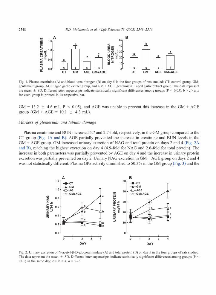
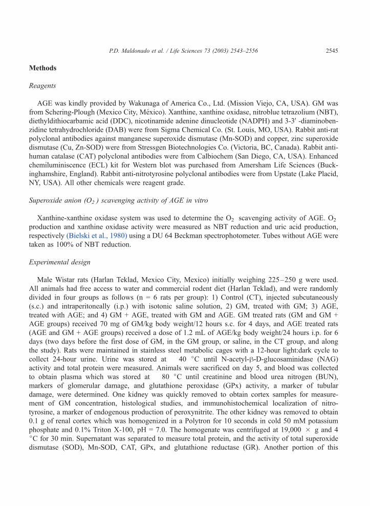
Fig. 1. Plasma creatinine (A) and blood urea nitrogen (B) on day 5 in the four groups of rats studied: CT: control group, GM:
gentamicin group, AGE: aged garlic extract group, and GM + AGE: gentamicin + aged garlic extract group. The data represent
the mean F SD. Different letter superscripts indicate statistically significant differences among groups (P < 0.05); b > c > a. n
for each group is printed in its respective bar.
P.D. Maldonado et al. / Life Sciences 73 (2003) 2543–25562548
GM = 13.2 F 4.6 mL, P < 0.05), and AGE was unable to prevent this increase in the GM + AGE
group (GM + AGE = 10.1 F 4.3 mL).
Markers of glomerular and tubular damage
Plasma creatinine and BUN increased 5.7 and 2.7-fold, respectively, in the GM group compared to the
CT group (Fig. 1A and B). AGE partially prevented the increase in creatinine and BUN levels in the
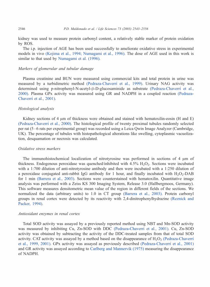
GM + AGE group. GM increased urinary excretion of NAG and total protein on days 2 and 4 (Fig. 2A
and B), reaching the highest excretion on day 4 (4.9-fold for NAG and 2.6-fold for total protein). The
increase in both parameters was partially prevented by AGE on day 4 and the increase in urinary protein
excretion was partially prevented on day 2. Urinary NAG excretion in GM + AGE group on days 2 and 4
was not statistically different. Plasma GPx activity diminished to 50.3% in the GM group (Fig. 3) and the
Fig. 2. Urinary excretion of N-acetyl-h-D-glucosaminidase (A) and total protein (B) on day 5 in the four groups of rats studied.
The data represent the mean F SD. Different letter superscripts indicate statistically significant differences among groups (P <
0.01) in the same day; c > b > a. n = 5–6.
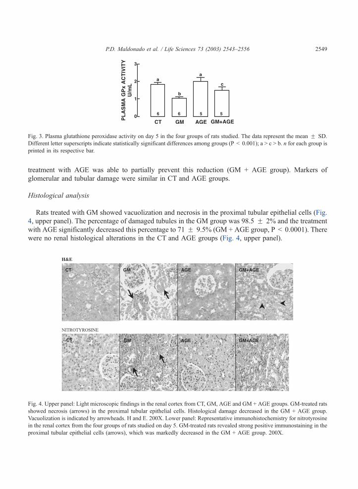
Fig. 3. Plasma glutathione peroxidase activity on day 5 in the four groups of rats studied. The data represent the mean F SD.
Different letter superscripts indicate statistically significant differences among groups (P < 0.001); a > c > b. n for each group is
printed in its respective bar.
P.D. Maldonado et al. / Life Sciences 73 (2003) 2543–2556 2549
treatment with AGE was able to partially prevent this reduction (GM + AGE group). Markers of
glomerular and tubular damage were similar in CT and AGE groups.
Histological analysis
Rats treated with GM showed vacuolization and necrosis in the proximal tubular epithelial cells (Fig.
4, upper panel). The percentage of damaged tubules in the GM group was 98.5 F 2% and the treatment
with AGE significantly decreased this percentage to 71 F 9.5% (GM + AGE group, P < 0.0001). There
were no renal histological alterations in the CT and AGE groups (Fig. 4, upper panel).
Fig. 4. Upper panel: Light microscopic findings in the renal cortex from CT, GM, AGE and GM + AGE groups. GM-treated rats
showed necrosis (arrows) in the proximal tubular epithelial cells. Histological damage decreased in the GM + AGE group.
Vacuolization is indicated by arrowheads. H and E. 200X. Lower panel: Representative immunohistochemistry for nitrotyrosine
in the renal cortex from the four groups of rats studied on day 5. GM-treated rats revealed strong positive immunostaining in the
proximal tubular epithelial cells (arrows), which was markedly decreased in the GM + AGE group. 200X.
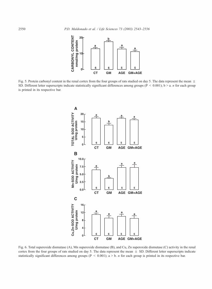
Fig. 5. Protein carbonyl content in the renal cortex from the four groups of rats studied on day 5. The data represent the mean FSD. Different letter superscripts indicate statistically significant differences among groups (P < 0.001); b > a. n for each group
is printed in its respective bar.
Fig. 6. Total superoxide dismutase (A), Mn superoxide dismutase (B), and Cu, Zn superoxide dismutase (C) activity in the renal
cortex from the four groups of rats studied on day 5. The data represent the mean F SD. Different letter superscripts indicate
statistically significant differences among groups (P < 0.001); a > b. n for each group is printed in its respective bar.
P.D. Maldonado et al. / Life Sciences 73 (2003) 2543–25562550
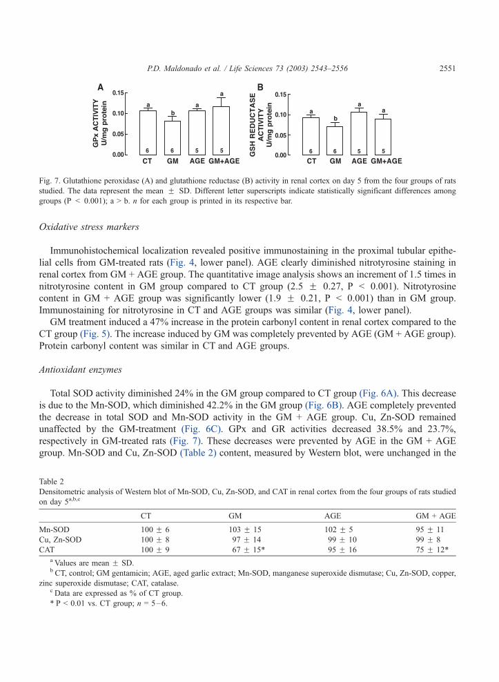
Fig. 7. Glutathione peroxidase (A) and glutathione reductase (B) activity in renal cortex on day 5 from the four groups of rats
studied. The data represent the mean F SD. Different letter superscripts indicate statistically significant differences among
groups (P < 0.001); a > b. n for each group is printed in its respective bar.
P.D. Maldonado et al. / Life Sciences 73 (2003) 2543–2556 2551
Oxidative stress markers
Immunohistochemical localization revealed positive immunostaining in the proximal tubular epithe-
lial cells from GM-treated rats (Fig. 4, lower panel). AGE clearly diminished nitrotyrosine staining in
renal cortex from GM + AGE group. The quantitative image analysis shows an increment of 1.5 times in
nitrotyrosine content in GM group compared to CT group (2.5 F 0.27, P < 0.001). Nitrotyrosine
content in GM + AGE group was significantly lower (1.9 F 0.21, P < 0.001) than in GM group.
Immunostaining for nitrotyrosine in CT and AGE groups was similar (Fig. 4, lower panel).
GM treatment induced a 47% increase in the protein carbonyl content in renal cortex compared to the
CT group (Fig. 5). The increase induced by GM was completely prevented by AGE (GM + AGE group).
Protein carbonyl content was similar in CT and AGE groups.
Antioxidant enzymes
Total SOD activity diminished 24% in the GM group compared to CT group (Fig. 6A). This decrease
is due to the Mn-SOD, which diminished 42.2% in the GM group (Fig. 6B). AGE completely prevented
the decrease in total SOD and Mn-SOD activity in the GM + AGE group. Cu, Zn-SOD remained
unaffected by the GM-treatment (Fig. 6C). GPx and GR activities decreased 38.5% and 23.7%,
respectively in GM-treated rats (Fig. 7). These decreases were prevented by AGE in the GM + AGE
group. Mn-SOD and Cu, Zn-SOD (Table 2) content, measured by Western blot, were unchanged in the
Table 2
Densitometric analysis of Western blot of Mn-SOD, Cu, Zn-SOD, and CAT in renal cortex from the four groups of rats studied
on day 5a,b,c
CT GM AGE GM + AGE
Mn-SOD 100 F 6 103 F 15 102 F 5 95 F 11
Cu, Zn-SOD 100 F 8 97 F 14 99 F 10 99 F 8
CAT 100 F 9 67 F 15* 95 F 16 75 F 12*a Values are mean F SD.b CT, control; GM gentamicin; AGE, aged garlic extract; Mn-SOD, manganese superoxide dismutase; Cu, Zn-SOD, copper,
zinc superoxide dismutase; CAT, catalase.c Data are expressed as % of CT group.
* P < 0.01 vs. CT group; n = 5–6.
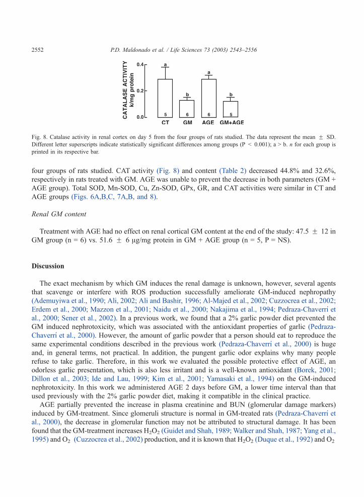
Fig. 8. Catalase activity in renal cortex on day 5 from the four groups of rats studied. The data represent the mean F SD.
Different letter superscripts indicate statistically significant differences among groups (P < 0.001); a > b. n for each group is
printed in its respective bar.
P.D. Maldonado et al. / Life Sciences 73 (2003) 2543–25562552
four groups of rats studied. CAT activity (Fig. 8) and content (Table 2) decreased 44.8% and 32.6%,
respectively in rats treated with GM. AGE was unable to prevent the decrease in both parameters (GM +
AGE group). Total SOD, Mn-SOD, Cu, Zn-SOD, GPx, GR, and CAT activities were similar in CT and
AGE groups (Figs. 6A,B,C, 7A,B, and 8).
Renal GM content
Treatment with AGE had no effect on renal cortical GM content at the end of the study: 47.5 F 12 in
GM group (n = 6) vs. 51.6 F 6 Ag/mg protein in GM + AGE group (n = 5, P = NS).
Discussion
The exact mechanism by which GM induces the renal damage is unknown, however, several agents
that scavenge or interfere with ROS production successfully ameliorate GM-induced nephropathy
(Ademuyiwa et al., 1990; Ali, 2002; Ali and Bashir, 1996; Al-Majed et al., 2002; Cuzzocrea et al., 2002;
Erdem et al., 2000; Mazzon et al., 2001; Naidu et al., 2000; Nakajima et al., 1994; Pedraza-Chaverrı et
al., 2000; Sener et al., 2002). In a previous work, we found that a 2% garlic powder diet prevented the
GM induced nephrotoxicity, which was associated with the antioxidant properties of garlic (Pedraza-
Chaverrı et al., 2000). However, the amount of garlic powder that a person should eat to reproduce the
same experimental conditions described in the previous work (Pedraza-Chaverrı et al., 2000) is huge
and, in general terms, not practical. In addition, the pungent garlic odor explains why many people
refuse to take garlic. Therefore, in this work we evaluated the possible protective effect of AGE, an
odorless garlic presentation, which is also less irritant and is a well-known antioxidant (Borek, 2001;
Dillon et al., 2003; Ide and Lau, 1999; Kim et al., 2001; Yamasaki et al., 1994) on the GM-induced
nephrotoxicity. In this work we administered AGE 2 days before GM, a lower time interval than that
used previously with the 2% garlic powder diet, making it compatible in the clinical practice.
AGE partially prevented the increase in plasma creatinine and BUN (glomerular damage markers)
induced by GM-treatment. Since glomeruli structure is normal in GM-treated rats (Pedraza-Chaverrı et
al., 2000), the decrease in glomerular function may not be attributed to structural damage. It has been
found that the GM-treatment increases H2O2 (Guidet and Shah, 1989; Walker and Shah, 1987; Yang et al.,
1995) and O2� (Cuzzocrea et al., 2002) production, and it is known that H2O2 (Duque et al., 1992) and O2
�
P.D. Maldonado et al. / Life Sciences 73 (2003) 2543–2556 2553
(Martınez-Salgado et al., 2002) induce mesangial cells contraction, altering the filtration surface area and
modifying the ultrafiltration coefficient factors, that decrease the glomerular filtration rate (GFR).
Moreover, the decrease in Mn-SOD activity favors the increase in O2� concentration, and this radical
can react with nitric oxide (NO, a vasodilator) to form peroxynitrite, a cytotoxic oxidant radical specie.
The inactivation of NO by O2� could also lead to a decrease in the GFR (Rivas-Cabanero et al., 1997).
These data indicate that the H2O2 and O2� could be involved in the decrease of GFR in GM-induced
nephrotoxicity, and that the protective effect of AGE, could be related with its ability to scavenge O2�
(Table 1) (Borek, 2001; Kim et al., 2001) and H2O2 (Borek, 2001; Ide and Lau, 1999), and/or with its
ability to totally prevent the decrease in Mn-SOD activity (Fig. 6). Additionally, Morihara et al. (2002)
found that AGE increases NO production, which also can help to prevent the decrease in the GRF.
Tubular damage was evaluated by measuring plasma GPx activity and the urinary excretion of NAG
and total protein. In rats treated with GM, the increase in urinary NAG and total protein excretion, and
the decrease in plasma GPx activity, could be associated with the necrosis of proximal tubules, the
primary site of drug accumulation. Especially considering that GPx is synthesized almost exclusively in
proximal tubular cells (Pedraza-Chaverrı et al., 2000). AGE partially prevented these alterations induced
by GM, and this prevention could be associated with the ability of AGE to partially ameliorate the
tubular necrosis.
It has been suggested that the oxidative stress induces tubular damage (Cuzzocrea et al., 2002; Guidet
and Shah, 1989; Walker and Shah, 1988; Yang et al., 1995). In this study, the carbonyl and nitrotyrosine
content showed an increase in oxidative stress in rats. It is known that the increase in ROS levels induces
cytotoxicity due to a concerted action of oxygen- (Cuzzocrea et al., 2002) and nitrogen-derived free
radicals (Mazzon et al., 2001). Furthermore, we found that AGE prevented the increase in carbonyl and
nitrotyrosine levels in renal cortex of rats treated with GM + AGE, effect that could be associated with
the antioxidant properties of AGE, and with its ability to prevent the decrease in antioxidant enzymes
activity (Mn-SOD, GPx and GR).
It is known that peroxynitrite anions impair Mn-SOD (MacMillan-Crow and Thompson, 1999) and
GPx (Padmaja et al., 1998) activity and O2� inactivates GPx (Blum and Fridovich, 1985; Rister and
Baehner, 1976) and CAT (Rister and Baehner, 1976). In this study, the decrease in Mn-SOD activity in
the GM group was independent from Mn-SOD protein content. Cu, Zn-SOD activity was unchanged,
perhaps due to the fact that Mn-SOD is more labile than Cu, Zn-SOD (Pedraza-Chaverrı et al., 2000).
The decrease in CAT activity is due to protein content and mRNA decreases in rats treated with GM
(Pedraza-Chaverrı et al., 2000). AGE-treatment totally prevented the decrease in Mn-SOD, GPx and GR
activities, effect that could be related to its ability to scavenge O2� (Table 1) (Borek, 2001; Kim et al.,
2001). The decrease in O2� levels would lead to the decrease of peroxynitrite levels, which is supported
by the decrease in nitrotyrosine content in the GM + AGE group (Fig 4, lower panel). AGE was unable
to prevent the decrease in CAT activity probably because the decrease is not only due to enzyme
inactivation but also to decreased enzyme quantity and mRNA content.
Our results indicate that the increase in oxidative stress, and the decrease in antioxidant enzymes Mn-
SOD, GPx, GR, and CAT activity, could be factors involved in the sequence of events leading to GM-
nephrotoxicity. Also the protective effect of AGE could be associated with its ability to prevent the
increase in oxidative stress and the decrease in the antioxidant enzymes activity (Mn-SOD, GPx, and
GR), observed in renal cortex of rats treated with GM. Additionally, since AGE is a complex mixture of
phytochemicals (Lawson, 1998), other potential mechanisms such as the ability of AGE to enhance
glutathione content (Wang et al., 1999) and to chelate metals (Dillon et al., 2003) could be involved in its
P.D. Maldonado et al. / Life Sciences 73 (2003) 2543–25562554
protective effect on GM-induced nephrotoxicity. In fact, it is known that GM induces iron mobilization
from renal cortical mitochondria (Ueda et al., 1993) and that deferoxamine, an iron chelator, is able to
prevent GM-induced nephrotoxicity (Walker and Shah, 1988).
The protective effect of AGE on GM-induced nephrotoxicity suggests that AGE contains compounds
that could potentially ameliorate GM-induced acute renal failure in rats. The AGE is a commercially
available garlic presentation which has been used in healthy volunteers (Dillon et al., 2002) and whose
composition has been reported (Lawson, 1998). It has been postulated that the compounds responsible of
its protective effect are S-allylcysteine (Borek, 2001; Kim et al., 2001; Numagami and Ohnishi, 2001;
Yamasaki et al., 1994), S-allymercaptocysteine (Borek, 2001), and alliin (Kourounakis and Rekka, 1991)
which are water soluble, odorless, and have antioxidant properties.
Conclusion
Our data indicate that AGE is a potent antioxidant both in vitro and in vivo. The antioxidant effect in
vivo may explain, at least in part, the protective effect of AGE in rats with GM-nephrotoxicity. The
potential ability of AGE to reduce GM-nephrotoxicity in patients remains to be studied.
Acknowledgements
This work was supported by CONACYT (#34920-M and #40009-M) and by PAEP (#203308). PDM
received a scholarship from CONACYT (#124981) and from DGAPA. We thank QFB Romelia Velasco
and QFB Tere Murguıa of Instituto Nacional de Pediatrıa for the measurement of renal cortical
gentamicin concentration.
References
Ademuyiwa, O., Ngaha, E.O., Ubah, F.O., 1990. Vitamin E and selenium in gentamicin nephrotoxicity. Humand and Exper-
imental Toxicology 9 (5), 281–288.
Ali, B.H., 1995. Gentamicin nephrotoxicity in humans and animals: some recent research. General Pharmacology 26 (7),
1477–1487.
Ali, B.H., Bashir, A.K., 1996. Effect of superoxide dismutase treatment on gentamicin nephrotoxicity in rats. General Phar-
macology 27 (2), 349–353.
Ali, B.H., 2002. The effect of treatment with the medicinal plant Rhazya stricta decne on gentamicin nephrotoxicity in rats.
Phytomedicine 9 (5), 385–389.
Al-Majed, A.A., Mostafa, A.M., Al-Rikabi, A.C., Al-Shabanah, O.A., 2002. Protective effects of oral arabic gum adminis-
tration on gentamicin-induced nephrotoxicity in rats. Pharmacological Research 46 (5), 445–451.
Amagase, H., Petesch, B.L., Matsuura, H., Kasuga, S., Itakura, Y., 2001. Intake of garlic and its bioactive components. Journal
of Nutrition 131 (3s), 955S–962S.
Barrera, D., Maldonado, P.D., Medina-Campos, O.N., Hernandez-Pando, R., Ibarra-Rubio, M.E., Pedraza-Chaverrı, J., 2003.
HO-1 induction attenuates renal damage and oxidative stress induced by K2Cr2O7. Free Radical Biology and Medicine
34 (11), 1390–1398.
Bielski, B.H., Shlue, G.G., Bajuk, S., 1980. Reduction of nitro blue tetrazolium by CO2� and O2
� radicals. Journal of Physical
Chemistry 84 (8), 830–833.
P.D. Maldonado et al. / Life Sciences 73 (2003) 2543–2556 2555
Blum, J., Fridovich, I., 1985. Inactivation of glutathione peroxidase by superoxide radical. Archives of Biochemistry and
Biophysics 240 (2), 500–508.
Borek, C., 2001. Antioxidant health effects of aged garlic extract. Journal of Nutrition 131 (3s), 1010S–1015S.
Carlberg, I., Mannervik, B., 1975. Purification and characterization of the flavoenzyme glutathione reductase from rat liver.
Journal of Biological Chemistry 250 (14), 5475–5480.
Cuzzocrea, S., Mazzon, E., Dugo, L., Serraino, I., Di Paola, R., Britti, D., De Sarro, A., Pierpaoli, S., Caputi, A., Masini, E.,
Salvemini, D., 2002. A role for superoxide in gentamicin-mediated nephropaty in rats. European Journal of Pharmacology
450 (1), 67–76.
Dillon, S.A., Lowe, G.M., Billington, D., Rahman, K., 2002. Dietary supplementation with aged garlic extract reduces plasma
and urine concentrations of 8-iso-prostaglandin F(2 alpha) in smoking and nonsmoking men and women. Journal of
Nutrition 132 (2), 168–171.
Dillon, S.A., Burmi, R.S., Lowe, G.M., Billington, D., Rahman, K., 2003. Antioxidant properties of aged garlic extract: an in
vitro study incorporating human low density lipoprotein. Life Sciences 72 (14), 1583–1594.
Duque, I., Garcıa-Escribano, C., Rodrıguez-Puyol, M., Dıez-Marques, M.L., Lopez-Novoa, J.M., Arribas, I., Hernando, L.,
Rodriguez-Puyol, D., 1992. Effects of reactive oxygen species on cultured rat mesangial cells and isolated rat glomeruli.
American Journal of Physiology 263 (3 Pt. 2), F466–F473.
Edson, R.S., Terrell, C.L., 1999. The aminoglycosides. Mayo Clinic Proceedings 74 (5), 519–528.
Erdem, A., Gundogan, N.U., Usubutun, A., Kilinc, K., Erdem, S.R., Kara, A., Bozkurt, A., 2000. The protective effect of
taurine against gentamicin-induced acute tubular necrosis in rats. Nephrology Dialysis and Transplantation 15 (8),
1175–1182.
Guidet, B.R., Shah, S.V., 1989. In vivo generation of hydrogen peroxide by rat kidney cortex and glomeruli. American Journal
of Physiology 256 (1 Pt. 2), F158–F164.
Ide, N., Lau, B.H., 1999. Aged garlic extract attenuates intracellular oxidative stress. Phytomedicine 6 (2), 125–131.
Ide, N., Lau, B.H., 2001. Garlic compounds minimize intracellular oxidative stress and inhibit nuclear factor-kappa b activa-
tion. Journal of Nutrition 131 (3s), 1020S–1026S.
Kim, K.M., Chun, S.B., Koo, M.S., Choi, W.J., Kim, T.W., Kwon, Y.G., Chung, H.T., Billiar, T.R., Kim, Y.M., 2001. Differ-
ential regulation of NO availability from macrophages and endothelial cells by the garlic component S-allyl cysteine. Free
Radical Biology and Medicine 30 (7), 747–756.
Kojima, R., Toyama, Y., Ohnishi, S.T., 1994. Protective effects of an aged garlic extract on doxorubicin-induced cardiotoxicity
in the mouse. Nutrition Cancer 22 (2), 162–173.
Kourounakis, P.N., Rekka, E.A., 1991. Effect on active oxygen species of alliin and allium sativum (garlic) powder. Research
Communications in Chemical Pathology and Pharmacology 74 (2), 249–252.
Lawson, L.D., 1998. Garlic: A review of its medicinal effects and indicated active compounds. In: Lawson, L.D., Bauer, R.
(Eds.), Phytomedicines of Europe: chemistry and biological activity. ACS Symposium Series, vol. 691. American Chemical
Society, Washington, pp. 176–209.
Lerner, S.A., Schmitt, B.A., Seligosohn, R., Matz, G.J., 1986. Comparative study of ototoxicity and nephrotoxicity in patients
randomly assigned to treatment with amikacin or gentamicin. American Journal of Medicine 80 (6B), 98–104.
MacMillan-Crow, L.A., Thompson, J.A., 1999. Tyrosine modifications and inactivation of active site manganese superoxide
dismutase mutant (Y34F) by peroxynitrite. Archives of Biochemistry and Biophysics 366 (1), 82–88.
Martınez-Salgado, C., Eleno, N., Tavares, P., Rodrıguez-Barbero, A., Garcıa-Criado, J., Bolanos, J.P., Lopez-Novoa, J.M.,
2002. Involvement of reactive oxygen species on gentamicin-induced mesangial cell activation. Kidney International 62 (5),
1682–1692.
Mazzon, E., Britti, D., De Sarro, A., Caputi, A.P., Cuzzocrea, S., 2001. Effect of N-acetylcysteine on gentamicin-mediated
nephropathy in rats. European Journal of Pharmacology 424 (1), 75–83.
Morihara, N., Sumioka, I., Moriguchi, T., Uda, N., Kyo, E., 2002. Aged garlic extract enhances production of nitric oxide. Life
Sciences 71 (5), 509–517.
Naidu, M.U., Shifow, A.A., Kumar, K.V., Ratnakar, K.S., 2000. Ginkgo biloba extract ameliorates gentamicin-induced neph-
rotoxicity in rats. Phytomedicine 7 (3), 191–197.
Nakajima, T., Hishida, A., Kato, A., 1994. Mechanisms for protective effects of free radical scavengers on gentamicin-mediated
nephropathy in rats. American Journal of Physiology 266 (3 Pt. 2), F425–F431.
Numagami, Y., Sato, S., Ohnishi, S.T., 1996. Attenuation of rat ischemic brain damage by aged garlic extracts: a possible
protecting mechanism as antioxidants. Neurochemistry International 29 (2), 135–143.

P.D. Maldonado et al. / Life Sciences 73 (2003) 2543–25562556
Numagami, Y., Ohnishi, S.T., 2001. S-allylcysteine inhibits free radical production, lipid peroxidation and neuronal damage in
rat brain ischemia. Journal of Nutrition 131 (3s), 1100S–1105S.
Padmaja, S., Squadrito, G.L., Pryor, W.A., 1998. Inactivation of glutathione peroxidase by peroxynitrite. Archives of Bio-
chemistry and Biophysics 349 (1), 1–6.
Pedraza-Chaverrı, J., Granados-Silvestre, M.A., Medina-Campos, O.N., Hernandez-Pando, R., 1999. Effect of the in vivo
catalase inhibition on aminonucleoside nephrosis. Free Radical Biology and Medicine 27 (3–4), 245–253.
Pedraza-Chaverrı, J., Maldonado, P.D., Medina-Campos, O.N., Olivares-Corichi, I.M., Granados-Silvestre, M.A., Hernandez-
Pando, R., Ibarra-Rubio, M.E., 2000. Garlic ameliorates gentamicin nephrotoxicity: relation to antioxidant enzymes. Free
Radical Biology and Medicine 29 (7), 602–611.
Pedraza-Chaverrı, J., Granados-Silvestre, M.A., Medina-Campos, O.N., Maldonado, P.D., Olivares-Corichi, I.M., Ibarra-Rubio,
M.E., 2001. Post-transcriptional control of catalase expression in garlic-treated rats. Molecular and Cellular Biochemistry
216 (1–2), 9–19.
Reznick, A.Z., Packer, L., 1994. Oxidative damage to proteins: spectrophotometric method for carbonyl assay. Methods in
Enzymology 233, 357–363.
Rister, M., Baehner, R.L., 1976. The alteration of superoxide dismutase, catalase, glutathione peroxidase, and NAD(P)H
cytochrome c reductase in guinea pig polymorphonuclear leukocytes and alveolar macrophages during hyperoxia. Journal
of Clinical Investigation 58 (5), 1174–1184.
Rivas-Cabanero, L., Rodrıguez-Lopez, A.M., Martınez-Salgado, C., Saura, M., Lamas, S., Lopez-Novoa, J.M., 1997. Genta-
micin treatment increases mesangial cell nitric oxide production. Experimental Nephrology 5 (1), 23–30.
Sener, G., Sehirli, A.O., Altunbas, H.Z., Ersoy, Y., Paskaloglu, K., Arbak, S., Ayanoglu-Dulger, G., 2002. Melatonin protects
against gentamicin-induced nephrotoxicity in rats. Journal of Pineal Research 32 (4), 231–236.
Ueda, N., Guidet, B., Shah, S.V., 1993. Gentamicin-induced mobilization of iron from renal cortical mitochondria. American
Journal of Physiology 265 (3 Pt. 2), F435–F439.
Walker, P.D., Shah, S.V., 1987. Gentamicin enhanced production of hydrogen peroxide by renal cortical mitochondria.
American Journal of Physiology 253 (4 Pt. 1), C495–C499.
Walker, P.D., Shah, S.V., 1988. Evidence suggesting a role for hydroxyl radical in gentamicin-induced acute renal failure in rats.
Journal of Clinical Investigation 81 (2), 334–341.
Wang, B.H., Zuzel, K.A., Rahman, K., Billington, D., 1999. Treatment with aged garlic extract protects against bromobenzene
toxicity to precision cut rat liver slices. Toxicology 132 (2–3), 215–225.
Yamasaki, T., Li, L., Lau, B.H., 1994. Garlic compounds protect vascular endothelial cells from hydrogen peroxide-induced
oxidant injury. Phytotheraphy Research 8 (7), 408–412.
Yang, C.L., Du, X.H., Han, Y.X., 1995. Renal cortical mitochondria are the source of oxygen free radicals enhanced by
gentamicin. Renal Failure 17 (1), 21–26.
Copyright © 2022 FDOKUMEN