Reduction of estrogen -induced transformation of mouse mammary epithelial cells by N-acetylcysteine
18
Reduction of estrogen-induced transformation of mouse mammary epithelial cells by N-acetylcysteine Divya Venugopal §,1 , Muhammad Zahid §,1 , Paula C Mailander 1 , Jane L. Meza 2 , Eleanor G. Rogan 1 , Ercole L. Cavalieri 1 , and Dhrubajyoti Chakravarti *,1 1 Eppley Institute for Research in Cancer and Allied Diseases, 986805 Nebraska Medical Center, Omaha, NE 68198-6805 2 Preventive and Societal Medicine, 984350 Nebraska Medical Center, Omaha, NE 68198-4350 Abstract A growing number of studies indicate that breast cancer initiation is related to abnormal estrogen oxidation to form an excess of estrogen-3,4-quinones, which react with DNA to form depurinating adducts and induce mutations. This mechanism is often called estrogen genotoxicity. 4-catechol estrogens, precursors of the estrogen-3,4-quinones, were previously shown to account for most of the transforming and tumorigenic activity. We examined whether estrogen-induced transformation can be reduced by inhibiting the oxidation of a 4-catechol estrogen to its quinone. We demonstrate that E6 cells (a normal mouse epithelial cell line) can be transformed by a single treatment with a catechol estrogen or its quinone. The transforming activities of 4-hydroxyestradiol and estradiol-3,4- quinone were comparable. N-acetylcysteine, a common antioxidant, inhibited the oxidation of 4- hydroxyestradiol to the quinone and consequent formation of DNA adducts. It also drastically reduced estrogen-induced transformation of E6 cells. These results strongly implicate estrogen genotoxicity in mammary cell transformation. Since N-acetylcysteine is well-tolerated in clinical studies, it may be a promising candidate for breast cancer prevention. Introduction In 1896, Beatson reported that removal of the ovary regressed breast cancer in women, suggesting a role of endogenous hormones in the disease [1]. Initial studies identified this hormone as estrogen, and found it to act by a receptor-mediated mechanism to cause breast cancer [2]. Later studies suggested that oxidative metabolites, formed as a result of abnormal estrogen metabolism in the breast, are also a major cause of this disease [3,4]. The abnormal metabolism primarily involves the formation of high levels of carcinogenic 4- catecholestrogens and their quinones (estrogen-3,4-quinones) [4,5]. A significant number of breast cancer patients appear to suffer from it. For example, in one study with 77 patients (49 controls and 28 breast cancer cases), 4-hydroxyestrogens, estrogen-3,4-quinones and their derivatives were found elevated in 54% of cases and 10% of controls [5]. This breast cancer phenotype can be the result of polymorphisms in estrogen-metabolizing genes, and/or * Address correspondence to: Dr. D. Chakravarti, Eppley Institute for Research in Cancer and Allied Diseases, 986805 Nebraska Medical Center, Omaha, NE 68198-6805, Tel: (402) 559-2951, Fax: (402) 559-8068, E-mail: [email protected]. § Equal contributors. Publisher's Disclaimer: This is a PDF file of an unedited manuscript that has been accepted for publication. As a service to our customers we are providing this early version of the manuscript. The manuscript will undergo copyediting, typesetting, and review of the resulting proof before it is published in its final citable form. Please note that during the production process errors may be discovered which could affect the content, and all legal disclaimers that apply to the journal pertain. NIH Public Access Author Manuscript J Steroid Biochem Mol Biol. Author manuscript; available in PMC 2009 August 7. Published in final edited form as: J Steroid Biochem Mol Biol. 2008 March ; 109(1-2): 22–30. doi:10.1016/j.jsbmb.2007.12.003. NIH-PA Author Manuscript NIH-PA Author Manuscript NIH-PA Author Manuscript
-
Upload
independent -
Category
Documents
-
view
0 -
download
0
Transcript of Reduction of estrogen -induced transformation of mouse mammary epithelial cells by N-acetylcysteine

Reduction of estrogen-induced transformation of mousemammary epithelial cells by N-acetylcysteine
Divya Venugopal§,1, Muhammad Zahid§,1, Paula C Mailander1, Jane L. Meza2, Eleanor G.Rogan1, Ercole L. Cavalieri1, and Dhrubajyoti Chakravarti*,11 Eppley Institute for Research in Cancer and Allied Diseases, 986805 Nebraska Medical Center,Omaha, NE 68198-68052 Preventive and Societal Medicine, 984350 Nebraska Medical Center, Omaha, NE 68198-4350
AbstractA growing number of studies indicate that breast cancer initiation is related to abnormal estrogenoxidation to form an excess of estrogen-3,4-quinones, which react with DNA to form depurinatingadducts and induce mutations. This mechanism is often called estrogen genotoxicity. 4-catecholestrogens, precursors of the estrogen-3,4-quinones, were previously shown to account for most ofthe transforming and tumorigenic activity. We examined whether estrogen-induced transformationcan be reduced by inhibiting the oxidation of a 4-catechol estrogen to its quinone. We demonstratethat E6 cells (a normal mouse epithelial cell line) can be transformed by a single treatment with acatechol estrogen or its quinone. The transforming activities of 4-hydroxyestradiol and estradiol-3,4-quinone were comparable. N-acetylcysteine, a common antioxidant, inhibited the oxidation of 4-hydroxyestradiol to the quinone and consequent formation of DNA adducts. It also drasticallyreduced estrogen-induced transformation of E6 cells. These results strongly implicate estrogengenotoxicity in mammary cell transformation. Since N-acetylcysteine is well-tolerated in clinicalstudies, it may be a promising candidate for breast cancer prevention.
IntroductionIn 1896, Beatson reported that removal of the ovary regressed breast cancer in women,suggesting a role of endogenous hormones in the disease [1]. Initial studies identified thishormone as estrogen, and found it to act by a receptor-mediated mechanism to cause breastcancer [2]. Later studies suggested that oxidative metabolites, formed as a result of abnormalestrogen metabolism in the breast, are also a major cause of this disease [3,4]. The abnormalmetabolism primarily involves the formation of high levels of carcinogenic 4-catecholestrogens and their quinones (estrogen-3,4-quinones) [4,5]. A significant number ofbreast cancer patients appear to suffer from it. For example, in one study with 77 patients (49controls and 28 breast cancer cases), 4-hydroxyestrogens, estrogen-3,4-quinones and theirderivatives were found elevated in 54% of cases and 10% of controls [5]. This breast cancerphenotype can be the result of polymorphisms in estrogen-metabolizing genes, and/or
* Address correspondence to: Dr. D. Chakravarti, Eppley Institute for Research in Cancer and Allied Diseases, 986805 Nebraska MedicalCenter, Omaha, NE 68198-6805, Tel: (402) 559-2951, Fax: (402) 559-8068, E-mail: [email protected].§Equal contributors.Publisher's Disclaimer: This is a PDF file of an unedited manuscript that has been accepted for publication. As a service to our customerswe are providing this early version of the manuscript. The manuscript will undergo copyediting, typesetting, and review of the resultingproof before it is published in its final citable form. Please note that during the production process errors may be discovered which couldaffect the content, and all legal disclaimers that apply to the journal pertain.
NIH Public AccessAuthor ManuscriptJ Steroid Biochem Mol Biol. Author manuscript; available in PMC 2009 August 7.
Published in final edited form as:J Steroid Biochem Mol Biol. 2008 March ; 109(1-2): 22–30. doi:10.1016/j.jsbmb.2007.12.003.
NIH
-PA Author Manuscript
NIH
-PA Author Manuscript
NIH
-PA Author Manuscript

alterations in the expression of the enzymes that favor the oxidation of estrogens to form thesemetabolites [4,6,7].
The 4-catecholestrogens and the estrogen-3,4-quinones may initiate breast cancer by agenotoxic mechanism [4]. The estrogen-3,4-quinones are the ultimate carcinogens, as theyreact with DNA to form depurinating adducts, which are detectable in the breast tissue [8] andin the urine [4]. Estrogen-DNA depurinating adducts show a strong association with breastcancer. For example, one study indicated that cancerous human breast tissue can have 30-foldmore estrogen-induced depurinating adducts than normal human breast [8], and another studyindicated that the urine of breast cancer patients as well as high risk women contain ∼2-foldmore of these adducts than normal women (Gaikwad et al, submitted). The depurinatingadducts spontaneously dissociate from DNA, forming abasic sites. In the breast, these abasicsites induce the mutations that may initiate breast cancer. These ideas are supported by theobservation that 4-catecholestrogens and estrogen-3,4-quinones account for the majority of themutagenic [9-12], transforming [3,13] and carcinogenic activities of estrogens [14-17].
To establish the initiating role of estrogen-3,4-quinones in breast cancer, it is necessary todemonstrate a direct link between oxidative estrogen metabolism and an early event of breastcancer. In a preliminary study, four antioxidants (N-acetylcysteine, resveratrol, melatonin andreduced lipoic acid) were examined for their ability to inhibit estrogen-DNA adduct formationin vitro [18]. These antioxidants, when added in equimolar doses to 4-catecholestrogens,showed 33-77% inhibition of DNA adduct formation [18]. In the present study, we haveexamined whether N-acetylcysteine can be used to explore the role of oxidative metabolites ofestrogen in breast cancer initiation. Specifically, we examined whether N-acetylcysteine canreduce estrogen-DNA adduct formation and inhibit estrogen-induced transformation of murinebreast epithelial cells. A demonstration that targeting estrogen metabolism can prevent thetransformation of murine breast epithelial cells may lead to new approaches to breast cancerprevention.
N-aceylcysteine is an aminothiol antioxidant and a precursor of cysteine and glutathione [19].It was initially used as a topical mucolytic drug [20] and as an antidote for paracetamolpoisoning [21]. Later, its antioxidant properties were found useful for preventing theprogression of chronic obstructive pulmonary disease (COPD) [22]. Other studies havedemonstrated that N-acetylcysteine has very low systemic toxicity and can cross the blood-brain barrier [23]. Its long-term use has been evaluated at 1-3 × 600 mg/d in clinical trials forthe prevention of chronic bronchitis [24,25] and renal dysfunction [26]. Thus, N-acetylcysteinehas desirable properties for long-term use. The discovery that N-acetylcysteine can preventmutagenesis by oxidative DNA damage [27-29] and decrease the incidence of pre-neoplasticand neoplastic lesions by various smoke-related chemical carcinogens in rodents [23,30-32]pointed to its chemopreventive potential. Unfortunately, clinical trials did not indicate N-acetylcysteine to be effective in preventing lung cancer in smokers [33].
Materials and MethodsChemicals and reagents
Estradiol (E2) was oxidized to 4-OHE2 and E2-3,4-Q as described previously [34]. E2-3,4-Qwas reacted with Ade and dG to generate the depurinating adduct standards as describedpreviously [34]. N-acetylcysteine was purchased from Sigma (St. Louis, MO).
Cell lines and culture conditionsMouse mammary epithelial cells (E6) were a gift from Dr. K.H. Cowan (Univ of NebraskaMedical Center, Omaha, NE) [35]. These cells were originally isolated from the mammary
Venugopal et al. Page 2
J Steroid Biochem Mol Biol. Author manuscript; available in PMC 2009 August 7.
NIH
-PA Author Manuscript
NIH
-PA Author Manuscript
NIH
-PA Author Manuscript

gland of the Brca1fl/fl mouse (loxP sites flanking exon 11 of the Brca1 gene). They wereimmortalized by infecting with HPV-16E6 (Neo+) retrovirus to inhibit p53. The E6 cellsexpress a full-length Brca1 protein and are considered “normal” cells. E6 cells were culturedin 1:1 Dulbecco's minimal essential medium: Ham's F-12 (DMEM:F-12, Mediatech, Herndon,VA) supplemented with 10% bovine growth serum (BGS, Hyclone, Logan, UT) in a 5%CO2 incubator at 37 °C.
Estrogen cytotoxicityExponentially-growing cells were seeded at a density of 5,000 cells/well in 96-well plates.After a day (Day 0), cells in one 96-well plate were counted by the MTT assay (see below),while other cells were treated with 4-OHE2 (5-100 μM) or E2-3,4-Q (5-100 μM) and incubatedfor 24 h. The estrogen solutions were made in acetonitrile (final concentration 0.007%).Following this incubation, cells were rinsed with PBS (Invitrogen, Carlsbad, CA), fresh mediawas added, and the cells were cultured for another three days. Cell numbers at days 1-3 weredetermined by the MTT assay. The effect of N-acetylcysteine on the growth of estrogen-treatedcells was similarly determined (used in 1:1 molar ratio).
In the MTT [3-(4, 5-dimethyl-thiazolyl-2)-2,5-diphenyltetrazolium bromide, Sigma] assay, themedia in the 96-well plate cultures were replaced with 100 μl fresh medium containing 25 μlof MTT (5 mg/ml in PBS), and incubated for 2 h at 37°C to allow the reduction of MTT bymetabolically-active cells to form a purple formazan precipitate. The precipitate was thensolubilized by adding 20% SDS in 1:1 DMF:H2O, pH 4.7 (100 μL) and incubating overnightat 37 °C. The purple color was read (at 570 nm) in a μQuant microplate spectrophotometer(Bio-Tek Instruments) and analyzed by the KCjunior (version 1.41) software. The absorbancevalues were converted into cell numbers using a standard curve constructed by plotting MTTassay absorbance against cell counts. Linear interpolation was used to estimate the IC50.Survival estimates are presented using means and 95% confidence intervals.
Analysis of estrogen-DNA adductsCell culture and treatment—E6 cells were cultured for 72 h in estrogen-free medium(phenol red-free DMEM-F12 with charcoal stripped FBS), and treated (20 × 106 cells) eitherwith N-acetylcysteine (30 μM, 30 min pre-treatment) and 4-OHE2 (30 μM) or with N-acetylcysteine (50 μM, 30 min pre-treatment) and E2-3,4-Q (50 μM) for 24 h. Following thetreatments, the media were harvested and supplemented with 2 mM ascorbic acid (to preventpossible decomposition of the compounds) and processed immediately. Media from E6 cellstreated with 10 μl acetonitrile (solvent) were used as controls.
Sample preparation for analyzing estrogen metabolites and DNA adducts—Varian C8 Certify II cartridges (Varian, Harbor City, CA) were equilibrated by sequentiallypassing 1 mL of methanol, distilled water, and potassium phosphate buffer (100 mM, pH 8)through them. The harvested media (40 mL) were adjusted to pH 8.0 with 1 mL of 1 Mpotassium phosphate buffer, and passed through these cartridges. The retained analytes in thecartridges were washed with the above phosphate buffer, eluted with 8:1:1:0.1 ofmethanol:acetonitrile:water:trifluoroacetic acid, and processed as described previously [34].
Analysis of estrogen metabolites and adducts—The eluted samples were analyzed inan HPLC apparatus equipped with a multi-channel electrochemical detector (Model 580solvent delivery modules, Model 540 auto-sampler fitted with a 12-channel CoulArrayelectrochemical detector, Environmental Sciences Association, Chelmsford, MA). Theanalytes and adducts were separated with solvent gradients generated by Solvent A [15:5:10:70of acetonitrile:methanol:CAA buffer (5.25% citric acid, 3.85% ammonium acetate, 11.5%acetic acid): water] and solvent B [50:20:10:20 of acetonitrile:methanol:CAA buffer:water].
Venugopal et al. Page 3
J Steroid Biochem Mol Biol. Author manuscript; available in PMC 2009 August 7.
NIH
-PA Author Manuscript
NIH
-PA Author Manuscript
NIH
-PA Author Manuscript

The samples were injected into a Phenomenex Luna-2 C-18 column (250 × 4.6 mm, 5 μm;Phenomenex, Torrance, CA), and eluted isocratically (90% solvent A:10% solvent B) for 10min, then by a linear gradient (up to 90% solvent B) in the next 35 min, at a flow rate of 1 ml/min. The 12 coulometric electrodes were set at potentials of −35, 10, 70, 140, 210, 280, 350,420, 490, 550, 620 and 690 mV. The analyte and adduct peaks were identified by their retentiontimes and peak height ratios between the dominant peak and the peaks in the two adjacentchannels. The data were quantified by comparing with known amounts of standards. The resultswere compared between groups using a Mann-Whitney test.
The results were confirmed by a MicroMass QuattroMicro triple stage quadrupole massspectrometer attached to a Waters Acquity UPLC (Waters, Milford, MA) as describedpreviously [34].
Soft agar colony formation assayCells (1.17 × 10⁶) were cultured for 24 h, and then treated with 4-OHE2 (3 or 30 μM) orE2-3,4-Q (5 or 50 μM) with or without equimolar amounts of N-acetylcysteine for another 24h. Next, the cells were harvested, counted (Coulter Z-series particle count and size analyzer,Beckman, Fullerton, CA) and seeded in 6-well plates for the soft agar assay. Briefly, 5000 cellswere suspended in 2 mL of 0.4 % Noble agar (Sigma, St Louis, MO) in 10% BGS (Hyclone)and poured over a 2-mL underlayer of 0.6 % Noble agar (in the culture medium) in each well.After 3 weeks of incubation at 37 °C in a 5% CO2 incubator, colonies were counted under aphase-contrast microscope. The results were compared between groups using a Mann-Whitneytest.
Analysis of H-ras mutationsThe procedure for mutation analysis has been described previously [9,36,37]. Briefly, the exon1-2 region of the mouse H-ras gene was PCR amplified, the product cloned in pUC18, therecombinant plasmids transformed in E.coli, and using the X-gal/IPTG color assay, bacterialcolonies harvested, cultured for the extraction of plasmids and the sequence of H-ras DNAanalyzed. The results were statistically evaluated by Fisher's Exact test.
Results4-OHE2 and E2-3,4-Q toxicity in E6 mammary epithelial cells
Survival plots for examining estrogen cytotoxicity were constructed by converting the MTTabsorbance values to cell numbers using a standard curve. The ratios of cell numbers at days0-3 with respect to those in the control wells at day 0 were plotted as a time course (Fig. 1).These experiments were conducted under exponential growth conditions, as can be seen fromthe growth of the untreated control cells.
Both 4-OHE2 and E2-3,4-Q showed a remarkably narrow spectrum of cytotoxicity in E6 breastepithelial cells. For 4-OHE2, a dose-dependent response was observed between 10-50 μM, andfor E2-3,4-Q this spectrum was between 25-70 μM. The duration of decline in cell numbers inthe 4-OHE2- or E2-3,4-Q-treated cells was dose-dependent. For example, at the high doses (≥50 μM for 4-OHE2 and ≥ 60 μM for E2-3,4-Q), cell numbers declined for all three days andthere was no regrowth beyond this period (not shown). At the low and intermediate doses (3-40μM 4-OHE2 and 25-40 μM E2-3,4-Q), cell numbers declined initially (compared to control)and then increased again (not shown). The short cytotoxic period followed by regrowthsuggests that these treatments of the estrogen metabolites induce acute cytotoxicity. Althoughthese results suggest 4-OHE2 to be more cytotoxic than E2-3,4-Q, it may be an experimentalartifact, as the extreme reactivity of the quinone with various cellular macromolecules mayminimize its availability for mechanisms that elicit the cytotoxic response.
Venugopal et al. Page 4
J Steroid Biochem Mol Biol. Author manuscript; available in PMC 2009 August 7.
NIH
-PA Author Manuscript
NIH
-PA Author Manuscript
NIH
-PA Author Manuscript

To determine the 50% killing dose (IC50), we chose estrogen doses within the linear declineperiods. For both 4-OHE2 and E2-3,4-Q, the cytotoxic doses showed linear declines up to 1 d.Therefore, the IC50 values were calculated at 1 d. The IC50 for 4-OHE2 is estimated to be 34μM and for E2-3,4-Q it is estimated to be 48 μM.
N-acetylcysteine inhibits estrogen cytotoxicityN-acetylcysteine is known to rescue cells from the cytotoxic effects of various chemicals.Several mechanisms of rescue have been indicated, including N-acetylcysteine blockingoxidative DNA damage [38-43] by reacting with reactive oxygen species [44]. Metabolism ofestrogens and similar compounds is known to induce oxidative DNA damage as a byproductof redox cycling between semiquinones and quinones [45-47]. A previous study showed thatN-acetylcysteine can inhibit cytotoxicity by the catechol estrogen 2-OHE2 by minimizing theformation of oxidative DNA damage by 2-OHE2 [48]. However, whether 2-OHE2-inducedoxidative DNA damage is the target of the protective activity of N-acetylcysteine is unclear.
Although ortho-semiquinones are thought to have poor ability to reduce oxygen to superoxide[41,49], both 2-OHE2 and 4-OHE2 are known to induce oxidative stress [48,50]. They havesimilar redox potentials [51,52], and form comparable levels of oxidative DNA damage[53-55]. Despite these similarities, 4-OHE2 is a much stronger carcinogen than 2-OHE2 [17,56,57] and it is detected in greater amounts in cancerous breast compared to normal humanbreast [5,58]. These results support the hypothesis that 4-OHE2 is the precursor of the quinonethat initiates breast cancer.
We treated E6 cells with 1:1 mixtures of N-acetylcysteine and either 4-OHE2 or E2-3,4-Q for24 h and examined cell survival at 48 h (Fig. 2). The results indicate that N-acetylcysteine canprotect cells from the cytotoxic effects of these estrogen metabolites. For example, the additionof N-acetylcysteine improved the mean survival of E6 cells treated with 30 μM 4-OHE2 from0% to 43.9% (i.e., a mean survival of 43.9%, 95% confidence interval, range 35.6% to 52.2%).Similarly, N-acetylcysteine improved the mean survival of E6 cells treated with 50 μM E2-3,4-Q from 0.7% to 64.5% (i.e., a mean survival of 63.8%, 95% confidence interval, range 41.8%to 85.8%). This level of protection is similar to that reported when MCF-10A human breastepithelial cells were treated with 2-OHE2 (10-20 μM) and N-acetylcysteine (10 mM) [48]. Thesimilarity of N-acetylcysteine rescue of E6 cells from cytotoxicity by 4-OHE2 or E2-3,4-Qsuggests that the quinone may play a critical role in cytotoxicity.
E2-3,4-Q has a relatively short half-life (t1/2 ≈ 45 min at pH 7.0) (unpublished results); it reactsrapidly with DNA [59] or self-polymerizes to form inert compounds [60]. Although E2-3,4-Qcan be reduced by NAD(P)H alone [61] or by NAD(P)H-quinone oxidoreductase 1 (NQO1)in vitro [62], previous studies suggest that the reduction of E2-3,4-Q is inefficient in cells[47,63]. Therefore, the cytotoxic effect of E2-3,4-Q may be related mainly to the chemicalreactions of the quinone.
A previous study also implicated E2-3,4-Q in 4-OHE2 cytotoxicity [41]. It was found thathypoxic or aerobic culture conditions did not alter 4-OHE2 cytotoxicity in human breastcarcinoma cells (MCF-7), but the addition of ascorbic acid or cysteine protected the cells,whereas a nitroxide (Tempol) increased the cytotoxicity. It was proposed that ascorbic acidand cysteine act by minimizing oxidation of 4-OHE2 to E2-3,4-Q, whereas Tempol acts byfavoring quinone formation [41].
N-acetylcysteine may act as a quencher of E2-3,4-Q to minimize estrogen cytotoxicity. Itconjugates with E2-3,4-Q to form 4-OHE2-2-NAcCys (Fig. 3A), thereby decreasing thequinone pool for the DNA adduct-forming reaction [18,64]. In addition, N-acetylcysteine,through the mercapturic acid biosynthesis pathway, can be transformed to cysteine, which is
Venugopal et al. Page 5
J Steroid Biochem Mol Biol. Author manuscript; available in PMC 2009 August 7.
NIH
-PA Author Manuscript
NIH
-PA Author Manuscript
NIH
-PA Author Manuscript

involved in the formation of glutathione that can also quench E2-3,4-Q. Furthermore, theintracellular glutathione level itself is related to cell survival [41].
N-acetylcysteine inhibits estrogen oxidation and DNA adduct formation in E6 cellsTo address the above issues, we examined the effects of N-acetylcysteine on estrogenmetabolism and DNA adduct formation (Fig. 3B & C). Treatment of the cells with 4-OHE2 orE2-3,4-Q produces three major conjugates (methoxy, glutathione and N-acetylcysteineconjugates) and two major DNA adducts (adenine- and guanine-depurinating adducts). Underthese conditions, relatively little of the 4-OHE2 remains free, a major portion is converted bythe abundant catechol-O-methyltransferase (COMT) to form the methoxy conjugates (4-OCH3E1/2)[5,6,65-67] and the remaining 4-OHE2 appears to be oxidized to form the E2-3,4-Q. The quinones have three major fates. Some are reduced by NAD(P)H:quinoneoxidoreductases back to the catechols [7,62], which are then methylated by COMT. Second,the quinones react with endogenous glutathione (GSH) to form glutathione conjugates (4-OHE1/2-2-SG), a part of which are catabolized by the mercapturic acid biosynthesis pathway,forming N-acetylcysteine conjugates (4-OHE1/2-2-NAcCys) [68]. Third, the quinones reactwith DNA and form adducts. E2-3,4-Q forms primarily (99.99%) depurinating DNA adducts(roughly equal amounts of N3Ade and N7Gua adducts) and 0.001% stable DNA adducts[59] Treatment of E6 cells with 4-OHE2 or E2-3,4-Q for 24 h showed the expected threeconjugates and the two DNA adducts (Fig. 3B &C).
We used moderately cytotoxic doses of 4-OHE2 and E2-3,4-Q for these experiments forefficient analysis of the estrogen analytes. As can be seen from Fig. 1, the 30 μM dose of 4-OHE2 kills ∼22 % of the cells and the 50 μM dose of E2-3,4-Q kills ∼59% of the cells at 24h. Under these conditions, the 4-OHE2 -treated cells generated greater quantities of the threeconjugates and the two DNA adducts than the E2-3,4-Q-treated cells (Fig. 3 B&C, note thescales on the Y-axes).
The addition of equimolar amounts of N-acetylcysteine measurably altered the levels of theestrogen conjugates. Specifically, in the 4-OHE2-treated cells (Fig. 3B), N-acetylcysteinealtered the methoxy conjugate levels from 19,192 ± 952 (SEM) to 45,600 ± 3,308 (SEM) pmol(138% increase, p=0.05), the glutathione conjugate levels from 36.7 ± 4.1 to 79.3 ± 6.4 (116%increase, p=0.05) and N-acetylcysteine-conjugate levels from 66.6 ± 6.4 to 112.7 ± 4.4 pmol /20 million cells (69% increase, p=0.05). In E2-3,4-Q-treated cells (Fig. 3C), N-acetylcysteinealtered the methoxy conjugate levels from 3079.7 ± 132.9 to 1260.7 ± 42.4 pmol (59% decrease,p=0.05), the glutathione conjugate levels from 36.7 ± 0.7 to 32.7 ± 3.3 pmol (no significantchange, p=0.2), and N-acetylcysteine-conjugate levels from 36 ± 3.1 to 78 ± 3.1/20 millioncells (117% increase, p=0.05). N-acetylcysteine can react directly with E2-3,4-Q to form the4-OHE2-2-NAcCys conjugate [64] (Fig. 3A). As expected, the addition of N-acetylcysteineincreased the levels of these conjugates in both 4-OHE2 and E2-3,4-Q-treated cultures.Similarly, N-acetylcysteine is a precursor of glutathione [19], which can react with E2-3,4-Qto produce a conjugate (Fig. 3A). N-acetylcysteine increased glutathione conjugate levels in4-OHE2-treated cultures. However, this was not observed in the E2-3,4-Q-treated cultures. Thisdifference in result could be due to rapid depletion of E2-3,4-Q by reaction with variousnucleophilic groups present in the cell and the medium, before sufficient glutathione isproduced from N-acetylcysteine.
The most remarkable result was that in 4-OHE2-treated cells, N-acetylcysteine caused a greatincrease in the levels of the 4-methoxy conjugates (Fig. 3B). This result may be explained withthe observation that the semiquinone produced by oxidation of 4-OHE2 in these cells, can bereduced back to 4-OHE2 by the thiol group of cysteine [41]. This process can increase the poolsize of 4-OHE2 that is methylated by catechol-O-methyltransferase (Fig. 3A). In contrast, N-acetylcysteine caused a decrease in the levels of 4-methoxy conjugates in the E2-3,4-Q-treated
Venugopal et al. Page 6
J Steroid Biochem Mol Biol. Author manuscript; available in PMC 2009 August 7.
NIH
-PA Author Manuscript
NIH
-PA Author Manuscript
NIH
-PA Author Manuscript
cells (Fig. 3C). In this case, the 4-OHE2 can only be obtained by reduction of the quinone, andN-acetylcysteine apparently interferes with this process. We are conducting further studies tounderstand the contributions of N-acetylcysteine towards conjugate formation.
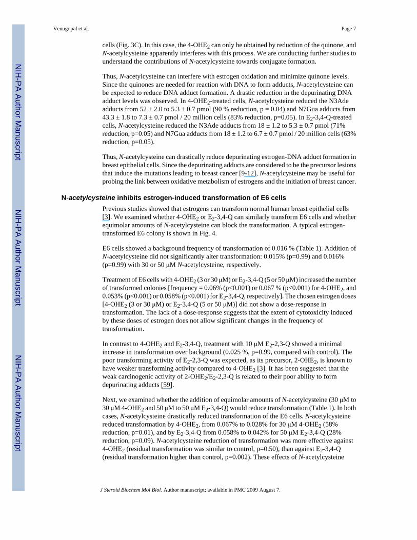
Thus, N-acetylcysteine can interfere with estrogen oxidation and minimize quinone levels.Since the quinones are needed for reaction with DNA to form adducts, N-acetylcysteine canbe expected to reduce DNA adduct formation. A drastic reduction in the depurinating DNAadduct levels was observed. In 4-OHE2-treated cells, N-acetylcysteine reduced the N3Adeadducts from 52 ± 2.0 to 5.3 ± 0.7 pmol (90 % reduction, p = 0.04) and N7Gua adducts from43.3 ± 1.8 to 7.3 ± 0.7 pmol / 20 million cells (83% reduction, p=0.05). In E2-3,4-Q-treatedcells, N-acetylcysteine reduced the N3Ade adducts from 18 ± 1.2 to 5.3 ± 0.7 pmol (71%reduction, p=0.05) and N7Gua adducts from 18 ± 1.2 to 6.7 ± 0.7 pmol / 20 million cells (63%reduction, p=0.05).
Thus, N-acetylcysteine can drastically reduce depurinating estrogen-DNA adduct formation inbreast epithelial cells. Since the depurinating adducts are considered to be the precursor lesionsthat induce the mutations leading to breast cancer [9-12], N-acetylcysteine may be useful forprobing the link between oxidative metabolism of estrogens and the initiation of breast cancer.
N-acetylcysteine inhibits estrogen-induced transformation of E6 cellsPrevious studies showed that estrogens can transform normal human breast epithelial cells[3]. We examined whether 4-OHE2 or E2-3,4-Q can similarly transform E6 cells and whetherequimolar amounts of N-acetylcysteine can block the transformation. A typical estrogen-transformed E6 colony is shown in Fig. 4.
E6 cells showed a background frequency of transformation of 0.016 % (Table 1). Addition ofN-acetylcysteine did not significantly alter transformation: 0.015% (p=0.99) and 0.016%(p=0.99) with 30 or 50 μM N-acetylcysteine, respectively.
Treatment of E6 cells with 4-OHE2 (3 or 30 μM) or E2-3,4-Q (5 or 50 μM) increased the numberof transformed colonies [frequency = 0.06% (p<0.001) or 0.067 % (p<0.001) for 4-OHE2, and0.053% (p<0.001) or 0.058% (p<0.001) for E2-3,4-Q, respectively]. The chosen estrogen doses[4-OHE2 (3 or 30 μM) or E2-3,4-Q (5 or 50 μM)] did not show a dose-response intransformation. The lack of a dose-response suggests that the extent of cytotoxicity inducedby these doses of estrogen does not allow significant changes in the frequency oftransformation.
In contrast to 4-OHE2 and E2-3,4-Q, treatment with 10 μM E2-2,3-Q showed a minimalincrease in transformation over background (0.025 %, p=0.99, compared with control). Thepoor transforming activity of E2-2,3-Q was expected, as its precursor, 2-OHE2, is known tohave weaker transforming activity compared to 4-OHE2 [3]. It has been suggested that theweak carcinogenic activity of 2-OHE2/E2-2,3-Q is related to their poor ability to formdepurinating adducts [59].
Next, we examined whether the addition of equimolar amounts of N-acetylcysteine (30 μM to30 μM 4-OHE2 and 50 μM to 50 μM E2-3,4-Q) would reduce transformation (Table 1). In bothcases, N-acetylcysteine drastically reduced transformation of the E6 cells. N-acetylcysteinereduced transformation by 4-OHE2, from 0.067% to 0.028% for 30 μM 4-OHE2 (58%reduction, p=0.01), and by E2-3,4-Q from 0.058% to 0.042% for 50 μM E2-3,4-Q (28%reduction, p=0.09). N-acetylcysteine reduction of transformation was more effective against4-OHE2 (residual transformation was similar to control, p=0.50), than against E2-3,4-Q(residual transformation higher than control, p=0.002). These effects of N-acetylcysteine
Venugopal et al. Page 7
J Steroid Biochem Mol Biol. Author manuscript; available in PMC 2009 August 7.
NIH
-PA Author Manuscript
NIH
-PA Author Manuscript
NIH
-PA Author Manuscript

provide a tool to directly correlate oxidative metabolism of estrogens with transformation ofbreast epithelial cells, which is expected to be an early event of breast cancer.
It is possible that the background frequency of transformation could be related to intrinsicproperties of the E6 cells, such as pre-existing mutations. To examine whether E6 cellscontained pre-existing mutations, we PCR amplified the exon 1-2 region of the H-ras gene(500 bp), cloned the product in pUC18, transformed into E.coli, isolated single colonies usingthe IPTG-X-gal colony color assay, extracted the recombinant plasmids and sequenced theinserts (Fig. 5). This PCR protocol for mutation analysis generates low levels of mutations(one mutation in ∼40 plasmids, mutation frequency= 5 × 10⁻⁵, calculated by dividing thenumber of mutations with the total number of bases sequenced) [9,12,37]. Analysis of 31plasmid clones from the E6 cells showed 8 mutations (mutation frequency = 51.6 × 10⁻⁵,p=0.01, Fisher's Exact test comparison of PCR-induced vs. pre-existing mutations). Seven ofthese mutations were G.C to T.A and the eighth mutation was A.T to T.A. Since the E6 cellscontained pre-existing mutations, a role of these mutations in spontaneous transformation ofE6 cells warrants consideration.
DiscussionThe E6 are normal mouse breast epithelial cells. Similar human cells exist (MCF-10A andMCF-10F). In particular, the MCF-10F cells are considered to be a good model of the humanbreast [13]. Unfortunately, both MCF-10A and 10F cells have low CYP1B1, and thus showpoor ability to oxidize estrogens [69]. Estrogen oxidation is more efficient in normal humanbreast [5]. Estrogens can transform both MCF-10A and 10F cells, but four treatments arerequired. Results presented here show that the E6 cells are efficient estrogen oxidizers, andcan be transformed with a single estrogen treatment. This study is the first to demonstrate thetransforming activity of estrogen-3,4-quinones.
The purpose of this study was to examine whether the anti-oxidant N-acetylcysteine can beused to test the hypothesis that depurinating DNA adduct formation by estrogen-3,4-quinonesis the initiating event in breast cancer. A transformation protocol that requires four treatmentscan model the combined effects of initiation and promotion from sustained carcinogenexposure. On the other hand, transformation by a single treatment of a carcinogen can reportits initiating activity. Therefore, for this study, we think that the E6 are more suitable than theMCF-10A and 10F cells.
N-acetylcysteine prevented estrogen-DNA damage and associated death of E6 cells (Figs 2 &3). In addition to preventing estrogen-DNA adduct formation, N-acetylcysteine stronglyinhibited estrogen-induced transformation (Table 1). The ability of N-acetylcysteine ininhibiting estrogen-induced transformation should be validated in appropriate human cells.However, we think that the present results provide a proof-of-principle and warrant theexploration of N-acetylcysteine in the prevention of breast cancer.
References1. Beatson GT. On the treatment of inoperable cases of carcinoma of the mamma: suggestions for a new
treatment with illustrative cases. Lancet 1896;2:104–107.2. Henderson BE, Feigelson HS. Hormonal carcinogenesis. Carcinogenesis 2000;21:427–433. [PubMed:
10688862]3. Russo J, Hasan Lareef M, Balogh G, Guo S, Russo IH. Estrogen and its metabolites are carcinogenic
agents in human breast epithelial cells. J Steroid Biochem Mol Biol 2003;87:1–25. [PubMed:14630087]
4. Cavalieri E, Chakravarti D, Guttenplan J, Hart E, Ingle J, Jankowiak R, Muti P, Rogan E, Russo J,Santen R, Sutter T. Catechol estrogen quinones as initiators of breast and other human cancers:
Venugopal et al. Page 8
J Steroid Biochem Mol Biol. Author manuscript; available in PMC 2009 August 7.
NIH
-PA Author Manuscript
NIH
-PA Author Manuscript
NIH
-PA Author Manuscript
implications for biomarkers of susceptibility and cancer prevention. Biochim Biophys Acta2006;1766:63–78. [PubMed: 16675129]
5. Rogan EG, Badawi AF, Devanesan PD, Meza JL, Edney JA, West WW, Higginbotham SM, CavalieriEL. Relative imbalances in estrogen metabolism and conjugation in breast tissue of women withcarcinoma: potential biomarkers of susceptibility to cancer. Carcinogenesis 2003;24:697–702.[PubMed: 12727798]
6. Singh S, Chakravarti D, Edney JA, Hollins RR, Johnson PJ, West WW, Higginbotham SM, CavalieriEL, Rogan EG. Relative imbalances in the expression of estrogen-metabolizing enzymes in the breasttissue of women with breast carcinoma. Oncol Rep 2005;14:1091–1096. [PubMed: 16142378]
7. Singh S, Zahid M, Gaikwad N, Chakravarti D, Cavalieri E, Rogan E. The effect of NAD(P)H:quinoneoxidoreductase 1 polymorphisms on estrogen metabolism in relation to initiation of breast cancer. ProcAm Assoc Cancer Res 2006;47:835.
8. Markushin Y, Zhong W, Cavalieri EL, Rogan EG, Small GJ, Yeung ES, Jankowiak R. Spectralcharacterization of catechol estrogen quinone (CEQ)-derived DNA adducts and their identification inhuman breast tissue extract. Chem Res Toxicol 2003;16:1107–1117. [PubMed: 12971798]
9. Chakravarti D, Mailander PC, Li KM, Higginbotham S, Zhang HL, Gross ML, Meza JL, Cavalieri EL,Rogan EG. Evidence that a burst of DNA depurination in SENCAR mouse skin induces error-pronerepair and forms mutations in the H-ras gene. Oncogene 2001;20:7945–7953. [PubMed: 11753677]
10. Zhao Z, Kosinska W, Khmelnitsky M, Cavalieri EL, Rogan EG, Chakravarti D, Sacks PG, GuttenplanJB. Mutagenic activity of 4-hydroxyestradiol, but not 2-hydroxyestradiol, in BB rat2 embryonic cells,and the mutational spectrum of 4-hydroxyestradiol. Chem Res Toxicol 2006;19:475–479. [PubMed:16544955]
11. Fernandez SV, Russo IH, Russo J. Estradiol and its metabolites 4-hydroxyestradiol and 2-hydroxyestradiol induce mutations in human breast epithelial cells. Int J Cancer 2006;118:1862–1868. [PubMed: 16287077]
12. Mailander PC, Meza JL, Higginbotham S, Chakravarti D. Induction of A.T to G.C mutations byerroneous repair of depurinated DNA following estrogen treatment of the mammary gland of ACIrats. J Steroid Biochem Mol Biol 2006;101:204–215. [PubMed: 16982187]
13. Russo J, Tahin Q, Lareef MH, Hu YF, Russo IH. Neoplastic transformation of human breast epithelialcells by estrogens and chemical carcinogens. Environ Mol Mutagen 2002;39:254–263. [PubMed:11921196]
14. Li JJ, Li SA. Estrogen carcinogenesis in hamster tissues: a critical review. Endocr Rev 1990;11:524–531. [PubMed: 2292241]
15. Yager JD. Endogenous estrogens as carcinogens through metabolic activation. J Natl Cancer InstMonogr 2000;27:67–73. [PubMed: 10963620]
16. Liehr JG. Is estradiol a genotoxic mutagenic carcinogen? Endocr Rev 2000;21:40–54. [PubMed:10696569]
17. Newbold RR, Liehr JG. Induction of uterine adenocarcinoma in CD-1 mice by catechol estrogens.Cancer Res 2000;60:235–237. [PubMed: 10667565]
18. Zahid M, Saeed M, Olson K, Gaikwad N, Rogan E, Cavalieri E. Inhibition of the formation of theN3Ade and N7Gua depurinating adducts after reaction of estradiol-3,4-quinone or lactoperoxidase-oxidized 4-hydroxyestradiol with DNA. Proc Am Assoc Cancer Res 2006;47:446.
19. Bonanomi L, Gazzaniga A. Toxicological, pharmacokinetic and metabolic studies on acetylcysteine.Eur J Respir Dis Suppl 1980;111:45–51. [PubMed: 6938410]
20. Webb WR. New Mucolytic Agents for Sputum Liquefaction. Postgrad Med 1964;36:449–453.[PubMed: 14212497]
21. Flanagan RJ. The role of acetylcysteine in clinical toxicology. Med Toxicol 1987;2:93–104. [PubMed:3574040]
22. Doelman CJ, Bast A. Oxygen radicals in lung pathology. Free Radic Biol Med 1990;9:381–400.[PubMed: 1705530]
23. De Flora S, Cesarone CF, Balansky RM, Albini A, D'Agostini F, Bennicelli C, Bagnasco M,Camoirano A, Scatolini L, Rovida A, et al. Chemopreventive properties and mechanisms of N-acetylcysteine. The experimental background. J Cell Biochem Suppl 1995;22:33–41. [PubMed:8538208]
Venugopal et al. Page 9
J Steroid Biochem Mol Biol. Author manuscript; available in PMC 2009 August 7.
NIH
-PA Author Manuscript
NIH
-PA Author Manuscript
NIH
-PA Author Manuscript

24. Tattersall AB, Bridgman KM, Huitson A. Acetylcysteine (Fabrol) in chronic bronchitis--a study ingeneral practice. J Int Med Res 1983;11:279–284. [PubMed: 6642068]
25. Grandjean EM, Berthet P, Ruffmann R, Leuenberger P. Efficacy of oral long-term N-acetylcysteinein chronic bronchopulmonary disease: a meta-analysis of published double-blind, placebo-controlledclinical trials. Clin Ther 2000;22:209–221. [PubMed: 10743980]
26. Goldenberg I, Shechter M, Matetzky S, Jonas M, Adam M, Pres H, Elian D, Agranat O,Schwammenthal E, Guetta V. Oral acetylcysteine as an adjunct to saline hydration for the preventionof contrast-induced nephropathy following coronary angiography. A randomized controlled trial andreview of the current literature. Eur Heart J 2004;25:212–218. [PubMed: 14972421]
27. De Flora S, Bennicelli C, Zanacchi P, Camoirano A, Morelli A, De Flora A. In vitro effects of N-acetylcysteine on the mutagenicity of direct-acting compounds and procarcinogens. Carcinogenesis1984;5:505–510. [PubMed: 6368036]
28. De Flora S, Bennicelli C, Zanacchi P, D'Agostini F, Camoirano A. Mutagenicity of active oxygenspecies in bacteria and its enzymatic or chemical inhibition. Mutat Res 1989;214:153–158. [PubMed:2671696]
29. De Flora S, Izzotti A, D'Agostini F, Cesarone CF. Antioxidant activity and other mechanisms of thiolsinvolved in chemoprevention of mutation and cancer. Am J Med 1991;91:122S–130S. [PubMed:1928203]
30. Kelloff GJ, Crowell JA, Boone CW, Steele VE, Lubet RA, Greenwald P, Alberts DS, Covey JM,Doody LA, Knapp GG, et al. Clinical development plan: N-Acetyl-l-cysteine. J Cell Biochem Suppl1994;20:63–73. [PubMed: 7616754]
31. De Flora S, Izzotti A, D'Agostini F, Balansky R. Mechanisms of N-acetylcysteine in the preventionof DNA damage and cancer, with special reference to smoking-related end-points. Carcinogenesis2001;22:999–1013. [PubMed: 11408342]
32. Balansky RM, Ganchev G, D'Agostini F, De Flora S. Effects of N-acetylcysteine in an esophagealcarcinogenesis model in rats treated with diethylnitrosamine and diethyldithiocarbamate. Int J Cancer2002;98:493–497. [PubMed: 11920607]
33. Lippman SM, Spitz MR. Lung cancer chemoprevention: an integrated approach. J Clin Oncol2001;19:74S–82S. [PubMed: 11560978]
34. Chakravarti D, Zahid M, Backora M, Myers EM, Weisenburger DD, Cavalieri EL, Rogan EG, JoshiSS. Ortho-quinones of benzene and estrogens induce hyperproliferation of human peripheral bloodmononuclear cells. Leuk Lymphoma 2006;47:2635–2644. [PubMed: 17169809]
35. Sgagias MK, Wagner KU, Hamik B, Stoeger S, Spieker R, Huber LJ, Chodosh LA, Cowan KH.Brca1-deficient murine mammary epithelial cells have increased sensitivity to CDDP and MMS. CellCycle 2004;3:1451–1456. [PubMed: 15492509]
36. Chakravarti D, Mailander P, Franzen J, Higginbotham S, Cavalieri EL, Rogan EG. Detection ofdibenzo[a,l]pyrene-induced H-ras codon 61 mutant genes in preneoplastic SENCAR mouse skinusing a new PCR-RFLP method. Oncogene 1998;16:3203–3210. [PubMed: 9671400]
37. Chakravarti D, Mailander PC, Cavalieri EL, Rogan EG. Evidence that error-prone DNA repairconverts dibenzo[a,l]pyrene-induced depurinating lesions into mutations: formation, clonalproliferation and regression of initiated cells carrying H-ras oncogene mutations in earlypreneoplasia. Mutat Res 2000;456:17–32. [PubMed: 11087892]
38. Hoffer E, Baum Y, Tabak A, Taitelman U. N-acetylcysteine increases the glutathione content andprotects rat alveolar type II cells against paraquat-induced cytotoxicity. Toxicol Lett 1996;84:7–12.[PubMed: 8597179]
39. Kanno S, Ishikawa M, Takayanagi M, Takayanagi Y, Sasaki K. Exposure to hydrogen peroxideinduces cell death via apoptosis in primary cultured mouse hepatocytes. Biol Pharm Bull1999;22:1296–1300. [PubMed: 10746159]
40. Zhou X, Zhao A, Goping G, Hirszel P. Gliotoxin-induced cytotoxicity proceeds via apoptosis and ismediated by caspases and reactive oxygen species in LLC-PK1 cells. Toxicol Sci 2000;54:194–202.[PubMed: 10746946]
41. Samuni AM, Chuang EY, Krishna MC, Stein W, DeGraff W, Russo A, Mitchell JB. Semiquinoneradical intermediate in catecholic estrogen-mediated cytotoxicity and mutagenesis: chemopreventionstrategies with antioxidants. Proc Natl Acad Sci U S A 2003;100:5390–5395. [PubMed: 12702779]
Venugopal et al. Page 10
J Steroid Biochem Mol Biol. Author manuscript; available in PMC 2009 August 7.
NIH
-PA Author Manuscript
NIH
-PA Author Manuscript
NIH
-PA Author Manuscript

42. Terasaka H, Kadoma Y, Sakagami H, Fujisawa S. Cytotoxicity and apoptosis-inducing activity ofbisphenol A and hydroquinone in HL-60 cells. Anticancer Res 2005;25:2241–2247. [PubMed:16158970]
43. Chan SY, Hilchie AL, Brown MG, Anderson R, Hoskin DW. Apoptosis induced by intracellularceramide accumulation in MDA-MB-435 breast carcinoma cells is dependent on the generation ofreactive oxygen species. Exp Mol Pathol 2007;82:1–11. [PubMed: 16624283]
44. Aruoma OI, Halliwell B, Hoey BM, Butler J. The antioxidant action of N-acetylcysteine: its reactionwith hydrogen peroxide, hydroxyl radical, superoxide, and hypochlorous acid. Free Radic Biol Med1989;6:593–597. [PubMed: 2546864]
45. Hiraku Y, Kawanishi S. Oxidative DNA damage and apoptosis induced by benzene metabolites.Cancer Res 1996;56:5172–5178. [PubMed: 8912853]
46. Cavalieri EL, Rogan EG, Chakravarti D. Initiation of cancer and other diseases by catechol ortho-quinones: a unifying mechanism. Cell Mol Life Sci 2002;59:665–681. [PubMed: 12022473]
47. Nutter LM, Zhou B, Sierra EE, Wu YY, Rummel MM, Gutierrez P, Abul-Hajj Y. Cellular biochemicaldeterminants modulating the metabolism of estrone 3,4-quinone. Chem Res Toxicol 1994;7:609–613. [PubMed: 7841338]
48. Hurh YJ, Chen ZH, Na HK, Han SY, Surh YJ. 2-Hydroxyestradiol induces oxidative DNA damageand apoptosis in human mammary epithelial cells. J Toxicol Environ Health A 2004;67:1939–1953.[PubMed: 15513894]
49. Kalyanaraman B, Korytowski W, Pilas B, Sarna T, Land EJ, Truscott TG. Reaction between ortho-semiquinones and oxygen: pulse radiolysis, electron spin resonance, and oxygen uptake studies. ArchBiochem Biophys 1988;266:277–284. [PubMed: 2845864]
50. Chen ZH, Na HK, Hurh YJ, Surh YJ. 4-Hydroxyestradiol induces oxidative stress and apoptosis inhuman mammary epithelial cells: possible protection by NF-kappaB and ERK/MAPK. Toxicol ApplPharmacol 2005;208:46–56. [PubMed: 15901486]
51. Cavalieri E. Minisymposium on endogenous carcinogens: the catechol estrogen pathway. Anintroduction. Polycyclic Aromatic Compd 1994;6:223–228.
52. Mobley JA, Bhat AS, Brueggemeier RW. Measurement of oxidative DNA damage by catecholestrogens and analogues in vitro. Chem Res Toxicol 1999;12:270–277. [PubMed: 10077490]
53. Han X, Liehr JG. 8-Hydroxylation of guanine bases in kidney and liver DNA of hamsters treated withestradiol: role of free radicals in estrogen-induced carcinogenesis. Cancer Res 1994;54:5515–5517.[PubMed: 7923187]
54. Lin PH, Nakamura J, Yamaguchi S, Asakura S, Swenberg JA. Aldehydic DNA lesions induced bycatechol estrogens in calf thymus DNA. Carcinogenesis 2003;24:1133–1141. [PubMed: 12807746]
55. Hiraku Y, Yamashita N, Nishiguchi M, Kawanishi S. Catechol estrogens induce oxidative DNAdamage and estradiol enhances cell proliferation. Int J Cancer 2001;92:333–337. [PubMed:11291067]
56. Liehr JG, Fang WF, Sirbasku DA, Ari-Ulubelen A. Carcinogenicity of catechol estrogens in Syrianhamsters. J Steroid Biochem 1986;24:353–356. [PubMed: 3009986]
57. Li JJ, Li SA. Estrogen carcinogenesis in Syrian hamster tissue: role of metabolism. Fed Proc1987;46:1858–1863. [PubMed: 3030825]
58. Liehr JG, Ricci MJ. 4-Hydroxylation of estrogens as marker of human mammary tumors. Proc NatlAcad Sci U S A 1996;93:3294–3296. [PubMed: 8622931]
59. Zahid M, Kohli E, Saeed M, Rogan E, Cavalieri E. The greater reactivity of estradiol-3,4-quinone vsestradiol-2,3-quinone with DNA in the formation of depurinating adducts: implications for tumor-initiating activity. Chem Res Toxicol 2006;19:164–172. [PubMed: 16411670]
60. Tabakovic K, Gleason WB, Ojala WH, Abul-Hajj YJ. Oxidative transformation of 2-hydroxyestrone.Stability and reactivity of 2,3-estrone quinone and its relationship to estrogen carcinogenicity. ChemRes Toxicol 1996;9:860–865. [PubMed: 8828921]
61. Nutter LM, Wu YY, Ngo EO, Sierra EE, Gutierrez PL, Abul-Hajj YJ. An o-quinone form of estrogenproduces free radicals in human breast cancer cells: correlation with DNA damage. Chem Res Toxicol1994;7:23–28. [PubMed: 8155821]
62. Gaikwad NW, Cavalieri E, Rogan E. NQO1-catalyzed reduction of estradiol-3,4-quinone.Implications for tumor initiation by estrogens. Proc Am Assoc Cancer Res 2006;47:445.
Venugopal et al. Page 11
J Steroid Biochem Mol Biol. Author manuscript; available in PMC 2009 August 7.
NIH
-PA Author Manuscript
NIH
-PA Author Manuscript
NIH
-PA Author Manuscript

63. Nutter LM, Ngo EO, Abul-Hajj YJ. Characterization of DNA damage induced by 3,4-estrone-o-quinone in human cells. J Biol Chem 1991;266:16380–16386. [PubMed: 1653233]
64. Cao K, Stack DE, Ramanathan R, Gross ML, Rogan EG, Cavalieri EL. Synthesis and structureelucidation of estrogen quinones conjugated with cysteine, N-acetylcysteine, and glutathione. ChemRes Toxicol 1998;11:909–916. [PubMed: 9705753]
65. Nakagomi M, Suzuki E. Quantitation of catechol estrogens and their N-acetylcysteine conjugates inurine of rats and hamsters. Chem Res Toxicol 2000;13:1208–1213. [PubMed: 11123960]
66. Devanesan P, Santen RJ, Bocchinfuso WP, Korach KS, Rogan EG, Cavalieri E. Catechol estrogenmetabolites and conjugates in mammary tumors and hyperplastic tissue from estrogen receptor-alphaknock-out (ERKO)/Wnt-1 mice: implications for initiation of mammary tumors. Carcinogenesis2001;22:1573–1576. [PubMed: 11532882]
67. Todorovic R, Devanesan P, Higginbotham S, Zhao J, Gross ML, Rogan EG, Cavalieri EL. Analysisof potential biomarkers of estrogen-initiated cancer in the urine of Syrian golden hamsters treatedwith 4-hydroxyestradiol. Carcinogenesis 2001;22:905–911. [PubMed: 11375897]
68. Boyland E, Chasseaud LF. The role of glutathione and glutathione S-transferases in mercapturic acidbiosynthesis. Adv Enzymol Relat Areas Mol Biol 1969;32:173–219. [PubMed: 4892500]
69. Lu F, Zahid M, Saeed M, Cavalieri EL, Rogan EG. Estrogen metabolism and formation of estrogen-DNA adducts in estradiol-treated MCF-10F cells The effects of 2,3,7,8-tetrachlorodibenzo-p-dioxininduction and catechol-O-methyltransferase inhibition. J Steroid Biochem Mol Biol. 2007in press
Venugopal et al. Page 12
J Steroid Biochem Mol Biol. Author manuscript; available in PMC 2009 August 7.
NIH
-PA Author Manuscript
NIH
-PA Author Manuscript
NIH
-PA Author Manuscript
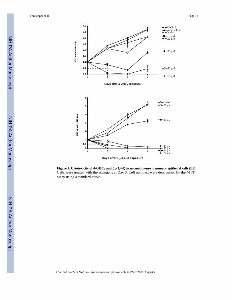
Figure 1. Cytotoxicity of 4-OHE2 and E2-3,4-Q in normal mouse mammary epithelial cells (E6)Cells were treated with the estrogens at Day 0. Cell numbers were determined by the MTTassay using a standard curve.
Venugopal et al. Page 13
J Steroid Biochem Mol Biol. Author manuscript; available in PMC 2009 August 7.
NIH
-PA Author Manuscript
NIH
-PA Author Manuscript
NIH
-PA Author Manuscript

Figure 2. N-acetylcysteine rescues E6 cells from 4-OHE2 and E2-3,4-Q cytotoxicityEquimolar amounts of N-acetylcysteine were added to the experiments at Day 0. Cell numberswere determined by the MTT assay using a standard curve.
Venugopal et al. Page 14
J Steroid Biochem Mol Biol. Author manuscript; available in PMC 2009 August 7.
NIH
-PA Author Manuscript
NIH
-PA Author Manuscript
NIH
-PA Author Manuscript

Figure 3. N-acetylcysteine inhibits estrogen-DNA adduct formation in E6 cellsE2 and E1 are interconverted in the cell; therefore, we combined the conjugates and adductsfrom E1 and E2 together. The quinone produced less conjugates and DNA adducts than thecatechol (note the difference in the Y-axes of B & C).
Venugopal et al. Page 15
J Steroid Biochem Mol Biol. Author manuscript; available in PMC 2009 August 7.
NIH
-PA Author Manuscript
NIH
-PA Author Manuscript
NIH
-PA Author Manuscript

Figure 4. Colony morphology of estrogen-transformed E6 cells
Venugopal et al. Page 16
J Steroid Biochem Mol Biol. Author manuscript; available in PMC 2009 August 7.
NIH
-PA Author Manuscript
NIH
-PA Author Manuscript
NIH
-PA Author Manuscript
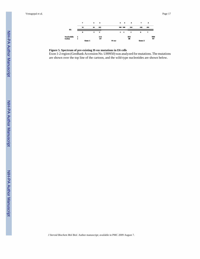
Figure 5. Spectrum of pre-existing H-ras mutations in E6 cellsExon 1-2 region (GenBank Accession No. U89950) was analyzed for mutations. The mutationsare shown over the top line of the cartoon, and the wild-type nucleotides are shown below.
Venugopal et al. Page 17
J Steroid Biochem Mol Biol. Author manuscript; available in PMC 2009 August 7.
NIH
-PA Author Manuscript
NIH
-PA Author Manuscript
NIH
-PA Author Manuscript

NIH
-PA Author Manuscript
NIH
-PA Author Manuscript
NIH
-PA Author Manuscript
Venugopal et al. Page 18
Table 1
TreatmentsTransformation Total
colonies/Total wells (%cells transformed *)
p-value** (comparedwith Control)
Estrogen metabolite (μM) N-acetylcysteine (μM)
Control - - 70/88 (0.016%) -
- 30 17/24 (0.015%) 0.99
- 50 18/24 (0.016%) 0.99
4-OHE2 3 - 58/19 (0.060%) <0.001
30 - 52/16 (0.067%) <0.001
30 30 21/15 (0.028%) 0.50
E2-3,4-Q 5 - 53/20 (0.053%) <0.001
50 - 60/20 (0.058%) <0.001
50 50 42/20 (0.042%) 0.002
E2-2,3-Q 10 - 27/24 (0.025%) 0.99
*5000 cells/well.
**P-values are adjusted for multiple comparisons with the control using the method of Bonferroni.
J Steroid Biochem Mol Biol. Author manuscript; available in PMC 2009 August 7.