Synthesis and Total Phenol Content of New Resveratrol Derivative

Treatment of FANCA Cells with Resveratrol and N-Acetylcysteine: A Comparative StudyMarta Columbaro1., Silvia Ravera2., Cristina Capanni3, Isabella Panfoli2, Paola Cuccarolo4,
Giorgia Stroppiana5, Paolo Degan4., Enrico Cappelli6*.
1 SC Laboratory of Musculoskeletal Cell Biology, IOR, Bologna, Italy, 2 DIFAR-Biochemistry Lab., University of Genova, Genova, Italy, 3 CNR-National Research Council of
Italy, Institute of Molecular Genetics, Unit of Bologna-IOR, Bologna, Italy, 4 S. C. Mutagenesis, IRCCS AOU San Martino – IST (Istituto Nazionale per la Ricerca sul Cancro),
Genova, Italy, 5 Centro di Diagnostica Genetica e Biochimica delle Malattie Metaboliche, Istituto Giannina Gaslini, Genova, Italy, 6 Hematology Unit, Istituto Giannina
Gaslini, Genova, Italy
Abstract
Fanconi anemia (FA) is a genetic disorder characterised by chromosome instability, cytokine ipersensibility, bone marrowfailure and abnormal haematopoiesis associated with acute myelogenous leukemia. Recent reports are contributing tocharacterize the peculiar FA metabolism. Central to these considerations appears that cells from complementation group A(FANCA) display an altered red-ox metabolism. Consequently the possibility to improve FA phenotypical conditions withantioxidants is considered. We have characterized from the structural and biochemical point of view the response of FANCAlymphocytes to N-acetyl-cysteine (NAC) and resveratrol (RV). Surprisingly both NAC and RV failed to revert all thecharacteristic of FA phenotype and moreover their effects are not super imposable. Our data suggest that we must beaware of the biological effects coming from antioxidant treatment.
Citation: Columbaro M, Ravera S, Capanni C, Panfoli I, Cuccarolo P, et al. (2014) Treatment of FANCA Cells with Resveratrol and N-Acetylcysteine: A ComparativeStudy. PLoS ONE 9(8): e104857. doi:10.1371/journal.pone.0104857
Editor: Mauro Salvi, University of Padova, Italy
Received April 30, 2014; Accepted July 17, 2014; Published August 15, 2014
Copyright: � 2014 Columbaro et al. This is an open-access article distributed under the terms of the Creative Commons Attribution License, which permitsunrestricted use, distribution, and reproduction in any medium, provided the original author and source are credited.
Data Availability: The authors confirm that all data underlying the findings are fully available without restriction. All relevant data are within the paper and itsSupporting Information files.
Funding: The authors have no funding or support to report.
Competing Interests: The authors have declared that no competing interests exist.
* Email: [email protected]
. These authors contributed equally to this work.
Introduction
Fanconi anemia (FA) is a genetic disorder characterised by
chromosome instability and cytokine hypersensitivity. Bone
marrow failure and abnormal haematopoiesis are associated with
high frequency to clonal hematopoietic stem cells (HSC)
expansion, acute myelogeneous leukemia via a mechanism
involving genomic instability and inflammation [1].
We recently reported [2,3] different structural abnormalities in
FANCA cells underlying an impaired mitochondrial functionality
affecting the energy metabolism. A defective respiration at the
mitochondrial oxidative phosphorylation complex I is associated
with a reduced ATP production and altered ATP/AMP ratio.
These defects are consistently associated with impaired oxygen
metabolism [4]. Therefore the possibility to improve FA patients
physiological state with antioxidants as therapy adjuvants appears
promising.
N-acetyl-cysteine (NAC) is a sulphydryl-group providing com-
pound which acts as a precursor of reduced glutathione (GSH) and
as direct scavenger of reactive oxygen species (ROS). Intracellular
reduced GSH is often depleted as a consequence of increased
oxidative stress and inflammation. Hence NAC can regulate the
red-ox status in the cells interfering with several signalling
pathways. NAC is widely used in clinical treatment [5] as support
in treatment of diseases related to oxidative stress. Actually almost
250 studies with NAC are enlisted in the Clinical Trials
governmental registry (www.clinicaltrials.gov).
Resveratrol (RV) is a naturally-occurring polyphenol mainly
found in grapes. A growing body of literature has demonstrated
the beneficial effects of RV on age-related metabolic deterioration
and its protective role in metabolic diseases. RV exerts its potent
anticarcinogenic effect by inducing apoptosis and inhibiting tumor
promoter-induced cell transformation [6]. RV protects against the
deregulation of energy homeostasis, up-regulates eNOS and many
cellular anti-oxidant enzymes, down regulates TNFa and NF-kB
expression and inhibits NADPH oxidases [7]. Moreover, crystal-
lographic studies showed that RV (and other related polyphenols)
directly inhibits the rotary mechanism of mitochondrial FoF1-ATP
synthase (ATP synthase) by binding to a site in c-subunit and
hence its ATP synthetic activity [8]. More than 70 clinical trials
with RV are actually listed in the National Institutes of Health
website.
In FA, the use of NAC and RV has already been proposed.
Treatment with NAC, in association with Lipoic Acid (LA) [9]
increased cellular viability as well as GSH and ATP contents, and
reduced spontaneous and DEB-induced chromosomal instability
in lymphocyte from FA patients. The protective abilities of NAC,
RV and tempol were compared in the FANCD2 murine model
[10]. NAC and RV partially corrected the abnormal cell cycle
state of the HSP cells and helped maintaining them in a quiescent
state. In turn tempol substantially delayed tumor onset apparently
PLOS ONE | www.plosone.org 1 August 2014 | Volume 9 | Issue 8 | e104857

without a beneficial effect on hematopoiesis. Finally an antioxidant
dietary formulate containing lysine, proline, ascorbic acid and
green tea extracts, was successfully reported to inhibit in vitro and
in vivo FANCA-associated head and neck squamous cell
carcinoma [11].
Notwithstanding the potential interest concerning these results
the still crucial and open question is that we do not know yet which
molecular mechanisms and metabolic pathways are relevant in the
FA pathological phenotype. Here we evaluate the biological effects
of RV and NAC, two most promising antioxidants which act with
different biochemical mechanisms.
Materials & Methods
Ethics statementStudy approval was obtained from the Ethics Committee at the
Gaslini Hospital, Genova, Italy (protocol Nu J5002 date: 24/9/
2010). Informed written consent was obtained from the adult
subjects and from parents, on the behalf of their children, involved
in the study. All clinical investigations were conducted according
to the principles expressed in the Declaration of Helsinki.
Cell culture and treatmentsFANCA primary fibroblast cell lines, isogenic FANCA primary
fibroblasts corrected with S11FAIN [12] retrovirus and wild type
(wt) cells were grown as monolayer at 37uC in RPMI
supplemented with 10% fetal calf serum and antibiotics. All the
cell lines between the 5th and 15th passage, were grown with the
same density and conditions. During treatment we didn’t observe
significant changes in the cellular viability. FANCA and wt
lymphoblast cell lines were grown at 37uC in RPMI supplemented
with 10% fetal calf serum and antibiotics. Primary lymphocytes
were isolated using Ficoll-Paque Plus and grown at 37uC in RPMI
supplemented with 10% fetal calf serum, antibiotics and phyto-
hemagglutinin (20 mg/ml). N-acetyl-cysteine (500 mM), resveratrol
(10 mM) were added to the cells once a day for five days. One hour
after the last treatment, cells were used for protein extracts or fixed
for electron microscopy experiments. FANCA corrected cells
always behaved alike wt [3].
Oxygen consumption measurementsTo measure oxygen consumption, an amperometric electrode
(Unisense-Microrespiration, Unisense A/S, Denmark) was used.
Experiments were performed in a closed chamber at 25uC. For
each experiment 500.000 cells were permeabilized with 0.03 mg/
ml digitonin for 1 minute, centrifuged for 9 minutes at 1000 rpm
and resuspended in: 137 mM NaCl, 0.7 mM NaH2PO4, 5 mM
KCl and 25 mM Tris HCl pH 7.4. The same medium was used in
the oximetric experiments. To stimulate the pathway composed by
the respiratory complexes I, III and IV, 10 mM pyruvate and
5 mM malate was added to the sample, while to stimulate the
pathway II, III and IV 20 mM succinate was used as respiratory
substrate. To confirm that the oxygen consumption was really due
to oxidative phosphorylation (OXPHOS) machinery, 0.1 mM
rotenone or 0.2 mM antimycin A were used as inhibitors for the
first and second pathway, respectively [13].
Electron transfer from Complex I to Complex IIIThe electron transfer between Complex I to Complex III was
studied spectrophotometrically, following the reduction of cyto-
chrome c at 550 nm. The extinction molar coefficient used for
reduced cytochrome c was 1 mM21 cm21. For each assay, 50 mg
of total proteins was used. The assay medium containing; 100 mM
Tris-HCl pH 7,4 and 0,03% cytochrome c. The reaction was
started with the addition of 0,7 mM NADH [3]. If the electron
transport between Complex I and Complex III is conserved, the
electrons pass from NADH to Complex I, then to Complex III via
coenzyme Q, and finally to cytochrome c.
Adenylate kinase (AK) assayAK activity was assayed spectrophotometrically, following
NADH oxidation at 340 nm [14]. ATP and GTP in the assay
mixture are needed to assay the ATP-AMP phosphotransferase
activity (AK1 + AK2) or the GTP-AMP phosphotransferase
activity (mitochondrial AK3), respectively. Results are reported for
the activity of the enzyme (U/mg) per sample.
ATP and AMP quantificationATP and AMP were measured according to the enzyme
coupling method of Bergmeyer et al. [15]. For ATP assay, medium
contained 20 mg of sample, 50 mM Tris- HCl pH 8.0, 1 mM
NADP, 10 mM MgCl2, and 5 mM glucose in 1 ml final volume.
Samples were analyzed spectrophotometrically before and after
the addition of 4 mg of purified hexokinase/glucose-6-phosphate
dehydrogenase (Boehringer). The rise in absorbance at 340 nm,
due to NADPH formation, was proportional to the ATP
concentration. For AMP assay, the medium contained 20 mg of
sample, 50 mM Tris-HCl pH 8.0, 1 mM NADH, 10 mM MgCl2,
and 10 mM phosphoenolpyruvate (PEP), 2 mM ATP in 1 ml final
volume. Samples were analyzed spectrophotometrically before and
after the addition of 4 mg of purified pyruvate kinase/lactate
dehydrogenase (Boehringer). The rise in absorbance at 340 nm,
due to NADH oxidation, was proportional to the AMP
concentration. For all biochemical experiments, protein concen-
trations were determined using the Bradford method.
Electron microscopyLymphocyte pellets from healthy donor and FANCA patients
(subjected or not to antioxidant treatments) were fixed with 2.5%
glutaraldehyde 0.1 M cacodylate buffer pH 7.6 for 1 h at room
temperature. After post-fixation with 1% OsO4 in cacodylate
buffer for 1 h, pellets were dehydrated in an ethanol series and
embedded in Epon resin. Ultrathin sections stained with uranyl-
acetate and lead citrate were observed with a Jeol Jem-1011
transmission electron microscope. Two hundred mitochondria
were examined for each sample.
CytometryROS were quantified by cytometry with 29,79-dichlorodihydro-
fluorescein diacetate (H2DCFH-DA). This dye is a cell-permeable,
nonfluorescent molecule, which is very sensitive to intracellular
redox change. When H2DCFH-DA enters into the cell it is cleaved
by intracellular esterases into 29,79-dichlorodihydrofluorescein
(H2DCF) which, in presence of H2O2, oxidize to the fluorescent
molecule dichlorofluorescein (DCF). Fluorescence from this probe
is measured by flow cytometry, as the dye is excited by the 488-nm
laser. H2DCFH-DA has a good specificity for H2O2, and it has
been shown that the fluorescence of the product DCF appears to
be mediated mainly by H2O2. Cytometry measures were
performed on a Cyan ADP cytometer (Beckman Coulter,
Mountain View, CA, USA) equipped with three laser lamps.Ten
thousand cells per sample were analyzed, and the results are
reported as the percentage of cells, relative to the relevant control,
that display a fluorescence shift.
Effects of Antioxidants in Fanconi Anemia
PLOS ONE | www.plosone.org 2 August 2014 | Volume 9 | Issue 8 | e104857

Statistical analysisData were analyzed by one-way ANOVA and unpaired two-tail
Student’s t test using instat software (GraphPad Software, Inc., La
Jolla, CA, USA). Data are expressed as mean 6 standard
deviation (SD) from 3 to 5 independent determinations performed
in duplicate. In the figures SD are shown as error bars. An error
probability with P,0.05 was selected as significant.
Results
1 – Cytometric ROS quantificationAn enhanced oxidative stress appears the common denominator
of the many altered functions in FANCA cells. In fact, we reported
[2,3] an enhanced ROS production in FANCA cells associated
with a scarce NADH utilization or availability at the mitochon-
drial respiratory Complex I. NAC and RV, although with different
mechanisms as discussed below, provide significant reduction in
intracellular ROS production. When FANCA cells were treated
with NAC or RV a significant decrease (p,0,01) in intracellular
ROS production was measured (Table 1). The large differences
reported among the samples analysed might be attributed to the
different functional characteristics of the mutant FANCA proteins
produced. In this context, the structure-function relationships in
mutant FANCA proteins (in the various patients) are however as
yet not fully available to-date and cannot be properly discussed at
this time. Castella et al. [19] reported cytoplasmic expression of
mutated FANCA protein without functional correlation between
genotype and phenotype. Moreover, this work evaluated the effect
of FANCA mutated protein only in the perspective of the DNA
repair activity without considering other possible functions for
FANCA protein. Indeed, these putative functions may be the
cause of the high variability observed.
2 – Biochemical Parameters2.1 - Oxygen consumption. As described by Ravera et al.
[3], FANCA cells display defective respiration trough the pathway
composed by Complex I, III and IV. In our experimental setting,
FANCA and wild type (wt) primary fibroblasts were treated with
500 mM NAC or 10 mM RV for 5 days. In these conditions the
ability of FANCA cells to respire from the Complex I, III and IV
pathway were fully restored (Fig. 1A) after NAC and only partially
after RV treatment.
Moreover RV reduced the ability of wt cells to respire both in
the presence of pyruvate/malate and with succinate, as respiring
substrates. This effect may be explained considering that RV is an
inhibitor of the activity of ATP synthase [16,17]. Namely, being
oxygen consumption coupled to ATP synthesis, when ATP
synthase is inhibited, the electron transport chain activity also
slows down, and so does oxygen consumption. In conclusion, even
though the effects of NAC and RV follow different pathways, the
net results is a reduction of ROS generated at the level of Complex
I. Genetic complementation by FANCA gene corrects the
respiratory defect. The same results were obtained in lymphoblast
cell lines from the same patients (data not shown).
2.2 - Complex I to Complex III electron
transfer. Defective respiration in FANCA cells is due to defect
in electron transfer from Complex I to Complex III and is
associated with reduced ATP production and altered ATP/AMP
ratio [3]. However, as shown above, the treatment of FANCA cells
with NAC restores their ability to respire through Complex I and,
accordingly, electron transfer efficiency between Complex I and
III is restored in FANCA at level comparable to wt cells. By
contrast, RV only partially recovered the FANCA cells electron
transfer efficiency. Also, in line with the results for oxygen
consumption, it reduced the electron transfer ability in wt cells
(Fig. 1B). The difference among the effect of NAC and RV likely
depends on their different target. In fact, being a precursor of
GSH NAC is really an antioxidant molecule able to decrease the
damage due to ROS production consequent to impaired
functioning of Complex I. By contrast, RV acts by inhibiting the
activity of ATP synthase, thus reducing the speed of electron
transport chain, i.e. of Complex I. This causes a lower production
of ROS thus allowing cell scavengers systems to work.
2.3 - Intracellular ATP and AMP quantification. In
FANCA the ATP/AMP ratio is very low with respect to the
control (Fig. 1C) in agreement with what can be expected from the
impairment of Complexes I-III electron transport. By contrast,
Table 1. Cytometric quantification of the ROS reduction (%) in FANCA cells after treatment with NAC and RV.
Samples ROS reduction (%)
+ NAC + RV
FANCA 1 32.67 31.28
FANCA 2 42.11 30.37
FANCA 3 27.70 37.03
FANCA 4 41.24 60.94
FANCA 5 41.40 28.71
FANCA 6 46.33 47.10
FANCA 7 35.85 64.18
FANCA 8 39.67 44.29
FANCA 9 44.81 57.17
FANCA 10 16.59 46.84
FANCA 11 59.26 n.d.
FANCA 12 45.50 43.94
FANCA 13 26.65 36.58
FANCA 14 77.84 16.31
doi:10.1371/journal.pone.0104857.t001
Effects of Antioxidants in Fanconi Anemia
PLOS ONE | www.plosone.org 3 August 2014 | Volume 9 | Issue 8 | e104857

after NAC treatment, the ATP level in FANCA cells was restored
and the AMP concentration decreases, although it remained
higher than in wt, allowing to reach a good ATP/AMP ratio
(Fig. 1C). In the case of RV treatment only a partial recovery of
ATP was seen in FANCA and a strong reduction in wt cells was
observed. This is likely related to the inhibitor effect of RV on
ATP synthase, considering that ATP synthase is the principal
source of ATP production for the cell.
2.4 - Adenylate Kinase (AK) activity. Adenylate Kinase
(AK) activities were also investigated. These enzymes catalyse the
interconversion of adenine nucleotides and contributing to the
regulation of the cellular energy homeostasis. In particular, we
assayed the ATP-AMP phosphotransferase isoforms (AK1 and
AK2), which catalyze the interconversion of ADP to ATP+AMP
and the GTP-AMP phosphotransferase isoform, typical of the
mitochondrial matrix, which utilizes GTP as donor for the
phosphate group to AMP [3]. In FANCA cells ATP-AMP
phosphotransferase activity was impaired while GTP-AMP
phosphotransferase activity was increased (Fig. 1D). Both NAC
and RV treatment restored FANCA ATP-AMP phosphotransfer-
ase activity, to a level similar to wt cells. By contrast, as already
observed [3], the GTP-AMP phosphotransferase activity was
higher in FANCA cells with respect to controls. Thus FANCA
cells appear to preferably use GTP, from Krebs cycle, as
alternative source of energy. This could be explained considering
that in FANCA cells the electron transport among Complex I and
III is impaired. Consequently ATP synthesis from oxidative
phosphorylation is lower with respect to control. Interestingly, RV
and NAC treatments do not restore the GTP-AMP phospho-
transferase activity to the control level, suggesting that energetic
metabolism restoration by the antioxidant treatment in FANCA
cells is not complete.
3 – Electron MicroscopyBy ultrastructural analysis (Fig. 2A), we observed striking
mitochondrial changes in FANCA lymphocytes after NAC, RV
or NAC plus RV treatments compared to untreated FANCA
lymphocytes Indeed in NAC treated FANCA1 lymphocytes many
mitochondria (30%) were greatly enlarged with disrupted cristae
and rarefaction of matrix density (Fig. 2b) rather than the swollen
shape mitochondrial observed in untreated FANCA1 lymphocytes
(Fig. 2a). On the contrary, NAC treatment did not cause
detectable changes in mitochondrial structure in FANCA2
lymphocytes (Fig. 2d) compared to untreated FANCA2 cells
(Fig. 2c). In fact, before and after NAC treatment, most of the
mitochondria appeared swollen with matrix rarefaction and
altered cristae. Surprisingly, in RV treated FANCA3 lymphocytes
a significant recovery in the mitochondria organization (40%)
consisting in elongated, small or rod –like shape mitochondria with
dense matrix and normal distribution of cristae (Fig. 2f), was seen
compared to untreated FANCA3 lymphocytes characterized by
altered swollen mitochondria with fewer cristae (80%) (Fig. 2e). In
untreated FANCA4 lymphocytes most mitochondria had normal
morphology, but 30% of them showed highly condensed matrix
with swollen internal cristae (Fig. 2g), which are indicative of
mitoptosis, a kind of dead mitochondria [18]. After NAC
treatment the number of mitochondria in mitoptosis decreases
and the cells shown normal mitochondrial structure (Fig. 2h).
However, the number of apoptotic cells increased with about 30%
of cells in apoptosis (data not shown). After RV treatments, we
Figure 1. Biochemical parameters in FANCA and wild-type (wt) cells untreated or treated with 500 mm nac or 10 mm rv for 5 days. A.Table reports values for oxygen consumption (mM O2/min/mg) in FANCA and wild type (wt) primary fibroblasts treated NAC or RV after inductionwith pyruvate/malate (Complexes I, III and IV), or succinate (Complexes II+III+IV). B. Assay of the electron transfer from Complex I to Complex III on wtand FANCA samples. Electron transfer between Complex I and III was measured following the reduction of cytochrome c at 550 nm after the additionof 0.7 mM NADH, the substrate of Complex I. Data are reported as mmol reduced Cytochrome c/min/mg. C. Figure shows the ratio among ATP andAMP concentration in wt and FANCA samples. D. Histogram reports a comparison of the ATP-AMP phosphotransferase activity (AK1+AK2 whitecolumns) and GTP-AMP phosphotransferase (AK3, black columns) activity on wt and FANCA samples. The activity is expressed as mmol of ADPproduced/min/mg. Data are expressed as mean 6 SD. Each panel is representative of at least five experiments.doi:10.1371/journal.pone.0104857.g001
Effects of Antioxidants in Fanconi Anemia
PLOS ONE | www.plosone.org 4 August 2014 | Volume 9 | Issue 8 | e104857
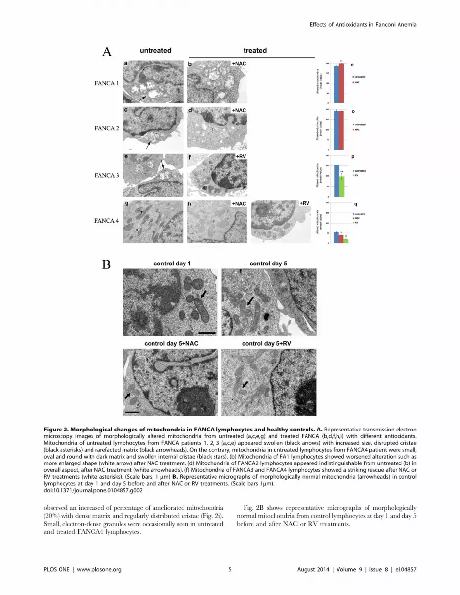
observed an increased of percentage of ameliorated mitochondria
(20%) with dense matrix and regularly distributed cristae (Fig. 2i).
Small, electron-dense granules were occasionally seen in untreated
and treated FANCA4 lymphocytes.
Fig. 2B shows representative micrographs of morphologically
normal mitochondria from control lymphocytes at day 1 and day 5
before and after NAC or RV treatments.
Figure 2. Morphological changes of mitochondria in FANCA lymphocytes and healthy controls. A. Representative transmission electronmicroscopy images of morphologically altered mitochondria from untreated (a,c,e,g) and treated FANCA (b,d,f,h,i) with different antioxidants.Mitochondria of untreated lymphocytes from FANCA patients 1, 2, 3 (a,c,e) appeared swollen (black arrows) with increased size, disrupted cristae(black asterisks) and rarefacted matrix (black arrowheads). On the contrary, mitochondria in untreated lymphocytes from FANCA4 patient were small,oval and round with dark matrix and swollen internal cristae (black stars). (b) Mitochondria of FA1 lymphocytes showed worsened alteration such asmore enlarged shape (white arrow) after NAC treatment. (d) Mitochondria of FANCA2 lymphocytes appeared indistinguishable from untreated (b) inoverall aspect, after NAC treatment (white arrowheads). (f) Mitochondria of FANCA3 and FANCA4 lymphocytes showed a striking rescue after NAC orRV treatments (white asterisks). (Scale bars, 1 mm) B. Representative micrographs of morphologically normal mitochondria (arrowheads) in controllymphocytes at day 1 and day 5 before and after NAC or RV treatments. (Scale bars 1mm).doi:10.1371/journal.pone.0104857.g002
Effects of Antioxidants in Fanconi Anemia
PLOS ONE | www.plosone.org 5 August 2014 | Volume 9 | Issue 8 | e104857

Discussion
FANCA cells display an altered red-ox metabolism [4]. The
possibility to improve this altered physiological conditions with the
employment of antioxidants has been consequently forwarded/
proposed. Here we evaluated the biological effects of two
antioxidants featuring different biochemical mechanisms. While
from the point of view of the biochemistry and the energy
metabolism NAC treatment appears advantageous, the here
reported results concerning cellular morphology suggests attention
especially in the view to employ NAC as therapeutic in FA. On the
contrary RV induces only a partial recovery of the biochemical
parameters, but great improvement in cellular morphology.
Our data suggest that we must be aware of possible pitfalls of
the employment of antioxidants as therapeutics. While short time
exposure (from few hours to 2 days) to NAC appears beneficial in
FA [9,20], upon mere observation of the biochemical parameters,
the effect of NAC appears positive for the FANCA cells. However
when we extend our observation to the cellular ultrastructure,
some cellular defects don’t improve and negative effects are seen,
such as worsening of mitochondrial structure. A possible
explanation for this could paradoxically be the recovery of
electron transport chain functioning by NAC. The pro-oxidant
FA phenotype cells is characterized by alterations in the electron
transport efficiency in the inner mitochondrial membrane with loss
of efficiency of electron transfer from Complex I to Complex III
which can in fact be restored by administration of exogenous
Coenzyme Q [3]. NAC decreases ROS production and normal-
izes mitochondrial respiration. Therefore it is expected that in FA
cells with increased ROS production, oxygen consumption will
increase in the presence of NAC (Fig. 1A). The ability of NAC to
restore Complex I activity is however inherently linked to an
increase in ROS production and therefore in worsening of the
already existent structural damage typical of FACA cells. This is
also suggestive of the existence of a primary damage to
mitochondrial membranes in the FANCA phenotype. The sole
increase in GSH reductive potential in FANCA cells appears
insufficient to restore a fully functional phenotype. NAC was
already reported [21] to be only partially effective in limiting GSH
depletion, particularly when employed in drugs targeted to
mitochondria.While the negative phenotypic outcomes from the
chronic NAC treatment in FANCA cells were almost unexpected
its possible detrimental effects are already known. The forced
increase in intracellular GSH was indeed reported to induce a
paradoxical triggering of mitochondrial oxidative stress [22]. After
either pharmacologic (NAC supplementation) or genetic (gluta-
mate cysteine ligase overexpression) maneuvers an initially more
reducing GSH redox potential or reductive stress was promptly
followed by a pathogenic mitochondrial oxidation. The GSH-
mediated reductive stress culminated with pro-oxidative conse-
quences in mitochondria contrary to the common belief that NAC
functions solely as an antioxidant.
The various lymphocytes here analysed do show various types of
mitochondrial alterations. It has been reported that no apparent
correlation exists between the genotypic defect and the phenotypic
expression [19]. However it also holds true that no molecular
function or metabolic regulation other than DNA repair has been
exploited to date.
The different response to NAC observed in the analysed
samples may be correlated to the different genetic background. It
appears clear, however, that the effect of NAC on the overall cell
population examined is null or negative. In FANCA1 NAC
treatment (fig. 2b) result in mitochondria with greatly enlarged,
disrupted cristae and rarefaction in matrix density where in
FANCA2 (fig. 2d) no detectable changes are seen in mitochondrial
structure; in FANCA4 (fig. 2h) we observe a normal mitochondrial
structure and a decrease in the number of mitochondria in
mitoptoses but with a concomitant increase in the number of
apoptotic cells. It appears peculiar that the level of the apoptotic
cells in the NAC treated sample equals the level of mitoptotic cells
in the untreated. It has been reported that ROS produced by the
respiratory chain inside mitochondria could also induce mito-
chondrial fission as the initial step of mitoptosis [23]. The same
degeneration in the mitochondrial reticulum here reported has
been described by us in FA fibroblasts [2].
RV treatment partially restored the FANCA biochemical
activity but resulted in significant recovery of a normal structure.
It may appear surprising that in the presence of RV mitochondrial
functions like ATP production and oxygen uptake are increased in
FANCA cells (and decreased in wild type cells, as expected). To
explain this apparent contradiction the cited defective phenotype
of Complex I in FANCA cells [3], as well as the results from
Gledhill et al. [8] should be taken into consideration. These
Authors, by structural analysis of ATPase crystallized in the
presence of RV demonstrated that it binds the inside surface of F1
npreventing both the synthetic and hydrolytic activities of ATPase
[8]. In this respect, RV cannot be considered a mere ROS
scavenger, rather its action is exerted also through mild control of
the ATP synthase activity. Being an inhibitor of ATP synthase [8],
RV slows down the electron transport chain activity and NADH
utilization by Complex I which has the result to lower free radical
production. It can be hypothesized that in these conditions aerobic
respiration is more efficient and more directed to ATP production
increasing O2 utilization finalized to H2O and not to ROS
production. RV alone inhibits ATP synthase and decreases
electron transport chain activity producing less free radicals. In
fact, when the ATP synthase is inhibited also the electron transport
chain activity slows, and consequently the oxygen consumption
decreases with a kind of indirect scavenging.
RV was shown to reduce mitochondrial respiration, and
Complex IV activity and to extend lifespan. In fact diminished
mitochondrial OXPHOS is associated with lower ROS produc-
tion and reduced longevity. A beneficial effect of RV in tumors
may derive in part by its preventing mitochondrial ATP synthesis,
thereby inducing apoptosis. RV was shown to decrease tumor cell
viability, an effect that can be reverted by overexpression of Bcl-2
[24]. However, RV may exert other effects, also in dependence of
the model used. For example, treatment of the whole animal
(mice) in vivo with RV causes induction of OXPHOS genes, an
effect likely related to activation of the protein deacetylase, SIRT1
[25].
We have also tested a combination of NAC and RV.
Unfortunately, the double treatment did not restore the correct
OXPHOS activity in FANCA-cells, nor the electron transport
from Complex I to Complex III (data not shown). We may explain
these results considering the single contribution from each
compound: NAC restores the functionality of Complex I, while
RV reduces mitochondrial respiration, and ATP synthesis, by
reversibly inhibiting ATP synthase. In the FANCA case, the
combination of the two has the effect of both blocking
mitochondrial respiration and improving the functioning of
Complex I: this would cause a backlog of electrons, augmenting
ROS production and increasing oxidative damage to the
mitochondrial inner membranes. Literature reports that a mild
uncoupling between electron transport chain and ATP synthase
induces a H+ leak and increases ROS production.
Ultrastructural analysis also showed that the combined treat-
ment of NAC plus RV did not improve mitochondrial alterations
Effects of Antioxidants in Fanconi Anemia
PLOS ONE | www.plosone.org 6 August 2014 | Volume 9 | Issue 8 | e104857

(data not shown). Instead, concomitant treatment with RV and
NAC worsened the mitochondrial phenotype (data not shown). In
this case ATP synthase blockade by RV on one hand and
promotion of electron transfer by NAC on the other produce an
increase in oxidative damage.
In conclusion, both NAC and RV failed to revert the
biochemical and structural phenotype FANCA phenotype. More-
over, their effects are not super imposable. Even though the NAC
treatment did restore the OXPHOS functionality in FA cells, the
phenotype did not improve; by contrast RV exerted a better effect
on FA cell phenotype, although it was not able to revert all the
characteristic of the mutant cells. Therefore, an antioxidant
therapy is likely to be beneficial in several pathological conditions;
however an evaluation of the effects of the single antioxidant
employed should be carefully scrutinized to avoid possible
detrimental consequences. Our knowledge of the proper use and
the effects of any single antioxidant, and for their combined use as
well, is as yet quite limited.
Acknowledgments
The samples were obtained from the ‘‘Cell Line and DNA Biobank from
Patients affected by Genetic Diseases’’ (G. Gaslini Institute) - Telethon
Genetic Biobank Network (Project No. GTB07001). AIRFA, ERG spa,
Cambiaso & Risso, Rimorchiatori Riuniti, Saar Depositi Oleari Portuali,
UC Sampdoria are aknowledged for supporting the activity of the Clinical
& Experimental Hematology Unit of G. Gaslini Institute.
Author Contributions
Conceived and designed the experiments: MC SR GS PC CC IP EC PD.
Performed the experiments: MC SR GS PC CC. Analyzed the data: PD
EC IP. Contributed reagents/materials/analysis tools: IP CC EC PD.
Contributed to the writing of the manuscript: EC PD.
References
1. Li X, Yang Y, Yuan J, Hong P, Freie B et al. (2004) Continuous in vivo infusionof interferon-gamma (IFN-gamma) preferentially reduces myeloid progenitor
numbers and enhances engraftment of syngeneic wild-type cells in Fancc-/-
mice. Blood 104: 1204–1209.2. Capanni C, Bruschi M, Columbaro M, Cuccarolo P, Ravera S et al. (2013)
Changes in vimentin, lamin A/C and mitofilin induce aberrant cell organizationin fibroblasts from Fanconi anemia complementation group A (FA-A) patients.
Biochimie 95: 1838–47.
3. Ravera S, Vaccaro D, Cuccarolo P, Columbaro M, Capanni C et al. (2013)Mitochondrial respiratory chain Complex I defects in Fanconi anemia
complementation group A. Biochimie 95: 1828–1837.4. Cappelli E, Ravera S, Vaccaro D, Cuccarolo P, Bartolucci M et al. (2013)
Mitochondrial respiratory complex I defects in Fanconi anemia. Trends Mol
Med 19: 513–514.5. Dodd S, Dean O, Copolov DL, Malhi GS, Berk M (2008) N-acetylcysteine for
antioxidant therapy: pharmacology and clinical utility. Expert Opinion onBiological Therapy 8: 1955–1962.
6. Dong Z (2003) Molecular mechanism of the chemopreventive effect ofresveratrol. Mutat Res. 523–524: 145–50.
7. Csiszar A (2011) Anti-inflammatory effects of resveratrol: possible role in
prevention of age-related cardiovascular disease. Ann N Y AcadSci 1215: 117–22.
8. Gledhill JR, Montgomery MG, Leslie AG, Walker JE (2007) Mechanism ofinhibition of bovine F1-ATPase by resveratrol and related polyphenols. Proc
Natl Acad Sci U S A. 104: 13632–7.
9. Ponte F, Sousa R, Fernandes AP, Goncalves C, Barbot J et al. (2012)Improvement of genetic stability in lymphocytes from Fanconi anemia patients
through the combined effect of a-lipoic acid and N-acetylcysteine. OrphanetJ Rare Dis 16: 7–28.
10. Zhang QS, Marquez-Loza L, Sheehan AM, Watanabe-Smith K, Eaton L et al.(2014) Evaluation of resveratrol and N-acetylcysteine for cancer chemopreven-
tion in a Fanconi anemia murine model. Pediatr Blood Cancer. 61: 740–742.
11. Roomi MW, Kalinovsky T, Roomi NW, Niedzwiecki A, Rath M (2012) In vitroand in vivo inhibition of human Fanconi anemia-associated head and neck
squamous cell carcinoma by a novel nutrient mixture. Int J Oncol. 41: 1996–2004.
12. Hanenberg H, Batish SD, Pollok KE, Vieten L, Verlander PC et al. (2002)
Phenotypic correction of primary Fanconi anemia T cells with retroviral vectorsas a diagnostic tool. Exp Hematol. 30: 410–20.
13. Ravera S, Panfoli I, Calzia D, Aluigi MG, Bianchini P et al. (2009) Evidence for
aerobic ATP synthesis in isolated myelin vesicles. Int J Biochem Cell Biol.41:
1581–1591.
14. Ravera S, Calzia D, Bianchini P, Diaspro A, Panfoli I (2007) Confocal laser
scanning microscopy of retinal rod outer segment intact disks: new labeling
technique. J Biomed Opt.12: 050501.
15. Bergmeyer HU, Grassl M, Walter HE (1983) Methods of Enzymatic Analysis,
Verlag-Chemie, Weinheim,p. 249.
16. Hong SM, Pedersen PL (2008) ATP Synthase and the Actions of Inhibitors
Utilized To Study Its Roles in Human Health, Disease, and Other Scientific
Areas. Microbiol Mol Biol Rev 72: 590–641.
17. Zheng J, Ramirez VD (2000) Inhibition of mitochondrial proton F0F1-ATPase/
ATP synthase by polyphenolic phytochemicals. Br. J. Pharmacol 130: 1115–
1123.
18. Jangamreddy JR, Los MJ (2012) Mitoptosis, a novel mitochondrial death
mechanism leading predominantly to activation of autophagy. Hepat Mon.
12(8):e6159.
19. Castella M, Pujol R, Callen E, Trujillo JP, Casado JA et al. (2011) Origin,
functional role, and clinical impact of Fanconi anemia FANCA mutations. Blood
117: 3759–69.
20. Kumari U, Ya Jun W, Huat Bay B, Lyakhovich A (2014) Evidence of
mitochondrial dysfunction and impaired ROS detoxifying machinery in Fanconi
Anemia cells. Oncogene 33: 165–172.
21. Cuccarolo P, Viaggi S, Degan P (2012) New insights into redox response
modulation in Fanconi’s anemia cells by hydrogen peroxide and glutathione
depletors. FEBS J. 279: 2479–94.
22. Zhang H, Limphong P, Pieper J, Liu Q, Rodesch CK et al. (2012) Glutathione-
dependent reductive stress triggers mitochondrial oxidation and cytotoxicity.
FASEB J. 26: 1442–51.
23. Pletjushkina OY, Lyamzaev KG, Popova EN, Nepryakhina OK, Ivanova OY
et al. (2006) Effect of oxidative stress on dynamics of mitochondrial reticulum.
BiochimBiophysActa. 1757: 518–24.
24. Low IC, Chen ZX, Pervaiz S. (2010) Bcl-2 modulates resveratrol-induced ROS
production by regulating mitochondrial respiration in tumor cells. Antioxid.
Redox Signal. 13, 807–819.
25. Lagouge M, Argmann C, Gerhart-Hines Z, Meziane H, Lerin C et al. (2006)
Resveratrol Improves Mitochondrial Function and Protects against Metabolic
Disease by Activating SIRT1 and PGC-1a. Cell, 127, 1109–1122.
Effects of Antioxidants in Fanconi Anemia
PLOS ONE | www.plosone.org 7 August 2014 | Volume 9 | Issue 8 | e104857
Copyright © 2022 FDOKUMEN